sclareol reduces cd4+ cd25+ foxp3+ t-reg cells in a breast
TRANSCRIPT

ISSN 1735-1383
Iran. J. Immunol. March 2013, 10 (1), 10-21
Shokoofe Noori, Zuhair Mohammad Hassan, Omid Salehian
Sclareol Reduces CD4+ CD25+ FoxP3+
T-reg Cells in a Breast Cancer Model
in Vivo
Article Type: RECEARCH ARTICLE
The Iranian Journal of Immunology is a quarterly Peer-Reviewed Journal Published by Shiraz Institute for Cancer Research and the
Iranian Society of Immunology & Allergy, Indexed by Several World Indexing Systems Including:
ISI, Index Medicus, Medline and Pubmed
For information on author guidelines and submission visit:
www.iji.ir
For assistance or queries, email:
Iran.J.Immunol. VOL.10 NO.1 March 2013 10
Sclareol Reduces CD4+ CD25+ FoxP3+
T-reg Cells in a Breast Cancer Model
in Vivo
Shokoofe Noori1*, Zuhair Mohammad Hassan2, Omid Salehian3 1Department of Biochemistry, Faculty of Medicine, Shahid Beheshti University of Medical Sciences, 2Department of Immunology, School of Medical Sciences, Tarbiat Modares University, 3Department of Exercise Physiology, Tehran University, Tehran, Iran ABSTRACT Background: Sclareol is a phytochemical used in people's diet in Southeast Asia. Ob-jective: To investigate the immunotherapeutic effectiveness of Sclareol against breast cancer by direct intraperitoneal injection. Methods: Sclareol was isolated and purified from Salvia sclarea. Effect of Sclareol on cell growth inhibition was evaluated by MTT assay. Intraperitoneally injected Sclareol effects on reducing the tumor volume and shifting the cytokine profile were investigated. We also assessed if intraperitoneally in-jected Sclareol could improve the outcome of cancer therapy through suppressing the regulatory T cells. Results: The results confirmed a significant decrease in the tumor size. Furthermore, a significant decrease in the level of IL-4 and an increase in the level of IFN-γ were noticed in the intraperitoneally injected Sclareol group (p<0.05). It was also observed that the splenocytes of treated animals significantly increase in cell prolif-eration assay. Moreover, measurements of splenic T regulatory cell indicated that in-traperitoneally injected Sclareol significantly decreased the number of splenic T regula-tory cell. Conclusion: Our results suggest that Sclareol, by reducing T-reg cells fre-quency and also tumor size can enhance the effect of cancer therapy as an immuno-stimulant. Noori S, et al. Iran J Immunol. 2013; 10(1):10-21
Keywords: IFN-γ, IL-4, Immunomodulator, Sclareol, T Regulatory Cell ---------------------------------------------------------------------------------------------------------*Corresponding author: Dr. Shokoofe Noori, Department of Biochemistry, Faculty of Medicine, Shahid Beheshti University of Medical Sciences, Tehran, Iran, Tel: (+) 98 21 23872570, e-mail: [email protected]

Antitumor and immunomodulatory properties of Sclareol
Iran.J.Immunol. VOL.10 NO.1 March 2013 11
INTRODUCTION Labdane-type diterpenes appear to be a main group of natural products exhibiting inter-esting biological activities. Sclareol as a ditertiary alcohol is a member of the labdane-type diterpenes (1,2,3). Novel strategies for cancer vaccines are highly desirable and new strategies that simul-taneously stimulate cell-mediated immunity against tumors while inhibiting or depleting regulatory T cells are necessary to improve the outcome of cancer immunotherapy (4). The profile of cytokine secretion divides T-helper cells into two subpopulations with different roles: the Th1 subset that secretes interleukin-2 and interferon γ (5,6), and the Th2 subset that produces IL-4 and IL-5 (7) IL-4 and IFN-γ have modulatory effects on macrophages, which are in some cases coincidental and in others opposing. It has also been found that IL-4 inhibits the production of IFN-γ from mononuclear cells (8,9). T-regs inhibit both the development and effector functions of tumor-specific T cells. CD4+CD25high T-regs accumulate at the tumor site (10-13), where they appear to di-rectly suppress cytotoxic T cell responses against the tumors (14,15). CD4+CD25high T-regs are also increased in metastatic lymph nodes and peripheral blood of patients with various types of cancers such as metastatic melanoma (16). We previously reported that the intratumorally injected Sclareol has anti-tumor activi-ties and also modulates the immune response reduces regulatory T cell and inhibits tu-mor growth in vivo (17). In the present study, as an aim, we emphasized on the direct correlation between the tumor sizes and immune response through affecting the fre-quency of T regulatory cells. The production of cytokines was also investigated as an indicator of effects of Sclareol when injected intraperitoneally. MATERIALS AND METHODS Isolation and Purification of Sclareol. One thousand grams of Salvia sclarea was col-lected from the North East of Iran. Dry plant was extracted using a mixture of n- hex-ane/ethyl acetate/methanol (1:1:1) and kept for 24 h in the room temperature. The ex-tract was then passed through a Wattman filter paper and the filtrate was evaporated at 45°C for 2 h by a vacuum rotary evaporator, and kept in freezer. The concentrated fil-trate was run through a silica gel column chromatography at different solvent polarities starting from a non-polar solvent (n-hexane) to a medium polarity (n-hexane/ ethyl ace-tate) and ending with ethyl acetate alone. Subsequently, mixtures of different concentra-tions of n-hexane/ ethyl acetate/methanol, with the increasing concentration of methanol towards the higher polarity, were used. Fractions were collected from these processes and the purity of components in each fraction was evaluated by Thin Layer Chromatog-raphy (TLC). Fractions 23-39 showed a single band on TLC. Then, Sclareol was identi-fied at the 500 MHz 1H-NMR spectra, using CDCl3 as the solvent. Sclareol was tested for endotoxin contamination before used. Limulus Amebocyte Lysate (LAL) Test, which is a quantitative test for gram-negative bacterial endotoxin, was used. The sample was mixed with the LAL supplied in the test kit and incubated at 37°C (±1°C) for 10 minutes. A substrate solution was then mixed with the LAL-sample and incubated at 37°C (±1°C) for an additional 6 minutes. The reaction was stopped with stop reagent. If endotoxin was present in the sample, a yellow color would develop. The absorbance of

Noori S, et al
Iran.J.Immunol. VOL.10 NO.1 March 2013 12
the sample was determined by spectrophotometry at 405-410 nm. The concentration of endotoxin can be calculated from a standard curve. Mice. Twenty Inbred female Balb/c mice, aged from six to eight weeks, were obtained from Pasteur Institute of Iran. Given free access to food and water, the mice were housed for one week and maintained under standard conditions prior to experimenta-tion. All the experiments were done according to the Animal Care and Use Protocol of Tarbiat Modares University (Tehran, Iran). All animals received humane care in com-pliance with the Guide for the Care and Use of Laboratory Animals (DHEW Publica-tion No. (NIH) 85-23, Revised 1985, Office of Science and Health Reports, DRR/NIH, Bethesda, MD 20205). Cell Culture. Erythroleukemic cell line (k562) was purchased from Pasteur Institute, Tehran, Iran. In this study, the cells were cultured in RPMI-1640 medium (GIBCO/BRL) supplemented with 10% FBS (GIBCO/BRL), 100 unit/ml penicillin/100 mg/ml streptomycin and 1% L-glutamine (GIBCO/BRL). All cells were grown in a hu-midified atmosphere containing 5% CO2 at 37°C. Human peripheral venous blood was obtained from healthy volunteers (aged 25-50 yr). Blood samples were obtained by venipuncture of the vein and blood was collected in sterile EDTA containing tubes. PBMCs were then obtained by centrifugation over 1.077 g/ml Ficoll-Hypaque gradient (Sigma, UK). Cells were washed in saline and finally resuspended in RPMI 1640 me-dium and cultured (5×10⁴ cells/ml) in 96-well flat-bottom culture plates in medium sup-plemented with different doses of Sclareol, 25 mm HEPES, 10% fetal calf serum (FCS), 2 mm L-glutamine, 100 U/ml penicillin, and 100 μg/ml streptomycin (Sigma-Aldrich) and the cell viability analysis was measured. Cell Viability Analysis. K562 Cell viability was studied by the MTT (3-(4, 5-methylthiazol-2-yl)-2, 5-diphenyl-tetrazolium bromide) viability assay. Briefly, growing cells were re-cultured (5000 cells/well) overnight in 96-well tissue culture plates. Dif-ferent doses of Sclareol were added to the respective wells, after 24 h, 48 h, and 72 h. Also, 20 µL MTT (Sigma-Aldrich, USA) was directly added to the medium of each well with a final concentration of 2 mg/ml. After 4 h of incubation, the medium contain-ing MTT was discarded and 100 µL DMSO (Sigma-Aldrich, USA) was added. Their relative cell viability was measured by scanning with an ELISA reader using a 570 nm filter. Antigen Preparation. Tumors were allowed to attain a volume of approximately 3000 mm3. Tumor from the breast cancer bearing Balb/c mice was excised and cell extract was prepared by making a suspension of tumor mass, passing through mesh, and subse-quently freezing/thawing. Tumor cell suspension was sonicated with power of 4 Watts for 30 seconds followed by 20-second incubation for three times, and finally the extract was filtered. Phenylmethylsulfonyl floride (PMSF) (1 mM) as inhibitor of serine prote-ases was added to the cell lysates to inactivate proteinases. Concentration of the extract was determined using the Bradford method and it was stored frozen at -20C until being used. Spontaneous Mouse Mammary Tumor Induction. Spontaneous mouse mammary tumor (SMMT) was developed in mice (18). The pathological report indicated that, it was composed of malignant cells with hyperchromatic nuclei and prominent nucleoli. Nuclear pleomorphism and mitotic figures are also observed in mitosis to vary from low to moderate frequency. Infiltration of tumor cells in the surrounding tissues and nests of carcinoma cells are seen; pathologically, it is similar to the invasive ductal carcinoma of human subjects.

Antitumor and immunomodulatory properties of Sclareol
Iran.J.Immunol. VOL.10 NO.1 March 2013 13
Tumor Transplantation and Planning of the in vivo Experiment. In this study, SMMT tissues were separated from the breast cancer bearing Balb/c mice and cut into pieces of sizes less than 0.5 cm3 by forceps and scalpel. Seventeen days later when the size of each tumor reached about 1500 mm3, the treatment with optimal dose of Sclareol according to the results previously obtained from the DTH test was started (17). After the appearance of tumor tissues, the mice were divided into three groups of five ani-mals. Experimental groups were injected intraperitoneally with 7.85 µg/mouse/day Sclareol and 20 mg/kg cyclophosphamid daily in a total volume of 0.1 ml of PBS. Tu-mor-bearing mice were treated for 19 consecutive days. The tumor volume was meas-ured in three groups using a digital caliper and calculated by the following formula: V= 1/2 × LW2 where V denotes volume, L represents length, and W shows width. Separation of Splenic Mononuclear Cells (MNCs). The control and treated tumor-bearing mice were sacrificed on the 6 th day; spleen was removed under sterile condi-tions and was suspended in PBS. The splenic cell suspension was RBC-lysed with a so-lution of 0.75% NH4Cl and Tris buffer (0.02%) (PH=7.4). The cells were washed and the single-cell suspension was prepared in RPMI 1640 containing stable glutamine (Cy-togen) and 10% heat inactivated fetal calf serum (Gibco, England). To define the viabil-ity and density of cells in the suspension, Trypan blue was used. The cells were counted with a homocytometer light microscopy. The viability of splenocytes was generally above 95%. After washing, the suspension was adjusted to 4×10⁶ cells/ml in RPMI 1640 supplemented with 10% FCS, 100 μg/ml streptomycin, and 100 IU/ml penicillin (complete RPMI), and kept at 4°C. Measurement of Cytokines by ELISA. Isolated splenic MNCs were cultured in 24 well plates as mentioned above. To stimulate the immune cells lysate antigens were added at 5µg/ml final concentrations. The mixture was then incubated for 72 h at 37ºC and 5% CO2. The supernatants were collected and frozen at -70C, till performing the cytokine assay. IFN-γ and IL-4 concentrations were measured using R&D DuoSet ELISA Development kit according to the manufacturers’ protocols. Splenocyte Proliferation Index. The animals of both test and control groups were killed by dislocation and their spleens were removed. Splenocytes were isolated using the needle perfusion method and sterile RPMI-1640. Erythrocytes were lysed at room temperature using the ACK lysis buffer (NH4Cl, KHCO3, and Na2EDTA). Density of the cells in suspension was counted with a homocytometer light microscopy and the vi-ability test was carried out using the trypan-blue dye exclusion method. RPMI-1640 supplemented with 10% heat-inactivated FBS, 100µg/ml streptomycin, 100 U/ml peni-cillin, 2 mM L-glutamine, and 25 mM HEPES, without phenol red indicator, were used. The plates were incubated for 36 h at 37°C in a humidified 5% CO2 atmosphere. Cell proliferation was defined with Bromodeoxyuridine (BrdU) labeling solution. The uptake of BrdU was detected using the cell proliferation BrdU kit (Roche Diagnostic GmbH, Mannheim, Germany) and expressed as the stimulation index (S.I.): t-test group, (PHA) P-positive control, N-negative control.
S.I.= [(T-N)/(P-N)]×100
Noori S, et al
Iran.J.Immunol. VOL.10 NO.1 March 2013 14
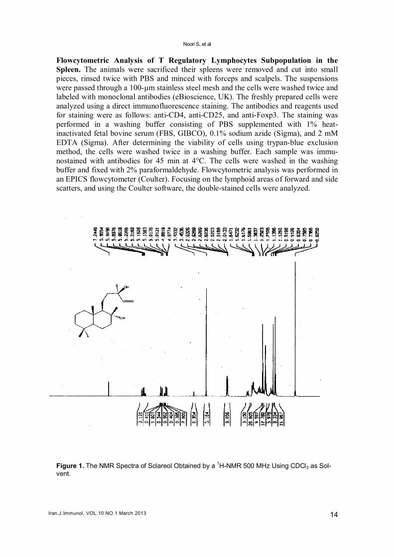
Flowcytometric Analysis of T Regulatory Lymphocytes Subpopulation in the Spleen. The animals were sacrificed their spleens were removed and cut into small pieces, rinsed twice with PBS and minced with forceps and scalpels. The suspensions were passed through a 100-μm stainless steel mesh and the cells were washed twice and labeled with monoclonal antibodies (eBioscience, UK). The freshly prepared cells were analyzed using a direct immunofluorescence staining. The antibodies and reagents used for staining were as follows: anti-CD4, anti-CD25, and anti-Foxp3. The staining was performed in a washing buffer consisting of PBS supplemented with 1% heat-inactivated fetal bovine serum (FBS, GIBCO), 0.1% sodium azide (Sigma), and 2 mM EDTA (Sigma). After determining the viability of cells using trypan-blue exclusion method, the cells were washed twice in a washing buffer. Each sample was immu-nostained with antibodies for 45 min at 4°C. The cells were washed in the washing buffer and fixed with 2% paraformaldehyde. Flowcytometric analysis was performed in an EPICS flowcytometer (Coulter). Focusing on the lymphoid areas of forward and side scatters, and using the Coulter software, the double-stained cells were analyzed.
Figure 1. The NMR Spectra of Sclareol Obtained by a
1H-NMR 500 MHz Using CDCl3 as Sol-vent.

Antitumor and immunomodulatory properties of Sclareol
Iran.J.Immunol. VOL.10 NO.1 March 2013 15
Statistical Analysis. The normality of the data and homogeneity of variances were tested with K-S and Levene's statistical tests, respectively. Then, one way analysis of variance (ANOVA) test was used to determine the statistical significance (p<0.05) be-tween values of test and control groups. Data were analyzed using SPSS software ver-sion 16 and results were expressed as the mean ± standard error (mean ± SE). RESULTS Characterization of the Isolated Molecules Using 1H-NMR Spectroscopy. The puri-fied material was characterized by 1H-NMR spectroscopy shown in Figure 1. Measurement of Cell Growth. To measure the effect of Sclareol on the tumor and normal cells, different doses of Sclareol were used in vitro. The results indicated that, Sclareol reduced the cell growth of k562 cell line in a dose and time-dependent manner by the MTT assay. The obtained data showed that, Sclareol was effective in growth in-hibition of k562 cells. Sclareol had no cytotoxic effects on the growth of peripheral blood lymphocytes (Figure 2).
Figure 2. Cytotoxicity effect of Sclareol against of K562 cell line. Cells were incubated with in-creasing concentrations (35 µM-75 µM) of Sclareol for 24 h, 48 h and 72 h. Cell viability was quantified by MTT assay. Bars represent the mean ± SEM of three independent experiments performed in triplicate, *p<0.05, **p<0.01, ***p<0.001 represent significant differences compared to control values, whereby control was set as 100%.

Noori S, et al
Iran.J.Immunol. VOL.10 NO.1 March 2013 16
The Effect of Intraperitoneally Injected Sclareol on Tumor Volume. In order to evaluate the effect of Sclareol on the tumor volumes of animals bearing breast cancer, 7.85 µg/mouse/day of Sclareol was injected intraperitoneally daily to each mouse and its tumor size was measured every day. The Sclareol-treated animals showed significant (p<0.05) decrease in the rate of tumor growth compared to control group, as is observed in Figure 3. Figure3. Tumour volume measurement in 19 consecutive-day treatment by 7.85 µg/mouse/day of Sclareol intraperitoneally injectied comparing to mice injected with control. The average rate of tumor growth in the animals treated with Sclareol was significantly (p<0.05) decreased com-pared to control animals.
The Effect of Intraperitoneally-Injected Sclareol on Shifting of IFN-γ and IL-4. In order to assess the Th1/Th2 cytokine shift in the tumor bearing animals, 15 animals in three groups were used; cyclophosphamid treated, Sclareol treated with 7.85 µg/mouse/day intraperitoneally and untreated. Animals were sacrificed and their splenocytes were obtained. Splenocytes wee re-stimulated with lysate antigens and the levels of IFN-γ and IL-4 production were assessed. The splenocytes of Sclareol-treated group showed a significant increase in the level of IFN-γ compared with control group. In addition, a significant decrease was noticed in the level of IL-4 production in the Sclareol-treated group, compared to control group (Figure 4). The Effect of Intraperitoneally Injected Sclareol on the Lymphocyte Proliferation Index. The data of the proliferation assays are shown in Figure 5. The data reflect the mean values of triplicates after stimulation with specific tumor antigen, using 1% FCS and 10×105 cells/ml. Comparing the results of both groups, the data indicated that, the proliferative responses of Sclareol-treated mice were significantly increased compared to control group.

Antitumor and immunomodulatory properties of Sclareol
Iran.J.Immunol. VOL.10 NO.1 March 2013 17
Figure 4. The level IFN-γ and IL-4 responses were measured in control, Sclareol and cyclo-phosphamid treated groups. The level of responses was determined by a R&D ELISA kit. The level of IFN-γ showed a significant increase in the Sclareol injected mice compared with control group, while IL-4 showed a significant decrease. Three independent experiments performed in duplicate. *p<0.05 was defined as the significant difference.
The Effect of Intraperitoneally-Injected Sclareol on the Level of T Regulatory Cells. The effect of intraperitoneal administration of Sclareol on the splenic T regula-tory lymphocytes (CD4+CD25+Foxp3+) in the animals bearing tumor was investigated using eBioscience monoclonal antibody kit and analyzed by EPICS flowcytometry. The results, shown in Figure 6, demonstrated a significant decrease in the frequency of spleen CD4+CD25+Foxp3+ T lymphocytes in the Sclareol-treated group, compared to control group.
5a 5b

Noori S, et al
Iran.J.Immunol. VOL.10 NO.1 March 2013 18
Figure 5. Splenocytes of Scalerol intraperitoneally injected mice were cultured in vitro and stimulated with lysate antigen. The proliferation response of the mice treated with Scalerol was enhanced. Lymphocyte proliferation response was shown as the stimulation index. *p<0.05 was defined as the significant difference.
Figure 6. Percentage of splenic CD4+ CD25+ FoxP3+ T cells in control, Sclareol and cyclo-phosphamid treated groups. The difference in splenic CD4+ CD25+ FoxP3+ T cell between ex-perimental groups and control was statistically significant (p<0.05). p<0.05 represents significant difference compared with control values.
DISCUSSION As a very valuable source for novel chemotherapeutic agents, natural plant compounds exhibit effective antitumour activities with a wide range of mechanisms. Despite alter-native approaches to the discovery of novel therapeutic agents, natural products have remained one of the best reservoirs of new molecules (19). For example Sclareol is a phytochemical used in perfumery and as a flavoring agent in people's diets in China and

Antitumor and immunomodulatory properties of Sclareol
Iran.J.Immunol. VOL.10 NO.1 March 2013 19
the Mediterranean area. Sclareol is generally reported to be inactive and non-toxic (20). However, this compound has demonstrated the ability to induce apoptosis in leukemic cell lines in a dose and time-dependent manner (21). There are increasing experimental and epidemiological evidences showing that plant-derived chemicals (phytochemicals) possess the potential to serve as chemo-preventive agents that arrest or reverse carcino-genesis (21-23). In this regard, scientists have previously provided experimental find-ings to demonstrate that the phytochemical diterpene, Sclareol, exhibits anticancer ac-tivities in vitro (24,25) and in vivo (26). Our laboratory has been focusing on the natural plant compounds which exhibit effec-tive antitumor activities. Whether the observed antitumor activity is related to the im-mune simulative activity (27-30). Our study was carried out in following steps: First, we purified Sclareol from the Salvia sclarea and characterized it by 1H-NMR spectroscopy. Second, we evaluated its effects on in vitro growth inhibition of tumor cell line by MTT assay. The data showed that Sclareol was effective in inhibition of K562 cell line growth, but did not affect normal lymphocytes growth. Third, as we initially proposed, Sclareol was involved in the en-hancement of antitumor immune responses through the modulation of cytokine produc-tion from the primed immune cells; we tested the effect of Sclareol on production of IFN-γ and IL-4. We observed that, the splenocytes of treated animals showed a signifi-cant increase in the level of IFN-γ production compared to control group (Figure 4). In addition, a significant decrease of IL-4 was noticed in the Sclareol-treated group. There-fore, it is suggested that Sclareol was responsible for the immunomodulatory activity and shift of cytokine pattern towards Th1 (IFN-γ) in Balb/c mice. In summary, this study confirms that Sclareol is able to reduce tumor size in a murine model through in-creasing the IFN-γ and decreasing the IL-4 levels and augmenting the Th1 immune re-sponse in an antigen-specific manner. Fourth, we also proposed that Sclareol may in-crease the proliferation of lymphocytes and enhance T-lymphocyte function. The results indicate that, Sclareol could augment the lymphocytes proliferation compared to control group. In addition, the measurement of splenic CD4+CD25+Foxp3 T lymphocytes indicated that, Sclareol significantly decreases the number of T-reg lym-phocytes compared to untreated control group (Figure 6). In the context of tumor im-munology, different studies have mentioned a suppressor role for CD4+CD25+Foxp3+ T cells against anti-tumor immunity. It has been observed that, either removing the regula-tory T cells or blocking the immunoregulatory pathway induced by regulatory T cells might improve the efficacy of tumor vaccines or the immunotherapy of cancer. Fur-thermore, the depletion of CD4+CD25+ T cells has enhanced the natural tumor immune-surveillance and induced the rejection of multiple immunogenic tumors in multiple strains of mice (31,33). In summary, Sclareol can inhibit the growth of tumor but not that of the normal cells in vitro. Sclareol has low toxicity and slows down the tumor growth in vivo and diverts the immune response towards Th1 by increasing the level of IFN-γ and decreasing the level of IL-4 as well as modulating the T regulatory cells. These parameters make Sclareol a candidate to be used as chemoimmunotherapy of cancer, while further study is required to confirm our proposal.

Noori S, et al
Iran.J.Immunol. VOL.10 NO.1 March 2013 20
ACKNOWLEDGEMENTS The financial support by the Research Council of Shahid Beheshti University of Medi-cal Sciences is gratefully acknowledged. REFERENCES
1 Hatziantoniou S, Dimas K, Georgopoulos A, Sotiriadou N, Demetzos C. Cytotoxic and antitumor activity of liposomeincorporated sclareol against cancer cell lines and human colon cancer xeno-grafts. Pharmacol Res. 2006; 53:80-7.
2 Dimas K, Kokkinopoulos D, Demetzos C, Vaos B, Marselos M, Malamas M, et al. The effect of sclareol on growth and cell cycle progression of human leukemic cell lines. Leuk Res. 1999; 23:217-34.
3 Dimas K, Demetzos C, Vaos V, Ioannidis P, Trangas T. Trangas T Labdane type diterpenes down-regulate the expression of c-Myc protein but not of Bcl-2, in human leukemia T-cell lines undergo-ing apoptosis. Leuk Res. 2001; 25:449-54.
4 Tamura Y, Peng P, Liu K, Daou M, Srivastava PK. Immunotherapy of tumors with autologous tumor-derived heat shock protein preparations. Science. 1997; 278:117-20.
5 Cherwinski HM, Schumacher JH, Brown KD, Mosmann TR. Two types of mouse helper T cell clone. III, Further differences in lymphokine synthesis between Th1 and Th2 clones revealed by RNA hybridization, functionally monospecific bioassays, and monoclonal antibodies. J exp Med. 1987; 166:1229-44.
6 Mosmann TR, Cherwinski H, Bond MW, Giedlin MA, Coffman RL. Two types of murine helper T cell clon. I. Definition according to profiles of lymphokine activities and secreted proteins. J Immunol. 1986; 136:2348-62.
7 Mosmann TR, Coffman RL. Th1 and Th2 cells: different patterns of lymphokine secretion lead to different functional properties. Annu Rev Immunol. 1989; 7:145-58.
8 Sakaguchi S. Naturally arising Foxp3-expressing CD25+CD4+ regulatory T cells in immunological tolerance to self and non-self. Nat Immunol. 2005; 6:345-52.
9 Mills KH, McGuirk P. Antigen-specific regulatory T cells--their induction and role in infection. Semin Immunol. 2004; 16:107-17.
10 Sakaguchi S. Naturally arising CD4+regulatory T cells for immunologic self-tolerance and nega-tive control of immune responses. Annu Rev Immunol. 2004; 22:531-62.
11 Vigouroux S, Yvon E, Biagi E, Brenner MK. Antigen-induced regulatory T cells. Blood. 2004; 104:26-33.
12 Curiel TJ, Coukos G, Zou L, Alvarez X, Cheng P, Mottram P. Specific recruitment of regulatory T cells in ovarian carcinoma fosters immune privilege and predicts reduced survival. Nat Med. 2004; 10:942-9.
13 Woo EY, Chu CS, Goletz TJ, Schlienger K, Yeh H, Coukos G, et al.Regulatory CD4+CD25+ T cells in tumors from patients with early-stage non-small cell lung cancer and late-stage ovarian cancer. Cancer Res. 2001; 61:4766-72.
14 Yu P, Lee Y, Liu W, Krausz T, Chong A, Schreiber H, et al. Intratumor depletion of CD4cells unmasks tumor immunogenicity leading to the rejection of late-stage tumors. J Exp Med. 2005; 201:779-91.
15 Shimizu J, Yamazaki S, Sakaguchi S. Induction of tumor immunity by removing CD25+CD4+ T cells: A common basis between tumor immunity and autoimmunity. J Immunol. 1999; 163:5211-8.
16 Golgher D, Jones E, Powrie F, Elliott T, Gallimore A. Depletion of CD25+ regulatory cells uncov-ers immune responses to shared murine tumor rejection ntigens. Eur J Immunol. 2002; 32:3267-75.
17 Noori S, Hassan ZM, Mohammadi M, Habibi Z, Sohrabi N, Bayanolhagh S. Sclareol modulates the Treg intra-tumoral infiltrated cell and inhibits tumor growth in vivo. Cellular Immunology. 2010; 263:148-53.
18 Hassan ZM, Yaraee R, Zare N, Ghazanfari T, Sarraf Nejad AH, Nazori B. Immunomodulatory af-fect of R10 fraction of garlic extract on natural killer activity. Int Immunopharmacol. 2003; 3:1483-9.
Antitumor and immunomodulatory properties of Sclareol
Iran.J.Immunol. VOL.10 NO.1 March 2013 21
19 Kinghorn AD, Farnsworth NR, Soejarto DD, Cordell GA, Pezzuto JM, Udeani GO, et al. Novel strategies for the discovery of plant-derived anticancer agents. Pure Appl Chem. 1999; 71:1611-8.
20 Jung M, Ko I, Lee S, Choi SJ, Youn BH, Kim SK. A concise synthesis and in vitro cytotoxicity of new labdane diterpenes. Bioorg Med Chem Lett. 1998; 8:3295-8.
21 Dimas K, Demetzos C, Mitakou S, Vaos B, Marselos M, Tzavaras T, et al. Cytotoxic activity and antiproliferative effects of a new semi-synthetic derivative ent-3â-OH-manoyl oxide, on human leukemic cell lines. Anticancer Res. 1999; 19:4065-72.
22 Singh RP, Agarwal R. Natural flavonoids targeting deregulated cell cycle progression in cancer cells. Curr Drug Targets. 2006; 7:345-54.
23 D’Incalci M, Steward WP, Gescher AJ. Use of cancer chemopreventive phytochemicals as anti-neoplastic agents. Lancet Oncol. 2005; 6:899-904.
24 Nishino H, Murakoshi M, Mou XY, Wada S, Masuda M, Ohsaka Y, et al. Cancer prevention by phytochemicals. Oncology. 2005; 69:38-40.
25 Li WX, Cui CB, Cai B, Yaor XS. Labdane-type diterpenes as new cell cycle inhibitors and apop-tosis inducers from Vitex trifolia L. J Asian Nat Prod Res. 2005; 7:95-105.
26 Dimas K, Papadaki M, Tsimplouli C, Hatziantoniou S, Alevizopoulos K, Pantazis P, Demetzos C. The proteome profile of two cell lines and their xenografts isolated from a patient with clear cell sarcoma (soft tissue melanoma). in vivo. Oncology 2009; 2323:63-8.
27 Hatziantoniou S, Dimas K, Georgopoulos A, Sotiriadou N, Demetzos C. Cytotoxic and antitumor activity of liposomeincorporated Sclareol against cancer cell lines and human colon cancer xeno-grafts. Pharmacol Res. 2006; 53:80-7.
28 Noori S; Taghikhani M; Hassan ZM, Allameh A, Mostafaei A. Tehranolide could shift the im-mune response towards Th1 and modulate the intra-tumor infiltrated T regulatory cells. Iran J Im-munol. 2009; 6:216-24.
29 Noori S, Taghikhani M, Hassan ZM, Allameha A, Mostafaei A. Tehranolide molecule modulates the immune response, reduces regulatory T cell and inhibits tumor growth in vivo. Mol. Immunol. 2010; 47:1579-84.
30 Langroudi L, Hassan ZM, Ebtekar M, Mahdavi M, Pakravan N, Noori S. A comparison of low-dose cyclophosphamide treatment with artemisinin treatment in reducing the number of regulatory T cells in murine breast cancer model. Int Immunopharmacol. 2010; 10:1055-61.
31 Noori S, Hassan ZM. Dihydroartemisinin shift the immune response towards Th1, inhibit the tu-mor growth in vitro and in vivo. Cell Immunol. 2011; 271:67-72.
32 Farsam V, Hassan ZM ,Zavaran-hosseini A ,Noori S, Mahdavi M, Ranjbar M. Antitumor and im-munomodulatory properties of artemether and its ability to reduce CD4+ CD25+ FoxP3+ T reg cells in vivo. Int Immunopharmacol. 2011; 11:1802-8.
33 Onizuka S, Tawara I, Shimizu J, Sakaguchi S, Fujita T, Nakayama E. Tumor rejection by in vivo administration of anti-CD25 (interleukin-2 receptor alpha) monoclonal antibody. Cancer Res. 1999; 31:28-33.