schwartz's principles of surgery, 9e_2-app
TRANSCRIPT
Print | Close Window
Note: Large images and tables on this page may necessitate printing in landscape mode.
Schwartz's Principles of Surgery > Part II. Specific Considerations > Chapter 30. The Appendix >
KEY POINTS
1. Appendectomy for appendicitis is the most commonly performed emergency operation in the world.
2. Despite the increased use of ultrasonography, computed tomographic scanning, and laparoscopy, the rate of misdiagnosis
of appendicitis has remained constant (15.3%), as has the rate of appendiceal rupture. The percentage of misdiagnosed
cases of appendicitis is significantly higher among women than among men.
3. Appendicitis is a polymicrobial infection, with some series reporting up to 14 different organisms cultured in patients with
perforation. The principal organisms seen in the normal appendix, in acute appendicitis, and in perforated appendicitis are
Escherichia coli and Bacteroides fragilis.
4. Antibiotic prophylaxis is effective in the prevention of postoperative wound infection and intra-abdominal abscess.
Antibiotic coverage is limited to 24 to 48 hours in cases of nonperforated appendicitis. For perforated appendicitis, 7 to 10
days of treatment is recommended.
5. Compared with younger patients, elderly patients with appendicitis often pose a more difficult diagnostic problem because
of the atypical presentation, expanded differential diagnosis, and communication difficulty. These factors contribute to the
disproportionately high perforation rate seen in the elderly.
6. The overall incidence of fetal loss after appendectomy is 4% and the risk of early delivery is 7%. Rates of fetal loss are
considerably higher in women with complex appendicitis than in those with negative appendectomy and those with simple
appendicitis. Removing a normal appendix is associated with a 4% risk of fetal loss and 10% risk of early delivery.
7. Recent data on appendiceal malignancies from the Surveillance, Epidemiology, and End Results program identified
mucinous adenocarcinoma as the most frequent histologic diagnosis, followed by adenocarcinoma, carcinoid, goblet cell
carcinoma, and signet-ring cell carcinoma.
ANATOMY AND FUNCTION
The appendix first becomes visible in the eighth week of embryologic development as a protuberance off the terminal portion
of the cecum. During both antenatal and postnatal development, the growth rate of the cecum exceeds that of the appendix,
so that the appendix is displaced medially toward the ileocecal valve. The relationship of the base of the appendix to the
cecum remains constant, whereas the tip can be found in a retrocecal, pelvic, subcecal, preileal, or right pericolic position
(Fig. 30-1). These anatomic considerations have significant clinical importance in the context of acute appendicitis. The three
taeniae coli converge at the junction of the cecum with the appendix and can be a useful landmark to identify the appendix.
The appendix can vary in length from <1 cm to >30 cm; most appendices are 6 to 9 cm long. Appendiceal absence,
duplication, and diverticula have all been described.1–4
Fig. 30-1.

Various anatomic positions of the vermiform appendix.
For many years, the appendix was erroneously viewed as a vestigial organ with no known function. It is now well recognized
that the appendix is an immunologic organ that actively participates in the secretion of immunoglobulins, particularly
immunoglobulin A. Although there is no clear role for the appendix in the development of human disease, recent studies
demonstrate a potential correlation between appendectomy and the development of inflammatory bowel disease. There
appears to be a negative age-related association between prior appendectomy and subsequent development of ulcerative
colitis. In addition, comparative analysis clearly shows that prior appendectomy is associated with a more benign phenotype
in ulcerative colitis and a delay in onset of disease. The association between Crohn's disease and appendectomy is less clear.
Although earlier studies suggested that appendectomy increases the risk of developing Crohn's disease, more recent studies
that carefully assessed the timing of appendectomy in relation to the onset of Crohn's disease demonstrated a negative
correlation. These data suggest that appendectomy may protect against the subsequent development of inflammatory bowel
disease; however, the mechanism is unclear.4
Lymphoid tissue first appears in the appendix approximately 2 weeks after birth. The amount of lymphoid tissue increases
throughout puberty, remains steady for the next decade, and then begins a steady decrease with age. After the age of 60
years, virtually no lymphoid tissue remains within the appendix, and complete obliteration of the appendiceal lumen is
common.1–4
ACUTE APPENDICITIS
Historical Background
Although ancient texts have scattered descriptions of surgery being undertaken for ailments sounding like appendicitis, credit
for performing the first appendectomy goes to Claudius Amyand, a surgeon at St. George's Hospital in London and Sergeant
Surgeon to Queen Ann, King George I, and King George II. In 1736, he operated on an 11-year-old boy with a scrotal hernia
and a fecal fistula. Within the hernial sac, Amyand found the appendix perforated by a pin. He successfully removed the
appendix and repaired the hernia.5
The appendix was not identified as an organ capable of causing disease until the nineteenth century. In 1824, Louyer-
Villermay presented a paper before the Royal Academy of Medicine in Paris. He reported on two autopsy cases of appendicitis
and emphasized the importance of the condition. In 1827, François Melier, a French physician, expounded on Louyer-
Villermay's work. He reported six autopsy cases and was the first to suggest the antemortem recognition of appendicitis.5
This work was discounted by many physicians of the era, including Baron Guillaume Dupuytren. Dupuytren believed that
inflammation of the cecum was the main cause of pathology of the right lower quadrant. The term typhlitis or perityphlitis
was used to describe right lower quadrant inflammation. In 1839, a textbook authored by Bright and Addison entitled
Elements of Practical Medicine described the symptoms of appendicitis and identified the primary cause of inflammatory
processes of the right lower quadrant.6 Reginald Fitz, a professor of pathologic anatomy at Harvard, is credited with coining
the term appendicitis. His landmark paper definitively identified the appendix as the primary cause of right lower quadrant
inflammation.7
Initial surgical therapy for appendicitis was primarily designed to drain right lower quadrant abscesses that occurred
secondary to appendiceal perforation. It appears that the first surgical treatment for appendicitis or perityphlitis without
abscess was carried out by Hancock in 1848. He incised the peritoneum and drained the right lower quadrant without
removing the appendix. The first published account of appendectomy for appendicitis was by Krönlein in 1886. However, this
patient died 2 days after operation. Fergus, in Canada, performed the first elective appendectomy in 1883.5
The greatest contributor to the advancement in the treatment of appendicitis was Charles McBurney. In 1889, he published
his landmark paper in the New York State Medical Journal describing the indications for early laparotomy for the treatment of
appendicitis. It is in this paper that he described the McBurney point as follows: "maximum tenderness, when one examines
with the fingertips is, in adults, one half to two inches inside the right anterior spinous process of the ilium on a line drawn to
the umbilicus."8 McBurney subsequently published a paper in 1894 describing the incision that bears his name.9 However,
McBurney later credited McArthur with first describing this incision. Semm is widely credited with performing the first
successful laparoscopic appendectomy in 1982.10
The surgical treatment of appendicitis is one of the great public health advances of the last 150 years. Appendectomy for
appendicitis is the most commonly performed emergency operation in the world. Appendicitis is a disease of the young, with
40% of cases occurring in patients between the ages of 10 and 29 years.11 In 1886, Fitz reported the associated mortality
rate of appendicitis to be at least 67% without surgical therapy.7 Currently, the mortality rate for acute appendicitis with
treatment is reported to be <1%.12
Incidence
The lifetime rate of appendectomy is 12% for men and 25% for women, with approximately 7% of all people undergoing
appendectomy for acute appendicitis during their lifetime. Over the 10-year period from 1987 to 1997, the overall
appendectomy rate decreased in parallel with a decrease in incidental appendectomy.11,13 However, the rate of
appendectomy for appendicitis has remained constant at 10 per 10,000 patients per year.14 Appendicitis is most frequently
seen in patients in their second through fourth decades of life, with a mean age of 31.3 years and a median age of 22 years.
There is a slight male:female predominance (1.2 to 1.3:1).11,13
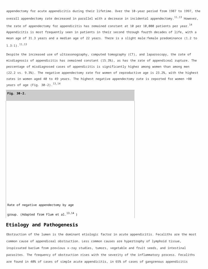
Despite the increased use of ultrasonography, computed tomography (CT), and laparoscopy, the rate of misdiagnosis of
appendicitis has remained constant (15.3%), as has the rate of appendiceal rupture. The percentage of misdiagnosed cases
of appendicitis is significantly higher among women than among men (22.2 vs. 9.3%). The negative appendectomy rate for
women of reproductive age is 23.2%, with the highest rates in women aged 40 to 49 years. The highest negative
appendectomy rate is reported for women >80 years of age (Fig. 30-2).13,14
Fig. 30-2.
Rate of negative appendectomy by age group.
(Adapted from Flum et al.13,14 )
Etiology and Pathogenesis
Obstruction of the lumen is the dominant etiologic factor in acute appendicitis. Fecaliths are the most common cause of
appendiceal obstruction. Less common causes are hypertrophy of lymphoid tissue, inspissated barium from previous x-ray
studies, tumors, vegetable and fruit seeds, and intestinal parasites. The frequency of obstruction rises with the severity of
the inflammatory process. Fecaliths are found in 40% of cases of simple acute appendicitis, in 65% of cases of gangrenous
appendicitis without rupture, and in nearly 90% of cases of gangrenous appendicitis with rupture.
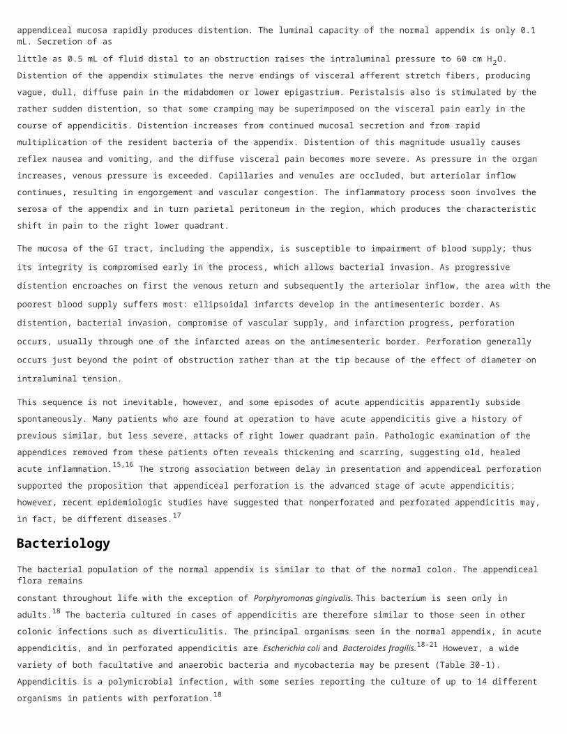
Traditionally the belief has been that there is a predictable sequence of events leading to eventual appendiceal rupture. The
proximal obstruction of the appendiceal lumen produces a closed-loop obstruction, and continuing normal secretion by the

appendiceal mucosa rapidly produces distention. The luminal capacity of the normal appendix is only 0.1 mL. Secretion of as
little as 0.5 mL of fluid distal to an obstruction raises the intraluminal pressure to 60 cm H2O. Distention of the appendix
stimulates the nerve endings of visceral afferent stretch fibers, producing vague, dull, diffuse pain in the midabdomen or
lower epigastrium. Peristalsis also is stimulated by the rather sudden distention, so that some cramping may be
superimposed on the visceral pain early in the course of appendicitis. Distention increases from continued mucosal secretion
and from rapid multiplication of the resident bacteria of the appendix. Distention of this magnitude usually causes reflex
nausea and vomiting, and the diffuse visceral pain becomes more severe. As pressure in the organ increases, venous
pressure is exceeded. Capillaries and venules are occluded, but arteriolar inflow continues, resulting in engorgement and
vascular congestion. The inflammatory process soon involves the serosa of the appendix and in turn parietal peritoneum in
the region, which produces the characteristic shift in pain to the right lower quadrant.
The mucosa of the GI tract, including the appendix, is susceptible to impairment of blood supply; thus its integrity is
compromised early in the process, which allows bacterial invasion. As progressive distention encroaches on first the venous
return and subsequently the arteriolar inflow, the area with the poorest blood supply suffers most: ellipsoidal infarcts develop
in the antimesenteric border. As distention, bacterial invasion, compromise of vascular supply, and infarction progress,
perforation occurs, usually through one of the infarcted areas on the antimesenteric border. Perforation generally occurs just
beyond the point of obstruction rather than at the tip because of the effect of diameter on intraluminal tension.
This sequence is not inevitable, however, and some episodes of acute appendicitis apparently subside spontaneously. Many
patients who are found at operation to have acute appendicitis give a history of previous similar, but less severe, attacks of
right lower quadrant pain. Pathologic examination of the appendices removed from these patients often reveals thickening
and scarring, suggesting old, healed acute inflammation.15,16 The strong association between delay in presentation and
appendiceal perforation supported the proposition that appendiceal perforation is the advanced stage of acute appendicitis;
however, recent epidemiologic studies have suggested that nonperforated and perforated appendicitis may, in fact, be
different diseases.17
Bacteriology
The bacterial population of the normal appendix is similar to that of the normal colon. The appendiceal flora remains
constant throughout life with the exception of Porphyromonas gingivalis. This bacterium is seen only in adults.18 The bacteria
cultured in cases of appendicitis are therefore similar to those seen in other colonic infections such as diverticulitis. The
principal organisms seen in the normal appendix, in acute appendicitis, and in perforated appendicitis are Escherichia coli and
Bacteroides fragilis.18–21 However, a wide variety of both facultative and anaerobic bacteria and mycobacteria may be
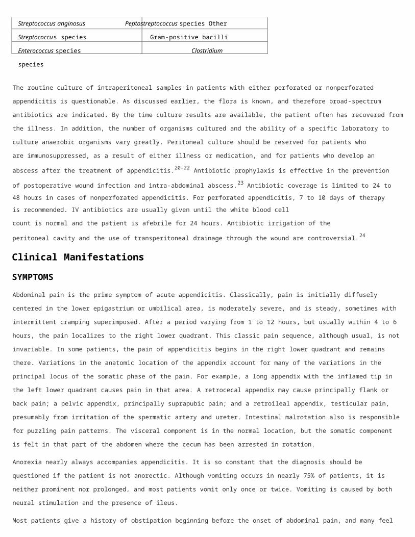
present (Table 30-1). Appendicitis is a polymicrobial infection, with some series reporting the culture of up to 14 different
organisms in patients with perforation.18
Table 30-1 Common Organisms Seen in Patients with Acute Appendicitis
Aerobic and Facultative Anaerobic
Gram-negative bacilli Gram-negative bacilli
Escherichia coli Bacteroides fragilis
Pseudomonas aeruginosa Other Bacteroides species
Klebsiella species Fusobacterium species
Gram-positive cocci Gram-positive cocci
Streptococcus anginosus Peptostreptococcus species
Other Streptococcus species Gram-positive bacilli
Enterococcus species Clostridium species
The routine culture of intraperitoneal samples in patients with either perforated or nonperforated appendicitis is questionable.
As discussed earlier, the flora is known, and therefore broad-spectrum antibiotics are indicated. By the time culture results
are available, the patient often has recovered from the illness. In addition, the number of organisms cultured and the ability
of a specific laboratory to culture anaerobic organisms vary greatly. Peritoneal culture should be reserved for patients who
are immunosuppressed, as a result of either illness or medication, and for patients who develop an abscess after the
treatment of appendicitis.20–22 Antibiotic prophylaxis is effective in the prevention of postoperative wound infection and
intra-abdominal abscess.23 Antibiotic coverage is limited to 24 to 48 hours in cases of nonperforated appendicitis. For
perforated appendicitis, 7 to 10 days of therapy is recommended. IV antibiotics are usually given until the white blood cell
count is normal and the patient is afebrile for 24 hours. Antibiotic irrigation of the peritoneal cavity and the use of
transperitoneal drainage through the wound are controversial.24
Clinical Manifestations
SYMPTOMS
Abdominal pain is the prime symptom of acute appendicitis. Classically, pain is initially diffusely centered in the lower
epigastrium or umbilical area, is moderately severe, and is steady, sometimes with intermittent cramping superimposed.
After a period varying from 1 to 12 hours, but usually within 4 to 6 hours, the pain localizes to the right lower quadrant. This
classic pain sequence, although usual, is not invariable. In some patients, the pain of appendicitis begins in the right lower
quadrant and remains there. Variations in the anatomic location of the appendix account for many of the variations in the
principal locus of the somatic phase of the pain. For example, a long appendix with the inflamed tip in the left lower quadrant
causes pain in that area. A retrocecal appendix may cause principally flank or back pain; a pelvic appendix, principally
suprapubic pain; and a retroileal appendix, testicular pain, presumably from irritation of the spermatic artery and ureter.
Intestinal malrotation also is responsible for puzzling pain patterns. The visceral component is in the normal location, but the
somatic component is felt in that part of the abdomen where the cecum has been arrested in rotation.
Anorexia nearly always accompanies appendicitis. It is so constant that the diagnosis should be questioned if the patient is
not anorectic. Although vomiting occurs in nearly 75% of patients, it is neither prominent nor prolonged, and most patients
vomit only once or twice. Vomiting is caused by both neural stimulation and the presence of ileus.
Most patients give a history of obstipation beginning before the onset of abdominal pain, and many feel that defecation
would relieve their abdominal pain. Diarrhea occurs in some patients, however, particularly children, so that the pattern of
bowel function is of little differential diagnostic value.
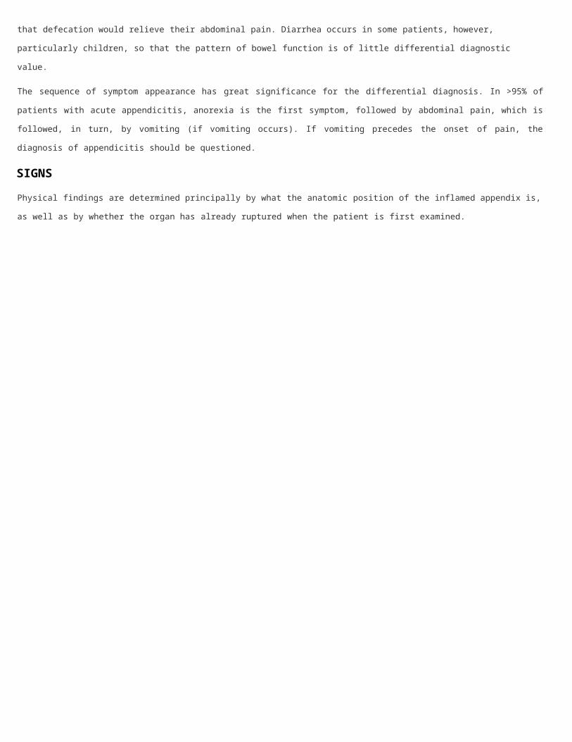
The sequence of symptom appearance has great significance for the differential diagnosis. In >95% of patients with acute
appendicitis, anorexia is the first symptom, followed by abdominal pain, which is followed, in turn, by vomiting (if vomiting
occurs). If vomiting precedes the onset of pain, the diagnosis of appendicitis should be questioned.
SIGNS
Physical findings are determined principally by what the anatomic position of the inflamed appendix is, as well as by whether
the organ has already ruptured when the patient is first examined.

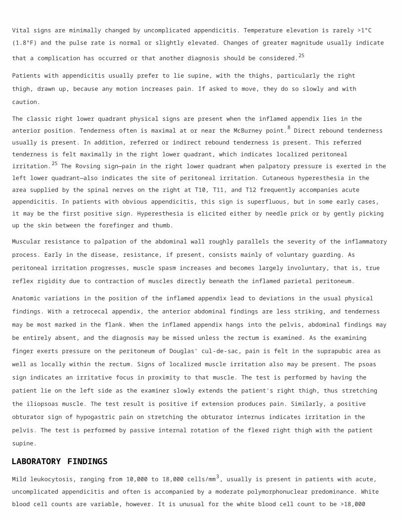
Vital signs are minimally changed by uncomplicated appendicitis. Temperature elevation is rarely >1°C (1.8°F) and the pulse
rate is normal or slightly elevated. Changes of greater magnitude usually indicate that a complication has occurred or that
another diagnosis should be considered.25
Patients with appendicitis usually prefer to lie supine, with the thighs, particularly the right thigh, drawn up, because any
motion increases pain. If asked to move, they do so slowly and with caution.
The classic right lower quadrant physical signs are present when the inflamed appendix lies in the anterior position.
Tenderness often is maximal at or near the McBurney point.8 Direct rebound tenderness usually is present. In addition,
referred or indirect rebound tenderness is present. This referred tenderness is felt maximally in the right lower quadrant,
which indicates localized peritoneal irritation.25 The Rovsing sign—pain in the right lower quadrant when palpatory pressure
is exerted in the left lower quadrant—also indicates the site of peritoneal irritation. Cutaneous hyperesthesia in the area
supplied by the spinal nerves on the right at T10, T11, and T12 frequently accompanies acute appendicitis. In patients with
obvious appendicitis, this sign is superfluous, but in some early cases, it may be the first positive sign. Hyperesthesia is
elicited either by needle prick or by gently picking up the skin between the forefinger and thumb.
Muscular resistance to palpation of the abdominal wall roughly parallels the severity of the inflammatory process. Early in the
disease, resistance, if present, consists mainly of voluntary guarding. As peritoneal irritation progresses, muscle spasm
increases and becomes largely involuntary, that is, true reflex rigidity due to contraction of muscles directly beneath the
inflamed parietal peritoneum.
Anatomic variations in the position of the inflamed appendix lead to deviations in the usual physical findings. With a
retrocecal appendix, the anterior abdominal findings are less striking, and tenderness may be most marked in the flank.
When the inflamed appendix hangs into the pelvis, abdominal findings may be entirely absent, and the diagnosis may be
missed unless the rectum is examined. As the examining finger exerts pressure on the peritoneum of Douglas' cul-de-sac,
pain is felt in the suprapubic area as well as locally within the rectum. Signs of localized muscle irritation also may be
present. The psoas sign indicates an irritative focus in proximity to that muscle. The test is performed by having the patient
lie on the left side as the examiner slowly extends the patient's right thigh, thus stretching the iliopsoas muscle. The test
result is positive if extension produces pain. Similarly, a positive obturator sign of hypogastric pain on stretching the
obturator internus indicates irritation in the pelvis. The test is performed by passive internal rotation of the flexed right thigh
with the patient supine.
LABORATORY FINDINGS
Mild leukocytosis, ranging from 10,000 to 18,000 cells/mm3, usually is present in patients with acute, uncomplicated
appendicitis and often is accompanied by a moderate polymorphonuclear predominance. White blood cell counts are variable,
however. It is unusual for the white blood cell count to be >18,000 cells/mm3 in uncomplicated appendicitis. White blood cell
counts above this level raise the possibility of a perforated appendix with or without an abscess. Urinalysis can be useful to
rule out the urinary tract as the source of infection. Although several white or red blood cells can be present from ureteral or
bladder irritation as a result of an inflamed appendix, bacteriuria in a urine specimen obtained via catheter generally is not
seen in acute appendicitis.26
Imaging Studies
Plain films of the abdomen, although frequently obtained as part of the general evaluation of a patient with an acute
abdomen, rarely are helpful in diagnosing acute appendicitis. However, plain radiographs can be of significant benefit in ruling
out other pathology. In patients with acute appendicitis, one often sees an abnormal bowel gas pattern, which is a nonspecific
finding. The presence of a fecalith is rarely noted on plain films but, if present, is highly suggestive of the diagnosis. A chest
radiograph is sometimes indicated to rule out referred pain from a right lower lobe pneumonic process.
Additional radiographic studies include barium enema examination and radioactively labeled leukocyte scans. If the appendix
fills on barium enema, appendicitis is excluded. On the other hand, if the appendix does not fill, no determination can be
made.27 To date, there has not been enough experience with radionuclide scans to assess their utility.
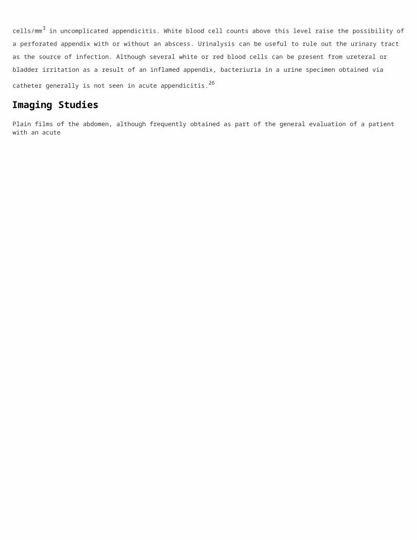
Graded compression sonography has been suggested as an accurate way to establish the diagnosis of appendicitis. The
technique is inexpensive, can be performed rapidly, does not require a contrast medium, and can be used even in pregnant
patients. Sonographically, the appendix is identified as a blind-ending, nonperistaltic bowel loop originating from the cecum.
With maximal compression, the diameter of the appendix is measured in the anteroposterior dimension. Scan results are
considered positive if a noncompressible appendix ≥6 mm in the anteroposterior direction is demonstrated (Fig. 30-3). The
presence of an appendicolith establishes the diagnosis. Thickening of the appendiceal wall and the presence of
periappendiceal fluid is highly suggestive. Sonographic demonstration of a normal appendix, which is an easily compressible,
blind-ending tubular structure measuring ≤5 mm in diameter, excludes the diagnosis of acute appendicitis. The study results
are considered inconclusive if the appendix is not visualized and there is no pericecal fluid or mass. When the diagnosis of
acute appendicitis is excluded by sonography, a brief survey of the remainder of the abdominal cavity should be performed to
establish an alternative diagnosis. In females of childbearing age, the pelvic organs must be adequately visualized either by
transabdominal or endovaginal ultrasonography to exclude gynecologic pathology as a cause of acute abdominal pain. The
sonographic diagnosis of acute appendicitis has a reported sensitivity of 55 to 96% and a specificity of 85 to 98%.28–30
Sonography is similarly effective in children and pregnant women, although its application is somewhat limited in late
pregnancy.
Fig. 30-3.

Sonogram of a 10-year-old girl who presented with nausea, vomiting, and abdominal pain. The appendix measured 10.0 mm in maximal anteroposterior diameter in both the noncompression (A) and compression (B) views.
Although sonography can easily identify abscesses in cases of perforation, the technique has limitations and results are user
dependent. A false-positive scan result can occur in the presence of periappendicitis from surrounding inflammation, a
dilated fallopian tube can be mistaken for an inflamed appendix, inspissated stool can mimic an appendicolith, and, in obese
patients, the appendix may not be compressible because of overlying fat. False-negative sonogram results can occur if
appendicitis is confined to the appendiceal tip, the appendix is retrocecal, the appendix is markedly enlarged and mistaken
for small bowel, or the appendix is perforated and therefore compressible.31
Some studies have reported that graded compression sonography improved the diagnosis of appendicitis over clinical
examination, specifically decreasing the percentage of negative explorations for appendectomies from 37 to 13%.32
Sonography also decreases the time before operation. Sonography identified appendicitis in 10% of patients who were
believed to have a low likelihood of the disease on physical examination.33 The positive and negative predictive values of
ultrasonography have impressively been reported as 91 and 92%, respectively. However, in a recent prospective multicenter
study, routine ultrasonography did not improve diagnostic accuracy or rates of negative appendectomy or perforation
compared with clinical assessment.
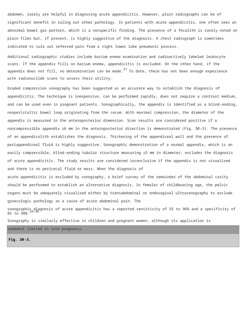
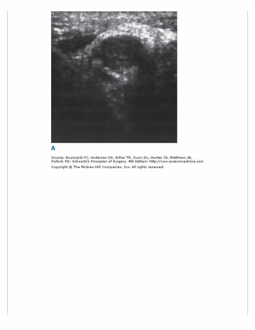
High-resolution helical CT also has been used to diagnose appendicitis. On CT scan, the inflamed appendix appears dilated
(>5 cm) and the wall is thickened. There is usually evidence of inflammation, with "dirty fat," thickened mesoappendix, and
even an obvious phlegmon (Fig. 30-4). Fecaliths can be easily visualized, but their presence is not necessarily
pathognomonic of appendicitis. An important suggestive abnormality is the arrowhead sign. This is caused by thickening of
the cecum, which funnels contrast agent toward the orifice of the inflamed appendix. CT scanning is also an excellent
technique for identifying other inflammatory processes masquerading as appendicitis.
Fig. 30-4.
Computed tomographic scans with findings positive for appendicitis. Note the thick-walled and dilated appendix (A) and mesenteric streaking and "dirty fat" (B).
Several CT techniques have been used, including focused and nonfocused CT scans and enhanced and nonenhanced helical
CT scanning. Nonenhanced helical CT scanning is important, because one of the disadvantages of using CT scanning in the
evaluation of right lower quadrant pain is dye allergy. Surprisingly, all of these techniques have yielded essentially identical
rates of diagnostic accuracy: 92 to 97% sensitivity, 85 to 94% specificity, 90 to 98% accuracy, and 75 to 95% positive and
95 to 99% negative predictive values.34–36 The additional use of a rectally administered contrast agent did not improve the
results of CT scanning.
A number of studies have documented improvement in diagnostic accuracy with the liberal use of CT scanning in the work-up
of suspected appendicitis. CT lowered the rate of negative appendectomies from 19 to 12% in one study,37 and the
incidence of negative appendectomies in women from 24 to 5% in another.38 The use of this imaging study altered the care
of 24% of patients studied and provided alternative diagnoses in half of the patients with normal appendices on CT scan.39
Despite the potential usefulness of this technique, there are significant disadvantages. CT scanning is expensive, exposes the
patient to significant radiation, and cannot be used during pregnancy. Allergy contraindicates the administration of IV
contrast agents in some patients, and others cannot tolerate the oral ingestion of luminal dye, particularly in the presence of
nausea and vomiting. Finally, not all studies have documented the utility of CT scanning in all patients with right lower
quadrant pain.40
A number of studies have compared the effectiveness of graded compression sonography and helical CT in establishing the
diagnosis of appendicitis. Although the differences are rather small, CT scanning has consistently proven superior. For
example, in one study, 600 ultrasounds and 317 CT scans demonstrated sensitivity of 80 and 97%, specificity of 93 and
94%, diagnostic accuracy of 89 and 95%, positive predictive value of 91 and 92%, and negative predictive value of 88 and
98%, respectively.30 In another study, ultrasound positively impacted the management of 19% of patients, compared with
73% of patients for CT. Finally, in a third study, the negative appendix rate was 17% for patients studied by ultrasonography
compared with a negative appendix rate of 2% for patients who underwent helical CT scanning.41 One concern about
ultrasonography is the high intraobserver variability.42
One issue that has not been resolved is which patients are candidates for imaging studies.43 This question may be moot,
because CT scanning routinely is ordered by emergency physicians before surgeons are even consulted. The concept that all
patients with right lower quadrant pain should undergo CT scanning has been strongly supported by two reports by Rao and
his colleagues at the Massachusetts General Hospital. In one, this group documented that CT scanning led to a fall in the
negative appendectomy rate from 20 to 7% and a decline in the perforation rate from 22 to 14%, as well as establishment of
an alternative diagnosis in 50% of patients.44 In the second study, published in the New England Journal of Medicine, Rao
and associates documented that CT scanning prevented 13 unnecessary appendectomies, saved 50 inpatient hospital days,
and lowered the per-patient cost by $447.45 In contrast, several other studies failed to prove an advantage of routine CT
scanning, documenting that surgeon accuracy approached that of the imaging study and expressing concern that the
imaging studies could adversely delay appendectomy in affected patients.46,47
The rational approach is the selective use of CT scanning. This has been documented by several studies in which imaging was
performed based on an algorithm or protocol.48 The likelihood of appendicitis can be ascertained using the Alvarado scale
(Table 30-2).49 This scoring system was designed to improve the diagnosis of appendicitis and was devised by giving
relative weight to specific clinical manifestation. Table 30-2 lists the eight specific indicators identified. Patients with scores of
9 or 10 are almost certain to have appendicitis; there is little advantage in further work-up, and they should go to the
operating room. Patients with scores of 7 or 8 have a high likelihood of appendicitis, whereas scores of 5 or 6 are compatible
with, but not diagnostic of, appendicitis. CT scanning is certainly appropriate for patients with Alvarado scores of 5 and 6, and
a case can be built for imaging for those with scores of 7 and 8. On the other hand, it is difficult to justify the expense,
radiation exposure, and possible complications of CT scanning in patients whose scores of 0 to 4 make it extremely unlikely
(but not impossible) that they have appendicitis.
Table 30-2 Alvarado Scale for the Diagnosis of Appendicitis
Manifestations Value
Symptoms Migration of pain 1
Anorexia 1
Nausea and/or vomiting 1
Signs Right lower quadrant tenderness 2
Rebound 1
Elevated temperature 1
Laboratory values Leukocytosis 2
Left shift in leukocyte count 1
Total points 10
Source: Reproduced with permission from Alvarado.49
Selective CT scanning based on the likelihood of appendicitis takes advantage of the clinical skill of the experienced surgeon
and, when indicated, adds the expertise of the radiologist and his or her imaging study. Figure 30-5 proposes a treatment
algorithm addressing the rational use of diagnostic testing.50
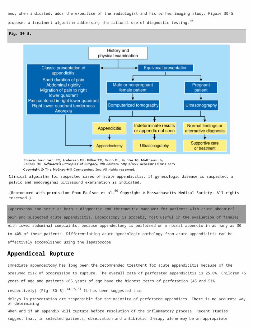
Fig. 30-5.
Clinical algorithm for suspected cases of acute appendicitis. If gynecologic disease is suspected, a pelvic and endovaginal ultrasound examination is indicated.
(Reproduced with permission from Paulson et al.50 Copyright © Massachusetts Medical Society. All rights reserved.)
Laparoscopy can serve as both a diagnostic and therapeutic maneuver for patients with acute abdominal pain and suspected
acute appendicitis. Laparoscopy is probably most useful in the evaluation of females with lower abdominal complaints,
because appendectomy is performed on a normal appendix in as many as 30 to 40% of these patients. Differentiating acute
gynecologic pathology from acute appendicitis can be effectively accomplished using the laparoscope.
Appendiceal Rupture
Immediate appendectomy has long been the recommended treatment for acute appendicitis because of the presumed risk of
progression to rupture. The overall rate of perforated appendicitis is 25.8%. Children <5 years of age and patients >65 years
of age have the highest rates of perforation (45 and 51%, respectively) (Fig. 30-6).14,15,51 It has been suggested that
delays in presentation are responsible for the majority of perforated appendices. There is no accurate way of determining
when and if an appendix will rupture before resolution of the inflammatory process. Recent studies suggest that, in selected
patients, observation and antibiotic therapy alone may be an appropriate treatment for acute appendicitis.17,52
Fig. 30-6.
Rate of appendiceal rupture by age group.
(Personal communication from David Flum, MD.)
Appendiceal rupture occurs most frequently distal to the point of luminal obstruction along the antimesenteric border of the
appendix. Rupture should be suspected in the presence of fever with a temperature of >39°C (102°F) and a white blood cell
count of >18,000 cells/mm3. In the majority of cases, rupture is contained and patients display localized rebound
tenderness. Generalized peritonitis will be present if the walling-off process is ineffective in containing the rupture.
In 2 to 6% of cases, an ill-defined mass is detected on physical examination. This could represent a phlegmon, which
consists of matted loops of bowel adherent to the adjacent inflamed appendix, or a periappendiceal abscess. Patients who
present with a mass have experienced symptoms for a longer duration, usually at least 5 to 7 days. Distinguishing acute,
uncomplicated appendicitis from acute appendicitis with perforation on the basis of clinical findings is often difficult, but it is
important to make the distinction because their treatment differs. CT scan may be beneficial in guiding therapy. Phlegmons
and small abscesses can be treated conservatively with IV antibiotics; well-localized abscesses can be managed with
percutaneous drainage; complex abscesses should be considered for surgical drainage. If operative drainage is required, it
should be performed using an extraperitoneal approach, with appendectomy reserved for cases in which the appendix is
easily accessible. Interval appendectomy performed at least 6 weeks after the acute event has classically been recommended
for all patients treated either nonoperatively or with simple drainage of an abscess.53,54
Differential Diagnosis
The differential diagnosis of acute appendicitis is essentially the diagnosis of the acute abdomen (see Chap. 35). This is
because clinical manifestations are not specific for a given disease but are specific for disturbance of a given physiologic
function or functions. Thus, an essentially identical clinical picture can result from a wide variety of acute processes within
the peritoneal cavity that produce the same alterations of function as does acute appendicitis.

The accuracy of preoperative diagnosis should be approximately 85%. If it is consistently less, it is likely that some
unnecessary operations are being performed, and a more rigorous preoperative differential diagnosis is in order. A diagnostic
accuracy rate that is consistently >90% should also cause concern, because this may mean that some patients with atypical,
but bona fide, cases of acute appendicitis are being "observed" when they should receive prompt surgical intervention. The
Haller group, however, has shown that this is not invariably true.55 Before that group's study, the perforation rate at the
hospital at which the study took place was 26.7%, and acute appendicitis was found in 80% of the patients undergoing
operation. By implementing a policy of intensive inhospital observation when the diagnosis of appendicitis was unclear, the
group raised the rate of acute appendicitis found at operation to 94%, but the perforation rate remained unchanged at
27.5%.55 The rate of false-negative appendectomies is highest in young adult females. A normal appendix is found in 32 to
45% of appendectomies performed in women 15 to 45 years of age.14
A common error is to make a preoperative diagnosis of acute appendicitis only to find some other condition (or nothing) at
operation. Much less frequently, acute appendicitis is found after a preoperative diagnosis of another condition. The most
common erroneous preoperative diagnoses—together accounting for >75% of cases—are, in descending order of frequency,
acute mesenteric lymphadenitis, no organic pathologic condition, acute pelvic inflammatory disease, twisted ovarian cyst or
ruptured graafian follicle, and acute gastroenteritis.
The differential diagnosis of acute appendicitis depends on four major factors: the anatomic location of the inflamed
appendix; the stage of the process (i.e., simple or ruptured); the patient's age; and the patient's sex.56–60
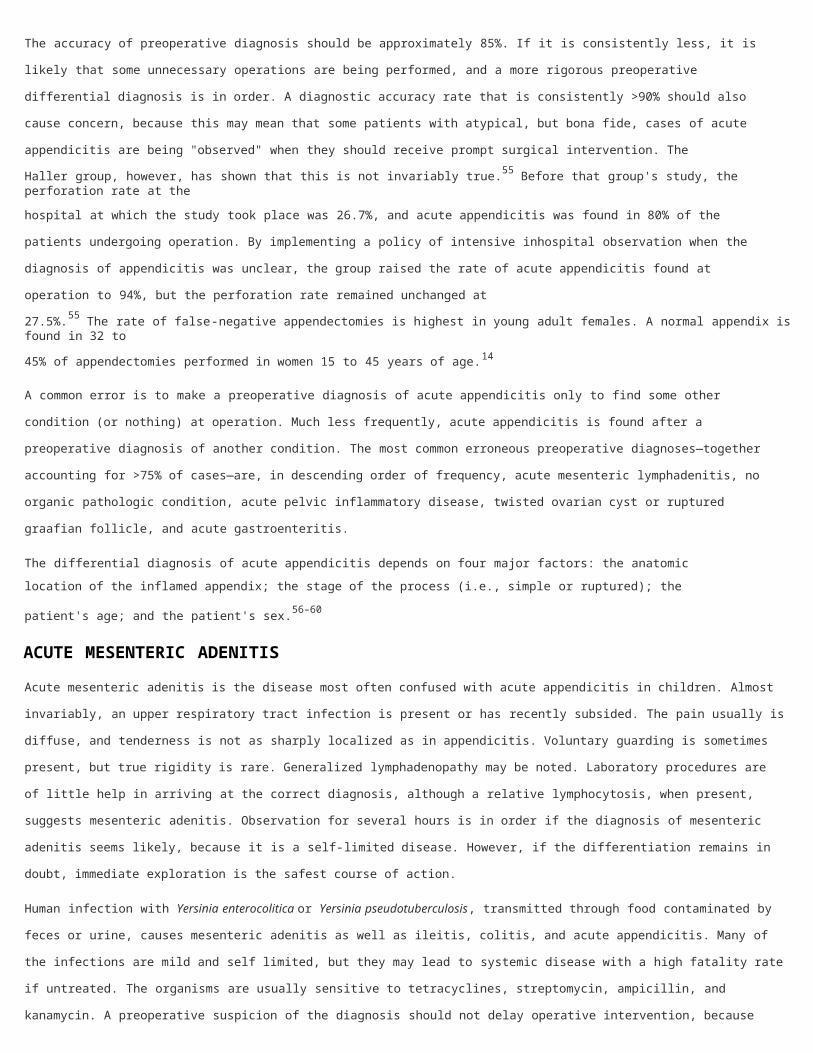
ACUTE MESENTERIC ADENITIS
Acute mesenteric adenitis is the disease most often confused with acute appendicitis in children. Almost invariably, an upper
respiratory tract infection is present or has recently subsided. The pain usually is diffuse, and tenderness is not as sharply
localized as in appendicitis. Voluntary guarding is sometimes present, but true rigidity is rare. Generalized lymphadenopathy
may be noted. Laboratory procedures are of little help in arriving at the correct diagnosis, although a relative lymphocytosis,
when present, suggests mesenteric adenitis. Observation for several hours is in order if the diagnosis of mesenteric adenitis
seems likely, because it is a self-limited disease. However, if the differentiation remains in doubt, immediate exploration is
the safest course of action.
Human infection with Yersinia enterocolitica or Yersinia pseudotuberculosis, transmitted through food contaminated by feces
or urine, causes mesenteric adenitis as well as ileitis, colitis, and acute appendicitis. Many of the infections are mild and self
limited, but they may lead to systemic disease with a high fatality rate if untreated. The organisms are usually sensitive to
tetracyclines, streptomycin, ampicillin, and kanamycin. A preoperative suspicion of the diagnosis should not delay operative
intervention, because appendicitis caused by Yersinia cannot be clinically distinguished from appendicitis due to other
causes. Approximately 6% of cases of mesenteric adenitis are caused by Yersinia infection.
Salmonella typhimurium infection causes mesenteric adenitis and paralytic ileus with symptoms similar to those of
appendicitis. The diagnosis can be established by serologic testing. Campylobacter jejuni causes diarrhea and pain that
mimics that of appendicitis. The organism can be cultured from stool.
GYNECOLOGIC DISORDERS
Diseases of the female internal reproductive organs that may erroneously be diagnosed as appendicitis are, in approximate
descending order of frequency, pelvic inflammatory disease, ruptured graafian follicle, twisted ovarian cyst or tumor,
endometriosis, and ruptured ectopic pregnancy.

Pelvic Inflammatory Disease
In pelvic inflammatory disease the infection usually is bilateral but, if confined to the right tube, may mimic acute
appendicitis. Nausea and vomiting are present in patients with appendicitis, but in only approximately 50% of those with
pelvic inflammatory disease. Pain and tenderness are usually lower, and motion of the cervix is exquisitely painful.
Intracellular diplococci may be demonstrable on smear of the purulent vaginal discharge. The ratio of cases of appendicitis to
cases of pelvic inflammatory disease is low in females in the early phase of the menstrual cycle and high during the luteal
phase. The careful clinical use of these features has reduced the incidence of negative findings on laparoscopy in young
women to 15%.
Ruptured Graafian Follicle
Ovulation commonly results in the spillage of sufficient amounts of blood and follicular fluid to produce brief, mild lower
abdominal pain. If the amount of fluid is unusually copious and is from the right ovary, appendicitis may be simulated. Pain
and tenderness are rather diffuse. Leukocytosis and fever are minimal or absent. Because this pain occurs at the midpoint of
the menstrual cycle, it is often called mittelschmerz.
Twisted Ovarian Cyst
Serous cysts of the ovary are common and generally remain asymptomatic. When right-sided cysts rupture or undergo
torsion, the manifestations are similar to those of appendicitis. Patients develop right lower quadrant pain, tenderness,
rebound, fever, and leukocytosis. If the mass is palpable on physical examination, the diagnosis can be made easily. Both
transvaginal ultrasonography and CT scanning can be diagnostic if a mass is not palpable.
Torsion requires emergent operative treatment. If the torsion is complete or longstanding, the pedicle undergoes thrombosis,
and the ovary and tube become gangrenous and require resection. Leakage of ovarian cysts resolves spontaneously,
however, and is best treated nonoperatively.24,56–61
Ruptured Ectopic Pregnancy
Blastocysts may implant in the fallopian tube (usually the ampullary portion) and in the ovary. Rupture of right tubal or
ovarian pregnancies can mimic appendicitis. Patients may give a history of abnormal menses, either missing one or two
periods or noting only slight vaginal bleeding. Unfortunately, patients do not always realize they are pregnant. The
development of right lower quadrant or pelvic pain may be the first symptom. The diagnosis of ruptured ectopic pregnancy
should be relatively easy. The presence of a pelvic mass and elevated levels of chorionic gonadotropin are characteristic.
Although the leukocyte count rises slightly (to approximately 14,000 cells/mm3), the hematocrit level falls as a consequence
of the intra-abdominal hemorrhage. Vaginal examination reveals cervical motion and adnexal tenderness, and a more
definitive diagnosis can be established by culdocentesis. The presence of blood and particularly decidual tissue is
pathognomonic. The treatment of ruptured ectopic pregnancy is emergency surgery.
ACUTE GASTROENTERITIS
Acute gastroenteritis is common but usually can be easily distinguished from acute appendicitis. Gastroenteritis is
characterized by profuse diarrhea, nausea, and vomiting. Hyperperistaltic abdominal cramps precede the watery stools. The
abdomen is relaxed between cramps, and there are no localizing signs. Laboratory values vary with the specific cause.
OTHER INTESTINAL DISORDERS
Meckel's Diverticulitis
Meckel's diverticulitis gives rise to a clinical picture similar to that of acute appendicitis. Meckel's diverticulum is located
within the distal 2 ft of the ileum. Meckel's diverticulitis is associated with the same complications as appendicitis and
requires the same treatment—prompt surgical intervention. Resection of the segment of ileum bearing the diverticulum with
end-to-end anastomosis can nearly always be done through a McBurney incision, extended if necessary, or laparoscopically.
Crohn's Enteritis
The manifestations of acute regional enteritis—fever, right lower quadrant pain and tenderness, and leukocytosis—often
simulate acute appendicitis. The presence of diarrhea and the absence of anorexia, nausea, and vomiting favor a diagnosis of
enteritis, but this is not sufficient to exclude acute appendicitis. In an appreciable percentage of patients with chronic regional
enteritis, the diagnosis is first made at the time of operation for presumed acute appendicitis. In cases of an acutely inflamed
distal ileum with no cecal involvement and a normal appendix, appendectomy is indicated. Progression to chronic Crohn's
ileitis is uncommon.
Colonic Lesions
Diverticulitis or perforating carcinoma of the cecum, or of that portion of the sigmoid that lies in the right side, may be
impossible to distinguish from appendicitis. These entities should be considered in older patients. CT scanning is often helpful
in making a diagnosis in older patients with right lower quadrant pain and atypical clinical presentations.
Epiploic appendagitis probably results from infarction of the colonic appendage(s) secondary to torsion. Symptoms may be
minimal, or there may be continuous abdominal pain in an area corresponding to the contour of the colon, lasting several
days. Pain shift is unusual, and there is no diagnostic sequence of symptoms. The patient does not look ill, nausea and
vomiting are unusual, and appetite generally is unaffected. Localized tenderness over the site is usual and often is associated
with rebound without rigidity. In 25% of reported cases, pain persists or recurs until the infarcted epiploic appendage is
removed.
OTHER DISEASES
Diseases or conditions not mentioned in the preceding sections that must be considered in the differential diagnosis include
foreign body perforations of the bowel, closed-loop intestinal obstruction, mesenteric vascular infarction, pleuritis of the right
lower chest, acute cholecystitis, acute pancreatitis, hematoma of the abdominal wall, epididymitis, testicular torsion, urinary
tract infection, ureteral stone, primary peritonitis, and Henoch-Schönlein purpura.
Acute Appendicitis in the Young
The establishment of a diagnosis of acute appendicitis is more difficult in young children than in the adult. The inability of
young children to give an accurate history, diagnostic delays by both parents and physicians, and the frequency of GI upset
in children are all contributing factors.62 In children the physical examination findings of maximal tenderness in the right
lower quadrant, the inability to walk or walking with a limp, and pain with percussion, coughing, and hopping were found to
have the highest sensitivity for appendicitis.63
The more rapid progression to rupture and the inability of the underdeveloped greater omentum to contain a rupture lead to
significant morbidity rates in children. Children <5 years of age have a negative appendectomy rate of 25% and an

appendiceal perforation rate of 45%. These rates may be compared with a negative appendectomy rate of <10% and a
perforated appendix rate of 20% for children 5 to 12 years of age.13,14 The incidence of major complications after
appendectomy in children is correlated with appendiceal rupture. The wound infection rate after the treatment of
nonperforated appendicitis in children is 2.8% compared with a rate of 11% after the treatment of perforated appendicitis.
The incidence of intra-abdominal abscess also is higher after the treatment of perforated appendicitis than after
nonperforated appendicitis (6% vs. 3%).23 The treatment regimen for perforated appendicitis generally includes immediate
appendectomy and irrigation of the peritoneal cavity. Antibiotic coverage is limited to 24 to 48 hours in cases of
nonperforated appendicitis. For perforated appendicitis IV antibiotics usually are given until the white blood cell count is
normal and the patient is afebrile for 24 hours. The use of antibiotic irrigation of the peritoneal cavity and transperitoneal
drainage through the wound are controversial. Laparoscopic appendectomy has been shown to be safe and effective for the
treatment of appendicitis in children.64
Acute Appendicitis in the Elderly
Compared with younger patients, elderly patients with appendicitis often pose a more difficult diagnostic problem because of
the atypical presentation, expanded differential diagnosis, and communication difficulty. These factors may be responsible for
the disproportionately high perforation rate seen in the elderly. In the general population, perforation rates range from 20 to
30%, compared with 50 to 70% in the elderly.65 In addition, the perforation rate appears to increase with age >80 years.66
Elderly patients usually present with lower abdominal pain, but on clinical examination, localized right lower quadrant
tenderness is present in only 80 to 90% of patients. A history of periumbilical pain migrating to the right lower quadrant is
reported infrequently. The usefulness of the Alvarado score appears to decline in the elderly. Fewer then 50% of the elderly
with appendicitis have an Alvarado score of ≥7.66 Although currently there are no criteria that definitively identify elderly
patients with acute appendicitis who are at risk of rupture, prioritization should be given to patients with a temperature of
>38°C (100.4°F) and a shift to the left in leukocyte count of >76%, especially if they are male, are anorectic, or have had
pain of long duration before admission.65
As a result of increased comorbidities and an increased rate of perforation, postoperative morbidity, mortality, and hospital
length of stay are increased in the elderly compared with younger populations with appendicitis. Although no randomized
trials have been conducted, it appears that elderly patients benefit from a laparoscopic approach to treatment of
appendicitis. The use of laparoscopy in the elderly has significantly increased in recent years. In general, laparoscopic
appendectomy offers elderly patients with appendicitis a shorter length of hospital stay, a reduction in complication and
mortality rates, and a greater chance of discharge to home (independent of further nursing care or rehabilitation).67
Acute Appendicitis during Pregnancy
Appendectomy for presumed appendicitis is the most common surgical emergency during pregnancy. The incidence is
approximately 1 in 766 births. Acute appendicitis can occur at any time during pregnancy.68 The overall negative
appendectomy rate during pregnancy is approximately 25% and appears to be higher than the rate seen in nonpregnant
women.68,69 A higher rate of negative appendectomy is seen in the second trimester, and the lowest rate is in the third
trimester. The diversity of clinical presentations and the difficulty in making the diagnosis of acute appendicitis in pregnant
women is well established. This is particularly true in the late second trimester and the third trimester, when many
abdominal symptoms may be considered pregnancy related. In addition, during pregnancy there are anatomic changes in the
appendix (Fig. 30-7) and increased abdominal laxity that may further complicate clinical evaluation. There is no association

between appendectomy and subsequent fertility.
Fig. 30-7.
Location of the appendix during pregnancy. ASIS = anterior superior iliac spine.
[Reproduced with permission from Metcalf A: The appendix, in Corson JD, Williamson RCN (eds): Surgery. London: Mosby,2001.]
Appendicitis in pregnancy should be suspected when a pregnant woman complains of abdominal pain of new onset. The most
consistent sign encountered in acute appendicitis during pregnancy is pain in the right side of the abdomen. Seventy-four
percent of patients report pain located in the right lower abdominal quadrant, with no difference between early and late
pregnancy. Only 57% of patients present with the classic history of diffuse periumbilical pain migrating to the right lower
quadrant. Laboratory evaluation is not helpful in establishing the diagnosis of acute appendicitis during pregnancy. The
physiologic leukocytosis of pregnancy has been defined as high as 16,000 cells/mm3. In one series only 38% of patients with
appendicitis had a white blood cell count of >16,000 cells/mm3.68 Recent data suggest that the incidence of perforated or
complex appendicitis is not increased in pregnant patients.69
When the diagnosis is in doubt, abdominal ultrasound may be beneficial. Another option is magnetic resonance imaging,
which has no known deleterious effects on the fetus. The American College of Radiology recommends the use of nonionizing
radiation techniques for front-line imaging in pregnant women.70 Laparoscopy has been advocated in equivocal cases,
especially early in pregnancy; however laparoscopic appendectomy may be associated with an increase in pregnancy-related
complications. In an analysis of outcomes in California using administrative databases, laparoscopy was found to be
associated with a 2.31 increased odds of fetal loss over open surgery.69
The overall incidence of fetal loss after appendectomy is 4% and the risk of early delivery is 7%. Rates of fetal loss are
considerably higher in women with complex appendicitis than in those with a negative appendectomy and with simple
appendicitis. It is important to note that a negative appendectomy is not a benign procedure. Removing a normal appendix is
associated with a 4% risk of fetal loss and 10% risk of early delivery. Maternal mortality after appendectomy is extremely rare
(0.03%). Because the incidence of ruptured appendix is similar in pregnant and nonpregnant women and because maternal
mortality is so low, it appears that the greatest opportunity to improve fetal outcomes is by improving diagnostic accuracy
and reducing the rate of negative appendectomy.68–71
Appendicitis in Patients with AIDS or HIV Infection
The incidence of acute appendicitis in HIV-infected patients is reported to be 0.5%. This is higher than the 0.1 to 0.2%
incidence reported for the general population.72 The presentation of acute appendicitis in HIV-infected patients is similar to
that in noninfected patients. The majority of HIV-infected patients with appendicitis have fever, periumbilical pain radiating
to the right lower quadrant (91%), right lower quadrant tenderness (91%), and rebound tenderness (74%). HIV-infected
patients do not manifest an absolute leukocytosis; however, if a baseline leukocyte count is available, nearly all HIV-infected
patients with appendicitis demonstrate a relative leukocytosis.72,73
The risk of appendiceal rupture appears to be increased in HIV-infected patients. In one large series of HIV-infected patients
who underwent appendectomy for presumed appendicitis, 43% of patients were found to have perforated appendicitis at
laparotomy.74 The increased risk of appendiceal rupture may be related to the delay in presentation seen in this patient
population.72,74 The mean duration of symptoms before arrival in the emergency department has been reported to be
increased in HIV-infected patients, with >60% of patients reporting the duration of symptoms to be longer than 24 hours.72
In early series, significant hospital delay also may have contributed to high rates of rupture.72 However, with increased
understanding of abdominal pain in HIV-infected patients, hospital delay has become less prevalent.72,75 A low CD4 count is
also associated with an increased incidence of appendiceal rupture. In one large series, patients with nonruptured appendices
had CD4 counts of 158.75 ± 47 cells/mm3 compared with 94.5 ± 32 cells/mm3 in patients with appendiceal rupture.72
The differential diagnosis of right lower quadrant pain is expanded in HIV-infected patients compared with the general
population. In addition to the conditions discussed elsewhere in this chapter, opportunistic infections should be considered as
a possible cause of right lower quadrant pain.72–75 Such opportunistic infections include cytomegalovirus (CMV) infection,
Kaposi's sarcoma, tuberculosis, lymphoma, and other causes of infectious colitis. CMV infection may be seen anywhere in the
GI tract. CMV infection causes a vasculitis of blood vessels in the submucosa of the gut, which leads to thrombosis. Mucosal
ischemia develops, leading to ulceration, gangrene of the bowel wall, and perforation. Spontaneous peritonitis may be caused
by opportunistic pathogens, including CMV, Mycobacterium avium-intracellulare complex, Mycobacterium tuberculosis,
Cryptococcus neoformans, and Strongyloides. Kaposi's sarcoma and non-Hodgkin's lymphoma may present with pain and a
right lower quadrant mass. Viral and bacterial colitis occur with a higher frequency in HIV-infected patients than in the
general population. Colitis should always be considered in HIV-infected patients presenting with right lower quadrant pain.
Neutropenic enterocolitis (typhlitis) should also be considered in the differential diagnosis of right lower quadrant pain in HIV-
infected patients.73,75
A thorough history and physical examination is important when evaluating any patient with right lower quadrant pain. In the

HIV-infected patient with classic signs and symptoms of appendicitis, immediate appendectomy is indicated. In those patients
with diarrhea as a prominent symptom, colonoscopy may be warranted. In patients with equivocal findings, CT scan is
usually helpful. The majority of pathologic findings identified in HIV-infected patients who undergo appendectomy for
presumed appendicitis are typical. The negative appendectomy rate is 5 to 10%. However, in up to 25% of patients AIDS-
related entities are found in the operative specimens, including CMV, Kaposi's sarcoma, and M. avium-intracellulare
complex.72,74
In a retrospective study of 77 HIV-infected patients from 1988 to 1995, the 30-day mortality rate for patients undergoing
appendectomy was reported to be 9.1%.72 More recent series report 0% mortality in this group of patients.75 Morbidity rates
for HIV-infected patients with nonperforated appendicitis are similar to those seen in the general population. Postoperative
morbidity rates appear to be higher in HIV-infected patients with perforated appendicitis. In addition, the length of hospital
stay for HIV-infected patients undergoing appendectomy is twice that for the general population.72,75 No series has been
reported to date that addresses the role of laparoscopic appendectomy in the HIV-infected population.
Treatment
Despite the advent of more sophisticated diagnostic modalities, the importance of early operative intervention should not be
minimized. Once the decision to operate for presumed acute appendicitis has been made, the patient should be prepared for
the operating room. Adequate hydration should be ensured, electrolyte abnormalities should be corrected, and pre-existing
cardiac, pulmonary, and renal conditions should be addressed. A large meta-analysis has demonstrated the efficacy of
preoperative antibiotics in lowering the infectious complications in appendicitis.23 Most surgeons routinely administer
antibiotics to all patients with suspected appendicitis. If simple acute appendicitis is encountered, there is no benefit in
extending antibiotic coverage beyond 24 hours. If perforated or gangrenous appendicitis is found, antibiotics are continued
until the patient is afebrile and has a normal white blood cell count. For intra-abdominal infections of GI tract origin that are
of mild to moderate severity, the Surgical Infection Society has recommended single-agent therapy with cefoxitin, cefotetan,
or ticarcillin-clavulanic acid. For more severe infections, single-agent therapy with carbapenems or combination therapy with
a third-generation cephalosporin, monobactam, or aminoglycoside plus anaerobic coverage with clindamycin or
metronidazole is indicated.24 The recommendations are similar for children.76
OPEN APPENDECTOMY
For open appendectomy most surgeons use either a McBurney (oblique) or Rocky-Davis (transverse) right lower quadrant
muscle-splitting incision in patients with suspected appendicitis. The incision should be centered over either the point of
maximal tenderness or a palpable mass. If an abscess is suspected, a laterally placed incision is imperative to allow
retroperitoneal drainage and to avoid generalized contamination of the peritoneal cavity. If the diagnosis is in doubt, a lower
midline incision is recommended to allow a more extensive examination of the peritoneal cavity. This is especially relevant in
older patients with possible malignancy or diverticulitis.
Several techniques can be used to locate the appendix. Because the cecum usually is visible within the incision, the
convergence of the taeniae can be followed to the base of the appendix. A sweeping lateral to medial motion can aid in
delivering the appendiceal tip into the operative field. Occasionally, limited mobilization of the cecum is needed to aid in
adequate visualization. Once identified, the appendix is mobilized by dividing the mesoappendix, with care taken to ligate the
appendiceal artery securely.
The appendiceal stump can be managed by simple ligation or by ligation and inversion with either a purse-string or Z stitch.
As long as the stump is clearly viable and the base of the cecum is not involved with the inflammatory process, the stump
can be safely ligated with a nonabsorbable suture. The mucosa is frequently obliterated to avoid the development of
mucocele. The peritoneal cavity is irrigated and the wound closed in layers. If perforation or gangrene is found in adults, the
skin and subcutaneous tissue should be left open and allowed to heal by secondary intent or closed in 4 to 5 days as a
delayed primary closure. In children, who generally have little subcutaneous fat, primary wound closure has not led to an
increased incidence of wound infection.
If appendicitis is not found, a methodical search must be made for an alternative diagnosis. The cecum and mesentery should
first be inspected. Next, the small bowel should be examined in a retrograde fashion beginning at the ileocecal valve and
extending at least 2 ft. In females, special attention should be paid to the pelvic organs. An attempt also should be made to
examine the upper abdominal contents. Peritoneal fluid should be sent for Gram's staining and culture. If purulent fluid is
encountered, it is imperative that the source be identified. A medial extension of the incision (Fowler-Weir), with division of
the anterior and posterior rectus sheath, is acceptable if further evaluation of the lower abdomen is indicated. If upper
abdominal pathology is encountered, the right lower quadrant incision is closed and an appropriate upper midline incision is
made.9
LAPAROSCOPIC APPENDECTOMY
Semm first reported successful laparoscopic appendectomy several years before the first laparoscopic cholecystectomy.10
However, the laparoscopic approach to appendectomy did not come into widespread use until after the success of
laparoscopic cholecystectomy. This may be due to the fact that appendectomy, by virtue of its small incision, is already a
form of minimal-access surgery.77
Laparoscopic appendectomy is performed under general anesthesia. A nasogastric tube and a urinary catheter are placed
before obtaining a pneumoperitoneum. Laparoscopic appendectomy usually requires the use of three ports. Four ports may
occasionally be necessary to mobilize a retrocecal appendix. The surgeon usually stands to the patient's left. One assistant is
required to operate the camera. One trocar is placed in the umbilicus (10 mm), and a second trocar is placed in the
suprapubic position. Some surgeons place this second port in the left lower quadrant. The suprapubic trocar is either 10 or
12 mm, depending on whether or not a linear stapler will be used. The placement of the third trocar (5 mm) is variable and
usually is either in the left lower quadrant, epigastrium, or right upper quadrant. Placement is based on location of the
appendix and surgeon preference. Initially, the abdomen is thoroughly explored to exclude other pathology. The appendix is
identified by following the anterior taeniae to its base. Dissection at the base of the appendix enables the surgeon to create a
window between the mesentery and the base of the appendix (Fig. 30-8A). The mesentery and base of the appendix are
then secured and divided separately. When the mesoappendix is involved with the inflammatory process, it is often best to
divide the appendix first with a linear stapler and then to divide the mesoappendix immediately adjacent to the appendix
with clips, electrocautery, Harmonic Scalpel, or staples (Fig. 30-8B and 30-8C). The base of the appendix is not inverted. The
appendix is removed from the abdominal cavity through a trocar site or within a retrieval bag. The base of the appendix and
the mesoappendix should be evaluated for hemostasis. The right lower quadrant should be irrigated. Trocars are
removed under direct vision.78,79
Fig. 30-8.

Laparoscopic resection of the appendix. Occasionally, if the appendix and mesoappendix are extremely inflamed, it is easier to divide the appendix at its base before division of the mesoappendix. A. A window is created in the mesoappendix closeto the base of the appendix. B. The linear stapler is then used to divide the appendix at its base. C. Finally the mesoappendix can be easily divided using the linear stapler.
[Reproduced with permission from Ortega JM, Ricardo AE: Surgery of the appendix and colon, in Moody FG (ed): Atlas ofAmbulatory Surgery. Philadelphia: WB Saunders, 1999.]
The utility of laparoscopic appendectomy in the management of acute appendicitis remains controversial. Surgeons may be
hesitant to implement a new technique because the conventional open approach already has proved to be simple and
effective. A number of articles in peer-reviewed journals have compared laparoscopic and open appendectomy, including >20
randomized, controlled trials and 6 meta-analyses.64,77,80–84 The overall quality of these randomized, controlled trials has
been limited by the failure to blind patients and providers as to the treatment modality used. Furthermore, investigators have
failed to perform prestudy sample size analysis for the outcomes studied.64 The largest meta-analysis comparing open to
laparoscopic appendectomy included 47 studies, 39 of which were studies of adult patients. This analysis demonstrated that
the duration of surgery and costs of operation were higher for laparoscopic appendectomy than for open appendectomy.
Wound infections were approximately half as likely after laparoscopic appendectomy as after open appendectomy. However,
the rate of intra-abdominal abscess was three times higher after laparoscopic appendectomy than after open
appendectomy.64
A principal proposed benefit of laparoscopic appendectomy has been decreased postoperative pain. Patient-reported pain on
the first postoperative day is significantly less after laparoscopic appendectomy. However, the difference has been calculated
to be only 8 points on a 100-point visual analogue scale. This difference is below the level of pain that an average patient is
able to perceive.62 Hospital length of stay also is statistically significantly less after laparoscopic appendectomy. However, in
most studies this difference is <1 day.64,77 It appears that a more important determinant of length of stay after
appendectomy is the pathology found at operation—specifically, whether a patient has perforated or nonperforated
appendicitis. In nearly all studies, laparoscopic appendectomy is associated with a shorter period before return to normal
activity, return to work, and return to sports.64,77,80–84 However, treatment and subject bias may have a significant impact
on the data. Although the majority of studies have been performed in adults, similar data have been obtained in children.64
There appears to be little benefit to laparoscopic appendectomy over open appendectomy in thin males between the ages of
15 and 45 years. In these patients, the diagnosis usually is straightforward. Open appendectomy has been associated with
outstanding results for several decades. Laparoscopic appendectomy should be considered an option in these patients, based
on surgeon and patient preference. Laparoscopic appendectomy may be beneficial in obese patients, in whom it may be
difficult to gain adequate access through a small right lower quadrant incision. In a retrospective study of 116 patients with a
mean body mass index of 35, postoperative length of stay was significantly shorter in the group undergoing laparoscopic
appendectomy, and there were fewer open wounds. In all obese patients in whom the procedure was completed
laparoscopically the incisions closed primarily, whereas the wounds closed primarily in only 58% of obese patients who
underwent open appendectomy. There was no difference in rates of wound infection; intra-abdominal abscess rates were not
reported.85
Diagnostic laparoscopy has been advocated as a potential tool to decrease the number of negative appendectomies
performed. However, the morbidity associated with laparoscopy and general anesthesia is acceptable only if pathology
requiring surgical treatment is present and is amenable to treatment using laparoscopic techniques. The question of leaving
a normal appendix in situ is a controversial one. Seventeen to 26% of appendices that appear normal at exploration are
found to have pathologic features on histologic analysis.80 The availability of diagnostic laparoscopy may actually lower the
threshold for exploration and thus adversely impact the negative appendectomy rate.86 Fertile women with presumed
appendicitis constitute the group of patients most likely to benefit from diagnostic laparoscopy. Up to one third of these
patients do not have appendicitis at exploration. In most of the patients without appendicitis, gynecologic pathology is
identified.87 A large meta-analysis demonstrated that in fertile women in whom appendectomy was deemed necessary,
diagnostic laparoscopy reduced the number of unnecessary appendectomies.64 In addition, the number of women without a
final diagnosis was smaller. It appears that leaving a normal-appearing appendix in fertile women with identifiable
gynecologic pathology is safe.87
In summary, it has not been resolved whether laparoscopic appendectomy is more effective in treating acute appendicitis
than the time-proven method of open appendectomy. It does appear that laparoscopic appendectomy is effective in the
management of acute appendicitis. Laparoscopic appendectomy should be considered part of the surgical armamentarium
available to treat acute appendicitis. The decision on how to treat a specific patient with appendicitis should be based on
surgical skill, patient characteristics, clinical scenario, and patient preference. Additional well-controlled, prospective, blinded
studies are needed to determine which subsets of patients may benefit from any given approach to the treatment of
appendicitis.
NATURAL ORIFICE TRANSLUMINAL ENDOSCOPIC SURGERY
Natural orifice transluminal endoscopic surgery (NOTES) is a new surgical procedure using flexible endoscopes in the
abdominal cavity. In this procedure, access is gained by way of organs that are reached through a natural, already-existing
external orifice. The hoped-for advantages associated with this method include the reduction of postoperative wound pain,
shorter convalescence, avoidance of wound infection and abdominal-wall hernias, and the absence of scars. The first case of
transvaginal removal of a normal appendix has recently been reported.88 Much work remains to determine if NOTES provides
any additional advantages over the laparoscopic approach to appendectomy.
ANTIBIOTICS AS DEFINITIVE THERAPY
Traditional management of acute appendicitis has emphasized emergent surgical management. This approach has been
based on the theory that, over time, simple appendicitis will progress to perforation, with resulting increases in morbidity and
mortality. As a result, a relatively high negative appendectomy rate has been accepted to avoid the possibility of progression
to perforation. Recent data suggest that acute appendicitis and acute appendicitis with perforation may be separate disease
entities with distinct pathophysiology. A time series analysis performed on a 25-year data set did not find a significant
negative relationship between the rates of negative appendectomy and perforation.17 A study analyzing time to surgery and
perforation demonstrated that risk of rupture is minimal within 36 hours of symptom onset. Beyond this point, there is about a
5% risk of rupture in each ensuing 12-hour period. However, in many patients the disease will have an indolent course. In one
study 10 of the 18 patients who did not undergo operation for ≥6 days after their symptoms began did not experience
rupture.89
Many acute abdominal conditions such as acute diverticulitis and acute cholecystitis are managed with urgent but not
emergent surgery. Moreover, evidence from submarine personnel who develop appendicitis suggests that nonoperative
management of appendicitis may be a viable treatment option. Sailors who develop appendicitis while stationed on
submarines do not have access to prompt surgical care. They are successfully treated with antibiotics and fluids days to
weeks after the initial attack until the ship can surface and they can be transferred to a hospital for care.90
A randomized study comparing antibiotic treatment with immediate appendectomy has been completed. Two hundred and
fifty-two men 18 to 50 years of age with the presumptive diagnosis of appendicitis were enrolled in the study between March
1996 and June 1999. For patients randomly assigned to antibiotic therapy, if symptoms did not improve within the first 24
hours, an appendectomy was performed. Participants were evaluated after 1 week, 6 weeks, and 1 year. Acute appendicitis
was found in 97% of the 124 patients randomly assigned to surgery. Six patients (5%) had perforated appendices. The
complication rate in the surgery group was 14% (17 of 124). Of the 128 patients enrolled in the antibiotic group, 15 patients
(12%) underwent operation within the first 24 hours due to lack of improvement in symptoms and apparent local peritonitis.
At operation seven patients (5%) had perforation. The rate of recurrence within 1 year was 15% (16 patients) in the group
treated with antibiotics. In five of these patients a perforated appendix was found at operation.52 Although it initially appears
from these data that the use of antibiotics alone may be reasonable therapy for acute appendicitis, there are several issues to
take into account. First, this study included only men between the ages of 18 and 50 and may not have broad applicability to
all patients with appendicitis, especially those populations known to have higher perforation rates. Second, the incidence of
perforation was 9% in the antibiotic group when patients requiring operation in both the acute and delayed settings are
considered. This compares unfavorably with the perforation rate of 5% for those patients operated on immediately. In
addition, the study follow-up was only 1 year, which suggests that patients receiving only antibiotic therapy may still be at
risk for the development of appendicitis. Finally, when patients are treated with antibiotics alone it is possible that diagnoses
of significant pathology such as carcinoid or carcinoma may be delayed.16 Because no laboratory test or clinical investigation
can reliably distinguish patients whose appendicitis is potentially amenable to conservative treatment, surgery still remains
the gold standard of care for patients with acute appendicitis.
INTERVAL APPENDECTOMY
The accepted approach for the treatment of appendicitis associated with a palpable or radiographically documented mass
(abscess or phlegmon) is conservative therapy with interval appendectomy 6 to 10 weeks later. This technique has been
quite successful and produces much lower morbidity and mortality rates than immediate appendectomy. Unfortunately, this
treatment is associated with greater expense and longer hospitalization time (8 to 13 days vs. 3 to 5 days).91
The initial treatment consists of IV antibiotics and bowel rest. Although this therapy is generally effective, there is a 9 to
15% failure rate, with operative intervention required at 3 to 5 days after presentation. Percutaneous or operative drainage
of abscesses is not considered a failure of conservative therapy.
Although the second stage of this treatment plan, interval appendectomy, has usually been carried out, the need for
subsequent operation has been questioned. The major argument against interval appendectomy is that approximately 50%
of patients treated conservatively never develop manifestations of appendicitis, and those who do generally can be treated
nonoperatively. In addition, pathologic examination of the resected appendix shows normal findings in 20 to 50% of cases.
On the other hand, the data clearly support the need for interval appendectomy. In a prospective series, 19 of 48 patients
(40%) who were successfully treated conservatively needed appendectomy at an earlier time (mean of 4.3 weeks) than the
10 weeks planned because of bouts of appendicitis.91 Overall, the rate of late failure as a consequence of acute disease
averages 20%. An additional 14% of patients either continue to have, or redevelop, right lower quadrant pain. Although the
appendix may occasionally be pathologically normal, persistent periappendiceal abscesses and adhesions are found in 80% of
patients. In addition, almost 50% have histologic evidence of inflammation in the organ itself. Several neoplasms also have
been detected in the resected appendices, even in those of children.16
The timing of interval appendectomy is somewhat controversial. Appendectomy may be required as early as 3 weeks after
conservative therapy. Two thirds of the cases of recurrent appendicitis occur within 2 years, and this is the outside limit.
Interval appendectomy is associated with a morbidity rate of ≤3% and a hospitalization time of 1 to 3 days. The laparoscopic
approach has been used and has been successful in 68% of procedures.92 In a more recent study in children, interval
appendectomy was performed successfully using the laparoscopic approach in all 35 patients.93
Prognosis
The mortality from appendicitis in the United States has steadily decreased from a rate of 9.9 per 100,000 in 1939 to 0.2 per
100,000 today. Among the factors responsible are advances in anesthesia, antibiotics, IV fluids, and blood products. Principal
factors influencing mortality are whether rupture occurs before surgical treatment and the age of the patient. The overall
mortality rate in acute appendicitis with rupture is approximately 1%. The mortality rate of appendicitis with rupture in the
elderly is approximately 5%—a fivefold increase from the overall rate. Death is usually attributable to uncontrolled sepsis—
peritonitis, intra-abdominal abscesses, or gram-negative septicemia. Pulmonary embolism continues to account for some
deaths.
Morbidity rates parallel mortality rates and are significantly increased by rupture of the appendix and, to a lesser extent, by
old age. In one report, complications occurred in 3% of patients with nonperforated appendicitis and in 47% of patients with
perforations. Most of the serious early complications are septic and include abscess and wound infection. Wound infection is
common but is nearly always confined to the subcutaneous tissues and responds promptly to wound drainage, which is
accomplished by reopening the skin incision. Wound infection predisposes the patient to wound dehiscence. The type of
incision is relevant; complete dehiscence rarely occurs in a McBurney incision.
The incidence of intra-abdominal abscess secondary to peritoneal contamination from gangrenous or perforated appendicitis
has decreased markedly since the introduction of potent antibiotics. The sites of predilection for abscesses are the
appendiceal fossa, pouch of Douglas, the subhepatic space, and between loops of intestine. In the latter site abscesses are
usually multiple. Transrectal drainage is preferred for an abscess that bulges into the rectum.
Fecal fistula is an annoying, but not particularly dangerous, complication of appendectomy that may be caused by sloughing
of the portion of the cecum inside a constricting purse-string suture; by slipping of the ligature off a tied, but not inverted,
appendiceal stump; or by necrosis from an abscess encroaching on the cecum.
Intestinal obstruction, initially paralytic but sometimes progressing to mechanical obstruction, may occur with slowly resolving
peritonitis with loculated abscesses and exuberant adhesion formation. Late complications are quite uncommon. Adhesive
band intestinal obstruction after appendectomy does occur, but much less frequently than after pelvic surgical therapy. The
incidence of inguinal hernia is three times higher in patients who have had an appendectomy. Incisional hernia is like wound
dehiscence in that infection predisposes to it, it rarely occurs in a McBurney incision, and it is not uncommon in a lower right
paramedian incision.94
CHRONIC APPENDICITIS
Whether chronic appendicitis is a true clinical entity has been questioned for many years. However, clinical data document
the existence of this uncommon disease.95 Histologic criteria have been established. Characteristically, the pain lasts longer
and is less intense than that of acute appendicitis but is in the same location. There is a much lower incidence of vomiting,
but anorexia and occasionally nausea, pain with motion, and malaise are characteristic. Leukocyte counts are predictably
normal and CT scans are generally nondiagnostic.
At operation, surgeons can establish the diagnosis with 94% specificity and 78% sensitivity. There is an excellent correlation
between clinical symptomatology, intraoperative findings, and histologic abnormalities. Laparoscopy can be used effectively in
the management of this clinical entity. Appendectomy is curative. Symptoms resolve postoperatively in 82 to 93% of
patients. Many of those whose symptoms are not cured or recur are ultimately diagnosed with Crohn's disease.95
APPENDICEAL PARASITES
A number of intestinal parasites cause appendicitis. Although Ascaris lumbricoides is the most common, a wide spectrum of
helminths have been implicated, including Enterobius vermicularis, Strongyloides stercoralis, and Echinococcus granulosis.
The live parasites occlude the appendiceal lumen, causing obstruction. The presence of parasites in the appendix at operation
makes ligation and stapling of the appendix technically difficult. Once appendectomy has been performed and the patient has
recovered, therapy with helminthicide is necessary to clear the remainder of the GI tract.
Amebiasis also can cause appendicitis. Invasion of the mucosa by trophozoites of Entamoeba histolytica incites a marked
inflammatory process. Appendiceal involvement is a component of more generalized intestinal amebiasis. Appendectomy
must be followed by appropriate antiamebic therapy (metronidazole).
INCIDENTAL APPENDECTOMY
Decisions regarding the efficacy of incidental appendectomy should be based on the epidemiology of appendicitis. The best
data were published by the Centers for Disease Control and Prevention based on the period from 1979 to 1984.11 During this
period, an average of 250,000 cases of appendicitis occurred annually in the United States. The highest annual incidence of
appendicitis was in patients 9 to 19 years of age (23.3 per 10,000 population). Males were more likely to develop
appendicitis than females. Accordingly, the incidence during teenage years was 27.6 in males and 20.5 in females per 10,000
population per year. Beyond age 19 years, the annual incidence fell. Among those >45 years of age, the annual incidence
was 6 in 10,000 males and 4 in 10,000 females. When the life table technique was used, the data identified a lifetime risk of
appendicitis of 8.6% in men and 6.7% in women. Although men were more likely to develop appendicitis, the preoperative
diagnosis was correct in 91.2% of men and 78.6% of women. Similarly, perforation occurred more commonly in men than in
women (19.2 vs. 17.8%). In contrast to the number of cases of appendicitis, 310,000 incidental appendectomies were
performed between 1979 and 1984, 62% of the total appendectomies in men and 17.7% of those in women. Based on these
data, 36 incidental appendectomies had to be performed to prevent one patient from developing appendicitis.96
The financial aspects of the decision to perform incidental appendectomy were assessed.97 For open appendectomy, there
was a financial disincentive to perform incidental appendectomy. On an annual basis, $20,000,000 had to be spent to save
the $6,000,000 cost of appendicitis. With the laparoscopic approach, it was cost effective to perform incidental
appendectomy only in patients <25 years of age and only if the reimbursement for surgeons was 10% of the usual and
customary charges. At a higher rate of reimbursement, incidental appendectomy was not cost effective in any age group.
Although incidental appendectomy is generally neither clinically nor economically appropriate, there are some special patient
groups in whom it should be performed during laparotomy or laparoscopy for other indications. These include children about
to undergo chemotherapy, the disabled who cannot describe symptoms or react normally to abdominal pain, patients with
Crohn's disease in whom the cecum is free of macroscopic disease, and individuals who are about to travel to remote places
where there is no access to medical or surgical care.98
Appendectomy is routinely carried out during performance of Ladd's procedure for malrotation, because displacement of the
cecum into the left upper quadrant would complicate the diagnosis of subsequent appendicitis.
TUMORS
Appendiceal malignancies are extremely rare. Primary appendiceal cancer is diagnosed in 0.9 to 1.4% of appendectomy
specimens.16 These tumors are only rarely suspected preoperatively. Fewer than 50% of cases are diagnosed at operation.99
Most series report that carcinoid is the most common appendiceal malignancy, representing >50% of the primary lesions of
the appendix.16,98,99 A review from the National Cancer Institute's Surveillance, Epidemiology, and End Results (SEER)
program found the age-adjusted incidence of appendiceal malignancies to be 0.12 cases per 1,000,000 people per year.99
Data from the SEER program identified mucinous adenocarcinoma as the most frequent histologic diagnosis (38% of total

reported cases), followed by adenocarcinoma (26%), carcinoid (17%), goblet cell carcinoma (15%), and signet-ring cell
carcinoma (4%).99 Five-year survival for appendiceal malignancies varies by tumor type. Patients with carcinoid tumors have
the best 5-year survival (83%), whereas those with signet-ring cell cancers have the lowest (18%).99,100
Carcinoid
The finding of a firm, yellow, bulbar mass in the appendix should raise the suspicion of an appendiceal carcinoid. The
appendix is the most common site of GI carcinoid, followed by the small bowel and then the rectum. Carcinoid syndrome is
rarely associated with appendiceal carcinoid unless widespread metastases are present, which occur in 2.9% of cases.
Symptoms attributable directly to the carcinoid are rare, although the tumor can occasionally obstruct the appendiceal lumen
much like a fecalith and result in acute appendicitis.16,100,101
The majority of carcinoids are located in the tip of the appendix. Malignant potential is related to size, with tumors <1 cm
rarely resulting in extension outside of the appendix or adjacent to the mass. The mean tumor size for carcinoids is 2.5
cm.100 Carcinoid tumors usually present with localized disease (64%). Treatment for tumors ≤1 cm is appendectomy. For
tumors larger than 1 to 2 cm located at the base or with lymph node metastases, right hemicolectomy is indicated (Fig. 30-
9). Despite these recommendations, SEER data indicate that proper surgery for carcinoids is not performed at least 28% of
the time.100
Fig. 30-9.
Algorithm for the management of patients with appendiceal carcinoid.
Adenocarcinoma
Primary adenocarcinoma of the appendix is a rare neoplasm with three major histologic subtypes: mucinous adenocarcinoma,
colonic adenocarcinoma, and adenocarcinoid.99 The most common mode of presentation for appendiceal carcinoma is that of
acute appendicitis. Patients also may present with ascites or a palpable mass, or the neoplasm may be discovered during an
operative procedure for an unrelated cause. The recommended treatment for all patients with adenocarcinoma of the
appendix is a formal right hemicolectomy. Appendiceal adenocarcinomas have a propensity for early perforation, although
they are not clearly associated with a worsened prognosis.101 Overall 5-year survival is 55% and varies with stage and
grade. Patients with appendiceal adenocarcinoma are at significant risk for both synchronous and metachronous neoplasms,
approximately half of which will originate from the GI tract.99
Mucocele
A mucocele of the appendix is an obstructive dilatation by intraluminal accumulation of mucoid material. Mucoceles may be
caused by one of four processes: retention cysts, mucosal hyperplasia, cystadenomas, and cystadenocarcinomas. The clinical
presentation of a mucocele is nonspecific, and often it is an incidental finding at operation for acute appendicitis. An intact
mucocele presents no future risk for the patient; however, the opposite is true if the mucocele has ruptured and epithelial
cells have escaped into the peritoneal cavity. As a result, when a mucocele is visualized at the time of laparoscopic
examination, conversion to open laparotomy is recommended. Conversion from a laparoscopic approach to a laparotomy
ensures that a benign process will not be converted to a malignant one through mucocele rupture. In addition, laparotomy
allows for thorough abdominal exploration to rule out the presence of mucoid fluid accumulations.99
The presence of a mucocele of the appendix does not mandate performance of a right hemicolectomy. The principles of
surgery include resection of the appendix, wide resection of the mesoappendix to include all the appendiceal lymph nodes,
collection and cytologic examination of all intraperitoneal mucus, and careful inspection of the base of the appendix. Right
hemicolectomy, or preferably cecectomy, is reserved for patients with a positive margin at the base of the appendix or
positive periappendiceal lymph nodes. Recently, a more aggressive approach to ruptured appendiceal neoplasms has been
advocated. This approach includes a thorough but minimally aggressive approach at initial laparotomy, as described earlier,
with subsequent referral to a specialized center for consideration of re-exploration and hyperthermic intraperitoneal
chemotherapy.101
Pseudomyxoma Peritonei
Pseudomyxoma peritonei is a rare condition in which diffuse collections of gelatinous fluid are associated with mucinous
implants on peritoneal surfaces and omentum. Pseudomyxoma is two to three times more common in females than in males.
Recent immunocytologic and molecular studies suggest that the appendix is the site of origin for the overwhelming majority
of cases of pseudomyxoma. Pseudomyxoma is invariably caused by neoplastic mucus-secreting cells within the peritoneum.
These cells may be difficult to classify as malignant because they may be sparse, widely scattered, and have a low-grade
cytologic appearance. Patients with pseudomyxoma usually present with abdominal pain, distention, or a mass. Primary
pseudomyxoma usually does not cause abdominal organ dysfunction. However, ureteral obstruction and obstruction of
venous return can be seen.102 Pseudomyxoma is a disease that progresses slowly and in which recurrences may take years
to develop or become symptomatic.102 In a series from the Mayo Clinic, 76% of patients developed recurrences within the
abdomen.103 Lymph node metastasis and distant metastasis are uncommon.
The use of imaging before surgery is advantageous to plan surgery. CT scanning is the preferred imaging modality. At
surgery a variable volume of mucinous ascites is found together with tumor deposits involving the right hemidiaphragm,
right retrohepatic space, left paracolic gutter, ligament of Treitz, and the ovaries in women. Peritoneal surfaces of the bowel
are usually free of tumor. Thorough surgical debulking is the mainstay of treatment. All gross disease and the omentum
should be removed. If not done previously, appendectomy is routinely performed. Hysterectomy with bilateral salpingo-
oophorectomy is performed in women. Survival is better in patients who undergo R0 or R1 resection than in patients who
undergo R2 resection (visible gross disease remaining).104 Because 5-year survival of mucinous appendiceal neoplasms is
only 30%, adjuvant intraperitoneal hyperthermic chemotherapy is advocated as a standard adjunct to radical cytoreductive
surgery.105 Cytoreductive surgery with intraperitoneal hyperthermic chemotherapy is a long, tedious procedure with
operative times of 300 to 1020 minutes reported. In addition, morbidity (38%) and mortality (6%) are high. Cytoreductive
surgery with intraperitoneal hyperthermic chemotherapy is associated with a 5-year survival of between 53 and 78%.
Survival is associated with initial patient performance status.104–106
Any recurrence should be investigated completely. Recurrences are usually treated by additional surgery. It is important to
note that surgery for recurrent disease is usually difficult and is associated with an increased incidence of unintentional
enterotomies, anastomotic leaks, and fistulas.102,103
Lymphoma
Lymphoma of the appendix is extremely uncommon. The GI tract is the most frequently involved extranodal site for non-
Hodgkin's lymphoma.107 Other types of appendiceal lymphoma, such as Burkitt's, as well as leukemia, have also been
reported.108 Primary lymphoma of the appendix accounts for 1 to 3% of GI lymphomas. Appendiceal lymphoma usually
presents as acute appendicitis and is rarely suspected preoperatively. Findings on CT scan of an appendiceal diameter ≥2.5
cm or surrounding soft tissue thickening should prompt suspicion of an appendiceal lymphoma. The management of
appendiceal lymphoma confined to the appendix is appendectomy. Right hemicolectomy is indicated if tumor extends
beyond the appendix onto the cecum or mesentery. A postoperative staging work-up is indicated before initiating adjuvant
therapy. Adjuvant therapy is not indicated for lymphoma confined to the appendix.108,109
REFERENCES
Entries Highlighted in Bright Blue Are Key References.
1. Geboes K: Appendiceal function and dysfunction: What are the implications for inflammatory bowel disease? Nat Clin
Pract Gastroenterol Hepatol 2:338, 2005.[PubMed: 16265384]
2. Ajmani ML, Ajmani K: The position, length and arterial supply of vermiform appendix. Anat Anz 153:369,
1983.[PubMed: 6881534]
3. Fitz RH: Persistent omphalo-mesenteric remains: Their importance in the causation of intestinal duplication, cyst
formation, and obstruction. Am J Med Sci 88:30, 1884.
4. Radford-Smith GL, Edwards JE, Purdie DM, et al. Protective role of appendicectomy on onset and severity of
ulcerative colitis and Crohn's disease. Gut 51:808, 2002. [PubMed: 12427781]
5. Ellis H: Appendix, in Schwartz SI (ed): Maingot's Abdominal Operations, 8th ed, vol. 2. Norwalk, Conn: Appleton-
Century-Crofts, 1985, p 1255.
6. Lewis F: Appendix, in Davis JH (ed): Clinical Surgery, 1st ed, vol. 1. St. Louis, Mo: Mosby, 1987, p 1581.