school of pharmacy department of health sciences
TRANSCRIPT

School of Pharmacy
Department of Health Sciences
Solubility Studies of Prilocaine and Lignocaine with Hydroxypropyl Beta
Cyclodextrin
Vaishaali B Munot
This thesis is presented for the Degree of
Master of Pharmacy
of
Curtin University of Technology
June 2007
brought to you by COREView metadata, citation and similar papers at core.ac.uk
provided by espace@Curtin

ACKNOWLEDGMENTS
I wish to express sincere appreciation to Professors Bruce Sunderland and John Parkin for their
assistance in the preparation of this manuscript. In addition, special thanks to IT officer John Hess for
his timely help.
I also wish to say my special gratitude to Laboratory Manager Mr. Michael Boddy whose familiarity
with the needs and ideas of the laboratory researching and his depth of knowledge in instruments and
basic chemistry and his way of calculation all were very helpful during the early researching phase of
this undertaking and whose good natured fun and lively personality created an environment for good
work in the laboratory.
Also I would like to take this opportunity to show my sincere thanks to my father Shri Babulal C
Munot and mother Mrs. Ratnamala B Munot with special affection towards my younger sister Pratibha
Aphale and my brother in law Rahul Aphale for boosting me to look up and for all their financial help
to allow me to complete the given task sincerely. I also thank my brother Vivek B Munot, and my
sister in law Vaishali V Munot for there kind words and support throughout this pursuit.
I would not be able to pursue my efforts so far without the constant support of my very close friends
Mr. Mark Rank, Mr. Rahul Patankar, Mr. Mandar Joshi, Mr. Kedar Mandalkar and Mr. Dhananjay
Mandalkar. This task again wouldn’t have been possible and easier with the support of my very good
friends Mr. Milind Acharya, Mr. Pranab Sarmah, Mr. Kausal Bagade, Mr. Chandrashekar Didi and Mr.
Guruprasad.
In the later phase of my thesis I highly counted and relied on the support of my friend Ram who did
more than enough to help me to lift and built optimism through my personal errors. My special thanks
are also extended towards Ms Neha Gandhi and Mr. Sebastian for their guidance in formatting my
thesis.
My very special thanks goes to Prof Michael Garlepp for being so wonderfully helpful and Professor
Bruce Sunderland again for his valuable insights and proper guidance throughout my masters and for
those extra hours that I was so in need of to put myself up the mark.
Thanks also to the members of the faculty and administrative staff that includes Jeffery Hughes,
Leanne Stafford, Cornelia Locher, Shelley Kinsella, Eric Helmerhorst, Ricardo Mancera, Leanne
Haggart, Michael Stack, Charmaine D’costa, Daphane Dsouza, Jennfer Ramsay, Jorge Martinez and
Robert Cvetkovski and Angela Samec for their support and valuable input.

My Dedication
My Life Is Full Of Odds and I Live By It
Yet there is more to learn for me from
Supervisor, Professor Bruce Sunderland
Head of Department, Professor Michael Garlepp
Laboratory Manager Michael Boddy
And I.T. Officer John Hess
This manuscript is personally contributed to
my Parents and my sister Pratibha, my supervisor
Professor Bruce Sunderland
Laboratory Manager Michael Boddy
And to my friend Ram


ABSTRACT
Formulation of local anaesthetics in different dosage forms, including those for oral,
parenteral, and topical application have being widely investigated. All of these
formulations include local anaesthetics in their salt forms. The lipophilic nature of the
bases of local anaesthetics may influence the rate of the pharmacological effect.
There has been very little research done towards this aspect of local anaesthetics.
Prilocaine base and lignocaine base possess greater lipophilicity than their salts. The
salt forms undergo dissociation in the body. To maximise the absorption rate
lipophilicity plays an important role. The aim of the present study is to evaluate the
potential of using prilocaine and lignocaine individually and in combination as bases
for parenteral formulations using cyclodextrins as complexing agents. Cyclodextrins
are widely used as complexing agents to increase the solubility of poorly soluble
drugs. Hydroxypropyl-β-cyclodextrin (HPβCD) was the first choice amongst the
different cyclodextrins to be evaluated as a solubility enhancer as it does not show
nephrotoxicity and is more bio-available compared to other cyclodextrins.
Method: Prilocaine base was prepared from its salt and lignocaine base was obtained
from Sigma Pharmaceuticals. Solubilities were examined individually and in
combination by the phase solubility method and complex formation investigated. The
mobile phase used was methanol:water (55:45) with phosphate buffer at pH 5.5. An
AL type solubility isotherm was obtained for the influence of HPβCD on the
solubilities of prilocaine and lignocaine. Complexation was investigated for both
prilocaine and lignocaine to HPβCD by NMR.
Results: The measured solubilities of prilocaine and lignocaine individually at 30%
HPβCD from 25°C to 42°C were 1.96-7.91 moles/L and 1.69-4.55 moles/L
respectively. The solubilities in combination were 0.91-3.68 moles/L for prilocaine
and 1.03-8.35 moles/L for lignocaine respectively. The NMR data suggested that
complexation involves the aromatic ring for both prilocaine and lignocaine apart from
methene and methyl groups for prilocaine and ethyl amide and aromatic methyl
groups for lignocaine.

TABLE OF CONTENTS
1 INTRODUCTION.............................................................................................. 1
1.1 CLASSIFICATION OF PAIN..................................................................... 1
1.2 MEDICATIONS USED IN PAIN ............................................................... 2
1.3 MECHANISM OF ACTION OF PRILOCAINE
HYDROCHLORIDE AND LIGNOCAINE HYDROCHLORIDE............. 2
1.4 FORMULATIONS OF LIGNOCAINE AND PRILOCAINE
HYDROCHLORIDES ................................................................................. 4
1.5 DRUG SOLUBILITY.................................................................................. 7
1.5.1 SOLUBILITY AND PARENTERAL FORMULATIONS.......................... 11
1.6 SOLUBILITY IMPROVEMENT BY THE COSOLVENCY
METHOD................................................................................................... 11
1.6.1 COSOLVENTS AND ADDED MATERIALS........................................... 13
1.6.2 THEORIES FOR COSOLVENCY ........................................................... 14
1.7 SOLUBILISATION OF DRUGS USING MICROEMULSIONS............ 15
1.7.1 SURFACTANTS FOR MICROEMULSIONS .......................................... 16
1.7.2 TYPES OF MICROEMULSIONS............................................................ 17
1.7.3 PHASE BEHAVIOUR OF MICROEMULSIONS IN IMPROVING
SOLUBILITY........................................................................................... 17
1.7.4 PROBLEMS ENCOUNTERED WITH MICROEMULSIONS............. 18
1.8 SELF-EMULSIFYING DRUG DELIVERY SYSTEM (SEDDS)............ 21
1.9 SOLUBILISATION OF DRUGS BY USE OF COMPLEXING
AGENTS.................................................................................................... 22
1.9.1 CYCLODEXTRINS AND SOLUBILISATION OF DRUGS .................... 23
1.9.2 ASSOCIATION CONSTANT AND CYCLODEXTRINS.......................... 25
1.9.3 PHYSICOCHEMICAL ASPECTS OF DRUG AND COMPLEXES........ 27
1.9.4 FACTORS INFLUENCING INCLUSION COMPLEX
FORMATION.......................................................................................... 27
1.9.5 APPLICATIONS OF CYCLODEXTRINS IN THE
PHARMACEUTICAL INDUSTRY .......................................................... 29
1.10 NEED FOR PARENTERAL FORMULATION OF PRILOCAINE
AND LIGNOCAINE ................................................................................. 32

1.10.1 OBJECTIVES OF THIS STUDY............................................................. 33
2 MATERIALS AND METHODS .................................................................... 34
2.1 MATERIALS............................................................................................. 34
2.2 METHODS ................................................................................................ 34
2.2.1 ULTRAVIOLET (UV) SPECTROPHOTOMETRY FOR
PRILOCAINE HYDROCHLORIDE, LIGNOCAINE
HYDROCHLORIDE, AND TETRACAINE HYDROCHLORIDE ........... 35
2.2.2 METHOD DEVELOPMENT FOR PRILOCAINE
HYDROCHLORIDE, LIGNOCAINE HYDROCHLORIDE, AND
TETRACAINE HYDROCHLORIDE ....................................................... 35
2.2.3 PREPARATION OF PRILOCAINE AND LIGNOCAINE BASES .......... 36
2.2.4 PREPARATION OF HPβCD SOLUTION .............................................. 37
2.2.5 MAINTAINING THE pH OF PRILOCAINE BASE AND
LIGNOCAINE BASE SOLUTION........................................................... 38
2.2.6 PREPARATION OF 0.5M SODIUM CARBONATE SOLUTION........... 38
2.2.7 PREPARATION OF 5M SODIUM HYDROXIDE SOLUTION.............. 38
2.3 SOLUBILITY STUDIES OF PRILOCAINE AND LIGNOCAINE
ALONE AND IN COMBINATION AT 25° C.......................................... 39
2.3.1 SOLUBILITY OF PRILOCAINE WITH HPβCD .................................... 39
2.3.2 SOLUBILITY OF LIGNOCAINE WITH HPβCD ................................... 39
2.3.3 SOLUBILITY OF PRILOCAINE AND LIGNOCAINE WITH
HPβCD.................................................................................................... 40
2.3.4 SOLUBILITY STUDIES OF PRILOCAINE AND LIGNOCAINE
ALONE AND IN COMBINATION AT ELEVATED
TEMPERATURES................................................................................... 40
2.4 NUCLEAR MAGNETIC RESONANCE (NMR) ..................................... 41
3 RESULTS.......................................................................................................... 42
3.1 DETERMINATION OF UV SPECTRA FOR PRILOCAINE
HYDROCHLORIDE, LIGNOCAINE HYDROCHLORIDE, AND
TETRACAINE HYDROCHLORIDE ....................................................... 42
3.1.1 STANDARD CURVES OF PRILOCAINE HYDROCHLORIDE,
LIGNOCAINE HYDROCHLORIDE, AND TETRACAINE
HYDROCHLORIDE................................................................................ 43

3.1.2 CALIBRATION ASSAYS FOR A COMBINATION OF
PRILOCAINE HYDROCHLORIDE AND LIGNOCAINE
HYDROCHLORIDE................................................................................ 47
3.2 SOLUBILITY STUDIES........................................................................... 49
3.2.1 SOLUBILITY STUDIES OF PRILOCAINE WITH HPßCD AT
25°C, 35°C, AND 42°C........................................................................... 49
3.2.2 SOLUBILITY DATA OF LIGNOCAINE WITH HPβCD AT 25°C,
35°C, AND 42°C ..................................................................................... 49
3.2.3 SOLUBILITY DETERMINATION OF A COMBINATION OF
PRILOCAINE AND LIGNOCAINE WITH HPβCD AT 25°C, 35°C,
AND 42°C ............................................................................................... 50
3.2.4 SOLUBILITIES AND ASSOCIATION CONSTANTS OF
PRILOCAINE AND LIGNOCAINE WITH HPβCD
INDIVIDUALLY AND IN COMBINATION AT 25˚C, 35˚C AND
42˚C......................................................................................................... 50
3.2.5 EFFECT OF ENTHALPY, ENTROPY AND FREE ENERGY ON
SOLUBILITIES STUDIES OF PRILOCAINE AND LIGNOCAINE
ALONE AND IN COMBINATION WITH HPβCD. ................................ 63
3.3 INVESTIGATION OF HPßCD COMPLEX FORMATION WITH
PRILOCAINE AND LIGNOCAINE BY NMR........................................ 69
3.4 GRAPHICAL COMPARISON OF CHEMICAL SHIFTS OF
PROTONS OF PRILOCAINE AND LIGNOCAINE IN THE
PRESENCE AND ABSENCE OF HPβCD ............................................... 77
4 DISCUSSION ................................................................................................... 79
5 CONCLUSIONS .............................................................................................. 88

TABLE OF FIGURES
Figure 1: Pain Classification ........................................................................................ 1
Figure 2: Chemical Structure of prilocaine hydrochloride. ......................................... 3
Figure 3: Chemical structure of lignocaine hydrochloride. ......................................... 3
Figure 4: Mechanism of action for lignocaine hydrochloride and prilocaine
hydrochloride (RNH+). ................................................................................................ 4
Figure 5: Schematic representation of the most commonly encountered self-
association structures in water, oil or a combination thereof..................................... 17
Figure 6: A hypothetical pseudo-ternary phase diagram of an oil/surfactant/water
system with emphasis on microemulsion and emulsion phases. Within the phase
diagram, existences fields are shown where conventional micelles, reverse micelles or
water-in-oil (w/o) microemulsions and oil in water o/w microemulsions are formed.
.................................................................................................................................... 18
Figure 7: Schematic representation of the A-type phase diagrams............................ 25
Figure 8: UV spectra of prilocaine hydrochloride, lignocaine hydrochloride, and
tetracaine hydrochloride............................................................................................. 42
Figure 9: Calibration graph of prilocaine hydrochloride at 230 nm by HPLC .......... 44
Figure 10: Calibration graph of lignocaine hydrochloride at 230 nm by HPLC ....... 45
Figure 11: Calibration graph of tetracaine hydrochloride at 230 nm by HPLC......... 46
Figure 12: Calibration graphs of prilocaine hydrochloride and lignocaine
hydrochloride in combination at 230 nm by HPLC................................................... 48
Figure 13: Solubility data for prilocaine at 25°C....................................................... 52
Figure 14: Solubility data for prilocaine at 35˚C ....................................................... 53
Figure 15: Solubility data for prilocaine at 42˚C ....................................................... 54
Figure 16: Solubility data for lignocaine at 25˚C ...................................................... 55
Figure 17: Solubility data for lignocaine at 35˚C ...................................................... 56
Figure 18: Solubility data for lignocaine at 42˚C ...................................................... 57
Figure 19: Solubility data for prilocaine at 25˚C ....................................................... 59
Figure 20: Solubility data for lignocaine at 25˚C ...................................................... 59
Figure 21: Solubility data for prilocaine at 35˚C ....................................................... 60
Figure 22: Solubility data for lignocaine at 35˚C ...................................................... 60
Figure 23: Solubility data for prilocaine at 42˚C ....................................................... 61

Figure 24: Solubility data for lignocaine at 42˚C ...................................................... 62
Figure 25: Linear fit graph showing R values for Prilocaine..................................... 64
Figure 26: Linear fit graph showing R values for lignocaine. ................................... 65
Figure 27: Linear fit graph showing R values for Prilocaine in presence of
Lignocaine.................................................................................................................. 66
Figure 28: Linear fit graph showing R values of Lignocaine in presence of Prilocaine.
.................................................................................................................................... 67
Figure 29: Chemical structure of prilocaine............................................................... 69
Figure 30: Chemical structure of lignocaine.............................................................. 70
Figure 31: 1H NMR spectra of (a) HPßCD, (b) prilocaine, (c) lignocaine, (d)
prilocaine-HPßCD complex and (e) lignocaine-HPßCD complex. ........................... 76
Figure 32: Chemical shift of prilocaine in the presence and absence of HPßCD...... 77
Figure 33: Chemical shift of lignocaine protons in the presence and absence of
HPßCD ....................................................................................................................... 78

1 INTRODUCTION
1.1 CLASSIFICATION OF PAIN
Happiness is what a man seeks throughout his life. Tranquility and rationality are the
cornerstones of happiness1. Happy feelings and harmony is what every heart desires,
but sometimes unknowingly body pain is associated with it and to produce tranquility
and rationality we need medications like narcotics, non-steroidal anti-inflammatory
drugs (NSAID’S) and anaesthetics.
Pain is an unpleasant sensory and emotional experience associated with actual or
potential tissue damage, or described in terms of such damage. Pain can be broadly
classified as acute pain and chronic pain. Acute pain is the normal predicted
physiological response to an adverse chemical, thermal, or mechanical stimulus
associated with surgery, trauma or acute illness2. Chronic pain is a state in which pain
is persistent and the cause of the pain cannot always be removed or is difficult to treat.
Chronic pain may be associated with a long term incurable or intractable medical
conditions or disease. Different types of pain can be summarised as shown in
Figure 1 below.
Figure 1: Pain Classification
Nociceptive pain arises from the stimulation of specific pain receptors that respond to
heat, cold, vibration, stretch and chemical stimuli released from damaged cells. Non-
nociceptive pain arises from within the peripheral and central nervous system where
the pain is being generated by nerve cell dysfunction3.

2
1.2 MEDICATIONS USED IN PAIN
A variety of medications is available to control pain. They fall into the categories of
analgesics, narcotics, and NSAID’S. These medications are generally suitable for
giving relief of pain not associated with a surgical procedure. Pain associated with
surgery is generally reduced by the intervention of general anaesthesia or by the use
of local anaesthetics. Commonly used general anaesthetics are halothane, isoflurane,
desflurane and a new compound sevoflurane with low solubility in the blood which
expedites "wash-out' and rapid recovery from anaesthesia3.
General anaesthetics are systemic in action and produce loss of consciousness,
whereas local anaesthetics are defined as agents that cause loss of sensation with or
without the loss of consciousness. Local anaesthetics produce anaesthesia at the site
of application such as lignocaine, prilocaine, tetracaine/amethocaine, ropivacaine,
bupivacaine, and mepivacaine. Local anaesthetics are used for the control of
preoperative and postoperative pain. They fall into two categories, the aminoamides
(amide ethers) and aminoesters (amine esters). Aminoamides include lignocaine,
bupivacaine, mepivacaine, and prilocaine and aminoesters include procaine,
chloroprocaine, and tetracaine. Prilocaine hydrochloride and lignocaine hydrochloride
are the oldest and most widely used local anaesthetics. They are used as
antiarrhythmic agents, for chronic pain relief in cases of neuralgia, and in dental
surgeries.
1.3 MECHANISM OF ACTION OF PRILOCAINE HYDROCHLORIDE
AND LIGNOCAINE HYDROCHLORIDE
A drug is often formulated into a dosage form in which it is more stable and the drug
then undergoes transformation in the biological system to show a biological action in
its desired form. A lipophilic drug is often more stable as its salt which is hydrophilic
and it then dissociates in the biological system as lipophilic and hydrophilic
components and the lipophilic component is absorbed by the cell membrane to
produce a desired action. Local anaesthetics such as prilocaine hydrochloride

(C13H20N2O.HCl) and lignocaine hydrochloride (C14H22N2O.HCl) show similar
mechanisms of action in the body.
CH3
NH C CH
CH3
NH
CH2
CH2
CH3
O
HCl
Figure 2: Chemical Structure of prilocaine hydrochloride.
CH3
NH
CH3
C
O
CH2 NCH2CH3
CH2CH3
HCl
Figure 3: Chemical structure of lignocaine hydrochloride.
They act by blocking the initiation and propagation of action potentials by blocking
the Na+ channels. The activity of lignocaine hydrochloride and prilocaine
hydrochloride is predominantly pH dependent and is increased in alkaline pH when
the proportion of ionised molecule is low. This is because the drugs have to penetrate
the nerve sheath and the axon membrane to reach the inner end of the Na+ channel.
Unionised molecules of the drug show greater penetration through cell membranes
because of the greater lipophilicity. The ionised form is not membrane permeable,
and therefore the penetration is very poor in acidic pH. This pH dependence can be
clinically important, since inflamed tissues are often acidic, and are thus resistant to
local anaesthetics4.
3

The mechanism of action for lignocaine hydrochloride and prilocaine hydrochloride
(RNH+) is well explained in Figure 4.
Figure 4: Mechanism of action for lignocaine hydrochloride and prilocaine hydrochloride
(RNH+).
RNH+ in the tissue is in equilibrium with RN and H+. The unionised form (RN) then
passes through the cell membrane as it possesses sufficient lipophilicity and again is
associated with H+ to form RNH+ which is an active form that binds to Na+ channel
to produce its action4. Thus if lipid solubility has to be increased the drugs should
remain in their unionised form. Thus a rapid onset of action after parenteral
administration is postulated as the dissociation step is eliminated and alkaline pH is
maintained, which facilitates penetration through cell membranes.
1.4 FORMULATIONS OF LIGNOCAINE AND PRILOCAINE
HYDROCHLORIDES
Lignocaine has been formulated as a microemulsion. In a recent research study a
pseudoternary phase diagram of the prepared lignocaine microemulsion with different
surfactants and cosurfactants was reported, the diameters of droplets were determined
and in addition viscosity, electric conductivity and refractivity. In addition its
appearance and system type was studied by electron microscopy. A stable lignocaine
microemulsion of the o/w type was formulated5. In another invitro study, potential of
the application of short term iontophoresis for the topical delivery of lignocaine
hydrochloride microemulsion was found to significantly increase the influx of the
4

5
lignocaine in a microemulsion compared to an aqueous drug solution under the same
iontophoresis protocol6.
A trial to determine the effectiveness of a lignocaine transdermal patch for chronic
pain was performed and proven effective for chronic pain7. Another study performed
on only two patients, with postherpetic neuralgia suggested that application of
lignocaine patches to the painful area helped to reduce the allodynic component of the
pain syndrome8 i.e. occurrence of pain other than the area stimulated. There are also
several anecdotal reports of the use of lignocaine patches in osteoarthritic knee pain
and myofascial trigger points which are not well documented8. A 4% liposomal
preparation of lignocaine called ELA max® is another topical preparation reported to
be as effective as EMLA® cream - a eutectic mixture of prilocaine and lignocaine, for
venipuncture in children, but with faster onset9,10.
Prilocaine an amide local anaesthetic, is a secondary amino derivative of toluidine. It
is somewhat less potent than lignocaine and considerably less toxic in peripheral
tissues. Clinically it produces less vasodilation and is similar to other amide local
anaesthetics in relative freedom from allergic reactions. Prilocaine’s primary limiting
factor clinically is the production of methemoglobinemia, a side effect caused by its
metabolite o-toluidine11. Prilocaine hydrochloride has been formulated mainly as a
solution for intravenous use in regional anaesthesia12. Encapsulation of prilocaine in
liposomes facilitated the controlled release of prilocaine increasing time duration of
the sensory nervous blockade and constituted a good choice to replace local
anaesthetic formulations13. Prilocaine has also been tried as a topical formulation in
comparison with lignocaine in terms of efficacy and safety for fiberoptic
bronchoscopy14. Pharmacokinetic studies of dermal penetration of prilocaine using a
microemulsion formulation found eight fold increase in dermal penetration rates by
compared to a conventional o/w type emulsion. Transdermal drug permeation from
the microemulsion, is related to the molecular mobility of the drugs in the vehicle,
which depends on the internal structure of the microemulsion. The study also
demonstrated a significant decrease in the lag time and indicated a mean increase in
dermal penetration rate of almost 2-fold that of a commercially available hydrogel15.

6
Both lignocaine and prilocaine have been formulated in combination and with other
local anaesthetics for improved efficacy. Some of the formulations can be
summarised as below. EMLA® cream is a successful formulation used topically in
many surgical procedures16. It is found to be effective as a cream in both paediatric
surgeries and in adults8, 17.
A lipid depot formulation of 1:1 mixture of lignocaine and prilocaine has been
studied in different concentrations and its use found favourable efficacy for sciatic
nerve block as the duration of sensory sciatic block was prolonged by the lipid depot
formulation compared to the aqueous solution18. Lignocaine with epinephrine and
prilocaine with felypressin are the most widely used solutions by dental practitioners
in comparison to other local anaesthetics in the UK19.
Other new formulations of lignocaine based local anaesthetics include a novel
lignocaine/tetracaine-based peel (a cream base that forms a flexible film on drying
and comes off easily) that has recently been developed. It is applied to the skin as a
cream and, once air dried, is removed as a flexible film. It may prove useful in
providing adequate dermal anaesthesia for dermatologic laser surgery20.
Lignocaine as such is widely formulated as parenteral and topical preparations as well
as an oral formulation in the form of Mexiletine®. The widely formulated
preparations of lignocaine include solutions, infusions21, microemulsions22, gels23,
creams, sprays and patches7.
Mexiletine®, has been used to reduce chronic pain episodes caused by
erythromelalgia21. Lignocaine infusion has also been found to give relief of pain
caused by erythromelalgia21. Lignocaine as a 1% and 2% solution with epinephrine is
available as a local anaesthetic and is given to produce local anaesthesia during
surgery, while a 5% hypertonic solution has been tried for spinal anaesthesia without
much success24. Lignocaine as a 4% gel has been used for laser assisted anaesthesia
prior to intravenous cannulation. Intravenous cannulation causes pain therefore, the
procedure is assisted with topical anaesthetic agents, but the absorption of topical

7
anaesthetics is limited by the stratum corneum, the outermost layer of the epidermis.
Laser irradiation helps remove the stratum corneum, leading to enhancement in
uptake of topical agents like lignocaine gel23.
1.5 DRUG SOLUBILITY
Though there are different dosage forms such as solid, liquid, and semisolid in which
a drug can be formulated, solutions are a desired dosage form for pharmaceuticals and
in biological systems because drugs in solution give more rapid action compared to
solid or semisolid dosage forms. Advantages of an oral formulation are convenience
and ease of handling. Potential for bioavailability to large patient populations is
achieved in solutions but faces the limitation of cost25. Lack of drug solubility is the
main hindrance if the drug, in its original state, has to be formulated in solution or a
parenteral form. Different methods have been tried to improve the solubility of drugs.
Solubility and solubility studies thus play an important role in pharmaceuticals
because they define the rate of dissolution of the drug. The higher the solubility of the
drug the more rapid is its rate of solution when other factors are equal, in the absence
of chemical reaction26. Aqueous solubility is the most desirable characteristic for both
oral and parenteral formulations.
Many factors govern the rate of drug dissolution in solution and hence the solubility.
The factors that are important for the discussion are the nature of the drug substance,
its hydrophobicity, its shape, its surface area, its state of ionisation, the influence of
pH, and drug pKa26.
The influence of the nature of the drug substance can be explained with the example
of novobiocin an aminocoumarin antibiotic used against Staphylococcus species. The
acid salt of novobiocin is absorbed, but its ionised state as the monosodium salt
shows an increase in solubility by about 300 times. The solution of ionised
novobiocin was found to be unstable. Knowledge about its shape and structure shed
some light on how to obtain a stable and more soluble form, thus the amorphous form
of novobiocin was found to give higher solubility than its crystalline form or its
sodium salt26.

8
The ionisation state of a drug also plays an important role and is governed by two
major factors. A) The entropy of mixing which favours complete miscibility of all
components. B) The difference between the sum of the drug-drug (DD) and water-
water (WW) interactions on the one hand and the drug-water (DW) interactions on
the other. This difference is related to the activity coefficient of the drug in water γw
by
RT ln γw = DD + WW – 2DW Eq (1)
If,
DD + WW -2DW > 0 Eq (2)
as is the case with non electrolytes in water, there will be less than complete mixing
and the drug will have a finite solubility in water. The greater the difference between
(the adhesive) and (cohesive) interactions, the lower the solubility will be.
Mathematically, the observed solubility of a solute Xw is given by the ideal solubility
and the activity coefficient as
log Xw = log Xi – log γw Eq (3)
Where Xw = observed solubility of a solute in water.
Xi = ideal solubility of a solute in water.
Both crystalline structure effects as reflected by Xi and solution interactions as
reflected by γw can contribute to the insolubility of a solute and both these factors can
be modified as a means of solubilising the drug27.

9
Many drugs are either weak organic acids or weak organic bases or their salts and the
degree to which these drugs would ionise in solution is dependent upon the pH. pH
thus is one of the primary influential properties that has an effect on the solubility of
most drugs that contain ionisable groups. The degree of ionisation is one of the most
important parameters considered for improving the solubility of acidic and basic
drugs. This can be illustrated by the example of tetracycline hydrochloride solution.
The hydrochloride in tetracycline lowers the pH of tetracycline hydrochloride
increasing its solubility. Similarly, erythromycin is labile at pH 4 and hence unstable
in the stomach contents. Erythromycin is an active form which shows antimicrobial
action. Thus conversion of erythromycin to erythromycin stearate makes it less
soluble in the stomach and is not as susceptible to degradation and dissociates in the
intestine yielding the free base. Thus, making erythromycin less soluble in the
stomach shows enhanced activity26.
To improve the solubility of certain drugs pH control and inclusion complex
formation methods can be used as solubilisation techniques. An example of this is
naringenin, a weakly acidic compound with low water solubility. A study evaluated
that the combined use of ionization and complexation increased the solubility of the
unionized and ionized naringenin. This study provides evidence of the role of pH, pKa
and complexation in increasing the total aqueous solubility. The study was carried out
at pH 4 and 8 with inclusion complex formation with parent beta cyclodextrin (βCD),
and its derivatives of 2-hydroxypropyl-βCD (HPβCD) and methyl βCD. An AL
profile obtained suggested that naringenin, both in the uncharged and charged state
formed soluble complexes in a 1:1 ratio, to different extents. The study also proposed
that the affinity of the unionized naringenin for the hydrophobic cavity of
cyclodextrin is higher than that of the ionized form, due to the more hydrophilic
character of this form28.
In another study performed on thiazolobenzimidazole similar results were obtained.
Thiazolobenzimidazole is an experimental drug for the treatment of AIDS and
exhibits low water solubility (11µg/mL) and is almost impossible to administer in an
injectable solution at a target concentration of 10 mg/mL. Thiazolobenzimidazole has

10
a single ionisable functional group which exhibits an increase in solubility with
decreasing pH consistent with a pKa of 3.55 and the maximum solubility attainable
by pH adjustment was only 0.4mg/mL (at pH 2). The inclusion complex of
thiazolobenzimidazole in either its neutral or protonated form with HPβCD was found
to improve solubility by forming 1:1 complexes. The equilibrium constants obtained
were at 81 and 1033 M-1 respectively, this giving a 3 fold greater solubility. Although
the formation of a protonated complex was less favoured in comparison to the neutral
complex, contribution of this species to the overall solubility of
thiazolobenzimidazole predominated at low pH. Thus, using a combined approach of
pH enhancement and complexation with HPβCD, gave a solubility enhancement of
three orders of magnitude29.
Factors such as polymorphism or nonsolvated crystals and anhydrate solutes also
affect drug solubility or dissolution and are related to drug absorption. In a recent
study the general trends of solubility ratios of polymorphs for 55 compounds (81
solubility ratios due to existence of multiple forms for some compounds) and the
ratios of anhydrate/hydrate for 17 compounds (924 ratios due to existence of multiple
forms) were evaluated. The study for polymorphs was based on both aqueous and
nonaqueous solubility data because polymorph solubility ratio is independent of the
solvent used, whereas for the anhydrate/hydrate solubility ratios, only aqueous
solubility ratios were used. The study was carried out in the temperature range of 20-
40ºC. The study revealed that polymorph solubility ratio and anhydrate/hydrate
solubility ratios were typically less than 2, but the anhydrate/hydrate solubility ratios
appeared to be more spread and higher than the typical ratio for nonsolvated
polymorphs30.
Like polymorphs the amorphous form of pharmacologically active materials has also
received considerable attention because it represents the most energetic solid state of
a material and thus provides the biggest advantage in terms of solubility and
bioavailability. For polymorphs the improved solubility can be estimated from the
knowledge of different thermodynamic properties of the different forms such as the
melting point, heat of fusion, and heat capacity of each form. The measurement and

11
estimation of the solubility and bioavailability improvements of the amorphous form
of a drug present a significant challenge because of the non equilibrium nature of the
amorphous state. The two major limitations that affect correct measurements are the
difficulty involving the accurate characterisation of the thermodynamic properties and
its tendency to rapidly revert to the crystalline state upon exposure to small quantities
of solvents31.
1.5.1 SOLUBILITY AND PARENTERAL FORMULATIONS
Solubility plays an important role when the drug is to be formulated for parenteral
purposes. Although insoluble drugs can be given in oral formulations in the form of
suspensions or emulsions, parenteral formulations are preferred to use solutions.
Parenteral administration of sparingly soluble substances, especially by the
intravenous (IV) route, is a major challenge in the pharmaceutical industry, and
several techniques have been used to increase the solubility of a drug. Additives, such
as salts, cosolvents, surfactants, and complexing agents are all means to improve the
solubility of an insoluble drug to an extent that can be predictable26.
Converting a drug to its salt form is one way of improving the solubility, but not all
drugs show biological activity or desired action in their salt form, and thus other
approaches for increasing the solubility include cosolvents, surfactants, complexation
ligands, and lipids32. Solvent modification is achieved by use of cosolvents such as
ethanol, propylene glycol, polyethylene glycol 400 and glycofurol. Surfactants used
to improve solubility fall into the categories of ionic and non ionic surfactants in
microemulsions and self-emulsifying drug delivery systems (SEDDS). Complexation
techniques include modification of solubility with complexing ligands like
cyclodextrin33. A new technique of forming an isotropic system with mono and
diglyceride agents such as Capmul® is also suggested as a method to improve the
solubility of an insoluble drug for parenteral formulation.
1.6 SOLUBILITY IMPROVEMENT BY THE COSOLVENCY METHOD

12
The most widely used method in the pharmaceutical sector so far for solubilisation of
an insoluble drug is cosolvency. It is a technique, in which the drug is more soluble in
a mixture of solvents than in one solvent alone, cosolvency has being employed for
wide range of dosage forms for the improvement of solubility34.
Cosolvents reduce the hydrogen bond density of aqueous systems that reduce the
cohesive force interactions of water and make water less effective in squeezing out
nonpolar solutes from the solution. The combination of the hydrogen group makes the
cosolvent part polar and part nonpolar thus reducing surface tension, dielectric
constant, and solubility parameters which result in increased solubility of nonpolar
solutes34. Thus cosolvents act by reducing the polarity by interfering with water-water
hydrogen bonds34.
Cosolvency has been utilised in different formulations including solids and liquids.
For example various concentrations (5-40%) of the solid binary systems with
polyethylene glycol 6000 were employed to increase solubility and dissolution of
meloxicam35. Cosolvency techniques have also found use in spray freezing of liquid
like in Danazol with polyvinyl alcohol, poloxamer 407, and polyvinylpyrrolidone K-
15 in a micronised powder formulation36.
Though cosolvency has been highly utilised in the design of many different
formulations, it has found its main use in parenteral dosage forms because of the
irritating effects of most surfactants and the low toxicity of many cosolvents, and
because of the relatively greater ability of cosolvents to solubilise nonpolar drugs.
The most frequently used low toxicity cosolvents for parenteral use are propylene
glycol, ethanol, glycerin, and polyethylene glycol27. For example drugs like
ketoprofen for aqueous injection have increased the aqueous solubility by using
hydrotropes and the cosolvency method37.
The solubility of Septrin® infusion has been improved by the cosolvency method.
Septrin® is an admixture of a poorly soluble weakly acidic drug sulphamethaoxazole
and the weakly basic drug trimethoprim. When mixed together the admixture is

13
incompatible and precipitates. Addition of 40% propylene glycol allows both
substances to coexist in solution form26.
For parenteral formulations such as an intravenous infusion of biphenyl-dimethyl
dicarboxylate (BDD) concentrate, cosolvency proved to be the method of choice.
BDD is a synthetic analogue of schizandrin C, one of the lignoid-type components
isolated from Fructus schizandrae, and has been widely prescribed for improvement
of liver function and symptoms of patients with liver disease. Its oral preparations
have limited bioavailability due to its extremely low solubility in water. A study using
a ternary solvent system of N,N-dimethylacetamide (DMA)/alcohol/water and
Cremophor EL/DMA/alcohol were found to effectively improve the solubility of
BDD in these cosolvents and surfactants, and the results showed that the cosolvent
systems were effective for solubilizing BDD up to the concentration that might be
employed for preparation of parenteral dosage forms38.
1.6.1 COSOLVENTS AND ADDED MATERIALS
Solubilisation of a drug with cosolvency alone can be a difficult task, due to the
ability of the drug to precipitate upon dilution in the blood stream. The addition of
surfactants or complexing agents can successfully overcome this problem. For
example the solubility of two poorly soluble drugs indomethacin and phenytoin was
studied using a mixture of DMA and dimethylsulfoxide (DMSO) and Gelucire® 44/14
as a surface active agent. Cosolvents DMA and DMSO affected the micellar
morphology. DMA helped form large structures by being entrapped in the
hydrophobic region of the micelles and DMSO reduced the interfacial layer. It was
found that Gelucire® as a surfactant did not significantly improve the solubility
profile of indomethacin and phenytoin34.
In another study flavopiridol [5,7-dihydroxy-8-(4-N-methyl-2-hydroxypyridyl)-6' -
chloroflavone hydrochloride] a flavanoid with weak electrolyte properties and an
intrinsic aqueous solubility of 0.024 mg/mL was combined with a buffer and
cyclodextrin and a buffer and cosolvent. It was known that cosolvency, complexation,
or pH control alone cannot produce an acceptable 10 mg/mL formulation that will not

14
precipitate when diluted in the blood stream. Therefore, a combination study was
undertaken to analyse if an acceptable 10 mg/mL formulation could be produced.
This study found that flavopiridol shows good stability for at least one year in 30%
HPβCD/0.1 M citrate buffer (pH 4.52) and does not precipitate for at least one hour
upon dilution with Sorensen's phosphate buffer pH 7.439.
Yet another study has combined the effects of cosolvency and cyclodextrin for a
nonpolar drug. Fluasterone, was studied by employing a mathematical model.
Fluasterone is a structural analogue of dehydroepiandrosterone used for cancer
treatment. This study determined the total drug solubility of fluasterone by the
summation of three drug species present in the solution: free drug [D], drug-ligand
binary complex [DL], and drug-ligand-cosolvent ternary complex [DLC]. The model
established the dependencies of the three species. The intrinsic drug solubility, [D(u)],
the cosolvent solubilizing power, δ, the binary and ternary intrinsic complexation
constants, K(b)(int) and K(t)(int), and the cosolvent destabilizing powers for the
binary and the ternary complexes, rho(b) and rho(t). The model explained the decline
in drug solubility produced by low cosolvent concentrations as well as the increased
solubility produced by high cosolvent concentrations that were observed at all
cyclodextrin concentrations40.
1.6.2 THEORIES FOR COSOLVENCY
Methods to determine cosolvency fall into log-linear models and non log-linear
models. The log-linear model is the simplest proposed model, which suggests that the
solute is not altered by the solvent, the crystal structure of the solute remains
unchanged, and the solvent does not dissolve in the solute and remains pure. The
model also suggests that the composition of a mixed solvent is a linear combination
of its components and that the free energy of mixing a solute with a mixed solvent,
Gmix, is a linear combination of its free energy of mixing with the component solvents.
∆ Gmix = fw ∆Gw + fc ∆Gc Eq (4)

15
Where ∆Gw is pure water, ∆Gc is pure solvent, and fw and fc are the volume fractions
of water and cosolvent in the mixture, respectively. Replacing the free energy terms
with their corresponding solubilities gives:
log Smix = fw logGw + fc logGc Eq (5)
where S is the molar solubility of the solute. Solubilities of methyl, propyl, and butyl
esters of p-hydroxy- and p-aminobenzoates have been determined in propylene
glycol:water mixtures and positive deviations were observed at high volume fractions
and negative deviations were observed at low volume fractions37. Log-linear
cosolvency models have been studied for solubilisation in the most common solvents
such as propylene glycol, ethanol, polyethylene glycol 400, and glycerin41.
Other linear models used to determine cosolvency are an extended Hildebrand
solubility approach, excess free energy equations, combined nearly ideal binary
solvent/Redlich-Kister equation and Margule equations. These can be converted into
a general single model which expresses the logarithm of mole fraction solubility of a
solute as a power series of volume fraction of the cosolvent. The non linear models
include the mixture response surface methods, two step solvation model and modified
Wilson model which can be converted to a nonlinear general form. It has also been
shown that the general single model and a non-linear general model are
mathematically identical42.
1.7 SOLUBILISATION OF DRUGS USING MICROEMULSIONS
Microemulsions, are a branch of emulsion technology and a concept that was
introduced in the 1940s by Hoar and Schulman when they successfully generated a
single phase dispersion by using a milky emulsion with hexanol43. Though the
concept is old it was found that these formulations attracted particular attention in
regards to improved solubility. For example, an improved solubility was obtained for
transdermal delivery of poorly water-soluble Vinca alkaloid derivative, vinpocetine
by use of oleic acid, Labrasol® (C8 and C10 polyglycolysed glycerides), Transcutol
P®, and double-distilled water. Vinpocetin showed about 3160-fold increased

16
solubility compared to that in water and the apparent permeation rate across excised
rat skin was improved44.
In yet another study, acyclovir, a poorly soluble drug, displayed higher solubility in
microemulsion formulations using Labrafac® (10%), Labrasol® (32%), Plurol
Oleique® (8%), and water (50%). The in vitro intraduodenal diffusion and in vivo
study revealed an increase of bioavailability by 12.78 times after oral administration
of the microemulsion formulation as compared with the commercially available
tablets45.
Apart from improving the solubility of many known drugs, microemulsions also act
as drug delivery vehicles by incorporating a wide range of drug molecules.
Microemulsions have been shown to be able to protect labile drugs, control drug
release, increase bioavailability and reduce patient variability46. Furthermore, it is
also possible to formulate microemulsions for most of the routes suitable for
administration such as oral47 ocular, pulmonary, topical48 and intravenous 46,49.
1.7.1 SURFACTANTS FOR MICROEMULSIONS
Surfactants/or surfactant mixtures/and or cosurfactants in microemulsions play an
important role in improving the solubility of drugs formulated as microemulsions, but
pose the greatest challenge in the design of a thermodynamically stable
microemulsion formulations47. The surfactant/mixture of surfactant/and/or
cosurfactant form a microstructure at the interface of a two phase system, forming a
one phase isotropic system. The surfactant can be non-ionic like polyoxyethylene
surfactants eg Brij 35 or sugar esters like sorbitan monooleate (Span 80)50, cationic,
or anionic like alkyltrimetylammonium bromide50 and sodium dodecyl sulphate, or
zwitterionic such as phospholipids like lecithin (phosphatidylcholine) commercially
available from soybean and eggs. Lecithin is very popular because it exhibits
excellent biocompatibility46. Combinations of ionic and non-ionic surfactants are also
found to be effective at increasing the extent of the microemulsion region.
Dependent on the polar head and a nonpolar tail and the self association of a
surfactant molecule, a number of different structures are formed giving an optically

isotropic microemulsion phase. This is represented in Figure 5, which shows different
phases that are formed by self-association of the surfactant46.
Figure 5: Schematic representation of the most commonly encountered self-association
structures in water, oil or a combination thereof.
1.7.2 TYPES OF MICROEMULSIONS
Three types of microemulsions that are formed are oil in water, (o/w emulsion),
where the volume of the oil fraction is less, or water in oil emulsion, (w/o emulsion),
where the volume of the water is less and a bi-continuous microemulsion where
amounts of water and oil are similar.
1.7.3 PHASE BEHAVIOUR OF MICROEMULSIONS IN IMPROVING
SOLUBILITY
The improvement or enhancement of solubility using microemulsions and the
surfactant properties can well be explained by the ternary phase behaviour diagram.
The phase behaviour of simple microemulsion systems is represented by oil, water
and surfactant. In the case of microemulsions for pharmaceutical applications, the
microemulsion will contain additional components such as a cosurfactant and/or drug.
In this case pseudo-ternary phase diagrams are used, where a corner will typically
17

represent a binary mixture of two components such as surfactant/cosurfactant,
water/drug or oil/drug46. The typical hypothetical pseudo-ternary phase diagram is
shown in Figure 6.
Figure 6: A hypothetical pseudo-ternary phase diagram of an oil/surfactant/water system with
emphasis on microemulsion and emulsion phases. Within the phase diagram, existences fields are
shown where conventional micelles, reverse micelles or water-in-oil (w/o) microemulsions and oil
in water o/w microemulsions are formed.
1.7.4 PROBLEMS ENCOUNTERED WITH MICROEMULSIONS
Microemulsions have been employed to increase the solubility of many drugs that are
practically insoluble in water, along with incorporation of proteins for oral, parenteral,
as well as percutaneous/transdermal use.
Although, microemulsions are considered to be thermodynamically stable systems
with low viscosity, using the correct surfactant and/or surfactant mixture and/or
cosurfactant in a correct concentration poses the greatest challenge in the design of a
thermodynamically stable microemulsion formulation47. To overcome this problem
much research has been reported. A recent study has shown that microemulsions
formulated using medium chain triglycerides as a nonpolar component and lecithin
18

19
and short chain alcohol/C3-C4 as a surfactant improves the stability of
microemulsions47. Another study suggested strategies to choose
surfactants/cosurfactants for the formation of a stable and dilutable microemulsion47.
Microemulsions have been used mainly in topical, oral, ocular, pulmonary, and
parenteral formulations. In topical formulations, microemulsions are mainly
employed to enhance the percutaneous, dermal, or epidermal absorption of the drug.
For example o/w and w/o microemulsions based on oleic acid as oil phase and
mixtures of Labrasol® and Plurol Oleique CC 497® as surfactant were employed in
the delivery of prostaglandin E151.
Another study evaluated absorption of oral nanocapsules of insulin dispersed in a
microemulsion for intragastric administration to diabetic rats using poly (iso-butyl
cyanoacrylate) (PBCA) for nanoencapsulation. The microemulsion consisted of a
mixture of medium-chain mono-, di- and tri-glycerides as the oil component,
polysorbate 80 and sorbitan mono-oleate as surfactants and an aqueous solution of
insulin. The intragastric administration of insulin-loaded nanocapsules dispersed in
the biocompatible microemulsion resulted in a significantly greater reduction in blood
glucose levels of diabetic rats than an aqueous insulin solution or insulin formulated
in the same microemulsion. This study demonstrated that the formulation of peptides
within PBCA nanocapsules that are administered dispersed in a microemulsion can
facilitate the oral absorption of encapsulated peptides52.
Microemulsions can be employed for IV use47, however, IV administration imposes
rigorous demands on the non-toxicity of the formulations33. Aqueous parenteral
formulations containing propofol using o/w microemulsion systems were developed
in a recent study. Propofol is an oily liquid and therefore was used as the oil phase
and its content fixed to 1%, w/w. Pseudoternary phase diagrams reflected the
concentration range of surfactant and cosurfactant and the optimum ratio between
them for microemulsion formation. The suitability of the microemulsion as a
parenteral formulation was evaluated from the stability and haemolysis tests. Among
the surfactants and cosurfactants screened, a mixture of Solutol HS 15-ethyl alcohol

20
(5/1) showed the largest o/w microemulsion region in the phase diagram. It was found
that 1% (w/w) of propofol was solubilised with 8% (w/w) of Solutol HS® 15-ethyl
alcohol (5/1) to obtain an average droplet size (150 nm). The content of propofol in
the systems was not significantly changed at 40°C for 8 weeks. The haemolysis test
also showed that this formulation was non-toxic to red blood cells. Thus, this study
proposed that propofol can successfully be solubilised with an o/w microemulsion
system53.
In another study an attempt was made to develop a poorly water-soluble lipophilic
drug ibuprofen eugenol ester using a phospholipid-based microemulsion. Ibuprofen
eugenol ester (IEE), a highly lipophilic compound, was synthesized from ibuprofen
and eugenol. A micromulsion system was formulated consisting of Miglyol 812®,
soybean lecithin (SbL) and poly (ethylene glycol), (660)-12-hydroxystearate (Solutol
HS-15®), and PEG 400 and ethanol as oil phase, along with surfactants and
cosurfactants, to form a stable parenteral microemulsion. The ibuprofen blood
concentration after intravenous administration of the microemulsion was determined
and compared with that of an ibuprofen solution. The solubility of IEE obtained in
this form was about 21,000 times higher than that in water. It was concluded that the
microemulsion system might be a promising intravenous dosage form of poorly
water-soluble lipophilic drugs54.
The solubility of flurbiprofen, a poorly water-soluble drug, was improved by
formulating it in an oil-in-water (o/w) microemulsion suitable for parenteral
administration. Varying ratios of oil to surfactant were prepared with ethyl oleate,
Tween 20 and an isotonic solution. The effect of the particle size of the
microemulsion and solubility of flurbiprofen in the microemulsion were studied. The
mean droplet diameter of microemulsions containing less than 1% (w/w) of
flurbiprofen was below 100 nm. However, the mean droplet diameters tended to
increase at room temperature. When the different systems were compared it was
found that the pharmacokinetic parameters of flurbiprofen after intravenous
administration of a flurbiprofen-loaded microemulsion to rats were not significantly
different from those of flurbiprofen in phosphate-buffered saline solution. The

21
maximum solubility of flurbiprofen in the microemulsion system was found to be 10
mg/ml. It was concluded that microemulsions of flurbiprofen prepared with ethyl
oleate and Tween 20 can be used as a parenteral drug carrier for this and other poorly
water-soluble drugs, provided that physical stability can be properly addressed55,52.
1.8 SELF-EMULSIFYING DRUG DELIVERY SYSTEM (SEDDS)
This method is a new approach to emulsions for improving the solubility of
incorporated drugs. Self-emulsifying drug delivery systems (SEDDS) are closely
related to but a different branch of emulsion systems. The two new approaches that
are applied to make the insoluble drugs soluble by this system are self-
microemulsifying drug delivery system (SMEDDS) and self-nanoemulsifying drug
delivery system (SNEDDS).
SMEDDS typically comprises a mixture of surfactant, oil and drug (known as the
concentrate) which when introduced into the body is rapidly dispersed to form
droplets of approximately the same size range as those observed in microemulsion
systems. Once dispersed such systems would be expected to behave in vivo in much
the same way as oil-in-water (o/w) microemulsions.
Self-nanoemulsifing systems (SNEDDS) are isotropic mixtures of oil, surfactants,
and cosurfactants along with the drug. When these systems come in contact with
gastrointestinal fluids, they disperse as very fine droplets in the nanometer size range.
The droplet size, turbidity, and drug release characteristics depend on formulation
variables, such as the nature and concentrations of the oil, surfactant, or cosurfactant56.
SEDDS systems are mainly used to improve the solubility of water insoluble drugs
for oral absorption. These systems have not been employed in parenteral formulations.
For example, a recent in vitro study of SEDDS and SMEDDS systems using
carvedilol, a poorly water soluble drug, showed an increase in the solubility,
dissolution rate, and, ultimately, oral bioavailability. SEDDS and SMEDDS showed
that the dissolution rate for carvedilol was more than double when compared with that
from tablets. Also the SEDDS formulation significantly improved the oral

bioavailability of carvedilol by 413% when compared with commercially available
tablets57.
1.9 SOLUBILISATION OF DRUGS BY USE OF COMPLEXING AGENTS
Complexation is defined as the reversible association of m molecules of a substrate S
with n molecules of a ligand species L to form a new species SmLn27. This can be
shown in the following Equations (6 and 7)
SmLnnLmS nKm⎯⎯ →←+ :
Eq (6)
The equilibrium constant Km:n for the interaction may be defined as
[ ][ ] nm LS
SmLnnKm][
: = Eq (7)
There are different types of complexes that are formed and these can be defined in
terms of association or complexation constants and equilibria27.
Types of complex Examples
Coordination Cis-Dichlorodiamineplatinum (II)
Chelates Calcium EDTA
Metal-olefin Ferrocene
Inclusion Digitonin-cholesterol
Molecular complexes Phenol-PEG; benzoic acid-caffeine
Cyclodextrins(CD’s) are heterogeneous, amorphous, hygroscopic substances,
produced in large quantities by a hydrolytic process and obtained as the primary
product from the splitting of the glycosidic linkage with one molecule of water58.
CD’s have a hydrophilic outer surface and a lipophilic central cavity. CD molecules
are relatively large with a number of hydrogen donors and acceptors and, thus, in
general they do not permeate lipophilic membranes. In the pharmaceutical industry
CD’s have mainly been used as complexing agents to increase aqueous solubility of
22
poorly soluble drugs, and to increase their bioavailability and stability. Studies in both
humans and animals have shown that CD’s can be used to improve drug delivery
from almost any type of drug formulation59.
The three major CD’s are crystalline, homogeneous, non-hygroscopic substances,
which are of a torus-like macro ring shape, built up from glucopyranose units. α- CD
or cyclomaltohexose comprises 6 glucopyranose units. The β-CD or
cyclomaltoheptose comprises 7 glucopyranose units. The γ- CD or cyclooctaamylose
comprises of 8 such units58.
1.9.1 CYCLODEXTRINS AND SOLUBILISATION OF DRUGS
To determine whether CD’s are the right choice as solubilisation enhancers for poorly
water soluble drug the CD utility number is used. The solubilisation of a poorly
soluble drug can be explained as follows:
The fundamental property that describes the strength of interaction between a drug
and a CD is the binding constant (or stability constant) K, which is related to the
thermodynamic property ∆G0, standard free energy change during complexation.
Eq (8)
The total aqueous solubility of drug (St, mol/L) in the presence of a given total CD
concentration (CCD, mol/L) is described by the following equation:
Eq (9)
where S0 is the aqueous solubility of the drug in the absence of CDs. For any
formulation where complete drug solubilization by CD complexation is required, the
practical utility of CDs as efficient solubilizers depends on (1) the binding constant, K,
(2) the drug intrinsic solubility, S0, (3) the dose of the drug, and (4) the maximum
workable amount of CD. Thus a new dimensionless number, named the CD utility
number (UCD) has been introduced to assess the feasibility of the use of CDs in
dosage forms depending on the above mentioned factors.
23

Let the dose of drug and total amount of workable CD be Dt (mol) and CDt (mol),
respectively. Also, let V (L) be the volume available for the formulation to dissolve.
This may be the volume of injectable solution or the volume available in the GI tract
for an immediate release oral dosage form to dissolve or volume inside a coated
modified release dosage form (e.g., osmotic pump tablet). For the entire dose of drug
to be in solution
Eq (10)
Substituting 9 into 10,
Eq (11)
Upon further rearrangement,
t
t
t DCDX
KSKS
DVS
0
00
1++ Eq (12)
The first term, SoV/Dt, is the inverse of the dose number, a dimensionless number that
has been used to classify drugs as poorly and highly water soluble. It is generally less
than one for poorly soluble drugs and therefore it is reasonable to assume that the first
term is negligible. The second term of eq. 12 can be defined as the CD utility number,
UCD and is expressed as:
Eq (13)
where mD and mCD are the drug dose and workable amount of CD in mg, respectively,
and MWD and MWCD are molecular weights of D and CD, respectively. When the
dimensionless number, UCD is greater than or equal to one, solubilization is
adequately provided by complexation by CDs. When the dimensionless number is
less than one, the complexation alone is not enough for complete solubilization. The
24

workable amount of CD, mCD can also be fixed based on the dosage form type,
weight or volume limit (tablet size), tonicity of the solution (parenteral or ophthalmic),
toxicity, cost etc. For the application of Eq. 13 in determining the utility of CD for a
specific drug formulation, only the value of the binding constant, K needs to be
determined60.
1.9.2 ASSOCIATION CONSTANT AND CYCLODEXTRINS
The association constant (K) is a constant that is determined by the association
between the drug molecule (D) and the cyclodextrin molecule (CD). One molecule of
(D) may be associated with one molecule of (CD), or two molecules of (D) may be
associated with one molecule of (CD), or one molecule of (D) may be associated with
two molecules of (CD). This can be explained by Figure 7 showing phase diagrams.
Figure 7: Schematic representation of the A-type phase diagrams
The type A phase diagram represents systems in which the complex formed is soluble
and does not precipitate regardless of the amount of ligand. Depending upon the type
of association between the (D) and the (CD) three types of variations are seen in the
phase diagram. The AL model represents the association constant of K1:1 which
means that one molecule of (D) forms a complex with one molecule of (CD) and a
linear relationship exhibits. This can be explained by the following equation where m
and n=1.
D + CD D - CD Eq (14) 25

Type AP system represents where one molecule of (D) forms a complex with two
molecules of (CD) and a positive deviation from linearity is obtained. This is
expressed as in the equation given below.
D + 2CD DCD2 Eq (15)
The type AN exhibits a negative deviation which represents a decreasing dependence
on CD added at higher CD concentration. This type is the least frequently
encountered system27.
2D + CD D2CD Eq (16)
Generally the most common stoichiometry of drug/CD complexes is 1:1, and is often
studied by the phase-solubility method. However, in recent years it has becoming
increasingly clear that solubilizing effects of CD’s are frequently due to the formation
of multiple inclusion and non-inclusion complexes. A study of the aqueous solubility
of 38 different drugs in aqueous solution, aqueous buffer solutions and aqueous
cyclodextrin solutions, found that the apparent stability constant (K1:1) of the 1:1
drug/cyclodextrin complexes calculated by the phase-solubility method shows strong
negative deviation from the intercept solubility (Sint) and the intrinsic solubility (S0)
for poorly soluble drugs with aqueous solubility <0.1mM (or approximately
0.03mg/mL). In the case of drugs with intrinsic solubility (S0) values greater than
1mM the (Sint) nearly equals the (S0). (S0) is in general much larger for poorly soluble
than the intercept of the phase-solubility diagram (Sint) resulting in non-linearity of
otherwise linear (AL-type) phase-solubility diagram. This leads to erroneous K1:1
values61.
It is not clear why the intercept of the phase-solubility diagram is below S0 but it
could be due to the non-ideality of water as a solvent. Usually we treat solvents as
homogenous and ideal but somewhat random structure of solvent molecules that are
more or less independent of each other. In recent years it has become increasingly
clear that water is a highly structured solvent with many unique physicochemical
26

27
properties that have yet to be explained at a molecular level. For example, the
molecular structure of water allows the water molecules to form a cage around non-
polar solutes without sacrificing much of their hydrogen bonding capacity. Structured
water can close on the solute like elastic net trapping one or more solute molecules.
This physicochemical property of water can be responsible for some of the solubility
irregularities observed in pure aqueous solutions61.
To avoid this discrepancy in solubilities a more accurate method for determination of
the solubilizing efficiency of CD is to determine their complexation efficiency (CE),
i.e. the concentration ratio between CD in a complex and free CD. CE is calculated
from the slope of the phase-solubility diagrams, it is independent of both S0 and Sint,
and more reliable when the influences of different pharmaceutical excipients on the
solubilization are being investigated61.
1.9.3 PHYSICOCHEMICAL ASPECTS OF DRUG AND COMPLEXES
Complexes form in aqueous solutions as the result of the additive effects of a variety
of intermolecular interactions. The forces which act are London dispersion, dipolar
(including hydrogen bonding), ionic, and hydrophobic forces. A single type of
bonding is not dominant in a solution between the drug and complexing agent.
However, most small molecules that form complexes exhibit molecular features by
forming intermolecular hydrogen bonding which results in a non-planar configuration.
It has been strongly suggested that planar configuration stack together and is
generally associated with aromatic moieties. However, this is not the case with most
of the drugs of pharmaceutical interest as they are non planar and/or non-aromatic.
For a drug to form a complex with a suitable agent both the drug and the complexing
agent should exhibit similar interaction chemically. Mostly this is exhibited by the
formation of hydrogen bonding between a drug and a complexing agent27.
1.9.4 FACTORS INFLUENCING INCLUSION COMPLEX FORMATION
The type of CD can influence the formation as well as the usefulness of drug/CD
complexes. For complexation, the cavity size of the CD should be suitable to
accommodate a drug molecule of a particular size62. Compared with neutral CDs,
complexation can be improved when the CD and the drug carry opposite charges but

28
may decrease when they carry the same charge. For many acidic drugs forming
anions, the cationic (2-hydroxy-3-[trimethylammonio] propyl)-β-CD has acted as an
excellent solubilizer. In the case of ionisable drugs, the presence of charge may play a
significant role in drug/CD complexation and hence a change in the solution pH can
vary the complexation constant. In general, ionic forms of drugs are weaker complex
forming agents than their nonionic forms63 but in the case of mebendazole, the un-
ionized form was less included in HPβCD than the cationic derivative64.
Temperature changes can affect drug/CD complexation. In most cases, increasing the
temperature decreased the magnitude of the apparent complexation constant of the
drug/CD complex and the effect was reported to be a result of possible reduction of
drug/CD interaction forces, such as van der Waals and hydrophobic forces with rise
of temperature. However, temperature changes may have negligible effect when the
drug/CD interaction is predominantly entropy driven (ie, resulting from the liberation
of water molecules hydrated around the charges of guest and host molecules through
inclusion complexation)64 .
The physicochemical properties of CDs, including their complexation ability, may be
greatly affected by the type, number, and the position of the substituents on the parent
CD molecule. The “degree of substitution” per se does not uniquely characterize a β-
CD derivative such as HPβCD. When produced under different conditions, the
physicochemical properties of HPβCD samples with the same degree of substitution
may not be identical owing to the possible occupancy of hydroxypropyl groups at
different positions on the parent CD molecule. Since the purity of CD can have a
significant effect on the final quality of the drug product and its marketability, it is
necessary to have a proper understanding of the following term that is used in
identification of CD purity65.
“Degree of substitution” (DS) is the average number of substituted hydroxyls per
glucopyranose unit of the CD ring. Since the number of reactive hydroxyls per mole
of glucopyranose unit is 3, the maximum numbers of substituents possible for α-, β-,
and γ-CDs are 18, 21, and 24, respectively65.

29
1.9.5 APPLICATIONS OF CYCLODEXTRINS IN THE
PHARMACEUTICAL INDUSTRY
The supramolecular characteristics, water solubility and wide availability of CDs,
plus their generally low toxicity, make them ideal candidates for various industrial
applications, especially for pharmaceutical applications. Improved water solubility,
bioavailability, or metabolic stability, have allowed the reformulation of many drugs
or to reduce the therapeutic dose of the active drug substance. Some studies on CD
also suggest the direct therapeutic use of CDs, for example, to facilitate the
elimination of a drug, like barbiturates from the blood circulation, or to sequester bile
acids in the gastrointestinal tract in order to reduce the endogenous cholesterol levels.
Unlike the more commonly used CD in pharmaceutics wherein CDs mainly act as
excipients given together with an active drug substance, the direct therapeutic use of
CDs is based on certain pharmacological effects resulting from in vivo CD-guest
complexation66. Cyclodextrins are growing in use in the pharmaceutical industry and
this is not limited to a particular dosage form, but has spread to almost every dosage
forms66.
The enhancing effects of CDs on the solubility, the dissolution rate, and the
bioavailability of the drug tacrolimus after oral administration to rats were examined
and compared with those after administration of a PROGRAF capsule containing the
solid dispersion formulation of tacrolimus. The study suggests that dimethyl β-CD
(DMβCD) is particularly useful in designing oral preparations of tacrolimus with an
enhanced bioavailability and a reduced variability in absorption67.
Cyclodextrins have effectively being used in ocular formulations either to enhance
the solubility or to increase the viscosity in conjunction with polyvinyl alcohol (PVA)
of the drug solution, so that the drug-eye contact is maximised. For example the
complexation of pilocarpine prodrug with sulfobutyl ether beta-CD (SBE7-β-CD),
with and without PVA, on the miotic response and eye irritation were studied in
pigmented rabbits. The pilocarpine prodrug formed 1:1 inclusion complexes with
variably substituted sulfobutyl ether derivatives of β-CD (SBE4-β-CD and SBE7-β-
CD), and 1:1 and 1:2 complexes with hydroxypropyl- β-CD (HPβCD) at pH 7.4.

30
Coadministered SBE7-β-CD eliminated the eye irritation due to the pilocarpine
prodrug, but also decreased the miotic response. Ocular absorption of the prodrug was
improved by increasing the viscosity of prodrug/SBE7-beta-CyD solution with PVA
without inducing any eye irritation68.
CD’s, and in particular HPβCD, have been found to improve the solubility and
stability of drugs for nasal delivery. A recent study evaluated the potential use of
HPβCD in the solubilisation and stabilization of prostaglandin E1 (PGE1). The
solubility and chemical stability of PGE1 were found to improve significantly upon
complexation with HPβCD. The nasal delivery of PGE1 from the complex
formulation when studied in Wistar rats and compared with intravenous
administration was found to cause a rapid decrease of blood pressure and exhibit an
obvious dose-efficacy relationship, showing results nearly similar to those obtained
for the intravenous route. Besides, the in vitro effect of the PGE1 complex on nasal
mucociliary movement was also investigated with a toad palate model. The PGE1
complex formulation exerted only minor effect on nasal mucociliary movement. The
results of the study indicated that the PGE1-HPβCD complex formulation for nasal
delivery is a very promising preparation with advantages such as rapid and effective
absorption, good chemical stability, ease of administration, and minor nasal
ciliotoxicity69.
Cyclodextrins were used for rectal administration of some drugs like cefmetazole. For
example in a study an inclusion complex of decanoic acid (DA) with alpha-
cyclodextrin (α-CD) was prepared as an additive of cefmetazole sodium (CMZ)
suppository and rectally administered to rabbits. The complexation was examined by
the phase solubility method, differential scanning calorimetry (DSC) and X-ray
diffractometry. Plasma concentration and area under the curve (AUC) values of CMZ
after rectal administration of a suppository containing DA/ α-CD complex to rabbits
increased significantly over those without additive70.
In recent years, CD’s have being evaluated for their solubility enhancement property
for many insoluble drugs for parenteral use and researchers have found a wide range

31
of interest in using cyclodextrins for the same. For example, phenytoin was studied to
increase its solubility by complexing it with varying concentrations of HPβCD and
creating an entirely aqueous formulation with a pH significantly closer to physiologic
pH (7.4). The two most promising formulations consisting of 40% HPβCD at pH 10.4
and 20% HPβCD at pH 11.0 were selected for further study. Both formulations were
aqueous with a significantly decreased pH compared to the original commercial
formulation (Parke-Davis, pH 12.0). The formulations were also found to
significantly decrease the tendency of phenytoin to precipitate in vitro. The tissue
irritation potential of the 20% w/v HPβCD formulation at pH 11.0 was also reduced
considerably compared to the commercial injection in a BALB/c mouse model71.
In another study, a parenteral formulation of the water-insoluble benzodiazepine,
diazepam, was developed. Different CD’s such as HPβCD, hydroxy-propyl-gamma-
cyclodextrin (HP-γ-CD), SBE-7-β-CD and maltosyl-beta-cyclodextrin (malt-β-CD)
were used as alternatives to cosolvents to increase the solubility for parenteral
injection. The greatest improvements in solubility (3.5 mg/ml in 40% CD) were found
by adding HPβCD, or SBE-7- β-CD. A parenteral aqueous diazepam solution was
prepared containing 10 mg diazepam/5 ml 30% HPβCD or SBE-7-β-CD solution.
The preparations met the requirements for parenteral administration. Sterilisation was
by filtration since autoclaving degrades the active compound. The stability with and
without pH adjustment to pH 5, was evaluated during an 18 months period and no
noticeable degradation was found72.
The complexation technique in parenteral products has also been utilised to reduce
toxicity and improve patient compliance. For example, mitomycin C (MMC), an
anticancer drug, can cause severe dermatological problems upon injection. It can
cause delayed erythema and/or ulceration occurring either at or distant from the
injection site for weeks or even months after administration. In an attempt to reduce
skin necrosis, complexation of MMC with HPβCD was studied in the presence and
absence of mannitol in order to help increase patient compliance and acceptance. It
was found that the mannitol present in the commercial formulation caused an increase
in the binding of MMC to HPβCD. Also isotonicity adjustment of hypotonic MMC

32
formulations by the addition of normal saline did not change the degree of
complexation with MMC. When evaluated for antitumor efficacy using the B-16
melanoma cell model no difference in antitumor activity between the complexed and
uncomplexed MMC formulations was observed73.
1.10 NEED FOR PARENTERAL FORMULATION OF PRILOCAINE AND
LIGNOCAINE
Topical preparations of prilocaine hydrochloride and lignocaine hydrochloride have
gained popularity over parenteral preparations commercially as well as in research,
because of the ease in formulation and application by a patient. Parenteral local
anaesthetics have had little research attention although they hold some advantages
such as:
1) Being used in localized treatment on any part of the body to block the nerves
locally to alleviate the underlying pain.
2) Providing a quick onset and longer duration of action compared to topical
preparations.
3) Being able to use such drugs in combination with other analgesic drugs. For
example, parenteral injection of lignocaine with adrenaline is used in many surgeries.
Similarly, in epidural anaesthesia for labour pain local anaesthetics alone were used
for many years, but are now generally administered in lower concentrations with
opioids to provide effective, synergistic analgesia while reducing a side effect of
motor nerve block associated with local anaesthetics74.
4) Producing very effective analgesia for a longer period of time post operatively
when given at the conclusion of a surgery.
Prilocaine and lignocaine show greater lipid solubility in their free base or molecular
form. Lignocaine in its hydrochloride salt form has been used for producing a local
anaesthetic effect parenterally. However, there has been very little research on

33
parenteral preparations of prilocaine and lignocaine in their molecular forms. These
drugs in their free base form possess a solubility problem for parenteral formulation.
These studies therefore seek to enhance the solubility of these drugs in their free base
form. The expectation is to improve the solubilities of the drugs by formulating them
as a solution using a complexing/solubilising agent. CD’s in particular HPβCD can be
used as effective complexing agents.
1.10.1 OBJECTIVES OF THIS STUDY
1. To develop and validate an analytical high performance liquid chromatography
method for three local anaesthetic drugs namely prilocaine hydrochloride, lignocaine
hydrochloride and tetracaine hydrochloride.
2. To evaluate HPβCD as complexing agent to increase the solubility of prilocaine
and lignocaine as their free bases leading to parenteral formulation as solutions for
rapid onset of action.
3. To evaluate the solubility of prilocaine and lignocaine alone and in combination.
4. To study the effect of complexation with HPβCD on the solubility of prilocaine
and lignocaine alone and in combination. In addition the temperature dependency will
be evaluated.
5. To evaluate the complex formation using nuclear magnetic resonance spectroscopy
(NMR).

34
2 MATERIALS AND METHODS
2.1 MATERIALS
Materials used were prilocaine hydrochloride (Lot no-39H0703, Sigma-Aldrich
Chemie, Germany), lignocaine hydrochloride (Lot no-52774, ICN Biomedicals, Ohio,
USA), tetracaine hydrochloride (Lot no 387H021, Hoechst Australia Ltd, Melbourne,
Australia), lignocaine (lot No-074K1685, Sigma-Aldrich Chemie, Germany),
hydroxy propyl β cyclodextrin (molecular weight - 1454.46 and degree of substitution
- 5.5) (Lot no-Y1933, Cerestar®, USA) sodium hydroxide (pellets) (Lot no-28353,
Merck Pty Ltd, Victoria), sodium carbonate anhydrous AR Grade (Lot no-FOG151,
Asia Pacific Speciality Chemicals Ltd, NSW, Australia). All other reagents used were
of analytical grade.
The solvents used were deionised water, passed through a Milli Q Apparatus
(Millipore Corporation, Bedford, USA), methanol (Labscan, Asia Co. Ltd, Bangkok,
Thailand). All other solvents used were of HPLC grade.
2.2 METHODS

35
2.2.1 ULTRAVIOLET (UV) SPECTROPHOTOMETRY FOR PRILOCAINE
HYDROCHLORIDE, LIGNOCAINE HYDROCHLORIDE, AND
TETRACAINE HYDROCHLORIDE
A Hewlett-Packard diode array UV spectrophotometer (HP 8952A) California, USA
was used to determine a common wavelength that could be used to detect the three
local anaesthetic drugs simultaneously. The samples were diluted with Milli-Q water.
Two wavelengths 220nm and 230nm were evaluated and 230nm was selected
because the extinct coefficient of absorbance for all the three local anaesthetics
namely prilocaine hydrochloride (HCl), lignocaine HCl and tetracaine HCl were
found to be equal.
2.2.2 METHOD DEVELOPMENT FOR PRILOCAINE HYDROCHLORIDE,
LIGNOCAINE HYDROCHLORIDE, AND TETRACAINE
HYDROCHLORIDE
2.2.2.1 HIGH PERFORMANCE LIQUID CHROMATOGRAPHY (HPLC)
An HPLC method was developed for prilocaine hydrochloride, lignocaine
hydrochloride and tetracaine hydrochloride using a Waters Associates Model 501
pump, IL, (Milford MA, USA) a 486 MS tunable absorbance detector and a Hewlett
Packard integrator (HP 3396A), (CA, USA). An Apollo silica column, C18, 5µ, 150 x
4.6mm ID, (Altech Associate, IL, USA) was used for separation of analytes.
The mobile phase was 60/40 v/v of methanol and water in 20mM acetate buffer,
pumped at the flow rate of 1.5mL/min. A 20µL injector loop, Rheodyne, (CA, USA)
was used. The detection was carried out at 230nm using an attenuation of 0.13 milli
absorbance units full scale.
For each of the drugs 10mg was weighed accurately and dissolved in approximately
25mL of water and the volume was made up to 100mL with water. Using this as

36
stock solution further dilutions were obtained over the concentration range of 1-
10µg/mL and injected onto the HPLC column. The calibration graph was prepared by
plotting the peak areas against the concentration of each drug and correlation
coefficients calculated.
The lower limit of detection was determined using the range of concentrations for
lignocaine hydrochloride, prilocaine hydrochloride, and tetracaine hydrochloride with
the signal to noise response ratio of 2H/h where H is the height of the peak
corresponding to the component and h = the absolute value of the largest noise
fluctuation from the baseline of the blank solution. The wavelength was 230 nm and
the attenuation was at 5.0. The intra- and inter-day precision was calculated with
samples of n = 6 and concentrations of 0.001, 0.004, 0.008, and 0.010mg/mL,
whereas interday precision was calculated with samples of n = 5 and concentrations
of 0.004, 0.008, 0.010mg/mL respectively.
2.2.2.2 HIGH PERFORMANCE LIQUID CHROMATOGRAPHY (HPLC)
A second assay was developed with only prilocaine hydrochloride and lignocaine
hydrochloride in combination.
The mobile phase consisted of 70:30 v/v methanol water in 10mM phosphate buffer
strength (pH = 5.5) pumped at 1.5mL/min through a 20µL injector loop. The
detection was carried out at 230nm.
The stock solution was prepared similar to the method described above and dilutions
were made in the concentration range of 10-100µg/mL and injected onto the HPLC
column. The calibration graph was prepared by plotting the areas under the curve
against the concentration of the drugs and correlation coefficients calculated.
2.2.3 PREPARATION OF PRILOCAINE AND LIGNOCAINE BASES
2.2.3.1 METHOD I- PRILOCAINE BY FREEZE DRYING
Prilocaine hydrochloride and lignocaine hydrochloride were precipitated using drop
wise addition of 5M sodium hydroxide prepared as a stock solution. The precipitate

37
obtained for prilocaine was an oily liquid. The oily liquid was collected and washed
with milli Q water and then it was freeze dried. It was then collected in an amber
bottle to protect it from light and sealed. For further protection aluminium foil was
wrapped around the bottle.
The lignocaine base was obtained similarly and the precipitate was washed with water
and filtered and dried at room temperature. It was then transferred to a glass bottle
and sealed.
2.2.3.2 METHOD II- PRILOCAINE BY EXTRACTION WITH
DICHLOROMETHANE (DCM)
The prilocaine obtained by the above method was not very pure and still contained
traces of water after freeze drying, so an extraction method with dichloromethane
(DCM) was employed to obtained prilocaine in pure form. This method involved
addition of DCM along with 5M NaOH to prilocaine hydrochloride. The oily mixture
was then shaken and washed with DCM thrice and the extract collected from the
separator. Anhyderous sodium sulphate was added to DCM solution and left for one
hour to take up the excess water content. Following filtration DCM was evaporated
by distillation and the oil was transformed to the bottle.
2.2.3.3 METHOD III- PRILOCAINE BY PRECIPITATION METHOD
A third approach to obtain prilocaine in its base form involved using 0.5M sodium
carbonate (Na2CO3) solution. To different concentrations of HPβCD solutions an
excess amount of prilocaine hydrochloride was added, then 1mL of the Na2CO3
solution was added to each concentration to effectively change prilocaine
hydrochloride to prilocaine base. The solutions were then shaken for 24 hours for
solubility evaluation.
2.2.4 PREPARATION OF HPβCD SOLUTION
A 30 % solution of HPβCD was prepared by the following procedure.
HPβCD (molecular weight 1454.44 and degree of substitution 5.5) was triturated
mildly in a glass mortar with a pestle to release the loose aggregates that might have

38
formed on standing. 30 g were then weighed accurately and the powder then
transferred to a flask. A magnetic stirrer was used to stir the solution at all times.
Small amounts of water were added each time and stirring adjusted to fast or medium
as HPβCD went in solution. After all the HPβCD was in solution the stirring was
continued for another 30 minutes to ensure a clear solution. It was transferred to a
100mL volumetric flask and washings were added to the volumetric flask to make up
the volume to 100mL. This solution was used as a stock solution for further studies.
2.2.5 MAINTAINING THE pH OF PRILOCAINE BASE AND LIGNOCAINE
BASE SOLUTION
A sodium carbonate solution was prepared and added to the stated concentrations of
HPβCD and prilocaine or lignocaine base to ensure that the drugs remained in their
molecular form. The pH was maintained at 10, which was 2 pH units above the pKa
(≈ 8.0). The pH meter used was a Microprocessor Bench pH Meter (HI 8417), Hanna
instruments, USA.
2.2.6 PREPARATION OF 0.5M SODIUM CARBONATE SOLUTION
A 50mL solution of 0.5M sodium carbonate was prepared as follows.
Sodium carbonate anhydrous (2.2g) was weighed accurately and dissolved with
minimum quantity of Milli Q water. The solution was then transferred to a 50 mL
volumetric flask and the washings transferred to the solution. This was then made to
volume.
2.2.7 PREPARATION OF 5M SODIUM HYDROXIDE SOLUTION
The 5M stock solution of sodium hydroxide was prepared using following procedure.
Sodium hydroxide was weighed accurately and transferred to a beaker. A minimum
amount of Milli Q water was added to dissolve sodium hydroxide completely. This
was then transferred to 100mL volumetric flask and the washings of the beaker added
to it. The final volume was achieved by adding Milli Q water after bringing it to 25°C.

39
2.3 SOLUBILITY STUDIES OF PRILOCAINE AND LIGNOCAINE ALONE
AND IN COMBINATION AT 25° C
2.3.1 SOLUBILITY OF PRILOCAINE WITH HPβCD
The solubility studies for prilocaine base and HPβCD were carried out using the
phase solubility method. The pKa of prilocaine is 7.975 therefore the solubility studies
were carried out at pH 10.0 using Na2CO3 buffer. The studies were carried out in the
concentration range of 0-30% w/v of HPβCD.
Excess amounts of prilocaine were added to glass vials containing different
concentrations of HPβCD with Na2CO3 as buffer. The vials were sealed with screw
caps and were shaken at 25 + 0.2°C for 24 h. A magnetic stirrer (Variomag model 20
P) at the speed of 300 was used for the samples. After 24 hours, the samples were
withdrawn from the vials and were centrifuged using an Eppendorf® mini spin
centrifuge at 10,000 rpm for 10 min. The samples were then filtered through a GHP
Acrodisc 13 mm Minispike Outlet 0.2µm syringe filters, (Gelman Pall, New York,
America). The first drops of the sample were rejected and the rest collected and
further dilutions prepared. Two dilutions of 0.1 in 10mL (1 in 10,000) were used.
These samples were then injected on to the HPLC column for analysis. The mobile
phase was 55:45 methanol:water with 20mM phosphate buffer at pH 5.5. The mobile
phase was changed from 70:30 methanol:water to 55:45 methanol:water and the
buffer remained the same.
2.3.2 SOLUBILITY OF LIGNOCAINE WITH HPβCD
The solubility studies with lignocaine base and HPβCD were determined using the
method described in Section 2.3.1. Lignocaine has a pKa of 7.875, and therefore the
solubility studies were carried out at pH 10.0 using Na2CO3. The concentration range
for solubility studies with HPβCD was from 0-30% w/v in buffer. The mobile phase
was 55:45 methanol:water with 20mM phosphate buffer at pH 5.5.

2.3.3 SOLUBILITY OF PRILOCAINE AND LIGNOCAINE WITH HPβCD
These studies were performed with a combination of prilocaine and lignocaine to
determine the complexation with HPβCD. The solubility studies were carried using
the same method as described in Section 2.3.1.
The individual solubilities of prilocaine and lignocaine and the solubilities of each in
combination were determined using the phase solubility diagram and the association
constant, K1:1, was calculated from Equation 1 given below:
= 1:1K)1(0 SlopeS
Slope−
Eq (16)
where, S0 is the solubility in buffer at pH 10.0.
2.3.4 SOLUBILITY STUDIES OF PRILOCAINE AND LIGNOCAINE
ALONE AND IN COMBINATION AT ELEVATED TEMPERATURES
Solubility studies for prilocaine, lignocaine, alone and in combination were carried
out using the method described in Section 2.3.1. A shaking incubator (Labline
Incubator), with the accuracy of (±0.2°C) was used to maintain temperatures. All the
micropipettes, glasswares including the volumetric flasks, and the GHP Acrodisc 13
mm Minispike Outlet 0.2µm Syringe filters along with the syringes were maintained
at the same temperatures of 35 and 42°C respectively to avoid precipitation.
The solutions of prilocaine base and lignocaine base alone and prilocaine with
lignocaine were centrifuged using an Eppendorf® centrifuge, at 4000 rpm set at the
temperatures of 35°C and 42°C respectively. The dilutions were done immediately
using glasswares and filters maintained at the same temperatures. The mobile phase
used remained the same with 20mM phosphate buffer at pH 5.5.
40

41
2.4 NUCLEAR MAGNETIC RESONANCE (NMR)
One-dimensional 1H NMR spectra of prilocaine-HPßCD complexes and lignocaine-
HPßCD complexes were recorded with a 600.13 MHz Bruker AV-600 spectrometer
at 25°C. The NMR’s were performed by Dr Lindsay Byrne (School of Biomedical,
Biomolecular and Chemical Sciences, University of Western Australia, WA).
Samples were dissolved in D2O. To each 1mL of 0.5M Na2CO3 buffer was added and
the mixture freeze dried to remove moisture. The samples were prepared with and
without HPßCD. The samples were finally redissolved in D2O prior to NMR analysis
and sealed in ampoules. The chemical shifts (∆δ) were reported as ppm and were
referenced to the residual water signal.

3 RESULTS
3.1 DETERMINATION OF UV SPECTRA FOR PRILOCAINE
HYDROCHLORIDE, LIGNOCAINE HYDROCHLORIDE, AND
TETRACAINE HYDROCHLORIDE
The UV spectra for prilocaine hydrochloride, lignocaine hydrochloride, and tetracaine
hydrochloride showed absorbance between 200 nm and 250 nm. The solvent used
was Milli Q water and eqimolar concentrations of all the three drugs were used. The
method is given in Section 2.2.1. The extinction coefficient of UV was found to be
adequate at 230nm for all the three drugs. Representative UV spectra of all the three
compounds are presented in Figure 8.
Figure 8: UV spectra of prilocaine hydrochloride, lignocaine hydrochloride, and tetracaine
hydrochloride
42
43
3.1.1 STANDARD CURVES OF PRILOCAINE HYDROCHLORIDE,
LIGNOCAINE HYDROCHLORIDE, AND TETRACAINE
HYDROCHLORIDE
The calibration graphs for prilocaine hydrochloride, lignocaine hydrochloride and
tetracaine hydrochloride were obtained using six different concentrations from 1-10
µg/mL by the HPLC assay at 230 nm. The linear correlation coefficients R obtained
for these three compounds were 0.999, 0.999, and 0.997 respectively for prilocaine
hydrochloride, lignocaine hydrochloride, and tetracaine hydrochloride. Reproducible
results were obtained with good separation of peaks. The retention times obtained for
prilocaine hydrochloride, lignocaine hydrochloride, and tetracaine hydrochloride
were 2.95 min, 6.38 min and 8.38 min respectively with a flow rate of 1.5 ml/min.
The method is described in Section 2.2.2.1. Table 1 to Table 3 represents the six
different concentrations of prilocaine hydrochloride, lignocaine hydrochloride, and
tetracaine hydrochloride and Figure 9 to Figure 11 represent their calibration graphs.
The HPLC assay was validated. The lower limit of detection for all the three
compounds was carried out at attenuation 5.0 and was estimated at 0.1 µg/mL and
varied below this concentration. The precision was calculated based on intraday and
interday assays. The intraday assay was performed with samples of n = 6 of the same
solutions at concentrations of 1.0, 4.0, 8.0, and 10.0 µg/mL. The intraday relative
standard deviations (RSD) values for prilocaine hydrochloride, lignocaine
hydrochloride and tetracaine hydrochloride were ranged from 0.43-2.90; 0.37-4.42,
and 2.15-5.20 respectively. The interday RSD values for the above three drugs were
based on n = 5 samples of the same solutions at the concentrations of 4.0, 8.0, and
10.0 µg/mL. The RSD values obtained varied between 0.59-1.46 for prilocaine
hydrochloride, 1.34-3.67 for lignocaine hydrochloride and 2.82-5.60 for tetracaine
hydrochloride. The retention times remained the same for all the three compounds.
The method is described in Section 2.2.2.1.

Table 1: Concentrations and peak areas of prilocaine hydrochloride
Concentration
(µg/mL)
Prilocaine hydrochloride area
at 230 nm x 10-5
1 2.61
2 5.01
4 9.74
6 14.6
8 19.6
10 24.3
0 2 4 6 8 1 00
5 0 0 0 0 0
1 0 0 0 0 0 0
1 5 0 0 0 0 0
2 0 0 0 0 0 0
2 5 0 0 0 0 0
Y = A + B * XA = 1 4 6 8 2 .5 2B = 2 4 2 1 7 2 .4 2R = 0 .9 9 9
Peak
Are
a of
(Pril
ocai
ne)
C o n c e n tra tio n o f P r ilo c a in e h yd ro c h lo r id e (µ g /m L )
Figure 9: Calibration graph of prilocaine hydrochloride at 230 nm by HPLC
44

Table 2: Concentrations and areas of lignocaine hydrochloride
Concentration
(µg/mL)
Lignocaine hydrochloride area
at 230 nm x 10-5
1 1.35
2 2.62
4 4.99
6 7.51
8 10.0
10 12.4
0 2 4 6 8 100
200000
400000
600000
800000
1000000
1200000
1400000
Y = A + B * XA = 12447.74B = 122983.79R = 0.999
Pea
k A
rea
of (L
igno
cain
e)
Concentration of Lignocaine hydrochloride (µg/mL)
Figure 10: Calibration graph of lignocaine hydrochloride at 230 nm by HPLC
45

Table 3: Concentrations and peak areas of tetracaine hydrochloride
Concentration
(µg/mL)
Tetracaine hydrochloride area
at 230 nm x 10-5
1 2.85
2 5.10
4 8.61
6 15.0
8 20.1
10 25.9
0 2 4 6 8 100
500000
1000000
1500000
2000000
2500000
3000000
Y = A + B * XA = -37247.910B = 257910.410R = 0.997P
eak
Are
aof (
Tetra
cain
e)
Concentration of Tetracaine hydrochloride(µg/m L)
Figure 11: Calibration graph of tetracaine hydrochloride at 230 nm by HPLC
46

47
3.1.2 CALIBRATION ASSAYS FOR A COMBINATION OF PRILOCAINE
HYDROCHLORIDE AND LIGNOCAINE HYDROCHLORIDE
The second assay was developed for a combination of prilocaine hydrochloride and
lignocaine hydrochloride in the range of 10-100 µg/mL (0.01-0.1 mg/mL) as
described in Section 2.2.2.2. The wavelength used was 230 nm, and the coefficients
of determination (R2) obtained were 0.9999 for both prilocaine hydrochloride and
lignocaine hydrochloride.
Table 4 shows the concentration range for prilocaine hydrochloride and lignocaine
hydrochloride with the corresponding areas. Figure 12 represents the calibration
graphs for prilocaine hydrochloride and lignocaine hydrochloride.
Table 4: Concentrations and areas of prilocaine hydrochloride and lignocaine hydrochloride
Concentration
in mg/mL
Prilocaine
hydrochloride area
at 230 nm x 10-5
Lignocaine
hydrochloride area
at 230 nm x 10-5
0.01 2.41 1.19
0.02 4.02 1.96
0.04 7.22 4.07
0.06 10.84 6.09
0.08 14.21 7.91
0.10 17.37 9.81

Calibration graphs of prilocaine hydrochloride and lignocaine hydrochloride in combination
y = 97.393x + 0.1542R2 = 0.9992
y = 167.75x + 0.6784R2 = 0.9997
0
5
10
15
20
0 0.02 0.04 0.06 0.08 0.1 0.12concentration in mg/mL
Are
a (m
illio
ns)
lignocaine hydrochloride
prilocaine hydrochloride
Figure 12: Calibration graphs of prilocaine hydrochloride and lignocaine hydrochloride in
combination at 230 nm by HPLC
The mobile phase for the development of the first assay was 60:40 methanol:water
with 20mM ammonium acetate buffer, whereas the mobile phase for the development
of second assay was 70:30 methanol:water with 20mM phosphate buffer. The mobile
phase concentration and buffer were changed to obtain sharper peaks and better
results. The retention times for prilocaine hydrochloride and lignocaine hydrochloride
changed from 2.95min and 6.38min to 2.92min and 6.30min respectively at the flow
rate of 1.5 mL/min.
48

3.2 SOLUBILITY STUDIES
The solubility experiments for prilocaine and lignocaine were carried out in buffer
solution at pH 10.0 at 25.0, 35.0, and 42.0°C. Section 3.2.4 represents solubilities and
association constants calculated for prilocaine with HPβCD and lignocaine with
HPβCD individually and prilocaine and lignocaine with HPβCD in combination. The
solubility temperature relationship was obtained by plotting log S (S = Solubility)
against the reciprocal of temperature in Kelvin. This is shown in Section 3.2.5.
3.2.1 SOLUBILITY STUDIES OF PRILOCAINE WITH HPßCD AT 25°C,
35°C, AND 42°C
High performance liquid chromatographic (HPLC) assays were used for the
determination of prilocaine as described in Sections 2.3.1 and 2.3.4. The system
provided a reproducible separation of prilocaine. The solubility curve of prilocaine in
the presence of 0-30% HPβCD was obtained at 25°C. The solubility S determined
over this concentration range of HPβCD and at 25°C was between 3.22-9.11 moles/L.
At 35°C the solubility obtained was between 1.96-8.51 moles/L and at 42°C the
solubility obtained was between 1.79-7.91 moles/L. The solubility So in water (buffer)
was calculated from the extrapolation of the line on the graph. The amount of
prilocaine was calculated taking its hydrochloride salt determination from the assay
into consideration. The mobile phase used at this stage was 55:45 methanol:water
with 20mM phosphate buffer at pH 5.5.The change in mobile phase was made to
move the prilocaine peak away from the void peak, and the retention time was thus
changed from 2.95 min to 3.21 min.
3.2.2 SOLUBILITY DATA OF LIGNOCAINE WITH HPβCD AT 25°C, 35°C,
AND 42°C
Solubility data for lignocaine were determined by HPLC at 25°C, 35°C, and 42°C
over 0-30% HPβCD by the method described in Sections 2.3.2 and 2.3.4. The system
provided a reproducible separation of lignocaine with the retention time of 7.9 min.
The change in mobile phase from 70:30 methanol:water to 55:45 methanol:water
moved the retention time of lignocaine from 6.30 min to 7.9 min. The solubilities S of
lignocaine obtained over 0-30% concentration range of HPβCD at 25°C were
between 1.69-4.41 moles/L, at 35°C the S value was between 1.04-4.22 moles/L, and

50
at 42°C the S value was between of 1.04-4.55 moles/L. Similar to prilocaine the
solubilities of lignocaine base were done taking into consideration its assayed
hydrochloride salt and the solubility graphs obtained at all temperatures were AL type.
3.2.3 SOLUBILITY DETERMINATION OF A COMBINATION OF
PRILOCAINE AND LIGNOCAINE WITH HPβCD AT 25°C, 35°C,
AND 42°C
Solubilities of prilocaine and lignocaine at 25°C and at elevated temperatures were
determined by HPLC using the method described in Sections 2.3.3 and 2.3.4.
Reproducible results were obtained for prilocaine and lignocaine with retention times
of 3.2 min and 7.9 min, respectively. The solubilities were obtained over the
concentration range of 0-30% of HPβCD. At 25°C the solubilities S were between
0.91-2.67 moles/L and 1.03-2.82 moles/L for prilocaine and lignocaine, respectively.
At 35°C the solubilities for prilocaine and lignocaine were 0.64-2.89 moles/L and
1.17-5.34 moles/L and at 42°C the solubilities for prilocaine and lignocaine were
0.66-3.68 moles/L and 1.80-8.35 moles/L, respectively. The curves obtained were of
the AL types (Figure 5).
Compared to the solubility data obtained alone for prilocaine and lignocaine the
overall decrease in solubility was obtained for prilocaine but an increase in solubility
was obtained for lignocaine in combination at higher temperatures.
3.2.4 SOLUBILITIES AND ASSOCIATION CONSTANTS OF PRILOCAINE
AND LIGNOCAINE WITH HPβCD INDIVIDUALLY AND IN
COMBINATION AT 25˚C, 35˚C AND 42˚C
All the solubility isotherms obtained at different temperatures were classified as AL
type implying formation of soluble complexes, and showing linear increase in
solubilities as a function of HPβCD concentration. The association constants

assuming a 1:1 stoichiometric complex ratio for each were calculated by Equation
(17).
)1(0 slopeSslopeK s −
= Eq (17)
The association constants were calculated on the basis of moles/L and plotted as a
function of concentration of HPβCD on X axis to concentration of prilocaine and/or
lignocaine on Y axis. Table 14 represents the association constants for prilocaine and
lignocaine alone and in combination.
Table 5 to
Table 7 show the values of solubilities in moles/L for prilocaine at 25˚C, 35˚C, and
42˚C.
Table 8 to Table 10 show the values of solubilities in moles/L for lignocaine at
elevated temperatures, and
Table 11 to Table 13 shows the values of solubilities in moles/L for prilocaine and
lignocaine in combination at elevated temperatures. Figure 13 to Figure 15 represent
graphs of linear fit for prilocaine, Figure 16 to Figure 18 represent graphs of linear fit
for lignocaine. Figure 19 and Figure 20 represents polynomial fit for prilocaine and
lignocaine in combination and Figure 21 to Figure 24 represent graphs of linear fit of
prilocaine and lignocaine in combination.
51

Table 5: Solubility data for prilocaine determined at 25°C
Concentration of HPßCD
moles/L x 102
Concentration of prilocaine
moles/L x 102
0.00 3.22
3.44 4.11
6.87 5.22
10.30 6.17
13.70 6.94
17.20 8.16
20.60 9.11
0.00 0.05 0.10 0.15 0.20
0.03
0.04
0.05
0.06
0.07
0.08
0.09
0.10Linear Fit of Prilocaine at 25°C
Con
cent
ratio
n of
Pril
ocai
ne m
oles
/L
Concentration of HPßCD moles/L
Figure 13: Solubility data for prilocaine at 25°C

Table 6: Solubility data for prilocaine determined at 35°C
Concentration of HPßCD
moles/L x 102
Concentration of prilocaine
moles/L x 102
0.00 1.96
3.44 3.14
6.87 4.44
10.30 4.98
13.70 7.04
17.20 7.44
20.60 8.51
0.00 0.05 0.10 0.15 0.200.01
0.02
0.03
0.04
0.05
0.06
0.07
0.08
0.09Linear Fit of Prilocaine at 35°C
Con
cent
ratio
n of
Pril
ocai
ne in
mol
es/L
Concentration of HPßCD in moles/L
Figure 14: Solubility data for prilocaine at 35˚C
53

Table 7: Solubility data for prilocaine determined at 42°C
Concentration of HPßCD
moles/L x 102
Concentration of prilocaine
moles/L x 102
0.00 1.79
3.44 2.96
6.87 3.51
10.30 4.58
13.70 6.23
17.20 7.31
20.60 7.91
0 .0 0 0 .0 5 0 . 1 0 0 . 1 5 0 . 2 00 .0 1
0 .0 2
0 .0 3
0 .0 4
0 .0 5
0 .0 6
0 .0 7
0 .0 8 L in e a r F i t o f P r i lo c a in e a t 4 2 ° C
Con
cent
ratio
n of
Pril
ocai
ne in
mol
es/L
C o n c e n t r a t io n o f H P ß C D in m o le s / L
Figure 15: Solubility data for prilocaine at 42˚C
The molar solubilities of prilocaine-HPβCD complex were found to decrease with
respect to the elevated temperatures. This suggests an exothermic reaction of the
complex at higher temperatures which is enthalpically dependent on temperature. The
complex is proposed to be formed between the hydrophobic moieties of the drug (the
54

aromatic ring of prilocaine) and HPβCD giving out heat in the process of
complexation.
Table 8: Solubility data for lignocaine determined at 25°C
Concentration of HPßCD
moles/L x 102
Concentration of lignocaine
moles/L x 102
0.00 1.69
3.44 1.89
6.87 2.72
10.30 3.38
13.70 3.75
17.20 4.17
20.60 4.41
0.00 0.05 0.10 0.15 0.20
0.015
0.020
0.025
0.030
0.035
0.040
0.045Linear Fit Graph of Lignocaine at 25°C
Con
cent
ratio
n of
Lig
noca
ine
in m
oles
/L
Concentration of HPßCD in moles/L
Figure 16: Solubility data for lignocaine at 25˚C
55

Table 9: Solubility data for lignocaine determined at 35°C
Concentration of HPßCD
moles/L x 102
Concentration of lignocaine
moles/L x 102
0.00 1.04
3.44 1.33
6.87 2.14
10.30 2.76
13.70 3.70
17.20 4.29
20.60 4.22
0.00 0.05 0.10 0.15 0.20
0.010
0.015
0.020
0.025
0.030
0.035
0.040
0.045 Linear Fit of Lignocaine at 35°C
Con
cent
ratio
n of
Lig
noca
ine
in m
oles
/L
Concentration of HPßCD in moles/L
Figure 17: Solubility data for lignocaine at 35˚C
56

Table 10: Solubility data for lignocaine determined at 42°C
Concentration of HPßCD
moles/L x 102
Concentration of lignocaine
moles/L x 102
0.00 1.04
3.44 1.43
6.87 2.11
10.30 3.12
13.70 4.17
17.20 4.41
20.60 4.55
0.00 0.05 0.10 0.15 0.20
0.010
0.015
0.020
0.025
0.030
0.035
0.040
0.045
0.050Linear Fit of Lignocaine at 42°C
Con
cent
ratio
n of
Lig
noca
ine
in m
oles
/L
Concentration of HPßCD in moles/L
Figure 18: Solubility data for lignocaine at 42˚C
57

Lignocaine do not show a significant change in molar solubility with lignocaine-
HPβCD complex at higher temperature to any extent. This suggests that the reaction
is almost independent of temperature change.
Table 11: Solubility data’s for prilocaine and lignocaine when in combination at 25°C
Concentration of HPßCD
moles/L x 102
Concentration of prilocaine
moles/L x 102
Concentration of lignocaine
moles/L x 102
0.00 0.91 1.03
3.44 1.69 1.58
6.87 2.29 2.20
10.30 2.49 2.45
13.70 2.66 2.59
17.20 2.75 2.77
20.60 2.67 2.82
Solubility data of Prilocaine in presence of Lignocaine at 25°C
00.5
11.5
22.5
3
0 5 10 15 20 25
concentration of HPßCD in moles/L
Conc
entr
atio
n of
Pr
iloca
ine
in m
oles
/L
58

Figure 19: Solubility data for prilocaine at 25˚C
Solubility data of Lignocaine in presence of Prilocaine at 25°C
00.5
11.5
22.5
3
0 5 10 15 20 25
Concentration of HPßCD in moles/L
Conc
entr
atio
n of
Li
gnoc
aine
in m
oles
/L
Figure 20: Solubility data for lignocaine at 25˚C
In Figure 17 and Figure 18, the first three points are linear, and the complex
formation is increasing with increasing concentrations of HPβCD. The figures do not
represent linear fit graphs. All the other S shaped concentrations of lignocaine and
prilocaine were linearly fit without much error.
Table 12: Solubility data’s for prilocaine and lignocaine when in combination at 35°C
Concentration of
HPßCD moles/L x 102
Concentration of
Prilocaine moles/L x 102
Concentration of
Lignocaine moles/L x 102
0.00 0.64 1.17
3.44 0.84 1.33
6.87 1.47 2.12
10.30 1.45 2.67
13.70 1.69 3.27
17.20 2.13 4.42
20.60 2.89 5.34
59

0 5 10 15 20
0.5
1.0
1.5
2.0
2.5
3.0 L inear F it o f Prilocaine in presence of L ignocaine at 35°C
Con
cent
ratio
n of
Pril
ocai
ne in
mol
es/L
Concentration of HPßCd in m oles/L
Figure 21: Solubility data for prilocaine at 35˚C
0.00 0.05 0.10 0.15 0.20
0.01
0.02
0.03
0.04
0.05
0.06
L inear F it o f L ignoca ine in P resence of P rilocaine at 35°C
Con
cent
ratio
n of
Lig
noca
ine
in m
oles
/L
C oncentration o f H PßC D in m oles/L
Figure 22: Solubility data for lignocaine at 35˚C
60

Table 13: Solubility data’s for prilocaine and lignocaine when in combination at 42°C
Concentration of HPßCD
moles/L x 102
Concentration of prilocaine
moles/L x 102
Concentration of lignocaine
moles/L x 102
0.00 0.66 1.80
3.44 1.06 2.98
6.87 1.67 3.95
10.30 1.96 5.57
13.70 2.45 6.38
17.20 3.40 7.72
20.60 3.68 8.35
0.00 0.05 0.10 0.15 0.20
0.005
0.010
0.015
0.020
0.025
0.030
0.035
0.040 Linear Fit of Prilocaine in presence of Lignocaine at 42°C
Con
cent
ratio
ns o
f Pril
ocai
ne in
mol
es/L
Concentrations of HPßCD in moles/L
Figure 23: Solubility data for prilocaine at 42˚C
61

0.00 0.05 0.10 0.15 0.200.01
0.02
0.03
0.04
0.05
0.06
0.07
0.08
0.09 Linear Fit of L ignocaine in presence of Prilocaine at 42°C
Con
cent
ratio
ns o
f Lig
noca
ine
in m
oles
/L
Concentrations of HPßCD in m oles/L
Figure 24: Solubility data for lignocaine at 42˚C
Table 14: Association constants (K) of prilocaine, lignocaine and prilocaine and lignocaine at
different temperatures
Drug Temperature ˚C Association Constants
Prilocaine 25˚C 12.7
35˚C 23.2
42˚C 22.8
Lignocaine 25˚C 9.9
35˚C 30.8
42˚C 21.1
Prilocaine and Lignocaine 25˚C 27.5 and 19.0
35˚C 12.5 and 18.1
42˚C 27.3 and 17.5
62

63
In combination, the overall association constant values for prilocaine seem to
decrease from 25°C to 35°C and then increase between 35°C and 42°C, whereas for
lignocaine it seems to decrease with rise in temperatures. Individually for prilocaine,
the association constants show an increase from 25°C to 35°C and then a slight
decrease, the same is true for lignocaine too. This suggests that there is probably more
than one mechanism involved in the formation of prilocaine-HPßCD and lignocaine-
HPßCD complexes. Also, it is noted that the association constants are relatively small
in value.
3.2.5 EFFECT OF ENTHALPY, ENTROPY AND FREE ENERGY ON
SOLUBILITIES STUDIES OF PRILOCAINE AND LIGNOCAINE
ALONE AND IN COMBINATION WITH HPβCD.
The enthalphy (∆H°), entropy (∆S°), and the free energy (∆G°) for the drugs were
calculated using the following equations, where RT = gas constant in joules/calories
Log S = -∆H° + constant Eq (18)
RT
∆G° = -2.303RTlog S Eq (19)
∆S° = ∆H° - ∆G° Eq (20)
T
The standard states were defined as unit molarity and the ∆H° was determined by
plotting reciprocal of temperature (T) in Kelvin versus log solubilities for prilocaine
and lignocaine. These data are listed in Table 15 to Table 18.
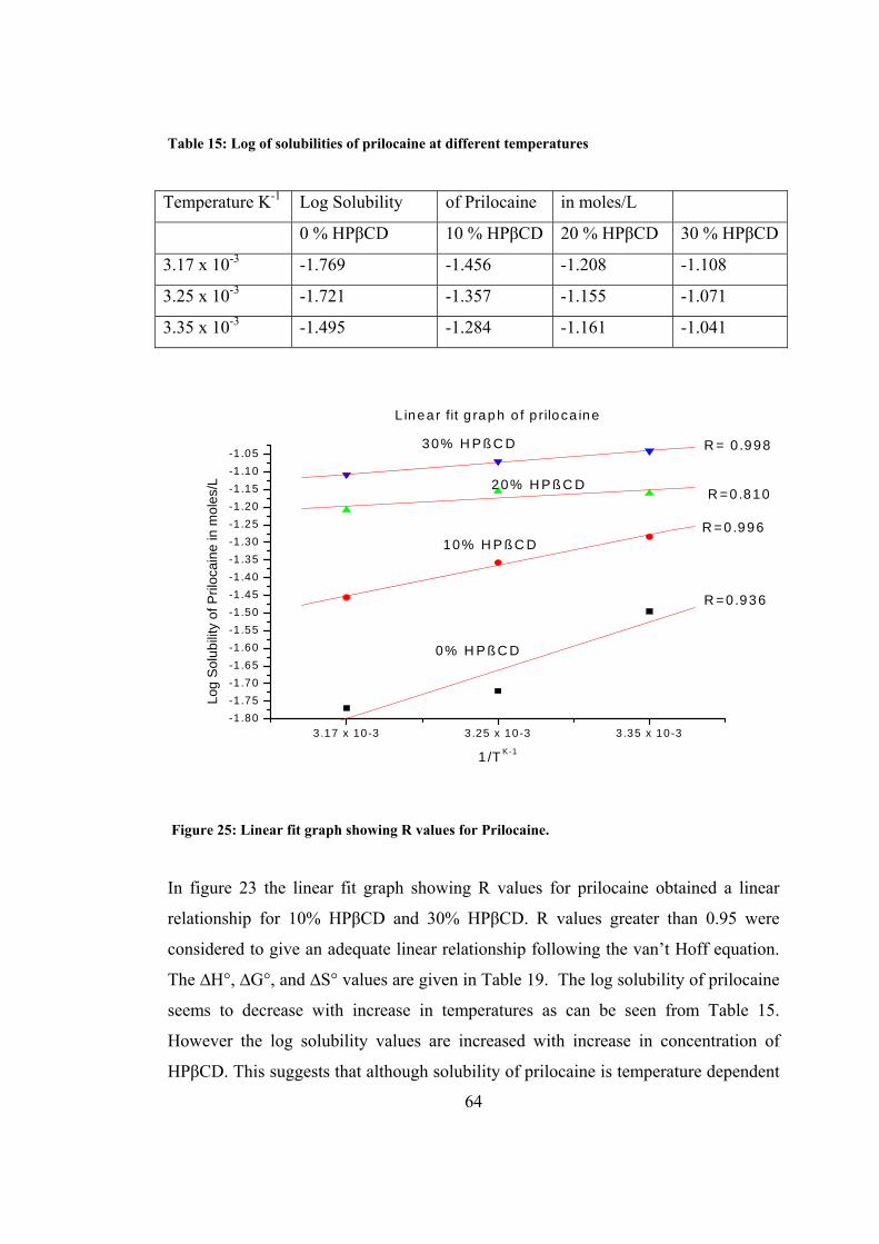
Table 15: Log of solubilities of prilocaine at different temperatures
Temperature K-1 Log Solubility of Prilocaine in moles/L
0 % HPβCD 10 % HPβCD 20 % HPβCD 30 % HPβCD
3.17 x 10⁻³ -1.769 -1.456 -1.208 -1.108
3.25 x 10⁻³ -1.721 -1.357 -1.155 -1.071
3.35 x 10⁻³ -1.495 -1.284 -1.161 -1.041
3.17 x 10⁻³ 3.25 x 10⁻³ 3.35 x 10⁻³-1 .80-1 .75-1 .70-1 .65-1 .60-1 .55-1 .50-1 .45-1 .40-1 .35-1 .30-1 .25-1 .20-1 .15-1 .10-1 .05
30% H P ß C D
20% H P ß C D
10% H P ß C D
0% H P ß C D
L inear fit g raph o f p riloca ine
R = 0 .998
R =0 .810
R =0.996
R =0.936
Log
Sol
ubili
ty o
f Pril
ocai
ne in
mol
es/L
1 /T K -1
Figure 25: Linear fit graph showing R values for Prilocaine.
In figure 23 the linear fit graph showing R values for prilocaine obtained a linear
relationship for 10% HPβCD and 30% HPβCD. R values greater than 0.95 were
considered to give an adequate linear relationship following the van’t Hoff equation.
The ∆H°, ∆G°, and ∆S° values are given in Table 19. The log solubility of prilocaine
seems to decrease with increase in temperatures as can be seen from Table 15.
However the log solubility values are increased with increase in concentration of
HPβCD. This suggests that although solubility of prilocaine is temperature dependent
64

an increase in solubility is obtained at higher concentrations of HPβCD. The enthalpy
and entropy values as in Table 19 give an idea of how the complexation of prilocaine-
HPβCD occurs. As is seen in Table 19 the ∆H° values are relatively similar and small
difference exists between enthalpies dependent upon the concentration of HPβCD. A
greater difference in entropy values was obtained and this suggests that complexation
is governed more by entropy rather than enthalpy. Hence the orientation of the drug
in the complex may be an important factor.
Table 16: Log of solubilities of lignocaine different temperatures
Temperature K-1 Log Solubility of Lignocaine in moles/L
0 % HPβCD 10 % HPβCD 20 %
HPβCD
30 % HPβCD
3.17 x 10⁻³ -2.000 -1.678 -1.387 -1.347
3.25x 10⁻³ -2.000 -1.699 -1.432 -1.377
3.35 x 10⁻³ -1.796 -1.596 -1.432 -1.367
3 .10 x 10 -3 3 .20x 10 -3 3 .35 x 10 -3
-2 .00
-1 .95
-1 .90
-1 .85
-1 .80
-1 .75
-1 .70
-1 .65
-1 .60
-1 .55
-1 .50
-1 .45
-1 .40
-1 .35
L in e a r fit g ra p h o f lig n o ca in e
3 0 % H P ß C D
2 0 % H P ß C D
1 0 % H P ß C D
0 % H P ß C D
R = -0 .6 5 4
R = -0 .8 6 6
R = 0 .7 5 3 3
R = 0 .8 6 6
Log
Solu
bilit
y of
Lig
noca
ine
in m
oles
/L
1 /T K -1
Figure 26: Linear fit graph showing R values for lignocaine.
65

In Figure 24 the linear fit graph shows that solubility of lignocaine increases with
increase in concentration of HPβCD, however it is not very significant change and
therefore when the temperature was raised almost no change in the log solubility
values were obtained when plotted against absolute temperature. The nonlinear
relationship was obtained for all the concentrations of HPβCD and thus van’t Hoff’s
equation was not followed between any two of the temperatures.
Table 17: Log solubilities of prilocaine and lignocaine in combination at different temperatures
Temperature K-
1
Log Solubility of Prilocaine in presence of Lignocaine in moles/L
0 % HPßCD 10 % HPßCD 20 % HPßCD 30 % HPßCD
3.17 x 10⁻³ -2.174 -1.795 -1.619 -1.444
3.25 x 10⁻³ -2.193 -1.833 -1.770 -1.540
3.35 x 10⁻³ -2.046 -1.641 -1.575 -1.574
3.10 x 10⁻³ 3.20 x 10⁻³ 3.35 x 10⁻³
-2.2
-2.1
-2.0
-1.9
-1.8
-1.7
-1.6
-1.5
-1.4
Linear fit graph of prilocaine in presence of lignocaine
30%HPßCD
20%HPßCD
10%HPßCD
0%HPßCD R=0.800
R=0.757R=0.215
R= -0.964
Log
Sol
ubili
ty o
f Pril
ocai
ne in
mol
es/L
1/TK-1
Figure 27: Linear fit graph showing R values for Prilocaine in presence of Lignocaine.
66

Linear fit graph showing R values for Prilocaine in presence of Lignocaine that shows
the complex formation of prilocaine in presence of lignocaine with HPβCD. The log
solubility seems to increase with increase in concentrations of HPβCD from 0 to 30%.
However the log solubility was found to decrease with increase in temperature. The
concentration of HPβCD (0%-20%) showed nonlinear relationships whereas a linear
relationship was obtained for 30% HPβCD and was associated with the negative ∆H°.
The ∆H° value obtained was very small and ∆S° value was very large suggesting
hydrophobic interactions during complex formation.
Table 18: Log solubilities of lignocaine in combination with prilocaine at different temperatures
Temperature K-1 Log
Solubility
of Lignocaine in presence of Prilocaine
in moles/L
0 % HPßCD 10 % HPßCD 20 %
HPßCD
30 % HPßCD
3.17 x 10⁻³ -1.745 -1.408 -1.201 -1.081
3.25 x 10⁻³ -1.958 -1.678 -1.495 -1.276
3.35 x 10⁻³ -2.076 -1.748 -1.677 -1.641
3 . 1 7 x 1 0 - 3 3 . 2 5 x 1 0 - 3 3 . 3 5 x 1 0 - 3- 2 . 2
- 2 . 0
- 1 . 8
- 1 . 6
- 1 . 4
- 1 . 2
- 1 . 0
L in e a r f i t g r a p h o f l i g n o c a in e i n p r e s e n c e o f p r i l o c a in e
3 0 % H P ß C D
2 0 % H P ß C D
1 0 % H P ß C D
0 % H P ß C D
R = - 0 . 9 8 5R = - 0 . 9 9 1R = - 0 . 9 4 7
R = - 0 . 9 8 7Log
Solu
bilit
y of
Lig
noca
ine
in m
oles
/L
1 / T K - 1
Figure 28: Linear fit graph showing R values of Lignocaine in presence of Prilocaine.
67

68
Linear fit graph showing R values of lignocaine in presence of prilocaine suggesting
that a linear relationship is followed at all the concentrations of HPβCD and van’t
Hoff equation is followed. The log solubility increases with increase in concentration
of HPβCD and with rise in temperature. However when ∆H° and ∆S° values were
calculated (Table 19) a small positive ∆H° and a large ∆S° was obtained suggesting
that the complex formation is driven by entropy. The negative R values gave a
negative intercept.
Table 19: Thermodynamic parameters of prilocaine and lignocaine following a linear
relationship at 25°C, 35°C and 42°C
Prilocaine ∆H° Jmol-1 ∆G° Jmol-1 ∆S° JK-1mol-1
0% HPßCD -2.62 20761.11 -65.92
10% HPßCD -1.65 14978.25 -47.55
30% HPßCD -0.64 9099.50 -28.89
Prilocaine in presence of
Lignocaine
∆H° Jmol-1 ∆G° Jmol-1 ∆S° JK-1mol-1
30% HPßCD -25.20 247650.25 786.27
Lignocaine in presence
of Prilocaine
∆H° Jmol-1 ∆G° Jmol-1 ∆S° JK-1mol-1
0% HPßCD 2.44 -10063.91 -39.94
10% HPßCD 2.38 -11487.31 -36.46
20% HPßCD 3.84 -25215.26 -80.04
30% HPßCD 4.50 -31825.02 -101.02

3.3 INVESTIGATION OF HPßCD COMPLEX FORMATION WITH
PRILOCAINE AND LIGNOCAINE BY NMR
Complex formation of prilocaine and lignocaine with HPßCD was also investigated
by NMR. NMR spectroscopy is one of the most used techniques to obtain information
about the geometry of inclusion complexes. The chemical formula of prilocaine is
C13H20N2O and the chemical formula of lignocaine is C14H22N2O. Figure 29 and
Figure 30 represents the chemical structures of prilocaine and lignocaine for
evaluating the chemical shift and changes in characterisation of peaks obtained by
complexation with HPßCD. Table 20 and Table 21 represents the protons of
prilocaine and lignocaine involved in a chemical shifts with and without HPßCD and
their ∆δ (ppm).
C H 3
N H C C H
C H 3
NH
C H 2
C H 2
C H 3
O
( C )
( D )
( G )( H )
( A )( B )
( E )
( F )( I )
( J )
Figure 29: Chemical structure of prilocaine.
Table 20: 1H NMR showing chemical shift for prilocaine with and without HPßCD
Assignments Prilocaine without
HPβCD (ppm)
Prilocaine with
HPβCD (ppm)
∆δ
(ppm)
A- triplet 0.905 0.763 - 0.142
B- multiplex 1.515 1.373 - 0.142
C- triplet 2.569 2.424 - 0.145
D- doublet 1.370 1.235 - 0.135
E- quadruplet 3.505 * *
F- singlet 2.214 2.132 - 0.082
G and I- broad doublet 7.296 7.113 - 0.183
H and J- broad multiplex 7.297 7.193 - 0.104
69

C H 3
N H
C H 3
C
O
C H 2 NC H 2 C H 3
C H 2 C H 3
H C l
Figure 30: Chemical structure of lignocaine
Table 21: 1H NMR showing chemical shift for lignocaine with and without HPßCD
Assignments Lignocaine without
HPβCD (ppm)
Lignocaine with
HPβCD (ppm)
∆δ
(ppm)
A – triplet 1.113 * *
B – quadruplet 2.710 2.570 - 0.140
C – singlet 3.404 * *
D – singlet 2.180 2.062 - 0.118
E – broad doublet 7.180 7.033 - 0.147
F – broad multiplex 7.218 7.065 - 0.153
An (*) means no chemical shift was obtained.
In the 1H-NMR peak assignments for free and complex prilocaine and lignocaine the
corresponding shifts ∆δ is represented as ∆δ = δ (complex) – δ (free). The prilocaine
and prilocaine HPßCD and ligocaine and lignocaine HPßCD complexes were
prepared as previously described in Section 2.4 and were dissolved in D2O. Chemical
shifts were given in parts per million relative to the signal obtained. 1H NMR studies
showed upfield shifts for all the protons from A-H for prilocaine and A-F for
lignocaine.
70

71
igure 31 (a), (b), (c), (d), and (e) represents the NMR peaks where,
e
ith HPβCD
ith HPβCD
F
(a) NMR of HPβCD
(b) NMR of prilocain
(c) NMR of prilocaine w
(d) NMR of lignocaine
(e) NMR of lignocaine w

Fig 29 (a) NMR of HPßCD
72

Fig 29 (b) NMR of Prilocaine
A- 0.905
B- 1.515
C- 2.569
D- 1.370
E- 3.505
F- 2.214
G- 7.296
H- 7.297
73

Fig 29 (c) NMR of Prilocaine with HPßCD
A- 0.763
B- 1.373
C- 2.424
D- 1.235
E- *
F- 2.132
G- 7.113
H- 7.193
74

Fig 29 (d) NMR of Lignocaine
A- 1.113
B- 2.710
C- 3.404
D- 2.180
E- 7.180
F- 7.218
75

Fig 29 (e) NMR of Lignocaine with HPßCD
A- *
B- 2.570
C- *
D- 2.062
E- 7.033
F- 7.065
Figure 31: 1H NMR spectra of (a) HPßCD, (b) prilocaine, (c) lignocaine, (d) prilocaine-HPßCD
complex and (e) lignocaine-HPßCD complex.
76

3.4 GRAPHICAL COMPARISON OF CHEMICAL SHIFTS OF PROTONS
OF PRILOCAINE AND LIGNOCAINE IN THE PRESENCE AND
ABSENCE OF HPβCD
Chemical shifts of protons for prilocaine and lignocaine in the presence and absence
of HPßCD are represented graphically in Figure 32 and Figure 33. The chemical shift
and change in peak character was observed where complexation for prilocaine and
lignocaine occurred through HPßCD. The NMR’s are given in Section 3.3. The peak
suppression due to complexation for prilocaine with HPßCD is represented
graphically in Figure 30. At ‘E’ no chemical shift was observed.
Chemical shift of prilocaine with and without HPßCD
0
2
4
6
8
A B C D E F G H
Chemical shift of protons
Val
ues o
f pro
ton
shift
Prilocaine without HPßCDPrilocaine with HPßCD
Figure 32: Chemical shift of prilocaine in the presence and absence of HPßCD
77
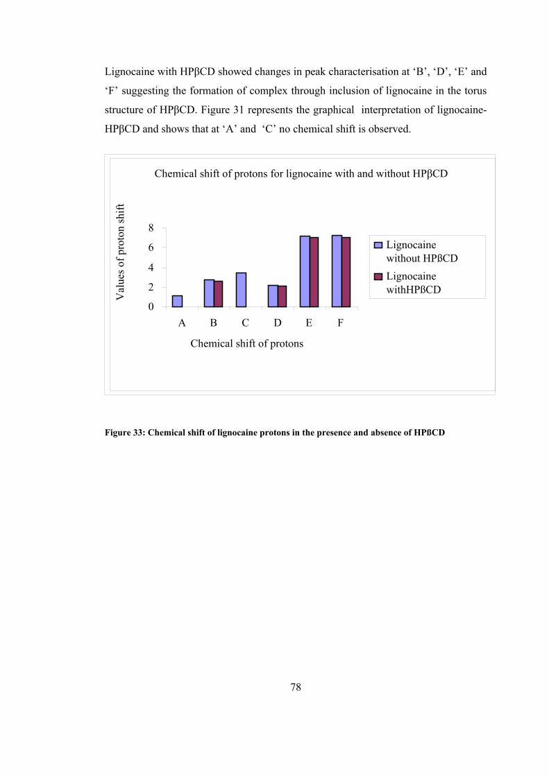
Lignocaine with HPβCD showed changes in peak characterisation at ‘B’, ‘D’, ‘E’ and
‘F’ suggesting the formation of complex through inclusion of lignocaine in the torus
structure of HPβCD. Figure 31 represents the graphical interpretation of lignocaine-
HPβCD and shows that at ‘A’ and ‘C’ no chemical shift is observed.
Chemical shift of protons for lignocaine with and without HPβCD
0
2
4
6
8
A B C D E F
Chemical shift of protons
Val
ues o
f pro
ton
shift
Lignocainewithout HPßCDLignocainewithHPßCD
Figure 33: Chemical shift of lignocaine protons in the presence and absence of HPßCD
78

79
4 DISCUSSION
The validated method was initially developed to separate three local anaesthetic drugs
namely prilocaine HCl, lignocaine HCl, and tetracaine HCl. The R values obtained
for prilocaine HCl, lignocaine HCl, and tetracaine HCl were 0.999, 0.999, and 0.997
respectively. Tetracaine HCl was later excluded from the studies as complex
formation with the combination of three drugs seemed unfeasible. A second method
was then developed only for prilocaine HCl and lignocaine HCl. Reproducible results
were obtained and the next step than was to determine the solubility of prilocaine and
lignocaine and to enhance this by complexing it with HPβCD.
Solubility is the basic requirement for any formulation that needs to be prepared in a
solution form. Also solubility enhancement has broad implications in parenteral
formulation design. Although many drugs are fairly soluble in aqueous solutions,
lipophilic drugs impose a solubility problem to be able to formulate as a solution. A
variety of solubilisation techniques has been studied and widely used including pH
adjustment, cosolvent addition, surfactant addition and cyclodextrin addition40.
Complexation with cyclodextrins is now a commonly used technique to improve drug
solubility from number of other methods.
Although ionised drugs show greater aqueous solubility, the unionised form
(molecular form) of drugs can be solubilised by complexation and pH adjustment.
Prilocaine and lignocaine have the pKa values of 7.9 and 7.8 respectively therefore
the pH of the solutions were maintained at 10.0. The lipophilic form of the drug may
be advantageous for rapid onset of action if sufficient can be delivered in a solution
formulation.
Complex formation with CD involves the release of enthalpy-rich water from the CD
cavity as the main driving force for complexation. Other mechanisms that may be
involved in complex formation include van der Waals interactions, hydrogen bonding
and hydrophobic interactions. In the case of α-CD, release of ring strain is also
thought to be involved with the driving force for compound–CD interaction76.

80
Accordingly, different types of solubility isotherms are obtained as a result of
complex formation with cyclodextrins which include AL, AP, and AN types of
isotherm. The AL type of solubility isotherms was obtained for prilocaine and
lignocaine with HPβCD. As mentioned above, the solubilities of prilocaine and
lignocaine at higher temperatures were found to differ individually. One reason that
can be proposed for an unexpected decrease in solubility of prilocaine at higher
temperatures is its existence as R and S stereoisomer’s. The spatial arrangements of
these two stereoisomers of prilocaine at higher temperatures may limit its complex
formation with HPβCD and affects its solubility and perhaps potency77.
Many drugs studied in combination have either shown increase in solubility as
expected or variations from solubility. Sometimes not the other drug but the cosolvent
used along with the complexing agent was found to compete with drug for the entry
into the HPβCD cavity decreasing the drug solubility. This has been found with the
drug fluasterone where an overall decrease in solubility and complex formation
occurred when complexed with HPβCD40. Although cosolvent can act as a space
regulating molecule for the drug molecule to fit into cyclodextrin, research done by
Pitha et al. reported that the complexation constant of testosterone with HPβCD was
10,000 fold lowered in 80% ethanol than in water40.
Almost similar solubility constants were obtained for prilocaine in combination with
lignocaine when complexed with HPβCD. The solubility of prilocaine was again
found to be decreased in the presence of lignocaine with a rise in temperature, but the
differences obtained for solubilities between the temperatures were very small. The
above proposed mechanism may hold true for prilocaine solubility in combination or
it may be true again that in the presence of the other drug prilocaine showed
hinderence in complex formation and thus its solubility decreased. However
lignocaine solubility was found to rise effectively with rise in temperature as
compared to its no change in solubility individually. It seems that in combination with
prilocaine, lignocaine solubility was favoured.

81
Although prilocaine and lignocaine share structural similarities the difference’s in
solubility of these two drugs individually and in combination suggests varying
patterns of complexation and the possibility of specific interactions or mixed modes
of binding between different components or functional groups in the structure of the
substrate and cyclodextrin. Earlier studies have reported the varying modes of
complex formation with the relatively common structures of steroids, when
complexed by SBE-7-β-CD, which only differed in the D ring substituents78.
Thus the theory of mixed mode interactions for complex formation with HPßCD in
for prilocaine and lignocaine individually and in combination at higher temperatures
cannot be overruled.
The variability in complex formation with cyclodextrins can again be explained by
the well documented experimental work of two hydrophobic drugs, indomethacin and
griseofulvin, which have structural resemblance to each other. This was proved by
various methods such as X-ray powder diffraction, modulated temperature differential
scanning calorimetry and dissolution tests, which were used for the characterisation
of solid state inclusion complexes of indomethacin and griseofulvin with β-CD, α-CD,
and γ-CD in PEG 6000. The results showed that for griseofulvin no complex was
formed with β-CD and this was partly represented by the steric hindrance of
griseofulvin, whereas indomethacin in the presence of polyethylene glycol 6000
(PEG 6000) showed complex formation with β-CD and γ-CD, but not with α-CD.
This was because α-CD showed good complexation with PEG 6000 in the melt and β-
CD and γ-CD showed minimum interaction with the polymer and therefore could
complex with indomethacin76.
The variability of CD-drug complex can be further explained regarding to polymer
complexation with different types of CD’s. An aqueous solution of propylene glycol
at low concentrations reduces the CD complexation of some drugs by acting as a
competing guest molecule79. The α-CD forms complexes with PEG of various
molecular weights but, β-CD does not form complexes with PEG. This might
promote the inclusion complex formation for the drug in this type of CD. It was

82
shown that β- and γ-CD form complexes with other polymers like polypropylene
glycol (PPG) and polyisobutylene, while α-CD does not form complexes with either
of these polymers. β- and γ-CD might therefore be more alike concerning their
interactions with different polymers80. Selection of the correct CD’s therefore favours
the complex formation with the drug molecule further enhancing its solubility effect.
In yet another study it was found that co-administration of hydroxypropyl methyl
cellulose (HPMC) with SBE-7-β-CD sodium salt reduced the amount of β-CD needed
in a solid dosage form of glibenclamide, without reducing its bioavailability and
solubility. Compared to formulation without HPMC, more of the β-CD was needed
for inclusion formation though the bioavailability was not affected in both of the
formulations81.
The above findings in regard to complex formation with HPβCD suggest that
different interactions at the molecular level exist during complexation and use of
different CD’s with different substituents can give different results. These findings
lend support to the results obtained for solubilities of prilocaine and lignocaine.
The relative thermodynamic parameters for the complexation process are the standard
free energy change (∆G°), the standard enthalpy change (∆H°), and the standard
entropy change (∆S°). As revealed in the earlier data complexation of prilocaine and
lignocaine with HPβCD was accompanied by negative ∆H°, reflecting an exothermic
process driven enthalpically. The enthalpy driven processes are associated with
intermolecular interactions via hydrogen bonding and van der Waal’s interactions.
However the ∆H° and ∆S° values were not very significant for prilocaine and
lignocaine and overall they suggested that an endothermic reaction is involved driven
by entropy and involving hydrophobic interactions. Also nonlinearity observed at
some of the HPβCD concentrations suggested that van’t Hoff’s equation was not
followed.
Lignocaine alone and prilocaine in the presence of lignocaine showed non-linearity
for the van’t Hoff equation. It was noted that the solubilities were little affected by

83
temperature changes. But for lignocaine in presence of prilocaine the rise in
solubilities was seen with rise in temperature and all the concentrations of HPβCD
showed that van’t Hoff equation was essentially followed as R value was above 0.95
for all.
The thermodynamic parameter ∆H° was relatively small as little change occurred in
the solubility with a rise in temperature for both prilocaine and lignocaine. On the
other hand ∆S° values were significantly large and so it can be said that complex
formation was largely governed by entropy change.
The solubilities obtained for lignocaine individually showed little temperature effect,
whereas in combination the solubilities seemed to increase to a greater extent. In
contrast the solubilities of prilocaine decreased individually and also in combination.
This indicated that more than one mechanism is involved in the complexation process
and there may be a structural influence and stereoisomeric factors playing a role
along with the hydrogen bonding giving varied solubility results. Also the theory
proposed below can give an insight between the solution and the drug-HPβCD (guest-
HPβCD) complex formation and the difference that exists in the mechanism of
complexation.
The thermodynamic effects of β-CD and factors that result in different patterns of
complexation apart from what is expected is well discussed in the complex formation
of β-CD with different derivatives of azoalkanes leading to a host-guest relationship.
Derivatives of the azoalkane 2,3-diazabicyclo[2,2,2]oct-2-ene, 1,4-dialkyl, 1,4-
dichloro, 1-hydroxymethyl, 1-aminomethyl, and 1-ammoniummethyl substituents
form host-guest inclusion complexes with β-cyclodextrin. In that experimental work
they were employed as probes to assess substituent effects on the kinetics and
thermodynamics of this complexation by using time-resolved and steady state
fluorimetry, UV spectrophotometry, induced circular dichroism (ICD) measurements,
and 1H NMR spectroscopy. The ICD was employed for the assignment of the
solution structures of the complexes, in particular the relative orientation of the guest
in the host (co-conformation).

84
Accordingly the structure of the β-CD and 1-aminomethyl complex was assumed to
give rise to the negative ICD signal, but the ICD spectra revealed that the complex of
azoalkane 1-aminomethyl differed from the other derivatives and gave rise to an ICD
band, which was significantly distorted, and also showed a bathochromic shift
relative to the UV spectrum. Moreover, the ICD of 1-amino methyl complex showed
a small positive component at shorter wavelengths in addition to the stronger negative
ones. These irregularities suggested that a large co-conformational variability exist,
which meant that complexes with different tilt angles may have been present in
solution. It was found based on the results that for 1-aminomethyl, in contrast to 1-
hydroxymethyl and 1-ammoniummethyl, the co-conformational spaces were not
restricted by hydrogen bonding with the upper rim. The co-conformers with different
tilt angles had quite different UV spectra (which reflected the depth of inclusion into
the nonpolar environment) and ICD effects (which reflected the geometrical
alignment with respect to the azo chromophore) and, thus, accounted for the observed
shifts for 1-aminomethyl.
The hydrophobicity order of the examined guest molecules was established to be 1,4-
dichloro > 1,4-dialkyl > 2,3-diazabicyclo[2,2,2]oct-2-ene > 1-aminomethyl > 1-
hydroxymethyl and > 1-ammoniummethyl. A higher hydrophobicity was expected to
result in a larger binding constant. Azoalkane 1,4-dialkyl did not show a higher
binding affinity than 2,3-diazabicyclo[2,2,2]oct-2-ene despite its higher
hydrophobicity. This was rationalized in terms of the solution structures, which
indicated that the lateral co-conformation was energetically most favourable one and
could not be attained for 1,4-dialkyl. Instead, a frontal mode of inclusion was adapted
for 1,4-dialkyl which was energetically less favourable. This lessened the increased
driving force due to its higher hydrophobicity and resulted in essentially the same
binding constant as for 2,3-diazabicyclo[2,2,2]oct-2-ene.
Regarding the hydrophilicity generally, the more hydrophilic guests 1-aminomethyl
and 1-ammoniummethyl should show lower binding constants than 2,3-
diazabicyclo[2,2,2]oct-2-ene and 1,4-dichloro as the ionic ammonium guest displays

85
the weakest binding. The difference in binding constants between 1-aminomethyl
(250 M-1) and 1-ammoniummethyl (20 M-1) was in line with the general observation
that the ionization of the guest causes a destabilization of CD complexes in water. But
for the hydroxyl and ammonium derivatives 1-hydroxymethyl and 1-
ammoniummethyl, for which hydrogen bonding was presumed, it actually showed a
weaker binding than the amino derivative, for which hydrogen bonding to the CD
should have not played a role at all. This could be adequately accounted for in terms
of guest hydrophobicity and a change in the co-conformation due to the introduction
of bridgehead substituents82.
The above mentioned findings support the variability in solubilities obtained for
prilocaine and lignocaine and confirms that expected solubilities may not be obtained
as a result of complex formation with HPβCD and that variation in results can be
obtained due to structural differences between two drugs, intermolecular changes
because of the presence of other drug in combination and/or sterioisomeric
interferences and again the type of structural interactions taking place between the
guest molecule and CD. Competitive binding between prilocaine and lignocaine for
CD sites may be another factor.
To study the complex formation of prilocaine and lignocaine with HPβCD further,
NMR spectra’s for prilocaine and lignocaine were obtained at 600 Hz. The NMR
suggested that different chemical shifts were involved for both prilocaine and
lignocaine with few similarities. The NMR details of free prilocaine and prilocaine-
HPβCD complex and lignocaine and lignocaine-HPβCD were presented earlier in
Section 3.3.
The NMR of prilocaine showed involvement of aromatic protons, along with other
methene and methyl groups and the NMR of lignocaine showed involvement of
aromatic protons along with aromatic methyl and ethyl amide groups.
When lignocaine HPβCD NMR was compared with the similar NMR studies of
another local anaesthetic benzocaine and β-CD the results have shown similarites

86
between the complex formation that occured at ethyl-CH3, ethyl-CH2, and at the
aromatic protons H1-H6 for benzocaine83.
In another study done for the selectivity in the binding and detection of charge diffuse
ions, the neutral, lipophilic peralkylated cyclodextrin derivatives were found to
selectively bind the onium ions like R-NH3+ and NMe4
+. When local anaesthetics
such as lignocaine were studied in the presence of 2, 6- didodecyl –β-cyclodextrin as
the sensing ionophore it gave a Nernstian response at the NH+Et2 binding site.
The response was obtained down to 10-5.6 mol dm-3 concentrations. The nernstian
behaviour obtained showed that NH+Et2 ion is potentially active site for binding with
the cyclodextrin resulting in complex formation. The study showed that neutral,
lipophilic cyclodextrin derivatives can be used for the chemoselective detection of a
variety of aryl and alkyl ammonium ions84. Similarly a chemical shift of a triplet and
a multiplet NH+Et2 for lignocaine HPβCD was observed at the NH+Et2 binding site
along with the chemical shifts of aromatic protons and aromatic methyl group. This
would be less likely at high pH values used in this study.
The NMR details of prilocaine located from earlier studies in the literature found the
involvement of chiral carbon. The chiral solvating agent, 2,2,2-trifluoro-1-(9-anthryl)
ethanol, was used to determine the diasteriosmeric interactions and assignments of the
configuration was based on the relative field position of resolved enantiomeric
signals85.
At present preparationjs of parenteral dosage forms of local anaesthetics in solution is
acheived by means of their hydrochloride salts. Therefore the pH of these
preparations can be as low as 3.0 to 4.5. A possible precipitation of the base at the
injection site may occur due to an increase in pH to the tissue pH after injection
exacerbated by a lowering of free base solubility at body temperature86. A
formulation therefore of higher pH range say about pH 10.0, will keep the drugs in
unprotonated form and intact in a complex until it reaches its site of action. Tissue
and blood pH will have little effect on the drug formulation as they are not used in

87
their salt forms. Research has shown that the pH of above 11.5 and below 4.0 can
produce some pain during administration of anaesthetics, but no pain was observed in
individuals at pH 10.0. Therefore the parenteral formulation of prilocaine and
lignocaine base at pH 10.0 would be safe for parenteral purpose87.

88
5 CONCLUSIONS
Prilocaine and lignocaine share structural similarities as both are amide type local
anaesthetics. It was expected that similar solubility results would be obtained for both
individually and in combination, but the difference’s in solubility of these two drugs
individually and in combination suggests varying patterns of complexation and the
possibility of specific interactions or mixed modes of binding between different
components or functional groups in the structure of the substrate and cyclodextrin.
The complex formation showed that entropy rather than enthalpy was governing the
reaction for complexation form individually and in combination for both prilocaine
and lignocaine as ∆H° values were very small. There was only a small temperature
effect on solubility limiting the accuracy with which these effects could be described
The AL type solubility isotherm was obtained for all the solubilities suggesting that
binding involves 1:1 association between guest and CD molecule.
The NMR data showed that aromatic rings of both prilocaine and lignocaine are
involved in complex formation apart from the methene and methyl groups for
prilocaine and ethyl amide and aromatic methyl groups for lignocaine.
Solubilities of prilocaine determined over the concentration range of 0-30% HPβCD
were found to increase with increasing concentration of HPβCD and solubilities of
lignocaine showed little increase with increase in concentration of HPβCD. With the
rise in temperature the solubilities of prilocaine decreased, whereas again temperature
showed little effect on the solubilities of lignocaine. The solubilities obtained for
prilocaine at the temperatures 25°C, 35°C, and 42°C were between 3.22-9.11 moles/L,
1.96-8.51 moles/L and 1.79-7.91 moles/L respectively. The solubilities obtained for
lignocaine at the temperatures from 25°C-42°C were 1.69-4.41 moles/L, 1.04-4.22
moles/L and 1.04-4.55 moles/L respectively.

89
In combination, the solubilities of prilocaine and lignocaine showed variation in
complexation with HPβCD from the solubilities obtained individually. Prilocaine in
combination showed increase in solubility with increase in concentration of HPβCD
but showed decrease in solubilities with rise in temperature. Lignocaine on the other
hand showed increase in solubilities with increase in concentrations of HPβCD and
with rise in temperatures. The solubilities obtained for prilocaine at 25°C, 35°C, and
42°C were 0.91-2.67 moles/L, 0.64-2.89 moles/L and 0.66-3.68 moles/L respectively
and the solubilities obtained for lignocaine were 1.03-2.82 moles/L, 1.17-5.34
moles/L and 1.80-8.35 moles/L respectively.
From the data obtained in combination of prilocaine and lignocaine one can say that
solubilities of prilocaine showed little change with the rise in temperature but the
solubilities of lignocaine was found to increase in contrast to its solubilities obtained
individually.
A pH of 10.0 used for the development of parenteral formulation should not cause
irritation at the site of injection, but more studies need to be done regarding the cell
permeability of HPβCD and drug complex formation and how fast the onset of action
is achieved compared to the dissociation of local anaesthetics as their hydrochloride
salts.

90
References:
1. Vadim Kotelnikov. In: Happiness.
2. Daniel B Carr LCG. Acute Pain. The Lancet. 1999; 353:2051-58.
3. Patel SS, Goa KL. Sevoflurane. A review of its pharmacodynamic and
pharmacokinetic properties and its clinical use in general anaesthesia. Drugs. 1996;
51(4):658-700.
4. Rang HP, Dale MM. Pharmacology. fourth edition ed. Sydney: Churchill
Livingstone; 1999; p. 638-640.
5. Zhu XL, Chen ZL, Li GF, Zeng K. [Preparation and electron microscopic
observation of lidocaine microemulsion]. Nan Fang Yi Ke Da Xue Xue Bao. 2006;
26(4):515-7, 525.
6. Sintov AC, Brandys-Sitton R. Facilitated skin penetration of lidocaine:
Combination of a short-term iontophoresis and microemulsion formulation. Int J
Pharm. 2006; 316:58-67.
7. Fishbain DA, Lewis JE, Cole B, Cutler B, Rosomoff HL, Rosomoff RS.
Lidocaine 5% patch: an open-label naturalistic chronic pain treatment trial and
prediction of response. Pain Med. 2006; 7(2):135-42.
8. Burgess C, Ravenscroft AJ. The lidocaine patch: a useful addition to the pain
clinic armamentarium. Anaesthesia. 2006; 61(4):412.
9. Goldman RD. ELA-max: A new topical lidocaine formulation. Ann
Pharmacother. 2004; 38(5):892-4.
10. Koh JL, Harrison D, Myers R, Dembinski R, Turner H, McGraw T. A
randomized, double-blind comparison study of EMLA and ELA-Max for topical
anesthesia in children undergoing intravenous insertion. Paediatr Anaesth. 2004;
14(12):977-82.
11. Rishiraj B, Epstein JB, Fine D, Nabi S, Wade NK. Permanent vision loss in
one eye following administration of local anesthesia for a dental extraction. Int J Oral
Maxillofac Surg. 2005; 34(2):220-3.
12. Karalezli N, Karalezli K, Iltar S, Cimen O, Aydogan N. Results of intravenous
regional anaesthesia with distal forearm application. Acta Orthop Belg. 2004;
70(5):401-5.

91
13. Cereda CM, de Araujo DR, Brunetto GB, De Paula E. Liposomal prilocaine:
preparation, characterization, and in vivo evaluation. J Pharm Pharm Sci. 2004;
7(2):235-40.
14. Tham EJ MS, Wright EM, Campbell IA, Mapleson WW. An assessment of
prilocaine as a topical anaesthetic agent for fibreoptic bronchoscopy in comparison
with lidocaine. Acta Anaesthesiol Scand. 1994; 38(5):442-7.
15. Kreilgaard M. Dermal pharmacokinetics of microemulsion formulations
determined by in vivo microdialysis. Pharm Res. 2001; 18(3):367-73.
16. Abu Al-Melh M, Andersson L, Behbehani E. Reduction of pain from needle
stick in the oral mucosa by topical anesthetics: a comparative study between
lidocaine/prilocaine and benzocaine. J Clin Dent. 2005; 16(2):53-6.
17. Tang MB, Goon AT, Goh CL. Study on the efficacy of ELA-Max (4%
liposomal lidocaine) compared with EMLA cream (eutectic mixture of local
anesthetics) using thermosensory threshold analysis in adult volunteers. J Dermatolog
Treat. 2004; 15(2):84-7.
18. Soderberg L, Dyhre H, Roth B, Bjorkman S. Ultralong peripheral nerve block
by lidocaine:prilocaine 1:1 mixture in a lipid depot formulation: comparison of in
vitro, in vivo, and effect kinetics. Anesthesiology. 2006; 104(1):110-21.
19. Corbett IP, Ramacciato JC, Groppo FC, Meechan JG. A survey of local
anaesthetic use among general dental practitioners in the UK attending postgraduate
courses on pain control. Br Dent J. 2005; 199(12):784-7; discussion 778.
20. Alster TS, Lupton JR. Evaluation of a novel topical anesthetic agent for
cutaneous laser resurfacing: a randomized comparison study. Dermatol Surg. 2002;
28(11):1004-6; discussion 1006.
21. Nathan A, Rose JB, Guite JW, Hehir D, Milovcich K. Primary
erythromelalgia in a child responding to intravenous lidocaine and oral mexiletine
treatment. Pediatrics. 2005; 115(4):e504-7.
22. Sintov AC, Shapiro L. New microemulsion vehicle facilitates percutaneous
penetration in vitro and cutaneous drug bioavailability in vivo. J Control Release.
2004; 95(2):173-183.

92
23. Singer AJ, Regev R, Weeks R, Tlockowski DS. Laser-assisted anesthesia
prior to intravenous cannulation in volunteers: a randomized, controlled trial. Acad
Emerg Med. 2005; 12(9):804-7.
24. Rodriguez-Chinchilla R, Rodriguez-Pont A, Pintanel T, Vidal-Lopez F.
Bilateral severe pain at L3-4 after spinal anaesthesia with hyperbaric 5% lignocaine.
Br J Anaesth [Case Report]. 1996; 76(2):328-329.
25. Gomez-Orellana I. Strategies to improve oral bioavaliability. Drug Delivery
[Review -Expert Opinion]. 2005; 2(3):419-433.
26. Florence AT, Attwood D. Physicochemical Principles of pharmacy. Third ed:
MacMillan press ltd., London; 1998; p. 152-153.
27. Yalkowsky SH. Techniques of solubilisation of the drugs. Marcel Dekker,
Madiso Avenye, New york,10016; 1981; p. 130.
28. Tommasini S, Calabro ML, Raneri D, Ficarra P, Ficarra R. Combined effect
of pH and polysorbates with cyclodextrins on solubilization of naringenin. J Pharm
Biomed Anal. 2004; 36(2):327-333.
29. Tinwalla AY, Hoesterey BL, Xiang T-x, Lim K, Anderson BD. Solubilization
of Thiazolobenzimidazole Using a Combination of pH Adjustment and Complexation
with 2-Hydroxypropyl-ß-Cyclodextrin. Pharm Res. 1993; 10(8):1136-1143.
30. Pudipeddi M, Serajuddin AT. Trends in solubility of polymorphs. J Pharm Sci.
2005; 94(5):929-39.
31. Hancock BC, Parks M. What is the true solubility advantage for amorphous
pharmaceuticals? Pharm Res. 2000; 17(4):397-404.
32. Zuo Z, Kwon G, Stevenson B, Diakur J, Wiebe LI. Flutamide-hydroxypropy-
beta-chiyclodextrin complex: formulation, physical characterization, and absorption
studies using the Caco-2 in vitro model. J Pharm Pharm Sci. 2000; 3(2):220-7.
33. von Corswant C, Thoren P, Engstrom S. Triglyceride-based microemulsion
for intravenous administration of sparingly soluble substances. J Pharm Sci. 1998;
87(2):200-8.
34. Kawakami K, Oda N, Miyoshi K, Funaki T, Ida Y. Solubilization behavior of
a poorly soluble drug under combined use of surfactants and cosolvents. Eur J Pharm
Sci. 28(1-2):7-14.

93
35. Vijaya Kumar SG, Mishra DN. Preparation, characterization and in vitro
dissolution studies of solid dispersion of meloxicam with PEG 6000. Yakugaku
Zasshi. 2006; 126(8):657-64.
36. Jiahui H, True LR, Judith B, Tim Y, Keith PJ, Robert OW, III. Improvement
of Dissolution Rates of Poorly Water Soluble APIs Using Novel Spray Freezing into
Liquid Technology. Pharm Res. 2002; 19(9):1278-1284.
37. Singhai AK, Jain S, Jain NK. Cosolvent solubilization and formulation of an
aqueous injection of ketoprofen. Pharmazie. 1996; 51(10):737-40.
38. Han SK, Kim GY, Park YH. Solubilization of biphenyl dimethyl
dicarboxylate by cosolvency. Drug Dev Ind Pharm. 1999; 25(11):1193-7.
39. Dannenfelser RM, Surakitbanharn Y, Tabibi SE, Yalkowsky SH. Parenteral
formulation of Flavopiridol (NSC-649890). PDA J Pharm Sci Technol. 1996;
50(6):356-9.
40. Li P, Zhao L, Yalkowsky SH. Combined effect of cosolvent and cyclodextrin
on solubilization of nonpolar drugs. J Pharm Sci. 1999; 88(11):1107-11.
41. Millard J, Alvarez-Nunez F, Yalkowsky S. Solubilization by cosolvents.
Establishing useful constants for the log-linear model. Int J Pharm. 2002; 245(1-
2):153-66.
42. Jouyban A, Chew NY, Chan HK, Sabour M, Acree WE, Jr. A unified
cosolvency model for calculating solute solubility in mixed solvents. Chem Pharm
Bull (Tokyo). 2005; 53(6):634-7.
43. Fialho SL, da Silva-Cunha A. New vehicle based on a microemulsion for
topical ocular administration of dexamethasone. Clin Experiment Ophthalmol. 2004;
32(6):626-32.
44. Hua L, Weisan P, Jiayu L, Ying Z. Preparation, evaluation, and NMR
characterization of vinpocetine microemulsion for transdermal delivery. Drug Dev
Ind Pharm. 2004; 30(6):657-66.
45. Ghosh PK, Majithiya RJ, Umrethia ML, Murthy RS. Design and development
of microemulsion drug delivery system of acyclovir for improvement of oral
bioavailability. AAPS PharmSciTech. 2006; 7(3):77.
46. Lawrence MJ, Rees GD. Microemulsion-based media as novel drug delivery
systems. Adv Drug Del Rev. 2000; 45(1):89-121.
94
47. Agatonovic-Kustrin S, Glass BD, Wisch MH. Strategy for the development of
a thermodynamically stable oral microemulsion. Curr Drug Discov Technol. 2004;
1(2):165-71.
48. Chen H, Chang X, Du D, Li J, Xu H, Yang X. Microemulsion-based hydrogel
formulation of ibuprofen for topical delivery. Int J Pharm. 2006.
49. Kawakami K, Yoshikawa T, Moroto Y, Kanaoka E, Takahashi K, Nishihara Y,
et al. Microemulsion formulation for enhanced absorption of poorly soluble drugs: I.
Prescription design. J Control Release. 2002; 81(1-2):65-74.
50. Razavizadeh BM, Mousavi-Khoshdel M, Gharibi H, Behjatmanesh-Ardakani
R, Javadian S, Sohrabi B. Thermodynamic studies of mixed ionic/nonionic surfactant
systems. J Colloid Interface Sci. 2004; 276(1):197-207.
51. Ho HO, Hwang MC, Tseng SL, Lin LH, Chen KT, Chiang HS, et al. The
percutaneous penetration of prostaglandin E1 and its alkyl esters. J Control Release.
1999; 58(3):349-55.
52. Watnasirichaikul S, Rades T, Tucker IG, Davies NM. In-vitro release and oral
bioactivity of insulin in diabetic rats using nanocapsules dispersed in biocompatible
microemulsion. J Pharm Pharmacol. 2002; 54(4):473-80.
53. Ryoo HK, Park CW, Chi SC, Park ES. Development of propofol-loaded
microemulsion systems for parenteral delivery. Arch Pharm Res. 2005; 28(12):1400-
4.
54. Zhao X, Chen D, Gao P, Ding P, Li K. Synthesis of ibuprofen eugenol ester
and its microemulsion formulation for parenteral delivery. Chem Pharm Bull (Tokyo).
2005; 53(10):1246-50.
55. Park KM, Kim CK. Preparation and evaluation of flurbiprofen-loaded
microemulsion for parenteral delivery. Int J Pharm. 1999; 181(2):173-9.
56. Taha EI, Al-Saidan S, Samy AM, Khan MA. Preparation and in vitro
characterization of self-nanoemulsified drug delivery system (SNEDDS) of all-trans-
retinol acetate. Int J Pharm. 2004; 285(1-2):109-19.
57. Wei L, Sun P, Nie S, Pan W. Preparation and evaluation of SEDDS and
SMEDDS containing carvedilol. Drug Dev Ind Pharm. 2005; 31(8):785-94.
58. Szejtli J. Cyclodextrins. In: Cyclodextrin Technology. Netherlands: Kluwer
Academic Publishers; 1988. p. 1-2.

95
59. Loftsson T, Jarho P, Masson M, Jarvinen T. Cyclodextrins in drug delivery.
Expert Opin Drug Deliv. 2005; 2(2):335-51.
60. Rao VM, Stella, V.J. When can cyclodextrins be considered for solubilization
purposes? J Pharm Sci. 2003; 92(5):927-932.
61. Loftsson T, Hreinsdottir D, Masson M. Evaluation of cyclodextrin
solubilization of drugs. Int J Pharm. 2005; 302(1-2):18-28.
62. Mura P, Adragna E, Rabasco AM, Moyano JR, Perez-Martinez JI, Arias MJ,
et al. Effects of the host cavity size and the preparation method on the
physicochemical properties of ibuproxam-cyclodextrin systems. Drug Dev Ind Pharm.
1999; 25(3):279-87.
63. Tros de Ilarduya MC, Martin C, Goni MM, Martinez-Oharriz MC.
Solubilization and interaction of sulindac with beta-cyclodextrin in the solid state and
in aqueous solution. Drug Dev Ind Pharm. 1998; 24(3):301-6.
64. Challa R, Ahuja A, Ali J, Khar RK. Cyclodextrins in drug delivery: an
updated review. AAPS PharmSciTech. 2005; 6(2):E329-57.
65. Blanchard J, Proniuk S. Some important considerations in the use of
cyclodextrins. Pharm Res. 1999; 16(12):1796-8.
66. Zhang M-Q. RDC. A review of recent applications of cyclodextrins for drug
discovery. Expert Opinion On Therapeutic Patents [Review]. 1999; 9(12):1697-1717.
67. Arima H, Yunomae K, Miyake K, Irie T, Hirayama F, Uekama K.
Comparative studies of the enhancing effects of cyclodextrins on the solubility and
oral bioavailability of tacrolimus in rats. J Pharm Sci. 2001; 90(6):690-701.
68. Jarho P, Jarvinen K, Urtti A, Stella VJ, Jarvinen T. Modified beta-
cyclodextrin (SBE7-beta-CyD) with viscous vehicle improves the ocular delivery and
tolerability of pilocarpine prodrug in rabbits. J Pharm Pharmacol. 1996; 48(3):263-9.
69. Gu FG, Cui FD, Gao YL. Preparation of prostaglandin E1-hydroxypropyl-
beta-cyclodextrin complex and its nasal delivery in rats. Int J Pharm. 2005; 290(1-
2):101-8.
70. Yanagi H, Ishikura N, Kenmotsu H, Yagi N, Goto K, Kato S, et al. [Effect of
inclusion complexation of decanoic acid with alpha-cyclodextrin on rectal absorption
of cefmetazole sodium suppository in rabbits]. Yakugaku Zasshi. 1991; 111(1):65-9.

96
71. Blanchard J, Ugwu SO, Bhardwaj R, Dorr RT. Development and testing of an
improved parenteral formulation of phenytoin using 2-hydroxypropyl-beta-
cyclodextrin. Pharm Dev Technol. 2000; 5(3):333-8.
72. Holvoet C, Heyden YV, Plaizier-Vercammen J. Inclusion complexation of
diazepam with different cyclodextrins in formulations for parenteral use. Pharmazie.
2005; 60(8):598-603.
73. Bhardwaj R, Dorr RT, Blanchard J. Approaches to reducing toxicity of
parenteral anticancer drug formulations using cyclodextrins. PDA J Pharm Sci
Technol. 2000; 54(3):233-9.
74. Lyons G, Columb M, Hawthorne L, Dresner M. Extradural pain relief in
labour: bupivacaine sparing by extradural fentanyl is dose dependent. Br J Anaesth.
1997; 78(5):493-7.
75. O'Neil MJ, editor. The Merck index. Thirteenth Edition ed: Merck Research
Laboratories Division of Merck and Company; 2001.
76. Wulff M, Alden M. Solid state studies of drug-cyclodextrin inclusion
complexes in PEG 6000 prepared by a new method. Eur J Pharm Sci. 1999; 8(4):269-
281.
77. Calvey TN. Chirality in anaesthesia. Anaesthesia. 1992; 47(2):93-94.
78. Zia V, Rajewski, RA, Stella, VJ. Thermodynamics of Binding of Neutral
Molecules to Sulfobutyl Ether β-Cyclodextrins (SBE-β-CDs): The Effect of Total
Degree of Substitution Pharm Res. 2000; 17(8):936-41.
79. Loftsson T, Brewster ME. Pharmaceutical applications of cyclodextrins. 1.
Drug solubilization and stabilization. J Pharm Sci. 1996; 85(10):1017-25.
80. A Harada JL, S Suzuki, M Kamachi. In: Complex formation between
polyisobutylene and cyclodextrins: inversion of chain-length selectivity between beta-
cyclodextrin and alpha- cyclodextrin. 1993
81. Savolainen J, Jarvinen K, Taipale H, Jarho P, Loftsson T, Jarvinen T. Co-
administration of a water-soluble polymer increases the usefulness of cyclodextrins in
solid oral dosage forms. Pharm Res. 1998; 15(11):1696-701.
82. Xiangyang Z, Gramlich G, Xiaojuan W, Werner MN. A Joint Structural,
Kinetic and Thermodynamic Investigation of Substituent Effects on Host-Guest

97
Complexation of Bicyclic Azoalkanes by ß-Cyclodextrin. Journal of American
Chemical Society. 2002; 124(2):254-63.
83. Pinto L, Fraceto, LF, Santana, MH, Pertinhez, TA, Junior, SO, de Paula, E.
Physico-chemical characterization of benzocaine-beta-cyclodextrin inclusion
complexes. J Pharm Biomed Anal. 2005; 39(5):956-63.
84. Parker D KR, Kelly PM, Palmer S. Selectivity in The Binding and Detection
of charge diffuse ions Pure appl. chem. 1996; 68(6):1219-23.
85. Hanna GM. 1H-NMR studies of drugs with chiral solvating agent: the direct
enantiomeric purity determination of the chiral anesthetic, prilocaine. Enantiomer.
2000; 5(3-4):303-12.
86. Nakano N. Temperature-dependent aqueous solubilities of lidocaine,
mepivacaine, and bupivacaine. J Pharm Sci. 1979; 68(5):667-8.
87. Klement W, Arndt JO. Pain on IV injection of some anaesthetic agents is
evoked by the unphysiological osmolality or pH of their formulations. Br J Anaesth.
1991; 66(2):189-95.