schizophrenia - oregon health & science university ... · pdf filecognitive deficits...
TRANSCRIPT

Schizophrenia
Bill Hoffman, MD
Psych 720
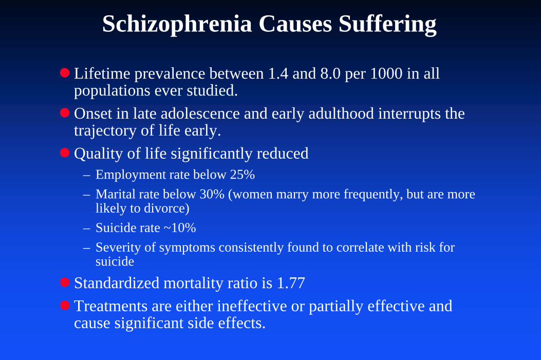
Schizophrenia Causes Suffering
Lifetime prevalence between 1.4 and 8.0 per 1000 in all populations ever studied.Onset in late adolescence and early adulthood interrupts the trajectory of life early.Quality of life significantly reduced
– Employment rate below 25%– Marital rate below 30% (women marry more frequently, but are more
likely to divorce)– Suicide rate ~10%– Severity of symptoms consistently found to correlate with risk for
suicide
Standardized mortality ratio is 1.77Treatments are either ineffective or partially effective and cause significant side effects.
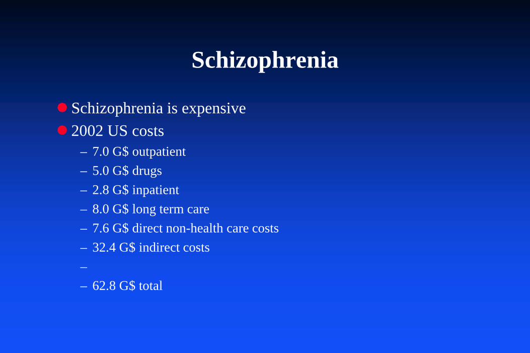
Schizophrenia
Schizophrenia is expensive2002 US costs
– 7.0 G$ outpatient– 5.0 G$ drugs– 2.8 G$ inpatient– 8.0 G$ long term care– 7.6 G$ direct non-health care costs– 32.4 G$ indirect costs–– 62.8 G$ total
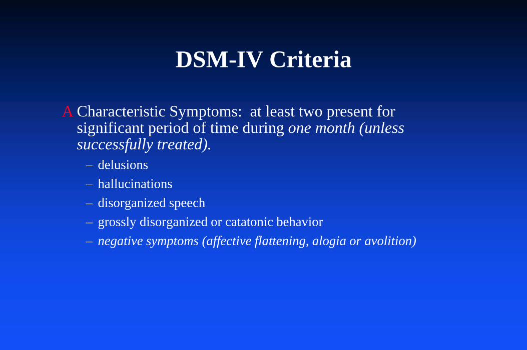
DSM-IV Criteria
A Characteristic Symptoms: at least two present for significant period of time during one month (unless successfully treated).
– delusions– hallucinations– disorganized speech– grossly disorganized or catatonic behavior– negative symptoms (affective flattening, alogia or avolition)

DSM-IV Criteria
A Characteristic Symptoms: B Social/Occupational Dysfunction:C Duration: At least 6 months
– one month of criterion A.– residual or prodromal periods characterized by negative symptoms or mild
criterion A symptoms.
D Schizoaffective or Mood Disorder Exclusion:E Substance/General Medical Condition Exclusion:
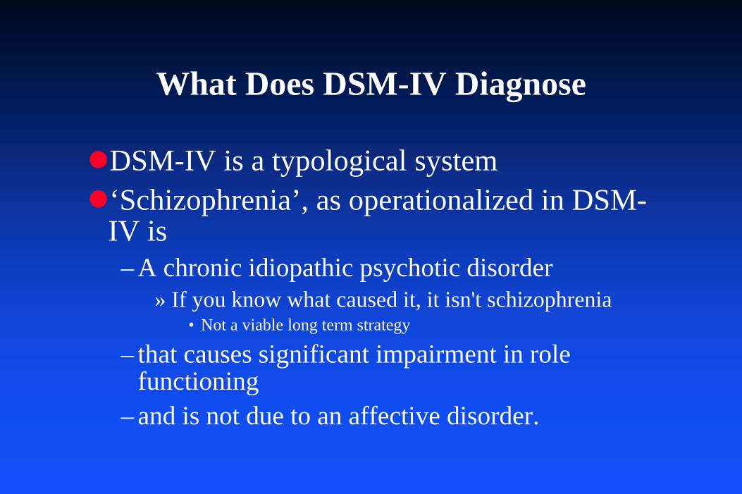
What Does DSM-IV Diagnose
DSM-IV is a typological system ‘Schizophrenia’, as operationalized in DSM-IV is– A chronic idiopathic psychotic disorder
» If you know what caused it, it isn't schizophrenia• Not a viable long term strategy
– that causes significant impairment in role functioning
– and is not due to an affective disorder.
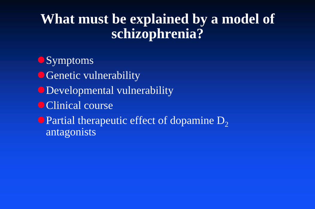
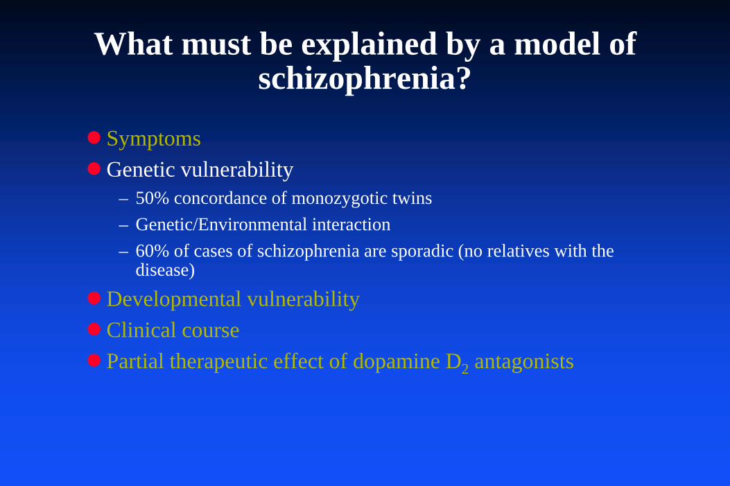
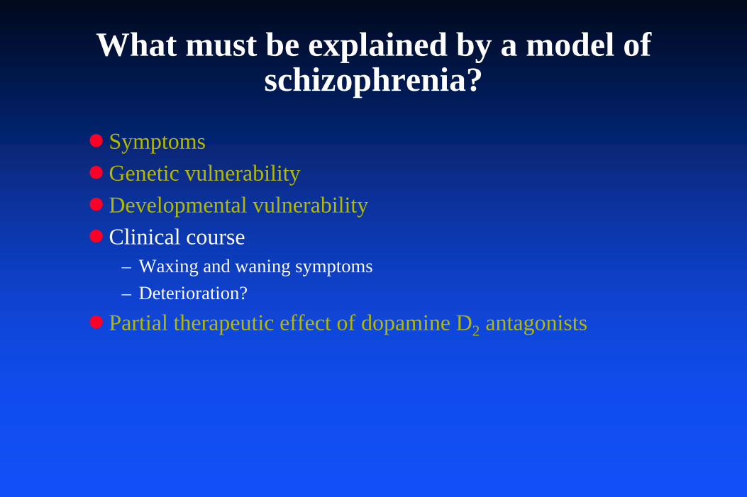
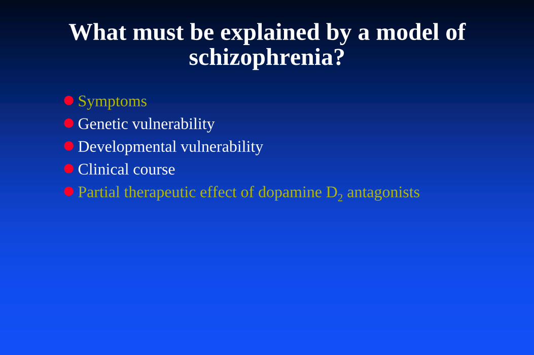
What must be explained by a model of schizophrenia?
SymptomsGenetic vulnerabilityDevelopmental vulnerabilityClinical coursePartial therapeutic effect of dopamine D2antagonists

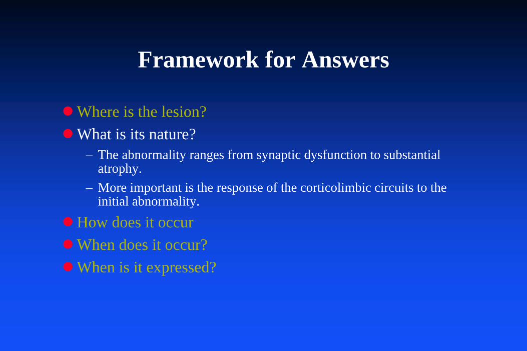
Framework for Answers
Where is the lesion?What is its nature?When does it occur?How does it occur?When is it expressed?
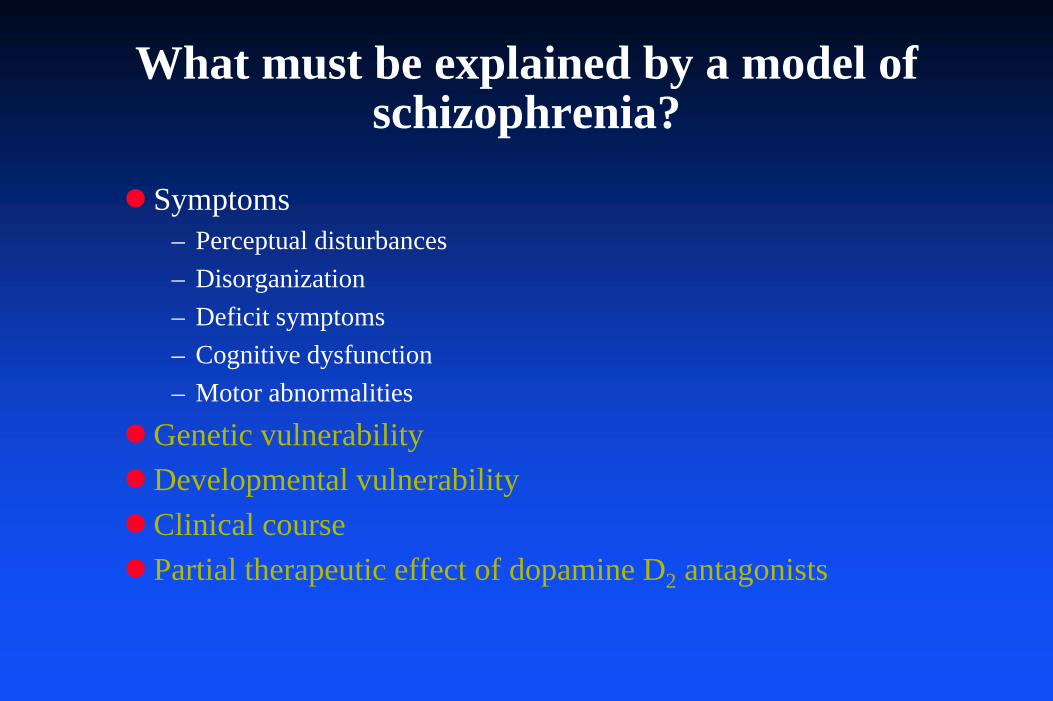
What must be explained by a model of schizophrenia?
Symptoms– Perceptual disturbances– Disorganization– Deficit symptoms– Cognitive dysfunction– Motor abnormalities
Genetic vulnerabilityDevelopmental vulnerabilityClinical coursePartial therapeutic effect of dopamine D2 antagonists
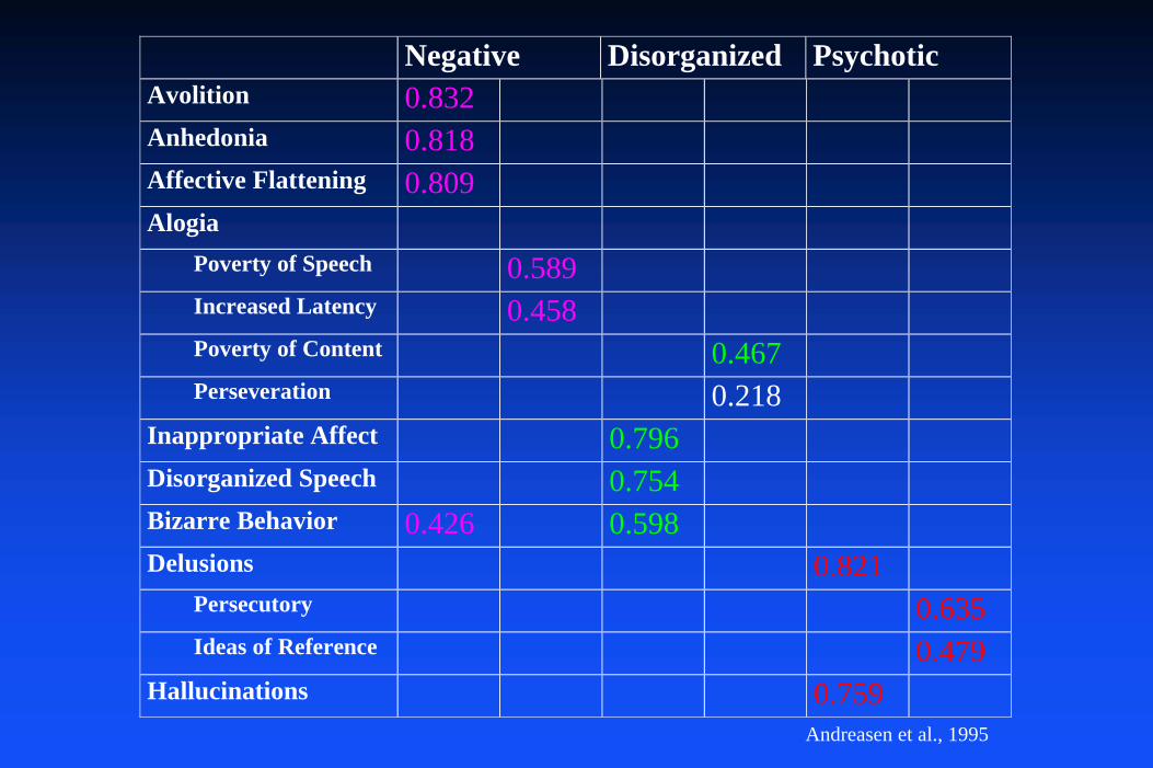
Negative Disorganized Psychotic Avolition 0.832 Anhedonia 0.818 Affective Flattening 0.809 Alogia Poverty of Speech 0.589 Increased Latency 0.458 Poverty of Content 0.467 Perseveration 0.218 Inappropriate Affect 0.796 Disorganized Speech 0.754 Bizarre Behavior 0.426 0.598 Delusions 0.821 Persecutory 0.635 Ideas of Reference 0.479 Hallucinations 0.759
Andreasen et al., 1995
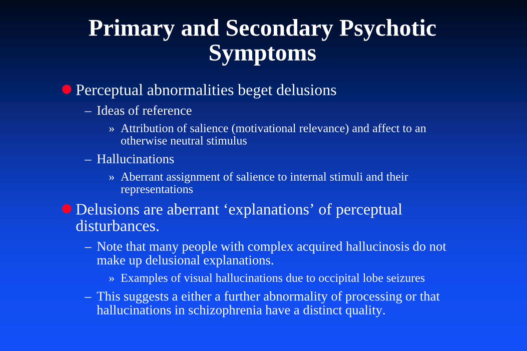
Primary and Secondary Psychotic Symptoms
Perceptual abnormalities beget delusions– Ideas of reference
» Attribution of salience (motivational relevance) and affect to an otherwise neutral stimulus
– Hallucinations» Aberrant assignment of salience to internal stimuli and their
representations
Delusions are aberrant ‘explanations’ of perceptual disturbances.
– Note that many people with complex acquired hallucinosis do not make up delusional explanations.
» Examples of visual hallucinations due to occipital lobe seizures– This suggests a either a further abnormality of processing or that
hallucinations in schizophrenia have a distinct quality.
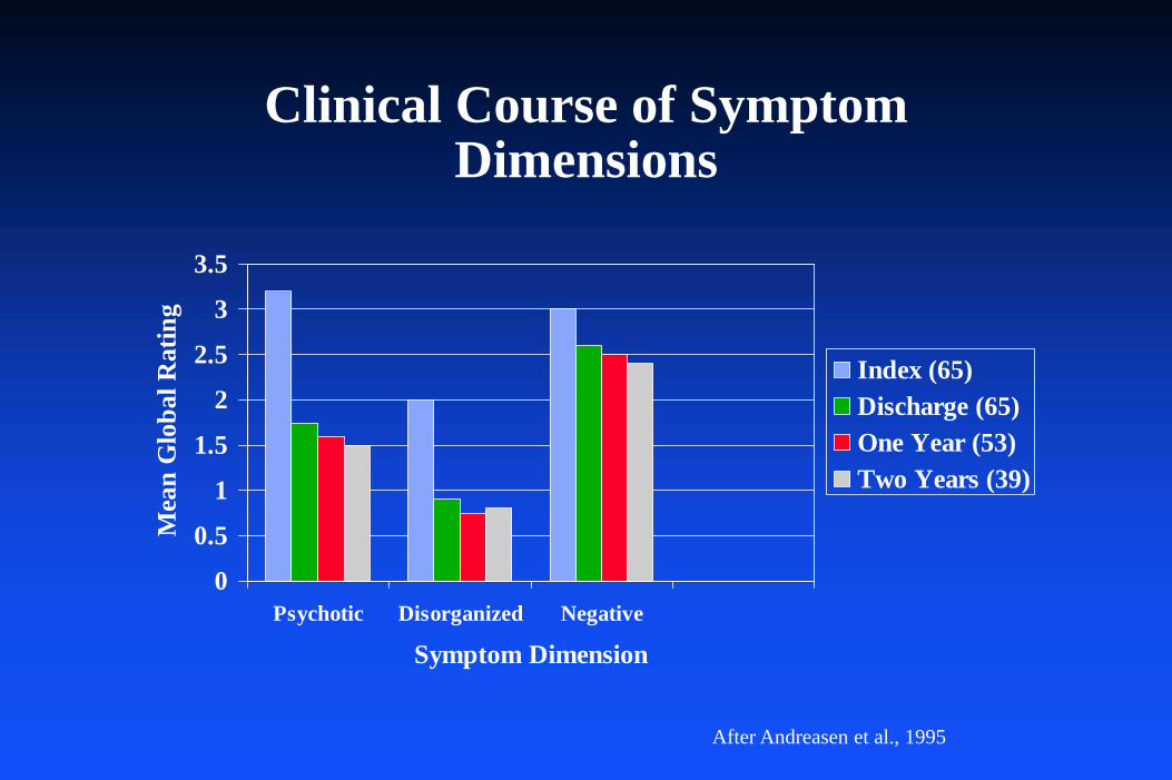
Clinical Course of Symptom Dimensions
0
0.5
1
1.5
2
2.5
3
3.5
Psychotic Disorganized Negative
Symptom Dimension
Mea
n G
loba
l Rat
ing
Index (65)Discharge (65)One Year (53)Two Years (39)
After Andreasen et al., 1995
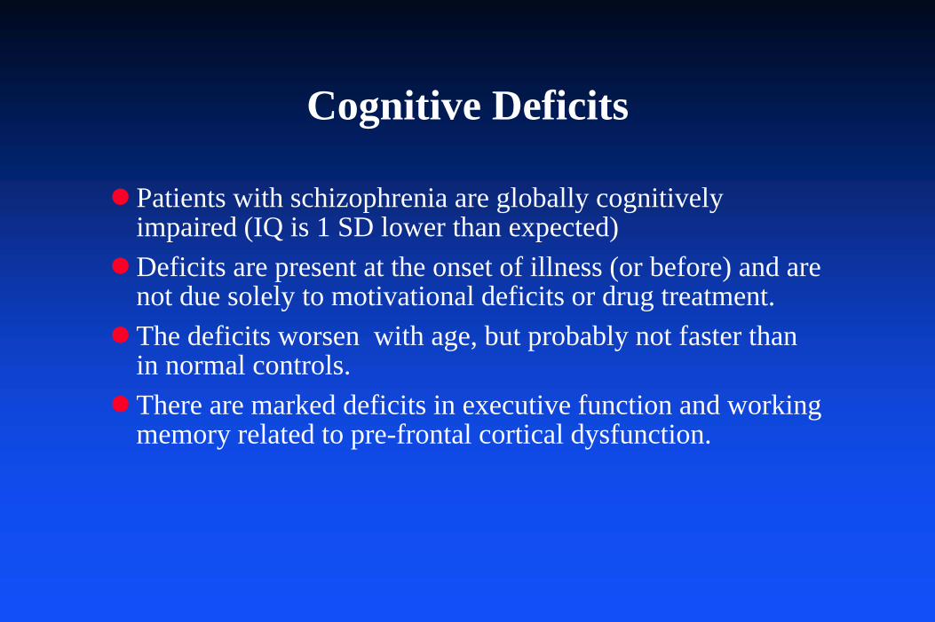
Cognitive Deficits
Patients with schizophrenia are globally cognitively impaired (IQ is 1 SD lower than expected)Deficits are present at the onset of illness (or before) and are not due solely to motivational deficits or drug treatment.The deficits worsen with age, but probably not faster than in normal controls.There are marked deficits in executive function and working memory related to pre-frontal cortical dysfunction.
What must be explained by a model of schizophrenia?
SymptomsGenetic vulnerability
– 50% concordance of monozygotic twins– Genetic/Environmental interaction– 60% of cases of schizophrenia are sporadic (no relatives with the
disease)
Developmental vulnerabilityClinical coursePartial therapeutic effect of dopamine D2 antagonists
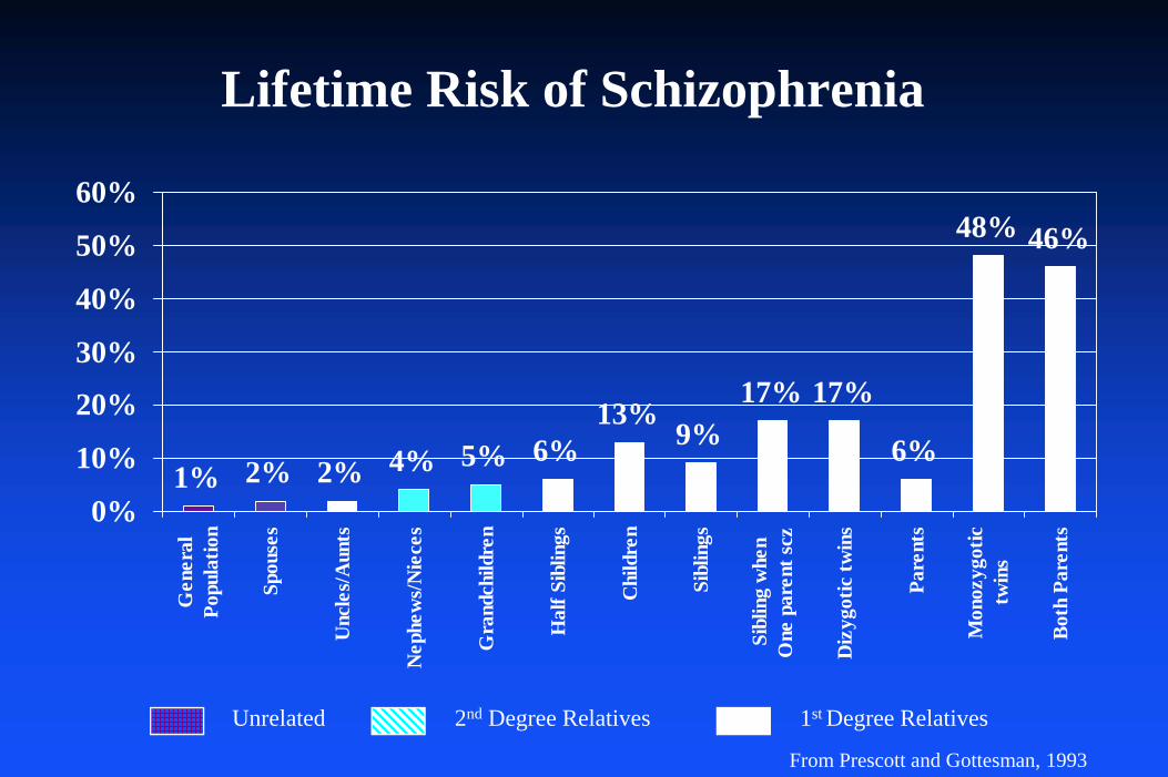
Lifetime Risk of Schizophrenia
1% 2% 2% 4% 5% 6%13%
9%17% 17%
6%
48% 46%
0%
10%
20%
30%
40%
50%
60%G
ener
alPo
pula
tion
Spou
ses
Unc
les/
Aun
ts
Nep
hew
s/N
iece
s
Gra
ndch
ildre
n
Hal
f Sib
lings
Chi
ldre
n
Sibl
ings
Sibl
ing
whe
nO
ne p
aren
t scz
Diz
ygot
ic tw
ins
Pare
nts
Mon
ozyg
otic
twin
s
Both
Par
ents
Unrelated 2nd Degree Relatives 1st Degree Relatives
From Prescott and Gottesman, 1993
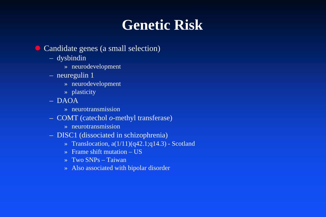
Genetic RiskCandidate genes (a small selection)
– dysbindin» neurodevelopment
– neuregulin 1» neurodevelopment» plasticity
– DAOA» neurotransmission
– COMT (catechol o-methyl transferase)» neurotransmission
– DISC1 (dissociated in schizophrenia)» Translocation, a(1/11)(q42.1;q14.3) - Scotland» Frame shift mutation – US» Two SNPs – Taiwan» Also associated with bipolar disorder

What must be explained by a model of schizophrenia?
SymptomsGenetic vulnerabilityDevelopmental vulnerability
– Peak incidence in first 10 years after puberty– Note that highest absolute risk is due to season of birth
Clinical coursePartial therapeutic effect of dopamine D2 antagonists
What must be explained by a model of schizophrenia?
SymptomsGenetic vulnerabilityDevelopmental vulnerabilityClinical course
– Waxing and waning symptoms– Deterioration?
Partial therapeutic effect of dopamine D2 antagonists
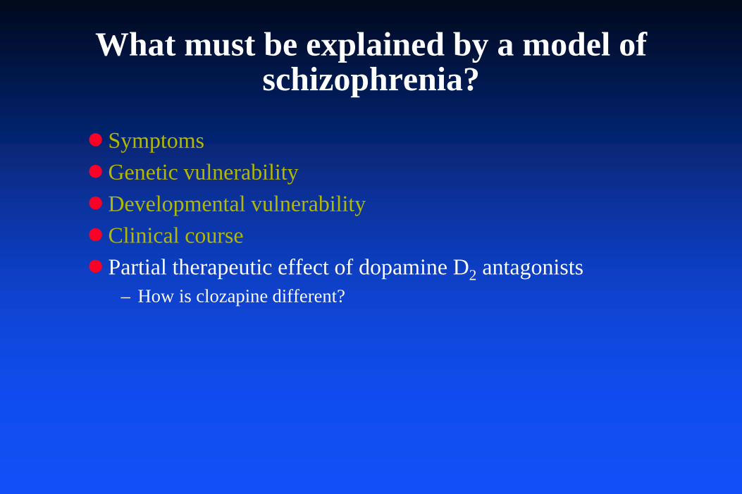
What must be explained by a model of schizophrenia?
SymptomsGenetic vulnerabilityDevelopmental vulnerabilityClinical coursePartial therapeutic effect of dopamine D2 antagonists
– How is clozapine different?
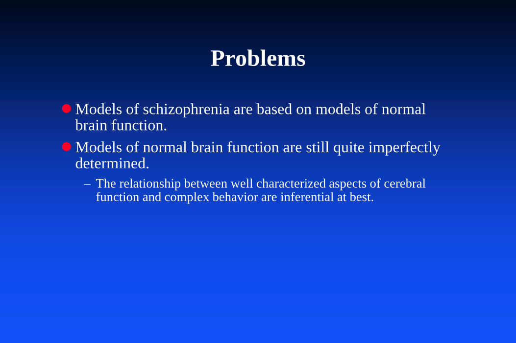
Problems
Models of schizophrenia are based on models of normal brain function.Models of normal brain function are still quite imperfectly determined.
– The relationship between well characterized aspects of cerebral function and complex behavior are inferential at best.
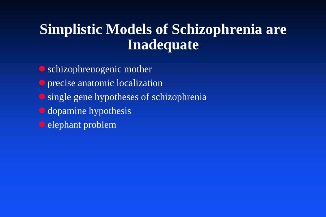
Simplistic Models of Schizophrenia are Inadequate
schizophrenogenic motherprecise anatomic localization single gene hypotheses of schizophreniadopamine hypothesiselephant problem
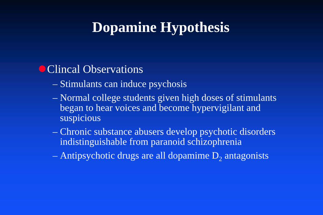
Dopamine Hypothesis
Clincal Observations– Stimulants can induce psychosis– Normal college students given high doses of stimulants
began to hear voices and become hypervigilant and suspicious
– Chronic substance abusers develop psychotic disorders indistinguishable from paranoid schizophrenia
– Antipsychotic drugs are all dopamime D2 antagonists

Correlation between D2 receptor ant-agonist Ki(nM) and antipsychotic efficacy (mg/da).
Dopamine hypothesis inferred that hyperdopaminergic activity leads to psychosis.
Despite intense effort, consistent evidence of abnormal D2 receptor number or affinity has eluded investigators.
Multiple studies have shown an increased DA release in striatum (SCZ > controls) after amphetamine treatment consistent with increased phasic dopaminergic activity.
Dopamine Hypothesis
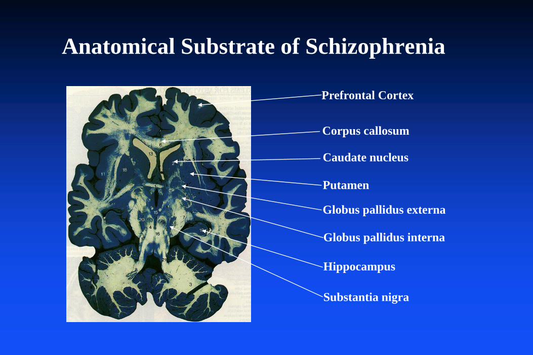
Anatomical Substrate of Schizophrenia
Corpus callosum
Caudate nucleus
Putamen
Globus pallidus externa
Globus pallidus interna
Substantia nigra
Hippocampus
Prefrontal Cortex
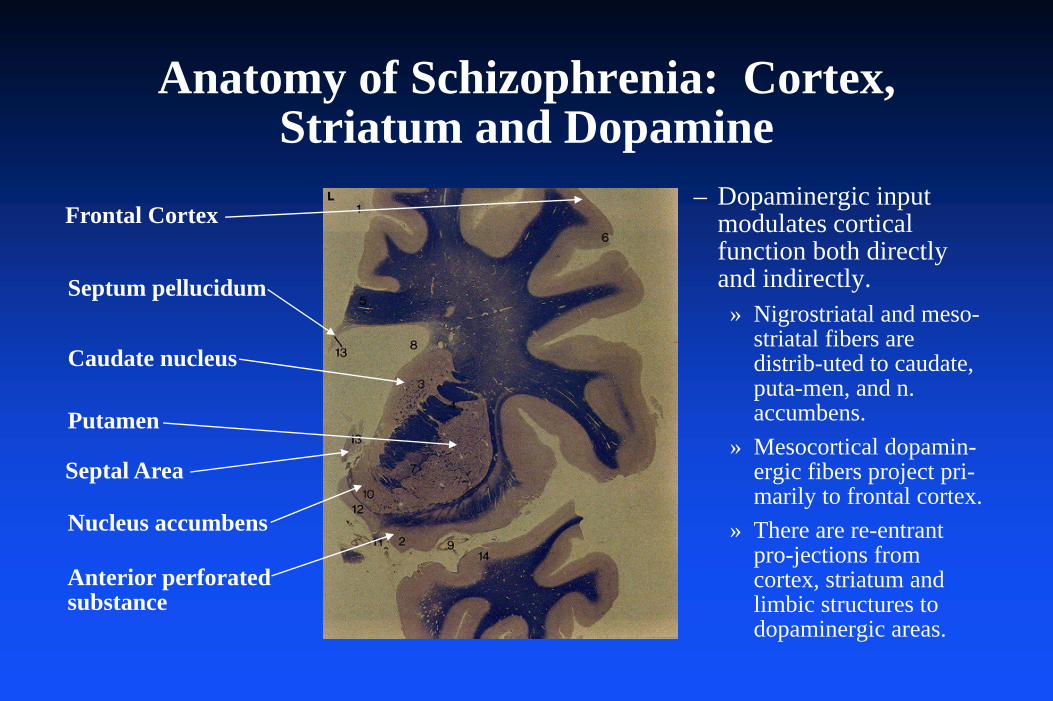
Anatomy of Schizophrenia: Cortex, Striatum and Dopamine
Septum pellucidum
Nucleus accumbens
Caudate nucleus
Anterior perforatedsubstance
Putamen
– Dopaminergic input modulates cortical function both directly and indirectly.
» Nigrostriatal and meso-striatal fibers are distrib-uted to caudate, puta-men, and n. accumbens.
» Mesocortical dopamin-ergic fibers project pri-marily to frontal cortex.
» There are re-entrant pro-jections from cortex, striatum and limbic structures to dopaminergic areas.
Septal Area
Frontal Cortex
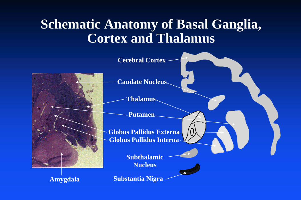
Schematic Anatomy of Basal Ganglia, Cortex and Thalamus
Thalamus
SubthalamicNucleus
Substantia Nigra
Caudate Nucleus
Putamen
Globus Pallidus ExternaGlobus Pallidus Interna
Cerebral Cortex
Amygdala
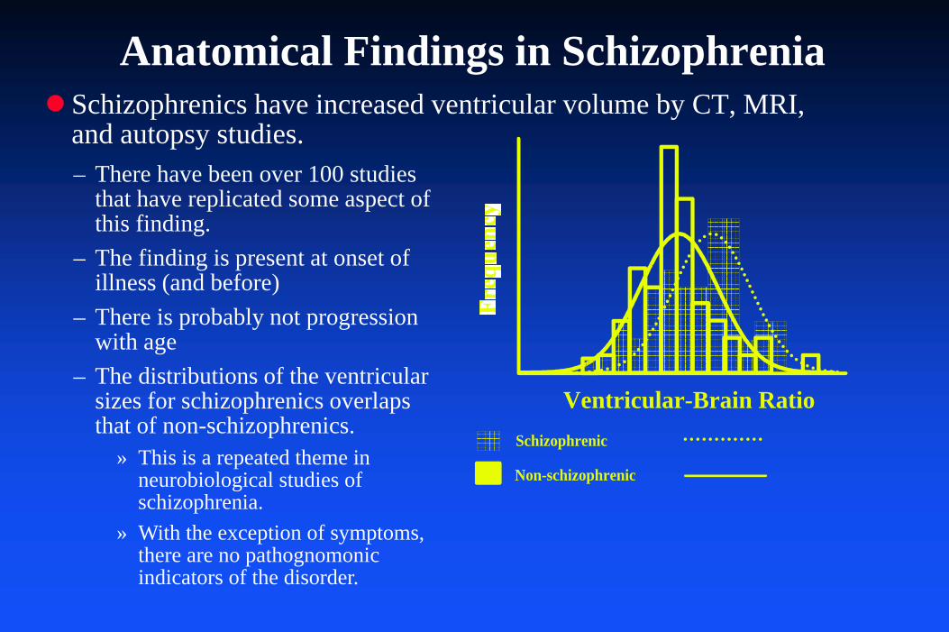
Schizophrenics have increased ventricular volume by CT, MRI, and autopsy studies.
Anatomical Findings in Schizophrenia
Ventricular-Brain RatioSchizophrenic
Non-schizophrenic
– There have been over 100 studies that have replicated some aspect of this finding.
– The finding is present at onset of illness (and before)
– There is probably not progression with age
– The distributions of the ventricular sizes for schizophrenics overlaps that of non-schizophrenics.
» This is a repeated theme in neurobiological studies of schizophrenia.
» With the exception of symptoms, there are no pathognomonic indicators of the disorder.
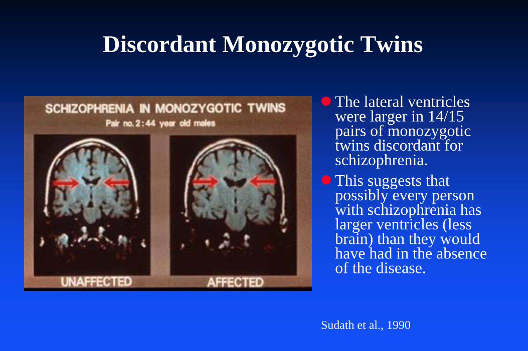
Discordant Monozygotic Twins
The lateral ventricles were larger in 14/15 pairs of monozygotic twins discordant for schizophrenia.This suggests that possibly every person with schizophrenia has larger ventricles (less brain) than they would have had in the absence of the disease.
Sudath et al., 1990
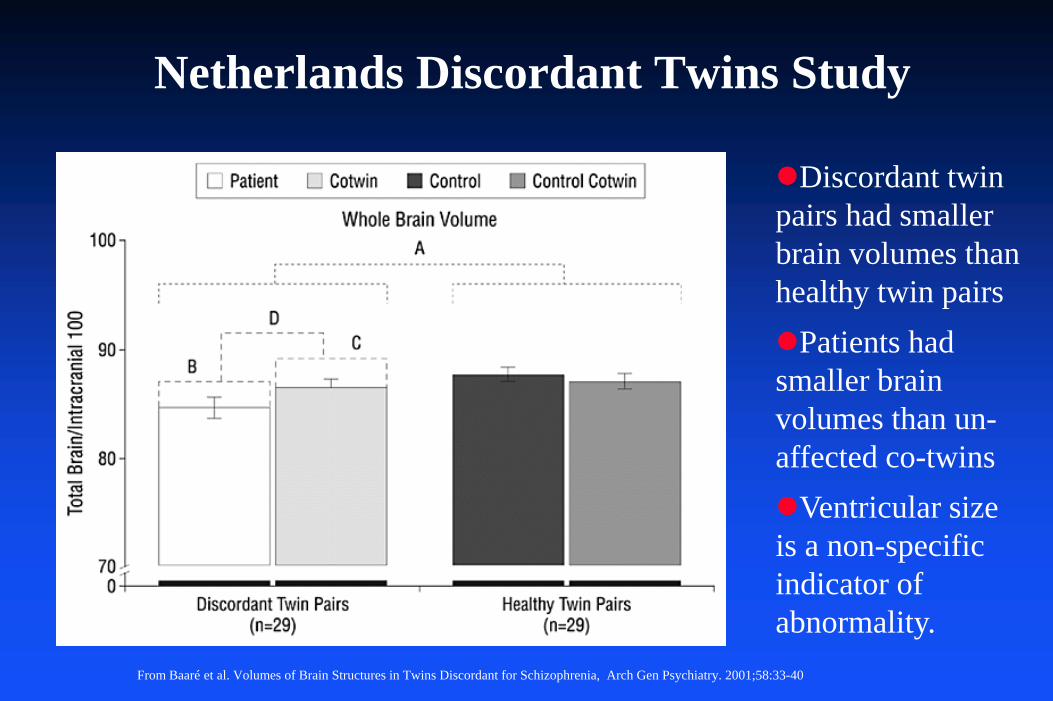
Netherlands Discordant Twins Study
From Baaré et al. Volumes of Brain Structures in Twins Discordant for Schizophrenia, Arch Gen Psychiatry. 2001;58:33-40
Discordant twin pairs had smaller brain volumes than healthy twin pairs
Patients had smaller brain volumes than un-affected co-twins
Ventricular size is a non-specific indicator of abnormality.
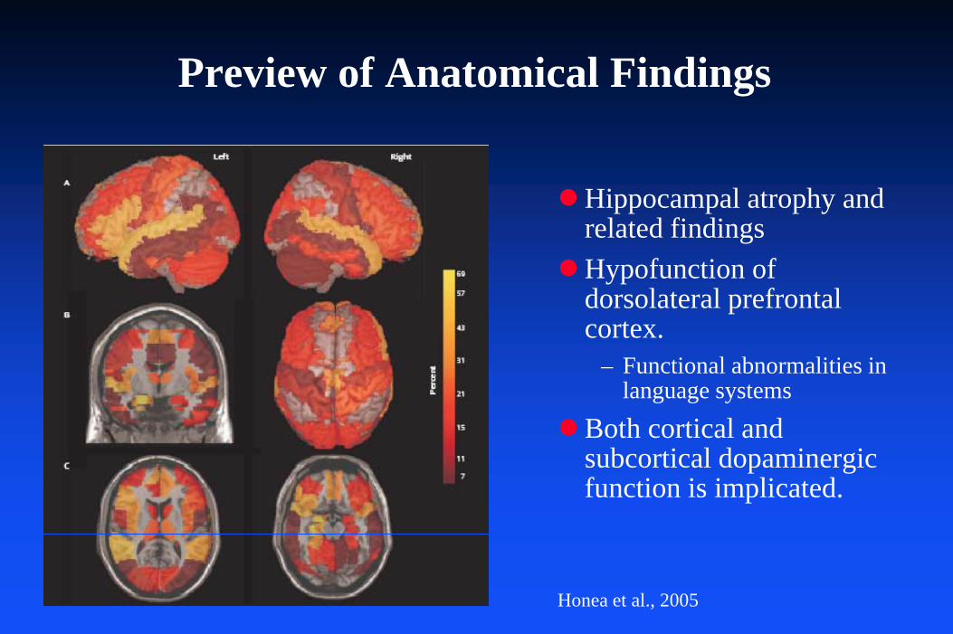
Preview of Anatomical Findings
Hippocampal atrophy and related findingsHypofunction of dorsolateral prefrontal cortex.
– Functional abnormalities in language systems
Both cortical and subcortical dopaminergic function is implicated.
Honea et al., 2005
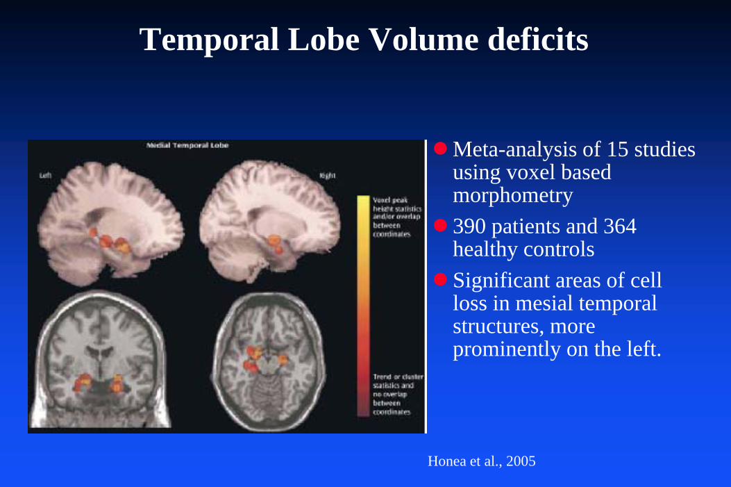
Temporal Lobe Volume deficits
Meta-analysis of 15 studies using voxel based morphometry390 patients and 364 healthy controlsSignificant areas of cell loss in mesial temporal structures, more prominently on the left.
Honea et al., 2005
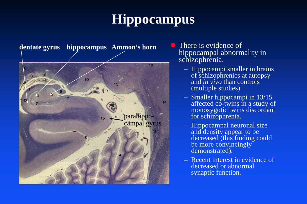
Hippocampus
There is evidence of hippocampal abnormality in schizophrenia.
– Hippocampi smaller in brains of schizophrenics at autopsy and in vivo than controls (multiple studies).
– Smaller hippocampi in 13/15 affected co-twins in a study of monozygotic twins discordant for schizophrenia.
– Hippocampal neuronal size and density appear to be decreased (this finding could be more convincingly demonstrated).
– Recent interest in evidence of decreased or abnormal synaptic function.
dentate gyrus hippocampus Ammon’s horn
parahippo-campal gyrus
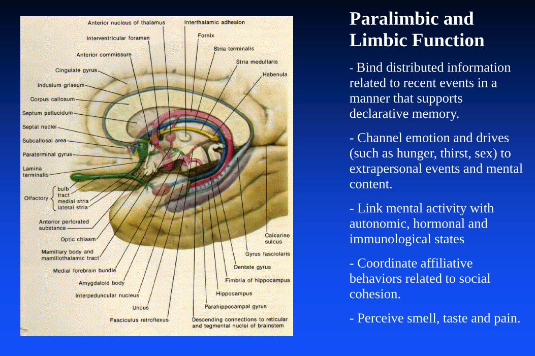
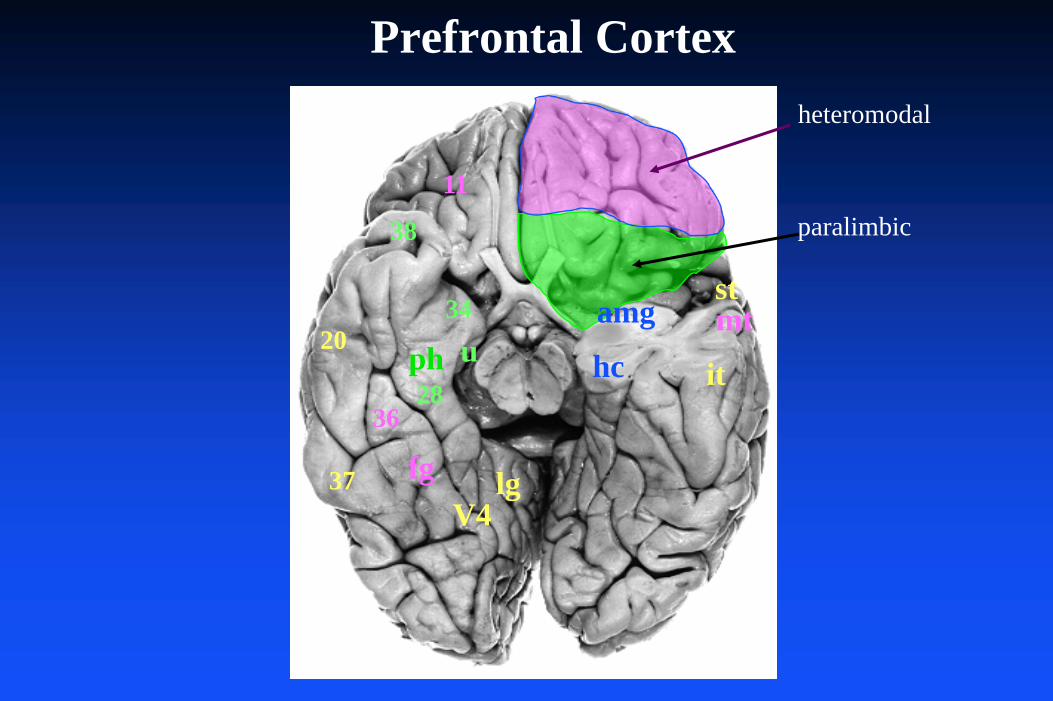
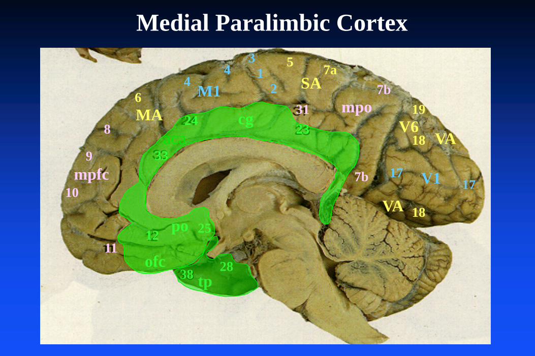
Paralimbic and Limbic Function- Bind distributed information related to recent events in a manner that supports declarative memory.
- Channel emotion and drives (such as hunger, thirst, sex) to extrapersonal events and mental content.
- Link mental activity with autonomic, hormonal and immunological states
- Coordinate affiliative behaviors related to social cohesion.
- Perceive smell, taste and pain.
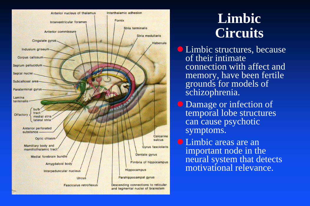
Limbic structures, because of their intimate connection with affect and memory, have been fertile grounds for models of schizophrenia.Damage or infection of temporal lobe structures can cause psychotic symptoms.Limbic areas are an important node in the neural system that detects motivational relevance.
Limbic Circuits
thalamicnuclei
N. Accumbens
Anterior Cingulate,prefrontal cortex
HippocampusAmygdala
Hypothalamus
VTN/SNc
ventralpallidum
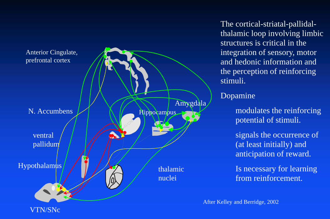
After Kelley and Berridge, 2002
The cortical-striatal-pallidal-thalamic loop involving limbic structures is critical in the integration of sensory, motor and hedonic information and the perception of reinforcing stimuli.
Dopamine
modulates the reinforcing potential of stimuli.
signals the occurrence of (at least initially) and anticipation of reward.
Is necessary for learning from reinforcement.

Psychotic Symptoms Suggest Aberrant Assignment of Salience
Hypothesis– Referential ideas result from erroneous pairing of salience with
otherwise neutral percepts– Hallucinations result from abnormal attribution of salience to
internal thoughts and memories.– Delusions result from further aberrant salience attribution during the
attempt to understand the perceptual distortions.
This hypothesis further suggests that there must be some manifestation of hyperdopaminergia or inappropriate DA release or activity in (at least) the mesolimbic projection.Critique
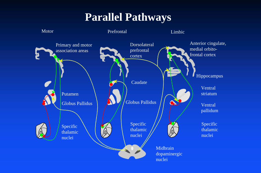
Putamen
Caudate
Motor Prefrontal Limbic
Globus Pallidus Globus Pallidus
Primary and motorassociation areas
Dorsolateralprefrontalcortex
Specificthalamicnuclei
Specificthalamicnuclei
Specificthalamicnuclei
Ventralstriatum
Ventralpallidum
Hippocampus
Midbraindopaminergicnuclei
Anterior cingulate,medial orbito-frontal cortex
Parallel Pathways
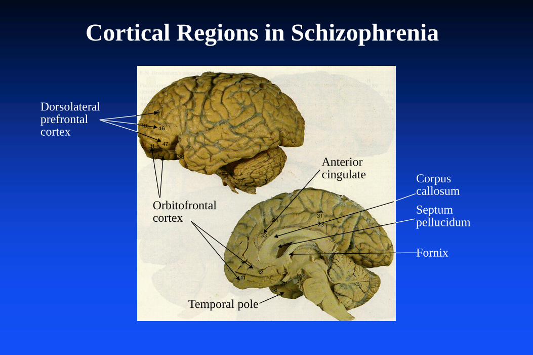
Dorsolateralprefrontalcortex
Orbitofrontalcortex
Anteriorcingulate
Temporal pole
CorpuscallosumSeptumpellucidum
Fornix
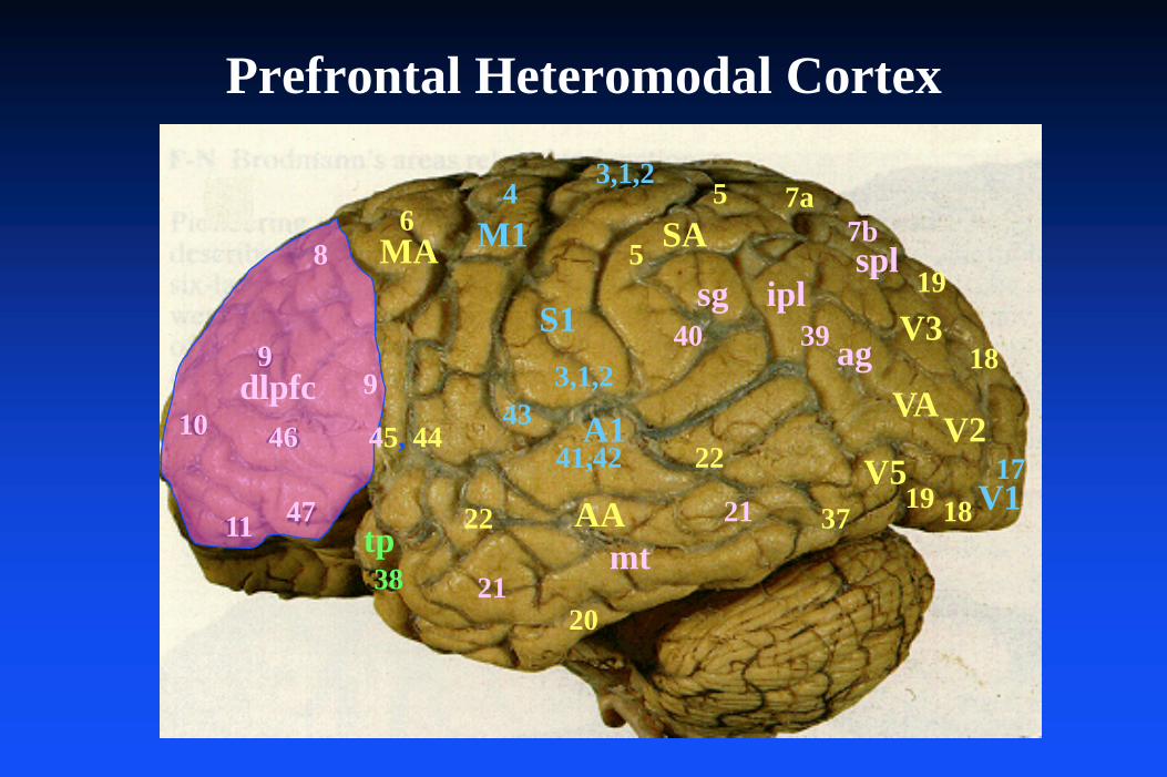
Cortical Regions in Schizophrenia
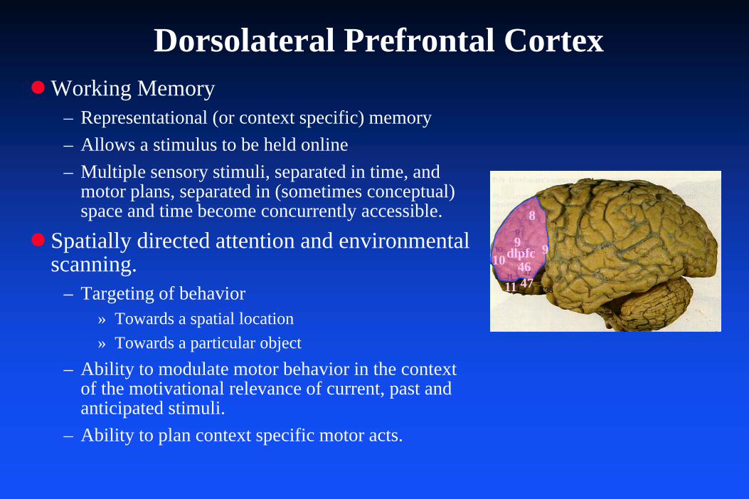
Dorsolateral Prefrontal CortexWorking Memory
– Representational (or context specific) memory– Allows a stimulus to be held online– Multiple sensory stimuli, separated in time, and
motor plans, separated in (sometimes conceptual) space and time become concurrently accessible.
Spatially directed attention and environmental scanning.
– Targeting of behavior» Towards a spatial location» Towards a particular object
– Ability to modulate motor behavior in the context of the motivational relevance of current, past and anticipated stimuli.
– Ability to plan context specific motor acts.
8
910 46
4711
dlpfc 9

Dorsolateral Prefrontal CortexDamage (loss of neurons) to DLPFC causes characteristic deficits.
– Lack of initiation due to decreased environmental scanning
– Poor cognitive flexibility due to difficulty shifting attention – stimulus boundedness
– Lack of emotional spontaneity (abulia) probably due to poor perception of hedonic events.
– Decrement in interpersonal involvement secondary to the above deficits.
– Poor self care due to lack of initiation, decreased ability to learn from complex social feedback.
These deficits are essentially identical to the negative symptoms of schizophrenia.
8
910 46
4711
dlpfc 9
Dorsolateral Prefrontal Cortex
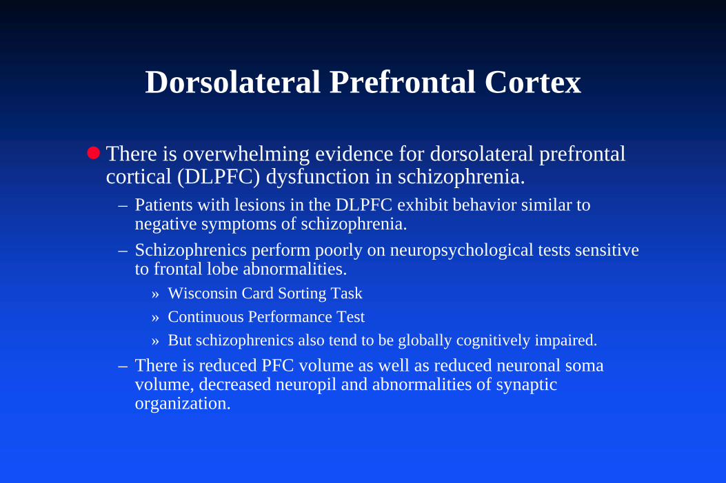
There is overwhelming evidence for dorsolateral prefrontal cortical (DLPFC) dysfunction in schizophrenia.
– Patients with lesions in the DLPFC exhibit behavior similar to negative symptoms of schizophrenia.
– Schizophrenics perform poorly on neuropsychological tests sensitive to frontal lobe abnormalities.
» Wisconsin Card Sorting Task» Continuous Performance Test» But schizophrenics also tend to be globally cognitively impaired.
– There is reduced PFC volume as well as reduced neuronal soma volume, decreased neuropil and abnormalities of synaptic organization.
Dorsolateral Prefrontal Cortex
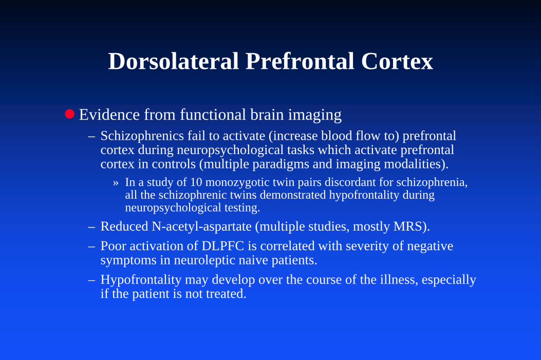
Evidence from functional brain imaging– Schizophrenics fail to activate (increase blood flow to) prefrontal
cortex during neuropsychological tasks which activate prefrontal cortex in controls (multiple paradigms and imaging modalities).
» In a study of 10 monozygotic twin pairs discordant for schizophrenia, all the schizophrenic twins demonstrated hypofrontality during neuropsychological testing.
– Reduced N-acetyl-aspartate (multiple studies, mostly MRS).– Poor activation of DLPFC is correlated with severity of negative
symptoms in neuroleptic naive patients.– Hypofrontality may develop over the course of the illness, especially
if the patient is not treated.
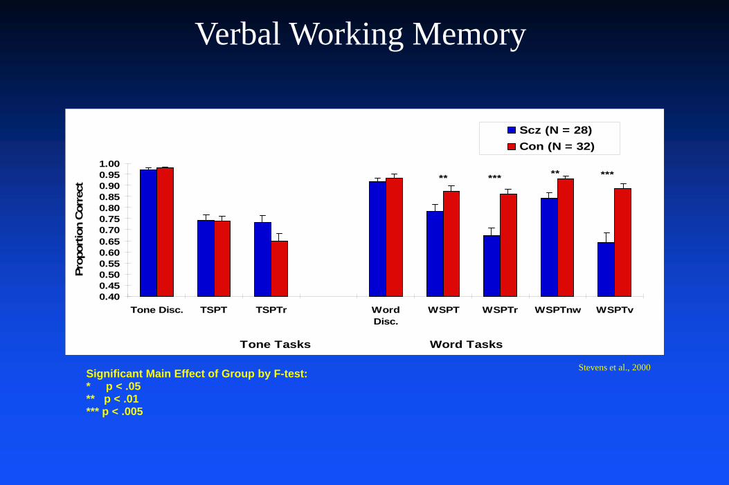
Significant Main Effect of Group by F-test:* p < .05** p < .01*** p < .005
0.400.450.500.550.600.650.700.750.800.850.900.951.00
Tone Disc. TSPT TSPTr WordDisc.
WSPT WSPTr WSPTnw WSPTv
Tone Tasks Word Tasks
Prop
ortio
n C
orre
ct
Scz (N = 28)Con (N = 32)
** *** *****
Stevens et al., 2000
Verbal Working Memory
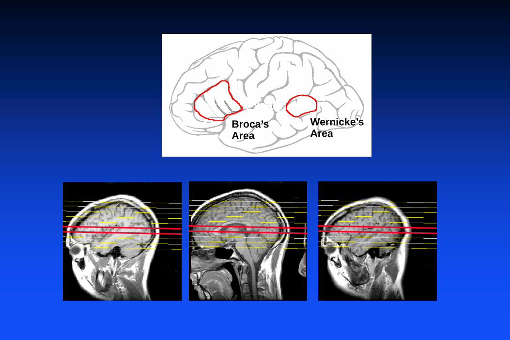
Broca’sArea
Wernicke’sArea
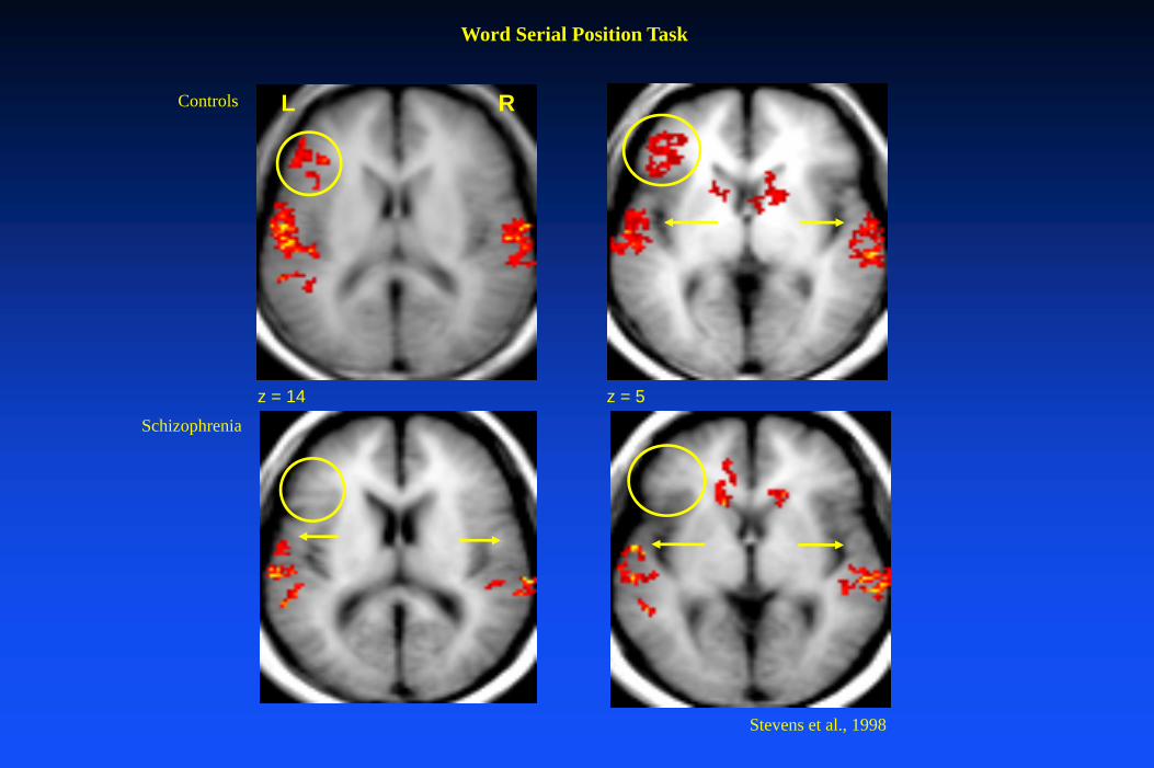
z = 14 z = 5
Controls
Schizophrenia
Word Serial Position Task
Stevens et al., 1998
L R
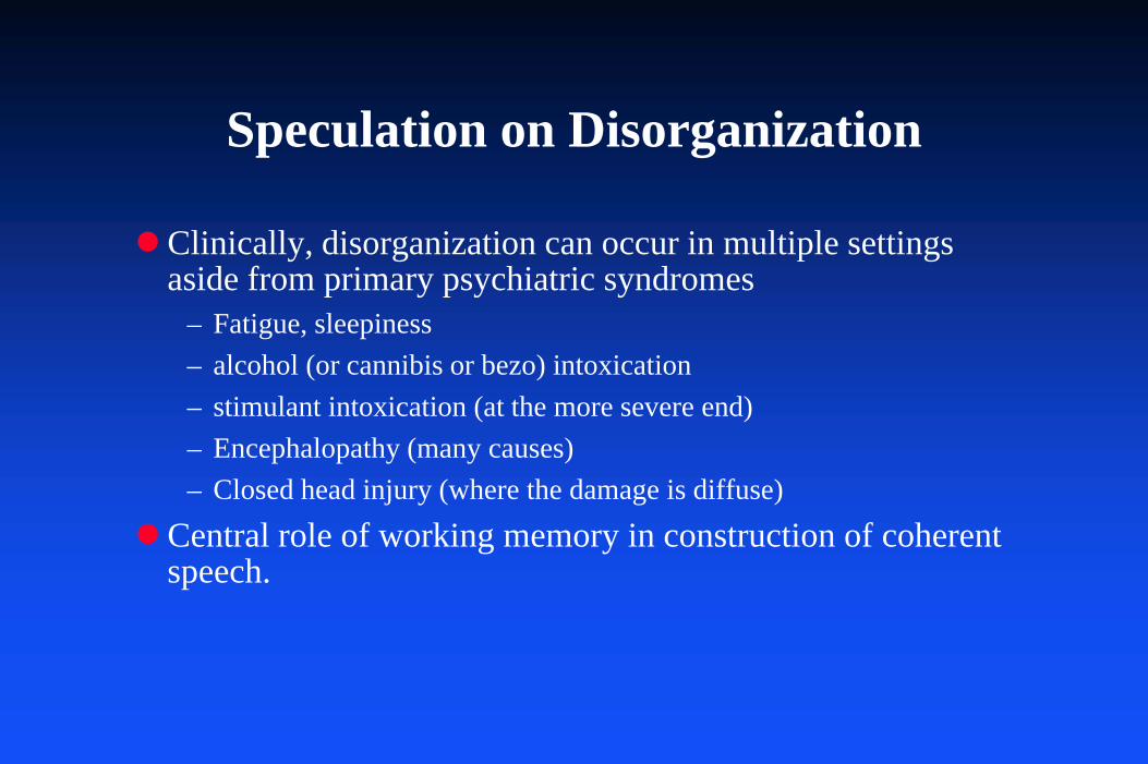
Speculation on Disorganization
Clinically, disorganization can occur in multiple settings aside from primary psychiatric syndromes
– Fatigue, sleepiness– alcohol (or cannibis or bezo) intoxication– stimulant intoxication (at the more severe end)– Encephalopathy (many causes)– Closed head injury (where the damage is diffuse)
Central role of working memory in construction of coherent speech.
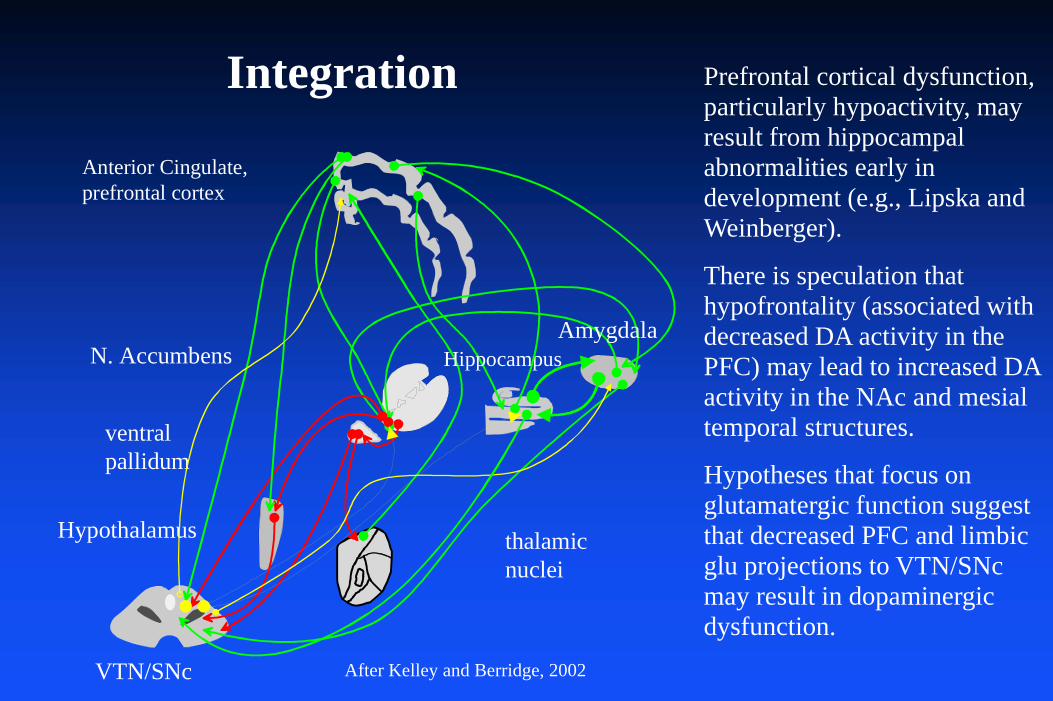
thalamicnuclei
N. Accumbens
Anterior Cingulate,prefrontal cortex
HippocampusAmygdala
Hypothalamus
VTN/SNc
ventralpallidum
After Kelley and Berridge, 2002
Prefrontal cortical dysfunction, particularly hypoactivity, may result from hippocampal abnormalities early in development (e.g., Lipska and Weinberger).
There is speculation that hypofrontality (associated with decreased DA activity in the PFC) may lead to increased DA activity in the NAc and mesial temporal structures.
Hypotheses that focus on glutamatergic function suggest that decreased PFC and limbic glu projections to VTN/SNc may result in dopaminergic dysfunction.
Integration
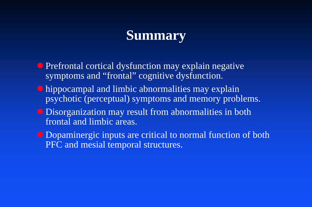
Summary
Prefrontal cortical dysfunction may explain negative symptoms and “frontal” cognitive dysfunction.hippocampal and limbic abnormalities may explain psychotic (perceptual) symptoms and memory problems.Disorganization may result from abnormalities in both frontal and limbic areas.Dopaminergic inputs are critical to normal function of both PFC and mesial temporal structures.
What must be explained by a model of schizophrenia?
SymptomsGenetic vulnerabilityDevelopmental vulnerabilityClinical coursePartial therapeutic effect of dopamine D2 antagonists


When is the Developmental Lesion?
Several lines of evidence support a neurodevelopmental model of schizophreniaNeurodevelopmental models hypothesize that a brain abnormality occur earlier (in utero to pre-adolescence), flaws subsequent development and increases the risk for schizophrenia when the person reaches adolescence.There is no smoking gun that points unequivocally to a particular period of development.
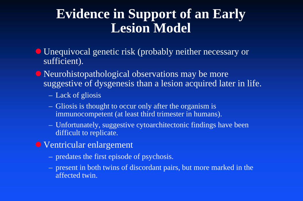
Evidence in Support of an Early Lesion Model
Unequivocal genetic risk (probably neither necessary or sufficient). Neurohistopathological observations may be more suggestive of dysgenesis than a lesion acquired later in life.
– Lack of gliosis– Gliosis is thought to occur only after the organism is
immunocompetent (at least third trimester in humans).– Unfortunately, suggestive cytoarchitectonic findings have been
difficult to replicate.
Ventricular enlargement– predates the first episode of psychosis.– present in both twins of discordant pairs, but more marked in the
affected twin.

Prospective studies of high risk children– Smaller brain volumes, larger ventricles.– Increased rate of minor physical anomalies– Abnormalities in coordination and involuntary movements– Attention and memory deficits– Socially isolated and maladaptive behavior
Home movies of schizophrenics as children allow reliable distinction between patients and normal age mates.Animal models show that parahippocampal lesions early in postnatal life can lead to behavioral anomalies after pubescence.
Evidence in Support of an Early Lesion Model
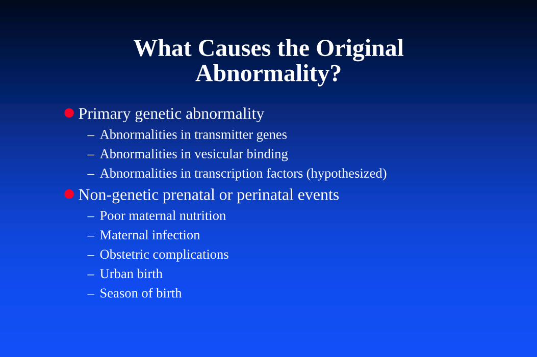
What Causes the Original Abnormality?
Primary genetic abnormality– Abnormalities in transmitter genes– Abnormalities in vesicular binding – Abnormalities in transcription factors (hypothesized)
Non-genetic prenatal or perinatal events– Poor maternal nutrition– Maternal infection– Obstetric complications– Urban birth– Season of birth

What Causes Expression of the Phenotype after Puberty?
Neurodevelopmental events– Synaptic pruning occurs in two major waves
» First few years post natal» Adolescence
– Myelination occurs in an orderly progression» Thalamic projections to PFC and hippocampus and frontotemporal
circuits myelinate during puberty and adolescence.– Secretion of sex hormones– Environmental ‘stress’
» Speculative role of hypothalamic-pituitary-adrenal system
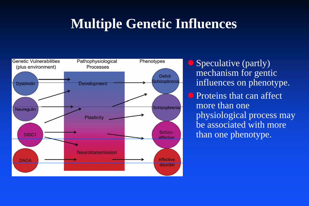
Multiple Genetic Influences
Speculative (partly) mechanism for gentic influences on phenotype.Proteins that can affect more than one physiological process may be associated with more than one phenotype.

Examples
The following examples range from less to more speculative and are meant to provide food for thought rather than definitive proof of an etiological mechanism.
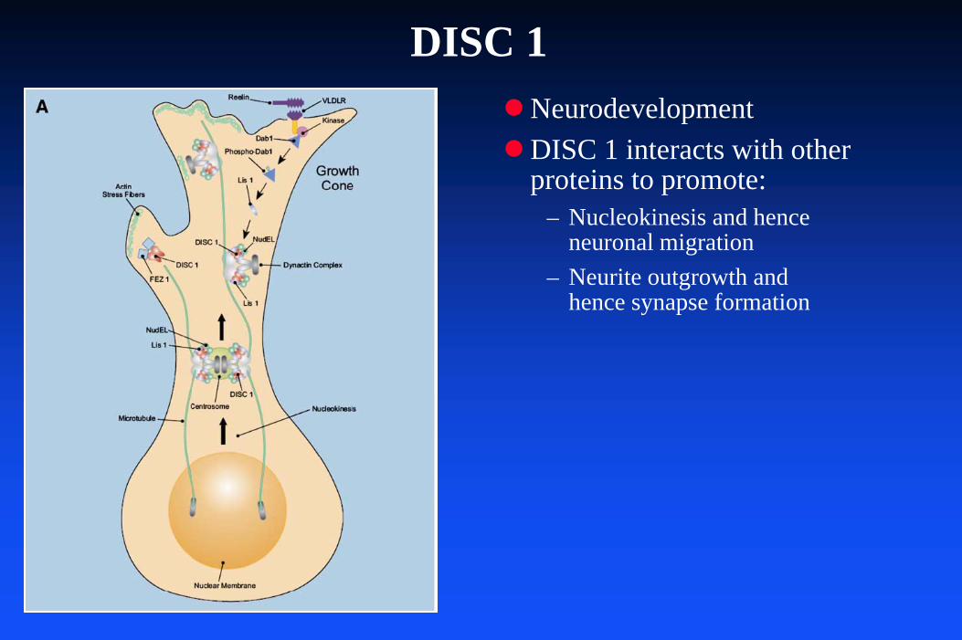
DISC 1NeurodevelopmentDISC 1 interacts with other proteins to promote:
– Nucleokinesis and hence neuronal migration
– Neurite outgrowth and hence synapse formation
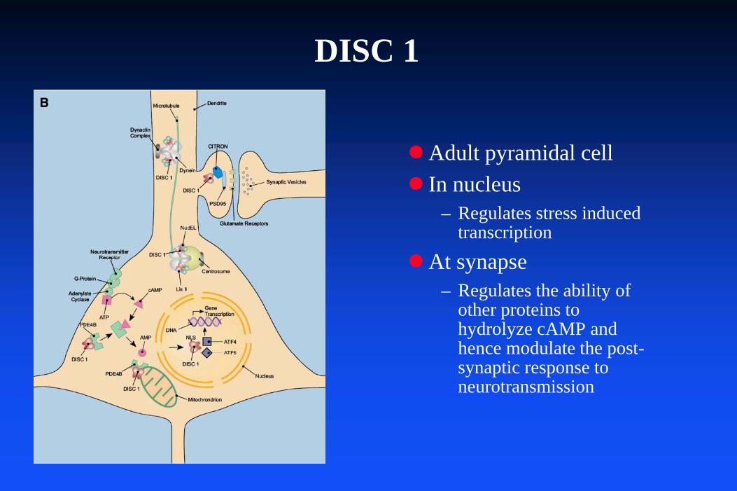
DISC 1
Adult pyramidal cellIn nucleus
– Regulates stress induced transcription
At synapse– Regulates the ability of
other proteins to hydrolyze cAMP and hence modulate the post-synaptic response to neurotransmission

DISC 1 and fMRI
SNP in DISC 1 associated with SCZ (SNP 10; Ser740Cys)Selected normal subjects with the two alleles (Ser or Cys) and scanned them while performing a working memory taskCys carrierss performed better on the task and showed enhanced activation of the HC relative to Ser homozygotes.
Callicot et al., 2005
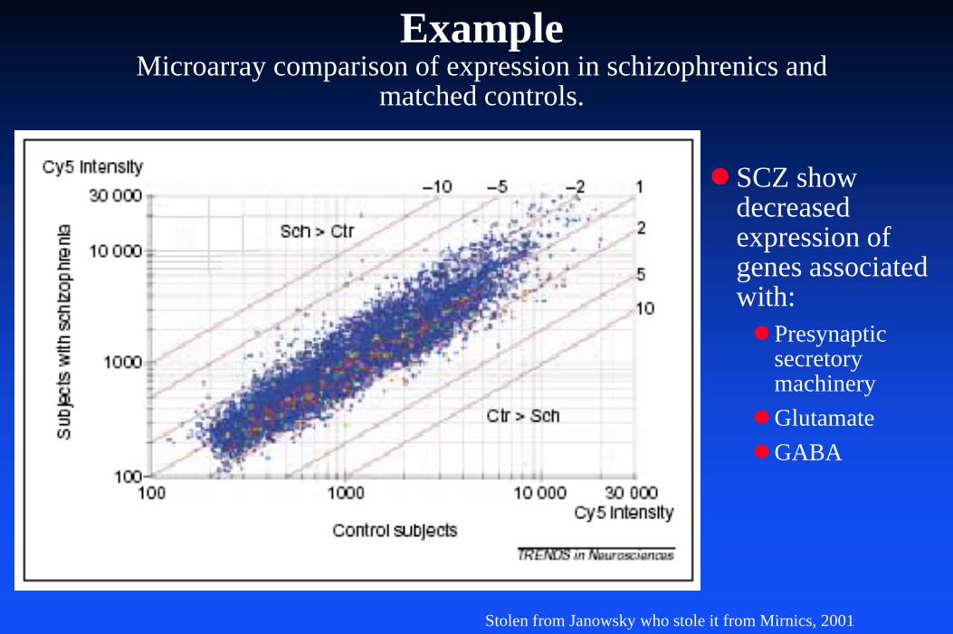
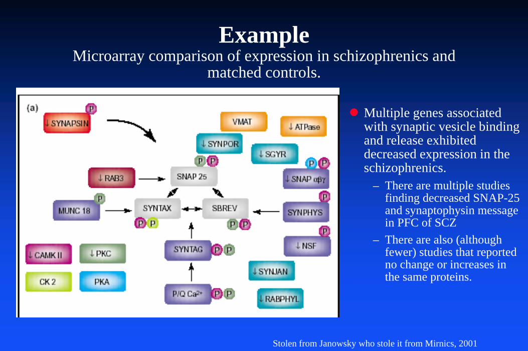
ExampleMicroarray comparison of expression in schizophrenics and
matched controls.
Stolen from Janowsky who stole it from Mirnics, 2001
SCZ show decreased expression of genes associated with:
Presynaptic secretory machineryGlutamateGABA
ExampleMicroarray comparison of expression in schizophrenics and
matched controls.
Multiple genes associated with synaptic vesicle binding and release exhibited decreased expression in the schizophrenics.
– There are multiple studies finding decreased SNAP-25 and synaptophysin message in PFC of SCZ
– There are also (although fewer) studies that reported no change or increases in the same proteins.
Stolen from Janowsky who stole it from Mirnics, 2001
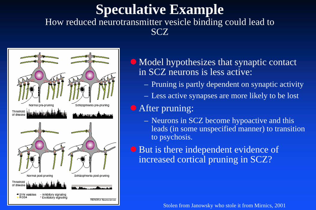
Speculative ExampleHow reduced neurotransmitter vesicle binding could lead to
SCZ
Stolen from Janowsky who stole it from Mirnics, 2001
Model hypothesizes that synaptic contact in SCZ neurons is less active:
– Pruning is partly dependent on synaptic activity– Less active synapses are more likely to be lost
After pruning:– Neurons in SCZ become hypoactive and this
leads (in some unspecified manner) to transition to psychosis.
But is there independent evidence of increased cortical pruning in SCZ?

Pruning Hypothesis, continued
Post mortem study of 15 SCZ, 15 non-SCZ psych and 15 controls.SCZ have fewer spines per dendrite.
From Glantz and Lewis, 2000

Caveats
It may be impossible to intuit the behavior of such a complex system.
– Complex systems inevitably behave in unpredictable fashions.
The anatomy and physiology are sufficiently complex that almost any “model” of schizophrenia might sound plausible.
Role of Development: Prenatal
Period of formation of the primary repertoire of neuronal connections.Primary repertoire refers to the specific set of neural linkages present at birth.
– Presumed primary time of action of genetic factors.» Speculation that expression of cell adhesion molecules is abnormal.
– Although bounded by genetic factors, epigenetic factors (including chance) determine the final form of the primary repertoire.
– The primary repertoire of each individual is unique: even identical twins and individual syngeneic mice have different brains.
– Intrauterine trauma, viral infections, exposure to toxins all may influence the primary repertoire.
Role of Development:Postnatal
Period of formation of the secondary repertoire.The secondary repertoire of neural links develops as a result of the individual’s environmental experience.
– Genetic factors influence the plasticity of the postnatal brain.
Behavioral aspects of the secondary repertoire include object perception, spatial orientation and learned stimulus response patterns.The secondary repertoire develops and is maintained via both anatomical and synaptic mechanisms.
– Considerable cell death, remodeling and synaptic pruning takes place between birth and puberty.
– Exposure to environmental stimuli and the individual’s motor and cognitive responses change the strength of synaptic connections.
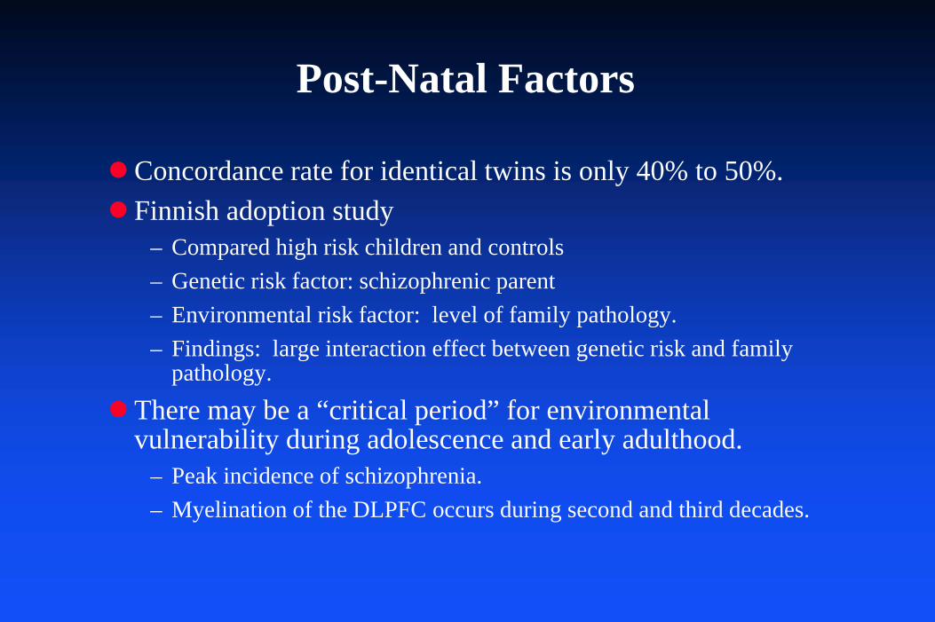
Post-Natal Factors
Concordance rate for identical twins is only 40% to 50%.Finnish adoption study
– Compared high risk children and controls– Genetic risk factor: schizophrenic parent– Environmental risk factor: level of family pathology.– Findings: large interaction effect between genetic risk and family
pathology.
There may be a “critical period” for environmental vulnerability during adolescence and early adulthood.
– Peak incidence of schizophrenia.– Myelination of the DLPFC occurs during second and third decades.
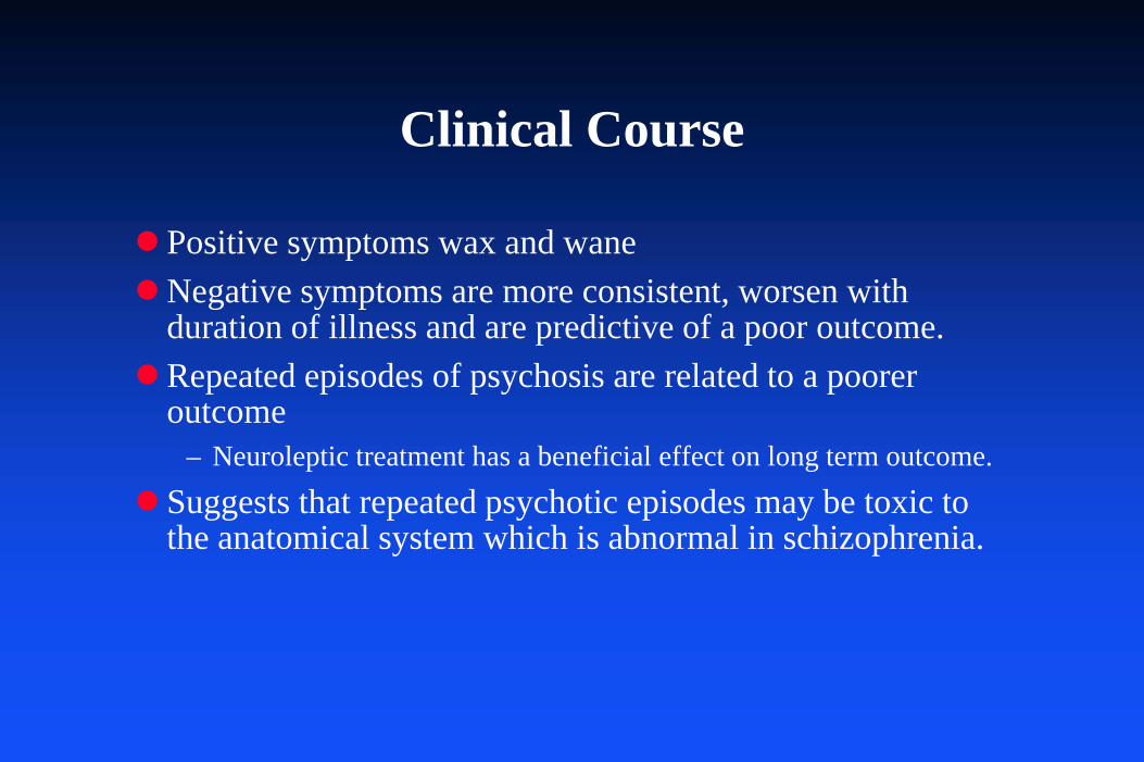
Clinical Course
Positive symptoms wax and waneNegative symptoms are more consistent, worsen with duration of illness and are predictive of a poor outcome.Repeated episodes of psychosis are related to a poorer outcome
– Neuroleptic treatment has a beneficial effect on long term outcome.
Suggests that repeated psychotic episodes may be toxic to the anatomical system which is abnormal in schizophrenia.
What must be explained by a model of schizophrenia?
SymptomsGenetic vulnerabilityDevelopmental vulnerabilityClinical coursePartial therapeutic effect of dopamine D2 antagonists
Framework for Answers
Where is the lesion?What is its nature?When does it occur?How does it occur?When is it expressed?

Framework for Answers
Where is the lesion?– Most of the evidence suggests cortical (hippocampus and DLPFC)
as the most likely locations for initial abnormality.– It is possible (and perhaps even probable) that abnormalities
anywhere in this system could give rise to a schizophrenic syndrome.
What is its nature?When does it occur?How does it occur?When is it expressed?
Framework for Answers
Where is the lesion?What is its nature?
– The abnormality ranges from synaptic dysfunction to substantial atrophy.
– More important is the response of the corticolimbic circuits to the initial abnormality.
How does it occurWhen does it occur?When is it expressed?

Framework for Answers
Where is the lesion?What is its nature?When does it occur?
– The abnormality initially occurs during prenatal development or at least quite early in development.
– Subsequent development may be flawed by its occurrence.– Onset of puberty enhances liklihood of expression.
How does it occur?When is it expressed?

Framework for Answers
Where is the lesion?What is its nature?When does it occur?How does it occur?
– A variety of genetic and epigenetic factors could cause the lesion(s)» Purely genetic factors could involve reduced expression of presynaptic
elements or abnormalities associated with particular neurotransmitters.» Trauma, toxins, infections, and many other influences could cause
schizophrenogenic lesions.
When is it expressed?

Framework for Answers
Where is the lesion?What is its nature?When does it occur?How does it occur?When is it expressed?
– Children with a schizophrenic diathesis may appear abnormal from early life.
– The peak expression occurs when the DLPFC myelinates and the brain undergoes the second wave of pruning during adolescence and early adulthood.
– The diathesis is less likely to result in the full syndrome in the absence of on-going environmental stressors.

To be continued…
Prefrontal Heteromodal Cortex
86
43,1,2
5
3,1,2
5
7a
19
19
18
1817
45, 4443
37
2038
9
10 46
4711
41,42
3940
22
22
21
dlpfc
MA M1
S1
A1
7b
21
VA
sg ipl
ag
spl
tpAA
mt
9
SA
V1
V2
V3
V5
Prefrontal Cortex
38
28
34
u
36
hc
amg
fg
ph
lg
stmt
it
37
20
V4
paralimbic11
heteromodal
Medial Paralimbic Cortex
24
33
12
38 28
2331
31
24
46
8
9
10
1125
1717
19
18
5 7a
18
7b
mpfc
ofc
po
tp
V1
VA
VA
SAM1MA cg
mpo
7b
V6aca

What must be explained by a model of schizophrenia?
SymptomsGenetic vulnerabilityDevelopmental vulnerabilityClinical coursePartial therapeutic effect of dopamine D2 antagonists

Schizophrenics Show a Decreased Flush Response to Topical Niacin
Erik MessamoreWe don’t know what it means.
-7 -6 -5 -4 -3 -2 -1 0Log Concentration (M)
-100
0
100
200
300
400
Sign
al +
/- SE
M
SchizophreniaControl
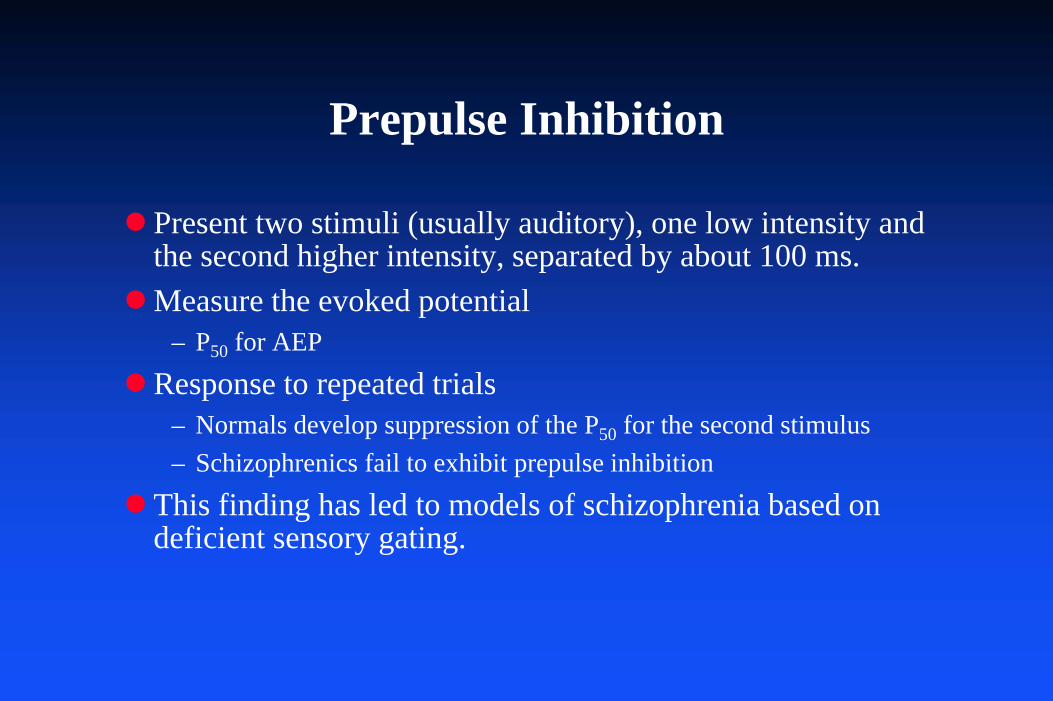
Prepulse Inhibition
Present two stimuli (usually auditory), one low intensity and the second higher intensity, separated by about 100 ms.Measure the evoked potential
– P50 for AEP
Response to repeated trials– Normals develop suppression of the P50 for the second stimulus– Schizophrenics fail to exhibit prepulse inhibition
This finding has led to models of schizophrenia based on deficient sensory gating.

Corticostriatal Function
Current models of basal ganglia function focus on massively parallel circuits connecting cortex, basal ganglia, thalamus and re-entry into cortex. Parallelism occurs both between topographically distinct cortical regions and within each region via neural circuits with opposing re-entrant actions on cortex.
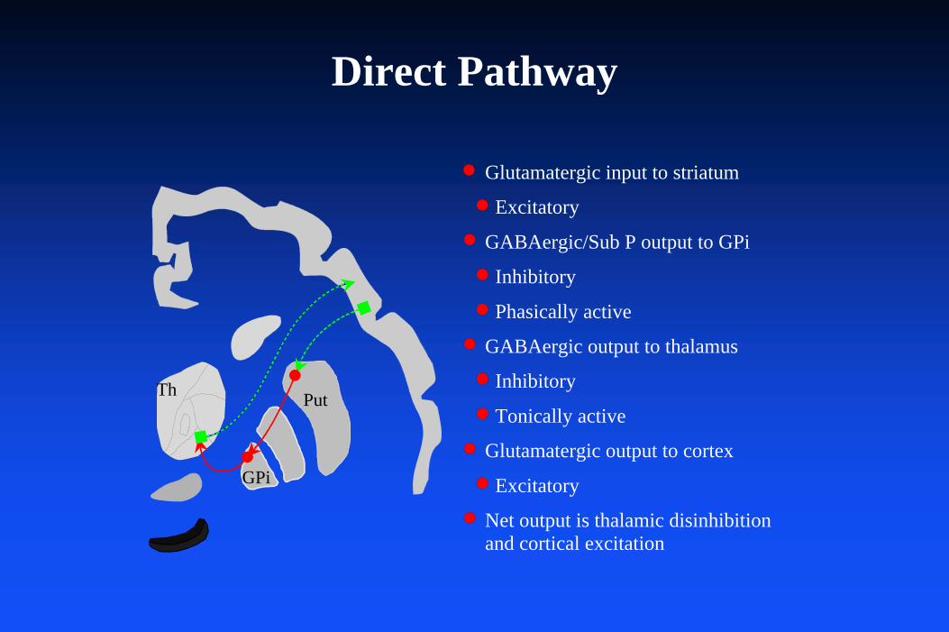
Direct Pathway
Put
GPi
Th
Glutamatergic input to striatum
Excitatory
GABAergic/Sub P output to GPi
Inhibitory
Phasically active
GABAergic output to thalamus
Inhibitory
Tonically active
Glutamatergic output to cortex
Excitatory
Net output is thalamic disinhibition and cortical excitation
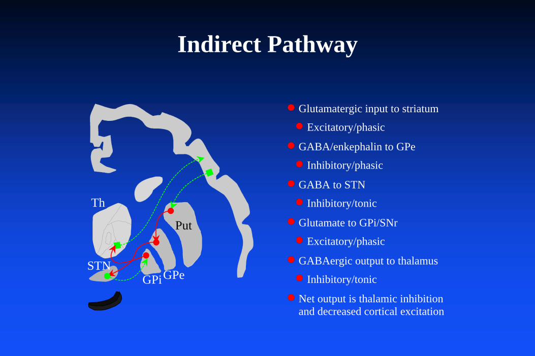
Indirect Pathway
Put
GPi
Th
STNGPe
Glutamatergic input to striatum
Excitatory/phasic
GABA/enkephalin to GPe
Inhibitory/phasic
GABA to STN
Inhibitory/tonic
Glutamate to GPi/SNr
Excitatory/phasic
GABAergic output to thalamus
Inhibitory/tonic
Net output is thalamic inhibition and decreased cortical excitation

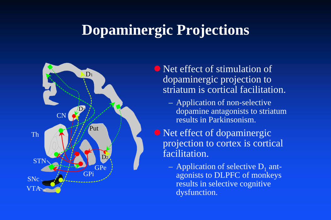
Dopaminergic Projections
GPeGPi
Put
STN
SNcVTA
D1
CN
Th
D1
D2
Direct Pathway– Modulated by stimulatory D1
receptors.
Indirect Pathway– Modulated by inhibitory D2
receptors.
Cortical Neurons– Modulated by stimulatory (?) D1
receptors.
There are reciprocal projec-tions from cortex and striatum to midbrain dopaminergic nuclei (not shown).
Dopaminergic Projections
GPeGPi
Put
STN
SNcVTA
D1
CN
Th
D1
D2
Net effect of stimulation of dopaminergic projection to striatum is cortical facilitation.
– Application of non-selective dopamine antagonists to striatum results in Parkinsonism.
Net effect of dopaminergic projection to cortex is cortical facilitation.
– Application of selective D1 ant-agonists to DLPFC of monkeys results in selective cognitive dysfunction.

Prenatal genetic or epigentic abnormalities establish a schizophrenic diathesis.Post-natal environmental factors have a profound effect on the expression of the diathesis.
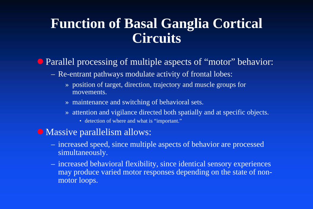
Function of Basal Ganglia Cortical Circuits
Parallel processing of multiple aspects of “motor” behavior:– Re-entrant pathways modulate activity of frontal lobes:
» position of target, direction, trajectory and muscle groups for movements.
» maintenance and switching of behavioral sets.» attention and vigilance directed both spatially and at specific objects.
• detection of where and what is “important.”
Massive parallelism allows:– increased speed, since multiple aspects of behavior are processed
simultaneously.– increased behavioral flexibility, since identical sensory experiences
may produce varied motor responses depending on the state of non-motor loops.
Caveats
It may be impossible to intuit the behavior of such a complex system.
– Complex systems inevitably behave in unpredictable fashions.
The anatomy and physiology are sufficiently complex that almost any “model” of schizophrenia might sound plausible.