scf c in ring h2 b iq in li
TRANSCRIPT

P1: FKZ/FHX/FGJ P2: FKZ/FHR/fgo QC: FhN/Anil T1: FhN
September 10, 1999 15:45 Annual Reviews AR092-15
?Annu. Rev. Cell Dev. Biol. 1999. 15:435–67
Copyright c� 1999 by Annual Reviews. All rights reserved
SCF AND CULLIN/RING H2-BASED
UBIQUITIN LIGASES
R. J. DeshaiesDepartment of Biology 156-29, California Institute of Technology, Pasadena, California91125, [email protected]
Key Words ubiquitin, ubiquitination, F box, Skp1, Cdc34
■ Abstract Protein degradation is deployed tomodulate the steady-state abundanceof proteins and to switch cellular regulatory circuits from one state to another by abruptelimination of control proteins. In eukaryotes, the bulk of the protein degradation thatoccurs in the cytoplasm and nucleus is carried out by the 26S proteasome. In turn,most proteins are thought to be targeted to the 26S proteasome by covalent attachmentof a multiubiquitin chain. Ubiquitination of proteins requires a multienzyme system.A key component of ubiquitination pathways, the ubiquitin ligase, controls both thespecificity and timing of substrate ubiquitination. This review is focused on a conservedubiquitin ligase complex known as SCF that plays a key role in marking a variety ofregulatory proteins for destruction by the 26S proteasome.
CONTENTS
Introduction . . . . . . . . . . . . . . . . . . . . . . . . . . . . . . . . . . . . . . . . . . . . . . . . . . . . 436Identification of SCF Ubiquitin Ligase Activity . . . . . . . . . . . . . . . . . . . . . . . 437Subunits of SCF . . . . . . . . . . . . . . . . . . . . . . . . . . . . . . . . . . . . . . . . . . . . . . . . 439Architecture of SCF Complexes and SCF Pathway Components . . . . . . . . . . 439Architecture of Skp1 . . . . . . . . . . . . . . . . . . . . . . . . . . . . . . . . . . . . . . . . . . . . . 444Architecture of Cdc53 . . . . . . . . . . . . . . . . . . . . . . . . . . . . . . . . . . . . . . . . . . . . 445Architecture of Hrt1 . . . . . . . . . . . . . . . . . . . . . . . . . . . . . . . . . . . . . . . . . . . . . 446Architecture of F Box Proteins . . . . . . . . . . . . . . . . . . . . . . . . . . . . . . . . . . . . . . 446Architecture of F Box Proteins: Cdc4 . . . . . . . . . . . . . . . . . . . . . . . . . . . . . . . . . 447Architecture of yCdc34 . . . . . . . . . . . . . . . . . . . . . . . . . . . . . . . . . . . . . . . . . . . 448
Post-Translational Control of SCF Pathway Components . . . . . . . . . . . . . . . . 448Post-Translational Regulation of Skp1 . . . . . . . . . . . . . . . . . . . . . . . . . . . . . . . . 449Post-Translational Regulation of Cdc53/Cullin . . . . . . . . . . . . . . . . . . . . . . . . . . 449Post-Translational Regulation of Hrt1 . . . . . . . . . . . . . . . . . . . . . . . . . . . . . . . . 450Post-Translational Regulation of F Box Proteins . . . . . . . . . . . . . . . . . . . . . . . . . 450Post-Translational Regulation of Cdc34 . . . . . . . . . . . . . . . . . . . . . . . . . . . . . . . 451
Substrates of the SCF Pathway . . . . . . . . . . . . . . . . . . . . . . . . . . . . . . . . . . . . . 451Mechanism of Action of SCF . . . . . . . . . . . . . . . . . . . . . . . . . . . . . . . . . . . . . . 454
1081-0706/99/1115-0435$08.00 435
Ann
u. R
ev. C
ell.
Dev
. Bio
l. 19
99.1
5:43
5-46
7. D
ownl
oade
d fro
m a
rjour
nals.
annu
alre
view
s.org
by C
ALI
FORN
IA IN
STIT
UTE
OF
TECH
NO
LOG
Y o
n 09
/08/
05. F
or p
erso
nal u
se o
nly.

P1: FKZ/FHX/FGJ P2: FKZ/FHR/fgo QC: FhN/Anil T1: FhN
September 10, 1999 15:45 Annual Reviews AR092-15
?436 DESHAIES
How Does Substrate Bind SCF? . . . . . . . . . . . . . . . . . . . . . . . . . . . . . . . . . . . . 454How is Ubiquitin Transferred From Cdc34 to Substrate? . . . . . . . . . . . . . . . . . . . 455How Does SCF Promote the Assembly of a Multiubiquitin Chain? . . . . . . . . . . . . 456
Diversification of SCF Function . . . . . . . . . . . . . . . . . . . . . . . . . . . . . . . . . . . . 457The F Box Hypothesis . . . . . . . . . . . . . . . . . . . . . . . . . . . . . . . . . . . . . . . . . . . . 457F Boxes, SOCS Boxes, and Others . . . . . . . . . . . . . . . . . . . . . . . . . . . . . . . . . . . 457
Conclusion and Perspectives . . . . . . . . . . . . . . . . . . . . . . . . . . . . . . . . . . . . . . 459
INTRODUCTION
Proteolysis in the eukaryotic cytosol typically involves the assembly of a substrate-linked ubiquitin chain, which targets specific proteins for degradation by the 26Sproteasome (Hochstrasser 1995). Ubiquitin is activated for transfer to substratethrough the ATP-dependent formation of a thioester bond with the ubiquitin-activating enzyme, E1. Ubiquitin is subsequently transferred to a member of afamily of ubiquitin-conjugating (E2) enzymes. Finally, thioesterified ubiquitin istransferred from E2 enzyme to a lysine residue of the target protein, either di-rectly or with the assistance of a ubiquitin ligase (E3). E3s bind directly to sub-strate, suggesting that they provide specificity in ubiquitination reactions. Onewell-characterized E3, known as E6-AP, also forms a thioester with ubiquitin asan intermediate in the transfer of ubiquitin from E2 to substrate (Scheffner et al1995). Because E3s dictate the specificity of ubiquitination reactions, it is likelythat protein degradation in vivo is controlled primarily by regulating E3 activityor E3-substrate interaction.SCFCdc4, the prototype of the SCF (for Skp1, Cdc53/Cullin, F box receptor; the
superscript denotes the identity of the F box subunit) family of ubiquitin ligases,was first defined in budding yeast by in vitro reconstitution (Feldman et al 1997,Skowyra et al 1997). Over the past few years, SCF pathway components havebeen identified and linked to diverse cellular processes in many eukaryotes. Inthis review, I describe the multiple lines of research that led to the discovery ofSCF ubiquitin ligases and review what is known about the subunits, architecture,regulation, substrates, mechanism of action, and functional diversification of SCFcomplexes. Several excellent reviews focused on various roles of the SCF complexhave been published recently (Elledge & Harper 1998, Krek 1998, Patton et al1998a). It is the goal of this article to provide a comprehensive review of currentknowledge. The SCF pathway was originally discovered in the budding yeastSaccharomyces cerevisiae and has been most thoroughly characterized in thisorganism. For the sake of simplicity, this review concentrates on budding yeastSCF, but draws extensively on the rapidly expanding literature onmammalian SCFto supplement the findings that have emerged from study of yeast SCF. In instanceswhere budding yeast and human SCF subunits have the same name, the proteinis preceded by either y or h to indicate the organism from which the protein isderived.
Ann
u. R
ev. C
ell.
Dev
. Bio
l. 19
99.1
5:43
5-46
7. D
ownl
oade
d fro
m a
rjour
nals.
annu
alre
view
s.org
by C
ALI
FORN
IA IN
STIT
UTE
OF
TECH
NO
LOG
Y o
n 09
/08/
05. F
or p
erso
nal u
se o
nly.

P1: FKZ/FHX/FGJ P2: FKZ/FHR/fgo QC: FhN/Anil T1: FhN
September 10, 1999 15:45 Annual Reviews AR092-15
?SCFUBIQUITIN LIGASES 437
IDENTIFICATION OF SCF UBIQUITINLIGASE ACTIVITY
Identification of the SCFCdc4 ubiquitin ligase complex stemmed from a geneticanalysis of theG1/S transition by Schwob et al (1994). These authors demonstratedthat budding yeast cdc4ts, cdc34ts, and cdc53tsmutants fail to enter S phase becausethey are unable to eliminate the S phase cyclin/cyclin-dependent kinase (CDK)inhibitor Sic1. Sic1 is normally destroyed as wild-type cells progress from G1 to Sphase but persists indefinitely in the cdcmutants. Subsequently, it was shown thatskp1ts mutants have a similar phenotype (Bai et al 1996). yCdc34 was a logicalcandidate for a Sic1 destabilizing factor because it possesses ubiquitin-conjugatingenzyme activity (Goebl et al 1988). In contrast, although genetic analysis suggesteda role for Cdc4, Cdc53, and Skp1 in Sic1 degradation, it was unclear what theseproteins might be doing, as they bore no resemblance to any known component ofubiquitin-dependent proteolytic pathways.The components of the SCF pathway were discovered and characterized in
several laboratories. cdc4ts and cdc34ts mutants were identified in screens for cellcycle mutants by Hartwell and colleagues (Pringle & Hartwell 1981), and thecorrespondinggeneswere clonedby complementation of the tsmutants (Goebl et al1988, Peterson et al 1984). The cdc53tsmutantwas identified in a screen formutantswith a cdc34ts-like phenotype, andCDC53was cloned by complementation of thismutant (Mathias et al 1996). The sequence of Cdc53 revealed that it is homologousto a family of proteins called cullins. The first reported cullin, cul-1, was identifiedin nematodes as a gene required for developmentally programmed transitions fromG1 phase of the cell cycle to G0 phase (Kipreos et al 1996). Cdc53 and Cdc4 wereshown to coprecipitate from yeast cell lysates with Cdc34, suggesting that theseproteins directly participate in protein ubiquitination (Mathias et al 1996). Cdc53was independently identified as a protein that copurified with the unstable G1cyclin Cln2 (Willems et al 1996). These authors demonstrated that Cdc53 bindsCln2 and that cdc53ts mutants are defective in Cln2 turnover. Lastly, ySKP1 wasidentified in a screen for genes that suppress cdc4ts mutants upon overexpression(Bai et al 1996). This screen also revealed the gene that encodes human cyclinF (Bai et al 1994). ySkp1 was also identified by Connelly & Hieter (1996) asa component of the centromere-binding CBF3 complex. The human orthologueof ySkp1 had previously been identified as a cyclin A/CDK2-associated protein,but its biochemical function was not known (Zhang et al 1995). Remarkably, theotherwise dissimilar Cdc4 and human cyclin F proteins were shown to share asmall sequence motif designated the F box (Bai et al 1996). The F box, which isfound in a large number of proteins, mediates binding of both Cdc4 and cyclin Fto ySkp1.The final discovery key to identification of SCF ubiquitin ligase stemmed from
biochemical reconstitution of Sic1 ubiquitination. First, ubiquitination of Sic1 incrude yeast extracts was shown to depend upon yCdc34, Cdc4, andG1 cyclin/CDK
Ann
u. R
ev. C
ell.
Dev
. Bio
l. 19
99.1
5:43
5-46
7. D
ownl
oade
d fro
m a
rjour
nals.
annu
alre
view
s.org
by C
ALI
FORN
IA IN
STIT
UTE
OF
TECH
NO
LOG
Y o
n 09
/08/
05. F
or p
erso
nal u
se o
nly.

P1: FKZ/FHX/FGJ P2: FKZ/FHR/fgo QC: FhN/Anil T1: FhN
September 10, 1999 15:45 Annual Reviews AR092-15
?438 DESHAIES
activity (Verma et al 1997b). These data suggested that the role identified for theseproteins in Sic1 turnover in vivo (Schneider et al 1996, Schwob et al 1994) waslikely to be direct. Skowyra et al (1997) and Feldman et al (1997) next demon-strated that Cdc4, Cdc53, and ySkp1 expressed in insect cells assemble into acomplex and that the purified complex functions as a ubiquitin ligase, promotingubiquitination of phosphorylated Sic1 by the yCdc34 ubiquitin-conjugating en-zyme. Skowyra et al (1997) also demonstrated that a distinct SCF complex canbe formed by replacing Cdc4 with another F box–containing protein, Grr1. Thisimportant finding suggested that SCFCdc4 may be the prototype for a broad arrayof SCF-like ubiquitin ligases whose substrate specificity is dictated by the identityof the F box subunit (see DIVERSIFICATIONOF SCF FUNCTION for a detaileddiscussion). In parallel with the reconstitution efforts, phosphorylation of Sic1 ona set of CDK consensus sites was shown to be necessary and sufficient to trigger itsubiquitination in vitro and degradation in vivo (Verma et al 1997a). Taken together,the results demonstrate that the G1/S transition in budding yeast is triggered bythe phosphorylation of Sic1 by G1 cyclin/CDK, followed by the ubiquitination ofphosphorylated Sic1 through the combined efforts of SCFCdc4 and yCdc34.Very recently, a fourth essential subunit of SCF, referred to as Roc1, Rbx1, or
Hrt1, was reported (Kamura et al 1999, Ohta et al 1999, Seol et al 1999, Tan et al1999). Throughout the remainder of this review I refer to the mammalian versionsof this protein as Roc1/Rbx1 and to the highly homologous budding yeast versionas Hrt1. This new protein was identified as a fourth subunit of SCFSKP2 complexespurified from HeLa cells (Tan et al 1999), as a hCul1-binding protein in a two-hybrid screen (Ohta et al 1999), as a fifth subunit of purified rat VHL/elonginC/elongin B/Cul2 complexes (Kamura et al 1999), and as a specific component ofySkp1 and Cdc53 immunoprecipitates (Seol et al 1999). Roc1/Rbx1/Hrt1 is ho-mologous to the Apc11 subunit of the Anaphase-Promoting Complex/Cyclosome(APC/C) (Zachariae et al 1998), and shares with both yeast and human Apc11 ahighly conserved zinc-binding RING-H2 domain.Budding yeastHRT1 is essential, and the lethal phenotype of an hrt1∆ allele can
be rescued by the highly homologous murine or human ROC1/RBX1 (Kamura et al1999, Ohta et al 1999, Seol et al 1999). Analysis of Hrt1-depleted cells and hrt1tsmutants revealed that turnover of the SCF substrates Sic1 and Cln2 requiresHRT1function, resulting in accumulation of these proteins in mutant cells (Kamuraet al 1999, Ohta et al 1999, Seol et al 1999). Hrt1 is also important for SCFfunction in vitro in that recombinant Hrt1 dramatically stimulates the ability ofSCF to ubiquitinate Cln1, Cln2, Sic1, and IκB, as well as to assemble unanchoredmultiubiquitin chains and promote autoubiquitination of yCdc34 (Kamura et al1999, Ohta et al 1999, Seol et al 1999, Skowyra et al 1999, Tan et al 1999). Theseobservations provoke a simple question—Why did the original reconstitution ofSCFCdc4 work in the absence of Hrt1 (Feldman et al 1997, Skowyra et al 1997)?Given the high degree of structural and functional conservation of this protein, itis likely that recombinant SCF complexes produced with the baculovirus systemincorporate an insect homolog of Hrt1. Assembly of active SCF complexes with
Ann
u. R
ev. C
ell.
Dev
. Bio
l. 19
99.1
5:43
5-46
7. D
ownl
oade
d fro
m a
rjour
nals.
annu
alre
view
s.org
by C
ALI
FORN
IA IN
STIT
UTE
OF
TECH
NO
LOG
Y o
n 09
/08/
05. F
or p
erso
nal u
se o
nly.

P1: FKZ/FHX/FGJ P2: FKZ/FHR/fgo QC: FhN/Anil T1: FhN
September 10, 1999 15:45 Annual Reviews AR092-15
?SCFUBIQUITIN LIGASES 439
subunits from different species has been noted before (Feldman et al 1997, Lyapinaet al 1998).
SUBUNITS OF SCF
Many putative SCF subunits, including a large number of F box proteins, havebeen identified in eukaryotes by database searches, genetic screens, and two-hybridscreens. These proteins contribute to a broad spectrum of cellular activities rangingfrom the auxin response in plants, withdrawal from the cell cycle in nematodes,entry into S phase and M phase of the cell cycle in budding yeast, signaling viatheWnt and hedgehog pathways inDrosophila and Xenopus, and innate immunityin human cells. Rather than discuss each of these proteins in detail, the basicproperties of those that have been described in the literature are summarized inTable 1. It is important to note that most of these proteins have not been confirmedto be members of functional ubiquitin ligase complexes. This may be especiallycritical for the F box proteins, since at least one known F box protein (Ctf13)appears to function primarily as a component of the budding yeast kinetochore(Connelly & Hieter 1996, Kaplan et al 1997, Stemmann & Lechner 1996).
ARCHITECTURE OF SCF COMPLEXES AND SCFPATHWAY COMPONENTS
The four subunits of SCFCdc4 assemble together to form a heterotetrameric ubiq-uitin ligase (Figure 1). Within this complex, the F box–containing Cdc4 subunitdirectly binds substrate, the Cdc53 and Hrt1 subunits recruit the Cdc34 ubiquitin-conjugating enzyme, andSkp1 helps to link theCdc4 andCdc53 subunits (Feldmanet al 1997, Skowyra et al 1997, Willems et al 1996, Patton et al 1998, Seol et al1999, Skowra et al 1999). Together, these four subunits make up the minimal SCFubiquitin ligase complex. Affinity purification of SCF complexes from buddingyeast suggests that other proteins associate with ySkp1 and Cdc53 (JH Seol, per-sonal communication), but it remains unclear whether these represent substrates,proteins restricted to specific SCF complexes, components of multisubunit F boxreceptors, or uncharacterized members of the core complex.In the following sections, the domain organization and properties of each SCF
subunit and how they contribute to the architecture of the complex are discussed(see Figure 2 for a summary of the structural organization of Skp1, Cdc53, Hrt1,and Cdc34). All known SCF subunits are highly conserved throughout eukaryotes.As evidence of the functional conservation of these proteins, hSKP1, hCUL1, andROC1/RBX1 have been shown to complement the corresponding budding yeastmutants and can assemble with yeast SCF subunits to form active complexes (Baiet al 1996, Lyapina et al 1998, Kamura et al 1999, Ohta et al 1999, Seol et al 1999,Skowyra et al 1999). Thus unless interactions between recombinant SCF subunits
Ann
u. R
ev. C
ell.
Dev
. Bio
l. 19
99.1
5:43
5-46
7. D
ownl
oade
d fro
m a
rjour
nals.
annu
alre
view
s.org
by C
ALI
FORN
IA IN
STIT
UTE
OF
TECH
NO
LOG
Y o
n 09
/08/
05. F
or p
erso
nal u
se o
nly.

P1: FKZ/FHX/FGJ P2: FKZ/FHR/fgo QC: FhN/Anil T1: FhN
September 10, 1999 15:45 Annual Reviews AR092-15
?440 DESHAIES
TABLE1
CandidateSCFpathwaycomponents:listofSCF
subunitsthathavebeencharacterizedtodate.Thislistincludesonlythose
proteinsforwhichindependentpapershavebeenpublisheddescribingtheirstructure,regulation,orfunction
Homology
Protein
domains
Organism
Function/properties
Reference
Cullins
Cdc53
Cullin
S.cerevisiae
RequiredforSCF
activity,cellcycleprogress
Matthiasetal1996
Cul-1
Cullin
C.elegans
Developmentallyprogrammedcellcycleexit
Kipreosetal1996
Cul1
Cullin
H.sapiens
p21,cyclinD,E2F-1turnover
Lisztwanetal1998a
Cul2
Cullin
H.sapiens
Reg.ofhypoxia-inducedmRNAaccumulation
Pauseetal1997,
Lonerganetal1998
Cul3
Cullin
H.sapiens
Repressedbysalicylate,inducedbyPM
ADuetal1998
Cul4A
Cullin
H.sapiens
Overexpressedinbreasttumors
Chenetal1998
Cul5
Cullin
H.sapiens
Vasopressin-activatedcalciummobilizingreceptor
Burnatowska-Hledinetal1995
Apc2
CHH.sapiens
EssentialsubunitofAPC/Cubiquitinligase
HYuetal1998
S.cerevisiae
Zachariaeetal1998
Pcu1
Cullin
S.pombe
EssentialhCullhomolog,reg.genomeploidy
Kominamietal1998
Pcu3
Cullin
S.pombe
hCul3-like;pcu3
viable,UVandHU-sensitive
Kominamietal1998
Hrt1/Roc1/Rbx1
Hrt1
RING-H2
S.cerevisiae
RequiredforSCF
ubiquitinligaseactivity
Ohtaetal1999b
Roc1/Rbx1
RING-H2
H.sapiens
ComponentofSCFubiquitinligase
seeHrt1references
Roc2
RING-H2
H.sapiens
SimilartoRoc1,assembleswithcullins
Ohtaetal1999
Skp1-like
Skp1
Many
AssemblyofSCF
Baietal1996
ElonginC
H.sapiens
Transcriptelongation,subunitofhCul2Ubligase
Pauseetal1997,
Lonerganetal1998
∆
Ann
u. R
ev. C
ell.
Dev
. Bio
l. 19
99.1
5:43
5-46
7. D
ownl
oade
d fro
m a
rjour
nals.
annu
alre
view
s.org
by C
ALI
FORN
IA IN
STIT
UTE
OF
TECH
NO
LOG
Y o
n 09
/08/
05. F
or p
erso
nal u
se o
nly.

P1: FKZ/FHX/FGJ P2: FKZ/FHR/fgo QC: FhN/Anil T1: FhN
September 10, 1999 15:45 Annual Reviews AR092-15
?SCFUBIQUITIN LIGASES 441
Fboxproteins
β-TrCPc
F/WD40
H.sapiens
Deg.ofCD4inHIV-infectedcells,I
κB,
β-catenin
Margottinetal1998,
(akaE3RS)
Consultβ-cateninandIκBentriesinTable2
Yaronetal1998
foradditionalreferences
Cdc4
F/WD40
S.cerevisiae
ReceptorforSic1,Far1,Cdc6
Feldmanetal1997,
Skowyraetal1997
COI1
F/LRR
A.thaliana
Jasmonateperception
Xieetal1998
FIM
FA.majus
ExpressionofMADS-boxproteindeficiens
Ingram
etal1997
Grr1
F/LRR
S.cerevisiae
ReceptorforG1cyclinCln2,glucoserepression
Barraletal1995,
Skowyraetal1997
MEKK-α
F/WD40
D.discoideum
Reg.oftimingandspatialpatterningduring
Chungetal1998
proteinkinase
development
Met30
F/WD40
S.cerevisiae
Reg.ofsulfurmetabolism
Pattonetal1998a
Reg.ofmorphogenesischeckpoint
Siaetal1998
NFB42
Fbox
H.sapiens
Overexpressionblocksproliferation;neuronal
Erhardtetal1998
Pop1
F/WD40
S.pombe
Reg.genomeploidy;deg.ofRum1,Cdc18
Kominami&
Toda1997
Pop2/Sud1
F/WD40
S.pombe
Reg.genomeploidy;deg.ofRum1,Cdc18
Kominamietal1998,
Jallepallietal1998
Scon-2
F/WD40
N.crassa
Reg.ofsulfurmetabolism
(similartoMet30)
Kumar&Paietta1998
SconB
F/WD40
A.nidulans
Reg.ofsulfurmetabolism
(similartoMet30)
Natorffetal1998
SEL-10
F/WD40
C.elegans
Reg.ofNotchpathwaysignaling
Hubbardetal1997
Skp2
F/LRR
H.sapiens
Reg.ofSphase;deg.ofE2F-1
Zhangetal1995,
Martietal1999
(continued)
Ann
u. R
ev. C
ell.
Dev
. Bio
l. 19
99.1
5:43
5-46
7. D
ownl
oade
d fro
m a
rjour
nals.
annu
alre
view
s.org
by C
ALI
FORN
IA IN
STIT
UTE
OF
TECH
NO
LOG
Y o
n 09
/08/
05. F
or p
erso
nal u
se o
nly.

P1: FKZ/FHX/FGJ P2: FKZ/FHR/fgo QC: FhN/Anil T1: FhN
September 10, 1999 15:45 Annual Reviews AR092-15
?442 DESHAIES
slimb
F/WD40
Drosophila
Negativereg.ofwinglessandHedgehogsignaling
Jiang&Struhl1998,
Theodosiouetal1998
TIR1
F/LRR
A.thaliana
Auxinperception
Rueggeretal1998
UFO
FA.thaliana
Properfloraldevelopment;homologofFIM
Leeetal1997
Cdc34/E2
Cdc34
UBC
S.cerevisiae
UbiquitinationanddegradationofSCFsubstrates
Goebletal1988
Cdc34
UBC
X.laevis
Reg.ofG1/S&G2/M;deg.ofXic1,Wee1
Yew&Kirschner1997,
Michael&Newport1998
Cdc34
UBC
H.sapiens
UbiquitinationanddegradationofSCFsubstrates
Plonetal1993
hUbcH5
UBC
H.sapiens
UbiquitinationofIκB
Yaronetal1998,
Spenceretal1999,
Ohtaetal1999
a SeealsoMichel&
Xiong1998,Lyapinaetal1998,ZKYuetal1998.
b SeealsoKamuraetal1999,Seoletal1999,Skowyraetal1999,Tanetal1999.
c Includes
β-TrCP-1and
β-TrCP-2(HOS).
Abbreviationsused:CH,cullinhomology;deg.,degradation;reg.,regulation;F,Fbox;LRR,leucine-richrepeats;UBC,ubiquitin-conjugatingenzyme;WD40,WD-40repeats.
TABLE1
Homology
Protein
domains
Organism
Function/properties
Reference
(continued)
Ann
u. R
ev. C
ell.
Dev
. Bio
l. 19
99.1
5:43
5-46
7. D
ownl
oade
d fro
m a
rjour
nals.
annu
alre
view
s.org
by C
ALI
FORN
IA IN
STIT
UTE
OF
TECH
NO
LOG
Y o
n 09
/08/
05. F
or p
erso
nal u
se o
nly.

P1: FKZ/FHX/FGJ P2: FKZ/FHR/fgo QC: FhN/Anil T1: FhN
September 10, 1999 15:45 Annual Reviews AR092-15
?Figure 1 Anatomy of the SCFCdc4 complex. The figure summarizes what is knownabout protein-protein interactions in the SCFCdc4 complex. Sic1 binds to Cdc4 onlywhen it is phosphorylated. By analogy to the interaction of β-TrCP with IκB (Yaronet al 1998), it is likely that the phosphate groups on Sic1 directly serve as part ofthe ligand that contacts Cdc4. The mechanism by which ubiquitin is transferred fromCdc34 to substrate is not understood. H, Hrt1; F, F box.
Figure 2 Domain organization of SCF subunits. See text for details. Numbers aboveeach protein refer to positions in the amino acid sequence. The arrow in the Skp1diagram is intended to convey the notion that F boxes bind to the N-terminal domain ofSkp1 but that the interaction is stabilized by the C-terminal domain. The asterisk in theCdc53 diagram indicates the position of the cdc53-1mutation and the arrow marks thesite of attachment of Rub1. The asterisks in the Cdc34 diagram refer to the positionsof four lysine residues that are targets of autoubiquitination.
Ann
u. R
ev. C
ell.
Dev
. Bio
l. 19
99.1
5:43
5-46
7. D
ownl
oade
d fro
m a
rjour
nals.
annu
alre
view
s.org
by C
ALI
FORN
IA IN
STIT
UTE
OF
TECH
NO
LOG
Y o
n 09
/08/
05. F
or p
erso
nal u
se o
nly.
P1: FKZ/FHX/FGJ P2: FKZ/FHR/fgo QC: FhN/Anil T1: FhN
September 10, 1999 15:45 Annual Reviews AR092-15
?444 DESHAIES
are tested with highly purified proteins or proteins isolated from a prokaryoticsource, it is difficult to exclude the possibility that the interaction involves otherproteins (or post-translational modifications) common to eukaryotic cells.
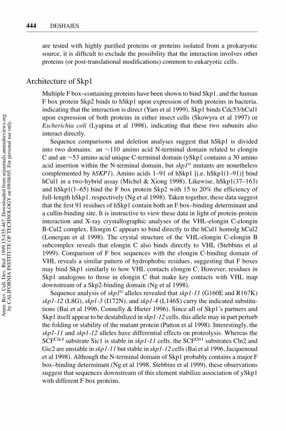
Architecture of Skp1
Multiple F box–containing proteins have been shown to bind Skp1, and the humanF box protein Skp2 binds to hSkp1 upon expression of both proteins in bacteria,indicating that the interaction is direct (Yam et al 1999). Skp1 binds Cdc53/hCul1upon expression of both proteins in either insect cells (Skowyra et al 1997) orEscherichia coli (Lyapina et al 1998), indicating that these two subunits alsointeract directly.Sequence comparisons and deletion analyses suggest that hSkp1 is divided
into two domains: an ∼110 amino acid N-terminal domain related to elonginC and an ∼53 amino acid unique C-terminal domain (ySkp1 contains a 30 aminoacid insertion within the N-terminal domain, but skp1ts mutants are nonethelesscomplemented by hSKP1). Amino acids 1–91 of hSkp1 [i.e. hSkp1(1–91)] bindhCul1 in a two-hybrid assay (Michel & Xiong 1998). Likewise, hSkp1(37–163)and hSkp1(1–65) bind the F box protein Skp2 with 15 to 20% the efficiency offull-length hSkp1, respectively (Ng et al 1998). Taken together, these data suggestthat the first 91 residues of hSkp1 contain both an F box–binding determinant anda cullin-binding site. It is instructive to view these data in light of protein-proteininteraction and X-ray crystallographic analyses of the VHL-elongin C-elonginB-Cul2 complex. Elongin C appears to bind directly to the hCul1 homolg hCul2(Lonergan et al 1998). The crystal structure of the VHL-elongin C-elongin Bsubcomplex reveals that elongin C also binds directly to VHL (Stebbins et al1999). Comparison of F box sequences with the elongin C-binding domain ofVHL reveals a similar pattern of hydrophobic residues, suggesting that F boxesmay bind Skp1 similarly to how VHL contacts elongin C. However, residues inSkp1 analogous to those in elongin C that make key contacts with VHL mapdownstream of a Skp2-binding domain (Ng et al 1998).Sequence analysis of skp1ts alleles revealed that skp1-11 (G160E and R167K)
skp1-12 (L8G), skp1-3 (I172N), and skp1-4 (L146S) carry the indicated substitu-tions (Bai et al 1996, Connelly & Hieter 1996). Since all of Skp1’s partners andSkp1 itself appear to be destabilized in skp1-12 cells, this allele may in part perturbthe folding or stability of the mutant protein (Patton et al 1998). Interestingly, theskp1-11 and skp1-12 alleles have differential effects on proteolysis. Whereas theSCFCdc4 substrate Sic1 is stable in skp1-11 cells, the SCFGrr1 substrates Cln2 andGic2 are unstable in skp1-11 but stable in skp1-12 cells (Bai et al 1996, Jacquenoudet al 1998). Although the N-terminal domain of Skp1 probably contains a major Fbox–binding determinant (Ng et al 1998, Stebbins et al 1999), these observationssuggest that sequences downstream of this element stabilize association of ySkp1with different F box proteins.
Ann
u. R
ev. C
ell.
Dev
. Bio
l. 19
99.1
5:43
5-46
7. D
ownl
oade
d fro
m a
rjour
nals.
annu
alre
view
s.org
by C
ALI
FORN
IA IN
STIT
UTE
OF
TECH
NO
LOG
Y o
n 09
/08/
05. F
or p
erso
nal u
se o
nly.

P1: FKZ/FHX/FGJ P2: FKZ/FHR/fgo QC: FhN/Anil T1: FhN
September 10, 1999 15:45 Annual Reviews AR092-15
?SCFUBIQUITIN LIGASES 445
Because ySkp1 can link Cdc4 to Cdc53, it has been suggested that it servesprimarily to link the cullin and F box subunits of SCF. Two observations suggestthat this view may be too simple. First, the F box protein Grr1 assembles withCdc53 in insect cells in the absence of ySkp1 (although it is difficult to rule out acontribution by insect Skp1) (Skowyra et al 1997). Second, hCul1 assembles withthe F box protein Skp2 in the absence of any other eukaryotic proteins (Lyapinaet al 1998), and a mutation in Skp2 that blocks binding of both human cyclin Aand hSkp1 has no effect on binding to hCul1 in human cells (Lisztwan et al 1998).Perhaps a more subtle function of Skp1 is to position the F box and cullin subunitsso that they are optimally placed to mediate transfer of ubiquitin from E2 enzymeto substrate.
Architecture of Cdc53
Cdc53 is subdivided into three domains: an N-terminal domain that binds ySkp1,an internal domain that recruits yCdc34 and Hrt1, and a short C-terminal domainof unknown function. Cdc53 lacking amino acids 448–748 [i.e. Cdc53(�448–748)] binds to both ySkp1 and F box proteins in coimmunoprecipitation assays,whereas Cdc53(�9–280) binds neither (Patton et al 1998). Moreover, a pointmutation in the conserved N-terminal domain residue Y133 disrupts associationbetween Cdc53 and ySkp1 (M Tyers, personal communication). Finally, the first219 amino acids of hCul1 bind both hSkp1 and hSkp2 in two-hybrid and GSTpull-down assays (Michel & Xiong 1998). Taken together, these data indicate thatthe first 219 amino acids of hCul1/Cdc53 comprise a Skp1-binding domain. Thebound Skp1 in turn facilitates association with F box proteins. Because bacteriallyexpressed hCul1 and hSkp1 interact, binding is likely to be direct (Lyapina et al1998). Interestingly, the N-terminal domain shows the lowest conservation amongcullins of the three domains discussed here, and hCul2-hCul5 do not bind hSkp1in a two-hybrid assay (Michel & Xiong 1998). This observation is considered inmore detail in the section Diversification of SCF Function.Whereas N-terminal deletionmutants of Cdc53 and hCul1 no longer bind Skp1,
they do bind to yCdc34 and Roc1/Rbx1, respectively (Ohta et al 1999, Pattonet al 1998). Thus the region C-terminal to the Skp1-binding domain constitutesa Cdc34/RING-H2 subunit-recruiting domain. The cdc53–1 mutation (R488C)lies within this segment and specifically disrupts Cdc53-yCdc34 interaction(Patton et al 1998). This region of Cdc53 is conserved with the APC/C subunitApc2, and is called the cullin homology (CH) domain (H Yu et al 1998, Zachariaeet al 1998). The CH domain likely represents a conserved module for linkinga ubiquitin-conjugating enzyme to a ubiquitin ligase. Given that (a) Hrt1 bindsdirectly to yCdc34 (Seol et al 1999), (b) Hrt1/Roc1/Rbx1 appears to bind directlyto Cdc53/hCul1 (Ohta et al 1999, Seol et al 1999, Tan et al 1999), (c) Hrt1 stabi-lizes the association of yCdc34 with Cdc53 (Skowyra et al 1999), and (d ) yCdc34and Roc1/Rbx1 bind overlapping regions of Cdc53/hCul1 (Ohta et al 1999, Patton
Ann
u. R
ev. C
ell.
Dev
. Bio
l. 19
99.1
5:43
5-46
7. D
ownl
oade
d fro
m a
rjour
nals.
annu
alre
view
s.org
by C
ALI
FORN
IA IN
STIT
UTE
OF
TECH
NO
LOG
Y o
n 09
/08/
05. F
or p
erso
nal u
se o
nly.

P1: FKZ/FHX/FGJ P2: FKZ/FHR/fgo QC: FhN/Anil T1: FhN
September 10, 1999 15:45 Annual Reviews AR092-15
?446 DESHAIES
et al 1998), it seems likely that Hrt1 helps tether yCdc34 to the CH domain bybinding directly to both proteins.The most confounding element of Cdc53 is its extreme C terminus. Whereas
this is the most highly conserved region of cullin proteins and is required forattachment of Rub1 (Lammer et al 1998, Patton et al 1998, Wada et al 1999), amutant that lacks this segment (e.g. amino acids 757–814) can complement cdc53∆(Lammer et al 1998, Patton et al 1998) and actively mediates Sic1 ubiquitinationin vitro (Feldman et al 1997).
Architecture of Hrt1
Hrt1 is a 121-amino acid protein that can be divided into two domains: a 54-aminoacid N-terminal domain and a 67-amino acid C-terminal domain that contains theRING-H2 finger. Both domains are conserved among eukaryotic Hrt1 homologs.Despite being such a small protein, Hrt1 binds Cdc53, yCdc34, and the F boxproteins Grr1 and Cdc4 (Kamura et al 1999, Seol et al 1999, Skowyra et al 1999).Binding of Hrt1 to yCdc34 is direct since it can be reconstituted with proteinsisolated from E. coli (Seol et al 1999). Binding of Hrt1/Roc1/Rbx1 to cullins andF box proteins is also likely to be direct. Roc1/Rbx1 and the related hRoc2 bindhCul1-hCul5 in both two-hybrid and co-immunoprecipitation assays (Ohta et al1999). Likewise, hApc11 binds the cullin-related hApc2. Thus all known proteinsthat contain cullin homology domains can assemble with RING-H2 proteins. CHand RING-H2 proteins may comprise a conserved heterodimeric core that liesat the heart of a diverse array of ubiquitin ligase complexes (see section titledF Boxes, SOCS Boxes, and Others).Mutation of conserved cysteine and histidine residues in the RING-H2 domain
abrogates the ability of Roc1/Rbx1 to complement yeast hrt1∆ mutants and sustainubiquitin ligase activity in vitro (Kamura et al 1999, Ohta et al 1999). TheRING-H2mutants still bind Cdc53/hCul1, suggesting that their defect arises from impairedinteraction with Cdc34. An intriguing feature of the RING-H2 domain is that itcontains three cysteines (at positions 66, 69, and 81 in Hrt1) that are not part ofthe canonical RING motif but are nevertheless conserved in all Hrt1/Roc1/Rbx1and Apc11 homologs. However, Roc1/Rbx1 lacking the first two cysteines canstill complement hrt1∆ cells, and hrt1 K72R C81R double mutants are viable(Kamura et al 1999, Skowyra et al 1999). Thus these additional cysteines are notabsolutely essential for activity.
Architecture of F Box Proteins
F box proteins come in many different forms. All F box proteins share the ∼45amino acid F box domain, but are otherwise very dissimilar. For example, Cdc4and hβ-TrCP/E3RS contain sevenWD-40 repeats in addition to the F box domain,whereas Grr1 and Skp2 each contain multiple leucine-rich repeats. Other repeatdomains are found in F box proteins, and some F box proteins are composed ofonly unique sequences outside of the F box (Patton et al 1998a).
Ann
u. R
ev. C
ell.
Dev
. Bio
l. 19
99.1
5:43
5-46
7. D
ownl
oade
d fro
m a
rjour
nals.
annu
alre
view
s.org
by C
ALI
FORN
IA IN
STIT
UTE
OF
TECH
NO
LOG
Y o
n 09
/08/
05. F
or p
erso
nal u
se o
nly.

P1: FKZ/FHX/FGJ P2: FKZ/FHR/fgo QC: FhN/Anil T1: FhN
September 10, 1999 15:45 Annual Reviews AR092-15
?SCFUBIQUITIN LIGASES 447
Numerous mutagenesis studies have confirmed that an intact F box is requiredfor association with Skp1. The most highly conserved residue in the F box is aproline near the N-terminal boundary that is typically preceded by a leucine. TheLeu-Pro dipeptide is required for association of Cdc4 (Bai et al 1996) and Ctf13(Kaplan et al 1997) with ySkp1. However, the conserved proline is not requiredfor interaction of Skp2 with hSkp1 (Yam et al 1999). The conserved Leu-Prodipeptide is absent from several F box family members identified in databasesearches (Patton et al 1998a; K Hofmann, personal communication). It remainsunclear whether these proteins bind Skp1. A more detailed understanding of theF box-Skp1 interface awaits a systematic mutagenesis of the F box and structuralstudies.Although the F box is required to bind Skp1, it may not be sufficient. For
example, the minimal F box of Grr1 does not bind ySkp1 in a two-hybrid as-say (Li & Johnston 1997), and an N-terminal fragment of Cdc4 that contains anF box but lacks theC-terminalWD-40 repeat domain coimmunoprecipitates poorlywith ySkp1 (Zhou & Howley 1998). Lastly, mutation of two residues near theC terminus of Skp2 (far downstream of the F box) disrupts binding of both humancyclin A and hSkp1 (Lisztwan et al 1998). Thus stable interaction between F boxproteins and Skp1 may typically require sequences lying outside the F box.
Architecture of F Box Proteins: Cdc4
Domain analyses have been performed on four F box proteins: Cdc4, hβ-TrCP/E3RS, Grr1, and Skp2. The results obtained for Cdc4 are discussed here. Cdc4can be divided into four domains: a unique N-terminal domain, the F box, aC-terminal domain that contains seven WD40 repeats, and an ∼50 amino acidsegment between the F box and WD-40 domains that appears to control turnoverof Cdc4 (Matthias et al 1999). An intact WD-40 domain is required for Cdc4 tobind Sic1 but not ySkp1 (Skowyra et al 1997), and the WD-40 region by itselfis sufficient to bind phosphorylated Sic1 (C Correll, personal communication).Whereas binding of phosphorylated Sic1 substrate to full-length Cdc4 is stronglystimulated by ySkp1 (Feldman et al 1997, Skowyra et al 1997), Skp1 has no effecton the binding of phospho-Sic1 to the isolatedWD-40 domain (C Correll, personalcommunication). Taken together, these observations suggest that the unoccupiedF box antagonizes the substrate-binding activity of the WD-40 domain. Such anautoinhibitory mechanism would insure that free F box subunits do not competewith intact SCF complexes for access to substrates.The WD-40 domain of the F box protein hβ-TrCP/E3RS has also been shown
to recruit substrates for ubiquitination (Margottin et al 1998, Yaron et al 1998).This activity has been exploited to construct dominant-negative mutations that se-lectively interfere with the destruction of hβ-TrCP/E3RS targets. The idea is that aWD-40 domain that lacks an associated F box competes with the intact protein forbinding substrate, but does not deliver bound substrates to SCF due to its inabilityto bind Skp1. Although this tactic has been applied successfully to stabilize CD4
Ann
u. R
ev. C
ell.
Dev
. Bio
l. 19
99.1
5:43
5-46
7. D
ownl
oade
d fro
m a
rjour
nals.
annu
alre
view
s.org
by C
ALI
FORN
IA IN
STIT
UTE
OF
TECH
NO
LOG
Y o
n 09
/08/
05. F
or p
erso
nal u
se o
nly.
P1: FKZ/FHX/FGJ P2: FKZ/FHR/fgo QC: FhN/Anil T1: FhN
September 10, 1999 15:45 Annual Reviews AR092-15
?448 DESHAIES
(Margottin et al 1998), IκB (Yaron et al 1998, Kroll et al 1999, Hatakeyama et al1999, Fuchs et al 1999), and β-catenin (Marikawa & Elinson 1998, Latres et al1999, Fuchs et al 1999, Hart et al 1999), overexpression of Cdc4’sWD-40 domainappears to have no effect on destruction of Sic1 (Zhou &Howley 1998). Neverthe-less, deletion of the F box from SCF substrate receptors is an attractive strategy forproducing dominant-negative proteins that interfere with the degradation of onlythose substrates that utilize the mutated F box protein as a receptor. In contrast, adominant-negative strategy aimed at the other subunits of SCF is likely to disablethe entire spectrum of SCF complexes present in the cell.Cdc4 expressed in insect cells is multimeric and is able to direct assembly of
a multimeric SCF complex (C Correll, personal communication). Likewise, theSchizosaccharomyces pombe Cdc4 homologs Pop1 and Pop2/Sud1 form hetero-and homo-oligomers (Kominami et al 1998, Wolf et al 1999). Although it re-mains unclear whether Pop1/Pop2 heterodimers assemble into multimeric SCFcomplexes, deletion analysis and characterization of Pop1/Pop2 chimeras suggestthat interaction of these proteins is important for their genetic function (Wolf et al1999). It remains unclear if higher-order assembly of SCF is crucial for its func-tion and if all F box-containing proteins oligomerize. In contrast to Cdc4, Pop1,and Pop2, the Cdc53 and ySkp1 subunits do not form stable homo-oligomers(C Correll, personal communication).
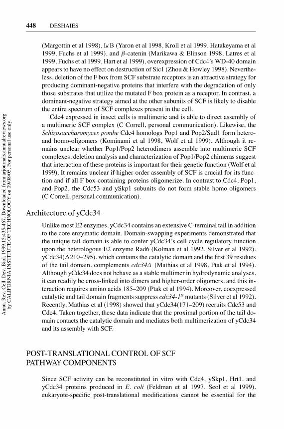
Architecture of yCdc34
Unlike most E2 enzymes, yCdc34 contains an extensive C-terminal tail in additionto the core enzymatic domain. Domain-swapping experiments demonstrated thatthe unique tail domain is able to confer yCdc34’s cell cycle regulatory functionupon the heterologous E2 enzyme Rad6 (Kolman et al 1992, Silver et al 1992).yCdc34(�210–295), which contains the catalytic domain and the first 39 residuesof the tail domain, complements cdc34∆ (Mathias et al 1998, Ptak et al 1994).Although yCdc34 does not behave as a stable multimer in hydrodynamic analyses,it can readily be cross-linked into dimers and higher-order oligomers, and this in-teraction requires amino acids 185–209 (Ptak et al 1994). Moreover, coexpressedcatalytic and tail domain fragments suppress cdc34-1tsmutants (Silver et al 1992).Recently, Mathias et al (1998) showed that yCdc34(171–209) recruits Cdc53 andCdc4. Taken together, these data indicate that the proximal portion of the tail do-main contacts the catalytic domain and mediates both multimerization of yCdc34and its assembly with SCF.
POST-TRANSLATIONAL CONTROL OF SCFPATHWAY COMPONENTS
Since SCF activity can be reconstituted in vitro with Cdc4, ySkp1, Hrt1, andyCdc34 proteins produced in E. coli (Feldman et al 1997, Seol et al 1999),eukaryote-specific post-translational modifications cannot be essential for the
Ann
u. R
ev. C
ell.
Dev
. Bio
l. 19
99.1
5:43
5-46
7. D
ownl
oade
d fro
m a
rjour
nals.
annu
alre
view
s.org
by C
ALI
FORN
IA IN
STIT
UTE
OF
TECH
NO
LOG
Y o
n 09
/08/
05. F
or p
erso
nal u
se o
nly.

P1: FKZ/FHX/FGJ P2: FKZ/FHR/fgo QC: FhN/Anil T1: FhN
September 10, 1999 15:45 Annual Reviews AR092-15
?SCFUBIQUITIN LIGASES 449
activity of these subunits. However, post-translational modifications of SCF sub-units may regulate SCF activity in vivo, modulate the shuttling of SCF complexesor components to different parts of the cell, or influence the remodeling of SCFcomplexes in response to cellular or environmental signals. The following sectionsdescribe what is known about the modification state, post-translational regulation,and localization of SCF subunits.
Post-Translational Regulation of Skp1
Skp1 purified from Dictyostelium discoideum cells contains a linear pentasac-charide chain attached to a hydroxylated proline at position 143 (Teng-umnuayet al 1998). Although this residue is conserved in budding yeast, Arabidopsis, andC. elegans Skp1, it is not present in the human or mouse proteins. Skp1 is alsophosphorylated upon expression in insect cells, but the significance of this modifi-cation has not been evaluated (Kaplan et al 1997). ySkp1 is stable (Zhou&Howley1998), and the levels of hSkp1 (Lisztwan et al 1998) and ySkp1 (JH Seol, per-sonal communication) do not vary during the cell cycle. Interestingly, both Skp1and Cul1 colocalize to the centrosome in animal cells (Freed et al 1999, Gstaigeret al 1999). It has been well established that proteolysis triggers the separation ofsister chromatids at the beginning of anaphase. SCF-dependent degradation mayplay an analogous role in centrosome duplication, since the proteasome inhibitorlactacystin and antibodies against hSkp1 and hCul1 block separation of centriolesin vitro (Freed et al 1999).
Post-Translational Regulation of Cdc53/Cullin
The best-characterized post-translational modification of SCF is the attachmentof the ubiquitin-related protein Rub1 (known as Nedd8 in humans) to Cdc53(Lammer et al 1998, Liakopoulos et al 1998), rabbit Cul-4A (Osaka et al 1998),and hCul2 (Wada et al 1999). Strikingly, Cdc53 and Cul-4A appear to be themost prominent attachment sites for Rub1/Nedd8 in budding yeast cells and rabbitreticulocyte lysates, respectively. Attachment of Rub1/Nedd8 to proteins (i.e. ru-binylation)mimics attachment of ubiquitin, and requires E1- and E2-like activities.The Rub1-activating enzyme is divided into two subunits that are homologous tothe N- and C-terminal halves of E1. The N-terminal subunit is known as Ula1/Enr2in budding yeast andAPP-BP1 in human, whereas the C-terminal subunit is knownas Uba3 in both organisms (Liakopoulos et al 1998, Osaka et al 1998, Gong &Yeh 1999). Genes encoding these subunits have been identified in a broad range ofeukaryotes (Lammer et al 1998). A Rub1/Nedd8-conjugating enzyme, Ubc12, hasalso been reported (Liakopoulos et al 1998, Osaka et al 1998, Gong & Yeh 1999).Human and yeast Ubc12 are homologous and are clearly related to ubiquitin-conjugating enzymes. It remains unclear whether rubinylation involves an E3-likeactivity. Accumulation of Rub1-Cdc53 conjugates in vivo is diminished in skp1tsmutants (Lammer et al 1998), but this may be due either to stabilization of Cdc53by ySkp1 (Patton et al 1998) or to an E3-like function of ySkp1 in the rubinylationpathway.
Ann
u. R
ev. C
ell.
Dev
. Bio
l. 19
99.1
5:43
5-46
7. D
ownl
oade
d fro
m a
rjour
nals.
annu
alre
view
s.org
by C
ALI
FORN
IA IN
STIT
UTE
OF
TECH
NO
LOG
Y o
n 09
/08/
05. F
or p
erso
nal u
se o
nly.

P1: FKZ/FHX/FGJ P2: FKZ/FHR/fgo QC: FhN/Anil T1: FhN
September 10, 1999 15:45 Annual Reviews AR092-15
?450 DESHAIES
Yeast cells that lack the genes encoding Rub1, Ula1/Enr2, or yUbc12 are vi-able and display no obvious phenotype (Lammer et al 1998, Liakopoulos et al1998). Thus decoration of Cdc53 with Rub1 is not crucial for SCF activity. How-ever, mutations that disable the rubinylation pathway render cells more sensitiveto the effects of mutations in SCF subunits (Lammer et al 1998). Thus Rub1appears to enhance SCF activity, at least under conditions in which SCF is crip-pled. Rubinylation of Cdc53 in turn may be governed by the assembly or ac-tivity of the SCF complex because the cdc53-1 mutation increases the fractionof Cdc53 that is conjugated with Rub1, whereas skp1ts mutations have the op-posite effect (Lammer et al 1998). The residues in hCul1 and hCul2 that arelinked to Rub1 have been mapped to the lysine in the conserved VRIMK se-quence (K686) (Wada et al 1999; S Schwartz, personal communication). Dele-tion of sequences downstream of the VRIMK element eliminates rubinylation ofCdc53, suggesting that other C-terminal domain sequences are required for propermodification.The ubiquitin-like modifier SUMO-1 is thought to influence localization of the
proteins to which it is attached. Centrosomal fractions have significant amountsof modified Cul1 but are devoid of the unmodified protein (Freed et al 1999),suggesting that rubinylation may likewise influence subcellular localization ofSCF complexes (see also Post-Translational Regulation of Skp1).Other than rubinylation, the cullins have not been reported to receive other post-
translationalmodifications.Moreover, Cdc53 is stable (Zhou&Howley 1998), andthe levels of hCul1 (Lisztwan et al 1998) and Cdc53 (JH Seol, personal commu-nication) do not vary during the cell cycle.
Post-Translational Regulation of Hrt1
Covalent modification, stability, and subcellular localization of Hrt1 proteins haveyet to be investigated. Hrt1 expressed in bacteria binds SCF components and stim-ulates SCFCdc4 ubiquitin ligase, indicating that no post-translational modificationsspecific to eukaryotic cells are absolutely required for Hrt1 activity (Seol et al1999).
Post-Translational Regulation of F Box Proteins
Protein degradation may play a key role in sculpting the repertoire of SCF com-plexes in vivo. The F box proteins Cdc4, Grr1, and Met30 are unstable, and theturnover of Cdc4 and Grr1 has been investigated in detail (Galan & Peter 1999,Zhou&Howley 1998, Mathias et al 1999). Cdc4 and Grr1 are unstable throughoutthe cell cycle, and both proteins are stabilized in proteasome, cdc53ts, cdc34ts, andskp1tsmutants. Both Cdc4 and Grr1 are ubiquitinated and are stabilized by expres-sion of mutant ubiquitin. Interestingly, an intact F box is required for turnover ofboth Cdc4 and Grr1.Based on these data, Zhou & Howley (1998) proposed that F box receptors and
their bound substrates are degraded as a unit to ensure a continuous release of free
Ann
u. R
ev. C
ell.
Dev
. Bio
l. 19
99.1
5:43
5-46
7. D
ownl
oade
d fro
m a
rjour
nals.
annu
alre
view
s.org
by C
ALI
FORN
IA IN
STIT
UTE
OF
TECH
NO
LOG
Y o
n 09
/08/
05. F
or p
erso
nal u
se o
nly.

P1: FKZ/FHX/FGJ P2: FKZ/FHR/fgo QC: FhN/Anil T1: FhN
September 10, 1999 15:45 Annual Reviews AR092-15
?SCFUBIQUITIN LIGASES 451
ySkp1/Cdc53/Hrt1 subcomplexes able to assemble with newly synthesized F boxreceptor subunits. Their hypothesis predicts that the rate of degradation of an F boxprotein should be directly proportional to the concentration of its substrates. Thisprediction has not been tested. I suggest an alternative model that predicts the rateof degradation of F box proteins to be inversely proportional to the concentrationof their substrates. According to this view, binding of substrate shields the F boxsubunit from autoubiquitination, and overexpression of substrate is thus predictedto stabilize the corresponding F box receptor. This substrate shield model is at-tractive in that it would ensure a direct correlation between the concentration of agiven F box protein and the concentration of its targets. Thus there would be noneed for the cell to regulate with a high degree of accuracy the synthesis of everyF box protein. If a given F box protein were synthesized in excess, its levels wouldbe quickly pared down to match substrate demand.Aside from the yeast F box proteins discussed above, Skp2 is also likely to be
unstable, since its levels fluctuate during the cell cycle, reaching a peak during Sphase (Lisztwan et al 1998).
Post-Translational Regulation of Cdc34
yCdc34 is phosphorylated on serine residues in vivo (Goebl et al 1994), but itremains unclear if this modification regulates its activity. yCdc34 isolated from E.coli supports SCF-dependent ubiquitination in a purified system (Feldman et al1997), indicating that phosphorylation is not required for its catalytic activity orinteraction with SCF. yCdc34 ubiquitinates itself both in vitro and in vivo (Baner-jee et al 1993, Goebl et al 1994). Autoubiquitination of yCdc34 requires a clusterof four lysine residues in the extreme C terminus (Banerjee et al 1993), deletion ofwhich has no apparent effect on yCdc34 function (Goebl et al 1994). Immunoblotanalyses reveal that yCdc34 levels are equivalent in cells arrested in G1, S, orM phase (Mathias et al 1998) and remain constant during progression througha synchronous cell cycle (Galan & Peter 1999; JH Seol, personal communica-tion). In short, there is no evidence that yCdc34 activity is regulated by eitherpost-translational modification or degradation.
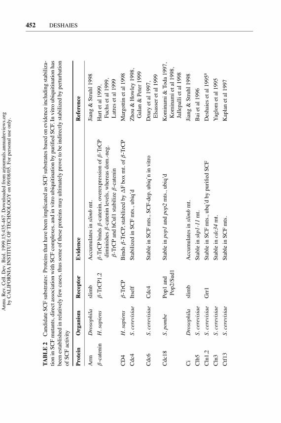
SUBSTRATES OF THE SCF PATHWAY
Many proteins have been implicated as substrates of SCF by a variety of criteria,including stabilization or accumulation in SCF pathway mutants, stabilizationupon expression of dominant-negative F box proteins, and reconstitution of SCF-dependent ubiquitination with either purified proteins or in a crude system. Thesesubstrates represent a broad spectrum of proteins that participate in a variety ofcellular functions, including regulation ofCDKactivity, activation of transcription,signal transduction, assembly of kinetochores, and DNA replication. Rather thandiscuss each substrate in detail, those known to date are described briefly in Table 2.
Ann
u. R
ev. C
ell.
Dev
. Bio
l. 19
99.1
5:43
5-46
7. D
ownl
oade
d fro
m a
rjour
nals.
annu
alre
view
s.org
by C
ALI
FORN
IA IN
STIT
UTE
OF
TECH
NO
LOG
Y o
n 09
/08/
05. F
or p
erso
nal u
se o
nly.
P1: FKZ/FHX/FGJ P2: FKZ/FHR/fgo QC: FhN/Anil T1: FhN
September 10, 1999 15:45 Annual Reviews AR092-15
?452 DESHAIES
TABLE2
CandidateSCFsubstrates:ProteinsthathavebeenimplicatedasSCFsubstratesbasedonevidenceincludingstabiliza-
tioninSCFmutants,directassociationwithSCFcomplexes,andinvitroubiquitinationbypurifiedSCF.Invitroubiquitinationhas
beenestablishedinrelativelyfewcases,thussomeoftheseproteinsmayultimatelyprovetobeindirectlystabilizedbyperturbation
ofSCFactivity
Protein
Organism
Receptor
Evidence
Reference
Arm
Drosophila
slimb
Accumulatesinslimbmt.
Jiang&Struhl1998
β-catenin
H.sapiens
β-TrCP1,2
β-TrCPbindsβ-catenin,overexpressionof
β-TrCP
Hartetal1999,
diminishes
β-cateninlevels,whereasdom.-neg.
Fuchsetal1999,
β-TrCPandhCul1stabilize
β-catenin
Latresetal1999
CD4
H.sapiens
β-TrCP
Bindsβ-TrCP,stabilizedby
�Fboxmt.of
β-TrCP
Margottinetal1998
Cdc4
S.cerevisiae
Itself
StabilizedinSCFmts.,ubiq’d
Zhou&Howley1998,
Galan&Peter1999
Cdc6
S.cerevisiae
Cdc4
StableinSCFmts.,SCF-dep.ubiq’ninvitro
Druryetal1997,
Elsasseretal1999
Cdc18
S.pombe
Pop1and
Stableinpop1andpop2mts.,ubiq’d
Kominami&
Toda1997,
Pop2/Sud1
Kominamietal1998,
Jallepallietal1998
CiDrosophila
slimb
Accumulatesinslimbmt.
Jiang&Struhl1998
Clb5
S.cerevisiae
Stableinskp1-11mt.
Baietal1996
Cln1,2
S.cerevisiae
Grr1
StableinSCFmts.,ubq’dbypurifiedSCF
Deshaiesetal1995a
Cln3
S.cerevisiae
Stableincdc34mt.
Yaglom
etal1995
Ctf13
S.cerevisiae
StableinSCFmts.
Kaplanetal1997
Ann
u. R
ev. C
ell.
Dev
. Bio
l. 19
99.1
5:43
5-46
7. D
ownl
oade
d fro
m a
rjour
nals.
annu
alre
view
s.org
by C
ALI
FORN
IA IN
STIT
UTE
OF
TECH
NO
LOG
Y o
n 09
/08/
05. F
or p
erso
nal u
se o
nly.

P1: FKZ/FHX/FGJ P2: FKZ/FHR/fgo QC: FhN/Anil T1: FhN
September 10, 1999 15:45 Annual Reviews AR092-15
?SCFUBIQUITIN LIGASES 453
CyclinD
H.sapiens
Skp2
StabilizedbyantisenseablationofSKP2
&CU
L1ZK
Yuetal1998
E2F-1
H.sapiens
Skp2
BindsSkp2,Skp2non-bindingmutantstable;ubq’d
Martietal1999
Far1
S.cerevisiae
Cdc4
StableinSCFmts.,Cdc34&Cdc4-dep.ubiq’ninvitro
Henchozetal1997
Gcn4
S.cerevisiae
Stableincdc34mt.
Kornitzeretal1994
Gic2
S.cerevisiae
Grr1
Stableingrrl
�,cdc34mts.,ubiq’d
Jacquenoudetal1998
Grr1
S.cerevisiae
Itself
StabilizedinSCFmts.,ubiq’d
Zhou&Howley1998,
Galan&Peter1999
IκB
H.sapiens
β-TrCP
Bindsβ-TrCP,stabilizedby
�Fmtofβ-TrCP,ubiq’n
Yaronetal1998b
(akaE3RS)
invitrobypurifiedSCFβ-TrCP
LIN-12
C.elegans
SEL-10
SEL-10neg.regulatesLIN-12pathway,bindsLIN-12
Hubbardetal1997
p21
H.sapiens
Skp2
StabilizedbyantisenseablationofSKP2andCU
L1ZK
Yuetal1998
p27
H.sapiens
UbiquitinatedbyCdc34;stabilizedbydom.-neg.Cdc34
Paganoetal1995
Rum1
S.pombe
Pop1
Stabilizedinpop1mt,ubiq’d
Kominami&
Toda1997
Sic1
S.cerevisiae
Cdc4
StableinSCFmts,ubiq’dbypurifiedSCF
Schwobetal1994,
Feldmanetal1997,
Skowyraetal1997
Swe1
S.cerevisiae
Met30
StableinSCFmts,Cdc34&Met30-dep.ubiq’ninvitro
Kaiseral1998
Wee1
X.laevis
Stabilizedbydom.-neg.Cdc34
Michael&Newport1998
Xic1
X.laevis
Stabilizedbydom.-neg.Cdc34
Yew&Kirschner1997
a SeealsoBarraletal1995,Schneideretal1998,Seoletal1999,Skowyraetal1999.
b SeealsoFuchsetal1999,Hatakeyamaetal1999,Krolletal1999,Ohtaetal1999,Spenceretal1999,Suzukietal1999,Tanetal1999,Winstonetal1999.
Abbreviations:dep.,dependent;dom.-neg.,dominant-negative;mt(s),mutant(s);ubiq’d,ubiquitinconjugateshavebeendetected;ubiq’n,ubiquitination.
Ann
u. R
ev. C
ell.
Dev
. Bio
l. 19
99.1
5:43
5-46
7. D
ownl
oade
d fro
m a
rjour
nals.
annu
alre
view
s.org
by C
ALI
FORN
IA IN
STIT
UTE
OF
TECH
NO
LOG
Y o
n 09
/08/
05. F
or p
erso
nal u
se o
nly.

P1: FKZ/FHX/FGJ P2: FKZ/FHR/fgo QC: FhN/Anil T1: FhN
September 10, 1999 15:45 Annual Reviews AR092-15
?454 DESHAIES
MECHANISMOF ACTION OF SCF
How Does Substrate Bind SCF?
In all the examples studied thus far in detail (ubiquitination of Cln1 and 2 bySCFGrr1, Sic1 by SCFCdc4, and IκB plus β-catenin by SCFβ−TrCP/E3RS; consultTable 2 for references), the target must be phosphorylated before it can bind andserve as a substrate for SCF. This common requirement for phosphorylation isremarkable given that the substrate-binding domains of Cdc4 and β-TrCP/E3RSare constructed of WD-40 repeats, whereas that of Grr1 is constructed of com-pletely dissimilar leucine-rich repeats (Kishi et al 1998). All known substrates ofSCFCdc4 –Sic1 (Feldman et al 1997, Skowyra et al 1997, Verma et al 1997), Gcn4(Y Chi, personal communication), Far1 (Henchoz et al 1997), and Cdc6 (Elsasseret al 1999) must be phosphorylated before they can be ubiquitinated. How doesphosphate drive the interaction of diverse substrates with SCF? Phosphopeptides(but not their unmodified counterparts) derived from IκB are able to compete withfull-length IκB for binding to β-TrCP/E3RS (Yaron et al 1998, Winston et al1999). These data suggest that the WD-40 domain of β-TrCP/E3RS constitutes aphospho-serine recognition module. It remains an open question whether all SCFcomplexes will prove to be specific for phosphorylated substrates.Several F box proteins contain WD-40 repeats. The properties of this domain
may allow SCF complexes to bind a diverse spectrum of substrates. The WD-40repeats of Cdc4 are predicted to fold into a β-propeller based on the crystal struc-ture of the WD-40 repeat domain of the Gβ subunit of heterotrimeric G proteins(Lambright et al 1996, Sondek et al 1996, Wall et al 1995). The face of the β-propeller in Gβ presents a broad platform for protein-protein interaction. Differentsignaling proteins interact with the face of Gβ’s propeller domain by forming sta-bilizing contacts with different residues on the propeller’s face, such that eacheffector makes a slightly different footprint on the platform (Ford et al 1998).This plasticity may explain why the known substrates of Cdc4 do not show anyapparent sequence homology with each other in their destabilizing domains. How-ever, since all SCFCdc4 substrates must be phosphorylated before they can bind,Cdc4’s β-propeller domain likely contains a phosphate-binding pocket that alignsthe substrate on the propeller’s surface.The requirement for substrate phosphorylation creates an opportunity to differ-
entially regulate the stability of distinct SCFCdc4 substrates.Whereas Sic1 and Far1are activated to bind Cdc4 following their phosphorylation by G1 cyclin/Cdc28(Feldman et al 1997, Henchoz et al 1997, Skowyra et al 1997), Cdc6 is targetedto SCF upon its phosphorylation by S cyclin/Cdc28 (Elsasser et al 1999). TheCdc42 GTPase effector Gic2 (Jaquenoud et al 1998) and the transcriptional ac-tivator Gcn4 (Y Chi, personal communication) are phosphorylated and targetedfor SCF-dependent ubiquitination by protein kinases other than Cdc28. By link-ing the SCF-dependent ubiquitination of substrates to the action of distinct ki-nases, it is possible to achieve substrate-specific degradation in response to diverse
Ann
u. R
ev. C
ell.
Dev
. Bio
l. 19
99.1
5:43
5-46
7. D
ownl
oade
d fro
m a
rjour
nals.
annu
alre
view
s.org
by C
ALI
FORN
IA IN
STIT
UTE
OF
TECH
NO
LOG
Y o
n 09
/08/
05. F
or p
erso
nal u
se o
nly.

P1: FKZ/FHX/FGJ P2: FKZ/FHR/fgo QC: FhN/Anil T1: FhN
September 10, 1999 15:45 Annual Reviews AR092-15
?SCFUBIQUITIN LIGASES 455
environmental or intracellular cues. For example, although SCFCdc4 is active in pre-START G1 extracts, Sic1 is stable until Cln/Cdc28 protein kinase is activated atSTART (Schneider et al 1996, Verma et al 1997a). Besides allowing for tempo-ral regulation, the requirement for substrate phosphorylation can be exploited inother ways. A Cln2 mutant that cannot bind Cdc28 is not phosphorylated andconsequently stabilized (Deshaies et al 1995, Lanker et al 1996), and a mutantof Gic2 that fails to bind yCdc42 is likewise not phosphorylated and is stabilized(Jaquenoud et al 1998). In both cases, the requirement for substrate phosphory-lation ensures that the attention of the degradation machinery is focused on thespecific subset of substrate molecules.
How is Ubiquitin Transferred From Cdc34 to Substrate?
The ubiquitin ligase best characterized to date, the HECT domain family mem-ber E6-AP, contains an essential cysteine residue that accepts ubiquitin from E2enzyme and subsequently transfers it to a lysine residue of the substrate p53(Scheffner et al 1995). Because HECT domain family members are the only ubiq-uitin ligases known to form a thioester intermediate with ubiquitin, it remains un-clear whether other ubiquitin ligases operate via a similar mechanism. All cysteineresidues in Cdc53 and ySkp1 are dispensable for function (Patton et al 1998), asare three conserved cysteines in Hrt1/Roc1/Rbx1 that are not part of the RING-H2consensus (Kamura et al 1999, Skowyra et al 1999). Thus if SCF forms an obligatecovalent intermediate with ubiquitin during its transfer from yCdc34 to substrate,the catalytic site must either be present on a prosthetic group (similar to the reac-tive thiol group in the acyl carrier protein of the fatty acid synthase complex), orthere must be an essential ubiquitin-accepting residue of SCF that remains to beidentified.Recentwork suggests that the cullin/RING-H2 subunits serve as the core ubiqui-
tin ligase module of the SCF complex. This conclusion stems from the observationthat cullin/RING-H2 heterodimers appear to be sufficient to activate assembly ofeither free (Ohta et al 1999, Tan et al 1999) or Cdc34-bound (Seol et al 1999,Skowyra et al 1999) multiubiquitin chains (note that it has not been rigorouslyexcluded that the free multiubiquitin chains reported by Tan, Ohta, and colleaguesare in fact not anchored to hCdc34 or hCul1).Intriguingly, whereas enzymes that use cysteine residues for catalysis (including
E1 enzyme) are sensitive to alkylating agents such as N-ethylmaleimide (NEM),Cdc53/Hrt1 pretreated with NEM retains its ability to activate autoubiquitinationof yCdc34 (Seol et al 1999).Moreover, polycations that lack sulfhydryls can substi-tute for SCF and promote yCdc34-dependent ubiquitination of Gcn4 or autoubiq-uitination of yCdc34. Based on these observations, Seol et al (1999) proposed thatcullin and RING-H2 proteins form a ubiquitin ligase module that operates by amechanismdistinct from that of theHECTdomain ligases. Cdc53/Hrt1may triggera conformational change in yCdc34-S-Ub that reduces the energy barrier to form-ing the oxyanionic intermediate that occurs in the transition state as the C terminus
Ann
u. R
ev. C
ell.
Dev
. Bio
l. 19
99.1
5:43
5-46
7. D
ownl
oade
d fro
m a
rjour
nals.
annu
alre
view
s.org
by C
ALI
FORN
IA IN
STIT
UTE
OF
TECH
NO
LOG
Y o
n 09
/08/
05. F
or p
erso
nal u
se o
nly.

P1: FKZ/FHX/FGJ P2: FKZ/FHR/fgo QC: FhN/Anil T1: FhN
September 10, 1999 15:45 Annual Reviews AR092-15
?456 DESHAIES
of ubiquitin is transferred from the catalytic cysteine of yCdc34 to the ε-aminogroup of a substrate lysine. A conformational switch in yCdc34 driven by specificinteraction with Cdc53/Hrt1 would minimize the possibility that yCdc34 (whichis present in substantial excess in vivo compared with SCF complexes; JH Seol,personal communication) would gratuitously ubiquitinate proteins with which itinevitably collides in the densely packed cell. A logical extension of this argumentis that the ubiquitination-promoting activity of Cdc53/Hrt1 may be gated withinthe SCF complex such that it is maximally expressed upon docking of substrateto the F box receptor subunit. Besides SCF and APC/C, a number of known orsuspected ubiquitin ligases contain a RING or RING-H2 domain, including Ubr1(Kwon et al 1998), Hrd1/Der3 (Bordallo et al 1998, Hampton et al 1996), SINA(Hu & Fearon 1999), Rad18 (Bailly et al 1997) and Mdm2 (Honda et al 1997). Itwill be interesting to see whether all RING-bearing ubiquitin ligases operate by acommon mechanism.It is worth noting that SCF occasionally associates with E2 enzymes other than
Cdc34. The β-TrCP/E3RS-dependent ubiquitination of IκB, for example, involvesa member of the hUbc5 family of E2 enzymes (Yaron et al 1998, Spencer et al1999). In addition, the Grr1-, ySkp1-, and Cdc53-dependent regulation of glucose-repressible genes in yeast does not require yCdc34, suggesting that another E2enzyme is involved (Li & Johnston 1997).
How Does SCF Promote the Assemblyof aMultiubiquitin Chain?
At first glance, it is difficult to envision how the assembly of a multiubiquitinchain occurs in three dimensions. If one’s reference point is the SCF complex,the substrate (i.e. the end of the growing ubiquitin chain) should move fartheraway with each cycle of ubiquitin addition. Nevertheless, SCF/yCdc34 rapidlyassembles multiubiquitin chains upon Sic1. One can envision several explanationsto account for processive assembly of multiubiquitin chains. For example, SCFmay trigger the assembly of an oligomer of yCdc34 that provides a template forthe assembly of a multiubiquitin chain. Because a tetraubiquitin chain is thoughtto be capable of targeting an appended protein for degradation (Piotrowski et al1997), induced tetramerization of yCdc34would be sufficient to yield degradation-competent substrate. Pure yCdc34 has already been shown to form dimers andhigher-order oligomers in vitro (Ptak et al 1994). An alternative possibility is thatthe growing multiubiquitin chain spools away from SCF/yCdc34 as a loop, suchthat the relative positions of yCdc34, SCF, and the growing end of the ubiquitinchain remain constant throughout the process of chain assembly. The ability toprocessively assemble a multiubiquitin chain is likely to be a crucial aspect ofSCF activity. If chain synthesis were to occur by a purely distributive mechanism,ubiquitin isopeptidases might disassemble the multiubiquitin chains before theygrew to a length sufficient to mediate high-affinity interaction of substrate with the26S proteosome.
Ann
u. R
ev. C
ell.
Dev
. Bio
l. 19
99.1
5:43
5-46
7. D
ownl
oade
d fro
m a
rjour
nals.
annu
alre
view
s.org
by C
ALI
FORN
IA IN
STIT
UTE
OF
TECH
NO
LOG
Y o
n 09
/08/
05. F
or p
erso
nal u
se o
nly.

P1: FKZ/FHX/FGJ P2: FKZ/FHR/fgo QC: FhN/Anil T1: FhN
September 10, 1999 15:45 Annual Reviews AR092-15
?SCFUBIQUITIN LIGASES 457
DIVERSIFICATION OF SCF FUNCTION
The F Box Hypothesis
Based on previous observations that the F box–containing proteins Cdc4 and Grr1are involved in the degradation of Sic1 (Schwob et al 1994) and G1 cyclins (Barralet al 1995), respectively, Bai et al (1996) proposed that F box–containing pro-teins serve to link different substrates to Skp1 and the ubiquitination machinery(Figure 3). The generality of their hypothesis has been tested for two members ofthe F box family of proteins, Cdc4 and Grr1. SCFCdc4 binds tightly to and catalyzesubiquitination of phosphorylated Sic1, but binds poorly to phosphorylated Cln1(Feldman et al 1997, Skowyra et al 1997). In contrast, SCFGrr1 binds tightly to andcatalyzes ubiquitination of phosphorylated Cln1 and Cln2 but fails to bind phos-phorylated Sic1 (Skowyra et al 1997, 1999; Seol et al 1999). Patton et al (1998)demonstrated that the F box proteins Cdc4, Grr1, and Met30 each form SCF com-plexes in vivo by assembling with Cdc53 and ySkp1, but that these proteins donot cross-assemble with each other, suggesting that each SCF complex is limitedto a single form of F box receptor (however, see Kominami et al 1998, Wolf et al1999). Moreover, whereas cdc53ts and cdc34ts mutants are defective in turnoverof all known SCF substrates, grr1∆ mutants degrade Sic1 with normal kineticsbut accumulate stable Cln2, and cdc4 mutants can degrade Cln2 but accumulatestable Sic1.Given that the budding yeast genome potentially encodes 17 F box proteins,
17 different SCF ubiquitin ligase complexes may exist in this organism (for acomprehensive display of budding yeast F box proteins, see Patton et al 1998a).However, at least one F box protein (the Ctf13 subunit CBF3) assembles into amacromolecular complex distinct from SCF (Connelly & Hieter 1996, Kaplanet al 1997, Stemmann & Lechner 1996). It remains to be seen how many F boxproteins assemble into ubiquitin ligase complexes. Nevertheless, even a subset ofthe known F box proteins might yield far more than 17 distinct SCF complexes,since there are two additional Cdc53-like proteins encoded in the budding yeastgenome, and at least some F box proteins can form heterodimers with each other(Kominami et al 1998, Wolf et al 1999).
F Boxes, SOCS Boxes, and Others
Recent work suggests that SCF may be a paradigm for a family of modular cullin-based ubiquitin ligases.Whereas Cdc53 and hCul1 bind tightly to Skp1, the hCul2,hCul3, mouse Cul4A, and hCul5 proteins do not detectably bind hSkp1 (Michel& Xiong 1998). In contrast, hCul2 binds elongin C, which shares homology withthe N-terminal region of Skp1 (Lonergan et al 1998). Elongin C in turn bindsdirectly to both the ubiquitin-related protein elongin B (Takagi et al 1996) and theVHL tumor suppressor protein (Takagi et al 1997). VHL shares with a number ofother proteins part of a sequence referred to as the suppressor of cytokine signaling
Ann
u. R
ev. C
ell.
Dev
. Bio
l. 19
99.1
5:43
5-46
7. D
ownl
oade
d fro
m a
rjour
nals.
annu
alre
view
s.org
by C
ALI
FORN
IA IN
STIT
UTE
OF
TECH
NO
LOG
Y o
n 09
/08/
05. F
or p
erso
nal u
se o
nly.

P1: FKZ/FHX/FGJ P2: FKZ/FHR/fgo QC: FhN/Anil T1: FhN
September 10, 1999 15:45 Annual Reviews AR092-15
?458 DESHAIES
Figure 3 The F box hypothesis. ySkp1/Cdc53/Hrt1 can interact with any one of anumber of different proteins that contain F boxes to yield distinct SCF complexes withdifferent substrate specificities. As shown, SCFCdc4 targets phospho-Sic1 for ubiquiti-nation (A), whereas SCFGrr1 targets phospho-Cln1 and phospho-Cln2 (B). Genetic datasuggest that Met4 may be a target of SCFMet30 (C ), but this has not yet been shown. Itis also not known whether SCFMet30 is specific for phosphoprotein substrates. H, Hrt1;F, F box.
Ann
u. R
ev. C
ell.
Dev
. Bio
l. 19
99.1
5:43
5-46
7. D
ownl
oade
d fro
m a
rjour
nals.
annu
alre
view
s.org
by C
ALI
FORN
IA IN
STIT
UTE
OF
TECH
NO
LOG
Y o
n 09
/08/
05. F
or p
erso
nal u
se o
nly.

P1: FKZ/FHX/FGJ P2: FKZ/FHR/fgo QC: FhN/Anil T1: FhN
September 10, 1999 15:45 Annual Reviews AR092-15
?SCFUBIQUITIN LIGASES 459
(SOCS) box, which was originally identified in proteins that negatively regulatecytokine-inducible signaling through the Jak/STAT pathway. SOCS box sequencesfrom VHL, SOCS-1 and SOCS-3 are necessary and sufficient to mediate bindingto elongin C (Kamura et al 1998, Kibel et al 1995, Zhang et al 1999). Taken to-gether, these observations suggest that hCul2 and elongin C may form the core ofa ubiquitin ligase complex (although this complex has not been shown to containubiquitin ligase activity) that also contains elongin B, Roc1/Rbx1 (Kamura et al1999), and any one of a number of SOCS box–containing proteins. The analogyto the SCF pathway, with its constellation of distinct F box receptors, is striking.Similarly, the APC/C, which contains both CH domain and RING-H2 subunits(Zacharine et al 1998, HYu et al 1998), interactswith at least two distinct substrate-targeting proteins (Cdc20 and Hct1/Cdh1). It will be interesting to see whether allcullins serve as core subunits of multicomponent ubiquitin ligases whose sub-strate specificity is diversified via a repertoire of interchangeable receptor subunits(Figure 4).A tantalizing glimpse of how cullin/RING-H2 based ubiquitin ligases might
be constructed is presented by the recently solved structure of the VHL-elonginC-elongin B (VCB) complex (Stebbins et al 1999). In the VCB complex, VHLand elongin C serve as analogs of the F box receptor and Skp1 subunits of SCF,respectively. There is no known subunit of SCF corresponding to the ubiquitin-likeelonginB, except perhaps theRub1modification uponCdc53. Stebbins et al (1999)raise the appealing notion that the interaction of VHL with elongin C mimics theinteraction of F box proteins with Skp1. An important goal will be to map thebinding sites for hCul2 (Lonergan et al 1998) and Roc1/Rbx1 (Kamura et al 1999)upon the surface of elongin C. It seems likely that they will be oriented in the samedirection as a putative substrate-docking site on VHL.
CONCLUSION AND PERSPECTIVES
Rapid progress has been made over the past few years in identifying and charac-terizing cullin-based ubiquitin ligases. Emerging evidence suggests that there isa broad array of distinct ubiquitin ligases based on the cullin/RING-H2 moduleand that these ligases participate in diverse regulatory pathways. Over the nextfew years, it is likely that progress will accelerate and that the list of distinct SCFcomplexes will expand as new F box proteins are identified.Wemay also witness asimilar expansion in the number of ubiquitin ligases based onCul2-Cul5.As the listof SCFand cullin-basedubiquitin ligase complexes grows longer, itwill be comple-mented by two parallel tracks of research. First, specific ubiquitin ligase complexeswill be linked to specific processes via gene knockout studies or by investigatorswho fortuitously isolate SCF components or cullins in forward genetic screens.Second, researchers studying proteins that are known to be unstable (e.g. tran-scription factors, signaling proteins, etc) will establish links between specific SCFcomplexes and the turnover of these proteins.
Ann
u. R
ev. C
ell.
Dev
. Bio
l. 19
99.1
5:43
5-46
7. D
ownl
oade
d fro
m a
rjour
nals.
annu
alre
view
s.org
by C
ALI
FORN
IA IN
STIT
UTE
OF
TECH
NO
LOG
Y o
n 09
/08/
05. F
or p
erso
nal u
se o
nly.
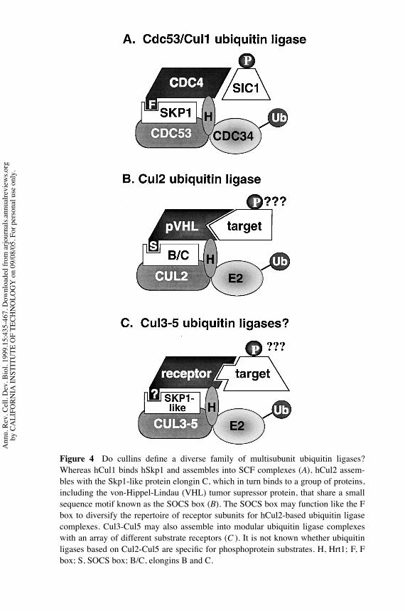
P1: FKZ/FHX/FGJ P2: FKZ/FHR/fgo QC: FhN/Anil T1: FhN
September 10, 1999 15:45 Annual Reviews AR092-15
?Figure 4 Do cullins define a diverse family of multisubunit ubiquitin ligases?Whereas hCul1 binds hSkp1 and assembles into SCF complexes (A), hCul2 assem-bles with the Skp1-like protein elongin C, which in turn binds to a group of proteins,including the von-Hippel-Lindau (VHL) tumor supressor protein, that share a smallsequence motif known as the SOCS box (B). The SOCS box may function like the Fbox to diversify the repertoire of receptor subunits for hCul2-based ubiquitin ligasecomplexes. Cul3-Cul5 may also assemble into modular ubiquitin ligase complexeswith an array of different substrate receptors (C ). It is not known whether ubiquitinligases based on Cul2-Cul5 are specific for phosphoprotein substrates. H, Hrt1; F, Fbox; S, SOCS box; B/C, elongins B and C.
Ann
u. R
ev. C
ell.
Dev
. Bio
l. 19
99.1
5:43
5-46
7. D
ownl
oade
d fro
m a
rjour
nals.
annu
alre
view
s.org
by C
ALI
FORN
IA IN
STIT
UTE
OF
TECH
NO
LOG
Y o
n 09
/08/
05. F
or p
erso
nal u
se o
nly.

P1: FKZ/FHX/FGJ P2: FKZ/FHR/fgo QC: FhN/Anil T1: FhN
September 10, 1999 15:45 Annual Reviews AR092-15
?SCFUBIQUITIN LIGASES 461
Against this backdrop of anticipated progress, two lines of directed researchmay prove to be of particular value. First, there is currently no general method foridentifying SCF substrates. Given that each SCF complex is likely to havemultiplesubstrates, any expansion in the repertoire of known ubiquitin ligase complexeswill intensify the need for efficient methods to identify substrates. Second, rapidevaluation of the function of newly discovered SCF or cullin/RING-H2 complexesin different cell lines, tissues, etc will require novel chemical or genetic strategiesto inhibit ubiquitin ligase activity. Since SCF has already been linked to a numberof important signaling pathways, a side benefit is that specific inhibitors may proveto have significant therapeutic benefits. Although there are no specific inhibitorsavailable for any known ubiquitination pathway, the great success of the variousproteasome inhibitors makes clear how valuable such drugs can be.
ACKNOWLEDGMENTS
I thankRichardBenarous, ZhijianChen, KayHofmann, Peter Jackson,WillyKrek,Matthias Peter, Mike Tyers, and members of my laboratory for providing preprintsand communicatingunpublisheddata. I also thankPeter Jackson andMatthiasPeterfor critical reading of the manuscript. Work on SCF in my laboratory is supportedby a grant from the National Institutes of Health, and a Young Investigator Awardfrom the Burroughs-Wellcome Fund.
Visit the Annual Reviews home page at www.AnnualReviews.org
LITERATURE CITED
Bai C, Richman R, Elledge SJ. 1994. Humancyclin-F. EMBO J. 13:6087–98
Bai C, Sen P, HofmannK,Ma L, GoeblM, et al.1996. SKP1 connects cell cycle regulators tothe ubiquitin proteolysis machinery througha novel motif, the F-box. Cell 86:263–74
Bailly V, Lauder S, Prakash S, Prakash L. 1997.Yeast DNA repair proteins Rad6 and Rad18form a heterodimer that has ubiquitin con-jugating, DNA binding, and ATP hydrolyticactivities. J. Biol. Chem. 272:23360–65
Banerjee A, Gregori L, Xu Y, Chau V. 1993.The bacterially expressed yeastCDC34 geneproduct can undergo autoubiquitination toform a multiubiquitin chain-linked protein.J. Biol. Chem. 268:5668–75
Barral Y, Jentsch S, Mann C. 1995. G(1) cyclinturnover and nutrient-uptake are controlled
by a common pathway in yeast. Genes Dev.9:399–409
Bordallo J, Plemper RK, Finger A, Wolf DH.1998. Der3p/Hrd1p is required for endo-plasmic reticulum-associated degradation ofmisfolded lumenal and integral membraneproteins. Mol. Biol. Cell 9:209–22
Burnatowska-HledinMA,SpielmanWS, SmithWL, Shi P, Meyer JM, Dewitt DL. 1995.Expression cloning of an AVP-activated,calcium-mobilizing receptor from rabbit kid-ney medulla. Am. J. Physiol. 268:1198–210
ChenLC,Manjeshwar S, LuY,MooreD, LjungBM, et al. 1998. The human homologue fortheCaenorhabditis elegans cul-4 gene is am-plified and overexpressed in primary breastcancers. Cancer Res. 58:3677–83
Chung CY, Reddy TB, Zhou K, Firtel RA.1998. A novel, putativeMEK kinase controls
Ann
u. R
ev. C
ell.
Dev
. Bio
l. 19
99.1
5:43
5-46
7. D
ownl
oade
d fro
m a
rjour
nals.
annu
alre
view
s.org
by C
ALI
FORN
IA IN
STIT
UTE
OF
TECH
NO
LOG
Y o
n 09
/08/
05. F
or p
erso
nal u
se o
nly.

P1: FKZ/FHX/FGJ P2: FKZ/FHR/fgo QC: FhN/Anil T1: FhN
September 10, 1999 15:45 Annual Reviews AR092-15
?462 DESHAIES
developmental timing and spatial patterningin Dictyostelium and is regulated by ubiqui-tin-mediated protein degradation.GenesDev.12:3564–78
Connelly C, Hieter P. 1996. Budding yeastSKP1 encodes an evolutionary conservedkinetochore protein required for cell cycleprogression. Cell 86:275–85
Deshaies RJ, Chau V, Kirschner M. 1995.Ubiquitination of the G1 cyclin Cln2p bya Cdc34p-dependent pathway. EMBO J. 14:303–12
Drury LS, Perkins G, Diffley JF. 1997. TheCdc4/34/53 pathway targets Cdc6p for pro-teolysis in budding yeast.EMBO J. 16:5966–76
DuM, Sansores-Garcia L, Zu Z,Wu KK. 1998.Cloning and expression analysis of a novelsalicylate suppressible gene, Hs-CUL-3,a member of cullin/Cdc53 family. J. Biol.Chem. 273:24289–92
ElledgeSJ,Harper JW.1998.The role of proteinstability in the cell cycle and cancer.Biochim.Biophys. Acta 1377:M61–70
Elsasser S, Chi Y, Yang P, Campbell JL. 1999.Phosphorylation controls timing of Cdc6pdestruction: a biochemical analysis. Mol.Biol. Cell. In press
Erhardt JA, Hynicka W, DiBenedetto A, ShenN, Stone N, et al. 1998. A novel F box pro-tein, NFB42, is highly enriched in neuronsand induces growth arrest. J. Biol. Chem.273:35222–27
Feldman R, Correll C, Kaplan K, DeshaiesR. 1997. A complex of Cdc4p, Skp1p, andCdc53p/cullin catalyzes ubiquitination of thephosphorylated CDK inhibitor Sic1p. Cell91:221–30
Ford C, Skiba N, Bae H, Daaka Y, Reuveny E,et al. 1998. Molecular basis for interactionsof G protein βγ subunits with effectors. Sci-ence 280:1271–74
Freed E, Lacey K, Huie P, Lyapina S, DeshaiesR, et al. 1999. The Skp1 and Cul1 ubiquitinligase components localize to the centrosomeand regulate the centrosome duplication cy-cle. Genes Dev. In press
Fuchs S, Chen A, Xiong Y, Pan Z, Ronai Z.1999. HOS, a human homolog of Slimb,forms an SCF complex with Skp1 andCullin1 and targets the phosphorylation-de-pendent degradation of IκB and β-catenin.Oncogene 18:2039–46
Galan J, Peter M. 1999. Ubiquitin-dependentdegradation of multiple F-box proteins by anautocatalytic mechanism. Proc. Natl. Acad.Sci. USA. In press
Goebl MG, Goetsch L, Byers B. 1994. TheUbc3 (Cdc34) ubiquitin-conjugating enzymeis ubiquitinated and phosphorylated in vivo.Mol. Cell. Biol. 14:3022–29
Goebl MJ, Yochem J, Jentsch S, McGrath JP,Varshavsky A, Byers B. 1988. The yeastcell cycle gene cdc34 encodes a ubiquitin-conjugating enzyme. Science 241:1331–34
Gong L, Yeh E. 1999. Identification of theactivating and conjugating enzymes of theNEDD8 conjugation pathway. J. Biol. Chem.274:12036–42
Gstaiger M, Marti A, Krek, W. 1999. Associa-tion of humanSCFSKP2 subunit p19SKP1withinterphase centrosomes and mitotic spindlepoles. J. Cell. Sci. 247:554–62
Hampton RY, Gardner RG, Rine J. 1996. Roleof 26S proteasome and HRD genes in thedegradation of 3-hydroxy-3-methylglutaryl-CoA reductase, an integral endoplasmicreticulum membrane protein.Mol. Biol. Cell7:2029–44
Hart M, Concordet J-P, Lassot I, Albert I, dellos Santos R, et al. 1999. The F-box pro-tein β-TrCP associates with phosphorylatedβ-catenin and regulates its activity in the cell.Curr. Biol. 9:207–10
Hatakeyama S, KitagawaM,NakayamaK, Shi-raneM,MatsumotoM, et al. 1999.Ubiquitin-dependent degradation of IκBα is medi-ated by a ubiquitin ligase Skp1/Cul1/F-boxprotein FWD1. Proc. Natl. Acad. Sci. USA96:3859–63
Henchoz S, Chi Y, Catarin B, Herskowitz I, De-shaies RJ, Peter M. 1997. Phosphorylation-and ubiquitin-dependent degradation of the
Ann
u. R
ev. C
ell.
Dev
. Bio
l. 19
99.1
5:43
5-46
7. D
ownl
oade
d fro
m a
rjour
nals.
annu
alre
view
s.org
by C
ALI
FORN
IA IN
STIT
UTE
OF
TECH
NO
LOG
Y o
n 09
/08/
05. F
or p
erso
nal u
se o
nly.

P1: FKZ/FHX/FGJ P2: FKZ/FHR/fgo QC: FhN/Anil T1: FhN
September 10, 1999 15:45 Annual Reviews AR092-15
?SCFUBIQUITIN LIGASES 463
cyclin-dependent kinase inhibitor Far1p inbudding yeast. Genes Dev. 11:3046–60
Hochstrasser M. 1995. Ubiquitin, proteasomes,and the regulation of intracellular proteindegradation. Curr. Opin. Cell Biol. 7:215–23
Honda R, Tanaka H, Yasuda H. 1997. Oncopro-teinMDM2 is a ubiquitin ligase E3 for tumorsuppressor p53. FEBS Lett. 420:25–27
Hu G, Fearon ER. 1999. Siah-1 N-terminalRING domain is required for proteolysisfunction, and C-terminal sequences regulateoligomerization and binding to target pro-teins. Mol. Cell. Biol. 19:724–32
Hubbard E, Wu G, Kitajewski J, Greenwald I.1997. sel-10, a negative regulator of lin-12activity in Caenorhabditis elegans, encodesa member of the CDC4 family of proteins.Genes Dev. 11:3182–93
Ingram G, Doyle S, Carpenter R, Schultz E, Si-mon R, Coen E. 1997. Dual role for fimbriatain regulating floral homeotic genes and celldivision in Antirrhinum. EMBO J. 16:6521–34
Jacquenoud M, Gulli M-P, Peter K, Peter M.1998. The Cdc42p effector Gic2p is targetedfor ubiquitin-dependent degradation by theSCFGrr1 complex. EMBO J. 17:5360–73
Jallepalli P, Tien D, Kelly T. 1998. sud1+targets cyclin-dependent kinase-phosphory-lated Cdc18 and Rum1 proteins for degra-dation and stops unwanted diploidization infission yeast. Proc. Natl. Acad. Sci. USA95:8159–64
Jiang J, Struhl G. 1998. Regulation of theHedgehog andWingless signalling pathwaysby the F-box/WD40-repeat protein Slimb.Nature 391:493–96
Kaiser P, Sia RA, Bardes EG, Lew DJ, ReedSI. 1998. Cdc34 and the F-box proteinMet30 are required for degradation of theCdk-inhibitory kinase Swe1. Genes Dev. 12:2587–97
Kamura T, Koepp DM, Conrad MN, SkowyraD, Moreland RJ, et al. 1999. Rbx1, a compo-nent of the VHL tumor suppressor complexand SCF ubiquitin ligase. Science 284:657–61
Kamura T, Sato S, Haque D, Liu L, Kaelin WGJr, et al. 1998. The Elongin BC complex in-teracts with the conserved SOCS-box motifpresent inmembers of the SOCS, ras,WD-40repeat, and ankyrin repeat families. GenesDev. 12:3872–81
KaplanKB,HymanAA, Sorger PK. 1997. Reg-ulating the yeast kinetochore by ubiquitin-dependent degradation and Skp1p-mediatedphosphorylation. Cell 91:491–500
Kibel A, Iliopoulos O, DeCaprio J, Kaelin W.1995. Binding of the von Hippel-Lindau tu-mor suppressor protein to Elongin B and C.Science 269:1444–46
Kipreos ET, Lander LE, Wing JP, He WW,Hedgecock EM. 1996. cul-1 is required forcell cycle exit in C. elegans and identifies anovel gene family. Cell 85:1–20
Kishi T, Seno T, Yamao F. 1998. Grr1 func-tions in the ubiquitin pathway in Saccha-romyces cerevisiae through association withSkp1. Mol. Gen. Genet. 257:143–48
Kolman CJ, Toth J, Gonda DK. 1992. Iden-tification of a portable determinant of cellcycle function within the carboxyl-terminaldomain of the yeast CDC34 (UBC3) ubiq-uitin conjugating (E2) enzyme. EMBO J.11:3081–90
Kominami K-I, Ochotorena I, Toda T. 1998.Two F-box/WD-repeat proteins Pop1 andPop2 form hetero- and homo-complexes to-gether with cullin-1 in the fission yeastSCF (Skp1-Cullin-1-F-box) ubiquitin ligase.Genes Cells 3:721–35
Kominami K, Toda T. 1997. Fission yeast WD-repeat protein Pop1 regulates genome ploidythrough ubiquitin-proteasome-mediated de-gradation of the CDK inhibitor Rum1 andthe S-phase initiator Cdc18. Genes Dev. 11:1548–60
Kornitzer D, Raboy B, Kulka RG, Fink GR.1994. Regulated degradation of the transcrip-tion factor GCN4. EMBO J. 13:6021–30
Krek W. 1998. Proteolysis and the G1-S transi-tion: the SCF connection.Curr. Opin. Genet.Dev. 8:36–42
KrollM,Margottin F,KohlA,Renard P,Durand
Ann
u. R
ev. C
ell.
Dev
. Bio
l. 19
99.1
5:43
5-46
7. D
ownl
oade
d fro
m a
rjour
nals.
annu
alre
view
s.org
by C
ALI
FORN
IA IN
STIT
UTE
OF
TECH
NO
LOG
Y o
n 09
/08/
05. F
or p
erso
nal u
se o
nly.
P1: FKZ/FHX/FGJ P2: FKZ/FHR/fgo QC: FhN/Anil T1: FhN
September 10, 1999 15:45 Annual Reviews AR092-15
?464 DESHAIES
H, et al. 1999. Inducible degradation of IκBα
by the proteasome requires interaction withthe F-box protein h-β-TrCP. J. Biol. Chem.274:7941–45
Kumar A, Paietta JV. 1998. An additional rolefor the F-box motif: gene regulation withinthe Neurospora crassa sulfur control net-work. Proc. Natl. Acad. Sci. USA 95:2417–22
Kwon YT, Reiss Y, Fried VA, Hershko A, YoonJK, et al. 1998. The mouse and human genesencoding the recognition component of theN-end rule pathway. Proc. Natl. Acad. Sci.USA 95:7898–903
Lambright DG, Sondek J, Bohm A, Skiba NP,Hamm HE, Sigler PB. 1996. The 2.0 A crys-tal structure of a heterotrimeric G protein.Nature 379:311–19
Lammer D, Mathias N, Laplaza JM, JiangW, Liu Y, et al. 1998. Modification ofyeast Cdc53p by the ubiquitin-related pro-tein Rub1p affects function of the SCFCdc4complex. Genes Dev. 12:914–26
Lanker S, Valdivieso MH, Wittenberg C. 1996.Rapid degradation of the G1 cyclin Cln2 in-duced by CDK-dependent phosphorylation.Science 271:1597–601
Latres E, Chiaur D, Pagano M. 1999. The hu-man F box protein β-Trcp associates with theCul1/Skp1 complex and regulates the stabil-ity of β-catenin. Oncogene 18:849–54
Lee I, Wolfe D, Nilsson O, Weigel D. 1997.A LEAFY co-regulator encoded by UN-USUAL FLORAL ORGANS. Curr. Biol.7:95–104
Li FN, Johnston M. 1997. Grr1 of Saccha-romyces cerevisiae is connected to the ubiq-uitin proteolysis machinery through Skp1:coupling glucose sensing to gene expressionand the cell cycle. EMBO J. 16:5629–38
Liakopoulos D, Doenges G, Matuschewski K,Jentsch S. 1998. A novel protein modifica-tion pathway related to the ubiquitin system.EMBO J. 17:2208–14
Lisztwan J, Marti A, Sutterluty H, Gstaiger M,Wirbelauer C, Krek W. 1998. Associationof human CUL-1 and ubiquitin-conjugating
enzyme CDC34 with the F-box proteinp45(SKP2): evidence for evolutionary con-servation in the subunit composition of theCDC34–SCF pathway. EMBO J. 17:368–83
Lonergan KM, Iliopoulos O, Ohh M, KamuraT, Conaway RC, et al. 1998. Regulationof hypoxia-inducible mRNAs by the vonHippel-Lindau tumor suppressor protein re-quires binding to complexes containing elon-gins B/C and Cul2. Mol. Cell. Biol. 18:732–41
Lyapina S, Correll C, Kipreos E, Deshaies R.1998. Human CUL1 forms an evolutionar-ily conserved ubiquitin ligase complex (SCF)with SKP1 and an F-box protein. Proc. Natl.Acad. Sci. USA 95:7451–56
Margottin F, Bour S, Durand H, Selig L, Beni-chou S, et al. 1998. A novel human WD pro-tein, hβTrCp, that interacts with HIV-1 Vpuconnects CD4 to theERdegradation pathwaythrough an F-box motif. Mol. Cell 1:565–74
MarikawaY,ElinsonRP. 1998.β-TrCP is a neg-ative regulator of Wnt/β-catenin signalingpathway and dorsal axis formation in Xeno-pus embryos. Mech. Dev. 77:75–80
Marti A, Wirbelauer C, Scheffner M, Krek W.1999. SCFSKP2 ubiquitin ligase/E2F-1 tran-scription factor complex formation underliesregulation of E2F-1 protein stability and Sphase promoting function. Nat. Cell Biol.1:14–19
MathiasN, Johnson S, Byers B,GoeblM. 1999.The abundance of cell cycle regulatory pro-tein Cdc4p is controlled by interactions be-tween its F box and Skp1p. Mol. Cell. Biol.19:1759–67
Mathias N, Johnson SL, Winey M, Adams AE,Goetsch L, et al. 1996. Cdc53 acts in concertwith Cdc4 and Cdc34 to control the G1 toS phase transition and identifies a conservedfamily of proteins.Mol. Cell. Biol. 16:6634–43
Mathias N, Steussy CN, Goebl MG. 1998. Anessential domain within Cdc34p is requiredfor binding to a complex containing Cdc4pand Cdc53p in Saccharomyces cerevisiae. J.Biol. Chem. 273:4040–45
Ann
u. R
ev. C
ell.
Dev
. Bio
l. 19
99.1
5:43
5-46
7. D
ownl
oade
d fro
m a
rjour
nals.
annu
alre
view
s.org
by C
ALI
FORN
IA IN
STIT
UTE
OF
TECH
NO
LOG
Y o
n 09
/08/
05. F
or p
erso
nal u
se o
nly.
P1: FKZ/FHX/FGJ P2: FKZ/FHR/fgo QC: FhN/Anil T1: FhN
September 10, 1999 15:45 Annual Reviews AR092-15
?SCFUBIQUITIN LIGASES 465
Michael WM, Newport J. 1998. Coupling ofmitosis to the completion of S phase throughCdc34-mediated degradation of Wee1. Sci-ence 282:1886–89
Michel JJ, Xiong Y. 1998. Human CUL-1, butnot other cullin family members, selectivelyinteracts with SKP1 to form a complex withSKP2 and cyclin A.Cell Growth Diff. 9:435–49
Natorff R, Piotrowska M, Paszewski A. 1998.The Aspergillus nidulans sulphur regulatorygene sconB encodes a protein with WD40repeats and an F-box. Mol. Gen. Genet.257:255–63
Ng RW, Arooz T, Yam CH, Chan IW, Lau AW,PoonRY. 1998. Characterization of the cullinand F-box protein partner Skp1. FEBS Lett.438:183–89
Ohta T, Michel JJ, Schottelius AJ, Xiong Y.1999. ROC1, a homolog of APC11, repre-sents a family of cullin partners with an as-sociated ubiquitin ligase activity. Mol. Cell3:535–41
Osaka F, Kawasaki H, Aida N, Saeki M, ChibaT, et al. 1998. A new NEDD8–ligating sys-tem for cullin-4A. Genes Dev. 12:2263–68
Pagano M, Tam SW, Theodoras AM, Beer-Romero P, Del Sal G, et al. 1995. Role of theubiquitin-proteasome pathway in regulatingabundance of the cyclin-dependent kinase in-hibitor p27. Science 269:682–85
PattonEE,WillemsAR, SaD,Kuras L, ThomasD, et al. 1998. Cdc53 is a scaffold proteinformultiple Cdc34/Skp1/F-box protein com-plexes that regulate cell division and me-thionine biosynthesis in yeast. Genes Dev.12:692–705; (erratum) Genes Dev. 19:3144
Patton EE,Willems AR, Tyers M. 1998a. Com-binatorial control in ubiquitin-dependentproteolysis: Don’t Skp the F-box hypothe-sis. Trends Genet. 14:236–43
Pause A, Lee S, Worrell RA, Chen DY, BurgessWH, et al. 1997. The von Hippel-Lindautumor-suppressor gene product forms a sta-ble complex with human CUL-2, a memberof the Cdc53 family of proteins. Proc. Natl.Acad. Sci. USA 94:2156–61
Peterson TA, Yochem J, Byers B, Nunn MF,Duesberg PH, et al. 1984. A relationship be-tween the yeast cell cycle genes CDC4 andCDC36 and the ets sequence of oncogenicvirus E26. Nature 309:556–58
Piotrowski J, Beal R, Hoffman L, WilkinsonKD, Cohen RE, Pickart CM. 1997. Inhibi-tion of the 26S proteasome by polyubiquitinchains synthesized to have defined lengths.J. Biol. Chem. 272:23712–21
Plon SE, Leppig KA, Do HN, Groudine M.1993. Cloning of the human homolog ofthe CDC34 cell cycle gene by complemen-tation in yeast. Proc. Natl. Acad. Sci. USA90:10484–88
Pringle JR, Hartwell LH. 1981. The Saccha-romyces cerevisiae cell cycle. In The Molec-ular Biology of the Yeast Saccharomyces–Life Cycle and Inheritance, ed. JN Strathern,EW Jones, JR Broach. pp. 97–142. ColdSpring Harbor, NY: Cold Spring Harbor Lab.Press
Ptak C, Prendergast JA, Hodgins R, Kay CM,Chau V, Ellison MJ. 1994. Functional andphysical characterization of the cell cy-cle ubiquitin-conjugating enzyme CDC34(UBC3). Identification of a functional deter-minant within the tail that facilitates CDC34self-association. J. Biol. Chem. 269:26539–45
Ruegger M, Dewey E, Gray WM, Hobbie L,Turner J, Estelle M. 1998. The TIR1 pro-tein of Arabidopsis functions in auxin re-sponse and is related to human SKP2 andyeast Grr1p. Genes Dev. 12:198–207
Scheffner M, Nuber U, Huibregtse JM. 1995.Protein ubiquitination involving an E1-E2-E3 enzyme ubiquitin thioester cascade. Na-ture 373:81–83
Schneider BL, Patton EE, Lanker S, Menden-hall MD, Wittenberg C, et al. 1998. YeastG1 cyclins are unstable in G1 phase. Nature395:86–89
Schneider BL, Yang QH, Futcher AB. 1996.Linkage of replication to STARTby the CDKinhibitor Sic1. Science 272:560–62
Schwob E, Bohm T, Mendenhall M, Nasmyth
Ann
u. R
ev. C
ell.
Dev
. Bio
l. 19
99.1
5:43
5-46
7. D
ownl
oade
d fro
m a
rjour
nals.
annu
alre
view
s.org
by C
ALI
FORN
IA IN
STIT
UTE
OF
TECH
NO
LOG
Y o
n 09
/08/
05. F
or p
erso
nal u
se o
nly.

P1: FKZ/FHX/FGJ P2: FKZ/FHR/fgo QC: FhN/Anil T1: FhN
September 10, 1999 15:45 Annual Reviews AR092-15
?466 DESHAIES
K. 1994. The B-type cyclin kinase inhibitorp40SIC1 controls the G1/S transition in Sac-charomyces cerevisiae. Cell 79:233–44
Seol J, Feldman R, Zachariae W, ShevchenkoA, Correll C, et al. 1999. Cdc53/cullin andthe essential Hrt1 RING-H2 subunit of SCFdefine a ubiquitin ligasemodule that activatesthe E2 enzyme Cdc34. Genes Dev. In press
SheaffRJ,GroudineM,GordonM,Roberts JM,Clurman BE. 1997. Cyclin E-CDK2 is a reg-ulator of p27Kip1. Genes Dev. 11:1464–78
SiaR,BardesE,LewD.1998.Control of Swe1pdegradation by the morphogenesis check-point. EMBO J. 17:6678–88
Silver ET, Gwozd TJ, Ptak C, Goebl M, El-lison MJ. 1992. A chimeric ubiquitin con-jugating enzyme that combines the cell cy-cle properties of CDC34 (UBC3) and theDNA repair properties of RAD6 (UBC2):implications for the structure, function andevolution of the E2s. EMBO J. 11:3091–98
Skowyra D, Craig K, Tyers M, Elledge S,Harper J. 1997. F-box proteins are receptorsthat recruit phosphorylated substrates to theSCF ubiquitin-ligase complex. Cell 91:209–19
Skowyra D, Koepp DM, Kamura T, ConradMN, Conaway RC, et al. 1999. Reconstitu-tion of G1 cyclin ubiquitination with com-plexes containing SCFGrr1 and rbx1. Sci-ence 284:662–65
Sondek J, BohmA, Lambright DG, HammHE,Sigler PB. 1996. Crystal structure of a G pro-teinβγ dimer at 2.1 angstrom resolution.Na-ture 379:369–74
Stebbins C, KaelinW, PavletichN. 1999. Struc-ture of the VHL-elongin C-elongin B com-plex: implications forVHL tumor suppressorfunction. Science 284:455–61
Stemmann O, Lechner J. 1996. The Saccha-romyces cerevisiae kinetochore contains acyclin-cdk complexing homolog as inden-tified by in vitro reconstitution. EMBO J.15:3611–20
Suzuki H, Chiba T, Kobayashi M, Takeuchi M,Suzuki T, et al. 1999. IκBα ubiquitination is
catalyzedby anSCF-like complex containingSkp1, cullin-1 and two F-box/WD4-repeatproteins, β-TrCP1 and β-TrCP2. Biochem.Biophys. Res. Comm. 256:127–32
Takagi Y, Conaway RC, Conaway JW. 1996.Characterization of elongin C functional do-mains required for interaction with elonginB and activation of elongin A. J. Biol. Chem.271:25562–68
Takagi Y, Pause A, Conaway RC, Conaway JW.1997. Identification of elongin C sequencesrequired for interaction with the von Hippel-Lindau tumor suppressor protein. J. Biol.Chem. 272:27444–49
Tan P, Fuchs SY,ChenA,WuK,GomezC, et al.1999. Recruitment of a ROC1-CUL1 ubiqui-tin ligase by Skp1 and HOS to catalyze theubiquitination of IκBα. Mol. Cell 3:527–33
Teng-umnuay P, Morris HR, Dell A, Panico M,Paxton T, West CM. 1998. The cytoplasmicF-box binding protein SKP1 contains a novelpentasaccharide linked to hydroxyproline inDictyostelium. J. Biol. Chem. 273:18242–49
Theodosiou NA, Zhang S, Wang WY, Xu T.1998. slimb coordinates wg and dpp ex-pression in the dorsal-ventral and anterior-posterior axes during limb development.De-velopment 125:3411–16
Verma R, Annan RS, Huddleston MJ, Carr SA,Reynard G, Deshaies RJ. 1997a. Phosphory-lation of Sic1p by G1 Cdk required for itsdegradation and entry into S phase. Science278:455–60
VermaR, FeldmanR,DeshaiesRJ. 1997b. SIC1is ubiquitinated in vitro by a pathway that re-quires CDC4, CDC34, and cyclin/CDK ac-tivities.Mol. Biol. Cell. 8:1427–37
Wada H, Yeh E, Kamitani T. 1999. Identi-fication of NEDD8-conjugation site in hu-man cullin-2.Biochem. Biophys. Res. Comm.257:100–5
Wall MA, Coleman DE, Lee E, Iniguez-LlhuiJA, Posner BA, et al. 1995. The structure ofthe G protein heterotrimer Giα1β1γ 2. Cell83:1047–58
Willems AR, Lanker S, Patton EE, CraigKL, Nason TF, et al. 1996. Cdc53 targets
Ann
u. R
ev. C
ell.
Dev
. Bio
l. 19
99.1
5:43
5-46
7. D
ownl
oade
d fro
m a
rjour
nals.
annu
alre
view
s.org
by C
ALI
FORN
IA IN
STIT
UTE
OF
TECH
NO
LOG
Y o
n 09
/08/
05. F
or p
erso
nal u
se o
nly.

P1: FKZ/FHX/FGJ P2: FKZ/FHR/fgo QC: FhN/Anil T1: FhN
September 10, 1999 15:45 Annual Reviews AR092-15
?SCFUBIQUITIN LIGASES 467
phosphorylated G1 cyclins for degradationby the ubiquitin proteolytic pathway. Cell86:453–63
Wolf D, McKeon F, Jackson P. 1999.F-box/WD-repeat proteins Pop1p andSud1p/ Pop2p form complexes that bind anddirect the proteolysis of Cdc18p. Curr. Biol.9:373–76
Xie DX, Feys BF, James S, Nieto-Rostro M,Turner JG. 1998. COI1: an Arabidopsis generequired for jasmonate-regulated defense andfertility. Science 280:1091–94
Yaglom J, Linskens MHK, Sadis S, RubinDM, Futcher B, Finley D. 1995. p34Cdc28-mediated control of CLN3 cyclin degrada-tion. Mol. Cell. Biol. 15:731–41
Yam CH, Ng RWM, Yi Siu W, Lau AWS, PoonRYC. 1999. Regulation of cyclin A-cdk2by SCF component Skp1 and F-Box proteinSkp2. Mol. Cell. Biol. 19:635–45
Yaron A, Hatzubai A, Davis M, Lavon I, AmitS, et al. 1998. Identification of the receptorcomponent of the IκBα-ubiquitin ligase.Na-ture 396:590–94
Yew PR, Kirschner MW. 1997. Proteolysisand DNA replication: the CDC34 require-ment in the Xenopus egg cell cycle. Science277:1672–76
Yu H, Peters JM, King RW, Page AM, Hie-
ter P, Kirschner MW. 1998. Identificationof a cullin homology region in a subunitof the anaphase-promoting complex. Science279:1219–22
Yu Z-K, Gervais J, Zhang H. 1998. HumanCUL-1 associates with the SKP1/SKP2 com-plex and regulates p21CIP1/WAF1 and cy-clin D proteins. Proc. Natl. Acad. Sci. USA95:11324–29
Zachariae W, Shevchenko A, Andrews PD,Ciosk R, Galova M, et al. 1998. Mass spec-trometric analysis of the anaphase-promotingcomplex from yeast: identification of a sub-unit related to cullins. Science 279:1216–19
Zhang H, Kobayashi R, Galaktionov K, BeachD. 1995. p19(SKP1) and p45(SKP2) areessential elements of the cyclin A-CDK2S phase kinase. Cell 82:912–25
Zhang J-G, Farley A, Nicholson S, Willson T,Zugaro L, et al. 1999. The conserved SOCSbox motif in suppressors of cytokine signal-ing binds to elongins B andC andmay couplebound proteins to proteasomal degradation.Proc. Natl. Acad. Sci. USA 96:2071–76
Zhou P, Howley PM. 1998. Ubiquitination anddegradation of the substrate recognition sub-units of SCF ubiquitin-protein ligases. Mol.Cell 2:571–80
Ann
u. R
ev. C
ell.
Dev
. Bio
l. 19
99.1
5:43
5-46
7. D
ownl
oade
d fro
m a
rjour
nals.
annu
alre
view
s.org
by C
ALI
FORN
IA IN
STIT
UTE
OF
TECH
NO
LOG
Y o
n 09
/08/
05. F
or p
erso
nal u
se o
nly.

Annual Review of Cell and Developmental Biology Volume 15, 1999
CONTENTSVacuolar Import of Proteins and Organelles from the Cytoplasm, Daniel J. Klionsky, Yoshinori Ohsumi 1
Blue-Light Photoreceptors In Higher Plants, Winslow R. Briggs, Eva Huala 33
Cooperation Between Microtubule- and Actin-Based Motor Proteins, Susan S. Brown 63
Molecular Mechanisms of Neural Crest Formation, Carole LaBonne, Marianne Bronner-Fraser 81
Lymphocyte Survival--The Struggle Against Death, Robert H. Arch, Craig B. Thompson 113
The Road Less Traveled: Emerging Principles of Kinesin Motor Utilization, Lawrence S. B. Goldstein, Alastair Valentine Philp 141
Proteins of the ADF/Cofilin Family: Essential Regulators of Actin Dynamics, James R. Bamburg 185
Visual Transduction in Drosophila, Craig Montell 231
Biochemical Pathways of Caspases Activation During Apoptosis, Imawati Budihardjo, Holt Oliver, Michael Lutter, Xu Luo, Xiaodong Wang
269
Regulation of Nuclear Localization: A Key to a Door, Arie Kaffman, Erin K. O'Shea 291
Actin-Related Proteins, D. A. Schafer, T. A. Schroer 341
Cell Polarity in Yeast, John Chant 365
Vertebrate Endoderm Development, James M. Wells, Douglas A. Melton 393
Neural Induction, Daniel C. Weinstein, Ali Hemmati-Brivanlou 411
SCF and CDC53/Cullin-Based Ubiquitin Ligases, R. J. Deshaies 435Integration of Signaling Networks that Regulate Dictyostelium Differentiation, Laurence Aubry, Richard Firtel 469
When to Switch to Flowering, Gordon G. Simpson, Anthony R. Gendall, Caroline Dean 519
Regulation of Mammalian O2 Homeostasis by Hypoxia-Inducible Factor 1, Gregg L. Semenza 551
Mechanisms of Viral Interference with MHC Class I Antigen Processing and Presentation, Jonathan W. Yewdell, Jack R. Bennink 579
Transport Between the Cell Nucleus and the Cytoplasm, Dirk Görlich, Ulrike Kutay 607
Ann
u. R
ev. C
ell.
Dev
. Bio
l. 19
99.1
5:43
5-46
7. D
ownl
oade
d fro
m a
rjour
nals.
annu
alre
view
s.org
by C
ALI
FORN
IA IN
STIT
UTE
OF
TECH
NO
LOG
Y o
n 09
/08/
05. F
or p
erso
nal u
se o
nly.

[PSI+]: An Epigenetic Modulator of Translation Termination Efficiency, Tricia R. Serio, Susan L. Lindquist 661
Adaptors for Clathrin-Mediated Traffic, Tomas Kirchhausen 705
Synaptic Vesicle Biogenesis, Matthew J. Hannah, Anne A. Schmidt, Wieland B. Huttner 733
The Translocon: A Dynamic Gateway at the ER Membrane, Arthur E. Johnson, Michael A. van Waes 799
Ann
u. R
ev. C
ell.
Dev
. Bio
l. 19
99.1
5:43
5-46
7. D
ownl
oade
d fro
m a
rjour
nals.
annu
alre
view
s.org
by C
ALI
FORN
IA IN
STIT
UTE
OF
TECH
NO
LOG
Y o
n 09
/08/
05. F
or p
erso
nal u
se o
nly.