scanning electron microscope analysis of enamel microstructure … · 2017-01-09 · scanning...
TRANSCRIPT
Scanning electron microscope analysis of enamelmicrostructure in a Polycotylid (Plesiosauria) from the PierreShale Group, South Dakota, U.S.A.
The teeth of polycotylid plesiosaurs are generally simple, cone shaped, non-serrated and
only slightly recurved without distinct carinae. The surface of crowns are characterized by
a series of vertical enamel wrinkles that are more highly developed on the lingual surface
of the crown, and decrease in width and number toward the apex. Some of the most
promising research related to fossil dentition, involves the analysis of surface and internal
dental microstructure. This study, is an attempt to examine and describe polycotylid
dental microstructure. It gives an overview of polycotylid plesiosaur enamel and dentine
microstructures using a scanning electron microscope. Enamel type and structures vary,
based on its position on the surface of the crown, and its perceived strength requirements.
The dentition layer is “honeycombed” with tubular structure, possibly to provide
nourishment to fast growing crowns. The study of crown microstructures may lead to a
better understanding of polycotylid niche preference in the late Cretaceous oceans.
PeerJ PrePrints | https://dx.doi.org/10.7287/peerj.preprints.1381v1 | CC-BY 4.0 Open Access | rec: 18 Sep 2015, publ: 18 Sep 2015
PrePrin
ts

1
Scanning electron microscope analysis of enamel 1
microstructure in a Polycotylid (Plesiosauria) from the 2
Pierre Shale Group, South Dakota, U.S.A. 3
4
Jason J. Testin 5
Iowa Western Community College, 6
Department of, Physical Science, Physics and Pre-Engineering, 7
2700 College Road, Council Bluffs, IA 51503 8
Abstract 9
The teeth of polycotylid plesiosaurs are generally simple, cone shaped, non-serrated and only 10
slightly recurved without distinct carinae. The surface of crowns are characterized by a series of vertical 11
enamel wrinkles that are more highly developed on the lingual surface of the crown, and decrease in 12
width and number toward the apex. Some of the most promising research related to fossil dentition, 13
involves the analysis of surface and internal dental microstructure. This study, is an attempt to examine 14
and describe polycotylid dental microstructure. It gives an overview of polycotylid plesiosaur enamel and 15
dentine microstructures using a scanning electron microscope. Enamel type and structures vary, based on 16
its position on the surface of the crown, and its perceived strength requirements. The dentition layer is 17
“honeycombed” with tubular structure, possibly to provide nourishment to fast growing crowns. The 18
study of crown microstructures may lead to a better understanding of polycotylid niche preference in the 19
late Cretaceous oceans. 20
Introduction 21
The study of enamel microstructure of both fossil and extant amniote taxa has been extensively 22
studied (Koenigswald and Sander 1997; Sander 2000; Hwang 2005; Stokosa 2005). According to Hwang 23
(2005), mammalian taxa have received preferential study due to distinctive prismatic enamel, easily seen 24
in thin section under polarized light. In comparison, most reptile taxa have nonprismatic enamel; 25
individual crystallites can only be differentiated using the scanning electron microscopy (Sander 2000; 26
Hwang 2005). 27
Polycotylids are a group of short-necked plesiosaurs known mainly from the late Cretaceous, 28
(Sato and Storrs 2000; O’Keefe 2004). Plesiosaurs are traditionally divided into two groups, the long-29
necked, small-headed elasmosaurids and the short-necked, and large headed pliosaurids (Everhart, 2005). 30
Polycotylid plesiosaurs have short necks with large heads, and have been commonly lumped with the 31
pliosaurids (O’Keefe 2004). However, Carpenter (1996) determined these late Cretaceous plesiosaurs are 32
more closely related to the long-necked elasmosaurids, placing Polycotylidae as a sister taxa to 33
PeerJ PrePrints | https://dx.doi.org/10.7287/peerj.preprints.1381v1 | CC-BY 4.0 Open Access | rec: 18 Sep 2015, publ: 18 Sep 2015
PrePrin
ts

2
Elasmosauridae. It is hypothesized (Everhart 2005) that the short-necked, long beaked polycotylids may 34
have evolved to fill the nice left behind by the extinction of pliosaurid plesiosaurs and ichthyosaurs earlier 35
in the Late Cretaceous. Along with other marine taxa, polycotylid fossil skeletons are recovered from the 36
rocks deposited by the late Cretaceous Western Interior Seaway of central North America (Carpenter 37
1996) as well as similarly aged deposits in Japan (Sato and Storrs 2000), Australia (Kear 2003) and 38
Russia (Arkhangelsky et al 2007). 39
The teeth of polycotylid plesiosaurs are cone shaped, un-serrated and slightly recurved. The 40
crowns are covered by a series of vertical enamel wrinkles that are more highly developed on the lingual 41
surface of the crown, and decrease in width as they move toward the apex (Figures 2 and 3). Polycotylid 42
crowns are, in all respects other than size, homodont, with little change in shape noticeable between teeth 43
in the anterior vs. posterior portion of the jaw. 44
Materials and Methods 45
The specimen (SDSM 86604) used in this project is located in the collections of the Museum of 46
Geology at the South Dakota School of Mines and Technology, and consists of an individual from the 47
sedimentary strata of the Western Interior Seaway deposits of the central United States. SDSM 86604 48
consists of cranial and tooth material from an unknown species of plesiosaur cf. Polycotylus, collected 49
from the upper part of the Boyer Bay Member of the Sharon Springs Fm., Pierre Shale Group of South 50
Dakota (Martin et al 2007). A second specimen (AMM 98.1.1) was observed for comparison and is 51
identified as Pahasapasaurus haas (Schumacher 2007). AMM 98.1.1 is on display at the Adam’s 52
Memorial Museum in Deadwood, South Dakota. Crowns from this specimen were measured and 53
described for morphology, but were not available for Scanning Electron Microscope (SEM) analysis. In 54
general, both specimens are in fair to poor condition, with few intact crowns. It is a shed crown from 55
SDSM 86604, with the field number JEM03-1, that was analyzed for this study. 56
57
Scanning Electron Microscope 58
The Scanning Electron Microscope (SEM) used was the South Dakota School of Mines and 59
Technologies’ Zeiss Supra40 Variable-Pressure Field-Emission Scanning Electron Microscope. The SEM 60
was set to High Pressure mode with an aperture of 30.00 μm, and a voltage of 10 kV. All images were 61
taken using the Secondary Electron Emission detector. 62
Sample Preparation 63
The base of the best-preserved crown was polished transversely, by hand to provide a surface for 64
examining the enamel structure in the SEM. The base of the crown was initially dipped in acetone to 65
remove any adhesive residue on the base of the crown. Grinding the base of the crown in a circular 66
pattern the surface was polished using 120 grit sandpaper, followed by a wet stone of 600 grit, then a 1 67
PeerJ PrePrints | https://dx.doi.org/10.7287/peerj.preprints.1381v1 | CC-BY 4.0 Open Access | rec: 18 Sep 2015, publ: 18 Sep 2015
PrePrin
ts
3
μm wet cloth with 1 μm Aluminum oxide powder, 0.3 μm cloth and powder, and finally 0.05 μm cloth 68
and powder. The last step in the polishing process consisted of a 10-second acid etching using 2 N HCl. 69
Following the polishing procedure, the sample was first coated in a thin layer of carbon. The 70
carbon was found to not coat the specimens adequately, so an additional gold coat was added; both 71
materials are used in the SEM to improve conduction of the electron beam, and prevent electrical 72
charging of the samples. The sample were taped to a glass slide, and then taped down to the SEM sample 73
holder using carbon-based tape. 74
Initially, attempts were made to examine the base of the crown using the High Pressure mode, at 75
low voltage (1 kV) with the standard aperture of 30.00 μm and a short working distance. After taking a 76
few measurements, we increased the voltage to 10 kV to use the Microwave detector and do a chemical 77
analysis of the enamel. After increasing the voltage to 10 kV, the bulk of the images of both the base 78
structure and enamel surface structures were obtained. 79
Results and Discussion 80
Results 81
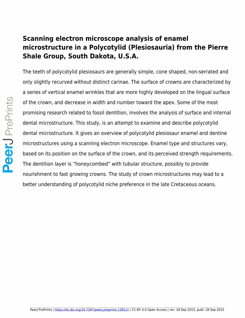
The enamel of reptiles, both extent and fossil, with a few exception, has been found to be prism-82
less (Frank et al 1984). Most reptiles share the basic amniote condition of possessing columnar enamel; 83
this remains true for the polycotylid sp. in this study. The enamel of SDSM 86604 using terminology set 84
forth by Koenigswald and Sander (1997) and Sander (1999, 2000) can be summarized as columnar, 85
possessing convergence at the crystallite level. Individual columnar unites are challenging to identify 86
(Figure 1) however zones of crystallite (enamel units) convergence are interpreted along with incremental 87
lines. The converging crystallite form roughly columnar features that are perpendicular to the enamel-88
denting junction (EDJ). Figure 1 also shows some evidence of incremental lines in the enamel that are 89
parallel to the EDJ and perpendicular to the crystallite columns. The convergent enamel crystallites that 90
make up the majority of the crowns enamel structure, with the exception of the enamel wrinkles. 91
According to Sanders (1999), the incremental line structures are the traces of intermittent growth in two-92
dimensional enamel segments. 93
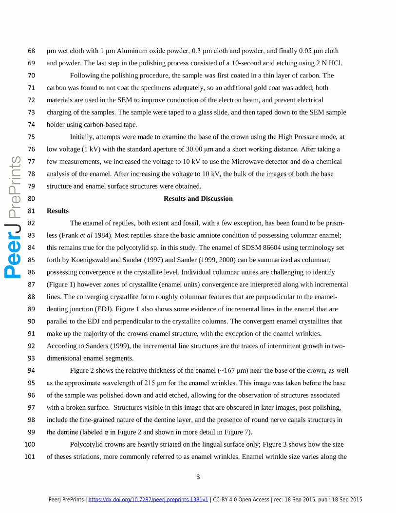
Figure 2 shows the relative thickness of the enamel (~167 μm) near the base of the crown, as well 94
as the approximate wavelength of 215 μm for the enamel wrinkles. This image was taken before the base 95
of the sample was polished down and acid etched, allowing for the observation of structures associated 96
with a broken surface. Structures visible in this image that are obscured in later images, post polishing, 97
include the fine-grained nature of the dentine layer, and the presence of round nerve canals structures in 98
the dentine (labeled α in Figure 2 and shown in more detail in Figure 7). 99
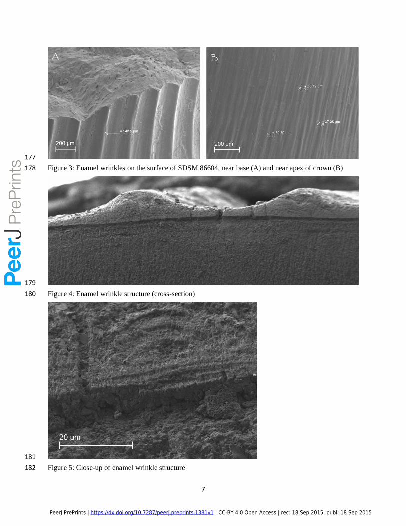
Polycotylid crowns are heavily striated on the lingual surface only; Figure 3 shows how the size 100
of theses striations, more commonly referred to as enamel wrinkles. Enamel wrinkle size varies along the 101
PeerJ PrePrints | https://dx.doi.org/10.7287/peerj.preprints.1381v1 | CC-BY 4.0 Open Access | rec: 18 Sep 2015, publ: 18 Sep 2015
PrePrin
ts
4
surface of the tooth basal to apical. The image on the left (A), shows the enamel wrinkles close to the base 102
of the crown, where the striations have a wavelength of ~250 μm. Image B, on the right, shows the same 103
crowns lingual surface, near the apex, where the distance between striations in reduced to ~40 μm. 104
Figure 4 shows a section of the lingual surface of SDSM 86604 viewed from the basal surface, 105
after the polishing procedure. The enamel-dentine junction is very distinct in the image, as a dark line 106
between the crystalline enamel and the more homologous dentine. It is evident from the image that the 107
enamel wrinkles are a function of varying thickness within the enamel layer, and not an external 108
expression of any internal dentine structures. 109
The enamel that makes up the wrinkles may differ from the enamel between the wrinkles in terms 110
of schmelzmuster. Schmelzmuster is defined as the three-dimensional arrangement of enamel kinds 111
present in a single crown (Koenigswald and Sander 1997). Figure 5 shows a more detailed view of the 112
structure within an enamel wrinkle, incremental lines are preset, visible near the EDJ. The enamel below 113
the wrinkles reflects the surface structure, convergent enamel columns forming where the wrinkle edges 114
meet. 115
Figure 6 shows the enamel-dentine junction on the labial side of the crown, where enamel 116
wrinkles are not present. Where enamel consists of as much as 99% inorganic material (hydroxyapatite), 117
dentine is made of as much as 75% organic matrix, collagen (Glimcher, et. al., 1990). This basic 118
difference in their chemical structures highlights the striking differentiation between the crystalline 119
enamel and the non-crystalline dentine seen in Figure 6. 120
As briefly mentioned in the discussion of Figure 2, Figure 7 is a close-up of the almost perfectly 121
round opening seen in the dentine. These structures may represent nerve canals in the dentine layer, or 122
the dentinal tubules. Near the enamel-dentine junction, these tubules are much less densely packed then 123
at the center of the crown. Figure 8 shows the dentinal tubule structure near the center of the crown. 124
These tubules form a honeycomb structure (A) through the center of the crown. Dentin tubules are 125
responsible for the porousness of dentine and allow for uninterrupted communication between the dentine 126
and the pulp layers (Lima et al 2009). In life, these dentinal tubules would have been filed with dentinal 127
fluid and possibly innervated. These structures would have connected the main nerve of the crown, in the 128
internal pulp cavity, with the dentine layer of the tooth, stopping just short of the EDJ. 129
Figure 9 shows the incidental growth rings in the dentine of the crown. Like tree rings, these 130
structures can be used to better understand the growth of the crown, as successive layer were laid down 131
during the growth of the crown. Since polycotylid lost and re-grew hundreds of crowns in a lifetime the 132
rings have no relationship to the age of the animal. 133
134
Conclusions 135
PeerJ PrePrints | https://dx.doi.org/10.7287/peerj.preprints.1381v1 | CC-BY 4.0 Open Access | rec: 18 Sep 2015, publ: 18 Sep 2015
PrePrin
ts

5
The high concentration of dentinal tubules closer to the center, pulp structure, of the crown 136
supports the idea that polycotylid crowns are growing quickly. The large number of dentinal tubules may 137
be connected to the greater need for innervation in polyphyodontic animals that were constantly shedding 138
and re-growing new crowns. These crowns would have needed high blood and nerve supply to grow 139
quickly to replace crowns lost naturally through the feeding process. Each tubule contains a rod-like 140
structure (Figure 8); the origin of these structures is still unclear; however, they may represent a non-141
mineralized material that had filled in the voids during the fossilization process. Mithiborwala et al 142
(2012) reported similar structures where they are reported to be resin tags, remnants of the adhesive used 143
to stabilize the fossil. Another possible that the rods represent the remnants of collagen structures that 144
once filled the tubules, the acid etching process would have removed the surround hydroxyapatite 145
minerals leaving the collagen to stick out above the surface. “Fossilized” collagen has recently been 146
reported in fossil reptiles, including a Cretaceous hadrosaur and Tyrannosaurs rex (Schweitzer, et. al. 147
2007 and 2009). 148
Parallel and columnar are the two most common crystallite forms of enamel in reptile dentition. 149
In parallel enamel, the hydroxyapatite crystals are parallel to one another and perpendicular to the 150
enamel-dentine junction; on the other hand, columnar enamel is more organized, making up units and 151
bundles (Stokosa 2005). SDSM 86604 shows the later of these enamel types. Columnar enamel is 152
generally considered the more robust enamel type, and in a study with theropod dentition (Stokosa 2005) 153
has suggested columnar enamel is more prevalent in organisms that ingest bone along with soft tissue. 154
Enamel structure may indicate that shell was an important part of polycotylid diets, as the teeth would be 155
more capable of withstanding the stress. Although columnar enamel structure does not directly confirm 156
such a diet, it presence might suggest polycotylids took advantage of the numerous ammonite taxa present 157
in their habit. 158
159
Acknowledgments 160
The author would like to thank Dr. Darrin Pagnac, Dr. James Martin for assistance with choosing 161
specimens for study from the South Dakota School of Mines and Technology collections, the South 162
Dakota Army Corp. of Engineers, Ms. Sally Shelton. Collections Manager and the staff of the SDSM&T 163
Museum of Geology, Dr. J. Foster Sawyer for being a friend and unofficial faculty advisor, Dr. Edward 164
Duke for assistance with the Scanning Electron Microscope, and interpretation of SEM results, Dr. 165
Maribeth Price, my academic advisor, for early assistance with forming the project proposal, Ms. Arlette 166
Hansen, curator of The Adams Museum in Deadwood, SD for specimen access, Dr. P. Martin Sander, 167
University of Bonn for assistance with obtaining research material, Dr. Kelvin K. Krause, D.D.S. for 168
discussions concerning comparison of SEM imagery to structures seen in human dentition microstructure, 169
PeerJ PrePrints | https://dx.doi.org/10.7287/peerj.preprints.1381v1 | CC-BY 4.0 Open Access | rec: 18 Sep 2015, publ: 18 Sep 2015
PrePrin
ts
6
170
Figures: 171
172
Figure 1: Scanning electron microscope image of SDSM 86604, enamel microstructure in transverse 173
section. 174
175
Figure 2: Enamel wrinkle structure near crown base 176
PeerJ PrePrints | https://dx.doi.org/10.7287/peerj.preprints.1381v1 | CC-BY 4.0 Open Access | rec: 18 Sep 2015, publ: 18 Sep 2015
PrePrin
ts
7
177
Figure 3: Enamel wrinkles on the surface of SDSM 86604, near base (A) and near apex of crown (B) 178
179
Figure 4: Enamel wrinkle structure (cross-section) 180
181
Figure 5: Close-up of enamel wrinkle structure 182
PeerJ PrePrints | https://dx.doi.org/10.7287/peerj.preprints.1381v1 | CC-BY 4.0 Open Access | rec: 18 Sep 2015, publ: 18 Sep 2015
PrePrin
ts

8
183
Figure 6: Enamel-dentine junction 184
185
Figure 7: Opening of dentine tubule canal in dentin layer 186
PeerJ PrePrints | https://dx.doi.org/10.7287/peerj.preprints.1381v1 | CC-BY 4.0 Open Access | rec: 18 Sep 2015, publ: 18 Sep 2015
PrePrin
ts

9
187
Figure 8: Dentine structures (A), rod structure within the dentine (B) 188
189
Figure 9: Incidental growth rings in dentin layer 190
191
References 192
Arkhangelsky, M.S., A.O. Averianov and E.M. Pervushov. 2007. Short-Necked Plesiosaurs of the Family 193
Plycotylidae from the Campanian of the Saratov Region. Paleontological Journal 41: 62-66. 194
Carpenter, K. 1996. A review of short-necked plesiosaurs from the Cretaceous of the Western Interior, 195
North America. N. Jb. Geol. Paläont., Abh. 201:259-287. 196
PeerJ PrePrints | https://dx.doi.org/10.7287/peerj.preprints.1381v1 | CC-BY 4.0 Open Access | rec: 18 Sep 2015, publ: 18 Sep 2015
PrePrin
ts

10
Everhart, M. J. 2005. Pliosaurs and Polycothlids; pp. 142-155, Oceans of Kansas: A Natural History of 197
the Western Interior Sea. Indiana University Press, Bloomington, IN. 198
Frank, R.M., Sigogneau-Russell, D., and Voegel, J.C., 1984, Tooth Ultrastructure of Late Triassic 199
Haramiyidae: Journal of Dental Research, v. 63, p. 661-664. 200
Glimcher, M.J., Cohen-Solal, L., Kossiva, D., and Ricqles, A.D., 1990, Biochemical Analyses of Fossil 201
Enamel and Dentin: Paleobiology, v. 16, p. 219-232. 202
Heckert, A.B. and J.A. Miller-Camp. 2013. Tooth enamel microstructure of Revueltosaurus and 203
Krzyzanowskisaurus (Reptilia: Archosauria) from the Upper Triassic Chinle Group, USA: 204
Implications for function, growth, and phylogeny. Palaeontologia Electronica 16: 23p 205
Hwang, S. H. 2005. Phylogenetic Patterns of Enamal Microstructure in Dinosaur Teeth. Journal of 206
Morphology 266: 208-240. 207
Kear, B. P. 2003. Cretaceous marine reptiles of Australia: a review of taxonomy and distribution. 208
Cretaceous Research 24:277-303. 209
Koenigswald, W.v. and P.M. Sander. 1997. Glossary of terms used for enamel microstructures; pp. 267-210
280 in Koenigswald, W.v. and P.M. Sander (eds.), Tooth enamel microstructure, Balkema, 211
Rotterdam 212
Lima, R. R., L. M. Araújo, P. R. Affonso, K. M. Maranhão and S. S. Lamarão. 2009. Scanning Electron 213
Microscopic Investigation of Dentinal Tubules in Cebus appella Dentitin. Ciêecia Animal 214
Brasileira 10: 1328-1331. 215
Martin, J. E., J. L. Bertog, and D. C. Parris. 2007. Revised lithostratigraphy of the lowr Pierre Shale 216
Group (Campanian) of central South Dakota, Including newly designated members; pp. 9-21 in J. 217
E. Martin and D. C. Parris (eds.), The Geology and Paleontology of the Late Cretaceous Marine 218
Deposits of the Dakotas: Geological Society of America Special Paper 427. 219
Massare, J. A. 1987. Tooth Morphology and Prey Preference of Mesozoic Marine Reptiles. Journal of 220
Vertebrate Paleontology 7:121-137. 221
Mithiborwala, S., V. Chaugule, A.K. Munshi and V. Patil. 2012. A comparison of the resin tag 222
penetration of the total etch and the self-etch dentin bonding systems in the primary teeth: An in 223
vitro study. Comtemporary Clinical Dentistry 3: 158-163. 224
O'Keefe, F. R. 2004. On the Cranial Anatomy of the Polycotylid Plesiosaurs, Including New Material of 225
Polycotylus latipinnis, Cope, from Alabama. Journal of Vertebrate Paleontology 24:326-340. 226
Sander, P.M. 1995. The microstructure of reptilian tooth enamel: terminology, function, and phylogeney. 227
Habilitation. Bonn University, 191 pp. 228
Sander, P.M., 1999. The microstructure of reptilian tooth enamel: Terminology, function, and phylogeny: 229
Münchner Geowissenschaftliche Abhandlungen, Reihe A, v. 38, p. 1-102. 230
PeerJ PrePrints | https://dx.doi.org/10.7287/peerj.preprints.1381v1 | CC-BY 4.0 Open Access | rec: 18 Sep 2015, publ: 18 Sep 2015
PrePrin
ts

11
Sander, P.M., 2000. Prismless enamel in amniotes: terminology, function, and evolution, in Teaford, 231
M.F., Smith, M.M., and Ferguseon, M.W.J., eds., Development, Function and Evolution of Teeth: 232
Cambridge, UK, Cambridge University Press, p. 92-106. 233
Sato, T., and G. W. Storrs. 2000. An Early Polycotylid Plesiosaur (Reptilia: Sauropterygia) from the 234
Cretaceous of Hokkaido, Japan. Journal of Paleontology 74:907-914. 235
Schumacher, B. A. 2007. A new polycotylid plesiosaur (Reptilia; Sauropterygia) from the Greenhorn 236
Limestone (Upper Cretaceous; lower upper Cenomanian), Black Hills, South Dakota; pp. 133-237
146 in J. E. Martin and D. C. Parris (eds.), The Geology and Paleontology of the Late Cretaceous 238
Marine Deposits of the Dakotas: Geological Society of America Special Paper 427. 239
Schweitzer, M.H., Suo, Z., Avci, R., Asara, J.M., Allen, M.A., Arce, F.T., and Horner, J.R., 2007, 240
Analyses of Soft Tissue from Tyrannosaurus rex suggest the Presence of Protein: Science, v. 316, 241
p. 277-280. 242
Schweitzer, M.H., Zheng, W., Organ, C.L., Avci, R., Suo, Z., Freimark, L.M., Lebleu, V.S., Duncan, 243
M.B., Heiden, M.G.V., Neveu, J.M., Lane, W.S., Cottrell, J.S., Horner, J.R., Cantley, L.C., 244
Kalluri, R., and Asara, J.M., 2009, Biomolecular Characterization and Protein Sequences of the 245
Campanian Hadrosaur B. canadensis: Science, v. 324, p. 626-631. 246
Stokosa, K. 2005. Enamel Microstructure Variation within the Theropoda; pp. 163-178 in K. Carpenter 247
(ed.), The Carnivorous Dinosaurs. Indiana University Press, Bloomington, IN. 248
Testin, J.J., 2011. Microscopic Dental Structure in a Polycotylid Plesiosaur (Diapsida: Plesiosauroidea) 249
from the Boyer Bay Member of the Sharon Springs Fm., Pierre Shale Group, South Dakota. GSA 250
Abstracts with Programs v. 43, no. 4. p. 85 251
252
PeerJ PrePrints | https://dx.doi.org/10.7287/peerj.preprints.1381v1 | CC-BY 4.0 Open Access | rec: 18 Sep 2015, publ: 18 Sep 2015
PrePrin
ts