saunders. guide to the dissection of the dog
TRANSCRIPT
Guide to the Dissection of the Dog 100
Make a sagittal incision completely through the thoracic wall 1 cm from the ventral median plane on each side. These incisions should extend from the thoracic inlet through the ninth costal carti-lage. The transversus thoracis muscle is a flat, fleshy muscle on the medial surface of the costal cartilages of ribs 2 though 8 (Figs. 3-4, 3-6). Its fas-cicles extend from the costochondral junctions to the sternum. Connect the caudal ends of the right and left sagittal incisions and free the sternum, ex-cept for the wide, thin fold of mediastinum that is now its only attachment.
On the right half of the thorax, clean and tran-sect the origin of the latissimus dorsi and reflect it toward the forelimb. Locate and transect the cau-dal portion of the origin of the serratus ventralis, exposing the ribs. Starting at the costal arch and using bone cutters, close to their vertebral articulation without damaging the sympathetic trunk. Reflect the tho-racic wall As this is done, cut the attachments of the internal abdominal oblique, transversus abdominis, and diaphragm from the ribs along the costal arch. If this is done carefully, the peritoneal cavity will not be opened. Reflect the left thoracic wall in a similar manner.
On the internal surface of the thoracic wall, no-tice the intercostal vessels and nerves coursing
along the caudal border of the ribs. Ventrally, the vessels bifurcate and anastomose with the ventral intercostal branches of the internal thoracic artery and vein. The intercostal nerves supply the inter-costal musculature. Their sensory branches were seen as lateral and ventral cutaneous branches.
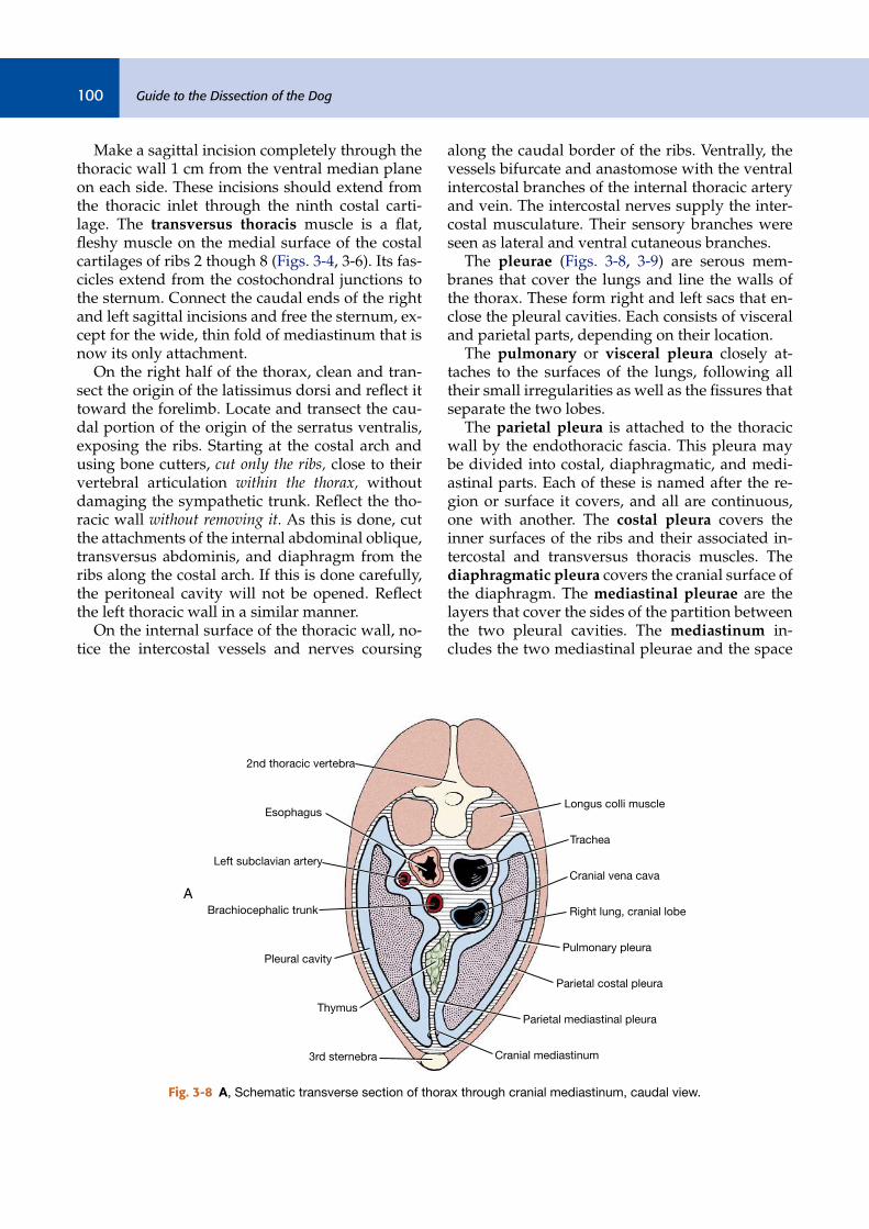
The pleurae (Figs. 3-8, 3-9) are serous mem-branes that cover the lungs and line the walls of the thorax. These form right and left sacs that en-close the pleural cavities. Each consists of visceral and parietal parts, depending on their location.
The pulmonary or visceral pleura closely at-taches to the surfaces of the lungs, following all their small irregularities as well as the fissures that separate the two lobes.
The parietal pleura is attached to the thoracic wall by the endothoracic fascia. This pleura may be divided into costal, diaphragmatic, and medi-astinal parts. Each of these is named after the re-gion or surface it covers, and all are continuous, one with another. The costal pleura covers the inner surfaces of the ribs and their associated in-tercostal and transversus thoracis muscles. The diaphragmatic pleura covers the cranial surface of the diaphragm. The mediastinal pleurae are the layers that cover the sides of the partition between the two pleural cavities. The mediastinum in-cludes the two mediastinal pleurae and the space
Brachiocephalic trunk
Pleural cavity
Thymus
3rd sternebra
2nd thoracic vertebra
A
Esophagus
Left subclavian artery
Cranial mediastinum
Parietal mediastinal pleura
Pulmonary pleura
Right lung, cranial lobe
Cranial vena cava
Trachea
Longus colli muscle
Parietal costal pleura
Fig. 3-8 A, Schematic transverse section of thorax through cranial mediastinum, caudal view.
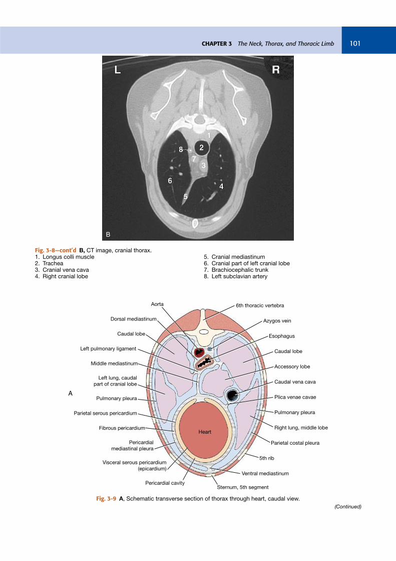
CHAPTER 3 The Neck, Thorax, and Thoracic Limb 101
1. Longus colli muscle2. Trachea3. Cranial vena cava4. Right cranial lobe
5. Cranial mediastinum6. Cranial part of left cranial lobe7. Brachiocephalic trunk8. Left subclavian artery
B
Fig. 3-8—cont’d B, CT image, cranial thorax.
Aorta
Dorsal mediastinum
Caudal lobe
Left pulmonary ligament
Middle mediastinum
Left lung, caudal
part of cranial lobe
Pulmonary pleura
Parietal serous pericardium
Fibrous pericardium
Pericardial
mediastinal pleura
Visceral serous pericardium
(epicardium)
Pericardial cavitySternum, 5th segment
Ventral mediastinum
5th rib
Parietal costal pleura
Right lung, middle lobe
Pulmonary pleura
Plica venae cavae
Caudal vena cava
Accessory lobe
Caudal lobe
Esophagus
Azygos vein
6th thoracic vertebra
Heart
A
Fig. 3-9 A, Schematic transverse section of thorax through heart, caudal view.
(Continued)

Guide to the Dissection of the Dog 102
between them. Enclosed in the mediastinum are the thymus, the lymph nodes, the heart, the aorta, the trachea, the esophagus, the vagus nerves, and other nerves and vessels. The pericardial medias-tinal pleura is that portion covering the heart.
The mediastinum can be divided into a cranial part, that lying cranial to the heart; a middle part, that containing the heart; a dorsal portion dorsal to the heart; a ventral portion, ventral to the heart; and a caudal part, lying caudal to the heart. The caudal mediastinum is thin. It attaches to the diaphragm far to the left of the median plane. Cranially, it is continuous with the middle mediastinum.
Note the passage of the esophagus through the mediastinum and the esophageal hiatus of the dia-phragm. At the esophageal hiatus, a thin layer of pleura, peritoneum, and enclosed connective tis-sue attaches the esophagus to the muscle of the diaphragm.
The plica venae cavae is a loose fold of pleura derived from the right caudal mediastinal portion of the pleural sac that surrounds the caudal vena
cava. The root of the lung is composed of pleura and the bronchi, vessels, and nerves entering the lung. Here the mediastinal parietal pleura is con-tinuous with the pulmonary pleura. Caudal to the hilus this connection forms a free border, known as the pulmonary ligament (Figs. 3-9, 3-10), between the caudal lobe of the lung and the mediastinum at the level of the esophagus. Observe this ligament. In thoracic surgery this must be cut to reflect the caudal lung lobe cranially.
The thymus (Figs. 3-8, 3-11, 3-12, 3-14, 3-16, 3-20) is a bilobed, compressed structure situated in the cranial mediastinum. It is largest in the young dog and usually atrophies with age until only a trace remains. When maximally developed, the caudal part of the thymus is molded on the cranial surface of the pericardium.
The internal thoracic artery (Figs. 3-14, 3-16 through 3-20) leaves the subclavian artery, courses ventrocaudally in the cranial mediasti-num, and disappears deep to the cranial border of the transversus thoracis muscle. It supplies many
B C
Fig. 3-9—cont’d B, CT image, midthorax. 1. Esophagus 2. Right principal bronchus 3. Carina of trachea 4. Ventral mediastinum-phrenicopericardial
ligament 5. Heart 6. Left pulmonary artery 7. Aorta
C, CT image, caudal thorax. 1. Right caudal lobe 2. Caudal vena cava 3. Accessory lobe 4. Plica venae cavae 5. Heart 6. Caudal mediastinum 7. Left caudal lobe 8. Esophagus 9. Aorta
CHAPTER 3 The Neck, Thorax, and Thoracic Limb 103
branches to surrounding structures—the phrenic nerve, the thymus, the mediastinal pleurae, and the dorsal intercostal spaces. The perforating branches to the superficial structures of the ventral third of the thorax have been seen. The anastomoses with the dorsal intercostal arteries on the medial side of the thoracic wall have been seen. Near the at-tachment of the costal arch with the sternum, the internal thoracic artery terminates in the muscu-lophrenic artery and the larger cranial epigastric artery. The latter has been dissected along with its
cranial superficial epigastric branch. The musculo-phrenic artery (Fig. 4-33) runs caudodorsally in the angle formed by the diaphragm and lateral thoracic wall. Dissect its origin. Cut the mediastinum near the sternum and reflect the sternum cranially.
Lungs
Each lung is divided into lobes based on the branching pattern of its principal bronchus into lobar bronchi (Fig. 3-13). The left lung (see Figs. 3-10, 3-11) is divided into cranial and
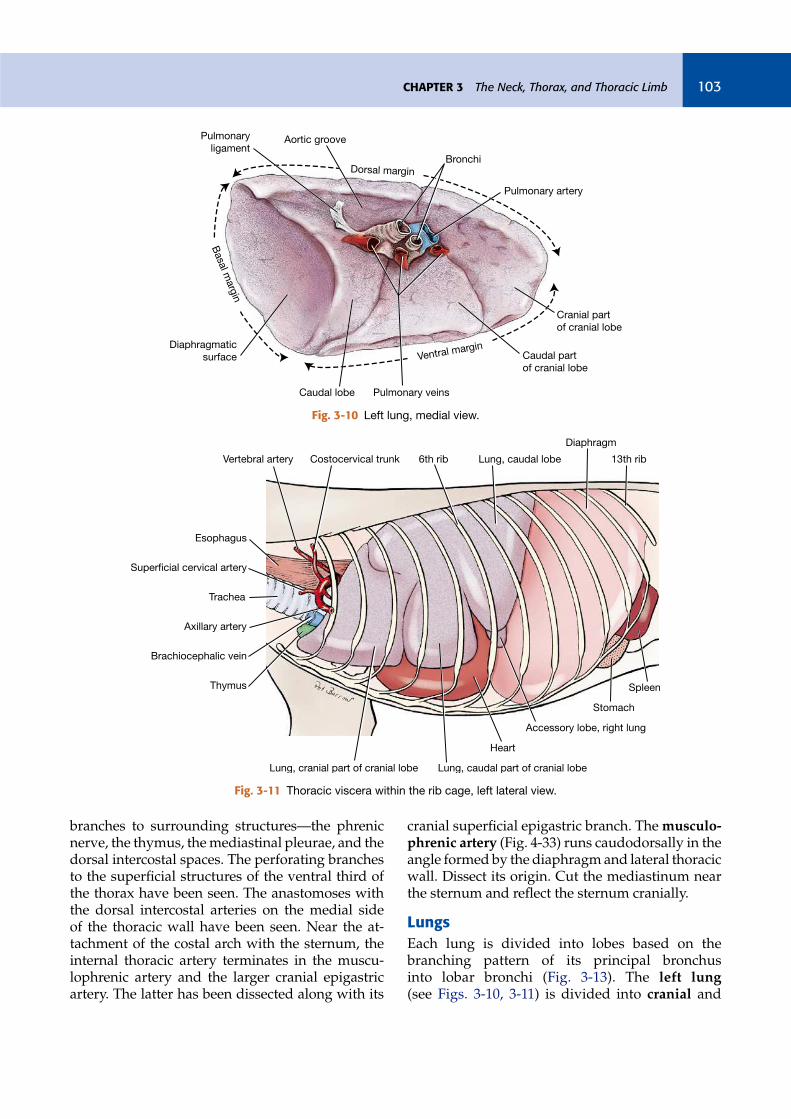
Dorsal margin
Basa
l marg
in
Ventral margin
Aortic groovePulmonary
ligament
Diaphragmatic
surface
Caudal lobe Pulmonary veins
Caudal part
of cranial lobe
Cranial part
of cranial lobe
Pulmonary artery
Bronchi
Fig. 3-10 Left lung, medial view.
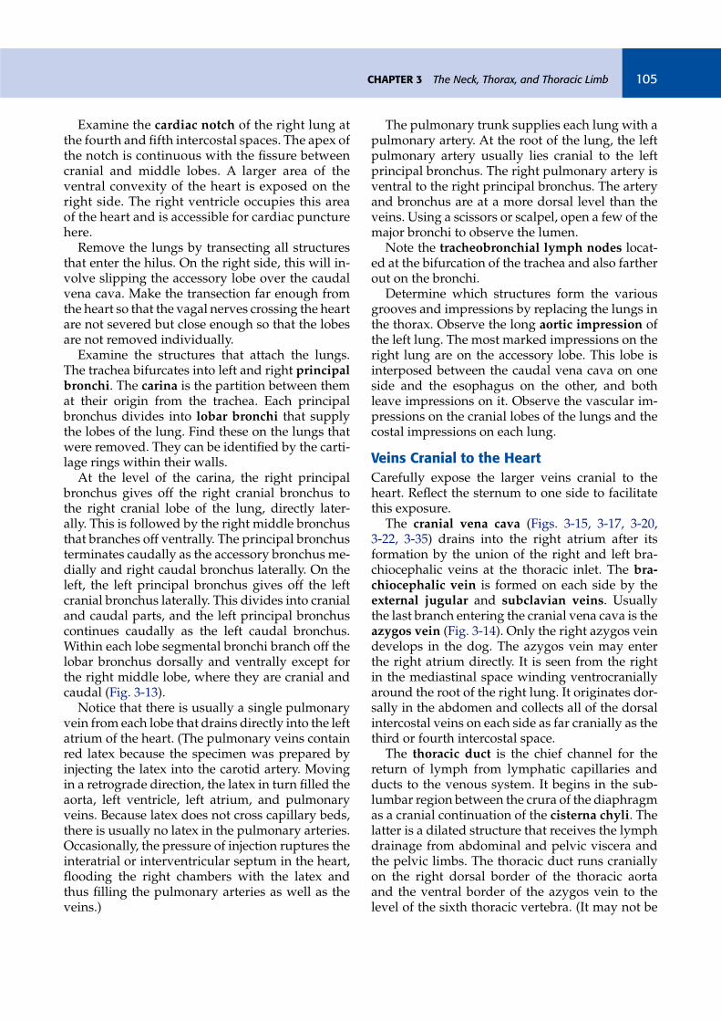
Costocervical trunkVertebral artery
Esophagus
Trachea
Superficial cervical artery
Axillary artery
Brachiocephalic vein
Thymus
Lung, cranial part of cranial lobe Lung, caudal part of cranial lobe
Heart
Accessory lobe, right lung
Stomach
Spleen
Diaphragm
13th ribLung, caudal lobe6th rib
Fig. 3-11 Thoracic viscera within the rib cage, left lateral view.

Guide to the Dissection of the Dog 104
caudal lobes by deep fissures. The cranial lobe is further divided into cranial and caudal parts. The right lung (Fig. 3-12) is divided into cra-nial, middle, caudal, and accessory lobes. A part of the accessory lobe can be seen from the
left through the caudal mediastinum (Fig. 3-20) or from the right through the plica venae cavae, where it lies in the space between these two struc-tures. Reflect the caudal lung lobes to observe this.
Cardiac notch
6th ribLung, caudal lobe
Stomach
Liver
Diaphragm
Lung, middle lobe Heart
Thymus
Brachiocephalic vein
Trachea
Superficial cervical artery
Costocervical trunk
Common carotid artery
Vertebral artery
Lung, cranial lobe
Fig. 3-12 Thoracic viscera within the rib cage, right lateral view.
Accessory
lobe
Right
caudal
lobe
Right
middle
lobe
Right
cranial
lobe
Left
caudal
lobe
Left
cranial
lobe
RB4V2
RB4D2
RB4V1
RB4D1
RB2C1
RB2R1
RB1D1
RB1V1
RB1V2
RB1D2
Trachea
RB3D1
RB3D2
RB3V2
RB3V1
RB3RB4
RB2
RPB
RB1
LB2
LB1LPB
LB2V2
LB2D2
LB2V1
LB2D1
LB1V1b
LB1V1
LB1V1a
LB1D1
LB1V2
LB1D2
Fig. 3-13 Schematic bronchial tree of the dog, in dorsal view. Letters and numbers identify the principal, lobar, and segmental bronchi by their bronchoscopic order of origin and their anatomical orientation. Lower case a and b represent subsegmental bronchi (From Amis TC, McKiernan BC: Systematic identification of endobronchial anatomy during bronchoscopy in the dog, Am J Vet Res 47:2649–2657, 1986.)
CHAPTER 3 The Neck, Thorax, and Thoracic Limb 105
Examine the cardiac notch of the right lung at the fourth and fifth intercostal spaces. The apex of the notch is continuous with the fissure between cranial and middle lobes. A larger area of the ventral convexity of the heart is exposed on the right side. The right ventricle occupies this area of the heart and is accessible for cardiac puncture here.
Remove the lungs by transecting all structures that enter the hilus. On the right side, this will in-volve slipping the accessory lobe over the caudal vena cava. Make the transection far enough from the heart so that the vagal nerves crossing the heart are not severed but close enough so that the lobes are not removed individually.
Examine the structures that attach the lungs. The trachea bifurcates into left and right principal bronchi. The carina is the partition between them at their origin from the trachea. Each principal bronchus divides into lobar bronchi that supply the lobes of the lung. Find these on the lungs that were removed. They can be identified by the carti-lage rings within their walls.
At the level of the carina, the right principal bronchus gives off the right cranial bronchus to the right cranial lobe of the lung, directly later-ally. This is followed by the right middle bronchus that branches off ventrally. The principal bronchus terminates caudally as the accessory bronchus me-dially and right caudal bronchus laterally. On the left, the left principal bronchus gives off the left cranial bronchus laterally. This divides into cranial and caudal parts, and the left principal bronchus continues caudally as the left caudal bronchus. Within each lobe segmental bronchi branch off the lobar bronchus dorsally and ventrally except for the right middle lobe, where they are cranial and caudal (Fig. 3-13).
Notice that there is usually a single pulmonary vein from each lobe that drains directly into the left atrium of the heart. (The pulmonary veins contain red latex because the specimen was prepared by injecting the latex into the carotid artery. Moving in a retrograde direction, the latex in turn filled the aorta, left ventricle, left atrium, and pulmonary veins. Because latex does not cross capillary beds, there is usually no latex in the pulmonary arteries. Occasionally, the pressure of injection ruptures the interatrial or interventricular septum in the heart, flooding the right chambers with the latex and thus filling the pulmonary arteries as well as the veins.)
The pulmonary trunk supplies each lung with a pulmonary artery. At the root of the lung, the left pulmonary artery usually lies cranial to the left principal bronchus. The right pulmonary artery is ventral to the right principal bronchus. The artery and bronchus are at a more dorsal level than the veins. Using a scissors or scalpel, open a few of the major bronchi to observe the lumen.
Note the tracheobronchial lymph nodes locat-ed at the bifurcation of the trachea and also farther out on the bronchi.
Determine which structures form the various grooves and impressions by replacing the lungs in the thorax. Observe the long aortic impression of the left lung. The most marked impressions on the right lung are on the accessory lobe. This lobe is interposed between the caudal vena cava on one side and the esophagus on the other, and both leave impressions on it. Observe the vascular im-pressions on the cranial lobes of the lungs and the costal impressions on each lung.
Veins Cranial to the Heart
Carefully expose the larger veins cranial to the heart. Reflect the sternum to one side to facilitate this exposure.
The cranial vena cava (Figs. 3-15, 3-17, 3-20, 3-22, 3-35) drains into the right atrium after its formation by the union of the right and left bra-chiocephalic veins at the thoracic inlet. The bra-chiocephalic vein is formed on each side by the external jugular and subclavian veins. Usually the last branch entering the cranial vena cava is the azygos vein (Fig. 3-14). Only the right azygos vein develops in the dog. The azygos vein may enter the right atrium directly. It is seen from the right in the mediastinal space winding ventrocranially around the root of the right lung. It originates dor-sally in the abdomen and collects all of the dorsal intercostal veins on each side as far cranially as the third or fourth intercostal space.
The thoracic duct is the chief channel for the return of lymph from lymphatic capillaries and ducts to the venous system. It begins in the sub-lumbar region between the crura of the diaphragm as a cranial continuation of the cisterna chyli. The latter is a dilated structure that receives the lymph drainage from abdominal and pelvic viscera and the pelvic limbs. The thoracic duct runs cranially on the right dorsal border of the thoracic aorta and the ventral border of the azygos vein to the level of the sixth thoracic vertebra. (It may not be
Guide to the Dissection of the Dog 106
visible.) Here it crosses the ventral surface of the fifth thoracic vertebra and courses on the left side of the middle mediastinal pleura. It continues cra-nioventrally through the cranial mediastinum to the left brachiocephalic vein, where it usually ter-minates (Fig. 3-17). The thoracic duct also receives the lymph drainage from the left thoracic limb and the left tracheal trunk (from the left side of the head and neck). The lymph drainage from the right thoracic limb and the right tracheal trunk (from the right side of the head and neck) form a right lymphatic duct that enters the venous system in the vicinity of the right brachiocephalic vein. There are often multiple terminations of a com-plicated nature, which may include swellings or anastomoses. All lymphatic channels will be dif-ficult to see unless they are congested with lymph or refluxed blood. They are frequently double.
Look for the thoracic duct. It is not always vis-ible, but it may be identified by the reddish brown or straw color of its contents and the numerous random constrictions in its wall. The tracheal trunks may be found in each carotid sheath or par-allel to the sheath and its contents.
Arteries Cranial to the Heart
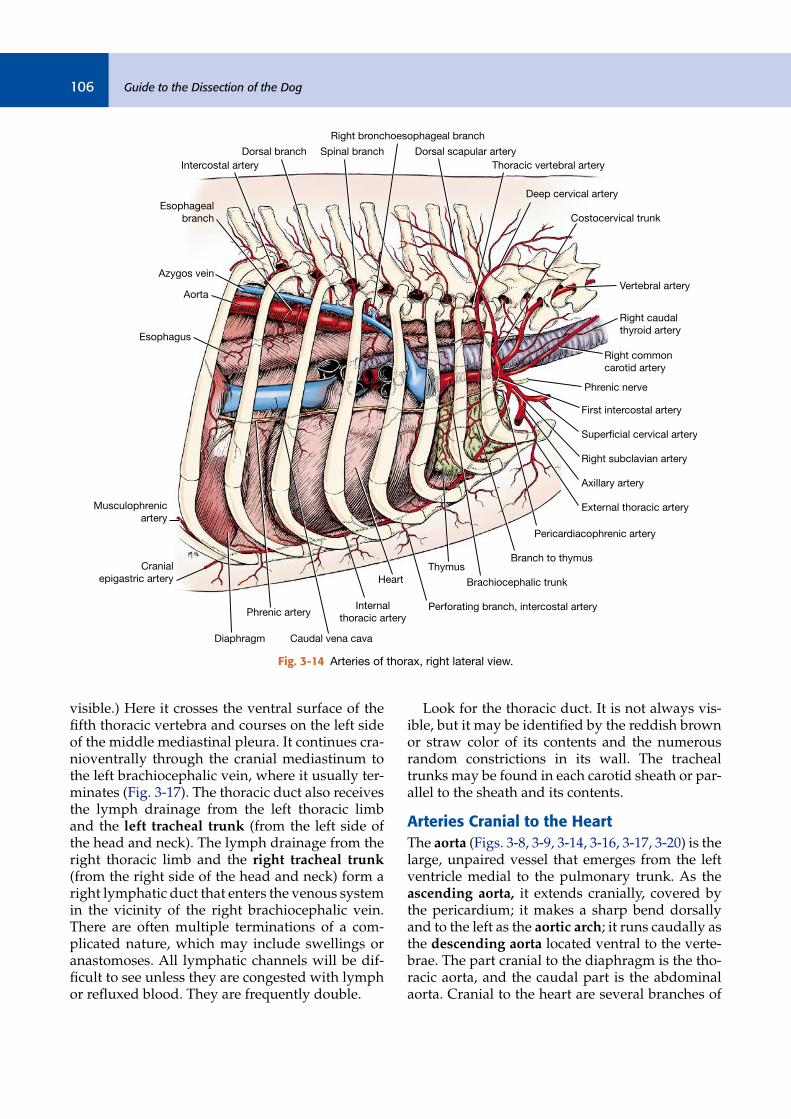
The aorta (Figs. 3-8, 3-9, 3-14, 3-16, 3-17, 3-20) is the large, unpaired vessel that emerges from the left ventricle medial to the pulmonary trunk. As the ascending aorta, it extends cranially, covered by the pericardium; it makes a sharp bend dorsally and to the left as the aortic arch; it runs caudally as the descending aorta located ventral to the verte-brae. The part cranial to the diaphragm is the tho-racic aorta, and the caudal part is the abdominal aorta. Cranial to the heart are several branches of
Right bronchoesophageal branch
Dorsal scapular artery
Thoracic vertebral artery
Deep cervical artery
Costocervical trunk
Vertebral artery
Right common
carotid artery
Right caudal
thyroid artery
First intercostal artery
Phrenic nerve
Right subclavian artery
Superficial cervical artery
Axillary artery
Pericardiacophrenic artery
External thoracic artery
Branch to thymus
Brachiocephalic trunk
Thymus
Perforating branch, intercostal artery
Heart
Internal
thoracic artery
Caudal vena cava
Phrenic artery
Diaphragm
Cranial
epigastric artery
Musculophrenic
artery
Esophagus
Aorta
Azygos vein
Esophageal
branch
Intercostal artery
Dorsal branch Spinal branch
Fig. 3-14 Arteries of thorax, right lateral view.

CHAPTER 3 The Neck, Thorax, and Thoracic Limb 107
the aorta. Reflect the veins that were dissected cra-nial to the heart to observe these arteries.
The right and left coronary arteries are branches of the ascending aorta that supply the heart mus-cle. They will be studied with the heart.
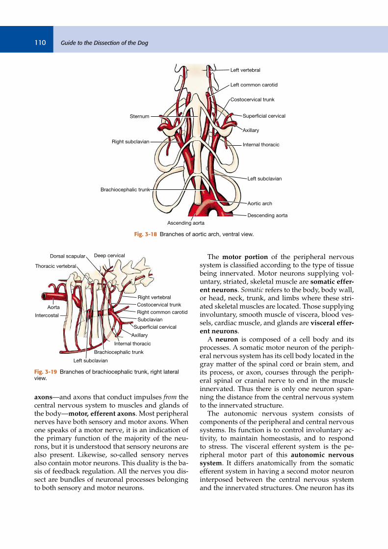
The brachiocephalic trunk (Figs. 3-14, 3-16 through 3-20), the first branch from the aortic arch, passes obliquely to the right across the ventral sur-face of the trachea. It gives rise to the left common carotid artery and terminates as the right common carotid artery and the right subclavian artery.
The left subclavian artery (Figs. 3-16 through 3-20) originates from the aortic arch beyond the level of the brachiocephalic trunk and passes obliquely to the left across the ventral surface of the esophagus.
The branches of the right and left subclavian ar-teries are similar; only the right subclavian artery will be described. For each artery described, there is a comparable vein with a similar area of distri-bution. The terminations of the veins are variable,
and they will not be dissected. Remove them when necessary to expose the arteries. The right subcla-vian artery has four branches that arise medial to the first rib or intercostal space. They are the verte-bral artery, the costocervical trunk, the superficial cervical artery, and the internal thoracic artery. Do not sever the nerves or arteries.
The vertebral artery (Figs. 3-14, 3-16 through 3-20) crosses the medial surface of the first rib and disappears dorsally between the longus colli and the scalenus muscles. It enters the transverse foramen of the sixth cervical vertebra and passes through the transverse foramina of the first six cer-vical vertebrae. It supplies both muscular branches to the cervical muscles and also spinal branches at each intervertebral foramen to the spinal cord and its coverings. At the level of the atlas, it terminates by entering the vertebral canal through the lateral vertebral foramen and contributes to the ventral spinal and basilar arteries. These will be seen later in the dissection of the nervous system.
Pharyngeal branch
Hyoid venous arch
Facial
Lingual
Thyroid cartilage
Cranial thyroid
Middle thyroid
Trachea
Right internal jugular
Caudal thyroid veins
Muscular branch
Internal thoracic trunk
Cranial vena cavaCostocervical veins
Brachiocephalic
Subclavian
Cephalic
Superficial cervical
Omobrachial
Left internal jugular
Esophagus
External jugular
Thyroid gland
Parathyroid gland
From medial retropharyngeal
lymph node
Cranial thyroid
Linguofacial
Maxillary
Cranial laryngeal
Laryngea impar
Lingual
Submental
Fig. 3-15 Veins of the neck, ventral aspect.
Guide to the Dissection of the Dog 108
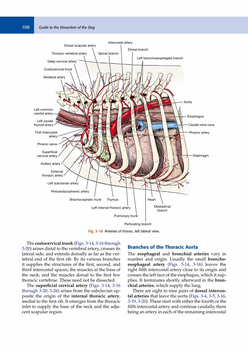
The costocervical trunk (Figs. 3-14, 3-16 through 3-20) arises distal to the vertebral artery, crosses its lateral side, and extends dorsally as far as the ver-tebral end of the first rib. By its various branches it supplies the structures of the first, second, and third intercostal spaces; the muscles at the base of the neck; and the muscles dorsal to the first few thoracic vertebrae. These need not be dissected.
The superficial cervical artery (Figs. 3-14, 3-16 through 3-20, 3-26) arises from the subclavian op-posite the origin of the internal thoracic artery, medial to the first rib. It emerges from the thoracic inlet to supply the base of the neck and the adja-cent scapular region.
Branches of the Thoracic Aorta
The esophageal and bronchial arteries vary in number and origin. Usually the small broncho-esophageal artery (Figs. 3-14, 3-16) leaves the right fifth intercostal artery close to its origin and crosses the left face of the esophagus, which it sup-plies. It terminates shortly afterward in the bron-chial arteries, which supply the lung.
There are eight to nine pairs of dorsal intercos-tal arteries that leave the aorta (Figs. 3-4, 3-5, 3-16, 3-19, 3-20). These start with either the fourth or the fifth intercostal artery and continue caudally, there being an artery in each of the remaining intercostal
Spinal branch
Intercostal artery
Dorsal branch
Left bronchoesophageal branch
Aorta
Esophagus
Caudal vena cava
Diaphragm
Phrenic artery
Mediastinal
branch
Heart
Perforating branch
Pulmonary trunk
Left internal thoracic artery
ThymusBrachiocephalic trunk
Pericardiacophrenic artery
External
thoracic artery
Left subclavian artery
Axillary artery
Left common
carotid artery
Vertebral artery
Costocervical trunk
Deep cervical artery
Superficial
cervical artery
Phrenic nerve
First intercostal
artery
Left caudal
thyroid artery
Thoracic vertebral artery
Dorsal scapular artery
Fig. 3-16 Arteries of thorax, left lateral view.

CHAPTER 3 The Neck, Thorax, and Thoracic Limb 109
spaces. Each lies close to the caudal border of the rib. The costocervical trunk supplies the first three or four intercostal spaces (Fig. 3-19). The dorsal costoabdominal artery courses ventrally, caudal to the last rib.
The phrenic nerves (Figs. 3-14, 3-16, 3-20) supply the diaphragm. Find each nerve as it passes through the thoracic inlet. The nerve aris-es from the ventral branches of the fifth, the sixth, and usually the seventh cervical nerves. Follow the phrenic nerves through the mediastinum to the diaphragm. Each is both motor and sensory to the corresponding half of the diaphragm ex-cept at its periphery. This part of the muscle re-ceives sensory fibers from the caudal intercostal nerves.
INTRODUCTION TO THE AUTONOMIC
NERVOUS SYSTEM
The nervous system is highly organized both an-atomically and functionally. It is composed of a central nervous system and a peripheral nervous system. The central nervous system includes the brain and the spinal cord. The peripheral nervous system comprises the cranial nerves, which con-nect with structures of the head and body, and the spinal nerves, which connect the spinal cord to structures of the neck, trunk, tail, and limbs. The peripheral nervous system can be further classified on the basis of anatomy and function. The periph-eral nerves contain axons that conduct impulses
the central nervous system—sensory, afferent
Right external jugular vein
Right subclavian artery
Superfricial cervical vein
Cephalic vein
Internal jugular vein
Brachiocephalic trunk
Subclavian vein
Caudal thyroid vein
Right brachiocephalic vein
Internal thoracic vein
Right costocervical vein
Cranial vena cava
Hepatic veins
Caudal vena cava
Right auricle
Right ventricle
Right coronary artery
Left coronary artery
Paraconal
interventricular branch
Left auricle
Left ventricle
Thoracic aorta
Right and left common carotid arteries
Costocervical trunk
Superficial cervical artery
Axillary artery
Vertebral artery
Internal thoracic artery
Left tracheal trk.
Thoracic duct
Subclavian vein
Left brachiocephalic vein
Left subclavian artery
Left costocervical vein
Aortic arch
Ligamentum arteriosum
Pulmonary trunk
Fig. 3-17 Heart and great vessels, ventral view.
Guide to the Dissection of the Dog 110
axons—and axons that conduct impulses the central nervous system to muscles and glands of the body—motor, efferent axons. Most peripheral nerves have both sensory and motor axons. When one speaks of a motor nerve, it is an indication of the primary function of the majority of the neu-rons, but it is understood that sensory neurons are also present. Likewise, so-called sensory nerves also contain motor neurons. This duality is the ba-sis of feedback regulation. All the nerves you dis-sect are bundles of neuronal processes belonging to both sensory and motor neurons.
The motor portion of the peripheral nervous system is classified according to the type of tissue being innervated. Motor neurons supplying vol-untary, striated, skeletal muscle are somatic effer-ent neurons. refers to the body, body wall, or head, neck, trunk, and limbs where these stri-ated skeletal muscles are located. Those supplying involuntary, smooth muscle of viscera, blood ves-sels, cardiac muscle, and glands are visceral effer-ent neurons.
A neuron is composed of a cell body and its processes. A somatic motor neuron of the periph-eral nervous system has its cell body located in the gray matter of the spinal cord or brain stem, and its process, or axon, courses through the periph-eral spinal or cranial nerve to end in the muscle innervated. Thus there is only one neuron span-ning the distance from the central nervous system to the innervated structure.
The autonomic nervous system consists of components of the peripheral and central nervous systems. Its function is to control involuntary ac-tivity, to maintain homeostasis, and to respond to stress. The visceral efferent system is the pe-ripheral motor part of this autonomic nervous system. It differs anatomically from the somatic efferent system in having a second motor neuron interposed between the central nervous system and the innervated structures. One neuron has its
Right subclavian
Sternum
Brachiocephalic trunk
Ascending aorta
Descending aorta
Aortic arch
Left subclavian
Internal thoracic
Axillary
Superficial cervical
Costocervical trunk
Left common carotid
Left vertebral
Fig. 3-18 Branches of aortic arch, ventral view.
Thoracic vertebral
Deep cervicalDorsal scapular
Right vertebral
Costocervical trunk
Right common carotid
Subclavian
Superficial cervical
Axillary
Internal thoracic
Brachiocephalic trunk
Left subclavian
Intercostal
Aorta
Fig. 3-19 Branches of brachiocephalic trunk, right lateral view.

CHAPTER 3 The Neck, Thorax, and Thoracic Limb 111
cell body located in the gray matter of the central nervous system. Its axon courses in the peripheral nerves only part of the way toward the structure to be innervated. Along the course of the periph-eral nerve is a gross enlargement called a gan-glion. By definition, a ganglion is a collection of neuronal cell bodies located outside the central nervous system. Some ganglia have a motor func-tion, others a sensory function. Groups of neuro-nal cell bodies within the central nervous system
are called nuclei. Autonomic ganglia contain the cell bodies of the second motor neurons in the pathway of the visceral efferent system. Their ax-ons complete the pathway to the structure being innervated. Because of its relationship to the cell bodies in the autonomic ganglia, the first visceral efferent neuron with its cell body in the central nervous system is called the preganglionic neu-ron. The cell body of the second neuron is in an autonomic ganglion. Its axon is postganglionic.
1
2 3 4 6 7 8 9 10
11
12
13
14
15
20
23
30
31
C5
C6
C7
C8
T1
5
16
17
18
19
LV
RV
2122
24
25
26
27
28
29
Fig. 3-20 Thoracic autonomic nerves, left lateral view, lung removed. 1. Vertebral artery and nerve 2. Communicating rami from cervicothoracic ganglion to
ventral branches of cervical and thoracic nerves 3. Left cervicothoracic ganglion 4. Ansa subclavia 5. Left subclavian artery 6. Left vagus nerve 7. Left recurrent laryngeal nerve 8. Left tracheobronchial lymph node 9. Sympathetic trunk ganglion 10. Sympathetic trunk 11. Ramus communicans 12. Aorta 13. Dorsal branch of vagus nerve 14. Esophagus 15. Ventral trunk of vagus nerve
16. Accessory lobe of lung (through caudal mediastinum) 17. Phrenic nerve to diaphragm 18. Paraconal interventricular a., v., and groove 19. Pulmonary trunk 20. Internal thoracic artery and vein 21. Brachiocephalic trunk 22. Cardiac autonomic nerves 23. Thymus 24. Cranial vena cava 25. Middle cervical ganglion 26. Left subclavian vein 27. Costocervical trunk 28. External jugular vein 29. Vagosympathetic trunk 30. Common carotid artery 31. Longus colli muscle