salomon manier hhs public access 1,2,3 john t....
TRANSCRIPT

The LIN28B/let-7 axis is a novel therapeutic pathway in Multiple Myeloma
Salomon Manier1,2,3, John T. Powers4, Antonio Sacco1, Siobhan V. Glavey1, Daisy Huynh1, Michaela R. Reagan1, Karma Z. Salem1, Michele Moschetta1, Jiantao Shi1, Yuji Mishima1, Catherine Roche-Lestienne3, Xavier Leleu2, Aldo M. Roccaro1, George Q. Daley4, and Irene M. Ghobrial1
1Department of Medical Oncology, Dana-Farber Cancer Institute, Harvard Medical School, Boston 02215 MA, USA
2Service des Maladies du Sang, CHRU Lille, 59000 Lille, France
3Jean-Pierre Aubert Research Centre, INSERM U1172, University Lille 2, 59000 Lille, France
4Division of Pediatric Hematology/Oncology, Children’s Hospital, Harvard Medical School, Boston 02215 MA, USA
Abstract
MYC is a major oncogenic driver of Multiple Myeloma (MM) and yet almost no therapeutic
agents exist that target MYC in MM. Here we report that the let-7 biogenesis inhibitor LIN28B correlates with MYC expression in MM and is associated with adverse outcome. We also
demonstrate that the LIN28B/let-7 axis modulates the expression of MYC, itself a let-7 target.
Further, perturbation of the axis regulates the proliferation of MM cells in vivo in a xenograft
tumor model. RNA sequencing and gene set enrichment analyses of CRISPR-engineered cells
further suggest that the LIN28/let-7 axis regulates MYC and cell cycle pathways in MM. We
provide proof-of-principle for therapeutic regulation of MYC through let-7 with an LNA-GapmeR
containing a let-7b mimic in vivo, demonstrating that high levels of let-7 expression repress tumor
growth by regulating MYC expression. These findings reveal a novel mechanism of therapeutic
targeting of MYC through the LIN28B/let-7 axis in MM that may impact other MYC dependent
cancers as well.
Users may view, print, copy, and download text and data-mine the content in such documents, for the purposes of academic research, subject always to the full Conditions of use: http://www.nature.com/authors/editorial_policies/license.html#terms
Corresponding author: Irene M. Ghobrial, Medical Oncology, Dana-Farber Cancer Institute, HIM 237, 77 Ave Louis Pasteur, Boston, Massachusetts 02115, USA. Phone: 617.632.4198; Fax: 617.632.4862; [email protected].
Conflicts of Interest DisclosureWe declare that we have no conflicts of interest.
Authors’ ContributionsS.M., J.T.P., G.Q.D., I.M.G designed research; S.M., A.S., S.V.G, D.H., M.R.R., Y.M. performed in vitro research; S.M., K.S., M.M. performed in vivo research; J.S. processed RNA-sequencing data; S.M., J.T.P., C.R-L., X.L, A.M.R., G.Q.D., I.M.G analyzed data; S.M., J.T.P., S.V.G, G.Q.D., I.M.G wrote the paper.
RNA-seq data have been deposited to the Gene Expression Omnibus (http://www.ncbi.nlm.nih.gov/geo/) under accession numbers GSE71100.
HHS Public AccessAuthor manuscriptLeukemia. Author manuscript; available in PMC 2017 April 24.
Published in final edited form as:Leukemia. 2017 April ; 31(4): 853–860. doi:10.1038/leu.2016.296.
Author M
anuscriptA
uthor Manuscript
Author M
anuscriptA
uthor Manuscript

Introduction
Multiple myeloma (MM), a tumor originating from plasma cells in the bone marrow (BM),
has an annual incidence of 6.3 new cases per 100,000 individuals1. Despite the major
advances in therapy for MM, it remains incurable and there are no targeted therapies for
MM, in part due to the lack of therapies that target specific oncogenes involved in the
pathogenesis of the disease. Genomic events such as chromosomal translocations, copy
number variation, somatic mutations, and epigenetic modifications all contribute to gene
deregulation of specific oncogenes or tumor suppressors during MM tumorigenesis2. Among
those, MYC plays a central role in the progression of the disease. Approximately two thirds
of newly diagnosed patients harbor MYC activation, which correlates with adverse clinical
outcome3. MYC activation is commonly driven by translocation or copy number gain of
chromosome 8q24, which contains the MYC locus4, 5. Despite the dominant role of MYC in
MM, there are very few therapeutic options targeting MYC. Previous studies have attempted
to target MYC by using a bromodomain inhibitor to target BET proteins, which regulate
MYC6, 7.
The let-7 miRNA was originally discovered in C. elegans as a regulator of developmental
timing and cell proliferation8. Let-7 expression increases as cells become more
differentiated. In humans, let-7 miRNAs comprise a family of 12 members distributed over 8
genomic loci9 that are often repressed in cancer10. Let-7 miRNAs function as a tumor
suppressor through regulation of key oncogenes, including MYC and RAS, by binding
specific sites in the mRNA 3'-UTRs and inhibiting translation of these targets11,12. Low
expression of let-7 family members is associated with poor prognosis in several cancer
types13, 14.
In humans, let-7g and let-7i are located individually on chromosomes 3 and 12 respectively.
The remaining let-7 family members are distributed among six miRNA clusters at
genetically distinct loci. The let-7a2 and let-7c clusters are involved in hematopoietic stem
and progenitor cell (HSPC) homeostasis by regulating the balance between TGFβ and Wnt
signaling15, whereas the let-7e cluster is highly expressed in HSPC and confers
hematopoietic phenotypes16. However, the exact role of the various let-7 family members in
mammalian development has not yet been fully elucidated17,18, in large part because it is
technically difficult to knock out multiple let-7 family members in the same individual cell.
Moreover, these multiple let-7 family members are likely to have functionally similar roles.
LIN28B is an RNA-binding protein highly expressed in stem cells and developing tissues
where it impairs the processing of let-7 precursors into mature, functional miRNAs19. Over-
expression of LIN28B has been reported in several cancers20 and is associated with
advanced disease and poor outcome in ovarian14, breast21, colon22, hepatocellular
carcinoma23, 24 and neuroblastoma25, 26. Transgenic LIN28B has been shown to induce
multiple tumors types in mice including liver, Wilms, colon, and neuroblastoma, all of which
solidify its oncogenic role24, 25,27,28. LIN28B has also been reported to act through a let-7-
independent manner, especially via regulation of IGF224.
Manier et al. Page 2
Leukemia. Author manuscript; available in PMC 2017 April 24.
Author M
anuscriptA
uthor Manuscript
Author M
anuscriptA
uthor Manuscript
While the LIN28B/let-7 axis has been implicated in the regulation of MYC in different
tumor types20, its potential as a therapeutic target has not yet been explored, specifically in
blood cancers. In this study, we define the mechanistic activity of the LIN28/let-7 axis in
clonal plasma cells and establish a potential therapeutic role of this pathway in targeting
MYC in MM, which could lead to significant therapeutic advances in MM and other
cancers.
Methods
Cell and primary cells
The MM cell lines MM.1S and RPMI8226 were purchased from ATCC; KMS12BM and
MOLP-8 were purchased from DSMZ and KMM-1 was purchased from JCRB Cell Bank.
The MM.1S GFP+Luc+ cell line was generated by retroviral transduction with the pGC-
GFP/Luc vector (gift of A. Kung, Dana-Farber Cancer Institute). Cells were authenticated by
short tandem repeat DNA profiling. Primary samples were obtained from bone marrow
aspiration from both MM patients and healthy controls. Plasma cells were isolated using
CD138+ microbead selection (Miltenyi Biotec®, Auburn, CA). All patients were diagnosed
with active MM at diagnosis or at relapse, based on criteria of the International Myeloma
Working Group29. Informed consent was obtained from all patients and healthy volunteers
in accordance with the Declaration of Helsinki protocol.
Lentivirus-mediated shRNA silencing
LIN28B shRNA in lentiviral plasmid (TRCN0000122191 and TRCN0000122599) and
control shRNA (SHC216V) were purchased from Sigma-Aldrich. For viral production, 293T
cells were transfected with lentiviral gag/pol, VSV-G, and the lentiviral plasmid, at a ratio of
1:0.4:1, using Lipofectamine 2000. Viral particles were harvested after 24hrs and 48hrs. Two
milliliters of viral supernatant were used to infect 1,000,000 cells in the presence of
Polybrene (8 ng/µL). Infected cells were selected on Puromycin antibiotic before subsequent
analysis.
Lentivirus-mediated CRISPR silencing
LentiCRISPRv2 (Addgene plasmid #52961) and lentiCRISPR:EGFPsgRNA-1 (#51760)
were gifts from Feng Zhang30. LIN28B sgRNA were designed using the MIT Optimized
CRISPR design tool. Sequences of sgRNA were: 5’-CATCGACTGGAATATCCAA G-3’ for
sgLIN28B#1 and 5’-CAGAGCAAACTATTCATGGA-3’ for sgLIN28B#2. Human U6
(hU6) primer 5’-GAGGGCCTATTTCCCATGATT-3' was used for validation by Sanger
sequencing after cloning. Lentivirus production was processed as above for shRNA lentiviral
production.
miRNA mimic transfection
Cell lines were transfected with hsa-let-7b mimic or with a control probe (mirVana miRNA
mimic, Life Technology) at final concentration of 40 nM, using Lipofectamine 2000
according to manufacturer’s instructions (Invitrogen). Culture medium was changed to
regular medium 24 hours after transfection and cells were used for functional assays at 48
hours. For the rescue experiment, MOLP-8 sgGFP or sgLIN28B#1 were transfected with a
Manier et al. Page 3
Leukemia. Author manuscript; available in PMC 2017 April 24.
Author M
anuscriptA
uthor Manuscript
Author M
anuscriptA
uthor Manuscript
control probe or a mix of anti-let-7a, b, d, and g (mirVana anti-miRNA, Life Technology) at
a final concentration of 40 nM, using X-tremeGENE 9 according to manufacturer’s
instructions (Roche, Life Science).
Quantitative reverse transcription PCR
Mature miRNA and mRNA expression were analyzed by qRT-PCR using SYBR green dye
on an Applied Biosystems AB7500 Real Time PCR System. All PCR reactions were run in
triplicate. Ct values were normalized on RNU6B and 18S, respectively, and relative changes
were calculated using the 2-ΔΔCt. The following primer sequences were used: LIN28B-F:
5’-GCCCCTTGGATATTCCAGTC-3’; LIN28B-R: 5’-TGACTCAAGGCCTTTGGAAG-3’;
MYC-F: 5’-TCGGTCCTCGGATTCTCTGCTCT-3’; MYC-R: 5’-
GCCTCCAGCAGAAGGTGATCCA-3’; KRAS-F: 5’-
TGTGTCTCATATCAGGTTGACGA-3’; KRAS-R: 5’ -
CAAGAGTCGAGTGTGGTCTCA-3’; CCND1-F: 5’-
TCTACACCGACAACTCCATCCG-3’; CCND1-R: 5’-
TCTGGCATTTTGGAGAGGAAGTG-3’; DICER1-F: 5’-
CTCCTACCACTACAATACTATCACT-3’; DICER1-R: 5’-
GGTCTTCATAAAGGTGCTTGGT-3’; E2F6-F: 5’-GCGGAGAGTGTATGACATCACC-3’;
E2F6-R: 5’-GTCAGAAAGTTCCTCCTGTAGCT-3’; HMGA1-F: 5’-
GAAGTGCCAACACCTAAGAGACC-3’; HMGA1-R: 5’-
GGTTTCCTTCCTGGAGTTGTGG-3’; pri-let-7d-F: 5’-
GCCAAGTAGAAGACCAGCAAG-3’; pri-let-7d-R: 5’-
CAAGGAAACAGGTTATCGGTG-3’; pri-let-7g-F: 5’-GTTCCTCCAGCGCTCCGTT-3’;
pri-let-7g-R: 5’-CCATTACCTGGTTTCCCAGAGA-3’. Sequences for full mature let-7 miRNA were used to design let-7 forward primers, in combination with universal 3’ miRNA
reverse primer.
Immunoblotting
Whole-cell lysates were subjected to SDS-PAGE, and transferred to polyvinyldene fluoride
(PVDF) membrane (Bio-Rad Laboratories). For immunoblotting we used antibodies against
LIN28B (Cell signaling #4196S), c-MYC (Cell signaling #9402S) and GAPDH (Cell
signaling #2118S).
Proliferation assay
Proliferation rates were measured by DNA synthesis, using [3H]-thymidine uptake (Perkin
Elmer, Boston, MA) as described31.
RNA-sequencing
RNA was extracted using Qiagen RNeasy Kit. Whole RNA (500ng) was subject to library
preparation with NEBNext Ultra RNA Library prep for Illumina kit (BioLabs). A single
unique index was assigned to each sample. Quality control of the libraries was evaluated by
Bioanalyzer analysis with High Sensitivity chips (Agilent). Libraries were quantified by
qPCR (Kapa assay) and multiplex before sequencing on a HiSeq 2000 (2×50bp paired-end
reads) at the Biopolymers Facility of Harvard Medical School. Cutadapt was used to trim
Manier et al. Page 4
Leukemia. Author manuscript; available in PMC 2017 April 24.
Author M
anuscriptA
uthor Manuscript
Author M
anuscriptA
uthor Manuscript

adapters; trimmed reads were aligned to Human reference genome (GRCh37) with tophat2;
and read counts for each gene was calculated by HT-seq. RNA-seq data have been deposited
to the Gene Expression Omnibus (http://www.ncbi.nlm.nih.gov/geo/) under accession
numbers GSE71100.
In vivo studies
SCID/bg mice (n=5/group) used for xenograft experiments were injected subcutaneously
with MOLP-8 cells that were infected with either a LIN28B shRNA, or a control shRNA.
Tumor volume (measured by caliper) were calculated by the formula: length × width2 ×
0.5232. To evaluate the effect of let-7 LNA-GapmeR (containing the sequence 5’-
AGGTAGTAGGTTGTGT-3’), SCID/bg mice (n=5/group) were i.v. injected with 5×10⁶
MM.1S GFP+Luc+ cells on day 0; followed by i.p. injections of let-7 (20 mg/kg) or control
LNA-GapmeR 2 times a week starting on day 2. Tumor growth was evaluated by
bioluminescence imaging (BLI) and mice were followed for survival.
Results
Deregulation of LIN28B/Let-7 axis in MM
We first sought to determine whether LIN28B is deregulated in MM, and therefore analyzed
two independent publicly available gene expression profiling datasets containing plasma
cells from patients with newly diagnosed MM and healthy control donors. These included
GSE24080 and GSE2658, containing 22 normal donor plasma cell samples and 559 plasma
cell samples from patients with newly diagnosed patients - both from UAMS - and
GSE16558, with 5 normal donor plasma cells and 65 plasma cell samples from patients with
newly diagnosed MM. We identified a significant overexpression of LIN28B in MM cells
compared to normal plasma cells in both datasets (Fig. 1a). We next sought to determine the
prognostic relevance of LIN28B in the survival of patients with MM. We performed Kaplan
Meier analysis on a cohort of 542 patients treated with Total Therapy 2 (GSE2658), and
observed that high expression of LIN28B was associated with significantly worse overall
survival, (p=0.0075) (Fig. 1b).
Therefore, we explored whether targeting the LIN28B/let-7 axis would have a
therapeutically relevant role in MM and silenced LIN28B using two lentiviral short hairpins
(shRNA) constructs that target the mRNA coding sequence in several LIN28B-expressing
MM cell lines: MOLP-8, KMS12BM, RPMI8226 and KMM-1. The shRNAs caused
degradation of LIN28B mRNA in all cell lines (Fig. 1c). Moreover, pri-let-7 RNA was not
modified as shown for pri-let-7g, consistent with the post-transcriptional regulation of let-7 by LIN28B (Supplemental Fig. 1a).
LIN28B regulates let-7 and MYC in MM
Consistent with the role of LIN28B in regulating let-7 expression, we observed de-
repression of let-7 family members in cells with LIN28B knockdown compared to non-
targeting control (Fig. 2a). We next analyzed the downstream targets of LIN28B/let-7 and
found a decreased protein expression of MYC in LIN28B-silenced cells in several MM cell
lines (Fig. 2b). Moreover, knockdown of LIN28B significantly impaired the proliferation of
Manier et al. Page 5
Leukemia. Author manuscript; available in PMC 2017 April 24.
Author M
anuscriptA
uthor Manuscript
Author M
anuscriptA
uthor Manuscript
these cell lines tested (Fig. 2c). Consistent with these observations, we identified, through
gene set enrichment analysis (GSEA), an enrichment of let-7 target genes in MM patient
samples who displayed a high level of LIN28B expression in two independent datasets
(GSE2658 and MMRC dataset), suggesting that LIN28B represses let-7 and indirectly
enriches let-7 target expression (Fig. 3a).
In addition, among let-7 target genes, MYC expression significantly correlated with LIN28B expression level in two independent datasets (GSE16558 and GSE2658). As shown in
Supplemental Fig. 1b, a strong correlation existed between LIN28B and MYC in MM tumor
cells from patient samples in GSE16558 and in total therapy 2 GSE2658, p<0.028 and
p<0.001, respectively.
To validate whether MM cells are dependent on LIN28B for growth, we examined tumor
growth of MOLP-8 cell line with a LIN28B specific hairpin or non-targeting control in a
xenograft mouse model using SCID/bg mice. Tumor growth was significantly lower in mice
injected subcutaneously with LIN28B knockdown compared to scrambled control (Fig. 3b),
resulting in a significant prolongation of survival in the experimental group, p = 0.0045 (Fig.
3c). In addition, MYC expression was significantly reduced in cells obtained ex-vivo from
mice injected with LIN28B knockdown compared to scrambled control (Supplemental Fig.
1c).
Together, these data suggest a deregulation of LIN28B/let-7 axis in a proportion of patients
with MM and can function as a novel therapeutic target of MYC regulation in MM.
MYC and cell cycle pathways are highly enriched in cells with high expression of LIN28B
To control for possible off-target effect of shRNAs and for incomplete LIN28B knockdown
mitigating the observed phenotype, we next used CRISPR/Cas9 technology to knock-out
(KO) LIN28B in the MOLP-8 cell line. Single guide RNAs (sgRNA) targeting exons 2 and 3
of LIN28B were used (Supplemental Fig. 2a). Significant decrease in LIN28B protein levels
were observed, indicating high frequency LIN28B KO in the cell population. MYC protein
level were similarly decreased (Fig. 4a). Moreover, LIN28B KO resulted in de-repression of
let-7, which was consistent with the shRNA experiment (Fig. 4b). Moreover, LIN28B KO
led to the reduced proliferation by thymidine uptake assay (Fig. 4c).
For further characterization of the LIN28B/let-7 axis in MM, we performed RNA-
sequencing of the LIN28B CRISPR-KO cells and GFP sgRNA control cells in triplicate.
LIN28B was confirmed to be the most significantly down-regulated gene in LIN28B-
silenced cells (Fig. 5a), confirming efficient knock-out. We then queried the top 150 down
regulated genes in LIN28B KO cells against the MSigDB ‘H’ hallmark, ‘c2’ canonical
pathways and ‘c3’ transcription factor target gene sets. The 10 most enriched gene sets were
present in E2F cell cycle pathway regulation (Fig. 5b). Of note, E2F2, a let-7 target gene,
was one of the most down-regulated genes in LIN28B KO cells. Moreover, we found a
significant enrichment of let-7 target genes in control compared to LIN28B KO cells (Fig.
6a). Using an unsupervised GSEA analysis of the whole data set against the MSigDB ‘H’
hallmark gene sets, we found that MYC pathway gene set was in the top 5 genes sets, which
further suggests that the LIN28B/let-7 axis regulates MYC in MM. Moreover, significant
Manier et al. Page 6
Leukemia. Author manuscript; available in PMC 2017 April 24.
Author M
anuscriptA
uthor Manuscript
Author M
anuscriptA
uthor Manuscript

enrichment of MYC pathway in control cells was also validated using several MYC pathway
gene sets (Fig. 6b). These results support a model where LIN28B represses let-7, thereby
enriching let-7 targets such as E2F2 and MYC in MM.
Given that LIN28B has been reported to act both in a let-7-dependent and independent
manner33, we next asked whether the functional role of LIN28B is mediated via let-7 in
MM. We thus performed a rescue experiment by transfection of a let-7 inhibitor into
LIN28B-KO MOLP-8 cells. We observed a significant decrease of let-7 family members in
LIN28B KO cells plus the anti-let-7 (Fig. 6c). Let-7 inhibition also significantly rescued cell
proliferation (Supplemental Fig. 2b), indicating that let-7 is likely the main target of direct
regulation by LIN28B, in let-7 dependent manner in MM.
Deregulation of let-7 in MM
Given that we demonstrated that LIN28B regulates MM proliferation through let-7, we
sought to define the direct role of let-7 in MM. We first assessed the expression level of let-7 family members in primary bone marrow MM CD138+ cells compared to healthy control
CD138+ bone marrow plasma cells by qRT-PCR. Let-7 miRNAs were lower in plasma cells
from six patients with relapsed MM and in four MM cell lines, as compared to plasma cells
from three healthy donors (Fig. 7a). To determine whether let-7 directly regulates MYC in
MM, we transfected a let-7b mimic into MM.1S cells (Supplemental Fig. 3a) and observed a
reduction of cell proliferation (Fig. 7b) as well as a decrease level of MYC protein
(Supplemental Fig. 3b). To validate these findings in patient samples, we assessed the
correlation between let-7 and MYC expressions in publicly available gene expression
profiling with matched miRNA array from MM patients (GSE16558). We found a
significant inverse correlation between expression of let-7b and g and the expression level of
MYC in a cohort of 60 patients (Fig. 7c). These data support the idea that LIN28B acts in a
let-7-dependent manner in MM and suggest that low expression levels of let-7 in MM
patients contributes to MYC dysregulation and tumor proliferation.
Let-7 as a potential therapeutic target that regulates MYC in MM
We next sought to confirm the importance of let-7 in MM in vivo and to assess whether let-7 could serve as a therapeutic strategy to directly target MYC in MM. We therefore developed
a let-7 Locked Nucleic Acid (LNA)-GapmeR mimic, which was designed based on the seed
region of let-7 miRNAs to efficiently mark let-7 target genes for degradation by RNAse H.
We first tested the ability of the let-7 LNA-GapmeR to decrease let-7 target genes in vitro. MM.1S cell line was cultured in presence of a control probe or three different concentrations
of let-7 LNA GapmeR (10nM, 100nM and 1uM). By qRT-PCR we observed a consistent
decrease of MYC, KRAS, CCND1, E2F6, DICER1 and HMGA1 expression levels in
parallel to increased concentration of the GapmeR (Supplemental Fig. 4a). We next tested
the let-7 LNA-GapmeR in vivo in a xenograft mouse model. SCID/bg mice were injected
with 5×10⁶ MM.1S GFP+Luc+cells intravenously, followed by intraperitoneal (i.p.)
injections of let-7 LNA-GapmeR two times a week. The treatment was well tolerated and
did not induce obvious toxicity or weight loss. The tumor growth was evaluated by BLI, and
was significantly reduced in the group of mice that received let-7 LNA-GapmeR as
compared to the control group, p=0.018 (Fig. 8a and Supplemental Fig. 4b). This was
Manier et al. Page 7
Leukemia. Author manuscript; available in PMC 2017 April 24.
Author M
anuscriptA
uthor Manuscript
Author M
anuscriptA
uthor Manuscript

associated with consequent significant survival benefit, p=0.026 (Fig. 8b). Ex-vivo, MM
cells were analyzed for MYC expression, confirming the repression of MYC in the let-7 LNA-GapmeR group (Fig. 8c). This experiment provides proof of principle that let-7 could
represent a new therapeutic approach targeting MYC in MM.
Discussion
In this study, we describe the role of the LIN28B/let-7 axis in MM and identify let-7 as a
potential new therapeutic approach for targeting MYC. High expression of LIN28B in MM
is associated with both adverse outcomes and MYC overexpression. LIN28B represses let-7 expression in MM cells, resulting in deregulation of MYC protein and cell proliferation in vitro and in vivo. A pathway enrichment analysis in LIN28B KO cells reveals the
importance of the MYC and E2F cell cycle pathways within the LIN28B/let-7 axis.
Moreover, LIN28B-induced proliferation and MYC deregulation is let-7 dependent. The
tumor growth impairment in vivo by administration of a let-7b based LNA-GapmeR
provides proof of principle for a new therapeutic option to target MYC in MM
(Supplemental Fig. 5).
Let-7 miRNAs have been described as tumor suppressor in several cancers, by targeting
major oncogenic pathways9. Copy number loss34–36 or epigenetic silencing37 of individual
let-7 family members has been reported in some cancers. In MM, several let-7 genes are
located in frequently deleted regions, such as let-7g at 3p21, let-7i at 12q14, or let-7a-2 at
11q24. These copy number aberrations might participate in deregulation of the LIN28B/let-7 axis in MM. Here, we have described a mechanism of regulation of let-7 miRNAs in MM
involving LIN28B, which inhibits let-7 miRNAs, resulting in deregulation of the MYC and
E2F cell cycle pathways. Although LIN28B has been reported in liver cancer to act through
both let-7-dependent and let-7-independent manners24, our findings suggest the
predominance of a let-7-dependent mechanism in MM.
LIN28B is located in the 6q21 cytogenetic band, which is amplified in some cases of
neuroblastoma, resulting in LIN28B overexpression25. In MM, previously published CGH
array did not find amplification at 6q21 locus38. The increased expression of LIN28B might
therefore result from epigenetic changes such as altered methylation or histone modification
or deregulation of upstream processes. MiR-125b was reported to inhibit LIN28B in
embryonic stem cells as well as in some case of cancers25, 39, 40. Interestingly, miR-125b is
located in 11q23, which is frequently deleted in MM. Of note, let-7 miRNAs themselves
have also been reported to regulate LIN28B expression, in a feedback loop manner41.
Moreover, a recent report suggests that inactivation of DIS3 increases LIN28B expression in
MM42, by impairing its mRNA degradation. DIS3 is an exosome endoribonuclease involved
in the turnover and degradation of mRNA in the cytoplasm. Strikingly, DIS3 is one of the
most frequently mutated genes in MM43, 44, further suggesting a central role for LIN28B/let-7 axis in MM.
Despite key roles for MYC in MM, there are very few therapeutic options targeting MYC.
Previous studies attempted to target MYC by using a bromodomain inhibitor to target BET
proteins, which regulate MYC6, 7. The clinical impact of these inhibitors is being elucidated
Manier et al. Page 8
Leukemia. Author manuscript; available in PMC 2017 April 24.
Author M
anuscriptA
uthor Manuscript
Author M
anuscriptA
uthor Manuscript
in early phase clinical trials with some potentially promising results in hematological
malignancies. Our findings provide proof of principle that therapeutic use of let-7 can allow
the repression of multiple oncogenes concurrently in MM. We show that in vivo use of let-7 effectively regulates MYC, which is an essential regulator of tumor progression in MM and
other cancers. Our findings indicate the importance of let-7 regulation in MM and suggest
that let-7 may be an effective therapeutic option that can directly target MYC in MM.
Supplementary Material
Refer to Web version on PubMed Central for supplementary material.
Acknowledgments
S.M. was supported by a grant from ARC Foundation. This work was supported by a grant from the National Cancer Institute (R01CA154648).
References
1. Howlader N, Noone AM, Yu M, Cronin KA. Use of imputed population-based cancer registry data as a method of accounting for missing information: application to estrogen receptor status for breast cancer. Am J Epidemiol. 2012 Aug 15; 176(4):347–356. [PubMed: 22842721]
2. Morgan GJ, Walker BA, Davies FE. The genetic architecture of multiple myeloma. Nat Rev Cancer. 2012 May; 12(5):335–348. [PubMed: 22495321]
3. Chng WJ, Huang GF, Chung TH, Ng SB, Gonzalez-Paz N, Troska-Price T, et al. Clinical and biological implications of MYC activation: a common difference between MGUS and newly diagnosed multiple myeloma. Leukemia. 2011 Jun; 25(6):1026–1035. [PubMed: 21468039]
4. Walker BA, Wardell CP, Murison A, Boyle EM, Begum DB, Dahir NM, et al. APOBEC family mutational signatures are associated with poor prognosis translocations in multiple myeloma. Nature communications. 2015; 6:6997.
5. Affer M, Chesi M, Chen WD, Keats JJ, Demchenko YN, Tamizhmani K, et al. Promiscuous MYC locus rearrangements hijack enhancers but mostly super-enhancers to dysregulate MYC expression in multiple myeloma. Leukemia. 2014 Aug; 28(8):1725–1735. [PubMed: 24518206]
6. Mertz JA, Conery AR, Bryant BM, Sandy P, Balasubramanian S, Mele DA, et al. Targeting MYC dependence in cancer by inhibiting BET bromodomains. Proc Natl Acad Sci U S A. 2011 Oct 4; 108(40):16669–16674. [PubMed: 21949397]
7. Delmore JE, Issa GC, Lemieux ME, Rahl PB, Shi J, Jacobs HM, et al. BET bromodomain inhibition as a therapeutic strategy to target c-Myc. Cell. 2011 Sep 16; 146(6):904–917. [PubMed: 21889194]
8. Reinhart BJ, Slack FJ, Basson M, Pasquinelli AE, Bettinger JC, Rougvie AE, et al. The 21-nucleotide let-7 RNA regulates developmental timing in Caenorhabditis elegans. Nature. 2000 Feb 24; 403(6772):901–906. [PubMed: 10706289]
9. Roush S, Slack FJ. The let-7 family of microRNAs. Trends Cell Biol. 2008 Oct; 18(10):505–516. [PubMed: 18774294]
10. Lu J, Getz G, Miska EA, Alvarez-Saavedra E, Lamb J, Peck D, et al. MicroRNA expression profiles classify human cancers. Nature. 2005 Jun 9; 435(7043):834–838. [PubMed: 15944708]
11. Sampson VB, Rong NH, Han J, Yang Q, Aris V, Soteropoulos P, et al. MicroRNA let-7a down-regulates MYC and reverts MYC-induced growth in Burkitt lymphoma cells. Cancer Res. 2007 Oct 15; 67(20):9762–9770. [PubMed: 17942906]
12. Johnson SM, Grosshans H, Shingara J, Byrom M, Jarvis R, Cheng A, et al. RAS is regulated by the let-7 microRNA family. Cell. 2005 Mar 11; 120(5):635–647. [PubMed: 15766527]
13. Takamizawa J, Konishi H, Yanagisawa K, Tomida S, Osada H, Endoh H, et al. Reduced expression of the let-7 microRNAs in human lung cancers in association with shortened postoperative survival. Cancer research. 2004 Jun 1; 64(11):3753–3756. [PubMed: 15172979]
Manier et al. Page 9
Leukemia. Author manuscript; available in PMC 2017 April 24.
Author M
anuscriptA
uthor Manuscript
Author M
anuscriptA
uthor Manuscript
14. Shell S, Park SM, Radjabi AR, Schickel R, Kistner EO, Jewell DA, et al. Let-7 expression defines two differentiation stages of cancer. Proc Natl Acad Sci U S A. 2007 Jul 3; 104(27):11400–11405. [PubMed: 17600087]
15. Emmrich S, Rasche M, Schoning J, Reimer C, Keihani S, Maroz A, et al. miR-99a/100~125b tricistrons regulate hematopoietic stem and progenitor cell homeostasis by shifting the balance between TGFbeta and Wnt signaling. Genes Dev. 2014 Apr 15; 28(8):858–874. [PubMed: 24736844]
16. Gerrits A, Walasek MA, Olthof S, Weersing E, Ritsema M, Zwart E, et al. Genetic screen identifies microRNA cluster 99b/let-7e/125a as a regulator of primitive hematopoietic cells. Blood. 2012 Jan 12; 119(2):377–387. [PubMed: 22123844]
17. Schulman BR, Esquela-Kerscher A, Slack FJ. Reciprocal expression of lin-41 and the microRNAs let-7 and mir-125 during mouse embryogenesis. Dev Dyn. 2005 Dec; 234(4):1046–1054. [PubMed: 16247770]
18. Wulczyn FG, Smirnova L, Rybak A, Brandt C, Kwidzinski E, Ninnemann O, et al. Post-transcriptional regulation of the let-7 microRNA during neural cell specification. FASEB J. 2007 Feb; 21(2):415–426. [PubMed: 17167072]
19. Viswanathan SR, Daley GQ, Gregory RI. Selective blockade of microRNA processing by Lin28. Science. 2008 Apr 4; 320(5872):97–100. [PubMed: 18292307]
20. Viswanathan SR, Powers JT, Einhorn W, Hoshida Y, Ng TL, Toffanin S, et al. Lin28 promotes transformation and is associated with advanced human malignancies. Nature genetics. 2009 Jul; 41(7):843–848. [PubMed: 19483683]
21. Feng C, Neumeister V, Ma W, Xu J, Lu L, Bordeaux J, et al. Lin28 regulates HER2 and promotes malignancy through multiple mechanisms. Cell cycle. 2012 Jul 1; 11(13):2486–2494. [PubMed: 22713243]
22. King CE, Cuatrecasas M, Castells A, Sepulveda AR, Lee JS, Rustgi AK. LIN28B promotes colon cancer progression and metastasis. Cancer research. 2011 Jun 15; 71(12):4260–4268. [PubMed: 21512136]
23. Guo Y, Chen Y, Ito H, Watanabe A, Ge X, Kodama T, et al. Identification and characterization of lin-28 homolog B (LIN28B) in human hepatocellular carcinoma. Gene. 2006 Dec 15.384:51–61. [PubMed: 16971064]
24. Nguyen LH, Robinton DA, Seligson MT, Wu L, Li L, Rakheja D, et al. Lin28b is sufficient to drive liver cancer and necessary for its maintenance in murine models. Cancer Cell. 2014 Aug 11; 26(2):248–261. [PubMed: 25117712]
25. Molenaar JJ, Domingo-Fernandez R, Ebus ME, Lindner S, Koster J, Drabek K, et al. LIN28B induces neuroblastoma and enhances MYCN levels via let-7 suppression. Nature genetics. 2012 Nov; 44(11):1199–1206. [PubMed: 23042116]
26. Diskin SJ, Capasso M, Schnepp RW, Cole KA, Attiyeh EF, Hou C, et al. Common variation at 6q16 within HACE1 and LIN28B influences susceptibility to neuroblastoma. Nature genetics. 2012 Oct; 44(10):1126–1130. [PubMed: 22941191]
27. Urbach A, Yermalovich A, Zhang J, Spina CS, Zhu H, Perez-Atayde AR, et al. Lin28 sustains early renal progenitors and induces Wilms tumor. Genes Dev. 2014 May 1; 28(9):971–982. [PubMed: 24732380]
28. Tu HC, Schwitalla S, Qian Z, LaPier GS, Yermalovich A, Ku YC, et al. LIN28 cooperates with WNT signaling to drive invasive intestinal and colorectal adenocarcinoma in mice and humans. Genes Dev. 2015 May 15; 29(10):1074–1086. [PubMed: 25956904]
29. International Myeloma Working G. Criteria for the classification of monoclonal gammopathies, multiple myeloma and related disorders: a report of the International Myeloma Working Group. British journal of haematology. 2003 Jun; 121(5):749–757. [PubMed: 12780789]
30. Shalem O, Sanjana NE, Hartenian E, Shi X, Scott DA, Mikkelsen TS, et al. Genome-scale CRISPR-Cas9 knockout screening in human cells. Science. 2014 Jan 3; 343(6166):84–87. [PubMed: 24336571]
31. Leleu X, Jia X, Runnels J, Ngo HT, Moreau AS, Farag M, et al. The Akt pathway regulates survival and homing in Waldenstrom macroglobulinemia. Blood. 2007 Dec 15; 110(13):4417–4426. [PubMed: 17761832]
Manier et al. Page 10
Leukemia. Author manuscript; available in PMC 2017 April 24.
Author M
anuscriptA
uthor Manuscript
Author M
anuscriptA
uthor Manuscript

32. Tomayko MM, Reynolds CP. Determination of subcutaneous tumor size in athymic (nude) mice. Cancer chemotherapy and pharmacology. 1989; 24(3):148–154. [PubMed: 2544306]
33. Shyh-Chang N, Daley GQ. Lin28: primal regulator of growth and metabolism in stem cells. Cell Stem Cell. 2013 Apr 4; 12(4):395–406. [PubMed: 23561442]
34. Zhang L, Volinia S, Bonome T, Calin GA, Greshock J, Yang N, et al. Genomic and epigenetic alterations deregulate microRNA expression in human epithelial ovarian cancer. Proc Natl Acad Sci U S A. 2008 May 13; 105(19):7004–7009. [PubMed: 18458333]
35. Nagayama K, Kohno T, Sato M, Arai Y, Minna JD, Yokota J. Homozygous deletion scanning of the lung cancer genome at a 100-kb resolution. Genes, chromosomes & cancer. 2007 Nov; 46(11):1000–1010. [PubMed: 17674361]
36. Yamada H, Yanagisawa K, Tokumaru S, Taguchi A, Nimura Y, Osada H, et al. Detailed characterization of a homozygously deleted region corresponding to a candidate tumor suppressor locus at 21q11-21 in human lung cancer. Genes, chromosomes & cancer. 2008 Sep; 47(9):810–818. [PubMed: 18523997]
37. Lu L, Katsaros D, de la Longrais IA, Sochirca O, Yu H. Hypermethylation of let-7a-3 in epithelial ovarian cancer is associated with low insulin-like growth factor-II expression and favorable prognosis. Cancer research. 2007 Nov 1; 67(21):10117–10122. [PubMed: 17974952]
38. Carrasco DR, Tonon G, Huang Y, Zhang Y, Sinha R, Feng B, et al. High-resolution genomic profiles define distinct clinico-pathogenetic subgroups of multiple myeloma patients. Cancer Cell. 2006 Apr; 9(4):313–325. [PubMed: 16616336]
39. Liang L, Wong CM, Ying Q, Fan DN, Huang S, Ding J, et al. MicroRNA-125b suppressesed human liver cancer cell proliferation and metastasis by directly targeting oncogene LIN28B2. Hepatology. 2010 Nov; 52(5):1731–1740. [PubMed: 20827722]
40. Wang J, Cao N, Yuan M, Cui H, Tang Y, Qin L, et al. MicroRNA-125b/Lin28 pathway contributes to the mesendodermal fate decision of embryonic stem cells. Stem Cells Dev. 2012 Jun 10; 21(9):1524–1537. [PubMed: 22277001]
41. Rybak A, Fuchs H, Smirnova L, Brandt C, Pohl EE, Nitsch R, et al. A feedback loop comprising lin-28 and let-7 controls pre-let-7 maturation during neural stem-cell commitment. Nat Cell Biol. 2008 Aug; 10(8):987–993. [PubMed: 18604195]
42. Segalla S, Pivetti S, Todoerti K, Chudzik MA, Giuliani EC, Lazzaro F, et al. The ribonuclease DIS3 promotes let-7 miRNA maturation by degrading the pluripotency factor LIN28B mRNA. Nucleic Acids Res. 2015 May 26; 43(10):5182–5193. [PubMed: 25925570]
43. Lohr JG, Stojanov P, Carter SL, Cruz-Gordillo P, Lawrence MS, Auclair D, et al. Widespread genetic heterogeneity in multiple myeloma: implications for targeted therapy. Cancer Cell. 2014 Jan 13; 25(1):91–101. [PubMed: 24434212]
44. Bolli N, Avet-Loiseau H, Wedge DC, Van Loo P, Alexandrov LB, Martincorena I, et al. Heterogeneity of genomic evolution and mutational profiles in multiple myeloma. Nature communications. 2014; 5:2997.
Manier et al. Page 11
Leukemia. Author manuscript; available in PMC 2017 April 24.
Author M
anuscriptA
uthor Manuscript
Author M
anuscriptA
uthor Manuscript

Figure 1. LIN28B expression level in MMLIN28B expression level in MM patients, compared to healthy individuals, by analysis of
(a) Left panel: GSE16558 (5 normal donor plasma cells and 65 plasma cell samples from
patients with newly diagnosed MM) and right panel: GSE24080 – containing 22 normal
donor plasma cell samples - and GSE2658 – 559 plasma cell samples from patients with
newly diagnosed patients - both from UAMS, which were normalized using GeneSpring. (b) Kaplan-Meier analysis of 542 patients with MM of the Total Therapy 2 cohort (GSE2658).
Patients were classified as high vs. low expression of LIN28B based on the mean expression
level. LIN28B was associated with significantly worse overall survival, (p=0.0075). (c) MOLP-8, KMS12BM, RPMI 8226 and KMM-1 cells infected with control shRNA, or 2
different LIN28B shRNAs, were studied for relative expression of LIN28B mRNA as
determined by qRT-PCR.
Manier et al. Page 12
Leukemia. Author manuscript; available in PMC 2017 April 24.
Author M
anuscriptA
uthor Manuscript
Author M
anuscriptA
uthor Manuscript

Figure 2. The LIN28B/let-7/MYC axis in Multiple Myeloma(a) relative expression of mature let-7 miRNA species as determined by quantitative PCR in
4 MM cell lines infected with control shRNA or 2 different LIN28B shRNAs. (b) Protein
blot analysis for LIN28B and MYC expression in MOLP-8, KMS12BM, RPMI 8226 and
KMM-1 cells infected with control shRNA or 2 different LIN28B shRNAs, and (c) proliferation assay by thymidine uptake over a 48hr time in the same cell lines. P values
were obtained by two-tailed Student t test (*P < 0.05)
Manier et al. Page 13
Leukemia. Author manuscript; available in PMC 2017 April 24.
Author M
anuscriptA
uthor Manuscript
Author M
anuscriptA
uthor Manuscript
Figure 3. The LIN28B/let-7 axis in data sets and in vivo(a) GSEA analysis showing an enrichment of let-7 target genes in MM patients who display
a high level of LIN28B expression in two independent datasets (GSE2658 and MMRC
dataset). (b) Tumor volume and (c) survival of SCID/bg mice (5 per group) injected
subcutaneously with 5.106 MOLP-8 cells expressing pLKO control shRNA or
pLKO.LIN28BshRNA; average survival time was 26 days versus 33 days, respectively, P =
0.0045. Bars indicate mean ± SD (n = 3).
Manier et al. Page 14
Leukemia. Author manuscript; available in PMC 2017 April 24.
Author M
anuscriptA
uthor Manuscript
Author M
anuscriptA
uthor Manuscript
Figure 4. LIN28B KO with CRISPR/Cas9 leads to MYC regulation and Let-7 upregulationMOLP-8 cell line was infected with a lentiCRISPR control (sgGFP) or 2 different
sgLIN28B and studied for (a) Protein blot analysis, (b) relative expression of let-7 miRNAs
by qRT-PCR and (c) proliferation assay by thymidine uptake. Bars indicate mean ± SD (n =
3). P values were obtained by two-tailed Student t test (*P < 0.01).
Manier et al. Page 15
Leukemia. Author manuscript; available in PMC 2017 April 24.
Author M
anuscriptA
uthor Manuscript
Author M
anuscriptA
uthor Manuscript
Figure 5. RNA sequencing and differential expression of genes downstream of LIN28BRNA sequencing was performed with MOLP-8 lentiCRISPR control and sgLIN28B#2. (a) Scatter plot showing differential expression of genes ranked by Z score (metric of fold
change and −log10 of the p value) of control cells against LIN28B-silenced cells. (b) Heat
map of the top 150 down-regulated and up-regulated genes in LIN28B-silenced cells. Bar
plot representing the p value of the top 10 gene sets enriched in the high-LIN28B signature.
Manier et al. Page 16
Leukemia. Author manuscript; available in PMC 2017 April 24.
Author M
anuscriptA
uthor Manuscript
Author M
anuscriptA
uthor Manuscript
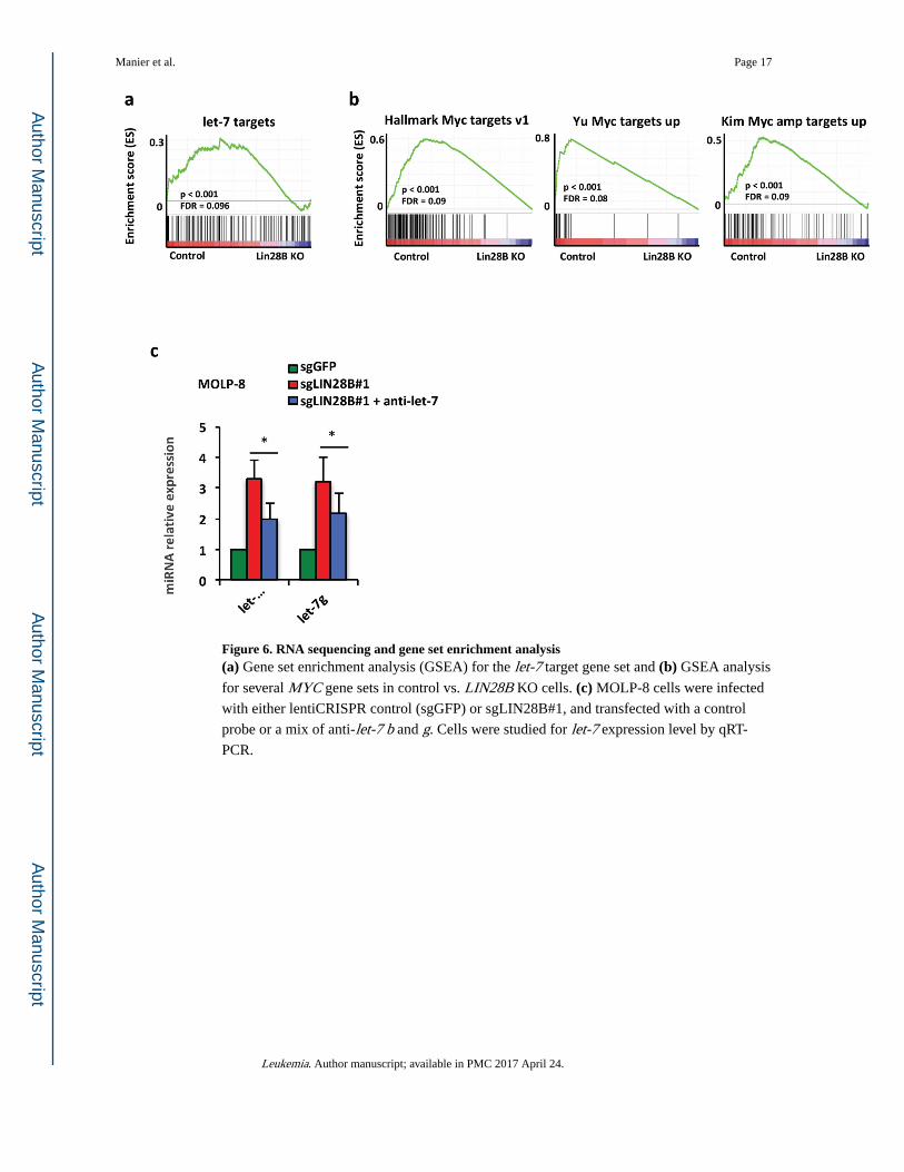
Figure 6. RNA sequencing and gene set enrichment analysis(a) Gene set enrichment analysis (GSEA) for the let-7 target gene set and (b) GSEA analysis
for several MYC gene sets in control vs. LIN28B KO cells. (c) MOLP-8 cells were infected
with either lentiCRISPR control (sgGFP) or sgLIN28B#1, and transfected with a control
probe or a mix of anti-let-7 b and g. Cells were studied for let-7 expression level by qRT-
PCR.
Manier et al. Page 17
Leukemia. Author manuscript; available in PMC 2017 April 24.
Author M
anuscriptA
uthor Manuscript
Author M
anuscriptA
uthor Manuscript
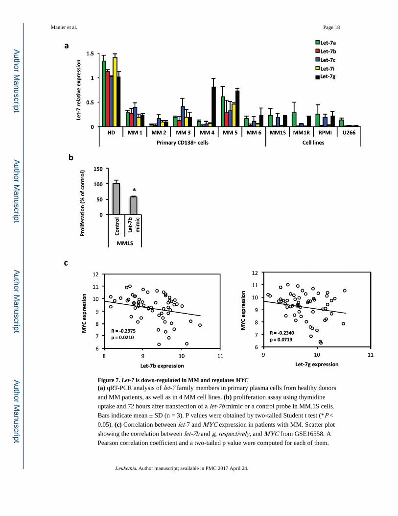
Figure 7. Let-7 is down-regulated in MM and regulates MYC(a) qRT-PCR analysis of let-7 family members in primary plasma cells from healthy donors
and MM patients, as well as in 4 MM cell lines. (b) proliferation assay using thymidine
uptake and 72 hours after transfection of a let-7b mimic or a control probe in MM.1S cells.
Bars indicate mean ± SD (n = 3). P values were obtained by two-tailed Student t test (*P <
0.05). (c) Correlation between let-7 and MYC expression in patients with MM. Scatter plot
showing the correlation between let-7b and g, respectively, and MYC from GSE16558. A
Pearson correlation coefficient and a two-tailed p value were computed for each of them.
Manier et al. Page 18
Leukemia. Author manuscript; available in PMC 2017 April 24.
Author M
anuscriptA
uthor Manuscript
Author M
anuscriptA
uthor Manuscript

Figure 8. Increased expression of let-7 in vivo decreases MM proliferation(a) Mice were followed by bioluminescence intensity (BLI), after injection of 5 million of
MM.1S GFP+Luc+cells. SCID/bg mice (5 per group) were injected i.p. 2 times a week with
20mg/kg of LNA control or LNA let-7 mimic. (b) Survival of the mice by Kaplan Meier
analysis. Average survival time was 35 days in control group versus 42 days in LNA let-7 mimic group, P = 0.0026. (c) qRT-PCR analysis of MYC in MM.1S cells ex vivo. Bars
indicate mean ± SD (n = 3). P values were obtained by two-tailed Student t test (*P < 0.05).
Manier et al. Page 19
Leukemia. Author manuscript; available in PMC 2017 April 24.
Author M
anuscriptA
uthor Manuscript
Author M
anuscriptA
uthor Manuscript