salivary gland tumors in transgenic mice with targeted ... · salivary gland tumors in transgenic...
TRANSCRIPT

Salivary Gland Tumors in Transgenic Mice with Targeted
PLAG1 Proto-Oncogene Overexpression
Jeroen Declercq,1Frederik Van Dyck,
1Caroline V. Braem,
1Isabelle C. Van Valckenborgh,
1
Marianne Voz,1Michel Wassef,
4Luc Schoonjans,
2Boudewijn Van Damme,
3
Laurence Fiette,5and Wim J.M. Van de Ven
1
1Laboratory for Molecular Oncology, Department of Human Genetics, K.U. Leuven and Flanders Interuniversity Institute for Biotechnology;2Thromb-X, n.v., Campus Gasthuisberg; 3Department of Morphology and Molecular Pathology, K.U. Leuven, Leuven, Belgium; 4ServiceCentral de Cytologie and Anatomie pathologique, Hopital Lariboisiere; and 5Unite de Recherche et d’Expertise en Histotechnologieet Pathologie, Institut Pasteur, Paris, France
Abstract
Pleomorphic adenoma gene 1 (PLAG1) proto-oncogene over-expression is implicated in various human neoplasias,including salivary gland pleomorphic adenomas. To furtherassess the oncogenic capacity of PLAG1 , two independentPLAG1 transgenic mouse strains were established, PTMS1 andPTMS2, in which activation of PLAG1 overexpression is Cremediated. Crossbreeding of PTMS1 or PTMS2 mice withMMTV-Cre transgenic mice was done to target PLAG1 over-expression to salivary and mammary glands, in the P1-Mcre/P2-Mcre offspring. With a prevalence of 100% and 6%,respectively, P1-Mcre and P2-Mcre mice developed salivarygland tumors displaying various pleomorphic adenomafeatures. Moreover, histopathologic analysis of salivary glandsof 1-week-old P1-Mcre mice pointed at early tumoral stages inepithelial structures. Malignant characteristics in the salivarygland tumors and frequent lung metastases were found inolder tumor-bearing mice. PLAG1 overexpression was shownin all tumors, including early tumoral stages. The tumorsrevealed an up-regulation of the expression of two distinct,imprinted gene clusters (i.e., Igf2/H19 and Dlk1/Gtl2). With alatency period of about 1 year, 8% of the P2-Mcre micedeveloped mammary gland tumors displaying similar histo-pathologic features as the salivary gland tumors. In conclu-sion, our results establish the strong and apparently directin vivo tumorigenic capacity of PLAG1 and indicate that thetransgenic mice constitute a valuable model for pleomorphicsalivary gland tumorigenesis and potentially for other glandsas well. (Cancer Res 2005; 65(11): 4544-53)
Introduction
Pleomorphic adenoma gene 1 (PLAG1) is a proto-oncogene onhuman chromosome 8q12 whose oncogenic activation is a crucialevent in the formation of pleomorphic adenomas of the salivary
glands (1, 2) and lipoblastomas (3–5). Recently, amplification andoverexpression of PLAG1 was reported for hepatoblastoma,indicating that the PLAG1 gene might also be implicated in themolecular pathogenesis of this childhood neoplasia (6). Altogether,these observations seem to emphasize a more general importanceof the PLAG1 gene in human benign solid tumor development.Recent studies have shown that PLAG1 and PLAGL2 seem also tobe implicated in leukemia (7). Indeed, PLAG1 and PLAGL2expression was increased in 20% of human acute myeloid leukemia(AML) samples, that contain in most cases the CBFB-MYH11 fusiongene (8, 9).PLAG1 encodes for a zinc finger transcription factor and has two
structurally related family members, PLAGL1 (PLAG-like 1) andPLAGL2 (10). The main mechanism of oncogenic activation ofPLAG1 involves recurrent chromosome translocations. This leadsto promoter exchange between PLAG1 , a gene differentiallyexpressed and primarily during fetal development and one of avariety of possible translocation partner genes, that are morebroadly and constitutively expressed. Chromosome breakpointsinvariably occur in the 5V noncoding region of PLAG1 , causing anexchange of regulatory control elements without affecting thecoding sequences of the gene. This process, also called promoterswapping, leads to ectopic expression of PLAG1 in the geneticallyaffected cells (1, 2). PLAG1 activation can also be caused by crypticrearrangements involving the gene region (11). It is assumed thatectopic PLAG1 overexpression causes deregulation of expression ofPLAG1 target genes leading to particular tumors in humans (1, 3).Structurally, the PLAG1 transcription factor contains seven
canonical C2H2 zinc fingers and a serine-rich COOH terminus withtransactivation capacity (10). PLAG1 specifically recognizes abipartite DNA-binding consensus sequence consisting of a coresequence, GRGGC, and a G cluster, GGG, which are separatedby 6 to 8 nucleotides (12). A nuclear localization sequence wasidentified (13) and PLAG1 is post-translationally modified bySUMOylation (14). In recent microarray analyses (15), genes wereidentified that are consistently induced or repressed by PLAG1 ,and these were classified into various functional categories. Amongthe classes of up-regulated PLAG1 target genes, the one encodinggrowth factors was the largest and included the genes for insulin-like growth factor II (IGF-II), cytokine-like factor-1 (CLF-1), bone-derived growth factor (BPGF-1), choriogonadotropin h chain(CGB), vascular endothelial growth factor (VEGF), and placentalgrowth factor (PIGF). From in silico evaluation of their promoterregions, it seemed that a large proportion of them harbor severalcopies of the specific bipartite DNA binding consensus sequence,suggesting that they constitute direct PLAG1 targets. Furthermore,the in silico studies indicated that PLAG1–down-regulated genes
Note: L. Fiette and W.J.M. Van de Ven should both be considered as last authors.M. Voz is currently at the Laboratoire de Biologie et de Genie Genetique, Institut de
Chimie, allee du 16ieme Aout, 4000 Sart-Tilman, Belgium.L. Fiette is currently at the Platform of Veterinary Diagnosis, CMU, 1 rue Michel
Servet, CH 1211, Geneva 4, Switzerland.C.V. Braem, I. Van Valckenborgh, and J. Declercq are aspirant fellows of the ‘‘Fonds
voor Wetenschappelijk Onderzoek Vlaanderen,’’ whereas F. Van Dyck is an aspirantfellow of the ‘‘Instituut voor de aanmoediging van Innovatie door Wetenschap enTechnologie in Vlaanderen’’.
Requests for reprints: Wim J.M. van de Ven, Laboratory for Molecular Oncology,Department of Human Genetics, K.U. Leuven, Herestraat 49, B-3000 Leuven, Belgium.Phone: 32-16-345987; Fax: 32-16-346073; E-mail: [email protected].
I2005 American Association for Cancer Research.
Cancer Res 2005; 65: (11). June 1, 2005 4544 www.aacrjournals.org
Research Article
Research. on June 3, 2020. © 2005 American Association for Cancercancerres.aacrjournals.org Downloaded from

are likely to be regulated indirectly by PLAG1 , because the typicalPLAG1 DNA binding consensus sequences are not present in theircorresponding promoter regions (15).The oncogenic potential of PLAG1 has been established in vitro
by transforming cultured cells via overexpression of PLAG1 (16).The results of these in vitro studies are consistent with the in vivoobservation that increased PLAG1 expression is important in thedevelopment of particular tumors, such as those mentioned before.To explore the role of PLAG1 in neoplastic transformation in thecontext of a complex organism, we have initially generatedtransgenic mice overexpressing PLAG1 early and ubiquitously.6
These studies revealed that such early and ubiquitous over-expression of PLAG1 leads to embryonic lethality. Now, we havedeveloped PLAG1 transgenic mouse strains, in which activation ofoverexpression of the transgene as well as the tissue distributionof such overexpression can be manipulated (e.g., by Cre-mediatedactivation and targeted expression, respectively). Two independentPLAG1 transgenic mouse strains were obtained and used to targetoverexpression of the PLAG1 gene to a restricted number oftissues, including salivary gland and mammary gland tissue.PLAG1 overexpression in the salivary glands leads to tumorformation. These PLAG1-induced salivary gland neoplasias werestudied in detail histopathologically and molecularly. Finally,induction of mammary gland tumors, with similar morphologicfeatures, was also studied.
Materials and Methods
Generation of transgenic mouse strains with Cre-mediated activa-tion of PLAG1 expression. A KpnI/XhoI cDNA fragment, comprising the
complete open reading frame of human PLAG1 followed by the coding
sequences for an HA-tag, was cloned into the EcoRI site of the pCAGGSvector DNA (17) using blunt end ligation (Fig. 1A). At the multiple
cloning site between the CAG promoter and the sequences encoding HA-
tagged PLAG1 , a loxP/PGK-Neo/loxP DNA fragment (a kind gift of
P. Kastner, Institut de Chimie Biologique, Universite Louis Pasteur,Strasbourg, France; ref. 18) was inserted as a Cre-dependent removable
stop cassette (Fig. 1A). This resulted in a DNA construct conditionally
allowing targeted activation of PLAG1 expression. The resulting DNA wasdigested with restriction endonucleases SalI and SfiI, and upon size frac-
tionation by electrophoresis, a 6.0-kb SalI/SfiI DNA fragment (Fig. 1A)
was purified using the QIAquick extraction kit (Qiagen, Chatsworth, CA).
This purified DNA was used for microinjection into the male pronucleusof fertilized eggs of (FVB � FVB) mice (Charles River Laboratories,
Brussels, Belgium). Upon oviductal implantation of these eggs in pseudo-
pregnant foster females, offspring was obtained and these mice were
bred to select PLAG1 transgenic founders. To target PLAG1 expression tothe salivary gland and mammary gland, these founders were crossed
with B6129-Tgn(MMTV-LTR/Cre)1Mam transgenic mice (The Jackson
Laboratories, Bar Harbor, ME; ref. 19).Genotyping by PCR analysis. Genotyping of candidate PLAG1
founders was done by PCR analysis of tail DNA using oligonucleotide
primers POS-1599 (5V-TTCTCAAGCATCGTCATCAT-3V) and h-globin(5V-AAAATTCCAACACACTATTGC-3V) at an annealing temperature of58jC. PCR analysis was also used to evaluate Cre-mediated excision of
the floxed stop cassette from the PLAG1 DNA construct in the transgenic
mouse strains. For that purpose, genomic DNA was isolated from various
tissues of transgenic mice using the DNeasy tissue kit (Qiagen) accordingto the manufacturer’s instructions. PCR analysis to detect Cre-mediated
excision of the floxed stop cassette was done using the following
primers: P1 forward 5V-CTACAGCTCCTGGGCAA-CGTGCTGG-3V or P2forward 5V-GCCTGAAGAACGAGATCAGCAGCC-3V in combination with
P2 reverse 5V-CACCACTTGTTGCGGCATGCAAGGCC-3V. The P1 forward/P2 reverse and P2 forward/P2 reverse set of primers amplified,
respectively, a DNA fragment of 500 or 600 bp. The selected annealing
temperature was 65jC.DNA transfection, SDS-PAGE, and Western blot analysis. Selected
DNAs (1 Ag) were (co)transfected into appropriate mammalian cells using
FuGENE 6 transfection reagent (Boehringer Mannheim, Mannheim,
Germany) according to the protocol of the manufacturer. About 24 hours
after transfection, cells were lysed in SDS-PAGE sample buffer [50 mmol/L
Tris-HCl (pH 6.8), 10% glycerol, 2% SDS, 4% h-mercaptoethanol], boiled,
and loaded on a 10% SDS-polyacrylamide gel. After size separation,
proteins were transferred electrophoretically to PROTEAN nitrocellulose
membranes (Schleicher and Schuell, Keene, NH). Expression of HA-tagged
PLAG1 was detected using mouse anti-HA monoclonal antibody (1:5,000,
Roche, Nutley, NJ). Enhanced chemiluminescence Western blotting was
done using Renaissance Western blotting detection reagents (Perkin-Elmer
Life Sciences, Norwalk, CT; ref. 20).Histopathology, immunohistochemistry, and immunofluorescence.
For histopathology, organs and tumors of mice were removed carefully,
fixed overnight in 4% paraformaldehyde, and embedded in paraffin using
routine procedures. For each tumor, two to three different areas wereselected to analyse histologically. Paraffin sections (5 Am) were stained with
H&E, Alcian Blue (pH 0.2, 1, or 2.5), or periodic acid-Schiff (PAS).
Immunostainings were done on 5 Am paraffin sections according to
standard procedures. Briefly, after antigen retrieval in citrate buffer
(10 mmol/L citric acid, pH 6), the mouse anti-human smooth muscle
actin (SMA; 1:500 DAKO, Carpinteria, CA), and the guinea pig anti-
human cytokeratin (8–18) antibody (1:50, Progen, Heidelberg, Germany)
were used as primary antibodies. Such antibodies are frequently used to
stain myoepithelial and epithelial cells, respectively (21). As a secondary
reagent to detect smooth muscle actin, the avidin-biotin complex system
was used. For detection of cytokeratin 8/18, a secondary rabbit anti-
guinea pig antibody (DAKO) was used, followed by the application of the
anti-rabbit Envision+ kit (DAKO) and development with 3,3V-diamino-
benzidine (Sigma FastTM DAB Tablet Set, Sigma, St. Louis, MO).
Proliferation was assessed in salivary gland tumors by bromodeoxyur-
idine (BrdUrd) incorporation. Briefly, mice received a single i.p. injection
of BrdUrd (cell proliferation labeling reagent, 1 ml/100 g, GE Healthcare,
Roosendaal, the Netherlands) 1 hour before sacrification. Paraffin-
embedded salivary gland samples were subjected to antigen retrieval in
citrate buffer (10 mmol/L citric acid, pH 6) and immunohistochemistry
using anti-BrdUrd-POD Fab (1/100, Roche). Tyramide signal amplification
was obtained with the TSATM Biotin System (Perkin-Elmer Life Sciences)
and STP-POD (1/100, DAKO).
Immunofluorescence analysis was used to assay PLAG1 protein
expression and nuclear staining [4V,6-diamidino-2-phenylindole (DAPI),
blue] in mouse salivary gland tumor tissue of P1-Mcre and P2-Mcre mice.
Five-micrometer cryostat sections were fixed in buffered 4.0% paraformal-
dehyde for 10 minutes, blocked in swine serum for 7 minutes (dilution
1:20), and incubated first with the primary rabbit polyclonal anti-PLAG1
antibody, PEM 190 or PEM195 (ref. 22; dilution 1:60) for 60 minutes at room
temperature and, secondarily, with fluorescein-labeled swine anti-rabbit
antibody (dilution 1:20; TRITC, red, DAKO) for 45 minutes. Sections were
mounted in Citifluor containing 0.5 Ag/AL of DAPI. The slides were finally
analyzed with a Zeiss Axiophot microscope equipped with UV optics (Carl
Zeiss, Inc., Thornwood, NY). Images were recorded with a CE200A CCD
camera (Photometrics, Tucson, AZ).
Northern blot analysis and DNA probes. Total RNA from mousetissues or tumors was isolated according to the method described by
Chomczynski et al. (23). Northern blot analysis was done as described
previously (24). The following probes were radiolabeled with [a-32P]dCTP
(MP Biomedicals, Asse-Relegem, Belgium) using the Megaprime DNALabeling kit (GE Healthcare): for PLAG1 , a 1.5-kb cDNA fragment encom-
passing the complete PLAG1 open reading frame; for Ig f2 , a 674-bp DNA
fragment containing the sequence between nucleotides 1253 and 1927 in
NM_010514; for H19 , a 1.5-kb EcoRI/XbaI DNA fragment of the AA408602cDNA clone; for Dlk1 , a 629-bp PCR fragment containing the sequence6 Unpublished results.
Salivary Gland Tumorigenesis in PLAG1 Transgenic Mice
www.aacrjournals.org 4545 Cancer Res 2005; 65: (11). June 1, 2005
Research. on June 3, 2020. © 2005 American Association for Cancercancerres.aacrjournals.org Downloaded from

between the nucleotides 64 and 1270 of NM_003836.3; for Gtl2 , a PCR
fragment containing the sequence between the nucleotides 284 and 778 of
AJ320506; and for the b-actin gene, a PCR fragment containing thesequence between nucleotides 825 and 1112 of mouse b-actin (X03672).
Results
Generation of two independent PLAG1+/� transgenic mousestrains with PLAG1 expression dependent on Cre-mediatedremoval of a stop cassette. Results of initial experiments togenerate PLAG1 transgenic mice indicated that early andubiquitous expression of the PLAG1 gene leads to embryonic
lethality. Therefore, a PLAG1 DNA construct was designed fromwhich a floxed ‘‘stop cassette’’ had to be removed via Cre-mediated excision to allow PLAG1 expression. The cloningstrategy is described in Materials and Methods and the geneticcomposition of the generated DNA construct is shown in Fig. 1A .The integrity and functionality of the PLAG1 DNA construct wasvalidated in vitro by Western blot analysis of HEK293T cells,transfected with DNA of the PLAG1 construct and this in thepresence or absence of DNA coding for the Cre enzyme. Upontransfection of only DNA representing the PLAG1 construct, thePLAG1 protein could not be detected (Fig. 1B , lane 1). Upon
Figure 1. A, schematic representation of the cloning strategy to obtain the PLAG1 transgene DNA construct used in the generation of the two PLAG1 transgenicmouse strains, PMTS1 and PMTS2. Human PLAG1 cDNA, containing the complete open reading frame (ORF ) for PLAG1 and sequences encoding a hemaglutinin tagat the 5V end of the PLAG1 cDNA, was cloned in the pCAGGS vector, bringing its expression under control of the CMV-IEE-AG promoter. Subsequently, a stopcassette was inserted between the CAG promoter and the PLAG1-HA coding sequences. The stop cassette consists of the Neo gene, which is flanked on bothsides by loxP sites and which is under expression control of the PGK promoter. Expression of PLAG1-HA is therefore dependent on Cre-mediated removal of the stopcassette. The Sal I/Sfi I DNA fragment, shown under ‘‘3’’ was used for zygote injection. CMV-IEE, CMV immediate early enhancer; AG, chicken h-actin/rabbith-globin hybrid promoter; pA, polyadenylation. B, functional validation in vitro cell culture of the PLAG1 transgene DNA construct used in the generation of thetwo PLAG1 transgenic mouse strains. Cre-mediated excision of the stop cassette was tested by Western blot analysis of lysates of 293T cells transfected with thePLAG1 transgene DNA construct shown under ‘‘2’’ in the absence (lane 1 ) or presence (lane 2) of pCAGGS-Cre plasmid DNA. As a postive control,a lysate of cells transfected with CMV-PLAG1-HA plasmid DNA expressing constitutively the PLAG1-HA protein was used. The PLAG1-HA protein was detectedusing a monoclonal antibody with specificity for the HA-tag. C, developing PLAG1�/� embryos and resorbed (R ) embryos, which presumably possess aPLAG1+/� genotype.
Cancer Research
Cancer Res 2005; 65: (11). June 1, 2005 4546 www.aacrjournals.org
Research. on June 3, 2020. © 2005 American Association for Cancercancerres.aacrjournals.org Downloaded from

cotransfection of the PLAG1 DNA and DNA encoding the Creenzyme, readily detectable levels of the PLAG1 protein werepresent (Fig. 1B , lane 2), indicating efficient Cre-mediated removalof the stop cassette.Upon independent zygote injection experiments using the SalI/
SfiI linearized and purified DNA fragment of about 6 kb (Fig. 1A)and subsequent breeding of the offspring, two independenthemizygous PLAG1+/� transgenic mouse strains (PTMS), PTMS1and PTMS2, were obtained. To test the integrity and functionalityof the PLAG1 DNA construct in vivo , PTMS1 and PTMS2 micewere crossed with PGK-Cre+/+ transgenic mice. It is known that thePGK-promoter is already expressed in the two-cell stage duringembryogenesis (25). Therefore, early and widespread PLAG1expression could be expected in the offspring. Intercrossing ofPTMS1 mice with PGK-Cre+/+ transgenic mice did not result in anyPGK-Cre+/�/PLAG1+/� offspring. This was most likely due to earlyembryonic lethality, as can be deduced from the fact that about50% (39 of 70) of the embryos appeared embryonically resorbed(Fig. 1C). The genotype of the resorbed embryos could not bedetermined, however, that of the normally developed embryos(31 of 70) was invariably PGK-Cre+/�/PLAG1�/� . In contrast,intercrossing of PTMS2 mice with PGK-Cre+/+ transgenic miceresulted in litters in which 50% of the mice were PGK-Cre+/�/PLAG1+/�. Although theoretically ubiquitous expression waspredicted for these PGK-Cre+/�/PLAG1+/� mice, PLAG1 expressionseems restricted mainly to the bladder on Northern blot analysis(data not shown). However, discriminating PCR analysis of DNAisolated from various tissues of these PGK-Cre+/�/PLAG1+/� mice,including stomach, small intestine, caecum, colon, heart, seminalvesicle, uterus, kidney, and bladder, clearly established thatexcision of the stop cassette had taken place, at least partially, inall these tissues (data not shown).Targeting stop cassette excision to the salivary glands
results in salivary gland tumor development. In previousin vitro studies, the tumorigenic capacity of PLAG1 was established
(16). The aim of the present studies was to establish whetherPLAG1 is capable to induce salivary gland tumors in mice and alsoto investigate in what way such tumors resemble humanpleomorphic adenomas with PLAG1 overexpression. Therefore,PTMS1 and PTMS2 mice were intercrossed with B6129-Tgn(MMTV-LTR/Cre)1Mam transgenic mice (19) to target stopcassette excision and subsequent PLAG1 expression activation toparticular tissues. This mouse strain was used because theexpression pattern of the Cre enzyme has been well defined andincludes the salivary and the mammary glands (19). Suchintercrossing between PTMS1 and B6129-Tgn(MMTV-LTR/Cre)1-Mam transgenic mice resulted in the MMTV-LTR-Cre+/�/PLAG1+/�
(P1-Mcre) offspring. Within 5 weeks, 100% (37 of 37) of the P1-Mcre mice developed a large tumor mass (Fig. 2A) in the ventralneck region, which upon necropsy always seemed to consist ofbilateral, multifocal salivary gland tumors (Fig. 2B). These tumorsseemed to preferentially originate from submandibulary salivaryglands, but tumors originating from the sublingual salivary glandswere also observed. In contrast, only about 6% (6 of 97) of the P2-Mcre mice developed salivary gland tumors, and all of theseoriginated from the submandibulary salivary glands, aroseunilaterally, and constituted a single tumor mass. Furthermore,the tumors in the P2-Mcre mice became apparent much later(i.e., in most cases only after a latency period of several months).Discriminating PCR analysis of DNA isolated from salivary glandtumors of these P1-Mcre and P2-Mcre mice clearly showed thatexcision of the stop cassette had taken place in all these tumors(data not shown).Activation of expression of the PLAG1 transgene in P1-Mcre
and P2-Mcre mice. To study activation of expression of thePLAG1 transgene after Cre-mediated excision of the stop cassetteresulting from intercrossing PTMS1 or PTMS2 mice with B6129-Tgn(MMTV-LTR/Cre)1Mam transgenic mice, Northern blot anal-ysis was done on RNA from tissues from the offspring using theopen reading frame sequences of the human PLAG1 cDNA as
Figure 2. A, large salivary gland tumormass in a 5-week-old MMTV-LTR -Cre+/�/PLAG1+/� mouse (P1-Mcre).B, macroscopic appearance at necropsy ofthe tumor mass of the 5-week-old P1-Mcremouse shown in (A ). C, Northernblot analysis of RNA isolated from salivarygland tissue of PTMS1 mice (lane 1) orvarious tissues of 5-week-oldP1-Mcre mice (lanes 2-14 ), includingsubmandibular salivary gland tumor(lane 2), mammary gland (lane 3), adrenalgland (lane 4), kidney (lane 5), stomach(lane 6), small intestine (lane 7), colon(lane 8), ovary (lane 9), uterus (lane 10),seminal vesicle (lane 11), testis (lane 12),lung (lane 13), and brain (lane 14).As molecular probes, a 32P-labeled 1.5-kbhuman PLAG1 cDNA probe correspondingto the complete open reading frame ofPLAG1 or a 290-bp b-actin probe wasused. The transcripts of PLAG1 andPLAG1 (arrows ) and their sizes.
Salivary Gland Tumorigenesis in PLAG1 Transgenic Mice
www.aacrjournals.org 4547 Cancer Res 2005; 65: (11). June 1, 2005
Research. on June 3, 2020. © 2005 American Association for Cancercancerres.aacrjournals.org Downloaded from
molecular probe (Fig. 2C). In salivary gland specimens of PTMS1mice (PLAG1+/�), no expression of the transgene could be detectedin line with the inhibition of expression by the stop cassette(Fig. 2C , lane 1). Furthermore, no expression of endogenous Plag1could be detected either (Fig. 2C , lane 1). Analysis of RNAs isolatedfrom a variety of tissue samples of salivary gland tumor-bearingmale and female 5-week-old P1-Mcre mice revealed that expressionof the PLAG1 transgene was very high in the samples of salivarygland tumors (Fig. 2C , lane 2). Furthermore, weak PLAG1expression was found in samples of mammary glands, ovary, andseminal vesicles (Fig. 2C, lanes 3, 9, and 11), and very weakexpression in testis and lung (Fig. 2C , lanes 12 and 13). Thiscorrelates with the expression pattern of the Cre enzyme in theB6129-Tgn(MMTV-LTR/Cre)1Mam transgenic mice as described byWagner et al. (19). Expression of the endogenous mouse Plag1 genewas also detected in some of the tissues tested, includingmammary gland, adrenal gland, kidney, stomach, ovary, testis,lung, and brain but at low and varying levels (Fig. 2C , lanes 3-6 , 9 ,12-14). Similar studies with tissues from salivary gland tumorbearing P2-Mcre mice (data not shown) also revealed high levels ofPLAG1 expression in these tumors.Time course of activation of PLAG1 overexpression. Salivary
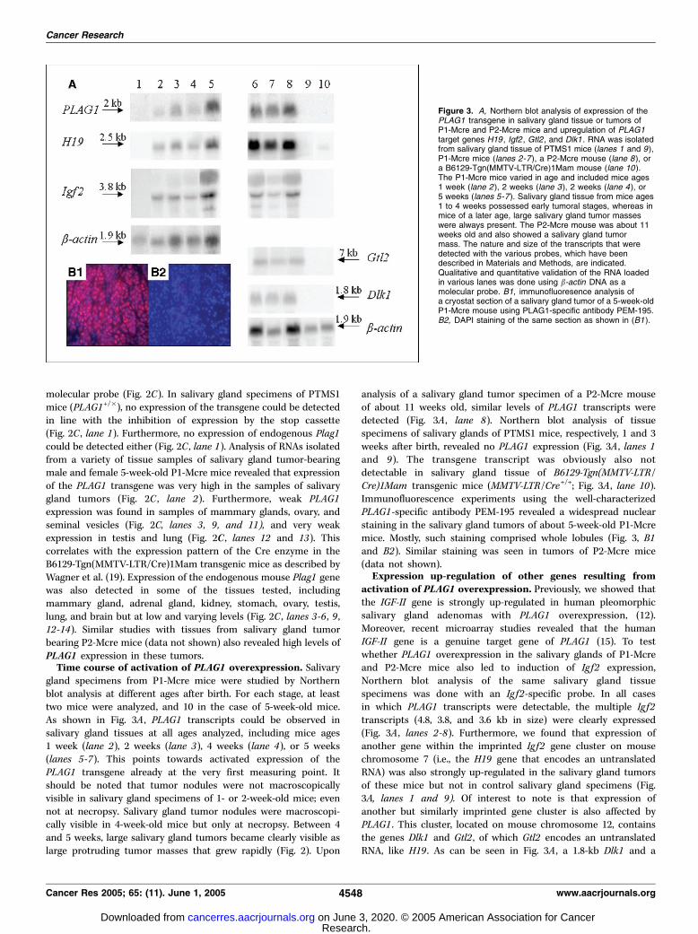
gland specimens from P1-Mcre mice were studied by Northernblot analysis at different ages after birth. For each stage, at leasttwo mice were analyzed, and 10 in the case of 5-week-old mice.As shown in Fig. 3A , PLAG1 transcripts could be observed insalivary gland tissues at all ages analyzed, including mice ages1 week (lane 2), 2 weeks (lane 3), 4 weeks (lane 4), or 5 weeks(lanes 5-7). This points towards activated expression of thePLAG1 transgene already at the very first measuring point. Itshould be noted that tumor nodules were not macroscopicallyvisible in salivary gland specimens of 1- or 2-week-old mice; evennot at necropsy. Salivary gland tumor nodules were macroscopi-cally visible in 4-week-old mice but only at necropsy. Between 4and 5 weeks, large salivary gland tumors became clearly visible aslarge protruding tumor masses that grew rapidly (Fig. 2). Upon
analysis of a salivary gland tumor specimen of a P2-Mcre mouseof about 11 weeks old, similar levels of PLAG1 transcripts weredetected (Fig. 3A , lane 8). Northern blot analysis of tissuespecimens of salivary glands of PTMS1 mice, respectively, 1 and 3weeks after birth, revealed no PLAG1 expression (Fig. 3A , lanes 1and 9). The transgene transcript was obviously also notdetectable in salivary gland tissue of B6129-Tgn(MMTV-LTR/Cre)1Mam transgenic mice (MMTV-LTR/Cre+/+; Fig. 3A , lane 10).Immunofluorescence experiments using the well-characterizedPLAG1-specific antibody PEM-195 revealed a widespread nuclearstaining in the salivary gland tumors of about 5-week-old P1-Mcremice. Mostly, such staining comprised whole lobules (Fig. 3, B1and B2). Similar staining was seen in tumors of P2-Mcre mice(data not shown).Expression up-regulation of other genes resulting from
activation of PLAG1 overexpression. Previously, we showed thatthe IGF-II gene is strongly up-regulated in human pleomorphicsalivary gland adenomas with PLAG1 overexpression, (12).Moreover, recent microarray studies revealed that the humanIGF-II gene is a genuine target gene of PLAG1 (15). To testwhether PLAG1 overexpression in the salivary glands of P1-Mcreand P2-Mcre mice also led to induction of Ig f2 expression,Northern blot analysis of the same salivary gland tissuespecimens was done with an Ig f2-specific probe. In all casesin which PLAG1 transcripts were detectable, the multiple Ig f2transcripts (4.8, 3.8, and 3.6 kb in size) were clearly expressed(Fig. 3A , lanes 2-8). Furthermore, we found that expression ofanother gene within the imprinted Ig f2 gene cluster on mousechromosome 7 (i.e., the H19 gene that encodes an untranslatedRNA) was also strongly up-regulated in the salivary gland tumorsof these mice but not in control salivary gland specimens (Fig.3A, lanes 1 and 9). Of interest to note is that expression ofanother but similarly imprinted gene cluster is also affected byPLAG1 . This cluster, located on mouse chromosome 12, containsthe genes Dlk1 and Gtl2 , of which Gtl2 encodes an untranslatedRNA, like H19 . As can be seen in Fig. 3A , a 1.8-kb Dlk1 and a
Figure 3. A, Northern blot analysis of expression of thePLAG1 transgene in salivary gland tissue or tumors ofP1-Mcre and P2-Mcre mice and upregulation of PLAG1target genes H19 , Igf2 , Gtl2 , and Dlk1 . RNA was isolatedfrom salivary gland tissue of PTMS1 mice (lanes 1 and 9),P1-Mcre mice (lanes 2 -7), a P2-Mcre mouse (lane 8), ora B6129-Tgn(MMTV-LTR/Cre)1Mam mouse (lane 10).The P1-Mcre mice varied in age and included mice ages1 week (lane 2 ), 2 weeks (lane 3), 2 weeks (lane 4), or5 weeks (lanes 5 -7). Salivary gland tissue from mice ages1 to 4 weeks possessed early tumoral stages, whereas inmice of a later age, large salivary gland tumor masseswere always present. The P2-Mcre mouse was about 11weeks old and also showed a salivary gland tumormass. The nature and size of the transcripts that weredetected with the various probes, which have beendescribed in Materials and Methods, are indicated.Qualitative and quantitative validation of the RNA loadedin various lanes was done using b-actin DNA as amolecular probe. B1, immunofluoresence analysis ofa cryostat section of a salivary gland tumor of a 5-week-oldP1-Mcre mouse using PLAG1-specific antibody PEM-195.B2, DAPI staining of the same section as shown in (B1 ).
Cancer Research
Cancer Res 2005; 65: (11). June 1, 2005 4548 www.aacrjournals.org
Research. on June 3, 2020. © 2005 American Association for Cancercancerres.aacrjournals.org Downloaded from

7-kb Gtl2 transcript are expressed in the salivary gland tumors(Fig. 3A , lanes 6-8) but not in the control glands (Fig. 3A , lanes9 and 10). These results clearly establish the up-regulation ofexpression of several genes in two distinct, but structurallysomewhat similar organized, imprinted gene clusters.Early tumoral stages of the salivary glands in P1-Mcre mice.
Submandibulary glands of P1-Mcre mice and those of the wild-typecontrols were examined microscopically at weekly intervalsstarting at 1 week after birth. Histologic analysis of salivary glandsof P1-Mcre mice ages 1 week (one case), 2 weeks (one case), and4 weeks ( four cases) revealed multiple foci most likely representingearly tumoral stages. Most of the lobules show these abnormalities.They contain epithelial cells with a large basal oval nucleus andbasophilic cytoplasm that are densely packed around a smallempty lumen. Dilated ducts with a flat epithelium, sometimesmultilayered, are also visible. Mitotic figures were regularlyobserved (Fig. 4A) and BrdUrd labeling revealed active proliferationin early as well as in later tumoral stages (Fig. 4B).
Pleomorphic adenoma in P1-Mcre and P2-Mcre mice. In P1-Mcre mice, the tumors were clearly visible macroscopically in theventral neck region after 5 weeks. Lobulated tumors were observedbilaterally at necropsy, consisting of a large white, encapsulatedmass. The largest tumors possessed hemorrhagic parts. Similarobservations were made at dissection of salivary gland tumors ofsix P2-Mcre mice (tumor 1-6, respectively, 11, 16, 23, 38, 36, and 70weeks old at sacrifice), but it should be emphasized that thosetumors arose after a much longer latency period (several months)and not bilaterally.Histologically, all salivary gland tumors from the P1-Mcre (12
tumors studied) as well as from the P2-M-Cre mice (six tumors)emerged from the submandibulary gland. The tumors showhistologic features that are reminiscent of those of humanpleomorphic adenomas. A thin capsule was usually visible,sometimes with capsular ingrowth of tumoral lobes. The tumorswere composed of epithelial structures and myoepithelial cells(Fig. 4C). Epithelial components of the tumors showed a high
Figure 4. A, early tumoral stage in a 4-week-old P1-Mcre mouse, submandibulary gland, H&E. Original magnification, �400. Irregular, tubular structures, withsometimes a multilayered epithelium are observed in loose stroma. Epithelial cells lining these structures are irregular, with nuclei of variable size, and loss theirnormal polarity. Mitotic figures are numerous (arrowheads ). B, BrdUrd incorporation into salivary gland tumor cells of a 5-week-old P1-Mcre mouse to visualizeproliferation. Original magnification, �400. C, pleomorphic adenoma features in a salivary gland tumor of a 23-week-old P2-Mcre mouse, H&E. Original magnification,�200. The tumor is composed of epithelial structures (tubes, ribbons ) in a myxoid stroma in which a few spindle or stellate cells are observed (arrows ).D, pleomorphic adenoma features in a salivary gland tumor of an 8-week-old P1-Mcre mouse, H&E. Original magnification, �200. Squamous differentiation with keratinpearls (asterisk).
Salivary Gland Tumorigenesis in PLAG1 Transgenic Mice
www.aacrjournals.org 4549 Cancer Res 2005; 65: (11). June 1, 2005
Research. on June 3, 2020. © 2005 American Association for Cancercancerres.aacrjournals.org Downloaded from

morphologic diversity, because within the same tumor, ductalstructures lined by one or more cellular rows, cysts filled with fluid,papillary patterns, solid sheets of cells, sebaceous differentiation,and frequently squamous areas with keratinization (Fig. 4D) couldbe observed. Spindle and stellate cells, more or less numerous, thathave the morphologic characteristics of myoepitelial cells wereclosely associated with epithelial islands. Immunohistochemicalanalysis showed that the epithelial parts of the tumors werestrongly positive for anti-cytokeratin 8/18 staining, as illustrated inFig. 5A . A high proportion of the spindle or stellate cells werepositive after anti-SMA staining, as illustrated in Fig. 5B . Themyxoid stroma (Fig. 5C) seemed alcianophilic, after Alcian Bluestaining at pH 0.2, 1, and 2.5 (Fig. 5D) and mucus secreting, PAS-positive cells, were numerous. In older tumors of the P1-Mcre mice(8 weeks to 4 months, eight tumors studied) and of the P2-Mcremice (six tumors), malignant features were observed. Theseincluded large areas of necrosis (Fig. 6A), hemorrhages and cellular
polymorphism. Besides some regions with typical characteristics ofpleomorphic adenomas, some of the older tumors of the P1-Mcreand P2-Mcre mice contained regions resembling carcinomas. In 3tumors of the P1-Mcre mice (9, 14, and 15 weeks old) and onetumor of the P2-Mcre mice (23 weeks old), lung metastases wereobserved. These showed the same histologic characteristics of theprimary salivary gland tumor as illustrated in Fig. 6B and C .Mammary gland tumors in P2-Mcre mice. Expression of the
PLAG1 transgene in the P1-Mcre mice was not restricted to thesalivary glands, which is consistent with the Cre expression patternreported for the B6129-Tgn(MMTV-LTR/Cre)1Mam transgenicmice (19). Normal mammary glands of the P2-Mcre mice alsoexpressed PLAG1 transcript levels detectable by Northern blotanalysis (Fig. 2C , lane 3). About 8% of the P2-Mcre mice developedtumors of the mammary gland with a latency period of about1 year. These tumors shared some histologic features with thesalivary gland tumors (Fig. 6D). They were also composed of
Figure 5. A, immunohistochemical staining, using an anti-CK8/18 antibody, of a salivary gland tumor of a 5-week-old P1-Mcre mouse. Original magnification, �200.Most of the tumoral cells in the ductal structures, solid sheets or areas of squamous differentiation are positive for CK8/18. B, immunohistochemical staining, usingan anti-SMA antibody, of a salivary gland tumor in a 34-week-old P2-Mcre mouse. Original magnification, �200. Spindle and stellate cells are positive, as well assome cells underlying the epithelial tumoral acini. C, pleomorphic adenoma features in a salivary gland tumor of a 5-week-old P1-Mcre mouse, H&E. Originalmagnification, �200. The tumor is composed of epithelial structures in a loose stroma. Components show a high morphological diversity, with acinar (arrow ) and ductalstructures, which are empty or filled with fluid (triangles ), lined by one or more cellular rows, or solid sheets of cells (asterisk). D, Alcian Blue staining (pH 2.5) ofa salivary gland tumor of a 2-month-old P1-Mcre mouse. Original magnification, �100. Myxoid stroma surrounding epithelial ducts and cysts is stained pale blue.
Cancer Research
Cancer Res 2005; 65: (11). June 1, 2005 4550 www.aacrjournals.org
Research. on June 3, 2020. © 2005 American Association for Cancercancerres.aacrjournals.org Downloaded from

epithelial and myoepithelial cells in a myxoid stroma. They alsoshowed solid areas of poorly differentiated basophilic epithelialcells, with a high mitotic index. A sebaceous differentiation wasoften seen. Mammary gland tumors were not observed in P1-Mcremice, because these mice had to be sacrificed for ethical reasonsbefore the age of 4 months because of their enormous salivarygland tumors. Nevertheless, some histologic abnormalities werefound in mammary glands of P1-Mcre mice, showing abnormaldifferentiation with sebaceous differentiation, multilayered cellsaround the ducts and cystic structures (data not shown).
Discussion
It is well established that overexpression of the developmentallyregulated PLAG1 gene occurs frequently in various benign humansolid tumors, such as pleomorphic salivary gland adenomas (1, 2),lipoblastomas (3, 4), and also in the childhood neoplasiahepatoblastoma (6). In the first two tumor types, it has beensuggested that overexpression of PLAG1 is directly responsible for
the formation of this type of tumors. This is mainly based on thecausal link between the cytogenetically and molecularly well-defined recurrent chromosome 8q12 aberrations and the invariablystrong up-regulation of PLAG1 expression in such tumors. Directsolid proof, however, is still lacking (1). Recently, PLAG1 andPLAGL2 have also been found implicated in AML (7, 9). Further-more, experiments have clearly established the in vitro oncogenicpotential of PLAG1 (16). Altogether, these findings have demarcat-ed the PLAG1 gene as a versatile proto-oncogene and attractedconsiderably attention to the study of its function. As an approachto assess in vivo the consequences of overexpression of PLAG1 inthe complex genetic background of a mammalian model system,two independent PLAG1 transgenic mouse strains were developedand various features are reported here.Our PLAG1 transgenic mouse strains constitute a valuable model
to study pleomorphic salivary gland tumorigenesis. With the P1-Mcre model, tumor progression can easily be followed in the time.Indeed, already after 1 week, 100% of the salivary glands of thesemice contain early tumoral stages in the ductal region. It should be
Figure 6. A, malignant features in an 8-week-old P1-Mcre mouse salivary gland tumor, H&E. Original magnification, �200. Necrotic area (n). Salivary glandtumor in a 2-month-old P1-Mcre mouse, with fusiform cells. Original magnification, �200. C, lung metastasis of the salivary gland tumor shown in (B), with similarmorphologic features. D, mammary gland tumor in a 34-week-old P2-Mcre mouse, H&E. Original magnification, �100. The tumor shows variable patterns: tubes(arrowheads ) or cysts (c ) that can be filled with an eosinophilic material, ribbons, among a myxoid pale blue stroma (s ). Cells undergoing sebaceous differentiation arefrequent (arrows ).
Salivary Gland Tumorigenesis in PLAG1 Transgenic Mice
www.aacrjournals.org 4551 Cancer Res 2005; 65: (11). June 1, 2005
Research. on June 3, 2020. © 2005 American Association for Cancercancerres.aacrjournals.org Downloaded from

noticed that the expression pattern of the Cre recombinase in thesalivary glands of B6129-Tgn(MMTV-LTR/Cre)1Mam transgenicmice is also limited to these regions (19). Already after 5 weeks, P1-Mcre mice develop pleomorphic adenomas, which share manyhistopathologic features with human pleomorphic adenomas. Likethe human tumors (26), they are mixed tumors containingepithelial and myoepithelial cells. Epithelial elements are highlypolymorphic within the tumor, because a tubular, acinar, cystic,papillary, or solid pattern with frequent squamous differentiationcan occur. Tumors are embedded in a pale myxoid stroma in whichfusiform to stellate myoepithelial SMA-positive cells are seen.Usually, the tumor is encapsulated and epithelial structures are welldifferentiated. In some of the older P1-Mcre and P2-Mcre mice,besides typical features of pleomorphic adenoma also malignantcharacteristics were observed, including areas of necrosis, hemor-rhages, and metastasis to the lungs. Some regions even resembledcarcinomas. Consistently, it has been well described that alsohuman pleomorphic adenomas can progress to malignancy. Inhumans, 2% to 17% of the pleomorphic adenomas can progress tocarcinomas ex pleomorphic adenomas (27–29). Sometimes, thepleomorphic part of the tumor is only very small. The malignantcharacteristics observed in our mouse model suggest a similarmalignant progression.Although the PLAG1-induced salivary gland tumors in the
transgenic mouse strains possess the basic hallmarks of humanpleomorphic adenoma, a major difference with the humantumors pertains to the aggressive growth of the tumors, at leastfor those of the P1-Mcre strain. The engineered geneticintervention and/or the different genetic background of themouse can most likely explain this difference. Human pleomor-phic adenomas of the salivary glands are assumed to arisefollowing a genetic accident, presumably in a single progenitorcell, and leading to a clonal activation of PLAG1 expression longafter birth. In contrast, in the PLAG1 transgenic mouse modelsystem, PLAG1 activation starts already during embryonicdevelopment and occurs multiclonally and at an early phase inthe epithelial duct cells of the developing salivary glands. Wecannot exclude that some of the alterations observed in the earlystages are developmental changes. In any case, a majorphenotypic effect of overexpression of the PLAG1 transgene isthe onset of fast growing salivary gland tumors.There are some differences between our two PLAG1 transgenic
mouse strains. First, in P1-Mcre mice lesions occur bilaterally andin most of the lobules. Moreover, 100% of the mice develop tumors.In contrast, however, in the P2-Mcre strain, the tumor frequency islow and the tumorigenic process has a long latency period (severalmonths). Moreover, such tumors arose invariably locally andunilaterally. Zhao et al. reported a similar variation in tumor
incidence in PLAG1 transgenic mouse strains (30). Second, crossingof the PTMS1 with PGK-cre mice to generate PGK-Cre+/�/Plag1+/�
mice resulted in embryonic lethality, due to early and ubiquitousexpression of the transgene. Embryonic lethality was not observedin similar experiments with PTMS2, probably because there isalmost no expression of the PLAG1 transgene in the offspring. Weassume that the differences in expression of the PLAG1 transgenein the two founder strains are due to the effect of the integrationsite of the transgene (31).Of interest to note, finally, is that after a latency period of
about 1 year, 8% of the P2-Mcre mice developed mammarygland tumors, which possessed histologic features similar tothose observed in the salivary gland tumors. Similar observationswere not made with the P1-Mcre mice, because they developedfast growing salivary gland tumors and had to be sacrificedbefore mammary gland tumors could arise. For this strain, moredirect and specific targeting of expression of the PLAG1transgene to the mammary gland, excluding the salivary gland,seems the strategy to follow. It should be mentioned that PLAG1expression has never been described in primary humanmammary gland tumors.In conclusion, our studies clearly establish first that ubiquitous
overexpression of the PLAG1 proto-oncogene leads to lethalityduring embryonic life. Second, a direct link was shown betweenoverexpression of the PLAG1 proto-oncogene and tumorigenesis.Third, activation of genes from independent imprinted geneclusters (Ig f2 , H19 , Dlk1 , and Gtl2 ; refs. 32–34) may play a rolein this tumorigenic process, although the precise nature of itremains to be established. In human salivary gland pleomorphicadenomas, the IGF-II gene, which is an established importantelement in tumor cell growth (35), has been identified as a genuinetarget gene of PLAG1 (12, 15). On the other hand, it is possible thatthe mechanism of imprinting is involved. Finally, the PTMS1 andPTMS2 strains described here have been designed in such a waythat activation of expression of the PLAG1 transgene is Cremediated. With the availability of mouse strains with differenttissue-specific Cre expression (36), valuable models can now beobtained to study the contribution of PLAG1 to the development ofother types of tumors.
Acknowledgments
Received 11/16/2004; revised 3/18/2005; accepted 3/22/2005.Grant support: Geconcerteerde Onderzoekacties 2002/010, the ‘‘Fonds voor
Wetenschappelijk Onderzoek Vlaanderen’’ grant G.0099.02, and the Cancer ResearchProgram of Fortis Bank Insurance 2002-2005.
The costs of publication of this article were defrayed in part by the payment of pagecharges. This article must therefore be hereby marked advertisement in accordancewith 18 U.S.C. Section 1734 solely to indicate this fact.
We thank C. Van den Broeck for technical assistance.
References1. Kas K, Voz ML, Roijer E, et al. Promoter swappingbetween the genes for a novel zinc finger proteinand h-catenin in pleiomorphic adenomas witht(3;8)(p21;q12) translocations. Nat Genet 1997;15:170–4.
2. Voz ML, Astrom AK, Kas K, et al. The recurrenttranslocation t(5;8)(p13;q12) in pleomorphic adenomasresults in upregulation of PLAG1 gene expressionunder control of the LIFR promoter. Oncogene 1998;16:1409–16.
3. Astrom A, D’Amore ES, Sainati L, et al. Evidence of
involvement of the PLAG1 gene in lipoblastomas. Int JOncol 2000;16:1107–10.
4. Hibbard MK, Kozakewich HP, Dal Cin P, et al. PLAG1fusion oncogenes in lipoblastoma. Cancer Res 2000;60:4869–72.
5. Morerio C, Rapella A, Rosanda C, et al. PLAG1-HAS2fusion in lipoblastoma with masked 8q intrachromoso-mal rearrangement. Cancer Genet Cytogenet 2005;156:183–4.
6. Zatkova A, Rouillard JM, Hartmann W, et al. Amplifi-cation and overexpression of the IGF2 regulator PLAG1in hepatoblastoma. Genes Chromosomes Cancer 2004;39:126–37.
7. Castilla LH, Perrat P, Martinez NJ, et al. Identificationof genes that synergize with Cbfb-MYH11 in thepathogenesis of acute myeloid leukemia. PNAS 2004;101:4924–9.
8. Liu P, Tarle SA, Hajra A, et al. Fusion betweentranscription factor CBF h/PEBP2 h and a myosinheavy chain in acute myeloid leukemia. Science 1993;261:1041–4.
9. Landrette SF, Kuo YH, Hensen K, et al. Plag1 andPlagl2 are oncogenes that induce acute myeloidleukemia in cooperation with Cbfb-MYH11. Blood2005;105:2900–7.
10. Kas K, Voz ML, Hensen K, Meyen E, Van de Ven WJ.
Cancer Research
Cancer Res 2005; 65: (11). June 1, 2005 4552 www.aacrjournals.org
Research. on June 3, 2020. © 2005 American Association for Cancercancerres.aacrjournals.org Downloaded from

Transcriptional activation capacity of the novel PLAGfamily of zinc finger proteins. J Biol Chem 1998;273:23026–32.
11. Astrom AK, Voz ML, Kas K, et al. Conservedmechanism of PLAG1 activation in salivary glandtumors with and without chromosome 8q12 abnormal-ities: identification of SII as a new fusion partner gene.Cancer Res 1999;59:918–23.
12. Voz ML, Agten NS, Van de Ven WJ, Kas K. PLAG1, themain translocation target in pleomorphic adenoma ofthe salivary glands, is a positive regulator of IGF-II.Cancer Res 2000;60:106–13.
13. Braem CV, Kas K, Meyen E, et al. Identification of akaryopherin a 2 recognition site in PLAG1, whichfunctions as a nuclear localization signal. J Biol Chem2002;277:19673–8.
14. Van Dyck F, Delvaux ELD, Van de Ven WJM,Chavez MV. Repression of the transactivating capacityof the oncoprotein PLAG1 by SUMOylation. J BiolChem 2004;279:36121–31.
15. Voz ML, Mathys J, Hensen K, et al. Microarrayscreening for target genes of the proto-oncogenePLAG1. Oncogene 2004;23:179–91.
16. Hensen K, Van Valckenborgh IC, Kas K, Van de VenWJ, Voz ML. The tumorigenic diversity of the threePLAG family members is associated with different DNAbinding capacities. Cancer Res 2002;62:1510–7.
17. Niwa H, Yamamura K, Miyazaki J. Efficient selectionfor high-expression transfectants with a novel eukary-otic vector. Gene 1991;108:193–9.
18. Adra CN, Boer PH, McBurney MW. Cloning andexpression of the mouse pgk-1 gene and the nucleotidesequence of its promoter. Gene 1987;60:65–74.
19. Wagner KU, McAllister K, Ward T, et al. Spatial andtemporal expression of the Cre gene under the control
of the MMTV-LTR in different lines of transgenic mice.Transgenic Res 2001;10:545–53.
20. Seshi B. Cell blotting: techniques for staining andmicroscopical examination of cells blotted on nitrocel-lulose paper. Anal Biochem 1986;157:331–42.
21. Mikaelian I, Blades N, Churchill GA, et al. Proteo-typic classification of spontaneous and transgenicmammary neoplasms. Breast Cancer Res 2004;6:R668–79.
22. Debiec-Rychter M, Van Valckenborgh I, Van denBroeck C, et al. Histologic localization of PLAG1(pleomorphic adenoma gene 1) in pleomorphic adeno-ma of the salivary gland: cytogenetic evidence ofcommon origin of phenotypically diverse cells. LabInvest 2001;81:1289–97.
23. Chomczynski P, Sacchi N. Single-step method ofRNA isolation by acid guanidinium thiocyanate-phenol-chloroform extraction. Anal Biochem 1987;162:156–9.
24. Hensen K, Braem C, Declercq J, et al. Targeteddisruption of the murine Plag1 proto-oncogene causesgrowth retardation and reduced fertility. Dev GrowthDiffer 2004;46:459–70.
25. Lallemand Y, Luria V, Haffner-Krausz R, Lonai P.Maternally expressed PGK-Cre transgene as a tool forearly and uniform activation of the Cre site-specificrecombinase. Transgenic Res 1998;7:105–12.
26. Attie JN, Sciubba JJ. Tumors of major and minorsalivary glands: clinical and pathologic features. CurrProbl Surg 1981;18:65–155.
27. Olsen KD, Lewis JE. Carcinoma ex pleomorphicadenoma: a clinicopathologic review. Head Neck 2001;23:705–12.
28. Mizui T, Ishimaru JI, Miyamoto K, Toida M.Malignant transformation of a gigantic pleomorphic
adenoma of the submandibular gland: a case report.J Oral Maxillofac Surg 2000;58:1422–4.
29. Roijer E, Nordkvist A, Strom A-K, et al. Translocation,deletion/amplification, and expression of HMGIC andMDM2 in a carcinoma ex pleomorphic adenoma. AmJ Pathol 2002;160:433–40.
30. Zhao XD, Yang WJ, Wang L, et al. Development ofsalivary gland tumors in pleomorphic adenoma gene 1transgenic mice. Zhonghua Yi Xue Yi Chuan Xue ZaZhi 2003;20:390–5.
31. Babinet C. Transgenic mice: an irreplaceable toolfor the study of mammalian development and biology.J Am Soc Nephrol 2000;11:S88–94.
32. Takada S, Paulsen M, Tevendale M, et al. Epigeneticanalysis of the Dlk1-Gtl2 imprinted domain on mousechromosome 12: implications for imprinting controlfrom comparison with Igf2-H19. Hum Mol Genet 2002;11:77–86.
33. Takada S, Tevendale M, Baker J, et al. Delta-like andgtl2 are reciprocally expressed, differentially methylatedlinked imprinted genes on mouse chromosome 12. CurrBiol 2000;10:1135–8.
34. Paulsen M, Takada S, Youngson NA, et al. Com-parative sequence analysis of the imprinted Dlk1-Gtl2 locus in three mammalian species reveals highlyconserved genomic elements and refines compari-son with the Igf2-H19 region. Genome Res 2001;11:2085–94.
35. Moorehead RA, Sanchez OH, Baldwin RM, KhokhaR. Transgenic overexpression of IGF-II induces spon-taneous lung tumors: a model for human lungadenocarcinoma. Oncogene 2003;22:853–7.
36. Jaisser F. Inducible gene expression and gene modi-fication in transgenic mice. J Am Soc Nephrol 2000;11:S95–100.
Salivary Gland Tumorigenesis in PLAG1 Transgenic Mice
www.aacrjournals.org 4553 Cancer Res 2005; 65: (11). June 1, 2005
Research. on June 3, 2020. © 2005 American Association for Cancercancerres.aacrjournals.org Downloaded from

2005;65:4544-4553. Cancer Res Jeroen Declercq, Frederik Van Dyck, Caroline V. Braem, et al.
Proto-Oncogene OverexpressionPLAG1Salivary Gland Tumors in Transgenic Mice with Targeted
Updated version
http://cancerres.aacrjournals.org/content/65/11/4544
Access the most recent version of this article at:
Cited articles
http://cancerres.aacrjournals.org/content/65/11/4544.full#ref-list-1
This article cites 36 articles, 13 of which you can access for free at:
Citing articles
http://cancerres.aacrjournals.org/content/65/11/4544.full#related-urls
This article has been cited by 7 HighWire-hosted articles. Access the articles at:
E-mail alerts related to this article or journal.Sign up to receive free email-alerts
Subscriptions
Reprints and
To order reprints of this article or to subscribe to the journal, contact the AACR Publications
Permissions
Rightslink site. (CCC)Click on "Request Permissions" which will take you to the Copyright Clearance Center's
.http://cancerres.aacrjournals.org/content/65/11/4544To request permission to re-use all or part of this article, use this link
Research. on June 3, 2020. © 2005 American Association for Cancercancerres.aacrjournals.org Downloaded from