safety assessment of oral administration of ethanol
TRANSCRIPT

ORIGINAL CONTRIBUTION Open Access
Safety assessment of oral administration ofethanol extract of Justicia carnea leaf inhealthy wistar rats: hematology,antioxidative and histology studiesEmmanuel Sina Akintimehin* , Kayode Olayele Karigidi, Tope Samuel Omogunwa and Foluso Olutope Adetuyi
Abstract
Background: Consumption of medicinal plants has diverse therapeutic benefits and could also have toxic effect.Justicia carnea is a medicinal plant that is used conventionally as blood tonic from time immemorial in Nigeria. Theaim of this study is to evaluate the safety of ethanol extract of J. carnea leaf assessing the hematology indices,organ antioxidant system and histology in healthy male wistar rats.
Methods: Powdered sample was extracted using absolute ethanol and concentrated to obtain a slurry paste of J.carnea ethanol extracts. Acute toxicity was determined in two phases using Lorke method. In subacute study, ratswere randomized into six groups of five rats per group: Group 1 (control) received distilled water, group 2, 3, 4, 5, 6received 50, 100, 500, 800 and 1200 mg/kg body weight of J. carnea ethanol extract once daily using oral gavage.At the end of 14th day of administration, rats were allowed to fast overnight, sacrificed to collect samples forbiochemical analysis and histopathological examination.
Results: The LD50 of extract was greater than 5000 mg/kg body weight. Higher doses (> 500 mg/kg) of extractsignificantly (p < 0.05) increased RBC, hemoglobin and platelet compared to the control. Liver superoxide dismutase(SOD) activity was significantly (p < 0.05) increased at 1200 mg/kg while other tested doses caused no detrimentaleffect on glutathione, catalase, SOD and malondialdehyde level in liver and kidney. Histopathological examinationof liver and kidney showed mild to severe pathological lesion in a dose dependent manner.
Conclusions: The results of this study suggests that ethanol extract of J. carnea leaf is relatively safe, could bebeneficial in alleviating hematology related abnormalities without causing adverse effects on endogenousantioxidant system. However, caution should be taken as higher dose at 1200 mg/kg could cause noticeable tissueinjury.
Keywords: Blood tonic, Sub – acute, Antioxidant system, Hematology, Histopathological
© The Author(s). 2021 Open Access This article is licensed under a Creative Commons Attribution 4.0 International License,which permits use, sharing, adaptation, distribution and reproduction in any medium or format, as long as you giveappropriate credit to the original author(s) and the source, provide a link to the Creative Commons licence, and indicate ifchanges were made. The images or other third party material in this article are included in the article's Creative Commonslicence, unless indicated otherwise in a credit line to the material. If material is not included in the article's Creative Commonslicence and your intended use is not permitted by statutory regulation or exceeds the permitted use, you will need to obtainpermission directly from the copyright holder. To view a copy of this licence, visit http://creativecommons.org/licenses/by/4.0/.
* Correspondence: [email protected] Unit, Department of Chemical Sciences, Olusegun AgaguUniversity of Science and Technology, Okitipupa, Nigeria
Akintimehin et al. Clinical Phytoscience (2021) 7:2 https://doi.org/10.1186/s40816-020-00234-4

BackgroundIn recent time, copious attention has been focused on theuse of medicinal plants in the management and treatmentof ailments such as anemia, diabetes and malaria. Due tothe local availability, easy access and relatively low cost,medicinal plants are gaining attentions in health careprogrammes. Based on estimation by World HealthOrganization (WHO), larger percentage (between 80 and90%) of the world’s population especially in developingcountries depends on traditional system of medicine [1, 2].Despite the therapeutic importance of medicinal plants,toxic substances have been shown to be present in largenumbers of plants investigated [3]. Contamination of medi-cinal plant could be as a result of contaminants (such asheavy metals, aflatoxin and pathogenic microbes) from soiland manner of herbal preparations [4].Consumption of medicinal plants without scrutinizing
its efficacy and safety can result in unexpected toxiceffects resulting in physiology changes of differentorgans in the body. Liver and kidney are the maintargets of toxicants because they are respectivelyinvolved in biotransformation and excretion of xenobi-otics. Hepatic and renal damage has been recently linkedwith the use of medicinal plants in the treatment ofvarious diseases [5, 6].Justicia carnea (belonging to Acanthaceae family) is a
flowering plant consisting ~ 600 species that are widelydistributed in the tropics and subtropics [7]. In variousparts of Africa, several species of Justicia are used intraditional medicine for the treatment of anemia, inflam-mation, fever, diarrhea, liver diseases and arthritis re-spiratory and gastrointestinal disorder [8, 9]. Recently, ithas also been reported that the species possess cardio-protective properties, antioxidant and are generally richin vitamins and minerals [10–12].In Nigeria, the leaf of Justicia carnea is usually pre-
pared with edible vegetables to make soup, boiled separ-ately in water to make tea or prepared by cooking withother medicinal plants for therapeutic purposes. Despitethe avalanche use of medicinal plant, preliminary toxicitystudies remain essential tools to ensure safe consump-tion and prevent unexpected toxicity that could arisefrom long term exposure. The aim of this study is toevaluate the effects of J. carnea leaf extract onhematology indices and organ antioxidant system inhealthy wistar rats.
MethodsPlant sampleFresh leaves of Justicia carnea were collected fromOkitipupa area Ondo State. The fresh leaves were thor-oughly washed under running tap, air dried under shade,pulverized and extracted with ethanol. Extraction wascarried out by maceration (72 h) following the method
of Onoagbe et al., [13]. Powdered sample (1000 g) wassoaked in absolute ethanol (3000 mL) with constantstirring. After 72 h of soaking, the wine (dark purplishred) colored filtrate was filtered using a double cheesecloth, concentrated under reduced pressure using rotaryevaporator and subsequently water bath at 40 °C toobtain a slurry (dark purple) extract termed Justiciacarnea ethanol extract (JCEE).
Experimental animalsA total number of forty two (42) male albino rats weigh-ing between 120 and 170 g were used in this study.These animals were obtained from the animal house ofthe Department of Chemical Sciences, Ladoke AkintolaUniversity of Technology Ogbomoso Oyo State, Nigeria.The animals were randomly distributed into cages andallowed to acclimatize for 2 weeks in a well ventilatedroom under natural lighting condition. The animalswere allowed free access to rat chow and water ad libi-tum. Treatments of experimental animals were in ac-cordance with the Principle of Laboratory Animal Caremanual guide of National Institute of Health [14] as ap-proved by the Institution Research Ethics committee.
Toxicity studyOral acute toxicity study on J. carnea ethanol extract(JCEE) was carried out according to Lorke [15] methodusing the total of twelve (12) albino rats in two phases.In phase I, rats were randomized into 3 groups of 3 ratsper group. Rats in the 3 different groups received 10,100 and 1000 mg/kg body weight of JCEE orally usingoral gavage. The animals were initially examined for anysigns of toxicity after 60 min of administration and fur-ther observed for a period of 24 h. The absence of ratmortality in phase I necessitated the conduct of the sec-ond phase. In phase two, three rats were separated into3 groups of one rat per group. Each of the rats received1500, 2900 and 5000mg/kg body weight of JCEE re-spectively and examined within 24 h for manifestation oftoxicity. Rats were further observed for extended hoursof 48 h to see if mortality would occur. In sub-acutetoxicity study, total of thirty (30) rats were randomizedinto six groups of five rats per group: Group 1 served ascontrol and was administered with 1 ml of distilled water(dH2O). Group 2, 3, 4, 5, 6 were respectively adminis-tered orally with 50, 100, 500, 800 and 1200 mg/kgbody weight of JCEE once daily for 14 days using anoral gavage. Rats were routinely observed for signs oftoxicity.
Experiment termination and biochemical analysisAt the end of the 14th day of administration, the ratswere allowed to fast overnight and sacrificed the follow-ing morning by anesthetizing each rat in a closed
Akintimehin et al. Clinical Phytoscience (2021) 7:2 Page 2 of 9

chamber containing drops of chloroform. Each rat wasdissected and blood samples were collected by cardiacpuncture using 5 mL into tubes containing ethylenedi-aminetetraacetic acid (EDTA) for hematological analysis.The liver and kidneys were excised and rinsed immedi-ately in a normal saline (0.9% Nacl) to remove bloodstain, dried between layers of Whatman filter paper,weighed and homogenized to obtain the supernatantfor biochemical analysis. Section of livers and kidneyswere excised and fixed in 10% formalin for histopath-ology examination.
Hematological analysisThe haematological indices of the blood samples weredetermined using an automated URIT – 2900 Plus 3Differential Haematological Analyzer. Parameter thatwere determined include total haemoglobin concentra-tion (HGB), packed cell volume (PCV), red blood cellcount (RBC), mean corpuscular haemoglobin (MCH),mean corpuscular haemoglobin concentration (MCHC),mean corpuscular volume (MCV), white blood cellcount (WBC), Lymphocyte count (LYM), MID – sizedcell (MID), Granulocyte Count (GRM) and plateletcount (PLT).
Total proteinTotal protein concentration in tissue homogenates weredetermined using biuret method Tietz [16]. Theprinciple is based on the formation of coloured complexwhen protein peptide interacts with cupric ions in an al-kaline medium. Sample (or standard solution) (0.01 ml)was mixed with diluted (1:4 v/v distilled water) biuret re-agent (100 mM sodium hydroxide, 16 mM sodium-potassium tartrate, 15 mM potassium iodide and 6mMcopper sulphate). The mixture was incubated at 28 °Cfor 30 min and the absorbance read at 550 nm againstthe reagent blank.
Total protein ¼ Absorbance of sampleAbsorbance of standard
� Concentration of standard
Oxidative stress markersCatalase activityCatalase activity (U/ml) was measured following themethod of Sinha [17]. The principle is based on the reduc-tion of dichromate in a weak acid medium (acetic acid) tochromic acetate which is further heated in the presence ofhydrogen peroxide to form an unstable intermediate (per-chromic acid) that can be monitored spectrophotometric-ally at 620 nm. In a reaction mixture containing 0.01Mphosphate buffer (pH 7.0), 0.5 ml of 0.2M H2O2 and 0.4ml distilled water, 0.5 ml of sample was added. The
reaction is terminated by the addition of 2 ml of dichro-moacetic acid mixture (potassium dichromate (5%) andglacial acetic acid; 1:3 v/v) and heated at 60 °C for 10min.Absorbance reading was taken at 620 nm regularly at aninterval of 1 min against reagent blank. The catalase activ-ity was calculated using the expression:
Catalase activity U=mlð Þ ¼ ΔA= min � TVε � SV � TP
Note: ΔA = change in absorbance, TV = total volume,TP = total protein, SV = sample volume, ɛ =molarextinction = 40M− 1 cm− 1.
Superoxide dismutase (SOD) activitySuperoxide dismutase was carried out using the methodof Sun and Zigma [18]. SOD activity was determined byits ability to inhibit the auto-oxidation of epinephrinedetermined by the increase in absorbance at 480 nm.Total reaction mixture of 3 ml contained 2.95 ml of 0.05M sodium carbonate buffer (pH 10.2), 0.02 ml tissuesample and 0.03ml of epinephrine (2 mM) in 0.005 NHCl was used to initiate the reaction. The referencecuvette contained 2.95 ml buffer, 0.03 ml of substrate(epinephrine) and 0.02 ml of water. The absorbance wastaken at interval of 1 min for 3 min at 480 nm.
SOD activity U=ml=mg proteinð Þ¼ ΔA= min � TV
ε � SV � TP
Note: ΔA = change in absorbance, TV = total volume,SV = sample volume, ɛ =molar extinction = 4020M− 1
cm− 1, TP = total protein.
Reduced glutathione (GSH)The concentration of reduced glutathione content of thetissues was estimated according to Sedlak and Lindsay[19] method. Exactly 1.0 ml of tissues (liver and kidney)was deproteinised using 0.1 ml of 10% TCA and centri-fuged at 650×g for 5 min to obtain supernatant. Further,0.5 ml of supernatant was treated with 0.5 ml of Ellmansreagent (containing 19.8 mg of 5′,5′ – dithiobis - (2-nitrobenzoic acid) (DTNB) in 100 ml of 0.1% sodium ni-trate) and 3.0 ml of phosphate buffer (0.2 M, pH 8.0).The absorbance was read at 412 nm against the reagentblank.
Concentration of reduced GSH ¼ ðΔA � TVÞðε � SV � TPÞ
Note: ΔA = change in absorbance, TV = total volume,SV = sample volume, TP = total protein, ɛ =molarextinction =1.34 × 10⁴M− 1 cm− 1.
Akintimehin et al. Clinical Phytoscience (2021) 7:2 Page 3 of 9

MalondialdehydeLipid peroxidation was determined by measuring theformation of thiobarbituric acid reactive substances(TBARS) according to the method described by Buegeand Aust [20]. Briefly, 1.0 ml of the sample was mixedwith 2 ml of TCA-TBA-HCl reagent (0.37% thiobarbitu-ric acid, 15% trichloroacetic acid and 0.24 N HCl in 1:1:1) and boiled at 100 °C in a water bath for 15 min. Themixture was allowed to cool and centrifuged at 650×gfor 10 min to obtain the supernatant. The absorbance ofthe supernatant was measured against reagent blank at532 nm.
Malondialdehyde ¼ A � TVð Þðε � SV � TP
Extinction coefficient (ε) of MDA – TBA complex =1.56 × 10⁵M− 1 cm− 1.Where, A = absorbance, TV = total volume, SV = sam-
ple volume, ɛ = extinction coefficient, TP = total protein.
Histopathological examinationThe liver and kidneys excised from all the experimentalrats were fixed in a labeled sample bottles containing10% formalin and processed for histological examin-ation. Tissues were processed automatically using auto-matic tissue processor (Leica TP 1020) and allowed topass through varying percentage of alcohol and absolutealcohol for the purpose of dehydration following dewax-ing in Xylene for 15 mins. Tissues were stained withhaematoxylin and eosin, mounted on glass slides andexamined under a standard light microscope.
Statistical analysisData were analyzed using one-way analysis of variance(ANOVA) using Statistical Product and Service Solu-tions (SPSS) version 17.0 and results were expressed asmean ± SEM. Differences between means were consid-ered to be significant at (p < 0.05) using LSD (LeastSquare Difference) post hoc test.
ResultsAcute toxicity resultResults obtained from the acute oral toxicity studyrevealed tolerance of experimental rat to acute adminis-tration of J. carnea ethanol extracts since no mortality
and signs of toxicity were observed after 24 h (Table 1).Mean lethal dose of the extract was found to be above5000 mg/kg body weight (bw).
Organ weightsThe weight of liver and kidney of rats were measuredimmediately after excising the organs and is presented inTable 2. Rats that received 50 and 100 mg/kg bodyweight JCEE showed significant (p < 0.05) increase inliver weight while other doses showed no significant dif-ference compared to the control. Kidney weight of ratstreated with JCEE showed no significant difference withexception in 800 mg/kg JCEE that demonstrated signifi-cant reduction.
Hematology indicesThe result of hematology indices is presented in Table 3.Ethanol extracts of J. carnea leaf increased RBC,hemoglobin and platelet with significant increase(p < 0.05) in groups that received higher dose (> 500 mg/kg b.w) of extract compared to the control. WBC re-duced in rats treated with doses of J. carnea with signifi-cant decrease (p < 0.05) in rats that received 50, 100 and1200 mg/kg body weight. Relative to control, lymphocyteand granulocyte exhibited no significant difference in alldoses while MID was elevated in treated groups withsignificant increase in rats administered with 500 and800 mg/kg body weight. Packed cell volume (PCV) andmean corpuscular volume (MCV) in treated rats withJCEE were statistically unperturbed compared to thecontrol. Mean corpuscular hemoglobin (MCH) andmean corpuscular hemoglobin concentration (MCHC)showed significant (p < 0.05) increased at 50 mg/kgwhile doses greater than 100 mg/kg showed no signifi-cant difference compared to the control.
Total protein and antioxidant system in organsThe effects of ethanol extract of J. carnea leaf on totalprotein concentration and antioxidant system in liver
Table 1 Acute oral toxicity of ethanol extracts of J. carnea
Extract Phase I Phase II
Dose (mg/kg bw.) Mortality Dose (mg/kg bw.) Mortality
JCEE 10 0/3 1600 0/1
100 0/3 2900 0/1
1000 0/3 5000 0/1
Table 2 Effects of J. carnea leaf ethanol extract on the weightof liver and kidney
Group Weight of Organs (grams)
Liver Kidney
Control (1 ml dH2O) 5.47 ± 0.21 1.19 ± 0.06
50 (mg/kg) 8.18 ± 0.25* 1.30 ± 0.05
100 (mg/kg) 7.05 ± 0.30* 1.22 ± 0.01
500 (mg/kg) 6.10 ± 0.46 1.10 ± 0.04
800 (mg/kg) 5.95 ± 0.69 0.85 ± 0.06*
1200 (mg/kg) 4.37 ± 0.33 0.88 ± 0.05
Data are presented as mean ± SEM, n = 5 determinations. Values with asterisk(*) in the same column indicate significant difference (p < 0.05) comparedto control
Akintimehin et al. Clinical Phytoscience (2021) 7:2 Page 4 of 9

and kidney of normal wistar rat is presented in Table 4.Compared to the control, total protein in liver and kid-ney were statistically not altered except rats that wereadministered with 1200mg/kg JCEE that showed signifi-cant (p < 0.05) decrease in the liver. Except the groupthat received 1200 mg/kg JCEE which showed significant(p < 0.05) elevation of SOD and MDA in the liver, otherdoses (< 1200 mg/kg) demonstrated no significant differ-ence compared to the control. In the kidney, SOD andMDA level were indistinguishable compared to the con-trol except SOD level in rats that received 800 mg/kgJCEE that reduced significantly (p < 0.05). While gradeddoses of J. carnea ethanol extract showed no significantdifference in the liver and kidney GSH compared to thecontrol, only rats that received 1200 mg/kg body weight
JCEE statistically decreased in the kidney. Liver andkidney in all treated groups maintained similar activitiesof catalase (CAT) compared to the control.
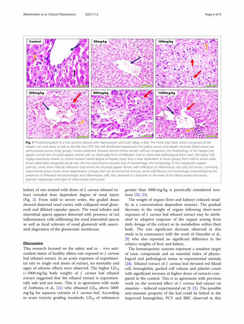
Histopathological studiesPhotomicrograph of liver sections stained with haema-toxylin and eosin (H & E) are presented in Fig. 1. Rela-tive to the control, liver of rats treated with doses of J.carnea ethanol extract showed varied degree of hepaticinjury in a dose dependent manner. With exception oflow doses that showed normal morphology of the hep-atocyte and sinusoid that is not infiltrated, high doses ofextract greater than 500 mg/kg showed mild fat conges-tion in the central venules with liver parenchyma show-ing area of hemorrhage compared to the control. The
Table 3 Effects of ethanol extracts of J. carnea leaf on hematology parameters
Group Control 50mg/kg 100mg/kg 500mg/kg 800mg/kg 1200mg/kg
Parameters
WBC (*109/L) 22.35 ± 2.34 16.17 ± 1.90* 13.20 ± 1.91* 17.55 ± 1.18 20.10 ± 1.10 15.65 ± 1.76*
LYM (%) 82.85 ± 3.84 78.25 ± 2.45 85.10 ± 1.56 72.65 ± 6.90 73.5 ± 1.44 83.75 ± 1.41
MID (%) 10.10 ± 1.59 10.40 ± 0.12 10.65 ± 1.47 15.35 ± 2.92* 17.5 ± 1.67* 9.75 ± 0.10
GRAN (%) 7.10 ± 2.25 11.35 ± 1.30 4.25 ± 0.09 12.00 ± 3.98 9.00 ± 0.23 6.50 ± 1.33
RBC (*1012/L) 5.42 ± 1.27 4.46 ± 2.19 5.32 ± 1.16 7.81 ± 0.25* 8.00 ± 0.36* 7.50 ± 0.06*
HGB (g/dL) 13.03 ± 0.72 13.60 ± 1.04 14.00 ± 0.23 14.05 ± 0.95 14.20 ± 0.52* 13.95 ± 0.66
PLT (*109/L) 422.50 ± 33.20 532.67 ± 74.19 689.50 ± 1.44* 719.00 ± 95.84* 637.00 ± 54.27* 630.00 ± 56.58*
PCV (%) 34.20 ± 5.83 37.00 ± 12.47 34.95 ± 5.17 43.70 ± 1.21 45.60 ± 1.15 43.70 ± 1.39
MCV (fL) 66.55 ± 4.88 70.45 ± 6.61 69.40 ± 5.37 56.10 ± 0.23 56.70 ± 1.44 56.25 ± 1.07
MCH (pg) 27.10 ± 5.14 95.77 ± 6.22* 31.00 ± 7.22 17.90 ± 0.64 17.70 ± 0.17 17.90 ± 0.64
MCHC (g/dL) 39.75 ± 4.82 104.05 ± 10.70* 43.10 ± 7.04 32.00 ± 1.27 31.40 ± 0.52 31.80 ± 0.52
Data are presented as mean ± SEM in n = 5 determinations. Values with asterisk (*) between groups indicate significant difference (p < 0.05) compared to controlWBC white blood cell count: LYM lymphocyte count: MID Mid – sized cell: GRAN granulocyte Count: RBC red blood cell count: HGB hemoglobin: PLT platelet count:PCV packed cell volume: MCV mean corpuscular volume: MCH mean corpuscular haemoglobin and MCHC mean corpuscular haemoglobin concentration
Table 4 Effects of JCEE on protein and antioxidant system in liver and kidney of healthy wistar rats
Groups Organ Total protein(g/l)
GSH (μmol/gof protein)
CAT (U/gof protein)
SOD (U/gof protein)
MDA (μmol/g of protein)× 103
Control (1 ml dH2O) Liver 50.86 ± 3.99 0.87 ± 0.11 3.00 ± 0.10 1.97 ± 1.08 29.81 ± 4.84
Kidney 61.49 ± 19.91 1.09 ± 0.37 3.08 ± 1.24 4.25 ± 0.58 16.19 ± 3.76
50 (mg/kg) Liver 50.52 ± 8.74 0.84 ± 0.06 3.57 ± 1.74 2.36 ± 0.74 26.15 ± 2.36
Kidney 52.85 ± 8.59 0.71 ± 0.14 3.98 ± 1.07 3.21 ± 0.44 20.01 ± 3.03
100 (mg/kg) Liver 49.53 ± 2.72 0.48 ± 0.09 1.91 ± 0.95 2.51 ± 1.42 27.66 ± 10.49
Kidney 49.86 ± 4.35 0.61 ± 0.09 3.57 ± 0.14 1.44 ± 0.52 15.17 ± 4.05
500 (mg/kg) Liver 43.54 ± 3.32 0.68 ± 0.19 3.37 ± 0.83 3.79 ± 0.59 42.85 ± 5.49
Kidney 49.19 ± 1.33 0.99 ± 0.17 3.10 ± 0.58 4.56 ± 1.62 19.05 ± 8.45
800 (mg/kg) Liver 42.89 ± 2.64 0.78 ± 0.08 2.93 ± 0.13 3.53 ± 1.29 43.70 ± 4.28
Kidney 50.19 ± 2.66 0.85 ± 0.16 3.83 ± 0.27 1.00 ± 0.35* 23.01 ± 8.86
1200 (mg/kg) Liver 38.22 ± 3.52* 0.86 ± 0.19 3.65 ± 0.81 5.38 ± 2.06* 48.98 ± 5.66 *
Kidney 52.85 ± 3. 50 0.48 ± 0.02* 3.80 ± 0.47 3.97 ± 3.56 23.35 ± 8.33
Data are presented as mean ± SEM: n = 5 determinations. Values with asterisk (*) in the same column indicate significant difference (p < 0.05) compared to control
Akintimehin et al. Clinical Phytoscience (2021) 7:2 Page 5 of 9
kidney of rats treated with doses of J. carnea ethanol ex-tract revealed dose dependent degree of renal injury(Fig. 2). From mild to severe order, the graded dosesshowed distorted renal cortex with collapsed renal glom-eruli and dillated capsular spaces. The renal tubules andinterstitial spaces appears distorted with presence of redinflammatory cells infiltrating the renal interstitial spacesas well as focal sclerosis of renal glomeruli with associ-ated degeration of the glomerular membrane.
DiscussionThis research focused on the safety and in – vivo anti-oxidant status of healthy albino rats exposed to J. carnealeaf ethanol extract. In an acute exposure of experimen-tal rats to single oral doses of extract, no mortality andsigns of adverse effects were observed. The higher LD50
(> 5000 mg/kg body weight) of J. carnea leaf ethanolextract suggested that the ethanol extract is experimen-tally safe and not toxic. This is in agreement with studyof Anthonia et al., [21] who obtained LD50 above 5000mg/kg for aqueous extracts of J. carnea leaf. Accordingto acute toxicity grading standards, LD50 of substances
greater than 5000 mg/kg is practically considered non-toxic [22, 23].The weight of organs (liver and kidney) reduced stead-
ily in a concentration dependent manner. The gradualdecrease in the weight of organs following short-termexposure of J. carnea leaf ethanol extract may be attrib-uted to adaptive response of the organs arising fromdaily dosage of the extract or its metabolites within theirbody. The non significant decrease observed in thisstudy is in consonance with the work of Onyeabo et al.,[9] who also reported no significant difference in therelative weights of liver and kidney.The hematopoietic systems represent a sensitive target
of toxic compounds and an essential index of physio-logical and pathological status in experimental animals[24]. Ethanol extract of J. carnea leaf elevated red bloodcell, hemoglobin, packed cell volume and platelet countwith significant increase at higher doses of extracts com-pared to the control. This is in agreement with previouswork on the reversed effect of J. carnea leaf extract onanaemia – induced experimental rat [9, 21]. The possibleanti-anaemic property of the leaf could be linked to theimproved hemoglobin, PCV and RBC observed in this
Fig. 1 Photomicrograph of a liver sections stained with Haematoxylin and Eosin (Mag. × 400). The Portal triad (blue arrow) composed of thehepatic vein and artery as well as the bile duct (PT), the well distributed hepatocytes (H) (yellow arrow) and hepatic sinusoids (black arrow) aredemonstrated across study groups. Control treatment showed normal central venules without congestion, the morphology of the hepatocytesappear normal, the sinusoids appear normal with no observable form of infiltration and no observable pathological lesion seen. 50 mg/kg-1200mg/kg treatments relative to control showed varied degree of hepatic injury that is dose dependent. In these groups, from mild to severe order;shows observable congested portal vein, the liver parenchyma showed area of harmorrhage, the morphology of the hepatocytes appearpyknotic, some show mild fat infiltration (red arrow) the sinusoids appear fibrotic with infiltration of inflammatory red cells (red arrow). Summarily,experimental group shows some degenerative changes that can be termed as necrosis, some mild fibrosis and hemorrhage characterized by thepresences of infiltrated red hemorrhagic and inflammatory cells. Also observed is a distortion in the walls of the blood vessels (red arrow),pyknotic hepatocytes and signs of inflammation (red arrow)
Akintimehin et al. Clinical Phytoscience (2021) 7:2 Page 6 of 9

study. Several medicinal plants such as Xylopia aethio-pica [25], Tectona grandis [26] and extracts of M. indica,A. hybridus and T. occidentalis [27] have also beenreported to elevate RBC, hemoglobin and packed cellvolume. The blood stimulating effects could be due tothe presence of dietary bioactive constituents that stimu-late activities of haematopoietic cells and stabilization ofblood in circulation [21].White blood cell counts usually increases following
foreign invaders (pathogens) resulting in normal bodyphysiological response which boost the body’s defensemechanisms [28, 29]. Decreased white blood cell countwas obtained in this report, the level of lymphocyte andgranulocyte was not altered by the extract while MID –sized cell was significantly elevated at 500 and 800mg/kg dose. The levels of white blood cell count, lympho-cyte, granulocyte and MID could indicate that ethanolextract of J. carnea leaf pose no toxicity or sub – acuteinflammation to the overall defense mechanism and im-munity of the experimental rats. Furthermore, increasedMID by the extracts could indicate macrophage forma-tion ability and pathogenic scavenging role.From this study, the significant increase in platelet
counts suggested that the extract could be useful in
preventing excessive blood loss (through blood clotting)and resistance of capillary membranes to leakage of redcells when blood vessels are damaged. Macrocytic andhypochromic anemia usually results due to increasedmean corpuscular volume (MCV) and decrease in meancorpuscular hemoglobin concentration [30]. In thisreport, MCV, MCH and MCHC in treated group werenot altered compared to the control. However, lowest(50 mg/kg) dose of extract significantly elevated MCHand MCHC.The indistinguishable differences of total protein in
the liver and kidney of rats treated with ethanol extractof J. carnea could indicate that the plant extract did notalter protein synthetic function of the organs. Significant(P < 0.05) decrease observed in liver total protein of ratadministered with 1200mg/kg of extract could be due tointrinsic factors in-vivo that might not necessarily indi-cate adverse reaction of the extracts. The significantdecrease in kidney GSH at 1200mg/kg could be due toample dietary bioactive constituents in the extract thatrequires frequent conjugation of GSH in the cell oroxidation to oxidized glutathione (GSSG). Previously, J.carnea leaf has been reported to contain high quantityof phytochemical constituents (such as saponins,
Fig. 2 Photomicrographs of kidney sections stained with Haematoxylin and Eosin (Mag. × 400). Control treated group showed normalmicromorphological sections. The renal cortex showed normal glomeruli with mesangial cells and capsular spaces appeared normal (whitearrow), the renal tubules appear normal (blue arrow) and interstitial spaces also observed normal (black arrow). No observable focal sclerosis ofrenal glomeruli, capsular spaces around the glomerulus appears normal with distinct layering of renal microcellular structures. 50 mg/kg-1200 mg/kg treatment relative to control treatment showed varied degree of renal injury that is dose dependent. In these groups, from mild to severeorder; the renal cortex appears distorted, Renal glomeruli and dillated capsular spaces appeared collapsed with characteristic pyknosis of themesangial cells, The renal tubules, interstitial spaces, appears distorted with presence of red inflammatory cells, hemorrhagic cells infiltrating therenal interstitial spaces as well as focal sclerosis of renal glomeruli with associated degeration of the glomerular membrane (red arrows)
Akintimehin et al. Clinical Phytoscience (2021) 7:2 Page 7 of 9

alkaloids and terpenoids), vitamins (A, C and E) andminerals [9, 10]. In-vivo studies have revealed that radi-cals produced from these dietary antioxidants are con-stantly regenerated back to their active form by GSH inthe ascorbate – glutathione cycle. The ability of glutathi-one to regenerate these dietary antioxidants has beenlinked to the redox state of glutathione disulphide-glutathione couple (GSSG/GSH) ratio [31]. Paradoxic-ally, oxidation of GSH has been reported to occur whenGSH interact with metal ions thereby generating super-oxide anions from the transfer of electrons from themetal ions to molecular oxygen [32]. As a consequence,depletion of reduced glutathione (GSH) either byremoval from the cell or oxidation to GSSG couldeventually lead to mild oxidative stress in cells andtissues [33, 34].Elevated activity of catalase in liver and kidney across
tested doses of J. carnea ethanol extract showed no sig-nificant effect compared to the control. This suggestedthat the extract did not alter tissue function in convert-ing harmful hydrogen peroxide to water and oxygen.Superoxide dismutase (SOD) activities were elevatedacross all doses in the liver with significant (p < 0.05) dif-ference only observed in the highest dose (1200 mg/kg)while kidney showed no significant difference comparedto the control. This could possibly indicate SOD stimu-lating effects of J. carnea leaf ethanol extract. SOD isinvolved in the dismutation of the highly reactive super-oxide anion to molecular oxygen (O2) and to less react-ive hydrogen peroxides (H2O2) species, whereas CATremoves hydrogen peroxide (H2O2) and toxic hydroxylradical into water molecule [35].The liver and kidney of rats administered with 500
mg/kg JCEE and above demonstrated slight increase inMDA level with significant (P < 0.05) increase onlyobserved in the liver of rats that received 1200 mg/kg ofextract. The significant increase in MDA level at 1200mg/kg might be due to the presence of active secondarymetabolites in the extracts that could have acted as pro-oxidant. Consequently, the effects of the extract onMDA level could indicate damage to cell membranelipids of the hepatocyte which could result to increasedgeneration of reactive oxygen species (ROS). It hasbeen reported previously that under certain condi-tions, dietary antioxidants such as ascorbic acid,tocopherol and carotenoids could act, occasionally, aspro-oxidants to initiate slight degree of oxidativestress microenvironment. However, the mechanismby which phytochemicals act as prooxidants in-vivo isstill under investigation [25].The liver histopathological examination of the groups
treated with J. carnea ethanol extract (JCEE) leaf showednormal liver architecture with slight abnormalities (such asmild fat congestion and hemorrhage) in the central venules
of groups that received high doses. Corroborating with theresults of the antioxidant system and weight of the liver, con-tinuous exposure to higher dose (1200mg/kg) of JCEE couldresult in hepatic damage. While the renal tubules across thetested groups were normal with the interstitial spaces show-ing moderate congestion and hemorrhage, there appearedsome abnormalities in the renal cortex (such as few scleroticglomeruli with moderately dilated capsular spaces) resultingin fair architectural structure of the kidney (Fig. 2). The re-sult from the kidney histology could be due to the presenceof active secondary metabolites and daily dosing of the ex-tract. Further study assessing the kidney indices (urea, cre-atinine and electrolytes) is however encouraged to ascertainthe safety of the plant extract on renal function.
ConclusionsIn conclusion, oral acute administration of J. carnea ethanolextract is safe because neither mortality nor any signs of tox-icity were observed in experimental rats. The Hematologicalsystem and antioxidant status of rats following 14 days ofsub-acute exposure tolerated the ethanol extract of J. carnealeaf. While histology of organs showed no adverse patho-logical lesions in the internal organs of rats, higher dose(1200mg/kg) over a period of time could cause noticeableliver and kidney injury. This study has confirmed the safetyof ethanol extract of J. carnea leaf and validates its conven-tional use as blood booster. However, serious caution of thedose should be taken into consideration when extrapolatingthis result for human consumption.
AbbreviationsJCEE: Justicia carnea ethanol extract; SEM: Standard error of mean;HCl: Hydrochloric Acid; EDTA: ethylenediaminetetraacetic acid; MID: Mid-sized cell; LD: Lethal dose; SOD: Superoxide dismutase;MDA: Malondialdehyde
AcknowledgementsThe authors acknowledge the Director and staff of Peak – Health Diagnosticand Research Laboratory, Ibadan, Oyo State, Nigeria, for the technicalsupport and interpretation of the histology.
Authors’ contributionsThe research work was carried out in collaboration between all authors. ESAconceived and designed the study. Authors TSO, KOK and ESA manageexperimental protocols and performed the experiments under thesupervision of FOA. Author ESA managed the literature searches and wrotethe first draft of the manuscript with the assistant of FOA. Authors KOK andESA performed the statistical analysis and managed the analyses of study. Allauthors read and approved the final draft of the manuscript.
Authors’ informationMr. E.S. Akintimehin is a lecturer in the Biochemistry unit of ChemicalSciences Department, Olusegun Agagu University of Science andTechnology, Okitipupa, Ondo State, Nigeria. He earned B.Sc. and M.Sc.degree in Biochemistry from Olabisi Onabanjo University, Ago-iwoye, andUniversity of Ibadan, Ibadan, Nigeria respectively.Mr. K.O. Karigidi is a lecturer in the Biochemistry unit of Chemical SciencesDepartment, Olusegun Agagu University of Science and Technology,Okitipupa, Ondo State, Nigeria. He has B.Sc., and M.Sc. degree inBiochemistry from University of Ado-Ekiti, Ekiti State, Nigeria and Universityof Ibadan, Nigeria respectively.
Akintimehin et al. Clinical Phytoscience (2021) 7:2 Page 8 of 9

Mr. T.S. Omogunwa is a graduate of Biochemistry from the Department ofChemical Sciences, Olusegun Agagu University of Science and Technology,Okitipupa, Ondo State, Nigeria. He is a research assistant in the BiochemistryUnit of the same Department.Prof. F.O. Adetuyi is a Professor of Biochemistry in the Department ofChemical Sciences, Faculty of Sciences, Olusegun Agagu University ofScience and Technology, Okitipupa, Ondo State, Nigeria.
FundingNot applicable.
Availability of data and materialsAll analyzed dataset to support the conclusions of this article is included astables in the uploaded supplementary files.
Ethics approval and consent to participateTreatments of all experimental animals were performed following Principleof Laboratory Animal Care manual guidebook of National Institute of Healthas approved by the Research Ethics Committee of Olusegun AgaguUniversity of Science and Technology, Okitipupa, Ondo State.
Consent for publicationNot applicable.
Competing interestsAs declared by all authors, no competing interests exist.
Received: 6 May 2020 Accepted: 7 December 2020
References1. Kone WM, Koffi AG, Bomisso EL, Tra Bi FH. Ethnomedical study and iron
content of some medicinal herbs used in traditional medicine in coted’ivoire for the treatment of anaemia. Afr J Tradit Complement Altern Med.2012;9(1):81–7 https://doi.org/10.4314/ajtcam.v9i1.12.
2. van Andel T., Carvalheiro LG. Why urban citizens in developing countriesuse traditional medicines: the case of suriname. hindawi publishingcorporation evidence-based complementary and alternative medicine.Article ID 687197, 2013; p. 13. https://doi.org/10.1155/2013/687197.
3. Mounanga MB, Mewonob L, Angone SA. Toxicity studies of medicinalplants used in sub-Saharan Africa. J Ethnopharmacol. 2015;174:618–27https://doi.org/10.1016/j.jep.2015.06.005.
4. Olaniyan JM, Hadiza LM, Hussaini AM, Musa BB, Abubakar SA. Acute andsub-acute toxicity studies of aqueous and methanol extracts of Nelsoniacampestris in rats. J Acute Disease. 2016;5(1):62–70 https://doi.org/10.1016/j.joad.2015.08.006.
5. Paes-Leme AA, Motta ES, De Mattos JCP. Assessment of Aloe vera (L)genotoxic potential on Escherichia coli and plasmid DNA. J Ethnopharmacol.2005;102(2):197–201. https://doi.org/10.1016/j.jep.2005.06.013.
6. Mapanga RF, Musabayane CT. The renal effects of blood glucose-loweringplant-derived extracts in diabetes mellitus-an overview. Ren Fail. 2010;32(1):132–8 https://doi.org/10.3109/08860220903367585.
7. Corrêa, GM., de C. Alcântara, AF. Chemical constituents and biologicalactivities of species of Justicia – a review. Braz. J. Pharmacogn. 2012; 22(1):220–238; https://doi.org/10.1590/S0102-695X2011005000196.
8. Badami S, Aneesh R, Sankar S, Sathishkumar MN, Suresh B, Rajan S.Antifertility activity of Derris brevipes variety coriacea. J Ethnopharmacol.2003;84:99–104.
9. Onyeabo C, Achi NK, Ekeleme-Egedigwe CA, Ebere CU, Okoro CK.Haematological and biochemical studies on Justicia carnea leave extract inphenylhydrazine induced-anemia in albino rats. Acta Sci Pol TechnolAliment. 2017;16(2):217–30 https://doi.org/10.17306/J.AFS.2017.0492.
10. Faiza R, Waqas KK, Adeel M, Muhammad G. Detection of bioactive fractionsof Justicia adhatoda leaves. Canadian J Appl Sci. 2013;1:388–98.
11. Radhika J, Sathya S, Jothi G, Japasheba JL. Cardioprotective role of Justiciatraquebareinsis Linn. Leaf extract in isoproterenol induced myocardialinfarction in albino rats. J. Appl. Pharm. Sci. 2013;3(4):124–8 https://doi.org/10.7324/JAPS.2013.3422.
12. Medapa S, Singh GRJ, Ravikumar V. The phytochemical and antioxidantscreening of Justicia wynaadensis. African J Plant Sci. 2011;5(9):489–92 https://academicjournals.org/journal/AJPS/article-full-text-pdf/6AA410310867.
13. Onoagbe IO, Attah V, Luther MM, Esekheigbe A. Hypoglycemic and anti-diabetic effects of Morinda lucida and Tetracera alnifora in normal andstreptozotocin-induced diabetic rats. W Afr J Biol Sci. 1999;9:1–8.
14. National Research Council. Occupational Health and Safety in the Care andUse of Research Animals. Washington, DC: The National Academies Press;1997. https://doi.org/10.17226/4988.
15. Lorke D. A new approach to practical acute toxicity testing. Arch Toxicol.1983;53:275–89.
16. Tietz NW. Clinical Guide to Laboratory tests. 3rd ed. Philadelphia: WB.Saunders; 1995. p. 268–73.
17. Sinha AK. Colorimetric assay of catalase. Anal Biochem. 1972;47:389–94.18. Sun M, Zigma S. An improved spectrophotometer assay of superoxide
dismutase based on epinephrine autoxidation. Anal Biochem. 1978;90:81–9.19. Sedlak J, Lindsay RH. Estimation of total, protein-bound, and non-protein
sulfhydryl groups in tissues with Ellman’s reagent. Anal Biochem. 1958;25(1):192–205.
20. Buege JA, Aust SD. Microsomal lipid peroxidation. Meth Enzymol. 1978;52:302–10.21. Anthonia OC, Ikechukwu UR, Uzoma NO, Sunday ELU. Nutritive properties of
aqueous extract Justicia carnea leaves and its effects on haematological andsome biochemical indices of anaemia induced male wistar albino rats. BiomedRes. 2019;30(4):645–54. https://doi.org/10.35841/biomedicalresearch.30-18-666.
22. Duan WL, Liang XM. Technical guidelines assembly of veterinary medicineresearch. Beijing: Chemical Press; 2011.
23. Olumese FE, Onoagbe IO, Eze GI, Omoruyi FO. Safety assessment of Uvariachamae root extract: acute and subchronic toxicity studies. J Afr Ass PhysiolSci. 2016;4(1):53–60.
24. Mukinda JT, Syce JA. Acute and chronic toxicity of the aqueous extract ofArtemisia afra in rodents. J Ethnopharmacol. 2007;112(1):138–44. https://doi.org/10.1016/j.jep.2007.02.011.
25. Oso BJ, Oyewo EB and Oladiji AT. Influence of ethanolic extracts of driedfruit of Xylopia aethiopica (Dunal) A. Rich on haematological and biochemicalparameters in healthy Wistar rats. Clin Phytosci 2019; 5:9. https://doi.org/10.1186/s40816-019-0104-4.
26. Diallo A, Gbeassor M, Vovor A, Eklu-Gadegbeku K, Aklikokou K, Agbonon A.Effect of Tectona grandis on phenylhydrazine induced anemia in rats.Fitoterapia. 2008;79:332–6 https://doi.org/10.1016/j.fitote.2008.02.005.
27. Ogbe RJ, Adoga GI, Abu AH. Antianaemic potentials of some plant extractson phenyl hydrazineinduced anemia in rabbits. J Med Plants Res. 2010;4(8):680–4 https://doi.org/10.5897/JMPR09.487.
28. Eyong EU, Umoh IB, Ebong PE, Eteng MU, Antai AB, Akpa AO. Haematoxiceffects following ingestion of Nigerian crude oil and crude oil pollutedshellfish by rats. Niger J Physiol Sci. 2004;19(1–2):1–6. https://doi.org/10.4314/njps.v19i1.32627.
29. Stover PJ, Caudill MA. Generic and epigenetic contributions to humannutrition and health: managing genome-diet interactions. J Am Diet Assoc.2008;108(9):1480–7. https://doi.org/10.1016/j.jada.2008.06.430.
30. Chanda S, Parekh J, Vaghasiya Y, Dave R, Baravalia Y, Nair R. Medicinal plants- from traditional use to toxicity assessment: a review. Inter J Pharm Sci Res.2015;6(7):2652–70. https://doi.org/10.13040/IJPSR.0975-8232.
31. Valko M, Leibfritz D, Moncol J, Cronin MTD, Mazur M, Telser J. Free radicalsand antioxidants in normal physiological functions and human disease.Inter J Biochem Cell Bio. 2007;39(1):44–84. https://doi.org/10.1016/j.biocel.2006.07.001.
32. Pompella A, Visvikis A, Paolicchi A, De Tata V, Casini AF. The changing facesof glutathione, a cellular protagonist. Biochem Pharmacol. 2003;66:1499–503.
33. Walters DM., Cho Hye-Youn, Kleeberger SR. Oxidative Stress andAntioxidants in the Pathogenesis of Pulmonary Fibrosis: A Potential Role forNrf2. Antioxidants & redox signaling Forum Review. 2008; 10:2. DOI: https://doi.org/10.1089/ars.2007.1901.
34. Oyenihi OR, Brooks NL, Oguntibeju OO. Effects of kolaviron on hepaticoxidative stress in streptozotocin induced diabetes. BMC Complem AlternMed. 2015;15:236. https://doi.org/10.1186/s12906-015-0760-y.
35. McCune LM, Johns T. Antioxidant activity in medicinal plants associatedwith the symptoms of diabetes mellitus used by the indigenous peoples ofthe north American boreal forest. J Ethnopharmacol. 2002;82:197–205.
Publisher’s NoteSpringer Nature remains neutral with regard to jurisdictional claims inpublished maps and institutional affiliations.
Akintimehin et al. Clinical Phytoscience (2021) 7:2 Page 9 of 9