s polyurethane/poly(2-(diethyl amino)ethyl methacrylate) s · case and several materials were...
TRANSCRIPT

Polímeros: Ciência e Tecnologia
ISSN: 0104-1428
Associação Brasileira de Polímeros
Brasil
Echeverría, María Gabriela; Pardini, Oscar Ricardo; Debandi, María Valeria; François,
Nora Judit; Daraio, Marta Edith; Amalvy, Javier Ignacio
Polyurethane/Poly(2-(Diethyl Amino)Ethyl Methacrylate) blend for drug delivery
applications
Polímeros: Ciência e Tecnologia, vol. 25, núm. 4, julio-agosto, 2015, pp. 336-343
Associação Brasileira de Polímeros
São Paulo, Brasil
Available in: http://www.redalyc.org/articulo.oa?id=47042200003
How to cite
Complete issue
More information about this article
Journal's homepage in redalyc.org
Scientific Information System
Network of Scientific Journals from Latin America, the Caribbean, Spain and Portugal
Non-profit academic project, developed under the open access initiative

http://dx.doi.org/10.1590/0104-1428.1716
SSSSSSSSSSSSSSSSSSSS
Polímeros , 25(4), 336-343, 2015336
Polyurethane/Poly(2-(Diethyl Amino)Ethyl Methacrylate) blend for drug delivery applications
María Gabriela Echeverría1, Oscar Ricardo Pardini1,2,3, María Valeria Debandi4, Nora Judit François4, Marta Edith Daraio4 and Javier Ignacio Amalvy1,2,3,5,6*
1Instituto de Investigaciones Fisicoquímicas Teóricas y Aplicadas - INIFTA, Centro Científico Tecnológico – CCT, Consejo Nacional de Investigaciones Científicas y Técnicas - CONICET,
Universidad Nacional de La Plata - UNLP, La Plata, BA, Argentina2Comisión de investigaciones Científicas de la Provincia de Buenos Aires - CICPBA,
La Plata, BA, Argentina3Centro de Investigación y Desarrollo en Tecnología de Pinturas - CIDEPINT, Comisión de Investigaciones Científicas de la Provincia de Buenos Aires – CICPBA, Centro Científico
Tecnológico – CCT, Consejo Nacional de Investigaciones Científicas y Técnicas - CONICET, La Plata, BA, Argentina
4Grupo de Aplicaciones de Materiales Biocompatibles, Facultad de Ingeniería, Universidad Nacional de Buenos Aires, Buenos Aires, Argentina
5Cátedra de Materiales Poliméricos, Facultad de Ingeniería, Universidad Nacional de La Plata, La Plata, Argentina
6CITEMA, Facultad Regional La Plata, Universidad Tecnológica Nacional, La Plata, Argentina*[email protected]
Sbstract
A pH-sensitive blend of polyurethane (PU) and poly(2-(diethyl amino)ethyl methacrylate (PDEA) with good film-forming capacity was prepared from the corresponding aqueous dispersions. The polymer matrix was first characterized by using FTIR, DSC, water vapor transmission and water swelling capacity at different pHs. The drug release profile of films was evaluated using a vertical Franz Cell and theophylline as model drug. The water swelling degree increases from 54 to 180% when the pH of the medium is changed from 6 to 2, demonstrating the pH-responsive behavior of the film. The in-vitro release studies indicate that an anomalous transport mechanism governs the theophylline release.
Keywords: polyurethanes, drug delivery systems, stimuli-sensitive polymers, swelling, theophylline.
1. Introduction
Blending of polymers may improve the product performance by producing materials having the desired properties or by improving specific properties[1]. Blends have many applications, but in particular they are being proposed for using in drug delivery based on polymers[2-4]; or on composites[5]. pH-sensitive systems are a particular case and several materials were proposed for preparing them, including silica particles[6,7], other inorganic particles[8], polymeric blends[9-12], copolymers[13-15] and hybrid materials[16,17].
Within pH-responsive systems, poly(N-isopropylacrylamide) (PNIPAm) is perhaps the most studied polymer[18-22]. These are network-like systems that contain pH-dependent ionizable groups. A slight pH variation can modify the network electrical charge, controlling the interaction between chains and, hence, the polymer mesh dimensions.
In previous work, we have studied 2-(diethyl amino)ethyl methacrylate (PDEA) microgels as matrices for controlled release using theophylline as a model drug[23]. Different controlled release of theophylline systems were also studied[24-29]. A previous study of PDEA-based microgels[30] shows that they are soft and film forming in contrast to high Tg microgels. The latex-to-microgel transition was observed
at around neutral pH, useful for human applications, and the swelling was found to be reversible[30]. However, depending on the environmental conditions, films formed from pure PDEA crosslinked microgels could be hard and brittle. A simple way to modify this is by blending the PDEA with a good film-forming polymer like polyurethanes. Our group has been working for a long time with polyurethane systems including blends and hybrids[31]. Preliminary work changing the PU/PDEA ratio indicated that 50/50 is a promising composition in terms of film quality, and the characterization of such a blend and their release properties are the subject of the present paper.
2. Experimental
2.1 Materials
Water was purified using a Millipore Simplicity System. Theophylline (Th, C7H8N4O2, Mw = 180.17), was purchased from Droguería Saporiti (Buenos Aires, Argentina). The 2-(diethylamino)ethyl methacrylate (DEA, Aldrich) and poly(propylene glycol)diacrylate (Aldrich) were treated with basic alumina in order to remove the inhibitor. Sodium dodecyl

Polyurethane/Poly(2-(Diethyl Amino)Ethyl Methacrylate) blend for drug delivery applications
Polímeros, 25(4), 336-343, 2015 337
sulfate (Riedel-de Haën) and potassium persulfate (Anedra) were used as received. Poly(ethylene glycol) monomethyl ether methacylate macromonomer (Mn = 2,000; Mw/Mn = 1.10, steric stabilizer in the microgel synthesis) was supplied by Cognis Performance Chemicals (Hythe, UK) as a 50% wt. % aqueous solution. Doubly distilled de-ionized water was used in every polymerization. Isophorone diisocyanate (Aldrich), 2-hydroxyethyl methacrylate (Aldrich), dibutyltin dilaurate (Aldrich), and triethylamine (U.V.E.) were of analytical grade and used as received. Polypropylene glycol 1000 (Voranol 2110, Dow) was dried and degassed at 80 °C at 1-2 mm Hg before use. Dimethylol propionic acid (Aldrich) was dried at 100 °C for 2 h in an oven.
Polyurethane (PU) was synthesized following a prepolymer mixing process by polyaddition of isophorone diisocyanate, polypropylene glycol, 2-hydroxyethyl methacrylate and 2,2-bis(hydroxymethyl)propionic acid[31]. Polymerization of PDEA was carried out according to previous work[30].
Films were prepared by mixing the required amount of both dispersions and casting them on a Teflon substrate by evaporating the water at 30 °C. Samples were thermally treated (cured) at 60 °C for 48 h to allow complete coalescence. The model drug theophylline was incorporated into the dispersion at 0.1 wt. %. The final Th concentration in the film was 0.9 wt %.
2.2 Films characterization
The FTIR spectra were measured in the transmission mode using a FTIR Nicolet 380 spectrometer. Theophylline spectrum was run as a KBr disk. The number of scans per experiment was 64. Spectra processing were performed using the software EZ Omnic.
DSC was performed using a Shimadzu DSC-60 instrument. Film samples were first heated to 120 °C at a rate of 30 °C.min–1 and cooled down at 30 °C.min–1 to -100 °C before scanning, to erase thermal history. Then the sample
was heated from –100 °C to 150 °C at 10 °C.min–1 and the sample was cooled down at 30 °C min–1 to -100 °C. A second heating was used for analysis and it was performed between - 100 °C and + 300 °C, at a heating rate of 10 °C.min–1. A nitrogen gas purge was applied.
The thickness of films was measured using a digital thickness meter (Schwyz, type II) to the nearest 0.001 mm.
The water vapor transmission (WVT) through the films was determined using a modified ASTM E96-00 method[32]. The water vapor permeation cells and their use are described elsewhere[33].
The swelling kinetics of the materials was performed in a stainless steel basket, in aqueous media at 25 °C. At fixed intervals, the film was removed from the basket, dried out with tissue paper and weighed to the nearest 0.1 mg, in order to obtain the mass of the swollen film. Experiments were carried out by duplicate.
Theophylline release assessments of the films were performed as described before[33] using a Flat Ground Joint type Franz Cell (PermeGear, Inc., USA) and a membrane of cellulose (average pore diameter = 48 Å; Mw cut-off = 12,000; Arthur Thomas CO., USA). The data were fitted only at short times (up to 6000 s) in order to preserve an almost ideal sink condition, which corresponds to a low drug concentration in the release medium.
3. Results and Discussion
Scheme 1 shows the chemical structures of PU and PDEA polymers.
The observation of the FTIR spectrum of the blend indicates differences in the N-H stretching vibration and in the carbonyl region when compared with pure polymers. Figure 1 shows the FTIR spectra of the PU (a), PDEA (b) and the PU/PDEA blend (50/50) (c) in the 3800 – 2600 cm–1
Scheme 1. Chemical structures of PU and PDEA.

Echeverría, M. G., Pardini, O. R., Debandi, M. V., François, N. J., Daraio, M. E., & Amalvy, J. I.
Polímeros , 25(4), 336-343, 2015338
(Figure 1a) and 1800 – 1000 cm–1 (Figure 1b) regions where changes are observed.
The PU spectrum shows a strong absorption at 3331 cm–1 arising from H-bonded N–H. A shoulder is also observed in the 3650 – 3480 cm–1 region. The incorporation of PDEA component (Figure 1 spectrum c) increases the intensity of the shoulder and broadens the N-H band, especially on the high wavenumber side. The maximum of the N-H stretching vibration band shifts slightly to 3333 cm–1, indicating an increment of free N-H bonds and, therefore, a decrease of the H-bonds formed with the carbonyl groups of PU.
In the C-H stretching region the contribution of the PDEA is observed as a shoulder at 2810 cm–1, but no shifts are observed (Figure 1 spectrum c).
In the C=O stretching region the band observed at 1716 cm–1 in the PU is attributed to H-bonded C=O. The band at 1729 cm–1 in the PDEA is assigned to the carbonyl stretching vibrations of the ester groups (Figure 1 spectrum b). The corresponding band in the blend is located at 1720 cm–1 (Figure 1 spectrum c).
In the C-O-C bands (stretching and bending) of the soft segments of PU (PPG), the band observed at 1240 cm–1 assigned to the asymmetrical stretching of C-O-C groups and the C-O stretching vibrations at 1109 cm–1 of the urethane and ether groups remain almost unchanged. The contribution of the ester group of PDEA is observed as a shoulder at 1150 cm–1. These features indicate some interaction of PU and PDEA polymer chains. This behavior is similar to those reported for PU/acrylic systems when increasing the acrylic content[34]. The observed changes were attributed to the breaking of the hydrogen bonding interactions in the PU and the formation of new H-bonds with the acrylic component[34].
Figure 2 shows the chemical structure and the FTIR spectrum of theophylline.
The main band of the FTIR spectra of theophylline in the high wavenumbers region is the N(7)-H stretching at 3448 cm–1[35]. A weak band observed at 3122 cm–1 is assigned to the C(8)-H stretching. In the low region below 2000 cm–1 the main bands are observed at 1717 cm–1 and 1668 cm–1 assigned to asymmetric and symmetric stretching vibrations of the C=O bonds and the band at 1568 cm–1 assigned to the C=N stretching vibrations. A weak band at 1610 cm–1 is assigned to the C=C stretching vibration[35].
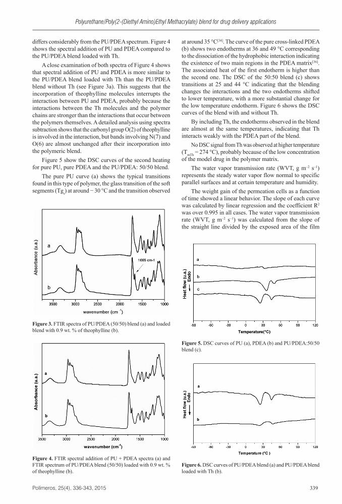
Figure 3a shows the FTIR spectrum of the PU/PDEA (50:50) film and Figure 3b the film loaded with Th 0.9 wt. %.
The band of Th at 1668 cm–1 assigned to the carbonyl stretching vibration is observed at 1665 cm–1 in the polymer matrix, indicating a low degree of interaction through the formation of H-bonds between the N-H groups of PU and the carbonyl groups of the Th. The N-H stretching band at 3331 cm–1 shifted to 3342 cm–1 and was broader on the high wavenumbers side, while the shoulder at 3520 cm–1 increased in intensity due to the increment of free N-H, as a consequence of the interaction of Th with the carbonyl groups. The shoulder at 2810 cm–1 also increases the intensity with the incorporation of Th.
The carbonyl stretching band of the PU/PDEA blend observed at 1720 cm–1 has slightly shifted to 1724 cm–1 after the incorporation of theophylline. Detailed comparisons of spectra show that the FTIR of the PU/PDEA containing Th
Figure 1. FTIR spectra of PU (a), PDEA (b) and PU/PDEA blend (50/50) (c) in the 3800 – 2600 cm–1 (a) and 1800 – 100 cm–1 (b) ranges. Figure 2. Chemical structure and FTIR spectrum of theophylline.
Polyurethane/Poly(2-(Diethyl Amino)Ethyl Methacrylate) blend for drug delivery applications
Polímeros, 25(4), 336-343, 2015 339
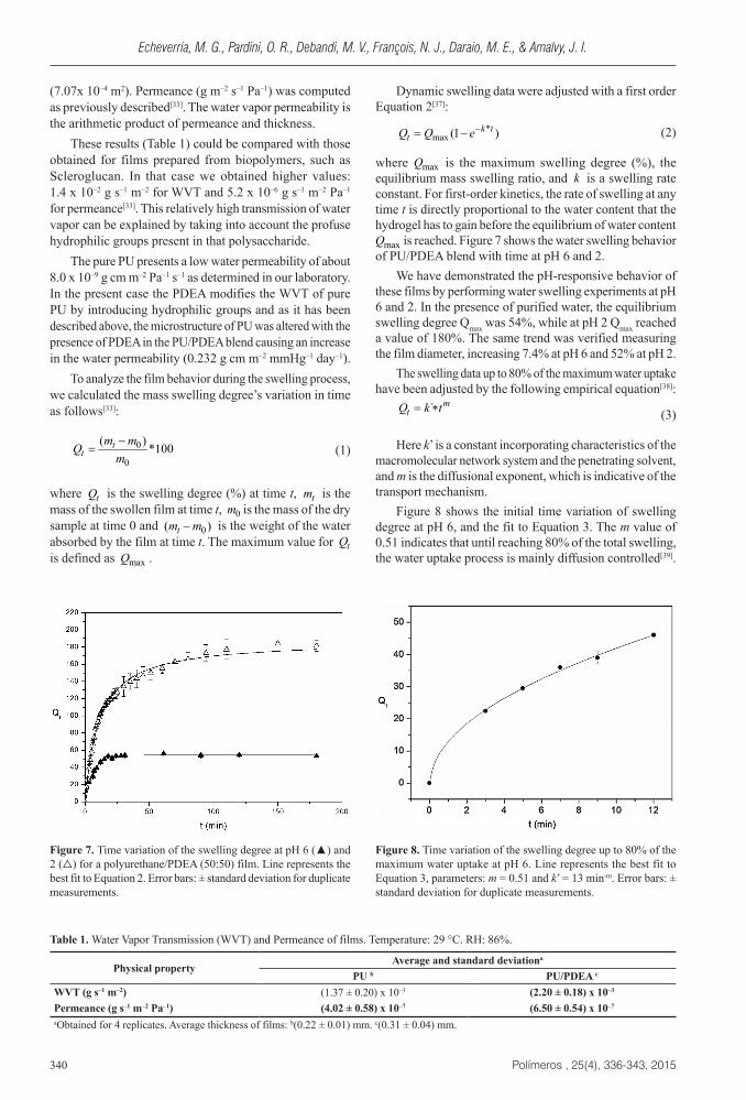
differs considerably from the PU/PDEA spectrum. Figure 4 shows the spectral addition of PU and PDEA compared to the PU/PDEA blend loaded with Th.
A close examination of both spectra of Figure 4 shows that spectral addition of PU and PDEA is more similar to the PU/PDEA blend loaded with Th than the PU/PDEA blend without Th (see Figure 3a). This suggests that the incorporation of theophylline molecules interrupts the interaction between PU and PDEA, probably because the interactions between the Th molecules and the polymer chains are stronger than the interactions that occur between the polymers themselves. A detailed analysis using spectra subtraction shows that the carbonyl group O(2) of theophylline is involved in the interaction, but bands involving N(7) and O(6) are almost unchanged after their incorporation into the polymeric blend.
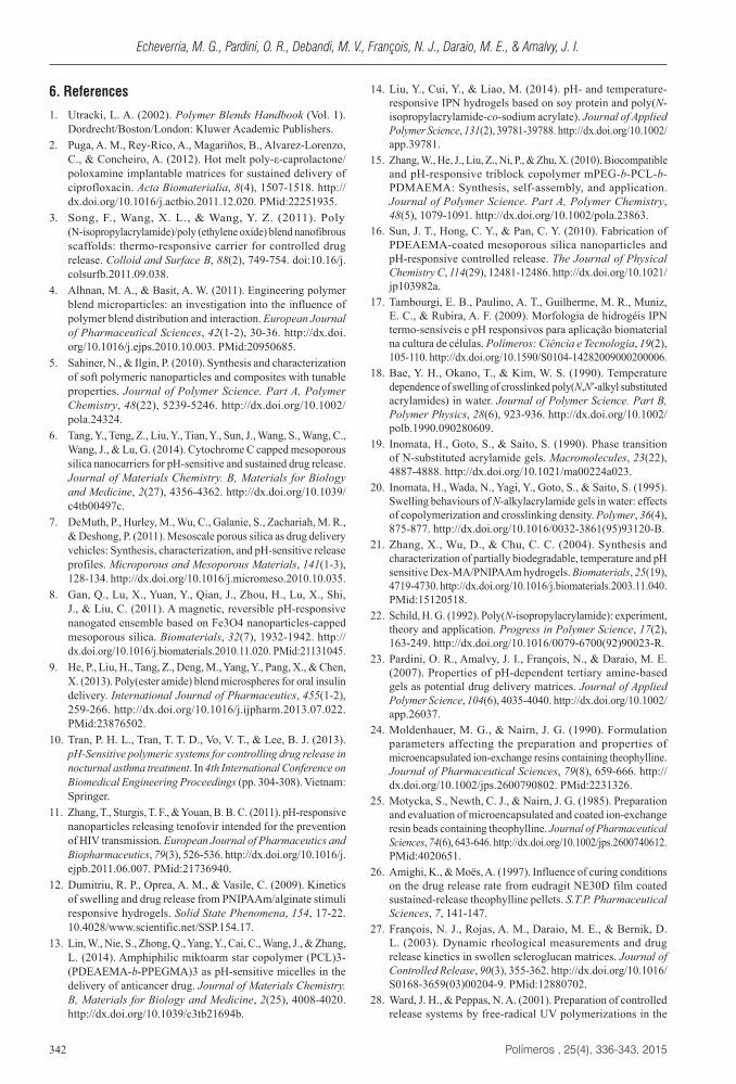
Figure 5 show the DSC curves of the second heating for pure PU, pure PDEA and the PU/PDEA: 50/50 blend.
The pure PU curve (a) shows the typical transitions found in this type of polymer, the glass transition of the soft segments (Tgs) at around − 30 °C and the transition observed
at around 35 °C[34]. The curve of the pure cross-linked PDEA (b) shows two endotherms at 36 and 49 °C corresponding to the dissociation of the hydrophobic interaction indicating the existence of two main regions in the PDEA matrix[36]. The associated heat of the first endotherm is higher than the second one. The DSC of the 50:50 blend (c) shows transitions at 25 and 44 °C indicating that the blending changes the interactions and the two endotherms shifted to lower temperature, with a more substantial change for the low temperature endotherm. Figure 6 shows the DSC curves of the blend with and without Th.
By including Th, the endotherms observed in the blend are almost at the same temperatures, indicating that Th interacts weakly with the PDEA part of the blend.
No DSC signal from Th was observed at higher temperature (TmTh = 274 °C), probably because of the low concentration of the model drug in the polymer matrix.
The water vapor transmission rate (WVT, g m–2 s–1) represents the steady water vapor flow normal to specific parallel surfaces and at certain temperature and humidity.
The weight gain of the permeation cells as a function of time showed a linear behavior. The slope of each curve was calculated by linear regression and the coefficient R2 was over 0.995 in all cases. The water vapor transmission rate (WVT, g m–2 s–1) was calculated from the slope of the straight line divided by the exposed area of the film
Figure 3. FTIR spectra of PU/PDEA (50/50) blend (a) and loaded blend with 0.9 wt. % of theophylline (b).
Figure 4. FTIR spectral addition of PU + PDEA spectra (a) and FTIR spectrum of PU/PDEA blend (50/50) loaded with 0.9 wt. % of theophylline (b).
Figure 5. DSC curves of PU (a), PDEA (b) and PU/PDEA:50/50 blend (c).
Figure 6. DSC curves of PU/PDEA blend (a) and PU/PDEA blend loaded with Th (b).
Echeverría, M. G., Pardini, O. R., Debandi, M. V., François, N. J., Daraio, M. E., & Amalvy, J. I.
Polímeros , 25(4), 336-343, 2015340
(7.07x 10⁻⁴ m2). Permeance (g m–2 s–1 Pa–1) was computed as previously described[33]. The water vapor permeability is the arithmetic product of permeance and thickness.
These results (Table 1) could be compared with those obtained for films prepared from biopolymers, such as Scleroglucan. In that case we obtained higher values: 1.4 x 10⁻² g s–1 m–2 for WVT and 5.2 x 10⁻⁶ g s–1 m–2 Pa–1 for permeance[33]. This relatively high transmission of water vapor can be explained by taking into account the profuse hydrophilic groups present in that polysaccharide.
The pure PU presents a low water permeability of about 8.0 x 10⁻⁹ g cm m–2 Pa–1 s–1 as determined in our laboratory. In the present case the PDEA modifies the WVT of pure PU by introducing hydrophilic groups and as it has been described above, the microstructure of PU was altered with the presence of PDEA in the PU/PDEA blend causing an increase in the water permeability (0.232 g cm m–2 mmHg–1 day–1).
To analyze the film behavior during the swelling process, we calculated the mass swelling degree’s variation in time as follows[33]:
0
0
( ) *100tt
m mQm−
= (1)
where tQ is the swelling degree (%) at time t, tm is the mass of the swollen film at time t, 0m is the mass of the dry sample at time 0 and 0( )tm m− is the weight of the water absorbed by the film at time t. The maximum value for tQis defined as maxQ .
Dynamic swelling data were adjusted with a first order Equation 2[37]:
*max (1 )k t
tQ Q e−= − (2)
where maxQ is the maximum swelling degree (%), the equilibrium mass swelling ratio, and k is a swelling rate constant. For first-order kinetics, the rate of swelling at any time t is directly proportional to the water content that the hydrogel has to gain before the equilibrium of water content
maxQ is reached. Figure 7 shows the water swelling behavior of PU/PDEA blend with time at pH 6 and 2.
We have demonstrated the pH-responsive behavior of these films by performing water swelling experiments at pH 6 and 2. In the presence of purified water, the equilibrium swelling degree Qmax was 54%, while at pH 2 Qmax reached a value of 180%. The same trend was verified measuring the film diameter, increasing 7.4% at pH 6 and 52% at pH 2.
The swelling data up to 80% of the maximum water uptake have been adjusted by the following empirical equation[38]:
´ mtQ k t= ∗ (3)
Here k’ is a constant incorporating characteristics of the macromolecular network system and the penetrating solvent, and m is the diffusional exponent, which is indicative of the transport mechanism.
Figure 8 shows the initial time variation of swelling degree at pH 6, and the fit to Equation 3. The m value of 0.51 indicates that until reaching 80% of the total swelling, the water uptake process is mainly diffusion controlled[39].
Table 1. Water Vapor Transmission (WVT) and Permeance of films. Temperature: 29 °C. RH: 86%.
Physical propertyAverage and standard deviationa
PU b PU/PDEA c
WVT (g s–1 m–2) (1.37 ± 0.20) x 10–3 (2.20 ± 0.18) x 10–3
Permeance (g s–1 m–2 Pa–1) (4.02 ± 0.58) x 10–7 (6.50 ± 0.54) x 10–7
aObtained for 4 replicates. Average thickness of films: b(0.22 ± 0.01) mm. c(0.31 ± 0.04) mm.
Figure 7. Time variation of the swelling degree at pH 6 (▲) and 2 (△) for a polyurethane/PDEA (50:50) film. Line represents the best fit to Equation 2. Error bars: ± standard deviation for duplicate measurements.
Figure 8. Time variation of the swelling degree up to 80% of the maximum water uptake at pH 6. Line represents the best fit to Equation 3, parameters: m = 0.51 and k’ = 13 min-m. Error bars: ± standard deviation for duplicate measurements.

Polyurethane/Poly(2-(Diethyl Amino)Ethyl Methacrylate) blend for drug delivery applications
Polímeros, 25(4), 336-343, 2015 341
For the initial swelling degrees (corresponding to the first 12 minutes) and taking into account that the constant m indicates Fickian behavior of water transport, the Equation 4 was used to estimate the diffusion coefficient D of water into the film[40,41]:
2 0.5 0.5max2* ( * / * ) *t DQ Q D t L K t= π = (4)
where L is the thickness of the dry film. Figure 9 shows the swelling degree vs. t0.5 and the fit to the Equation 4. The obtained value for KD was 13.3 min–0.5.
Taking into consideration the average film thickness L = 0.31 mm, Equation 4 leads to D = 7.55 x 10⁻⁷ cm2 s–1. This value is of the same order of magnitude as those obtained by Brazel and Peppas for two polymeric systems, poly(2-hydroxyethyl methacrylate-co-methylmethacrylate) (poly(HEMA-co-MMA)) and poly(vinyl alcohol) (PVA)[39].
Curves of Th concentration (Ct) released in the lower compartment of the vertical diffusion cell as a function of time (t) were plotted and the cumulative concentration of Th was adjusted to a power-law type relationship[42,43]:
* ntm k tm∞
= (5)
Here tm and m∞ are the cumulative amount of drug released after a time t and at infinite time, respectively, k is a constant related to kinetic behavior and experimental conditions and n is the exponent depending on the release process. Both m∞ and k were incorporated in a constant K , and Equation 6 was used to fit the data:
* ntC K t= (6)
where tC is the molar concentration of Th in the receptor compartment at time t.
Figure 10 shows the cumulative concentration of theophylline (Th) as a function of release time for a polyurethane-PDEA (50:50) film. The polymeric matrix was loaded with Th 0.9 wt. % and the release was performed at pH 6.
Equation 6 describes the drug delivery kinetics and it is only valid for the first 60% of the fractional release. For thin disks when n is equal to 0.5 the drug is said to diffuse with Fickian behavior. For n = 1 the behavior is called Case II diffusion, controlled by the relaxation of the macromolecular chains. Finally, anomalous transport behavior, which is intermediate between Fickian and Case II, is known as non-Fickian diffusion[31]. In our case the n value of 0.67 points to an anomalous drug transports. The Th-polymer interaction could modify the release kinetics. Hsiue et al. have studied the interaction of Th and an acrylic polymer (Eudragit L) and the effect on the release process[44]. They suggested interaction with the polymer via the C=O and C=N and they observed broadening and shifting of some FTIR bands. They also found that the percentage of Th hydrogen bonding with the acrylic polymer decreases with the increase of Th concentration release. They studied concentrations as high as 30 wt. %. However at the responsive pH of the polymer the kinetics did not change very much.
4. Conclusions
A blend of PDEA based hydrogel with polyurethane was prepared having good film forming properties and adequate for drug delivery applications. The blending of PDEA with PU modifies the interactions as revealed by the DSC and FTIR analysis. The water uptake process on such a blend is mainly diffusion controlled, but the release of the model drug theophylline in water follows an anomalous drug transport process. By changing the PDEA content it is expected to control permeation rates.
5. Acknowledgements
We are grateful to ANPCyT (PICT 2011 - 0238), CICPBA and Universidad de Buenos Aires (Grant UBACyT 2011-2014) for their financial assistance.
Figure 9. Swelling degree obtained at pH 6 as a function of square root of time. Line represents the best fit to Equation 4 with parameter KD = 13.3 min-0.5. Error bars: ± standard deviation for duplicate measurements.
Figure 10. Cumulative concentration of Th as a function of release time for a polyurethane/PDEA (50:50) film, loaded with theophylline 0.9% w/w. Line represents the best fit to Equation 6, parameters: n = 0.67 ; k =1.2 x 10⁻⁶ M.s-n.
Echeverría, M. G., Pardini, O. R., Debandi, M. V., François, N. J., Daraio, M. E., & Amalvy, J. I.
Polímeros , 25(4), 336-343, 2015342
6. References
1. Utracki, L. A. (2002). Polymer Blends Handbook (Vol. 1). Dordrecht/Boston/London: Kluwer Academic Publishers.
2. Puga, A. M., Rey-Rico, A., Magariños, B., Alvarez-Lorenzo, C., & Concheiro, A. (2012). Hot melt poly-ε-caprolactone/poloxamine implantable matrices for sustained delivery of ciprofloxacin. Acta Biomaterialia, 8(4), 1507-1518. http://dx.doi.org/10.1016/j.actbio.2011.12.020. PMid:22251935.
3. Song, F., Wang, X. L., & Wang, Y. Z. (2011). Poly (N-isopropylacrylamide)/poly (ethylene oxide) blend nanofibrous scaffolds: thermo-responsive carrier for controlled drug release. Colloid and Surface B, 88(2), 749-754. doi:10.16/j.colsurfb.2011.09.038.
4. Alhnan, M. A., & Basit, A. W. (2011). Engineering polymer blend microparticles: an investigation into the influence of polymer blend distribution and interaction. European Journal of Pharmaceutical Sciences, 42(1-2), 30-36. http://dx.doi.org/10.1016/j.ejps.2010.10.003. PMid:20950685.
5. Sahiner, N., & Ilgin, P. (2010). Synthesis and characterization of soft polymeric nanoparticles and composites with tunable properties. Journal of Polymer Science. Part A, Polymer Chemistry, 48(22), 5239-5246. http://dx.doi.org/10.1002/pola.24324.
6. Tang, Y., Teng, Z., Liu, Y., Tian, Y., Sun, J., Wang, S., Wang, C., Wang, J., & Lu, G. (2014). Cytochrome C capped mesoporous silica nanocarriers for pH-sensitive and sustained drug release. Journal of Materials Chemistry. B, Materials for Biology and Medicine, 2(27), 4356-4362. http://dx.doi.org/10.1039/c4tb00497c.
7. DeMuth, P., Hurley, M., Wu, C., Galanie, S., Zachariah, M. R., & Deshong, P. (2011). Mesoscale porous silica as drug delivery vehicles: Synthesis, characterization, and pH-sensitive release profiles. Microporous and Mesoporous Materials, 141(1-3), 128-134. http://dx.doi.org/10.1016/j.micromeso.2010.10.035.
8. Gan, Q., Lu, X., Yuan, Y., Qian, J., Zhou, H., Lu, X., Shi, J., & Liu, C. (2011). A magnetic, reversible pH-responsive nanogated ensemble based on Fe3O4 nanoparticles-capped mesoporous silica. Biomaterials, 32(7), 1932-1942. http://dx.doi.org/10.1016/j.biomaterials.2010.11.020. PMid:21131045.
9. He, P., Liu, H., Tang, Z., Deng, M., Yang, Y., Pang, X., & Chen, X. (2013). Poly(ester amide) blend microspheres for oral insulin delivery. International Journal of Pharmaceutics, 455(1-2), 259-266. http://dx.doi.org/10.1016/j.ijpharm.2013.07.022. PMid:23876502.
10. Tran, P. H. L., Tran, T. T. D., Vo, V. T., & Lee, B. J. (2013). pH-Sensitive polymeric systems for controlling drug release in nocturnal asthma treatment. In 4th International Conference on Biomedical Engineering Proceedings (pp. 304-308). Vietnam: Springer.
11. Zhang, T., Sturgis, T. F., & Youan, B. B. C. (2011). pH-responsive nanoparticles releasing tenofovir intended for the prevention of HIV transmission. European Journal of Pharmaceutics and Biopharmaceutics, 79(3), 526-536. http://dx.doi.org/10.1016/j.ejpb.2011.06.007. PMid:21736940.
12. Dumitriu, R. P., Oprea, A. M., & Vasile, C. (2009). Kinetics of swelling and drug release from PNIPAAm/alginate stimuli responsive hydrogels. Solid State Phenomena, 154, 17-22. 10.4028/www.scientific.net/SSP.154.17.
13. Lin, W., Nie, S., Zhong, Q., Yang, Y., Cai, C., Wang, J., & Zhang, L. (2014). Amphiphilic miktoarm star copolymer (PCL)3-(PDEAEMA-b-PPEGMA)3 as pH-sensitive micelles in the delivery of anticancer drug. Journal of Materials Chemistry. B, Materials for Biology and Medicine, 2(25), 4008-4020. http://dx.doi.org/10.1039/c3tb21694b.
14. Liu, Y., Cui, Y., & Liao, M. (2014). pH- and temperature-responsive IPN hydrogels based on soy protein and poly(N-isopropylacrylamide-co-sodium acrylate). Journal of Applied Polymer Science, 131(2), 39781-39788. http://dx.doi.org/10.1002/app.39781.
15. Zhang, W., He, J., Liu, Z., Ni, P., & Zhu, X. (2010). Biocompatible and pH-responsive triblock copolymer mPEG-b-PCL-b-PDMAEMA: Synthesis, self-assembly, and application. Journal of Polymer Science. Part A, Polymer Chemistry, 48(5), 1079-1091. http://dx.doi.org/10.1002/pola.23863.
16. Sun, J. T., Hong, C. Y., & Pan, C. Y. (2010). Fabrication of PDEAEMA-coated mesoporous silica nanoparticles and pH-responsive controlled release. The Journal of Physical Chemistry C, 114(29), 12481-12486. http://dx.doi.org/10.1021/jp103982a.
17. Tambourgi, E. B., Paulino, A. T., Guilherme, M. R., Muniz, E. C., & Rubira, A. F. (2009). Morfologia de hidrogéis IPN termo-sensíveis e pH responsivos para aplicação biomaterial na cultura de células. Polímeros: Ciência e Tecnologia, 19(2), 105-110. http://dx.doi.org/10.1590/S0104-14282009000200006.
18. Bae, Y. H., Okano, T., & Kim, W. S. (1990). Temperature dependence of swelling of crosslinked poly(N,N′-alkyl substituted acrylamides) in water. Journal of Polymer Science. Part B, Polymer Physics, 28(6), 923-936. http://dx.doi.org/10.1002/polb.1990.090280609.
19. Inomata, H., Goto, S., & Saito, S. (1990). Phase transition of N-substituted acrylamide gels. Macromolecules, 23(22), 4887-4888. http://dx.doi.org/10.1021/ma00224a023.
20. Inomata, H., Wada, N., Yagi, Y., Goto, S., & Saito, S. (1995). Swelling behaviours of N-alkylacrylamide gels in water: effects of copolymerization and crosslinking density. Polymer, 36(4), 875-877. http://dx.doi.org/10.1016/0032-3861(95)93120-B.
21. Zhang, X., Wu, D., & Chu, C. C. (2004). Synthesis and characterization of partially biodegradable, temperature and pH sensitive Dex-MA/PNIPAAm hydrogels. Biomaterials, 25(19), 4719-4730. http://dx.doi.org/10.1016/j.biomaterials.2003.11.040. PMid:15120518.
22. Schild, H. G. (1992). Poly(N-isopropylacrylamide): experiment, theory and application. Progress in Polymer Science, 17(2), 163-249. http://dx.doi.org/10.1016/0079-6700(92)90023-R.
23. Pardini, O. R., Amalvy, J. I., François, N., & Daraio, M. E. (2007). Properties of pH-dependent tertiary amine-based gels as potential drug delivery matrices. Journal of Applied Polymer Science, 104(6), 4035-4040. http://dx.doi.org/10.1002/app.26037.
24. Moldenhauer, M. G., & Nairn, J. G. (1990). Formulation parameters affecting the preparation and properties of microencapsulated ion-exchange resins containing theophylline. Journal of Pharmaceutical Sciences, 79(8), 659-666. http://dx.doi.org/10.1002/jps.2600790802. PMid:2231326.
25. Motycka, S., Newth, C. J., & Nairn, J. G. (1985). Preparation and evaluation of microencapsulated and coated ion-exchange resin beads containing theophylline. Journal of Pharmaceutical Sciences, 74(6), 643-646. http://dx.doi.org/10.1002/jps.2600740612. PMid:4020651.
26. Amighi, K., & Moës, A. (1997). Influence of curing conditions on the drug release rate from eudragit NE30D film coated sustained-release theophylline pellets. S.T.P. Pharmaceutical Sciences, 7, 141-147.
27. François, N. J., Rojas, A. M., Daraio, M. E., & Bernik, D. L. (2003). Dynamic rheological measurements and drug release kinetics in swollen scleroglucan matrices. Journal of Controlled Release, 90(3), 355-362. http://dx.doi.org/10.1016/S0168-3659(03)00204-9. PMid:12880702.
28. Ward, J. H., & Peppas, N. A. (2001). Preparation of controlled release systems by free-radical UV polymerizations in the

Polyurethane/Poly(2-(Diethyl Amino)Ethyl Methacrylate) blend for drug delivery applications
Polímeros, 25(4), 336-343, 2015 343
presence of a drug. Journal of Controlled Release, 71(2), 183-192. http://dx.doi.org/10.1016/S0168-3659(01)00213-9. PMid:11274750.
29. Shaheen, S. M., & Yamaura, K. (2002). Preparation of theophylline hydrogels of atactic poly(vinyl alcohol)/NaCl/H2O system for drug delivery system. Journal of Controlled Release, 81(3), 367-377. http://dx.doi.org/10.1016/S0168-3659(02)00085-8. PMid:12044575.
30. Amalvy, J. I., Wanless, E. J., Li, Y., Michailidou, V., Armes, S. P., & Duccini, Y. (2004). Synthesis and characterization of novel pH-responsive microgels based on tertiary amine methacrylates. Langmuir, 20(21), 8992-8999. http://dx.doi.org/10.1021/la049156t. PMid:15461478.
31. Pardini, O. R., & Amalvy, J. I. (2008). FTIR, 1H-NMR spectra, and thermal characterization of water-based polyurethane/acrylic hybrids. Journal of Applied Polymer Science, 107(2), 1207-1214. http://dx.doi.org/10.1002/app.27188.
32. American Society for Testing and Materials. (1995). ASTM E96: standard test methods for water vapor transmission of material. Philadelphia.
33. François, N. J., & Daraio, M. E. (2009). Preparation and characterization of scleroglucan drug delivery films: The effect of freeze-thaw cycling. Journal of Applied Polymer Science, 112(4), 1994-2000. http://dx.doi.org/10.1002/app.29651.
34. Peruzzo, P. J., Anbinder, P. S., Pardini, O. R., Costa, C. A., Leite, C. A., Galembeck, F., & Amalvy, J. I. (2010). Polyurethane/acrylate hybrids: Effects of the acrylic content and thermal treatment on the polymer properties. Journal of Applied Polymer Science, 116(5), 2694-2705. http://dx.doi.org/10.1002/app.31795.
35. Tarulli, S., & Baran, E. J. (1993). Spectroscopic behaviour of the two C=O stretching vibrations in free and complexed theophylline. Journal of Raman Spectroscopy : JRS, 24(3), 139-141. http://dx.doi.org/10.1002/jrs.1250240305.
36. Shibayama, M., Morimoto, M., & Nomura, S. (1994). Phase Separation Induced Mechanical Transition of Poly(N-isopropylacrylamide)/Water Isochore Gels. Macromolecules, 27(18), 5060-5066. http://dx.doi.org/10.1021/ma00096a031.
37. Alupei, I. C., Popa, M., Hamcerencu, M., & Abadie, M. J. M. (2002). Superabsorbant hydrogels based on xanthan and
poly(vinyl alcohol): 1. The study of the swelling properties. European Polymer Journal, 38(11), 2313-2320. http://dx.doi.org/10.1016/S0014-3057(02)00106-4.
38. Peppas, N. A., & Franson, N. M. (1983). The swelling interface number as a criterion for prediction of diffusional solute release mechanisms in swellable polymers. Journal of Polymer Science: Polymer Physics, 21, 983-997. http://dx.doi.org/10.1002/pol.1983.180210614.
39. Brazel, C. S., & Peppas, N. A. (1999). Mechanisms of solute and drug transport in relaxing, swellable, hydrophilic glassy polymers. Polymer, 40(12), 3383-3398. http://dx.doi.org/10.1016/S0032-3861(98)00546-1.
40. Satish, C. S., & Shivakumar, H. G. (2007). Dynamic swelling and in vitro release of insulin from semiinterpenetrating polymer networks of poly(vinyl alcohol) and poly(methacrylic acid). Indian Journal of Pharmaceutical Sciences, 69(1), 58-63. http://dx.doi.org/10.4103/0250-474X.32109.
41. Bosch, P., Fernández, A., Salvador, E. F., Corrales, T., Catalina, F., & Peinado, C. (2005). Polyurethane-acrylate based films as humidity sensors. Polymer, 46(26), 12200-12209. http://dx.doi.org/10.1016/j.polymer.2005.10.113.
42. Ritger, P. L., & Peppas, N. A. (1987). A simple equation for description of solute release I. Fickian and non-fickian release from non-swellable devices in the form of slabs, spheres, cylinders or discs. Journal of Controlled Release, 5(1), 23-36. http://dx.doi.org/10.1016/0168-3659(87)90034-4.
43. Ritger, P. L., & Peppas, N. A. (1987). A simple equation for description of solute release II. Fickian and anomalous release from swellable devices. Journal of Controlled Release, 5(1), 37-42. http://dx.doi.org/10.1016/0168-3659(87)90035-6.
44 . Hsiue, G. H., Liao, C. M., & Lin, S. Y. (1998). Effect of drug-polymer interaction on the release characteristics of methacrylic acid copolymer microcapsules containing theophylline. Artificial Organs, 22(8), 651-656. http://dx.doi.org/10.1046/j.1525-1594.1998.04804.x. PMid:9702316.
Received: Apr. 06, 2014 Revised: Jan. 19, 2015
Accepted: Feb. 09, 2015