s l i d e 1 anthrax is a serious zoonotic disease that can affect most
TRANSCRIPT

Anthrax
Center for Food Security and Public Health 2011 1
Slide 1
Anthrax
Malignant Pustule, Malignant Edema, Woolsorters’ Disease, Ragpickers’
Disease, Maladi Charbon, Splenic Fever
Anthrax is a serious zoonotic disease that can affect most mammals and
several species of birds, but is particularly important in herbivores. The
word anthrax is derived from a Greek word meaning charcoal or
carbuncle. Anthrax, the disease, likely originated 6,000 to 7,000 years
ago in Mesopotamia and Egypt, where agricultural civilization was first
recorded. Scientific literature first contained a reference to anthrax in
1769, by Fournier. Common names include: Malignant Pustule,
Malignant Edema, Woolsorters’ Disease, Ragpickers’ Disease, and
Maladi Charbon.
Slide 2
Overview
• Organism
• History
• Epidemiology
• Transmission
• Disease in Animals
• Disease in Humans
• Prevention and Control
Center for Food Security and Public Health, Iowa State University, 2011
In today’s presentation we will cover information regarding the
organism that causes anthrax and its history. We will also talk about the
epidemiology of the disease, how it is transmitted, species that it affects
and clinical and necropsy signs observed. Finally, we will address
prevention and control measures for anthrax, including actions to take if
anthrax is suspected.
Slide 3
THE ORGANISM
Slide 4
The Organism
• Bacillus anthracis
• Large, gram-positive, non-motile rod
• Two forms
–Vegetative, spore
• Over 1,200 strains
• Nearly worldwide distribution
Center for Food Security and Public Health, Iowa State University, 2011
Bacillus anthracis is a gram-positive rod that exists in two forms: the
vegetative bacillus and the spore. Within an infected host, spores
germinate to produce the vegetative forms which eventually kill the
host. These bacilli are released by the dying or dead animal into the
environment (usually soil under the carcass). There they sporulate,
ready to be taken up by another animal. Although vegetative forms of
B. anthracis grow and multiply readily in normal laboratory nutrient
agars or broths, they are more "fragile" than the vegetative forms of
other Bacillus species, dying in simple environments such as water or
milk. B. anthracis is dependent on sporulation for species survival
making it an obligate pathogen. Anthrax can be found nearly worldwide
and has roughly 1,200 various strains.
[The top photo show non-hemolytic Bacillus anthracis on sheep blood
agar with “Medusa head” appearance (non-pigmented, dry, ground
glass surface, edge irregular with comma projections); the bottom
image shows Gram stained B. anthracis bacilli. Both images from CDC
Public Health Image Library.]

Anthrax
Center for Food Security and Public Health 2011 2
Slide 5
The Spore
• Sporulation requires:
–Poor nutrient conditions
–Presence of oxygen
• Spores
–Very resistant
–Survive for decades
–Taken up by host and germinate
• Lethal dose 2,500 to 55,000 spores
Center for Food Security and Public Health, Iowa State University, 2011
When conditions are not conducive to growth and multiplication of
vegetative bacilli, B. anthracis tends to form spores. Sporulation
requires a nutrient poor environment and the presence of free oxygen.
The spore form is the predominant phase in the environment, can
survive for decades in soil, and it is through the uptake of spores that
anthrax is contracted. Heavy rains, alternating with dry periods, may
concentrate the spores and result in outbreaks among grazing animals.
The spore form of anthrax is markedly resistant to biological extremes
of heat, cold, pH, desiccation, and chemicals (and thus to disinfection).
Spores are not produced in the unopened carcass (within the anaerobic
environment of an infected host the organism is in the vegetative form).
It is estimated that 2,500 to 55,000 spores represent the lethal inhalation
dose for humans.
[The image shows an electron photomicrograph of a Bacillus anthracis
spore (arrowhead) partially surrounded by the pseudopod of a cultured
macrophage (x137,000) from Dixon TC et al. Anthrax. N.Engl.J.Med.
1999;341:815-26.]
Slide 6
HISTORY
Slide 7
Sverdlovsk, Russia, 1979
• 94 people sick – 64 died
• Soviets blamed contaminated meat
• Denied link to biological weapons
• 1992–President Yeltsin admits outbreak
related to military facility
–Western scientists find victim clusters downwind from facility
• Caused by faulty exhaust filter
Center for Food Security and Public Health, Iowa State University, 2011
On April 2, 1979, there was an outbreak of anthrax in the Soviet city of
Sverdlovsk (now called Ekaterinburg) about 850 miles east of Moscow.
Ninety-four people were affected, and at least 64 people died from
inhalational anthrax. The Soviet government claimed that the outbreak
was caused by intestinal anthrax from contaminated meat. The US
believed that the Soviet Union was violating the Biological Weapons
Convention signed in 1972. Thirteen years later, in 1992, President
Boris Yeltsin admitted that the anthrax outbreak was the result of
military activity at the facility. Western scientists were allowed to
investigate in 1992 and found that all the victims were clustered along a
straight line downwind from the military facility. Livestock in the same
area also died of anthrax. The outbreak was caused by a faulty exhaust
filter which was removed and not immediately replaced allowing
aerosolized anthrax spores to escape the facility.

Anthrax
Center for Food Security and Public Health 2011 3
Slide 8
South Africa, 1978-1980
• Anthrax used by Rhodesian and South African apartheid forces
–Thousands of cattle died
–10,738 human cases
–182 known deaths
–Black Tribal lands only
Center for Food Security and Public Health, Iowa State University, 2011
South Africa’s National Party and apartheid regime of the late 1970s
are known to have used biological and chemical weapons. These efforts
included covert attacks throughout neighboring nations including
Rhodesia (present day Zimbabwe). It is believed that within Rhodesia,
from 1978 - 80, thousands of cattle died from intentionally introduced
anthrax resulting in a critical food shortage. 10,738 people contracted
the disease, resulting in 182 deaths. Outbreaks were almost wholly
limited to black inhabited Tribal Trust Lands.
Slide 9
Tokyo, 1993
• Aum Shinrikyo– Japanese religious cult
– “Supreme truth”
• Attempt at biological terrorism–Released anthrax from office building
–Vaccine strain used
–No human injuries
Center for Food Security and Public Health, Iowa State University, 2011
In 1993, the Aum Shinrikyo, a Japanese religious cult meaning
“supreme truth” made several unsuccessful attempts at biological
terrorism through the release of anthrax in Tokyo. Anthrax was released
from the cult’s Tokyo office building laboratory. Police and media
reported foul smells, brown steam, some pet deaths, and stains on cars
and sidewalks. Fortunately, the strain of anthrax used in the attacks was
the vaccine strain, lacking in essential toxic properties. As a result no
human injuries were reported in any of these events.
[The image shows Aum Shinrikyo leader Shoko Asahara. Source:
Chronology of Aum Shinrikyo's CBW activities, Center for
Nonproliferation Studies, Monterey Institute of International Studies.
Mangold,T.; Goldberg,J. Plague wars: The terrifying reality of
biological warfare.]
Slide 10
U.S., 2001
Center for Food Security and Public Health, Iowa State University, 2011
In 2001, anthrax-contaminated letters were mailed to prominent figures
in the U.S. Four recovered letters were sent to: Tom Brokaw at NBC,
the New York Post, Senator Tom Daschle, and Senator Patrick Leahy.
All were believed to have been mailed from Trenton, NJ. This terrorist
attack showed how readily anthrax could spread if manufactured in
specific ways. Anthrax spores were able to escape sealed envelopes,
contaminate postal machinery, and infect postal workers and mail
recipients. For example, a 94 year-old woman died in Connecticut from
anthrax, possibly from contaminated mail. In addition, mail sorting
machines were also found to be positive at the facility.
[These screen captures are from CNN media postings in 2001]
Slide 11
U.S., 2001
• 22 cases
–11 cutaneous
–11 inhalational; 5 deaths
• Cutaneous case
–7 month-old boy
–Visited ABC newsroom
–Open sore on arm
–Anthrax positive
Center for Food Security and Public Health, Iowa State University, 2011
There were a total of 22 cases of anthrax in the U.S. in 2001; eleven
inhalation and 11 cutaneous. Five deaths occurred. The index case was
a photojournalist in Florida; two postal workers died in Maryland, and a
hospital supply worker died in New York City. The elderly Connecticut
farm woman previously described also died.
A 7 month-old boy who visited the ABC newsroom on September 28,
2001 became infected. The child had an elevated white blood count,
was afebrile, and had a 2-cm open sore with surrounding erythema and
induration that oozed clear yellow fluid. There was swelling and
erythema of the entire arm. The initial diagnosis was Loxosceles
reclusa spider bite. After anthrax exposure was reported at another
television network, two punch biopsies were taken. PCR and
immunostaining for Bacillus anthracis were positive.

Anthrax
Center for Food Security and Public Health 2011 4
[This photo shows the open sore lesion in the child previously
described. Source: Roche K, Chang W., Lazarus H. Cutaneous Anthrax
Infection. New England Journal of Medicine 2001;345:1611.]
Slide 12
U.S., 2001
• CDC survey of health officials
–7,000 reports regarding anthrax
• 1,050 led to lab testing
–1996-2000
• Less than 180 anthrax inquiries
• Antimicrobial prophylaxis
–Ciprofloxacin
• 5,343 prescriptions
Center for Food Security and Public Health, Iowa State University, 2011
During the time period September 11 – October 17, 40 state and
territorial health officials responded to a CDC telephone survey. An
estimated 7,000 reports had been received at the health departments;
approximately 4,800 required phone follow-ups, and 1,050 reports led
to testing of suspicious materials at a public health laboratory. In
comparison, fewer than 180 anthrax threats were reported between
1996 and 2000.
The antibiotic Ciprofloxacin was offered to many people in the
aftermath of the anthrax mailings in 2001. 5,343 people were prescribed
the medication for 60 days; 44% adhered to the 60 day treatment. Side
effects were experienced by 3,032 people (57%) of those taking
antibiotic prophylaxis, and included diarrhea, abdominal pain,
dizziness, nausea, and vomiting.
Slide 13
TRANSMISSION
Slide 14
Human Transmission
• Cutaneous
–Contact with infected tissues, wool, hide, soil
–Biting flies
• Inhalational
–Tanning hides, processing wool or bone
• Gastrointestinal
–Undercooked meat
Center for Food Security and Public Health, Iowa State University, 2011
There are three main routes of anthrax transmission in humans.
Cutaneous anthrax may occur when handling infected tissues, wool,
hides, soil, and products made from contaminated hides or hair, such as
drums, rugs, or brushes. Biting flies are also suspected of being
mechanical vectors of anthrax to humans under certain conditions.
Inhalational anthrax has been associated with tanning hides and
processing wool or bone. Gastrointestinal anthrax occurs when
individuals eat undercooked contaminated meat from animals that have
died of anthrax. Laboratory acquired cases have also occurred.
[The top image shows wool hanging outdoors. Bottom image: Cattle
hides. Source (for both): pixabay.com-public domain]

Anthrax
Center for Food Security and Public Health 2011 5
Slide 15
Human Transmission
• Tanneries
• Textile mills
• Wool sorters
• Bone processors
• Slaughterhouses
• Laboratory workers
Center for Food Security and Public Health, Iowa State University, 2011
People with occupational exposure to animal products are at risk of
anthrax infection. This includes workers in tanneries, textile mills, wool
sorters, and others. In the 1960s, unvaccinated mill workers were
‘chronically exposed’ to anthrax; case rates of 0.6 to 1.4% were
observed. B. anthracis was recovered from the nose and pharynx of
14% of healthy workers in one study; in another study, workers were
inhaled 600 to 1300 spores during the working day with no ill effect. A
well-documented outbreak of pulmonary anthrax occurred in one mill
with a similar level of contamination.
[Photo shows two men shearing sheep. Source: geograph.org.uk-
creative-commons]
Slide 16
Animal Transmission
• Bacteria present in hemorrhagic exudate from mouth, nose, anus
• Oxygen exposure
–Spores form
–Soil contamination
• Sporulation does not occur in a closed carcass
• Spores viable for decades
Center for Food Security and Public Health, Iowa State University, 2011
In infected animals, large numbers of bacteria are present in
hemorrhagic exudates from the mouth, nose, and anus. When they are
exposed to oxygen, these bacteria form spores and contaminate the soil.
Sporulation also occurs if a carcass is opened and oxygen exposure
occurs – sporulation does not occur inside a closed carcass. Anthrax
spores can remain viable for decades in the soil or animal products,
such as dried or processed hides and wool. Spores can survive for two
years in water, 10 years in milk, and up to 71 years on silk threads.
Vegetative organisms are thought to be destroyed within a few days
during the decomposition of unopened carcasses.
Slide 17
Animal Transmission
• Ingestion
–Most common
–Herbivores
• Contaminated soil
• Heavy rainfall, drought
–Carnivores
• Contaminated meat
• Inhalation
• Mechanical (insects)
Center for Food Security and Public Health, Iowa State University, 2011
In animals, transmission occurs by ingestion and possibly inhalation of
spores, although entry through skin lesions has not been ruled out.
Herbivores usually become infected when they ingest sufficient
numbers of spores in soil or on plants in pastures. Outbreaks are often
associated with heavy rainfall, flooding, or drought. Contaminated bone
meal and other feed can also spread this disease. Carnivores usually
become infected after eating contaminated meat. Vultures and flies may
disseminate anthrax mechanically after feeding on infected carcasses.
[This photo shows cattle grazing in a pasture. Source: U.S. Department
of Agriculture, NRCS]
Slide 18
EPIDEMIOLOGY
Anthrax
Center for Food Security and Public Health 2011 6
Slide 19
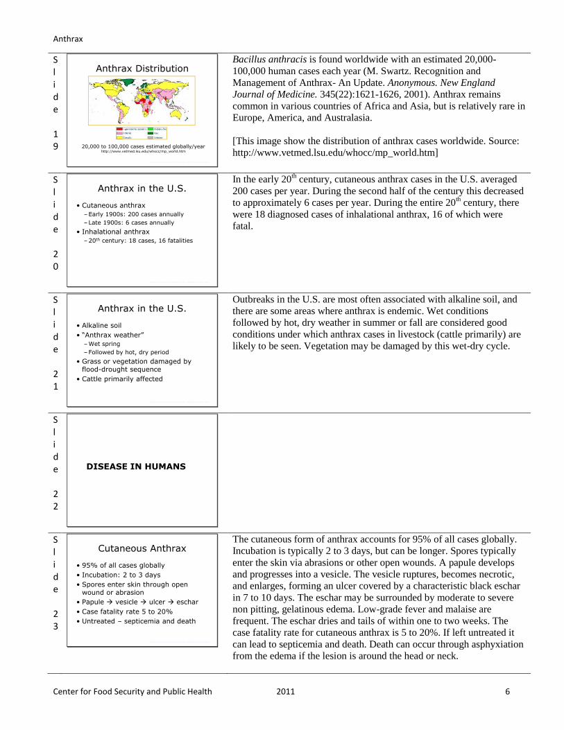
Anthrax Distribution
20,000 to 100,000 cases estimated globally/yearhttp://www.vetmed.lsu.edu/whocc/mp_world.htm
Center for Food Security and Public Health, Iowa State University, 2011
Bacillus anthracis is found worldwide with an estimated 20,000-
100,000 human cases each year (M. Swartz. Recognition and
Management of Anthrax- An Update. Anonymous. New England
Journal of Medicine. 345(22):1621-1626, 2001). Anthrax remains
common in various countries of Africa and Asia, but is relatively rare in
Europe, America, and Australasia.
[This image show the distribution of anthrax cases worldwide. Source:
http://www.vetmed.lsu.edu/whocc/mp_world.htm]
Slide 20
Anthrax in the U.S.
• Cutaneous anthrax
–Early 1900s: 200 cases annually
– Late 1900s: 6 cases annually
• Inhalational anthrax
–20th century: 18 cases, 16 fatalities
Center for Food Security and Public Health, Iowa State University, 2011
In the early 20th century, cutaneous anthrax cases in the U.S. averaged
200 cases per year. During the second half of the century this decreased
to approximately 6 cases per year. During the entire 20th century, there
were 18 diagnosed cases of inhalational anthrax, 16 of which were
fatal.
Slide 21
Anthrax in the U.S.
• Alkaline soil
• “Anthrax weather”
–Wet spring
–Followed by hot, dry period
• Grass or vegetation damaged by flood-drought sequence
• Cattle primarily affected
Center for Food Security and Public Health, Iowa State University, 2011
Outbreaks in the U.S. are most often associated with alkaline soil, and
there are some areas where anthrax is endemic. Wet conditions
followed by hot, dry weather in summer or fall are considered good
conditions under which anthrax cases in livestock (cattle primarily) are
likely to be seen. Vegetation may be damaged by this wet-dry cycle.
Slide 22
DISEASE IN HUMANS
Slide 23
Cutaneous Anthrax
• 95% of all cases globally
• Incubation: 2 to 3 days
• Spores enter skin through open wound or abrasion
• Papule vesicle ulcer eschar
• Case fatality rate 5 to 20%
• Untreated – septicemia and death
Center for Food Security and Public Health, Iowa State University, 2011
The cutaneous form of anthrax accounts for 95% of all cases globally.
Incubation is typically 2 to 3 days, but can be longer. Spores typically
enter the skin via abrasions or other open wounds. A papule develops
and progresses into a vesicle. The vesicle ruptures, becomes necrotic,
and enlarges, forming an ulcer covered by a characteristic black eschar
in 7 to 10 days. The eschar may be surrounded by moderate to severe
non pitting, gelatinous edema. Low-grade fever and malaise are
frequent. The eschar dries and tails of within one to two weeks. The
case fatality rate for cutaneous anthrax is 5 to 20%. If left untreated it
can lead to septicemia and death. Death can occur through asphyxiation
from the edema if the lesion is around the head or neck.

Anthrax
Center for Food Security and Public Health 2011 7
Slide 24
Center for Food Security and Public Health, Iowa State University, 2011
Day 2
Day 4
Day 6
Day 6
Day 10
[This slide shows the progression of cutaneous lesions. The head,
forearms, and hands are most often affected. Source: Centers for
Disease Control and Prevention at
http://www.bt.cdc.gov/agent/anthrax/anthrax-images/cutaneous.asp]
Slide 25
Case Study: Cutaneous Anthrax
• North Dakota, 2000
• 67 year old man
• Helped in disposal of 5 cowsthat died of anthrax
• Developed cutaneous anthrax
• Recovered with treatment
Center for Food Security and Public Health, Iowa State University, 2011
During the year 2000, 32 farms were quarantined for anthrax in the
U.S. From July 6-Sept 24, 157 animals died. A 67 year old man, who
helped dispose of 5 cows that had died of anthrax, developed cutaneous
anthrax several days later. He was prescribed ciprofloxacin and
recovered fully.
Slide 26
Gastrointestinal Anthrax
• Incubation: 2 to 5 days
• Severe gastroenteritis common
–Consumption of undercooked or contaminated meat
• Case fatality rate: 25 to 75%
• GI anthrax not documented in U.S.
–Suspected in Minnesota outbreak
Center for Food Security and Public Health, Iowa State University, 2011
The symptoms of gastrointestinal anthrax appear typically two to five
days (15 hours up to 7 days) after the ingestion of undercooked meat
containing anthrax spores. Two forms of gastrointestinal anthrax can
occur. The oropharyngeal form is not well known. Symptoms include
sore throat, dysphagia, fever, hoarseness, and swelling of the neck. The
abdominal form is more common, and consists of severe gastroenteritis
consisting of nausea, vomiting, fever, and abdominal pain progressing
rapidly to severe, bloody diarrhea. The primary intestinal lesions are
ulcerative and occur mainly in the terminal ileum or caecum. The case
fatality rate is 25 to 75%. GI anthrax has rarely been described as
occurring in the US; however, there have been no confirmed clinical
cases reported to public health authorities.
Slide 27
Case Study: Gastrointestinal Anthrax
• Minnesota, 2000
• Downer cow approved for slaughter by local veterinarian
• 5 family members ate meat
–2 developed GI signs
• 4 more cattle died
• B. anthracis isolated from farm but not from humans
Center for Food Security and Public Health, Iowa State University, 2011
In late July 2000, a downer cow was approved for slaughter and family
consumption by the local veterinarian on a farm in Northern Minnesota.
Five family members ate well-cooked steak and hamburgers over the
next few weeks; two reported gastrointestinal signs of diarrhea,
abdominal pain, and fever. When four more animals died, a carcass was
tested and B. anthracis was isolated. While anthrax was never isolated
from the human cases, they were placed on chemoprophylaxis and
anthrax vaccinination was initiated.
[Source: CDC. Human Ingestion of Bacillus anthracis-Contaminated
Meat- Minnesota, August, 2000. Anonymous. MMWR 49(36):813-816,
2000.]

Anthrax
Center for Food Security and Public Health 2011 8
Slide 28
Inhalational Anthrax
• Incubation: 1 to 7 days
• Initial phase
–Nonspecific (mild fever, malaise)
• Second phase
–Severe respiratory distress
–Dyspnea, stridor, cyanosis, mediastinal widening, death in 24 to 36 hours
• Case fatality: 75 to 90% (untreated)
Center for Food Security and Public Health, Iowa State University, 2011
The incubation period for inhalational anthrax is 1 to 7 days. There is
some evidence that inhaled anthrax spores may take as long as 60 days
to cause illness. In the initial phase, anthrax signs are nonspecific; they
may include mild fever, malaise, myalgia, nonproductive cough, and
some chest or abdominal pain. Illness progresses within 2 to 3 days
leading to fever, dyspnea, cyanosis, stridor, mediastinal widening, and
subcutaneous edema of the chest and neck. The second stage of disease
is characterized by rapidly deteriorating health within 24 to 36 hours.
The case fatality rate is 75 to 95% if untreated. Antibiotics and medical
care within the first 48 hours of the onset of signs are indicated.
[Source: M. Swartz. Recognition and Management of Anthrax-An
Update. New England Journal of Medicine 345(22):1621-1626, 2001.]
Slide 29
Center for Food Security and Public Health, Iowa State University, 2011
This photo shows X-ray evidence of mediastinal widening and pleural
effusion, two common findings of inhalational anthrax.
[Source: J. Jernigan, D. Stephens, D. Ashford, C. Omenaca, M. Topiel,
M. Gallbraith, M. Tapper, and et al. Bioterrorism-Related Inhalational
Anthrax: The First 10 Cases Reported in the United States. Emerging
Infectious Diseases 7:933-944, 2001.]
Slide 30
Diagnosis in Humans
• Identification of B. anthracis
–Blood, skin, secretions
• Culture
• PCR
• Serology
–ELISA
• Nasal swabs
–Screening tool
Center for Food Security and Public Health, Iowa State University, 2011
Anthrax is diagnosed by finding B. anthracis in clinical samples (i.e.
blood, skin lesions, or respiratory secretions), or by isolating the
organism in culture. Polymerase chain reaction (PCR) techniques can
also be used to identify B. anthracis. Antibodies develop late in the
course of disease, and serology is only useful in retrospective studies.
An ELISA test was approved by the FDA (in collaboration with the
CDC) in 2004. This test is quicker and easier to interpret than previous
antibody testing methods, and it can be completed in less than one hour.
In 2001, nasal swabs were used as a rapid exposure assessment tool,
and as a tool for rapid environmental assessment. Nasal swabs are not
used for diagnosing anthrax and are not 100% effective in determining
all who may have been exposed.
[Photo: Lung tissue from a fatal case showing Bacillus anthracis
granular antigen staining inside a perihilar macrophage (red arrow) and
intra- and extracellular bacilli (black arrow). (Immunohistochemical
assay with a mouse monoclonal anti-B. anthracis cell-wall antibody
and detection with alkaline phosphatase and naphthol fast red, original
magnification 100X). Source: J. Jernigan, D. Stephens, D. Ashford, C.
Omenaca, M. Topiel, M. Gallbraith, M. Tapper, and et al. Bioterrorism-
Related Inhalational Anthrax: The First 10 Cases Reported in the
United States. Emerging Infectious Diseases 2001;7:933-944.]

Anthrax
Center for Food Security and Public Health 2011 9
Slide 31
Treatment
• Penicillin
–Most natural strains susceptible
• Additional antibiotic options
–Ciprofloxacin
• Treatment of choice in 2001
• No strains known to be resistant
–Doxycycline
• Course of treatment: 60 days
Center for Food Security and Public Health, Iowa State University, 2011
Penicillin has been the drug of choice for anthrax for many decades,
and only very rarely has penicillin resistance been found in naturally
occurring isolates. Some strains, particularly those used in bioterrorist
attacks, may be resistant to penicillin. For this reason, the CDC
recommends other antibiotics as the initial treatment, particularly for
systemic disease, until antibiotic susceptibility has been determined.
Antibiotics are effective only against the vegetative stage of B.
anthracis, and not against spores. Treatment is continued for at least 60
days in inhalational anthrax, as spores may be able to remain dormant
in the lungs and germinate during that time. Supportive therapy may
also be necessary, particularly for the inhalational and gastrointestinal
forms. Effective treatment depends on early recognition of the
symptoms: treatment for cutaneous anthrax is usually effective, but the
inhalational and gastrointestinal forms are difficult to recognize early
and the mortality rates are higher.
Slide 32
Center for Food Security and Public Health, Iowa State University, 2011
Effective treatment depends on early recognition of the symptoms;
however, anthrax may resemble the common cold or flu. This chart
describes some of the differences between inhalational anthrax and
more common diseases. With inhalational anthrax, there is no runny
nose. However, chest discomfort and vomiting are common.
[Source: MMWR. Considerations for Distinguishing Influenza-Like
Illness from Inhalational Anthrax Vol 50, No 44; 986-6 11/09/2001]
Slide 33
Prevention and Control
• Humans protected by preventing disease in animals
−Veterinary supervision
−Trade restrictions
• Improved industry standards
• Safety practices in laboratories
• Post-exposure antibiotic prophylaxis
Center for Food Security and Public Health, Iowa State University, 2011
Humans can protect themselves by preventing disease in animals.
Veterinary supervision of animal production and slaughter also helps
prevent contact with infected livestock or animal products. Trade
restrictions may be placed on certain animal products from countries
where anthrax is common and uncontrolled. Improvements in industry
standards have decreased the occupational risks for people exposed to
imported hides, wool, bone meal, and other animal products. In
laboratories, good safety practices, including the use of biological
safety cabinets, should be employed. Veterinarians should use
protective clothing and equipment when examining sick animals. They
should also avoid opening the carcasses of suspected cases. Post-
exposure antibiotic prophylaxis is recommended for people who have
been exposed to aerosolized anthrax spores. Treatment should be
continued for at least 60 days. Simultaneous antibiotics and vaccination
can be used in exposed humans, as human anthrax vaccines, unlike
livestock vaccines, are not live.

Anthrax
Center for Food Security and Public Health 2011 10
Slide 34
Vaccination
• Cell-free filtrate
• At risk groups–Veterinarians
–Lab workers
– Livestock handlers
–Military personnel
• Immunization series–Five IM injections over 18-week period
–Annual booster
Center for Food Security and Public Health, Iowa State University, 2011
The U.S. human anthrax vaccine is a cell-free filtrate produced from an
avirulent strain; it contains no whole bacteria, dead or alive. The
vaccine was developed during the 1950s and 1960s for humans, and
was licensed by the FDA in 1970. Since then, it has been administered
to at-risk wool mill workers, veterinarians, laboratory workers,
livestock handlers, and others than handle animal hides or furs. Anthrax
vaccination is mandatory for designated military and emergency-
essential personnel, as well as comparable Department of Defense civil
employees. The vaccine is manufactured by one company, the BioPort
Corporation. The primary immunization series consists of five
intramuscular injections given at day 0, week 4, months 6, 12, and 18.
Following the priming series, annual booster injections of the vaccine
are recommended.
[This photo shows a military corpsman receiving a vaccine. Source:
U.S. Navy via commons.wikimedia.org]
Slide 35
Vaccine Side Effects
• Injection site reactions
–Mild: 30% men, 60% women
–Moderate:1 to 5%
–Severe:1%
• Systemic effects rare
–Muscle or joint aches, headache, rash, chills, fever, nausea, loss of appetite
• No long-term side effects noted
Center for Food Security and Public Health, Iowa State University, 2011
Vaccine side effects include injection site reactions. About 30% of men
and 60% of women experience mild local reactions -- not unlike other
vaccinations. 1 to 5% of individuals experience moderate local
reactions. Severe local reactions occur at a rate of 1%. System reactions
occur in less than 0.2% of people. There have been no patterns of long-
term side effects from the vaccine, neither persistent nor delayed side.
Slide 36
ANIMALS AND ANTHRAX
Slide 37
Clinical Signs
• Many species affected
–Ruminants at greatest risk
• Three forms
–Peracute
• Ruminants (cattle, sheep, goats, antelope)
–Acute
• Ruminants and equine
–Subacute-chronic
• Swine, dogs, cats
Center for Food Security and Public Health, Iowa State University, 2011
Virtually all mammals and some birds can contract anthrax. Clinical
signs in animals differ by the species, with ruminants being the most at
risk. The peracute form most often affects ruminants, including cattle,
sheep, and goats. The acute form will affect ruminants, as well as
horses. The subacute or chronic form most often affects swine, dogs,
and cats. The incubation period varies from 1-20 days. In herbivores,
infections become apparent after 3-7 days, while in pigs it usually takes
1-2 weeks.
[This antelope is hemorrhaging from the nose. Source: World Health
Organization]

Anthrax
Center for Food Security and Public Health 2011 11
Slide 38
Ruminants
• Peracute–Sudden death
• Acute–Tremors, dyspnea
–Bloody discharge from body orifices
• Chronic (rare)–Pharyngeal and lingual edema
–Death from asphyxiation
Center for Food Security and Public Health, Iowa State University, 2011
In ruminants, peracute systemic disease is common; sudden death may
be the only sign. Staggering, trembling, and dyspnea may be seen in
some animals, followed by rapid collapse, terminal convulsions, and
death. In the acute form, clinical signs may be apparent for up to 2 days
before death. In this form, fever and excitement may be followed by
depression, stupor, disorientation, muscle tremors, dyspnea, and
congested mucous membranes. Pregnant cows may abort, and milk
production can drop severely. Bloody discharges from the nose, mouth,
and anus are sometimes seen. Occasionally, infections in ruminants are
characterized by subcutaneous edematous swellings, most often in the
ventral neck, thorax, and shoulders. Anthrax in wild herbivores varies
with the species, but tends to resemble the disease in cattle.
Subacute to chronic infections occur in less susceptible species such as
pigs, but can also be seen in cattle, horses, dogs, and cats. The main
symptoms are pharyngeal and lingual edema, with animals dying from
asphyxiation. Extensive, localized, subcutaneous edema of the ventrum,
including the neck, sternum, and flank can also be seen. The carcass
will decompose fairly rapidly leading to bloating, but rigor mortis will
not be complete. Dark, tarry blood may ooze from body orifices.
Treatment with antibiotics can be successful if begun early in the
course of the disease.
[Photo shows a cow carcass with hemorrhaging from the nose. Source:
Dr. Danelle Bickett-Weddle, Iowa State University, College of
Veterinary Medicine]
Slide 39
Differential Diagnosis (Ruminants)
• Blackleg
• Botulism
• Poisoning
–Plants, heavy metal, snake bite
• Lightning strike
• Peracute babesiosis
Center for Food Security and Public Health, Iowa State University, 2011
Differential diagnoses for anthrax in ruminants include other potential
causes of acute death, such as blackleg, botulism, poisoning (plants,
heavy metals, and snake bite), lightning strike, and peracute babesiosis.
Slide 40
Equine
• Acute
–Fever, anorexia, colic, bloody diarrhea
–Swelling in neck
• Dyspnea
• Death from asphyxiation
–Death in 1 to 3 days
• Insect bite
–Hot, painful swelling at site
Center for Food Security and Public Health, Iowa State University, 2011
Photo from WHO
Horses typically develop acute disease. Common symptoms in this
species include fever, chills, anorexia, depression, severe colic, and
bloody diarrhea. Swellings may be seen in the neck, sternum, lower
abdomen, and external genitalia. Dyspnea can occur due to the swelling
of the neck. Affected animals usually die within 1 to 3 days, but some
animals can survive up to a week. If a horse contracts anthrax via a bite
from an insect, a localized, hot, painful, subcutaneous swelling will
occur. The swelling will soon spread to the throat (causing dyspnea),
sternum, abdomen, and external genitalia.
[This photo shows a dead zebra with hemorrhaging from the nose.
Source: World Health Organization]

Anthrax
Center for Food Security and Public Health 2011 12
Slide 41
Pigs
• Acute disease uncommon
• Subacute to chronic
– Localized swelling of throat
• Dyspnea
• Asphyxiation
–Anorexia
–Vomiting, diarrhea
Center for Food Security and Public Health, Iowa State University, 2011
Septicemia and sudden death occurs occasionally in pigs. More often,
pigs have mild subacute to chronic infections characterized by localized
swelling and systemic signs, such as fever and enlarged lymph nodes.
Some animals develop rapidly progressive swelling of the throat, with
dyspnea and difficulty swallowing; these animals may suffocate.
Intestinal involvement with anorexia, vomiting, diarrhea, or
constipation is less common. Some pigs with anthrax recover.
Recovered, asymptomatic animals may have signs of localized
infections in the tonsils and cervical lymph nodes at slaughter.
Slide 42
Carnivores
• Relatively resistant
– Ingestion of contaminated raw meat
• Subacute to chronic–Fever, anorexia, weakness
–Necrosis and edema of upper GI tract
– Lymphadenopathy and edemaof head and neck
–Death
• Due to asphyxiation, toxemia, septicemia
Center for Food Security and Public Health, Iowa State University, 2011
Anthrax infection is generally rare in dogs and cats, but can occur when
they ingest contaminated carcasses or animal by-products. Clinically
apparent anthrax in dogs, cats, and wild carnivores resembles the
disease in pigs. A recent review of published cases in dogs suggests that
massive swelling of the head, neck, and mediastinum is the most
common symptom in this species. In the published cases, death was
usually the result of toxemia and shock, but swelling of the throat and
suffocation could also have been a factor. Hemorrhagic gastroenteritis
was reported in one dog, in addition to a swollen foreleg and ptyalism.
Severe acute gastroenteritis has also been reported in other carnivores
and omnivores.
Slide 43
Diagnosis and Treatment
• Necropsy not advised!
• Do not open carcass!
• Samples of peripheral blood needed
–Cover collection site with disinfectant soaked bandage to prevent leakage
• Treatment
–Penicillin, tetracyclines
• Reportable disease
Center for Food Security and Public Health, Iowa State University, 2011
Necropsy of affected carcasses is not advised. Due to poor clotting, a
blood sample can be taken post-mortem from a superficial vessel, such
as an ear vein, or with a Anthrax syringe from any available vein. The
collection site should be covered with a disinfectant-soaked bandage to
prevent leakage of contaminated blood. Blood smears should be made
and the blood submitted for isolation of B. anthracis. The organism is
very sensitive to penicillin and tetracyclines, and these are the
antibiotics of choice if infection is detected early. Care must be taken in
handling infected tissues or carcasses because the organism can
penetrate cuts in the skin, resulting in localized infection with
subsequent dissemination. Anthrax is a notifiable disease.
Slide 44
Case Study: Canine Anthrax
• Golden retriever, 6 yrs old– 2 day history of ptyalism
and swelling of right front leg
– Temperature 106°F, elevated WBC
– Died same day
• Necropsy – Splenomegaly, friable liver, blood in stomach
– 2x2 cm raised hemorrhagic leg wound
– Some pulmonary congestion
Center for Food Security and Public Health, Iowa State University, 2011
In 1991 in Mississippi a 6 year old golden retriever presented with a 2
day history of ptyalism and swelling of the right front leg, elevated
temperature (106°F), and elevated white blood cell count (25,900/μl).
The dog died the same day. Necropsy showed splenomegaly, a friable
liver, blood in the stomach and intestines, a 2x2 cm raised hemorrhagic
wound on the RF leg, some pulmonary congestion, and bacteria in
hepatic sinusoids, renal glomerular capillaries, and myocardial blood
vessels. Death was attributed to toxemia/septicemia. Anthrax was
confirmed with electron microscopy.
[This photo shows a golden retreiver. Source: commons.wikimedia.org]

Anthrax
Center for Food Security and Public Health 2011 13
Slide 45
Case Study:Canine Anthrax
• Source of exposure in question
–Residential area
–1 mile from livestock
–No livestock deaths in area
–Dove hunt on freshly plowed field 6 days prior to onset
• Signs consistent with ingestion but cutaneous exposure not ruled out
Center for Food Security and Public Health, Iowa State University, 2008
The circumstances of this case leave several unanswered questions
regarding the source of exposure of this dog to anthrax. Exposure was
considered unlikely during the 6 days prior to death because the dog
was kept at home, albeit unrestrained, in a residential area with no
livestock within approximately 0.8 km. No livestock deaths had been
reported at this site. Exposure of the dog was presumed to have
occurred 6 days prior to initial clinical signs (7 days prior to death)
during a dove hunt over a freshly plowed field. One possible source
includes carrion ingested in adjacent wooded areas, although a carcass
was not found. Ingestion would be the most likely explanation for the
gastrointestinal hemorrhage found at necropsy. A second possibility is
percutaneous infection, such as a wound or an insect bite. Although
cutaneous anthrax has not been described in animals, wound
contamination could not be ruled out.
Slide 46
Vaccination
• Livestock in endemic areas
• Sterne strain
– Live encapsulated spore vaccine
• No U.S. vaccine for pets
–Used in other countries
–Adjuvant may cause reactions
• Working dogs may be at risk
Center for Food Security and Public Health, Iowa State University, 2011
Annual vaccination of livestock in endemic areas is recommended. The
most widely used vaccine is the Sterne-strain vaccine. It is a non-
encapsulated, live variant strain of B. anthracis developed in 1937.
Immunity develops 7 to 10 days after vaccination. The vaccine
produced in the U.S. is licensed for use in livestock only (cattle, sheep,
horses, goats, and swine). No U.S. anthrax vaccine is licensed for use in
pets. In other countries, live spore vaccines produced from the Sterne
strain have been used to vaccinate pets and exotic species. The vaccine
contains saponin as an adjuvant and its use in cats and dogs may
produce injection site reactions. Cases in domestic cats are very rare.
Working dogs might put themselves at risk by exposure to dead
carcasses.
Slide 47
Animals and Anthrax
• Anthrax should always be high on differential list when:
–High mortality rates observed in herbivores
–Sudden deaths with unclotted blood from orifices occur
–Localized edema observed
• Especially neck of pigs or dogs
Center for Food Security and Public Health, Iowa State University, 2011
Anthrax should always be high on a differential list when there is high
mortality in a group of herbivores, sudden death occurs with unclotted
blood from orifices, and localized edema occurs, especially of the neck
in pigs and dogs.
Slide 48
PREVENTION AND CONTROL

Anthrax
Center for Food Security and Public Health 2011 14
Slide 49
Prevention and Control
• Report to authorities
• Quarantine the area
• Do not open carcass
• Minimize contact
• Wear protective clothing
–Latex gloves, face mask
Center for Food Security and Public Health, Iowa State University, 2011
Anthrax is a notifiable disease; if anthrax is suspected the state
veterinarian and local health officials should be contacted. Do not open
the carcass to perform a necropsy due to the potential for contamination
and exposure. Make sure there is minimal contact with the carcass by
establishing a quarantine area, generally for 21 days after the last
anthrax death. Wear protective clothing, such as a face mask and
gloves, if it is necessary to work with the dead animal. Be sure to cover
any areas of broken skin on yourself so that no infectious organisms
come in contact with those areas. [Photo of a veterinarian in Mali
preparing to vaccinate cattle for anthrax. Source: J. VanAcker/Food and
Agriculture Organization of the United Nations (FAO)]
Slide 50
Prevention and Control
• Local regulations determine carcass disposal options
– Incineration
–Deep burial
• Decontaminate soil
• Remove organic material and disinfect structures
Center for Food Security and Public Health, Iowa State University, 2011
Since necropsies are not advised, it is best to burn or bury the carcasses
and all contaminated materials, then decontaminate the soil with 5% lye
or quicklime (anhydrous calcium oxide). If structures have been
contaminated, remove the organic material and disinfect with an
approved chemical. [Photo of carcass burial. Source: Environmental
Health Services, Department of Public Health, Kings County,
California]
Slide 51
Prevention and Control
• Isolate sick animals
• Discourage scavengers
• Use insect control or repellants
• Prophylactic antibiotics
• Vaccination
– In endemic areas
–Endangered animals
Center for Food Security and Public Health, Iowa State University, 2011
Other measures to prevent disease in animals include isolating sick
animals from the rest of the herd immediately, discouraging scavengers
from the area, and using insect repellants to prevent fly dispersal of the
organism. In endemic areas, modified live vaccines can prevent anthrax
in livestock, if authorities feel it is necessary. Livestock can be
vaccinated annually, before the season when outbreaks generally occur.
These vaccines have also been used to protect cheetahs and endangered
ruminants, including the black rhinoceros (photo from Lewa Wildlife
Conservancy, http://www.lewa.org). Because the vaccine is a modified
live type, prophylactic antibiotics cannot be given concurrently;
however, animals may be vaccinated when antibiotic treatment has
concluded. [This photo shows a black rhinoceros from Etosha National
Park in Namibia. Source: Frank Vassen, www.flickr.com/creative-
commons]
Slide 52
Disinfection
• Spores resistant to heat, sunlight, drying and many disinfectants
• Disinfectants–Formaldehyde (5%)
–Glutaraldehyde (2%)
–Sodium hydroxide (NaOH) (10%)
–Bleach
• Gas or heat sterilization
• Gamma radiation
Center for Food Security and Public Health, Iowa State University, 2011
Anthrax spores are resistant to heat, sunlight, drying, and many
disinfectants. They can be killed with formaldehyde or 2% glutaraldehyde;
overnight soaking is recommended. A 10% NaOH or 5% formaldehyde
solution can be used for stockyards, pens, and other equipment. Sodium
hypochlorite has also been recommended for some purposes. The
sporicidal effectiveness of hypochlorite solutions varies with the pH and
the concentration of free available chlorine. To become an effective
sporicidal agent, household bleach must be diluted with water to increase
the free available chlorine, and adjusted to pH 7. Prolonged contact is
recommended. Gaseous sterilization can be accomplished with chlorine
dioxide, formaldehyde gas, and other methods, under specific conditions of
humidity and temperature. Sterilization is also possible by heating to
121°C (250°F) for at least 30 min. Gamma radiation has been used to
decontaminate animal products, as well as mail from contaminated postal
facilities. Exposed arms and hands can be washed with soap and hot water
then immersed for one minute in a disinfectant such as an organic iodine
solution or 1 ppm solution of mercuric perchloride. Clothing should be
cleaned and boiled.

Anthrax
Center for Food Security and Public Health 2011 15
Slide 53
Disinfection
• Preliminary disinfection– 10% formaldehyde
– 4% glutaraldehyde (pH 8.0-8.5)
• Cleaning– Hot water, scrubbing, protective clothing
• Final disinfection: one of the following– 10% formaldehyde
– 4% glutaraldehyde (pH 8.0-8.5)
– 3% hydrogen peroxide,
– 1% peracetic acid
Center for Food Security and Public Health, Iowa State University, 2011
Where practical, cleaning of all surfaces should be done by
straightforward washing and scrubbing using ample hot water.
Protective clothing should be worn. For final disinfection, one of the
following disinfectants should be applied at a rate of 0.4 liters per
square meter for an exposure time of at least 2 hours: 10%
formaldehyde (approximately 30% formalin), 4% glutaraldehyde (pH
8.0-8.5), 3% hydrogen peroxide, or 1% peracetic acid. Hydrogen
peroxide and peracetic acid are not appropriate if blood is present.
When using glutaraldehyde, hydrogen peroxide, or peracetic acid, the
surface should be treated twice with an interval of at least one hour
between applications. Formaldehyde and glutaraldehyde should not be
used at temperatures below 10oC. After the final disinfection, closed
spaces, such as rooms or animal houses, should be well ventilated
before use. The effectiveness of the disinfection procedure cannot be
assumed, and attempts should be made to confirm it has been adequate
by means of swabs and culture.
Slide 54
Biological Terrorism: Estimated Effects
• 50 kg of spores
–Urban area of 5 million
–Estimated impact
• 250,000 cases of anthrax
• 100,000 deaths
• 100 kg of spores
–Upwind of Wash D.C.
–Estimated impact
• 130,000 to 3 million deaths
Center for Food Security and Public Health, Iowa State University, 2011
Previous acts of biological terrorism have been small in scale. It is
estimated that in a city of 5 million people, a release of 50 kg of anthrax
spores (10 km upwind and 2 km wide) would extend over 20 km in 2
hours. This would result in 500,000 people placed at risk. There would
be an estimated 250,000 illness and 125,000 deaths. A 1993 report by
the U.S. Congressional Office of Technology Assessment estimated
that 130,000 to 3 million deaths may occur following the aerosolized
release of 100 kg of anthrax spores upwind of Washington D.C.
Slide 55
Additional Resources
• World Organization for Animal Health (OIE)– www.oie.int
• U.S. Department of Agriculture (USDA)– www.aphis.usda.gov
• Center for Food Security and Public Health– www.cfsph.iastate.edu
• USAHA Foreign Animal Diseases(“The Gray Book”)– www.aphis.usda.gov/emergency_response/do
wnloads/nahems/fad.pdf
Center for Food Security and Public Health, Iowa State University, 2011
Slide 56
Acknowledgments
Development of this presentation was made possible through grants provided to
the Center for Food Security and Public Health at Iowa State University, College of Veterinary Medicine from
the Centers for Disease Control and Prevention, the U.S. Department of Agriculture,
the Iowa Homeland Security and Emergency Management Division, and the
Multi-State Partnership for Security in Agriculture.
Authors: Radford Davis, DVM, MPH, DACVPM; Jamie Snow, DVM; Katie Steneroden, DVM; Anna Rovid Spickler, DVM, PhD;
Reviewers: Dipa Brahmbhatt, VMD; Katie Spaulding, BS; Glenda Dvorak, DVM, MPH, DACVPM; Kerry Leedom Larson, DVM, MPH, PhD
Center for Food Security and Public Health, Iowa State University, 2011
Last reviewed: March 2011