routine identification of aluminium … minerals (1993) 28, 85-99 routine identification of...
TRANSCRIPT

Clay Minerals (1993) 28, 85-99
R O U T I N E I D E N T I F I C A T I O N OF A L U M I N I U M H Y D R O X I D E P O L Y M O R P H S W I T H T H E L A S E R
R A M A N M I C R O P R O B E
K. A . R O D G E R S
Department of Geology, University of Auckland, Private Bag, Auckland, New Zealand
(Received 15 January 1992; revised 23 June 1992)
A B S T R A C T: The laser Raman microprobe spectrum of gibbsite displays four intense v (OH)- stretching bands at ~3365, 3435, 3525 and 3618 cm 1, bayerite shows three near 3425, 3545 and 3564 cm -1, nordstrandite has three near 3492, 3566 and 3623 cm -1, and doyleite one broad band centred at -3545 cm-i with sometimes a prominent shoulder near 3615 cm-1. The four polymorphs also possess distinctly different Raman signatures of medium to strong bands in the region 100-- 1200 cm 1. These differences provide a non-destructive routine determinative procedure by direct microsampling of untreated crystals, fragments, and microcrystalline powders. Sample preparation is minimal. Excessive noise and non-quenchable fluorescence are the principle limitations of the technique.
Gibbsite and bayerite may be distinguished by Raman spectroscopy (Huneke et al., 1980), with the laser Raman microprobe providing a non-destructive method of analysing small quantities particularly where the habit and/or fine grain size defy other techniques (e.g. Crestin-Desjobert et al., 1987). The Raman spectra of other polymorphs of aluminium hydroxide have not been recorded. This study determines those of nordstrandite and doyleite, and assesses some of the potentials and limitations of the method as a routine determinative procedure for such minerals.
Gibbsite, bayerite and nordstrandite are well-defined, natural crystalline forms of aluminium hydroxide. A triclinic modification of gibbsite was recognized by Saalfield (1960) among single crystals from the Urals. Chao et al. (1985) identified a further polymorph, doyleite, from two localities in Quebec, although reservations as to its status have been expressed by Wefers & Misra (1987). Published infrared (IR) spectral data for these species vary (e.g. Fredrickson, 1954; Vivien et al., 1973; Chao et al., 1987; Hsu, 1989).
Twenty two samples from the collections of the Natural History Museum, London, of the Australian Museum, and of private individuals, were examined. A brief description of those found to contain at least one polymorph is given in the Appendix. The sole example available of "gibbsite" from the Urals proved to be largely hydrotalcite and no triclinic gibbsite was identified.
S A M P L E P R E P A R A T I O N
The preparation of mineral samples for Raman microprobe analysis depends on the pur- pose of the individual analysis and the amount and nature of the sample available. As with X-ray powder diffraction, a single method of preparation and presentation of sample to the radiation and detectors is desirable for comparisons of results within a set (cf. Moore &
�9 1993 The Mineralogical Society

86 K. A. Rodgers
Reynolds, 1989). Variations in grain size, the presence of extraneous substances, and particle orientation with respect to both laser and detectors, may affect the resulting spectrum and need to be recognized when comparing results. These factors are particularly pertinent where a range of specimens are irradiated directly by the focused laser beam without any pre-treatment. In microsampling, a representative sample can be difficult to obtain except where particle size is less than the focused beam such that a quantity of crystallites or fragments can be irradiated.
The appropriate preparation in the present study proved somewhat minimal. Some specimens were presented directly to the focused beam. With others, suitable fragments were prised free and accumulated on a glass microscope slide. Where necessary, these were further reduced in size by firmly but gently crushing with a second slide. Except as described below, where X-ray powder diffraction mounts were microsampled, no samples were ground in order to avoid straining and deforming crystal structures or inducing a change of state. No chemical treatment was attempted until a sample had consistently failed to yield a spectrum, and then consisted only of removing iron and manganese oxides and organic materials (e.g. Clavert et al. 1979). Minimal treatment avoids altering a sample in some unanticipated way, particularly where fresh, reactive surfaces were exposed on crushing.
If a representative monomineralic sample was not obtained or where doubts existed as to homogeneity, particles for analysis were selected using a standard petrographic micro- scope. The microscope used for laser microanalysis lacked polarizers to assist in identifying grains.
Standard microscope glass slides were used to support the mineral grains and artificial preparations. As in X-ray powder diffraction, these offer an ease of application and with some samples a glass powder mount was taken directly from the diffractometer and used in Raman microsampling without further treatment. However, glass slide mounts have disadvantages. The technique favours preferred orientation and where X-ray mounts are prepared from a slurry, the finest particles are concentrated on top of a mount, in the position preferentially sampled by the laser beam. Further, where a sampled particle is very thin, the Raman spectrum invariably contains appreciable noise associated with spectra excited from the glass. A spectrum from a glass slide showed weak broad bands at about 300-600, 800 and 1075 cm -1.
E X P E R I M E N T A L P R O C E D U R E S
The Raman spectra were recorded from 3300-3700 c m - 1 and 100-1200 cm -1 using a Jobin- Yvon U1000 spectrometer with slit settings of 500 ~tm, a bandpass of 4-5 cm -1, and 514.5 nm Ar + as the exciting line. Integration time was 3 s for each incremental step of 1 cm 1. An uncoated • objective of a Nikon microscope was used for all microsampling, the conventional arrangement of stage and lens giving 180~ scattering geometry. Monochannel Ga-As photo-multiplier detectors (Burle C31034A) were employed operat- ing at -30~ and 1800 V.
Power loss throughout the system was marked. Only some 10-15% of the laser power was available at the sampling lens. Good quality results from single crystals were obtained from 25 mW of laser power measured at sample. Higher power levels, up to 150 mW, sometimes improved spectral quality, e.g. for sample AM-D44000, but could also be counter productive. Count rates given below are by way of example only and would be expected to vary with differing experimental conditions.

Identification orAl hydroxides by laser Raman microprobe 87
Typically 10 6 to 10 a2 photons from the laser are required to generate one Raman photon (e.g. Delhaye & Dhamelincourt, 1987). Fractures, inclusions, imperfections and growth planes impede passage of scattered photons to the detectors. Colourless, transparent crystals and 001 cleavages, e.g. samples AM-D48300, AM-D49268, and unstained white microcrystalline samples and irregular flakes, e.g. AM-Dl1284, AM-D49262, either whole or as fragments, presented few microsampling problems and yielded excellent spectra. The optics were focused on the top of the fragment to the point where it appeared slightly defocused. The laser operating at -200 mW was introduced and the crystal cautiously quenched (photodegraded with time) using increasing power until background in the (OH)- stretching region was below 20,000 counts s I at 100 mW, measured at sample.
Generally signal to noise ratios among these coarser-grained specimens were low with background count rates for pellucid cleavages of the order of 3-7000 counts s 1 at 100 mW (at sample) near 3300 cm -1. Quench times were low to insignificant. Translucent to semi- opaque fragments yielded higher signal to noise levels which could often, but not always, be improved by quenching at high power e.g. AM-D11426.
In later sessions, the system was routinely calibrated before scanning, so as to give 200,000+ counts s 1 at 1088 cm 1 from a transparent Icelandic Spar cleavage at a power level of 15 mW at sample. Such a pre-scan system check improved consistency of results with powdered and fine-grained material These materials appeared under the sample lens as clusters of tiny crystallites. The coarser of these materials, e.g. AM-D11695, AM- D47734, provided straightforward results. With others, initial count rates were often in excess of 250,000 counts s -1 near 3300 cm -1 even at low power levels e.g. BM-64525, AU- 3066. These dropped on quenching to rates of <20,000 counts s l, after which v (OH)- stretching bands were readily detected above noise. High background count rates were not always improved by quenching, even for protracted periods e.g. BM-1985,147. Many fine- grained specimens needed a marked defocus to show satisfactory scattering bands e.g. BM-64525.
Typical results from such fine-grained materials were found with algal infested bayerite grains from Raoul Island (Rodgers et al., 1989). These appeared dark under the sampling microscope and yielded initial counts >120,000 counts s 1 at 50 mW (at sample) at 3500 cm -1, with no scattering bands observable in a scanned spectrum. Quenching for 10 min at 100 mW (at sample) dropped the count rate in this region to -31,000 counts s 1 when (OH)-stretching bands could be discerned in a scan, while the overall background continued to decay. Similar results were found with freshly prepared artificial bayerite precipitates which invariably quenched very rapidly.
Fluorescence was the principal hindrance in obtaining results, once identity of a troublesome sample had been confirmed by X-ray powder diffraction. Raman results were seldom satisfactory where backgrounds were in excess of 35,000 count s -1. Contrawise, a Raman spectrum, albeit of varying quantity, was invariably obtained from a proven aluminium hydroxide polymorph when background count rates were <30,000 s -1. Fluorescence from organic contaminants decayed under intense laser light due to photochemical oxidation whereas Fe- and Mn-sourced fluorescence remained stable.
Nordstrandite specimens were prone to fluoresce more than gibbsites and bayerites. This may be related to their provenance in soil and weathering horizons. For example, red- stained fragments of BM-1962,229 nordstrandite mostly gave an unquenchable background count rate >500,000 s -1. Only a few small clear basal cleavages yielded distinct (OH)- stretching bands, after quenching for 30 min at 100 mW (at sample) to a background of

88 K. A . Rodgers
65,000 counts s -1 near 3500 cm -1. One small red-brown stained cleavage produced a weak spectrum above a background of --100,000 counts s - 1 - t h e sole sample found to do so. In contrast, the extremely fine-grained, white Stradner Kogel nordstrandite, AM-D49262, from a lava vug, showed no fluorescence and little noise.
E X P E R I M E N T A L R E S U L T S A N D D I S C U S S I O N
3 3 0 0 - 3 7 0 0 c m -1 region
Each aluminium hydroxide modification shows a distinct and consistent Raman signature in the v (OH)-stretching region (Fig. 1). Gibbsite displays four prominent bands at about 3365, 3435, 3525 and 3618 cm -1, nordstrandite three at 3492, 3566 and 3623 cm -1, and doyleite one broad band centred near 3545 c m - 1 that sometimes'has a prominent shoulder near 3615 cm 1. Huneke et al. (1980) and Cooney et al. (1989) reported bayerite as having three bands at 3425, 3545 and 3564 cm 1. Present results suggests those at 3425 and 3545 cm -1 incorporate component sub-bands.
Variations in Raman band shapes, widths and relative intensities can reflect the degree of crystalline order, the relative strengths and population densities of bonds giving rise to particular scattering bands, the concentration of a polymorph in mixed samples, and polarization of different vibration modes affected by sampling geometries (cf. infrared results of Elderfield & Hem, 1973). Differences between the vibrational spectra of gibbsite and bayerite in the v (OH)-stretching region have been related to the position and strength
'0
E
10000
5000
a) ~ 6000! ~,,, Z
400(
200
20000 c)
10000
g
o 3400
100001
, !
5o00 i ! \
" i r 't
3600 3400 3600
V c m -~
FIG. I. Typical Raman spectra in v (OH)-stretching region from 3300-3700 cm 1 of fragments of aluminium hydroxide polymorphs microsampled on glass mounts. Intensities have been rescaled to arbitrary zero offset. (a) Gibbsite on crocoite, Dundas, Tansmania, AM-D46576 (Rodgers, 1992); (b) bayerite from waste deposit Campbell Island (Rodgers et aL, 1991); (c) nordstrandite, Gunong Kapor, Borneo, BM-1962,229 (Wall et al., 1962); (d) doyleite, Mt Saint-Hilaire, Quebec (Chao etal.,
1985).

Identification o f A1 hydroxides by laser Raman microprobe 89
of hydrogen bonds within and between the hexagonally close-packed hydroxyl layers (e.g. Russell et al., 1974). The location and direction of hydrogen bonds in nordstrandite and doyleite have not been determined. No single crystals of bayerite and norsdstrandite were examined in the present study and a lack of an internal intensity standard within powdered samples precludes firm conclusions (cf. Cunningham & Goldberg, 1983), but some general observations can be indicated for each species.
Gibbsite. Raman spectral data for gibbsite of Huneke et al. (1980), Cunningham & Goldberg (1983) and Crestin-Desjobert et al. (1987) show the four prominent v (OH)- stretching bands recorded here (Fig. 2). The Iron Baron spectrum (Fig. 2a) was derived from a single translucent to transparent 001 cleavage taken from crystals such as shown in Fig. 3a. That of Fig. 2b was from 001 cleavage fragments of crystallites from Moneo, New Caledonia (Fig. 3b). The spectrum for Dundas gibbsite, pseudomorphous after crocoite, in Fig. 2c was obtained directly from in situ 10-20/*m crystallites forming the pseudomorph wall (Fig. 3c). The relative intensities of the Raman bands appear little affected by grain size and habit but appear influenced by sampling geometry,
4000 2 0 0 0
0
6 0 0 0 a) co
g
15ooo b)
+ oooot L/7, +oo g
c) ~ eo
10000 ( ~ ~ ~ ~ g
6 0 0 0
0 3 4 0 0 3600
,~ .u crn- '
FIG. 2. Raman microprobe spectra of gibbsite in v (OH)-stretching region from 3300-3700 cm-t: (a) single transparent 001 cleavage, Iron Baron, South Australia, AM-D48300; (b) group of ?001 cleavage fragments, Moneo, New Caledonia, AM-D11284; (c) microcrystalline pseudomorph after
crocoite, Dundas, Tasmania, AM-D44000, directly microsampled (Rodgers, 1992).

90 K. A . Rodgers
b
FIG. 3. Scanning electron microscope photographs of gibbsite. (a) Contrasting habits, Iron Baron, South Australia, AM-D48300. The form and habit of the central euhedral crystal is similar to those shown in Goldschmidt (1918, 4, Tafel 110, Figs. 1 & 4). (b) Concretionary, finely crystalline habit, Moneo, New Calidonia, AM-Dl1695. (c) Microcrystalline pseudomorph after hollow crocoite,
Dundas, Tasmania, AM-D44000 (Rodgers, 1992).
the Dundas spectrum lacking the 001 orientation present in the Iron Baron and Moneo samples.
Bayerite. No monomineralic natural specimens of bayerite were available. The bayerite data discussed here comes from fine-grained aluminium hydroxide waste products (Cooney et al., 1989; Rodgers et al., 1989; Rodgers et al., 1991) and synthetic, microcrystalline bayerite prepared according to Schoen & Roberson (1970).
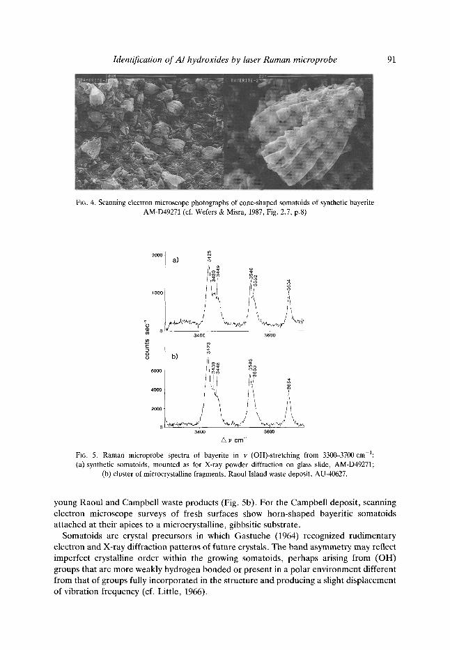
Rapid neutralization of alkaline aluminate solutions and subsequent ageing of the precipitate yields a well established sequence, X-ray indifferent ~ poorly crystallized boehmite ~ crystalline trihydroxide (e.g. Mackenzie et al., 1962; Schoen & Roberson, 1970; van Straten et al., 1984). Raman microanalysis of artificial precipitates yielded results consistent with this sequence, a Raman indifferent spectrum giving way on ageing to spectra in which asymmetrical bayerite bands became progressively dominant. Subordinate gibbsite bands occurred in one precipitate. Scanning electron microscope examination of the aged precipitate showed bayerite to occur mainly as somatoids with a characteristic cone-shaped habit, and commonly <3 ~m in size (Fig. 4).
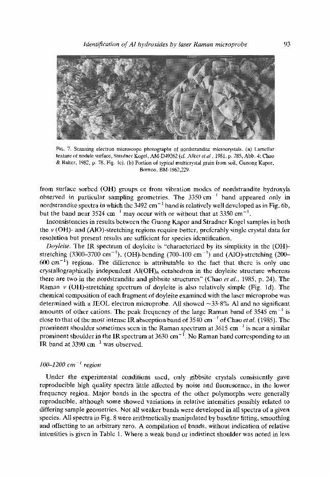
All Raman scattering (OH) bands from the artificial somatoids display an asymmetry such that the bands at about 3425 and 3545 cm -1 appear to incorporate component sub- bands (Fig. 5a). This asymmetry is less marked in spectra from mature bayerite crystallites at Raoul (Rodgers et al., 1989) and Campbell (Fig. lb) and is generally absent in spectra of the more crystalline aluminium hydroxide phases studied. It occurs in spectra derived from
Identification of AI hydroxides by laser Raman microprobe 91
F~6.4. Scanning electron microscope photographs of cone-shaped somatoids of synthetic bayerite AM-D49271 (cf. Wefers & Misra, 1987, Fig. 2.7, p.8)
2 0 0 0
I O
0
a) g
3400 3600
E ~
o~- m o 6000 ~
4O013
20(10
0 , - 3 4 0 0 3 6 0 0
Z~ 1,' e m -~
FIG. 5. Raman microprobe spectra of bayerite in v (OH)-stretching from 3300-3700 cm-l: (a) synthetic somatoids, mounted as for X-ray powder diffraction on glass slide, AM-D49271;
(b) cluster of microcrystalline fragments, Raoul Island waste deposit, AU-40627.
young Raoul and Campbell waste products (Fig. 5b). For the Campbel l deposit, scanning electron microscope surveys of fresh surfaces show horn-shaped bayeritic somatoids attached at their apices to a microcrystalline, gibbsitic substrate.
Somatoids are crystal precursors in which Gastuche (1964) recognized rudimentary electron and X-ray diffraction patterns of future crystals. The band asymmetry may reflect imperfect crystalline order within the growing somatoids, perhaps arising f rom (OH) groups that are more weakly hydrogen bonded or present in a polar environment different from that of groups fully incorporated in the structure and producing a slight displacement of vibration frequency (cf. Little, 1966).

92 K. A. Rodgers
Nordstrandite. Of the four nordstrandites studied, only two showed Raman scattering bands.
Nordstrandite from Stradner Kogel (Alker et al., 1981) gave clear v (OH)-stretching bands at 3492, 3566 and 3623 cm -1 (Fig. 6a). In some fragments the 3492 and 3623 cm 1 bands broadened towards their base and the background between the bands at 3492 and 3566 cm -a was elevated, merging into the two bands. This elevation occurs where a weak 3534 cm -] band is seen in some spectra of sample BM-1969,229.
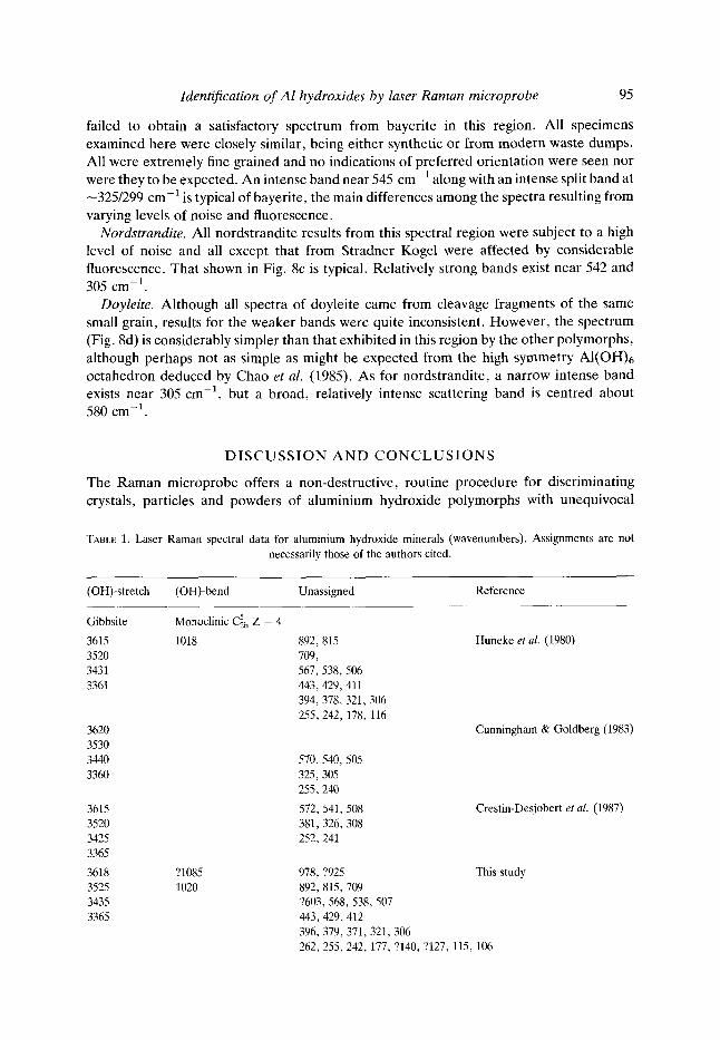
The mineral at Stradner Kogel occurs as white to pale grey, flinty, hemispherical nodules, 1-2 mm in diameter in cavities of a nephelinite. The surface of the hemispheres consists of very thin (<0.1/~m) laminae of nordstrandite ranging from 0.5-3/~m across, stacked in offset lamellar packets 5-10/~m high (Fig. 7a). The crystallographic orientation of these laminae may parallel the dominant 110 of nordstrandite (cf. Chao & Baker, 1982) and the spectral results may have been influenced by this orientation.
Sample BM-1962,229 from Guong Kapor consists of sand-sized grains separated from red soil. The extremely high noise, high fluorescence response of most crushed fragments discussed earlier, is typical for nordstrandites obtained from soils and weathering and oxidizing environments. An unquenchable background fluorescence with a count rate >300,000 s i was the norm obscuring Raman scattering bands. Small clear cleavages from one multicrystal grain (Fig. 7b) yielded distinct v (OH)-stretching bands (Figs. lc and 6b) similar to those found from the Stradner Kogel nordstrandite. A similar but lower quality Raman spectrum was obtained from this grain macrosampled using a Kr + laser.
Some nordstrandite spectra showed weak bands at about 3350 and 3534 cm 1, that at 3524 cm -a is coincident with a pronounced scattering band of gibbsite but the lower frequency band is centred about 15 cm -1 below that of gibbsite at 3365 cm -1. While weak bands in the nordstrandite spectra may be related to traces of impurities, they may also arise
2 0 0 0
1 0 0 0
r 0 E
I b) o 1 5 0 0 0
1 0 0 0 0
a)
co
i
A i
5 0 0 0
i
3 4 0 0 3 6 0 0
A v c m ~
FIG. 6. Raman microprobe spectra of nordstrandite in v (OH)-stretching from 3300-3700 cm ~: (a) fragments from nodule in nephelenite cavity, Stradner Kogel, AM-D49262; (b) fragment of red-
stained grain from soil, Gunong Kapor, Borneo, BM-1962,229.
Identification o f A l hydroxides by laser Raman microprobe
FIG. 7. Scanning electron microscope photographs of nordstrandite microcrystals. (a) Lamellar texture of nodule surface, Stradner Kogel, AM-D49262 (cf. Alker et al., 1981, p. 285, Abb. 4; Chao & Baker, 1982, p. 78, Fig. lc). (b) Portion of typical multicrystal grain from soil, Gunong Kapor,
Borneo, BM-1962,229.
93
from surface sorbed (OH) groups or from vibration modes of nordstrandite hydroxyls observed in particular sampling geometries. The 3350 cm x band appeared only in nordstrandite spectra in which the 3492 cm-1 band is relatively well developed as in Fig. 6b, but the band near 3524 cm -1 may occur with or without that at 3350 cm -1.
Inconsistencies in results between the Guong Kapor and Stradner Kogel samples in both the v (OH)- and (A10)-stretching regions require better, preferably single crystal data for resolution but present results are sufficient for species identification.
Doyleite. The IR spectrum of doyleite is "characterized by its simplicity in the (OH)- stretching (3300-3700 cm-1), (OH)-bending (700-100 cm -1) and (AIO)-stretching (200- 600 cm -1) regions. The difference is attributable to the fact that there is only one crystallographically independent AI(OH)6 octahedron in the doyleite structure whereas there are two in the nordstrandite and gibbsite structures" (Chao et al., 1985, p. 24). The Raman v (OH)-stretching spectrum of doyleite is also relatively simple (Fig. ld). The chemical composition of each fragment of doyleite examined with the laser microprobe was determined with a JEOL electron microprobe. All showed -33 .8% A1 and no significant amounts of other cations. The peak frequency of the large Raman band of 3545 cm-1 is close to that of the most intense IR absorption band of 3540 cm 1 of Chao et al. (1985). The prominent shoulder sometimes seen in the Raman spectrum at 3615 cm -1 is near a similar prominent shoulder in the IR spectrum at 3630 cm 1. No Raman band corresponding to an IR band at 3390 cm-1 was observed.
100-1200 cm -1 region
Under the experimental conditions used, only gibbsite crystals consistently gave reproducible high quality spectra little affected by noise and fluorescence, in the lower frequency region. Major bands in the spectra of the other polymorphs were generally reproducible, although some showed variations in relative intensities possibly related to differing sample geometries. Not all weaker bands were developed in all spectra of a given species. All spectra in Fig. 8 were arithmetically manipulated by baseline fitting, smoothing and offsetting to an arbitrary zero. A compilation of bands, without indication of relative intengities is given in Table 1. Where a weak band or indistinct shoulder was noted in less

94
8 0 0 0 -
4 0 0 0 �84
~0 0
K. A . Rodgers
3 0 O 0
ol
a) F b) 2000
e~
coo
o~ C ) 6000
ooo
500 1000
d)
i
= I
500 1000
A ~, c m -~
FIG. 8. Typical Raman microprobe spectra in the region 100-1200 cm 1 of fragments of aluminium hydroxide polymorphs microsampled on glass mounts. Intensities have been rescaled to arbitrary zero offset. (a) Gibbsite microcrystals pseudomorphous after crocoite, Dundas, Tasmania, AM- D44000; (b) synthetic bayerite somatoids; (c) red-stained nordstrandite grain from soil, Gunong Kapor, Borneo, BM-1962,229 (Wall et al., 1962); (d) doyleite cleavage, Mt Saint-Hilaire, Quebec, AM-D49268 (Chao et al., 1985). Note that this record may include some scattering from the glass
mount.
than three spectra, or where its position was inconsistent, this has been indicated by a question mark before the peak value.
Irrespective of such limitations, as in the (OH)-stretching region, each aluminium hydroxide modification shows a distinct Raman signature with a consistent pattern of medium to very strong bands (Fig. 8). The principal differences in the vibrational spectra of the aluminium hydroxide polymorphs below ~800 cm-1 have been related by Vivian et al.
(1973) to differences in the symmetry of the AI(OH)6 unit. These authors assigned all absorption bands in the IR spectra in this low-frequency region to vibrations arising from A1-O bonds, but attempts by them at factor group analysis, using published structural data proved satisfactory only for gibbsite. Wefers & Misra (1987) in their compilation were more conservative in assigning 1R bands to A1-O vibrations. Bands in the low-frequency region may also arise from librational motions of hydroxyls, lattice modes, and vibrations of hydrogen bonds.
Gibbsite. The gibbsite (Fig. 8a) matches that of Huneke et al. (1980) and Cooney et al.
(1989) particularly as to band shape, width and relative intensity. All gibbsite specimens in the present study showed this basic pattern with the strong doublet at 538/568 cm -1 and the narrow intense band at 321 cm -1 being typical of this species. Some spectra showed variation in relative intensities of some lines suggesting variations in sampling geometry.
Bayerite. The synthetic bayerite spectrum of Fig. 8b agrees with that given by Cooney et al. (1989) from aluminium hydroxide waste from Raoul Island. Huneke et al. (1980)
Identification o f A l hydroxides by laser Raman microprobe 95
failed to obtain a satisfactory spectrum from bayerite in this region. All specimens examined here were closely similar, being either synthetic or from modern waste dumps. All were extremely fine grained and no indications of preferred orientation were seen nor were they to be expected. An intense band near 545 cm -1 along with an intense split band at -325/299 cm-1 is typical of bayerite, the main differences among the spectra resulting from varying levels of noise and fluorescence.
Nordstrandite. All nordstrandite results from this spectral region were subject to a high level of noise and all except that from Stradner Kogel were affected by considerable fluorescence. That shown in Fig. 8c is typical. Relatively strong bands exist near 542 and 305 cm -1.
Doyleite. Although all spectra of doyleite came from cleavage fragments of the same small grain, results for the weaker bands were quite inconsistent. However, the spectrum (Fig. 8d) is considerably simpler than that exhibited in this region by the other polymorphs, although perhaps not as simple as might be expected from the high symmetry AI(OH)6 octahedron deduced by Chao et al. (1985). As for nordstrandite, a narrow intense band exists near 305 cm 1, but a broad, relatively intense scattering band is centred about 580 cm -1.
D I S C U S S I O N A N D C O N C L U S I O N S
The Raman microprobe offers a non-destructive, routine procedure for discriminating crystals, particles and powders of aluminium hydroxide polymorphs with unequivocal
TABLE 1. Laser Ramao spectral data for aluminium hydroxide minerals (wavenumbers). Assignments are not necessarily those of the authors cited.
(OH)-stretch (OH)-bend Unassigned Reference
Gibbsite MonoclinicC~h Z = 4
3615 1018 892, 815 3520 709, 3431 567,538, 506 3361 443, 429,411
394,378,321,306 255,242, 178, 116
3620 3530 3440 3360
3615 3520 3425 3365
3618 3525 3435 3365
?1085 1020
570, 540,505 325,305 255,240
572,541,508 381,326,308 252,241
Huneke et al. (1980)
Cunningham & Goldberg (1983)
Crestin-Desjobert et al. (1987)
978, ?925 This study 892, 815,709 ?603,568,538, 507 443,429, 412 396, 379,371,321,306 262, 255,242, 177, ?140, ?127, 115, 106

96 K . A . R o d g e r s
TABLE 1. Continued
(OH)-stretch (OH)-bend Unassigned Reference
Bayerite Monoclinic ~h Z = 2
3651 Huneke et al. (1980) 3542 3421
3651 866 Rodgers et al. (1989) 3541 545 Cooney et al. (1989) 3420 435
388, 322 297, 249,240, 106
3654 ?1085 ?981 This study 3545 1061 893,868 3425 1005 545,525
?480, 445,437 390, 360, 321 297, 250, 240, 203 ?(163-155), 147,141,118, 107
Nordstrandite Yriclinic Cll Z = 4 (Chao & Baker, 1982)
3492 ?1095 ?985, ?898 This study 3566 657, 634 3623 595, ?560, 541,506
493, ?464, 437, 412 390, 378, ?355,344, 313,305 285,266, 251, ?237, 227, ?216 ?177, 118, 108
Doyleite Triclinic P]- Z = 2 (Chao et al., 1985)
3545 ?1080 936, ?840, ?806 This study 3615 580
392, 305 279, 229, ?208 ?187, ?158, 124, 117, ?107
? Generally indicates weak to medium weak bands seen in less than three spectra or in spectra of only one specimen.
identification of a species being made from either the (OH)- or (A10)-s t re tching bands. The technique can assist in identifying components in mixtures of these polymorphs and can do so on a small scale from very fine-grained mater ial such as the products of exper imental crystallization studies. In part icular , the Raman microprobe offers an al ternative procedure in characterizing poor ly crystalline components in soils and waste products where a combinat ion of several determinat ive methods is often necessary (cf. Hsu, 1989).
Excessive noise and non-quenchable fluorescence pose the principle l imitations to using the technique in routine mineralogical and environmental studies, al though noise seldom affects the full spectral range. Fluorescent materials may be removed by chemical pre- t rea tment although this may risk altering the specimen in some unant ic ipated way. The availabili ty of an al ternative exciting line such as Kr + is helpful, but not essential.
The present study has been confined to aluminium hydroxides but the principles and

Identification o r A l hydroxides by laser Raman microprobe 97
p rocedures are of genera l appl ica t ion for the t echn ique to be used m o r e widely in rou t ine
minera l de te rmina t ions . M o n o c h a n n e l count ing , par t icular ly whe re m o r e than one spectra l
range is be ing scanned, may limit the usefulness of the laser R a m a n m i c r o p r o b e in this
regard.
A C K N O W L E D G M E N T S
This study was supported by a Visiting Fellowship awarded by the Australian Museum. Thanks are due to the Keeper of Mineralogy, Natural History Museum, Professors R. Cooney and Saalfeld, Drs G. Chao, J. Seakins, F. L. Sutherland and K. Wefers, and G. Avern, C. Cantrell, M. Kumvaj, R. Pogson, R. Ratajczak, T. Smith and T. Trnski for assistance. Equipment funding was provided by the University of Auckland Research Committee, the University Grants Committee, and the New Zealand Lottery Grants Board (Scientific).
R E F E R E N C E S
ALKER A., GOLOB P., POSTL W. & WALTINGER H. (1981) Hydrotalcite, Nordstrandit und Motukoreait vom Stradner Kogel, stidlich Gleichenberg, Steiermark. Mitt. Bl. Abt. Miner. Landesmuseum Joanneum 49, 279-292.
CALVERT C.S., WEEO S.B. & BUOL S.W. (1979) Estimation of amorphous material, 1:1 type layer silicates, and gibbsite dissolved in hot alkali treatment of soil clay. J. Soil Sci. Soc. Am. 43, 778--781.
CHAO G.Y. & BAKER J. (1982) Nordstrandite from Mont St-Hilaire, Quebec. Can Miner. 20, 77-85. CHAO G.Y., BAKER J., SABINA A.P. & ROBERTS A.C. (1985) Doyleite, a new polymorph of AI(OH)3, and its
relationship to bayerite, gibbsite and nordstrandite. Can. Miner. 23, 21-28. COONEY R.P., ROOGERS K.A. & GREGORY M.R. (1989) Laser Raman spectrum of bayerite from Raoul Island,
Kermadec Group, South Pacific. Vulkanologiya i Seismologiya No.5, 97-99 (in Russian). CRESTIN-DESJOBERT S., CRUEGE F. • GOUT R. (1987) The use of the Raman microprobe for studying bauxites. Terra
Cognita 7, 14. CUNNINGHAM K.M. & GOLDBERG M.C. (1983) A reexamination of the effects of adsorbates on the Raman spectrum
of gibbsite. Soil Sci. 136, 102-109. DELHAYE M. t~ DHAMELINCOURT P. (1987) Instrumentation Raman spectroscopy. Terra Cognita 7, 15. ELDERFIELD H. & HEM J.D. (1973) The development of crystalline structure in aluminium hydroxide polymorphs on
ageing. Mineral. Mag. 39, 89-96. FREDER1CKSON L.D. (1954) Characterization of hydrated aluminas by infrared spectroscopy. Application to the
study of bauxite ores. Anal. Chem. 26, 1883-1885. GASTUCHE M.C. (1964) The octahedral layer. Clays Clay Miner. 12, 471--493. GOLDSCHMIDT V. (1918) Atlas der Krysallformen Taflen 4, Karl Winters, Heidelberg. HUNEKE J.T., CRAMER R.E., ALAVREZ R, & EL-SwAIFY S.A. (1980) The identification of gibbsite and bayerite by
laser Raman spectroscopy. J. Soil Sci. Soc. Am. 44, 130--134. Hsu P.H. (1989) Aluminium hydroxides and oxyhydroxides. Pp. 331-378 in: Minerals in Soil Environments (J.B.
Dixon & S.B. Weed, editors) Soil Science Society of America, Book Series 1. LittLE L.H. (1966) Infrared Spectra of Adsorbed Species. Academic Press, London, New York. MACKENZIE R.C., MELDAU R. & GARD J.A. (1962) The ageing of sesquioxide gels II. Alumina gels. Mineral. Mag.
33, 145-157. MOOI~E D.M. & REYNOLDS R.C. (1989) X-ray Diffraction and Identification and Analysis of Clay Minerals. Oxford
University Press, Oxford. RODGERS K.A. (1992) The laser Raman identity of gibbsite pseudomorphous after crocoite from Dundas, Tasmania.
Pap. Proc. Roy. Soc. Tasm. 126, 1-5. RODGERS K.A., GREGORY M.R. & BARTON R. (1991) Bayerite, nordstrandite, gibbsite, brucite, and pseudo-
boehmite in discharged caustic waste from Campbell Island, southwest Pacific. Clay Clay Miner. 39, 103-107. RODGERS K.A., GREGORY M.R. & COONEY R.P. (1989) Bayerite, AI(OH)3, from Raoul Island, Kermadec Group,
South Pacific. Clay Miner. 24, 531-538. RODGERS K.A., GREGORY M.R., COURTNEY S.F. ~,z SHANKL1N J.D. (1992) Aluminium hydroxide polymorphs in a
waste deposit from Faraday Base, Argentine Islands. N. Jb. Miner. Mh. 1992(3), 127-133. RUSSELL J.D., PARFITr R.L., FRASER A.R. & FARMER V.C. (1974) Surface structures of gibbsite, goethite and
phosphated goethite. Nature 248, 220-221.

98 K. A . R o d g e r s
SAALFELD H. (1960) Struckuren des Hydrargillits und der Zwischenstufen beim Entw~issern. N. Jb. Miner Abh. 95, 1-87.
SCHOEN R. & ROBERSON C.E. (1970) Structures of aluminium hydroxides and geochemical implications. Am. Miner. 55, 43-77.
WALL J.R.D., WOLVENDEN E.B., BEARD E.H. & DEANS T. (1962) Nordstrandite in soil from west Sarawak, Borneo. Nature 196, 264-265.
WEFERS K. & MISRA C. (1987) Oxides and hydroxides of aluminium. Aloca Technical Paper 19, 92 pp. (revised). WILMOT R.D. & YOUNG B. (1985) Aluminite and other aluminium minerals from Newhaven, Sussex: the first
occurrence of nordstrandite in Great Britain. Proc. Geol. Ass. 96, 47-52. VAN STRATEN H.A., HOLTKAMP B.T.W. & DE BRVYN P.L. (1984) Precipitation from supersaturated aluminate
solutions. I Nucleation and growth of solid phases at room temperature. J. Coll. lnterf. Sci. 98, 342. VIVlEN D., STEGMAN M-C. & MAZ~RES C. (1973) Contribution a l'6tude par spectroscopic infrarouge et RMN large
bande des hydroxydes d'aluminium gibbsite, bayerite, nordstrandite. J. Chim. Phys. 70, 1502-1507.
A P P E N D I X
Samples examined by laser Raman microscopy.
AM-D 11284. Gibbsite with "cobaltiferous manganese", Perseverance Mine, Moneo, New Caledonia. Translucent, off-white, chalcedony-like, globular crystalline mass with radial fibrolamellar structure showing a pronounced splintery fracture along fibres. Raman: gibbsite.
AM-D11695. Gibbsite with "cobaltiferous manganese". Perseverance Mine, Moneo, New Caledonia. Small transparent crystals and translucent, globular, chalcedony-like layers with fibrolamellar stucture akin to Dl1284, and splintering in white, translucent, cryptocrystalline flakes with subconchoidal fracture. Interlayered with black, metallic, cryptocrystalline lithophorite and cryptomelane. Raman: gibbsite.
AM-DC42734. Gibbsite on crocoite, Tansmania. Acicular, prisms of crocoite invested with tufts and balls of white microcrystalline gibbsite. Raman: gibbsite.
AM-D44000. Gibbsite replacing crocoite, Adelaide Mine, Dundas, Tasmania. Gossan coated with wad and encrusted by radiating acicular hollow prisms of crocoite partly pseudomorphed by mierocrystalline euhedral gibbsite, 10-50/~m dia. (Rodgers, 1992). Raman: gibbsite.
AM-D46576. Gibbsite on crocoite, Adelaide Mine, Dundas, Tasmania. Pisolitic stalactites of gibbsite lining cavity in gossan containing crocoite. Raman: gibbsite; XRD: gibbsite.
AM-D48300. Gibbsite with iron and manganese oxides, Iron Baron, South Australia. Small crystals and worn translucent to clear globular aggregates with fibrolamellar structure and splintery fracture. Raman: gibbsite.
BM-64525 (AM-D49264). Bayerite, Hove, Brighton, East Sussex, England. Glistening, microcrystalline, white aggregate with patchy orange-yellow staining. Raman: gibbsite + minor bayerite; XRD: gibbsite.
BM-1907,666 (AM-D49265). Gibbsite intermixed with allophane. East Sussex, England. Glistening, micro- crystalline, white concretionary fragment. Raman: nil; XRD: nil.
BM-1962,229 (AM-D49266). Nordstrandite, Gunong Kapor, Bau mining district, West Sarawak. (Wall et al., 1962). Sand-sized grains separated from red soil. Angular to subhedral stained red-brown grains, 0.4-0.8 mm dia. Raman: nordstrandite.
Identification orAl hydroxides by laser Raman microprobe 99
BM-1985,147 (AM-D49267). Nordstrandite, West Cliff, Newhaven, Sussex. Soft, friable, microcrystalline pale aggregate, stained with limonite and including allophane. (Wilmot & Young, 1985). Raman: poor spectrum, high fluorescence, ?gibbsite; XRD: gibbsite.
AM-D49262. Nordstrandite with hydrotalcite on phillipsite in vug of hauyne nephelinite, Stradner Kogel, Gleichenberg, Austria (Alker et al., 1981). White, 1-2 mm alia., hemispheres of fine-grained, flinty nordstrandite imbedded with euhedral ?trigonal prisms of hydrotalcite and nested on microcrystalline phillipsite lining a vug in a hauyne-nephelinite. Raman: nordstrandite; XRD: nordstrandite.
AM-D49268. Doyleite, Mont Saint-Hilaire, Quebec. Soft, brittle, colourless and transparent to translucent platy crystals with a vitreous to pearly lustre and a pronounced cleavage. (Chao et al., 1985). Raman: doyleite; chem. anal.: 33.8 wt% AI.
AU-3066 (AM-D49269). Campbell Island hydrogen generator waste spill. Fine-grained, alternating hard and soft, millimetre thick laminae of aluminium hydroxide. (Rodgers et al. , 1991). Raman: bayerite + gibbsite; XRD: bayerite + gibbsite + nordstrandite.
AU-3077-82, Faraday Base, Argentine Islands, hydrogen generator waste spill. Pale-grey, moist, granular paste, which disaggregated into very fine white powder variously clumped, upon freeze-drying. (Rodgers et aL, 1992). Raman: gibbsite + minor bayerite; XRD: gibbsite + minor bayerite.
AU-40627 (AM-D49270). Raoul Island hydrogen generator waste spill. White to greenish-grey, multi-lamellar, botryoidal, microcrystalline, concretionary masses. (Rodgers et al. , 1989; Cooney et al. , 1989). Raman: bayerite; XRD: bayerite; chem. anal.: 60~56% A1203.
AM-D49271. Synthetic bayerite. Transparent microcrystalline somatoids prepared according to recipe of Schoen & Roberson (1970). (i) Raman: bayerite + minor ?gibbsite; XRD: bayerite + "vitreous SiO" (JCPDS 3-1092); (ii) Raman: bayerite + minor ?gibbsite; XRD: bayerite + ?boehmite.
All samples described are now vested in the collection of the Australian Museum. The original provenance of a specimen is indicated by: AM = Australian Museum, Sydney; AU = University of Auckland; BM = Natural History Museum, London; and followed where possible by the catalogue number of that institution. Where a new Australian Museum catalogue number has been assigned this is shown in parenthesis.