roquin binds microrna-146a and argonaute2 to regulate ... · sashika richards1, anil verma4, e....
TRANSCRIPT

ARTICLE
Received 3 Jun 2014 | Accepted 9 Jan 2015 | Published 20 Feb 2015
Roquin binds microRNA-146a and Argonaute2to regulate microRNA homeostasisMonika Srivastava1,*, Guowen Duan1,*, Nadia J. Kershaw2,*, Vicki Athanasopoulos1, Janet H.C. Yeo3,
Toyoyuki Ose4, Desheng Hu5, Simon H.J. Brown6, Slobodan Jergic6, Hardip R. Patel7,8, Alvin Pratama1,
Sashika Richards1, Anil Verma4, E. Yvonne Jones4, Vigo Heissmeyer5,9, Thomas Preiss7,10, Nicholas E. Dixon6,
Mark M.W. Chong3,*, Jeffrey J. Babon2,* & Carola G. Vinuesa1,*
Roquin is an RNA-binding protein that prevents autoimmunity and inflammation via
repression of bound target mRNAs such as inducible costimulator (Icos). When Roquin is
absent or mutated (Roquinsan), Icos is overexpressed in T cells. Here we show that Roquin
enhances Dicer-mediated processing of pre-miR-146a. Roquin also directly binds Argonaute2,
a central component of the RNA-induced silencing complex, and miR-146a, a microRNA that
targets Icos mRNA. In the absence of functional Roquin, miR-146a accumulates in T cells. Its
accumulation is not due to increased transcription or processing, rather due to enhanced
stability of mature miR-146a. This is associated with decreased 30 end uridylation of the
miRNA. Crystallographic studies reveal that Roquin contains a unique HEPN domain and
identify the structural basis of the ‘san’ mutation and Roquin’s ability to bind multiple RNAs.
Roquin emerges as a protein that can bind Ago2, miRNAs and target mRNAs, to control
homeostasis of both RNA species.
DOI: 10.1038/ncomms7253 OPEN
1 Department of Pathogens and Immunity, John Curtin School of Medical Research, Canberra, Australian Capital Territory 2601, Australia. 2 Division ofStructural Biology, Walter and Eliza Hall Institute and The University of Melbourne, Melbourne, Victoria 3052, Australia. 3 Genomics and Immunologylaboratory, St Vincent’s Institute of Medical Research, Fitzroy, Victoria 3065, Australia. 4 Wellcome Trust Centre for Human Genetics, University of Oxford,Roosevelt Drive, Oxford OX3 7BN, UK. 5 Helmholtz Zentrum Munchen, Institute of Molecular Immunology, D-81377 Munchen, Germany. 6 Centre forMedical and Molecular Bioscience, University of Wollongong and Illawarra Health and Medical Research Institute, Wollongong, New South Wales 2522,Australia. 7 Department of Genome Biology, John Curtin School of Medical Research, Canberra, Australian Capital Territory 2601, Australia. 8 GenomeDiscovery Unit, John Curtin School of Medical Research, Canberra, Australian Capital Territory 2601, Australia. 9 Ludwig-Maximilians-Universitat Munchen,Institute for Immunology, D-80336 Munchen, Germany. 10 Victor Chang Cardiac Research Institute, Darlinghurst, New South Wales 2010, Australia. * Theseauthors contributed equally to this work. Correspondence and requests for materials should be addressed to C.G.V. (email: carola.vinuesa @anu.edu.au).
NATURE COMMUNICATIONS | 6:6253 | DOI: 10.1038/ncomms7253 | www.nature.com/naturecommunications 1
& 2015 Macmillan Publishers Limited. All rights reserved.

Posttranscriptional gene regulation by trans-acting RNA-binding proteins is a rapid and efficient way to modify geneexpression and cellular responses. Mutations in these trans-
acting factors are often associated with pathology1. The proteinRoquin (encoded by Rc3h1), known to bind target mRNAs andpromote their decay, has been found to be crucial in themaintenance of peripheral immune tolerance. Mice either lackingRoquin/Roquin2 or homozygous for the Roquin ‘san’ mutation(sanroque mice) have impaired posttranscriptional regulation ofmRNAs in T cells and macrophages2–6. Roquin is a ubiquitouslyexpressed RING-E3 ubiquitin ligase family member containing ahighly conserved ROQ domain required for RNA binding andlocalization to stress granules7,8 and a CCCH zinc finger motif.Roquin binds a stem-loop structure termed the constitutive decayelement (CDE) within the 30-UTR of Tnf mRNA and recruits theCcr4-Caf1-Not deadenylation machinery leading to mRNAdecay6. mRNA degradation can also be mediated by Roquin’srecruitment of Rck/Edc4/Dcp1a decapping complexes and isthought to occur independently of miRNA activity8.
MicroRNAs (miRNAs) are small 20� 22 nt non-coding RNAsthat regulate specific mRNA targets mainly by translationalinhibition and/or mRNA decay9. They are involved in mostcellular responses and their dysregulation has been shown to beassociated with autoimmune diseases10. Synthesized in thenucleus as long primary transcripts, they are cleaved intoprecursor miRNAs (pre-miRNAs) and exported to thecytoplasm where the hairpin structure is processed by theRNaseIII complex Dicer/Tarbp2 into the mature B22 nt longimperfect miRNA duplex. This is loaded into the miRNA RNA-induced silencing complex (miRISC), comprised of severalproteins including GW182 and Ago2. Once loaded into themiRISC, miRNAs interact with sequences within targetmRNAs9,11.
Although much is known about miRNA biogenesis andfunction, relatively little is known about miRNA homeostasisand its importance in immunity or autoimmunity. Specificnucleotides present in mature miRNAs, terminal-end modifica-tions of miRNAs like adenylation or uridylation and subcellularlocalization have been shown to affect miRNA homeostasis butnone of these mechanisms has yet been shown to regulate miRNAlongevity in mammals12. Ribonucleases such as Sdn1 and Xrn1regulate miRNA half-life in Arabidopsis and Caenorhabditiselegans, respectively13 but evidence of a similar role inmammalian cells is lacking. Recently Eri1, a highly conserved30-to-50 exoribonuclease, was shown to regulate miRNAhomeostasis in murine lymphocytes but the exact mechanism isstill unknown14. Here we show that Roquin is one such regulatorof miRNA longevity. Roquin binds to miR-146a and its target IcosmRNA and promotes their decay. Icos and miR-146a limit Tfollicular helper (Tfh) cells to prevent the development ofautoantibodies and autoimmunity15. Roquin also forms acomplex with the RISC component Ago2. These data implicateRoquin as a facilitator of miRNA-mediated target decay.
ResultsRoquin represses miR-146a levels within T cells. We previouslyreported that several miRNAs were upregulated in T cellshomozygous for the hypomorphic ‘san’ allele of Roquin3.Genome-wide miRNA profiling of purified naive CD4þ T cells(Fig. 1a) from Roquinsan/san and Roquinþ /þ control littermatesrevealed 15 miRNAs were overexpressed by 4twofold inRoquinsan/san T cells and none were downregulated (Fig. 1b andSupplementary Table 1). miR-146a (mmu-miR-146a-5p) andmiR-21 (mmu-miR-21-5p) showed the greatest change: 25-foldand 14-fold, respectively. Quantitative real-time PCR (qRT–PCR)
confirmed the increase in miRNA expression levels (Fig. 1c),which was not present in B cells, murine embryonic fibroblasts(MEFs) or other T-cell subsets (Supplementary Fig. 1a,b).Quantification of miR-146a in naive CD4þ T cells lacking bothRoquin and Roquin-2 also revealed miR-146a accumulation(Fig. 1d) confirming that the failure of Roquinsan to limit miR-146a was a reflection of loss of function of the Roquin paralogues.
To test whether Roquinsan was acting within T cells to causethe increase in miR-146a, we constructed mixed bone marrowchimeric mice. For this, mice labelled with a congenic marker(Ly5b) were sublethally irradiated and reconstituted with a 50:50mix of Roquinsan/san.Ly5b and Roquinþ /þ .Ly5a bone marrowcells. A control group was reconstituted with a 50:50 mix ofRoquinþ /þ .Ly5b and Roquinþ /þ . Ly5a bone marrow. Stemcells present in the bone marrow can reconstitute the hemato-poietic compartment of irradiated recipient mice and the
Roq
uinsa
n/sa
n
mir-146amir-21
0
1
2
3
4
5
6
0
1
2
3
4
5
+/+Roquin
*** **
012345678
Roquin +/+ +/+ +/+ san/sana b a bLy5
Roquin +/+ +/+ +/+ san/sana b a bLy5
2-ddc
t of m
iR-1
46a
2-ddc
t of m
iR-2
1
CD4 CD44
Roquin +/+
Roquinsan/san
B22
0
CD
25
012345678
0
5
10
* NS
miR
-146
a ex
pres
sion
(rel
ativ
e to
sno
RN
A20
2)
2-ddc
t of m
iR-1
46a
2-ddc
t of m
iR-2
1
Rc3h1f fl/fl:Rc3h2 fl/fl;CD4-cre
Rc3h1fl/fl:Rc3h2 fl/fl (wt)
Roquinsan/san
san/san+/+ san/san
Roquin +/+
15
10
5
0
2 4 6 8 10 12 14 16 18
Figure 1 | Loss of Roquin causes T cell-autonomous miRNA accumulation.
(a) Flow cytometric stains from Roquinsan/san and Roquinþ /þ T cells
showing the gating strategy used to sort CD4þCD44loCD25� naive
T cells. (b) Scatter plot of microRNA expression in sorted naive T cells
obtained by microarray analysis. (c) qRT–PCR showing relative expression
levels of mature miR-146a and miR-21 in naive T cells, normalized to U6.
(d) Relative expression levels of miR-146a measured by qRT–PCR in
Rc3h1fl/fl;Rc3h2fl/fl; CD4-Cre double knockout naive T cells normalized
to snoRNA202. (e,f) qRT–PCR analysis of miR-146a (e) and miR-21
(f) in Roquinsan/san.Ly5b: Roquinþ /þ .Ly5a or of Roquinþ /þ .Ly5b:
Roquinþ /þ .Ly5a mixed chimeras. Each dot represents an individual mouse
and the bar represents the median value in each group. U-test: *Po0.02,
**Po0.05, ***Po0.005. See also Supplementary Fig. 1a,b.
ARTICLE NATURE COMMUNICATIONS | DOI: 10.1038/ncomms7253
2 NATURE COMMUNICATIONS | 6:6253 | DOI: 10.1038/ncomms7253 | www.nature.com/naturecommunications
& 2015 Macmillan Publishers Limited. All rights reserved.

congenic markers Ly5a/b allow identification of T cells fromRoquin mutant and wild-type origin. miR-146a was upregulatedin naive CD4þ T cells in a cell-autonomous fashion: onlyRoquinsan/san (Ly5b) cells had elevated levels of this miRNAcompared with Roquinþ /þ (Ly5a) cells in the same mice(Fig. 1e). In the case of miR-21, wild-type cells also had elevatedlevels of this miRNA compared with the control chimeras(Fig. 1f) suggesting a cell-extrinsic mechanism although a cell-intrinsic component could not be excluded. These results showthat Roquin acts in T cells to repress miR-146a and possibly othermiRNAs.
Roquin enhances Dicer-mediated processing of pre-miR-146a.To map the stage at which Roquin represses miR-146a accumu-lation, we investigated the amount of primary and precursormiRNAs present in naive T cells. qRT–PCR using three differentsets of primers to specifically amplify primary miR-146a in naiveT cells revealed no differences in the presence of Roquinsan
(Fig. 2a), and this observation extended to primers in the pre-miR-146a region (Fig. 2b). Analysis of miR-146a by northernblotting confirmed the selective accumulation of maturemiR-146a in Roquinsan/san (Fig. 2c), and no accumulation ofcontrol miR-150, a miRNA that did not change in Roquinsan/san
T cells. These results point to mature miR-146a accumulatingeither during or after Dicer-mediated processing.
To investigate Roquin-mediated processing of precursormiRNAs, we next quantified mature miR-146a in MEFs sufficientor deficient in Dicer expression. MEFs were retrovirallytransduced with Roquinwt, Roquinsan or empty vector expressedfrom an IRES-GFP reporter construct. Twenty-four hours aftertransduction, all the cells were treated with tamoxifen thatremoved Dicer from the CreER expressing cells only, and cellswere sorted for GFP expression after 1 week. miR-146aaccumulation was observed in Dicer-expressing MEFs transducedwith Roquinsan but accumulation did not occur in the absence ofDicer (Fig. 2d). This indicates that Roquin-induced miR-146aaccumulation requires Dicer-mediated processing.
We next investigated whether Roquin itself has miRNAprocessing activity in vitro and can cleave pre-miRNAs into theirmature form. Unlike Dicer, Roquin alone did not cleave pre-miR-146a (Fig. 3a). Addition of Roquin enhanced Dicer’s cleavageability by B2.5-fold (Fig. 3b) suggesting that it may form a partof the Dicer processing complex. Importantly, the ‘san’ mutationin Roquin did not further enhance Dicer’s function (Fig. 3b)suggesting that Roquin-induced enhancement of Dicer-mediatedprocessing is not the cause of mature miR-146a accumulation inthe presence of Roquinsan.
0.0
0.5
1.0
Roquin
NS1.5
NSNS
0.0Roquin
0.5
1.0
1.5
2.0NS
Vector
Roquinwt
Roquinsan
wt Dicer1–/–
0.0
0.5
1.0
1.5
2.0
2.5 * **
miR
-146
a U
6 sn
RN
A
miR
-150
Precursor
san/
san
+/+
Mature20
30
40506070
80100150
70
80
150100
Pri-mmu-miR-146a
Precursor
Mature
(iii)
2-ddc
t of m
iR-1
46a
Msan/
san
+/+M
2-ddc
t of p
ri-m
iR-1
46a
+/+ san/san +/+ san/san +/+ +/+san/san san/san
(i) (ii) (iii)2-d
dct o
f pre
-miR
-146
a
(i)
(ii)
Figure 2 | Post-transcriptional Dicer dependent accumulation of miR-146a. (a) Relative expression of pri-miR-146a in naive T cells sorted from
Roquinþ /þ and Roquinsan/san mice measured by qRT–PCR using three different sets of primers shown as (i), (ii) and (iii) in the schematic. NS, not
significant (Po0.05, U-test). (b) The levels of pre-miR-146a in Roquinþ /þ and Roquinsan/san naive T cells measured by qRT–PCR. (c) Northern blots
showing precursor and mature miRNA transcripts (arrows) of miR-146a and miR-150 in naive T cells of Roquinsan/san and Roquinþ /þ mice. U6 snRNA
was used as a loading control and is shown below the miR-146a northern blot. Weaker less abundant bands are miRNA decay products. M, marker.
(d) Relative expression of miR-146a in Dicer-floxed-Rosa26-Cre-ER MEFs (Dicer1� /� ) and CD4-CRE (wt) control MEFs assessed by qRT–PCR. MEFs were
retrovirally transduced with Roquinwt and Roquinsan containing a GFP reporter or with an empty GFP vector control. After 2 days, MEFs were treated
with tamoxifen to delete Dicer. GFP-positive cells were sorted 7 days later. Each dot represents individual mice (a,b) or technical replicates (d) and the bars
represent the median value in each group. U-test: *Po0.05, **Po0.005. Results are representative of two independent experiments.
NATURE COMMUNICATIONS | DOI: 10.1038/ncomms7253 ARTICLE
NATURE COMMUNICATIONS | 6:6253 | DOI: 10.1038/ncomms7253 | www.nature.com/naturecommunications 3
& 2015 Macmillan Publishers Limited. All rights reserved.

Roquin controls mature miRNA half-life. An explanation forthe accumulation of mature miR-146a is increased stability. miR-146a decay was investigated after treatment with actinomycin D3
which prevents on-going transcription. The amount of miR-146adropped below 20% of initial levels within 0.5 h of actinomycin Dtreatment in wild-type naive CD4þ T cells, whereas no decay wasobserved in Roquinsan/san cells up to 3 h after treatment, pointingto increased stability as the cause of miR-146a accumulation(Fig. 4a). To study the effects of Roquin on miRNA half-life inisolation from transcription-related events, we transfectedHEK293T cells (that express a negligible amount of miR-146a),with synthetic precursor-like miR-146a and either wild-type ormutant Roquin-expressing constructs. Five days post transfection,miR-146a levels had decayed by 50% in the presence of Roquinwt,but only by 10% with Roquinsan (Fig. 4b). A scramble controlmiRNA had no effect (Supplementary Fig. 2a). In parallelexperiments, retroviruses expressing pre-miR-146a along withretroviral Roquinwt IRES-GFP or Roquinsan IRES-GFP were usedto transduce wild-type MEFs. GFPþ cells were sorted 7 days laterand miR-146a was again found to be significantly higher inRoquinsan expressing cells (Fig. 4c). Together these resultsdemonstrate that Roquin acts to regulate miRNA decay.
Intracellular localization of miR-146a was investigated byin situ hybridization. Cytoplasmic miR-146aþ granules werereadily visible in Roquinsan/san naive T cells but not clearlyappreciable in control T cells, which precluded comparativequantification. miR-146a did not co-localize with the P-bodymarker Dcp-1, whereas some miR-146aþ granules wereeIF3þ (Supplementary Fig. 2b) suggesting localization to stressgranules. miRNAs are stored in exosomes particularlywhen found in excess16. Consistent with this, there was
increased miR-146a within exosomes in Roquinsan/san T cellsin the presence of comparable total numbers of exosomes(Supplementary Fig. 2c–e).
MiRNA editing and tailing17 has been shown to affect miRNAstability in plants18 and mammals19,20. Decreased uridylation hasbeen specifically associated with increased miRNA stability andabundance19–21. To investigate possible differences in non-templated additions of miR-146a in the presence of mutantRoquin, small RNA deep sequencing from Roquinþ /þ andRoquinsan/san T cells was carried out. As expected, reads wereprimarily 22 nt in length (Supplementary Fig. 3a), and differentialexpression analysis (Supplementary Fig. 3b) confirmed the trendsseen with the earlier microarray data (Fig. 1b). ActivatedRoquinsan/san CD4þ T cells expressed higher levels of miR-146a (Fig. 5a). Overall, non-templated addition was seen largelyrestricted to A and U and there was no pronounced global changebetween wild-type and mutant (Fig. 5b). Next, we rankedmiRNAs by change in mono-uridylation. Out of 364 analysedmiRNAs, miR-146a alongside 39 additional miRNAs were in thetop quartile of decreased uridylation (Supplementary Fig. 3c):compared with Roquinþ /þ T cells, miRNA-146a uridylationdecreased by 4% in Roquinsan/san (Fig. 5c) T cells. In summary,defective Roquin leads to an increase in miR-146 half-lifeassociated with decreased mono-uridylation and accumulationof the miRNA in exosomes.
Anti-flag /GFP IP (beads)
––––1
++––2
+–+–3
+––+4
0.00
0.05
0.10
0.15
Rat
io o
f miR
-146
a(m
atur
e/pr
ecur
sor)
FLAG-DicerGFPFLAG-DICER
Roquinwt-GFP
Roquinsan-GFP
–
–
–2
–
+
–3
–
–
+4
+
+
–5
+
–
–1
+
–
+6
Mature
Pre
M
20
30
40506070
20
30405060
Roquinwt-GFPRoquinsan-GFP
Figure 3 | Roquin enhances Dicer mediated pre-miR-146a processing.
(a) In vitro processing of pre-miR-146a in HEK293T cells after transfection
with FLAG-Dicer (lane 1), Roquinwt-GFP (lane 3) or Roquinsan-GFP (lane 4).
Co-transfection of FLAG-Dicer with either Roquinwt-GFP (lane 5) or
Roquinsan-GFP (lane 6) is included. No transfection control is shown in
lane 2. (b) In vitro processing of pre-miR-146a in HEK293T cells after
transfection with FLAG-Dicer (lane 2) alone, or co-transfection of
FLAG-Dicer with either Roquinwt-GFP (lane 3) or Roquinsan-GFP (lane 4).
Lane 1: No transfection control. The lower panels show densitometric
analysis of the northern blots to quantify 32P. The ratios of the mature
versus precursor miRNA intensities are plotted. Intermediates or low
abundance compared with the fully digested products were not included
in the quantification.
Day 2 Day 5
NS
*
0.0
0.5
1.0
1.5
**
0.0 0.5 1.0 1.5 2.0 2.5 3.0
10
100
miR
-146
a (%
)
0.0
0.5
1.0
1.5
2.0
2.5
**
NS
GFP vector
Roquinwt-GFPRoquinsan-GFP
MEFsHEK293T
1
GFP vector Roquinwt-GFP
Roquinsan-GFP
2-ddc
t of m
iR-1
46a
2-ddc
t of m
iR-1
46a
Time after actinomycin D (h)
Roquin+/+
Roquinsan/san
Figure 4 | Roquin regulates miR-146a stability in vitro and in vivo. (a)
miR-146a in Roquinsan/san and Roquinþ /þ naive T cells measured by qRT–
PCR and normalized to comparatively stable U6 snRNA. The cells were
treated with actinomycin D (10 mg ml� 1) for the times indicated. The
amount of miR-146a at 0 h was assigned 100%. The error bar represents
the range of the data for two experiments. (b) Pre-miR-146a was
transfected along with Roquinwt-GFP or Roquinsan-GFP in HEK293T cells
and qRT–PCR for miR-146a was performed on GFP-positive cells on days 2
and 5 post transfection. See also Supplementary Fig. 2a. (c) miR-146a in
MEFs 7 days after retroviral transduction of precursor miR-146a along with
Roquinwt or Roquinsan. Each dot represents a technical replicate and the
bars represent the median value in each group. U-test: *Po0.01,
**Po0.001, NS, not significant. The data are representative of two
independent experiments.
ARTICLE NATURE COMMUNICATIONS | DOI: 10.1038/ncomms7253
4 NATURE COMMUNICATIONS | 6:6253 | DOI: 10.1038/ncomms7253 | www.nature.com/naturecommunications
& 2015 Macmillan Publishers Limited. All rights reserved.

Roquin binds to miR-146a in vitro and in vivo. MiR-146a bindstwo Icos target sites (TS1 and TS2) that flank the CDE within Icos30-UTR (Supplementary Fig. 6)15. Roquin7,8 and miR-146a alsorepress Icos mRNA (Pratama et al., submitted). To test whetherRoquin can bind miR-146a, we immunoprecipitated Roquin frommouse CD4þ T cell lysates and quantified the amount of miR-146a in the precipitate by qRT–PCR. The pull-down using ananti-Roquin polyclonal antibody contained approximatelyfourfold more miR-146a compared with pull-downs usingisotype control or Roquin antibody pre-incubated with ablocking Roquin peptide (Fig. 5d). U6 was amplified as acontrol; no statistically significant increase specific to Roquinantibody was observed (Fig. 5d). These results show that Roquinforms a complex with mature miR-146a.
To confirm that this was a direct interaction, we used surfaceplasmon resonance (SPR) experiments with the amino (N)-terminal 1� 484 fragment of Roquin (RoquinWT1–484) thatcontains the ROQ and CCCH RNA-binding domains previouslyshown to bind ICOS mRNA by SPR7. Both wild-type and mutantRoquin (RoquinM199R1–484) were capable of binding miR-146a(Fig. 5e,f) but not miR-21 (data not shown). Binding affinity wasapproximately threefold higher for mutant Roquin (Fig. 5e,f) aspreviously observed for Icos mRNA7.
The structure of Roquin reveals novel RNA-binding domains.To gain insight into the molecular details of the interaction ofRoquin with RNA, we determined the crystal structures of the
Rel
ativ
e re
spon
se
Time (s)
Res
pons
e (R
U)
Log([ROQ1–484/M])
–9 –8 –7 –6 –5
IP: IgG
NoAb
Roquin
Roq p
ep IP: IgG
NoAb
Roquin
Roq p
ep0
1
2
4
5
3
**
miR
-146
a(%
of t
otal
inpu
t)
U6
(% o
f tot
al in
put)
1
2
4
3
NS
0
5
RoquinM199R1–484
RoquinWT1–484
0
2
4
6
0
5,000
10,000
15,000
50
40
30
20
10
0
Roquin+/+ Roquinsan/sanA C G U A C G U
miR
NA
rea
ds (
CP
M)
Roquin+/+
Roquinsan/san
0.0
0.2
0.4
0.6
0.8
1.0300
200
100
0
300
200
100
0
0 200 400 6000 200 400 600
Rat
io o
f non
-tem
plat
ednu
cleo
tide
at +
1 po
sitio
n
% M
ono-
urid
ylat
ion
(Roq
uin+
/+ -
Roq
uinsa
n/sa
n )
miR
-150
miR
-146
a
miR
-150
miR
-146
a
RoquinWT1–484RoquinM199R1–484
Figure 5 | Roquin binds to miR-146a. (a) Normalized counts per million (CPM) of miR-146a and miR-150 in sRNA deep-sequencing from Roquinsan/san
versus Roquinþ /þ T cells. (b) The ratio of non-templated addition of nucleotides at þ 1 position in Roquinsan/san and Roquinþ /þ T cells. A, adenine; C,
cytosine; G, guanine and U, uracil. The two ‘hinges’ are the first and third quartile, the notches extend to ±1.58� interquartile range/sqrt(no. of
observations) and whiskers extend to the data range. (c) The percentage of mono-uridylated to exactly matching mature miR-146a or miR-150 was
calculated from Roquinþ /þ and Roquinsan/san littermates using the sRNA deep sequencing data set, the percentage change was calculated. sRNA: small
RNA. (d) qRT–PCR analysis of endogenous miR-146a and U6 in immunoprecipitates from total human tonsil lymphocytes using anti-IgG, no antibody, anti-
Roquin IgG (Roquin) and anti-Roquin IgG pre-incubated with a blocking Roquin peptide (Roquin-pep). 10% of the whole cell lysate (total input) was
removed before immunoprecipitation and the results are presented as percentage of total input. **P¼0.05 (U-test), NS, not significant. Each dot
represents a technical replicate. The results are representative of five independent experiments. (e,f) SPR study of the binding of RoquinM199R1–484 and
RoquinWT1–484 to immobilized 5’-biotinylated pre-miR-146a. (e) Blank-subtracted Biacore sensograms for RoquinM199R1–484 (twofold dilutions, 1,000–
3.9 nM) and RoquinWT1–484 (2,000–7.8 nM) binding to pre-miR-146a. Protein concentrations increased from the bottom to the top curve. (f) Binding
isotherms for equilibrium responses. The solid curves were calculated from the derived KD values for a 1:1 interaction of 110±20 (for M199R) and
370±50 nM (WT). Fit Rmax values were 330±20 (for WT) and 242±13 response units (M199R).
NATURE COMMUNICATIONS | DOI: 10.1038/ncomms7253 ARTICLE
NATURE COMMUNICATIONS | 6:6253 | DOI: 10.1038/ncomms7253 | www.nature.com/naturecommunications 5
& 2015 Macmillan Publishers Limited. All rights reserved.
ROQ domain (residues 177–326, to 2.2 Å) and a larger constructcontaining the RING, ROQ and zinc-finger domains (residues1–484, to 2.75 Å) that incorporated the ‘san’ mutation M199R(Table 1). There were two copies of the ROQ domain in theasymmetric unit of the first structure, with a dimer interface of759 Å2. Although there was only one molecule in the asymmetricunit of the larger Roquin construct, an identical dimer interfacewas preserved in the crystal lattice. Although recombinantRoquin protein appeared to be monomeric in solution (data notshown), conservation of the dimer interface in two differentcrystal forms suggests that it may be biologically relevant(Supplementary Fig. 4a).
The structure of Roquin1–484 revealed novel and unexpecteddomain architecture (Fig. 6a). The RING and ROQ domainscould be clearly discerned while there was no electron densityassociated with the zinc-finger domain, suggesting it is connectedto the core of the protein by a flexible linker. Surprisingly, regionsof the protein, both N- and carboxy (C)-terminal to the ROQdomain combined to form a HEPN (higher eukaryotes andprokaryotes nucleotide-binding) domain that was not predictedon the basis of sequence. Between the ROQ and HEPN domainsis a large concave, positively charged surface reminiscent ofnucleic acid-binding sites (Fig. 6b). The HEPN domain consists ofa bundle of five a-helices (Fig. 6c, light blue) in an up-down-down-up-down configuration. HEPN domains are ubiquitous inbacteria and have been incorporated into several highereukaryotic proteins such as human SACSIN (PDB ID 3O10).
This was identified as the closest structural homologue usingDALI, with root mean squared deviation (r.m.s.d.) of 2.4 Å(Fig. 6c, grey). An additional helix is N-terminal to the HEPNdomain, providing extra contact between it and the RINGdomain.
In Roquin, the 150-residue ROQ domain is inserted betweenhelices 2 and 3 of the HEPN domain (Fig. 6a, beige) and is mostlya-helical. The domain boundaries differed slightly from thosesuggested by earlier sequence analyses (Fig. 6a). It has seven a-helices interrupted by a short b-strand between helices 2 and 3and a b-hairpin between helices 4 and 5 (a1-a2-b1-a3-a4-a2-a3-a5-a6-a7). There are no other examples of the overall fold of theROQ domain in the Protein Data Bank, and therefore this can beconsidered a new protein fold. The closest structural homologueis the RNA-binding domain of NusB (Fig. 6d, r.m.s.d. 3.3 Å over88 residues). The ROQ domain can be subdivided into anN-terminal winged helix–turn–helix (a2 to b3), followed by aC-terminal helix–turn–helix (lacking the ‘wing’ of the first, a5 toa7). These were preceded by an extra helix (a1) that appears tostabilize the relative orientation of the two sub-domains. Thewinged helix–turn–helix (P197–E271) has B30% sequenceidentity to DNA-binding Forkhead proteins, and clear structuralhomology to DNA/RNA-binding proteins including CDT1(r.m.s.d. 2.8 Å over 83 residues) and ADAR1 (2.6 Å over 62residues; Fig. 6d).
The RING domain of Roquin adopts the typical zinc-coordinated fold typical of many E3 ubiquitin ligase RINGdomains. Two atoms of zinc could be clearly discerned in theRoquin structure. One was coordinated by the side-chains of fourcysteine residues (C14, C17, C38, C41), while the other wascoordinated by two cysteines, a histidine and an aspartate (C33,H35, C50, D53) (Fig. 6e). The presence of an aspartate in thecoordination sphere of the second zinc is unusual but notunique22.
A comparison of our Roquin1–484 structure with that of thewild-type ROQ domain demonstrated that the M199R mutationinduces only a minor conformational perturbation but one that islikely to interfere with binding to either an RNA or a proteinligand (Fig. 6f). Although M199 and R199 occupied a similarspatial position, the presence of the mutation (located at the Nterminus of a2) resulted in distortion of the C terminus of nearbya3 and the loop that follows it (Fig. 6f, Supplementary Fig. 4b). Inparticular, E231 and R233 are shifted significantly and F234 flipsout from its position between a2 and a3 to become completelysolvent exposed. In fact, the conformational change caused by thismutation allows for a crystal contact that is not present in thewild-type protein. RoquinWT1–484 did not crystallize under thisor any other condition.
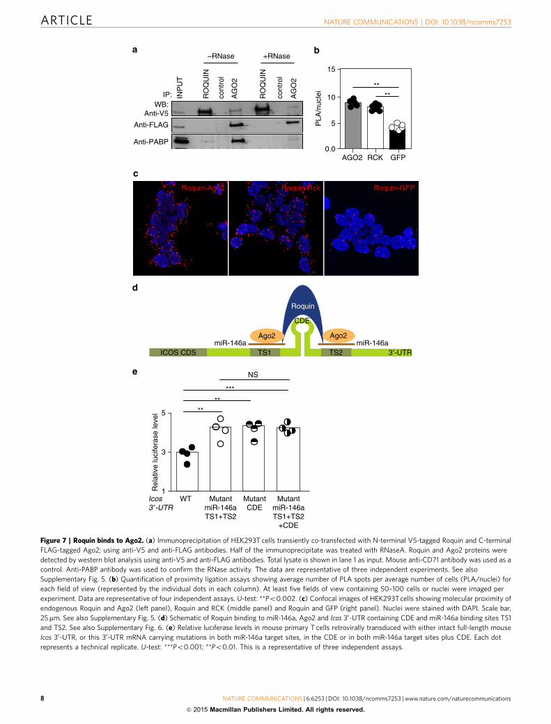
Roquin interacts with the core component of miRISC. Roquinbinds both miR-146a and its mRNA target (Icos), and it sharesstructural homology with proteins that bind RISC proteins(i.e., ADAR1 (ref. 23)) suggesting that Roquin may itself interactwith components of the miRISC complex. We first looked atwhether Roquin interacted with Dicer but did not detect an inter-action between these proteins when coexpressed in HEK293T(Supplementary Fig. 5a). To investigate a possible interactionbetween Roquin and the central miRISC component Ago2, V5-Roquin and Ago2-Flag were transfected into 293T cells and thetagged proteins immunoprecipitated. Western blots revealed aweak unidirectional interaction between Roquin and Ago2. Thisweak one-way interaction was also evident when we immuno-precipitated endogenous Roquin with Ago2 from the murine cellline EL4 (Supplementary Fig. 5b). The interaction was RNAindependent as it was also seen in the presence of RNase, which
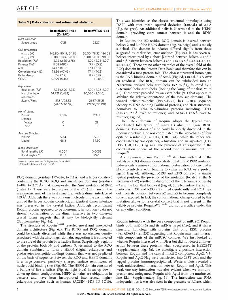
Table 1 | Data collection and refinement statistics.
RoquinM199R1–484(Zn SAD)
RoquinWT133–344
Data collectionSpace group C121 C2221
Cell dimensionsa, b, c (Å) 142.80, 80.19, 54.86 55.00, 78.32, 184.08a, b, g (�) 90.00, 111.06, 90.00 90.00, 90.00, 90.00
Resolution (Å)w 2.75 (2.85–2.75) 2.20 (2.28–2.20)Rmerge (%)w 11.08 (486) 9.7 (55.2)I/s(I) 14.42 (0.52) 17.4 (2.8)Completeness (%) 98.56 (97.93) 97.4 (90.3)Redundancy 7.6 (7.7) 8.7 (6.9)CC1/2w 0.999 (0.16) (0.862)
RefinementResolution (Å)w 2.75 (2.90–2.75) 2.20 (2.28–2.20)No. of uniquereflections
14,921 (1,463) 20,060 (2,040)
Rwork/Rfree 21.84/25.53(41.07/40.02)
21.67/25.21(22.59/30.00)
No. of atomsProtein 2,836 2,332Ligands
Water 0 21Zinc 2 0
Average B-factorsProtein 50.4 39.90Ligand 68.3 94.90
R.m.s. deviationsBond lengths (Å) 0.004 0.0053Bond angles (�) 0.87 0.928
Values in parentheses are for highest-resolution shell.wCriteria for data cut-off from ref. 53.
ARTICLE NATURE COMMUNICATIONS | DOI: 10.1038/ncomms7253
6 NATURE COMMUNICATIONS | 6:6253 | DOI: 10.1038/ncomms7253 | www.nature.com/naturecommunications
& 2015 Macmillan Publishers Limited. All rights reserved.

abolished RNA-mediated interaction of PABP1 with Ago2 (ref.24; Fig. 7a). To confirm that this interaction occurred in vivobetween endogenously expressed proteins, we used in situproximity ligation assay (PLA), which allows the detection oftransient or weak protein interactions. The interaction was pro-bed using mouse anti-Ago2 and rabbit anti-Roquin. Closeproximity (o20 nm) allows ligation and amplification of com-plementary DNA oligonucleotides on secondary antibodies andvisualization of individual protein interactions as fluorescent dots.Antibody staining was optimized on endogenous expression ofAgo2, Roquin and RCK (Supplementary Fig. 5c) and on exo-genous expression of GFP (data not shown). PLA detected
interaction between Ago2 and Roquin similar to that of Roquinwith its known protein partner RCK8 and significantly abovebackground levels seen with GFP (Fig. 7b,c). These resultssupport that Ago2 is a bona fide and close binding partner ofRoquin.
Roquin has been shown to bind CDEs within target mRNAs6.We compared the consequences of mutating the CDE within theIcos 30-UTR and/or the miR-146a target sites or both (Fig. 7d).Mouse primary T cells were retrovirally transduced with thedifferent Icos 30-UTR mRNA luciferase vectors (Fig. 7e).Mutations in the miR-146a target sites exerted comparablerepression defects as the CDE mutation, and there were no
RING domain
RING
RING ROQ
ROQ ZnF
ZnFC-HEPNN-HEPN
138 337
399326
RING N-HEPN
C-HEPNROQ
ZnF
177128751
ROQ domain
ROQROQ
RINGRING
HEPN
ADAR1NusB
C17 C38 C33 D53
C50H35C41C14
C38 C41C50 C33
D53
F234R233
R233
M199R
F234
R233R233
F234
M199
M199R
M199
F234
H35
C14C17
N-TERM
Zn2+ Zn2+
HEPN
HEPNdomain
i ii
Figure 6 | Crystal structure of Roquin fragments. (a) Structural and schematic representations of Roquin1–484. (i) Overall architecture of Roquin1–484
(RING domain¼ red, ROQ domain¼ beige, HEPN¼ light blue, additional helix packing against the HEPN domain¼ dark blue. (ii) Top: Schematic of the
known domain boundaries of Roquin before this work, middle/bottom: domain boundaries revealed by the crystal structure. Note that the ROQ domain is
an insertion in the HEPN domain. The ZnF (CCCH) domain was present in the crystallized protein but is not visible in the crystal structure. (b) Electrostatic
surface diagram of Roquin 1–484 (180 degrees rotated left to right), highlighting positively charged surfaces likely to be involved in RNA binding.
The surface indicated in the right hand panel is in a very similar location to the surface used by ADAR1 to bind nucleic acids. The orientation in the left
hand panel is identical to that in a(i). (c) Structural alignment of Roquin HEPN domain with HEPN domain from human Sacsin, (PDB ID:3O10, grey).
(d) Structural alignment of Roquin ROQ domain (beige) with NusB (PDB ID: 2JR0, orange) and the winged helix-turn-helix from ADAR1 (PDB ID:
1QBJ, orange). (e) Structure of the Roquin RING domain and schematic showing the residues involved in coordinating the two zinc atoms. (f) Residue
F234 is flipped out of the structure in the ROQ M199R mutant, becoming solvent exposed. left: wild-type (green); middle: M199R (beige); right: overlay.
See also Supplementary Fig. 4.
NATURE COMMUNICATIONS | DOI: 10.1038/ncomms7253 ARTICLE
NATURE COMMUNICATIONS | 6:6253 | DOI: 10.1038/ncomms7253 | www.nature.com/naturecommunications 7
& 2015 Macmillan Publishers Limited. All rights reserved.
ICOS CDS
Roquin
Ago2miR-146a miR-146a
Ago2
TS1 TS2 3′-UTR
CDE
Anti-FLAG
Anti-PABP
INP
UT
RO
QU
IN
AG
O2
cont
rol
RO
QU
IN
AG
O2
cont
rol
+RNase–RNase
WB:Anti-V5
0.0
5
10
15
****
AGO2 RCK GFP
PLA
/nuc
lei
IP:
Roquin-Ago2 Roquin-Rck
1
3
5 **
NS
**
***
Rel
ativ
e lu
cife
rase
leve
l
WT MutantmiR-146aTS1+TS2
MutantCDE
MutantmiR-146aTS1+TS2
+CDE
Icos3 ′-UTR
Roquin-GFP
Figure 7 | Roquin binds to Ago2. (a) Immunoprecipitation of HEK293T cells transiently co-transfected with N-terminal V5-tagged Roquin and C-terminal
FLAG-tagged Ago2; using anti-V5 and anti-FLAG antibodies. Half of the immunoprecipitate was treated with RNaseA. Roquin and Ago2 proteins were
detected by western blot analysis using anti-V5 and anti-FLAG antibodies. Total lysate is shown in lane 1 as input. Mouse anti-CD71 antibody was used as a
control. Anti-PABP antibody was used to confirm the RNase activity. The data are representative of three independent experiments. See also
Supplementary Fig. 5. (b) Quantification of proximity ligation assays showing average number of PLA spots per average number of cells (PLA/nuclei) for
each field of view (represented by the individual dots in each column). At least five fields of view containing 50–100 cells or nuclei were imaged per
experiment. Data are representative of four independent assays. U-test: **Po0.002. (c) Confocal images of HEK293T cells showing molecular proximity of
endogenous Roquin and Ago2 (left panel), Roquin and RCK (middle panel) and Roquin and GFP (right panel). Nuclei were stained with DAPI. Scale bar,
25mm. See also Supplementary Fig. 5. (d) Schematic of Roquin binding to miR-146a, Ago2 and Icos 30-UTR containing CDE and miR-146a binding sites TS1
and TS2. See also Supplementary Fig. 6. (e) Relative luciferase levels in mouse primary T cells retrovirally transduced with either intact full-length mouse
Icos 30-UTR, or this 30-UTR mRNA carrying mutations in both miR-146a target sites, in the CDE or in both miR-146a target sites plus CDE. Each dot
represents a technical replicate. U-test: ***Po0.001; **Po0.01. This is a representative of three independent assays.
ARTICLE NATURE COMMUNICATIONS | DOI: 10.1038/ncomms7253
8 NATURE COMMUNICATIONS | 6:6253 | DOI: 10.1038/ncomms7253 | www.nature.com/naturecommunications
& 2015 Macmillan Publishers Limited. All rights reserved.

additive effects among the different sets of mutations (Fig. 7e).These results show that miR-146a is as effective as Roquin inrepressing Icos suggesting that they may act in the same pathway,that is, Roquin acts together with miR-146a to exert some or all ofthe suppression.
DiscussionOur work identifies a role of the immune modulator Roquin inregulating mammalian miRNA homeostasis by promotingmature miR-146a degradation. T cells expressing Roquinsan orlacking Roquin and Roquin-2 displayed comparable accumula-tion of miR-146a indicating that miRNA dysregulation is due tothe loss of the function of both Roquin paralogues. Although 15miRNAs were elevated at least twofold, miR-146a and miR-21were the ones most affected. Of these, only miR-146a accumu-lated in a clearly cell-autonomous manner, suggesting a selectiveeffect of Roquin in the control of miR-146a homeostasis. Some ofthe other upregulated miRNAs included miR-29b, miR-155 andmiR-500, which like miR-146a have been linked either to humanor to mouse lupus25.
Accumulation of miR-146a in Roquinsan/san T cells was notfound to be a transcriptionally regulated event: neither theprimary nor precursor miR-146a levels were different betweenRoquin mutant and wild-type T cells. Moreover, Roquin does nothave a Dicer-like activity that cleaves pre-miRNAs into matureforms. Roquin did enhance miR-146a processing by Dicer butthis was not further enhanced with Roquinsan excluding this as apossible cause of miR-146a accumulation in Roquin mutant Tcells. The presence of mutant Roquin did prolong the half-life ofmiR-146a, pointing to posttranscriptional stabilization of miR-146a as the cause of the accumulation.
At least two non-mutually exclusive mechanisms may explainmiR-146a accumulation in the presence of mutant Roquin:(i) target-mediated microRNA decay (TMMD) and (ii) decreasedmono-uridylation. Target mRNAs are not innocently submissive;they can actively regulate the lifespan of the miRNAs that targetthem. In C. elegans, binding of miRNAs to their targets canpromote target-mediated microRNA protection26, whereas in flyand mammalian cells. the opposite phenomenon—TMMD hasbeen described27–29. Given that Roquin binds not only miRNAand target mRNA, but also the RISC component Ago2, it is likelythat in the absence of Roquin, a large fraction of cellular miR-146a and target mRNAs are not bound to RISC and are thereforeprotected from decay by TMMD. Roquin can bind to miR-146a,Ago2 and at least one of miR-146a’s targets, Icos mRNA. Roquinmay bring miRNAs and their targets into proximity to recruit orhelp stabilize RISC. Other RNA-binding proteins have beenshown to exert the opposite effect, for example, HuR aids miRISCdissociation from its target RNA30. Decreased miR-146a mono-uridylation found in the absence of functional Roquin may alsocontribute to its increased stability: a role for uridylation inenhancing miRNA decay has been described in plants18 andmammals19,20. Intriguingly, HEPN domains are reported innucleotidyl-transferases31. This activity is required for terminalmiRNA uridylation; it is therefore tempting to speculate thatRoquin may form part of a nucleotidyl–transferase complex topromote uridylation of a subset of miRNAs including miR-146a.Of 39 miRNAs that showed decreased uridylation, only 6appeared to accumulate in the presence of mutant Roquin.Thus, if decreased uridylation is linked to increased miR-146astability, this effect is likely to be selective for a small subset ofmiRNAs, perhaps depending on simultaneous recognition ofparticular miRNA:mRNA sequences and structures.
Our Roquin structure reveals that the N terminus is composedof RING, HEPN, ROQ and CCCH domains. The structure of the
HEPN and ROQ domains suggests that they are highly likely toboth function as RNA-binding domains. There are dozens ofdifferent classes of RNA-binding domains in eukaryotes32 MostRNA-binding proteins contain multiple RNA-binding units toallow for sequence- or structure-specific RNA recognition33. Inthe case of Roquin, we observe at least three different potentialRNA-binding domains, the ROQ, HEPN and CCCH domains.The presence of the HEPN domain was unexpected based onsequence analysis as it is discontinuous in the primary sequenceas a result of insertion of the ROQ domain between two a-helicesthat are usually separated in HEPN domains by a variable loop.
To allow for recognition of larger RNA sequences or multipleRNAs, many RNA-binding proteins either dimerize or containflexible linkers between the individual RNA-binding domains33.Roquin appears to use both of these approaches: we see evidenceof dimerization as well as the presence of a flexible linker betweenthe HEPN and CCCH domains. One feature common to manyRNA-binding domains is the presence of a basic region on theirsurface that promotes their interaction with the negativelycharged nucleic acid backbone. There are two highly positivelycharged surfaces on Roquin: one is located on the HEPN domainand its interface with the ROQ domain, whereas the other isentirely within the ROQ domain. The latter is the same as thesurface used by ADAR1 to bind nucleic acids. ADAR1 complexeswith precursor miRNAs and with Dicer through direct protein–protein interaction to also enhance Dicer-mediated pre-miRNAcleavage and facilitate loading of miRNA onto the RISC23.Roquin also appears to enhance both Dicer activity and facilitateRISC assembly. This may be related to the significant structuralsimilarity between the N-terminal subdomain of the ROQdomain and the RNA-binding domains of ADAR1: all of themhave winged helix–turn–helix structures.
Comparison of wild-type and M199R ROQ domains revealedthat the ‘san’ mutation causes a structural change in the proteinthat exposes an otherwise buried hydrophobic residue (F234).Three separate studies in which structures of the ROQ and HEPNdomains of Roquin have been solved were published while thispaper was under review34–36. The two structures presented byTan et al., are of Roquin bound to RNA CDE motifs fromhmgxb3 and TNF mRNAs. These CDEs occupy the two RNAbinding sites on the ROQ and HEPN domains we predicted onthe basis of electrostatic considerations (Fig. 6b) and the twostructures are highly similar to the apo structure presented here(r.m.s.d. of 1.9 and 1.1 Å, respectively) indicating that the HEPNand ROQ domains of Roquin undergo only minor structuralalterations upon RNA binding. No conclusions regarding theRING domain can be drawn as it is absent in all other structuralstudies. A comparison of our san mutant Roquin with that ofRNA-bound wild-type Roquin structures34–36 indicates that thesan mutation does not induce any structural perturbation ineither of the RNA binding sites (with the exception of a slightshift in the position of Serine 238). Therefore the severephenotype seen in the sanroque mouse is unlikely to be due toaltered RNA binding by Roquin and may instead be due to afailure, by Roquin, to interact with other (protein) components ofthe mRNA-silencing machinery. This is the subject of futureexperiments.
Accumulation of miR-146a was more pronounced in naive Tcells, known to express genes with longer 30-UTR than those inproliferating T cells37. The components of the miRNA machineryalso become less abundant as T cells differentiate into effectorcells38. In Dicer� /� MEFs, Roquin regulates Icos mRNAindependently of miRNAs8. The same study also reported thatthe level of Icos repression by Roquin increased with the length ofthe Icos 30-UTR, indicating that multiple sites in the 30-UTR maybe necessary to induce full repression by Roquin. It is therefore
NATURE COMMUNICATIONS | DOI: 10.1038/ncomms7253 ARTICLE
NATURE COMMUNICATIONS | 6:6253 | DOI: 10.1038/ncomms7253 | www.nature.com/naturecommunications 9
& 2015 Macmillan Publishers Limited. All rights reserved.

likely that Roquin regulates Icos and other target mRNAs viaseveral mechanisms that include microRNA-mediated repressionin some cell types.
Transcriptome-wide studies have shown that Roquin controlsthe degradation of numerous mRNAs with conserved CDE stem-loop motifs6. Mutations in the CDE exerted exactly the sameinhibitory effect on Icos mRNA degradation by Roquin as themutations in miR-146a target sites, with no additive effect. Thus,it is possible that the suppressive effect of CDE and miR-146aoperates through the same pathway. Roquin may bind Icos CDEand bring miR-146a to its target sites to facilitate miR-146arepression of Icos. Collaboration between AU-rich element-binding microRNAs and AU-rich element-binding proteins isemerging as a prominent mechanism of 30-UTR-mediatedregulation of gene expression in mammalian cells39,40. As tothe question of whether binding of Roquin to the CDE facilitatesbinding of Roquin to miR-146a, a study published while thispaper was under review34 confirms that HsRoquin-1 has twobinding sites within the ROQ domain. RNA-binding studiesshowed that the ROQ domain can bind CDEs in one site anddsRNA in the other and that this binding can occursimultaneously and also independently. Combined, these resultsand ours suggest that binding of Roquin to the CDE may not beneeded to facilitate binding of Roquin to miR-146a. Interestinglyboth RNA-binding sites were shown to be necessary for mRNAdecay. Roquin, with its unique structure and different RNA-binding domains may thus be important not only for regulatingmiRNA homeostasis but also for miRNA function. Furtherunderstanding of this Roquin-miR-146a axis may illuminatepathogenesis of inflammatory and autoimmune diseases.
MethodsMice and bone marrow chimeras. C57BL/6(B6) and Roquinsan/san (sanroque)mice were housed in pathogen free conditions at the Australian Phenomics facilityat Australian National University (ANU). Seven-to-ten-week-old mice of bothgenders were used throughout. Rc3h1fl/fl;Rc3h2fl/fl; CD4-Cre mice were housed in apathogen-free barrier facility at the Helmholtz Zentrum, Munchen. For bonemarrow chimera reconstitutions, 2� 106 cells were injected intravenously into 10–12-week-old sublethally irradiated (500 cGy, X-rad) C57BL/6 Ly5a recipients andanalysed 12 weeks after reconstitution. The ANU Animal Ethics and Experi-mentation Committee approved all animal procedures.
Flow cytometry and microRNA microarray. Single-cell suspensions from spleenand lymph nodes were surface stained for 30 min at 4 �C. Mouse CD4þ naive Tcells (CD4þB220�CD44loCD25� ) were sorted using mouse anti-CD4 1:400(BioLegend cat#100422 ), anti-CD44 1:200 (BioLegend cat#103006), CD25 1:200(BioLegend cat#101910) and B220 1:100 (BioLegend cat#103208). For bone mar-row chimeras, CD45.1 1:200 (BioLegend cat#110722 )and CD45.2 1:200 (BioLe-gend cat no#109824) congenic markers were used. Total RNA from T cells fromfour wt and four mutant mice was extracted using mirVana microRNA isolation kit(Ambion) and miRNA microarrays were performed using Agilent mouse miRNA-v1_95_May07 chips at the Ramaciotti Center for Genomics (UNSW) and analysedusing the Genespring software (Agilent Technologies, Inc.)
qRT–PCR and northern blot. Total RNA was extracted using TRIzol (LifeTechnologies). Complementary DNA (cDNA) was prepared using miScript RT kit(Qiagen). qRT–PCR for mature and precursor miR-146a, miR-21 and control U6was performed using the miScript Primer/Precursor Assays (Qiagen) and amplifiedon an ABI 7900 Prism light cycler in the Biomolecular Resource Facility, ANU. Theprimer sequences used for detecting primary miR-146a are provided inSupplementary Table 2. The relative expression was calculated using the 2� ddct
method41.For measurements of miR-146a in Rc3h1fl/fl:/Rc3h2fl/fl;Cd4-cre; FACS-sorted
naive (CD4þCD62LþCD44� ) T cells from three Rc3h1fl/fl:/Rc3h2fl/fl (wt) andRc3h1fl/fl:/Rc3h2fl/fl;Cd4-cre mice and one Roquinsan/san mouse were used. miRNA-specific cDNA was prepared using TaqMan MicroRNA Reverse Transcription kit(Applied Biosystems). The expression of miR-146a was measured by qRT–PCRusing mmu-miR-146a, snoRNA202 Taqman microRNA Assay (AppliedBiosystems) on a Light cycler 480II with the Light cycler 480 SW 1.5 software(Roche).
For northern blot hybridization, 10 mg of total RNA extracted from naive T cellswas fractionated on polyacrylamide TBE gel and electroblotted onto Amersham
Hybond-Nþ membrane (GE Healthcare), The miRCURY LNA probes for miR-146a, miR-150 and U6 (Exiqon) were radiolabelled at 50 end with g-32P ATP andhybridized to the membrane overnight. Radioactive signals were detected with aTyphoon FLA9000 (GE Healthcare).
Transfections and retroviral transductions. HEK293T cells were transfectedwith plasmid DNA in six-well plates using Lipofectamine 2000. Roquinwt,Roquinsan or empty vector expressed from an IRES-GFP. Co-transfections of DNAvectors (4mg per well) along with a synthetic control or precursor hsa-miR-146a(50 pM, Ambion) was done using Lipofectamine 2000 (Invitrogen) as per themanufacturer’s protocol. The reporter constructs used in transfections and theretroviral transduction method is described previously2.
In vitro RNA cleavage assays. In vitro RNA cleavage was performed as essentiallyas described42. In brief, synthetic pre-mir-146a oligonucleotides (Integrated DNATechnology) were radiolabelled by 50 phosphorylation with polynucleotide kinase(NEB) and g32P-ATP. Resulting radiolabelled RNAs were then mixed with anti-Flag (or anti-Flag plus anti-GFP) agarose beads (Sigma) coated with lysates fromHEK293T cells transiently transfected with various tagged constructs. Resultingcleavage products were resolved on a urea/acrylamide gel, then visualized andquantified by phosphorimaging.
Co-immunoprecipitations. Cells were lysed in IP buffer (1% NP-40, 20 mMTris.HCl (pH¼ 7.4), 150 mM NaCl, 1� EDTA-free protease inhibitor cocktail(Roche)) for 20 min on ice. The clarified cell lysates were precleared overnight withmagnetic Protein G Dynabeads (Invitrogen). The lysates were incubated with 2 mgof antibodies for 1 h and then with Protein G Dynabeads for an additional 3 h at4 �C before being washed and resuspended in 20 ml of 3X sample buffer (360 mMTris (pH¼ 6.8), 6% SDS, 30% b-mercaptoethanol, 30% glycerol, 0.15% bromo-phenol blue). The samples were boiled at 70 �C for 10 mins before use in SDS–polyacrylamide gel electrophoresis(SDS–PAGE). Antibodies used for ectopicallyexpressed constructs were mouse anti-V5 (clone SV5-Pk1, Serotec cat#MCA1360),anti-Flag (clone M2, Sigma cat#F1804), and mouse anti-CD71 (clone MEM-75,Sigma cat# SAB4700515-100UG). Anti-PABP (Abcam, cat#ab21060) was used forassessing RNase activity. For endogenous IPs, mouse anti-Ago2 mAb (Wakocat#014-22023), polyclonal rabbit anti-Roquin (Novus cat#NB100-656) or controlmouse anti-CD71 (clone MEM-75, Sigma cat# SAB4700515-100UG) were used..
In situ PLA. PLA was performed according to the manufacturer’s protocol(Duolink kit, Olink Bioscience, Uppsala, Sweden). Briefly, HEK293T cells weregrown on coverslips and fixed at room temperature (RT) with 3.7% formaldehydefor 20 min. After three washes with phosphate-buffered saline (PBS), cells wereblocked and permeabilized for 60 min in 5% BSA/0.3% Triton-X100 at RT andincubated with primary antibodies at optimized dilutions (rabbit anti-ROQUIN1:75 (Novus Biologicals); with either mouse anti-RCK 1:150 (Santa Cruz Bio-technology Inc.); mouse anti-Ago2 1:200 (WAKO Pure Chemical Industries) ormouse anti-GFP 1:100 (Roche)) overnight in a humid chamber at 4 �C. Slides werewashed three times in PBS/0.05% Tween20 and incubated with mouse minus andrabbit plus PLA probes for 1 h at 37 �C. Ligation was carried out for 30 min andamplification for 100 min at 37 �C. Slides were washed, dried at RT in the dark andmounted in mounting media with DAPI (Olink) to stain nuclei. The images weretaken on a Leica SP5 confocal microscope with a pin hole of 95.5 mm and a HCxPLAPO lambda blue � 631.4 oil objective and quantified using the free softwareImageJ (NIH, Maryland, USA).
RNA immunoprecipitation. For RIP a buffy coat containing lymphocytes wasobtained using Ficoll-Paque (GE Healthcare) gradient separation. Total lympho-cytes (60� 106) from tonsils were lysed thoroughly in 1 ml of polysome lysis buffer(100 mM KCl, 5 mM MgCl2, 10 mM HEPES, 0.5% NP-40, 1 mM DTT, 0.1 U ml� 1
RNAse out and 25ml ml� 1 protease inhibitor). The total lysate was precleared with50 ml Protein G Dynabeads (Life Technologies) for 1 h at 4 �C and simultaneously,antibody-bead complex was formed by incubating 15 ml protein G Dynabeads with3 mg of anti-human Roquin (Bethyl Laboratories cat#A300–514A) antibody orcontrol IgG antibody (Santa Cruz cat# sc-2027) for 2 h at 4 �C. A control wasincluded in which the lysate was incubated with Roquin antibody and a blockingpeptide. This blocking peptide is the one used for immunization of rabbits toproduce the polyclonal anti-Roquin IgG antibody (Bethyl Laboratories, cat #.A300–514A). 10% of the precleared total lysate was saved for use as total input andthe rest of the lysate was used for immunoprecipitation. The precleared lysate wascombined with antibody–bead complex and incubated at 4 �C for 4 h. After fourwashes with polysome lysis buffer, the last wash was done with polysome lysisbuffer containing 1 M urea for 5 min at 4 �C on a rotating mixer. The beads werethen resuspended in 100 ml of polysome lysis buffer containing 0.1% SDS andincubated at 50 �C for 30 min. After incubation, an equal volume of phenol–chloroform was added and RNA was extracted using mirVana miRNA isolation kit(Ambion). RNA was also extracted from the total input. cDNA was prepared usingmiScript RT kit (Qiagen) and used for qRT–PCR for miRNAs using miScriptprimers and SYBR Green kit from Qiagen. The Ct values were recorded and
ARTICLE NATURE COMMUNICATIONS | DOI: 10.1038/ncomms7253
10 NATURE COMMUNICATIONS | 6:6253 | DOI: 10.1038/ncomms7253 | www.nature.com/naturecommunications
& 2015 Macmillan Publishers Limited. All rights reserved.

normalized to the Ct values of the total input. The amount of bound miRNA wasplotted as percentage enrichment with respective antibody immunoprecipitation.
Luciferase assays in primary T cells. Wild-type or mutated mouse Icos 30-UTR(miR-146a TS1/2; CDE or combined TS1/2þCDE mutation) DNA fragmentswere generated with XhoI-NotI (GeneArt; sequence presented in SupplementaryFig. 6) and cloned into retroviral dual-luciferase reporter vector miR-Sens (kind giftfrom P. Mathijs Voorhoeve). These vectors were then transfected into Phoenixcells, and retroviral supernatants were harvested 48 h post transfection. Primary Tcells were retrovirally transduced by spinoculation. The Dual-Glo LuciferaseReporter kit (Promega Corporation) was used to measure luciferase as describedpreviously43.
Small RNA deep sequencing. T cells from spleen and lymph nodes were enrichedwith an EasySep Mouse T Cell Enrichment Kit (Stemcell, cat# 19751) to 490%purity. RNA was prepared using TRIzol (Life Technologies) and the concentrationand integrity of total RNA was verified with an Agilent 2100 Bioanalyser. SmallRNA library construction and deep sequencing was done by Illumina HiSeqTechnology (Beijing Genomics Institute at Shenzhen). All raw reads were firsttrimmed to remove the sequencing adaptor (50-TCGTATGCCGTCTTCTGCTTGT-30) by using the Trimmomatic software44 (parameters: -threads 6 -phred64input.fq output.t.fq ILLUMINACLIP:adaptor.fa:2:30:10 MINLEN:18) retainingreads that were at least 18 nt long. Then reads were mapped to the mouse referencesequence consisting of the genome sequence (GRCm38), 18s rRNA (gi|374088232)and 28 s rRNA (gi|120444900) using Bowtie (v1.0.1) software45. Parameters formapping were chosen such that three mismatches were allowed in the 18 nt seedregion (-n 3 –l 18), the sum of the quality values at the mismatch positions couldnot exceed 150 (-e 150) and all possible alignments fulfilling these requirementswere reported (-a). Other parameters that may influence the alignment output were--nomaqround --maxbts 800 -y --chunkmbs 4096. Alignment files were processedto retain alignments of a read with only minimum mismatches using a Perlscript (Supplementary Information 1).
Expression counts. All 18–26 nt long sequenced tags were assigned to a maturemiRNA if their 50 start position was within ± 3 nt of the 50 start position of themature miRNA (miRBase v21). We call these tags as miRNA mapped tags fromhere on. Tag counts for mature miRNA were normalized to correct for the librarysize as follows. If the total tags obtained for a sample is denoted as ‘N’, and the rawtags count for a mature miRNA is denoted as ‘n’, then the normalized tag count forthe mature miRNA refers to (n� 1,000,000)/N.
30 non-templated modifications/additions. All miRNA-mapped tags that are(a) 1 nt longer than the annotated mature miRNA, and (b) identical to the genomereference sequence except the last nucleotide were considered as tags showing theevidence of 30 non-templated modifications/additions (Supplementary Fig. 7).The percentage for A,C,G or T non-templated modification/addition wascalculated with respect to the sum of mature and all non-templated tags. Forexample, as indicated in the Supplementary Fig. 7, the percentage for the non-templated T would be t� 100/(mþ tþ cþ g).
Protein expression and purification. Roquin1–484. GST-Roquin1–484 proteinswere expressed in BL21DE3 grown overnight at 18 �C in 0.1 mM zinc acetate and1 mM IPTG. Pelleted cells were sonicated in R-buffer (20 mM Tris-HCl pH 7.5,500 mM NaCl, 0.1 mM zinc acetate) with protease inhibitors, RNase A, DNase andlysozyme (Roche). Clarified supernatant was bound to Glutathione Sepharose resinand the tag cleaved with PreScission Protease (GE Healthcare). Eluates werefractionated on a Superdex 200 26/60 column (GE Healthcare). Fractions wereanalysed by SDS–PAGE and proteins concentrated to 30 mg ml� 1.
Roq domain. The DNA fragment encoding the roq domain (amino acid residues122–344) was inserted into the expression plasmid pDEST17 by recombination.The expression construct contains C-terminal His tag. The protein wasoverproduced in Escherichia coli strain B834 (DE3). Single colonies were pickedand allowed to grow in media containing TB Overnight Express (Novagen) for 5–6 h at 37 �C followed by 20 h at 25 �C. The cell pellet was collected and frozen at80 �C until use. For protein purification, the cell pellet was resuspended in buffer A(25 mM Tris (pH 7.5), 500 mM NaCl, 30 mM imidazole) with 0.1% Tween 20,protease inhibitors and DNase. The suspension was passed through a cell disruptorat 30 k.p.s.i. and then centrifuged for 30 min at 30,000g at 4 �C. The soluble fractionwas transferred to an Akta Express equipped with a Ni-nitrilo-triacetic acid columnconnected in-line to a Hiload 16/60 Superdex 75 gel filtration column (GEHealthcare). The Ni-nitrilo-triacetic acid column was washed using buffer A, elutedusing buffer B (25 mM Tris (pH 7.5), 250 mM NaCl, 250 mM imidazole) beforetransfer to the gel filtration column equilibrated in buffer C (20 mM Tris (pH 7.5),200 mM NaCl). Peak fraction collection was performed using the Akta Expresssoftware.
Surface plasmon resonance (SPR). RoquinWT1–484 and RoquinM199R1–484
were freshly dialyzed into SPR buffer (10 mM Tris-HCl pH 7.4, 150 mM NaCl,1 mM TCEP, 0.05% surfactant P-20). Measurements were made at 20 �C on aBiacore T100 SPR instrument (GE Healthcare) at a flow rate of 50 ml min� 1.50-Biotinylated mmu-miR-146a (50-CUGAGAACUGAAUUCCAUGGGUUAUA
UCAAUGUCA-30) was purchased from Shanghai Gene Pharma Co. Biotinylated.The RNA was dissolved in SPR buffer, heated to 80 �C for 5 min and cooled slowlyto allow secondary structure formation. It was immobilized at 20 nM onto a singleflow cell of a streptavidin-coated Biacore (SA) chip (GE Healthcare) by injection at10 ml min� 1 for B100 s to get 200 response units. Details of association/dis-sociation measurements are described in Supplementary Methods. A second flowcell was left underivatized for blank subtraction. Chip surfaces were regenerated byinjection of 1 M MgCl2 for 1 min at 5 ml min� 1, leaving the RNA intact whileremoving bound protein. Association was measured for 180 s and Roquin wasallowed to dissociate for 300 s before regeneration of the chip surfaces by injectionof 1 M MgCl2 for 1 min at 5 ml min� 1, leaving the RNA intact while removingbound protein. RoquinWT1–484 was used in twofold serial dilutions from 2,000–7.8 nM, whereas RoquinM199R1–484 was from 1,000–3.9 nM, with injections foreach protein done in random order. Although experiments were repeated twice,progressive degradation of the immobilized RNA prevented use of replicates indata fitting. A data set comprising the first 10 cycles measured for each protein wasselected to minimize effects from chip degradation.
Crystallization and structure determination. Roq domain (Roquin 133–344).Crystals of the Roq domain were grown by sitting drops at 20 �C by mixing10 mg ml� 1 of the protein (20 mM Tris-HCl pH7.5, 200 mM NaCl) with thecondition including lithium chloride 0.2 M and polyethylene 20% Glycol 3350.Crystals were cryo-protected with mother liquor plus 22% glycerol including 0.5 MKl for 20 s before vitrification in liquid nitrogen. MAD data were collected at 100 Kon beamline BM14 at ESRF (Grenoble, France) at wavelengths of 1.55000 (peak)and 0.95350 Å (remote). The diffraction data were processed and scaled withHKL2000 program package46. Twenty-one iodine sites in an asymmetric unit wereassigned using SHELXD47 and used for the SAD phase calculation with SOLVE48,resulting in the initial mean figure-of merit of 0.27 for all reflections. We improvedthe phases using the program DM49. Further structure refinement was carried outby PHENIX50 using the remote data and manual model building with coot51. Thefinal model showed an Rfree factor of 21.4% and an R factor of 25.9%. Detailedstatistics are summarized in Table 1. Ramachandran Statistics: 97.3% favoured,2.7% allowed, zero outliers.
RoquinM199R1–484. Immediately before crystallization, protein was diluted to10 mg ml� 1 using 20 mM Tris-HCl pH 7.5, 0.1 mM zinc acetate, to reduce theconcentration of NaCl to 167 mM. Crystallization was accomplished by hanging-drop vapour-diffusion at 20 �C using 2 ml drops, and a drop-ratio of 1:1protein:precipitant. Precipitant was 0.1 M CHES pH 9.5, 7–12% PEG 8000. Crystalsgrew from phase separation as B100 mm rhombohedra. Crystals were cryo-protected with mother liquor plus 25% ethylene glycol before vitrification in liquidnitrogen. Zinc-SAD data were collected at 100 K at the Australian Synchrotron onbeamline MX2, at a wavelength of 1.265102 Å. Data were integrated using XDS52
and scaled using XSCALE. Anomalous signal was present to B5 Å (6.54–5.35 Å:Anomalous correlation 0.41, SigAno 1.3). Initially a data cut-off of 3.1 Å was used.A molecular replacement solution was found using PHASER, using the ROQdomain (above) as a model. Phases were improved using the anomalous signalfrom zinc. Two Zn sites were found, and phases were calculated with a mean figureof merit of 0.45 for all reflections. Structure refinement was carried out inPHENIX50 using and manual model building with coot51. After several rounds ofrefinement, data were re-processed to include data to 2.75 Å based on a CC1/2
40.1, as suggested by Karplus and Deiderichs53, with a significant improvement inmap quality. Ramachandran statistics: 98.33% favoured, 1.67% allowed, zerooutliers.
Quantification of secreted exosomes. Splenocytes were stained with B220-FITC,CD3e-PE and 7AAD. B220-FITC� CD3-PEhi/7-AADlo T cells were sorted. Thecells were incubated in anti-CD3e (BD Pharmingen) pre-coated six-well plates(5 mg ml� 1) in exosome-depleted complete RPMI medium at 1� 106 cells ml� 1
with 2 mg ml� 1 anti-CD28 antibody (BD Pharmingen) and 20 U ml� 1 of rIL-2 at37 �C with 5% CO2 for 48 h. After centrifugation for 5 min at 210 g, the supernatantwas filtered through a 0.22-mm filter (Merck Millipore) and exosomes harvested byultracentrifugation at 100,000g for 1 h at 4 �C. The exosome pellet was split intotwo. Half was resuspended in PBS, stained with anti-mouse-CD9-PE antibody(BioLegend) and run on a flow cytometer along the controls (PBS only, PBS plusanti-CD9-PE and unlabelled exosomes). RNA was extracted from the other half ofthe exosome pellet using TRIzol (Life Technologies). All-in-one miRNA qRT–PCRdetection kit (GeneCopoeia) and validated primers for mature miRNAs (Gene-Copoeia) were used to quantify mmu-miR-146a-5p and mmu-miR-150-5p.Amplification was detected on a 7900 HT Fast Real-Time PCR system (AppliedBiosystems) and relative amounts were calculated using the 2� dct.
In situ hybridization. Naive T cells were sorted from Roquinsan/san andRoquinþ /þ mice. The cells were fixed in 3.7% formaldehyde for 20 min at RT.Once fixed, cells were washed once with dH2O (13,000 r.p.m. for 10 min) andspotted at 10,000 cells per 25 ml on superfrost plus slides (Thermo Scientific). Theslides were incubated at 37 �C. Once the slides were completely dry, they werewashed once with 100% ethanol and treated with proteinase K (500 ng ml� 1) for5 min at RT. The slides were incubated with the prehybridization solution (Enzo
NATURE COMMUNICATIONS | DOI: 10.1038/ncomms7253 ARTICLE
NATURE COMMUNICATIONS | 6:6253 | DOI: 10.1038/ncomms7253 | www.nature.com/naturecommunications 11
& 2015 Macmillan Publishers Limited. All rights reserved.

Life Sciences, cat#33808) at 55 �C for 15 min. The cells were hybridized with 2 pMRNA probe at 55 �C in a moist chamber overnight. Alexa-488 conjugated miR-146a LNA probe and Alexa-488 conjugated scrambled LNA probe were purchasedfrom Exiqon. After probe hybridization, cells were blocked with blocking buffer(0.2� SSCþ 2% BSA) for 10 min at RT. Primary antibody, Dcp1A (gift from J.Lykke-Andersen, University of California San Diego), and eIf3 were added at adilution of 1/250 and incubated at RT for 2 h at 4 �C. After three washes with PBST(PBSþ 0.01% Tween), the slides were incubated with a secondary antibody, don-key-anti-goat-Alexa 568 (Molecular Probes, Invitrogen, cat#A-11057) at RT for 2 h.The cells were washed three times, 10 min each with PBST. Before a final wash withPBST, cells were incubated with DAPI for 2 min. Cells were mounted in Vecta-shield (Vector Laboratories) and Images were taken using an Olympus IX71microscope with DP controller software (Olympus) and compiled using AdobePhotoshop software.
References1. Castello, A., Fischer, B., Hentze, M. W. & Preiss, T. RNA-binding proteins in
Mendelian disease. Trends Genet. 29, 318–327 (2013).2. Vinuesa, C. G. et al. A RING-type ubiquitin ligase family member required to
repress follicular helper T cells and autoimmunity. Nature 435, 452–458(2005).
3. Yu, D. et al. Roquin represses autoimmunity by limiting inducible T-cellco-stimulator messenger RNA. Nature 450, 299–303 (2007).
4. Pratama, A. et al. Roquin-2 shares functions with its paralog Roquin-1 in therepression of mRNAs controlling T follicular helper cells and systemicinflammation. Immunity 38, 669–680 (2013).
5. Vogel, K. U. et al. Roquin paralogs 1 and 2 redundantly repress the Icos andOx40 costimulator mRNAs and control follicular helper T cell differentiation.Immunity 38, 655–668 (2013).
6. Leppek, K. et al. Roquin promotes constitutive mRNA decay via a conservedclass of stem-loop recognition motifs. Cell 153, 869–881 (2013).
7. Athanasopoulos, V. et al. The ROQUIN family of proteins localizes tostress granules via the ROQ domain and binds target mRNAs. FEBS J. 277,2109–2127 (2010).
8. Glasmacher, E. et al. Roquin binds inducible costimulator mRNA and effectorsof mRNA decay to induce microRNA-independent post-transcriptionalrepression. Nat. Immunol. 11, 725–733 (2010).
9. Bartel, D. P. MicroRNAs: target recognition and regulatory functions. Cell 136,215–233 (2009).
10. Pauley, K. M., Cha, S. & Chan, E. K. MicroRNA in autoimmunity andautoimmune diseases. J. Autoimmun. 32, 189–194 (2009).
11. Krol, J., Loedige, I. & Filipowicz, W. The widespread regulation of microRNAbiogenesis, function and decay. Nat. Rev. Genet. 11, 597–610 (2010).
12. Zhang, Z., Qin, Y. W., Brewer, G. & Jing, Q. MicroRNA degradation andturnover: regulating the regulators. Wiley Interdiscip. Rev. RNA 3, 593–600(2012).
13. Grosshans, H. & Chatterjee, S. MicroRNAses and the regulated degradation ofmature animal miRNAs. Adv. Exp. Med. Biol. 700, 140–155 (2010).
14. Thomas, M. F. et al. Eri1 regulates microRNA homeostasis and mouselymphocyte development and antiviral function. Blood 120, 130–142 (2012).
15. Pratama, A. et al. MicroRNA-146a regulates ICOS-ICOSL signaling to limitaccumulation of T follicular helper cells and germinal centers. Nat. Commun.(2015).
16. Mittelbrunn, M. et al. Unidirectional transfer of microRNA-loaded exosomesfrom T cells to antigen-presenting cells. Nat. Commun. 2, 282 (2011).
17. Yang, W. et al. Modulation of microRNA processing and expression throughRNA editing by ADAR deaminases. Nat. Struct. Mol. Biol. 13, 13–21 (2006).
18. Ibrahim, F. et al. Uridylation of mature miRNAs and siRNAs by the MUT68nucleotidyltransferase promotes their degradation in Chlamydomonas. Proc.Natl Acad. Sci. USA 107, 3906–3911 (2010).
19. Knouf, E. C., Wyman, S. K. & Tewari, M. The human TUT1 nucleotidyltransferase as a global regulator of microRNA abundance. PLoS ONE 8, e69630(2013).
20. Choi, Y. S., Patena, W., Leavitt, A. D. & McManus, M. T. Widespread RNA 3’-end oligouridylation in mammals. RNA 18, 394–401 (2012).
21. Jones, M. R. et al. Zcchc11-dependent uridylation of microRNA directscytokine expression. Nat. Cell. Biol. 11, 1157–1163 (2009).
22. Deshaies, R. J. & Joazeiro, C. A. RING domain E3 ubiquitin ligases. Annu. Rev.Biochem. 78, 399–434 (2009).
23. Ota, H. et al. ADAR1 forms a complex with Dicer to promote microRNAprocessing and RNA-induced gene silencing. Cell 153, 575–589 (2013).
24. Hock, J. et al. Proteomic and functional analysis of Argonaute-containingmRNA-protein complexes in human cells. EMBO Rep. 8, 1052–1060 (2007).
25. Liang, D. & Shen, N. MicroRNA involvement in lupus: the beginning of a newtale. Curr. Opin. Rheumatol. 24, 489–498 (2012).
26. Chatterjee, S., Fasler, M., Bussing, I. & Grosshans, H. Target-mediated protectionof endogenous microRNAs in C. elegans. Dev. Cell. 20, 388–396 (2011).
27. Ameres, S. L. et al. Target RNA-directed trimming and tailing of small silencingRNAs. Science 328, 1534–1539 (2010).
28. Baccarini, A. et al. Kinetic analysis reveals the fate of a microRNA followingtarget regulation in mammalian cells. Curr. Biol. 21, 369–376 (2011).
29. Cazalla, D., Yario, T. & Steitz, J. A. Down-regulation of a host microRNA by aHerpesvirus saimiri noncoding RNA. Science 328, 1563–1566 (2010).
30. Kundu, P., Fabian, M. R., Sonenberg, N., Bhattacharyya, S. N. & Filipowicz, W.HuR protein attenuates miRNA-mediated repression by promoting miRISCdissociation from the target RNA. Nucleic Acids Res. 40, 5088–5100 (2012).
31. Grynberg, M., Erlandsen, H. & Godzik, A. HEPN: a common domain inbacterial drug resistance and human neurodegenerative proteins. TrendsBiochem. Sci. 28, 224–226 (2003).
32. Anantharaman, V., Koonin, E. V. & Aravind, L. Comparative genomics andevolution of proteins involved in RNA metabolism. Nucleic Acids Res. 30,1427–1464 (2002).
33. Lunde, B. M., Moore, C. & Varani, G. RNA-binding proteins: modular designfor efficient function. Nat. Rev. Mol. Cell. Biol. 8, 479–490 (2007).
34. Tan, D., Zhou, M., Kiledjian, M. & Tong, L. The ROQ domain of Roquinrecognizes mRNA constitutive-decay element and double-stranded RNA. Nat.Struct. Mol. Biol. 21, 679–685 (2014).
35. Schlundt, A. et al. Structural basis for RNA recognition in roquin-mediatedpost-transcriptional gene regulation. Nat. Struct. Mol. Biol. 21, 671–678 (2014).
36. Schuetz, A., Murakawa, Y., Rosenbaum, E., Landthaler, M. & Heinemann, U.Roquin binding to target mRNAs involves a winged helix-turn-helix motif. Nat.Commun. 5, 5701 (2014).
37. Sandberg, R., Neilson, J. R., Sarma, A., Sharp, P. A. & Burge, C. B. Proliferatingcells express mRNAs with shortened 30 untranslated regions and fewermicroRNA target sites. Science 320, 1643–1647 (2008).
38. Bronevetsky, Y. et al. T cell activation induces proteasomal degradation ofArgonaute and rapid remodeling of the microRNA repertoire. J. Exp. Med. 210,417–432 (2013).
39. Sharma, S. et al. The interplay of HuR and miR-3134 in regulation of AU richtranscriptome. RNA Biol. 10, 1283–1290 (2013).
40. Kim, B. C. et al. Wig1 prevents cellular senescence by regulating p21 mRNAdecay through control of RISC recruitment. EMBO J. 31, 4289–4303 (2012).
41. Livak, K. J. & Schmittgen, T. D. Analysis of relative gene expression data usingreal-time quantitative PCR and the 2(-Delta Delta C(T)) Method. Methods 25,402–408 (2001).
42. Chong, M. M. et al. Canonical and alternate functions of the microRNAbiogenesis machinery. Genes Dev. 24, 1951–1960 (2010).
43. Beillard, E. et al. miR-Sens--a retroviral dual-luciferase reporter to detectmicroRNA activity in primary cells. RNA 18, 1091–1100 (2012).
44. Bolger, A. M., Lohse, M. & Usadel, B. Trimmomatic: a flexible trimmer forIllumina sequence data. Bioinformatics 30, 2114–2120 (2014).
45. Langmead, B., Trapnell, C., Pop, M. & Salzberg, S. L. Ultrafast and memory-efficient alignment of short DNA sequences to the human genome. GenomeBiol. 10, R25 (2009).
46. Otwinowski, ZM, W. Processing of X-ray diffraction data collected inOscillation mode. Methods Enzymol. 276, 307–326 (1997).
47. Schneider, T. R. & Sheldrick, G. M. Substructure solution with SHELXD. ActaCrystallogr. D Biol. Crystallogr. 58, 1772–1779 (2002).
48. Terwilliger, T. C. & Berendzen, J. Automated MAD and MIR structure solution.Acta Crystallogr. D Biol. Crystallogr. 55, 849–861 (1999).
49. Collaborative Computational Project, Number 4. The CCP4 suite: programs forprotein crystallography. Acta Crystallogr. D Biol. Crystallogr. 50, 760–763(1994).
50. Adams, P. D. et al. The Phenix software for automated determination ofmacromolecular structures. Methods 55, 94–106 (2011).
51. Emsley, P., Lohkamp, B., Scott, W. G. & Cowtan, K. Features and developmentof Coot. Acta Crystallogr. D Biol. Crystallogr. 66, 486–501 (2010).
52. Kabsch, W. Integration, scaling, space-group assignment and post-refinement.Acta Crystallogr. D Biol. Crystallogr. 66, 133–144 (2010).
53. Karplus, P. A. & Diederichs, K. Linking crystallographic model and dataquality. Science 336, 1030–1033 (2012).
AcknowledgementsWe thank Joseph J. B. Cockburn, Nick S. Berrow, David Alderton, David I. Stuart,Raymond J. Owens for crystallographic work; the JCSMR Microscopy and CytometryResource facility (MCRF) for assistance with FACS and microscopy and the Biomole-cular Resource Facility (BRF) for sequencing. T.P. acknowledges funding through anNHMRC project grant. J.J.B is funded by ARC future fellowship. This work was fundedby an Australian Research Council grant and a NHMRC Elizabeth Blackburn Fellowshipawarded to C.G.V.
Author contributionsM.S., and G.D. performed most of the experiments and analysed the data. V.A., J.H.C.Y.,D.H., S.H.J.B., S.J., A.P. and S.R. helped with the experiments and data analysis.N.J.K., T.O., A.V., E.Y.J. and J.J.B. contributed to the crystal structure of Roquin. H.R.P.and T.P. performed sRNA deep-sequencing analysis. V.A., V.H., M.M.W.C., T.P. andN.E.D. provided intellectual input, expertise and critical reading of the manuscript.
ARTICLE NATURE COMMUNICATIONS | DOI: 10.1038/ncomms7253
12 NATURE COMMUNICATIONS | 6:6253 | DOI: 10.1038/ncomms7253 | www.nature.com/naturecommunications
& 2015 Macmillan Publishers Limited. All rights reserved.

M.S. and C.G.V. wrote the manuscript. C.G.V. designed the research and co-supervisedthe project with V.A.
Additional informationAccession codes: Coordinates and structure factors for Roquin177–326 (ROQ domain)and Roquin1–484 have been deposited in the Protein Data Bank, accession numbers 3X10and 4TXA, respectively. All miRNA sequencing data has been submitted to NCBI SRA,accession numbers SRR1724421 and SRR1724420.
Supplementary Information accompanies this paper at http://www.nature.com/naturecommunications
Competing financial interests: The authors declare no competing financial interests.
Reprints and permission information is available online at http://npg.nature.com/reprintsandpermissions/
How to cite this article: Srivastava, M. et al. Roquin binds microRNA-146aand Argonaute2 to regulate microRNA homeostasis. Nat. Commun. 6:6253doi: 10.1038/ncomms7253 (2015).
This work is licensed under a Creative Commons Attribution 4.0International License. The images or other third party material in this
article are included in the article’s Creative Commons license, unless indicated otherwisein the credit line; if the material is not included under the Creative Commons license,users will need to obtain permission from the license holder to reproduce the material.To view a copy of this license, visit http://creativecommons.org/licenses/by/4.0/
NATURE COMMUNICATIONS | DOI: 10.1038/ncomms7253 ARTICLE
NATURE COMMUNICATIONS | 6:6253 | DOI: 10.1038/ncomms7253 | www.nature.com/naturecommunications 13
& 2015 Macmillan Publishers Limited. All rights reserved.