root-branch anatomical investigation of eriotheca ... · longui et al.. – root-branch anatomical...
TRANSCRIPT

Scientia
ForeStaliS
23Sci. For., Piracicaba, v. 40, n. 93, p. 023-033, mar. 2012
Root-branch anatomical investigation of Eriotheca gracilipes young trees: a biomechanical and ecological approach
Análise anatômica raiz-ramo de árvores jovens de Eriotheca gracilipes: uma abordagem biomecânica e ecológica
Eduardo Luiz Longui¹, Renata Argosini de Brito Garcia Silva², Diego Romeiro³, Israel Luiz de Lima4, Sandra Monteiro Borges Florsheim5 e Antônio Carlos Galvão de Melo6
Resumo
Neste estudo avaliaram-se amostras do lenho da raiz, caule em três alturas e ramo de três árvores jovens de Eriotheca gracilipes (K. Schum.) A. Robyns – Malvaceae. As árvores eram provenientes da Floresta Estadual de Assis em área de cerrado strictu sensu. Os resultados revelaram que as alterações encontra-das no sentido axial podem refletir diferentes estádios de desenvolvimento do câmbio vascular, contudo também indicam um ajuste das plantas às condições ambientais, como a presença de um período seco entre abril e setembro e as características da vegetação com árvores ocorrendo esparsamente distribuídas e, portanto, sujeitas as tempestades de vento e/ou chuva.
Palavras-chave: anatomia da madeira, paineira do cerrado, transporte de água, resistência mecânica, variação axial.
Abstract
This study evaluated the wood samples of root, stem at three heights, and the branches of three young trees of Eriotheca gracilipes (K. Schum.) A. Robyns - Malvaceae. The trees were collected in the Assis State Forest in a region of the cerrado strictu sensu. The anatomical analysis followed the usual techniques for wood anatomy. The results revealed that the changes found in the axial direction may reflect different stages of development of the vascular cambium, but also indicate an adaptation of plants to environmental conditions, such as the presence of a dry period between April and September and the characteristics of the vegetation with sparsely distributed trees and, therefore, subject to windstorms and/or rain.
Keywords: wood anatomy, paineira do cerrado, water transport, mechanical strength, axial variation.
¹Pesquisador Doutor da Seção de Madeira e Produtos Florestais do Instituto Florestal – Rua do Horto, 931 – Horto Flores-tal – São Paulo, SP – 02377-000, CP 1322 - E-mail: [email protected]
²Acadêmica do curso de Ciências biológicas, Universidade Paulista UNIP, São Paulo, SP, Brasil. Bolsista FUNDAP - E-mail: [email protected]
³Biólogo, Mestrando em Biodiversidade e Meio Ambiente: Instituto de Botânica - Rua Miguel Stéfano - Água Funda - CEP 04301-902, CP 68041 - E-mail: [email protected] Doutor da Seção de Madeira e Produtos Florestais do Instituto Florestal – Rua do Horto, 931 – Horto Flores-tal – São Paulo, SP – 02377-000, CP 1322 - E-mail: [email protected] Doutora da Seção de Madeira e Produtos Florestais do Instituto Florestal – Rua do Horto, 931 – Horto Florestal – São Paulo, SP – 02377-000, CP 1322 - E-mail: [email protected] Doutor da Floresta Estadual de Assis - Estrada Assis-Lutécia, km 9 - Zona Rural - Assis, SP - 19800-000, CP 104 - E-mail: [email protected]
INTRODUCTION
The morphological and anatomical design of a plant is critical for its growth, development, reproduction, and thus to its Fitness. The leaves must be positioned to capture light efficiently; branches and stems must grow and be sustained to ensure light capture, static loads, such as the mass of the crown, and dynamic loads, a result of wind storms, rain or arboreal animals; the roots must
be able to penetrate the soil to provide support, water and mineral nutrients. In anatomical terms, the conductive system must transport solutions under different pressures and in many cases to be able to flow several meters above ground. Wood must present elasticity to ensure the integrity and prevent cells’ breakage or slipping. Summarizing, the biomechanical design of a plant should be ‘’smart’’ to promote growth and adequate competitiveness (READ; STOKES, 2006).

Longui et al. – Root-branch anatomical investigation of Eriotheca gracilipes young trees: a biomechanical and ecological approach
24Sci. For., Piracicaba, v. 40, n. 93, p. 023-033, mar. 2012
The wood in the trees provides essential functions, such as the ascent of sap, water reserves, starch and other substances and mechanical support. To investigate how plants optimize these competing functions, it is important to obtain a more integrated understanding of the trees’ functionality (CHAVE et al., 2009). According to Zimmermann and Brown (1971), during their life, the trees are constantly subjected to different mechanical forces such as compression and bending causing different growth rates, annual increases in the total mass and the mechanical effects of bending and swaying under the influence of wind as well as heating and cooling that causes the expansion and shrinkage of tissues.
In this context, it is essential to study the anatomical features, correlating them with the physical and mechanical properties. For example, the increase in vessel diameter from pith to bark can consequently increase the water transport, an important factor when the tree grows and there is an increase in biomass and more leaves and cells that must receive water and nutrients. Thus, the larger vessel diameters are more efficient in hydraulic conductivity, which could increase the capacity of photosynthesis. In addition to vessel diameter, the frequency of these cells can be used to calculate the potential for water conduction in the stem (HACKE et al., 2005). While the largest area of vessels increases the hydraulic potential, there is a mechanical implication, resulting in a decrease in wood density and consequent in strength properties (BAAS et al., 2004).
The parenchyma cells store carbohydrates in periods of low photosynthetic production, and those which are in contact with the vessels play an important role in the recovery of embolized vessels (SALLEO et al., 2004). Moreover, the rays contribute to the wood resistance by preventing sliding of the growth layers (MATTHECK; KUBLER, 1995).
Wood fibers are responsible for supporting the wood (EVERT; EICHHORN, 2006) and are structured to make stems and branches that can resist gravitational forces and maintain or restore the original orientation of the stem (BARNETT; JERONIMIDIS, 2003). Variations in size and frequency of fibers account for changes in the properties of wood (MARTÍNEZ-CABRERA et al., 2009).
The anatomy and consequently the structure of wood result from genetic, environmental and even anthropogenic factors (WODZICKI, 2001). Thus, depending on variations in these
characteristics, plants develop cell and tissue changes to improve their Fitness.
From the biomechanical context proposed above, the present study analyzed young trees of Eriotheca gracilipes (K. Schum.) A. Robyns - Malvaceae, native but not endemic to Brazil, popularly known as paineira-do-cerrado, paina-do-campo or imbiru. The species is semidecidous, heliophytic, and occurs mainly in dry, well-drained land. It is distributed in the Amazon, Caatinga, Cerrado and Semi-deciduous Seasonal Forest in the northern, northeastern, midwestern and southeastern states of Brazil. In Cerrado areas, the trees reach 4-6 meters, whereas in Seasonal Forest individuals are much higher, reaching up to 17 meters (DUARTE, 2010; LORENZI, 2002).
Our objective was to investigate qualitative and quantitative anatomical features of root, stem (three heights) and branch of young Eriotheca gracilipes trees, aiming at testing the hypothesis that it is possible to detect variations in wood anatomy that may be related to their biomechanical behavior.
MATERIAL AND METHODS
Samples from three Eriotheca gracilipes young trees were collected in Assis State Forest (ASF), located in the Municipality of Assis, State of São Paulo, latitude 22°34’19” S, longitude 50°23’32” W and altitude of 588 m. In the ASF, different forms of cerrado occur, and the collection took place in “Cerrado Strictu Sensu” (MAX et al., 2007). The soil of the area has low natural fertility and good permeability (BOGNOLA et al., 2003).
The climate of Assis is Cwa, warm with dry winters, mean annual temperature of 20°C and the mean maximum 25°C and the average minimum 18°C. The average annual rainfall is 1441 mm (CEPAGRI, 2010). The region has a dry season between April and September, and the months of April and August have a water deficit (Figure 1).
The trees were identified in the field and were removed from the soil to obtain samples of roots (about 20 cm deep); three different heights of the stem: stem base (10 cm above ground), half of the stem, the stem top (the height in meters varied according to tree height) and branch (first branch observed), see Figure 2 and Table 1. Some material was incorporated into the xylarium of the Forestry Institute (SPSFw).

25Sci. For., Piracicaba, v. 40, n. 93, p. 023-033, mar. 2012
Figure 1. Average monthly sum of precipitation, water deficit (DEF-1), water surplus (WS), and mean temperature (line) at the city of Assis - 1961-1990 (CEPAGRI, 2010).
Figura 1. Somatória das médias mensais, déficit de água (DEF-1), excedente de água (WS), e temperatura média (linha) na cidade de Assis -1961-1990 (CEPAGRI, 2010).
Figure 2. Sampling in the five positions of Eriotheca gracilipes young trees.
Figura 2. Amostragem nas cinco posições das árvores jovens de Eriotheca gracilipes.
1.5 cm³ samples were obtained from each disc, with the exception of the branches with dimensions lower than 1 cm; then samples consisted of the entire disc. For standardization, the region near the bark was studied in all samples.
From each sample, fragments were taken for the study of dissociated cells, which were prepared with acetic acid and hydrogen peroxide (1:1) and stained with 1% alcoholic safranin (BERLYN; MIKSCH, 1976). The samples were softened in water and 20-µm-thick sections were prepared on a sliding Zeiss Hyrax S50 microtome. Sections of each sample were clarified with sodium hypochlorite 60% and stained with 1% safranin (SASS, 1951). Slides were prepared for analysis and measurements taken of diameter and vessel frequency and vessel element length; diameter of intervessel and vessel-ray pits, height, width and frequency of rays; length, diameter, lumen and wall thickness of fibers. For each feature n = 25, except for pits, when n = 10. Measurements and characterization of wood were performed according to the IAWA Committee (1989).
Due to the high proportion of parenchyma cells and consequent weakness, root samples were embedded in paraffin (Paraffin Blockform Erst. P42-44°C) to allow sectioning. According to the dimensions of each sample, a certain amount of paraffin was heated in a beaker and
hotplate until it became liquid. The samples were immersed and boiled for a few minutes in paraffin. After paraffin cooling with sample included. The block was sectioned in the microtome following the methodology above.
The microscopic characterization and measure-ments of cells were performed in an Olympus mi-croscope CX 31, which has polarized light; digital Olympus camera EVOLT E-330 and computer with image analysis software - Image - Pro Plus 6.3.
Descriptive analysis was performed to obtain the mean of each variable. Then, from the normal distribution of data, a parametric analysis of variance (One Way Analysis of Variance) was applied; when there was a significant difference, multiple comparisons test (Tukey) was performed to identify pair determinants
N° Xylarium (SPSFw)
Height (m)
Diameter/RO (cm)
Diameter/ST1 (cm)
Diameter/ST2 (cm)
Diameter/ST3 (cm)
Diameter/BR (cm)
3977 2.3 8 4.5 4.5 3 0.83978 5.3 10 9 7.2 6 0.73979 3.1 11 5 3 3 0.8
Table 1. Information on Eriotheca gracilipes young trees.Tabela 1. Information on Eriotheca gracilipes young trees.
RO = Root; ST1 = Stem base; ST2 = Mid-stem; ST3 = Top-stem; BR = Branch.

Longui et al. – Root-branch anatomical investigation of Eriotheca gracilipes young trees: a biomechanical and ecological approach
26Sci. For., Piracicaba, v. 40, n. 93, p. 023-033, mar. 2012
Figure 3. Root-branch variations of the vessels. VEL = Vessel element length, VD = Vessel diameter, VF = Vessel frequency. Distinct letters differ statistically at P<0.05 by Tukey test.
Figura 3. Variação raiz-ramo dos vasos. VEL = Comprimento dos elementos de vaso, VD = Diâmetro dos vasos, VF = Frequência dos vasos. Letras distintas diferem significativamente em P<0,05 pelo teste de Tukey.
Figure 4. Variation in diameter (a) and vessel fre-quency as a function of axial position.
Figura 4. Variação no diâmetro (a) e frequência de va-sos em função da posição axial.
of differences. Linear regression analysis was employed among the anatomical features and the different tree heights.
RESULTS
The vessel length was higher in the root, without a significant difference among the three positions of the stem; the base of the stem did not differ from the branch (Figure 3). The vessel diameter varied significantly among positions and the highest values were observed in the root and stem base. Moreover, there was a negative correlation with the axial positions towards root-branch (Figures 4a 5a-c). In contrast, the vessel frequency was lower in root and stem base - C1 (Figure 3) and showed positive correlation with the axial position (Figures 4b and 5a-c).
From the analysis with polarized light, large amounts of starch grains were detected in the wood of E. gracilipes, which occurred in all positions, but more abundantly in the root (Figures 5a-c). The proportion of cells was not determined, but in the root there is clearly a higher presence of parenchyma cells and a small proportion of fibers. In contrast, at the base of the stem, there is apparently a higher fiber proportion (Figures 5a-c).
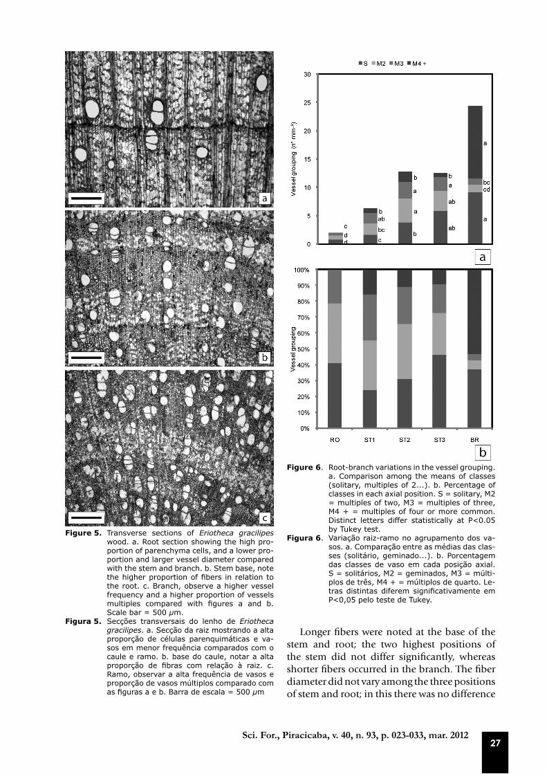
In the branch, the highest frequencies of solitary vessels and multiples of four or more elements occurred, with 40 and 50% respectively of total vessel area (1 mm²). The frequency of
multiple vessels ranged from three positions, however, the lowest values occurred in the root and branch (Figures 5a-c and 6a-b).
Both inter-vessel pits and the vessel-ray pits varied among the five axial positions with no apparent trend (Figure 7).
Measurements of the rays in the root were not performed due to difficulty in visualizing the ray cells that overlapped with the parenchyma cells that occur on a large scale, associated with the tiny proportion of fibers and vessels. The ray height did not differ among the three positions of the stem, and the position two (S2) did not differ from branch. The ray frequency varied according to the height (Figure 8). Although the rays did not statistically vary in width among the positions S1 and BR by using multiple comparison tests, regression analysis showed a negative correlation with axial position (Figure 8-10).
27Sci. For., Piracicaba, v. 40, n. 93, p. 023-033, mar. 2012
Figure 5. Transverse sections of Eriotheca gracilipes wood. a. Root section showing the high pro-portion of parenchyma cells, and a lower pro-portion and larger vessel diameter compared with the stem and branch. b. Stem base, note the higher proportion of fibers in relation to the root. c. Branch, observe a higher vessel frequency and a higher proportion of vessels multiples compared with figures a and b. Scale bar = 500 µm.
Figura 5. Secções transversais do lenho de Eriotheca gracilipes. a. Secção da raiz mostrando a alta proporção de células parenquimáticas e va-sos em menor frequência comparados com o caule e ramo. b. base do caule, notar a alta proporção de fibras com relação à raiz. c. Ramo, observar a alta frequência de vasos e proporção de vasos múltiplos comparado com as figuras a e b. Barra de escala = 500 µm
Figure 6. Root-branch variations in the vessel grouping. a. Comparison among the means of classes (solitary, multiples of 2...). b. Percentage of classes in each axial position. S = solitary, M2 = multiples of two, M3 = multiples of three, M4 + = multiples of four or more common. Distinct letters differ statistically at P<0.05 by Tukey test.
Figura 6. Variação raiz-ramo no agrupamento dos va-sos. a. Comparação entre as médias das clas-ses (solitário, geminado...). b. Porcentagem das classes de vaso em cada posição axial. S = solitários, M2 = geminados, M3 = múlti-plos de três, M4 + = múltiplos de quarto. Le-tras distintas diferem significativamente em P<0,05 pelo teste de Tukey.
Longer fibers were noted at the base of the stem and root; the two highest positions of the stem did not differ significantly, whereas shorter fibers occurred in the branch. The fiber diameter did not vary among the three positions of stem and root; in this there was no difference

Longui et al. – Root-branch anatomical investigation of Eriotheca gracilipes young trees: a biomechanical and ecological approach
28Sci. For., Piracicaba, v. 40, n. 93, p. 023-033, mar. 2012
Figure 7. Root-branch variations of the pits. IP = In-tervessel pits, VRP = Vessel-ray pits. Dis-tinct letters differ statistically at P<0.05 by Tukey test.
Figura 7. Variação raiz-ramo das pontoações. IP = Pontoações intervasculares, VRP = Ponto-ações raio-vasculares. Letras distintas di-ferem significativamente em P<0,05 pelo teste de Tukey.
Figure 8. Root-branch variations of the rays. RW = Ray width, RH = Ray height, RF = Ray frequency. Distinct letters differ statistically at P<0.05 by Tukey test.
Figura 8. Variação raiz-ramo dos raios. RW = Largura dos raios, RH = Altura dos raios, RF = Frequ-ência dos raios. Letras distintas diferem signi-ficativamente em P<0,05 pelo teste de Tukey.
Figure 9. Variation in ray width according to axial position.
Figura 9. Variação na largura dos raios em função da posição radial.
Figure 10. Tangential sections of Eriotheca gracilipes wood. a. Note the higher proportion of parenchyma cells in the root. b. Section of the branch, chan-ge in the ray dimensions compared with the root. Scale bar = 500 µm.
Figura 10. Secções tangenciais do lenho de Eriotheca gra-cilipes. a. Notar a maior proporção de células parenquimáticas na raiz. b. Secção do ramo, di-ferença na dimensão dos raios comparado com a raiz. Barra de escala = 500 µm.
from the branch. The fiber lumen diameter showed no clear variation among positions while the fiber wall thickness showed a lower value in the branch than those observed in the root and stem in all three positions (Figure 11). Regression analysis showed no significant difference among the fiber features and the root-branch positions.

29Sci. For., Piracicaba, v. 40, n. 93, p. 023-033, mar. 2012
DISCUSSION
There was no clear relationship between the vessel element length and water transport in E. gracilipes. However, Carlquist (2001) mentions that many authors believe that the functional vessel element length does not influence significantly the conductivity. The pattern observed between vessel element length and ecological factors is not at random, and the elements are shorter in dicotyledons of drier habitats, as compared to their relatives in more humid habitats. Shorter vessel elements are associated with wood less at risk with regards the formation of air bubbles. Applying this idea to individual variation, it is proposed that root wood, due to the greater vessel element length, is less safe than the other positions. However, more direct relationships with the water transport are noted with the vessel diameter and frequency.
Thus, the decrease in vessel diameter in branch-root direction apparently indicates a potential reduction in water conduction, but the increase in the vessel frequency in the same direction suggests that there is a compensation, which leads us to interpret that water conduction is similar throughout the trees. However, when considering efficiency or safety, according to Hacke et al. (2005), vessels of larger diameter are more efficient, but have greater vulnerability to
cavitation. Thus, it is proposed that in E. gracilipes there is an increase in the safety towards root-branch. In the latter axial position, there is also the largest number of vessel multiples of four or more elements, other characteristics associated with the safe transport of water, because water can flow to other vessels through pits among adjacent vessels and following the transpiration stream, until embolized vessels are refilled with water (SALLEO et al., 2004).
Some authors, such as James et al. (2003), who studied four species occurring in tropical forest in Panama (Anacardium excelsum, Ficus insipida, Schefflera morototoni and Cordia alliodora) found no consistent axial trend for the vessel diameter and frequency of the stem base to the crown, but reported a marked decrease in the vessel diameter of the crown base to the terminal branches. Others, such as Stokke and Manwiller (1994), with Quercus velutina, cite similar results to our study with a gradual and significant increase in vessel frequency from root to branch.
With regard to pits, because of difficulty in the preparation of sections, our analysis is confined only to the tangential diameter, which did not show any pattern in E. gracilipes that could be associated with safety or efficiency in conducting water. However, more specific studies of pits mention that its structure, pore size and membrane thickness directly influence the conductivity, cavitation resistance and strength of vessel cell walls to resist implosion due to the intensity of current flow transpiration (JANSEN et al., 2009).
In addition to the water transport, the vessel diameter and frequency influence the mechanical strength of the trees against the wind, rain or load of arboreal animals. As the vessel lumen do not contribute to the mass and thus to the wood density, a property which is considered an indicator of strength properties, regions of the wood with larger and more frequent vessel diameters have lower resistance (BAAS et al., 2004).
Analyzing the change in the vessel diameter and frequency towards root-branch, apparently similar to water conduction, there was also a compensation in the distribution of empty spaces (vessel lumen), which suggests an equal division of the axial mechanical strength. However, the branch has a large proportion of vessel multiples of four or more cells, so we intuitively speculate that this configuration characterizes a more fragile tissue, due to the occurrence of several vessels together, which can decrease the resistance in this portion.
Figure 11. Root-branch variations of the fibers. FD = Fiber diameter, FLD = Fiber lumen diameter, FWT = Fi-ber wall thickness, FL = Fiber length. Distinct let-ters differ statistically at P<0.05 by Tukey test.
Figura 11. Variação raiz-ramo das fibras. FD = Diâmetro das fibras, FLD = Diâmetro do lume das fibras, FWT = Espessura da parede das fibras, FL = Compri-mento das fibras. Letras distintas diferem signi-ficativamente em P<0,05 pelo teste de Tukey.

Longui et al. – Root-branch anatomical investigation of Eriotheca gracilipes young trees: a biomechanical and ecological approach
30Sci. For., Piracicaba, v. 40, n. 93, p. 023-033, mar. 2012
According to Mattheck and Kubler (1995), the trees have a flexibility strategy that occurs in the peripheral parts (smaller branches), which can be understood as follows: to bend the branches and consequently direct the leaves in the wind direction decreases the exposed area. If this action still does not work, the breaking of branches may be regarded as a “safety valve”, a mechanism to protect the tree. In physical terms, it can be understood as a principle of the minimum lever arms, as when the minor branch is broken, the largest branch or stem is protected because it reduces the lever arm and consequently the movement of force. This strategy may in part explain the apparent greater mechanical fragility of the E. gracilipes branches.
The ray cells are important in storage and mobilization of carbohydrates during periods of low photosynthetic production and, according to Clearwater and Goldstein (2005), can also provide sugars from starch hydrolysates which would help in the recovery of embolized vessels. Even without measuring the percentage of parenchyma cells in the five positions, it can easily be seen that the root has the largest number of these cells in relation to other positions of the stem and branch; thus it is the region with greatest potential for starch storage.
Considering the climatic characterization of Assis, it is proposed that the abundance of parenchyma at root (cells with potential starch grain storage) may contribute to the resistance of E. gracilipes to the dry period that occurs historically in ASF between April to August, with water deficit in these two months (Figure 1). Assis et al. (2011) mentioned that water availability is the main factor to explain the gradient of Cerrado vegetation in places where the climate and fertile soil do not vary spatially. According to Lorenzi (2002) and Oliveira (2006), E. gracilipes is a semideciduous species. Therefore, during part of the year the leaf area decreases, suggesting a reduction in the photosynthetic rate, which reinforces the role of sugars as energy reserve. According to Pallardy (2007), plants can maintain respiration and growth from the reserve carbohydrate when food is not supplied directly from photosynthesis. Another important strategy is the presence of photosynthetic furrows on the bark of “paineiras”, which help in growth when the tree is leafless (LORENZI, 2002).
Parenchyma cells in abundance observed in E. gracilipes can also relate to water storage. According to Arnold (2008), many species of
Malvaceae (formerly Bombacaeae) are able to store water in the stem. Storage is an important factor in the plant/water relationship (GARTNER; MEINZER, 2005). Pratt et al. (2007) related water transport, biomechanics and reserves in the stems and roots of nine Rhamnaceae species and found a higher proportion of parenchyma in the root compared with the stem. Olson and Carquist (2001), in an extensive study of the different habits of the Moringa genus, observed several strategies for storing water in the root and stem. The authors suggest that besides the water supply in parenchyma cells, this function could also be performed by septate fibers which would provide additional sugar reserve and mechanical support. E. gracilipes appears to employ different strategies for water supply, because gelatinous fibers were observed mainly at the top of stem and branch. Authors like Paviani (1978) and Marcati et al. (2001) relate gelatinous fibers positively with water storage, because the cellulose that forms the gelatinous layer has water affinity. Thus, in E. gracilipes, storage at the top of the stem and branch is performed by the parenchyma and septate fibers, and almost entirely in parenchyma cells of the root.
Structurally, the largest number of parenchyma cells relates positively with higher mechanical fragility of the stems compared with root and branch since the parenchyma cells are more fragile than the vessel elements and especially the fibers.
However, according to Mattheck and Kubler (1995), the rays, also formed by parenchyma cells, but arranged in the radial direction (with respect to the horizontal axis of the stem), play an important biomechanical function in a way similar to the role of the steel beams in concrete. When a stem is bent, the rays prevent the slipping of growth layers one over the other like the pages of a book when folded. Thus, the rays help to prevent the slipping of the growth layers by shear stress, locking them and acting as a “bolt”. Carlquist (1975) adds that in many dicotyledons ray cells have thick and lignified walls which make the wood stronger.
There was a significant decrease in ray width from the base to stem and branch. Our group found similar results in Xylopia aromatica (ROMEIRO et al., 2010), originating from the same area of E. gracilipes in this study. Although this result is also related to the maturation of the wood, it indicates a higher locking capacity of the rays in the lower regions of the stem.

31Sci. For., Piracicaba, v. 40, n. 93, p. 023-033, mar. 2012
When the plants receive loads resulting from wind or rain, the stems resist deflections at their base by both the geometry of the root system and the pressure exerted by soil on the roots. If the roots are superficial, the soil has little pressure and its contribution is small in tree resistance to overload. Thus the geometry of the roots has a great influence on support: unlike when the roots are deep, the soil pressure increases and consequently its contribution is higher in tree resistance to wind or rain (NIKLAS, 1992).
In anatomical terms, when there is a scant contribution of the soil to tree resistance, in addition to geometry, it is proposed that the wood should be tougher, with a high proportion of fibers with thick walls that would support higher shear stresses and tissue breakdown. In the studied trees of E. gracilipes, although not quantified, there is clearly a higher fiber proportion at the stem base, which can strengthen this region. The root tissue, due to the high proportion of parenchyma cells, seems most fragile when compared with the stem and branch. However, at the time of collection it was observed that the trees had very deep roots compared to 12 other species collected in the same area, including Xylopia aromatica, studied by Romeiro et al. (2010). Thus, the occurrence of deep roots in E. gracilipes suggests a compensation for the anatomical weaknesses.
According to the quantitative data of the fibers, the specialized support cells, a distinction that explains effective participation in the tree structure, was not observed. Only the fiber length was greater at the base of the stem, which may have a mechanical relationship or just be a result of a mature cambium able to produce longer cells.
Despite its role in water reserve, gelatinous fibers are also associated with the reaction wood (BURGER; RICHTER, 1991; PANSHIN; DE ZEEUW, 1964). Gelatinous fibers were found abundantly at the top of the stem and in the branch, which may indicate the presence of reaction wood.
According to Pilate et al. (2004), the gelatinous layer of the fibers directly influences the mechanical properties of the wood. Burger and Richter (1991) described the occurrence of gelatinous fibers in the inclined branches and stems, observed in plants of mountain slopes or those subject to great efforts to support. Thus, the presence of gelatinous fibers increases the tensile strength of the xylem in response to environmental factors such as wind (BAMBER, 2001; DONALDSON, 2008). If the “use” of
gelatinous fibers fails, the plants still have the flexibility strategy described above by Mattheck and Kubler (1995).
The E. gracilipes plants were in the flat area and open physiognomy, and for this reason it is proposed that the result is related to a response to the wind, because the vegetation was composed of sparsely distributed trees, allowing for wind incidence between them. Moreover, the trees were young and thinner at the higher positions, therefore more susceptible to sway due to wind.
CONCLUSIONS
The presence of vessels with a smaller diameter, as well as higher frequency and proportion of vessel multiples of four or more elements, suggest a greater safety for water transport. In mechanical terms, the vessels features in the branch should relate to a greater fragility in relation to the three positions of the stem.
The abundance of parenchyma, mainly in the root, indicates the potential storage of starch and water. Besides, the mechanical fragility may be compensated by deep roots.
The presence of gelatinous fibers can either collaborate in water storage or be a reflection of reaction wood.
REFERENCES
ARNOLD, M.A. Landscape Plants for Texas and Environs. 3ed. Champaign : Stipes Publishing Co., 2008. 1340p.
ASSIS, A.C.C.; COELHO, R.M.; PINHEIRO, E.S.; DURIGAN, G. Water availability determines physiognomic gradient in an area of low-fertility soils under Cerrado vegetation. Plant Ecology. Oxford, v.212, n.7, p.1135-1147, 2011.
BAAS, P.; EWERS, F.W.; DAVIS, S.D.; WHEELER, E.A. Evolution of xylem physiology. In: POOLE, I.; HEMSLEY, A. (Eds). Evolution of Plant Physiology. London: Elsevier Academic Press, 2004. p.273-295.
BAMBER, R.K. A general theory for the origin of growth stresses in reaction wood: how trees stay upright. IAWA Journal, Leiden, v.22, p.205-212, 2001
BARNETT, J.R.; JERONIMIDIS, G. Reaction wood. In: BARNETT, J.R.; JERONIMIDIS, G (Eds.). Wood quality and its biological basis. Oxford: Blackwell Publishing Ltd, 2003. p.118-136.

Longui et al. – Root-branch anatomical investigation of Eriotheca gracilipes young trees: a biomechanical and ecological approach
32Sci. For., Piracicaba, v. 40, n. 93, p. 023-033, mar. 2012
BERLYN, G.P.; MIKSCHE. J.P. Botanical microtechnique and cytochemistry. Iowa State University Press, 1976.
BOGNOLA, I.A.; PRADO, H.; MENK, J.R.F.; JOAQUIM, A.C.; LEPSCH, I.F. Levantamento pedológico semidetalhado do Estado de São Paulo: Quadrícula de Assis. II. Memorial Descritivo. Campinas: Instituto Agronômico, 2003. 54p. (Série Pesquisa APTA. Boletim Científico, 08).
BURGER, L.M.; RICHTER, H.G. Anatomia da Madeira. São Paulo: Nobel, 1991. 154p.
CARLQUIST, S. Comparative wood anatomy: systematic, ecological and evolutionary aspects of dicotyledons wood. Berlin: Springer Verlag, 2001
CARLQUIST, S. Ecological strategies of xylem evolution. Los Angeles: University of California Press, 1975.
CEPAGRI - CENTRO DE PESQUISAS METEOROLÓGICAS E CLIMÁTICAS APLICADAS A AGRICULTURA. Clima dos Municípios Paulistas. Disponível em: <http://www.cpa.unicamp.br/outras-informacoes/clima-dos-municipios-paulistas.html>. Acesso em 20 abr.2010.
CHAVE, J., COOMES, D.; JANSEN, S.; LEWIS, S.; SWENSON, N.; ZANNE, A. Towards a worldwide wood economics spectrum. Ecology Letters, Paris, v.12, p.351-366, 2009.
CLEARWATER, M.J.; GOLDSTEIN, G. Embolism repair and long distance water transport. In: HOLBROOK, N.M.; ZWIENNIECKI, M.A. (Eds.) Vascular transport in plants. Amsterdam: Elsevier Inc., 2005. p.375-400.
DONALDSON L. Microfibril Angle: Measurement, Variation And Relationships – A Review. IAWA Journal, Leiden, v.29, p.345-386, 2008.
DUARTE, M.C. Eriotheca gracilipes (K.Schum.) A.Robyns. In: Lista de Espécies da Flora do Brasil. Rio de Janeiro: Jardim Botânico do Rio de Janeiro, 2010. Disponível em: <http://floradobrasil.jbrj.gov.br/2010/FB025739> . Acesso em 04 mar. 2011.
EVERT, R.F.; EICHHORN, S.E. Esau´s plant anatomy: meristems, cells, and tissues of the plant body: their structure, function, and development. 3ed. New York: Wiley-Liss, 2006. 624p.
GARTNER, B.L.; MEINZER, F. Structure function relationships in sapwood water transport and storage. In: HOLBROOK, N.M.; ZWIENNIECKI, M.A. (Eds.) Vascular transport in plants. Amsterdam: Elsevier Inc., 2005. p.307-332.
HACKE, U.G., SPERRY, J.S.; PITTERMANN, J. Efficiency versus safety tradeoffs for water conduction in angiosperm vessels versus gymnosperm tracheids. In: HOLBROOK, N.M.; ZWIENNIECKI, M.A. (Eds.) Vascular transport in plants. Amsterdam: Elsevier Inc., 2005. p.333-354.
IAWA COMMITTEE. IAWA list of microscopic features for hardwood identification. IAWA Bulletin, Leiden, v.3, n.10, p.219-332, 1989.
JAMES, S.A.; MEINZER, F.C.; GOLDSTEIN, G.; WOODRUFF, D.; JONES, T.; RESTOM, T.; MEJIA, M.; CLEARWATER, M.; CAMPANELLO, P. Axial and radial water transport and internal water storage in tropical forest canopy trees. Oecologia, Berlin, v.134, p.37-45, 2003.
JANSEN, S.; CHOAT, B.; PLETSERS, A. Morphological variation of intervessel pit membranes and implications to xylem function in angiosperms. American Journal of Botany, Columbus, v.96, n.2, p.409-419, 2009.
LORENZI, H. Árvores Brasileiras: manual de identificação e cultivo de plantas arbóreas do Brasil. 2 ed. Nova Odessa: Plantarum, 2002. v.2. 384p.
MARCATI, C.R., ANGYALOSSY-ALFONSO, V.; BENETATI, L. Anatomia comparada do lenho de Copaifera langsdorffii Desf. (Leguminosae-Caesalpinoideae) de floresta e cerradão. Revista Brasileira de Botânica, São Paulo, v.24, p.311-320, 2001.
MARTÍNEZ-CABRERA, H.I.; JONES, C.S.; ESPINO, S.; SCHENK, H.J. Wood anatomy and wood density in shrubs: responses to varying aridity along transcontinental transects. American Journal of Botany, Columbus, v.96, p.1388-1398, 2009.

33Sci. For., Piracicaba, v. 40, n. 93, p. 023-033, mar. 2012
MATTHECK, C.; KUBLER, H. Wood – The internal optimization of trees. Berlin: Spring-Verlag. 1995. 129p.
MAX, J.C.M.; MELO, A.C.G.; HONDA, E.A.; DURIGAN, G.; MALÍCIA, L.C.; SOUZA, M.B.M.; CARDOSO, M.M.; BÔAS, O.V.; RAMOS, V.S. & CONTIÉRI, W.A. Plano de manejo da Floresta Estadual de Assis. IF Série Registros, São Paulo, 30, p.1-80, 2007.
NIKLAS, K.J. Plant biomechanics: an engineering approach to plant form and function. Chicago: The University of Chicago Press, 1992. 607p.
OLIVEIRA, J.S. Variações estruturais do lenho de espécies de cerrado do Botucatu – SP. Dissertação (Mestrado em Ciências Biológicas - Botânica) – Universidade Estadual Paulis “Júlio de Mesquita Filho”, Botucatu, 2006.
OLSON, M.E.; CARLQUIST, S. Stem and root anatomical correlations with life form diversity, ecology, and systematics in Moringa (Moringaceae). Botanical Journal of the Linnean Society, London, v.135, n.4, p.315-348, 2001.
PALLARDY, S.G. Physiology of woody plants. 3ed. San Diego: Academic Press - Elsevier Inc., 2007. 464p.
PANSHIN, A.J.; DE ZEEUW. C. Textbook of Wood Technology: structure, identification, properties and uses of the commercial woods of the United States and Canada. 3.ed. Nova York: McGraw-Hill, 1964. 643p.
PAVIANI, T.I. Anatomia vegetal e cerrado. Ciência e Cultura, Campinas, v.30, p.1076-1086, 1978.
PILATE, G.; CHABBERT, B.; CATHALA, B.; YOSHINAGA, A.; LEPLE, J.C.; LAURANS, F.; LAPIERRE, C. & RUEL, K. Lignification and tension wood. Comptes Rendus Biologies, Paris, v.327, p.889 - 901. 2004.
PRATT, R.B.; JACOBSEN, A.L.; EWERS, F.W.; DAVIS, S.D. Relationships among xylem transport, biomechanics and storage in stems and roots of nine Rhamnaceae species of the California chaparral. New phytologist, Oxford, v.174, p.787-798, 2007.
READ, J.; STOKES, A. Biomechanics in an ecological context. American Journal of Botany, Columbus, v.93, p.1546-1565, 2006.
ROMEIRO, D.; LONGUI, E.L.; MORENO, N.B.; HERREIRA, L.A.; LIMA, I.L.; FLORSHEIM, S.M.B.; MELO, A.C.G. Características anatômicas quantitativas da raiz, tronco e ramo em árvores jovens de Xylopia aromatica (Lam.) Mart. IF Série Registros, São Paulo, n.43, p.107-112, 2009.
SALLEO, S., LO GULLO, M.A.; TRIFILÒ, P.; NARDINI, A. New evidence for a role of vessel-associated cells and phloem in the rapid xylem refilling of cavitated stems of Laurus nobilis L. Plant. Cell and Environment, Oxford, v.27, p.1065-1076, 2004.
SASS, J.E. Botanical Microtechniche. Ames: The Iowa State College Press,1951. 326p.
STOKKE, D.D.; MANWILLER, F.G. Proportions of wood elements in stem, branch, and root wood of black oak (Quercus velutina). IAWA Journal, Leiden, v.15, n.3, p.301-310, 1994.
WODZICKI, T.J. Natural factors affecting wood structure. Wood Science and Technology, New York, v.35, p.5-26, 2001.
ZIMMERMANN, M.H.; BROWN, C.L. Trees: structure and function. New York: Springer-Verlag, 1971.
Recebido em 06/04/2011Aceito para publicação em 05/12/2011
