roles of major histocompatibility complex class ii in ...jvi.asm.org/content/88/14/7764.full.pdf ·...
TRANSCRIPT

Roles of Major Histocompatibility Complex Class II in InducingProtective Immune Responses to Influenza Vaccination
Eunju O,a Young-Tae Lee,a Eun-Ju Ko,a Ki-Hye Kim,a Yu-Na Lee,a Jae-Min Song,a,b Young-Man Kwon,a Min-Chul Kim,a,c
Daniel R. Perez,d Sang-Moo Kanga
Center for Inflammation, Immunity, and Infection, Institute for Biomedical Sciences, Georgia State University, Atlanta, Georgia, USAa; Department of Global MedicalScience, Sungshin Women’s University, Seoul, South Koreab; Animal and Plant Quarantine Agency, Anyang, Gyeonggi-do, South Koreac; Department of VeterinaryMedicine, University of Maryland, College Park, Maryland, USAd
ABSTRACT
Major histocompatibility complex class II-deficient (MHC-II KO; A��/�) mice were used to assess the roles of MHC-II mole-cules in inducing protective immune responses to vaccination. After vaccination with influenza A/PR8 virus-like particle (VLP)vaccine, in vivo and in vitro vaccine antigen-specific IgG isotype antibodies were not detected in MHC-II KO mice, which isquite different from CD4 T cell-deficient mice that induced vaccine-specific IgG antibodies. The deficiency in MHC-II did notsignificantly affect the induction of antigen-specific IgM antibody in sera. MHC-II KO mice that were vaccinated with influenzaVLP, whole inactivated influenza virus, or live attenuated influenza virus vaccines were not protected against lethal infectionwith influenza A/PR8 virus. Adoptive transfer of fractionated spleen cells from wild-type mice to MHC-II KO mice indicated thatCD43� cell populations with MHC-II contributed more significantly to producing vaccine-specific IgG antibodies than CD43�
B220� conventional B cell or CD4 T cell populations, as well as conferring protection against lethal infection. Bone marrow-derived dendritic cells from MHC-II KO mice showed a significant defect in producing interleukin-6 and tumor necrosis factoralpha cytokines. Thus, results indicate that MHC-II molecules play multiple roles in inducing protective immunity to influenzavaccination.
IMPORTANCE
Major histocompatibility complex class II (MHC-II) has been known to activate CD4 T helper immune cells. A deficiency inMHC-II was considered to be equivalent to the lack of CD4 T cells in developing host immune responses to pathogens. However,the roles of MHC-II in inducing protective immune responses to vaccination have not been well understood. In the presentstudy, we demonstrate that MHC-II-deficient mice showed much more significant defects in inducing protective antibody re-sponses to influenza vaccination than CD4 T cell-deficient mice. Further analysis showed that CD43 marker-positive immunecells with MHC-II, as well as an innate immunity-simulating adjuvant, could rescue some defects in inducing protective immuneresponses in MHC-II-deficient mice. These results have important implications for our understanding of host immunity-induc-ing mechanisms to vaccination, as well as in developing effective vaccines and adjuvants.
Vaccination is the most effective measure for preventing infec-tious diseases, including influenza, a highly contagious respi-
ratory disease resulting in widespread morbidity and mortality.Most licensed human vaccines are based on their capability toinduce protective humoral antibodies that block infection or re-duce pathogen loads, although cellular immune responses are alsoimportant (1–3). However, mechanisms by which vaccination in-duces effective protective immunity have not been well under-stood yet.
A model for producing protein antigen-specific immunoglob-ulin G (IgG) antibodies initiates with antigen uptake by antigen-presenting cells such as dendritic cells (DCs), macrophages, and Bcells. In particular, DCs after antigen uptake migrate to secondarylymphoid tissues from peripheral sites. Antigen-presenting cellspresent peptide fragments of processed antigens on their surfacesin the context of major histocompatibility complex class II(MHC-II) molecules (4). Specific CD4� T cells are activated andundergo clonal expansion after recognition of antigenic peptide/MHC-II on antigen-presenting cells via a T cell receptor. In themeantime, naive B cells internalize and process a specific antigenbound by surface immunoglobulin receptors, presenting anti-genic peptides in the context of MHC-II molecules. The T cell help
to drive the B cell response is initiated by recognizing peptide/MHC-II on the B cell surfaces via T cell receptor through thespecific CD4� T cells. Subsequently, T cell-derived signaling mol-ecules and cytokines initiate B cell proliferation and direct immu-noglobulin isotype switching (5–7). In this model, cognate T andB cell interaction is a requirement for B cell IgG responses andisotype switching.
This scenario of cognate T and B cell interactions through the Tcell receptor and peptide-MHC complex does not appear to fullyexplain the strong humoral responses that are rapidly generatedagainst many pathogens probably due to low frequencies of anti-gen-specific T and B cells at the time of initial antigen encounter.Alternative T cell help for B cell isotype-switched IgG responses
Received 13 March 2014 Accepted 18 April 2014
Published ahead of print 23 April 2014
Editor: T. S. Dermody
Address correspondence to Sang-Moo Kang, [email protected].
Copyright © 2014, American Society for Microbiology. All Rights Reserved.
doi:10.1128/JVI.00748-14
7764 jvi.asm.org Journal of Virology p. 7764 –7775 July 2014 Volume 88 Number 14
on June 21, 2018 by guesthttp://jvi.asm
.org/D
ownloaded from

might be mediated by secreted cytokines or nonspecific molecularinteractions between adjacent cells (8, 9). It is noteworthy thatDCs are capable of retaining antigens in a form that is recognizedby B cells and also provide signals that direct isotype switching inT cell-dependent humoral responses (10–12).
The normal development of mature T cells needs their inter-actions with MHC molecules in the thymus. MHC-II-deficient(MHC-II KO) mice were found to be deficient in mature CD4� Tcell-mediated immune responses (13). Previous studies usedMHC-II KO mouse models to study the roles of CD4� T cellsand/or MHC-II molecules in inducing host CD8� cytotoxic T cellimmune responses to viral, bacterial, and parasitic infections (14–20). The apparent efficacy of comparable or less control of infect-ing pathogens was attributed to the intact activity of CD8� cyto-toxic T cells despite the deficiency of CD4� T cells. Polyomavirusinfection of mice with a deficiency of functional ��� T cells or��� and ��� T cells induced IgM and IgG antiviral antibodies(21, 22). Vesicular stomatitis virus (VSV) infection in ��� T cell-deficient mice induced IgG antibody responses (23–26). Our pre-vious studies have shown that mucosal or systemic immunizationof CD4� T cell-deficient mice with inactivated influenza virus canalso induce antigen-specific isotype-switched IgG antibody re-sponses, virus neutralizing antibodies, and protection (27, 28).
The potential roles of MHC-II molecules in inducing immuneresponses to vaccination largely remain unknown. In this study,we investigated host immune responses and protection againstlethal challenge after vaccination of MHC-II KO mice with a re-combinant influenza A/PR8 virus-like particle (VLP), whole inac-tivated influenza virus, or 2009 H1N1 pandemic influenza viruslive attenuated vaccine. We demonstrate that, despite the presenceof naturally occurring IgG antibodies at significant levels, MHC-IIKO mice were not able to induce antigen-specific IgG antibodyresponses to influenza vaccination and thus were not protected.CD43� cell populations with MHC-II molecules were found toinduce protective antibody responses by adoptive-transfer stud-ies, suggesting additional roles for MHC-II in addition to activat-ing CD4 T cells.
MATERIALS AND METHODSMice and cells. MHC-II KO (I-A��/�) and wild-type C57BL/6 (B6 WT)mice were purchased from the Jackson Laboratory (Bar Harbor, ME) andbred at the animal facility at Georgia State University. BALB/c mice werepurchased from Harlan Laboratories (Indianapolis, IN). Spodoptera fru-giperda Sf9 insect cells (American Type Culture Collection, CRL-1711) forthe production of recombinant baculoviruses (rBVs) and influenza VLPswere cultured in SF900-II serum-free medium (Invitrogen, Carlsbad, CA)at 27°C.
Preparation of influenza VLPs and virus. Preparation of influenzaVLP vaccines containing the hemagglutinin (HA) protein as a major pro-tective antigen has been well described in our previous studies (29–33). Toproduce influenza VLPs, SF9 insect cells were coinfected with rBVs ex-pressing influenza virus M1 gene and HA derived from the strain of A/PR/8/34 H1N1 (A/PR8) virus. Influenza VLPs in the culture supernatantswere harvested at 3 days after infection and purified as described previ-ously (29, 34). H1N1 influenza A viruses A/PR8 (29) and A/California/04/2009 (a kind gifts from Richard Webby) were propagated in allantoiccavity of 10-day-old embryonated hen’s eggs (32). An att.NL virus is anattenuated 2:6 reassortant containing the surface HA and neuraminidase(NA) genes of A/Netherlands/602/09 (H1N1) (35) and the internal genesfrom the attenuated A/turkey/Ohio/313053/04 (H3N2) virus. Rg20A/Texas/5/2009, kindly provided by Ruben Donis, is a reassortant virus
containing six A/PR/8/34 internal genes and A/Texas/5/2009 HA and NA(32).
Immunization and viral challenge infection. MHC-II KO mice (8 to10 weeks old) and B6 WT mice (8 to 10 weeks old) were intramuscularlyimmunized with influenza A/PR8 VLPs (3 or 10 �g) at weeks 0 and 4. Forin vivo infection, mice were anesthetized with isoflurane (Baxter, Deer-field, IL), intranasally challenged with A/PR8 virus (5 50% lethal dose[LD50]) at 3 weeks after boost immunization and sacrificed at 4 days afterchallenge or monitored for 14 days for measuring body weights and sur-vival rates to assess the protective efficacy of vaccination. For the serumprotective efficacy, 6- to 7-week-old female naive BALB/c mice were in-fected with a lethal dose A/PR8 virus mixed with heat-inactivated sera(56°C for 30 min) from each of the indicated groups after incubation atroom temperature for 30 min (36, 37).
To understand the protective immune response from live and inacti-vated whole viral vaccines, B6 WT mice and MHC-II KO mice were in-tranasally prime-boost inoculated with a live attenuated att.NL virus(105 tissue culture infective doses per mouse) or intramuscularly boostimmunized with inactivated Rg20 A/Texas/5/2009 (Rg20i; 5 �g/mouse).Mice were challenged with 2009 pandemic H1N1 virus (5 LD50) 4 weeksafter boost immunization. All animal studies were approved and con-ducted under the guidelines by Georgia State University’s IACUC(A11026). Mice were euthanized if their body weight loss exceeded 25%.
Flow cytometry analysis of MHC-II KO mice phenotype. Spleen cellswere analyzed by fluorescence-activated cell sorting (FACS) to check phe-notypes of MHC-II KO and B6 WT mice. Isolated spleen cells were stainedwith fluorescence-conjugated monoclonal antibodies specific to cell phe-notypes, MHC-II–FITC (M5/114.15.2; eBioscience, San Diego, CA),CD19-PE (6D5; eBioscience), B220-PE Cy7 (RA3-6B2; BD Pharmingen),CD3-Pacific Blue (17A2; Biolegend), CD4-APC (GK1.5; eBioscience),and CD8-PerCP (53-6.7; BD Pharmingen). Dead cells are excluded bystaining with near-infrared fluorescent reactive dye (Invitrogen, Eugene,OR). Live lymphocytes were gated according to their sizes, and granularitywas defined in the forward light scatter (FSC) and side light scatter (SSC)plot. Cell acquisition was performed with a multilaser multiparameteranalysis cytometer (LSR-II and LSRFortessa; BD Biosciences, MountainView, CA), and the data were analyzed using FlowJo software (v7.6.4; TreeStar, Inc., Ashland, OR).
Determination of serum antibody responses. Blood samples werecollected from the retro-orbital plexus puncture using heparinized micro-capillaries (Drummond Scientific Company, Broomall, PA) after anesthe-tization of the animals by isoflurane inhalation. Sera were collected 18days after prime and 10 days after boost immunization and stored at�20°C until analysis. Influenza virus-specific total IgG, IgM and isotypeantibodies (Southern Biotechnology, Birmingham, AL) were determinedusing standard enzyme-linked immunosorbent assay (ELISA) as de-scribed previously (29). Purified mouse IgG, IgM, and isotype antibodieswere used as standards to determine the relative antibody concentrationsin immune sera from optical spectrophotometer readings at 450 nm(ELx800; BioTek, Winooski, VT).
Virus-specific antibody secreting cell responses. Freshly isolatedspleen and bone marrow cells (5 105 cells/well) were incubated for 24 hin the 96-well culture plates coated with either culture medium or A/PR8VLP (400 ng/well) for in vitro antigenic stimulation. Antibody-secretingcell responses in the spleen and bone marrow were determined after 5 daysof in vitro culture. The levels of antibodies specific to A/PR8 VLP vaccinein the culture plates were determined by ELISA as previously described(29, 38).
Determination of T cell responses. Spleen cells were obtained fromthe mice sacrificed at 4 days postchallenge. Gamma interferon (IFN-�)-and interleukin-4 (IL-4)-secreting cell spots were determined on Multi-Screen 96-well filter plates (Millipore, Billerica, MA) coated with cyto-kine-specific capture antibodies as described previously (29). Briefly, 5 105 spleen cells (per well) were cultured in anti-mouse IFN-� or anti-mouse IL-4 (300 ng/100 �l/well) precoated plates with or without A/PR8
Roles of MHC Class II in Immunity
July 2014 Volume 88 Number 14 jvi.asm.org 7765
on June 21, 2018 by guesthttp://jvi.asm
.org/D
ownloaded from
VLP (2 �g/ml) as an antigenic stimulator. After 36 h of incubation, thenumber of IFN-�- or IL-4-secreting cell spots was counted using aBioreader 5000-E� (Biosys, Miami, FL).
Analysis of lung and BALF samples. Bronchoalveolar lavage fluids(BALF) were obtained by infusing 1 ml of phosphate-buffered saline(PBS) using a 18Gx11/4 catheter (Exelint International, Tokyo, Japan) intothe lungs via the trachea, and lung samples were also collected at day 4postchallenge. Cells were removed after centrifugation, and inflammatorycytokine (IL-6) in lung extracts and BALF were analyzed by using Ready-Set-Go cytokine kits (eBioscience) according to the manufacturer’s pro-tocol. To determine lung virus titers at 4 days postchallenge with A/PR8virus, 10-day-old embryonated hen’s eggs were infected with 10-fold se-rially diluted lung extracts for 3 days, and lung virus titers were calculatedas described previously (38).
Adoptive cell transfer. The CD4�, CD43�, or CD43� cell fractionswere isolated from the spleen cells of naive B6 WT mice by the MACSsystem using antibody-coated microbeads (Miltenyi Biotec, BergischGladbach, Germany) according to the manufacturer’s instructions.Briefly, mouse spleens from the naive B6 WT group were minced usingglass slides, passed through a cell strainer (40-�m pore size; Becton Dick-inson, Franklin Lakes, NJ), resuspended in 0.5% bovine serum albumin(Sigma, St. Louis, MO) with 2 mM EDTA in PBS, and incubated with CD4microbeads. Flowthrough cells in a magnetic field were applied to theCD43 microbeads to purify CD43 positive and negative cell fractions.Cells bound or unbound to the column were collected, and nucleated cellswere counted by using a hemacytometer after trypan blue dead-cell exclu-sion. The purification of MACS-isolated cell populations was determinedby cell surface staining and flow cytometric analysis prior to adoptive celltransfer to MHC-II KO mice. Enriched CD4�, CD43�, or CD43� cellswere intravenously injected via tail vein into recipient naive MHC-II KOmice, which were then immunized with 10 �g of A/PR8 VLPs intramus-cularly 3 to 4 h after adoptive cell transfer. Blood samples were collected 2weeks after immunization, and IgG antigen-specific antibody levels weredetermined by ELISA.
Generation of BMDCs. Bone marrow-derived DCs (BMDCs) wereobtained from B6 WT and MHC-II KO mice. BM cells were harvestedfrom femur and tibia, and the red blood cells were removed by using redblood cell lysis buffer (Sigma). The cells were cultured in the presence of10 ng of mouse recombinant granulocyte macrophage-colony-stimulat-ing factor (Invitrogen)/ml to generate DCs. After 6 to 10 days of culture,the generated DCs were harvested and used for experiments. After 2 daysof BMDC culture in the presence of influenza (A/PR8) VLP vaccine (10�g/ml) or lipopolysaccharide (LPS; 0.2 �g/ml), the cultured DCs wereharvested and stained with specific antibodies. After blocking Fc receptorswith CD16/32 antibodies, the cells were stained with DC surface markerantibodies (eBioscience): phycoerythrin-labeled anti-mouse CD40 (clone1C1O) and CD86 (clone GL1) and fluorescein isothiocyanate (FITC)-labeled anti-mouse CD80 (clone 16-10A1). To measure the amount ofcytokines, mouse IL-12p70-, IL-6-, and tumor necrosis factor alpha(TNF-�)-specific ELISA Ready-Set-Go kits (eBioscience) were used. Theculture supernatants were harvested, and cytokine ELISAs were per-formed according to the manufacturer’s protocol.
Statistics. Unless otherwise stated, all results are presented as means �the standard error. Statistical analysis was performed using a two-tailedStudent t test and one-way analysis of variance when comparing two ormore different groups, respectively. The data were analyzed using Prismsoftware (GraphPad Software, Inc., San Diego, CA). Probability values of�0.05 were considered statistically significant.
RESULTSNaive MHC-II KO mice are able to produce natural IgG anti-bodies. It is important to confirm that MHC-II KO mice have theMHC-II-deficient phenotype. We determined the phenotypes ofT cell and B cell populations of spleen cells in MHC-II KO micecompared to B6 WT mice by flow cytometry (Fig. 1). Spleen cells
from MHC-II KO mice showed 2-fold lower levels of CD3�,significantly lower levels of CD4� cells (2.7% versus 18.8%) andMHC-II� cells (2.05% versus 61.9%), and a slightly higher level ofCD8, CD19, and B220 expression compared to WT control mice(Fig. 1A and B). Peripheral blood lymphocytes from naiveMHC-II KO mice also had a similar deficiency of CD4� andMHC-II� cell populations (data not shown). These results indi-cate that the MHC-II KO mice are an authentic strain of mice witha MHC-II deficiency.
Next, we compared the levels of natural antibodies, IgG, IgM,and isotypes from naive B6 WT and MHC-II KO mice using astandard ELISA method. An approximately 2- to 3-fold-lower butstill significant level of IgG antibody and a moderately lower levelof IgM antibody were observed in naive MHC-II KO mice com-pared to wild-type mice (Fig. 1C). Since the B6 mouse strain is tohave the gene for isotype IgG2c instead of IgG2a (39), we exam-ined IgG1, IgG2b, and IgG2c antibodies in sera by ELISA. Inter-estingly, MHC-II KO mice showed a similar level of IgG2c anti-body compared to that of B6 WT mice and, on the other hand,approximately 2- to 4-fold-lower levels of IgG1 and IgG2b anti-bodies (Fig. 1C). Therefore, MHC-II KO mice can generate natu-ral IgG and isotype-switched antibodies not specific to an antigen,although their levels were moderately lower than those in B6 WTmice.
MHC-II KO mice do not induce antigen-specific IgG isotype-switched antibodies. To determine the roles of an MHC-II mol-ecule in inducing protective immune responses to vaccination, B6WT and MHC-II KO mice were intramuscularly immunized with3 �g of A/PR8 HA VLPs. WT mice showed significantly increasedlevels of IgG antibodies after prime and boost immunizations (16-fold higher levels of IgG after boost) (Fig. 2A). In contrast to B6WT mice, significant levels of IgG antibodies specific to virus werenot detected in the sera of MHC-II KO mice after prime and boostimmunizations, similar to naive mice (Fig. 2A). However, IgMantibodies specific to the A/PR8 viral antigens were induced athigh levels in immunized MHC-II KO mice, and there was nostatistical difference in IgM levels between MHC-II KO and B6WT mice (Fig. 2A). There was no significant boosting effect onIgM antibodies even in wild-type mice, which is different fromIgG antibodies. IgG2c isotype antibody showed the highest levelamong IgG isotypes in B6 WT mice. However, levels of vaccineantigen-specific IgG isotype antibodies were not significantly de-tected in MHC-II KO mice, which were similar to naive mice (Fig.2B). These results indicate that MHC-II KO mice have a signifi-cant defect in inducing IgG and its IgG isotype antibodies in re-sponse to vaccination with influenza VLPs.
To compare vaccine-specific IgG antibody levels in CD4knockout mice (CD4KO) after PR8 VLP vaccination, we intra-muscularly immunized MHC-II KO, CD4KO, and B6 WT mice(n 6/group) with 3 �g of influenza PR8 VLP vaccine at weeks 0and 4 (Fig. 2C). At 4 weeks after boost immunization, A/PR8virus-specific IgG antibody levels were measured (ng/ml) (Fig.2C). Although vaccine-specific IgG antibody levels of CD4KOwere ca. 30 to 40% of those for B6 WT mice, CD4KO mice showedsignificantly higher IgG antibody levels compared to MHC-II KOor naive mice. These results suggest that MHC-II KO mice haveadditional defects in inducing vaccine-specific IgG antibody re-sponses compared to CD4KO mice.
MHC-II KO mice induce IgM but not IgG antibody-secretingcell responses. To further confirm the capacity of MHC-II KO
O et al.
7766 jvi.asm.org Journal of Virology
on June 21, 2018 by guesthttp://jvi.asm
.org/D
ownloaded from

mice to induce vaccine-specific humoral responses, we analyzedantibody-secreting cell responses that may detect small numbersof vaccine-specific B cells. Spleen and bone marrow cells wereobtained from vaccinated B6 WT and MHC-II KO mice at day 4postchallenge infection with A/PR8 virus and subjected to in vitroculture plates coated with the same VLP vaccine antigen (Fig. 3).Spleen and bone marrow cells from B6 WT mice showed asimilar high level of IgG antibodies captured on vaccine anti-gen coated plates, but not those cells from MHC-II KO or naivemice (Fig. 3A).
Spleen cells from naive, immunized B6 WT and MHC-II KOmice at day 4 postchallenge showed higher levels of IgM antibodyproduction upon vaccine antigen stimulation in vitro compared tomedium controls (P � 0.001). Lower but significant levels of IgMantibodies were detected in spleen cell cultures of both naive andimmunized MHC-II KO mice (Fig. 3B). In bone marrow cells,VLP vaccine-specific IgM antibodies were detected at a higherlevel in immunized WT mice compared to those from both naiveand immunized MHC-II KO mice (P � 0.001, Fig. 3B). Also, bonemarrow cells from immunized B6 WT but not from MHC-II KOmice displayed statistically significant higher levels of IgM anti-body production compared to medium controls (P � 0.001, Fig.3B). Consistent with levels of antibodies secreted into culture su-pernatants, only immunized WT mice showed antigen-specific
IgG antibody-secreting cell spots in spleen and bone marrow cells,and a low level of IgM antibody-secreting spleen cell spots wasdetected in MHC-II KO mice (data not shown). MHC-II KO micefailed to generate vaccine antigen-specific IgG antibody-secretingcell responses after vaccination despite their capability to produceIgM antibody-secreting splenic B cells in response to vaccinationor in vitro stimulation with influenza VLP vaccines.
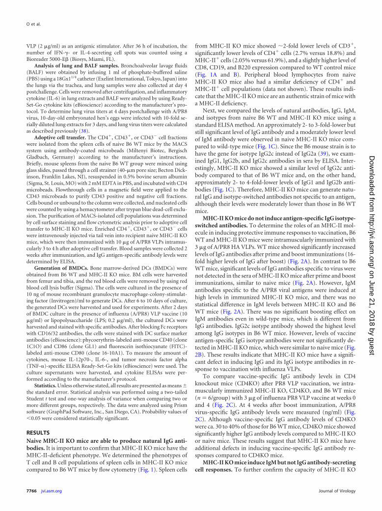
Immunized MHC-II KO mice show a defect in controllingchallenge virus and inducing cytokine-secreting cellular re-sponses. To determine the protective efficacy of vaccinatedMHC-II KO mice, the levels of proinflammatory cytokine IL-6and virus titers were determined at day 4 after A/PR8 virus chal-lenge infection (Fig. 4A and B). The level of IL-6 was low or belowthe detection limit in both BALF and lung extracts from the B6WT immunized mice. In contrast, the B6 WT naive and MHC-IIKO immunized mice showed similar patterns of high levels of IL-6in both BALF and lung extract samples (Fig. 4A and B). The levelsof the virus titers indicate the efficiency of virus clearance afterchallenge. Influenza VLP immunized B6 WT mice showed over100- to 1,000-fold lower levels of virus titers compared to those inimmunized MHC-II KO or naive control groups. That is, highviral loads were found in lungs and BALF samples from naive B6WT and naive and immunized MHC-II KO mice (Fig. 4B). Theseresults indicate that vaccination of MHC-II KO mice failed to
FIG 1 Immune cell phenotypes and serum antibodies from naive MHC-II KO mice. (A) B6 WT mice; (B) MHC-II KO (MHC-II-deficient) mice. Spleen cellswere stained with fluorescence-conjugated antibodies specific to cell surface markers (CD3, CD4, CD8, CD19, B220, and MHC-II), and live lymphocytes weregated based on their size and granularity defined in the forward light scatter (FSC) and side light scatter (SSC) plot. The FACS profiles shown are representativeof two independent experiments. (C) Total serum IgG, IgM, and isotype-switched IgG antibodies were measured in naive B6 WT and MHC-II KO mice. Totalnatural antibodies were captured using goat anti-mouse IgG, IgM, and isotype-switched antibodies. The relative antibody concentrations were determined andcalculated from standard curves of purified mouse antibodies. The data shown are the representative of three independent experiments that were consistentlyreproducible. Naive B6, a pooled serum sample of unimmunized C57BL/6 WT mice (n 4); naive MHCII KO, a pooled serum sample of unimmunized MHC-IIKO mice (n 4). *, P � 0.05; **, P � 0.01.
Roles of MHC Class II in Immunity
July 2014 Volume 88 Number 14 jvi.asm.org 7767
on June 21, 2018 by guesthttp://jvi.asm
.org/D
ownloaded from

control and clear the challenge virus, whereas the vaccinated B6WT mice were highly effective in clearing the virus after challenge.
To observe T cell immune responses, freshly isolated spleencells 4 days after challenge with A/PR8 virus from naive or immu-nized WT and MHC-II KO mice were cultured on enzyme-linkedimmunospot (ELISPOT) plates to determine IFN-�- and IL-4-secreting cell spots using VLPs as an antigenic stimulator (Fig. 4Cand D). Immunized WT mice displayed a significant level of IFN-�-secreting cell spots in responses to A/PR8 influenza VLPs, butnone of the other groups—including immunized MHC-II KOmice—showed IFN-�-producing cell spots (Fig. 4C). Similarly,immunized B6 WT mice showed the highest number of IL-4-secreting cell spots, but only background levels of IL-4 spots weredetected in spleen cells from immunized MHC-II KO mice (Fig.4D). These results suggest that MHC-II KO mice have also a defectin inducing T cell immune responses.
VLP vaccination of MHC-II KO mice do not confer effectiveprotection. We wanted to determine whether vaccination ofMHC-II KO mice would confer protection against viral infection.Naive and vaccinated WT and MHC-II KO mice were infectedwith a lethal infectious dose of A/PR8 virus at 3 weeks after im-munization with A/PR8 VLPs. Naive mice showed severe bodyweight loss and had to be euthanized by day 8 postchallenge. Sim-ilarly, immunized MHC-II KO mice lost, on average, over 20%their body weight and showed partial protection, with 25% sur-vival rates (Fig. 5A and B). All naive mice after influenza virusinfection died between days 7 and 8. In contrast, immunized B6WT mice showed a moderate level of body weight loss, with ca.10% on average at a peak point, and then all mice fully recoveredwith 100% survival rates (Fig. 5A and B).
A goal of vaccination is to induce antibodies capable of confer-
FIG 2 Immunization of MHC-II KO mice does not induce vaccine-specific IgG antibodies. (A) Total IgG and IgM antibodies specific for inactivated influenza(A/PR8) virus antigen on day 18 post-prime and day 10 post-boost immunization. (B) Isotype-switched IgG antibodies specific for inactivated influenza (A/PR8)virus antigen after boost immunization. A/PR8 inactivated virus was used as an ELISA plate-coating antigen (400 ng/100 �l/well). (C) Serum antibody levels inmice after PR8 VLP vaccination. Groups of mice (n 6) were intramuscularly immunized with 3 �g of influenza PR8 VLP vaccine at weeks 0 and 4. At 4 weeksafter boost immunization, A/PR8 virus-specific antibody levels were measured (ng/ml). Relative antibody concentrations were determined using a standardmeasurement of mouse purified IgG, IgM, and each isotype specific to A/PR8 virus. Naive, unimmunized mice B6 WT mice; WT, B6 WT mice (n 9); KO,MHC-II KO mice (n 9); CD4KO, CD4 knockout mice. nd, not detectable or similar to naive controls; ns, not significant; **, P � 0.01; ***, P � 0.001.
FIG 3 Immunized MHC-II KO mice do not have antigen-specific IgG anti-body-secreting cell responses. In vitro production of IgG (A) and IgM (B)antibodies specific to the vaccine antigen (influenza A/PR8 VLPs). Spleen(SPL) and bone marrow (BM) cells were collected from naive B6 WT (n 3),naive MHC-II KO (n 3), boosted B6 WT (n 5), and boosted MHC-II KO(n 5) mice at day 4 after challenge with A/PR8 virus (5 LD50). Media,5-day-old in vitro cultures under medium only; PR8 VLP, 5-day-old in vitrocultures with influenza PR8 VLP vaccines coated on the plates. SPL and BMcells were plated at concentrations of 5 105 cells/well. A/PR8 VLP-specificIgG and IgM antibodies in culture supernatants are represented by opticaldensity readings at 450 nm. Each value represents the mean � the standarderror in quadruplicate. ns, not significant; ***, P � 0.001.
O et al.
7768 jvi.asm.org Journal of Virology
on June 21, 2018 by guesthttp://jvi.asm
.org/D
ownloaded from
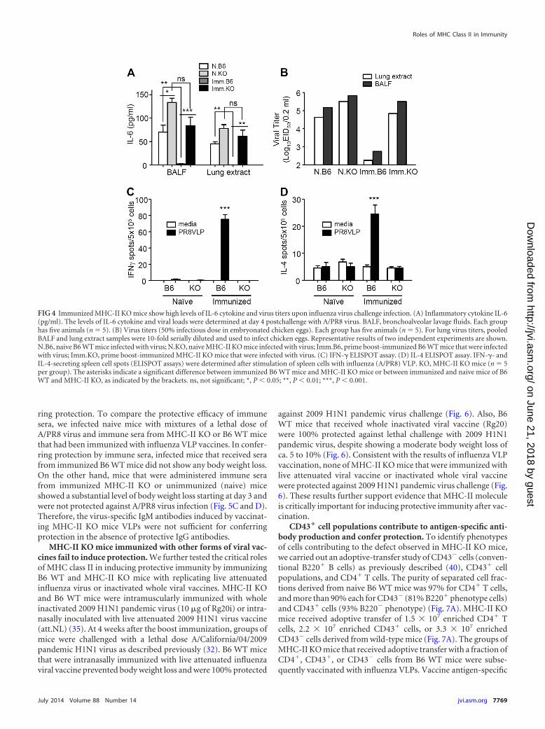
ring protection. To compare the protective efficacy of immunesera, we infected naive mice with mixtures of a lethal dose ofA/PR8 virus and immune sera from MHC-II KO or B6 WT micethat had been immunized with influenza VLP vaccines. In confer-ring protection by immune sera, infected mice that received serafrom immunized B6 WT mice did not show any body weight loss.On the other hand, mice that were administered immune serafrom immunized MHC-II KO or unimmunized (naive) miceshowed a substantial level of body weight loss starting at day 3 andwere not protected against A/PR8 virus infection (Fig. 5C and D).Therefore, the virus-specific IgM antibodies induced by vaccinat-ing MHC-II KO mice VLPs were not sufficient for conferringprotection in the absence of protective IgG antibodies.
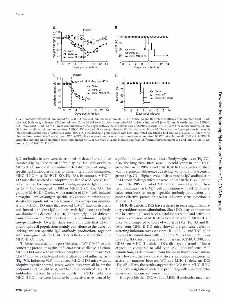
MHC-II KO mice immunized with other forms of viral vac-cines fail to induce protection. We further tested the critical rolesof MHC class II in inducing protective immunity by immunizingB6 WT and MHC-II KO mice with replicating live attenuatedinfluenza virus or inactivated whole viral vaccines. MHC-II KOand B6 WT mice were intramuscularly immunized with wholeinactivated 2009 H1N1 pandemic virus (10 �g of Rg20i) or intra-nasally inoculated with live attenuated 2009 H1N1 virus vaccine(att.NL) (35). At 4 weeks after the boost immunization, groups ofmice were challenged with a lethal dose A/California/04/2009pandemic H1N1 virus as described previously (32). B6 WT micethat were intranasally immunized with live attenuated influenzaviral vaccine prevented body weight loss and were 100% protected
against 2009 H1N1 pandemic virus challenge (Fig. 6). Also, B6WT mice that received whole inactivated viral vaccine (Rg20)were 100% protected against lethal challenge with 2009 H1N1pandemic virus, despite showing a moderate body weight loss ofca. 5 to 10% (Fig. 6). Consistent with the results of influenza VLPvaccination, none of MHC-II KO mice that were immunized withlive attenuated viral vaccine or inactivated whole viral vaccinewere protected against 2009 H1N1 pandemic virus challenge (Fig.6). These results further support evidence that MHC-II moleculeis critically important for inducing protective immunity after vac-cination.
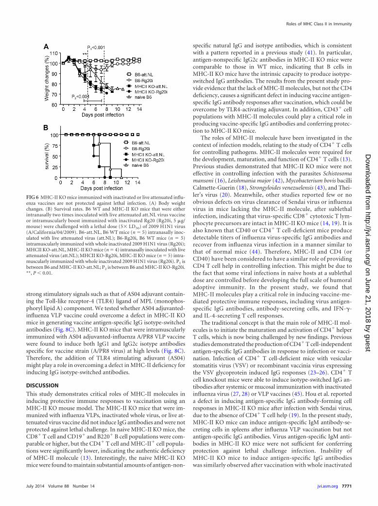
CD43� cell populations contribute to antigen-specific anti-body production and confer protection. To identify phenotypesof cells contributing to the defect observed in MHC-II KO mice,we carried out an adoptive-transfer study of CD43� cells (conven-tional B220� B cells) as previously described (40), CD43� cellpopulations, and CD4� T cells. The purity of separated cell frac-tions derived from naive B6 WT mice was 97% for CD4� T cells,and more than 90% each for CD43� (81% B220� phenotype cells)and CD43� cells (93% B220� phenotype) (Fig. 7A). MHC-II KOmice received adoptive transfer of 1.5 107 enriched CD4� Tcells, 2.2 107 enriched CD43� cells, or 3.3 107 enrichedCD43� cells derived from wild-type mice (Fig. 7A). The groups ofMHC-II KO mice that received adoptive transfer with a fraction ofCD4�, CD43�, or CD43� cells from B6 WT mice were subse-quently vaccinated with influenza VLPs. Vaccine antigen-specific
FIG 4 Immunized MHC-II KO mice show high levels of IL-6 cytokine and virus titers upon influenza virus challenge infection. (A) Inflammatory cytokine IL-6(pg/ml). The levels of IL-6 cytokine and viral loads were determined at day 4 postchallenge with A/PR8 virus. BALF, bronchoalveolar lavage fluids. Each grouphas five animals (n 5). (B) Virus titers (50% infectious dose in embryonated chicken eggs). Each group has five animals (n 5). For lung virus titers, pooledBALF and lung extract samples were 10-fold serially diluted and used to infect chicken eggs. Representative results of two independent experiments are shown.N.B6, naive B6 WT mice infected with virus; N.KO, naive MHC-II KO mice infected with virus; Imm.B6, prime boost-immunized B6 WT mice that were infectedwith virus; Imm.KO, prime boost-immunized MHC-II KO mice that were infected with virus. (C) IFN-� ELISPOT assay. (D) IL-4 ELISPOT assay. IFN-�- andIL-4-secreting spleen cell spots (ELISPOT assays) were determined after stimulation of spleen cells with influenza (A/PR8) VLP. KO, MHC-II KO mice (n 5per group). The asterisks indicate a significant difference between immunized B6 WT mice and MHC-II KO mice or between immunized and naive mice of B6WT and MHC-II KO, as indicated by the brackets. ns, not significant; *, P � 0.05; **, P � 0.01; ***, P � 0.001.
Roles of MHC Class II in Immunity
July 2014 Volume 88 Number 14 jvi.asm.org 7769
on June 21, 2018 by guesthttp://jvi.asm
.org/D
ownloaded from
IgG antibodies in sera were determined 14 days after adoptivetransfer (Fig. 7A). The transfer of wild-type CD43� cells or PBS toMHC-II KO mice did not induce detectable levels of antigen-specific IgG antibodies similar to those in sera from immunizedMHC-II KO mice (MHC-II KO, Fig. 7A). In contrast, MHC-IIKO mice that received an adoptive transfer of wild-type CD43�
cells produced the largest amount of antigen-specific IgG antibod-ies (P � 0.01 compared to PBS or MHC-II KO, Fig. 7A). Thegroup of MHC-II KO mice with a transfer of CD4� cells induceda marginal level of antigen-specific IgG antibodies, which is notstatistically significant. We determined IgG isotypes in immunesera of MHC-II KO mice that received CD43� fractionated cellsand showed the highest IgG antibody levels. IgG1 isotype antibodywas dominantly detected (Fig. 7B). Interestingly, this is differentfrom immunized B6 WT mice that induced predominantly IgG2cisotype antibody. Overall, these results indicate that CD43�
phenotypic cell populations mainly contribute to the defect oflacking antigen-specific IgG antibody production, togetherwith a marginal role for CD4� T cells that are also deficient inMHC-II KO mice.
To better understand the possible roles of WT CD43� cells inconferring protection against influenza virus challenge infection,MHC-II KO mice with or without adoptive transfer of naive WTCD43� cells were challenged with a lethal dose of influenza virus(Fig. 7C). Influenza VLP-immunized MHC-II KO mice withoutadoptive transfer showed severe weight loss, were all below theendpoint (25% weight loss), and had to be sacrificed (Fig. 7C).Antibodies induced by adoptive transfer of CD43� cells intoMHC-II KO mice were found to be protective, as evidenced by
significantly lower levels (ca. 12%) of body weight losses (Fig. 7C).Also, the lung virus titers were 15-fold lower in the CD43�
group than in the PBS control of MHC-II KO mice, although therewas no significant difference due to high variations in the controlgroup (Fig. 7D). Higher levels of virus-specific IgG antibodies inBALF upon challenge infection were induced in the CD43� groupthan in the PBS control of MHC-II KO mice (Fig. 7E). Theseresults indicate that CD43� cell populations with MHC-II mole-cules contribute to antigen-specific antibody production andconfer effective protection against influenza virus infection inMHC-II KO mice.
MHC-II-deficient DCs have a defect in secreting inflamma-tory cytokines upon stimulation. Since DCs play an importantrole in activating T and B cells, cytokine secretion and activationmarker expression of MHC-II-deficient DCs from MHC-II KOmice were compared to those of wild-type DCs (Fig. 8A and B).DCs from MHC-II KO mice showed a significant defect insecreting inflammatory cytokines (IL-6, IL-12, and TNF-�) inrespond to stimulation with influenza VLPs (A/PR8 VLP) orLPS (Fig. 8A). Also, the activation markers (CD40, CD80, andCD86) on MHC-II-deficient DCs displayed a trend of lowerexpression compared to wild-type DCs upon influenza VLPstimulation, as determined from the mean fluorescence inten-sity. However, there was no statistical significance in expressingactivation markers between WT and MHC-II-deficient DCs(Fig. 8B). Here, the results suggest that DCs from MHC-II KOmice have a significant defect in producing inflammatory cyto-kines upon vaccine antigen stimulation.
It is possible that DCs without MHC-II molecules may need
FIG 5 Protective efficacy of immunized MHC-II KO mice and immune sera from MHC-II KO mice. (A and B) Protective efficacy of immunized MHC-II KOmice. (A) Body weight changes. (B) Survival rates. Naive B6 WT (n 4), boost-immunized B6 wild-type (immu WT [n 5]), and boost-immunized MHC-IIKO (immu MHC-II KO [n 4]) mice were intranasally challenged with a lethal infectious dose of A/PR/8/34 virus (5 LD50). #, One mouse survived. (C andD) Protective efficacy of immune sera from MHC-II KO mice. (C) Body weight changes. (D) Survival rates. Naive BALB/c mice (n 5/group) were intranasallyinfected with a lethal dose of A/PR/8/34 virus (10 LD50) that had been preincubated with heat-inactivated sera (final 4-fold dilutions). Naive, A/PR/8/34 virusplus sera from naive B6 WT mice; Immu WT, A/PR/8/34 virus plus immune sera from prime boost-immunized B6 WT mice; Immu MHC-II KO, A/PR/8/34virus plus immune sera from prime boost-immunized MHC-II KO mice. P values indicate significant differences between immu WT and immu MHC-II KOgroups. *, P � 0.05; **, P � 0.01.
O et al.
7770 jvi.asm.org Journal of Virology
on June 21, 2018 by guesthttp://jvi.asm
.org/D
ownloaded from
strong stimulatory signals such as that of AS04 adjuvant contain-ing the Toll-like receptor-4 (TLR4) ligand of MPL (monophos-phoryl lipid A) component. We tested whether AS04 adjuvanted-influenza VLP vaccine could overcome a defect in MHC-II KOmice in generating vaccine antigen-specific IgG isotype-switchedantibodies (Fig. 8C). MHC-II KO mice that were intramuscularlyimmunized with AS04 adjuvanted-influenza A/PR8 VLP vaccinewere found to induce both IgG1 and IgG2c isotype antibodiesspecific for vaccine strain (A/PR8 virus) at high levels (Fig. 8C).Therefore, the addition of TLR4 stimulating adjuvant (AS04)might play a role in overcoming a defect in MHC-II deficiency forinducing IgG isotype-switched antibodies.
DISCUSSION
This study demonstrates critical roles of MHC-II molecules ininducing protective immune responses to vaccination using anMHC-II KO mouse model. The MHC-II KO mice that were im-munized with influenza VLPs, inactivated whole virus, or live at-tenuated virus vaccine did not induce IgG antibodies and were notprotected against lethal challenge. In naive MHC-II KO mice, theCD8� T cell and CD19� and B220� B cell populations were com-parable or higher, but the CD4� T cell and MHC-II� cell popula-tions were significantly lower, indicating the authentic deficiencyof MHC-II molecule (13). Interestingly, the naive MHC-II KOmice were found to maintain substantial amounts of antigen-non-
specific natural IgG and isotype antibodies, which is consistentwith a pattern reported in a previous study (41). In particular,antigen-nonspecific IgG2c antibodies in MHC-II KO mice werecomparable to those in WT mice, indicating that B cells inMHC-II KO mice have the intrinsic capacity to produce isotype-switched IgG antibodies. The results from the present study pro-vide evidence that the lack of MHC-II molecules, but not the CD4deficiency, causes a significant defect in inducing vaccine antigen-specific IgG antibody responses after vaccination, which could beovercome by TLR4-activating adjuvant. In addition, CD43� cellpopulations with MHC-II molecules could play a critical role inproducing vaccine-specific IgG antibodies and conferring protec-tion to MHC-II KO mice.
The roles of MHC-II molecule have been investigated in thecontext of infection models, relating to the study of CD4� T cellsfor controlling pathogens. MHC-II molecules were required forthe development, maturation, and function of CD4� T cells (13).Previous studies demonstrated that MHC-II KO mice were noteffective in controlling infection with the parasites Schistosomamansoni (16), Leishmania major (42), Mycobacterium bovis bacilliCalmette-Guerin (18), Strongyloides venezuelensis (43), and Thei-ler’s virus (20). Meanwhile, other studies reported few or noobvious defects on virus clearance of Sendai virus or influenzavirus in mice lacking the MHC-II molecule, after sublethalinfection, indicating that virus-specific CD8� cytotoxic T lym-phocyte precursors are intact in MHC-II KO mice (14, 19). It isalso known that CD40 or CD4� T cell-deficient mice producedetectable titers of influenza virus-specific IgG antibodies andrecover from influenza virus infection in a manner similar tothat of normal mice (44). Therefore, MHC-II and CD4 (orCD40) have been considered to have a similar role of providingCD4 T cell help in controlling infection. This might be due tothe fact that some viral infections in naive hosts at a sublethaldose are controlled before developing the full scale of humoraladoptive immunity. In the present study, we found thatMHC-II molecules play a critical role in inducing vaccine-me-diated protective immune responses, including virus antigen-specific IgG antibodies, antibody-secreting cells, and IFN-�-and IL-4-secreting T cell responses.
The traditional concept is that the main role of MHC-II mol-ecules is to initiate the maturation and activation of CD4� helperT cells, which is now being challenged by new findings. Previousstudies demonstrated the production of CD4� T cell-independentantigen-specific IgG antibodies in response to infection or vacci-nation. Infection of CD4� T cell-deficient mice with vesicularstomatitis virus (VSV) or recombinant vaccinia virus expressingthe VSV glycoprotein induced IgG responses (23–26). CD4� Tcell knockout mice were able to induce isotype-switched IgG an-tibodies after systemic or mucosal immunization with inactivatedinfluenza virus (27, 28) or VLP vaccines (45). Hou et al. reporteda defect in inducing antigen-specific IgG antibody-forming cellresponses in MHC-II KO mice after infection with Sendai virus,due to the absence of CD4� T cell help (19). In the present study,MHC-II KO mice can induce antigen-specific IgM antibody-se-creting cells in spleens after influenza VLP vaccination but notantigen-specific IgG antibodies. Virus antigen-specific IgM anti-bodies in MHC-II KO mice were not sufficient for conferringprotection against lethal challenge infection. Inability ofMHC-II KO mice to induce antigen-specific IgG antibodieswas similarly observed after vaccination with whole inactivated
FIG 6 MHC-II KO mice immunized with inactivated or live attenuated influ-enza vaccines are not protected against lethal infection. (A) Body weightchanges. (B) Survival rates. B6 WT and MHC-II KO mice that were eitherintranasally two times inoculated with live attenuated att.NL virus vaccineor intramuscularly boost immunized with inactivated Rg20 (Rg20i, 5 �g/mouse) were challenged with a lethal dose (5 LD50) of 2009 H1N1 virus(A/California/04/2009). B6-att.NL, B6 WT mice (n 5) intranasally inoc-ulated with live attenuated virus (att.NL); B6-Rg20i, B6 WT mice (n 5)intramuscularly immunized with whole inactivated 2009 H1N1 virus (Rg20i);MHCII KO-att.NL, MHC-II KO mice (n 4) intranasally inoculated with liveattenuated virus (att.NL); MHCII KO-Rg20i, MHC-II KO mice (n 5) intra-muscularly immunized with whole inactivated 2009 H1N1 virus (Rg20i). P1 isbetween B6 and MHC-II KO-att.NL; P2 is between B6 and MHC-II KO-Rg20i.**, P � 0.01.
Roles of MHC Class II in Immunity
July 2014 Volume 88 Number 14 jvi.asm.org 7771
on June 21, 2018 by guesthttp://jvi.asm
.org/D
ownloaded from

or live attenuated viral vaccines (data not shown). Developingprotective immunity to vaccination may be different fromCD8� T cell-dependent virus clearance in MHC-II KO miceafter sublethal infection (14, 19). Immunization of CD4� T cellknockout mice with inactivated whole virus was shown to in-duce protective immunity against lethal infection of influenzaA/PR8 virus (27, 28). We also found that immunization ofCD4� T knockout mice with influenza A/PR8 VLP vaccinesinduced virus-specific IgG antibodies at substantial levels com-pared to those in wild-type mice (Fig. 2C). Therefore, it isspeculated that an MHC-II molecule may have other additionalroles that are not well defined in inducing antigen-specific IgGantibodies, which is different from its role in activating CD4�
T helper cells after vaccination or infection.In an attempt to better understand the defective mechanism in
MHC-II KO mice incapable of inducing antigen-specific IgG an-tibody responses, an adoptive-transfer experiment of wild-typecell fractions with MHC-II molecules was performed (Fig. 7). Thetransfer of CD43� fraction (conventional B220� B cells) did notproduce detectable levels of antigen-specific IgG antibodies inMHC-II KO mice after vaccination. Provision of wild-type CD4�
T cells to MHC-II KO mice produced antigen-specific IgG only ata marginal level. Unexpectedly, the adoptive transfer of CD43�
populations led to inducing significant levels of antigen-specificIgG and IgG1 isotype antibodies, as well as protective immunity tolethal challenge infection in MHC-II KO mice after influenza VLPvaccination. Nonetheless, after influenza VLP vaccination, IgG2band IgG2c isotype antibodies were not detected at significant lev-
els in MHC-II KO mice with CD43� cell adoptive transfer fromnaive B6 WT mice (data not shown). In B6 WT mice, IgG2b andIgG2c antibodies were induced dominantly after influenza VLPvaccination. Thus, naive CD43� B220� cells (from naive wild-type mice) alone would have limited capacity to fully restore otherIgG isotypes in a deficiency of CD4 T cells in MHC-II KO mice.Importantly, addition of AS04 adjuvant to influenza VLP vaccinescould overcome the MHC-II defects in inducing isotype-switchedIgG antibodies in MHC-II KO mice after influenza vaccination.This is highly significant in understanding possible action mech-anisms of adjuvant and vaccines.
CD43 was shown to be expressed on plasma cells and noncon-ventional splenic B-1 cells, as well as some granulocytes, includingmonocytes, macrophages, and DCs (46, 47), but not on conven-tional mature B (B-2) cells (48). B220� B cells in the CD43�
spleen cell fraction from wild-type mice were found to be 81%,whereas the majority (93%) of the CD43� fraction was B220�
cells (data not shown). In wild-type adult spleens, conventional Bcells with CD43� (B-2) are predominantly resting follicular B cellsand marginal and immature B cells. CD43� fractions were used todetermine the roles of B cells by adoptive transfer, together withcognate CD4� T cells (40). Thus, the roles of combined conven-tional B cells and CD4� T cells remain to be determined in thecontext of MHC-II KO mice. It might be possible that CD43�
B220� phenotypic populations are plasmablasts and/or plasma-like cells contributing to the production of antigen-specific IgGantibodies without CD4� T cell help despite their small popula-tions. DCs were reported to retain antigens in a form that is rec-
FIG 7 Adoptive transfer of B6 WT fractionated CD43� cells to MHC-II KO mice confers protective immunity. (A) Vaccine-specific IgG antibody responses inMHC-II KO mice with adoptive transfer. Naive MHC-II KO mice (n 4 for each group) received CD43�, CD4�, and CD43� cell populations fractionated fromspleens of naive B6 WT mice. CD43�, 3.3 107 enriched CD43� cells per MHC-II KO mouse; CD4�, 1.5 107 enriched CD4� T cells per MHC-II KO mouse;CD43�, 2.2 107 enriched CD43� cells per MHC-II KO mouse; PBS, no B6 WT cells (a negative control); MHCII.KO, a serum ELISA control from MHC-II KOmice immunized with influenza VLP vaccine. (B) IgG isotypes (IgG1 and IgG2c) in immune sera of MHC-II KO mice with adoptive transfer of naive wild-typeCD43� cells after influenza VLP immunization. MHC-II KO mice with or without CD43� adoptive transfer were intramuscularly immunized with influenzaA/PR8 VLPs. KO, MHC-II KO mice without adoptive transfer prior to immunization; KO�CD43, MHC-II KO mice with CD43� cell adoptive transfer prior toimmunization. (C to E) Protective roles of wild-type CD43� cells in MHC-II KO mice. MHC-II KO mice with (KO�CD43) or without (KO) CD43� adoptivetransfer were intramuscularly immunized with influenza A/PR8 VLPs and then challenged with influenza virus 2 weeks later. (C) Body weight changes. (D) Lungviral loads. (E) Virus-specific IgG antibodies in BALF. ns, not significant; †, all mice were euthanized. Statistical significances were compared between KO andKO�CD43: *, P � 0.05; **, P � 0.01; ***, P � 0.0001.
O et al.
7772 jvi.asm.org Journal of Virology
on June 21, 2018 by guesthttp://jvi.asm
.org/D
ownloaded from

ognized by B cells and enhance the differentiation of naive B cellsinto plasma cells (10, 11, 49). Alternatively, a small fraction ofCD43� CD11c� and CD43� CD11b� cells present in the CD43�
fraction from wild-type spleen cells might have contributed toIgG-producing B cell responses without CD4� T cell help in ad-opted MHC-II KO mice. In support of this idea, CD11c� andCD11b� cells were found to be ca. 12 and 14%, respectively, in theCD43� fraction (data not shown). Also, we found that MHC-II-deficient BMDCs showed a significant defect in secreting inflam-matory cytokines in response to stimulation with a VLP vaccineantigen, indicating that MHC-II deficiency might have resulted ina microenvironment not sufficient to effectively initiate antigen-specific IgG antibody immune responses. MHC-II KO micewould not have an intrinsic defect in producing inflammatorycytokines since higher levels of IL-6 were detected in naive andimmunized MHC-II KO mice after lethal influenza virus infectiondue to high viral replication. It is possible that DCs (and/or B cells)without MHC-II molecules may need strong stimulatory signals,such as TLR4-activating AS04 adjuvant, compared to wild-typeantigen-presenting cells. This hypothesis is supported by the in-
duction of vaccine-specific IgG isotype-switched antibodies inMHC-II KO mice that were immunized with AS04-adjuvantedinfluenza VLP vaccination. Taken together, the results presentedhere show that MHC-II molecules have additional roles otherthan simply stimulating the development and activation of CD4�
T cells.In summary, we found that MHC-II was a critical molecule in
inducing antigen-specific IgG antibody responses and protectiveimmunity against lethal challenge infection. CD43� (B220�/low)populations with MHC-II were found to play an essential role ininducing IgG isotype-switched antibody responses to vaccinationand conferring protection in MHC-II KO mice. Antigen-present-ing cells, such as DCs from MHC-II KO mice, were defective inproducing inflammatory cytokines. The addition of innate immu-nity-stimulating adjuvants could overcome the defect of MHC-IIKO mice in inducing IgG isotype-switched antibody responses tovaccination. Further studies are needed to further define thesemechanisms by which the CD43� populations, MHC-II mole-cules, and adjuvants contribute to inducing antigen-specific IgGantibodies and protective immunity after vaccination.
FIG 8 Defective DCs from MHC-II KO mice and the effects of AS04 adjuvant on inducing IgG isotype-switched antibodies in MHC-II KO mice. (A) Cytokinesproduced by BM-derived DCs. (B) Activation marker expression levels of BMDCs. In vitro enriched BMDCs were treated with influenza virus PR8 VLP vaccines(10 �g/ml). WT DCs, B6 WT mouse BMDCs; KO DCs or MHC-II KO DCs, MHC-II KO mouse BMDCs. LPS was used as a positive control. Mean values of eachtriplicate are shown out of two independent experiments. nd, not detectable or similar to medium controls; **, P � 0.01; ***, P � 0.001 compared to WT DCs.Fold increase in activation markers is determined by mean fluorescence intensity. (C) IgG and isotype antibodies specific for vaccine strain (A/PR8 virus) weremeasured in immune sera from MHC-II KO mice immunized with vaccine only (Vac) or AS04-adjuvanted vaccines (Vac�AS04; A/PR8 VLP [10 �g] with alum[100 �g] plus monophosphoryl lipid A [10 �g per mouse]).
Roles of MHC Class II in Immunity
July 2014 Volume 88 Number 14 jvi.asm.org 7773
on June 21, 2018 by guesthttp://jvi.asm
.org/D
ownloaded from

ACKNOWLEDGMENTS
This study was in part supported by NIH/NIAID grants AI105170(S.-M.K.), AI093772 (S.-M.K.), and P30AI050409.
We thank T. Kang for careful reading of the manuscript.
REFERENCES1. Plotkin SA. 2005. Vaccines: past, present and future. Nat. Med. 11:S5–
S11. http://dx.doi.org/10.1038/nm1209.2. Plotkin SA. 2010. Correlates of protection induced by vaccination.
Clin. Vaccine Immunol. 17:1055–1065. http://dx.doi.org/10.1128/CVI.00131-10.
3. Plotkin SA. 2008. Vaccines: correlates of vaccine-induced immunity.Clin. Infect. Dis. 47:401– 409. http://dx.doi.org/10.1086/589862.
4. Banchereau J, Steinman RM. 1998. Dendritic cells and the control ofimmunity. Nature 392:245–252. http://dx.doi.org/10.1038/32588.
5. Bishop GA, Hostager BS. 2001. B lymphocyte activation by contact-mediated interactions with T lymphocytes. Curr. Opin. Immunol. 13:278 –285. http://dx.doi.org/10.1016/S0952-7915(00)00216-8.
6. Clark EA, Ledbetter JA. 1994. How B and T cells talk to each other.Nature 367:425– 428. http://dx.doi.org/10.1038/367425a0.
7. Stavnezer J. 2000. Immunology: a touch of antibody class. Science 288:984 –985.
8. Marshall D, Sealy R, Sangster M, Coleclough C. 1999. TH cells primedduring influenza virus infection provide help for qualitatively distinct an-tibody responses to subsequent immunization. J. Immunol. 163:4673–4682.
9. Snapper CM, Mond JJ. 1993. Towards a comprehensive view of immu-noglobulin class switching. Immunol. Today 14:15–17. http://dx.doi.org/10.1016/0167-5699(93)90318-F.
10. Wykes M, Pombo A, Jenkins C, MacPherson GG. 1998. Dendritic cellsinteract directly with naive B lymphocytes to transfer antigen and initiateclass switching in a primary T-dependent response. J. Immunol. 161:1313–1319.
11. Fayette J, Dubois B, Vandenabeele S, Bridon JM, Vanbervliet B, Du-rand I, Banchereau J, Caux C, Briere F. 1997. Human dendritic cellsskew isotype switching of CD40-activated naive B cells toward IgA1 andIgA2. J. Exp. Med. 185:1909 –1918. http://dx.doi.org/10.1084/jem.185.11.1909.
12. Dubois B, Vanbervliet B, Fayette J, Massacrier C, Van Kooten C, BriereF, Banchereau J, Caux C. 1997. Dendritic cells enhance growth anddifferentiation of CD40-activated B lymphocytes. J. Exp. Med. 185:941–951. http://dx.doi.org/10.1084/jem.185.5.941.
13. Grusby MJ, Johnson RS, Papaioannou VE, Glimcher LH. 1991. Deple-tion of CD4� T cells in major histocompatibility complex class II-deficientmice. Science 253:1417–1420. http://dx.doi.org/10.1126/science.1910207.
14. Tripp RA, Sarawar SR, Doherty PC. 1995. Characteristics of the influ-enza virus-specific CD8� T cell response in mice homozygous for disrup-tion of the H-2lAb gene. J. Immunol. 155:2955–2959.
15. Rahemtulla A, Fung-Leung WP, Schilham MW, Kundig TM, SambharaSR, Narendran A, Arabian A, Wakeham A, Paige CJ, Zinkernagel RM.1991. Normal development and function of CD8� cells but markedlydecreased helper cell activity in mice lacking CD4. Nature 353:180 –184.http://dx.doi.org/10.1038/353180a0.
16. Angyalosi G, Pancre V, Herno J, Auriault C. 1998. Immunologicalresponse of major histocompatibility complex class II-deficient (A�°)mice infected by the parasite Schistosoma mansoni. Scand. J. Immunol.48:159 –169. http://dx.doi.org/10.1046/j.1365-3083.1998.00372.x.
17. Oliveira SC, Splitter GA. 1995. CD8� type 1 CD44hi CD45 RBlo Tlymphocytes control intracellular Brucella abortus infection as demon-strated in major histocompatibility complex class I- and class II-deficientmice. Eur. J. Immunol. 25:2551–2557. http://dx.doi.org/10.1002/eji.1830250922.
18. Ladel CH, Daugelat S, Kaufmann SH. 1995. Immune response to My-cobacterium bovis bacille Calmette-Guerin infection in major histocom-patibility complex class I- and II-deficient knockout mice: contribution ofCD4 and CD8 T cells to acquired resistance. Eur. J. Immunol. 25:377–384.http://dx.doi.org/10.1002/eji.1830250211.
19. Hou S, Mo XY, Hyland L, Doherty PC. 1995. Host response to Sendaivirus in mice lacking class II major histocompatibility complex glycopro-teins. J. Virol. 69:1429 –1434.
20. Njenga MK, Pavelko KD, Baisch J, Lin X, David C, Leibowitz J,Rodriguez M. 1996. Theiler’s virus persistence and demyelination in ma-
jor histocompatibility complex class II-deficient mice. J. Virol. 70:1729 –1737.
21. Szomolanyi-Tsuda E, Le QP, Garcea RL, Welsh RM. 1998. T-Cell-independent immunoglobulin G responses in vivo are elicited by live-virus infection but not by immunization with viral proteins or virus-likeparticles. J. Virol. 72:6665– 6670.
22. Szomolanyi-Tsuda E, Welsh RM. 1996. T cell-independent antibody-mediated clearance of polyoma virus in T cell-deficient mice. J. Exp. Med.183:403– 411. http://dx.doi.org/10.1084/jem.183.2.403.
23. Bachmann MF, Kundig TM, Kalberer CP, Hengartner H, ZinkernagelRM. 1993. Formalin inactivation of vesicular stomatitis virus impairs T-cell- but not T-help-independent B-cell responses. J. Virol. 67:3917–3922.
24. Bachmann MF, Bast C, Hengartner H, Zinkernagel RM. 1994. Im-munogenicity of a viral model vaccine after different inactivation pro-cedures. Med. Microbiol. Immunol. 183:95–104. http://dx.doi.org/10.1007/BF00277160.
25. Bachmann MF, Hengartner H, Zinkernagel RM. 1995. T helper cell-independent neutralizing B cell response against vesicular stomatitis virus:role of antigen patterns in B cell induction? Eur. J. Immunol. 25:3445–3451. http://dx.doi.org/10.1002/eji.1830251236.
26. Maloy KJ, Odermatt B, Hengartner H, Zinkernagel RM. 1998. Inter-feron gamma-producing �� T cell-dependent antibody isotype switchingin the absence of germinal center formation during virus infection. Proc.Natl. Acad. Sci. U. S. A. 95:1160 –1165. http://dx.doi.org/10.1073/pnas.95.3.1160.
27. Sha Z, Kang SM, Compans RW. 2005. Mucosal immunization ofCD4(�) T cell-deficient mice with an inactivated virus induces IgG andIgA responses in serum and mucosal secretions. Virology 331:387–395.http://dx.doi.org/10.1016/j.virol.2004.10.014.
28. Sha Z, Compans RW. 2000. Induction of CD4� T-cell-independentimmunoglobulin responses by inactivated influenza virus. J. Virol. 74:4999 –5005. http://dx.doi.org/10.1128/JVI.74.11.4999-5005.2000.
29. Quan FS, Huang C, Compans RW, Kang SM. 2007. Virus-like particlevaccine induces protective immunity against homologous and heterolo-gous strains of influenza virus. J. Virol. 81:3514 –3524. http://dx.doi.org/10.1128/JVI.02052-06.
30. Quan FS, Kim YC, Vunnava A, Yoo DG, Song JM, Prausnitz MR,Compans RW, Kang SM. 2010. Intradermal vaccination with influenzavirus-like particles by using microneedles induces protection superior tothat with intramuscular immunization. J. Virol. 84:7760 –7769. http://dx.doi.org/10.1128/JVI.01849-09.
31. Quan FS, Sailaja G, Skountzou I, Huang C, Vzorov A, Compans RW,Kang SM. 2007. Immunogenicity of virus-like particles containing mod-ified human immunodeficiency virus envelope proteins. Vaccine 25:3841–3850. http://dx.doi.org/10.1016/j.vaccine.2007.01.107.
32. Quan FS, Vunnava A, Compans RW, Kang SM. 2010. Virus-like particlevaccine protects against 2009 H1N1 pandemic influenza virus in mice.PLoS One 5:e9161. http://dx.doi.org/10.1371/journal.pone.0009161.
33. Quan FS, Yoo DG, Song JM, Clements JD, Compans RW, Kang SM.2009. Kinetics of immune responses to influenza virus-like particles anddose-dependence of protection with a single vaccination. J. Virol. 83:4489 – 4497. http://dx.doi.org/10.1128/JVI.02035-08.
34. Quan FS, Steinhauer D, Huang C, Ross TM, Compans RW, Kang SM.2008. A bivalent influenza VLP vaccine confers complete inhibition ofvirus replication in lungs. Vaccine 26:3352–3361. http://dx.doi.org/10.1016/j.vaccine.2008.03.055.
35. Pena L, Vincent AL, Ye J, Ciacci-Zanella JR, Angel M, Lorusso A,Gauger PC, Janke BH, Loving CL, Perez DR. 2011. Modifications in thepolymerase genes of a swine-like triple-reassortant influenza virus to gen-erate live attenuated vaccines against 2009 pandemic H1N1 viruses. J.Virol. 85:456 – 469. http://dx.doi.org/10.1128/JVI.01503-10.
36. Song JM, Van Rooijen N, Bozja J, Compans RW, Kang SM. 2011.Vaccination inducing broad and improved cross protection against mul-tiple subtypes of influenza A virus. Proc. Natl. Acad. Sci. U. S. A. 108:757–761. http://dx.doi.org/10.1073/pnas.1012199108.
37. Song JM, Wang BZ, Park KM, Van Rooijen N, Quan FS, Kim MC, JinHT, Pekosz A, Compans RW, Kang SM. 2011. Influenza virus-likeparticles containing M2 induce broadly cross protective immunity. PLoSOne 6:e14538. http://dx.doi.org/10.1371/journal.pone.0014538.
38. Song JM, Hossain J, Yoo DG, Lipatov AS, Davis CT, Quan FS, ChenLM, Hogan RJ, Donis RO, Compans RW, Kang SM. 2010. Protectiveimmunity against H5N1 influenza virus by a single dose vaccination with
O et al.
7774 jvi.asm.org Journal of Virology
on June 21, 2018 by guesthttp://jvi.asm
.org/D
ownloaded from
virus-like particles. Virology 405:165–175. http://dx.doi.org/10.1016/j.virol.2010.05.034.
39. Gray D, Dullforce P, Jainandunsing S. 1994. Memory B cell developmentbut not germinal center formation is impaired by in vivo blockade ofCD40-CD40 ligand interaction. J. Exp. Med. 180:141–155. http://dx.doi.org/10.1084/jem.180.1.141.
40. Jegerlehner A, Maurer P, Bessa J, Hinton HJ, Kopf M, Bachmann MF.2007. TLR9 signaling in B cells determines class switch recombination toIgG2a. J. Immunol. 178:2415–2420. http://dx.doi.org/10.4049/jimmunol.178.4.2415.
41. Madsen L, Labrecque N, Engberg J, Dierich A, Svejgaard A, Benoist C,Mathis D, Fugger L. 1999. Mice lacking all conventional MHC class IIgenes. Proc. Natl. Acad. Sci. U. S. A. 96:10338 –10343. http://dx.doi.org/10.1073/pnas.96.18.10338.
42. Erb K, Blank C, Ritter U, Bluethmann H, Moll H. 1996. Leishmania majorinfection in major histocompatibility complex class II-deficient mice:CD8� T cells do not mediate a protective immune response. Immunobi-ology 195:243–260. http://dx.doi.org/10.1016/S0171-2985(96)80043-X.
43. Rodrigues RM, Silva NM, Goncalves AL, Cardoso CR, Alves R,Goncalves FA, Beletti ME, Ueta MT, Silva JS, Costa-Cruz JM. 2009.Major histocompatibility complex (MHC) class II but not MHC class Imolecules are required for efficient control of Strongyloides venezuelen-
sis infection in mice. Immunology 128:e432– e441. http://dx.doi.org/10.1111/j.1365-2567.2008.02995.x.
44. Lee BO, Rangel-Moreno J, Moyron-Quiroz JE, Hartson L, Makris M,Sprague F, Lund FE, Randall TD. 2005. CD4 T cell-independent anti-body response promotes resolution of primary influenza infection andhelps to prevent reinfection. J. Immunol. 175:5827–5838. http://dx.doi.org/10.4049/jimmunol.175.9.5827.
45. Yao Q, Zhang R, Guo L, Li M, Chen C. 2004. Th cell-independentimmune responses to chimeric hemagglutinin/simian human immuno-deficiency virus-like particles vaccine. J. Immunol. 173:1951–1958. http://dx.doi.org/10.4049/jimmunol.173.3.1951.
46. Rosenstein Y, Santana A, Pedraza-Alva G. 1999. CD43, a molecule withmultiple functions. Immunol. Res. 20:89 –99. http://dx.doi.org/10.1007/BF02786465.
47. Yedidia Y, Ben-Neriah Y, Jung S. 1998. Primary B cells essentially lackconstitutive NF-�B activity. Eur. J. Immunol. 28:30 –36.
48. Wells SM, Kantor AB, Stall AM. 1994. CD43 (S7) expression identifiesperipheral B cell subsets. J. Immunol. 153:5503–5515.
49. Fayette J, Durand I, Bridon JM, Arpin C, Dubois B, Caux C, Liu YJ,Banchereau J, Briere F. 1998. Dendritic cells enhance the differentiationof naive B cells into plasma cells in vitro. Scand. J. Immunol. 48:563–570.http://dx.doi.org/10.1046/j.1365-3083.1998.00471.x.
Roles of MHC Class II in Immunity
July 2014 Volume 88 Number 14 jvi.asm.org 7775
on June 21, 2018 by guesthttp://jvi.asm
.org/D
ownloaded from