role of therapeutic devices in enhancing speech
TRANSCRIPT

East Tennessee State UniversityDigital Commons @ East
Tennessee State University
Electronic Theses and Dissertations Student Works
8-2012
Role of Therapeutic Devices in Enhancing SpeechIntelligibility and Vocal Intensity in an Individualwith Parkinson’s DiseaseSwetha SwaminathanEast Tennessee State University
Follow this and additional works at: https://dc.etsu.edu/etd
Part of the Speech Pathology and Audiology Commons
This Thesis - Open Access is brought to you for free and open access by the Student Works at Digital Commons @ East Tennessee State University. Ithas been accepted for inclusion in Electronic Theses and Dissertations by an authorized administrator of Digital Commons @ East Tennessee StateUniversity. For more information, please contact [email protected].
Recommended CitationSwaminathan, Swetha, "Role of Therapeutic Devices in Enhancing Speech Intelligibility and Vocal Intensity in an Individual withParkinson’s Disease" (2012). Electronic Theses and Dissertations. Paper 1475. https://dc.etsu.edu/etd/1475

Role of Therapeutic Devices in Enhancing Speech Intelligibility and Vocal Intensity in an
Individual with Parkinson’s Disease
________________________
A thesis
presented to
the faculty of the Department of Audiology and Speech-Language Pathology
East Tennessee State University
In partial fulfillment
of the requirements for the degree
Master of Science in Communicative Disorders
________________________
by
Swetha Swaminathan
August 2012
________________________
Ms. Chayadevie Nanjundeswaran
Dr. Marc Fagelson
Dr. Vijaya Guntupalli
Dr. Brenda Louw
Keywords: Parkinson’s Disease, Behavioral Speech Therapy, Speech Intelligibility, Average
Intensity, Ambulatory Phonation Monitor, Auditory Masker

2
ABSTRACT
Role of Therapeutic Devices in Enhancing Speech Intelligibility and Vocal Intensity in an
Individual with Parkinson’s Disease
by
Swetha Swaminathan
The prevailing speech therapy techniques for treating hypokinetic dysarthria in individuals with
Parkinson’s disease (PD) yields improvements within the clinical setting, however, maintenance
and generalization of acquired behaviors continue to be a challenge. The purpose of this study
was to investigate the effects of portable therapeutic devices including Ambulatory Phonation
Monitor with biofeedback (APM) and auditory masker in maintenance and carryover of
improved speech. Our participant was an individual diagnosed with PD for the past 25 years who
continued to display speech disturbances despite undergoing several behavioral speech therapy
programs and neurosurgical procedures. Speech intelligibility and average intensity measures
under automatic, elicited, and spontaneous speech tasks were recorded pre- and postusage of
APM and auditory masker for a period of 1 week each. Preliminary findings showed no
significant difference in the measures between means (P>0.05) across all tasks for both the
devices. Suggestions for future research on therapeutic devices are discussed.

3
DEDICATION
Thank you, God, for being very kind to me and bestowing me with wisdom and
perseverance needed for my pursuit. I take this opportunity to express my sincere love and
gratitude to my parents – Swaminathan and Geetha and my brother Amar. The ongoing
encouragement and unflinching support you have provided throughout my life has helped me
achieve the goals I once only dreamed of. I would also like to express my gratefulness to Appana
Rao mama and Sarala mami. I couldn’t have made it through this process without your prayers
and blessings. I love you all!

4
ACKNOWLEDGEMENTS
I would like to express my sincere appreciation to my committee chair Ms. Chayadevie
Nanjundeswaran for the numerous hours she has spent guiding me through this endeavor. Thank
you very much ma’am for being an amazing and patient mentor. I owe my deepest gratitude to
my committee members – Dr. Marc Fagelson, Dr. Brenda Louw, and Dr. Vijaya Guntupalli, for
their generous time and expertise to better my work.
It is with immense gratitude that I acknowledge the participant and his wife for their
involvement in this study. Their endless motivation and flexibility has contributed to the success
of this project.
I would like to express acknowledgment to my speech path buddies, not to mention,
Lauren, Nelda, and Lizzy, whose friendship, hospitality, knowledge, and wisdom have
supported, enlightened, and entertained me over the course of my Graduate studies.
Finally, I would also like to thank my friends Srikaanth and Harini for their willingness to
provide assistance and advice not limiting to statistics, which helped a great deal all through this
process.

5
CONTENTS
Page
ABSTRACT ……………………………………………………………………............ 2
DEDICATION ................................................................................................................ 3
ACKNOWLEDGEMENTS ............................................................................................ 4
LIST OF TABLES .......................................................................................................... 8
LIST OF FIGURES ......................................................................................................... 9
Chapter
1. INTRODUCTION .......................................................................................... 10
Nature and Purpose of the Study ............................................................. 10
Need for the Study ................................................................................... 13
2. REVIEW OF THE LITERATURE ................................................................. 15
Characteristics .......................................................................................... 15
Respiratory System ...................................................................... 16
Phonatory System ........................................................................ 17
Articulatory System ..................................................................... 18
Resonatory System ....................................................................... 20
Co-Occurring Neurological Deficits ............................................. 20
Treatment .................................................................................................. 21
Pharmacological Treatment .......................................................... 21
Surgical Treatment ....................................................................... 24
Ablative Surgery .............................................................. 24

6
Chapter Page
Thalamatomy …………………………………... 24
Pallidotomy ……………………………………. 25
Deep Brain Stimulation ................................................... 26
Speech Therapy ........................................................................... 27
Speaking Rate .................................................................. 28
Clear Speech .................................................................... 30
Prosody Therapy .............................................................. 31
Loudness Therapy ............................................................ 32
Therapeutic Devices ......................................................... 34
Biofeedback Devices ............................................ 34
Auditory Masker ................................................... 36
3. METHOD ......................................................................................................... 39
Design …………………………………………………………………... 39
Participant ………………………..……………………………... 39
Procedure ……………………………………………………….. 39
Pre-experimental Protocol ….…………………………... 40
Treatment Protocol ………….………………………….. 41
Week 1 – APM with Biofeedback ….…………………... 41
Pretreatment Measures ……………….……….. .. 42
Protocol ……………….………………………... 43
Posttreatment Measures ………………………... 43
Week 2 …………………………….……..….………….. 44

7
Chapter Page
Week 3 – AM ……………………….…………………... 44
Pretreatment Measures ………………………...... 44
Protocol ……………….………………………... 44
Posttreatment Measures ………………….……… 45
4. RESULTS .......................................................................................................... 46
Data Analysis ............................................................................................. 46
Speech Intelligibility ...................................................................... 46
Average Amplitude ........................................................................ 48
5. DISCUSSION .................................................................................................... 51
Clinical Implications .................................................................................. 51
Conclusion .................................................................................................. 53
REFERENCES ................................................................................................................... 54
APPENDICES ................................................................................................................... 76
Appendix A: Questionnaire A …………................................................................ 76
Appendix B: Questionnaire B ………………......................................................... 77
Appendix C: Questionnaire C................................................................................. 78
Appendix D: Time Journal – Ambulatory Phonation Monitor .............................. 79
Appendix E: Time Journal – Auditory Masker ……….......................................... 80
VITA……………………………………………………………………………….……... 81

8
LIST OF TABLES
Table Page
1. Average Speech Intelligibility Scores – Pre- and Posttreatments ................................. 47
2. Average Amplitude (dB) – Pre- and Posttreatments ..................................................... 48
3. Retrospective Data from APM during Functional Communication ............................... 49

9
LIST OF FIGURES
Figure Page
1. General Experimental Protocol .................................................................................... 40
2. Week 1 – Treatment with APM with Biofeedback ...................................................... 42
3. Week 2 – Treatment with AM ..................................................................................... 45

10
CHAPTER 1
INTRODUCTION
Nature and Purpose of the Study
“Hypokinetic dysarthria is a perceptually distinctive motor speech disorder associated
with basal ganglia circuit pathology” (Duffy, 2005). Speech in Parkinson’s disease (PD), which
is a prototype of hypokinetic dysarthria, is characterized by perceptual features such as reduced
loudness (hypophonia), reduced prosodic pitch inflection (monotone), hoarse voice, imprecise
articulation, and festination (acceleration of words at the end of sentences) (Darley, Aronson, &
Brown, 1975; Duffy 2005). These speech abnormalities may adversely affect the patient’s social,
economic, and psychological wellbeing (Oxtoby, 1982; Pitcairn, Clemie, Gray, & Pentland,
1990; Ramig et al., 2001). The treatment to alleviate motor and speech symptoms exhibited by
individuals with PD includes pharmacological and surgical treatments and speech therapy.
Despite availability of many treatment alternatives, the management of speech and voice
disorders in PD has been challenging for both medical and rehabilitation practitioners.
Medical treatments consisting of neuropharmacological and neurosurgical approaches for
the treatment of PD have had consistent positive outcomes and effects on the motor limb
functions, but their effects on the associated speech and voice disorders have been insignificant
and less compelling (Baker, Ramig, Luschei, & Smith, 1997; Kleinow, Smith, & Ramig, 2001).
Some examples of medical treatments that have produced positive outcomes on limb function
and insignificant changes in speech include the treatment with levodopa (Louis, 2001; Rigrodsky
& Morrison, 1970; Thanvi, Lo, & Robinson, 2007; Wolfe, Garvin, Bacon, & Waldrop, 1975),
the treatment with fetal dopamine transplant (Baker et al., 1997); bilateral thalamotomy and
pallidotomy surgeries (Ghika et al., 1999; Schulz, Peterson, Sapienza, Greer, & Friedman, 1999);

11
and Deep Brain Stimulation (DBS), which has shown worsening of hypokinetic dysarthria post-
surgery (Iulianella, Adams, & Gow, 2008; Tripoliti et al., 2011).
Traditional behavioral speech therapy techniques focusing on articulation, rate, and
prosody involve conscious training to coordinate the respiratory, phonatory, and articulatory
system and have proven to be beneficial within the treatment setting, although generalization and
carryover of the treatment effects have been questionable (Fox, Morrison, Ramig, & Sapir, 2002;
Johnson & Pring, 1990; Ramig et al., 2001; Weiner & Singer, 1989). However, research over the
past 20 years including a series of randomized control trials have established Lee Silverman
Voice Treatment (LSVT®
) as an efficacious behavioral treatment that improves vocal fold
adduction and overall voice and speech production in individuals with PD (Pinto et al., 2004;
Ramig et al., 2001; Yorkston, Spencer, & Duffy, 2003). The LSVT® is an alternative, intensive,
behavioral speech treatment that emphasizes high-effort, repetitive, loud phonations to improve
respiratory, laryngeal, and articulatory functions during speech (Ramig, Countryman, Thompson,
& Horii, 1995; Ramig & Dromey, 1996). To overcome the sensory mismatch between perceived
vocal effort and vocal output in individuals with PD, LSVT® also accentuates simultaneous
sensory awareness training, achieved by cueing and consistently asking individuals to “speak
loud” (Fox et al., 2002). Though studies on LSVT® for individuals with PD have produced
positive long-term (2-year) treatment outcomes for group data in a controlled clinic environment,
successful maintenance of treatment effects to nonclinical environment offers challenge (Fox et
al., 2002). There is a questionable transfer and generalization of the improved speech
characteristics during conversational speech in natural setting following LSVT® treatment
(Adams & Dykstra, 2009; Bourdreaux, 2011).

12
Another alternative treatment for individuals with PD with decreased loudness is the use
therapeutic devices that include loudness biofeedback devices (e.g., APM), vocal amplifiers,
Altered Auditory Feedback (AAF) devices including Delayed Auditory Feedback (DAF)
devices, and auditory maskers. A biofeedback device is used in individuals who speak at a
normal intensity level with cues but lack insight into maintenance of vocal intensity (Rubow &
Swift, 1985), a condition that is commonly observed in individuals with PD. The Ambulatory
Phonation Monitor (APM) (Cheyne, Hanson, Genereux, Stevens, & Hillman, 2003), a
biofeedback device, offers long-term continuous tracking of vocal parameters and also provides
feedback to the user when the target phonatory behaviors, such as increased or decreased
intensity are not maintained (Hillman, 2004). Most studies conducted on the use of biofeedback
treatment have concluded that it has a potential to impact speech production and communicative
effectiveness, on the other hand there is a lack of efficacy data for use of APM with biofeedback
(Yorkston, Spencer, & Duffy, 2003). Auditory masker, another alternative treatment option to
behavioral speech techniques similar to biofeedback devices, has been used to improve loudness
in individuals with PD (Adam & Lang, 1992). Masking works on a well-known phenomenon,
Lombard effect, first described by Etienne Lombard in 1911 as the spontaneous tendency of
speakers to increase their vocal intensity when talking under the presence of noise. The Lombard
effect helps enhance speech output in individuals with PD (Gryczka et al., 2011). Studies done
on small group of individuals with PD have shown that the use of white noise masking in clinical
settings for individuals with Parkinson’s disease results in an increase in vocal intensity ranging
from 2.1 to 7.5dB (Adam & Lang, 1992). However, there has been no study till date measuring
the changes in vocal intensity after using portable white noise masker in a nonclinical setting.

13
The primary purpose of this study was to examine and document the changes in vocal
intensity and speech intelligibility with the use of two kinds of feedback devices, (a) APM with
biofeedback relying on tactile feedback, and (b) the auditory masker relying on auditory
feedback. The participant for the study was an individual with PD, who had not benefitted by
LSVT® and other traditional behavioral therapy techniques. Additionally, the aim of this study
was to investigate the possible use of the devices in everyday life and to determine patient
satisfaction on the use of the devices.
Need for the Study
Current literature on behavioral speech techniques and LSVT® indicates improvement in
speech characteristics within clinical settings; however, carryover and maintenance of the
improved speech characteristics to nonclinical environment has been a challenge (Adams &
Dykstra, 2009; Allan, 1970; Bourdreaux, 2011; Sarno, 1968; Johnson & Pring, 1990; Weiner &
Singer, 1989; Yorkston, Spencer, & Duffy, 2003). This indicates the need to investigate alternate
treatment methods using therapeutic devices and their role in helping individuals with PD
generalize their improved speech characteristics to a variety of speech tasks in both clinical and
natural settings. This case study aimed at elucidating the everyday use of portable therapeutic
devices such as APM and auditory masker (AM) for maintaining and generalizing the improved
vocal loudness and speech intelligibility in an individual with PD. The specific research
questions were:
1. Are therapeutic devices helpful in facilitating improvement in vocal intensity and
intelligibility during spontaneous, elicited, and automatic speech tasks in an
individual with PD?

14
2. Which of the two devices, APM and AM, is perceived to be more comfortable for
long-term everyday use in natural settings?
Conditionally, it is hypothesized that the present study will aid in identifying and
promoting the use of APM with biofeedback and auditory maskers for individuals with PD who
have not been observed to have long-term improvements in vocal loudness and intelligibility
with LSVT and traditional speech therapy techniques.

15
CHAPTER 2
REVIEW OF THE LITERATURE
Parkinson’s disease (PD), a neurodegenerative disorder with progressive impairment in
motor functions and cognition, is the most common movement disorder in the world (Albin,
2006; de Lau & Breteler, 2006). According to National Institute of Neurological Disorders and
Stroke (2012) incidence of PD increases with age, with an average onset age of 60 years,
although 5% to 10% of the individuals with PD experience ‘early onset’ with symptoms
beginning before the age of 50. It is estimated that in the United States more than 500,000 people
suffer from this disease, with 50% more prevalence in men compared to women (NINDS, 2012).
PD is attributed to the depletion of dopaminergic neurons in the substantia niagra of the basal
ganglia (Spencers et al., 2010), which reduces secretion of dopamine resulting in a loss of ability
to execute smooth, controlled movements (Baker et al., 1997). Recent studies have indicated that
PD may also be attributed to the loss of nerve endings that produce the neurotransmitter
norepinephrine, thus causing deficiency of norepinephrine. Norepinephrine functions as a
neurotransmitter, and also a stress hormone controlling many automatic functions of the body,
such as pulse and blood pressure. In consequence, the loss of norepinephrine in individuals with
PD is also believed to contribute to the nonmotor features observed, including fatigue,
abnormalities of blood pressure regulation, and emotional disorders (NINDS, 2012).
Characteristics
The diagnosis of PD is based on the symptoms exhibited by the individual due to the lack
of a current definitive test (Jankovic, 2007). The clinical criteria for the diagnosis, or the cardinal
features of PD, can be grouped under the acronym TRAP: (1) Tremor at rest- stereotyped,
rhythmic involuntary movement; (2) Rigidity - abnormal muscle tone and increased resistance;

16
(3) Akinesia or bradykinesia - slowness or no movement; and (4) Postural instability - the
impairment of mechanisms responsible for maintenance of upright posture during standing or
walking (Jankovic, 2007).
Most of the above listed cardinal features make an impact on the respiratory, phonatory,
articulatory, and/or resonatory subsystems of speech production, affecting the speech in
individuals with PD (Swigert, 1997). Reports indicate that around 75%-89% of individuals with
PD experience voice and speech disorders (Fox et al., 2002). The dysarthric characteristics of
individuals with PD can be analyzed and documented by kinematic, spirometric, perceptual,
acoustic, aerodynamic, videostroboscopic, and electromyographic measures of the various
subsystems (Baker et al., 1998; Darling & Huber, 2011; Dromey et al., 1995; Gerratt & Ward,
1984; Johnson & Pring, 1990; Moore & Scudder, 1989; Ramig, 1992; Ramig et al., 1994, 1995;
Smith et al., 1995; Solomon & Hixon., 1993; Tjaden, 2011).
Respiratory System
Patients with PD display reduced vital capacity and reduced expiratory drive, resulting in
increased breathing rate coupled with irregularities in breathing pattern (deep breathing and
hyperventilation) (Solomon & Hixon, 1993; Stewart, 2000). These respiratory irregularities
make an impact in production of speech, which might include decreased loudness, production of
fewer words, faster interpause speech rate (more rapid speech rate between pauses), and/or
longer and more frequent pauses (Hammem & Yorkston, 1996; Metter & Hanson, 1986; Pitcairn
et al., 1990; Solomon & Hixon, 1993). Specifically, individuals with PD exhibit reduced intra-
oral pressures during /p/ production in a syllable repetition task (Netsell et al., 1975; Solomon &
Hizon, 1993) and decreased sustenance of prolonged vowel phonation (Boshes, 1966; Canter,

17
1965; Mueler, 1971). Reports indicate that individuals with PD might also display higher lung
volume initiation and termination (increased breathing rate) during extemporaneous speech than
in reading (Huber & Darling, 2011).
Phonatory System
Hypokinetic dysarthria also affects the phonatory system. Phonatory characteristics of
individuals with PD analyzed by perceptual measures display reduced loudness, monotonous
pitch and loudness, reduced stress, variable rate of speech, short rushes of speech, and hoarseness
(Darley et al., 1969a; Ramig et al., 2004). Acoustic measures demonstrate higher fundamental
frequency, reduced maximum phonation time, higher jitter-shimmer percentage, and increased
voice onset time (VOT) in individuals with PD (Canter et al., 1965b; Dogan et al., 2008; Forrest
et al., 1989; Ramig et al., 1988). Slightly more pronounced phonatory disturbances are displayed
by individuals with PD when compared to articulatory disturbances under clinical-perceptual
ratings (Ackerman & Ziegler, 1991; Logemann et al., 1978; Logemann & Fisher, 1981). The two
explanations for the more prominent phonatory disturbances can be accounted by (1) progressive
involvement of speech organs beginning at the laryngeal level and proceeding in the oral
direction; and (2) the increased vulnerability of the laryngeal apparatus to the pathophysiological
processes underlying PD (Ackerman & Ziegler, 1991; Logemann & Fisher, 1981). Physiological
and neuropathological mechanisms attributing to the disordered phonatory characteristics in
individuals with PD include (1) reduction in speech motor output (2) deficiency in sensory
perception, and (3) abnormal structural changes in the larynx. Reduced speech motor output,
caused by reduced TA muscle amplitude (Baker, Ramig, Luschei & Smith, 1998) may lead to
decrease in neural drive to the muscles of the speech mechanism. This in turn might result in

18
reduced vocal loudness (hypophonia), reduced pitch inflection (hypoprosodia), and also reduced
range of articulatory movements (hypokinetic articulation) (Albin, Young, & Penny, 1989;
Penny & Young, 1983; Ramig et al., 2004). The reduced speech motor output observed in
individuals with PD may also be explained by basal ganglia dysfunction, age-related muscle
atrophy, or a combination of these conditions (Duffy, 2005). Secondly, disordered phonatory
characteristics in individuals with PD may likewise be attributed to deficiency in sensory
perception that prevents them from accurately regulating (internal cueing or scaling) the optimal
amount of effort to produce adequate loudness (Demirci, Grill, McShane, & Hallet, 1995).
Reduced vocal loudness levels in voluntary tasks (Canter, 1965a), and inability to reflexively
regulate their volume in conversational speech without explicit volume instructions (Ho et al.,
1999; Schulz & Grant, 2000) support this view of individuals with PD having deficiency in
sensory perception. Thirdly, few studies have documented structural changes in the larynx
through videoendoscopic and videostroboscopic studies that can attribute specifically to reduced
loudness exhibited by individuals with PD. Laryngeal abnormalities in the form of bowed vocal
cords, and an abnormally large glottic aperture have been observed, which results in incomplete
approximation of the vocal cords and thus decreasing vocal loudness during speech (Hanson et
al., 1984; Smith et al., 1995).
Articulatory System
Individuals with PD exhibit disordered production of consonants and vowels,
disfluencies, and variable rates of speech. Kinematic and acoustic studies have revealed that
individuals with PD display ‘undershooting’ of articulatory gestures (Ackermann & Ziegler,
1991; Forrest et al., 1989). Notably, there is reduction of articulatory precision in stop
consonants (/t/, /d/, /k/, and /g/) that can be attributed to consistent reductions in both peak

19
velocity and amplitude of mandibular and labial openings (Ackermann & Ziegler, 1991; Forrest
et al., 1989; Logemann & Fisher, 1981). Individuals with PD also exhibit misarticulations during
production of affricates /tʃ/ and /ʤ/, and fricatives /s/, /z/, /ʃ/, and /f/ that have been attributed to
inadequate tongue elevation to achieve complete closure while producing affricates, and close
constriction of the airway in the production of lingual fricatives (Logemann & Fisher, 1981).
Various other explanations are available to explain the phenomenon of misarticulation in
individuals with PD. It has been suggested that the weakness exhibited as smaller Muscle Action
Potential (MAP) under EMG studies during the production of stop consonants is probably of
neurogenic origin as opposed to muscle contractile weakness, muscular fatigue, or deficits at the
myoneural juncture (Netsell et al., 1975). The combination of the neurogenic weakness and the
acceleration phenomenon combine to produce "articulatory undershoot" in individuals with PD
(Logemann & Fisher, 1981; Netsell et al., 1975). Individuals with PD exhibit impaired vowel
articulation, characterized by reduced vowel articulation index, reduced formant transitions, and
restricted acoustic vowel space (Ackermann & Ziegler, 1991). This can also be contributed by
reduced movement of the articulators (Forrest et al., 1989; Skodda, Visser, & Schlegel, 2011;
Tjaden et al., 2005). Imprecise articulation of consonants and vowels in individuals with PD
impacts the ability to perform diadochokinetic tasks such as Alternate Motion Rate (AMR) and
Sequential Motion Rate (SMR) that involve rapid movements of the lips, tongue tip, and back of
the tongue required, for e.g., repetition of /papapa/ or /pataka/ respectively (Canter, 1965b;
Connor et al., 1989; Hirose et al., 1981). The reduction in rate of movement has been attributed
to increased levels of tonic resting and background activity (Leanderson et al., 1971; Moore &
Scudder, 1989; Netsell et al., 1975) and also due to loss of reciprocity between agonist and
antagonistic muscles (Leanderson et al., 1971). In addition to disordered productions of

20
consonants and vowels, 15% to 45% of individuals with PD also exhibit stuttering like speech
disfluencies typically at the beginning of the utterance or after a pause characterized by rapid and
blurred phoneme repetitions (Logemann et al., 1973; Sapir et al., 2001). Moreover, prosodic
deficits and disordered rates of speech have also been consistently reported in individuals with
PD. Typical characteristics of the prosodic deficits in hypokinetic dysarthria include
monoloudness, reduction of stress, and monopitch; however, significantly higher pitch levels and
reduced pitch range have been documented in individuals with PD (Canter, 1963,1965a). There
is high variability in rate of speech exhibited by individuals with PD, with 6% to 13% of the
population exhibiting rapid rate or short rushes of speech (Canter 1965a; Canter 1965b;
Logemann et al., 1978) and some reports with evidence supporting presence of speech rates
slower than normal rates (Canter 1963). It has been concluded that the hypokinetic dysarthria is
by no means homogenous with respect to speech rate (Ackermann & Ziegler, 1991).
Resonatory System
Resonatory system is also affected in individuals with PD. Hypernasality is a perceptual
quality associated with excessive nasal air emission due to velopharyngeal insufficiency that may
be caused by paresis or paralysis of levator veli palitini and superior constrictor muscles of the
pharynx or inappropriately timed closure and opening of the port (Darley, Aronson, & Brown,
1969a; McWilliams, Morris, & Shelton, 1990). Hypernasality may be seen in some individuals
with PD (Logemann et al., 1978). Aerodynamic and kinematic studies have indicated reduced
velopharyngeal (VP) movements that can be positively attributed to the severity of the disease
(Hoodin & Gilbert, 1989).

21
Co-Occurring Neurological Deficits
The patterns of hypokinetic dysarthria and the extent of the involvement of each speech
subsystem in individuals with PD are highly variable. The disease severity, dysarthria severity,
task type, coexisting conditions, and/or specific neurological substrate affected are some of the
factors assumed to influence the variability (Schulz & Grant, 2000). The existence of other co-
occurring neurological deficits such as dementia (Aarsland et al., 2007), cognitive deterioration
(Hely et al., 2005), sensory processing deficits (Stamey et al., 2007; Tinnazi et al., 2006), and
psychiatric and sleep disturbances (Gjerstad et al., 2006) may also account for the variability
observed in the speech and voice exhibited by the individuals with PD (Schulz & Grant, 2000).
Treatment
Management of the motor and speech symptoms observed in individuals with PD is
multi-fold, including medical, surgical, and behavioral therapy.
Pharmacological Treatment
Medications developed and prescribed to treat PD include those that replace dopamine
(Levadopa/L-dopa), and those that enhance dopamine levels (dopamine agonists) (Schulz &
Grant, 2000). L-dopa is a frequently prescribed and widely used drug that emulates the effects of
natural dopamine. L-dopa is always combined with carbidopa that produces Sinemet, the
principle medication for treating PD (Marsden & Parkes, 1977). Carbidopa also prevents
conversion of L-dopa to dopamine before crossing the blood brain barrier and hence increases
cerebral levodopa bioavailability (Rao et al., 2006). With respect to improvement in speech
characteristics, long-term effects of L-dopa seem to be far less consistent (De Letter, Santens, &
Borsel, 2005). There have been reports of subjective short-term improvements in L-dopa therapy

22
that include improved voice quality, pitch variation, and articulation and improved rate, pause
and rhythm during oral reading (Critchley, 1981; Rigrodsky & Morrison, 1970; Wolfe et al.,
1975). Some studies have documented positive effects on fundamental frequency (Sanabria et
al., 2001) and significant improvement of word intelligibility, posttreatment with L-dopa (De
Letter et al., 2005). Labial pressure as measured by nonspeech and speech tasks has shown
improvement following L-dopa administration (Nakano, Zubick, & Tyler, 1973). However, it has
to be noted that no obvious and consistent speech improvement has been recorded when
compared to dramatic improvement in limb symptoms with L-dopa treatment (Rigrodsky &
Morrison, 1970). Several other studies have not found significant subjective improvement in
speech (Quaglieri & Celesia, 1977), changes in oral function (Gentil, Tournier, Pollack, &
Benabid, 1999), acoustic measures of vowels (Poluha, Teulings, & Brookshire, 1998), or speech
breathing (Solomon & Hixon, 1993) post L-dopa treatment. On the contrary, worsening of
speech with exacerbation of disfluencies due to L-dopa treatment has been documented (Louis,
2001). Discrepancies in speech and voice functions observed in individuals with PD undergoing
treatment with L-dopa can also be due to patient-related differences of severity of dysarthria,
dosage levels, etc., across the studies conducted (Schulz & Grant, 2000). Having stated that L-
dopa is particularly effective at controlling bradykinesia and rigidity (Goestz et al., 2004), certain
studies have testified that motor complications such as hypokinesia, dyskinesia, and dystonia
associated with long-term levodopa treatment in Parkinson’s disease are common and they can
be more disabling than the disease itself (Thanvi et al., 2007). After 5 years of levodopa therapy,
nearly 50% of patients develop motor complications and after 10 years nearly 100% of patients
are affected by them (Verhagen & Metman, 2002). Motor complications are significantly more
common with levodopa therapy compared with monotherapy with dopamine agonists. As the

23
disease progresses, the individuals with PD on L-dopa may also experience a “wearing-off”
effect characterized by a shorter duration of benefit from each levodopa dose, hence causing the
motor symptoms to re-emerge. This can be attributed to L-dopa’s relavitely short half-life of
~1.5 hours. This “on-off” effect is characterized by unpredictable abrupt fluctuations in motor
state from when the medication is effective and symptoms are controlled (“on”) and when
parkinsonian symptoms worsen (“off”) (Rao et al., 2006). The resulting motor complications can
be treated by adding a dopamine agonist (dopamine level enhancer), monoamine oxidase-B
(MAO-B) inhibitor, or catechol O-methyltransferase (COMT) inhibitor (Rao et al., 2006).
Dopamine agonists including apomorphine, bromocriptine (Parlodel), lisuride, pergolide
(Permax), cabergoline, quinpirole, ropinirole (Requip), and pramipexole (Mirapex) enhance the
dopamine levels in the brain (Schulz & Grant, 2000). Dopamine agonists are shown to reduce the
effects of “off” time and worsening of motor impairments, reducing the need for L-dopa and also
prolong the effect of dopamine (Goetz et al., 2005; Tolosa & Valldeoriola, 1994). COMT
inhibitors such as tolcapone (Tasmar) also aid in decreasing the degradation of L-dopa,
extending its half-life and thus reducing the “off” time (Jankovic & Marsden, 1993).
MAO-B inhibitors such as selegiline (Deprenyl) aid in inhibiting the degradation of
dopamine and also prolong the anti-Parkinsonian action of L-dopa (Shea et al., 1993).
Improvement in measures of rate and range of oral motor diadochokinesis and in measures of
vital capacity and words per exhalation were observed during speech reading in individuals
under selegilline (Shea et al., 1993). However, dopamine agonists, COMT inhibitors, and
MAO-B inhibitors may not be well tolerated by frail elderly patients and those with cognitive
impairment. They are also associated with excessive daytime sleepiness (Verhagen & Metman,
2002).

24
Despite abrupt fluctuations in motor state and possible motor complications with
prolonged use, L-dopa remains to be most effective in treating the symptoms of Parkinson’s
disease. Recent studies have established that after an initial period of dramatic benefit with the
use of L-dopa several limitations that include fluctuations, dyskinesias, and dystonias that can be
very disabling and difficult to treat become apparent (Thanvi et al., 2007). Even though
dopamine agonists and MAO-B inhibitors help in reducing the “off-time” with progression of
Parkinson’s disease, there is often a need to add L-dopa when dopamine agonists alone fails to
improve symptoms (Allain et al., 2000), which again results in associated motor complications
such as dyskinesia, dystonia, and hypokinesia. In addition to these problems, long-term use of
the drugs can cause confusion, dementia, hallucinations, and delusions (Calne, 1995). These
factors may indicate the need for surgical intervention to aid in long-term improvement of motor
functions.
Surgical Treatment
Neurosurgery is generally recommended for patients experiencing increased severity of
motor fluctuations or disabling dyskinesia due to long-term use of PD drugs (Weaver et al.,
2005). There are two major surgical approaches to PD: (1) Ablative surgery (i.e. thalamotomy
and pallidotomy); and (2) deep brain stimulation (DBS) of the thalamus, internal globus pallidus
(GPi), and subthalamic nucleus (STN).
Ablative Surgery. Ablative surgery can be of two types. They are as follows:
Thalamotomy. It is a surgical procedure of lesioning the ventralis intermedius (VIM) of
the ventrolateral thalamus (Grossman & Hamilton, 1993) that interrupts the increased excitatory
outflow from the thalamus (Marsden & Obeso, 1994). This is accomplished with a technique
known as stereotactic surgery in which “a thin probe is delicately inserted into the brain through

25
a hole in the skull” (Stern & Lees, 1990). Lesions in the ventral intermediate nucleus are highly
effective in the alleviation of parkinsonian tremor in more than 85% of patients (Jankovic et al.,
1995; Kelly & Gillingham, 1980). This method is used to treat severe drug-resistant
Parkinsonian tremor and also for unilateral or asymmetric PD where tremor predominates
(Eskandar et al., 2001; Tasker et al., 1983). Speech has not been shown to improve
postoperatively after VIM thalamotomy, but indeed a deterioration of speech is observed after
the procedure and as PD progresses (Tasker et al., 1983). Unilateral operations of the thalamus in
the dominant hemisphere produces speech disturbances such as dysarthria, monotonous voice,
slow speech (Jenkins 1968), decreased vocal loudness, and articulation difficulties (Allan et al.,
1966), than in nondominant hemispheres. Bilateral talamotomy is performed to relieve bilateral
tremor and rigidity (Grossman & Hamilton, 1993). However, speech problems resulting from
bilateral thalamotomy include persistent worsening of dysarthria (Tasker et al., 1983).
Additionally, bilateral thalamotomies result in excessively high rate of cognitive and speech
problems (Mastumoto et al., 1976) that prevents the use of this procedure for most patients with
Parkinson’s disease. For the many ill-effects post thalamotomy, many of the other surgical
options are considered for treating individuals with PD.
Pallidotomy. This procedure involves lesioning the globus pallidus internus (GPi) of the
basal ganglia, which interrupts the increased inhibitory outflow from the globus pallidus
(Marsden & Obeso, 1994). Dopamine is found in high concentrations in the corpus striatum
under normal circumstances, whereas for persons with PD dopamine input into the corpus
striatum is depleted, resulting in over activity of the GPi, which is inhibitory to the thalamus and
brainstem (Eller & Dan, 1997). Lesioning the GPi thus causes the release of inhibition to the
thalamic and brainstem motor centers. This lesion may improve all major Parkinsonian

26
symptoms, including bradykinesia, contralateral tremor, rigidity, and dyskinesias (Grossman &
Hamilton, 1993; Laitinen et al., 1992). Pallidotomy for Parkinson’s disease has been largely
restricted to unilateral procedures because of reports of significant hypophonia, dysarthria, and
worsening cognitive and neuropsychiatric function after bilateral pallidotomy (Intemann et al.,
2001). Studies have indicated that mildly dysarthric Parkinson’s patients may benefit most from
unilateral pallidotomy, perhaps due to less overall destruction of the basal ganglia sensorimotor
control circuits involved in oral facial functions, thus increasing the chances to observe
improvements on vocal intensity and articulatory measures postsurgery (Schulz & Grant, 2000).
Deep Brain Stimulation (DBS). It is a procedure that refers to the electrical stimulation of
the thalamus, the subthalamic nucleus (STN), or the GPi for treatment of Parkinsonian
symptoms. It involves placing a small quadripolar electrode in the ventral intermediate nucleus
(VIM) of the thalamus, the subthalamic nucleus (STN), and/or GPi with continuous stimulation
to the areas at frequencies below 100 hertz (Grossman & Hamilton, 1993). In contrast to
thalamotomy or DBS of the VIM, DBS of the GPi and STN has reliably alleviated all the
cardinal motor symptoms of Parkinson’s disease including akinesia and bradykinesia, rigidity,
tremor, and gait (Ghika et al., 1998; Kumar et al., 2000). However, most studies examining the
effects of DBS have shown worsening of hypokinetic dysarthria postsurgery (Iulianella et al.,
2008; Tripoliti et al., 2011). Individuals with PD postbilateral STN stimulation displayed
reduced intelligibility during reading and spontaneous speech (Rousseauax et al., 2004).
Deterioration in both acoustic and perceptual measures for an individual during stimulation-on
vs. stimulation-off conditions were also reported (Narayana et al., 2004). Stimulation of the
ventral-oral nucleus of the thalamus produced silencing and slowing of speech (Schaltenbrand,
1975). On the contrary, some of the recent literature examining deep brain stimulation of the

27
subthalamic nucleus (STN-DBS) for management of PD symptoms have reported positive
effects of this surgery on velopharyngeal control during syllable production (Hammer et al.,
2011), acoustic voice variables (Dromey et al., 2000), stuttering (Walker et al., 2009), and glottic
tremor (Klostermann et al., 2008). These changes are considered to be insignificant clinical
changes, moreover many of these studies have relied on the Unified Parkinson’s Disease Rating
Scale (UPDRS) speech item (item 18) as a means of measuring functional speech improvement.
Item 18 in UPDRS classifies speech as normal or unintelligible on a scale of 0 to 4, which may
be insufficiently sensitive for measuring changes in voice and speech (Rousseaux et al., 2004).
The reasons for the disparate responses of speech, nonspeech, and limb function to STN DBS
can be attributed to the apparent differences that exist in the neural innervation, motor origins
and motor organization between motor-speech and motor-limb systems. The neural mechanisms
contributing to speech, voice, and swallowing disorders associated with PD are not generally
understood (Fox et al., 2002).
All of the neurosurgical procedures have shown consistent desirable effects on motor-
limb characteristics of PD but not on motor-speech characteristics (Baker et al., 1997, Kleinow et
al., 2001). This has necessitated the need for supplementation with behaviorally-based
techniques addressing speech and voice issues in individuals with PD.
Speech Therapy
Even though speech impairments which occur in around 75%-89% of individuals
diagnosed with PD appear to be obvious incentives for speech therapy, only 3%-4% receive
treatment (Fox et al., 2002). Explanations for this discrepancy include that (1) Because speech
treatment has previously not been successful for individuals with PD, physicians do not refer
them for therapy, (2) Individual performs well with the help of external cues in the quiet

28
examination room of the physician during follow-up visits, (3) Compensatory techniques adapted
by the individual during the initial stages might make the caregivers unaware of the problem
(Ramig, Fox & Sapir, 2007). On the contrary, those individuals with PD receiving treatment for
dysarthria tend to show improvement in their speech intelligibility compared to patients who
have not received speech therapy (Johnson & Pring, 1990, Robertson & Thompson, 1984; Scott
& Caird, 1983). Speech therapy involves using behavioral therapy techniques focusing on
training to control rate of speech, prosody, clear speech (articulation), and loudness; and/or using
therapeutic devices such as biofeedfack devices, auditory masking, and DAF. The choice of
therapy is based on the patient’s need (Stewart, 2000).
Speaking Rate. Speech rate is often considered as a powerful modifiable variable for
improving the intelligibility of dysarthric speech, but the correlation between rate and
intelligibility is unknown (Duffy, 2005; Marshall & Karrow, 2002; Yorkston et al., 1992). Some
individuals with PD exhibit faster rates of speech than individuals without PD (Hammen &
Yorkston, 1996). Rate control in the form of a slower-than-typical rate has long been used as a
clinical technique for improving intelligibility in dysarthria (Yorkston, Hakel, Beukelman, &
Fager, 2007). Rate control has been achieved by using traditional therapy of increasing pauses
and/or stretching out articulation and also with the use of external pacing devices that include
DAF, pacing board, metronome, computer software such as PACER (Hammen & Yorkston,
1996), behavioral instructions, and biofeedback (Duffy, 1995; Yorkston, Beukelman, Strand, &
Bell, 1999; Yorkston et al., 2007). Reports suggest that slowed articulatory rates in dysarthric
individuals are associated with articulatory displacements and vocal tract shapes that more
closely approximate those of healthy speakers (Adams, 1994; Caliguiri, 1989; Turner, Tjaden, &
Weismer, 1995). As articulatory rate is slowed, articulatory displacements tend to increase,

29
resulting in an expanded acoustic working space and phonetic events that are more acoustically
distinct (Tjaden & Welding, 2004). While using PACER, a computer pacing software, it has
been found that individuals with PD demonstrated shorter speech duration, frequent pauses, and
more time per pause than the control group (Hammen & Yorkston, 1996). It has also been
identified that when individuals with PD are paced at 60% of habitual reading rate, their speech
duration, i.e. the duration of pauses, moves towards a more normal value (Hammen & Yorkston,
1996). Fifty percent of 27 speakers with various neurological diagnoses and dysarthrias exhibited
a significant 20% improvement in scaled intelligibility when using rate reduction methods such
as pacing boards, alphabet board, and delayed auditory feedback with delays of 50ms, 100ms,
and 150ms (Van Nuffelen et al., 2010). No significant differences in intelligibility measures or
articulation rate (AR) or speaking rate (SR) between DAF50ms, DAF100ms, or DAF150ms has
been identified, nor has the ideal delay for DAF. However, combining DAF and prolonged
speech caused increased intelligibility scores in one of three subjects when compared to using
DAF only (Dagenais, Southwood, & Lee, 1998). Studies have shown that speakers with
dysarthria can voluntarily reduce overall articulation rate for sentence-level material or a reading
passage (Lowit, Brendel, Dobinson, & Howell, 2006; McRae, Tjaden, & Schoonings, 2002;
Turner & Weismer, 1993). It was also established that speaking slower on demand is a more
naturalistic rate control method as compared to assisted techniques like delayed auditory
feedback, alphabet supplementation or pacing board; however, speaking slower on demand
seemed to be the least efficient rate control method in conversational speech (Van Nuffelen et
al., 2010). Also, factors predicting those individuals who will benefit from therapeutic techniques
aimed at reducing speech rate are poorly understood, although the type of dysarthria, habitual
speaking rate, and overall speech severity did not differentiate individuals who did and did not

30
experience improved intelligibility when using rate reduction (Van Nuffelen et al. 2010).
Moreover, knowledge of how speakers with dysarthria voluntarily adjust pause location, pause
time, and articulation time to accomplish an overall reduced speech rate is incomplete (Van
Nuffelen et al. 2010). In contrast to positive results on improved intelligibility postrate reduction,
reports also suggest that rate control might have an inverse effect on intelligibility even though a
significant reduction in articulation rate (AR) and speaking rate (SR) have been reported (Van
Nuffelen et al., 2009). Overall, it can be concluded with the help of recent studies that rate
reduction can help in improving intelligibility of speech in most individuals with PD.
Clear Speech. Clear speech has been elicited with instructions to speak as clearly and
precisely as possible (Picheny et al., 1985; Schum, 1996), and it has been found to increase
intelligibility when compared to conversational speech in individuals with and without PD
(Bradlow, Kraus, & Hayes, 2003; Goberman & Elmer, 2005; Hargus Ferguson & Kewley-Port,
2002; Helfer, 1997; Picheny et al., 1985; Schum, 1996). This increase in intelligibility with clear
speech production has been found to be independent of both listener factors and speaker factors
(Bradlow et al., 2003; Picheny et al., 1985; Schum, 1996). In the past, clear speech production
has been studied as a strategy for increasing the intelligibility of speech produced for listeners
with hearing impairments (Ferguson & Kewley-Port, 2007). Acoustic analyses conducted have
revealed decreased articulation rate, increased frequency and length of pauses, increased
fundamental frequency (Fo), increased variability of speaking Fo, and increased intensity of
certain consonants with clear speech in neurologically normal individuals (Bradlow et al., 2003;
Picheny et al., 1986). The few published studies investigating clear speech in dysarthria
associated with PD suggest that relative to habitual conversational speech clear or hyperarticulate
speech is associated with reduced articulatory rate, increased mean fundamental frequency, and

31
increased speaking fundamental frequency variability in both reading and monologue tasks
(Dromey, 2000; Goberman & Elmer, 2004). However, there is a certain need for further studies
focusing on determining whether or not the production of clear speech improves the perceptual
characteristics or intelligibility of speech in PD and also whether the improvements would
generalize outside of clinical setting (Goberman & Elmer, 2005; Tjaden & Welding, 2011).
Prosody Therapy. Prosody is defined as that aspect of spoken language encompassing the
rhythm, intonation, and stress conveying form and meaning and emotional state of the speaker
(Monrad-Krohn, 1957). It is responsible for conveying subtle changes of meaning independent of
words or grammatical order and also makes a major contribution to the emotional content of
speech (Monrad-Krohn, 1957; Scott & Caird, 1983). Prosodic abnormalities in speech attributes
to the ‘excess/equal stress’ patterning noted in individuals with PD (Monrad-Krohn, 1957;
Yorkston et al., 2007). Effects of variety of treatment approaches have been studied, which
include the use of behavioral instruction and biofeedback devices. Speech therapy focusing on
increasing awareness of the prosodic problems and practicing more normal patterns of intonation
in conversational speech in addition to intonational exercises have resulted in improvement in
prosodic characteristics of speech in individuals with PD (Scott & Caird, 1983). In addition to
therapy techniques, use of visual feedback device resulted in 25% more improvement in prosodic
characteristics than using prosodic exercises alone (Scott & Caird, 1983). Individuals with PD
showed significant improvement post prosody-focused therapy with visual aid (Visispeech), the
Frenchay Dysarthria Scale, and also in several other secondary speech measures including
increased volume, fundamental frequency, and pitch range when compared to individuals who
had not received therapy (Johnson & Pring, 1990). Another case study described the positive
long-term effects of using computer assisted auditory and visual feedback (SpeechViewer) in

32
attaining the target F0 and speaking rate (LeDorze, 1992). The participant was instructed to
model the desirable speech behavior during sentence reading with the help of a real-time display
of F0 and intensity against time spread over 25 therapy sessions. The improved prosody and
intelligibility of speech was also found to have maintained 10 weeks posttreatment (LeDorze,
1992). It should be noted that biofeedback was found to be effective in many studies involving
individuals with PD (LeDorze, 1992; Scott & Caird, 1983) and this implies the need for further
studies on utility of therapeutic biofeedback devices in long-term everyday use.
Loudness Therapy. Maximizing intelligibility is an important treatment goal for many
patients with dysarthria. Based on the recent findings of increased fundamental frequency (F0)
variation in the loud condition relative to that produced in reduced speech rate and habitual
condition in individuals with dysarthria, it has been concluded that therapeutic techniques
focusing on increasing vocal loudness might be preferred to techniques focusing on rate
reduction for maximizing intelligibility (Tjaden & Wilding, 2011). An increased vocal intensity
is accompanied by a reduction in articulatory rate as well as enhanced F0 variation in dysarthria
(Yorkston et al., 2007), and increase in vocal intensity has direct association with a more precise
articulation (Carrara et al., 1997; Countinho et al., 2009; Ramig et al., 1994). A popular speech
therapy technique adapted for treating loudness issues related to hypokinetic dysarthria in PD is
the Lee Silverman Voice Treatment (LSVT®). The LSVT
® is a widely used behavioral therapy
primarily focusing on increasing vocal loudness by increasing phonatory effort (Ramig et al.,
1995). LSVT® is designed to address the issues of decreased speech motor output and deficiency
in sensory perception associated with PD by its five essential concepts: (1) focus on voice, (2)
improve sensory perception of effort, (3) administer treatment in high effort style, (4) treat
intensively, and (5) quantify treatment related changes (Ramig et al., 2004). LSVT® emphasizes

33
on multiple repetitions of simple high effort vocal productions within the context of an intensive
therapy regimen to improve respiratory, phonatory, and articulatory functions during speech
(Fox et al., 2002; Ramig et al., 1994, 1995, 2001). Various studies have focused on establishing
the efficacy of LSVT® through a wide range of outcome measures and study designs (Adams &
Dykstra, 2009; Fox et al., 2002; Ramig et al., 2004). Research has indicated the effectiveness of
LSVT® in attenuating respiratory and laryngeal function abnormalities associated with PD (Fox
et al., 2002). Decrease in pretreatment hyperfunctional behavior (false vocal fold closure,
laryngeal elevation) (Countryman & Ramig, 1993) and increased subglottal air pressure and
maximum flow declination rate accompanying increased vocal SPL (Ramig & Dromey, 1996)
have been documented following treatment with LSVT®. Articulation (Ramig et al., 2001),
amplitude of articulatory movements (Fox et al., 2002), phonatory stability (Dromey et al.,
1995), and orofacial expression (Fox et al., 2002) have all been shown to improve with LSVT®.
Although LSVT® represents an uncommon and impressive effort at establishing efficacy
evidence in the treatment of speech disorders in PD, it raises few concerns (Adams & Dysktra,
2009). LSVT® has a primary focus of treatment on the intensity (laryngeal), which can be too
narrow to be applicable to most hypokinetic dysarthrias where non-laryngeal processes such as
oral articulation, velopharyngeal control, repiratory and postural control may lead to reduced
intensity levels (Adams & Dykstra, 2009). Another foremost concern is that most of the efficacy
studies done on LSVT® have obtained measures from clinical settings. Although evidence for
positive effects of LSVT®
is strong comparative to other behavioral treatments, long-term
maintenance of effects with and without ongoing treatment needs to be established (Yorkston et
al., 2003). The vocal parameters (amplitude and fundamental frequency) have been measured in
the laboratory, with limited information on the ability to generalize clinically achieved vocal

34
intensity to extemporaneous speech in natural setting (Bourdreaux, 2011). This implies a definite
requisite for future studies focusing on innovative treatment options focusing on transfer and
maintenance of improved speech characteristics to natural setting.
Therapeutic Devices. Recent research has focused on the role of therapeutic devices in
the treatment of hypokinetic dysarthria. Devices include wearable intensity biofeedback device
(Ambulatory Phonation Monitor-APM) and masking device.
Biofeedback Devices. According to Rubrow (1984), “Biofeedback is a process of
transducing a physiological variable, transforming the signal to extract useful information and
displaying that information to the subject in a format that will facilitate learning to regulate the
physiological variable” (p. 1). Biofeedback devices can transduce and display the vocal
parameters to the speakers and are hence well suited to aid in impairments resulting from the
respiratory and phonatory systems such as vocal loudness (Yorkston et al., 2003). A portable
microcomputer based biofeedback device was developed to generalize improved speech
characteristics outside the clinic (Rubow & Swift 1985). Three sets of speech samples, one each
in clinical setting and natural setting without feedback and one in natural setting with feedback,
were obtained using the microcomputer. The microcomputer provided data for the measurement
of treatment transfer, and it recorded the time of occurrence for each low-intensity alarm
generated by decrease in speech intensity and the total speaking time between the alarms. It was
found that the average alarm interval in the clinic increased and a substantial portion of that
increase was retained outside the clinic while wearing the feedback device. There was significant
improvement in perceptual dimensions of loudness, rate, and stress that include reduction in
articulatory breakdown, imprecise consonants, monopitch, monoloudness, breathiness, and
vowel distortions (8-9 parameters on 12), and improvement in acoustic measures with

35
spectrographic analyses revealing predominant periodic vibration with good formant structure
and reduced noise component. These data suggests the utility of a microcomputer-based
wearable device for assessing treatment effects as well as for improving transfer (Rubow & Swift
1985).
Another study compared the effectiveness of speech therapy with and without Vocalite, a
voice-operated light source as visual feedback. The therapy involved intensive period of prosodic
exercises aimed at improving loudness and pitch variations. Results indicated a positive 45%
improvement on the ratings of speech prosody under visual reinforcement when compared to a
33% improvement without feedback (Scott & Caird, 1983). Studies have consistently proven that
visual and auditory feedback assists in greater percentage of improvement in prosody and
intelligibility, when compared to behavioral therapy without visual and auditory feedback
(Johnson & Pring, 1990; LeDorze, 1992). In addition to auditory and visual feedback devices,
effects of tactile feedback devices in improving speech characteristics in individuals with PD
have also been investigated.
Ambulatory Phonation Monitor (APM) is a wearable monitor and a tactile biofeedback
system for provision of long-term, continuous tracking of parameters of vocal function (Cheyne,
Hanson, Genereux, Steven, & Hillman, 2003). In addition to collecting objective data on
fundamental frequency (F0), sound pressure level (SPL), phonation duration and periodicity via
an accelerometer, which measures vibration of neck surface during phonation, the APM can
gather data continuously for up to 10 hours approximately (Hillman, 2004). Hauser et al, 2005
determined the effect of using of APM with biofeedback when provided in conjunction with
LSVT®, on maintaining the target loudness level. Baseline data were obtained from two
participants with PD before the initiation of treatment with LSVT® and APM biofeedback.

36
Results indicated that the combined use of LSVT® and APM in individuals with PD did not
demonstrate better maintenance of target loudness levels, as well no reports of consistent
increase in vocal intensity over the course of LSVT therapy. The author attributed the reasons of
technical and patient scheduling issues along with protocol violations and small number of
participants for the unanticipated findings (Hauser et al., 2005). Boudreaux et al, 2011 used APM
to determine objective differences in vocal parameters including mean fundamental frequency,
mean amplitude, and total phonation time in 10 older individuals with and without PD. 93% of
the participants found that the APM did not affect their speech in any way, and comfort in public
was rated 4.67 out of 5, and 3.82 out of 5 for APM being a comfortable device. 7% of the
participants who reported that the APM affected their speech were in the PD group. These
participants also commented that the APM served as an external cue and reminded them to use
the techniques learned in their previous sessions (Boudreaux et al, 2011), thereby insinuating the
possibility of APM serving as a good fit for long-term every day wear in aiding individuals with
PD.
It can be inferred from all the studies that biofeedback devices demonstrate possibilities
of altering the physiological variables and perceptual speech characteristics, thus displaying
positive potential to improve communication effectiveness in individuals with dysarthria
(Yorkston et al., 2003).
Auditory Masker. Another well-known biological phenomenon that induces variations in
loudness levels is the Lombard’s effect (Adams & Lang, 1992). It is the decreased ability of the
auditory system to detect one sound in the presence of another due to auditory masking (Gulick,
Gescheider, & Frisina, 1989). In other words, Lombard’s effect describes the predisposition to

37
increase the vocal loudness in the presence of noise. However, the underlying mechanism of this
phenomenon is still unclear (Nonanka et al, 1997). The effect of white noise of 40, 70, and 90
dBSL during phonation tasks in individuals with PD, whose hearing thresholds were below
20dBHL was determined. It was established that the vocal utterance intensity and frequency was
progressive and proportional to the increase in masking, thus resulting in improvement in vocal
utterance stability (Gryczka et al., 2011; Quedas et al., 2007). Studies have shown that
Lombard’s effect resulted in an increase in vocal intensity ranging from 2.1-7.5dB, during a
reading task when subjected to auditory masking at 90dBSPL through headphones, than that
produced when the participants were instructed to speak at their maximum intensity level
(Adams & Lang, 1992). Marked improvements of voice in terms of vocal utterance stability
(intensity and fundamental frequency) were documented when the individuals with PD were
subjected to binaural auditory masking of 100dBSPL through headphones when compared to
conditions of 150ms delay in auditory feedback and habitual listening (Countinho et al., 2009).
Similarly, it has also been determined that individuals with PD produce higher mean SPL under
70dBA of background multitalker noise than at a level they perceived to be ‘comfortable’ and
‘twice as comfortable’ (Darling & Huber, 2010). It can be concluded from these studies that
auditory masking results in greater improvements in fundamental frequency and vocal intensity
than when compared to conditions involving instructions to speak louder or using delayed
auditory feedback. It also has to be noted that individuals with PD produced the most efficient
respiratory patterns in the noise condition as compared to other loudness conditions (Sadagopan
& Huber, 2007). This effective use of the respiratory system may have produced large enough
gains in SPL to overcome the small mouth opening, which suggests that individuals with PD
may use the respiratory system to a greater extent than articulatory system while speaking in the

38
presence of background noise than when under instructions to speak at a specific level (Darling
& Huber, 2010; Sadagopan & Huber, 2007). While these studies have explored the immediate
effects of background noise in the speech of individuals with PD in a clinical setting, no study
has been testified the long-term effects of using an in-the-ear auditory masker during
extemporaneous speech in a natural setting.
Regardless of the availability of variety of speech treatment options aimed and proven to
improve loudness, intelligibility, and rate of speech in clinical settings, carryover and
maintenance of these improved speech characteristics during spontaneous speech has been a
challenge (Adams & Dykstra, 2009; Allan, 1970; Bourdreaux, 2011; Johnson & Pring, 1990;
Sarno, 1968; Weiner & Singer, 1989; Yorkston, Spencer, & Duffy, 2003). This necessitates the
need for other treatment options that possibly have the potential to help individuals with PD
generalize their improved speech characteristics to a variety of speech tasks in both clinical and
natural settings. In this case study the primary aim is to determine the effects of everyday use of
portable therapeutic devices such as APM and auditory masker for improving vocal intensity and
speech intelligibility in an individual with PD. The specific research questions are:
1. Are therapeutic devices helpful in facilitating improvement in vocal intensity and
intelligibility on spontaneous, elicited, and automatic speech tasks in an individual
with PD?
2. Which of the two devices, APM and AM, is perceived to be more comfortable for
long-term everyday use in natural settings?

39
CHAPTER 3
METHOD
Design
This is a within-subject case study comparing treatment effectiveness of APM and AM in
an individual with PD. Both treatments were administered for an equal period of 1 week. The
dependent variables in this study were: (1) average amplitude (vocal intensity) and (2) perceived
intelligibility of speech. Independent variables were therapeutic devices, (1) APM with the
biofeedback device, and (2) AM.
Participant
A 74-year-old male, native speaker of English, with a 25-year diagnosis of PD, and a
recent history of head injury served as our participant. His speech was characterized by
monopitch, decreased loudness, imprecise consonants, hypernasal resonance and nasal air
emission. He had previously received speech services using LSVT®, but weak voice, decreased
loudness, and decreased intelligibility during conversational speech continued to persist. He also
underwent deep brain stimulation in the past, which resulted in an attenuation of his motor-limb
symptoms with no improvement in his speech characteristics. The participant was fluent in
reading with the use of reading glasses, and his hearing seemed adequate to converse and follow
verbal commands without issues. The participant was medicated throughout the treatment and
testing, and no changes in medications were made during the study period.
Procedure
Figure 1 displays the general experimental protocol followed during the study.

40
Figure 1. General Experimental Protocol
Pre-experimental Protocol. Prior to the actual treatment protocol, the participant’s hearing
thresholds were obtained at 500,1000, and 2000 Hz in a sound treated room using GSI-61
audiometer. The participant’s pure tone average (PTA) was found to be 13.3 and 1.6; and
threshold for white noise to be 0dB and -5dB, in his right and left ear respectively. The auditory
maskers were programmed to produce noise at 40dBSL of the white noise thresholds,
specifically at 40dBSPL and 35dBSPL for right and left ear respectively.
General experimental
procedure
Experimental protocol Pre-experimental
protocol
1. Hearing threshold
2. Vocal parameters
Week 2
No treatment Week 1(APM)
1. Baseline measures
on day 1
2. APM with
biofeedback
(68.4dB) for days 1
to 5
3. Post-tx measures on
day 5
Week 3(AM)
1. Baseline measures
on day 1
2. AM at 40dBSL for
days 1 to 5
3. Post-tx measures on
day 5

41
Baseline vocal parameters of average intensity and fundamental frequency over a typical
8-hour day were determined by fitting the participant with the APM. Below is an outline of the
fitting and calibration process. (1) Before the fitting APM was connected to the computer and
designated microphone using the company guidelines, (2) Following this, the accelerometer
sensor was attached to the participant’s throat precisely at midline in the hollow area above the
sternal notch and below the larynx using the secure adhesive glue, (3) The wire was then fed
down his shirt exiting at the waist, which was plugged into the APM, (4) As part of the
calibration process, the participant was instructed to sustain phonation on the vowel /a/,
beginning softly and increasing his volume to the loudest he can produce, (5) Having achieved
adequate calibration, the clinician initiated the monitoring phase, disconnected the APM from the
computer and the microphone, and placed the APM in the waist pouch. The participant was
instructed to wear the device all day long and keep it safely away from water. Data were
retrieved from the APM, at the participant’s residence following a typical 8-hour day involving
conversation with his spouse and family. Based on the average amplitude of 63.4dB from the
baseline data, the biofeedback level for week 1 was determined to be 68.4dB, which is +5dB of
the average amplitude.
Treatment Protocol. All voice recordings were obtained in a quiet room occupied by the
participant and the primary examiner. The participant was seated in a chair and the data
collection took approximately 20 minutes for every session. Treatment protocol followed the
schedule described below.
Week 1 – APM with Biofeedback. Figure 2 presents the steps involved in week 1 of the
experimental protocol

42
Figure 2. Week 1 – Treatment with APM with Biofeedback
Pretreatment Measures. On day 1, baseline (pretreatment) value of the participant’s
speech intelligibility and average amplitude (average loudness) was measured and documented
using Computerized Speech Lab (CSL) Model 5400 (Kay Elemetrics Corp). The default
calibration settings of the CSL were used, and the microphone was kept at a regulated distance of
approximately 12 inches (30 cm) from the participant’s mouth. Combination of automatic,
elicited, and spontaneous speech tasks were chosen to assess the change in intelligibility and
average amplitude, and the possible effects of the varying cognitive load in each task on the
measured parameters. Intelligibility of speech was calculated for tasks including reading,
counting, and conversational speech. The average amplitude logged during ‘loud phonation’ and

43
‘comfortable phonation’ in addition to the tasks of reading, counting and conversation was
documented.
Protocol. Subsequently, the APM was calibrated, and a biofeedback level of 68.4dB
(+5dB of the baseline average amplitude) was set. Tactile-vibratory feedback was provided by
the device when the participant’s vocal intensity dropped below the preset level, in turn cueing
him to speak louder. The participant was given instructions to wear the device all day long, keep
it safely away from water, and simply to disconnect the sensor and the wire connecting the
sensor to the APM unit before bed. To remove the throat sensor, the patient was provided with
an adhesive remover aid with instructions to lift one edge of the sensor and gently peel away
from skin. An alcohol wipe was also provided to remove residual adhesive that may have been
left on the skin. The participant was instructed to place the sensor in the pouch provided along
with the APM, which the clinician collected the following day at the participant’s residence. On
each day the clinician retrieved the previous day’s data from the APM, calibrated and fitted the
participant with the APM, and the biofeedback device (68.4dB) prior to the start of the day.
During the days 1 through 5, when the participant conducted his usual daily activities, the APM
collected data, analyzed it, and provided real time feedback when the voiced input was below
threshold level, via a small belt-worn vibrator. Further, the participant was provided with a time
journal (see appendix D) to document the estimated amount of talking time in minutes for every
2 hours in a typical 8-hour day, in addition to phonation time data from the APM, which is an
index of total speaking time.
Posttreatment Measures. A posttreatment measure of intelligibility of speech and average
amplitude was obtained without APM at the end of Day 5 by following similar protocol

44
implemented in obtaining pretreatment measures. The participant was then provided with
questionnaire A (see appendix A) to gather data about the effectiveness and comfort of using
APM with biofeedback.
Week 2. No treatment was administered.
Week 3 – AM. Figure 3 displays the steps involved in week 3 of the experimental
protocol.
Pretreatment Measures. On day 1, baseline (pretreatment) value of the participant’s
speech intelligibility and average amplitude (average loudness) was measured and documented
using the same protocol implemented during week 1.
Protocol. Based on the participant’s white noise thresholds of 0dB and -5dB for right and
left ear, the behind-the-ear (BTE) auditory maskers (AM) were programmed to produce noise at
40dBSL, specifically, 40dBSPL and 35dBSPL for right and left ear respectively. The participant
was fitted with the maskers and was instructed to use them at home during functional
communication on days 1 through 5. The clinician visited the participant at his residence every
morning to document for participant-clinician contact period similar to the APM week. The main
purpose of this was to ensure no clinician bias between the treatment weeks. During the visit the
participant was fitted with the auditory maskers and was provided a time journal (see Appendix
E) to document the estimated amount of talking time every 2 hours for a typical 8-hour day.

45
Figure 3. Week 3 – Treatment with AM
Posttreatment Measures. A posttreatment measure of intelligibility of speech and average
amplitude was obtained without AM at the end of Day 5 by following similar protocol
implemented in obtaining pretreatment measures. Following this, the participant was provided
with two short questionnaires - questionnaire B (see Appendix B), to gather data about the
effectiveness and comfort of using AM; and questionnaire C (see Appendix C) to gather data
about overall satisfaction of the treatments undergone.

46
CHAPTER 4
RESULTS
Data Analysis
The purpose of this study was to determine changes in speech intelligibility and average
amplitude in an individual with PD under two kinds of feedback devices, (a) APM that relies on
tactile feedback, and (b) a white noise auditory masker (AM) that inhibits auditory feedback.
Additionally, the study helped to investigate the immediate effects of each of the devices,
possible use of the devices in everyday life, and patient satisfaction on the use of the devices.
Data analyses primarily consisted of visual inspection and comparison of descriptive data and
paired t-test looking for systemic and meaningful significance or changes related to speech
intelligibility and average amplitude between the two treatment devices. It has to be noted that
the participant missed wearing his palatal lift prosthesis during posttreatment AM measures, in
exception to wearing the prosthesis for the other measurement intervals.
Speech Intelligibility
The PI rated intelligibility for all tasks. In addition, inter-rater reliability for intelligibility
was established through the use of a graduate Speech-Language Pathology student listening to
speech samples of different tasks in a quiet room. Intelligibility was determined by counting the
number of clearly articulated words in various speech tasks including reading, conversation, and
counting. Credit was given for singular or plural listening errors (e.g., if the subject said ‘‘coat’’
and the listener wrote ‘‘coats,’’ it was scored as correct) but for no other morphologically related
words (e.g., swift for swiftly). Percentage intelligibility was calculated using the formula below:
# of intelligible words x 100
Total # of words

47
Inter-rater reliability for intelligibility scores was calculated using Pearson product-
moment correlation coefficient, a measure of the strength of a linear association between the
ratings of the two listeners. Intelligibility scores demonstrated good inter-rater agreement, with r-
=0.998 for pre- and post APM, and r=0.999 for pre- and post-AM. Because the Pearson product-
moment correlation coefficients were high for both the devices, an average of the values for each
individual task was determined to compare the differences pre- and posttreatment. Table 1
presents data for speech intelligibility pre- and post-APM and AM.
Table 1
Average Speech Intelligibility Scores – Pre- and Posttreatments
Task Pre-APM Post-APM Pre-AM Post-AM
Reading 97.04% 79.25% 92.84% 87.81%
Counting 95% 90% 95% 87.5%
Conversation 60.35% 61.85% 45.1% 45.71%
No positive changes in speech intelligibility were observed after using both the devices
across three different speech tasks. On the contrary, a decline in speech intelligibility scores for
the tasks of reading and counting after the use of APM and AM was noticed. A dependent t-test,
also called as the paired t-test, was used to compare the means of the two different groups of pre-
and post-APM; and pre- and post-AM. No significant difference between the means were
observed (P>0.05).
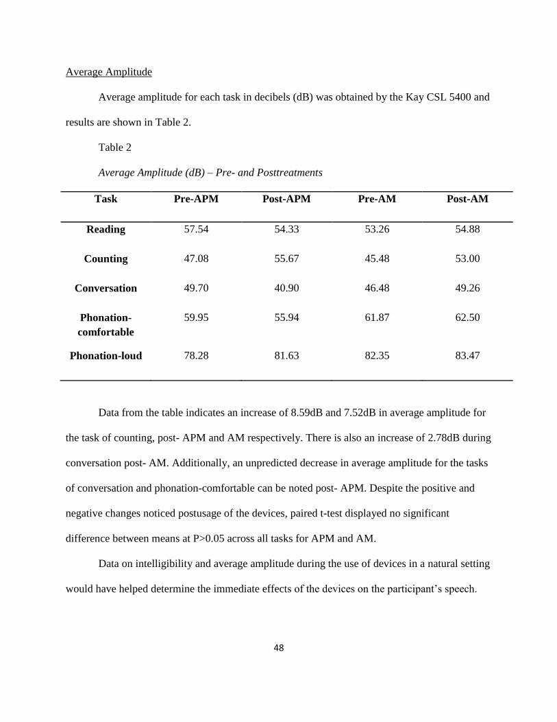
48
Average Amplitude
Average amplitude for each task in decibels (dB) was obtained by the Kay CSL 5400 and
results are shown in Table 2.
Table 2
Average Amplitude (dB) – Pre- and Posttreatments
Task Pre-APM Post-APM Pre-AM Post-AM
Reading 57.54 54.33 53.26 54.88
Counting 47.08 55.67 45.48 53.00
Conversation 49.70 40.90 46.48 49.26
Phonation-
comfortable
59.95 55.94 61.87 62.50
Phonation-loud 78.28 81.63 82.35 83.47
Data from the table indicates an increase of 8.59dB and 7.52dB in average amplitude for
the task of counting, post- APM and AM respectively. There is also an increase of 2.78dB during
conversation post- AM. Additionally, an unpredicted decrease in average amplitude for the tasks
of conversation and phonation-comfortable can be noted post- APM. Despite the positive and
negative changes noticed postusage of the devices, paired t-test displayed no significant
difference between means at P>0.05 across all tasks for APM and AM.
Data on intelligibility and average amplitude during the use of devices in a natural setting
would have helped determine the immediate effects of the devices on the participant’s speech.

49
Table 3 displays the retrospective data from APM (week 1) during functional communication at
home, on average amplitude, %compliance, and # of times triggered, for days 1 through 5.
Table 3
Retrospective Data from APM during Functional Communication
Parameter Day 1 Day 2 Day 3 Day 4 Day 5 Average
Average
amplitude
(dB)
65.11 65.31 68.93 58.32 62.61 64.05
%
compliance
44.77% 37.14% 54.17% 12.98% 29.57% 35.72%
No. of times
triggered
1138 606 444 377 516 616
It can be observed that there is a notable increase in average amplitude, with
simultaneous decrease in the frequency of a trigger, demonstrating the effectiveness of the APM
with biofeedback for days 1, 2, and 3. There is a drastic drop in average amplitude by 10.61dB
on day 4, during which the participant had experienced a fall. As per the participant’s report in
his time journal, the accelerometer was detached from his neck around the same time of his fall,
which is depicted by the decrease in the percent compliance and number of times triggered.
Unfortunately, there is no retrospective data available during functional communication at home
for AM to compare and account for the immediate effects of the device on the participant’s
speech characteristics in a natural setting.

50
Another purpose of the study was to determine if either of the devices is perceived to be
more comfortable for potential long-term use. Questionnaire A and B were designed to rate APM
and AM respectively on a scale of 1-5 (1 being extremely satisfied or comfortable and 5 being
extremely dissatisfied or uncomfortable) (see Appendixes A and B). Questionnaire C was
designed to establish the participant’s satisfaction response comparing the two treatments, on a
scale of 1-5 (1 being extremely satisfied or convenient and 5 being extremely dissatisfied or
inconvenient) (see Appendix C). In questionnaires A and B, the participant rated the
effectiveness of APM and AM on his speech to be 1 and 3, and comfort in public and
convenience to wear to be 5 and 3 for APM and AM respectively. These ratings display the
participant’s perception of APM to be the more effective device on his speech, and AM to be the
more comfortable and convenient device to wear. In contrast, in Questionnaire C, the participant
rated his overall satisfaction with the treatment device to be 4 and 2 for APM and AM,
suggesting he found AM to be the most effective and convenient therapeutic device towards the
end of the research study.

51
CHAPTER 5
DISCUSSION
This study was an attempt to determine if there is improvement in speech intelligibility
and vocal loudness by using portable therapeutic devices such as APM and AM in an individual
with PD, who could not transfer or generalize vocal loudness learned through traditional speech
therapy techniques and LSVT®.
Clinical Implications
The findings of this case study demonstrated no improvement in speech intelligibility and
vocal loudness after using APM and AM individually for a period of 1 week. On the contrary,
nominal drop in scores of intelligibility and average amplitude was observed subsequently after
using the devices. Moreover, contrasting results were obtained from the participant satisfaction
questionnaires between the effectiveness, convenience, and comfort of use of the two devices.
The collective results from questionnaires A and B that rated the devices individually depicted
that the participant found APM to be the more effective device on his speech, whereas results
from questionnaire C that rated the participant’s satisfaction of the treatments, described AM to
be the most effective device. This discussion addresses some factors that could potentially have
contributed to these unexpected and variable findings, as well as some recommendations for
future work. Factors discussed are confounding effects, sample size, and study design.
The participant during the course of the study experienced unanticipated weak vocal
health and decline in general health due to a fall during day 4 of week 1, and the participant’s
performance was determined to be subsiding since the beginning of week 2. This could have
directed to the 15% decline in speech intelligibility during conversation from post-APM (week

52
1) to pre-AM (week 3) measures. However, it has to be noted that there is a 4.29dB increase in
average amplitude on day 5 during week 1 (Table 3) indicating the participant’s ability to adapt
and perform based on the purpose of the APM device. Also during post- AM measures an
average increase of 3dB was noticed across all tasks, consistent with the existing literature on
positive effects of clinical masking (Adam & Lang, 1992; Countinho et al., 2009; Gryczka et al.,
2011; Quedas et al., 2007), although, the participant forgot to wear his palatal lift prosthesis.
However, by further spectral analyses it was determined that nasalence contributed to the
concentration of energy in the low frequency, resulting in an improvement in average amplitude.
Due to these confounding factors, the true effects of AM on the participant could not be
established. Even though this case study is one of the first studies to compare effects of two
therapeutic devices on the speech of an individual with PD, the results obtained did not reveal an
improvement in speech characteristics from the use of the deivces. Preliminary conclusion from
these data should be approached with caution given the single case study, and other medical
factors associated with the participant. Future studies involving larger sample size might reduce
variability in the results and also aid in generalizing the outcomes for this population.
In this case study it is likely that the tactile and auditory feedback may not have been
sufficiently perceptible to elicit a consistent and improved response from the participant. The
idiosyncratic nature of PD compels the thought that individuals with PD will require different
levels of feedback threshold in order to produce increases in speech intensity and intelligibility
(Adam & Lang, 1992). Data on speech intelligibility and average amplitude during
conversational speech while wearing the device would assist in presenting evidence on the
immediate effects of the therapeutic devices, further determining the adequate level of feedback

53
and its influence on speech intelligibility and vocal amplitude. Future studies can implement
multiple baseline measures with different feedback levels throughout the experimental procedure
in order to document the influence of the device on the individual’s speech in clinical as well as
nonclinical environments. Upcoming studies on therapeutic devices can also analyze the speech
samples at different time intervals rather than estimating the average value for vocal intensity
and/or speech intelligibility. Such measures of analyzing a speech sample at different time points
may enhance and provide information on speech performance, including, vocal amplitude and
intelligibility during initiation and termination of speech. Supplementary qualitative data from
the spouse or caregiver on the participant’s speech, in addition to the questionnaires given to the
participants, will help the researcher determine the effects of therapeutic devices from a listener’s
perspective, in nonclinical environments.
Conclusion
Though the results of this case study have not been favorable, it has opened avenues for
further studies with larger sample size, multiple baseline measures, and qualitative data from
spouse or caregiver to determine the effects of therapeutic devices in improving speech
intelligibility and vocal loudness in individuals with PD. It may also prove useful to extend the
testing period for each of the devices by a couple of weeks so that the participant is provided
with reasonable amount of time to get adapted to respond to the cue in both clinical and natural
environment.

54
REFERENCES
Aarsland, D., Kvaloy, J.T., Andersen, K., Larsen, J.P., Tang, M.X., Lolk, A, Kragh-Sorenson, P.,
. . . Marden, K. (2007). The effect of age of onset of PD on risk of dementia. Journal of
Neurology, 254, 38-45.
Ackermann, H., & Ziegler, W. (1991). Articulatory deficits in Parkinsonian dysarthria: An
acoustic analysis. Journal of Neurology, Neurosurgery, and Psychiatry, 54, 1093–1098.
Adams, S. G. (1994). Accelerating speech in a case of hypokinetic dysarthria: Descriptions and
treatment. In Motor speech disorders: Advances in assessment and treatment. (pp. 218-
223). Baltimore, MD: Paul A. Brookes.
Adams, S.G., & Dykstra, A. (2009). Hypokinetic dysarthria. In: M. R. McNeil (2nd
ed.), Clinical
management of sensorimotor speech disorders (pp, 166-186). New York: Thieme.
Adams, S.G., & Lang, A.E. (1992). Can the Lombard effect be used ti improve low voice
intensity in Parkinson’s disease? European Journal of Disordered Communication, 27,
121-127.
Ahlskog, J.E., Muenter, M.D. (2001). Frequency of levodopa-related dyskinesias and motor
fluctuations as estimated from the cumulative literature. Movement Disorders, 16, 448-
458.
Albin, R, L. (2006). Parkinson’s disease: Background, diagnosis, and initial management. Clinics
in Geriatric Medicine, 22, 735-751.
Albin, R. L., Young, A. B., & Penny, J. B. (1989). The functional anatomy of basal ganglia
disorders. Trends in Neuroscience, 12, 366-375.
Allain, H., Schuck, S., & Mauduit, N. (2000). Depression in Parkinson's disease . British
Medical Journal, 320, 1287-1288.

55
Allan, C.M. (1970). Treatment of non-fluent speech resulting from neurological disease-
treatment of dysarthria. British Journal of Disorders of Communication, 5, 3-5.
Allan, C.M., Turner, J.W., & Gadea-Ciria, M. (1966). Investigations into speech disturbances
following stereotaxic surgery for Parkinsonism. British Journal of Disorders of
Communication, 1, 55–59.
Aronson, A.E. (1990). Clinical voice disorders. New York: Thieme Stratton.
Baker, K., Ramig, L.O., Johnson, A., & Freed, C. (1997). Preliminary speech and voice analysis
following fetal dopamine transplants in 5 people with Parksinson’s disease. Journal of
Speech and Hearing Research, 40, 615-626.
Baker, K.K., Ramig, L.O., Luschei, E.S., & Smith, M.E. (1998). Thyroarytenoid muscle activity
associated with hypophonia in Parkinson disease and aging. Neurology, 51, 1592–1598.
Berardelli, A., Rothwell, J. C., Thompson, P. D., & Hallett, M. (2001). Pathophysiology of
bradykinesia in Parkinson’s disease. Brain, 124, 2131–2146.
Boshes, B. (1966). Voice changes in Parkinsonism. Journal of Neurosurgery, 24, 286–290.
Bourdreaux, D.M. (2011). Using the Ambulatory Phonation Monitor to measure the vocal
parameters of older people with and without Parkinson’s disease. Retrieved from
http://etd.lsu.edu/docs/available/etd-04192011-095645/
Bradlow, A., Kraus, N., & Hayes, E. (2003). Speaking clearly for children with learning
disabilities: Sentence perception in noise. Journal of Speech Language and Hearing
Research, 46, 80-97.
Brendel, B., Lowit, A., & Howell, P. (2004). The effects of delayed and frequency shifted
feedback on speakers with Parkinson’s disease. Journal of Medical Speech-Language
Pathology, 12, 131-138.

56
Caligiuri, M.P. (1989). The influence of speaking rate on articulatory hypokinesia in
Parkinsonian dysarthria. Brain and Language, 36, 493–502.
Calne, D.B. (1994). Early idiopathic Parkinsonism: Initiation and optimization of treatment.
Clinical Neuropharmacology, 17(Suppl. 2), S14–S18.
Canter, G.J. (1963). Speech characteristics of patients with Parkinson’s disease: I. Intensity,
pitch, and duration. Journal of Speech and Hearing Disorders, 28, 221–229.
Canter, G.J. (1965a). Speech characteristics of patients with Parkinson’s disease: II.
Physiological support for speech. Journal of Speech and Hearing Disorders, 30(1), 44–
49.
Canter, G.J. (1965b). Speech characteristics of patients with Parkinson’s disease: III.
Articulation, diadochokinesis, and over-all speech adequacy. Journal of Speech and
Hearing Disorders, 30, 217–224.
Carrara, A.E., Mourao, L.F., Ferraz, H.B., Behlau, M.S., Pontes, P.L., & Andrade, A.L.F. (1997).
Effect of voice rehabilitation on oral communication of Parkinson’s disease patients. Acta
Neurologica Scandinavica, 96, 199-205.
Connor, N.P., Ludlow, C.L., & Schulz, G.M. (1989). Stop consonant production in isolated and
repeated syllables in Parkinson’s disease. Neuropsychologia, 27, 829–838.
Countinho, S.B., Diaferia, G., Oliveira, G., & Behlau, M. (2009). Voice and speech of
individuals with Parkinson’s disease during amplification, delay and masking situations.
Pro-Fono Revista de Atualizacao Cientifica, 21, 219-224.
Countryman, S., & Ramig, L.O. (1993). Effects of intensive voice therapy on voice deficits
associated with bilateral thalamotomy in Parkinson disease: A case study. Jounal of
Medical Speech-Language Pathology, 1, 233–250.

57
Critchley, E. M. (1981). Speech disorders of Parkinsonism: A review. Journal of Neurology,
Neurosurgery & Psychiatry, 44, 751-758.
Dagenais, P. A., Southwood, M. H., & Lee, T. L. (1998). Rate reduction methods for improving
speech intelligibility of dysarthria speakers with Parkinson's disease. Journal of Medical
Speech-Language Pathology, 6(3), 143-158.
Darley FL., Aronson AE., & Brown J.R. (1975). Motor speech disorders. Philadelphia:
W.B.Saunders.
Darley, F.L., Aronson, A.E., & Brown, J.R. (1969a). Clusters of deviant speech dimensions in
the dysarthrias. Journal of Speech and Hearing Research, 12, 462–496.
Darley, F.L., Aronson, A.E., & Brown, J.R. (1969b). Differential diagnostic patterns of
dysarthria. Journal of Speech and Hearing Research, 12, 246–269.
Darling, M., & Huber, J. E. (2011). Changes to articulatory kinematics in response to loudness
cues in individuals with Parkinson's disease. Journal of Speech, Language, and Hearing
Research, 54, 1247-1259.
De Lau, L. M., & Breteler, M. M. (2006). Epidemiology of Parkinson's disease. Lancet
Neurology, 5, 525-535.
DeLetter, M., Santens, P., & Borsel, J. V. (2005). The effects of levodopa on word intelligibility
in Parkinson's disease. Journal of Communication Disorders, 38, 187-196.
Demirci, M., Grill, S., McShane, L., & Hallet, M. (1995). Impairment of kinesthesia in
Parkinson's disease. Neurology, 45, A218.
Dogan, M. M., Koseoglu, M., Can, C., Sehitoglu, M. A., & Gunal, D. I. (2008). Voice
abnormalities and their relation with motor dysfunction in Parkinson’s disease. Acta
Neurologica Scandinavica, 117(1), 26-34.

58
Downie, A.W., Low, J.M., & Lindsay, D.D. (1981). Speech disorder in Parkinsonism:
Usefulness of delayed auditory feedback in selected cases. British Journal of Disorders
of Communication, 16, 135–139.
Dromey, C. (2000). Articulatory kinematics in patients with Parkinson disease using different
speech treatment approaches. Journal of Medical Speech Language Pathology, 8, 155-
161.
Dromey, C., Kumar, R., Lang, A.E., & Lozano, A.M. (2000). An investigation of the effects of
subthalamic nucleus stimulation on acoustic measures of voice. Movement Disorders, 15,
1132–1138.
Dromey, C., Ramig, L.O., & Johnson, A.B. (1995). Phonatory and articulatory changes
associated with vocal intensity in Parkinsons disease: A case study. Journal of Speech
and Hearing Research, 38, 751-764.
Duffy, J. R. (2005). Motor speech disorders: Substrates, differential diagnosis, and management.
(2nd ed.). San Louis, MO: Mosby.
Eller, T.W., & Dan, D.A. (1997). Stereotactic pallidotomy for treatment of Parkinson’s disease.
AORN Journal, 65, 903–918.
Eskander, E.N., Cosgrove, G.R., & Shinbou, L.A. (2001). Surgical treatment of Parkinson’s
disease. Journal of American Medical Association, 286, 3056-3059.
Fazzini, E., Dogali, M., Sterio, C., Eidelberg, O.J., & Beric, A. (1997). Stereotactic pallidotomy
for Parkinson's disease: A long-term follow up of unilateral pallidotomy. Neurology, 48,
1273-1277.

59
Ferguson, S. H., & Kewley-Port, D. (2007). Talker differences in clear and conversational
speech: Acoustic characteristics of vowels. Journal of Speech, Language, and Hearing
research, 50, 1241-1255.
Forrest, K., Weismer, G., & Turner, G.S. (1989). Kinematic, acoustic, and perceptual analyses of
connected speech produced by Parkinsonian and normal geriatric adults. Journal of the
Acoustic Society of America, 85, 2608–2622.
Fox, C.M., Morrison, C.E., Ramig, L.O., & Sapir, S. (2002). Current perspectives on Lee
Silverman voice treatment (LSVT) for individuals with ideopathic Parkinson's disease.
American Journal of Speech-Language Pathology, 11, 111-123.
Gentil, M., Garcia-Ruiz, P., Pollak, P., & Benabid, A.L. (2000). Effect of bilateral deep-brain
stimulation on oral control of patients with parkinsonism. European Neurology, 44, 147–
152.
Ghika, J., Ghika-Schmid, F., Fankhauser, H., Assal, G., Vingerhoets, F., Albanese, A., . . . Favre,
J. (1999). Bilateral contemporaneous posteroventral pallidotomy for the treatment of
Parkinson’s disease: neuropsychological and neurological side effects: Report of four
cases and review of the literature. Journal of Neurosurgery, 91, 313-321.
Ghika, J., Villemure, J.G., Fankhauser, H., Favre, J., Assal, G., & Ghika-Schmid, F. (1998).
Efficiency and safety of bilateral contemporaneous pallidal stimulation (deep brain
stimulation) in levodopa-responsive patients with Parkinson’s disease with severe motor
fluctuations: a 2-year follow-up review. Journal of Neurosurgery, 89, 713-718.
Gjerstad, M.D., Alves, G., & Wentzel-Larsen, T. (2006). Excessive daytime sleepiness in
Parkinson’s disease: is it the drugs or the disease? Neurology, 57, 388-393.

60
Goberman, A. M., & Elmer, L. W. (2005). Acoustic analysis of clear versus conversational
speech in individuals with Parkinson disease. Journal of Communication Disorders, 38,
215-230.
Goetz, C.G., Poewe, W., Rascol, O., & Sampaio, C. (2005). Evidence-based medical review
update: pharmacological and surgical treatments of Parkinson’s disease: 2001 to 2004.
Movement Disorders, 20, 523-539.
Greene, M.C.L., & Watson, B.W. (1968). The value of speech amplification in Parkinson’s
disease patients. Folia Phoniatrica et Logopedia, 20, 250–257.
Grossman, R.G., & Hamilton, W.J. (1993). Surgery for movement disorders. In J. Jankovic & E.
Tolosa (Eds.), Parkinson’s disease and movement disorders (pp. 531–548). Baltimore,
MD: Williams & Wilkins.
Gryczka, T., Lipski, S., & Meister, I. G. (2011). Improvement of speech through the Lombard
effect in patients with dysarthria related to Parkinson’s disease. Basal Ganglia, 1(2), 114-
115.
Gulick, W. L., Gescheider, G. A., & Frisina, R. D. (1989). Hearing: Physiological acoustics,
neural coding, and psychoacoustics. New York, NY: Oxford University Press.
Hammen, V.L., & Yorkston, K.M. (1996). Speech and pause characteristics following speech
rate reduction in hypokinetic dysarthria. Journal of Communication Disorders, 29, 429–
444.
Hammer, M.J., Barlow, S.M., Lyons, K.E., & Pahwa, R. (2010). Subthalamic nucleus deep brain
stimulation changes speech respiratory and laryngeal control in Parkinson’s disease.
Journal of Neurology, 257, 1692–1702.

61
Hammer, M.J., Barlow, S.M., Lyons, K.E., & Pahwa, R. (2011). Subthalamic nucleus deep brain
stimulation changes velopharyngeal control in Parkinson’s disease. Journal of
Communication Disorders, 44(1), 37–48.
Hanson, D. G., Gerratt, B. R., & Paul, P. H. (1984). Cinegraphic observations of laryngeal
function in Parkinson's disease. The Laryngoscope, 94, 348-353.
Hargus Ferguson, S., & Kewley-Port, D. (2002). Vowel intelligibility in clear and conversational
speech for normal-hearing and hearing-impaired listeners. Journal of the Acoustical
Society of America, 112, 259–271.
Hauser, C.W. (2005). Use of ambulatory biofeedback to supplement Lee Silverman Voice
Treatment in patients with idiopathic Parkinson’s disease. Retrieved from
http://gradworks.umi.com/14/27/1427439.html
Helfer, K. (1997). Auditory and auditory-visual perception of clear and conversational speech.
Journal of Speech, Language, and Hearing Research, 40, 432–443.
Hely, M.A., Morris, J.G., Reid, W.G., & Trafficante, R. (2005). Sydney multicenter study of
Parkinson’s disease: Non-L-dopa-responsive problems dominate at 15 years. Movement
Disorders 20, 190-199.
Hillman, R.E., Heaton, J.T., Masaki, A., Zeitels, S.M., & Cheyne, H.A. (2006). Ambulatory
monitoring of disordered voices. Annals of Otology, Rhinology & Laryngology, 115, 795-
801.
Hirose, H., Kiritani, S., Ushijima, T., Yoshioka, H., & Sawashima, M. (1981). Patterns of
dysarthric movements in patients with Parkinsonism. Folia Phoniatrica et Logopedia, 33,
204–215.

62
Ho, A.K., Bradshaw, J.L., Iansek, R., & Alfredson, R. (1999). Speech volume regulation in
Parkinson’s disease: Effects of implicit cues and explicit instructions. Neuropsychologia,
37, 1453-1460.
Hoodin, R.B., & Gilbert, H.R. (1989). Parkinsonian dysarthria: An aerodynamic and perceptual
description of velopharyngeal closure for speech. Folia Phoniatrica et Logopedia, 41,
249–258.
Huber, J. E., & Darling, M. (2011). Effect of Parkinson's disease on the production of structured
and unstructured speaking tasks: Respiratory physiologic and linguistic considerations.
Journal of Speech Language and Hearing Research, 54(1), 33-46.
Intemann, P.M., Masterman, D., & Subramanian, I., et al. (2001). Staged bilateral pallidotomy
for treatment of Parkinson disease. Journal of Neurosurgery, 94, 437-444.
Iulianella, I., Adams, S.G., & Gow, A.K. (2008).Effects of sub-thalamic deep brain stimulation
on speech production in Parkinson’s Disease: A critical review of the literature.
Canadian Journal of Speech-Language Pathology & Audiology, 32(2), 85–91.
Jankovic, J. (2007). Parkinson’s disease: Clinical features and diagnosis. Journal of
Neurosurgery and Psychiatry, 79, 368-376.
Jankovic, J., & Marsden, C.D. (1993). Therapeutic strategies in Parkinson’s disease. In J.
Jankovic & E. Tolosa (Eds.), Parkinson’s disease and movement disorders (pp. 115–
144). Baltimore, MD: Williams & Wilkins.
Jankovic, J., Cardoso, F., Grossman, R.G., & Hamilton, W.J. (1995). Outcome after stereotactic
thalamotomy for parkinsonian, essential, and other types of tremor. Neurosurgery, 37,
680-686

63
Jenkins, A.C. (1968). Speech defects following stereotaxic operations for the relief of tremor and
rigidity in Parkinsonism. The Medical Journal of Australia, 7, 585–588.
Johnson, J.A, & Pring, TR. (1990). Speech therapy and Parkinson's disease: A review and further
data. British Journal of Disorders of Communication, 25, 183-194.
Kelly, P.J., & Gillingham, F.J. (1980). The long-term results of stereotaxic surgery and l-dopa
therapy in patients with Parkinson’s disease: A 10 year follow-up study. Journal of
Neurosurgery,53, 332-337.
Kelly, P.J., Ahlskog, J.E., Goerss, S.J., Daube, J.R., Duffy, J.R., & Kall, B.A. (1987). Computer-
assisted stereotactic ventralis lateralis thalamotomy with microelectrode recording control
in patients with Parkinson’s disease. Mayo Clin Proc, 62, 655-664.
Kleinow, J., Smith, A., & Ramig, L.O. (2001). Speech motor stability in IPD: Effects of rate and
loudness manipulations. Journal of Speech, Language and Hearing Research, 44, 1044-
1051.
Klostermann, F., Ehlen, F., Vesper, J., Nubel, K., Gross, M., Marzinzik, F., . . . Sappok, T.
(2008). Effects of subthalamic deep brain stimulation on dysarthrophonia in Parkinson’s
disease. Journal of Neurology, Neurosurgery, and Psychiatry, 79, 522–529.
Krause, J.C., Braida, L.D. (2004). Acoustic properties of naturally produced clear speech at
normal speaking rates. Journal of Acoustical Society of America, 15, 362-378.
Kumar, R., Lang, A.E., Rodriguez-Oroz, M.C., Lozano, A.M., Pollak, P., Benabid, A.L., . . .
Obeso, A.J. (2000). Deep brain stimulation of the globus pallidus pars interna in
advanced Parkinson’s disease. Neurology, 55, 34-39.

64
Laitinen, L.V., Bergenheim, A.T., & Hariz, M.I. (1992). Ventroposterolateral pallidotomy can
abolish all Parkinsonian symptoms. Stereotactic and Functional Neurosurgery, 58, 14–
21.
Le Dorze, G., Dioone, L., Ryalls, J., Julien, M., & Ouellet, L. (1992). The effects of speech and
language therapy for a case of dysarthria associated with Parkinson's disease. European
Journal of Disorders of Communication, 27, 313-324.
Leanderson, R., Meyerson, B. A., & Persson, A. (1971). Effect of l-dopa on speech in
Parkinsonism: An EMG study of labial articulatory function. Journal of Neurology,
Neurosurgery, and Psychiatry, 34, 379-381.
Lisker, L., & Abramson, A. S. (1964). A cross-language study of voicing in initial stops:
Acoustical measurements. Word, 20, 384-422.
Logemann, J. A., Blonsky, E. R., Fisher, H. B. & Boshes, B. (1973). A cineradiographic study of
lingual function in Parkinson's disease. Paper presented at the Annual Convention of the
American Speech and Hearing Association, Detroit.
Logemann, J.A., & Fisher, H.B. (1981). Vocal tract control in Parkinson’s disease: Phonetic
feature analysis of misarticulations. Journal of Speech and Hearing Disorders, 46, 348–
352.
Logemann, J.A., Fisher, H.B., Boshes, B., & Blonsky, E.R. (1978). Frequency and co-occurrence
of vocal tract dysfunctions in the speech of a large sample of Parkinson patients. Journal
of Speech and Hearing Disorders, 43, 47–57.
Lombard, E. (1911). Le signe de l`elevation de la voix. Larynx, 37, 101-119.
Louis, E. D., Winfield, L., Fahn, S., & Ford, B. (2001). Speech dysfluency exacerbated by
levodopa in Parkinson's disease. Movement Disorders, 16, 562-565.

65
Lowit, A., Brendel, B., Dobinson, C., & Howell, P. (2006). An investigation into the influences
of age, pathology and cognition on speech production. Journal of Medical Speech-
Language Pathology, 1, 253-262.
Luschei, E.S., Ramig, L.O., Baker, K.L., & Smith, M.E. (1999). Discharge characteristics of
laryngeal single motor units during phonation in young and older adults and in persons
with Parkinson disease. Journal of Neurophysiology, 81, 2131–2139.
Marsden, C. D., & Obeso, J. A. (1994). The functions of the basal ganglia and the paradox of
stereotaxic surgery in Parkinson’s disease. Brain, 117, 877–897.
Marsden, C.D., & Parkes, J.D. (1977). Success and problems of long-term levodopa therapy in
Parkinson’s disease. The Lancet, 1, 345–349.
Marshal, R. C., & Karrow, C. M. (2002). Retrospective examination of failed rate-control .
American Journal of Speech-Language Pathology, 11, 3-16.
Matsumoto, K., Asano, T., Baba, T., Miyamoto, T., & Ohmoto, T. (1976). Long-term follow-up
results of bilateral thalamotomy for Parkinsonism. Applied Neurophysiology, 39, 257–
260.
McRae, P. A., Tjaden, K., & Schoonings, B. (2002). Acoustic and perceptual consequences of
articulatory rate change in Parkinson disease. Journal of Speech, Language, and Hearing
Research, 45(1), 35-50.
McWilliams, B. J., Morris, H. L., & Shelton, R. L. (1990). Cleft palate speech (2nd ed.).
Philadelphia: B.C. Becker.
Metman, L. V. (2002). Recognition and treatment of response fluctuations in Parkinson's
disease: Review article. Amino Acids, 23(1-3), 141-145.

66
Metter, E. J., & Hanson, W. R. (1986). Clinical and acoustical variability in hypokinetic
dysarthria. Journal of Communication Disorders, 19, 347–366.
Monrad-Krohn, G.H. (1957). The third element of speech: prosody in the neuro-psychiatric
clinic. The British Journal of Psychiatry, 103, 326-331.
Moore, C. A., & Scudder, R. R. (1989). Coordination of jaw muscle activity in Parkinsonian. In
Recent advances in clinical dysarthria. Boston: College-Hill: Pro Ed.
Morley, D.E. (1955). The rehabilitation of adults with dysarthric speech. Journal of Speech and
Hearing Disorders, 20(1), 58–64.
Mueller, P.B. (1971). Parkinson’s disease: Motor-speech behavior in a selected group of patients.
Folia Phoniatrica, 23, 333–345.
Nakano, K. K., Zubick, H., & Tyler, H. R. (1973). Speech defects of parkinsonian patients:
Effects of levodopa therapy on speech intelligibility. Neurology, 23, 865-870.
Narayana, S., Jacks, A., Robin, D.A., Poizner, H., Zhang, W., Franklin, C., et al. (2009). A
noninvasive imaging approach to understanding speech changes following deep brain
stimulation in Parkinson’s disease. American Journal of Speech-Language Pathology,18,
146–161.
National Institute of Neurological Disorders and Stroke (NINDS). (2012, Feb 03). Parkinson's
disease: Hope through research. Retrieved from
http://www.ninds.nih.gov/disorders/parkinsons_disease/detail_parkinsons_disease.htm
Netsell, R., Daniel, B., & Celesia, G.G. (1975). Acceleration and weakness in Parkinsonian
dysarthria. Journal of Speech and Hearing Disorders, 40, 170–177.
Nonaka, S., Takahashi, R., Enomoto, K., Katada, A., & Unno, T. (1996). Lombard reflex during
PAG-induced vocalization in decerebrate cats. Neuroscience Research, 29, 283-289.

67
Oxtoby, M. (1982). Parkinson’s disease patients and their social needs. London: Parkinson’s
Disease Society.
Parkinson's Disease: Hope Through Research. (2012, February 3). National Insitute of
Neurological Disorders and Stroke. Retrieved February 4, 2012, from
http://www.ninds.nih.gov/disorders/parkinsons_disease/detail_parkinsons_disease.htm
Penny Jr, J. B., & Young, A. B. (1983). Speculations on the functional anatomy of basal ganglia
disorders. Annual Review of Neuroscience, 6, 73-94.
Picheny, M.A., Durlach, N.I., & Braida, L.D. (1985). Speaking clearly for the hard of hearing I:
Intelligibility differences between clear and conversational speech. Journal of Speech and
Hearing Research, 28, 96–103.
Picheny, M.A., Durlach, N.I., Braida, L.D. (1986). Speaking clearly for the hard of hearing. II.
Acoustic characteristics of clear and conversational speech. Journal of Speech Language
and Hearing Research, 29, 434-446.
Pinto, S., Ozancak, C., Tripoliti, E., Thobois, S., Limousin-Dowsey, P., & Auzou, P. (2004).
Treatments for dysarthria in Parkinson’s disease. The Lancet Neurology, 3, 547-556.
Pitcairn, T.K., Clemie, S., Gray, J.M., & Pentland, B. (1990). Impressions of Parkinsonian
patients from their recorded voices. British Journal of Disorders of Communication, 25,
85–92.
Poluha, P. C., Teulings, H. L., & Brookshire, R. H. (1998). Handwriting and speech changes
across the levodopa cycle in Parkinson’s disease. Acta Psychologica, 100(1-2), 71-84.
Quaglieri, C.E., & Celesia, G.G. (1977). Effect of thalamotomy and levodopa therapy on the
speech of Parkinson patients. European Neurology, 15, 34–39.

68
Quedas, A., Duprat, A.C., & Gasparini, G. (2007). Lombard’s effect’s implications in intensity,
fundamental frequency and stability on the voice of individuals with Parkinson’s disease.
Revista brasileira de otorrinolaringologia, 73, 675-683.
Ramig, L. O. The LSVT Story. Retrieved February 4, 2012, from
http://www.ncvs.org/research/lsvt-history.html
Ramig, L.O. (1993). Therapy for patients with Parkinson's disease. NCVS-Status and Progress
Report-5, 83-90.
Ramig, L.O. (1994). Speech therapy for patients with Parkinson's disease. In Koller, W. and
Paulson, G. (eds.) Therapy of Parkinson's disease. New York, NY: Marcel Dekker.
Ramig, L.O., & Bonitati, C.M., (1991). The efficacy of voice therapy for patients with
Parkinson's disease. NCVS-Status and Progress Report-1, 61-86.
Ramig, L.O., Bonitiati, C., Lemke, J., & Horii, Y. (1994). Voice treatment for patients with
Parkinson’s disease: Development of an approach and preliminary efficacy data. Journal
of Medical Speech-Language Pathology, 3, 191-209.
Ramig, L.O., Countryman, S., Thompson, L.L., & Horii, Y. (1995). Comparison of two forms of
intensive treatment for Parksinson disease. Journal of Speech and Hearing Research, 38,
1232-1251.
Ramig, L.O., & Dromey, C. (1996). Aerodynamic mechanisms underlying treatment-related
changes in vocal intensity in patients with Parkinson disease. Journal of Speech and
Hearing Research, 39, 798–807.
Ramig, L.O., Fox, C., & Sapir, S. (2004), Parkinson’s disease: Speech and voice disorders and
their treatment with the Lee Silverman Voice Treatment. Seminars in Speech and
Language, 25, 169-180.

69
Ramig, L.O., Mead, C., Scherer, R., Horii, Y., Larson, K., & Kohler, D. (1988, February).
Voice therapy and Parkinson's disease: A longitudinal study of efficacy. Paper presented
at the Clinical Dysarthria Conference, San Diego, CA.
Ramig, L.O., Sapir, S., Countryman, S., Pawlas, A.A., O’Brien, C., Hoehn, M., & Thompson,
L.L. (2001). Intensive voice treatment (LSVT®) for patients with Parkinson’s disease: A
2 year follow up. Journal of Neurology, Neurosurgery & Psychiatry, 71, 493-498.
Rao, S.S., Hofmann, L.A., & Shakil, A. (2006). Parkinson’s disease: Diagnosis & treatment.
American Academy of Family Physicians, 74, 2046-2056.
Rigrodsky, S., & Morrison, E.B. (1970). Speech changes in Parkinsonism during L-dopa
therapy: preliminary findings. Journal of the American Geriatrics Society, 18, 142-151.
Robertson, S. J., & Thomson, F. (1984). Speech therapy in Parkinson's disease: A study of the
efficacy and long-term effects of intensive treatment. British Journal of Disorders of
Communication, 19, 213-224.
Rousseau, B., & Watts, C.R. (2002). Susceptibility of speakers with Parkinson disease to delayed
feedback. Journal of Medical Speech-Language Pathology, 10(1), 41.
Rousseaux, M., Krystkowiak,P., Kozlowski,O., Ozsancak,C., Blond,S., & Destee, A. (2004).
Effects of subthalamic nucleus stimulation on Parkinsonian dysarthria and speech
intelligibility. Journal of Neurology, 251(3), 327-334.
Rubow, R., & Swift, E. (1985). A microcomputer-based wearable biofeedback device to improve
transfer of treatment in Parkinsonian dysarthria. Journal of Speech and Hearing
Disorders, 50, 178-185.

70
Rubrow, R.T, Rosenbek, J.C., Collins, M.J., & Celesia, G.G. (1984). Reduction of hemifacial
spasm and dysarthria following EMG biofeedback. Journal of Speech and Hearing
Disorders, 49, 26-33.
Sadagopan, N., & Huber, J. E. (2007). Effects of loudness cues on respiration in individuals with
Parkinson’s disease. Movement Disorders, 22, 651–659.
Sanabria, J., Ruiz, P. G., Gutierrez, R., Marquez, F., Escobar, P., Gentil, M., & Cenjor, C.
(2001). The effect of levodopa on vocal function in Parkinson's disease. Clinical
Neuropharmacology, 24, 99-102.
Sapir, S., Pawlas, A.A., Ramig, L.O., Countryman, S., O'Brien, C., Hoehn, M.M., & Thompson,
L.A. (2001). Voice and speech abnormalities in Parkinson’s disease: Relation to severity
of motor symptoms, duration of disease, medication, depression, gender, and age.
Journal of Medical Speech Language Pathology, 9, 213-226.
Sapir, S., Ramig, L. O., Hoyt, P., Countryman, S., O'Brien, C., & Hoehn, M. (2002). Speech
loudness and quality 12 months after intensive voice treatment (LSVT) for Parkinson's
disease: A comparison with an alternative speech treatment. Folia Phoniatrica et
Logopaedica, 54, 296-303.
Sapir, S., Ramig, L.O., & Fox, C. (2008). Speech and swallowing disorders in Parkinson’s
disease. Current Opinion in Otolaryngology & Head and Neck Surgery, 16, 205-210.
Sapir, S., Spielman, J.L., Ramig L.O. (2007). Effects of intensive voice treatment (the Lee
Silverman Voice Treatment [LSVT]) on vowel articulation in dysarthric individuals with
idiopathic Parkinson disease: Acoustic and perceptual findings. Journal of Speech,
Language and Hearing Research, 50, 899–912.

71
Sarno, M.T. (1968). Speech impairment in Parkinson’s disease. Archives of Physical Medicine
and Rehabilitation, 49, 269–275.
Schaltenbrand, G. (1975). The effects on speech and language of stereotactical stimulation in
thalamus and corpus callosum. Brian and Language, 2, 70–77.
Schulz, G. M., & Grant, M. K. (2000). Effects of speech therapy and pharmacologic and surgical
treatments on voice and speech in parkinson's disease: A review of the literature. Journal
of Communication Disorders, 33(1), 59-88.
Schulz, G.M., Greer, M., & Friedman, W. (1998). Effects of pallidotomy surgery on Parkinson’s
patients sentence productions. Paper presented to the Speech Motor Control conference,
Tucson, AZ.
Schulz, G.M., Peterson, T., Sapienza, C.M., Greer, M., & Friedman, W. (1999). Voice and
speech characteristics of persons with Parkinson’s disease pre- and post-pallidotomy
surgery: Preliminary findings. Journal of Speech, Language and Hearing Research, 42,
1176–1194.
Schum, D. (1996). Intelligibility of clear and conversational speech of young and elderly talkers.
Journal of the American Academy of Audiology, 7, 212–218.
Scott, S.A & Caird, F.L. (1983). Speech therapy for Parkinson's disease. Journal of Neurology,
Neurosurgery, and Psychiatry, 46, 140-144.
Shea, B.R., Drummond, S.S., Metzer, W.S., & Krueger, K.M. (1993). Effect of selegiline on
speech performance in Parkinson’s disease. Folia Phoniatrica Logopedia, 45, 40–46.
Sidtis, J.J., & Sidtis, L.D. (2003). A neurobehavioral approach to dysprosody. Seminars in
Speech and Language, 24, 93-106.

72
Skodda, S., Visser, W., & Schlegal, U. (2011). Vowel articulation in Parkinson's disease. Journal
of Voice, 25, 467-472.
Smith, M.E., Ramig, L.O., Dromey, C., Perez, K.S., & Samandari, R. (1995). Intensive voice
treatment in Parkinson’s disease: Laryngostroboscopic findings. Journal of Voice, 9,
453–459.
Solomon, N.P., & Hixon, T.J. (1993). Speech breathing in Parkinson’s disease. Journal of
Speech and Hearing Research, 36, 294–310.
Spencer, K., Sanchez, J., McAllen, A., & Weir, P. (2010). Speech and cognitive-linguistic
function in Parkinson’s disease. Perspectives on Neurophysiology and Neurogenic
Speech and Language Disorders, 20, 31-38.
Spielman, J. (2011).Intensive voice treatment (LSVT LOUD) for Parkinson’s disease following
deep brain stimulation of the subthalamic nucleus. Journal of Communication Disorders,
doi:10.1016/ j.jcomdis.2011.05.003
Stamey, W.P., & Jankovic, J. (2007). Shoulder pain in Parkinson’s disease. Movement
Disorders, 22, 247-248.
Stern, G., & Lees, A. (1990). Parkinson’s Disease: The facts. New York, NY: Oxford University
Press.
Stewart, C., (2000). Speech-Language therapy for patients with Parkinson’s disease. Loss, Grief
and Care, 8, 151-155.
Svec, J.G., Titze, I.R., & Popolo, P.S. (2005). Estimation of sound pressure levels of voiced
speech from skin vibration of the neck. Journal of Acoustical Society of America, 117,
1386-1394.
Swigert, N.B. (1997). The source for dysarthria. East Moline, IL: LinguiSystems.

73
Tasker, R.R., Lang, A.E., & Lozano, A.M. (1997). Pallidal and thalamic surgery for Parkinson’s
disease. Experimental Neurology, 144, 35–40.
Tasker, R.R., Siqueira, J., Hawrylyshyn, P., & Organ, L.W. (1983). What happened to VIM
thalamotomy for Parkinson’s disease? Applied Neurophysiology, 46, 68-83.
Thanvi, B., Lo, N., & Robinson, T. (2007). Levodopa-induced dyskinesia in Parkinson’s disease:
clinical features, pathogenesis, prevention and treatment. Postgraduate Medical Journal,
83, 384-388.
Tinazzi, M, Del Vesco, C., & Fincati, E. (2006). Pain and motor complications in Parkinson’s
disease. Journal of Neurology, Neurosurgery and Psychiatry, 77, 822-825.
Tjaden, K., & Wilding, G. (2011a). Speech and pause characteristics associated with voluntary
rate reduction in Parkinson's disease and Multiple Sclerosis. Journal of Communication
Disorders, 44, 655-665.
Tjaden, K., & Wilding, G. (2011b). The impact of rate reduction and increased loudness on
fundamental frequency characteristics in dysarthria. Folia Phoniatrica et Logopaedica,
63, 178-186.
Tjaden, K., & Wilding, G. E. (2005). Effect of rate reduction and increased loudness on acoustic
measures of anticipatory coarticulation in Multiple Sclerosis and Parkinson's disease .
Journal of Speech, Language, and Hearing Research, 48, 261-277.
Tolosa, E., & Valldeoriola, F. (1994). Mid-stage Parkinsonism with mild motor fluctuations.
Clinical Neuropharmacology, 17(2), 19–31.
Trail, M., Fox, C., Ramig, L.O., Sapir, S., Howard, J., & Lai, E.C. (2005). Speech treatment for
Parkinson’s disease. Neuro Rehabilitation, 20, 205-221.

74
Tripoliti, E., Zrinzo, L., Martinez-Torres, I., Frost, E., Pinto, S., Foltynie, T., Holl, E., . . .
Limousin, P. (2011). Effects of sub-thalamic stimulation on speech of consecutive
patients with Parkinson disease. Neurology, 76(1), 80–86.
Turner, G. S., & Weismer, G. (1993). Characteristics of speaking rate in the dysarthria associated
with amyotrophic lateral sclerosis. Journal of Speech and Hearing Research, 33, 1134-
1144.
Turner, G. S., Tjaden, K., & Weismer, G. (1995). The influence of speaking rate on vowel space
and speech intelligibility for individuals with amyotrophic lateral sclerosis. Journal of
Speech & Hearing Research, 38, 1001-1013.
Van Nuffelen, G., De Bodt, M., Vanderwegen, J., Van de Heyning, P., & Wuyts, F. (2010).
Effect of rate control on speech production and intelligibility in Dysarthria. Folia
Phoniatrica et Phonetica, 62, 110-119.
Van Nuffelen, G., Bodt, M., Wuyts, F., & Van de Heyning, P. (2009). The effect of rate control
on speech rate. Folia Phoniatrica et Logopaedica, 61, 69-75.
Walker, H.C., Phillips, D.E., Boswell, D.B., Guthrie, B.L., Guthrie, S.L., Nicholas, A.P.,
Montgomery, E.B., . . . Watts, R.L. (2009). Relief of acquired stuttering associated with
Parkinson’s disease by unilateral left subthalamic brain stimulation. Journal of Speech,
Language, and Hearing Research, 52, 1652–1657.
Weaver, F., Follett, K., Hur, K., Ippolito, D., & Stern, M. (2005). Deep brain stimulation in
Parkinson’s disease: A meta-analysis of patient outcomes. Journal of Neurosurgery, 103,
956-967.
Weiner, W. J., & Singer, C. (1989). Parkinson's disease and nonpharmacologic treatment
programs. Journal of American Geriatrics Society, 37, 359-363.

75
Wenke, R. J., Cornwell, P., & Theodoros, D. G. (2010). Changes to articulation following
LSVT(R) and traditional dysarthria therapy in non-progressive dysarthria. International
Journal of Speech-Language Pathology, 12, 203-220.
Wolfe, V.I., Garvin, J.S., Bacon, M., & Waldrop, W. (1975). Speech changes in Parkinson’s
disease during treatment with L-dopa. Journal of Communication Disorders, 8(3), 271-
279.
Yorkston, K. M., Beukelman, D. R., Strand, E. A., & Bell, K. R. (1999). Management of motor
speech disorders in children and adults (2nd ed.). Austin, TX: Pro-Ed.
Yorkston, K. M., Hakel, M., Beukelman, D. R., & Fager, S. (2007). Evidence for effectiveness
of treatment of loudness, rate, or prosody in dysarthria: a systematic review. Journal of
Medical Speech-Language Pathology, 15(2), xi-xxxvi.
Yorkston, K. M., Hammen, V. L., Beukelman, D. R., & Traynor, C. D. (1990). The effect of rate
control on the intelligibility and naturalness of dysarthric speech. Journal of Speech and
Hearing Disorders, 55, 550-560.
Yorkston, K.M., Spencer, K.A., & Duffy, J.R. (2003). Behavioral management of
respiratory/phonatory dysfunction from dysarthria: A systematic review of evidence.
Journal of Medical Speech-Language Pathology, 11, xiii-xxxviii.

76
APPENDICES
Appendix A
Questionnaire – A (Adapted with permission from Boudreaux, 2011)
1. Rate your satisfaction on the effectiveness of treatment on your speech intelligibility. (1 =
extremely satisfied; 5 = extremely dissatisfied)
1 2 3 4 5
2. Rate your satisfaction on the effectiveness of treatment on your vocal loudness. (1 =
extremely satisfied; 5 = extremely dissatisfied)
1 2 3 4 5
3. How was it to use the Ambulatory Phonation Monitor as instructed? (1 = very
convenient; 5 = very inconvenient)
1 2 3 4 5
4. Was the Ambulatory Phonation Monitor a comfortable device to wear? (1 = very
comfortable; 5 = not very comfortable)
1 2 3 4 5
5. How comfortable were you wearing the Ambulatory Phonation Monitor in public? (1 =
very comfortable; 5 = not very comfortable)
1 2 3 4 5
6. Did you feel comfortable speaking while wearing the Ambulatory Phonation Monitor? (1
= very comfortable; 5 = not very comfortable)
1 2 3 4 5
7. Do you feel that the using the Ambulatory Phonation Monitor affected your speech in any
way?
No
Yes. How?
8. Did you experience any other difficulties with the Ambulatory Phonation Monitor?

77
Appendix B
Questionnaire – B (Adapted with permission from Boudreaux, 2011)
1. Rate your satisfaction on the effectiveness of treatment on your speech intelligibility. (1 =
extremely satisfied; 5 = extremely dissatisfied)
1 2 3 4 5
2. Rate your satisfaction on the effectiveness of treatment on your vocal loudness. (1 =
extremely satisfied; 5 = extremely dissatisfied)
1 2 3 4 5
3. How was it to use the auditory masker as instructed? (1 = very convenient; 5 = very
inconvenient)
1 2 3 4 5
4. Was the auditory masker a comfortable device to wear? (1 = very comfortable; 5 = very
uncomfortable)
1 2 3 4 5
5. How comfortable were you wearing the auditory masker in public? (1 = very
comfortable; 5 = very uncomfortable)
1 2 3 4 5
6. Did you feel comfortable speaking while wearing the auditory masker? (1 = very
comfortable; 5 = very uncomfortable)
1 2 3 4 5
7. Do you feel that using the auditory masker affected your speech in any way?
No
Yes. How?
8. Did you experience any difficulties with the auditory masker?

78
Appendix C
Questionnaire - C
Key: Instrument A – Ambulatory Phonation Monitor; Instrument B – Auditory masker
1. To what extent did you like the use of instrument A to improve your speech intelligibility
and loudness? (1 = extremely; 5 = not at all)
1 2 3 4 5
2. To what extent did you like the use of instrument B to improve your speech intelligibility
and loudness? (1 = extremely; 5 = not at all)
1 2 3 4 5
3. How convenient was it to use the instrument A? (1 = very convenient; 5 = very
inconvenient)
1 2 3 4 5
4. How convenient was it to use the instrument B? (1 = very convenient; 5 = very
inconvenient)
1 2 3 4 5
5. Taking all things into account, how satisfied or dissatisfied are you with the treatment
with instrument A? (1 = extremely satisfied; 5 = extremely dissatisfied)
1 2 3 4 5
6. Taking all things into account, how satisfied or dissatisfied are you with the treatment
with instrument B? (1 = extremely satisfied; 5 = extremely dissatisfied)
1 2 3 4 5

79
Appendix D
Time Journal - Ambulatory Phonation Monitor
(Adapted with permission from Boudreaux, 2011)
Please write down an approximation of how much you spoke in every 2-hour time slot.
Hours 1-2 (9.00-11.00)
Hours 3-4 (11.00-1.00)
Hours 5-6 (1.00-3.00)
Hours 7-8 (3.00-5.00)

80
Appendix E
Time Journal – Auditory Masker
Adapted with permission from Boudreaux, 2011
Please write down an approximation of how much you spoke in every 2-hour time slot.
Hours 1-2 (9.00-11.00)
Hours 3-4 (11.00-1.00)
Hours 5-6 (1.00-3.00)
Hours 7-8 (3.00-5.00)

81
VITA
SWETHA SWAMINATHAN
Personal Data:
Date of Birth: April 18, 1989
Place of Birth: Ernakulam, Kerala, India
Marital Status: Single
Education:
Sri Sankara Vidyashramam, Chennai, TN, India 2006
B.S. Audiology and Speech-Language Pathology, Sri
Ramachandra University, Chennai, TN, India 2010
M.S. Communicative Disorders, East Tennessee State
University, Johnson City, Tennessee 2012
Professional Experience:
Graduate Assistant, College of Nursing, East Tennessee
State University 2010-2012
Publications:
Ramkumar, V., & Swaminathan, S. (2011). Validity and
Reliability of a Tamil Translation of the Tinnitus Handicap
Inventory. Journal of Indian Speech and Hearing
Association, 25(2), 122-127.
Honors:
Member of the East Tennessee State University Chapter of
the Phi Kappa Phi