role of platelet-derived growth factors in physiology and...
TRANSCRIPT

REVIEW
Role of platelet-derived growth factorsin physiology and medicineJohanna Andrae,1,2 Radiosa Gallini,1 and Christer Betsholtz1,2,3,4
1Department of Medical Biochemistry and Biophysics, Karolinska Institutet, SE 171 77 Stockholm, Sweden; 2LudwigInstitute for Cancer Research, Stockholm Branch, Karolinska Institutet, SE 171 77 Stockholm, Sweden; 3Departmentof Medicine, Karolinska Institutet, SE 171 77 Stockholm, Sweden
Platelet-derived growth factors (PDGFs) and their recep-tors (PDGFRs) have served as prototypes for growth fac-tor and receptor tyrosine kinase function for more than25 years. Studies of PDGFs and PDGFRs in animal de-velopment have revealed roles for PDGFR-� signaling ingastrulation and in the development of the cranial andcardiac neural crest, gonads, lung, intestine, skin, CNS,and skeleton. Similarly, roles for PDGFR-� signalinghave been established in blood vessel formation andearly hematopoiesis. PDGF signaling is implicated in arange of diseases. Autocrine activation of PDGF signal-ing pathways is involved in certain gliomas, sarcomas,and leukemias. Paracrine PDGF signaling is commonlyobserved in epithelial cancers, where it triggers stromalrecruitment and may be involved in epithelial–mesen-chymal transition, thereby affecting tumor growth, an-giogenesis, invasion, and metastasis. PDGFs drive patho-logical mesenchymal responses in vascular disorderssuch as atherosclerosis, restenosis, pulmonary hyperten-sion, and retinal diseases, as well as in fibrotic diseases,including pulmonary fibrosis, liver cirrhosis, scleroder-ma, glomerulosclerosis, and cardiac fibrosis. We reviewbasic aspects of the PDGF ligands and receptors, theirdevelopmental and pathological functions, principles oftheir pharmacological inhibition, and results usingPDGF pathway-inhibitory or stimulatory drugs in pre-clinical and clinical contexts.
Platelet-derived growth factor (PDGF) was identifiedmore than three decades ago as a serum growth factor forfibroblasts, smooth muscle cells (SMCs), and glia cells(Kohler and Lipton 1974; Ross et al. 1974; Westermarkand Wasteson 1976). Human PDGF was originally iden-tified as a disulfide-linked dimer of two different poly-peptide chains, A and B, separable using reversed phasechromatography (Johnsson et al. 1982). The B-chain(PDGF-B) was characterized by amino acid sequencing,
revealing a close homology between PDGF-B and theproduct of the retroviral oncogene v-sis of simian sar-coma virus (SSV) (Doolittle et al. 1983; Waterfield et al.1983). Subsequent studies confirmed that the human cel-lular counterpart (c-sis) was identical to PDGF-B andthat autocrine PDGF activity was sufficient for SSVtransformation in vitro. This was a paradigm-shiftingdiscovery about the relationship between neoplastic celltransformation and normal growth control. For the firsttime, the importance of autocrine growth stimulation inneoplastic transformation was demonstrated. As dis-cussed below, it is now well established that autocrinePDGF stimulation plays a role also in some human can-cers.
PDGF-A was characterized by cDNA cloning (Bet-sholtz et al. 1986). This resolved a paradoxical lack ofcorrelation between secretion of PDGF-like growth fac-tors from tumor cell lines and their expression of c-sis; itturned out that most such cell lines express PDGF-A andsecrete PDGF-AA homodimers (Heldin et al. 1986). To-gether with the demonstration that PDGF-BB ho-modimers are produced by SSV-transformed or PDGF-B-expressing cells, these results showed that the PDGFfamily consisted of three proteins—PDGF-AA, PDGF-AB, and PDGF-BB—encoded by two genes, PDGF-A andPDGF-B (for review, see Heldin and Westermark 1999).This view lasted for more than 15 years until combina-tions of genomic and biochemical efforts identified twoadditional PDGF genes and proteins—PDGF-C (Li et al.2000) and PDGF-D (Bergsten et al. 2001; LaRochelle etal. 2001). The currently known PDGF genes and poly-peptides belong to a family of structurally and function-ally related growth factors including also the vascularendothelial growth factors (VEGFs) (Fredriksson et al.2004a). PDGF/VEGF growth factors are conservedthroughout the animal kingdom (Fig. 1) and form part ofa large superfamily of proteins containing cystine knots(McDonald and Hendrickson 1993).
The PDGFs have crucial roles during development,but there is limited evidence for normal physiologicalfunctions in the adult. Increased PDGF activity has beenlinked with several diseases and pathological conditions,however. Causal pathogenic roles of the PDGFs havebeen established for some diseases, providing prospects
[Keywords: PDGF receptor; cancer; development; fibrosis; platelet-de-rived growth factor]4Corresponding author.E-MAIL [email protected]; FAX 46-8-313445.Article is online at http://www.genesdev.org/cgi/doi/10.1101/gad.1653708.
1276 GENES & DEVELOPMENT 22:1276–1312 © 2008 by Cold Spring Harbor Laboratory Press ISSN 0890-9369/08; www.genesdev.org
Cold Spring Harbor Laboratory Press on March 20, 2020 - Published by genesdev.cshlp.orgDownloaded from

for therapy using PDGF antagonists. PDGF receptor-in-hibiting substances are now extensively tested in pre-clinical models as well as in human clinical trials. Inaddition, recombinant human PDGF-BB has been intro-duced in the clinic as a wound-healing therapy.
The present review summarizes current knowledgeabout PDGF functions in health and disease. We providea brief background to PDGF biochemistry and cell biol-ogy and discuss how some of the cellular responses toPDGFs relate to functions in mammalian developmentand disease. In this context, we also discuss the recentlyestablished roles of PDGF/VEGF-like growth factors(PVFs) in invertebrates. We summarize how differentmechanisms contribute to the regulation of bioavailabil-ity and tissue distribution of the PDGFs, which are keyparameters during development. For detailed informa-tion on particular aspects of PDGF biology, such as sig-nal transduction and the many reported effects of PDGFsin cell culture, the reader is referred to other reviews andoriginal literature, some of which are cited below.
The PDGF/VEGF family of ligands and receptors
Mammalian PDGF/VEGFs
All PDGFs and VEGFs are dimers of disulfide-linkedpolypeptide chains (for review, see Heldin and Wester-mark 1999). In mammals, a total of nine different genesencode four different PDGF chains (PDGF-A, PDGF-B,PDGF-C, and PDGF-D) and five different VEGF chains(VEGF-A, VEGF-B, VEGF-C, VEGF-D; and placenta
growth factor, PlGF) (for review, see Ferrara et al. 2003;Fredriksson et al. 2004a). One heterodimer (PDGF-AB)has been demonstrated in human platelets. Although thePDGF-AB heterodimer is endowed with somewhat dif-ferent signaling properties from the homodimers (Ekmanet al. 1999), its physiological importance remains un-clear. PDGF-AB occurrence in platelets may be specificto humans (Stroobant and Waterfield 1984). Also, theendogenous expression patterns of PDGF-A and PDGF-Bare generally nonoverlapping (Hoch and Soriano 2003),suggesting that heterodimers are infrequent in vivo.Presently, evidence for genetic interactions betweenpdgfa and pdgfb is also lacking (Li et al. 2000). Thus,although there may be special cases for heterodimer for-mation and function within the PDGF ligand family, ho-modimers appear to dominate, at least during develop-ment.
Mammalian PDGFs and VEGFs separate into four dis-tinguishable classes of proteins (Fig. 1). All memberscarry a growth factor core domain containing a con-served set of cysteine residues. The core domain is nec-essary and sufficient for receptor binding and activation.Classification into PDGFs or VEGFs is based on receptorbinding. It has been generally assumed that PDGFs andVEGFs are selective for their own receptors. This viewwas recently challenged by the demonstration thatVEGF-A may bind to and activate PDGF receptors inbone-marrow-derived mesenchymal stem cells (Ball etal. 2007). This study also challenges the general viewthat VEGFs target mainly endothelial cells, whereasmesenchymal cells are targeted by PDGFs. Further chal-
Figure 1. The PDGF/VEGF family in mammals and invertebrates. Mammalian PDGFs fall into two classes (I and II) distinguished bythe presence of basic retention motifs (A and B) or CUB domains (C and D). Mammalian VEGFs also fall into two classes (III and IV).C. elegans (Ce) and Drosophila (D) PVFs are most similar to VEGF-C and VEGF-D based on domain organization but may functionallybe most similar to VEGF-A, VEGF-B, and PlGF.
Platelet-derived growth factor
GENES & DEVELOPMENT 1277
Cold Spring Harbor Laboratory Press on March 20, 2020 - Published by genesdev.cshlp.orgDownloaded from
lenge to the functional distinctions between PDGFs andVEGFs comes from findings that VEGF-C and PDGF-Aboth regulate oligodendrocyte development, however,through distinct receptors. VEGFs and PDGFs also bothappear to function in hematopoietic development, neu-rogenesis, and neuroprotection. These functions are fur-ther discussed below.
Mammalian PDGF receptors
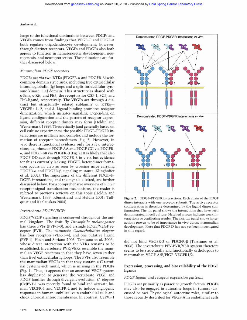
PDGFs act via two RTKs (PDGFR-� and PDGFR-�) withcommon domain structures, including five extracellularimmunoglobulin (Ig) loops and a split intracellular tyro-sine kinase (TK) domain. This structure is shared withc-Fms, c-Kit, and Flt3, the receptors for CSF-1, SCF, andFlt3-ligand, respectively. The VEGFs act through a dis-tinct but structurally related subfamily of RTKs—VEGFRs 1, 2, and 3. Ligand binding promotes receptordimerization, which initiates signaling. Depending onligand configuration and the pattern of receptor expres-sion, different receptor dimers may form (Heldin andWestermark 1999). Theoretically (and generally based oncell culture experiments), the possible PDGF–PDGFR in-teractions are multiple and complex and include the for-mation of receptor heterodimers (Fig. 2). However, invivo there is functional evidence only for a few interac-tions; i.e., those of PDGF-AA and PDGF-CC via PDGFR-�, and PDGF-BB via PDGFR-� (Fig. 2).It is likely that alsoPDGF-DD acts through PDGFR-� in vivo, but evidencefor this is currently lacking. PDGFR heterodimer forma-tion occurs in vivo as seen by crossing mice carryingPDGFR-� and PDGFR-� signaling mutants (Klinghofferet al. 2002). The importance of the different PDGF–P-DGFR interactions, and the signals elicited, are furtherdiscussed below. For a comprehensive overview of PDGFreceptor signal transduction mechanisms, the reader isreferred to previous reviews on this topic (Heldin andWestermark 1999; Rönnstrand and Heldin 2001; Tall-quist and Kazlauskas 2004).
Invertebrate PDGF/VEGFs
PDGF/VEGF signaling is conserved throughout the ani-mal kingdom. The fruit fly Drosophila melanogasterhas three PVFs (PVF-1–3), and a single PDGF/VEGF re-ceptor (PVR). The nematode Caenorhabditis eleganshas four receptors (VER-1–4), and one putative ligand(PVF-1) (Hoch and Soriano 2003; Tarsitano et al. 2006),whose direct interaction with the VERs remains to beestablished. Invertebrate PVR/VERs resemble the mam-malian VEGF receptors in that they have seven (ratherthan five) extracellular Ig loops. The PVFs also resemblethe mammalian VEGFs in that they contain a C-termi-nal cysteine-rich motif, which is missing in the PDGFs(Fig. 1). Thus, it appears that an ancestral VEGF systemhas duplicated to generate the vertebrate VEGF andPDGF families through divergent evolution. C. elegans(Ce)PVF-1 was recently found to bind and activate hu-man VEGFR-1 and VEGFR-2 and to induce angiogenicresponses in human umbilical vein endothelial cells andchick chorioallantoic membranes. In contrast, CePVF-1
did not bind VEGFR-3 or PDGFR-� (Tarsitano et al.2006). The invertebrate PFV-PVR/VER system thereforeseems to be structurally and functionally orthologous tomammalian VEGF-A/B/PlGF–VEGFR1/2.
Expression, processing, and bioavailability of the PDGFligands
PDGF ligand and receptor expression patterns
PDGFs act primarily as paracrine growth factors. PDGFsmay also be engaged in autocrine loops in tumors (dis-cussed below). Physiological autocrine roles, similar tothose recently described for VEGF-A in endothelial cells
Figure 2. PDGF–PDGFR interactions. Each chain of the PDGFdimer interacts with one receptor subunit. The active receptorconfiguration is therefore determined by the ligand dimer con-figuration. The top panel shows the interactions that have beendemonstrated in cell culture. Hatched arrows indicate weak in-teractions or conflicting results. The bottom panel shows inter-actions proven to be of importance in vivo during mammaliandevelopment. Note that PDGF-D has not yet been investigatedin this regard.
Andrae et al.
1278 GENES & DEVELOPMENT
Cold Spring Harbor Laboratory Press on March 20, 2020 - Published by genesdev.cshlp.orgDownloaded from

(Lee et al. 2007), have not been demonstrated for thePDGFs. PDGFs are generally produced by discrete popu-lations of cells and act locally to drive different cellularresponses (for review, see Hoch and Soriano 2003; Bet-sholtz 2004). Both PDGF and PDGFR expression pat-terns are spatio-temporally regulated in vivo during de-velopment and in certain physiological hypertrophic re-sponses. PDGF expression in cultured cells is dynamicand responsive to a variety of stimuli, including hypoxia,thrombin, cytokines, and growth factors, includingPDGF itself (for review, see Heldin and Westermark1999). Also, PDGFR expression is dynamic. Generalmesenchymal expression of PDGFRs is low in vivo, butincreases dramatically during inflammation and in cul-ture. Several factors induce PDGFR expression, includ-ing TGF-�, estrogen (probably linked to hypertrophicsmooth muscle responses in the pregnant uterus), inter-leukin-1� (IL-1�), basic fibroblast growth factor-2 (FGF-2), tumor necrosis factor-�, and lipopolysaccharide (Hel-din and Westermark 1999 and references therein).
The detailed expression patterns of the individualPDGF ligands and receptors are complex and have beenreviewed elsewhere (Heldin and Westermark 1999; Hochand Soriano 2003). There are some general patterns, how-ever: PDGF-B is mainly expressed in vascular endotheli-al cells, megakaryocytes, and neurons. PDGF-A andPDGF-C are expressed in epithelial cells, muscle, andneuronal progenitors. PDGF-D expression is less wellcharacterized, but it has been observed in fibroblasts andSMCs at certain locations (possibly suggesting autocrinefunctions via PDGFR-�). PDGFR-� is expressed in mes-enchymal cells. Particularly strong expression ofPDGFR-� has been noticed in subtypes of mesenchymalprogenitors in lung, skin, and intestine and in oligoden-drocyte progenitors (OPs) (discussed further below).PDGFR-� is expressed in mesenchyme, particularly invascular SMCs (vSMCs) and pericytes.
The mammalian PDGF and PDGFR genes are locatedon different chromosomes, and their transcriptionalregulation seems largely independent. It remains to beestablished if some of the overlapping expression pat-terns for PDGF-A and PDGF-C result from commontranscription regulatory mechanisms. The transcrip-tional regulation of the PDGF-A and PDGF-B genes hasbeen extensively studied and is reviewed elsewhere (Hel-din and Westermark 1999; Kaetzel 2003). Little is stillknown about the transcriptional regulation of PDGF-Cand PDGF-D and the PDGFRs.
PDGF biosynthesis and secretion
PDGF biosynthesis and processing are controlled at mul-tiple levels and differ for the different PDGFs. There iscurrently no evidence for regulated secretion of thePDGFs, which instead appears to be constitutively re-leased (Fruttiger et al. 2000). PDGF-A and PDGF-B be-come disulfide-linked into dimers already as propep-tides. PDGF-C and PDGF-D have been less studied inthis regard. PDGF-A and PDGF-B contain N-terminalpro-domains that are removed intracellularly by furin or
related proprotein convertases (for review, see Fredriks-son et al. 2004a). N-terminal processing is necessary forPDGF-A to acquire receptor-binding ability (for review,see Heldin and Westermark 1999; Fredriksson et al.2004a). Likely, PDGF-B also requires N-terminal propep-tide removal to become active.
In contrast, PDGF-C and PDGF-D are not processedintracellularly but are instead secreted as latent (condi-tionally inactive) ligands (for review, see Fredriksson etal. 2004a; Reigstad et al. 2005). Activation in the extra-cellular space requires dissociation of the growth factordomain from the CUB domain (Fig. 1). Plasmin and tis-sue plasminogen activator (tPA) have been demonstratedto proteolytically remove the CUB domain in PDGF-C,rendering it biologically active (Fredriksson et al. 2004b).Although the endogenous protease(s) responsible forPDGF-C activation in vivo remains to be identified, tPAendogenously expressed in cultured fibroblasts activatesPDGF-CC expressed by the same cells. Plasmin cancleave and activate also PDGF-D, but tPA cannot (Fred-riksson et al. 2004b). TPA needs to interact with both theCUB domain and the core domain in order for cleavageand activation of PDGF-C to occur, which likely ex-plains this specificity.
Extracellular retention and distribution of PDGFs
Spatially uneven distribution (gradients and depots) ofgrowth factors, cytokines, and morphogens defines theirbiological activity and action range. Diffusion of PDGFin the tissue interstitium is regulated by binding to ex-tracellular matrix components (Fig. 3). For PDGF-A andPDGF-B, such binding is accomplished in part by thepositively charged C-terminal motifs (referred to as re-tention motifs) containing a high proportion of basicamino acid residues (Fig. 1). PDGF-C and PDGF-D lackbasic retention motifs, but CUB domains are implicatedin protein–protein and protein–carbohydrate interac-tions in other contexts and may regulate extracellulardistribution of latent PDGF-C and PDGF-D. The pres-ence of the retention motif is determined by alternativesplicing in PDGF-A and by alternative C-terminal pro-teolytic processing in PDGF-B (Fig. 3). Alternative splic-ing has also been demonstrated for several of the mem-bers of the VEGF family, leading to the formation ofmultiple isoforms that differ in extracellular matrixbinding (Fig. 1; for review, see Ferrara et al. 2003). Alter-native splicing of the PDGF-A transcript is cell type-specific and differs both among tumor cell lines (Afra-khte et al. 1996) and in different organs during develop-ment (J. Andrae, H. Bostrom, and C. Betscholtz, unpubl.).
C-terminal proteolytic processing of PDGF-B may takeplace intracellularly or extracellularly. The endogenousprotease(s) responsible for C-terminal cleavage of PDGF-Bhas not been identified, but thrombin is a putative can-didate for extracellular proteolysis (Kelly et al. 1993).Certain cells transfected with PDGF-B expression vec-tors secrete soluble PDGF-BB into the conditioned me-dium. However, a major part of endogenously expressedPDGF-B becomes trapped on the cell surface or in the
Platelet-derived growth factor
GENES & DEVELOPMENT 1279
Cold Spring Harbor Laboratory Press on March 20, 2020 - Published by genesdev.cshlp.orgDownloaded from

extracellular matrix, where it subsequently can be re-leased by thrombin (Kelly et al. 1993; Soyombo and Di-Corleto 1994).
Insights into the role of PDGF retention have comefrom studies of PDGF interaction with heparan sulfateproteoglycans (HSPGs) and phenotypic analysis ofPDGF-B retention motif knockout mice. PDGFs bind toheparin and HSPGs similar to many other growth factorsand morphogens with critical functions during develop-ment (e.g., hedgehogs, bone morphogenetic protieins[BMPs], and Wnts) (Feyzi et al. 1997; Lustig et al. 1999;Lin 2004; Häcker et al. 2005; Abramsson et al. 2007).
Targeted deletion of the PDGF-B retention motif in miceleads to pericyte detachment from the microvessel wall(Abramsson et al. 2003; Lindblom et al. 2003). Reducedheparan sulfate (HS) N-sulfation (caused by lack of theenzyme N-deacetylase/N-sulfotransferase-1) similarlyleads to pericyte detachment and delayed pericyte mi-gration in vivo (Abramsson et al. 2007). This is probablycaused by attenuated PDGF-BB binding to HS. PDGF-BB/HS interaction appears to depend on overall N- and O-sulfation of HS, whereas saccharide fine structure ap-pears to be of lesser importance. Taken together, avail-able evidence suggests a model in which PDGF-BBsecreted from endothelial cells interacts with HS at theendothelial surface or in the periendothelial matrix. Thiswould lead to local deposits of PDGF-BB, which, in turn,are critical for the correct investment of pericytes in thevessel wall. HS binding is also necessary for proper lo-calization and function of VEGF-A (Ruhrberg et al. 2002).It was shown recently that HS may act in trans—i.e., frompericytes—to potentiate VEGF-receptor function in endo-thelial cells (Jakobsson et al. 2006). Similarly, HS expressedon endothelial cells may function to enhance PDGF-BB-mediated PDGFR-� signaling in neighboring pericytes.
PDGF binds also to certain non-HSPG extracellularproteins, but the physiological relevance of these inter-actions is unclear. Binding of PDGF-B has been demon-strated to �-2-macroglobulin (Bonner and Osornio-Var-gas 1995), possibly acting as a scavenger for PDGF-Bthrough low-density lipoprotein (LDL) receptor-relatedprotein (LRP) receptors on macrophages and other cells(Bonner et al. 1995). PDGF-B also binds to SPARC andadiponectin, which may trap the growth factor in theextracellular space (Raines et al. 1992; Arita et al. 2002).
PDGF receptor signaling transduction and downstreamevents
Dimerization is the key event in PDGF receptor activa-tion as it allows for receptor autophosphorylation on ty-rosine residues in the intracellular domain (Kelly et al.1991). Autophosphorylation activates the receptor ki-nase and provides docking sites for downstream signal-ing molecules (Kazlauskas and Cooper 1989). Docking ofreceptor substrates and further signal propagation in-volves protein–protein interactions through specific do-mains; e.g., Src homology 2 (SH2) and phosphotyrosine-binding (PTB) domains recognizing phosphorylated ty-rosines, SH3 domains recognizing proline-rich regions,pleckstrin homology (PH) domains recognizing mem-brane phospholipids, and PDZ domains recognizing C-terminal-specific sequences (for review, see Heldin et al.1998). Most of the PDGFR effectors bind to specific siteson the phosphorylated receptors through their SH2 do-mains.
PDGFR-induced signaling pathways
Both PDGFR-� and PDGFR-� engage several well-char-acterized signaling pathways—e.g. Ras-MAPK, PI3K, andPLC-�—which are known to be involved in multiple cel-lular and developmental responses (Fig. 4). PDGFRs con-
Figure 3. Processing and extracellular retention of the PDGFs.(Top panel) Through alternative splicing, PDGF-A may be trans-lated into a protein with or without a retention motif (green).Both isoforms may bind and activate PDGFR-� (active). Het-erodimers between the long and short forms of PDGF-A are notillustrated but can likely form since both splice isoforms areproduced by the same cells in most situations. (Middle panel)PDGF-B is produced as a single precursor containing the reten-tion motif. This protein may be secreted, in which case it getstrapped at the external side of the cell membrane or in pericel-lular matrix such as the basement membrane, where it is activeand participates in pericyte recruitment. In platelets, PDGF-B isprocessed intracellularly into a soluble and active isoform lack-ing the retention motif. There is experimental evidence for traf-ficking of a proportion of synthesized PDGF-B toward degrada-tion without prior secretion. (Bottom panel) PDGF-C andPDGF-D are produced and secreted as inactive growth factorscontaining a CUB domain (yellow). The CUB domain may helpin localizing these PDGFs in the extracellular space. ActivePDGF-C and PDGF-D are produced through extracellular pro-teolysis.
Andrae et al.
1280 GENES & DEVELOPMENT
Cold Spring Harbor Laboratory Press on March 20, 2020 - Published by genesdev.cshlp.orgDownloaded from

nect to Ras-MAPK mainly through the adaptor proteinsGrb2 and Shc. Grb2 binds the activated PDGFR throughits SH2 domain and complexes Sos1 through its SH3domains. Sos1 in turn activates Ras, leading to down-stream activation of Raf-1 and the MAPK cascade.MAPK signaling activates gene transcription, leading tostimulation of cell growth, differentiation, and migra-tion (for review, see Bar-Sagi and Feramisco 1986; Segerand Krebs 1995).
PI3K is a family of enzymes phosphorylating phos-phoinositides. Effectors of PI3K signaling include serine/threonine kinases such as Akt/PKB (Burgering and Coffer1995; Franke et al. 1995), some members of the PKCfamily including atypical isoforms (Nakanishi et al.1993; Akimoto et al. 1996), p70 S6 kinase (Chung et al.1994), JNK (Lopez-Ilasaca et al. 1997), and small GTPasesof the Rho family (Hawkins et al. 1995). Activation ofthe PI3K pathway by PDGFRs promotes actin reorgani-zation, directed cell movements, stimulation of cellgrowth, and inhibition of apoptosis (Hu et al. 1995).
PLC-� binds PDGFRs, which results in its activationthrough phosphorylation (for review, see Tallquist andKazlauskas 2004). PLC-� activation leads to mobiliza-tion of intracellular calcium ions and the activation ofPKC (Berridge 1993). The effects of PDGFR-mediatedPLC-� activation include stimulation of cell growth andmotility (Kundra et al. 1994).
Several additional signaling molecules are engaged byPDGFRs, including enzymes, adaptors, and transcriptionfactors. Activation of the Src TK promotes Myc tran-scription and mitogenic responses (for review, see Erpeland Courtneidge 1995). Also, members of the Fer and FesTK family bind to PDGFRs (Kim and Wong 1995). PKC-�is phosphorylated by PDGFR-�, leading to its activationand translocation to the cell membrane (Li et al. 1996).This signal may be involved in cell differentiation. The
adaptors Nck and Crk bind to PDGFRs through theirSH2 domain and are involved in activation of JNK (Nish-imura et al. 1993; Su et al. 1997). The adaptor Grb7 con-tains a SH2 domain and binds PDGFR-� (Yokote et al.1996). STAT transcription factors may bind to PDGF re-ceptors, leading to their phosphorylation and activation(Darnell 1997).
PDGF receptors interact also with integrins, which en-hances cell proliferation, migration, and survival (for re-view, see Assoian 1997; Frisch and Ruoslahti 1997).PDGFR interaction with integrins helps localizingPDGFRs and interacting molecules at focal adhesions,which are sites where several signaling pathways initiateand cross-talk (Clark and Brugge 1995). Recently, Na+/H+ exchanger regulatory factors (NHERFs) were shownto bind PDGFR-� and link it with focal adherence kinaseand the cortical actin cytoskeleton (James et al. 2004), aswell as to N-cadherin (Theisen et al. 2007) and the phos-phatase PTEN (Fig. 4; Takahashi et al. 2006). Additionalevidence for compartmentalization of PDGFRs and theirdownstream signals within cells comes from the intrigu-ing observation that PDGFR-� (but not PDGFR-�) local-izes to the primary cilium of fibroblasts (Schneider et al.2005). Mutants that fail to form cilia do not activatePDGFR-� but maintain PDGFR-� signaling. Primary ciliaare also essential for hedgehog signaling (Eggenschwilerand Anderson 2007). Hedgehog receptors (patched) andPDGFR-� show a conspicuous overlap in their expressionpattern during development (Karlsson et al. 1999, 2000),and it is thus possible that this correlation extends tosignaling at the subcellular level as well.
Negative control of PDGFR signaling
PDGF signaling is carefully regulated by feedback con-trol mechanisms. Stimulatory and inhibitory signals
Figure 4. PDGFR signal transduction and links to the cytoskeleton. (A) The intracellular domains of PDGFR-� and PDGFR-� andsome of their direct interactors are illustrated. Arrows imply links to major signal transduction pathways and secondary effectors.Negative feedback signaling is indicated in red. (B) Schematic illustration of how PDGFR-� may link to the cytoskeleton and to othersignaling components of focal adhesions through the adapter NHERF, the merlin and ezrin/radixin/moezin family of cytoskeletallinkers (MERM), and focal adhesion kinase (FAK).
Platelet-derived growth factor
GENES & DEVELOPMENT 1281
Cold Spring Harbor Laboratory Press on March 20, 2020 - Published by genesdev.cshlp.orgDownloaded from
arise in parallel, and the ultimate response depends onthe balance between these signals (for review, see Heldinet al. 1998). The SHP-2 tyrosine phosphatase bindsPDGFR through its SH2 domain and dephosphorylatesthe receptor and its substrates (Lechleider et al. 1993).Ras-GAP, which negatively regulates Ras, also bindsPDGFR-� through its SH2 domain (Fantl et al. 1992).
Ligand occupancy of PDGFRs promotes endocytoticreceptor internalization. The major destiny of internal-ized PDGFRs seems to be lysosomal degradation, therebylimiting the duration of PDGFR signaling (Heldin et al.1982; Sorkin et al. 1991; Mori et al. 1995). Recycling ofPDGFR-�, but not PDGFR-�, was recently observed incells deficient for the phosphatase TC-PTP (Karlsson etal. 2006), which is a negative regulator of PDGFR-� phos-phorylation (Persson et al. 2004). Trafficking toward ly-sosomal degradation of PDGFR-� depends on interac-tions with c-Cbl and receptor ubiquitination. Theadapter protein Alix, which interacts with the C-termi-nal domain of PDGFR-�, facilitates ubiquitination anddegradation of c-Cbl, thereby inhibiting PDGFR-� down-regulation (Lennartsson et al. 2006).
Cellular responses to PDGFR signaling
Some of the cellular responses to PDGFs take placewithin seconds to minutes after PDGFR activation andare independent of gene expression and protein synthe-sis. PDGFR-� and PDGFR-� mediate similar but notidentical rapid responses. Both receptors stimulate rear-rangement of actin filaments, but only PDGFR-� pro-motes formation of circular ruffles. PDGFR-� also mo-bilizes calcium ions more efficiently than PDGFR-�(Diliberto et al. 1992; Fatatis and Miller 1997). PDGFR-�inhibits gap junctional communication between cellsthrough phosphorylation of the gap junction protein con-nexin 43 (Hossain et al. 1998). It is unclear whether thisability is shared with PDGFR-�.
In addition to the rapid post-transcriptional responses,PDGFRs (like other RTKs) induce fast transcriptionalchanges involving so-called immediate early genes(IEGs) (Cochran et al. 1983). IEGs are direct targets of thetranscription factors that get activated (by post-transla-tional modification) through various signaling pathways.The IEG responses are likely necessary for many of thelong-term effects of PDGFs in vitro and in vivo. To whatextent the IEG responses contribute specificity toPDGFR signaling is, however, unclear. Different RTKsinduce virtually identical sets of IEGs (Fambrough et al.1999). Also, different signaling pathways activated bythe same receptor (PDGFR-�) induce largely overlappingsets of IEGs in vitro (Fambrough et al. 1999). From thesestudies, it would appear that quantitative rather thanqualitative differences in the IEG responses mediate thespecific responses to different RTKs and signaling path-ways. This view is supported by in vivo analysis of anallelic series of pdgfrb tyrosine-to-phenylalanine muta-tions disrupting connection to different substrates andsignaling pathways (Tallquist et al. 2003). Mutations insingle or multiple tyrosine residues led to quantitatively
different but qualitatively similar developmental effects(Tallquist et al. 2003). In contrast, similar analyses ofPDGFR-� revealed strikingly different roles of the differ-ent downstream signaling pathways. For PDGFR-�, thedisruption of signaling through PI3K alone has no, oronly minor, developmental consequences (Heuchel et al.1999; Tallquist et al. 2000a). In contrast, PI3K is indis-pensable for PDGFR-� function during development(Klinghoffer et al. 2002). Intriguingly (and in further con-trast to PDGFR-�), deficient coupling to Src fromPDGFR-� resulted in specific problems with the oligo-dendrocyte lineage, whereas other processes, such asskeletal development, remained normal.
The different IEGs appear to cooperate in their regula-tion of downstream cellular and developmental events.By analyzing gene trap mutants for >20 IEGs down-stream from PDGFR signaling, a striking degree of phe-notypic overlap and genetic interaction was revealed.Different mutated IEGs produced qualitatively similarresponses. Moreover, the combination of mutations inseveral genes strengthened specific phenotypic out-comes known to depend on PDGFR signaling, such ascraniofacial, cardiovascular, or kidney developmentalprocesses (Schmahl et al. 2007). The large overlap be-tween the signaling pathways, IEGs, and biological pro-cesses argue in favor of a model in which specificity isgenerated through a combination of quantitative differ-ences in magnitude and duration of the responses occur-ring at different levels in the signaling cascade. One shallnot forget, however, that a major part of the specificity ofthe PDGFRs in developmental functions depends on celltype- and context-specific PDGFR expression. The ex-pression patterns of the PDGFRs correlate with the ma-jor sites of their functions; e.g., PDGFR-� is strongly ex-pressed in OPs and PDGFR-� in pericyte progenitors.Lack of redundancy is therefore dependent in part on thedifferent transcriptional regulation of the two pdgfrgenes. By knocking in a PDGFR-� intracellular domaininto the pdgfra gene, phenotypically normal animalswere obtained, showing that PDGFR-� signaling canfully substitute for PDGFR-� signaling if it is expressedat the right place and time (Klinghoffer et al. 2001). Thereverse replacement, however, indicates that PDGFR-�signaling can only partially compensate for the loss ofPDGFR-� signaling. Targeted replacements in PDGFR-�with other RTK signaling domains have also demon-strated partial rescue, at best (Klinghoffer et al. 2001;Hamilton et al. 2003). Taken together, these data suggestthat specificity of PDGFR signaling is achieved througha combination of cell type-specific expression and differ-ential engagement of downstream signaling pathways.
Principles of genetic and pharmacological PDGFand PDGFR inhibition
Several strategies to block PDGF signaling have beenapplied to evaluate the role of PDGF signaling in biologi-cal and pathological processes. Targeted gene inactiva-tion has been invaluable in the analysis of PDGF physi-ology in mice. Dominant-negative PDGFs or PDGFRs
Andrae et al.
1282 GENES & DEVELOPMENT
Cold Spring Harbor Laboratory Press on March 20, 2020 - Published by genesdev.cshlp.orgDownloaded from

have been used in mammalian cell culture experimentsand in Xenopus laevis. Since both PDGFs and PDGFRsrequire dimerization for functionality, any nonfunc-tional PDGF ligand or receptor retaining the ability todimerize is a potential dominant-negative mutant. Forexample, N-terminal processing-deficient PDGF ligands,or PDGF receptors with inactivated or truncated kinasedomains, act in this way. The PDGF/PDGFR complex isalso amenable to pharmacological inhibition in severaldifferent ways (Fig. 5). Neutralizing antibodies exist forPDGF ligands and receptors and have been used exten-sively in experiments evaluating the importance ofPDGF signaling in pathogenic processes. Aptamers (sta-bilized oligonucleotides that bind proteins with veryhigh specificity) have been developed for the inhibitionof PDGF-B and have been used to test the pathogenic roleof this protein in various rodent disease models.
One of the most efficient ways to block PDGFR sig-naling is to inhibit the PDGFR kinase activity. Kinaseinhibitors act by binding at or near the ATP-bindingpocket of the kinase domain. Several kinase inhibitorshave been developed that block PDGFRs, but the inhibi-tors available so far are not completely specific. One ofthem, imatinib mesylate (imatinib, STI571, or Gleevec),inhibits PDGFR-� and PDGFR-�, the bcr–abl fusion pro-tein (toward which it was developed) (Carroll et al. 1997),c-kit, and Flt3. Imatinib has been approved by the USFood and Drug Administration for treatment of patientswith Philadelphia chromosome-positive chronic mye-logenous leukemia (carrying the bcl–abl fusion protein)(Druker et al. 2001) and gastrointestinal stromal tumors(GIST) carrying activating mutations in c-kit orPDGFR-� (Demetri et al. 2002). Imatinib has also dem-onstrated clinical efficacy against myeloproliferative dis-eases involving rearrangements of the PDGFRB gene(Apperley et al. 2002) and dermatofibrosarcoma protu-berans (DP), a tumor carrying translocated and activatedPDGF-B genes (McArthur et al. 2005). Several otherPDGFR kinase inhibitors have been developed (for re-view, see Levitzki 2004) and tested in clinical trials(Homsi and Daud 2007). All PDGFR kinase inhibitorsused so far also block c-Kit and Flt3, two of the other
three receptors with similar domain organization asPDGFRs. This specificity problem must be kept in mindwhen kinase inhibitors are used in order to evaluate therole of PDGF signaling in (patho)physiological processes.Imatinib nevertheless shows promise as a drug for anumber of clinical conditions in which PDGFR signalingis activated (discussed in subsequent sections). The sideeffects of imatinib also appear to be minor from a generalperspective as well as in comparison with other PDGFRinhibitors tested so far (for review, see Homsi and Daud2007).
Currently, other strategies for small-molecule-medi-ated inhibition of PDGFR signaling are scarce. A recentstudy identified a nuclease-hypersensitive element inthe PDGF-A promoter as a putative target for small mol-ecule pharmacologicals, however. The guanine-richstrand of the DNA in this region forms a dynamic G-quadruplex, which upon binding of the small moleculeTMPyP4 becomes stabilized, silencing the PDGF-A pro-moter (Qin et al. 2007). How specific this inhibition isand whether this can be used to shut down PDGF-Aexpression in vivo remain to be established.
Developmental functions of PDGFs/PDGFRs
Developmental roles of PDGFs and PDGFRs have beenunraveled mainly by way of gene-targeted mice. A largenumber of knockout, knock-in, or transgenic mutantshave been used to identify specific cell types that areprimary targets of PDGF signaling during development.The importance of these findings lies not only in theinformation achieved about the in vivo roles of PDGFs,but also in the new knowledge gained about some of thecell types regulated by PDGFs. For example, the criticalroles of microvascular pericytes (Lindahl et al. 1997a),lung alveolar SMCs (Boström et al. 1996), and gastroin-testinal villus cluster cells (Karlsson et al. 2000) were allpinpointed for the first time through the analysis ofPDGF-B and PDGF-A knockout mice, respectively. Im-portant developmental roles for PDGF/PDGFR-like pro-teins have also been demonstrated in nonmammalianvertebrates. As discussed below, some of these appear to
Figure 5. Principles of pharmacological inhibition ofPDGF signaling. PDGFs can be blocked by neutralizingantibodies, recombinant dimeric soluble PDGFR extra-cellular domains, or nucleic acids (aptamers), which allbind to the PDGFs with high affinity. PDGFRs can beblocked by antibodies or dominant-negative ligands.PDGFR function may be blocked by kinase inhibitors.The possibility of silencing the PDGF-A promoter byhelp of small-molecule pharmacologicals has emergedrecently.
Platelet-derived growth factor
GENES & DEVELOPMENT 1283
Cold Spring Harbor Laboratory Press on March 20, 2020 - Published by genesdev.cshlp.orgDownloaded from

be analogous to the roles of the mammalian PDGFs andVEGFs.
Role of PDGF signaling in gastrulation and formationof cranial and cardiac neural crest
Several observations suggest that PDGF-A and PDGFR-�have early developmental functions. Xenopus embryosinjected with dominant-negative PDGF receptor mRNAshow aberrant gastrulation, in which the involuting pro-spective head mesodermal cells fail to migrate beneaththe overlying PDGF-A-positive blastocoel roof ectoderm(Ataliotis et al. 1995). Upon PDGFR inhibition, the me-sodermal cells detach from the ectoderm and undergoapoptosis. Rescue from apoptosis did not rescue the mi-gration defect, suggesting that PDGFR(s) independentlycontrols both processes (Van Stry et al. 2004). In contrastto these effects, inhibition of PDGFR signaling did notaffect the migration and survival of axial mesoderm inXenopus. Axial mesoderm develops through a differentmorphogenetic process (mediolateral intercalation) com-pared with head mesoderm and at a distance from (andtherefore probably independent of) the PDGF-A-express-ing ectoderm (Van Stry et al. 2004).
A role for PDGF signaling during gastrulation is alsosuggested from experiments in sea urchins. PDGFR ex-pression occurs in the developing mesoderm, and differ-ent strategies to inhibit PDGF or PDGFR function resultin gastrulation defects (Ramachandran et al. 1995, 1997).A role for PDGF signaling in the mesoderm during ze-brafish gastrulation has also been proposed (Montero andHeisenberg 2004). The PDGFR-specific kinase inhibitorAG1296 inhibited formation of mesendodermal cell pro-trusions and cell polarization in vivo in fish embryos. Invitro, PDGF-A stimulated these activities (Montero andHeisenberg 2004). Taken together, data from frog, seaurchin, and fish suggest an evolutionary conserved rolefor PDGF signaling during gastrulation in deutero-stomes.
In mouse embryo development, PDGF-A expressiononsets early and can be detected already in the preim-plantation embryo (Rappolee et al. 1988; Mercola et al.1990; Palmieri et al. 1992). At this stage, PDGF-A is co-expressed with PDGFR-� in the blastocyst inner cellmass. In spite of these early expression patterns, a rolefor PDGFs during mouse gastrulation is not apparent(Hoch and Soriano 2003). PDGF-A and PDGFR-�-nullmouse embryos nevertheless show severe impairment ofearly mesenchymal derivatives in both embryo and ex-traembryonic tissues (Hamilton et al. 2003). A propor-tion of these embryos die before or at embryonic day 10.5(E10.5), which is, however, variable and genetic back-ground-dependent (Hoch and Soriano 2003).
Although early developmental routes may differ be-tween vertebrate species, some of the later consequencesof gastrulation defects in Xenopus resemble the pheno-types observed in certain PDGF/R mutant mice. PDGFRsignaling-inhibited Xenopus embryos develop reducedanterior structures and defective vertebral arch closure(spina bifida). Similar defects are observed in mouse
PDGFR-� knockouts and PDGF-A/C double knockoutsdue to problems with the developing cranial neural crestand axial skeleton. Neural crest mesenchyme expressesPDGFR-� strongly in mouse (Morrison-Graham et al.1992), zebrafish (Liu et al. 2002a), and Xenopus (Hoet al. 1994). Complete or neural crest-specific PDGFR-�knockout in mice leads to severe developmental defectsin neural crest mesenchyme derivatives, such as the car-diac outflow tract, the thymus, and the skeletal compo-nents of the facial region (Morrison-Graham et al. 1992;Soriano 1997; Tallquist and Soriano 2003). Both PDGF-Aand PDGF-C are expressed at locations toward whichneural crest cells migrate, such as the branchial archepithelium (Orr-Urtreger and Lonai 1992; Ding et al.2000; Aase et al. 2002; Liu et al. 2002b). For cardiac neu-ral crest development, roles for both PDGFR-� andPDGFR-� have been demonstrated (Richarte et al. 2007).PDGFR-� is important mainly for nonneuronal cardiacneural crest development (Morrison-Graham et al. 1992).PDGFR-� and PDGF-B appear to play a role in neuronalcardiac neural crest development, as both PDGFR-� andPDGF-B knockouts display abnormal cardiac innerva-tion (Van den Akker et al. 2008). Thus, PDGFR-� andPDGFR-� signaling appear to have complementary rolesin cardiac neural crest development.
Directed cell migration—a conserved morphogeneticfunction of PDGFs
In addition to the mentioned anatomical similarities be-tween the phenotypes resulting from PDGFR inhibitionin Xenopus and PDGFR-� ablation in mice, the cellularprocesses that depend on PDGFR-� signaling as well asthe critical signaling pathways engaged appear to besimilar (Soriano 1997; Van Stry et al. 2004). For example,signaling through PI3K downstream from PDGFR-� ap-pears to be critical during early embryonic developmentin mouse (Klinghoffer et al. 2002), Xenopus (Symes andMercola 1996), and zebrafish (Montero et al. 2003).
Cell migration is a well-documented effect of PDGFstimulation in vitro (Heldin and Westermark 1999), butour understanding of PDGF-dependent cell movementsin vivo is limited. PDGF signaling is undoubtedly re-quired for the spreading of various populations of cells indevelopmental processes. This includes spreading of oli-godendrocyte precursors in the spinal cord, of neuralcrest mesenchymal cells toward the branchial pouchesand cardiac outflow tract, and of pericytes along newlyformed angiogenic sprouts. The mechanisms by whichthe cells spread, and how PDGF regulates this process,remain obscure for most of the situations, however. Re-cent studies in Drosophila, Xenopus, and mouse havedemonstrated a role for PDGF/VEGF family members indirected cell migration in at least some developmentalprocesses in vivo.
The mechanism of PDGF-A/PDGFR-�-mediated me-sodermal cell migration has been analyzed in some detailin Xenopus (Nagel et al. 2004). Using an in vitro explantmodel and a range of genetic tools, it was demonstratedthat the blastocoel roof establishes a matrix-bound gra-
Andrae et al.
1284 GENES & DEVELOPMENT
Cold Spring Harbor Laboratory Press on March 20, 2020 - Published by genesdev.cshlp.orgDownloaded from

dient of PDGF-A (long splice version) along which thePDGFR-�-positive mesodermal cells migrate direction-ally (Fig. 6). When graded signaling was disrupted experi-mentally, directional migration by mesodermal cells to-ward the animal pole was replaced by random migration(Nagel et al. 2004). This role for PDGF-A during Xenopusgastrulation resembles that of Drosophila PVF1 in theguidance of border cell migration in the egg chamber(Duchek et al. 2001). Also, there signaling is paracrine,with PVF1 expressed in the oocyte and PVR in the bordercells. The border cells move as an aggregate from whicha single cell takes a lead position by extending a longcellular protrusion in the direction of the migration ofthe cell aggregate. The directionality of this process de-pends on spatially graded signaling exerted by PVF1(Fulga and Rørth 2002) in concert with the DrosophilaEGFR ligands Keren and Spitz (McDonald et al. 2006).Specific misexpression of PVF1 misdirects the bordercells to new locations (McDonald et al. 2003). However,uniform misexpression of PVF1 abolishes formation ofthe cellular protrusion and disrupts directionality of themigration, similar to the effect of uniform PDGF-A mis-expression in the Xenopus blastocoel roof. These twoexamples of PDGF/PVF-induced cell migration are
analogous to the role of VEGF-A in endothelial sproutguidance during angiogenesis. There, graded distributionof VEGF-A directs filopodial extensions and migratorybehavior of endothelial tip cells, resulting in proper ori-entation of the angiogenic sprouts (Ruhrberg et al. 2002;Gerhardt et al. 2003). Thus, the ability to guide cell mi-gration through the formation of growth factor gradientsin the extracellular space appears to be common to sev-eral members of the PDGF/VEGF family, and conservedbetween vertebrates and invertebrates (Fig. 6).
PDGF signaling in organogenesis
During post-implantation development, PDGF-A expres-sion occurs in epithelia, nervous tissue, myotome, andvascular and visceral smooth muscle. PDGFR-�, in con-trast, is expressed by most mesenchymal cell popula-tions, and often in a reciprocal pattern compared withPDGF-A (for review, see Ataliotis and Mercola 1997;Hoch and Soriano 2003). A few sites of epithelialPDGFR-� expression have been noticed; e.g., lens epi-thelium (Reneker and Overbeek 1996), limb apical ecto-dermal ridge, and the early epithelial somite (Soriano1997).
Analysis of various gene-targeted and transgenic mod-els for PDGF-A and PDGFR-� demonstrates the impor-tance of PDGF-A as an epithelium-derived factor pro-moting proliferation and spreading of nearby locatedPDGFR-�-positive mesenchymal cells. Thus, PDGF-A/PDGFR-� is a common signaling pair in epithelial–mes-enchymal interaction, acting in concert with other sig-naling molecules such as hedgehogs, FGFs, BMPs, andWnts. The complex reciprocal signaling involved in tis-sue induction should be kept in mind, as some of thephenotypes in PDGF-A or PDGFR-� mutants may reflectsecondary consequences of the loss of mesenchymalcells that were primarily dependent on PDGF-A.
Lung and intestine—the formation of large specializedepithelial surfaces
Pdgfa−/− mice that survive birth develop a lung emphy-sema-like condition reflecting a complete failure of al-veolar septum formation (Boström et al. 1996). Alveo-genesis is a postnatal process in mouse, and it is drivenby alveolar SMCs, which are missing in PDGF-A knock-outs. These cells likely originate from clusters ofPDGFR-�-positive mesenchymal cells that form near theepithelial buds during the pseudoglandular stage of lungdevelopment. Subsequently, the cells detach from theclusters to spread into the walls of the alveolar sacs,where they participate in (or initiate the onset of) alveo-genesis (Fig. 7A). In PDGF-A knockouts, PDGFR-�-posi-tive mesenchymal cell clusters develop normally, butsubsequent spreading of the cells to the alveolar sacculesfails (Lindahl et al. 1997b; Boström et al. 2002). Thissuggests that PDGF-AA, expressed by the lung epithe-lium, acts in paracrine mode to regulate the proliferationand migration of the PDGFR-�-positive alveolar SMCprogenitors.
Figure 6. PDGFs, PVFs, and VEGFs direct cell migration indevelopmental processes. (Top panel) PDGF-A (long splice ver-sion) is expressed by the ectodermal cells of the blastocoel roofand gets deposited in a gradient that drives mesodermal cellmovements along the blastocoel roof. (Middle panel) PVF1 isexpressed by the oocyte and distributes in a graded fashion inthe Drosophila egg chamber. This guides migration of the bor-der cells toward the oocyte. (Bottom panel) Graded distributionof VEGF-A directs filopodial extensions and migration of theendothelial tip cells, thereby orienting the angiogenic sprouttoward the highest VEGF-A concentration.
Platelet-derived growth factor
GENES & DEVELOPMENT 1285
Cold Spring Harbor Laboratory Press on March 20, 2020 - Published by genesdev.cshlp.orgDownloaded from

Transgenic gain-of-function approaches have also beenused to study the role of PDGFs in the lung. Ectopictransgenic expression of PDGF-A, PDGF-B, and PDGF-Cin distal lung epithelium resulted in two different phe-notypes. Mice expressing PDGF-A or PDGF-C died peri-natally as a result of enlarged lungs with thickened mes-enchyme and poorly differentiated cells (Li and Hoyle
2001; Zhuo et al. 2006). This phenotype likely reflectsmassive overproliferation of PDGFR-�-positive alveolarSMC progenitors. PDGF-B expression provided a differ-ent lung histology, comprising enlarged airspaces, in-flammation, and fibrosis (Zhuo et al. 2006). This is some-what surprising since PDGF-B should be able to activatePDGFR-� and thereby produce the same severe lung phe-
Figure 7. Developmental roles of PDGFs. (A) During the saccular phase of lung development, PDGF-A secreted by the epithelium(yellow) drives spreading and proliferation of alveolar SMC progenitors (green). These cells differentiate into alveolar SMC and driveformation and maintain alveolar walls by producing deposits of elastin. (B) In intestinal development, the epithelium producesPDGF-A to drive proliferation of mesenchymal cells with a critical role in intestinal villus formation. (C) In the developing skin,PDGF-A secreted from keratinocytes promote proliferation of mesenchymal cells with functions in hair follicle morphogenesis. (D) Intesticular development, PDGF-A (and PDGF-C) secreted from the epithelial tubes drives expansion of mesenchymal cells that sub-sequently differentiate into testosterone-producing Leydig cells. The testosterone production is in turn necessary for further testiculardevelopment and spermatogenesis. (E) In the developing CNS (the example illustrates the cerebellum), PDGF-A drives proliferativeexpansion of OPs, which subsequently myelinate nerve fibers throughout the CNS. Lack of PDGF-A leads to a complete absence ofmyelin in peripheral parts of the CNS, such as the optic nerve. (F) PDGF-C (and PDGF-A) is critical for development of the palatalshelves. PDGF-C is produced in the epithelium and acts on mesenchymal cells (green) in the shelves. (G) PDGF-B produced byendothelial cells (orange) drives proliferation and spreading of vSMC and pericytes in conjunction with angiogenesis and arteriogenesis.(H) In developing kidney glomeruli, PDGF-B expressed by glomerular endothelium drives the proliferation of mesangial cells (green).
Andrae et al.
1286 GENES & DEVELOPMENT
Cold Spring Harbor Laboratory Press on March 20, 2020 - Published by genesdev.cshlp.orgDownloaded from
notype as the PDGF-A and PDGF-C transgenes. Thesedifferences may therefore reflect the different extracel-lular distribution and bioavailability of the differentPDGFs, as discussed previously.
PDGF-A knockout mice also develop abnormally fewand generally misshapen gastrointestinal villi. Villus for-mation is preceded by proliferation and clustering ofPDGFR-�-positive cells subjacent to the epithelium (Fig.7B). As the villus grows in length, the PDGFR-�-positivecells detach from the cluster and distribute as a singlemesenchymal cell layer lining the basement membrane.The exact role of these cells in intestinal villus forma-tion is not understood. However, PDGF-A/PDGFR-� sig-naling appears to secure renewal of this population ofcells during the consecutive waves of villus formationthat take place during intestinal development (Karlssonet al. 2000). Clustering of PDGFR-�-positive cells coin-cides with proliferative quiescence and onset of BMP2and BMP4 expression by these cells (Karlsson et al.2000). These BMPs likely signal to adjacent villus epi-thelial cells expressing BMPR1a. Transgenic expressionof the BMP inhibitor noggin in intestinal epithelium ledto ectopic formation of intestinal crypts (Haramis et al.2004), as well as to increased epithelial expression ofPDGF-A, and increased mesenchymal growth (Batts etal. 2006). This suggests that BMP expression by the vil-lus cluster serves to restrict de novo crypt formation andmesenchymal expansion. However, the role of BMPsduring gastrointestinal development is likely more com-plex. Transgenic models with early onset noggin expres-sion in the intestinal epithelium show a different phe-notype involving reduced villus numbers, resemblingthat in PDGF-A knockouts (Batts et al. 2006). Thus,BMPs may have different roles at different stages of gas-trointestinal development.
Endodermal derivatives such as lung and gut are clas-sical sites for inductive epithelial–mesenchymal interac-tions. The specific expression patterns for PDGF-A andPDGFR-� combined with the mouse knockout pheno-types suggest a general role for endodermal epithelium-derived PDGF-A to promote proliferation (and possiblyalso migration and differentiation) of specific popula-tions of mesenchymal cells that drive morphogenesis oflung alveolar septa and intestinal villi. Both these pro-cesses involve folding of the epithelial surface and theformation of a vascularized mesenchymal core of eachfold. Alveolar and intestinal surface areas are severelycompromised in pdgfa−/− mice as a result of hypoplasiaof the specialized PDGFR-�-positive mesenchymal cells.PDGF-A/PDGFR-� signaling is therefore instrumentalin the formation of large surfaces for specialized interac-tion and molecular exchange with the environment; i.e.,respiration and absorption (Fig. 7A,B).
Skin
PDGF-A and PDGFR-� have been implicated in develop-ment of the dermis, both in mice through genetic analy-ses (Betsholtz 2004) and in Xenopus using PDGFR-inhib-iting substances (the TK inhibitor AG1296 and soluble
PDGFR-� ectodomains) (Utoh et al. 2003). PDGF-A,PDGF-C, and PDGFR-� mouse knockouts all displaydermal defects. PDGFR-�-null mice show severe dermalmesenchymal hypoplasia and skin blistering (Soriano1997). A similar but milder phenotype is observed inPDGF-C knockouts (Ding et al. 2004). Postnatal PDGF-A-null mice show progressive loss of dermal mesen-chyme and reduced hair development (Karlsson et al.1999). Conversely, dermal injection of PDGF-AA orPDGF-BB in mice triggers resting hair follicles to enterthe hair growth cycle (Tomita et al. 2006). The role ofPDGF-A in the developing skin appears to be to securethe renewal of PDGFR-�-positive mesenchymal cellsthat are consumed as they are recruited to hair folliclesto become dermal papillae and dermal sheaths (Fig. 7C;Karlsson et al. 1999).
Testis
During embryonic testicular development, tubular epi-thelial cells express PDGF-A, whereas interstitial mes-enchymal cells express PDGFR-�. Postnatal pdgfa−/−
mice display reduced testicular size and show loss ofadult Leydig cells, reduced circulating testosterone, anddisrupted spermatogenesis (Gnessi et al. 2000). This phe-notype could be explained with the failure of develop-ment of the adult Leydig cells, the mayor testosterone-producing cells of the postnatal mammal (Fig. 7D;Gnessi et al. 2000). Studies of the PDGFR-�-null em-bryos revealed a more severe testicular phenotype in-volving a deficiency also in the fetal population of Leydigcells. This may be explained by an early overlappingfunction of PDGF-A and PDGF-C, both of which are ex-pressed in the fetal male gonad (Brennan et al. 2003). It isnot yet clear if lack of PDGF-A/C leads to Leydig celldeficiency entirely through proliferative arrest and pro-gressive depletion of Leydig cell precursors, or if PDGFRsignaling also controls Leydig cell differentiation.
Kidney
The interstitial kidney mesenchyme expresses PDGFR-�, and PDGFR-�-null mice show severe mesenchymalhypoplasia in the embryonic kidney. Formation of inter-stitial kidney mesenchyme seems to occur through thecombined activity of PDGF-A and PDGF-C, which areexpressed in different parts of the developing kidney (Liet al. 2000). Neither individual knockout for PDGF-A orPDGF-C showed any defect in the kidney mesenchyme,but the double PDGF-A/C knockout phenocopied thePDGFR-�-null mutant kidney phenotype (Li et al. 2000;Ding et al. 2004).
Lens
Autocrine PDGF-A–PDGFR-� expression occurs in lensepithelium and appears to play a role during lens devel-opment. Transgenic overexpression of PDGF-A in thelens leads to hyperproliferation and ectopic differentia-
Platelet-derived growth factor
GENES & DEVELOPMENT 1287
Cold Spring Harbor Laboratory Press on March 20, 2020 - Published by genesdev.cshlp.orgDownloaded from

tion of the lens epithelium into lens fiber cells (Renekerand Overbeek 1996). In vitro studies confirmed thatPDGF can induce proliferation in lens epithelium, butthat the differentiation into fiber cells required the pres-ence of FGF (Kok et al. 2002). PDGFR-� is also expressedin the lens, and PDGF-D was recently found to be ex-pressed in the iris and ciliary body leading to accumula-tion of PDGF-D protein in aqueous humor. In eye ante-rior segment organ culture, anti-PDGF-D specificallyand potently inhibited lens epithelial cells proliferation,suggesting that PDGF-D secreted from the iris/ciliarybody regulates lens epithelial proliferation (Ray et al.2005).
Role of PDGFs in glial cell developmentand neuroprotection
Oligodendrocytes are the myelin-forming glia cells of theCNS. PDGFR-�-positive OPs originate as bilateral rowsof cells along the spinal cord central canal around E12 inthe mouse (Pringle and Richardson 1993). PDGFR-� sig-naling is not required for the specification of OPs, butthe further proliferation and spreading of OPs in the CNSdepend on PDGF-A signaling through PDGFR-� (Fig. 7E;Calver et al. 1998; Fruttiger et al. 1999). Studies usingPDGFR-� signaling mutants implicate both Src and PI3Kas critical mediators of OP proliferation (Klinghoffer etal. 2002). Transgenic mice expressing different amountsof PDGF-A in the CNS have revealed that OPs prolifer-ate until the growth factor becomes limiting; i.e., whenOPs “consume” PDGF-A at a faster rate than it is pro-duced (van Heyningen et al. 2001). When available PDGFis limiting (or withdrawn experimentally), OPs differen-tiate into myelinating oligodendrocytes. PDGF-A drivesnot only the proliferation of OPs in the embryo, but italso determines the number of OPs in the adult brain(Woodruff et al. 2004). Increased OP densities in theadult do not lead to enhanced remyelination followingexperimental demyelination, suggesting that the num-ber of OPs is not limiting for myelin repair in adult mice.It is noteworthy that the normal number of OPs is alsonot limiting for myelination in wild-type mouse em-bryos. Instead, OPs are normally produced in excessnumbers. pdgfa+/− mice become normally myelinated inspite of half the normal number of OPs. Only when theOP densities fall very low, as in the complete absence ofPDGF-A, myelination deficiency becomes apparent(Fruttiger et al. 1999). Available genetic data suggest thatPDGF-A is a critical ligand for PDGFR-� in oligodendro-cyte development. PDGF-A is expressed by neurons andastrocytes throughout the CNS and is constitutively re-leased from neural cell bodies (Fruttiger et al. 2000).PDGF-C is also expressed in the developing and adultCNS (Aase et al. 2002), but it is not known if it has a rolefor OP development.
PDGF-A and PDGFR-� appear to regulate also astro-cyte recruitment in the developing retina. PDGF-A-nullmice show retinal astrocyte hypoplasia (Gerhardt et al.2003), and mice ectopically overexpressing PDGF-A in
the retina develop astrocytic hyperplasia (Fruttiger et al.1996; Gerhardt et al. 2003).
In vitro experiments have implicated PDGF signalingin neural development and function (Smits et al. 1991).The patterns of expression of PDGFR-� and PDGFR-� invarious populations of embryonic neuronal cells or pro-genitors and the expression of PDGF-B in postnatal neu-rons are consistent with such roles (for review, see Hel-din and Westermark 1999; Hoch and Soriano 2003). Neu-ron-specific ablation of PDGF-B was, however, notassociated with apparent developmental abnormalitiesin the CNS or with changes in the astroglial response tolocal injuries (Enge et al. 2003). Neuron-specific ablationof PDGFR-� also did not produce apparent developmen-tal defects. However, these mice showed exacerbatedcerebral damage following certain brain injury experi-ments; e.g., cryogenic injury and NMDA-induced exci-totoxicity (Ishii et al. 2006). Conversely, PDGF admin-istration to the CNS protected against NMDA-inducedinjury (Egawa-Tsuzuki et al. 2004). These studies suggestthat PDGFR-� signaling might exert a neuroprotectivefunction in the adult mouse. PDGF signaling, possiblythrough PDGFR-�, has also been implicated in neuro-pathic pain following nerve injury (Narita et al. 2005).
Preliminary evidence suggests that invertebrate PVFsplay a role in glial development in both C. elegans andDrosophila. C. elegans VERs are expressed by CNS cellsanalogous to glia (Popovici et al. 2002), and the Dro-sophila PVR is expressed by ventral midline glia (Cho etal. 2002). The possible developmental roles of inverte-brate PVRs remain to be elucidated, but the expressionpatterns of VERs/PVR and PVFs in the invertebrate CNSmay suggest an evolutionarily conserved role for PDGFfamily members in neuronal/glia development. Since thePVFs are more similar to VEGFs than to PDGFs, it isworth pointing out that also the mammalian VEGFshave been implicated as neurogenic and/or neuroprotec-tive molecules (for review, see Storkebaum and Carme-liet 2004; Greenberg and Jin 2005). Targeted mutagenesisof the VEGF-A promoter region results in motoneurondeath and the development of amyotrophic lateral scle-rosis-like disease (Oosthuyse et al. 2001). Recently,VEGF-C signaling through VEGFR-3 was shown to playa role in oligodendrocyte development (Le Bras et al.2006). Thus, an ancient role of a common PDGF/VEGFancestor in neural development may have evolved intoroles for both PDGFs and VEGFs in oligodendrocyte de-velopment and neuroprotection.
Development of the axial skeleton, palate, and teeth
PDGFR-� is expressed throughout the early (epithelial)somites but becomes progressively localized to thesclerotome and dermatome as differentiation of the so-mite proceeds (Soriano 1997). PDGF-A and PDGF-C areboth expressed in the myotome (Orr-Urtreger and Lonai1992; Ding et al. 2000; Aase et al. 2002). PDGFR-�-nullembryos show abnormally shaped and sometimes fusedsomites, defects that correlate with later occurring ab-normalities in ribs and vertebrae (Soriano 1997; Tallquist
Andrae et al.
1288 GENES & DEVELOPMENT
Cold Spring Harbor Laboratory Press on March 20, 2020 - Published by genesdev.cshlp.orgDownloaded from

et al. 2000b). The expression patterns of PDGF-A/C andPDGFR-� suggest paracrine interactions between myo-tome, sclerotome, and dermatome, but the exact role ofthese signals in somite formation and differentiation isunclear. A function in chondrogenesis has been sug-gested based on experiments with mouse and chick mi-cromass explant cultures (Ataliotis 2000; Tallquist et al.2000b).
PDGFR-�-null or PI3K signaling mutants developskeletal abnormalities, including defective vertebralneural arch formation, which results in spina bifida (So-riano 1997; Klinghoffer et al. 2002). PDGF-C appears tohave a nonredundant role in vertebral development,since its ablation leads to spina bifida, although this de-fect is milder than that observed in PDGFR-� knockouts(Ding et al. 2004). Double knockout of PDGF-A andPDGF-C leads to severe spina bifida similar to the abla-tion of PDGFR-�. This suggests a role also for PDGF-A invertebral development, but this role appears partially re-dundant with PDGF-C since PDGF-A-null mice havenormal vertebrae (H. Boström and C. Betsholtz, 1996;unpubl.).
PDGF-C–PDGFR-� signaling plays a critical role inthe formation of the palate (Fig. 7F). PDGFR-�-null em-bryos show cleft face and cleft palate, and PDGFR-� PI3Ksignaling mutants display a fully penetrant cleft palate(Soriano 1997; Klinghoffer et al. 2002). PDGF-C knock-outs display cleft palate (fully penetrant in 129 sv ge-netic background) (Ding et al. 2004). PDGF-A-null miceshow partially penetrant cleft palate, and the doublePDGF-A/C knockout phenocopies the cleft face pheno-type of PDGFR-� knockouts (Ding et al. 2004). Thus, asfor vertebral development, palate formation appears tononredundantly involve PDGF-C, whereas PDGF-Aplays a role that is partially redundant with that ofPDGF-C.
Why palatal shelves fail to fuse in PDGF-C-null em-bryos is not fully understood. PDGFR-� is expressed inthe mesenchyme and PDGF-C in the palate epithelium,so the primary defect likely occurs in the mesenchyme.PDGFR-�-deficient palatal shelves may fuse if placedside by side in organ culture (Xu et al. 2005). This sug-gests that absence of PDGFR-� (or PDGF-C) impairs ap-position of the shelves in vivo, which is necessary fortheir subsequent fusion. A conspicuous loss of expres-sion of MMP2 in the PDGFR-�-deficient shelf mesen-chyme may suggest that remodeling of extracellular ma-trix is a prerequisite for shelf apposition and fusion invivo (Xu et al. 2005). Moreover, recent work in zebrafishsuggests that PDGF-A mediates attraction of PDGFR-�-positive cranial neural crest cells to the oral ectoderm,where crest-derived signals induce expression of hedge-hog and the transcription factor pitx2 (Eberhart et al.2008). The same study identified a microRNA (Mirn140),which binds to the 3� untranslated region of thePDGFR-� mRNA and negatively regulates its translation(Eberhart et al. 2008).
PDGF-A and PDGFR-� are reciprocally expressed dur-ing tooth morphogenesis. In the absence of PDGFR-�,odontoblast proliferation and differentiation take place,
but ECM formation is perturbed, leading to abnormalgrowth of the dental cusp. The mechanisms are unclear,but as in palate development, PDGFR-� may regulateMMP2 and extracellular matrix remodeling (Xu et al.2005).
Role of PDGF-B and PDGFR-� in the developmentof the cardiovascular system
In the mouse embryo, PDGF-B expression is concen-trated to the developing vascular endothelium (Lindahlet al. 1997a). PDGFR-� is expressed by perivascularmesenchymal cells likely representing vascular muralcell (vSMC and pericyte) progenitors (Hellström et al.1999). PDGF-B is particularly strongly expressed in tipcells of angiogenic sprouts and in the endothelium ofgrowing arteries; i.e., at sites where pericytes are activelyrecruited and the vSMC population is expanding (Hell-ström et al. 1999; Gerhardt et al. 2003). The spatio-tem-poral expression patterns of PDGF-B and PDGFR-� to-gether with the results of endothelium-specific knock-out of PDGF-B (Enge et al. 2002; Bjarnegård et al. 2004)suggest that PDGF-BB released from endothelial cellspromotes recruitment and proliferation of adjacent mu-ral cell progenitors (Fig. 7G).
Pericyte and vSMC deficiency is seen in PDGF-B- andPDGFR-�-null embryos already at the onset of angio-genic sprouting (e.g., at E10 in the developing CNS). Thedegree of loss of these cells differs between different or-gans. This likely reflects the role of PDGF-B/PDGFR-�as a selective rather than inductive signal in pericytes/vSMC development. Induction of pericyte/vSMC differ-entiation from perivascular mesenchyme, as occurs, forexample, around the early aorta, takes place indepen-dently of PDGF-B or PDGFR-�. Genetic analysis insteadimplicates TGF� signaling in this step (for review, seeArmulik et al. 2005). In contrast, the recruitment of peri-cytes from a pre-existing pool of such cells, by means ofcomigration and proliferation, is completely dependenton PDGF-B/PDGFR-� signaling. This would explain thenear-complete loss of pericytes in organs such as theCNS in the absence of PDGF-B or PDGFR-�.
In spite of severe mural cell hypoplasia, PDGF-B andPDGFR-� knockout embryos continue to develop untilE16–E19, at which time widespread hemorrhage andedema cause embryonic lethality. Histological analysisat E14.5 and later revealed abnormal kidney glomeruli,capillary microaneurysms, cardiac muscle hypotrophy,and placental defects (Levéen et al. 1994; Soriano 1994;Lindahl et al. 1997a, 1998; Hellström et al. 1999; Ohls-son et al. 1999). Lack of pericytes coincides with endo-thelial hyperplasia and an abnormally variable capillarydiameter, suggesting that pericytes exert negative con-trol of endothelial cell proliferation (Hellström et al.2001). Lack of pericytes also leads to excessive mem-brane folding and cytoplasmic protrusions at the luminalsurface of the endothelial cells. This suggests that peri-cytes control distribution of plasma membrane betweenthe luminal and abluminal sides of the endothelium.The loss of a smooth luminal endothelial surface and the
Platelet-derived growth factor
GENES & DEVELOPMENT 1289
Cold Spring Harbor Laboratory Press on March 20, 2020 - Published by genesdev.cshlp.orgDownloaded from

variable vessel diameter likely impairs perfusion, leadingto hypoxia and secondary vascular defects (Hellström etal. 2001).
In kidney glomeruli and in the labyrinthine layer ofmouse placenta, failure of recruitment of pericytes to thenewly formed blood vessels generates specific and highlysimilar vascular defects. Normally, both organs developcomplex networks of capillaries, which create the largesurfaces needed for filtration/excretion in the kidney andexcretion/absorption in the placenta. In PDGF-B andPDGFR-� knockout kidneys, mesangial cells are notproperly recruited into the developing glomerular tufts,and as a consequence capillary branching fails (Levéen etal. 1994; Soriano 1994; Lindahl et al. 1998). These dataidentify PDGFR-�-positive mesangial cells as a primarytarget for endothelium-derived PDGF-BB in glomerulardevelopment (Fig. 7H). The pericyte loss in PDGF-B andPDGFR-� knockouts is less severe in the placenta thanin the kidney, but nevertheless dramatically affects theshape of the blood vessels (Ohlsson et al. 1999). The vas-cular phenotypes of PDGF-B- or PDGFR-�-deficient kid-ney and placenta suggest that mesangial cells and pla-centa pericytes control capillary branching, possiblythrough intussusceptive vessel splitting.
Most of the defects of PDGF-B and PDGFR-� knock-out mutants appear secondary to the mural cells’ hypo-plasia and resulting vascular defects. However, recentanalyses of the complex heart phenotype in PDGF-B andPDGFR-� knockout embryos suggest a general problemwith epicardial derivatives. This leads to complex mal-formations including ventricular septum defects and ab-normal development of the atrioventricular valves (Vanden Akker et al. 2008). Combined with the PDGF-B/PDGFR-� expression patterns in the developing heart (VanDen Akker et al. 2005), these defects suggest that PDGF-Bproduced by developing coronary endothelium and endo-cardium may play a role in epicardial epithelial-to-mesenchymal transition (EMT) and recruitment of epi-cardium-derived mesenchymal cells into the myocar-dium.
PDGFs in hematopoietic development
In Drosophila embryos, PVR signaling is required for thespreading of hemocytes, precursors of the blood cell lin-eage. Lack of functional PVR causes stalling and cluster-ing of hemocytes along their normal migratory route(Cho et al. 2002). This phenotype was initially inter-preted as a migration defect, but later studies haveshown that the hemocyte stalling and clustering arecaused mainly by apoptotic cell death of hemocytesfollowed by phagocytic engulfment by macrophages(Brückner et al. 2004). These observations are relevantin light of the problems to interpret cell-spreading de-fects in mice lacking PDGF-A or PDGF-B. It is pos-sible that the failed spreading of oligodendrocytes, alveo-lar SMC, and pericytes in PDGF-A and PDGF-B knock-outs, as discussed above, does not result from lack ofdirected migration but rather from increased apoptosis,decreased proliferation, or premature differentiation (or
even complex secondary phenomena), either of whichmay disrupt cell spreading along specific paths. Furtheranalyses of the fate of the alveolar SMC and pericyteprogenitors are needed to distinguish between these pos-sibilities.
In addition to the major role of PVR in hemocyte sur-vival, a role in migration was indeed indicated throughsurvival rescue experiments (Brückner et al. 2004). Ad-ditional studies further demonstrated that PVF2 andPVF3 expressed in the CNS, and PVF2 expressed alongthe dorsal vessel, drive hemocyte migration along thesestructures (Wood et al. 2006). PVF1 appears unable toredirect hemocyte migration (Cho et al. 2002), and PVF2(but not PVF1) drives hemocyte proliferation (Munier etal. 2002). The PVF selectivity in hemocyte development(PVF2 and PVF3) is thus opposite to that in border cellmigration (discussed above), where PVF1 is the activeligand. How two different PDGF/VEGF ligands can pro-duce cell type-specific responses via the same receptor ispresently unclear, but a coreceptor involvement, similarto that of the neuropilins 1 and 2 for the mammalianVEGF receptors, seems plausible.
Whereas mammalian VEGF is clearly involved in he-matopoietic development (Gerber and Ferrara 2003), therole for PDGF signaling is less obvious. PDGF-B andPDGFR-� knockout mice show hematological defects(Levéen et al. 1994; Soriano 1994), but lethally irradiatedrecipients of PDGF-B or PDGFR-�-deficient hematopoi-etic stem cells have normal hematopoiesis (Kaminski etal. 2001). This suggests that the hematological distur-bances observed in the knockout embryos are secondaryto the vascular problems. However, a role for PDGFRsignaling in the early differentiation of hematopoietic/endothelial precursors is suggested from genetic andpharmacological gain-of-function experiments (Rolny etal. 2006). This study identified a transient PDGFR-�-positive hemangioprecursor cell, which responds toPDGF-B by differentiating toward an endothelial fate atthe expense of hematopoietic differentiation. These datamay also resolve some of the conflict results aboutPDGFR-� expression in endothelial cells (Heldin andWestermark 1999), since there may be instances whenPDGFR-� expression ensues in endothelial cells (such asupon in vitro culturing), although it appears that differ-entiated endothelial cells do not normally expressPDGFR-� in vivo.
PDGF and disease
PDGF functions have been implicated in a broad range ofdiseases. For a few of them—i.e., some cancers—there isstrong evidence for a causative role of PDGF signaling inthe human disease process. In these cases, genetic aber-rations cause uncontrolled PDGF signaling in the tumorcells. There are also numerous examples of disease pro-cesses in animal models where a functional role forPDGF signaling has been demonstrated through geneticor pharmacological loss-of-function approaches. Typi-cally, different PDGF pathway inhibitors have been usedto reverse the disease process. The limitation with most
Andrae et al.
1290 GENES & DEVELOPMENT
Cold Spring Harbor Laboratory Press on March 20, 2020 - Published by genesdev.cshlp.orgDownloaded from

of these studies is that the applied inhibitors are notcompletely specific. For many of the animal models, therelevance for the corresponding human diseases is alsounclear. For yet a number of diseases, the evidence forinvolvement of PDGF signaling is purely correlative, orinferred from phenotypes caused in transgenic animalsby ectopic PDGF overexpression. Keeping the limita-tions of these studies in mind, evidence for the involve-ment of PDGF signaling in several disease processes isgrowing rapidly. With potent and nontoxic PDGFR in-hibitors at hand (such as imatinib), current ideas aboutcausative roles of PDGF signaling in disease can now beput to the test also in humans.
Below, we tentatively divided diseases in whichPDGFs are implicated into three groups—tumors, vascu-lar diseases, and fibroses. These groupings are clearly notdefinitive and the PDGF-driven pathogenic processes inthe various diseases are sometimes common; e.g., theformation of fibrotic reactions in conjunction with tu-mors and chronic inflammatory conditions. In general,two types of cells appear to respond in a pathologicalfashion to PDGFs—SMCs and fibroblasts—promotingvessel wall pathologies and fibrotic tissue scarring, re-spectively. Another general remark is that PDGFR-� ap-pears to be the dominant PDGFR involved in vascularpathology, whereas growing evidence instead suggests apivotal role for PDGFR-� signaling in various types ofmesenchymal cell/fibroblast-driven pathologies. Thesedifferential pathological roles for the two PDGFRs re-semble the developmental roles summarized above, inwhich PDGFR-� signaling has a key role in vascular mu-ral cell formation, whereas PDGFR-� has both generaland specific roles in the development of various mesen-chymal and fibroblastic cell compartments.
PDGF signaling in tumor biology
In their review on the hallmarks of cancer, Hanahan andWeinberg assign six acquired capabilities to cancercells—self-sufficiency in growth signals, insensitivity toanti-growth signals, escape from apoptosis, sustained an-giogenesis, tissue invasion and metastasis, and limitlessreplicative potential (Hanahan and Weinberg 2000). Nu-merous studies have demonstrated that self-sufficiencyin growth signals may be established for certain celltypes through autocrine growth stimulatory loops in-volving PDGF-B/PDGFR-� signaling (Fig. 8; for review,see Heldin and Westermark 1999). Autocrine PDGFstimulation in vitro has the same effects as prolongedexogenous PDGF stimulation of the same cells (Johnssonet al. 1985) and therefore does not provide a cell with afull repertoire of malignant behaviors. By injectingPDGF-B-producing retroviruses into the mouse brain, itwas recently established that PDGF stimulation cooper-ates with other genetic changes caused by retroviral in-sertions in order to induce a fully malignant tumor phe-notype (Uhrbom et al. 1998). Thus, in the simplestscenario, autocrine PDGF signaling contributes to tu-morigenesis by driving proliferative expansion of clones
of preneoplastic and/or genetically unstable cells, whichbecome fully malignant through further genetic alter-ation. However, a more multifaceted role of PDGFs incancer biology is now emerging. In addition to providinga cell-autonomous proliferative stimulus, autocrinePDGF signaling seems to play a role also in the invasionand metastasis of certain epithelial cancers. Further-more, paracrine PDGF signaling likely plays a role in therecruitment of different types of stromal cells. The im-portance of tumor angiogenesis in the supply of oxygenand nutrients to the expanding tumor mass is well es-tablished. Recent studies also highlight the possible in-fluence of the vascular stroma on metastasis, as well asof nonvascular stroma cells on neoplastic cells growth,survival, and metastasis (Fig. 8). Moreover, by regulatingstroma cell functions, PDGF signaling may limit drugdelivery to tumors through effects on interstitial tissuepressure. Based on current experimental evidence, PDGFsignaling may therefore be implicated causally (andhence constitute a putative drug target) in at least threeof the six Hanahan/Weinberg’s cancer cell traits—self-sufficient growth, angiogenesis, and metastasis—andfurthermore in resistance to cytotoxic therapy.
Autocrine PDGF signaling in cancer—the caseof glioblastoma
Autocrine PDGF loops have been proposed to play a rolein different types of malignant brain tumor of glial origin(gliomas). Virtually all tested human glioma cell linesand fresh tumor isolates express multiple PDGFs andPDGFRs (Nistér et al. 1988; Hermanson et al. 1992), es-pecially PDGF-A, PDGF-C, and PDGFR-�, which are al-most ubiquitously found, but also PDGF-B, PDGF-D,and PDGFR-� (Lokker et al. 2002). In various experi-mental systems, overexpression of PDGFs has beenshown to cause the formation of glioma-like tumors(Uhrbom et al. 1998; Dai et al. 2001). Inhibition ofPDGF/PDGFRs has been shown to slow down gliomacell growth in experimental models (Uhrbom et al. 2000;Lokker et al. 2002; Servidei et al. 2006). Inhibitors ofPDGFR signaling, such as imatinib, have recently en-tered clinical trials for high-grade glioma patients (New-ton 2003).
The mechanism by which PDGF expression is acti-vated in gliomas is poorly understood. TGF� pathwayactivation was demonstrated to correlate with high pro-liferation and poor prognosis in gliomas. In search ofmechanisms, it was found that TGF� signaling throughSmad2/3/4 activates PDGF-B transcription and secre-tion, and that the effect of TGF� on glioma cell prolif-eration depends on PDGF-B/PDGFR signaling (Bruna etal. 2007). This confirms earlier indications that PDGFmight be involved in the response to TGF� in glioma(Jennings et al. 1997). The response to TGF� correlatedwith the methylation status of the PDGF-B gene. Theseresults demonstrate that the proliferative response toTGF� in human glioma, and perhaps also the poor prog-nosis associated with TGF� pathway activation in this
Platelet-derived growth factor
GENES & DEVELOPMENT 1291
Cold Spring Harbor Laboratory Press on March 20, 2020 - Published by genesdev.cshlp.orgDownloaded from

disease, is mediated by PDGF-B expression and function.In part, this may be dictated by absence of epigeneticsilencing of the PDGF-B gene.
Perhaps the most interesting scenario from both bio-logical and therapeutic standpoints would be if gliomatumor stem cells respond to PDGFs. Data in support ofsuch a scenario were recently published (Jackson et al.2006). Adult neural stem cells in the subventricularzone, capable of generating both oligodendrocytes andneurons, were shown to express PDGFR-� and respondto PDGF by proliferation, leading to the formation ofglioma-like hyperplasias. Neuroepithelial cells expressPDGFR-� as early as E8.5 (Andrae et al. 2001), but therole of PDGFR-� at this time of development is not wellunderstood. However, PDGFR-�-positive stem cells ca-pable of generating oligodendrocytes develop later, andsome of them are retained throughout life. It is thereforean intriguing possibility that adult PDGFR-�-positivestem cells constitute the cellular origin of gliomas andthat the autocrine PDGF/PDGFR loops operating in suchtumors reflect a normal role for PDGFs in regulating
self-renewal and differentiation in this stem cell popula-tion.
Mutations in PDGF and PDGFR genes leadingto autocrine PDGF signaling in cancer
There are several ways by which genetic alterations maylead to overexpression or altered function of the gene prod-uct in tumors; e.g., gene amplification, translocation, andactivating mutations. All of these have been described inhuman tumors with high PDGF expression (Fig. 9).
There are several reports on PDGFR-A amplifications,especially in glioblastomas (Fleming et al. 1992; Her-manson et al. 1996). Here, overexpression of PDGFR-�appears to precede amplification of the gene. Up-regula-tion of PDGFR-� protein is detected in most gliomas ofall malignant grades, the more malignant the higher ex-pression. In the most malignant form, gene amplificationoccurs in a subset of the tumors (Hermanson et al. 1996).PDGFR-A amplifications have also been described inanaplastic oligodendrogliomas (Smith et al. 2000), esoph-
Figure 8. PDGFs in tumor biology. PDGFs are frequently produced by tumor cells and may affect tumor growth and disseminationin several different ways. PDGFs may directly (thick arrows) stimulate tumor cell growth and EMT. PDGFs may further be involvedin the recruitment of tumor fibroblasts and pericytes. Tumor fibroblasts may in turn produce factors that directly act on the tumorcells to promote their proliferation and migration (HGF, CXCL12, FGF2, and FGF7). Tumor fibroblasts may also secrete angiogenicfactors that help in sustaining tumor angiogenesis. Tumor fibroblasts may also promote metastasis by secreting CCL5, which inducesmetastatic behavior of the tumor cells. Tumor cell-derived PDGF-B and PDGF-D may, if produced in excess (such that the PDGF-Bgradient established by the endothelial cells is overwhelmed), promote detachment of pericytes, which may facilitate metastasis. Seethe text for references to the different examples. The symbols are adapted from Hanahan and Weinberg (2000).
Andrae et al.
1292 GENES & DEVELOPMENT
Cold Spring Harbor Laboratory Press on March 20, 2020 - Published by genesdev.cshlp.orgDownloaded from

ageal squamous cell carcinoma (Arai et al. 2003), andpulmonary artery intimal sarcomas (Zhao et al. 2002). Itshould be kept in mind that the amplification of a genedoes not prove its involvement in the disease process—another gene within the amplicon might instead be criti-cal. A high frequency of amplifications within a specificgroup of tumors implies a role in tumor development,however, suggesting that further studies of pulmonarysarcomas could be of interest (Zhao et al. 2002). Thisstudy comprised only eight patients, but PDGFR-A am-plifications were found in five of them.
Most GISTs carry activating mutations in the Kit re-ceptor tyrosine kinase, and imatinib is currently used totreat these patients. In a subset of GISTs with wild-typeKit, gain-of-function mutations in PDGFR-A have beendetected (Heinrich et al. 2003; Hirota et al. 2003). Kit andPDGFR-� mutations are mutually exclusive, and 85%–90% of all GISTs carry one of the two mutations.PDGFR-� mutations are found either in the juxtamem-brane region or in any of the two TK domains and lead toconstitutive activation of the receptor. Signaling throughPI3K and STAT3 is thought to be of most importance forthis activation (for review, see Ali et al. 2007).
DP is a rare, slow-growing, infiltrative dermal sar-coma. More than 95% of all DPs have a unique translo-cation of the PDGF-B gene, which results in a fusion ofcollagen type 1 �1 chain (COL1A1) and PDGF-B genes,such that the PDGF-B exon 2 ends up under the controlof the COL1A1 promoter (Simon et al. 1997). This re-sults in overexpression of PDGF-B in skin fibroblasts(which normally strongly express COL1A1), creating anautocrine stimulatory loop that drives cell proliferationand fibrosis (for review, see Sirvent et al. 2003). The rolefor active PDGFR signaling in DP is supported by experi-ments in culture and animals. When transplanted ontomice, DP tumor growth is inhibited by imatinib(Sjöblom et al. 2001).
Most translocations involving PDGFRs result in a con-
stitutively active receptor. In myeloid disorders and leu-kemias, at least six different translocations of thePDGFR-B gene have been reported (Sirvent et al. 2003).Most of these have only been found once, but the ETV6–PDGFR-B fusion gene has been identified >30 times andhas also been more extensively characterized (Golub etal. 1994). The ETV6–PDGFR-B fusion results in a proteincontaining the transmembrane and catalytic intracellu-lar part of the receptor, a protein that cannot bind toPDGF but is nevertheless constitutively active.
Fusion of the FIP1L1 and PDGFR-A genes is found ina subset of patients with hypereosinophilic syndrome(HES) (Cools et al. 2003a; Griffin et al. 2003). HES ischaracterized by high levels of eosinophils, and in somepatients, HES develops into acute leukemia. TheFIP1L1–PDGFR-A fusion gene has been identified also insystemic mastocytosis (Pardanani et al. 2003). TheFIP1L1–PDGFR-A fusion protein is a constitutively ac-tive tyrosine kinase that can transform hematopoieticcells. Mice infected with retroviral FIP1L1–PDGFR-� de-velop myeloproliferative disease within a few weeks(Cools et al. 2003b). The paradigm for constitutive acti-vation of PDGFs through translocation-mediated fusionwith other proteins is enforced dimerization mediated bythe fusion partner. The constitutive activity of theFIP1L1–PDGFR-� fusion protein, however, is indepen-dent of FIP1L1 and dimerization. Instead, it involves thedeletion of an autoinhibitory PDGFR-� juxtamembranedomain (Stover et al. 2006).
Autocrine PDGF signaling in epithelial cancers
Autocrine PDGF signaling has been implicated in glio-blastomas and sarcomas (tumors supposed to originatefrom PDGFR-positive cells). PDGF production in carci-nomas, which is frequently observed (Heldin and Wes-termark 1999), is generally thought to act in a paracrinefashion to promote tumor stroma; i.e., mesenchymal
Figure 9. Genetic causes of PDGF/PDGFR deregu-lation. Translocation in the PDGF-B gene leads to anautocrine loop that sustains tumor cell proliferationin DP. Chromosomal translocations and deletionslead to the production of dimeric fusion proteins in-volving the intracellular PDGFR-� domain. Ampli-fication of the PDGFR-A gene is frequently found inglioblastoma. GISTs may carry activating point mu-tations in the PDGFR-� kinase domain. See the textfor details and references.
Platelet-derived growth factor
GENES & DEVELOPMENT 1293
Cold Spring Harbor Laboratory Press on March 20, 2020 - Published by genesdev.cshlp.orgDownloaded from

cells and blood vessels (Pietras et al. 2003a; Furuhashi etal. 2004). However, the possibility also exists that auto-crine signaling may play a role in carcinomas in conjunc-tion with ectopic onset of PDGFR expression (Heldinet al. 1988). Expression of PDGF has been found to cor-relate with advanced tumor stages and unfavorable prog-nosis in human breast carcinoma (Seymour et al. 1993).Increased expression of PDGF pathway genes hasalso been correlated with EMT (Jechlinger et al. 2003).Using PDGF-neutralizing antibodies, dominant-negativePDGFR expression, or imatinib, it was recently demon-strated that autocrine PDGF signaling maintains EMTand promotes metastasis in mouse mammary carcinoma(Fig. 8; Jechlinger et al. 2006). Thus, PDGF expression inepithelial cancers may have an unexpected role in EMTand tumor dissemination, but it remains to be estab-lished if this phenomenon is common or rare.
Paracrine PDGF signaling and the recruitmentof tumor stroma
Tumor stroma consists of nonneoplastic cells (and extra-cellular matrix) recruited into the tumor from the circu-lation or neighboring normal tissue. The stroma makesup a variable proportion of the tumor mass—certain car-cinomas may consist of >90% of fibrous stroma—and thecellular composition of the stroma varies as well. Usu-ally, the stroma is divided into a vascular part consistingof endothelial cells and associated mural cells (tumorpericytes) and a fibrous component consisting of mesen-chymal cells (tumor fibroblasts).
PDGF expression is commonly found in the neoplasticcomponent of different human tumors. PDGFR expres-sion is regularly found in the stromal compartment ofcarcinomas, including skin, breast, colorectal, lung, pan-creas, prostate, ovarian, and others (for review, see Hel-din and Westermark 1999; Ostman 2004). Analyses ofexperimental tumors have confirmed the ability of tu-mor cell-derived PDGFs to contribute to the recruitmentof both fibroblastic and vascular tumor stroma (Forsberget al. 1993; Skobe and Fusenig 1998; Shao et al. 2000;Pietras et al. 2007). Through its effects on tumor stroma,PDGF signaling may directly or indirectly promote tu-mor growth, blood perfusion, metastatic dissemination,and drug resistance (Fig. 8).
Vascular stroma—PDGFR-� expression in solid tu-mors occurs mainly in vascular mural cells. PDGF-BB orPDGF-DD expression in experimental fibrosarcoma ormelanoma has been shown to enhance pericyte recruit-ment to tumor vessels, correlating with tumor vesselstabilization, tumor cell survival, and tumor growth(Abramsson et al. 2003; Furuhashi et al. 2004). The im-portance of pericyte recruitment in tumor angiogenesishas also been indicated through studies of tumor growthin the RIP-Tag mice. In this model, it was demonstratedthat pharmacological inhibition of PDGFR-� signaling intumor pericytes synergized with the inhibition ofVEGFR signaling in endothelial cells in suppressing tu-mor growth (Bergers et al. 2003).
While the quantity of recruited pericytes may affect
tumor vessel function and tumor growth, there are alsorecent notions that the quality of the pericyte coatingmay affect hematogeneous tumor dissemination. In-creased metastasis was reported in mouse mutantswhere tumor pericytes detach from the vessels as a resultof loss of HSPG binding of PDGF-B or NCAM deficiency(Fig. 8; Xian et al. 2006). Low pericyte coverage is com-monly observed in tumor vasculature (Schlingemann etal. 1991; Hashizume et al. 2000; Abramsson et al. 2002).Moreover, low expression of smooth muscle markerscorrelates positively with metastatic propensity in hu-man solid tumors (Ramaswamy et al. 2003). Based onthese findings, it would appear dangerous to inhibit peri-cytes as part of an anti-tumor angiogenesis strategy, as itmight simultaneously impair tumor vessel function andincrease metastasis (Gerhardt and Semb 2008). Furtherexperiments are required to distinguish between possiblebenefits and drawbacks, with therapy aimed at inhibit-ing tumor pericyte recruitment.
Tumor fibroblasts—Tumor fibroblasts have for longbeen viewed as passive component of tumors. Emergingdata suggest, however, that these cells may affect themalignant behavior of the neoplastic cells and contributeto tumor progression and resistance to therapy. It hasbeen demonstrated that (PDGF-B-driven) recruitment offibroblasts increases the progression of immortal humankeratinocytes from a nontumorigenic to a tumorigenicphenotype (Skobe and Fusenig 1998). Recent studies pin-pointed hepatocyte growth factor as the likely mediatorof this effect (Fig. 8; Lederle et al. 2006). Recruited fibro-blasts may also contribute to tumor angiogenesis. Usingvegfa gene-inactivated transplantable tumors, it wasdemonstrated that tumor cell-derived PDGF-AA pro-moted recruitment of PDGFR-�-positive fibroblasts,which in turn produced VEGF-A and rescued tumor an-giogenesis (Fig. 8; Dong et al. 2004). The production ofangiogenic growth factors by tumor fibroblasts may alsoexplain why experimental PDGF-B overexpression inmelanoma leads to prominent recruitment of both fibro-blastic and vascular stroma components (Forsberg et al.1993). In preclinical models of human lung tumors,PDGF-A and PDGF-C were shown to recruit PDGFR-�-positive tumor fibroblasts, thereby promoting tumorgrowth (Tejada et al. 2006). PDGF-driven fibroblast re-cruitment was recently also demonstrated to drive tu-mor cell proliferation in a model of mouse cervical can-cer, where the recruited fibroblasts secrete FGF2 andFGF7 (Fig. 8; Pietras et al. 2007). PDGFR inhibition byimatinib or PDGFR antibodies, or inhibition of thestroma-derived FGFs through ligand trapping, were botheffective in reducing tumor growth in this model. Otherstudies have demonstrated the importance of cancer-as-sociated fibroblasts in supporting tumor growth throughsecretion of SDF-1/CXCL12, which binds to CXCR4 ontumor cells (Orimo et al. 2005). Also, cancer fibroblast-derived CCL5/RANTES may enhance metastatic behav-ior of tumor cells (Fig. 8; Karnoub et al. 2007). It may beanticipated (but remains to be established) that SDF-1-and CCL5-positive stromal fibroblasts are recruited intothe tumors by tumor cell-derived PDGFs.
Andrae et al.
1294 GENES & DEVELOPMENT
Cold Spring Harbor Laboratory Press on March 20, 2020 - Published by genesdev.cshlp.orgDownloaded from

Interstitial tumor pressure—Solid tumors commonlydisplay high interstitial fluid pressure (IFP), which re-duces fluid convection rate across the capillary wall. In-creased IFP has been suggested to interfere with druguptake in malignancies. Earlier work suggested thatPDGFR-� signaling could regulate IFP in normal tissues(Heuchel et al. 1999). Consistent with a role of PDGF sig-naling also for tumor IFP, PDGFR inhibition by imatinibwas shown to enhance uptake and/or anti-tumor effectsof cytotoxic drugs (Pietras et al. 2001, 2002, 2003a,b;Hwang et al. 2003; for review, see Heldin et al. 2004).
PDGF signaling in vascular disorders
Atherosclerosis and restenosis
PDGF expression has been reported in virtually everycell type of the atherosclerotic arterial wall, as well as ininfiltrating inflammatory cells (for review, see Raines2004). All PDGFs (A–D) and, in particular, A and B havebeen found in atherosclerotic lesions at increased levelscompared with the normal vessel wall (Raines 2004).PDGFR-� and PDGFR-� are also expressed at increasedlevels in atherosclerotic vessel wall vSMCs. The mecha-nisms leading to increased PDGF and PDGFR expressionin conjunction with atherosclerosis are not known. Sev-eral conditions associated with cardiovascular disease af-fect PDGF or PDGFR gene expression, such as increasedblood pressure and �-adrenergic stimulation of the vesselwall, which enhances vascular PDGF-A expression(Majesky et al. 1990; Negoro et al. 1995); reduced endo-thelial shear stress, which induces both PDGF-A andPDGF-B (Mondy et al. 1997); and hypercholesterolemia,which dramatically increases PDGF-A and PDGF-B ex-pression in circulating mononuclear cells (Mondy et al.1997).
In the response-to-injury hypothesis of atherosclerosispathogenesis, PDGF released from aggregating plateletsat sites of endothelial injury was assigned a key role inthe migration of vSMC from the media into the intima,as well as in the subsequent proliferation of the vSMCsat this site (Ross and Glomset 1976; Ross 1993). Thecurrent view of atherogenesis is, however, that of a localchronic inflammation. Lipoprotein retention and the in-filtration and activation of different types of immunecells are believed to play key roles (Figure 10; Hansson2005). PDGF is one of many factors secreted in the vesselwall by activated immune cells. A number of differentgain- and loss-of-function experiments suggest a role forPDGF in animal models of atherosclerosis. Specifically,in different animal models of acute arterial injury byballoon catheterization, neointimal SMC accumulationwas reduced by the administration of various PDGFpathway inhibitors, including neutralizing PDGF (AB)antibodies (Ferns et al. 1991; Lewis et al. 2001), PDGF-Baptamers (Leppänen et al. 2000), PDGFR kinase inhibi-tors (Banai et al. 1998; Yamasaki et al. 2001), andPDGFR-neutralizing antibodies (Giese et al. 1999; Hartet al. 1999). Restenosis following angioplasty of athero-sclerotic vessels in minipigs (Bilder et al. 1999) and
chronic cardiac transplant rejection-induced atheroscle-rosis in rats (Sihvola et al. 1999) were also inhibited byPDGFR-blocking kinase inhibitors. In ApoE-deficientmice, atherosclerotic lesions were inhibited by neutral-izing PDGFR-� antibodies (Sano et al. 2001) and aPDGFR blocking kinase inhibitor (Kozaki et al. 2002).Conversely, infusion of PDGF-BB (Jawien et al. 1992) orlocal transfection of a PDGF-B expression vector (Nabelet al. 1993) led to increased SMC proliferation and in-tima thickening in certain models of arterial injury. The
Figure 10. PDGFs and atherosclerosis. (A) Schematic illustra-tion of a section through an arterial wall with an atheroscleroticplaque containing vSMC (green), inflammatory and immunecells (blue), and lipids (dark yellow). Invading inflammatorycells release a large number of growth factors and cytokines thatmaintain a chronic inflammatory reaction. PDGFs released bythe inflammatory cells (and perhaps also from the endothelialcells) help in attracting vSMC from the media to the intima(light yellow). In a stable plaque, the intimal vSMC forms asubendothelial fibrous cap. Red arrows indicate routes of cellrecruitment from the circulation and the arterial media. (B)LRP1 is a critical negative regulator of PDGFR-� in vSMC. Thiseffect appears to be enhanced by ApoE. LRP1 inhibits PDGFR-�transcription (via TGF-�) and promotes PDGFR-� degradation(via c-Cbl). The interplay between LRP1 and PDGFR-� is com-plex. PDGF-B may bind to LRP1, and PDGFR-� phosphorylatesLRP1.
Platelet-derived growth factor
GENES & DEVELOPMENT 1295
Cold Spring Harbor Laboratory Press on March 20, 2020 - Published by genesdev.cshlp.orgDownloaded from

mentioned results were all obtained using tools inhibit-ing or activating signaling through PDGFR-�. Evidenceis currently lacking for a role of PDGFR-� in neointimalproliferative responses to acute or chronic arterial injury.
Support for a functional role of PDGF-B and PDGFR-�in different aspects of atherosclerosis or restenosis hasbeen obtained also using genetic models of PDGF-Bor PDGFR-� deficiency. Analysis of mouse chimerascomposed of pdgfrb+/+ and pdgfrb−/− cells demonstratedlimited pdgfrb−/− cell contribution to the vSMC lineage(Crosby et al. 1998), as expected given the critical role forPDGFR-� in vSMC development. Carotid ligation ex-periments in such chimeras demonstrated a low propor-tion of pdgfrb−/− cells in the neointima but an increasedcontribution to the media. This suggests impaired mi-gration of vSMC from the media to the intima in theabsence of functional PDGFR-� (Buetow et al. 2003). An-other type of chimeric situation was obtained by bonemarrow transplantations of PDGF-B-deficient marrowinto lethally irradiated, ApoE-deficient recipients(Kozaki et al. 2002). The lack of PDGF-B production byinfiltrating inflammatory cells led to a small and tran-sient inhibition of neointimal fibrous cap formation. Thereason for the relatively modest effect may be thatPDGF-B has the opposing effects (on atherosclerosis) ofbeing anti-inflammatory, on the one hand, and stimulat-ing vSMC proliferation in the vessel wall, on the other(Tang et al. 2005).
One of the most striking results in support of a centralrole for PDGF-BB, PDGFR-�, and SMC migration andproliferation in atherogenesis comes from studies ofLRP1. LRP1 is a multifunctional transmembrane recep-tor, which is expressed by many cell types and binds abiologically diverse array of ligands. LRP1 is tyrosinephosphorylated in response to PDGF-BB. LRP1 bindsPDGF-BB and coimmunoprecipitates with PDGFR-�.Thus, biochemical data suggest a molecular interactionbetween PDGFR-� and LRP1, as well as PDGFR-�-de-pendent tyrosine phosphorylation of LRP1 (Boucher etal. 2002; Loukinova et al. 2002; Newton et al. 2005). Cellculture data also suggest that ApoE binding to LRP1 in-hibits PDGF-BB-induced phosphorylation of LRP1 andPDGF-BB-dependent SMC migration and proliferation.SMC-specific knockout of LRP1 leads to a markedly en-hanced atherosclerotic response in an LDL receptor-de-ficient background (Boucher et al. 2003). This responsewas preceded by a dramatic increase in PDGFR-� expres-sion and autophosphorylation in SMC. Thus, LRP1 nor-mally limits PDGFR-� expression and function in vas-cular SMC. This function appears to involve at least twoindependent and synergizing mechanisms: (1) LRP1 in-hibition of thrombospondin-mediated TGF� activationand signaling through Smad2/3, leading to decreasedPDGFR-� expression (Boucher et al. 2007); and (2) LRP1-dependent endocytosis and c-Cbl-mediated ubiquitinyla-tion of PDGFR-� (Takayama et al. 2005). Inhibition ofPDGFR signaling by imatinib had a major protective ef-fect on atherosclerosis development in SMC-specificLRP1 knockouts. These results demonstrate an athero-protective role of LRP1, which appears to be mediated,
fully or partially, through suppression of PDGFR-� ex-pression and signaling in vSMC (Fig. 10B).
Pulmonary hypertension
Pulmonary arterial hypertension (PAH) is a severe con-dition that may lead to right cardiac ventricular failureand death. The etiology and pathogenesis of PAH ispoorly understood. Pulmonary artery vSMC hyperplasiais a hallmark of the disease, and up-regulated expressionof PDGFs and PDGFRs has been correlated with PAH invarious experimental animal models (Humbert et al.1998; Balasubramaniam et al. 2003) and in humans(Schermuly et al. 2005). The basis for increased PDGFRsignaling in PAH is unknown. However, recent data mayprovide links to ApoE and adiponectin. The combinedaction of ApoE deficiency and insulin resistance (leadingto lower adiponectin levels) promoted development ofPAH in mice. This correlated with the ability of ApoEand adiponectin to suppress proliferation of pulmonaryartery vSMC (Hansmann et al. 2007). Both these proteinsinhibit PDGF signaling in pulmonary vSMC. ApoE pro-motes down-regulation of PDGFR-� (Boucher et al.2003), perhaps through binding to LRP1, as suggested foraortic vSMC (see above and Fig. 10B). Adiponectin ap-pears to bind PDGF-B and scavenge it (Arita et al. 2002;Wang et al. 2005).
The cellular origin of the proliferative pulmonary ar-tery vSMC in PAH remains unclear. Increased expres-sion of PDGFR-� in PAH has been observed in perivas-cular fibroblast-like cells, suggesting that these cellsmay differentiate into SMCs in conjunction with pulmo-nary hypertension (Jones et al. 2006).
Imatinib was recently shown to reverse experimentalhypertension in two animal models of PAH (Schermulyet al. 2005). A promising preliminary result with ima-tinib has also been presented in a human case of PAH(Ghofrani et al. 2005). Imatinib reverted pulmonary ar-terial vSMC proliferation and neointima formation, pro-cesses linked to PDGF signaling in other vascular beds.The effects of imatinib on PAH may thus reflect inhibi-tion of PDGFR signaling. As mentioned, imatinib is nota completely specific inhibitor of PDGFRs, but the otherknown targets of the drug (Abl, Kit, and Flt3) have notbeen implicated in vSMC growth. The therapeutic ef-fects of imatinib in the animal models of PAH were bet-ter and the side effects smaller than those observed withpreviously attempted therapies (Schermuly et al. 2005).
Retinal vascular disease
A pathogenic role for PDGF-B has been implicated inischemic retinopathies such as proliferative diabetic ret-inopathy, proliferative vitreoretinopathy, and choroidalneovascularization. Intraocular injection of PDGF-B-in-hibiting aptamers was shown to protect against retinaldetachment in a rabbit model of proliferative retinopa-thy (Akiyama et al. 2006). Inhibition of PDGFR signalingby antibodies or imatinib was also shown to enhance the
Andrae et al.
1296 GENES & DEVELOPMENT
Cold Spring Harbor Laboratory Press on March 20, 2020 - Published by genesdev.cshlp.orgDownloaded from

therapeutic effect of anti-VEGF-A treatment in multiplemouse models of ocular neovascularization (Jo et al.2006). Somewhat conflicting results have been obtainedin other studies, however. Imatinib treatment of a ratROP model increased VEGF and VEGFR levels and pro-moted pathological angiogenesis (Wilkinson-Berka et al.2004). Focal neovascular retinal disease resembling pro-liferative diabetic retinopathy has also been reported intwo different mouse mutants with PDGF-B deficiencyand accompanying loss of retinal pericytes (Enge et al.2002; Lindblom et al. 2003). Lack of one copy of thepdgfb gene also enhanced retinal pericyte loss and endo-thelial death in conjunction with diabetes in mice(Hammes et al. 2002). Taken together, these studies sug-gest that PDGF-B may play a role in the pathogenesis ofvitreoretinopathy, while pericyte loss (caused byPDGF-B deficiency) may trigger the development of adiabetic retinopathy-like condition. Pericyte loss is ahallmark of human diabetic retinopathy (for review, seeBetsholtz 2004) and may be causally involved in itspathogenesis, as suggested by the animal studies, but itis not known if diabetes-induced pericyte loss reflectschanges in PDGF signaling in the retina.
Role of PDGF signaling in fibrotic diseases
Chronic fibrotic changes may occur in several differentorgans and organ systems. PDGF signaling has been im-plicated in several fibrotic conditions and is assumed toplay a role in driving proliferation of cells with a myofi-broblast phenotype. Tissue fibrosis also involves exces-sive deposition of extracellular matrix, which may inpart reflect increased PDGF activity, but likely also in-creased TGF� function. Together, excessive mesenchy-mal cell proliferation and matrix deposition lead to tis-sue scarring and progressive loss of organ function.
Evidence for a role of PDGF signaling in human fi-brotic disease is mainly circumstantial and correlative.However, as discussed below, a causative role in severalanimal models of lung and cardiac fibrosis and glomeru-losclerosis has been demonstrated by specific loss- andgain-of-function approaches. Also, provocative new re-sults suggest that PDGFR-activating autoantibodies mayparticipate in autoimmune fibroses. An emerging picturecommon to most fibrotic diseases is that inflammatorycells such as activated macrophages release PDGFs. Thesame cells also produce inflammatory cytokines thatpromote up-regulation of PDGFRs on mesenchymalcells (for review, see Bonner 2004). As a result, PDGF-mediated proliferation of mesenchymal cells may be ahallmark of all chronic inflammations (Fig. 11).
Pulmonary fibrosis (PF)
PF involves the proliferation of myofibroblasts in alveo-lar walls, pulmonary blood vessels, and bronchi. In hu-mans, PF may occur as a result of different types of en-vironmental exposures leading to acute toxic or chronicinflammatory responses in the lung. Chronic allergic re-
actions (asthma), chronic pulmonary transplant rejec-tion, or idiopathic causes may also lead to PF. Most hu-man PFs have been associated with increased PDGF lev-els in the lung tissue or bronchoalveolar lavage fluid.These results have been confirmed in a variety of rodentmodels of lung injury and fibrosis (Bonner 2004). Studiesin animal models have also revealed that lung fibroblastsrespond to PDGFs by increased proliferation and cell mi-gration. Alveolar macrophages appear to be the principalsource of PDGF (mainly B) in PF (Martinet et al. 1987;Antoniades et al. 1990; Bonner 2004). PDGF-A andPDGF-C expression has also been detected in other celltypes in the injured or fibrotic lung (Homma et al. 1995;Zhuo et al. 2004). The mechanisms that mediate en-hanced PDGF ligand expression in the process of PF arepoorly understood. However, inflammatory cytokinessuch as IFN-�, IL-1�, and IL-13 have been implicated(Bonner 2004). Both PDGFR-�- and PDGFR-�-mediatedsignals have been assigned roles in PF. PDGFR-� isstrongly up-regulated in the lung mesenchyme in re-sponse to inflammatory mediators such as IL-1� andTGF-� (Fig. 11A). Up-regulated PDGFR-� has also beenobserved in the lung as a response to a number of envi-ronmental factors, including asbestos and air pollutionparticulates (Fig. 11A; Bonner et al. 1993). PDGFR-� ex-pression in the lung appears constitutive and unaffectedby inflammatory stimuli, but may nevertheless play arole in mediating fibroproliferative responses alone or inheterodimer configuration with PDGFR-�. These expres-sion data suggest that PDGF-induced signals may con-stitute a target for treatment of PF. Support for such aconcept has been obtained in animal models. Three dis-tinct TK inhibitors, which all potently inhibit PDGFRsbut have otherwise different selectivity profiles (SU9518,SU11657, and imatinib), were all shown to reduce lungfibrosis and increase life span in a model of radiation-induced lung injury (Abdollahi et al. 2005). In this model,all four PDGFs were up-regulated. In another study, mes-enchymal cell proliferation and collagen accumulationwere reduced by the PDGFR TK inhibitor AG1296 in amodel of metal-induced lung fibrosis (Rice et al. 1999).
Liver fibrosis
Liver fibrosis (and its end stage cirrhosis) has a variety ofcauses, including drugs, alcohol, infections, metabolicchanges, and idiopathic etiologies. The key fibrogeniccells are the stellate cells, which are sinusoidal vesselpericytes. Quiescent stellate cells are normally negativefor PDGFR-�, and, unlike other pericytes, they do notdepend on PDGF-B or PDGFR-� for their development(Hellström et al. 1999). PDGFR-� expression is, however,turned on in activated stellate cells both in vitro and invivo in animals and humans (Fig. 11B; for review, seeBonner 2004). This transition renders stellate cellshighly responsive to PDGF-B and PDGF-D. TGF� ap-pears to be a major inducer of PDGFR-� in stellate cells.Several different cell types in the reactive liver produceTGF�, including the stellate cells themselves. When ac-tivated, resident liver macrophages (Kupffer cells) are a
Platelet-derived growth factor
GENES & DEVELOPMENT 1297
Cold Spring Harbor Laboratory Press on March 20, 2020 - Published by genesdev.cshlp.orgDownloaded from

major source of PDGF-B, but infiltrating cells contributetoo (Fig. 11B; Pinzani et al. 1996). Increased PDGF-Dexpression has been detected in the perisinusoidal andperiportal areas in experimental liver fibrosis in rats, butthe producer cell type has not been established(Borkham-Kamphorst et al. 2007).
Dermal fibrosis
Scleroderma (also known as systemic sclerosis) is an au-toimmune disorder characterized primarily by progres-
sive dermal and vascular fibrosis. Other organs are af-fected too, including lung, heart, esophagus, intestine,and kidney. Increased expression of both PDGFs andPDGFRs has been demonstrated in scleroderma skin (forreview, see Bonner 2004). A role for PDGF signaling inbleomycin-induced experimental scleroderma in micewas also suggested by the therapeutic effect of imatinibin this model (Distler et al. 2007).
As for other fibroses, PDGF-B expression seems to oc-cur in activated macrophages and infiltrating inflamma-tory cells (Fig. 11C; Gay et al. 1989). In addition, sclero-
Figure 11. PDGFs and fibrosis. (A) In lung fibrosis,PDGFs released from alveolar macrophages promoteproliferation of alveolar (myo)fibroblasts and fibrogen-esis. The latter express PDGFR-�, which is up-regulatedby inflammatory cytokines, some of which are in-creased by air pollutants. (B) In liver fibrosis, hepaticstellate cells up-regulate PDGFR-� in response toTGF-�. PDGFs produced by resident and invading mac-rophages contribute to stellate cell proliferation and fi-brogenesis. (C) In scleroderma, PDGFs released by mac-rophages promote proliferation of dermal (myo)fibro-blasts. Fibroblast PDGFR-� and PDGF expression isup-regulated in response to Il-1� and TGF-�. Circulat-ing autoantibodies that activate PDGF receptor havebeen described in scleroderma patients. (D) In a varietyof renal diseases and disease models, PDGF-B releasedfrom invading macrophages and PDGF-D released frommesangial cells may drive mesangial cell proliferationand matrix deposition, leading to glomerulosclerosis.(E) In transgenic models, the expression of PDGFs inmyocardial cells drives proliferative expansion ofPDGFR-�-positive cardiac fibroblasts and collagen de-position, leading to severe cardiac fibrosis.
Andrae et al.
1298 GENES & DEVELOPMENT
Cold Spring Harbor Laboratory Press on March 20, 2020 - Published by genesdev.cshlp.orgDownloaded from

derma myofibroblasts appear to be constitutively pheno-typically changed in comparison with normal skinfibroblasts, perhaps indicating that they have a distinctorigin. Possibly, they originate from pericytes since theincreased PDGFR-� expression was mainly observedaround blood vessels in the scleroderma skin (Klareskoget al. 1990). A pericyte origin has also been suggested formyofibroblasts in scarring tissue and tumors (Lindmarket al. 1993; Reuterdahl et al. 1993). One distinct featureof scleroderma myofibroblasts is that they constitutivelyexpress PDGF-A in an autocrine loop driven by Il-1� (Fig.11C; Kawaguchi et al. 1999). This appears to contrastwith normal fibroblasts in which transient PDGF-A ex-pression occurs after exposure to mitogens (Paulsson etal. 1987). Another feature that distinguishes sclerodermafibroblasts from their normal counterparts is that TGF�increases the expression of PDGFR-� and enhances themitogenic effect of PDGF-A in the former (Fig. 11C) butseems to have opposite effects on the latter (Yamakage etal. 1992). Down-regulation of Smad7 and up-regulationof Smad3 in scleroderma fibroblasts may account for thisdifference. Also thrombin and Il-1� have been implicatedin the enhanced responsiveness till PDGF stimulation inscleroderma fibroblasts (for review, see Bonner 2004).
It was recently reported that autoantibodies directedtoward the PDGF receptors are present in the serumfrom patients with scleroderma (Fig. 11C; Baroni et al.2006). Autoantibodies are commonly found in autoim-mune disorders and sometimes play direct pathogenicroles. A well-known example is Grave’s disease, whereagonistic autoantibodies for the thyroid-stimulating hor-mone receptor activate the thyroid gland, leading to hy-perthyroidism. PDGFR autoantibodies were detected inserum from 46 out of 46 scleroderma patients but in nocontrols. The antibodies activated both PDGFR-� andPDGFR-� phosphorylation and downstream signalingthrough the Ras-MAPK pathway. Moreover, the antibod-ies were shown to convert normal human fibroblastsinto collagen type-1-expressing myofibroblasts in vitro.This is an interesting finding with potential openings fornew diagnostics. If these antibodies play a direct role inscleroderma pathogenesis, their elimination may also beconsidered as a putative therapy. Preliminary data sug-gest that PDGF autoantibodies are present also in pa-tients with chronic graft-versus-host disease (Svegliati etal. 2007). The possibility of PDGFR activation throughautoantibodies is a novel and provocative concept in au-toimmune fibrotic diseases, which will most certainlybe extensively explored over coming years.
Renal fibrosis
Renal fibrosis is obligatorily linked with the chronic re-nal failure caused by diabetes and other systemic dis-eases (such as hypertension), toxicity, autoimmune andIg complex diseases, as well as idiopathic (often mono-genic) diseases. Fibrosis may affect the glomeruli, inwhich case mesangial cells react. In tubulointerstitialfibrosis mesenchymal cells of less known identity andorigin proliferate and deposit ECM. As discussed above,
mesangial cell development depends on a PDGF-B–PDGFR-� paracrine loop. Tubulointerstitial mesen-chyme development instead depends on combinedPDGF-A and PDGF-C signaling over PDGFR-�. Severalobservations suggest involvement of the same signalingpathway during renal fibrosis. Glomerular disease is fre-quently accompanied by up-regulated expression ofPDGFR-� (Fig. 11D; Fellström et al. 1989). In experimen-tal animal models of mesangioproliferative disease, neu-tralizing PDGF antibodies, PDGF-B-binding aptamers, orimatinib all prevent mesangial cell proliferation and ma-trix accumulation (Johnson et al. 1992; Floege et al. 1999;Gilbert et al. 2001). Also, systemic administration ofPDGF-BB promotes mesangial cell proliferation in theabsence of other discernable effects, suggesting thatthese cells are able to respond to PDGF-B independent ofprior inflammatory activation (Tang et al. 1996). A rela-tively modest (twofold) increase in circulating plasmaPDGF-B levels also correlated with increased numbers ofmesangial cells in mice with increased megakaryocytesand platelets due to overexpression of thrombopoietin(Shimoda et al. 2007). All PDGFs (A–D) have been shownto be up-regulated in conjunction with renal fibroses.Evidence for a role of PDGF-A and PDGF-C remainscircumstantial, but PDGF-D has been shown to play arole in experimental mesangioproliferative glomerulo-nephritis in rats by use of a PDGF-D-specific neutraliz-ing antibody (Ostendorf et al. 2006). The source(s) ofPDGF-B during mesangioproliferative pathology is notentirely clear. Endothelial cells may contribute, as theydo during development, but it is also possible thatPDGF-B is released by invading mononuclear cells.PDGF-D appears to be strongly up-regulated in the mes-angial cells themselves during their pathological expan-sion (Ostendorf et al. 2003) and might act in an autocrineloop (Fig. 11D).
Cardiac fibrosis
Cardiac fibrosis may occur as a result of cardiac infarc-tion, hypertension, chronic rejection, or endocrine disor-ders. Cardiac fibrosis is believed to involve the prolifera-tion and excessive collagen deposition by interstitial car-diac fibroblasts (Fig. 11E). Indications for a role of PDGFsignaling in cardiac fibrosis mainly come from gain- andloss-of-function studies in experimental animals. Micetreated with neutralizing PDGFR antibodies followingmyocardial infarction show attenuated healing re-sponses depending on antibody specificity. PDGFR-�blockade results in impaired maturation of cardiacneovessels, consistent with the roles of PDGFR-� signal-ing in mural cell recruitment and vascular development.PDGFR-� blockade, on the other hand, did not affect thevasculature but decreased collagen deposition, suggest-ing a role for this receptor in fibroblasts activation(Zymek et al. 2006). Transgenic studies also support fora role of PDGFR-� in cardiac fibroblast activation. Over-expression of PDGF-C in the heart of transgenic miceinduced diffuse cardiac fibrosis with extensive hyperpla-sia of fibroblast-like cells and large collagen deposits
Platelet-derived growth factor
GENES & DEVELOPMENT 1299
Cold Spring Harbor Laboratory Press on March 20, 2020 - Published by genesdev.cshlp.orgDownloaded from

(Pontén et al. 2003). This coincided with dilated cardio-myopathy in females, whereas males developed hyper-trophic response. PDGF-D overexpression in the heartpromoted diffuse cardiac fibrosis but also increasedthickness of the vSMC coat in the coronary vessels(Pontén et al. 2005). Adeno-associated virus (AAV)-me-diated transfer of PDGF-A, PDGF-C, or PDGF-D accel-erated cardiac fibrosis and chronic rejection in hetero-typic heart transplants in rats (Tuuminen et al. 2006).PDGF-AA is a potent inducer of proliferation in cardiacfibroblasts in vitro (Simm et al. 1998). AAV-derivedPDGF-B lacked this effect, which may seem paradoxicalsince PDGF-B can activate both PDGFR-� and PDGFR-�. One possible explanation for this result is thatPDGF-B bioavailability in the heart is limited due tointeractions with ECM. Consistent with this idea, wefound that heart-specific transgenic PDGF-A expressionled to excessive cardiac fibrosis and death within a fewweeks after birth, whereas PDGF-B expression from thesame promoter resulted in a much weaker fibrotic re-sponse (P. Lindblom, C. Bondjers, and C. Betsholtz, un-publ.).
PDGFs have been implicated in the cardiac fibroticresponse to angiotensin II (Nishioka et al. 2007). Angio-tensin-II-induced cardiac fibrosis is reduced in mice lack-ing one copy of the transcription factor KLF5, correlatingwith reduced cardiac expression of PDGF-A. Evidencefor direct binding of KLF5 to the PDGF-A promoter hasbeen presented, and regulation of expression of PDGF-Aby KLF5 occurs also in other organs (Shindo et al. 2002).Together, the mentioned results implicate PDGFR-�stimulation as a major fibrogenic process in the heart,whereas PDGFR-� stimulation in the heart affectsmainly vascular mural cells leading to vessel wall pa-thology.
Polymorphisms in PDGF and PDGFR genes linkedto diseases
Single nucleotide polymorphisms have been found inPDGF and PDGFR genes, but most of them currentlylack significant association with disease. A possible linkbetween neural tube defects (including spina bifida andanencephaly) and polymorphic sites in the humanPDGFR-A promoter has been described (Joosten et al.2001). Five different haplotypes were identified. Two ofthem were overrepresented in patients with sporadicspina bifida, and these were associated with increasedtranscriptional activity of PDGFR-A. Together with theresults of PDGFR-� knockout or knock-in mutagenesis,these findings indicate that the correct level of signalingvia PDGFR-� is crucial for proper development of neuralarches and suggest that both too high or too low levels ofPDGFR-� expression or signaling may result in spinabifida. Association between different combinations ofPDGFR-A promoter polymorphisms and the severity ofchildhood asthma was recently suggested, but these dataappear preliminary and in need of confirmation (Wu etal. 2006).
Clinical use of PDGF-BB to improve wound healing
Recombinant human PDGF-BB (becaplermin) has beenintroduced in the clinic as a topical treatment forchronic neuropathic lower-extremity diabetic ulcers(Robson et al. 1992). It has also been used against pres-sure ulcers and to speed up healing in various surgicalprocedures. The tissue repair mechanisms induced byPDGF-BB appear to involve fibroblast proliferation, col-lagen production, and neovessel formation (Pierce et al.1994) and may involve direct effects on PDGFR-�-ex-pressing mesenchymal cells, but also up-regulated ex-pression of the long splice variant of PDGF-A (Pierce etal. 1995). Clinical efficacy has been demonstrated in sev-eral phase III studies (Smiell et al. 1999), and the com-bined results suggest that topical application of PDGF-BB is safe and well tolerated (for review, see Edmonds etal. 2000; Perry et al. 2002). A recent study involving >900patients confirmed that topical treatment with PDGFimproved healing of chronic full-thickness diabetic footulcers (Steed 2006). Local delivery of PDGF-BB has alsobeen tested in patients with severe periodontal diseaseand found to increase biomarkers for bone metabolismand turnover (Sarment et al. 2006) and provide improvedperiodontal regeneration (Nevins et al. 2003).
Outlook
The small size of the PDGF/PDGFR family has been agreat advantage for the elucidation of the numerous de-velopmental and pathological roles of the PDGF/Rs bygenetic and pharmacological methods. Thus far, devel-opmental studies in mammals have mostly used com-plete knockouts (PDGF-A, PDGF-B, and PDGF-C, andPDGFR-� and PDGFR-�), which are all lethal at embry-onic or early postnatal time points. The critical andnonredundant functions of the PDGFs and PDGFRs inorganogenesis have on the one hand facilitated analysisof PDGF physiological functions, while on the otherhand restricted information to developmental stages be-fore onset of lethality. In addition to this limitation, themultiorgan effects and systemic consequences of someof the phenotypes may produce secondary effects thatconfound interpretations. Problems with systemic ef-fects of cardiovascular failure (which are notorious inmice) may to some extent be avoided by analyzing fish.PDGFs and PDGFRs are expressed but have not yet beensystematically functionally analyzed in zebrafish em-bryos. Such studies would clearly be of value.
However, genetic approaches in mice are far from ex-hausted. The phenotypic consequences of knockout ofPDGF-D have still not been reported, but null mice arecurrently available and under study. So far, application ofconditional mutagenesis approaches to PDGF biologyhas been limited, and further studies using inducible andtissue-specific knockouts of PDGF ligands and receptorswill most certainly provide us with more correct anddetailed information about the developmental roles ofPDGF signaling. In addition, such studies will tell us towhat extent PDGF signaling is of physiological impor-
Andrae et al.
1300 GENES & DEVELOPMENT
Cold Spring Harbor Laboratory Press on March 20, 2020 - Published by genesdev.cshlp.orgDownloaded from

tance in adult animals. PDGFs are indeed expressed inmany normal cell types in adult mammals, often at sig-nificant levels, but the physiological role of this expres-sion remains unclear. These questions are obviously im-portant considering that potent PDGFR inhibitors, suchas imatinib, are being introduced into the clinic. Ima-tinib is already in clinical use for certain uncommonneoplasias, but as exemplified in this review, there areclearly prospects for broad application of PDGF/R an-tagonists in more prevalent cancers, vascular disorders,and fibrotic conditions. Clinical trials with imatinib arealready ongoing for some of these conditions.
Huge amounts of information on PDGFR signalinghave accumulated over the past decade, but our under-standing about how signaling specificity is obtaineddownstream from the individual PDGFRs, as well as inselected cell types at specific stages of development, isstill limited. To what extent the cell systems commonlyused for in vitro studies represent and reflect the criticalPDGF signaling pathways in vivo remains largely un-clear. A typical example is the PI3K pathway down-stream from PDGFR-�, which appears relatively insig-nificant in vivo in contrast to what might have beenpredicted from in vitro analyses. In marked contrast, thesame signaling pathway appears to be absolutely essen-tial for PDGFR-� function during development. Thereare also obvious challenges in interpreting the biologicalsignificance of the intersection between signalingthrough PDGFR and other receptor systems. Cross-talkbetween PDGFR signaling and signaling through otherRTKs, G protein-coupled receptors, and integrins (andmore) adds immensely to signaling complexity. Again,most if not all of these effects have been analyzed invitro, while the relevance in vivo remains unknown anda formidable challenge to tackle. Most likely, progresswill come with technologies that allow signalingthrough distinct pathways to be monitored in intact or-ganisms with cellular resolution and in real time. Thereis still a long way to go before this can be accomplishedfor PDGFRs, but progress is being made in several areastoward this goal.
As summarized in the present review, imatinib andother efficient PDGF pathway inhibitors have been in-valuable in the search of physiological and pathologicalprocesses that require PDGFR signaling. Since availablekinase inhibitors are not entirely specific, positive re-sults must be followed up using more specific PDGFpathway inhibitors before firm conclusions can be madeconcerning the involvement of PDGF signaling for theparticular process. Nevertheless, imatinib and similarsubstances are excellent research tools that allow effi-cient screening for putative PDGF-dependent patho-physiological processes in experimental animals. Clini-cal trials with imatinib will also reveal which humansituation may benefit from PDGFR inhibition, and con-versely, putative side effects will highlight physiologicalprocesses that depend on PDGF signaling.
Cancer research is an area where PDGF biology hasgone through a recent revival. There are now numerousexamples of the direct involvement of mutated PDGF
signaling components in cancer, but the affected tumortypes are relatively uncommon. Nevertheless, theprogress is important, especially since some of these tu-mor types, particularly glioblastoma, are extremely ag-gressive and in urgent need for therapeutic options. Con-cerning the potential involvement of PDGF signaling incommon cancers, it remains to be established if PDGFinvolvement in EMT goes beyond anecdotal examples inexperimental tumors. However, this is clearly an impor-tant area worthy of further investigation. Perhaps moreinteresting is the emerging evidence for a broader role ofPDGF signaling in tumor stroma recruitment, and, inturn, roles for tumor stroma in tumor growth, metasta-sis, and therapeutic resistance. The progress in this areais quite recent and based primarly on experimental tu-mor models. Clearly much more work is needed beforewe may conclude about any general role(s) of PDGF sig-naling in tumor stromogenesis. The possibility of findingtherapeutic tagets in (putatively PDGF-dependent) fibro-blastic or vascular tumor stroma is being developed alsoin humans, obviously reinforced by emerging positiveresults of anti-angiogenic therapy.
In summary, studies of PDGFs and PDGFRs in animaldevelopment and disease have revealed common prin-ciples for growth factor function in organogenesis, can-cer, vascular inflammation, and tissue fibrosis. This re-view briefly summarizes the basic aspects of PDGF func-tion and discusses in somewhat more detail thedevelopmental roles of PDGFs in animals, focusing ongastrulation, neural crest, epithelial organogenesis, glialcell development, skeletal development, angiogenesis,and hematopoiesis. The numerous suggested roles ofPDGF signaling in different diseases, ranging from can-cer to vascular disorders and fibrotic diseases, are dis-cussed in depth. We also review the current principles ofpharmacological inhibition and stimulation of PDGFsignaling and its applications in research and clinicalpractice.
Acknowledgments
The unpublished work we cited was supported by grants fromthe Ludwig Institute for Cancer Research, the Swedish CancerFoundation, the Association for International Cancer Research(UK), and the Wallenberg, Soderberg, and Lundberg Founda-tions.
References
Aase, K., Abramsson, A., Karlsson, L., Betsholtz, C., and Eriks-son, U. 2002. Expression analysis of PDGF-C in adult anddeveloping mouse tissues. Mech. Dev. 110: 187–191.
Abdollahi, A., Li, M., Ping, G., Plathow, C., Domhan, S.,Kiessling, F., Lee, L.B., McMahon, G., Gröne, H.J., Lipson,K.E., et al. 2005. Inhibition of platelet-derived growth factorsignaling attenuates pulmonary fibrosis. J. Exp. Med. 201:925–935.
Abramsson, A., Berlin, O., Papayan, H., Paulin, D., Shani, M.,and Betsholtz, C. 2002. Analysis of mural cell recruitment totumor vessels. Circulation 105: 112–117.
Abramsson, A., Lindblom, P., and Betsholtz, C. 2003. Endothe-
Platelet-derived growth factor
GENES & DEVELOPMENT 1301
Cold Spring Harbor Laboratory Press on March 20, 2020 - Published by genesdev.cshlp.orgDownloaded from

lial and nonendothelial sources of PDGF-B regulate pericyterecruitment and influence vascular pattern formation in tu-mors. J. Clin. Invest. 112: 1142–1151.
Abramsson, A., Kurup, S., Busse, M., Yamada, S., Lindblom, P.,Schallmeiner, E., Stenzel, D., Sauvaget, D., Ledin, J.,Ringvall, M., et al. 2007. Defective N-sulfation of heparansulfate proteoglycans limits PDGF-BB binding and pericyterecruitment in vascular development. Genes & Dev. 21:316–331.
Afrakhte, M., Nistér, M., Ostman, A., Westermark, B., andPaulsson, Y. 1996. Production of cell-associated PDGF-AAby a human sarcoma cell line: Evidence for a latent autocrineeffect. Int. J. Cancer 68: 802–809.
Akimoto, K., Takahashi, R., Moriya, S., Nishioka, N., Taka-yanagi, J., Kimura, K., Fukui, Y., Osada, S., Mizuno, K., Hirai,S., et al. 1996. EGF or PDGF receptors activate atypicalPKC� through phosphatidylinositol 3-kinase. EMBO J. 15:788–798.
Akiyama, H., Kachi, S., Silva, R.L., Umeda, N., Hackett, S.F.,McCauley, D., McCauley, T., Zoltoski, A., Epstein, D.M.,and Campochiaro, P.A. 2006. Intraocular injection of anaptamer that binds PDGF-B: A potential treatment for pro-liferative retinopathies. J. Cell. Physiol. 207: 407–412.
Ali, Y., Lin, Y., Gharibo, M.M., Gounder, M.K., Stein, M.N.,Lagattuta, T.F., Egorin, M.J., Rubin, E.H., and Poplin, E.A.2007. Phase I and pharmacokinetic study of imatinib mes-ylate (Gleevec) and gemcitabine in patients with refractorysolid tumors. Clin. Cancer Res. 13: 5876–5882.
Andrae, J., Hansson, I., Afink, G.B., and Nistér, M. 2001. Plate-let-derived growth factor receptor-� in ventricular zone cellsand in developing neurons. Mol. Cell. Neurosci. 17: 1001–1013.
Antoniades, H.N., Bravo, M.A., Avila, R.E., Galanopoulos, T.,Neville-Golden, J., Maxwell, M., and Selman, M. 1990.Platelet-derived growth factor in idiopathic pulmonary fibro-sis. J. Clin. Invest. 86: 1055–1064.
Apperley, J.F., Gardembas, M., Melo, J.V., Russell-Jones, R.,Bain, B.J., Baxter, E.J., Chase, A., Chessells, J.M., Colombat,M., Dearden, C.E., et al. 2002. Response to imatinib mesyl-ate in patients with chronic myeloproliferative diseases withrearrangements of the platelet-derived growth factor recep-tor �. N. Engl. J. Med. 347: 481–487.
Arai, H., Ueno, T., Tangoku, A., Yoshino, S., Abe, T., Kawauchi,S., Oga, A., Furuya, T., Oka, M., and Sasaki, K. 2003. Detec-tion of amplified oncogenes by genome DNA microarrays inhuman primary esophageal squamous cell carcinoma: Com-parison with conventional comparative genomic hybridiza-tion analysis. Cancer Genet. Cytogenet. 146: 16–21.
Arita, Y., Kihara, S., Ouchi, N., Maeda, K., Kuriyama, H., Oka-moto, Y., Kumada, M., Hotta, K., Nishida, M., Takahashi,M., et al. 2002. Adipocyte-derived plasma protein adiponec-tin acts as a platelet-derived growth factor-BB-binding pro-tein and regulates growth factor-induced commonpostreceptor signal in vascular smooth muscle cell. Circula-tion 105: 2893–2898.
Armulik, A., Abramsson, A., and Betsholtz, C. 2005. Endothe-lial/pericyte interactions. Circ. Res. 97: 512–523.
Assoian, R.K. 1997. Anchorage-dependent cell cycle progres-sion. J. Cell Biol. 136: 1–4.
Ataliotis, P. 2000. Platelet-derived growth factor A modulateslimb chondrogenesis both in vivo and in vitro. Mech. Dev.94: 13–24.
Ataliotis, P. and Mercola, M. 1997. Distribution and functionsof platelet-derived growth factors and their receptors duringembryogenesis. Int. Rev. Cytol. 172: 95–127.
Ataliotis, P., Symes, K., Chou, M.M., Ho, L., and Mercola, M.
1995. PDGF signalling is required for gastrulation of Xeno-pus laevis. Development 121: 3099–3110.
Balasubramaniam, V., Le Cras, T.D., Ivy, D.D., Grover, T.R.,Kinsella, J.P., and Abman, S.H. 2003. Role of platelet-derivedgrowth factor in vascular remodeling during pulmonary hy-pertension in the ovine fetus. Am. J. Physiol. Lung Cell. Mol.Physiol. 284: L826–L833. doi: 10.1152/ajplung.00199.2002.
Ball, S.G., Shuttleworth, C.A., and Kielty, C.M. 2007. Vascularendothelial growth factor can signal through platelet-derivedgrowth factor receptors. J. Cell Biol. 177: 489–500.
Banai, S., Wolf, Y., Golomb, G., Pearle, A., Waltenberger, J.,Fishbein, I., Schneider, A., Gazit, A., Perez, L., Huber, R., etal. 1998. PDGF-receptor tyrosine kinase blocker AG1295 se-lectively attenuates smooth muscle cell growth in vitro andreduces neointimal formation after balloon angioplasty inswine. Circulation 97: 1960–1969.
Baroni, S.S., Santillo, M., Bevilacqua, F., Luchetti, M., Spadoni,T., Mancini, M., Fraticelli, P., Sambo, P., Funaro, A., Ka-zlauskas, A., et al. 2006. Stimulatory autoantibodies to thePDGF receptor in systemic sclerosis. N. Engl. J. Med. 354:2667–2676.
Bar-Sagi, D. and Feramisco, J.R. 1986. Induction of membraneruffling and fluid-phase pinocytosis in quiescent fibroblastsby ras proteins. Science 233: 1061–1068.
Batts, L.E., Polk, D.B., Dubois, R.N., and Kulessa, H. 2006. Bmpsignaling is required for intestinal growth and morphogen-esis. Dev. Dyn. 235: 1563–1570.
Bergers, G., Song, S., Meyer-Morse, N., Bergsland, E., and Hana-han, D. 2003. Benefits of targeting both pericytes and endo-thelial cells in the tumor vasculature with kinase inhibitors.J. Clin. Invest. 111: 1287–1295.
Bergsten, E., Uutela, M., Li, X., Pietras, K., Ostman, A., Heldin,C.H., Alitalo, K., and Eriksson, U. 2001. PDGF-D is a spe-cific, protease-activated ligand for the PDGF �-receptor. Nat.Cell Biol. 3: 512–516.
Berridge, M.J. 1993. Inositol trisphosphate and calcium signal-ling. Nature 361: 315–325.
Betsholtz, C. 2004. Insight into the physiological functions ofPDGF through genetic studies in mice. Cytokine GrowthFactor Rev. 15: 215–228.
Betsholtz, C., Johnsson, A., Heldin, C.H., Westermark, B., Lind,P., Urdea, M.S., Eddy, R., Shows, T.B., Philpott, K., and Mel-lor, A.L. 1986. cDNA sequence and chromosomal localiza-tion of human platelet-derived growth factor A-chain and itsexpression in tumour cell lines. Nature 320: 695–699.
Bilder, G., Wentz, T., Leadley, R., Amin, D., Byan, L., O’Conner,B., Needle, S., Galczenski, H., Bostwick, J., Kasiewski, C., etal. 1999. Restenosis following angioplasty in the swine coro-nary artery is inhibited by an orally active PDGF-receptortyrosine kinase inhibitor, RPR101511A. Circulation 99:3292–3299.
Bjarnegård, M., Enge, M., Norlin, J., Gustafsdottir, S., Fredriks-son, S., Abramsson, A., Takemoto, M., Gustafsson, E.,Fässler, R., and Betsholtz, C. 2004. Endothelium-specific ab-lation of PDGFB leads to pericyte loss and glomerular, car-diac and placental abnormalities. Development 131: 1847–1857.
Bonner, J.C. 2004. Regulation of PDGF and its receptors in fi-brotic diseases. Cytokine Growth Factor Rev. 15: 255–273.
Bonner, J.C. and Osornio-Vargas, A.R. 1995. Differential bind-ing and regulation of platelet-derived growth factor A and Bchain isoforms by � 2-macroglobulin. J. Biol. Chem. 270:16236–16242.
Bonner, J.C., Goodell, A.L., Coin, P.G., and Brody, A.R. 1993.Chrysotile asbestos upregulates gene expression and produc-tion of �-receptors for platelet-derived growth factor (PDGF-
Andrae et al.
1302 GENES & DEVELOPMENT
Cold Spring Harbor Laboratory Press on March 20, 2020 - Published by genesdev.cshlp.orgDownloaded from

AA) on rat lung fibroblasts. J. Clin. Invest. 92: 425–430.Bonner, J.C., Badgett, A., Hoffman, M., and Lindroos, P.M. 1995.
Inhibition of platelet-derived growth factor-BB-induced fi-broblast proliferation by plasmin-activated � 2-macroglobu-lin is mediated via an � 2-macroglobulin receptor/low den-sity lipoprotein receptor-related protein-dependent mecha-nism. J. Biol. Chem. 270: 6389–6395.
Borkham-Kamphorst, E., van Roeyen, C.R., Ostendorf, T.,Floege, J., Gressner, A.M., and Weiskirchen, R. 2007. Pro-fibrogenic potential of PDGF-D in liver fibrosis. J. Hepatol.46: 1064–1074.
Boström, H., Willetts, K., Pekny, M., Levéen, P., Lindahl, P.,Hedstrand, H., Pekna, M., Hellström, M., Gebre-Medhin, S.,Schalling, M., et al. 1996. PDGF-A signaling is a criticalevent in lung alveolar myofibroblast development and alveo-genesis. Cell 85: 863–873.
Boström, H., Gritli-Linde, A., and Betsholtz, C. 2002. PDGF-A/PDGF �-receptor signaling is required for lung growth andthe formation of alveoli but not for early lung branchingmorphogenesis. Dev. Dyn. 223: 155–162.
Boucher, P., Liu, P., Gotthardt, M., Hiesberger, T., Anderson,R.G., and Herz, J. 2002. Platelet-derived growth factor me-diates tyrosine phosphorylation of the cytoplasmic domainof the low density lipoprotein receptor-related protein in ca-veolae. J. Biol. Chem. 277: 15507–15513.
Boucher, P., Gotthardt, M., Li, W.P., Anderson, R.G., and Herz,J. 2003. LRP: Role in vascular wall integrity and protectionfrom atherosclerosis. Science 300: 329–332.
Boucher, P., Li, W.P., Matz, R.L., Takayama, Y., Auwerx, J.,Anderson, R.G., and Herz, J. 2007. LRP1 functions as anatheroprotective integrator of TGF� and PDFG signals in thevascular wall: Implications for Marfan syndrome. PLoS ONE2: e448. doi: 10.1371/journal.pone.0000448.
Brennan, J., Tilmann, C., and Capel, B. 2003. Pdgfr-� mediatestestis cord organization and fetal Leydig cell development inthe XY gonad. Genes & Dev. 17: 800–810.
Brückner, K., Kockel, L., Duchek, P., Luque, C.M., Rørth, P.,and Perrimon, N. 2004. The PDGF/VEGF receptor controlsblood cell survival in Drosophila. Dev. Cell 7: 73–84.
Bruna, A., Darken, R.S., Rojo, F., Ocaña, A., Peñuelas, S., Arias,A., Paris, R., Tortosa, A., Mora, J., Baselga, J., et al. 2007.High TGF�-Smad activity confers poor prognosis in gliomapatients and promotes cell proliferation depending on themethylation of the PDGF-B gene. Cancer Cell 11: 147–160.
Buetow, B.S., Tappan, K.A., Crosby, J.R., Seifert, R.A., and Bo-wen-Pope, D.F. 2003. Chimera analysis supports a predomi-nant role of PDGFR� in promoting smooth-muscle cell che-motaxis after arterial injury. Am. J. Pathol. 163: 979–984.
Burgering, B.M. and Coffer, P.J. 1995. Protein kinase B (c-Akt) inphosphatidylinositol-3-OH kinase signal transduction. Na-ture 376: 599–602.
Calver, A.R., Hall, A.C., Yu, W.P., Walsh, F.S., Heath, J.K., Bet-sholtz, C., and Richardson, W.D. 1998. Oligodendrocytepopulation dynamics and the role of PDGF in vivo. Neuron20: 869–882.
Carroll, M., Ohno-Jones, S., Tamura, S., Buchdunger, E., Zim-mermann, J., Lydon, N.B., Gilliland, D.G., and Druker, B.J.1997. CGP 57148, a tyrosine kinase inhibitor, inhibits thegrowth of cells expressing BCR–ABL, TEL–ABL, and TEL–PDGFR fusion proteins. Blood 90: 4947–4952.
Cho, N.K., Keyes, L., Johnson, E., Heller, J., Ryner, L., Karim, F.,and Krasnow, M.A. 2002. Developmental control of bloodcell migration by the Drosophila VEGF pathway. Cell 108:865–876.
Chung, J., Grammer, T.C., Lemon, K.P., Kazlauskas, A., andBlenis, J. 1994. PDGF- and insulin-dependent pp70S6k acti-
vation mediated by phosphatidylinositol-3-OH kinase. Na-ture 370: 71–75.
Clark, E.A. and Brugge, J.S. 1995. Integrins and signal transduc-tion pathways: The road taken. Science 268: 233–239.
Cochran, B.H., Reffel, A.C., and Stiles, C.D. 1983. Molecularcloning of gene sequences regulated by platelet-derivedgrowth factor. Cell 33: 939–947.
Cools, J., DeAngelo, D.J., Gotlib, J., Stover, E.H., Legare, R.D.,Cortes, J., Kutok, J., Clark, J., Galinsky, I., Griffin, J.D., et al.2003a. A tyrosine kinase created by fusion of the PDGFRAand FIP1L1 genes as a therapeutic target of imatinib in idio-pathic hypereosinophilic syndrome. N. Engl. J. Med. 348:1201–1214.
Cools, J., Stover, E.H., Boulton, C.L., Gotlib, J., Legare, R.D.,Amaral, S.M., Curley, D.P., Duclos, N., Rowan, R., Kutok,J.L., et al. 2003b. PKC412 overcomes resistance to imatinibin a murine model of FIP1L1-PDGFR�-induced myeloprolif-erative disease. Cancer Cell 3: 459–469.
Crosby, J.R., Seifert, R.A., Soriano, P., and Bowen-Pope, D.F.1998. Chimaeric analysis reveals role of Pdgf receptors in allmuscle lineages. Nat. Genet. 18: 385–388.
Dai, C., Celestino, J.C., Okada, Y., Louis, D.N., Fuller, G.N.,and Holland, E.C. 2001. PDGF autocrine stimulation dedif-ferentiates cultured astrocytes and induces oligodendroglio-mas and oligoastrocytomas from neural progenitors and as-trocytes in vivo. Genes & Dev. 15: 1913–1925.
Darnell, J.E. 1997. STATs and gene regulation. Science 277:1630–1635.
Demetri, G.D., von Mehren, M., Blanke, C.D., Van den Abbeele,A.D., Eisenberg, B., Roberts, P.J., Heinrich, M.C., Tuveson,D.A., Singer, S., Janicek, M., et al. 2002. Efficacy and safetyof imatinib mesylate in advanced gastrointestinal stromaltumors. N. Engl. J. Med. 347: 472–480.
Diliberto, P.A., Gordon, G.W., Yu, C.L., Earp, H.S., and Her-man, B. 1992. Platelet-derived growth factor (PDGF) � recep-tor activation modulates the calcium mobilizing activity ofthe PDGF � receptor in Balb/c3T3 fibroblasts. J. Biol. Chem.267: 11888–11897.
Ding, H., Wu, X., Kim, I., Tam, P.P., Koh, G.Y., and Nagy, A.2000. The mouse Pdgfc gene: Dynamic expression in embry-onic tissues during organogenesis. Mech. Dev. 96: 209–213.
Ding, H., Wu, X., Boström, H., Kim, I., Wong, N., Tsoi, B.,O’Rourke, M., Koh, G.Y., Soriano, P., Betsholtz, C., et al.2004. A specific requirement for PDGF-C in palate forma-tion and PDGFR-� signaling. Nat. Genet. 36: 1111–1116.
Distler, J.H., Jüngel, A., Huber, L.C., Schulze-Horsel, U.,Zwerina, J., Gay, R.E., Michel, B.A., Hauser, T., Schett, G.,Gay, S., et al. 2007. Imatinib mesylate reduces production ofextracellular matrix and prevents development of experi-mental dermal fibrosis. Arthritis Rheum. 56: 311–322.
Dong, J., Grunstein, J., Tejada, M., Peale, F., Frantz, G., Liang,W.C., Bai, W., Yu, L., Kowalski, J., Liang, X., et al. 2004.VEGF-null cells require PDGFR � signaling-mediated stro-mal fibroblast recruitment for tumorigenesis. EMBO J. 23:2800–2810.
Doolittle, R.F., Hunkapiller, M.W., Hood, L.E., Devare, S.G.,Robbins, K.C., Aaronson, S.A., and Antoniades, H.N. 1983.Simian sarcoma virus onc gene, v-sis, is derived from thegene (or genes) encoding a platelet-derived growth factor.Science 221: 275–277.
Druker, B.J., Talpaz, M., Resta, D.J., Peng, B., Buchdunger, E.,Ford, J.M., Lydon, N.B., Kantarjian, H., Capdeville, R., Ohno-Jones, S., et al. 2001. Efficacy and safety of a specific inhibi-tor of the BCR-ABL tyrosine kinase in chronic myeloid leu-kemia. N. Engl. J. Med. 344: 1031–1037.
Duchek, P., Somogyi, K., Jékely, G., Beccari, S., and Rørth, P.
Platelet-derived growth factor
GENES & DEVELOPMENT 1303
Cold Spring Harbor Laboratory Press on March 20, 2020 - Published by genesdev.cshlp.orgDownloaded from

2001. Guidance of cell migration by the Drosophila PDGF/VEGF receptor. Cell 107: 17–26.
Eberhart, J.K., He, X., Swartz, M.E., Yan, Y.L., Song, H., Boling,T.C., Kunerth, A.K., Walker, M.B., Kimmel, C.B., andPostlethwait, J.H. 2008. MicroRNA Mirn140 modulates Pdgfsignaling during palatogenesis. Nat. Genet. 40: 290–298.
Edmonds, M., Bates, M., Doxford, M., Gough, A., and Foster, A.2000. New treatments in ulcer healing and wound infection.Diabetes Metab. Res. Rev. 16 (Suppl. 1): S51–S54.
Egawa-Tsuzuki, T., Ohno, M., Tanaka, N., Takeuchi, Y., Ura-moto, H., Faigle, R., Funa, K., Ishii, Y., and Sasahara, M.2004. The PDGF B-chain is involved in the ontogenic sus-ceptibility of the developing rat brain to NMDA toxicity.Exp. Neurol. 186: 89–98.
Eggenschwiler, J.T. and Anderson, K.V. 2007. Cilia and devel-opmental signaling. Annu. Rev. Cell Dev. Biol. 23: 345–373.
Ekman, S., Thuresson, E.R., Heldin, C.H., and Rönnstrand, L.1999. Increased mitogenicity of an �� heterodimeric PDGFreceptor complex correlates with lack of RasGAP binding.Oncogene 18: 2481–2488.
Enge, M., Bjarnegård, M., Gerhardt, H., Gustafsson, E., Kalén,M., Asker, N., Hammes, H.P., Shani, M., Fässler, R., andBetsholtz, C. 2002. Endothelium-specific platelet-derivedgrowth factor-B ablation mimics diabetic retinopathy.EMBO J. 21: 4307–4316.
Enge, M., Wilhelmsson, U., Abramsson, A., Stakeberg, J., Kühn,R., Betsholtz, C., and Pekny, M. 2003. Neuron-specific abla-tion of PDGF-B is compatible with normal central nervoussystem development and astroglial response to injury. Neu-rochem. Res. 28: 271–279.
Erpel, T. and Courtneidge, S.A. 1995. Src family protein tyrosinekinases and cellular signal transduction pathways. Curr.Opin. Cell Biol. 7: 176–182.
Fambrough, D., McClure, K., Kazlauskas, A., and Lander, E.S.1999. Diverse signaling pathways activated by growth factorreceptors induce broadly overlapping, rather than indepen-dent, sets of genes. Cell 97: 727–741.
Fantl, W.J., Escobedo, J.A., Martin, G.A., Turck, C.W., del Ro-sario, M., McCormick, F., and Williams, L.T. 1992. Distinctphosphotyrosines on a growth factor receptor bind to specificmolecules that mediate different signaling pathways. Cell69: 413–423.
Fatatis, A. and Miller, R.J. 1997. Platelet-derived growth factor(PDGF)-induced Ca2+ signaling in the CG4 oligodendroglialcell line and in transformed oligodendrocytes expressing the�-PDGF receptor. J. Biol. Chem. 272: 4351–4358.
Fellström, B., Klareskog, L., Heldin, C.H., Larsson, E.,Rönnstrand, L., Terracio, L., Tufveson, G., Wahlberg, J., andRubin, K. 1989. Platelet-derived growth factor receptors inthe kidney-upregulated expression in inflammation. KidneyInt. 36: 1099–1102.
Ferns, G.A., Raines, E.W., Sprugel, K.H., Motani, A.S., Reidy,M.A., and Ross, R. 1991. Inhibition of neointimal smoothmuscle accumulation after angioplasty by an antibody toPDGF. Science 253: 1129–1132.
Ferrara, N., Gerber, H.P., and LeCouter, J. 2003. The biology ofVEGF and its receptors. Nat. Med. 9: 669–676.
Feyzi, E., Lustig, F., Fager, G., Spillmann, D., Lindahl, U., andSalmivirta, M. 1997. Characterization of heparin and hepa-ran sulfate domains binding to the long splice variant ofplatelet-derived growth factor A chain. J. Biol. Chem. 272:5518–5524.
Fleming, T.P., Saxena, A., Clark, W.C., Robertson, J.T., Old-field, E.H., Aaronson, S.A., and Ali, I.U. 1992. Amplificationand/or overexpression of platelet-derived growth factor re-ceptors and epidermal growth factor receptor in human glial
tumors. Cancer Res. 52: 4550–4553.Floege, J., Ostendorf, T., Janssen, U., Burg, M., Radeke, H.H.,
Vargeese, C., Gill, S.C., Green, L.S., and Janjic, N. 1999.Novel approach to specific growth factor inhibition in vivo:antagonism of platelet-derived growth factor in glomerulo-nephritis by aptamers. Am. J. Pathol. 154: 169–179.
Forsberg, K., Valyi-Nagy, I., Heldin, C.H., Herlyn, M., and Wes-termark, B. 1993. Platelet-derived growth factor (PDGF) inoncogenesis: Development of a vascular connective tissuestroma in xenotransplanted human melanoma producingPDGF-BB. Proc. Natl. Acad. Sci. 90: 393–397.
Franke, T.F., Yang, S.I., Chan, T.O., Datta, K., Kazlauskas, A.,Morrison, D.K., Kaplan, D.R., and Tsichlis, P.N. 1995. Theprotein kinase encoded by the Akt proto-oncogene is a targetof the PDGF-activated phosphatidylinositol 3-kinase. Cell81: 727–736.
Fredriksson, L., Li, H., and Eriksson, U. 2004a. The PDGF fam-ily: Four gene products form five dimeric isoforms. CytokineGrowth Factor Rev. 15: 197–204.
Fredriksson, L., Li, H., Fieber, C., Li, X., and Eriksson, U. 2004b.Tissue plasminogen activator is a potent activator of PDGF-CC. EMBO J. 23: 3793–3802.
Frisch, S.M. and Ruoslahti, E. 1997. Integrins and anoikis. Curr.Opin. Cell Biol. 9: 701–706.
Fruttiger, M., Calver, A.R., Krüger, W.H., Mudhar, H.S., Micha-lovich, D., Takakura, N., Nishikawa, S., and Richardson,W.D. 1996. PDGF mediates a neuron–astrocyte interactionin the developing retina. Neuron 17: 1117–1131.
Fruttiger, M., Karlsson, L., Hall, A.C., Abramsson, A., Calver,A.R., Boström, H., Willetts, K., Bertold, C.H., Heath, J.K.,Betsholtz, C., et al. 1999. Defective oligodendrocyte devel-opment and severe hypomyelination in PDGF-A knockoutmice. Development 126: 457–467.
Fruttiger, M., Calver, A.R., and Richardson, W.D. 2000. Plate-let-derived growth factor is constitutively secreted from neu-ronal cell bodies but not from axons. Curr. Biol. 10: 1283–1286.
Fulga, T.A. and Rørth, P. 2002. Invasive cell migration is initi-ated by guided growth of long cellular extensions. Nat. CellBiol. 4: 715–719.
Furuhashi, M., Sjöblom, T., Abramsson, A., Ellingsen, J., Micke,P., Li, H., Bergsten-Folestad, E., Eriksson, U., Heuchel, R.,Betsholtz, C., et al. 2004. Platelet-derived growth factor pro-duction by B16 melanoma cells leads to increased pericyteabundance in tumors and an associated increase in tumorgrowth rate. Cancer Res. 64: 2725–2733.
Gay, S., Jones, R.E., Huang, G.Q., and Gay, R.E. 1989. Immu-nohistologic demonstration of platelet-derived growth factor(PDGF) and sis-oncogene expression in scleroderma. J. In-vest. Dermatol. 92: 301–303.
Gerber, H.P. and Ferrara, N. 2003. The role of VEGF in normaland neoplastic hematopoiesis. J. Mol. Med. 81: 20–31.
Gerhardt, H. and Semb, H. 2008. Pericytes: Gatekeepers in tu-mour cell metastasis? J. Mol. Med. 86: 135–144.
Gerhardt, H., Golding, M., Fruttiger, M., Ruhrberg, C., Lund-kvist, A., Abramsson, A., Jeltsch, M., Mitchell, C., Alitalo,K., Shima, D., et al. 2003. VEGF guides angiogenic sproutingutilizing endothelial tip cell filopodia. J. Cell Biol. 161:1163–1177.
Ghofrani, H.A., Seeger, W., and Grimminger, F. 2005. Imatinibfor the treatment of pulmonary arterial hypertension. N.Engl. J. Med. 353: 1412–1413.
Giese, N.A., Marijianowski, M.M., McCook, O., Hancock, A.,Ramakrishnan, V., Fretto, L.J., Chen, C., Kelly, A.B., Koziol,J.A., Wilcox, J.N., et al. 1999. The role of � and � platelet-derived growth factor receptor in the vascular response to
Andrae et al.
1304 GENES & DEVELOPMENT
Cold Spring Harbor Laboratory Press on March 20, 2020 - Published by genesdev.cshlp.orgDownloaded from

injury in nonhuman primates. Arterioscler. Thromb. Vasc.Biol. 19: 900–909.
Gilbert, R.E., Kelly, D.J., McKay, T., Chadban, S., Hill, P.A.,Cooper, M.E., Atkins, R.C., and Nikolic-Paterson, D.J. 2001.PDGF signal transduction inhibition ameliorates experi-mental mesangial proliferative glomerulonephritis. KidneyInt. 59: 1324–1332.
Gnessi, L., Basciani, S., Mariani, S., Arizzi, M., Spera, G., Wang,C., Bondjers, C., Karlsson, L., and Betsholtz, C. 2000. Leydigcell loss and spermatogenic arrest in platelet-derived growthfactor (PDGF)-A-deficient mice. J. Cell Biol. 149: 1019–1026.
Golub, T.R., Barker, G.F., Lovett, M., and Gilliland, D.G. 1994.Fusion of PDGF receptor � to a novel ets-like gene, tel, inchronic myelomonocytic leukemia with t(5;12) chromosom-al translocation. Cell 77: 307–316.
Greenberg, D.A. and Jin, K. 2005. From angiogenesis to neuro-pathology. Nature 438: 954–959.
Griffin, J.H., Leung, J., Bruner, R.J., Caligiuri, M.A., and Briese-witz, R. 2003. Discovery of a fusion kinase in EOL-1 cellsand idiopathic hypereosinophilic syndrome. Proc. Natl.Acad. Sci. 100: 7830–7835.
Häcker, U., Nybakken, K., and Perrimon, N. 2005. Heparan sul-phate proteoglycans: The sweet side of development. Nat.Rev. Mol. Cell Biol. 6: 530–541.
Hamilton, T.G., Klinghoffer, R.A., Corrin, P.D., and Soriano, P.2003. Evolutionary divergence of platelet-derived growthfactor � receptor signaling mechanisms. Mol. Cell. Biol. 23:4013–4025.
Hammes, H.P., Lin, J., Renner, O., Shani, M., Lundqvist, A.,Betsholtz, C., Brownlee, M., and Deutsch, U. 2002. Pericytesand the pathogenesis of diabetic retinopathy. Diabetes 51:3107–3112.
Hanahan, D. and Weinberg, R.A. 2000. The hallmarks of cancer.Cell 100: 57–70.
Hansmann, G., Wagner, R.A., Schellong, S., Perez, V.A.,Urashima, T., Wang, L., Sheikh, A.Y., Suen, R.S., Stewart,D.J., and Rabinovitch, M. 2007. Pulmonary arterial hyper-tension is linked to insulin resistance and reversed by per-oxisome proliferator-activated receptor-� activation. Circu-lation 115: 1275–1284.
Hansson, G.K. 2005. Inflammation, atherosclerosis, and coro-nary artery disease. N. Engl. J. Med. 352: 1685–1695.
Haramis, A.P., Begthel, H., van den Born, M., van Es, J.,Jonkheer, S., Offerhaus, G.J., and Clevers, H. 2004. De novocrypt formation and juvenile polyposis on BMP inhibition inmouse intestine. Science 303: 1684–1686.
Hart, C.E., Kraiss, L.W., Vergel, S., Gilbertson, D., Kenagy, R.,Kirkman, T., Crandall, D.L., Tickle, S., Finney, H., Yarran-ton, G., et al. 1999. PDGF� receptor blockade inhibits inti-mal hyperplasia in the baboon. Circulation 99: 564–569.
Hashizume, H., Baluk, P., Morikawa, S., McLean, J.W., Thurs-ton, G., Roberge, S., Jain, R.K., and McDonald, D.M. 2000.Openings between defective endothelial cells explain tumorvessel leakiness. Am. J. Pathol. 156: 1363–1380.
Hawkins, P.T., Eguinoa, A., Qiu, R.G., Stokoe, D., Cooke, F.T.,Walters, R., Wennström, S., Claesson-Welsh, L., Evans, T.,and Symons, M. 1995. PDGF stimulates an increase in GTP-Rac via activation of phosphoinositide 3-kinase. Curr. Biol.5: 393–403.
Heinrich, M.C., Corless, C.L., Duensing, A., McGreevey, L.,Chen, C.J., Joseph, N., Singer, S., Griffith, D.J., Haley, A.,Town, A., et al. 2003. PDGFRA activating mutations in gas-trointestinal stromal tumors. Science 299: 708–710.
Heldin, C.H. and Westermark, B. 1999. Mechanism of actionand in vivo role of platelet-derived growth factor. Physiol.Rev. 79: 1283–1316.
Heldin, C.H., Wasteson, A., and Westermark, B. 1982. Interac-tion of platelet-derived growth factor with its fibroblast re-ceptor. Demonstration of ligand degradation and receptormodulation. J. Biol. Chem. 257: 4216–4221.
Heldin, C.H., Johnsson, A., Wennergren, S., Wernstedt, C., Bet-sholtz, C., and Westermark, B. 1986. A human osteosarcomacell line secretes a growth factor structurally related to ahomodimer of PDGF A-chains. Nature 319: 511–514.
Heldin, N.E., Gustavsson, B., Claesson-Welsh, L., Hammacher,A., Mark, J., Heldin, C.H., and Westermark, B. 1988. Aber-rant expression of receptors for platelet-derived growth fac-tor in an anaplastic thyroid carcinoma cell line. Proc. Natl.Acad. Sci. 85: 9302–9306.
Heldin, C.H., Ostman, A., and Rönnstrand, L. 1998. Signaltransduction via platelet-derived growth factor receptors.Biochim. Biophys. Acta 1378: F79–F113. doi: 10.1016/S0304-419X(98)00015-8.
Heldin, C.H., Rubin, K., Pietras, K., and Ostman, A. 2004. Highinterstitial fluid pressure—An obstacle in cancer therapy.Nat. Rev. Cancer 4: 806–813.
Hellström, M., Kalén, M., Lindahl, P., Abramsson, A., and Bet-sholtz, C. 1999. Role of PDGF-B and PDGFR-� in recruit-ment of vascular smooth muscle cells and pericytes duringembryonic blood vessel formation in the mouse. Develop-ment 126: 3047–3055.
Hellström, M., Gerhardt, H., Kalén, M., Li, X., Eriksson, U.,Wolburg, H., and Betsholtz, C. 2001. Lack of pericytes leadsto endothelial hyperplasia and abnormal vascular morpho-genesis. J. Cell Biol. 153: 543–553.
Hermanson, M., Funa, K., Hartman, M., Claesson-Welsh, L.,Heldin, C.H., Westermark, B., and Nistér, M. 1992. Platelet-derived growth factor and its receptors in human gliomatissue: Expression of messenger RNA and protein suggeststhe presence of autocrine and paracrine loops. Cancer Res.52: 3213–3219.
Hermanson, M., Funa, K., Koopmann, J., Maintz, D., Waha, A.,Westermark, B., Heldin, C.H., Wiestler, O.D., Louis, D.N.,von Deimling, A., et al. 1996. Association of loss of hetero-zygosity on chromosome 17p with high platelet-derivedgrowth factor � receptor expression in human malignantgliomas. Cancer Res. 56: 164–171.
Heuchel, R., Berg, A., Tallquist, M., Ahlén, K., Reed, R.K., Ru-bin, K., Claesson-Welsh, L., Heldin, C.H., and Soriano, P.1999. Platelet-derived growth factor � receptor regulates in-terstitial fluid homeostasis through phosphatidylinositol-3�
kinase signaling. Proc. Natl. Acad. Sci. 96: 11410–11415.Hirota, S., Ohashi, A., Nishida, T., Isozaki, K., Kinoshita, K.,
Shinomura, Y., and Kitamura, Y. 2003. Gain-of-function mu-tations of platelet-derived growth factor receptor � gene ingastrointestinal stromal tumors. Gastroenterology 125: 660–667.
Ho, L., Symes, K., Yordán, C., Gudas, L.J., and Mercola, M. 1994.Localization of PDGF A and PDGFR � mRNA in Xenopusembryos suggests signalling from neural ectoderm and pha-ryngeal endoderm to neural crest cells. Mech. Dev. 48: 165–174.
Hoch, R.V. and Soriano, P. 2003. Roles of PDGF in animal de-velopment. Development 130: 4769–4784.
Homma, S., Nagaoka, I., Abe, H., Takahashi, K., Seyama, K.,Nukiwa, T., and Kira, S. 1995. Localization of platelet-de-rived growth factor and insulin-like growth factor I in thefibrotic lung. Am. J. Respir. Crit. Care Med. 152: 2084–2089.
Homsi, J. and Daud, A.I. 2007. Spectrum of activity and mecha-nism of action of VEGF/PDGF inhibitors. Cancer Control14: 285–294.
Hossain, M.Z., Ao, P., and Boynton, A.L. 1998. Platelet-derived
Platelet-derived growth factor
GENES & DEVELOPMENT 1305
Cold Spring Harbor Laboratory Press on March 20, 2020 - Published by genesdev.cshlp.orgDownloaded from

growth factor-induced disruption of gap junctional commu-nication and phosphorylation of connexin43 involves pro-tein kinase C and mitogen-activated protein kinase. J. Cell.Physiol. 176: 332–341.
Hu, Q., Klippel, A., Muslin, A.J., Fantl, W.J., and Williams, L.T.1995. Ras-dependent induction of cellular responses by con-stitutively active phosphatidylinositol-3 kinase. Science268: 100–102.
Humbert, M., Monti, G., Fartoukh, M., Magnan, A., Brenot, F.,Rain, B., Capron, F., Galanaud, P., Duroux, P., Simonneau,G., et al. 1998. Platelet-derived growth factor expression inprimary pulmonary hypertension: Comparison of HIV sero-positive and HIV seronegative patients. Eur. Respir. J. 11:554–559.
Hwang, R.F., Yokoi, K., Bucana, C.D., Tsan, R., Killion, J.J.,Evans, D.B., and Fidler, I.J. 2003. Inhibition of platelet-de-rived growth factor receptor phosphorylation by STI571(Gleevec) reduces growth and metastasis of human pancre-atic carcinoma in an orthotopic nude mouse model. Clin.Cancer Res. 9: 6534–6544.
Ishii, Y., Oya, T., Zheng, L., Gao, Z., Kawaguchi, M., Sabit, H.,Matsushima, T., Tokunaga, A., Ishizawa, S., Hori, E., et al.2006. Mouse brains deficient in neuronal PDGF receptor-�develop normally but are vulnerable to injury. J. Neurochem.98: 588–600.
Jackson, E.L., Garcia-Verdugo, J.M., Gil-Perotin, S., Roy, M.,Quinones-Hinojosa, A., VandenBerg, S., and Alvarez-Buylla,A. 2006. PDGFR �-positive B cells are neural stem cells inthe adult SVZ that form glioma-like growths in response toincreased PDGF signaling. Neuron 51: 187–199.
Jakobsson, L., Kreuger, J., Holmborn, K., Lundin, L., Eriksson, I.,Kjellén, L., and Claesson-Welsh, L. 2006. Heparan sulfate intrans potentiates VEGFR-mediated angiogenesis. Dev. Cell10: 625–634.
James, M.F., Beauchamp, R.L., Manchanda, N., Kazlauskas, A.,and Ramesh, V. 2004. A NHERF binding site links the �P-DGFR to the cytoskeleton and regulates cell spreading andmigration. J. Cell Sci. 117: 2951–2961.
Jawien, A., Bowen-Pope, D.F., Lindner, V., Schwartz, S.M., andClowes, A.W. 1992. Platelet-derived growth factor promotessmooth muscle migration and intimal thickening in a ratmodel of balloon angioplasty. J. Clin. Invest. 89: 507–511.
Jechlinger, M., Grünert, S., Tamir, I.H., Janda, E., Lüdemann, S.,Waerner, T., Seither, P., Weith, A., Beug, H., and Kraut, N.2003. Expression profiling of epithelial plasticity in tumorprogression. Oncogene 22: 7155–7169.
Jechlinger, M., Sommer, A., Moriggl, R., Seither, P., Kraut, N.,Capodiecci, P., Donovan, M., Cordon-Cardo, C., Beug, H.,and Grünert, S. 2006. Autocrine PDGFR signaling promotesmammary cancer metastasis. J. Clin. Invest. 116:1561–1570.
Jennings, M.T., Hart, C.E., Commers, P.A., Whitlock, J.A., Mar-tincic, D., Maciunas, R.J., Moots, P.L., and Shehab, T.M.1997. Transforming growth factor � as a potential tumorprogression factor among hyperdiploid glioblastoma cul-tures: Evidence for the role of platelet-derived growth factor.J. Neurooncol. 31: 233–254.
Jo, N., Mailhos, C., Ju, M., Cheung, E., Bradley, J., Nishijima, K.,Robinson, G.S., Adamis, A.P., and Shima, D.T. 2006. Inhi-bition of platelet-derived growth factor B signaling enhancesthe efficacy of anti-vascular endothelial growth factortherapy in multiple models of ocular neovascularization.Am. J. Pathol. 168: 2036–2053.
Johnson, R.J., Raines, E.W., Floege, J., Yoshimura, A., Pritzl, P.,Alpers, C., and Ross, R. 1992. Inhibition of mesangial cellproliferation and matrix expansion in glomerulonephritis in
the rat by antibody to platelet-derived growth factor. J. Exp.Med. 175: 1413–1416.
Johnsson, A., Heldin, C.H., Westermark, B., and Wasteson, A.1982. Platelet-derived growth factor: Identification of con-stituent polypeptide chains. Biochem. Biophys. Res. Com-mun. 104: 66–74.
Johnsson, A., Betsholtz, C., Heldin, C.H., and Westermark, B.1985. Antibodies against platelet-derived growth factor in-hibit acute transformation by simian sarcoma virus. Nature317: 438–440.
Jones, R., Capen, D., and Jacobson, M. 2006. PDGF and mi-crovessel wall remodeling in adult lung: Imaging PDGF-R�
and PDGF-BB molecules in progenitor smooth muscle cellsdeveloping in pulmonary hypertension. Ultrastruct. Pathol.30: 267–281.
Joosten, P.H., Toepoel, M., Mariman, E.C., and Van Zoelen, E.J.2001. Promoter haplotype combinations of the platelet-de-rived growth factor �-receptor gene predispose to humanneural tube defects. Nat. Genet. 27: 215–217.
Kaetzel, D.M. 2003. Transcription of the platelet-derivedgrowth factor A-chain gene. Cytokine Growth Factor Rev.14: 427–446.
Kaminski, W.E., Lindahl, P., Lin, N.L., Broudy, V.C., Crosby,J.R., Hellström, M., Swolin, B., Bowen-Pope, D.F., Martin,P.J., Ross, R., et al. 2001. Basis of hematopoietic defects inplatelet-derived growth factor (PDGF)-B and PDGF �-recep-tor null mice. Blood 97: 1990–1998.
Karlsson, L., Bondjers, C., and Betsholtz, C. 1999. Roles forPDGF-A and sonic hedgehog in development of mesenchy-mal components of the hair follicle. Development 126:2611–2621.
Karlsson, L., Lindahl, P., Heath, J.K., and Betsholtz, C. 2000.Abnormal gastrointestinal development in PDGF-A andPDGFR-� deficient mice implicates a novel mesenchymalstructure with putative instructive properties in villus mor-phogenesis. Development 127: 3457–3466.
Karlsson, S., Kowanetz, K., Sandin, A., Persson, C., Ostman, A.,Heldin, C.H., and Hellberg, C. 2006. Loss of T-cell proteintyrosine phosphatase induces recycling of the platelet-de-rived growth factor (PDGF) �-receptor but not the PDGF�-receptor. Mol. Biol. Cell 17: 4846–4855.
Karnoub, A.E., Dash, A.B., Vo, A.P., Sullivan, A., Brooks, M.W.,Bell, G.W., Richardson, A.L., Polyak, K., Tubo, R., and Wein-berg, R.A. 2007. Mesenchymal stem cells within tumourstroma promote breast cancer metastasis. Nature 449: 557–563.
Kawaguchi, Y., Hara, M., and Wright, T.M. 1999. EndogenousIL-1� from systemic sclerosis fibroblasts induces IL-6 andPDGF-A. J. Clin. Invest. 103: 1253–1260.
Kazlauskas, A. and Cooper, J.A. 1989. Autophosphorylation ofthe PDGF receptor in the kinase insert region regulates in-teractions with cell proteins. Cell 58: 1121–1133.
Kelly, J.D., Haldeman, B.A., Grant, F.J., Murray, M.J., Seifert,R.A., Bowen-Pope, D.F., Cooper, J.A., and Kazlauskas, A.1991. Platelet-derived growth factor (PDGF) stimulatesPDGF receptor subunit dimerization and intersubunit trans-phosphorylation. J. Biol. Chem. 266: 8987–8992.
Kelly, J.L., Sánchez, A., Brown, G.S., Chesterman, C.N., andSleigh, M.J. 1993. Accumulation of PDGF B and cell-bindingforms of PDGF A in the extracellular matrix. J. Cell Biol.121: 1153–1163.
Kim, L. and Wong, T.W. 1995. The cytoplasmic tyrosine kinaseFER is associated with the catenin-like substrate pp120 andis activated by growth factors. Mol. Cell. Biol. 15: 4553–4561.
Klareskog, L., Gustafsson, R., Scheynius, A., and Hällgren, R.
Andrae et al.
1306 GENES & DEVELOPMENT
Cold Spring Harbor Laboratory Press on March 20, 2020 - Published by genesdev.cshlp.orgDownloaded from

1990. Increased expression of platelet-derived growth factortype B receptors in the skin of patients with systemic scle-rosis. Arthritis Rheum. 33: 1534–1541.
Klinghoffer, R.A., Mueting-Nelsen, P.F., Faerman, A., Shani,M., and Soriano, P. 2001. The two PDGF receptors maintainconserved signaling in vivo despite divergent embryologicalfunctions. Mol. Cell 7: 343–354.
Klinghoffer, R.A., Hamilton, T.G., Hoch, R., and Soriano, P.2002. An allelic series at the PDGF�R locus indicates un-equal contributions of distinct signaling pathways duringdevelopment. Dev. Cell 2: 103–113.
Kohler, N. and Lipton, A. 1974. Platelets as a source of fibroblastgrowth-promoting activity. Exp. Cell Res. 87: 297–301.
Kok, A., Lovicu, F.J., Chamberlain, C.G., and McAvoy, J.W.2002. Influence of platelet-derived growth factor on lens ep-ithelial cell proliferation and differentiation. Growth Factors20: 27–34.
Kozaki, K., Kaminski, W.E., Tang, J., Hollenbach, S., Lindahl, P.,Sullivan, C., Yu, J.C., Abe, K., Martin, P.J., Ross, R., et al.2002. Blockade of platelet-derived growth factor or its recep-tors transiently delays but does not prevent fibrous cap for-mation in ApoE null mice. Am. J. Pathol. 161: 1395–1407.
Kundra, V., Escobedo, J.A., Kazlauskas, A., Kim, H.K., Rhee,S.G., Williams, L.T., and Zetter, B.R. 1994. Regulation ofchemotaxis by the platelet-derived growth factor receptor-�.Nature 367: 474–476.
LaRochelle, W.J., Jeffers, M., McDonald, W.F., Chillakuru, R.A.,Giese, N.A., Lokker, N.A., Sullivan, C., Boldog, F.L., Yang,M., Vernet, C., et al. 2001. PDGF-D, a new protease-acti-vated growth factor. Nat. Cell Biol. 3: 517–521.
Le Bras, B., Barallobre, M.J., Homman-Ludiye, J., Ny, A., Wyns,S., Tammela, T., Haiko, P., Karkkainen, M.J., Yuan, L., Mu-riel, M.P., et al. 2006. VEGF-C is a trophic factor for neuralprogenitors in the vertebrate embryonic brain. Nat. Neuro-sci. 9: 340–348.
Lechleider, R.J., Sugimoto, S., Bennett, A.M., Kashishian, A.S.,Cooper, J.A., Shoelson, S.E., Walsh, C.T., and Neel, B.G.1993. Activation of the SH2-containing phosphotyrosinephosphatase SH-PTP2 by its binding site, phosphotyrosine1009, on the human platelet-derived growth factor receptor.J. Biol. Chem. 268: 21478–21481.
Lederle, W., Stark, H.J., Skobe, M., Fusenig, N.E., and Mueller,M.M. 2006. Platelet-derived growth factor-BB controls epi-thelial tumor phenotype by differential growth factor regu-lation in stromal cells. Am. J. Pathol. 169: 1767–1783.
Lee, S., Chen, T.T., Barber, C.L., Jordan, M.C., Murdock, J., De-sai, S., Ferrara, N., Nagy, A., Roos, K.P., and Iruela-Arispe,M.L. 2007. Autocrine VEGF signaling is required for vascularhomeostasis. Cell 130: 691–703.
Lennartsson, J., Wardega, P., Engström, U., Hellman, U., andHeldin, C.H. 2006. Alix facilitates the interaction betweenc-Cbl and platelet-derived growth factor �-receptor andthereby modulates receptor down-regulation. J. Biol. Chem.281: 39152–39158.
Leppänen, O., Janjic, N., Carlsson, M.A., Pietras, K., Levin, M.,Vargeese, C., Green, L.S., Bergqvist, D., Ostman, A., and Hel-din, C.H. 2000. Intimal hyperplasia recurs after removal ofPDGF-AB and -BB inhibition in the rat carotid artery injurymodel. Arterioscler. Thromb. Vasc. Biol. 20: E89–E95.
Levéen, P., Pekny, M., Gebre-Medhin, S., Swolin, B., Larsson, E.,and Betsholtz, C. 1994. Mice deficient for PDGF B showrenal, cardiovascular, and hematological abnormalities.Genes & Dev. 8: 1875–1887.
Levitzki, A. 2004. PDGF receptor kinase inhibitors for the treat-ment of PDGF driven diseases. Cytokine Growth FactorRev. 15: 229–235.
Lewis, C.D., Olson, N.E., Raines, E.W., Reidy, M.A., and Jack-son, C.L. 2001. Modulation of smooth muscle proliferationin rat carotid artery by platelet-derived mediators and fibro-blast growth factor-2. Platelets 12: 352–358.
Li, J. and Hoyle, G.W. 2001. Overexpression of PDGF-A in thelung epithelium of transgenic mice produces a lethal pheno-type associated with hyperplasia of mesenchymal cells. Dev.Biol. 239: 338–349.
Li, W., Michieli, P., Alimandi, M., Lorenzi, M.V., Wu, Y., Wang,L.H., Heidaran, M.A., and Pierce, J.H. 1996. Expression of anATP binding mutant of PKC-� inhibits Sis-induced transfor-mation of NIH3T3 cells. Oncogene 13: 731–737.
Li, X., Pontén, A., Aase, K., Karlsson, L., Abramsson, A., Uutela,M., Bäckström, G., Hellström, M., Boström, H., Li, H., et al.2000. PDGF-C is a new protease-activated ligand for thePDGF �-receptor. Nat. Cell Biol. 2: 302–309.
Lin, X. 2004. Functions of heparan sulfate proteoglycans in cellsignaling during development. Development 131: 6009–6021.
Lindahl, P., Johansson, B.R., Levéen, P., and Betsholtz, C. 1997a.Pericyte loss and microaneurysm formation in PDGF-B-de-ficient mice. Science 277: 242–245.
Lindahl, P., Karlsson, L., Hellström, M., Gebre-Medhin, S., Wil-letts, K., Heath, J.K., and Betsholtz, C. 1997b. Alveogenesisfailure in PDGF-A-deficient mice is coupled to lack of distalspreading of alveolar smooth muscle cell progenitors duringlung development. Development 124: 3943–3953.
Lindahl, P., Hellström, M., Kalén, M., Karlsson, L., Pekny, M.,Pekna, M., Soriano, P., and Betsholtz, C. 1998. ParacrinePDGF-B/PDGF-R� signaling controls mesangial cell devel-opment in kidney glomeruli. Development 125: 3313–3322.
Lindblom, P., Gerhardt, H., Liebner, S., Abramsson, A., Enge,M., Hellström, M., Backstrom, G., Fredriksson, S., Land-egren, U., Nyström, H.C., et al. 2003. Endothelial PDGF-Bretention is required for proper investment of pericytes inthe microvessel wall. Genes & Dev. 17: 1835–1840.
Lindmark, G., Sundberg, C., Glimelius, B., Påhlman, L., Rubin,K., and Gerdin, B. 1993. Stromal expression of platelet-de-rived growth factor �-receptor and platelet-derived growthfactor B-chain in colorectal cancer. Lab. Invest. 69: 682–689.
Liu, L., Chong, S.W., Balasubramaniyan, N.V., Korzh, V., andGe, R. 2002a. Platelet-derived growth factor receptor �
(pdgfr-�) gene in zebrafish embryonic development. Mech.Dev. 116: 227–230.
Liu, L., Korzh, V., Balasubramaniyan, N.V., Ekker, M., and Ge,R. 2002b. Platelet-derived growth factor A (pdgf-�) expres-sion during zebrafish embryonic development. Dev. GenesEvol. 212: 298–301.
Lokker, N.A., Sullivan, C.M., Hollenbach, S.J., Israel, M.A., andGiese, N.A. 2002. Platelet-derived growth factor (PDGF) au-tocrine signaling regulates survival and mitogenic pathwaysin glioblastoma cells: Evidence that the novel PDGF-C andPDGF-D ligands may play a role in the development of braintumors. Cancer Res. 62: 3729–3735.
Lopez-Ilasaca, M., Li, W., Uren, A., Yu, J.C., Kazlauskas, A.,Gutkind, J.S., and Heidaran, M.A. 1997. Requirement ofphosphatidylinositol-3 kinase for activation of JNK/SAPKsby PDGF. Biochem. Biophys. Res. Commun. 232: 273–277.
Loukinova, E., Ranganathan, S., Kuznetsov, S., Gorlatova, N.,Migliorini, M.M., Loukinov, D., Ulery, P.G., Mikhailenko,I., Lawrence, D.A., and Strickland, D.K. 2002. Platelet-de-rived growth factor (PDGF)-induced tyrosine phosphoryla-tion of the low density lipoprotein receptor-related protein(LRP). Evidence for integrated co-receptor function betwennLRP and the PDGF. J. Biol. Chem. 277: 15499–15506.
Lustig, F., Hoebeke, J., Simonson, C., Ostergren-Lundén, G.,
Platelet-derived growth factor
GENES & DEVELOPMENT 1307
Cold Spring Harbor Laboratory Press on March 20, 2020 - Published by genesdev.cshlp.orgDownloaded from

Bondjers, G., Rüetchi, U., and Fager, G. 1999. Processing ofPDGF gene products determines interactions with gly-cosaminoglycans. J. Mol. Recognit. 12: 112–120.
Majesky, M.W., Daemen, M.J., and Schwartz, S.M. 1990. � 1-ad-renergic stimulation of platelet-derived growth factorA-chain gene expression in rat aorta. J. Biol. Chem. 265:1082–1088.
Martinet, Y., Rom, W.N., Grotendorst, G.R., Martin, G.R., andCrystal, R.G. 1987. Exaggerated spontaneous release ofplatelet-derived growth factor by alveolar macrophages frompatients with idiopathic pulmonary fibrosis. N. Engl. J. Med.317: 202–209.
McArthur, G.A., Demetri, G.D., van Oosterom, A., Heinrich,M.C., Debiec-Rychter, M., Corless, C.L., Nikolova, Z.,Dimitrijevic, S., and Fletcher, J.A. 2005. Molecular and clini-cal analysis of locally advanced dermatofibrosarcoma protu-berans treated with imatinib: Imatinib Target ExplorationConsortium Study B2225. J. Clin. Oncol. 23: 866–873.
McDonald, N.Q. and Hendrickson, W.A. 1993. A structural su-perfamily of growth factors containing a cystine knot motif.Cell 73: 421–424.
McDonald, J.A., Pinheiro, E.M., and Montell, D.J. 2003. PVF1, aPDGF/VEGF homolog, is sufficient to guide border cells andinteracts genetically with Taiman. Development 130: 3469–3478.
McDonald, J.A., Pinheiro, E.M., Kadlec, L., Schupbach, T., andMontell, D.J. 2006. Multiple EGFR ligands participate inguiding migrating border cells. Dev. Biol. 296: 94–103.
Mercola, M., Wang, C.Y., Kelly, J., Brownlee, C., Jackson-Grusby, L., Stiles, C., and Bowen-Pope, D. 1990. Selectiveexpression of PDGF A and its receptor during early mouseembryogenesis. Dev. Biol. 138: 114–122.
Mondy, J.S., Lindner, V., Miyashiro, J.K., Berk, B.C., Dean, R.H.,and Geary, R.L. 1997. Platelet-derived growth factor ligandand receptor expression in response to altered blood flow invivo. Circ. Res. 81: 320–327.
Montero, J.A. and Heisenberg, C.P. 2004. Gastrulation dynam-ics: Cells move into focus. Trends Cell Biol. 14: 620–627.
Montero, J.A., Kilian, B., Chan, J., Bayliss, P.E., and Heisenberg,C.P. 2003. Phosphoinositide 3-kinase is required for processoutgrowth and cell polarization of gastrulating mesendoder-mal cells. Curr. Biol. 13: 1279–1289.
Mori, S., Tanaka, K., Omura, S., and Saito, Y. 1995. Degradationprocess of ligand-stimulated platelet-derived growth factor�-receptor involves ubiquitin-proteasome proteolytic path-way. J. Biol. Chem. 270: 29447–29452.
Morrison-Graham, K., Schatteman, G.C., Bork, T., Bowen-Pope,D.F., and Weston, J.A. 1992. A PDGF receptor mutation inthe mouse (Patch) perturbs the development of a non-neu-ronal subset of neural crest-derived cells. Development 115:133–142.
Munier, A.I., Doucet, D., Perrodou, E., Zachary, D., Meister, M.,Hoffmann, J.A., Janeway, C.A., and Lagueux, M. 2002. PVF2,a PDGF/VEGF-like growth factor, induces hemocyte prolif-eration in Drosophila larvae. EMBO Rep. 3: 1195–1200.
Nabel, E.G., Yang, Z., Liptay, S., San, H., Gordon, D., Hauden-schild, C.C., and Nabel, G.J. 1993. Recombinant platelet-derived growth factor B gene expression in porcine arteriesinduce intimal hyperplasia in vivo. J. Clin. Invest. 91: 1822–1829.
Nagel, M., Tahinci, E., Symes, K., and Winklbauer, R. 2004.Guidance of mesoderm cell migration in the Xenopus gas-trula requires PDGF signaling. Development 131: 2727–2736.
Nakanishi, H., Brewer, K.A., and Exton, J.H. 1993. Activation ofthe zeta isozyme of protein kinase C by phosphatidylinositol
3,4,5-trisphosphate. J. Biol. Chem. 268: 13–16.Narita, M., Usui, A., Narita, M., Niikura, K., Nozaki, H.,
Khotib, J., Nagumo, Y., Yajima, Y., and Suzuki, T. 2005.Protease-activated receptor-1 and platelet-derived growthfactor in spinal cord neurons are implicated in neuropathicpain after nerve injury. J. Neurosci. 25: 10000–10009.
Negoro, N., Kanayama, Y., Haraguchi, M., Umetani, N., Nish-imura, M., Konishi, Y., Iwai, J., Okamura, M., Inoue, T., andTakeda, T. 1995. Blood pressure regulates platelet-derivedgrowth factor A-chain gene expression in vascular smoothmuscle cells in vivo. An autocrine mechanism promotinghypertensive vascular hypertrophy. J. Clin. Invest. 95: 1140–1150.
Nevins, M., Camelo, M., Nevins, M.L., Schenk, R.K., andLynch, S.E. 2003. Periodontal regeneration in humans usingrecombinant human platelet-derived growth factor-BB(rhPDGF-BB) and allogenic bone. J. Periodontol. 74: 1282–1292.
Newton, H.B. 2003. Molecular neuro-oncology and develop-ment of targeted therapeutic strategies for brain tumors. Part1: Growth factor and Ras signaling pathways. Expert Rev.Anticancer Ther. 3: 595–614.
Newton, C.S., Loukinova, E., Mikhailenko, I., Ranganathan, S.,Gao, Y., Haudenschild, C., and Strickland, D.K. 2005. Plate-let-derived growth factor receptor-� (PDGFR-�) activationpromotes its association with the low density lipoproteinreceptor-related protein (LRP). Evidence for co-receptor func-tion. J. Biol. Chem. 280: 27872–27878.
Nishimura, R., Li, W., Kashishian, A., Mondino, A., Zhou, M.,Cooper, J., and Schlessinger, J. 1993. Two signaling mol-ecules share a phosphotyrosine-containing binding site inthe platelet-derived growth factor receptor. Mol. Cell. Biol.13: 6889–6896.
Nishioka, T., Suzuki, M., Onishi, K., Takakura, N., Inada, H.,Yoshida, T., Hiroe, M., and Imanaka-Yoshida, K. 2007.Eplerenone attenuates myocardial fibrosis in the angiotensinII-induced hypertensive mouse: Involvement of tenascin-Cinduced by aldosterone-mediated inflammation. J. Cardio-vasc. Pharmacol. 49: 261–268.
Nistér, M., Libermann, T.A., Betsholtz, C., Pettersson, M.,Claesson-Welsh, L., Heldin, C.H., Schlessinger, J., and Wes-termark, B. 1988. Expression of messenger RNAs for plate-let-derived growth factor and transforming growth factor-�and their receptors in human malignant glioma cell lines.Cancer Res. 48: 3910–3918.
Ohlsson, R., Falck, P., Hellström, M., Lindahl, P., Boström, H.,Franklin, G., Ahrlund-Richter, L., Pollard, J., Soriano, P., andBetsholtz, C. 1999. PDGFB regulates the development of thelabyrinthine layer of the mouse fetal placenta. Dev. Biol.212: 124–136.
Oosthuyse, B., Moons, L., Storkebaum, E., Beck, H., Nuyens, D.,Brusselmans, K., Van Dorpe, J., Hellings, P., Gorselink, M.,Heymans, S., et al. 2001. Deletion of the hypoxia-responseelement in the vascular endothelial growth factor promotercauses motor neuron degeneration. Nat. Genet. 28: 131–138.
Orimo, A., Gupta, P.B., Sgroi, D.C., Arenzana-Seisdedos, F., De-launay, T., Naeem, R., Carey, V.J., Richardson, A.L., andWeinberg, R.A. 2005. Stromal fibroblasts present in invasivehuman breast carcinomas promote tumor growth and angio-genesis through elevated SDF-1/CXCL12 secretion. Cell121: 335–348.
Orr-Urtreger, A. and Lonai, P. 1992. Platelet-derived growth fac-tor-A and its receptor are expressed in separate, but adjacentcell layers of the mouse embryo. Development 115: 1045–1058.
Ostendorf, T., van Roeyen, C.R., Peterson, J.D., Kunter, U., Eit-
Andrae et al.
1308 GENES & DEVELOPMENT
Cold Spring Harbor Laboratory Press on March 20, 2020 - Published by genesdev.cshlp.orgDownloaded from

ner, F., Hamad, A.J., Chan, G., Jia, X.C., Macaluso, J., Gazit-Bornstein, G., et al. 2003. A fully human monoclonal anti-body (CR002) identifies PDGF-D as a novel mediator of mes-angioproliferative glomerulonephritis. J. Am. Soc. Nephrol.14: 2237–2247.
Ostendorf, T., Rong, S., Boor, P., Wiedemann, S., Kunter, U.,Haubold, U., van Roeyen, C.R., Eitner, F., Kawachi, H., Star-ling, G., et al. 2006. Antagonism of PDGF-D by human an-tibody CR002 prevents renal scarring in experimental glo-merulonephritis. J. Am. Soc. Nephrol. 17: 1054–1062.
Ostman, A. 2004. PDGF receptors-mediators of autocrine tu-mor growth and regulators of tumor vasculature and stroma.Cytokine Growth Factor Rev. 15: 275–286.
Palmieri, S.L., Payne, J., Stiles, C.D., Biggers, J.D., and Mercola,M. 1992. Expression of mouse PDGF-A and PDGF �-receptorgenes during pre- and post-implantation development: Evi-dence for a developmental shift from an autocrine to a para-crine mode of action. Mech. Dev. 39: 181–191.
Pardanani, A., Ketterling, R.P., Brockman, S.R., Flynn, H.C.,Paternoster, S.F., Shearer, B.M., Reeder, T.L., Li, C.Y., Cross,N.C., Cools, J., et al. 2003. CHIC2 deletion, a surrogate forFIP1L1–PDGFRA fusion, occurs in systemic mastocytosisassociated with eosinophilia and predicts response to ima-tinib mesylate therapy. Blood 102: 3093–3096.
Paulsson, Y., Hammacher, A., Heldin, C.H., and Westermark, B.1987. Possible positive autocrine feedback in the prereplica-tive phase of human fibroblasts. Nature 328: 715–717.
Perry, B.H., Sampson, A.R., Schwab, B.H., Karim, M.R., andSmiell, J.M. 2002. A meta-analytic approach to an integratedsummary of efficacy: A case study of becaplermin gel. Con-trol. Clin. Trials 23: 389–408.
Persson, C., Sävenhed, C., Bourdeau, A., Tremblay, M.L.,Markova, B., Böhmer, F.D., Haj, F.G., Neel, B.G., Elson, A.,Heldin, C.H., et al. 2004. Site-selective regulation of platelet-derived growth factor � receptor tyrosine phosphorylation byT-cell protein tyrosine phosphatase. Mol. Cell. Biol. 24:2190–2201.
Pierce, G.F., Tarpley, J.E., Allman, R.M., Goode, P.S., Serdar,C.M., Morris, B., Mustoe, T.A., and Vande Berg, J. 1994. Tis-sue repair processes in healing chronic pressure ulcerstreated with recombinant platelet-derived growth factor BB.Am. J. Pathol. 145: 1399–1410.
Pierce, G.F., Tarpley, J.E., Tseng, J., Bready, J., Chang, D., Ken-ney, W.C., Rudolph, R., Robson, M.C., Vande Berg, J., andReid, P. 1995. Detection of platelet-derived growth factor(PDGF)-AA in actively healing human wounds treated withrecombinant PDGF-BB and absence of PDGF in chronic non-healing wounds. J. Clin. Invest. 96: 1336–1350.
Pietras, K., Ostman, A., Sjöquist, M., Buchdunger, E., Reed,R.K., Heldin, C.H., and Rubin, K. 2001. Inhibition of plate-let-derived growth factor receptors reduces interstitial hy-pertension and increases transcapillary transport in tumors.Cancer Res. 61: 2929–2934.
Pietras, K., Rubin, K., Sjöblom, T., Buchdunger, E., Sjöquist, M.,Heldin, C.H., and Ostman, A. 2002. Inhibition of PDGF re-ceptor signaling in tumor stroma enhances antitumor effectof chemotherapy. Cancer Res. 62: 5476–5484.
Pietras, K., Sjöblom, T., Rubin, K., Heldin, C.H., and Ostman, A.2003a. PDGF receptors as cancer drug targets. Cancer Cell 3:439–443.
Pietras, K., Stumm, M., Hubert, M., Buchdunger, E., Rubin, K.,Heldin, C.H., McSheehy, P., Wartmann, M., and Ostman, A.2003b. STI571 enhances the therapeutic index of epothiloneB by a tumor-selective increase of drug uptake. Clin. CancerRes. 9: 3779–3787.
Pietras, K., Pahler, J., Bergers, G., and Hanahan, D. 2007. Func-
tions of paracrine PDGF signaling in the pro-angiogenic tu-mor stroma revealed by pharmacological targeting. PloSMed. 5: e19. doi: 10.1371/journal.pmed.0050019.
Pinzani, M., Milani, S., Herbst, H., DeFranco, R., Grappone, C.,Gentilini, A., Caligiuri, A., Pellegrini, G., Ngo, D.V., Ro-manelli, R.G., et al. 1996. Expression of platelet-derivedgrowth factor and its receptors in normal human liver andduring active hepatic fibrogenesis. Am. J. Pathol. 148: 785–800.
Pontén, A., Li, X., Thorén, P., Aase, K., Sjöblom, T., Ostman, A.,and Eriksson, U. 2003. Transgenic overexpression of plate-let-derived growth factor-C in the mouse heart induces car-diac fibrosis, hypertrophy, and dilated cardiomyopathy. Am.J. Pathol. 163: 673–682.
Pontén, A., Folestad, E.B., Pietras, K., and Eriksson, U. 2005.Platelet-derived growth factor D induces cardiac fibrosis andproliferation of vascular smooth muscle cells in heart-spe-cific transgenic mice. Circ. Res. 97: 1036–1045.
Popovici, C., Isnardon, D., Birnbaum, D., and Roubin, R. 2002.Caenorhabditis elegans receptors related to mammalianvascular endothelial growth factor receptors are expressed inneural cells. Neurosci. Lett. 329: 116–120.
Pringle, N.P. and Richardson, W.D. 1993. A singularity of PDGF�-receptor expression in the dorsoventral axis of the neuraltube may define the origin of the oligodendrocyte lineage.Development 117: 525–533.
Qin, Y., Rezler, E.M., Gokhale, V., Sun, D., and Hurley, L.H.2007. Characterization of the G-quadruplexes in the duplexnuclease hypersensitive element of the PDGF-A promoterand modulation of PDGF-A promoter activity by TMPyP4.Nucleic Acids Res. 35: 7698–7713.
Raines, E.W. 2004. PDGF and cardiovascular disease. CytokineGrowth Factor Rev. 15: 237–254.
Raines, E.W., Lane, T.F., Iruela-Arispe, M.L., Ross, R., and Sage,E.H. 1992. The extracellular glycoprotein SPARC interactswith platelet-derived growth factor (PDGF)-AB and -BB andinhibits the binding of PDGF to its receptors. Proc. Natl.Acad. Sci. 89: 1281–1285.
Ramachandran, R.K., Govindarajan, V., Seid, C.A., Patil, S., andTomlinson, C.R. 1995. Role for platelet-derived growth fac-tor-like and epidermal growth factor-like signaling pathwaysin gastrulation and spiculogenesis in the Lytechinus sea ur-chin embryo. Dev. Dyn. 204: 77–88.
Ramachandran, R.K., Wikramanayake, A.H., Uzman, J.A.,Govindarajan, V., and Tomlinson, C.R. 1997. Disruption ofgastrulation and oral–aboral ectoderm differentiation in theLytechinus pictus embryo by a dominant/negative PDGF re-ceptor. Development 124: 2355–2364.
Ramaswamy, S., Ross, K.N., Lander, E.S., and Golub, T.R. 2003.A molecular signature of metastasis in primary solid tumors.Nat. Genet. 33: 49–54.
Rappolee, D.A., Brenner, C.A., Schultz, R., Mark, D., and Werb,Z. 1988. Developmental expression of PDGF, TGF-�, andTGF-� genes in preimplantation mouse embryos. Science241: 1823–1825.
Ray, S., Gao, C., Wyatt, K., Fariss, R.N., Bundek, A., Zelenka, P.,and Wistow, G. 2005. Platelet-derived growth factor D, tis-sue-specific expression in the eye, and a key role in controlof lens epithelial cell proliferation. J. Biol. Chem. 280: 8494–8502.
Reigstad, L.J., Varhaug, J.E., and Lillehaug, J.R. 2005. Structuraland functional specificities of PDGF-C and PDGF-D, thenovel members of the platelet-derived growth factors family.FEBS J. 272: 5723–5741.
Reneker, L.W. and Overbeek, P.A. 1996. Lens-specific expres-sion of PDGF-A alters lens growth and development. Dev.
Platelet-derived growth factor
GENES & DEVELOPMENT 1309
Cold Spring Harbor Laboratory Press on March 20, 2020 - Published by genesdev.cshlp.orgDownloaded from

Biol. 180: 554–565.Reuterdahl, C., Sundberg, C., Rubin, K., Funa, K., and Gerdin, B.
1993. Tissue localization of � receptors for platelet-derivedgrowth factor and platelet-derived growth factor B chain dur-ing wound repair in humans. J. Clin. Invest. 91: 2065–2075.
Rice, A.B., Moomaw, C.R., Morgan, D.L., and Bonner, J.C. 1999.Specific inhibitors of platelet-derived growth factor or epi-dermal growth factor receptor tyrosine kinase reduce pulmo-nary fibrosis in rats. Am. J. Pathol. 155: 213–221.
Richarte, A.M., Mead, H.B., and Tallquist, M.D. 2007. Coopera-tion between the PDGF receptors in cardiac neural crest cellmigration. Dev. Biol. 306: 785–796.
Robson, M.C., Phillips, L.G., Thomason, A., Robson, L.E., andPierce, G.F. 1992. Platelet-derived growth factor BB for thetreatment of chronic pressure ulcers. Lancet 339: 23–25.
Rolny, C., Nilsson, I., Magnusson, P., Armulik, A., Jakobsson,L., Wentzel, P., Lindblom, P., Norlin, J., Betsholtz, C., Heu-chel, R., et al. 2006. Platelet-derived growth factor receptor-�promotes early endothelial cell differentiation. Blood 108:1877–1886.
Rönnstrand, L. and Heldin, C.H. 2001. Mechanisms of platelet-derived growth factor-induced chemotaxis. Int. J. Cancer 91:757–762.
Ross, R. 1993. The pathogenesis of atherosclerosis: A perspec-tive for the 1990s. Nature 362: 801–809.
Ross, R. and Glomset, J.A. 1976. The pathogenesis of athero-sclerosis (first of two parts). N. Engl. J. Med. 295: 369–377.
Ross, R., Glomset, J., Kariya, B., and Harker, L. 1974. A platelet-dependent serum factor that stimulates the proliferation ofarterial smooth muscle cells. Proc. Natl. Acad. Sci. 71: 1207–1210.
Ruhrberg, C., Gerhardt, H., Golding, M., Watson, R., Ioannidou,S., Fujisawa, H., Betsholtz, C., and Shima, D.T. 2002. Spa-tially restricted patterning cues provided by heparin-bindingVEGF-A control blood vessel branching morphogenesis.Genes & Dev. 16: 2684–2698.
Sano, H., Sudo, T., Yokode, M., Murayama, T., Kataoka, H.,Takakura, N., Nishikawa, S., Nishikawa, S.I., and Kita, T.2001. Functional blockade of platelet-derived growth factorreceptor-� but not of receptor-� prevents vascular smoothmuscle cell accumulation in fibrous cap lesions in apolipo-protein E-deficient mice. Circulation 103: 2955–2960.
Sarment, D.P., Cooke, J.W., Miller, S.E., Jin, Q., McGuire, M.K.,Kao, R.T., McClain, P.K., McAllister, B.S., Lynch, S.E., andGiannobile, W.V. 2006. Effect of rhPDGF-BB on bone turn-over during periodontal repair. J. Clin. Periodontol. 33: 135–140.
Schermuly, R.T., Dony, E., Ghofrani, H.A., Pullamsetti, S.,Savai, R., Roth, M., Sydykov, A., Lai, Y.J., Weissmann, N.,Seeger, W., et al. 2005. Reversal of experimental pulmonaryhypertension by PDGF inhibition. J. Clin. Invest. 115: 2811–2821.
Schlingemann, R.O., Rietveld, F.J., Kwaspen, F., van de Kerkhof,P.C., de Waal, R.M., and Ruiter, D.J. 1991. Differential ex-pression of markers for endothelial cells, pericytes, and basallamina in the microvasculature of tumors and granulationtissue. Am. J. Pathol. 138: 1335–1347.
Schmahl, J., Raymond, C.S., and Soriano, P. 2007. PDGF signal-ing specificity is mediated through multiple immediate earlygenes. Nat. Genet. 39: 52–60.
Schneider, L., Clement, C.A., Teilmann, S.C., Pazour, G.J.,Hoffmann, E.K., Satir, P., and Christensen, S.T. 2005.PDGFR�� signaling is regulated through the primary ciliumin fibroblasts. Curr. Biol. 15: 1861–1866.
Seger, R. and Krebs, E.G. 1995. The MAPK signaling cascade.FASEB J. 9: 726–735.
Servidei, T., Riccardi, A., Sanguinetti, M., Dominici, C., andRiccardi, R. 2006. Increased sensitivity to the platelet-de-rived growth factor (PDGF) receptor inhibitor STI571 in che-moresistant glioma cells is associated with enhanced PDGF-BB-mediated signaling and STI571-induced Akt inactivation.J. Cell. Physiol. 208: 220–228.
Seymour, L., Dajee, D., and Bezwoda, W.R. 1993. Tissue plateletderived-growth factor (PDGF) predicts for shortened survivaland treatment failure in advanced breast cancer. Breast Can-cer Res. Treat. 26: 247–252.
Shao, Z.M., Nguyen, M., and Barsky, S.H. 2000. Human breastcarcinoma desmoplasia is PDGF initiated. Oncogene 19:4337–4345.
Shimoda, H.K., Yamamoto, M., Shide, K., Kamezaki, K., Mat-suda, T., Ogawa, K., Harada, M., and Shimoda, K. 2007.Chronic thrombopoietin overexpression induces mesangio-proliferative glomerulopathy in mice. Am. J. Hematol. 82:802–806.
Shindo, T., Manabe, I., Fukushima, Y., Tobe, K., Aizawa, K.,Miyamoto, S., Kawai-Kowase, K., Moriyama, N., Imai, Y.,Kawakami, H., et al. 2002. Krüppel-like zinc-finger tran-scription factor KLF5/BTEB2 is a target for angiotensin IIsignaling and an essential regulator of cardiovascular remod-eling. Nat. Med. 8: 856–863.
Sihvola, R., Koskinen, P., Myllärniemi, M., Loubtchenkov, M.,Häyry, P., Buchdunger, E., and Lemström, K. 1999. Preven-tion of cardiac allograft arteriosclerosis by protein tyrosinekinase inhibitor selective for platelet-derived growth factorreceptor. Circulation 99: 2295–2301.
Simm, A., Nestler, M., and Hoppe, V. 1998. Mitogenic effect ofPDGF-AA on cardiac fibroblasts. Basic Res. Cardiol. 93(Suppl. 3): 40–43.
Simon, M.P., Pedeutour, F., Sirvent, N., Grosgeorge, J., Mino-letti, F., Coindre, J.M., Terrier-Lacombe, M.J., Mandahl, N.,Craver, R.D., Blin, N., et al. 1997. Deregulation of the plate-let-derived growth factor B-chain gene via fusion with colla-gen gene COL1A1 in dermatofibrosarcoma protuberans andgiant-cell fibroblastoma. Nat. Genet. 15: 95–98.
Sirvent, N., Maire, G., and Pedeutour, F. 2003. Genetics of der-matofibrosarcoma protuberans family of tumors: From ringchromosomes to tyrosine kinase inhibitor treatment. GenesChromosomes Cancer 37: 1–19.
Sjöblom, T., Shimizu, A., O’Brien, K.P., Pietras, K., Dal Cin, P.,Buchdunger, E., Dumanski, J.P., Ostman, A., and Heldin,C.H. 2001. Growth inhibition of dermatofibrosarcoma pro-tuberans tumors by the platelet-derived growth factor recep-tor antagonist STI571 through induction of apoptosis. Can-cer Res. 61: 5778–5783.
Skobe, M. and Fusenig, N.E. 1998. Tumorigenic conversion ofimmortal human keratinocytes through stromal cell activa-tion. Proc. Natl. Acad. Sci. 95: 1050–1055.
Smiell, J.M., Wieman, T.J., Steed, D.L., Perry, B.H., Sampson,A.R., and Schwab, B.H. 1999. Efficacy and safety of becap-lermin (recombinant human platelet-derived growth factor-BB) in patients with nonhealing, lower extremity diabeticulcers: A combined analysis of four randomized studies.Wound Repair Regen. 7: 335–346.
Smith, J.S., Wang, X.Y., Qian, J., Hosek, S.M., Scheithauer, B.W.,Jenkins, R.B., and James, C.D. 2000. Amplification of theplatelet-derived growth factor receptor-A (PDGFRA) geneoccurs in oligodendrogliomas with grade IV anaplastic fea-tures. J. Neuropathol. Exp. Neurol. 59: 495–503.
Smits, A., Kato, M., Westermark, B., Nistér, M., Heldin, C.H.,and Funa, K. 1991. Neurotrophic activity of platelet-derivedgrowth factor (PDGF): Rat neuronal cells possess functionalPDGF �-type receptors and respond to PDGF. Proc. Natl.
Andrae et al.
1310 GENES & DEVELOPMENT
Cold Spring Harbor Laboratory Press on March 20, 2020 - Published by genesdev.cshlp.orgDownloaded from
Acad. Sci. 88: 8159–8163.Soriano, P. 1994. Abnormal kidney development and hemato-
logical disorders in PDGF �-receptor mutant mice. Genes &Dev. 8: 1888–1896.
Soriano, P. 1997. The PDGF � receptor is required for neuralcrest cell development and for normal patterning of thesomites. Development 124: 2691–2700.
Sorkin, A., Westermark, B., Heldin, C.H., and Claesson-Welsh,L. 1991. Effect of receptor kinase inactivation on the rate ofinternalization and degradation of PDGF and the PDGF �-receptor. J. Cell Biol. 112: 469–478.
Soyombo, A.A. and DiCorleto, P.E. 1994. Stable expression ofhuman platelet-derived growth factor B chain by bovine aor-tic endothelial cells. Matrix association and selective pro-teolytic cleavage by thrombin. J. Biol. Chem. 269: 17734–17740.
Steed, D.L. 2006. Clinical evaluation of recombinant humanplatelet-derived growth factor for the treatment of lower ex-tremity ulcers. Plast. Reconstr. Surg. 117 (7 Suppl.): 143S–149S. doi: 10.1097/01.prs.0000222526.21512.4c.
Storkebaum, E. and Carmeliet, P. 2004. VEGF: A critical playerin neurodegeneration. J. Clin. Invest. 113: 14–18.
Stover, E.H., Chen, J., Folens, C., Lee, B.H., Mentens, N.,Marynen, P., Williams, I.R., Gilliland, D.G., and Cools, J.2006. Activation of FIP1L1-PDGFR� requires disruption ofthe juxtamembrane domain of PDGFR� and is FIP1L1-inde-pendent. Proc. Natl. Acad. Sci. 103: 8078–8083.
Stroobant, P. and Waterfield, M.D. 1984. Purification and prop-erties of porcine platelet-derived growth factor. EMBO J. 3:2963–2967.
Su, Y.C., Han, J., Xu, S., Cobb, M., and Skolnik, E.Y. 1997. NIKis a new Ste20-related kinase that binds NCK and MEKK1and activates the SAPK/JNK cascade via a conserved regula-tory domain. EMBO J. 16: 1279–1290.
Svegliati, S., Olivieri, A., Campelli, N., Luchetti, M., Poloni, A.,Trappolini, S., Moroncini, G., Bacigalupo, A., Leoni, P.,Avvedimento, E.V., et al. 2007. Stimulatory autoantibodiesto PDGF receptor in patients with extensive chronic graft-versus-host disease. Blood 110: 237–241.
Symes, K. and Mercola, M. 1996. Embryonic mesoderm cellsspread in response to platelet-derived growth factor and sig-naling by phosphatidylinositol 3-kinase. Proc. Natl. Acad.Sci. 93: 9641–9644.
Takahashi, Y., Morales, F.C., Kreimann, E.L., and Georgescu,M.M. 2006. PTEN tumor suppressor associates with NHERFproteins to attenuate PDGF receptor signaling. EMBO J. 25:910–920.
Takayama, Y., May, P., Anderson, R.G., and Herz, J. 2005. Lowdensity lipoprotein receptor-related protein 1 (LRP1) con-trols endocytosis and c-CBL-mediated ubiquitination of theplatelet-derived growth factor receptor � (PDGFR �). J. Biol.Chem. 280: 18504–18510.
Tallquist, M. and Kazlauskas, A. 2004. PDGF signaling in cellsand mice. Cytokine Growth Factor Rev. 15: 205–213.
Tallquist, M.D. and Soriano, P. 2003. Cell autonomous require-ment for PDGFR� in populations of cranial and cardiac neu-ral crest cells. Development 130: 507–518.
Tallquist, M.D., Klinghoffer, R.A., Heuchel, R., Mueting-Nelsen, P.F., Corrin, P.D., Heldin, C.H., Johnson, R.J., andSoriano, P. 2000a. Retention of PDGFR-� function in mice inthe absence of phosphatidylinositol 3�-kinase and phospho-lipase C� signaling pathways. Genes & Dev. 14: 3179–3190.
Tallquist, M.D., Weismann, K.E., Hellström, M., and Soriano, P.2000b. Early myotome specification regulates PDGFA ex-pression and axial skeleton development. Development 127:5059–5070.
Tallquist, M.D., French, W.J., and Soriano, P. 2003. Additiveeffects of PDGF receptor � signaling pathways in vascularsmooth muscle cell development. PLoS Biol. 1: E52. doi:10.1371/journal.pbio.0000052.
Tang, W.W., Ulich, T.R., Lacey, D.L., Hill, D.C., Qi, M., Kauf-man, S.A., Van, G.Y., Tarpley, J.E., and Yee, J.S. 1996. Plate-let-derived growth factor-BB induces renal tubulointerstitialmyofibroblast formation and tubulointerstitial fibrosis. Am.J. Pathol. 148: 1169–1180.
Tang, J., Kozaki, K., Farr, A.G., Martin, P.J., Lindahl, P., Betsh-oltz, C., and Raines, E.W. 2005. The absence of platelet-derived growth factor-B in circulating cells promotes im-mune and inflammatory responses in atherosclerosis-proneApoE−/− mice. Am. J. Pathol. 167: 901–912.
Tarsitano, M., De Falco, S., Colonna, V., McGhee, J.D., andPersico, M.G. 2006. The C. elegans pvf-1 gene encodes aPDGF/VEGF-like factor able to bind mammalian VEGF re-ceptors and to induce angiogenesis. FASEB J. 20: 227–233.
Tejada, M.L., Yu, L., Dong, J., Jung, K., Meng, G., Peale, F.V.,Frantz, G.D., Hall, L., Liang, X., Gerber, H.P., et al. 2006.Tumor-driven paracrine platelet-derived growth factor re-ceptor � signaling is a key determinant of stromal cell re-cruitment in a model of human lung carcinoma. Clin. Can-cer Res. 12: 2676–2688.
Theisen, C.S., Wahl, J.K., Johnson, K.R., and Wheelock, M.J.2007. NHERF links the N-cadherin/catenin complex to theplatelet-derived growth factor receptor to modulate the actincytoskeleton and regulate cell motility. Mol. Biol. Cell 18:1220–1232.
Tomita, Y., Akiyama, M., and Shimizu, H. 2006. PDGF iso-forms induce and maintain anagen phase of murine hair fol-licles. J. Dermatol. Sci. 43: 105–115.
Tuuminen, R., Nykänen, A., Keränen, M.A., Krebs, R., Alitalo,K., Koskinen, P.K., and Lemström, K.B. 2006. The effect ofplatelet-derived growth factor ligands in rat cardiac allograftvasculopathy and fibrosis. Transplant. Proc. 38: 3271–3273.
Uhrbom, L., Hesselager, G., Nistér, M., and Westermark, B.1998. Induction of brain tumors in mice using a recombinantplatelet-derived growth factor B-chain retrovirus. CancerRes. 58: 5275–5279.
Uhrbom, L., Hesselager, G., Ostman, A., Nistér, M., and Wes-termark, B. 2000. Dependence of autocrine growth factorstimulation in platelet-derived growth factor-B-inducedmouse brain tumor cells. Int. J. Cancer 85: 398–406.
Utoh, R., Shigenaga, S., Watanabe, Y., and Yoshizato, K. 2003.Platelet-derived growth factor signaling as a cue of the epi-thelial–mesenchymal interaction required for anuran skinmetamorphosis. Dev. Dyn. 227: 157–169.
Van Den Akker, N.M., Lie-Venema, H., Maas, S., Eralp, I.,DeRuiter, M.C., Poelmann, R.E., and Gittenberger-DeGroot, A.C. 2005. Platelet-derived growth factors in the de-veloping avian heart and maturating coronary vasculature.Dev. Dyn. 233: 1579–1588.
Van den Akker, N.M.S., Winkel, L.C.J., Nisancioglu, M.H.,Maas, S., Wisse, L.J., Armulik, A., Poelmann, R.E., Lie-Ven-ema, H., Betsholtz, C., and Gittenberger-de Groot, A.C.2008. PDGF-B signaling is important for murine cardiac de-velopment: Its role in developing atrioventricular valves,coronaries, and cardiac innervation. Dev. Dyn. 237: 494–503.
van Heyningen, P., Calver, A.R., and Richardson, W.D. 2001.Control of progenitor cell number by mitogen supply anddemand. Curr. Biol. 11: 232–241.
Van Stry, M., McLaughlin, K.A., Ataliotis, P., and Symes, K.2004. The mitochondrial-apoptotic pathway is triggered inXenopus mesoderm cells deprived of PDGF receptor signal-ing during gastrulation. Dev. Biol. 268: 232–242.
Platelet-derived growth factor
GENES & DEVELOPMENT 1311
Cold Spring Harbor Laboratory Press on March 20, 2020 - Published by genesdev.cshlp.orgDownloaded from

Wang, Y., Lam, K.S., Xu, J.Y., Lu, G., Xu, L.Y., Cooper, G.J., andXu, A. 2005. Adiponectin inhibits cell proliferation by inter-acting with several growth factors in an oligomerization-dependent manner. J. Biol. Chem. 280: 18341–18347.
Waterfield, M.D., Scrace, G.T., Whittle, N., Stroobant, P.,Johnsson, A., Wasteson, A., Westermark, B., Heldin, C.H.,Huang, J.S., and Deuel, T.F. 1983. Platelet-derived growthfactor is structurally related to the putative transformingprotein p28sis of simian sarcoma virus. Nature 304: 35–39.
Westermark, B. and Wasteson, A. 1976. A platelet factor stimu-lating human normal glial cells. Exp. Cell Res. 98: 170–174.
Wilkinson-Berka, J.L., Babic, S., De Gooyer, T., Stitt, A.W., Ja-worski, K., Ong, L.G., Kelly, D.J., and Gilbert, R.E. 2004.Inhibition of platelet-derived growth factor promotes peri-cyte loss and angiogenesis in ischemic retinopathy. Am. J.Pathol. 164: 1263–1273.
Wood, W., Faria, C., and Jacinto, A. 2006. Distinct mechanismsregulate hemocyte chemotaxis during development andwound healing in Drosophila melanogaster. J. Cell Biol. 173:405–416.
Woodruff, R.H., Fruttiger, M., Richardson, W.D., and Franklin,R.J. 2004. Platelet-derived growth factor regulates oligoden-drocyte progenitor numbers in adult CNS and their responsefollowing CNS demyelination. Mol. Cell. Neurosci. 25: 252–262.
Wu, L.S., Tan, C.Y., Wang, L.M., Lin, C.G., and Wang, J.Y. 2006.Variant in promoter region of platelet-derived growth factorreceptor-� (PDGFR�) gene is associated with the severityand allergic status of childhood asthma. Int. Arch. AllergyImmunol. 141: 37–46.
Xian, X., Håkansson, J., Ståhlberg, A., Lindblom, P., Betsholtz,C., Gerhardt, H., and Semb, H. 2006. Pericytes limit tumorcell metastasis. J. Clin. Invest. 116: 642–651.
Xu, X., Bringas, P., Soriano, P., and Chai, Y. 2005. PDGFR-�signaling is critical for tooth cusp and palate morphogenesis.Dev. Dyn. 232: 75–84.
Yamakage, A., Kikuchi, K., Smith, E.A., LeRoy, E.C., and Tro-janowska, M. 1992. Selective upregulation of platelet-de-rived growth factor � receptors by transforming growth fac-tor � in scleroderma fibroblasts. J. Exp. Med. 175: 1227–1234.
Yamasaki, Y., Miyoshi, K., Oda, N., Watanabe, M., Miyake, H.,Chan, J., Wang, X., Sun, L., Tang, C., McMahon, G., et al.2001. Weekly dosing with the platelet-derived growth factorreceptor tyrosine kinase inhibitor SU9518 significantly in-hibits arterial stenosis. Circ. Res. 88: 630–636.
Yokote, K., Margolis, B., Heldin, C.H., and Claesson-Welsh, L.1996. Grb7 is a downstream signaling component of platelet-derived growth factor �- and �-receptors. J. Biol. Chem. 271:30942–30949.
Zhao, J., Roth, J., Bode-Lesniewska, B., Pfaltz, M., Heitz, P.U.,and Komminoth, P. 2002. Combined comparative genomichybridization and genomic microarray for detection of geneamplifications in pulmonary artery intimal sarcomas and ad-renocortical tumors. Genes Chromosomes Cancer 34: 48–57.
Zhuo, Y., Zhang, J., Laboy, M., and Lasky, J.A. 2004. Modulationof PDGF-C and PDGF-D expression during bleomycin-in-duced lung fibrosis. Am. J. Physiol. Lung Cell. Mol. Physiol.286: L182–L188. doi: 10.1152/ajplung.00083.2003.
Zhuo, Y., Hoyle, G.W., Shan, B., Levy, D.R., and Lasky, J.A.2006. Over-expression of PDGF-C using a lung specific pro-moter results in abnormal lung development. TransgenicRes. 15: 543–555.
Zymek, P., Bujak, M., Chatila, K., Cieslak, A., Thakker, G.,Entman, M.L., and Frangogiannis, N.G. 2006. The role ofplatelet-derived growth factor signaling in healing myocar-dial infarcts. J. Am. Coll. Cardiol. 48: 2315–2323.
Andrae et al.
1312 GENES & DEVELOPMENT
Cold Spring Harbor Laboratory Press on March 20, 2020 - Published by genesdev.cshlp.orgDownloaded from

10.1101/gad.1653708Access the most recent version at doi: 22:2008, Genes Dev.
Johanna Andrae, Radiosa Gallini and Christer Betsholtz Role of platelet-derived growth factors in physiology and medicine
References
http://genesdev.cshlp.org/content/22/10/1276.full.html#ref-list-1
This article cites 330 articles, 127 of which can be accessed free at:
License Freely available online through the Genes & Development Open Access option.
ServiceEmail Alerting
click here.right corner of the article or
Receive free email alerts when new articles cite this article - sign up in the box at the top
Copyright © 2008, Cold Spring Harbor Laboratory Press
Cold Spring Harbor Laboratory Press on March 20, 2020 - Published by genesdev.cshlp.orgDownloaded from