role of molecular genetics in hemophilia: from diagnosis ... · copies.14,15 these inversions...
TRANSCRIPT

Role of Molecular Genetics in Hemophilia: FromDiagnosis to TherapyGiridhara Rao Jayandharan, Ph.D. 1 Arun Srivastava, Ph.D. 2 Alok Srivastava, M.D. 1
1Department of Hematology/Centre for Stem Cell Research, ChristianMedical College, Vellore, India
2Division of Cellular and Molecular Therapy, Department of Pediatrics,University of Florida College of Medicine, Gainesville, Florida
Semin Thromb Hemost 2012;38:64–78.
Address for correspondence and reprint requests Giridhara RaoJayandharan, Ph.D., Department of Haematology/Centre for Stem CellResearch, Christian Medical College, Vellore-632004, Tamil Nadu, India(e-mail: [email protected]).
Hemophilia A and hemophilia B are X-linked bleeding disor-ders caused by a deficiency in blood coagulation factor (F) VIIIor FIX, respectively. The disease has an incidence of 1 in 5000and 1 in 25,000 male births, respectively, with a prevalence ofapproximately half-a-million people worldwide. No ethnic orgeographic predisposition has been defined.1 Both F8 and F9genesmap to the long armofX chromosomeatXq28 andXq27,separated by 35cM.2–4 While the F8 gene has 26 exonsspanning 186 kb,5 the F9 gene is relatively smaller (34 kb),and has 8 exons3 (►Fig. 1). Mutations in both these genes (F8,n� 2179; F9, n� 1097) including a variety of deletions,insertions, missense, nonsense, and splice-site mutations,apart from the common intron 1 and intron 22 inversions inthe F8gene, havebeen reported to cause the clinical phenotype(HGMD®, Human Gene Mutation Database. http://www.hgmd.cf.ac.uk/ac/gene.php?gene¼ F8; http://www.hgmd.cf.ac.uk/ac/gene.php?gene¼ F9).
Molecular genetic diagnosis of this condition remains animportant and integral part of its evaluation. Apart fromhelping our understanding of the functional biology of thesetwo genes, this information is useful for genotype–phenotypecorrelations as well as understanding the basis of inhibitordevelopment or for newer approaches of hemophilia therapysuch as development of newer clotting factor concentratesand gene therapy. This article reviews the applications ofmolecular genetics in hemophilia, in general, and how such
techniques can be useful for optimizing patient care, inparticular.
Inheritance of Hemophilia
There is a 50% chance that a carrier mother will transmit thedefective X-linked gene to themale or female child. All femaleoffspring born to a hemophilic father are obligatory carriers(►Fig. 2). To identify the females at risk of being a carrier, it isimportant to understand the inheritance. Sporadic casesresult from de novo mutations. Apart from assessing levelsof FVIII coagulant (FVIII:C), molecular genetic analysis isrequired to reliably determine carrier status. However, oneneeds to consider the potential risk of somatic mosaicism infamilies with sporadic hemophilia (�10%), as it causes uncer-tainty about the recurrence risk in parents who appear to benoncarriers. In this situation, conventional mutation detec-tion procedures may fail to detect the underlying geneticdefect if the proportion of mutated alleles is<5% of wild-typeallele background.6
Approach to Genetic Diagnosis
There are two different approaches to the genetic evaluationof hemophilia. Analysis of single-nucleotide polymorphism(SNP) or microsatellite variable number tandem repeat
Keywords
► hemophilia► genetic diagnosis► phenotype► variation► inhibitor► gene therapy
Abstract Despite significant advancements, state-of-the-art care remains inaccessible to patientswith hemophilia, especially those from developing countries. Thus, innovative ap-proaches in the management of this condition are needed to improve their qualityof life. In this context, genetic studies in hemophilia have contributed to the betterunderstanding of its biology, the detection of carriers, and prenatal diagnosis, and evenfostering newer therapeutic strategies. This article reviews the applications ofmoleculargenetics in hemophilia, in general, and how such techniques can be useful foroptimizing patient care, in particular.
Issue Theme Hot Topics III; Guest Editor,Emmanuel J. Favaloro, Ph.D., M.A.I.M.S.,F.F.Sc. (RCPA)
Copyright © 2012 by Thieme MedicalPublishers, Inc., 333 Seventh Avenue,New York, NY 10001, USA.Tel: +1(212) 584-4662.
DOI http://dx.doi.org/10.1055/s-0031-1300953.ISSN 0094-6176.
64

(VNTR) markers in the F8 or F9 gene to track the defective Xchromosome in the family (linkage analysis) or identificationof the disease causing mutation in the defective F8 or F9 gene(direct mutation detection) are employed.7,8 Before embark-ing on genetic diagnosis, it is imperative that detailed clinicalevaluation and factor assays be available. Postgenetic testcounseling for the family is also an important part of genetictesting to help the family make an informed choice for theirchildbirth. For linkage analysis, DNA samples are requiredfrom the affected patient(s) and the parents to understandthe inheritance pattern. For direct mutation detection, DNAsamples are required from the proband and anyother affectedpatient in the family, if available, to improve the accuracy ofdiagnosis.
Linkage AnalysisSNP are commonly detected by polymerase chain reaction(PCR) amplification of the target site followed by restrictionfragment length polymorphism7 whereas VNTR are detectedby conventional polyacrylamide gel electrophoresis9 or byfluorescent PCR and capillary electrophoresis.10 The keyrequirement for linkage analysis is the heterozygosity ofthe polymorphic marker in the mother of the index case.
This requires a strategy for sequential analysis of differentpolymorphisms in F8 or F9 genes depending on heterozygos-ity rates in the population.
Although the principle on which linkage analysis isapplied to hemophilia A and hemophilia B is similar, theseverity of hemophilia A in the pedigree influences thediagnostic strategy employed. Many laboratories11–13 indeveloping countries use linkage analysis following longPCR detection of two common mutations in the F8 gene,the intron 1 or intron 22 inversions caused by a homolo-gous recombination with one of their two extrageniccopies.14,15 These inversions constitute the molecular basisfor hemophilia A in �45 to 50% of patients with severedisease.16 In inversion negative cases and in patients withmoderate or mild hemophilia A, several polymorphisms inthe F8 gene may be tracked (►Fig. 3).16 Some of thesepolymorphisms such as HindIII/BclI are in linkage disequi-librium thereby reducing the overall informativity of thisapproach. However, the use of two VNTR together withHindIII and XbaI biallelic polymorphisms allows gene track-ing in up to 80% families.8 In studies performed in Indianpopulation, XbaI has been identified as the most informa-tive marker (70%) for linkage analysis followed by HindIII
Figure 1 Organization of human factor 8 and factor 9 genes. Factor 8 gene is 186 kilobases (kb) in length and encodes a messenger RNA of �9 kb.The newly synthesized factor VIII protein molecule is composed of a presequence of 19 amino acids and amature peptide of 2332 amino acids. Themature multidomain factor VIII protein contains triplicated A domains, duplicated C domains, and a single B domain. The arginine residues, whichare the sites for proteolytic activation, are R372, R740, R1689. Activated factor VIII is a heterotrimer in which the dimeric N-terminal heavy chain isheld together with the monomeric C-terminal light chain by a metal ion bridge (Ca2 þ ). Factor 9 is 1/6th the size of factor 8 gene, � 34 kb andencoding a transcript of �1.4 kb. The mature factor IX protein consists of a pre- and pro-sequence and a mature peptide of 415 amino acids (totallength, 461 amino acids). Activated factor IX has an N-terminal light chain and a C-terminal heavy chain held together by a disulphide bridgebetween cysteine resides 132 and 279. GLA, “GLA” domain, in which 12 glutamic acid residues undergo post-translational gamma-carboxylationby a vitamin K-dependent carboxylase; EGF, epidermal growth factor-like domain; activation peptide released after proteolytic activation atarginine 145 and arginine 180; catalytic, the serine protease domain responsible for cleavage of factor X to Xa.
Seminars in Thrombosis & Hemostasis Vol. 38 No. 1/2012
Role of Molecular Genetics in Hemophilia Jayandharan et al. 65

(60%), intron 13 CA repeats (57%), intron 22 CA repeats(50%), DXS52 VNTR (23%), and intron 7 G!A polymorphism(7%) in F8 gene. The combined use of these markers isinformative in 92% of hemophilia A families.10 Based onthese data a comprehensive algorithm for linkage analysisof hemophilia A has been proposed (►Fig. 4). For linkageanalysis in hemophilia B, the polymorphisms studied in theF9 gene are detailed in ►Fig. 3.16 By combining threemarkers, namely, DdeI/intron 1, XmnI/intron3, and HhaI/3′UTR in F9 gene a cumulative informativeness of 80% maybe achieved.16
The feasibility of this approach is restricted by severalfactors. These include the requirement of multiple membersincluding one affected member from the same family and thesignificant chance (�1%) of an erroneous result frompotentialrecombination between the mutant gene and polymorphicsite. The approach may be diagnostic in only �85 to 90% offamilies. In view of considerable ethnic and geographicalvariation in the allele frequencies of these polymorphisms,7
it is necessary to establish the informativeness of thesepolymorphisms in different populations.8,17 Varying repeatlengths attributed to DNA polymerase slippage could occurbetween generations within a family.18 Therefore microsat-
ellite data should be interpreted with caution in a linkagestudy. Despite its drawbacks, linkage analysis is widely usedin developing countries.
Direct Mutation DetectionDirect detection of disease causing mutation has a near100% accuracy and is informative in over 95% of familieswith hemophilia A and hemophilia B.17 It is equally effi-cient and sensitive in detecting mutations in both familialand sporadic hemophilia, even in the absence of a proband.In �45 to 50% cases with severe hemophilia A, twocommon inversions at intron 1 or intron 22 are detected,16
and therefore are first screened by PCR-based protocolseither before linkage analysis or the point mutation screen-ing in most laboratories.
The strategy employed for point mutation screening in-cludes amplification of the F8 or F9 gene (exonic and theirflanking intronic regions, the 5′UTR and 3′UTR) by PCRfollowed by detection of mutations by various screeningmethods or/and DNA sequencing. For the F9 gene, this iseasier as it has only 8 exons, the largest of which is less than 2kb. In contrast, the large size and complexity of the F8 genenecessitates amplifications of genomic DNA in over 30 frag-ments to cover the target regions.19 Variousmutation screen-ing techniques can be used to screen PCR products of F8 or F9genes, such as single-strand conformation polymorphism,20
denaturing gradient gel electrophoresis,21 conformation-sensitive gel electrophoresis (CSGE),19 and denaturing highpressure liquid chromatography22 with sensitivities rangingfrom80 to 98%. Abnormal PCR product profiles are sequencedto identify the nucleotide change. Modification of thesemutation screening methods such as multiplexing of ampli-fication reactions and CSGE (�13 vs. 33 PCR reactions forlarger genes such as F8) has been described (►Fig. 5),23,24
which has significantly reduced the cost and time for directmutation screening in hemophilia and also in other disordersof hemostasis.25 However, with the declining cost of DNAsequencing reagents the adoption of direct nucleotide se-quence analysis is becoming a viable option even for servicelaboratories.26 Indeed, because of limited number of teststhat are involved in genetic testing, it can be easier to set upreliable genetic testing service as opposed to tests ofhemostasis.
One also needs to be cautious with the use and interpre-tation of mutation data. Despite the utility and superiority ofdirect mutation detection, a disease-causing mutation is notidentified in the F8 gene in� 5% of cases with hemophilia A,27
affecting genetic diagnoses in these families. The genotype–phenotype correlation of novel mutations identified is chal-lenging, but can be generally predicted (►Fig. 6). It must be,however, noted that establishing the causality of a novelmissense mutation relies mostly on a series of candidateexplanations based on their effect on the structure of FVIII/FIX proteins and location, their evolutionary conservationbetween species as well as in related proteins and theirabsence in the general population.28 Some patients can alsohave two independent causative mutations (K1439fs;R1966Q) as described in a patient with familial hemophilia
Figure 2 X-linked inheritance in hemophilia: The inheritance is shownin families where either mother is a carrier (A) or the father is ahemophilic patient (B).
Seminars in Thrombosis & Hemostasis Vol. 38 No. 1/2012
Role of Molecular Genetics in Hemophilia Jayandharan et al.66

A with one being a de novo mutation.29 Such instancesincrease the likelihood of erroneous data reporting. Recentstudies have also demonstrated that missense mutationswithin B domain of FVIII other than those at glycosylationor protease cleavage sites may not be causative of hemophil-ia.30 This illustrates the importance of expression studies forascribing causality of novel missense substitutions, a tech-
nology that is impractical to follow in the setting of a servicelaboratory.
Advances in Molecular ScreeningDNA microarray-based approach for the screening of muta-tions in hemophilia A31 and hemophilia B,32 and real-timePCR (Light Cycler™, Roche Applied Science, Indianopolis, IN)
Figure 3 Commonly used polymorphic markers in factor 8 (A) or factor 9 (B) genes for linkage analysis. Factor 8 gene intron 7 G/A, intron 13 (CA)n,intron 18 BclI, intron 19 HindIII, intron 22 XbaI, intron 22 MspI, intron 22 (CA)n, intron 25 BglI are shown. Factor 9 gene 5′ MseI, intron 1 DdeI,intron 3 XmnI, intron 4 TaqI, intron 4 MspI, exon 6 MnlI, and 3′ HhaI are shown.
Figure 4 Algorithm for linkage analysis of hemophilia A in India (Adapted from Jayandharan et al, 2004.10) Families are first screened for thecommon intron 22 and intron 1 inversion mutation in factor 8 gene. In inversion negative families, sequential analysis of the polymorphic markersis performed. Using this strategy, genetic diagnosis can be offered to �90% of families.
Seminars in Thrombosis & Hemostasis Vol. 38 No. 1/2012
Role of Molecular Genetics in Hemophilia Jayandharan et al. 67

for determining carrier status in families with gross deletionsin F8 gene have been described.33 These choices reflect thefact that there are many options for post-PCR product analy-sis. However, all these need further evaluation and validationbefore clinical application.
Quality Assurance in Genetic Testing
With a steady growth in the number of laboratories that offergenetic tests for hemostatic disorders worldwide and in theabsence of international frameworks to regulate them, labo-ratories rely largely on various internal quality control andexternal quality assessment and proficiency testing programstomaintain the quality and integrity of their reporting data.34
In countries such as in the United Kingdom, proficiencytesting for the diagnosis of hemophilia is offered by theNational External Quality Assessment Scheme (UK-NEQAS)and similar programs also exist in most developed countriesfrom North America, Western Europe as well as in
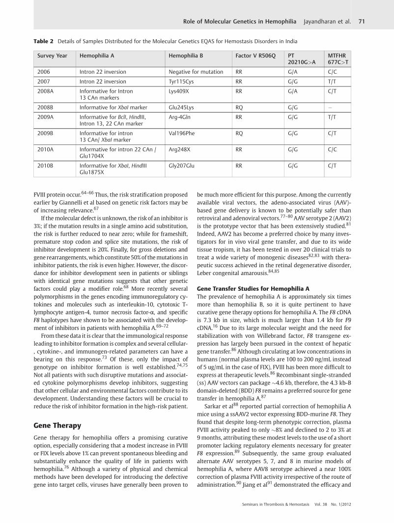
Australia.35–38 Participation in such programs is mandatoryfor laboratory certification in these countries. However,except for data from a few laboratories, a significant gapexists in knowledge about the practices of molecular genetictesting laboratories across the world. To address this, weinitiated a questionnaire survey among laboratories in boththe developing and developed countries including partici-pants from Argentina, Australia, Belgium, Brazil, Canada,China, Germany, India, Italy, Japan, Netherlands, Thailand,and United Kingdom (n¼ 19).39 Our data showed consider-able differences in the personnel standards in these laborato-ries while surprisingly, the type and number of tests offereddid not vary considerably among them (►Table 1). However,examination of the quality assurance practices in the sur-veyed laboratories showed wide variability in laboratorypractices during the preanalytical, analytical, and postana-lytical stages of genetic testing.39 Only 43% of laboratories indeveloping countries (vs. 100% in developed countries) par-ticipated in any kind of proficiency testing program (►Fig. 7).We have initiated an External Quality Assessment Scheme(EQAS) for molecular genetic analysis of hematological dis-orders for laboratories in India since 2006.39 Two surveys areconducted each year. Nine laboratories currently participatein the program for thrombophilia (100%), hemophilia A (44%),and hemophilia B (55%) modules. Eight External QualityAssessment (EQA) cycles have been completed and theirdetails are provided in ►Table 2. For genetic testing ofhemophilia, linkage analysis was most commonly usedwith only one laboratory performed direct mutation analysisusing CSGE and DNA sequencing. All laboratories performinghemophilia B genetic testing used CSGE and DNA sequencing.Response rate for this EQA schemes are between 70 and 80%.In the last two cycles, a reporting accuracy of over 90% wasnoted for thrombophilia mutations while it was �70% for thehemophilia genetic testing. A performance report is providedto all participants. Our experience suggests that EQAS forgenetic tests can be effectively established in developingcountries and efforts should be made to increase the aware-ness and benefits of voluntary participation in such programs.
Genetic Basis for the PhenotypicHeterogeneity in Severe Hemophilia
Patients conventionally classified as having severe hemophil-ia (<1% of normal clotting activity) usually have 15 to 35spontaneous joint and muscle bleeds per year without anytreatment.40–42 The vast majority (60–70%) of patients withhemophilia falls into this group. However, within this group,there is considerable heterogeneity in clinical presentation. Asubset of these patients (10 to 15%) with severe hemophiliahave clinically mild disease.41–44 Variations in the bleedingfrequency, age at first bleeding, and extent of joint damagehave all been reported in patients with severe hemophilia bymany groups.41–47 Though such phenotypic heterogeneity isintriguing, only a few studies have attempted to address itsbasis. Factors, such as varying levels of FVIII:C activity (below1%),48,49 pharmacokinetics of the replaced clotting factorconcentrate,50 the type of mutation, and the concomitant
Figure 5 Direct mutation screening by multiplex polymerase chainreaction (PCR) and conformation sensitive gel electrophoresis. Fol-lowing its isolation from peripheral blood, genomic DNA from patientsand normal control are amplified for factor 8 or factor 9 gene codingand flanking intronic regions by a multiplex PCR. For multiplex PCR,amplifications with identical annealing temperatures but producingdifferent fragment sizes were grouped together (F8¼ 13 groups, F9¼4 groups). These amplicons are then screened by conformationsensitive gel electrophoresis (CSGE) a heteroduplex based mutationscreening method that relies on the differential migration of DNAheteroduplexes in comparison with homoduplexes during polyacryl-amide gel electrophoresis under mildly denaturing conditions. Finally,PCR fragments displaying heteroduplexes are screened by DNA se-quencing to confirm the nature of nucleotide change. A representativegel picture for multiplex PCR group 3 of factor 8 gene is shown in thisfigure.
Seminars in Thrombosis & Hemostasis Vol. 38 No. 1/2012
Role of Molecular Genetics in Hemophilia Jayandharan et al.68

presence of prothrombotic factors51–53 have been reported toimpact the phenotype of severe hemophilia (►Table 3).54
Our clinical observations among minimally treated pa-tients with severe hemophilia has revealed two types ofheterogeneity as described earlier.55 The first relates to thefrequency of bleeding as is commonly recognized and it islikely that the balance of hemostasis factors determines this(the hemostasis component). The second relates to the degreeof synovial reaction and the extent of damage to joint carti-lage among those who bleed frequently (the vascular andinflammatory component). There are some patients whodevelop serious arthropathy even with a moderate numberof bleeds while there are others who maintained good jointsin spite of many bleeds (►Fig. 8). We therefore hypothesizedthat differences in the overall hemostatic potential and thevascular inflammatory responses impact this variation. Sub-sequently, our data showed that apart from the primarydisease causing mutations in F8 or F9 genes, an Arg353Glnfunctional polymorphism in F7 gene and the coinheritance ofinterferon gammaþ 874G>A polymorphism may contributeto this phenotypic variation.56,57 However, the data need tobe substantiated in larger and diverse groups of patients in amulticenter setting, especially when the classification ofmilder phenotype of severe hemophilia is not uniform acrossthe literature.55 Apart from such studies, it is also crucial toidentify other molecular and cellular determinants that con-tribute to hemophilic arthropathy, to reveal targets for inter-
vention and to design potential treatment strategies toprevent or delay the onset of blood-induced arthropathy.
Inhibitor Development
The development of inhibitors to FVIII/FIX represents a majortherapeutic problem in the treatment of hemophilia. Fortu-nately, and for reasons that are largely unexplained, theincidence of inhibitors to FIX, 3 to 5%, is significantly lessthan those to FVIII where figures of 30% are now wellsubstantiated.58–60 The propensity for inhibitor developmenthas at least two genetic components, one of which relates tothe type of clotting factor genemutation and the other(s) thatlikely involves elements of the immune system. In case ofhemophilia A and hemophilia B, patients who carry a severemolecular defect (large deletions, inversions, and nonsensemutations) that result in the complete absence of the coagu-lant protein seem to have a higher propensity to developinhibitors compared with those with mild molecular defectssuch as missense or splice site mutations, where someresidual FVIII/FIX antigen is present.60–62 This is supportedby the reported inhibitor prevalence of 21 to 88% in hemo-philia A and 6 to 60% in hemophilia B patients with severedefects as opposed <10% prevalence in patients with mildmolecular defects.63 Exceptions to this are patients withmissense mutations where inhibitors develop when confor-mational changes within the immunogenic domains of the
Figure 6 Genotype-phenotype correlation in hemophilia. Deletions or insertions in the factor 8 or factor 9 gene lead to a shift in frame of thetranslation and generally cause severe hemophilia. Exceptions to these are recurrent deletions in a polyrun of A nucleotides such as in the factor 8gene. They result mostly in severe and occasionally moderate disease. Point mutations caused by a single nucleotide change can result in anonsense substitution predicting abrupt premature termination of translation. Other type of point mutations can result in missense substitution(e.g., Cys!Arg) or affect splice junctions of intron-exon boundaries and their disease severity depends on the location and any particular functionof the amino acid affected.
Seminars in Thrombosis & Hemostasis Vol. 38 No. 1/2012
Role of Molecular Genetics in Hemophilia Jayandharan et al. 69

Table 1 Data on Personnel Standards and the Type and Number of Genetic Tests Done for Hemophilia in Various Laboratories acrossthe World
Category Developed (n¼ 11) Developing (n¼ 8) p Value
Setting Hospital-based 91% 100%
Research-based 9% �Urban 100% 88%
Laboratory Director M.D.Ph.D.M.S.M.D.þ Ph.D.
36%36%9%18%
75%12.5%12.5%�
ns
Experience (y) 20 (7–40) 14 (11–28) 0.028
Laboratory personnel Number 4 (2–82) 4 (2–7) ns
Supervisor (experience, y) 20 (10–30) 10 (5–17) 0.01
Technician(collective experience, y)
22 (2–110) 12 (5–20) ns
Genetic service provided Proportion of laboratories offering tests p Value
Developed (n¼ 11) Developing (n¼ 8) Total
Hemophilia A 100% 88% 94%
Linkage analysis 10% � 6%
Direct mutation screeningand/or DNA sequencing
70% 43% 59%
Both linkage and directmutation detection
20% 57% 35%
No. of cases/year 32 (10–118) 33 (11–60) 0.856
Hemophilia B 73% 88% 81%
Linkage analysis 0% 17% 7%
Direct mutation screeningand/or DNA sequencing
89% 67% 80%
Both linkage and directmutation detection
11% 17% 13%
No. of cases/year 9 (2–35) 7 (4–19) 0.791
ns¼ p>0.05.
Figure 7 Proficiency testing characteristics of laboratories (n ¼ 19) surveyed in our questionnaire survey.
Seminars in Thrombosis & Hemostasis Vol. 38 No. 1/2012
Role of Molecular Genetics in Hemophilia Jayandharan et al.70
FVIII protein occur.64–66 Thus, the risk stratification proposedearlier by Giannelli et al based on genetic risk factors may beof increasing relevance.67
If themolecular defect is unknown, the risk of an inhibitor is3%; if the mutation results in a single amino acid substitution,the risk is further reduced to near zero; while for frameshift,premature stop codon and splice site mutations, the risk ofinhibitor development is 20%. Finally, for gross deletions andgene rearrangements,which constitute 50%of themutations ininhibitor patients, the risk is even higher. However, the discor-dance for inhibitor development seen in patients or siblingswith identical gene mutations suggests that other geneticfactors could play a modifier role.68 More recently severalpolymorphisms in the genes encoding immunoregulatory cy-tokines and molecules such as interleukin-10, cytotoxic T-lymphocyte antigen-4, tumor necrosis factor-α, and specificF8 haplotypes have shown to be associated with the develop-ment of inhibitors in patients with hemophilia A.69–72
From these data it is clear that the immunological responseleading to inhibitor formation is complex and several cellular-, cytokine-, and immunogen-related parameters can have abearing on this response.73 Of these, only the impact ofgenotype on inhibitor formation is well established.74,75
Not all patients with such disruptive mutations and associat-ed cytokine polymorphisms develop inhibitors, suggestingthat other cellular and environmental factors contribute to itsdevelopment. Understanding these factors will be crucial toreduce the risk of inhibitor formation in the high-risk patient.
Gene Therapy
Gene therapy for hemophilia offers a promising curativeoption, especially considering that a modest increase in FVIIIor FIX levels above 1% can prevent spontaneous bleeding andsubstantially enhance the quality of life in patients withhemophilia.76 Although a variety of physical and chemicalmethods have been developed for introducing the defectivegene into target cells, viruses have generally been proven to
bemuchmore efficient for this purpose. Among the currentlyavailable viral vectors, the adeno-associated virus (AAV)-based gene delivery is known to be potentially safer thanretroviral and adenoviral vectors.77–80AAV serotype 2 (AAV2)is the prototype vector that has been extensively studied.81
Indeed, AAV2 has become a preferred choice by many inves-tigators for in vivo viral gene transfer, and due to its widetissue tropism, it has been tested in over 20 clinical trials totreat a wide variety of monogenic diseases82,83 with thera-peutic success achieved in the retinal degenerative disorder,Leber congenital amarousis.84,85
Gene Transfer Studies for Hemophilia AThe prevalence of hemophilia A is approximately six timesmore than hemophilia B, so it is quite pertinent to havecurative gene therapy options for hemophilia A. The F8 cDNAis 7.3 kb in size, which is much larger than 1.4 kb for F9cDNA.16 Due to its large molecular weight and the need forstabilization with von Willebrand factor, F8 transgene ex-pression has largely been pursued in the context of hepaticgene transfer.86 Although circulating at low concentrations inhumans (normal plasma levels are 100 to 200 ng/mL insteadof 5 ug/mL in the case of FIX), FVIII has been more difficult toexpress at therapeutic levels.86 Recombinant single-stranded(ss) AAV vectors can package �4.6 kb, therefore, the 4.3 kb-Bdomain-deleted (BDD) F8 remains a preferred source for genetransfer in hemophilia A.87
Sarkar et al88 reported partial correction of hemophilia Amice using a ssAAV2 vector expressing BDD-murine F8. Theyfound that despite long-term phenotypic correction, plasmaFVIII activity peaked to only �8% and declined to 2 to 3% at9months, attributing thesemodest levels to the use of a shortpromoter lacking regulatory elements necessary for greaterF8 expression.89 Subsequently, the same group evaluatedalternate AAV serotypes 5, 7, and 8 in murine models ofhemophilia A, where AAV8 serotype achieved a near 100%correction of plasma FVIII activity irrespective of the route ofadministration.90 Jiang et al91 demonstrated the efficacy and
Table 2 Details of Samples Distributed for the Molecular Genetics EQAS for Hemostasis Disorders in India
Survey Year Hemophilia A Hemophilia B Factor V R506Q PT20210G>A
MTFHR677C>T
2006 Intron 22 inversion Negative for mutation RR G/A C/C
2007 Intron 22 inversion Tyr115Cys RR G/G T/T
2008A Informative for Intron13 CAn markers
Lys409X RR G/A C/T
2008B Informative for XbaI marker Glu245Lys RQ G/G �2009A Informative for BclI, HindIII,
Intron 13, 22 CAn markerArg-4Gln RR G/G T/T
2009B Informative for intron13 CAn/ XbaI marker
Val196Phe RQ G/G C/T
2010A Informative for intron 22 CAn /Glu1704X
Arg248X RR G/G C/C
2010B Informative for XbaI, HindIIIGlu1875X
Gly207Glu RR G/G C/T
Seminars in Thrombosis & Hemostasis Vol. 38 No. 1/2012
Role of Molecular Genetics in Hemophilia Jayandharan et al. 71

Table
3Factorsco
ntributingto
variationin
clinical
phen
otyp
ein
patien
tswithseve
rehe
mop
hilia
Referen
ce51
118
119
48
120
121
53
122
56
123
124
125
126
127
128
129
130
50,132,133
131
Clin
ical
features
Age
atfirstjointblee
d�
Gen
etic
factors
F8/F9mutation
��
FVL16
91G>A
��
��
��
��
�PT
2021
0G>A
��
��
��
MTH
FR67
7C>T
��
��
�F7
Arg35
3Gln
�TF
5′UTR
I/D
�EP
CR23
bpI/D
�Fibrino
genßArg44
8Lys
�PA
I-14G
/5G
�HFE
C28
2Y�
Plasmatic
factors
Proc
oagu
lant
��
��
Anticoa
gulant
��
��
��
Fibrino
lytic
��
�FV
IIIha
lf-life
��
Cellularfactors
��s
tudied
FVL-Factor
V16
91G>Amutation
PT20
210G
>A-p
rothrombinG20
210A
mutation.
MTH
FR67
7C>Tmutation-m
ethy
lene
tetrah
ydrofolate
redu
ctase
TF5′UTR
I/D-T
issuefactor
5′un
tran
slated
region
insertionor
deletion
polymorph
ism
EPCR:E
ndothe
lialp
rotein
Creceptor,e
xon3,
23bpinsertion/de
letion
polymorph
ism
HFE
C28
2Y-h
emoc
hrom
atosisge
necysteine
!tyrosine
polymorph
ism
atco
don28
2.Cellularfactorsde
note
thetotaln
umbe
rof
circulatingen
dothelialc
ellsan
dprog
enitorsstud
iedby
thisgrou
p.
Seminars in Thrombosis & Hemostasis Vol. 38 No. 1/2012
Role of Molecular Genetics in Hemophilia Jayandharan et al.72

safety of AAV-canine F8 vectors of serotypes 2, 5, 6, and 8 inachieving long-term liver-specific FVIII expression, in bothhemophilia A mice and dogs. Generally, a very high dose of>1012 vg/kg (AAV2 serotype) vector is required to obtain FVIIIactivity of 2 to 4% of normal in hemophilia A mice and dogs.This underscores the need to develop additional strategies fora successful F8 gene transfer before it can be tested in humans.
Apart from use of AAV8 serotype-based vectors, severalother strategies are being tested to optimize AAV-F8 deliveryin preclinical models. One of the promising approaches is thecoadministration of a self-complementary (sc)AAV vector con-taining protein phosphatase 5 gene (scAAV-PP5) as a helpervirus, which improves the transgene expression (>fivefold)from a conventional ssAAV vector by interfering with thetranscriptional block induced by a host cell protein,FKBP52.92,93 In addition, the use of exogenous agents such asproteasome inhibitors to achieve improvedFVIII expressionandimmune evasion,94 the optimization of F8 transgene cassettebythe use of a transsplicing vector95 have also been proposed.
Gene Transfer Studies for Hemophilia BFor several reasons, such as the small size of the F9 cDNA andthe relatively easy end-point laboratory measurements of FIXactivity, hemophilia B has been a long-standing target of
interest in the development of AAV-based gene transfertherapeutics.96 Substantial multiyear correction of hemo-philia B has been documented in animal models usingmuscle/liver-directed gene transfer.97–103 Due to the stabilityof expression of most foreign transgene products in murinetissues coupled with absence of prior exposure of AAV inthese models, AAV was considered as minimally immuno-genic for many years.103,104 But, it has been difficult to attainsustained expression of FIX in human clinical trials with AAVvectors.
In the first clinical trial for hemophilia B105,106
(NCT00076557, http://clinicaltrials.gov), muscle-directedgene transfer of some 2 to 6 � 1011 vg/kg of AAV-F9 resultedin only a modest increase (<2%) in FIX levels in the eightpatients treated on a dose-escalation model. This study alsoestablished for the first time the safety of the gene transferprotocol. Subsequently, in a Phase I/II clinical trial for hepaticFIX gene transfer (NCT00515710, http://clinicaltrials.gov), asubject with severe hemophilia B (<1% FIX activity) obtaineda therapeutic level of expression (�10% of normal FIX levels),exactly as the canine data had predicted for this vector dose(2� 1012 vg/kg).107 However, expression gradually declined tobaseline between 1 and 2months after gene transfer concomi-tant with a transient rise in liver enzyme levels. No antibodiesagainst FIX were formed. Subsequent studies revealed a CD8þ
T-cell response to AAV2 capsid and suggested that MHC Ipresentation of input capsid to reactivated memory T cellsled to elimination of transduced hepatocytes.108–110
In amore recent hemophilia B clinical trial (NCT00979238,http://clinicaltrials.gov),111 the use of a scAAV8 vector todeliver an optimized F9 transgene cassette in four hemophiliaB patients has shown promise, with a sustained FIX expres-sion of 2 to 4% at significantly lower vector does (2 to 6� 1010
vg/kg). However, in two patients who received 6� 1010 vg/kgof scAAV8-F9 vector, a dose-dependent activation of capsid-specific T cells against AAV8 vectors have been noted but hasnot had an effect on FIX transgene expression, so far.
Although responses to AAV-FIX hepatic gene transfer inhumans have not been fully reconciled with results fromanimal studies, a theme has emerged from clinical trials, thatis, immune response and toxicity correlates with high vectordoses. Transaminitis in the AAV-FIX liver gene transfer wasonly observed at AAV2 doses of �5� 1011 vg/kg.107 Morerecently, in an attempt to block CD8þ T-cell responses againstAAV1 capsid inmuscle-directed gene transfer in patientswithlipoprotein lipase deficiency, immune suppression with cy-closporine and mycophenolate mofetil was effective at lowervector doses (3� 1011 vg/kg) but failed to prevent IFN-γCD8þ T-cell responses against capsid at high doses (1�1012 vg/kg).112 These data suggest that combination strate-gies to attenuate capsid- or transgene-specific immune re-sponses either by developing novel AAV vectors or AAV-specific transient immunosuppression protocol would berequired to achieve long-term liver-directed gene transferof FIX. Recent developments in the field to achieve this havebeen encouraging.
The generation of modified AAV vectors containing muta-tions of the surface-exposed tyrosine residues (►Fig. 9) has
Figure 8 Radiological pictures of joints with and without intra-articular damage. The figure shows the radiological picture of kneejoints in patients with severe hemophilia A. On the panel A, we see aseverely damaged knee joint. This patient had 37 bleeds in thepreceding year and all his joints were affected. His Pettersson scorewas 27 and the clinical score was 10. On the panel B, the radiologicalpicture shows a normal joint architecture. This patient had no bleedsand none of his joints were affected. This is a classic example of amilder form of severe hemophilia. Even among those patients whobleed frequently (Panels C, D), some have minimal synovial reaction,but loss of articular space and flexion deformity (Panel C) while othershave hypertrophic synovium but their articular space is preserved withfull range of motion (Panel D).
Seminars in Thrombosis & Hemostasis Vol. 38 No. 1/2012
Role of Molecular Genetics in Hemophilia Jayandharan et al. 73

been shown to protect vector particles from proteasomedegradation.113 This tyrosine mutant vector after adminis-tration of a vector dose that only results in subtherapeutic andtransient expression with wild-type AAV2 encapsidated vec-tor has resulted in a long-term therapeutic and tolerogenicexpression of human F9 in a murine model of hemophiliaB.114 In addition, it has been recently identified that the hostcell nuclear factor kappa B is the major regulator of innate/adaptive immune response against AAV vectors and specificinhibition of this activation can result in sustained transgeneexpression from AAV vectors.115 However, these and othernovel approaches116,117 need to be rigorously scrutinized inhigher animal models before they can be tested in humans.
Conclusions
Genetic studies in hemophilia have given patients and theirtreating physicians better options for the management of thiscondition. However, there is also scope and promise forfurther research in this field to achieve a better outcome.Despite applying sensitivemethods for mutation detection byPCR-based analysis of genomic DNA, a causative mutation isnot identified in the F8 gene in �2 to 5% patients with severehemophilia A. Newer strategies are needed for such cases.Epistatic factors that affect the clinical severity and inhibitordevelopment in this condition need further definition todevelop strategies for risk prediction. Further refinementsin gene therapy vectors are needed to translate the successseen in preclinical models into patients with hemophilia.
NoteA recent published report from Dr Nathwani's group hasdemonstrated that peripheral-vein infusion of scAAV8vectors expressing human FIX improved the bleedingphenotype in patients with hemophilia B. Importantly,the authors have circumvented the host immune-mediat-ed clearance of AAV-transduced hepatocytes with a shortcourse of glucocorticoids.111,134 These strategies offerrenewed hope for the success of gene therapy in patientswith hemophilia.
AcknowledgmentsWe would like to thank all the laboratory directors whoparticipated in the survey reported here. They include Dr.Alok Srivastava, Christian Medical College, Vellore, India;Dr. Ampaiwan Chuansumit, International HemophiliaTraining Center, Bangkok; Dr. Anjali Kelkar, Sahyadri Spe-cialty Laboratories, Pune, India; Dr. Anne Goodeve, Shef-field Diagnostic Genetic Service, Sheffield, U.K; Dr. BaiXiao, Capital Medical University, Beijing, China; Dr. CarlosD. De Brasi, Sección Genética Molecular de la Hemofilia,Buenos Aires, Argentina; Dr. Derrick J. Bowen & Peter W.Collins, The Arthur Bloom Haemophilia Centre, Cardiff, U.K; Dr. David Lillicrap, Queen’s University, Kingston,Canada; Dr. Don Bowden, Monash Medical Centre, Victo-ria, Australia; Dr. Flora Peyvandi, Angelo Bianchi BonomiHemophilia and Thrombosis Center, Milan, Italy; Dr. J.K.Ploos van Amstel, University Medical Center Utrecht,
Figure 9 Representative image of AAV2-mediated transduction of HeLa cells with multiple tyrosine-mutant (Y444 þ 500þ 730F) capsid scAAV2-EGFP vectors. (A) Cells were either mock-infected or infected by wild-type and triple mutant scAAV2-EGFP vectors (a kind gift from Dr. ArunSrivastava, University of Florida, Gainesville, FL) at a multiplicity of infection of 2000 vg/cell. Transgene expression was detected by fluorescencemicroscopy 48 hour postinfection. Original magnification 100�. (B) Quantitative analyses of tyrosine mutant -AAV2 transduction efficiency inHeLa cells shows a sixfold increase in EGFP expression . �p< 0.05 versus WT scAAV2-EGFP.
Seminars in Thrombosis & Hemostasis Vol. 38 No. 1/2012
Role of Molecular Genetics in Hemophilia Jayandharan et al.74

Netherlands; Dr. Lannoy Nathalie, Cliniques UniversitairesSaint-Luc, Brussels, Belgium; Dr. M.D. Williams, Birming-ham Children’s Hospital NHS Foundation Trust,Birmingham, U.K; Dr. Margareth Castro Ozelo, State Uni-versity of Campinas, Campinas, Brazil; Dr. Johannes Old-enburg, Institute for Experimental Haematology andTransfusion medicine, Bonn, Germany; Dr. Midori Shima,Nara Medical University, Kashihara, Japan; and Dr. RenuSaxena, All India Institute of Medical Sciences, New Delhi,India.
The first author (GRJ) is supported by research grantsfrom the Department of Science of Technology, Govern-ment of India (Swarnajayanti Fellowship 2011), Depart-ment of Biotechnology, Government of India (InnovativeYoung Biotechnologist Award 2010-BT/03/IYBA/2010;Grant BT/PR14748/MED/12/491/2010) and an Early CareerInvestigator Award-2010 from Bayer Hemophilia AwardsProgram, Bayer Inc.
References1 O’Mahoney B. Global haemophilia care challenge and opportu-
nities: World Federation of Hemophilia. 20022 Gitschier J, Wood WI, Goralka TM, et al. Characterization of the
human factor VIII gene. Nature 1984;312(5992):326–3303 Yoshitake S, Schach BG, Foster DC, Davie EW, Kurachi K. Nucleo-
tide sequence of the gene for human factor IX (antihemophilicfactor B). Biochemistry 1985;24(14):3736–3750
4 Drayna D, White R. The genetic linkage map of the human Xchromosome. Science 1985;230(4727):753–758
5 Gitschier JWW, Wood WI, Goralka TM, et al. Characterization ofthe human factor VIII gene. Nature 1984;312(5992):326–330
6 Leuer M, Oldenburg J, Lavergne JM, et al. Somatic mosaicism inhemophilia A: a fairly common event. Am J Hum Genet 2001;69(1):75–87
7 Peake IR, Lillicrap DP, Boulyjenkov V, et al. Haemophilia: strate-gies for carrier detection and prenatal diagnosis. Bull WorldHealth Organ 1993;71(3-4):429–458
8 Peyvandi F. Carrier detection and prenatal diagnosis of hemo-philia in developing countries. Semin Thromb Hemost 2005;31(5):544–554
9 Lalloz MR, Schwaab R, McVey JH, Michaelides K, Tuddenham EG.Haemophilia A diagnosis by simultaneous analysis of two vari-able dinucleotide tandem repeats within the factor VIII gene. Br JHaematol 1994;86(4):804–809
10 Jayandharan G, Shaji RV, George B, Chandy M, Srivastava A.Informativeness of linkage analysis for genetic diagnosis ofhaemophilia A in India. Haemophilia 2004;10(5):553–559
11 de Carvalho FM, de Vargas Wolfgramm E, Paneto GG, et al.Analysis of factor VIII polymorphic markers as ameans for carrierdetection in Brazilian families with haemophilia A. Haemophilia2007;13(4):409–412
12 Ranjan R, Biswas A, Kannan M, Meena A, Deka D, Saxena R.Prenatal diagnosis of haemophilia A by chorionic villus samplingand cordocentesis: all India Institute of Medical Science experi-ence. Vox Sang 2007;92(1):79–84
13 Fang Y, Wang XF, Dai J, Wang HL. A rapid multifluorescentpolymerase chain reaction for genetic counselling in Chinesehaemophilia A families. Haemophilia 2006;12(1):62–67
14 Liu Q, Nozari G, Sommer SS. Single-tube polymerase chainreaction for rapid diagnosis of the inversion hotspot of mutationin hemophilia A. Blood 1998;92(4):1458–1459
15 Bagnall RD, Waseem N, Green PM, Giannelli F. Recurrent inver-sion breaking intron 1 of the factor VIII gene is a frequent cause ofsevere hemophilia A. Blood 2002;99(1):168–174
16 Bowen DJ. Haemophilia A and haemophilia B: molecular insights.Mol Pathol 2002;55(2):127–144
17 Peyvandi F, Jayandharan G, Chandy M, et al. Genetic diagnosis ofhaemophilia and other inherited bleeding disorders. Haemo-philia 2006;12(Suppl 3):82–89
18 Jeffreys AJ, Royle NJ, Wilson V, Wong Z. Spontaneous mutationrates to new length alleles at tandem-repetitive hypervariableloci in human DNA. Nature 1988;332(6161):278–281
19 Williams IJ, Abuzenadah A, Winship PR, et al. Precise carrierdiagnosis in families with haemophilia A: use of conformationsensitive gel electrophoresis for mutation screening and poly-morphism analysis. Thromb Haemost 1998;79(4):723–726
20 Pieneman WC, Deutz-Terlouw PP, Reitsma PH, Briët E. Screeningfor mutations in haemophilia A patients by multiplex PCR-SSCP,Southern blotting and RNA analysis: the detection of a geneticabnormality in the factor VIII gene in 30 out of 35 patients. Br JHaematol 1995;90(2):442–449
21 Kogan S, Gitschier J. Mutations and a polymorphism in the factorVIII gene discovered by denaturing gradient gel electrophoresis.Proc Natl Acad Sci U S A 1990;87(6):2092–2096
22 Herbert O, Trossaërt M, Boisseau P, Fressinaud E, Gerson F.Evaluation of denaturing high-performance liquid chromatogra-phy (DHPLC) in the screening of mutations in hemophilia Bpatients. J Thromb Haemost 2004;2(12):2267–2269
23 Jayandharan G, Shaji RV, ChandyM, Srivastava A. Identification offactor IX gene defects using a multiplex PCR and CSGE strategy-afirst report. J Thromb Haemost 2003;1(9):2051–2054
24 Jayandharan G, Shaji RV, Baidya S, Nair SC, Chandy M, SrivastavaA. Identification of factor VIII genemutations in 101 patientswithhaemophilia A: mutation analysis by inversion screening andmultiplex PCR and CSGE and molecular modelling of 10 novelmissense substitutions. Haemophilia 2005;11(5):481–491
25 JayandharanG, Nelson EJ, Baidya S, ChandyM, Srivastava A. A newmultiplex PCR and conformation-sensitive gel electrophoresisstrategy for mutation detection in the platelet glycoproteinalphaIIb and beta3 genes. J Thromb Haemost 2007;5(1):206–209
26 Silva Pinto C, Fidalgo T, Salvado R, et al. Molecular diagnosis ofhaemophilia A at Centro Hospitalar de Coimbra in Portugal: studyof 103 families - 15 new mutations. Haemophilia 2012;18(1):129–138
27 Oldenburg J, El-Maarri O. New insight into the molecular basis ofhemophilia A. Int J Hematol 2006;83(2):96–102
28 Goodeve AC. Another step towards understanding hemophilia Amolecular pathogenesis. J Thromb Haemost 2010;8(12):2693–2695
29 Tizzano EF, Venceslá A, Baena M, et al. First report of twoindependent point factorVIII mutations in a family with haemo-philia a: a word of caution for carrier diagnosis. Thromb Haemost2005;94(3):675–677
30 Ogata K, Selvaraj SR, Miao HZ, Pipe SW. Most factor VIII B domainmissense mutations are unlikely to be causative mutations forsevere hemophilia A: implications for genotyping. J ThrombHaemost 2011;9(6):1183–1190
31 Berber E, Leggo J, Brown C, et al. DNA microarray analysis for thedetection of mutations in hemophilia A. J Thromb Haemost2006;4(8):1756–1762
32 Chan K, Sasanakul W, Mellars G, et al. Detection of knownhaemophilia B mutations and carrier testing by microarray.Thromb Haemost 2005;94(4):872–878
33 Tizzano EF, Barceló MJ, Baena M, et al. Rapid identification offemale haemophilia A carriers with deletions in the factor VIIIgene by quantitative real-time PCR analysis. Thromb Haemost2005;94(3):661–664
Seminars in Thrombosis & Hemostasis Vol. 38 No. 1/2012
Role of Molecular Genetics in Hemophilia Jayandharan et al. 75
34 Favaloro EJ. Learning from peer assessment: the role of theexternal quality assurance multilaboratory thrombophilia testprocess. Semin Thromb Hemost 2005;31(1):85–89
35 Perry DJ, Goodeve A, Hill M, Jennings I, Kitchen S, Walker I; UKNEQAS for Blood Coagulation. The UK National External QualityAssessment Scheme (UK NEQAS) for molecular genetic testing inhaemophilia. Thromb Haemost 2006;96(5):597–601
36 Preston FE, Kitchen S, Jennings I, Woods TA. A UK NationalExternal Quality Assessment scheme (UK Neqas) for moleculargenetic testing for the diagnosis of familial thrombophilia.Thromb Haemost 1999;82(5):1556–1557
37 Favaloro EJ, Bonar R, Sioufi J, et al; RCPA QAP in Haematology.Multilaboratory testing of thrombophilia: current and past prac-tice in Australasia as assessed through the Royal College ofPathologists of Australasia Quality Assurance Program for Hema-tology. Semin Thromb Hemost 2005;31(1):49–58
38 College of American Pathologists Proficiency Testing Program.Accessed on January 2, 2012
39 Preston FE, Lippi G, Favaloro EJ, Jayandharan GR, Edison ES,Srivastava A. Quality issues in laboratory haemostasis. Haemo-philia 2010;16(Suppl 5):93–99
40 Schramm W, Royal S, Kroner B, et al; for the European haemo-philia economic study group. Clinical outcomes and resourceutilization associated with haemophilia care in Europe. Haemo-philia 2002;8(1):33–43
41 Molho P, Rolland N, Lebrun T, et al. Epidemiological survey of theorthopaedic status of severe haemophilia A and B patients inFrance. The French Study Group. [email protected]. Haemophilia 2000;6(1):23–32
42 Aledort LM, Haschmeyer RH, Pettersson H; The OrthopaedicOutcome Study Group. A longitudinal study of orthopaedic out-comes for severe factor-VIII-deficient haemophiliacs. J InternMed1994;236(4):391–399
43 Bolton-Maggs PH, Pasi KJ. Haemophilias A and B. Lancet 2003;361(9371):1801–1809
44 PollmannH, Richter H, RingkampH, Jürgens H.When are childrendiagnosed as having severe haemophilia and when do they startto bleed? A 10-year single-centre PUP study. Eur J Pediatr1999;158(Suppl 3):S166–S170
45 Ramgren O. Haemophilia in Sweden. III. Symptomatology, withspecial reference to differences between haemophilia A and B.Acta Med Scand 1962;171;237–242
46 Rainsford SG, Hall A. A three-year study of adolescent boyssuffering from haemophilia and allied disorders. Br J Haematol1973;24(5):539–551
47 Blanchette P, Rivard G, Israels S, et al; Association of HemophiliaClinic Directors of Canada and Canadian Association of Nurses inHemophilia Care. A survey of factor prophylaxis in the Canadianhaemophilia A population. Haemophilia 2004;10(6):679–683
48 Beltrán-Miranda CP, Khan A, Jaloma-Cruz AR, Laffan MA. Throm-bin generation and phenotypic correlation in haemophilia A.Haemophilia 2005;11(4):326–334
49 Shima M, Matsumoto T, Fukuda K, et al. The utility of activatedpartial thromboplastin time (aPTT) clot waveform analysis in theinvestigation of hemophilia A patients with very low levels offactor VIII activity (FVIII:C). Thromb Haemost 2002;87(3):436–441
50 van Dijk K, van der Bom JG, Lenting PJ, et al. Factor VIII half-lifeand clinical phenotype of severe hemophilia A. Haematologica2005;90(4):494–498
51 Arbini AA,Mannucci PM, Bauer KA. Lowprevalence of the factor VLeiden mutation among “severe” hemophiliacs with a “milder”bleeding diathesis. Thromb Haemost 1995;74(5):1255–1258
52 Escuriola Ettingshausen C, Halimeh S, Kurnik K, et al. Symptom-atic onset of severe hemophilia A in childhood is dependent onthe presence of prothrombotic risk factors. Thromb Haemost2001;85(2):218–220
53 Ghosh K, Shetty S, Mohanty D. Milder clinical presentation ofhaemophilia A with severe deficiency of factor VIII as measuredby one-stage assay. Haemophilia 2001;7(1):9–12
54 van Dijk K, van der Bom JG, Fischer K, Grobbee DE, van den BergHM. Do prothrombotic factors influence clinical phenotype ofsevere haemophilia? A review of the literature. Thromb Haemost2004;92(2):305–310
55 Jayandharan GR, Srivastava A. The phenotypic heterogeneity ofsevere hemophilia. Semin Thromb Hemost 2008;34(1):128–141
56 Jayandharan GR, Nair SC, Poonnoose PM, et al. Polymorphism infactor VII gene modifies phenotype of severe haemophilia. Hae-mophilia 2009;15(6):1228–1236
57 Jayandharan GR, Chapla A, Nair SC, et al. A polymorphism inInterferon gamma gene impacts the extent of joint damage inpatients with severe hemophilia. Blood 2010;110;A546 (Suppl.)Abstract 546
58 Franchini M, Mannucci PM. Inhibitors of propagation of coagula-tion (factors VIII, IX and XI): a review of current therapeuticpractice. Br J Clin Pharmacol 2011;72(4):553–562
59 Ehrenforth S, KreuzW, Scharrer I, et al. Incidence of developmentof factor VIII and factor IX inhibitors in haemophiliacs. Lancet1992;339(8793):594–598
60 Goodeve AC, Peake IR. The molecular basis of hemophilia A:genotype-phenotype relationships and inhibitor development.Semin Thromb Hemost 2003;29(1):23–30
61 High KA. Factor IX: molecular structure, epitopes, and mutationsassociated with inhibitor formation. Adv Exp Med Biol 1995;386;79–86
62 Chambost H. Assessing risk factors: prevention of inhibitors inhaemophilia. Haemophilia 2010;16(Suppl 2):10–15
63 Oldenburg J, Schröder J, Brackmann HH, Müller-Reible C,Schwaab R, Tuddenham E. Environmental and genetic factorsinfluencing inhibitor development. Semin Hematol 2004; 41(1):(Suppl 1):82–88
64 Fakharzadeh SS, Kazazian HH Jr. Correlation between factor VIIIgenotype and inhibitor development in hemophilia A. SeminThromb Hemost 2000;26(2):167–171
65 Fay PJ, Jenkins PV. Mutating factor VIII: lessons from structure tofunction. Blood Rev 2005;19(1):15–27
66 Graw J, Brackmann HH, Oldenburg J, Schneppenheim R, SpannaglM, Schwaab R. Haemophilia A: from mutation analysis to newtherapies. Nat Rev Genet 2005;6(6):488–501
67 Green PM, Montandon AJ, Bentley DR, Giannelli F. Genetics andmolecular biology of haemophilias A and B. Blood Coagul Fibri-nolysis 1991;2(4):539–565
68 Astermark J, Berntorp E, White GC, Kroner BL; MIBS Study Group.The Malmö International Brother Study (MIBS): further supportfor genetic predisposition to inhibitor development in hemophil-ia patients. Haemophilia 2001;7(3):267–272
69 Astermark J, Oldenburg J, Pavlova A, Berntorp E, Lefvert AK; MIBSStudy Group. Polymorphisms in the IL10 but not in the IL1betaand IL4 genes are associated with inhibitor development inpatients with hemophilia A. Blood 2006;107(8):3167–3172
70 Viel KR, Ameri A, Abshire TC, et al. Inhibitors of factor VIII in blackpatients with hemophilia. N Engl J Med 2009;360(16):1618–1627
71 Astermark J, Wang X, Oldenburg J, Berntorp E, Lefvert AK; MIBSStudy Group. Polymorphisms in the CTLA-4 gene and inhibitordevelopment in patients with severe hemophilia A. J ThrombHaemost 2007;5(2):263–265
72 Astermark J, Oldenburg J, Carlson J, et al. Polymorphisms in theTNFA gene and the risk of inhibitor development in patients withhemophilia A. Blood 2006;108(12):3739–3745
73 White GC II, Kempton CL, Grimsley A, Nielsen B, Roberts HR.Cellular immune responses in hemophilia: why do inhibitorsdevelop in some, but not all hemophiliacs? J Thromb Haemost2005;3(8):1676–1681
Seminars in Thrombosis & Hemostasis Vol. 38 No. 1/2012
Role of Molecular Genetics in Hemophilia Jayandharan et al.76

74 Key NS. Inhibitors in congenital coagulation disorders. Br JHaematol 2004;127(4):379–391
75 Goodeve A. The incidence of inhibitor development according tospecific mutations—and treatment? Blood Coagul Fibrinolysis2003;14(Suppl 1):S17–S21
76 High KA. The Jeremiah Metzger Lecture: gene therapy for in-herited disorders: from Christmas disease to Leber’s amaurosis.Trans Am Clin Climatol Assoc 2009;120;331–359
77 Donahue RE, Kessler SW, Bodine D, et al. Helper virus induced Tcell lymphoma in nonhuman primates after retroviral mediatedgene transfer. J Exp Med 1992;176(4):1125–1135
78 Hacein-Bey-Abina S, Von Kalle C, Schmidt M, et al. LMO2-associ-ated clonal T cell proliferation in two patients after gene therapyfor SCID-X1. Science 2003;302(5644):415–419
79 Kohn DB, Sadelain M, Dunbar C, et al; American Society of GeneTherapy (ASGT). American Society of Gene Therapy (ASGT) ad hocsubcommittee on retroviral-mediated gene transfer to hemato-poietic stem cells. Mol Ther 2003;8(2):180–187
80 Blacklow N. Parvoviruses and Human Disease. Boca Raton: CRCPress;1988
81 Daya S, Berns KI. Gene therapy using adeno-associated virusvectors. Clin Microbiol Rev 2008;21(4):583–593
82 Flotte TR. Recent developments in recombinant AAV-mediatedgene therapy for lung diseases. Curr Gene Ther 2005;5(3):361–366
83 Kaplitt MG, Feigin A, Tang C, et al. Safety and tolerability of genetherapy with an adeno-associated virus (AAV) borne GAD genefor Parkinson’s disease: an open label, phase I trial. Lancet2007;369(9579):2097–2105
84 Bainbridge JW, Smith AJ, Barker SS, et al. Effect of gene therapy onvisual function in Leber’s congenital amaurosis. N Engl J Med2008;358(21):2231–2239
85 Maguire AM, Simonelli F, Pierce EA, et al. Safety and efficacy ofgene transfer for Leber’s congenital amaurosis. N Engl J Med2008;358(21):2240–2248
86 Wang L, Herzog RW. AAV-mediatedgene transfer for treatment ofhemophilia. Curr Gene Ther 2005;5(3):349–360
87 Gnatenko DV, Saenko EL, Jesty J, Cao LX, Hearing P, Bahou WF.Human factor VIII can be packaged and functionally expressed inan adeno-associated virus background: applicability to haemo-philia A gene therapy. Br J Haematol 1999;104(1):27–36
88 Sarkar R, XiaoW, Kazazian HH Jr. A single adeno-associated virus(AAV)-murine factor VIII vector partially corrects the hemophiliaA phenotype. J Thromb Haemost 2003;1(2):220–226
89 Walsh CE, Chao H. Parvovirus-mediated gene transfer for thehaemophilias. Haemophilia 2002;8(Suppl 2):60–67
90 Sarkar R, Tetreault R, Gao G, et al. Total correction of hemophilia Amice with canine FVIII using an AAV 8 serotype. Blood 2004;103(4):1253–1260
91 Jiang H, Lillicrap D, Patarroyo-White S, et al. Multiyear therapeu-tic benefit of AAV serotypes 2, 6, and 8 delivering factor VIII tohemophilia A mice and dogs. Blood 2006;108(1):107–115
92 Jayandharan GR, Zhong L, Li B, Kachniarz B, Srivastava A. Strate-gies for improving the transduction efficiency of single-strandedadeno-associated virus vectors in vitro and in vivo. Gene Ther2008;15(18):1287–1293
93 Jayandharan GR, Zhong L, Sack BK, et al. Optimized adeno-associated virus (AAV)-protein phosphatase-5 helper virusesfor efficient liver transduction by single-stranded AAV vectors:therapeutic expression of factor IX at reduced vector doses. HumGene Ther 2010;21(3):271–283
94 Monahan PE, Lothrop CD, Sun J, et al. Proteasome inhibitorsenhance gene delivery by AAV virus vectors expressing largegenomes in hemophilia mouse and dog models: a strategy forbroad clinical application. Mol Ther 2010;18(11):1907–1916
95 Sabatino DE, Lange AM, Altynova ES, et al. Efficacy and safety oflong-term prophylaxis in severe hemophilia A dogs followingliver gene therapy using AAV vectors. Mol Ther 2011;19(3):442–449
96 Mingozzi F, High KA. Therapeutic in vivo gene transfer for geneticdisease using AAV: progress and challenges. Nat Rev Genet2011;12(5):341–355
97 Herzog RW, Hagstrom JN, Kung SH, et al. Stable gene transfer andexpression of human blood coagulation factor IX after intramus-cular injection of recombinant adeno-associated virus. Proc NatlAcad Sci U S A 1997;94(11):5804–5809
98 Snyder RO, Miao CH, Patijn GA, et al. Persistent and therapeuticconcentrations of human factor IX in mice after hepatic genetransfer of recombinant AAV vectors. Nat Genet 1997;16(3):270–276
99 Nathwani AC, Gray JT, McIntosh J, et al. Safe and efficienttransduction of the liver after peripheral vein infusion of self-complementary AAV vector results in stable therapeutic expres-sion of human FIX in nonhuman primates. Blood 2007;109(4):1414–1421
100 Nathwani AC, Gray JT, Ng CY, et al. Self-complementary adeno-associated virus vectors containing a novel liver-specific humanfactor IX expression cassette enable highly efficient transductionof murine and nonhuman primate liver. Blood 2006;107(7):2653–2661
101 Wang L, Takabe K, Bidlingmaier SM, Ill CR, Verma IM. Sustainedcorrection of bleeding disorder in hemophilia B mice by genetherapy. Proc Natl Acad Sci U S A 1999;96(7):3906–3910
102 Monahan PE, Samulski RJ, Tazelaar J, et al. Direct intramuscularinjection with recombinant AAV vectors results in sustained ex-pression in a dogmodel of hemophilia. Gene Ther 1998;5(1):40–49
103 Niemeyer GP, Herzog RW, Mount J, et al. Long-term correction ofinhibitor-prone hemophilia B dogs treated with liver-directedAAV2-mediated factor IX gene therapy. Blood 2009;113(4):797–806
104 Snyder RO, Miao C, Meuse L, et al. Correction of hemophilia B incanine and murine models using recombinant adeno-associatedviral vectors. Nat Med 1999;5(1):64–70
105 Manno CS, Chew AJ, Hutchison S, et al. AAV-mediated factor IXgene transfer to skeletal muscle in patients with severe hemo-philia B. Blood 2003;101(8):2963–2972
106 KayMA,Manno CS, RagniMV, et al. Evidence for gene transfer andexpression of factor IX in haemophilia B patients treated with anAAV vector. Nat Genet 2000;24(3):257–261
107 Manno CS, Pierce GF, Arruda VR, et al. Successful transduction ofliver in hemophilia by AAV-Factor IX and limitations imposed bythe host immune response. Nat Med 2006;12(3):342–347
108 Mingozzi F, Maus MV, Hui DJ, et al. CD8(þ ) T-cell responses toadeno-associated virus capsid in humans. Nat Med 2007;13(4):419–422
109 Mingozzi F, High KA. Immune responses to AAV in clinical trials.Curr Gene Ther 2007;7(5):316–324
110 Zaiss AK,MuruveDA. Immunity to adeno-associatedvirus vectorsin animals and humans: a continued challenge. Gene Ther2008;15(11):808–816
111 Basner_Tschakarjan E, Mingozzi F, Chen Y, Nathwani AC,Tuddenham EGD, Rosales C, McIntosh MF, Riddell A, RustagiP, Glader B, Kay MA, Allay J, Coleman J, Sleep S, Gray J, Reiss U,Nienhuis AW, Davidoff AM, High KA. Dose-dependent activationof capsid-specific T cells after AAV serotype 8 vector adminstra-tion in a clinical study for hemophilia BMolecular Therapy2011;19, Suppl 1:Abstract 602.
112 Mingozzi F, Kleefstra A, Meulenberg J, et al. Modulation of T cellresponse to the AAV capsid in subjects undergoing intramusculargene transfer for lipoprotein lipase deficiency. Hum Gene Ther2008;19(10):1090
113 Zhong L, Li B, Mah CS, et al. Next generation of adeno-associatedvirus 2 vectors: point mutations in tyrosines lead to high-efficiency transduction at lower doses. Proc Natl Acad Sci U S A2008;105(22):7827–7832
114 Markusic DM, Herzog RW, Aslanidi GV, et al. High-efficiencytransduction and correction of murine hemophilia B using AAV2
Seminars in Thrombosis & Hemostasis Vol. 38 No. 1/2012
Role of Molecular Genetics in Hemophilia Jayandharan et al. 77

vectors devoid of multiple surface-exposed tyrosines. Mol Ther2010;18(12):2048–2056
115 Jayandharan GR, Aslanidi G, Martino AT, et al. Activation of theNF-κB pathway by AAV vectors and its implications in immuneresponse and gene therapy. Proc Natl Acad Sci U S A 2011;108(9):3743–3748
116 Nathwani AC, Cochrane M, McIntosh J, et al. Enhancing transduc-tion of the liver by adeno-associated viral vectors. Gene Ther2009;16(1):60–69
117 Martino AT, Suzuki M, Markusic DM, et al. The genome of self-complementary adeno-associated viral vectors increases Toll-likereceptor 9-dependent innate immune responses in the liver.Blood 2011;117(24):6459–6468
118 Ahmed R, Kannan M, Choudhry VP, Saxena R. Does the MTHFR677T allele alter the clinical phenotype in severe haemophilia A?Thromb Res 2003;109(1):71–72
119 Barnes C, Blanchette V, Lillicrap D, et al. Different clinical pheno-type in triplets with haemophilia A. Haemophilia 2007;13(2):202–205
120 Biguzzi E, Mancuso P, Franchi F, et al. Circulating endothelial cells(CECs) and progenitors (CEPs) in severe haemophiliacs withdifferent clinical phenotype. Br J Haematol 2009;144(5):803–805
121 Cruz E, Porto G, Morais S, Campos M, de Sousa M. HFE mutationsin the pathobiology of hemophilic arthropathy. Blood 2005;105(8):3381–3382
122 GrünewaldM, Siegemund A, Grünewald A, Konegan A, KokschM,Griesshammer M. Paradoxical hyperfibrinolysis is associatedwith a more intensely haemorrhagic phenotype in severe con-genital haemophilia. Haemophilia 2002;8(6):768–775
123 Klintman J, Berntorp E, Astermark J; MIBS Study Group. Thrombingeneration in vitro in the presence of by-passing agents in siblingswith severe haemophilia A. Haemophilia 2010;16(1):e210–e215
124 Lee DH, Walker IR, Teitel J, et al. Effect of the factor V Leidenmutation on the clinical expression of severe hemophilia A.Thromb Haemost 2000;83(3):387–391
125 Nowak-Göttl U, Escuriola C, Kurnik K, et al. Haemophilia andthrombophilia. What do we learn about combined inheritance ofboth genetic variations? Hamostaseologie 2003;23(1):36–40
126 Ninivaggi M, Dargaud Y, van Oerle R, de Laat B, Hemker HC,Lindhout T. Thrombin generation assay using factor IXa as atrigger to quantify accurately factor VIII levels in haemophilia A. JThromb Haemost 2011;9(8):1549–1555
127 Petkova R, Chakarov S, Horvath A, Ganev V, Kremensky I. Coexis-tence of a common prothrombotic risk factor and hemophilia inthe Bulgarian hemophilic population:genotype/phenotype cor-relations. Balkan J Med Genet 2001;4(3–4):37–39
128 Santagostino E, Mancuso ME, Tripodi A, et al. Severe hemophiliawith mild bleeding phenotype: molecular characterization andglobal coagulation profile. J Thromb Haemost 2010;8(4):737–743
129 Schulman S, Eelde A, Holmström M, Ståhlberg G, Odeberg J,Blombäck M. Validation of a composite score for clinical severityof hemophilia. J Thromb Haemost 2008;6(7):1113–1121
130 Shetty S, Vora S, Kulkarni B, et al. Contribution of naturalanticoagulant and fibrinolytic factors in modulating the clinicalseverity of haemophilia patients. Br J Haematol 2007;138(4):541–544
131 Yee DL. Platelets asmodifiers of clinical phenotype in hemophilia.Scientific World J 2006;6;661–668
132 van Dijk K, Fischer K, van der Bom JG, Grobbee DE, van den BergHM. Variability in clinical phenotype of severe haemophilia:the role of the first joint bleed. Haemophilia 2005;11(5):438–443
133 van Dijk K, van der Bom JG, Fischer K, de Groot PG, van den BergHM. Phenotype of severe hemophilia A and plasma levels ofrisk factors for thrombosis. J Thromb Haemost 2007;5(5):1062–1064
134 Nathwani AC, Tuddenham EG, Rangarajan S, et al. Adenovirus-associated virus vector-mediatedgene transfer in hemophilia B. NEngl J Med 2011;365(25):2357–2365
Seminars in Thrombosis & Hemostasis Vol. 38 No. 1/2012
Role of Molecular Genetics in Hemophilia Jayandharan et al.78