role of formate and hydrogen in the syntrophic degradation
TRANSCRIPT

Role of Formate and Hydrogen
in the Syntrophic Degradation of
Propionate and Butyrate
CENTRALE LANDBOUWCATALOGUS
0000 0610 3226

Promotor: Dr. A.J.B. Zehnder
hoogleraar in de microbiologie
Co-promotor: Dr. Ir. A.J.M. Stams
Universitair docent bij de vakgroep microbiologie

^iôl^ x l^i^h
Xiuzhu Dong
Role of Formate and Hydrogen
in the Syntrophic Degradation of
Propionate and Butyrate
Proefschrift
ter verkrijging van de graad van doctor
in de landbouw- en milieuwetenschappen,
op gezag van de rector magnificus,
Dr. C. M. Karssen,
in het openbaar te verdedigen
op vrijdag 9 december 1994
des namiddags te half twee in de aula
van de Landbouwuniversiteit te Wageningen

CIP-DATA KONINKLIJKE BIBLIOTHEEK, DEN HAAG
Xiuzhu Dong
Role of formate and hydrogen in the syntrophic degradation
of propionate and butyrate/ Xiuzhu Dong.- [S.I.: s.n.]. - 111.
Thesis Wageningen. - With réf. - With summary in Dutch and in Chinese.
ISBN 90-5485-333-6
Subjuct headings: bacteria/ formate / hydrogen.
This research was carried out at the Department of Microbiology, Wageningen Agricultural University, The Netherlands.
The publication or this thesis was financially suported by the Department of Microbiology, Wageningen Agricultural University.
LANDBOUWUNIVERSITEIT WAGENINGEN

MgZo\] i#fa
PROPOSITIONS
1. During the period when interest in the pathways of intermediary metabolism was at
its height, anaerobic bacteria were star performers in the repertoire of the
microbiologist. — Thauer & Morris, 1984.
2. The letter "R" is the most difficult one to pronounce in many kinds of languages,
especially the Dutch "R" for chinese people.
3. It is indeed necessary for a "sandwich" Ph.D. student to have the two thick outer
layers to get enough "nutrition".
4. Interspecies H2 or formate transfer: that is the question.
5. Syntrophism exists not only in the microbial world, but also in the human society.
6. Music calms you when you are excited, encourages you when you are depressed, and
consoles you when you are distressed.
7. When you ask a question, Chinese will deny the question itself, while English will
deny the question of its meaning.
8. The importance of syntrophic degradation is well established for compounds which
can not be degraded in another way. However, the importance of syntrophic consortia
Â

in the degradation of other compounds is often underestimated.-- Stams, 1994.
9. One can reduce the size of manuscript by using the compact language Chinese.
10. When one is only eager to reach the final achievement, one will miss the joy of
failures and success.
11. A good scientist is the one who can demonstrate a significant principle with a simple
approach.
12. A frog at the bottom of a well only sees a piece of the sky.

Contents
Chapter Page
1 General Introduction 1
2. Anaerobic degradation of propionate by a mesophilic
acetogenic bacterium in coculture and triculture with
different methanogens 37
3. Butyrate oxidation by Syrurophospora bryantii in coculture
with different methanogens and in pure culture with
pentenoate as electron acceptor 57
4. Evidence for H2 and formate formation during syntrophic
butyrate and propionate degradation 79
5. Localization of the enzymes involved in H2 and formate
metabolism in Syntrophospora bryantii 97
6. Localization of the enzymes involved in the H2 and formate
metabolism of a mesophilic propionate-oxidizing bacterium 115
7. Summary 131
8. Samenvatting 135
/> % 140
Acknowledgements 142
Curriculum vitae 143
Chapter 1
General introduction

Chapter 1
Mineralization of organic complex in methanogenic environments
In methanogenic environments mineralization of complex organic matter to the most
oxidized form of carbon (CO2) and the most reduced form of carbon (CFL,) is carried out
sequentially. Ultimately, C02 is the terminal sink of electrons in a series of oxidative
reactions. Methanogenic environments are widely distributed in nature. These include,
wetwoods of living trees (Schink et al. 1981) where cellulose and pectins are degraded
anaerobically, digestive tracts of higher animals and insects (Hungate 1966; Bryant 1977;
Wolin 1981), and freshwater sediments (Rudd & Taylor 1980; Crill et al. 1991).
Anaerobic bioreactors are artificial methanogenic environments, which are used for e.g.
the stabilization of sewage sludge and for the treatment of a variety of industrial wastes.
The widest applied bioreactor for anaerobic wastewater treatment is the UASB (Upfiow
Anaerobic Sludge Bed) reactor (Lettinga & Hulshoff Pol 1991) which contains well
settleable methanogenic granular sludge with a high methanogenic activity. Other types of
anaerobic bioreactors are the Fluidized Bed reactor (Iza 1991), the Anaerobic Filter
(Young 1991) and the Expanded Granular Sludge Bed reactor (de Man et al. 1988).
Different physiological types of bacteria are involved in the methanogenic mineralization
of complex organics. Methanogens occupy the terminal position of this anaerobic food
web. They are specialized in the degradation of a very, limited number of substrates.
Methanobacterium bryantii (Bryant et al. 1967) and Methanobrevibacter arboriphilus
(Zeikus & Henning 1975; Zehnder & Wuhrmann 1977) utilize only H2/C02, Methanothrix
(Methanosaeta) sp. (Huser et al. 1982; Patel & Sprott 1990) only use acetate while e.g.

General Introduction
Methanospirillum hungatei and Methanobacterium formicicum can use H2/C02 and
formate (Schauer et al. 1982; Bryant & Boone 1987). The most versatile methanogens are
Methanosarcina sp.; they can use H2/C02, acetate, methanol, methylated amines and
pyruvate (Whitman et al. 1992; Jones 1991). During methanogenesis, organic polymers
are first hydrolyzed and fermented by fermentative organisms to the methanogenic
precursors H2/C02, formate, and acetate, and to reduced organic compounds, such as
propionate, butyrate, lactate, succinate and ethanol. These reduced compounds can be
oxidized further to the methanogenic substrates by acetogenic bacteria (Mah et al. 1990;
Stams 1994). Figure 1 shows how in methanogenic environments the degradation of
organic matter is achieved by consortia of fermentative organisms, acetogenic and
methanogenic bacteria, about 76% of organic matter is degraded via reduced organic
intermediates (Toerien & Hattingh 1969; Wolin 1974; Zehnder 1978; Bryant 1979).
Therefore, acetogenesis is a key process in the mineralization of organic matter in
methanogenic environments.
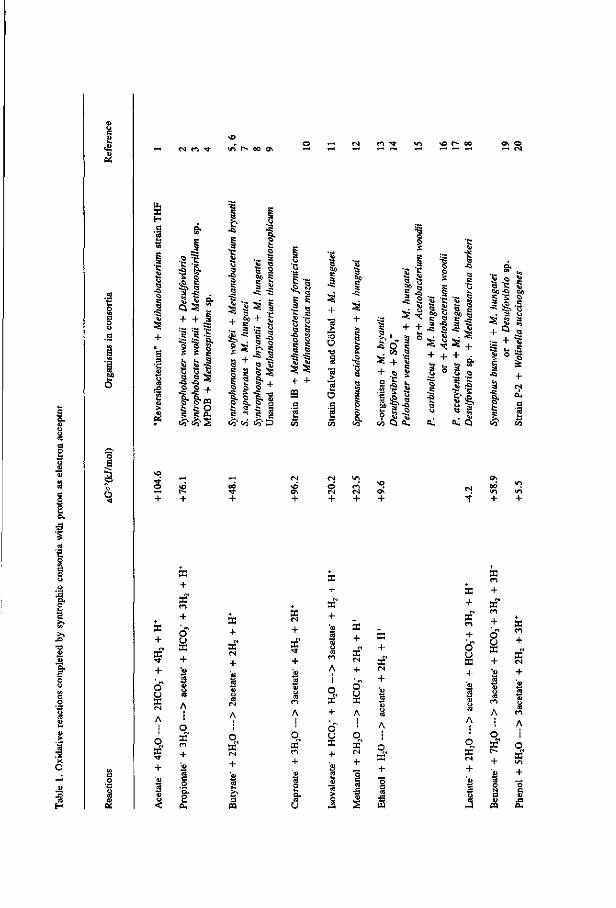
Syntrophic degradation
Syntrophism is defined as that the cooperation of partners depends entirely on each other
to perform the metabolism (Schink 1992). Under methanogenic conditions, almost all the
acetogenic reactions can only be carried out by obligate syntrophic consortia (Schink
1992; Stams 1994). This as a result of the fact that the oxidation of these compounds is
energetically unfavorable (table 1), and/or that at least one intermediate step involved in

Chapter 1
Fermentation Acetogenesis
4%
Complex organics
76% Short-chain
fatty acids
oo /
H2
\ &> \ %
\ 20% X Acetate
Methanogenesis
N** C H 4
x o\o
Fig. 1. Methanogenic mineralization of complex organic matter and the percentage flow of the
energy content involved in complex matter to methane formation (From McCarty, 1982).
their degradation is energetically difficult. The products H2, formate and to some extent
also acetate have to be removed by methanogens to "pull" the reactions. Low
concentrations of H2 and formate, and in some cases also low acetate concentration are
essential. The highest H2, formate and acetate concentrations that can be tolerated during
syntrophic oxidation are dependent on the thermodynamics of the oxidative reactions
(table 1). E.g. the maximum H2 partial pressure that can be tolerated is 14.5, 45 and
1000 Pa for propionate, butyrate and ethanol oxidation, respectively. For these
calculations, it was assumed that CHt partial pressure is at 2500 Pa, the concentrations of
acetate, propionate, butyrate and bicarbonate are 25 mM. Methanogens catabolize H2,
formate and acetate efficiently; the reported threshold values for H2 are around 10 Pa (59
nM in liquid), and those for formate and acetate are around 10 /*M, respectively (Cord-
Ruwisch et al. 1988; Schauer et al. 1982; Jetten et al. 1990).
4
—< e*i fi - * vi t^ oo o\
•a O
1
-o
o
o o.
C 'S 'S § •S a
<3S
•3 -3 '£* i ; a ft,
^ ? f
feto
S -S
5 + 1 + s - 1 1 8 S e s
S •§ -s
&? S- s s- s ê §• § 1 = ^ = g
•5 c
Ü
+ + ,
* O c . co a
s i * 1̂1 "s? .A
« 1 3 « .
-5 X
+ +
•g -g >̂ -ft
II ft. Q
a o c
-I <£. -S
+ Q a :3 + =5 S u .5
a.
S + +
+
+
3? + O u X
A
O
af +
X
+ af ci + O U a +
3 8 A
O af CI
+ Si es e
a + af + '«0
S o CÖ
A
i q, af + 'o>
35
+ af + '<0
S 0)
u m A i
O af + Si
a + af + « aï y CÖ
en
A
O
af + o u a + 's
+ a + a + o" u a A
O, af + S
a + a" Ol
+ Si CS
'S o cö
A
O af +
s CU O <
a + a"
o u a
a c-i
+ a" en
+ O
a
a +
«N
+
A S
« o X es
+
S
is
o a + '<D M
3 4>
« o a + , O
a

i ?3 c3
oo **
— rt ga o
s d
£ o
's 3
T3 U i
O
u
o S ON
oo
o 2 3
GO g •O S
M — —• a
2 -g
O *
2 2 "
X +
X + X c-l
+ d u »
m •a
o o
ca
2 £
« -
o DG ~H <U
~ <D
O O
>* 5
— £0
General Introduction
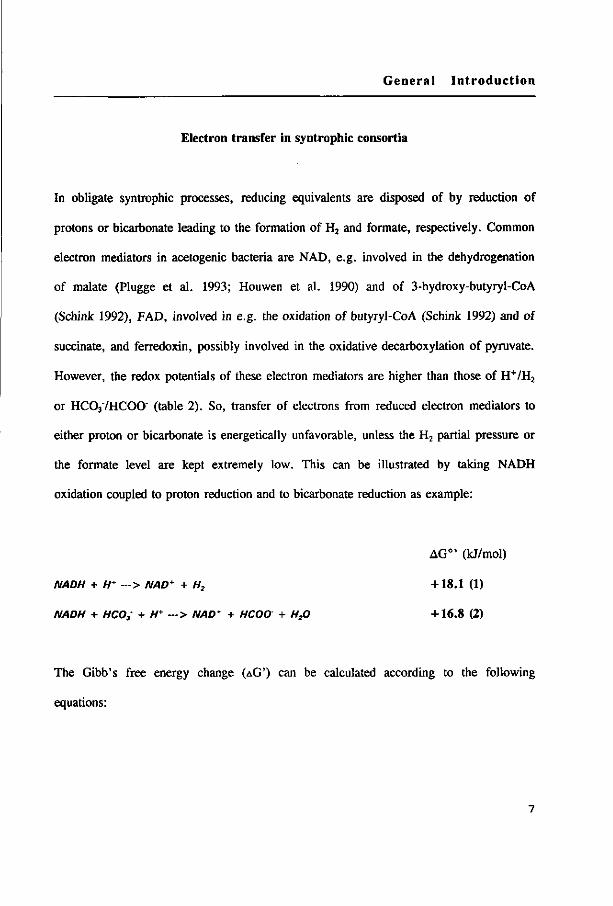
Electron transfer in syntrophic consortia
In obligate syntrophic processes, reducing equivalents are disposed of by reduction of
protons or bicarbonate leading to the formation of H2 and formate, respectively. Common
electron mediators in acetogenic bacteria are NAD, e.g. involved in the dehydrogenation
of malate (Plugge et al. 1993; Houwen et al. 1990) and of 3-hydroxy-butyryl-CoA
(Schink 1992), FAD, involved in e.g. the oxidation of butyryl-CoA (Schink 1992) and of
succinate, and ferredoxin, possibly involved in the oxidative decarboxylation of pyruvate.
However, the redox potentials of these electron mediators are higher than those of H+/H2
or HC037HCOO" (table 2). So, transfer of electrons from reduced electron mediators to
either proton or bicarbonate is energetically unfavorable, unless the H2 partial pressure or
the formate level are kept extremely low. This can be illustrated by taking NADH
oxidation coupled to proton reduction and to bicarbonate reduction as example:
AG°' (kJ/mol)
NADH + H* —> NAD* + H2 +18.1 (1)
NADH + HCO3 + H* — > NAD* + HCOO + H:0 +16.8 (2)
The Gibb's free energy change (AG') can be calculated according to the following
equations:

Chapter 1
AG' = AG- • RTJn ̂ ^ AG' = AG°' + RTln ^AD^HCOO^ [NADH][H1 [NADH]iHliHCOj]
Where AG° ' represents free energy change at standard conditons, T=298°K, pH 7, 1 M
for solutes, and 105 Pa for gases, and R is the gas constant (8.24 kJ.mor'.T1). AG' of
these reactions becomes zero when the PH2 is 76 Pa and the formate concentration is 32
pM, assuming that [NAD+] = [NADH] and [HC03]=25 mM. Fig.2 shows the effects of
H2 (A) and formate (B) on the Gibb's free energy change of electron transfer components
when coupled to H2 and formate production, respectively.
3. "a <
40
30
20
10
0
A
•
•
/ /
/ / /
B
/
/ / /
. / . . / .
FAD/FADH
•220 mV
NAD/NADH
•320 mV
-398 mV
logpH(Pa) log Formate (mM)
Fig. 2. Diagram of the dependence of the Gibb's free energy changes of the redox couples prevailing in
biological systems on the hydrogen partial pressure (A) and the formateconcantration (B) when the oxidations
are coupled to proton reduction (A) and to bicarbonate (B) reduction.

General Introduction
table 2. Redox potentials of the redox couples possibly involved in the anaerobic
degradation of propionate and butyrate, and their free energy changes when coupled to
proton reduction (AG°',) or when coupled to bicarbonate reduction (AG0,2)
a
Redox couples E°'(mV) AG°',(kJ/mol) AG°'2(kJ/mol)
Acetyl-CoA/pyruvate
C02/formate
H+/H2
Fd(ox)/Fd(red)
NAD(P)+/NAD(P)H
FAD/FADH2
-490
-432
-414
-398
-320
-220
Acetoacetyl-Co A/3-OH-butyryl-Co A -190
Oxaloacetate27malate2~ -172
Crotonyl-CoA/butyryl-CoA -126"
Fumarate27succinate2" +33
-12.2
+ 1.3
+3.1
+ 18.1
+37.4
+31.6
+47.7
+75.2
+86.2
-13.5
-1.3
+ 1.7
+ 16.8
+36.1
+30.3
+46.4
+73.9
+ 84.7
a: Except the data from references b, all the data of E°'are taken from Thauer et al. (1977). The AG° ' values
are calculated according to the equation AG"' = -II.F.AE"'.
b: Gustafson et al. (1986).
Chapter 1
Hydrogen flux and formate flux in syntrophic cultures
As syntrophic reactions are completed by more than one organism, interspecies mass
transfer is essential. The conversion rates are determined by the flux (J) of H2 or formate
in cultures. The flux depends on the diffusion coefficient (D), the H2 and formate
concentration gradients (C), the diffusion distance (d) between H2/formate-producing and
H2/formate-consuming cells, and the surface area of the H2/formate-producing organism
(A). Fluxes can be described by a simple Fick's diffusion equation (Schink & Thauer
1988).
C - C J = -AD-— -molsee1
d
Aggregation and juxtaposition of bacterial communities appears to be very important in
syntrophic degradation. If the different bacteria are in the close vicinity of each other the
diffusion distance is minimized. The high rates of CH4 formation in UASB reactors are
achieved because they contain granular sludge with a high biomass density. Disruption of
the structure of methanogenic granules reduced the propionate degradation rate by 90 %
(Grotenhuis et al. 1991). Other examples are also described, e.g. the degradation rates of
butyrate and propionate decreased 35 and 25% after disintegration of the thermophilic
granules (Schmidt & Ahring 1993), separating ethanol-oxidizing and H2-utilizing bacteria
by a dialysis membrane led to an increase in the doubling time by 7 to 11 hours (Stieb &
Schink 1987), while shortening the interbacterial distances in syntrophic cultures increased
10

General Introduction
the CH4 formation rate from propionate (Stams et al. 1992) and from butyrate (Dwyer et
al. 1988; Schmidt & Ahring 1993).
The steepness of the H2 and formate concentration gradient is another parameter which
affects the fluxes. As described above the maximal H2 and formate concentration that can
be reached by acetogens depends on the thermodynamics of the conversions (Stams 1994).
The same is true for the threshold values for formate and H2 that can be reached by
H2/formate-utilizing anaerobes. The threshold values for H2 of methanogens and sulfate
reducers are 3-16 and 1-3 Pa, respectively (Cord-Ruwisch et al. 1988; Dwyer et al. 1988;
Boone et al. 1989; Stams et al. 1992; Boone & Bryant 1980). The maximum specific
growth rates of propionate-, butyrate- and benzoate-degrading acetogenic bacteria in
coculture with methanogens were 0.10, 0.19 and 0.10 day"1, respectively; while in
cocultures with sulfate reducers the growth rates were 0.19, 0.31 and 0.13 day',
respectively (Boone & Bryant 1980; Mclnerney et al. 1979; Mountfort & Bryant 1982).
These differences were explained by the differences in H2 gradients (Dolfing 1988).
Interspecies H2 transfer
Since it was discovered that Methanobacillus omelianskii consisted of an ethanol-oxidizing
bacterium (S organism) and Methanobacterium bryantii, Bryant et al. (1967) assumed that
H2 transfer could play a role in syntrophic degradation. Hungate (1967) was the first to
demonstrate that H2 was an intermediate in fermentative processes in the rumen. This has
led to the development of a 3-step model for the anaerobic mineralization of organic
11

Chapter 1
matter, and the concept of interspecies H2 transfer was developed by Wolin (1976; 1982).
Convincing evidences for the role of H2 were provided for those syntrophic consortia in
which only H2-scavengers are present, such as in the classical ethanol-oxidizing M.
omelianskii culture (Bryant et al. 1967), a butyrate-degrading culture with S. wolfei
(Mclnerney et al. 1979; 1981), a thermophilic butyrate-oxidizing culture (Henson &
Smith 1985) and a thermophilic propionate-oxidizing culture (Stams et al. 1992). Indeed
in such syntrophic cocultures H2 partial pressures could be measured close to those which
can be calculated from thermodynamic considerations. In a syntrophic ethanol-oxidizing
culture an equilibrium H2 partial pressure in the range of 500-2000 Pa was measured
(Stieb & Schink 1987; Seitz et al. 1988). Whereas the H2 partial pressure was only at 30-
34 Pa in a thermophilic propionate-oxidizing coculture (Stams et al. 1992) and 95 Pa in a
butyrate-oxidizing culture (Dwyer et al. 1988). Moreover, inhibition of propionate and
butyrate oxidation by addition of H2 was observed in mesophilic propionate-acclimatized
sludge and in a thermophilic butyrate-oxidizing culture, respectively (Fukuzaki et al.
1990; Ahring & Westermann 1988).
Interspecies formate transfer
Formate as an alternative electron carrier during syntrophic degradation was proposed for
the first time by Bryant et al. (1967), and later on by Mclnerney et al. (1979) and Boone
& Bryant (1980). However, Thiele & Zeikus (1988) proposed that formate transfer might
even be more important than H2 transfer. It was observed that the minimum threshold
12

General Introduction
levels for H2 of the methanogens prevailing in some anaerobic ecosystems were about 6
times higher than the H2 gas levels observed in such environments (Thiele et al. 1988),
and that the turnover of the H2 pool could only account for 5-10% to the measured
electron transfer rates. Thiele & Zeikus (1988) put forward the interspecies formate
transfer theory based on that: i) many H2-consumers in syntrophic cultures can use
formate; ii) the redox potential of the couple H+/H2 and HC037formate are nearly same;
iii) some acetogenic bacteria are able to form formate during syntrophic degradation
(Hungate 1967; Thiele & Zeikus 1988).
Although S. wolfei degraded butyrate in coculture with Methanobacterium bryantii, the
growth rate and CH4 production were increased significantly in the coculture with M.
hungatei (Mclnerney et al. 1979). M. hungatei is often abundantly present in many
syntrophic consortia (table 1), and formate transfer clearly occurred in a coculture with
Desulfovibrio baarsii (Zindel et al. 1988), a bacterium which utilizes only formate but not
H2. Direct experimental evidence for formate formation was obtained for the syntrophic
conversion of ethanol to CH4 in microbial floes of a reactor treating whey (Thiele &
Zeikus 1988). Formate accumulated when methanogenesis in the floes was inhibited and
formate formation was dependent on C02 and ethanol but not on H2. Other evidence for
formate transfer was obtained with a coculture of S. wolfei and M. formicicum by Boone
et al. (1989) using a diffusion model. They calculated that the formate diffusion rate
could account for the CH4 formation rate in the culture but the diffusion rate of H2 could
not. Thiele et al. (1988) calculated that the total ratio of the dissolved formate and H2
concentration should be between 10 and 200 in pure and mixed cultures of fermentative
13
Chapter 1
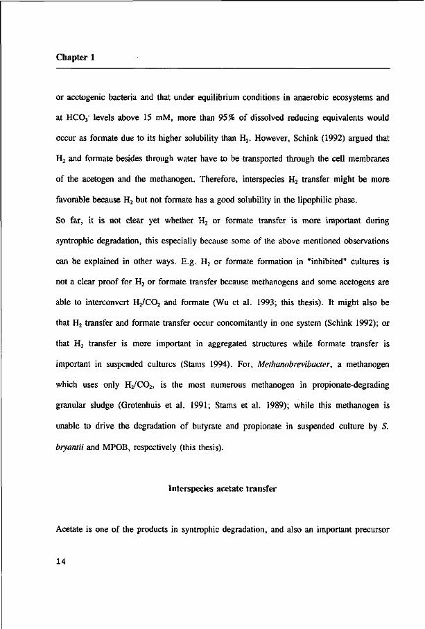
or acetogenic bacteria and that under equilibrium conditions in anaerobic ecosystems and
at HC03" levels above 15 mM, more than 95% of dissolved reducing equivalents would
occur as formate due to its higher solubility than H2. However, Schink (1992) argued that
H2 and formate besides through water have to be transported through the cell membranes
of the acetogen and the methanogen. Therefore, interspecies H2 transfer might be more
favorable because H2 but not formate has a good solubility in the lipophilic phase.
So far, it is not clear yet whether H2 or formate transfer is more important during
syntrophic degradation, this especially because some of the above mentioned observations
can be explained in other ways. E.g. H2 or formate formation in "inhibited" cultures is
not a clear proof for H2 or formate transfer because methanogens and some acetogens are
able to interconvert H2/C02 and formate (Wu et al. 1993; this thesis). It might also be
that H2 transfer and formate transfer occur concomitantly in one system (Schink 1992); or
that H2 transfer is more important in aggregated structures while formate transfer is
important in suspended cultures (Stams 1994). For, Methanobrevibacter, a methanogen
which uses only H2/C02, is the most numerous methanogen in propionate-degrading
granular sludge (Grotenhuis et al. 1991; Stams et al. 1989); while this methanogen is
unable to drive the degradation of butyrate and propionate in suspended culture by S.
bryantii and MPOB, respectively (this thesis).
Interspecies acetate transfer
Acetate is one of the products in syntrophic degradation, and also an important precursor
14

General Introduction
of methanogenesis. It contributes for 70% of the CH4 formation in anaerobic digesters
degrading organic matter (Mah et al. 1990; Gujer & Zehnder 1983). Acetate removal by
methanogens can affect the energetics of various reactions to different extents. Low
acetate concentrations are absolutely required in isovalerate oxidation, where 3 moles of
acetate and only 1 mol of H2 are formed. Theoretically, H2 removal by methanogens can
change the Gibb's free energy change of the overall reaction in isovalerate oxidation from
+20.2 kJ/mol isovalerate (Eq. 3) to -13.6 kJ/mol (Eq. 4); while acetate removal by
methanogens can result in a Gibb's free energy change of -71.6 kJ/mol (Eq. 5). The
addition of 1 mM acetate prevented this methanogenic coculture from growth and
degrading isovalerate in a fresh subculture (Stieb & Schink 1986).
AG°'(kJ/mol)
CSH9°Î + HC°3 + H2° - 3 C2#3°2 + H2 + H* +202 (3>
C5Hg02' + 5/4HCO; + l/2H20 - 3C2H302 + l/4Ctf4 + 3/4/T -13.6 (4)
C5H902' + 4H20 - 3CH4 + H2 + H* + IHCO; -71.6 (5)
Acetate is the only intermediate transferred in the methanogenic degradation of acetone
(Platen & Schink 1987), and Methanothrix sp. is the only methanogen in the coculture.
Inhibition of methanogenesis by bromoethane sulfonic acid (BrES) or ethylene led to
acetate accumulation, while the acetone degradation rate decreased. Although the
oxidation of acetone to acetate is exergonic (Eq. 6), acetate removal obviously was
essential for energy conservation during acetone conversion (Eq. 7).
15

Chapter 1
AG°'(kJ/mol)
CH3COCH3 + HCO; - 2CH3COO' + H* -34.2 (6)
CH3COCH3 + 2H20 - 2CH^ + H* + HCOj -89.2 (7)
Acetate accumulation also retards propionate and butyrate degradation in syntrophic
consortia. Addition of 3.3 mM acetate decreased the propionate consumption by 60% in
methanogenic sludge (Gorris et al. 1989) while addition of Methanosarcina to the
coculture of S. wolfei and M. hungatei increased the butyrate degradation rate by 30%
(Beaty & Mclnerney 1989). These observations suggest that acetate transfer can occur
either together with H2/formate transfer or independently.
Propionate oxidation
Propionate is an important intermediate in methanogenic mineralization of organic matter.
It accounts for 35 % to the methane formation in a digester fermenting animal waste
(Mah et al. 1990). Complete mineralization of propionate into methane and CO2 will
liberate 56.6 kJ. This is about the amount needed to synthesize 1 mol of ATP (Schink
1992).
Propionate' + l.75H20 - 1.75C#4 + I.ISHCO; + 0.25JT AG° ' = -56.6U/mol
This process is performed by 3 types of bacteria; acetogenic bacteria degrade propionate
16

General Introduction
into H2/formate and acetate, which then are converted into methane by H2/formate-
utilizing and aceticlastic methanogens, respectively. Therefore, the energy which is
conserved during propionate oxidation has to be shared by the 3 kinds of bacteria.
So far, only a few of propionate-oxidizing cultures are documented. Syntrophobacter
wolinii is the first propionate oxidizer which was enriched from a methanogenic
enrichment from an anaerobic municipal sewage digestor and was obtained in a defined
coculture with a Desulfovibrio sp. (Boone & Bryant 1980). Only very recently a binary
culture of S. wolinii and M. hungatei was obtained (Dörner 1992). Stams et al. (1993)
described another mesophilic propionate-oxidizing bacterium (MPOB) in coculture with a
Methanospirillum sp.. They succeeded to get a methanogen-free culture by growing it on
fumarate. In the absence of methanogens this bacterium is able to i) reduce fumarate to
succinate with H2 or formate as electron donors, ii) oxidize propionate with fumarate as
electron acceptors, iii) ferment fumarate to succinate and C02. 16S rRNA sequence
analysis revealed that S. wolinii and MPOB were phylogenetically related with sulfate-
reducing bacteria (Harmsen et al. 1993; 1994), and indeed these two bacteria are able to
couple propionate oxidation to sulfate reduction (Dörner 1992; Van Kuijk unpublished
results). Some other more or less defined syntrophic propionate-oxidizing cultures have
been obtained (Koch et al. 1983; Mucha et al. 1988; Robbins 1988; Wu et al. 1992;
Dörner 1992; Stams et al. 1992).
Experiments with 14C and 13C labelled propionate revealed that a randomizing pathway is
used for propionate oxidation (Koch et al. 1983; Houwen et al. 1987, 1991; Robbins
1988). These findings led to propose the propionate oxidation pathway as given in Fig. 3,
17

Chapter 1
CoA
Propionate
+35.6 •I
propionyl-CoA
+20.9
methylmalonyl-CoA
0
succinyl-CoA
-35.1
succinate
CO 2 —
+86.2
2[H]
acetate f »- ATP
_ADP acety l®
; • CoA
^. Pi acetyl-CoA
co2-« ,2[H]
CoA pyruvate
-13
+9.0
-12.2
-27.2
oxaloacetate
S 2[H]
( malate
fumarate
+47.7
-3.8
Fig. 3. Pathway of syntrophic propionate oxidation and theA G° ' values (kJ/mo|) of the
intermediate steps when the oxidation reactions are coupled to proton reduction.
which was similar to that found in Desulfobulbus propionicus (Stams et al. 1984).
Enzyme measurement in cell extracts of S. wolinii (Houwen et al. 1990) and MPOB
(Plugge et al. 1993) confirmed that methylmalonyl-CoA pathway is involved in syntrophic
propionate oxidation. However, the difference between the two organisms is that
propionate is activated to propionyl-CoA via a propionate kinase in S. wolinii, while the
activation is achieved via a propionate:acetyl-CoA HS-CoA transferase in MPOB. The
oxidation of 1 mol of propionate would yield 1 ATP by substrate level phosphorylation.
During propionate oxidation, reducing equivalents are released in the oxidations of
18

General Introduction
succinate to fumarate, malate to oxaloacetate and pyruvated to acetyl-CoA. The midpoint
redox potential of the redox couple succinate/fumarate is +30 mV. While the redox
potentials of H+/H2 and HC037HCOO" are -414 mV and -432 mV, respectively. To make
succinate oxidation coupled to H2 or formate formation feasible, H2 or formate levels of
10"9 Pa and 10"8 ^M would be required. However, these values are far lower than can be
created by H2/formate-utilizing methanogens. Therefore, a reversed electron transport
mechanism has to be proposed. It has been speculated that 2/3 ATP would be invested to
drive this reversed electron transport (Stains 1994). Evidence for the existence of a
reversed electron transport mechanism was obtained with a syntrophic glycolate-oxidizing
bacterium (Friedrich et al. 1991; Friedrich & Schink 1993), a butyrate oxidizer-
Syntrophomonas wolfei (Wallrabenstein & Schink 1994) and an acetate-oxidizing sulfur-
reducing Desulfuromonas acetoxidam (Paulsen et al. 1986).
Butyrate oxidation
Butyrate is another important intermediate during mineralization of biopolymers in
methanogenic environments. It accounts for 8% of the total methane formation in a
digestor fermenting animal waste (Mah et al. 1990). As with propionate conversion,
mineralization of butyrate into methane and C02 also requires 3 different types of
bacteria, viz butyrate-oxidizing acetogens, H2/fonnate-utilizing and aceticlastic
methanogens. The overall equation for butyrate conversion is :
19
Chapter 1
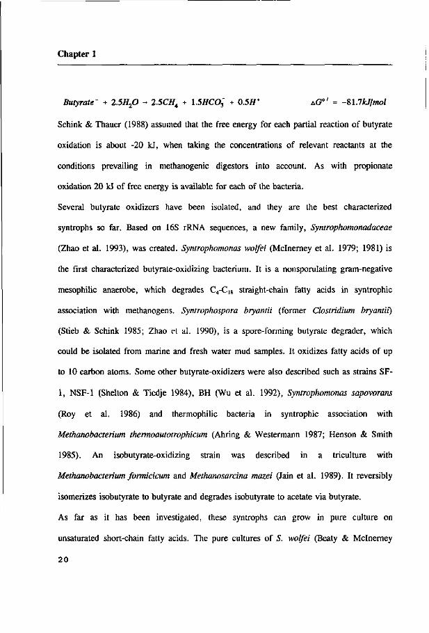
Butyrate' + 2.5H20 - 2.5CH4 + l.SHCOj + 0.5H* AGO/ = -Sl.lkJ/mol
Schink & Thauer (1988) assumed that the free energy for each partial reaction of butyrate
oxidation is about -20 kJ, when taking the concentrations of relevant reactants at the
conditions prevailing in methanogenic digestors into account. As with propionate
oxidation 20 kJ of free energy is available for each of the bacteria.
Several butyrate oxidizers have been isolated, and they are the best characterized
syntrophs so far. Based on 16S rRNA sequences, a new family, Syntrophomonadaceae
(Zhao et al. 1993), was created. Syntrophomonas wolfei (Mclnerney et al. 1979; 1981) is
the first characterized butyrate-oxidizing bacterium. It is a nonsporulating gram-negative
mesophilic anaerobe, which degrades C4-C,8 straight-chain fatty acids in syntrophic
association with methanogens. Syntrophospora bryantii (former Clostridium bryantii)
(Stieb & Schink 1985; Zhao et al. 1990), is a spore-forming butyrate dégrader, which
could be isolated from marine and fresh water mud samples. It oxidizes fatty acids of up
to 10 carbon atoms. Some other butyrate-oxidizers were also described such as strains SF-
1, NSF-1 (Shelton & Tiedje 1984), BH (Wu et al. 1992), Syntrophomonas sapovorans
(Roy et al. 1986) and thermophilic bacteria in syntrophic association with
Methanobacterium thermoautotrophicum (Ahring & Westermann 1987; Henson & Smith
1985). An isobutyrate-oxidizing strain was described in a triculture with
Methanobacterium formicicum and Methanosarcina mazei (Jain et al. 1989). It reversibly
isomerizes isobutyrate to butyrate and degrades isobutyrate to acetate via butyrate.
As far as it has been investigated, these syntrophs can grow in pure culture on
unsaturated short-chain fatty acids. The pure cultures of S. wolfei (Beaty & Mclnerney
20

General Introduction
Butyrate-
Butyryl-CoA
^2[H]
+35.6
+74.1
Crotonyl-CoA
r ^ ° +3.3
3-Hydroxybutyryl-CoA
V, 2[H] +31.6
1987) and S. bryantii (Zhao et al.
1990) were obtained by growth on
crotonate. Both bacteria ferment
crotonate to butyrate and acetate. S.
wolfei is able to couple butyrate
oxidation to the reduction of
unsaturated alkanes (Kasper et al.
1987), while S. bryantii is able to
couple butyrate oxidation to the
reduction of pentenoate to valerate
(Chapter 3, this thesis).
Syntrophic butyrate oxidation
proceeds through the crotonyl-CoA
pathway (Fig. 4). This has been
demonstrated for S. wolfei (Wofford
et al. 1986) and for 5. bryantii
(Schink 1992). Butyrate is activated
to butyryl-CoA via a butyrate:acetyl-
CoA HSCoA transferase. Two acetate are formed as end products and reducing
equivalents are released in the oxidation of butyryl-CoA to crotonyl-CoA and of 3-
hydroxy-butyryl-CoA to acetoacetyl-CoA. These reducing equivalents are formed at the
level of FADH2 and NADH, respectively. One ATP is synthesized by substrate level
r
Acetoacetyl-CoA
/ \ CoASI-r—i
/ \ * Acetyl-CoA Acetyl-CoA
\ +9.0
Acetyl-© ADP
-13 fA Acetate-
».ATP
Acetate-
Fig. 4. Pathway of syntrophic butyrate oxidation and the aG"
values (kj/mol) of the intermediate steps when the oxidations
are coupled to proton.
21

Chapter 1
phosphorylation in the acetate kinase reaction. Because the reducing equivalents are
formed at redox potential of -126 mV (butyryl-CoA dehydrogenase) and -190 mV (3-OH-
butyryl dehydrogenase), for butyryl-CoA oxidation, H2 and formate levels as low as 10"8
Pa and 10"7 /*M are required, when the electrons are disposed as H2 and formate,
respectively. Thauer & Morris (1984) speculated that butyrate-oxidizers spend part of the
ATP formed by substrate level phosphorylation to drive a reversed electron transport
reaction to release the electron at a lower redox potential. The butyryl-CoA
dehydrogenase and hydrogenase of S. wolfei were demonstrated to be partly membrane-
bound (Dörner 1992; Wallrabenstein & Schink 1994). Moreover, H2 formation could be
inhibited by a protonophore (CCCP) or by an ATPase inhibitor (DCCD). These
observations support the existence of a reversed electron transfer mechanism in butyrate
oxidizing bacteria.
Outline of the thesis
For clarifying the relative importance of H2 and formate transfer during syntrophic
degradation of propionate and butyrate, a propionate-oxidizer (MPOB) and a butyrate-
oxidizer S. bryantii were used.
Pure cultures of the acetogens offer the ability to construct artificial defined methanogenic
cocultures. Chapter 2 deals with propionate degradation in cocultures of MPOB with
different methanogens, and the relative importance of H2 and formate is deduced by
determining the degradation kinetics in the different cocultures. Moreover, the
22
General Introduction
requirement of low formate concentration during syntrophic propionate oxidation is also
determined in a triculture of MPOB, a formate-cleaving bacterium and a H2-consuming
methanogen. Chapter 3 describes butyrate degradation by S. bryantii in different
methanogenic cocultures and tricultures, and in single culture with pentenoate as electron
acceptor. The effect of acetate removal on degradation of propionate and butyrate was
determined as well (Chapter 2 and 3). Chapter 4 provides evidence for the formation of
both H2 and formate during butyrate oxidation by S. bryantii and during propionate
oxidation by MPOB. The effect of HC03 on H2 and formate formation is also described
in this chapter.
The localization of the enzymes responsible for H2 and formate production in the
acetogens and the kinetic properties of hydrogenase and formate dehydrogenase are
determined in S. bryantii and MPOB. These results are presented in Chapter 5 and 6,
respectively. Finally, in chapter 7 the observations presented in this thesis are
summarized.
References:
Ahring BK & Westermann P (1987) Thermophilic anaerobic degradation of butyrate by a
butyrate-utilizing bacterium in coculture and triculture with methanogenic bacteria.
Appl. Environ. Microbiol. 53:429-433
Ahring BK & Westermann P (1988) Product inhibition of butyrate metabolism by acetate
and hydrogen in a thermophilic coculture. Appl. Environ. Microbiol. 54:2393-2397
23

Chapter 1
Barik S, Brulla WJ & Bryant MP (1985) PA-1, a versatile anaerobe obtained in pure
culture, catabolizes benzenoids and other compounds in syntrophy with
hydrogenotrophs, and p-2 plus Wolinella sp. degrades benzenoids. Appl. Environ.
Microbiol. 50:304-310.
Beaty PS & Mclnerney MJ (1987) Growth of Syntrophomonas wolfei in pure culture on
crotonate. Arch. Microbiol. 147:389-393
Beaty PS & Mclnerney MJ (1989) Effects of organic acid anion on the growth and
metabolism of Syntrophomonas wolfei in pure culture and in defined consortia. Appl.
Environ. Microbiol. 55:977-983.
Boone DR & Bryant MP (1980) Propionate-degrading bacterium, Syntrophobacter wolinii
sp. nov. gen. nov., from methanogenic ecosystems. Appl. Environ. Microbiol.
40:626-632
Boone DR, Johnson RL & Liu Y (1989) Diffusion of the interspecies electron carriers H2
and formate in methanogenic ecosystems, and applications in the measurement of KM
for H2 and formate uptake. Appl. Environ. Microbiol. 55:1735-1741
Bryant MP (1977) Microbiology of the rumen, p. 287-304, In Swenson MJ (ed.),
Physiology of domestic animals, 9th rev. ed. Cornell University Press, Ithaca, N. Y.
Bryant MP (1979) Microbial methane production-theoretical aspects. J. Anim. Sei.
48:193-201.
Bryant MP & Boone DR (1987) Isolation and characterization of Methanobacterium
formicicum MF. Int. J. Syst. Bacteriol. 37:171.
Bryant MP, Campbell L, Reddy CA & Crabill MR (1977) Growth of Desulfovibrio in
24

General Introduction
lactate or ethanol media low in sulfate in associdation with H2-utilizing methanogenic
bacteria. Appl. Environ. Microbiol. 33:1162-1169.
Bryant MP, Wolin EA, Wolin MJ & Wolfe RS (1967) Methanobacteium omelianskii, a
symbiotic association of two species of bacteria. Arch. Microbiol. 59:20-31.
Cord-Ruwisch R & Ollivier B (1986) Interspecific hydrogen transfer during methanol
degradation by Sporomusa acidovorans and hydrogenophilic anaerobes. Arch.
Microbiol. 144:163-165
Cord-Ruwisch R, Seitz HJ & Conrad R (1988) The capacity of hydrogenotrophic
anaerobic bacteria to complete traces of hydrogen depends on the redox potential of
the terminal electron acceptors. Arch. Microbiol. 149:350-357
Crill PM, Harriss RC & Bartlett KB (1991) Methane fluxes from terrestrial wetland
environments. In: Rogers JE & Whitman WB (Eds) Microbial production and
consumption of greenhouse gases: methane, nitrogen oxides, and halomethanes (pp
175-187). American Society for Microbiology, Washington
Dolfing J (1988) Acetogenesis In: Zehnder AJB (Ed) Biology of anaerobic microor
ganisms (pp 417-468) John Wiley & Sons, New York
Dörner C (1992) Biochemie und Energetik der Wasserstoff-Freisetzung in der syntrophen
Vergärung von Fettsäuren und Benzoat. Dissertation, University of Tübingen
Dwyer DF, Weeg-Aerssens E, Shelton DR & Tiedje JM (1988) Bioenergetic conditions
of butyrate metabolism by a syntrophic, anaerobic bacterium in coculture with
hydrogen-oxidizing methanogenic and sulfidogenic bacteria. Appl. Environ. Microbiol.
54:1354-1359
25

Chapter 1
Friedrich M, Laderer U & Schink B (1991) Fermentative degradation of glycolic acid by
defined syntrophic cocultures. Arch. Microbiol. 156:398-404
Friedrich M & Schink B (1993) Hydrogen formation from glycolate driven by reversed
electron transport in membrane vesicles of a syntrophic glycolate-oxidizing bacterium.
Eur. J. Biochem. 217:233-240
Fukuzaki S, Nishio N, Shobayshi M & Nagai S (1990) Inhibition of the fermentation of
propionate to methane by hydrogen, acetate and propionate. Appl. Environ.
Microbiol. 56:719-723.
Gorris LGM, van Deursen JMA van der Drift C & Vogels GD (1989) Inhibition of
propionate by acetate in methanogenic fluidized bed reactors. Biotechnol. Lett. 11:61-
66.
Grotenhuis JTC, Smit M, Plugge CM, Xu Y, Van Lamineren AAM, Stams AJM, &
Zehnder AJB (1991) Bacteriological composition and structure of granular sludge
adapted to different substrates. Appl Environ Microbiol 57:1942-1949
Gujer W & Zehnder AJB (1983) Conversion processes in anaerobic digestion. Wat. Sei.
Tech. 15:127-167
Gustafson WG, Feinberg BA & McFarland JT (1986) Energetics of ß-oxidation.
Reduction potentials of general fatty acyl-CoA dehydrogenase, electron transfer
flavoprotein, and fatty acyl-CoA substrates. J. Biol. Chem. 261:7733-7741.
Harmsen HJM, Wullings B, Akkermans ADL, Ludwig W & Stams AJM (1993)
Phylogenetic analysis of Syntrophobacter wolinii reveals a relationship with sulfate-
reducing bacteria. Arch. Microbiol. 160:238-240
26

General Introduction
Harmsen HJM, Kengen H, Akkermans ADL & Stams AJM (1994) Phylogenetic analysis
of syntrophic propionate oxidizing bacteria in enrichments. Appl. Syst. Bacteriol.
(Submitted).
Henson JM & Smith PH (1985) Isolation of a butyrate-utilizing bacterium in coculture
with Methanobacterium thermoautotrophicum from a thermophilic digester. Appl.
Environ. Microbiol. 49:1461-1466
Houwen FP, Dijkema C, Schoenmakers CHH, Stams AJM & Zehnder AJB (1987) 13C-
NMR study of propionate degradation by a methanogenic coculture. FEMS Microbiol.
Lett. 41:269-274
Houwen FP, Dijkema C, Stams AJM & Zehnder AJB (1991) Propionate metabolism in
anaerobic bacteria; determination of carboxylation reactions with 13C-NMR
spectroscopy. Biochim. Biophys. Acta 1056:126-132
Houwen FP, Plokker J, Dijkema C & Stams AJM (1990) Enzymatic evidence for
involvement of the methylmalonyl-CoA pathway in propionate oxidation by Syntrop-
hobacter wolinii. Arch. Microbiol. 155:52-55
Hungate RE (1966) The rumen ahnd its microbes. Academic Press, New York
Hungate RE (1967) Hydrogen as an intermediate in the rumen fermentation. Arch.
Mikrobiolo. 59:158-164.
Huser BA, Wuhrmann K & Zehnder AJB (1982) Methanothrix soehngenii gen. nov. sp.
nov., a new acetotrophic non-hydrogenoxidizing methanogen bacterium. Arch.
Microbiol. 132:1-9.
Iza J (1991) Fluidized bed reactors for anaerobic wastewater treatment. Wat. Sei.
27

Chapter 1
Technol. 24:109-132
Jain MK, Wu WM & Zeikus JG (1989) Isomerization mediated conversion of iso-
butyrate and butyrate to methane by syntrophic biomethanation consortia, abstra.O-52,
(p 313). Abstr. 89th Annu. Meet. Am. Soc. Microbiol. 1989. American Society for
Microbiology, Washington, D.C.
Jetten MSM, Stams AJM & Zehnder AJB (1990) Acetate threshold values and acetate
activating enzymes in methanogenic bacteria. FEMS Microbiol. Ecol. 73:339-344
Jones WJ (1991) Diversity and physiology of methanogens. In: Rogers JE & Whitman
WB (Eds) Microbial production and consumption of greenhouse gases: methane,
nitrogen oxides, and halomethanes (pp 39-55). American Society for Microbiology,
Washington
Kasper HF, Holland AJ & Mountfort DO (1987) Simultaneous butyrate oxidation by
Syntrophomonas wolfei and catalytic olefin reduction in the absence of interspecies
hydrogen transfer. Arch. Microbiol. 147:334-339
Koch ME, Dolfing J, Wuhrmann K & Zehnder AJB (1983) Pathways of propionate
degradation by enriched methanogenic cultures. Appl. Environ. Microbiol. 45:1411-
1414
I.ettinga G & Hulshoff-Pol LW (1991) UASB process design for various types of waste
waters. Wat. Sei. Tech. 24:88-107
Mah RA, Xun LY, Boone DR, Ahring B, Smith PH & Wilkie A (1990) Methanogenesis
from propionate in sludge and enrichment systems. In: Belaich JP, Bruschi M &
Garcia JL (Eds) Microbiology and biochemistry of strict anaerobes involved in
28

General Introduction
interspecies hydrogen transfer (pp 99-119). Plenum Press, New York
de Man AWA, van der Last ARM & Lettinga G (1988) The use of EGSB and UASB
anaerobic systems for low strength soluble and complex wastewaters at temperatures
ranging from 8 to 30°C. In Hall ER & Hobson PN (eds.), Proceedings of the Fifth
International Symposium on Anaerobic Digestion (pp735-738), Bologna, Italy.
McCarty PL (1982) One hundred years of anaerobic treatment. In Hughes DE,
Stafford DA, Wheaty BI, Baader W, Lettinga G, Nyns EJ and Verstraete W (eds). In
Anaerobic Digestion 1981 (pp3-22). Elsevier, Amsterdam.
Mclnerney MJ & Bryant MP (1981) Anaerobic degradation of lactate by syntrophic
associations of Methanosarcina barken and Desulfovibrio species and effect of H2 on
acetate degradation. Appl. Environ. Microbiol. 41:346-354.
Mclnerney MJ, Bryant MP, Hespell RB & Costerton JW (1981) Syntrophomonas wolfei
gen.nov. sp.nov, an anaerobic syntrophic, fatty acid-oxidizing bacterium. Appl.
Environ. Microbiol. 41:1029-1039
Mclnerney MJ, Bryant MP & Pfennig N (1979) Anaerobic bacterium that degrades fatty
acids in syntrophic association with methanogens. Arch. Microbiol. 122:129-135
Mountfort DO & Bryant MP (1982) Isolation and characterization of an anaerobic
benzoate-degrading bacterium from sewage sludge. Arch. Microbiol. 133:249-256
Mucha H, Lingens F & Trösch W (1988) Conversion of propionate to acetate and
methane by syntrophic consortia. Appl. Microbiol. Biotechnol. 27:581-586
Patel GB & Sprott GD (1990) Methanosaeta concilii gen. nov., sp. nov., ("Methanothrix
concilii") and Methanosaeta thermoacetophila nom. rev., comb. nov. Int. J. Syst.
29

Chapter 1
Bacteriol. 40:79-82
Paulsen J, Kroger A & Thauer RK (1986) ATP-driven succinate oxidation in the
catabolism of Desulforomonas acetoxidans. Arch. Microbiol. 144:78-83
Platen H & Schink B (1987) Methanogenic degradation of acetone by an enrichment
culture. Arch. Microbiol. 149:136-141
Plugge CM, Dijkema C & Stams AJM (1993) Acetyl-CoA cleavage pathway in a
syntrophic propionate oxidizing bacterium growing on fumarate in the absence of
methanogens. FEMS Microbiol. Lett. 110:71-76
Robbins JE (1988) A proposed pathway for catabolism of propionate in methanogenic
cocultures. Appl. Environ. Microbiol. 54:1300-1301
Roy F, Samain E, Dubourguier HC & Albagnac G (1986) Syntrophomonas sapovorans
sp. nov. a new obligately proton-reducing anaerobe oxidizing saturated and
unsaturated long chain fatty acids. Arch. Microbiol. 145:142-147
Rudd JWM & Taylor CD (1980) Methane cycling in aquatic environments. Adv. Aquat.
Microbiol. 2:77-150
Schauer NL, Brown DP & Ferry JG (1982) Kinetics of formate metabolism in
Methanobacterium formicicum and Methanospirillum hungatei. Appl. Environ.
Microbiol. 44: 540-554
Schink B (1984) Fermentation of 2,3-butanediol by Pelobacter carbinolicus sp. nov. and
Pelobacter propionicus sp. nov., and evidence for propionate formation from C2
compounds. Arch. Microbiol. 137:33-41
Schink B (1985) Mechanisms and kinetics of succinate and propionate degradation in
30

General Introduction
anoxie freshwater sediments and sewage sludge. J. Gen. Microbiol. 131:643-650
Schink B (1992) Syntrophism among prokaryotes. In: Balows A, Trüper HG, Dworkin
M, Harder W & Schleifer KH (Eds) The Prokaryotes (pp 276-299) Springer Verlag,
New York
Schink B & Stieb M (1983) Fermentative degradation of polyethylene glycol by a strictly
anaerobic, gram-negative, nonsporeforming bacterium, Pelobacter venetianus sp. nov.
Appl. Environ. Microbiol. 45:1905-1913
Schink B & Thauer RK (1988) Energetics of syntrophic methane formation and the
influence of aggregation. In: Lettinga G, Zehnder AJB, Grotenhuis JTC & Hulshoff
Pol LW (Eds), Granular anaerobic sludge; microbiology and technology (pp 5-17).
Pudoc, Wageningen
Schink B, Ward JC & Zeikus JG (1981) Microbiology of wetwood: importance of pectin
degradation and Clostridium species in living trees. Appl. Environ. Microbiol. 42:526-
532
Schmidt JE & Ahring BK (1993) Effects of hydrogen and formate on the degradation of
propionate and butyrate in thermophilic granules from an upflow anaerobic sludge
blanket reactor. Appl. Environ. Microbiol. 59:2546-2551
Seitz HJ, Schink B & Conrad R (1988) Thermodynamics of hydrogen metabolism in
methanogenic cocultures degrading ethanol or lactate. FEMS Microbiol. Lett 55:119-
124
Shelton DR & Tiedje JM (1984) Isolation and partial characterization of bacteria in an
anaerobic consortium that mineralizes 3-chlorobenzoic acid. Appl. Environ.
31

Chapter 1
Microbiol. 48:840-848
Stams AJM (1994) Metabolic interactions between anaerobic bacteria in methanogenic
environments. Antonie van Leeuwenhoek (In press).
Stams AJM, Van Dijk J, Dijkema C & Plugge CM (1993) Growth of syntrophic
propionate-oxidizing bacteria with fumarate in the absence of methanogenic bacteria.
Appl. Environ. Microbiol. 59:1114-1119
Stams AJM, Grolle KCF, Frijters CTMJ & Van Lier JB (1992) Enrichment of
thermophilic propionate-oxidizing bacteria in syntrophy with Methanobacterium
thermoautotrophicum or Methanobacterium thermoformiticum. Appl. Environ.
Microbiol. 58:346-352
Stams AJM, Grotenhuis JTC & Zehnder AJB (1989) Structure function relationship in
granular sludge. In: Hattori T, Ishida Y, Maruyama, Monta RY & Uchida A (Eds)
Recent advances in microbial Ecology (pp 440-445). Japan Scientific Societies Press,
Tokyo
Stams AJM & Hansen TA (1984) Fermentation of glutamate and other compounds by
Acidaminobacter hydrogenoformans gen. nov. sp. nov, an obligate anaerobe isolated
from black mud. Studies with pure cultures and mixed cultures with sulfate-reducing
and methanogenic bacteria. Arch. Microbiol. 137:329-337
Stams AJM, Kremer DR, Nicolay K, Weenk GH & Hansen TA (1984) Pathway of
propionate formation in Desulfobulbus propionicus. Arch. Microbiol. 139:167-173
Stieb M & Schink B (1985) Anaerobic degradation of fatty acids by Clostridium bryantii
sp. nov., a sporeforming obigately syntrophic bacterium. Arch. Microbiol. 140:387-
32

General Introduction
390
Stieb M & Schink B (1986) Anaerobic degradation of isovalerate by a defined
methanogenic coculture. Arch. Microbiol. 144:291-295
Stieb M & Schink B (1987) Cultivation of syntrophic anaerobic bacteria in membrane-
separated culture devices. FEMS Microbiol. Ecol. 45:71-76
Thauer RK, Jungermenn K & Decker K (1977) Energy conservation in chemotrophic
anaerobic bacteria. Bacteriol. Rev. 41:100-180
Thauer RK & Morris JG (1984) Metabolism of chemotrophic anaerobes: old views and
new aspects. In: Kelly DP & Carr NG (Eds) The microbe 1984: part 2. Prokaryotes
and eukaryotes (pp 123-168). Cambridge University Press, Cambridge
Thiele JH & Zeikus JG (1988)) Control of interspecies electron flow during anaerobic
digestion: significance of formate transfer versus hydrogen transfer during syntrophic
methanogenesis in floes. Appl. Environ. Microbiol. 54:20-29
Thiele JH, Chartrain M & Zeikus JG (1988) Control of interspecies electron flow during
anaerobic digestion: role of the floe formation. Appl. Environ. Microbiol. 54:10-19
Toerien DF & Hattingh WHJ (1969) Anaerobic digestion. I. The microbiology of
anaerobic digestion. Water Research 3: 385-416
Wallrabenstein C & Schink B (1994) Evidence of reversed electron transport in
syntrophic butyrate or benzoate oxidation by Syntrophomonas wolfei and Syntrophus
buswellii. Arch. Microbiol. 162:136-142
Whitman WB, Bowen TL & Boone DR (1992) The methanogenic bacteria. In: Balows A ,
Triiper HG, Dworkin M, Harder W and Schleifer KH (eds) The Prokaryotes
33

Chapter 1
(pp719-968). Springer Verlag, New York
Wofford NQ, Beaty PS & Mclnerney MJ (1986) Preparation of cell-free extracts and the
enzymes involved in fatty acid metabolism in Syntrophomonas wolfei. J. Bacteriol.
167:179-185
Wolin MJ (1974) metabolic interactions among intestinal microorganism. Ann. J.
Clin. Nutr. 27:1320-1328
Wolin MJ (1976) Interactions between H2-producing and methane-producing species. In:
Schlegel HG, Gottschalk G & Pfennig N (Eds) Microbial formation and utilization of
gases (H2, CH4, CO) (pp 14-15). Goltze, Göttingen
Wolin MJ (1981) Fermentation in the rumen and human large intestine. Science 213:163-
168
Wolin MJ (1982) Hydrogen transfer in microbial communities. In: Bull AT & Slater JH
(Eds) Microbial interactions and communities (pp 323-356). Academic Press, London
Wu WM, Jain MK, de Macario EC, Thiele JH & Zeikus JG (1994) Microbial
composition and characterization of prevalent methanogens and acetogens isolated
from syntrophic methanogenic granules. Appl. Environ. Microbiol. 38:282-290
Wu WM, Jain MK & Zeikus JG (1994) Anaerobic degradation of normal- and branched-
chain fatty acids with four or more carbons to methane by a syntrophic
methanogenic triculture. Appl. Environ. Microbiol. 60:2220-2226
Wu WM, Rickley RF, Jain MK & Zeikus JG (1993) Energetics and regulations of
formate and hydrogen metabolism by Methanobacterium formicicum. Arch. Microbiol.
159:57-65
34

General Introduction
Young JC (1991) Factors affecting the design and performance of upflow anaerobic
filters. Wat. Sei. Tech. 24:133-155
Zehnder AJB (1978) Ecology of methane formation In: Mitchell R (ed) Water pollution
microbiology, vol. 2 (pp349-376). John Wiley and Sons, London
Zehnder AJB & Wuhrmann K (1977) Physiology of a Methanobacterium strain AZ.
Arch. Microbiol. 111:199-205
Zeikus JG & Henning DL (1975) Methanobacterium arbophilicum sp. nov. An obligate
anaerobe isolated from wetwood of living trees. Antonie van Leeuwenhoek 41:543-552
Zhao H, Yang D, Woese CR & Bryant MP (1990) Assignment of Clostridium bryantii to
Syntrophospora bryantii gen. nov., comb. nov. on the basis of a 16S rRNA sequence
analysis of its crotonate-grown pure culture. Int. J. Syst. Bacterial. 40:40-44
Zhao H, Yang D, Woese CR & Bryant MP (1993) Assignment of fatty aeid-ß-oxidizing
syntrophic bacteria to Syntrophomonadaceae fam. nov. on the basis of 16S rRNA
sequences. Int. J. Syst. Bacterial. 43:278-286
Zindel U, Freudenberg W, Reith M, Andreesen JR, Schnell J & Widdel F (1988)
Eubacteriwn acidaminophilum sp. nov., a versatile amino acid degrading anaerobe
producing or utilizing H2 or formate. Description and enzymatic studies. Arch.
Microbiol. 150:254-266
Zinder SH & Koch M (1984) Non-aceticlastic methanogenesis from acetate: acetate
oxidation by a thermophilic syntrophic coculture. Arch, microbiol. 138:263-373
35

Chapter 2
Anaerobic degradation of propionate by a mesophilic
acetogenic bacterium in coculture and triculture with
different methanogens
Xiuzhu Dong, Caroline M. Plugge, and Alfons J. M. Stams Appl. Environ. Microbiol. 60:2834-2838, 1994.

Chapter 2
ABSTRACT
A mesophilic acetogenic bacterium (MPOB) oxidized propionate to acetate and C02 in
cocultures with the formate- and hydrogen-utilizing methanogens Methanospirillum hungatei
and Methanobacteriumformicicum. Propionate oxidation did not occur in cocultures with two
Methanobrevibacter strains, which grew only with hydrogen. Tricultures consisting of
MPOB, one of the Methanobrevibacter strains and organisms which are able to convert
formate into H2/C02 (Desulfovibrio Gil or a homoacetogenic bacterium EE121) also
degraded propionate. MPOB, in the absence of methanogens, was able to couple propionate
conversion to fumarate reduction. This propionate conversion was inhibited by hydrogen and
by formate. Formate and hydrogen blocked the energetically unfavorable succinate oxidation
to fumarate involved in propionate catabolism. Low formate and low hydrogen concentrations
are required for the syntrophic degradation of propionate by MPOB. In triculture with M.
hungatei and the aceticlastic Methanothrix soehngenii, propionate was degraded faster than
in biculture with M. hungatei, indicating that low acetate concentrations are favorable for
propionate oxidation as well.
INTRODUCTION
Propionate is an important intermediate in the degradation of organic matter under
methanogenic conditions. It may account for 35 % of the total methanogenesis in digestors
(17). Propionate oxidation is accomplished by syntrophic consortia of acetogenic and
38

Propionate oxidation
methanogenic bacteria (2, 6, 17, 25). Due to unfavorable energetics propionate oxidation to
acetate is only possible when the products hydrogen and/or formate (Table 1 ; equation 1 and
2) are kept low by methanogens (Table 1; equation 3 and 4). At present, the relative
importance of formate and hydrogen in syntrophic degradation is not clear. Several
syntrophic cocultures have been described in which only H2-consuming methanogens were
present (2, 16, 25, 26). In these cocultures H2 inhibited the degradation (1, 22). However,
some observations made in mixed populations could not easily be explained by interspecies
hydrogen transfer. Thiele and Zeikus (30) put forward that interspecies formate transfer
might be important as well. Boone et al (3) investigated a butyrate-oxidizing coculture of
Syntrophomonas wolfei and Methanobacteriumformicicum and calculated by using a formate
and hydrogen diffusion model that the high methane formation rate could not be explained
by interspecies H2 transfer, but it could be explained by interspecies formate transfer. As H2
and formate concentrations in methanogenic ecosystems are extremely low, direct evidence
for the quantitative importance of H2 or formate transfer is difficult to obtain in mixed
populations.
Recently, we succeeded in growing a mesophilic propionate-oxidizing bacterium (MPOB)no
fumarate in the absence of methanogens (27). The bacterium is able to oxidize propionate to
acetate coupled to the reduction of fumarate to succinate. In addition, the bacterium can
ferment fumarate to succinate and C02 or reduce fumarate to succinate with hydrogen and
formate as electron donors. The ability to grow in the absence of methanogens offered the
ability to construct different defined co- and tricultures. By using methanogens that can use
both hydrogen and formate and methanogens that can only use hydrogen, the role of
39

Chapter 2
hydrogen and formate in the syntrophic degradation of propionate could be studied directly.
In this investigation we also used organisms that are able to interconvert hydrogen and
formate (Table 1, equation 6). For this purpose we chose a homoacetogen and Desulfovibrio
sp. The effect of the presence of the aceticlastic Methanothrix soehngenii on syntrophic
degradation of propionate was studied as well.
Table 1. Reactions possibly involved in the syntrophic degradation of propionate
Reaction equations AG°'(kJ/mol)
Propionate oxidation:
1. CH3CH2COO- + 3H20 - > CHjCOO" + HC03" + 3H2+ H+ +76.5
2. CH3CH2COO- + 2HCCV -> CHjCOO" + 3HCOO + H+ +72.4
Methanogenesis:
3. 4H2 + HCO3- + H+ -> CH4 + 3H20 -135.6
4. 4HCOO- + H+ + H20 —> CH4 + 3HCCV -130.1
5. CH3COO- + H20 - > CH4 + HCO3- -31
Formate conversion:
6. HCOO- + H20 - > H2 + HCOj +1.3
AG°' values were taken from Thauer et al 1977 (29).
40
Propionate oxidation
MATERIAL AND METHODS
Organisms. The mesophilic propionate-oxidizing bacterium (MPOB) was described before
(21,27). Methanospirillum hungatei DSM 864 (8), Methanobacteriumformicicwn DSM 1535
(5), and the Methanobrevibacter arboriphilus strains DSM 1125 (32) and DSM 744 (31) were
purchased from the German Collection of Microorganisms (Braunschweig, Germany).
Methanothrix soehngenii {Methanosaeta concilii) (13) was from our culture collection.
Desulfovibrio Gil (2, 18) was isolated by us from a coculture with Syntrophobacter wolinii
DSM 2805. The homoacetogenic bacterium EE121 was isolated at our laboratory (20).
Media and cultivation. MPOB was routinely grown in a mineral medium containing sodium
fumarate (6.5 g/1) as sole carbon and energy source under a gas phase of 162 kPa N2/C02
(80/20) (27). Except for Methanothrix soehngenii, all methanogens, Desulfovibrio Gil and
the homoacetogen EE121 were cultivated in a basal mineral medium with a composition as
described previously (10). However, the trace minerals Na2Se03 and Na2W04 were omitted,
and 0.25 g cysteine, 0.25 g yeast extract, 0.25 g biotrypticase and 0.16 g sodium acetate
were added per liter medium. The gas phase was 162 kPa H2/C02 (80/20). For the routine
growth of Desulfovibrio G i l , 2.84 g/liter sodium sulfate was added to the medium. The
homoacetogen EE121 was routinely grown on glucose (5 g/1) under a gas phase of N2/C02
(80/20). For the triculture experiment, Desulfovibrio Gil and homoacetogen EE121 were
grown in the media containing 1.7 g/1 formate under N2/C02 (80/20). Methanothrix
soehngenii was grown in the same medium as MPOB except that a tenfold higher
41

Chapter 2
concentration of vitamins and a twofold higher concentration of trace elements were used;
sodium acetate (5 g/1) was the carbon source. All the bacteria were cultivated in 50 ml
medium in 120-ml serum vials (Aluglas Verenigde Bedrijven B. V., Amersfoort, the
Netherlands) which were closed with butyl rubber stoppers (Rubber B.V., Hilversum, the
Netherlands) and aluminum caps. The incubation temperature was 36±1°C.
Propionate oxidation by bi- and tri-cultures. Bacterial cultures were grown in the media
described above and when growth had ceased, cocultures were constructed by mixing
aseptically the MPOB culture and the different hydrogen- and acetate-degrading cultures. The
inoculation sizes are given below. Unless stated otherwise, propionate was added as carbon
and energy source, and the gas phase was N2/C02 (80/20). At different time intervals gas and
liquid samples were taken and analyzed. Propionate degrading rates were estimated from the
linear part of the propionate degradation curves.
Effect of H2 and formate on the growth of MPOB in the absence of methanogens. MPOB
was cultivated in six 120-ml serum vials containing 50 ml mineral medium with 40 mM
fumarate and 20 mM propionate as substrates and a gas phase of N2/C02 (80/20). After
about one month of incubation, two of the vials were flushed with 162 kPa H2/C02 (80/20),
whereas to two other vials formate (20 mM, final concentration) was added. Two vials
served as controls. Incubation was continued, and after various periods of time samples were
taken and analyzed.
42

Propionate oxidation
Analytical methods. Methane and hydrogen were measured on a Packard-Becker 417 gas
Chromatograph as described before (11). Formate, malate, fumarate, succinate, acetate and
propionate were measured by HPLC (7).
RESULTS
Propionate degradation by cocultures and tricultures with different methanogens. To tt
the ability of different methanogens to act as the syntrophic partner organism in the
syntrophic degradation of propionate, 10 ml of à fumarate-grown culture of MPOB was
added to 40 ml of a hydrogen-grown culture of the methanogens. The gas phase was changed
to N2/C02 and propionate was added (15 mM). The cocultures of MPOB with M. hungatei
(Fig. 1A) and M. formicicum (Fig. IB) degraded propionate to acetate and methane. The
propionate degradation rates were about 0.8 mM/day and 0.4 mM/day, respectively. The
methane yield was somewhat lower than expected because some fumarate (2-3 mM) was still
present in the cocultures. The cocultures of MPOB with M. hungatei and M. formicicum
could be subcultured by transfer in fresh media with propionate as the sole substrate.
Propionate degradation was not observed in the cocultures of MPOB with M. arboriphilus
DSM 1125, M. arboriphilus DSM 744 and M. soehngenii (results not shown). These
incubations were continued for more than two months, but even after that period of time no
propionate degradation had occurred. To test whether the distance between MPOB and the
H2-consuming methanogen is of importance, bacteria were immobilized. By the addition of
FeCl2 (2 mM, final concentration), the sulfide which was present in the media precipitated
43

Chapter 2
25 O 10 16 20 25 30 S 10 15 20
Time (days) Time (days) FIG. 1. Propionate ( • ) degradation, and acetate ( • ) and methane (A) production by MPOB in coculture with Methanospirillum hungatei DSM 864 (A) and Methanobacterium formicicum DSM 1535 (B).
as FeS. During precipitation anaerobic bacteria co-precipitated leading to high cell densities
within the precipitates (26). Also under these conditions propionate was only degraded in
cocultures with M. hungatei and M. formicicum. The degradation rates were almost the same
as observed in the suspended cocultures (data not shown).
The effect of the additional presence of an acetate-degrading methanogen is shown in Figure
2. The methanogenic triculture was constructed by mixing 20 ml M. soehngenii culture, 20
ml M. hungatei culture, and 10 ml MPOB culture. The biculture was constructed as
described above. In the triculture 21 raM propionate was degraded and 28.3 mmol/1 methane
was produced within 18 days. The degradation rate was about 1.4 mM/day. Tricultures in
which M. hungatei was replaced by one of the Methanobrevibacter strains did not degrade
propionate (results not shown).
Propionate degradation by tricultures consisting of MPOB, a H2-utiIizing methanogen
and a formate cleaving bacterium. Desulfovibrio in the absence of sulfate (9) is able to
44

Propionate oxidation
Time (days)
FIG. 2. Propionate (B,D) degradation, and acetate ( # , 0 ) and methane ( A ,A) production by MPOB in triculture with Methanospirillum hungatei and Melhanothrix soehngenii (closed symbols) and in biculture with Methanospirillum hungatei (open symbols).
cleave formate into H2 and C02 (Table 1, equation 6) and a homoacetogenic bacterium
EE121 (20) is able to convert formate partly into H2/C02 and partly into acetate. Because the
AG°' of formate cleavage is around zero, a complete conversion of formate to H2 is only
possible when the H2 is taken away by a H2-utilizing methanogen. A possible role of formate
in propionate oxidation can be demonstrated when propionate oxidation can be coupled to a
H2-consuming methanogen in the presence of a bacterium able to cleave formate at a high
rate. Desulfovibrio Gl 1 in the absence of sulfate cleaved about 4 mM formate into 4 raM H2,
and the homoacetogen EE121 converted 4 mM formate into about 2 mmol/1 H2 in 7 days,
the remainder presumably being converted to acetate. After the addition of M. arboriphilus
(DSM 1125) a much faster degradation of formate was observed, about 17 mM in 7 days.
A triculture was constructed by mixing 30 ml of a formate-pregrown culture of Desulfovibrio
45

Chapter 2
Gil or the homoacetogen EE121, 10 ml M. arboriphilus DSM1125 and 10 ml MPOB.
Figure 3 shows that propionate was degraded in the tricultures, whereas in the cocultures
with either the homoacetogen EE121, Desulfovibrio Gil or M. arboriphilus alone no
degradation of propionate was observed (results not shown). In the triculture with
Desulfovibrio Gi l the propionate concentration had decreased by about 6 mM in 2 months
o E E. c o
c <a o c o O
10 20 30 40 50 10 20 30 40 50
Time (days) Time (days) FIG. 3. Propionate ( • ) degradation and acetate ( • ) and methane (A) production by tricultures of MPOB, Metlmnobrevibacter arboriphilus DSM 1125, and Desulfovibrio strain Gil (A) and MPOB JAethanobrevibacter arboriphilus DSM 1125, and the homoacetogenic bacterium EE121 (B).
(Fig.3A), which corresponded with a degradation rate of about 0.15 mM/day. In the
triculture with the homoacetogen EE121 a somewhat faster propionate degradation was
observed; the propionate concentration decreased by 8 mM in 2 months (Fig. 3B),
corresponding to a rate of 0.23 mM/day.
Effect of H2 and formate on propionate degradation by MPOB in the absence of methanogen.
46

Propionate oxidation
To study the effect of hydrogen and formate on propionate degradation in the absence of
methanogens, 162 kPa H2/C02 or 18 raM formate were added to MPOB growing on fumarate
+ propionate. Fig. 4A shows that propionate degradation was totally inhibited by both H2
and formate, and acetate was even converted back to propionate. Fumarate conversion was
not affected either in the culture with H2 or in the culture with formate (Fig. 4B). As
described previously, the ratio of fumarate degraded to succinate produced was almost 1:1
in the presence of formate or hydrogen, but in the absence of these electron donors the ratio
was about 7:6 (27).
**•*• o fc E c o m
4 - * C 0) o c Ü O
14
12
10
8
b
2
X A V y
• - %
-
_
r • '
^X formate y ^ "*•-:. ^ / * ' •- . . .
'w-fr '•l{ •'-y •
^V'' / i y / • V
/ A H2 vv / 1 m\
4/ \~i'~,
i , ! ,
20 40 60
Time (days)
20 40 60
Time (days)
FIG. 4. Effect of H2 and formate on propionate degradation (A) and fumarate degradation (B) by MPOB growing on propionate plus fumarate. Symbols: • propionate, • acetate, G fumarate and O succinate. Solid lines represent the experiment without H2 and formate. The dotted lines and dashed lines represent the experiments in the presence of 162 kPa H2 and 20 inM formate, respectively. The addition of H2 or formate is indicated by the arrows.
47

Chapter 2
DISCUSSION
Thermodynamically both hydrogen and formate may be involved in the interspecies electron
flow in syntrophic methanogenic cultures. The propionate-oxidizing bacterium (MPOB)
degraded propionate in coculture with methanogens which were able to use both hydrogen
and formate. However, in cocultures with methanogens which are only able to use hydrogen,
propionate was not degraded, unless bacteria were present which are able to convert formate
into H2 + C02. These findings indicate clearly that low formate and low hydrogen
concentrations are required for the syntrophic propionate oxidation by MPOB, and They
give support to the hypothesis that besides hydrogen transfer formate transfer is an important
process as well (3, 30). Our data do not allow to quantify the relative importance of the two
processes. Boone et al (3) calculated that in a syntrophic butyrate-degrading culture formate
transfer rate was 98-fold higher than the rate of hydrogen transfer.
The propionate-degradation rate in the coculture with M. formicicum was lower than in the
coculture with M. hungatei, 0.8 mM/day vs. 0.4 mM/day, respectively. The KM value of the
formate dehydrogenase of M. formicicum is 0.58 mM and that of the enzyme of M. hungatei
is 0.22 mM, the formate threshold values of these methanogens are 26 /J.M and 15 /*M,
respectively (23). In contrast, the KM values of the hydrogenase of these two methanogens
are about the same, 6 and 5 /*M, and the threshold values for hydrogen are 16.6 and 17.8
nM, respectively. In syntrophic degradation the rate of conversion is determined by the rate
of diffusion of hydrogen and/or formate from the acetogenic to the methanogenic bacterium.
Diffusion rates of H2 and formate are determined by the distance between the bacteria, the
48

Propionate oxidation
diffusion coefficients of H2 and formate, and the gradients of H2 and formate between the
bacteria (24). Therefore, the differences in the propionate degradation rates in the cocultures
can be explained best when the propionate oxidation rates are mainly determined by the
concentration of formate and not by the concentration of hydrogen. Propionate is oxidized
to acetate via the methylmalonyl-CoA pathway (10, 12, 16). The oxidation of succinate to
fumarate coupled to bicarbonate reduction to formate (AG°' = 84.7 kJ/mol) or coupled to
proton reduction to hydrogen (AG°' = 86.2 kJ/mol) is the energetically most difficult step
of this pathway. MPOB is also able to reduce fumarate to succinate with hydrogen or formate
as electron donors (27), and therefore, both hydrogen and formate will severely inhibit the
succinate oxidation step during syntrophic growth on propionate. This implies that both the
formate and the hydrogen concentrations have to be kept low in order to let propionate
oxidation proceed. The observation that propionate can be degraded in tricultures in which
a bacterium is present which is able to convert formate into hydrogen strongly suggests that
hydrogen transfer can occur provided that low formate concentrations are maintained.
Inhibition by formate might also explain why the shortening of the distance between MPOB
and Methanobrevibacter by co-precipitation with FeS was without effect, and also why in
tricultures consisting of this H2-consuming methanogen and Methanothrix soehngenii
propionate degradation did not occur despite the fact that both the hydrogen and the acetate
concentration were kept low. At present it is not clear yet how formate is formed by MPOB.
During propionate oxidation reducing equivalents are formed in three steps: succinate
oxidation to fumarate, malate oxidation to oxaloacetate and pyruvate oxidation to acetyl-CoA.
Cell free extracts of MPOB contained hydrogenase and formate dehydrogenase activity (21),
49
Chapter 2
indicating that the bacterium is able to form both hydrogen and formate. MPOB was aslo
able to interconverted H2 and formate. However, hydrogen was only formed when the
formate concentration was above 0.9 mM and formate formation only occurred when the H2
partial pressure was above 80 kPa (unpublished data). During syntrophic growth on
propionate H2 and formate concentrations were much lower, about 16 Pa and < 0.1 mM,
respectively. Formate can also be formed when pyruvate oxidation is catalyzed by a pyruvate
formate lyase rather than by a ferredoxin dependent pyruvate dehydrogenase (15, 28). Thus
far we were unable to detect pyruvate formate lyase activity in cell extracts of MPOB.
It can be calculated that propionate oxidation can not pulled by low acetate concentrations
alone. Therefore, it was not supprising that cocultures of MPOB and M. soehngenii did not
degrade propionate. However, propionate was degraded much faster in tricultures consisting
of MPOB, M. hungatei and M. soehngenii than in bicultures in which this aceticlastic
methanogen was absent. The propionate degradation rates were 1.4 and 0.8 mM/day,
respectively. The lowest acetate concentration that can be reached by M. soehngenii is about
10 fiM (14). Such low acetate concentrations will be favorable for the syntrophic oxidation
of propionate. It can be calculated the AG' value of propionate oxidation is about 17 kJ lower
when the acetate concentration is 10 pM rather than 10 mM. The beneficial effect of low
acetate concentrations on syntrophic degradation of fatty acids has been reported before (1,
4, 27). In addition, syntrophic conversion of acetone + C02 to 2 acetate (AG°' = -31.0
kJ/mol) is completely driven by interspecies acetate transfer (19).
50

Propionate oxidation
REFERENCES
1. Aliring, B.K., and P. Westermann 1988. Product inhibition of butyrate metabolism
by acetate and hydrogen in a thermophilic coculture. Appl. Environ. Microbiol.
54:2393-2397.
2. Beaty, P.S., and M.J. Mclnerney 1989. Effects of organic acid anions on the
growth and metabolism of Syntrophomonas wolfei in pure culture and in defined
consortia. Appl. Environ. Microbiol. 55:977-983.
3. Boone, D.R., and M.P. Bryant 1980. Propionate-degrading bacterium,
Syntrophobacter wolinii sp. nov. gen. nov., from methanogenic ecosystems. Appl.
Environ. Microbiol. 40:626-632.
4. Boone, D.R., R.L. Johnson, and Y. Liu 1989. Diffusion of the interspecies electron
carriers H2 and formate in methanogenic ecosystems and its implications in the
measurement of K,„ for H2 or formate uptake. Appl. Environ. Microbiol. 55:1735-
1741.
5. Bryant, M.P., and D.R. Boone 1987. Isolation and characterization of
Methanobacterium formicicum MF. Int. J. Syst. Bacteriol. 37:171.
6. Dolfing, J. 1988. Acetogenesis, p.417-468. In: Zehnder A.J.B. (ed), Biology of
anaerobic microorganisms. John Wiley and Sons, New York.
7. Dong, X., P.J.Y.M.J. Schyns, and A.J.M. Stams 1991. Degradation of
galactomannan by a Clostridium butyricum strain. Antonie van Leeuwenhoek. 60:109-
114.
51

Chapter 2
8. Ferry, J.G., P.H. Smith, and R.S. Wolfe 1974. Methanospirillum, a new genus of
methanogenic bacteria, and characterization of Methanospirillum hungatei sp.
nov. Int. J. Syst. Bacteriol. 24:465-469.
9. Fukuzaki, S., N. Nishio, M. Shobayashi, and S. Nagai 1990. Inhibition of the
fermentation of propionate to methane by hydrogen, acetate, and propionate. Appl.
Environ. Microbiol. 56:719-723.
10. Guyot, J.P., and A. Brauman 1986. methane production from formate by
syntrophic association of Methanobacterium bryantii and Desulfovibrio vulgaris JJ.
Appl. Environ. Microbiol. 52:1436-1437.
11. Houwen, F.P., G. Cheng, G.E. Folkers , W.M.J.G. van den Heuvel, and C.
Dijkema 1988. Pyruvate and fumarate conversion by a methanogenic propionate-
oxidizing coculture. p.62-70. In: Lettinga G., A.J.B. Zehnder, J.T.C. Grotenhuis,
and L.W. Hulshoff Pol (eds). Granular anaerobic sludge; Microbiology and
technology. Pudoc, Wageningen, The Netherlands.
12. Houwen, F.P., C. Dijkema, C.H.H. Schoenmakers, A.J.M. Stains, and A.J.B.
Zehnder 1987. 13C-NMR study of propionate degradation by a methanogenic
coculture. FEMS Letters 41:269-274.
13. Houwen, F.P., J. Plokker, A.J.M. Stains, and A.J.B. Zehnder 1990. Enzymatic
evidence for the involvement of the methylmalonyl-CoA pathway in propionate
oxydation by Syntrophobacter wolinii. Arch. Microbiol. 155:52-55.
14. Huser, B.A., K. Wuhrmann, and A.J.B. Zehnder 1982. Methanothrix soehngenii
gen. nov. sp. nov., a New acetotrophic non-hydrogen-oxidizing methane bacterium
52

Propionate oxidation
Arch. Microbiol. 132:1-9.
15. Jetten, M.S.M., A.J.M. Stams, and A.J.B. Zehnder 1990. Acetate threshold
values and acetate activating enzymes in methanogenic bacteria. FEMS Microbiol.
Ecol. 73:339-344.
16. Jungermann, K., and G. Schön 1974. Pyruvate formate lyase in Rhodospirillum
rubrum Ha adapted to anaerobic dark conditions. Arch. Microbiol. 99:109-116.
17. Koch, M., J. Doffing, K. Wuhrmann, and A.J.B. Zehnder 1983. Pathway of
propionate degradation by enriched methanogenic cultures. Appl. Environ. Microbiol.
45:1411-1414.
18. Mah, R.A., L.-Y. Xun, D.R. Boone, B. Ahring, P.H. Smith, and A. Wilkie
1990. Methanogenesis from propionate in sludge and enrichment systems, p.99-111,
In: Bélaich J.-P., M. Bruschi, and J.-L. Garcia (eds), Microbiology and biochemistry
of strict anaerobes involved in interspecies hydrogen transfer. FEMS Symposium
No.54. Plenum Press. New York.
19. Mclnerney, M. J., M.P. Bryant, and N. Pfennig 1979. Anaerobic bacterium that
degrades fatty acids in syntrophic association with methanogens. Arch. Microbiol.
122:129-135.
20. Platen, H., and B. Schink 1987. Methanogenic degradation of acetone by an
enrichment culture. Arch. Microbiol. 149:136-141.
21. Plugge, C.M., C. Dijkema, and A.J.M. Stams 1993. Acetyl-CoA cleavage pathway
in a syntrophic propionate oxidizing bacterium growing on fumarate in the absence
of methanogens. FEMS Microbiology letters 110:71-76.
53

Chapter 2
22. Plugge, C.M., J.T.C. Grotenhuis, and A.J.M. Stams 1990. Isolation and
characterization of an ethanol-degrading anaerobe from methanogenic granular sludge.
p.439-442, In: Bélaich J.-P., M. Bruschi, and J.-L. Garcia (eds), Microbiology and
biochemistry of strict anaerobes involved in interspecies hydrogen transfer. FEMS
Symposium No.54. Plenum Press. New York.
23. Schauer, N.L., D.P. Brown, and J.G. Ferry 1982. Kinetics of formate metabolism
in Methanobacterium formicicwn and Methanospirillum hungatei. Appl. Environ.
Microbiol. 44:549-554.
24. Schink, B., and R.K. Thauer 1988. Energetics of syntrophic methane formation and
the influence of aggregation, p. 5-17. In: Lettinga, G., A.J.B. Zehnder, J.T.C.
Grotenhuis, L.W. Hulshoff Pol (eds), Granular anaerobic sludge; microbiology and
technology. Pudoc, Wageningen, The Netherlands.
25. Schink, B. 1992. Syntrophism among Prokaryotes p.276-299. In A. Balows, H.G.
Trüper, M. Dworkin, W. Harder and K. Schleifer (eds). The Prokaryotes second
edition. Speringer-Verlag, New York, Berlin, Heidelberg.
26. Stams, A.J.M., J. van Dijk, C. Dijkema, and CM. Plugge 1993. Growth of
syntrophic propionate-oxidizing bacteria with fumarate in the absence of
methanogenic bacteria. Appl. Environ. Microbiol. 59:1114-1119.
27. Stams, A.J.M., K.C.F. Grolle, C.T.M.J. Frijters, and J.B. Van Lier 1992.
Enrichment of thermophilic propionate-oxidizing bacteria in syntrophy with
Methanobacterium thermoautotrophicum or Methanobacterium thermoformicicum .
Appl. Environ. Microbiol. 58:346-352.
54
Propionate oxidation
28. Thauer, R.K., K. Jungermann and K. Decker 1977. Energy conservation in
chemotrophic anaerobic bacteria. Bacterid. Rev. 41:100-180.
29. Thauer, R.K., F.H. Kirchniawy, and K. Jungermann 1972. Properties and
function of the pyruvate-formate-lyase reaction in Clostridia. Eur. J. Biochem. 27:282-
290.
30. Thiele, J.H., and J.G. Zeikus 1988. Control of interspecies electron flow during
anaerobic digestion: significance of formate transfer versus hydrogen transfer during
syntrophic methanogensis in floes. Appl. Environ. Microbiol. 54:20-29.
31. Zehnder, A.J.B., and K. Wuhrmann 1977. Physiology of a Methanobacterium
strain AZ . Arch. Microbiol. 111:199-205.
32. Zeikus, J.G., and D.L. Henning 1975. Methanobacterium arbophilicum sp. nov.
An obligate anaerobe isolated from wetwood of living trees. Antonie van
Leeuwenhoek 41:543-552.
55

Chapter 3
Butyrate degradation by Syntrophospora bryantii in
coculture with different methanogens and in pure culture
with pentenoate as electron acceptor
Xiuzhu Dong, Guangsheng Cheng, and Alfons J. M. Stams Appl. Microbiol. Biotechnol. 1994 (In press)

Chapter 3
Abstract
Syntrophospora bryantii degraded butyrate in coculture with methanogens which can use both
hydrogen and formate for growth, but not in coculture with methanogens which metabolize
only hydrogen, suggesting that in suspended cultures formate may be a more important
electron carrier in the syntrophic degradation of butyrate than hydrogen. Syntrophic butyrate
oxidation was inhibited by the addition of 20 mM formate or the presence of 130 kPa
hydrogen. In the absence of methanogens, S. bryantii is able to couple the oxidation of
butyrate to acetate to the reduction of pentenoate to valerate. Under these conditions, up to
300 Pa hydrogen was measured in the gas phase and up to 0.3 mM formate in the liquid
phase. S. bryantii was unable to grow syntropically with the aceticlastic methanogen
Methanothrix soehngenii. However in triculture with Methanospirillum hungatei and M.
soehngenii, S. bryantii degraded butyrate faster than in a biculture with only M. hungatei.
While addition of 50 mM acetate significantly decreased the degradation rate of S. bryantii
coculture with M. hungatei, showing that a low acetate concentration is advantageous.
Hydrogenase and formate dehydrogenase activities were demonstrated in cell-free extracts
of S. bryantii.
Introduction
Butyrate is an important intermediate in the anaerobic conversion of complex organic matter
under methanogenic conditions. Its oxidation may account for 8% of the total methane
formation in bioreactors (Mah et al. 1990). In methanogenic habitats, butyrate oxidation is
58

Butyrate degradation
accomplished by syntrophic consortia of acetogenic and methanogenic bacteria (Table 1).
Acetogenic bacteria oxidize butyrate to two acetate. This reaction is highly endergonic, and
therefore methanogens are needed to pull butyrate oxidation either by the removal of
hydrogen or by the removal of formate (Mclnerney et al. 1981a; Henson and Smith 1985;
Stieb and Schink 1985; Boone et al. 1989). The acetate which is formed during butyrate
oxidation is cleaved by aceticlastic methanogens. Table 1 shows how the Gibbs free energy
change of the reactions involved in butyrate oxidation depends on the concentration of
formate, hydrogen and acetate. Threshold values of methanogens for hydrogen, formate and
acetate are 2.5-100 Pa, 15-26 juM and 7-200 fiM (Seitz et al. 1988; Schauer et al. 1982;
Jetten et al. 1990).
Up to now it is unclear whether during syntrophic degradation hydrogen transfer or
formate transfer is the most important mechanism by which reducing equivalents are shuttled
from the acetogens to the methanogens. The occurrence of hydrogen transfer has been
demonstrated in butyrate-degrading cocultures in which methanogens were present which only
catabolize hydrogen but not formate (Mclnerney et al. 1981a,b; Henson and Smith 1985).
A possible role for formate transfer was put forward by Thiele and Zeikus (1988). Boone et
al (1989) calculated by using diffusion models that the rate of methane formation in a
coculture of Syntrophomonas wolfei and Methanobacterium formicicum could not be
explained by hydrogen transfer, but it could be explained by formate transfer.
Butyrate-oxidizers Syntrophomonas wolfei (Mclnerney et al. 1981a) and Syntrophospora
(Clostridium) bryantii (Mclnerney et al. 1979; Stieb and Schink 1985; Zhao et al. 1989)
degrade butyrate and other short-chain fatty acids in syntrophic association with methanogens
59

Chapter 3
or sulfate-reducing bacteria. They can also grow in pure culture on crotonate and other short-
chain unsaturated fatty acids (Beaty and Mclnerney 1987; Zhao et al. 1989; Amos et al.
1990). This fermentative property was used to obtain pure cultures of the two bacteria. In
this study cocultures of S. bryantii with different methanogens were constructed to investigate
the role of formate and hydrogen transfer. In addition, the formation of H2 and formate
during butyrate oxidation and the presence of hydrogenase and formate dehydrogenase were
investigated.
Table 1. Reactions possibly involved in the syntrophic degradation of butyrate, and the Gibbs
free energy changes of the reactions"
Reactions AG" AG', AG2 AG',
(kJ/mol)
butyrate degradation:
l.CH,CH2CH2COO+2H20—> 2CH,COO+H++2H2 48.1 -5.3 -11.7
2. CHjCH2CH2COO'+2HC03 —> 2CH,COO+H++2HCOO 45.5 -10.5 -14.3
Methanogenesis:
3. 4H2 + C02 —> CH4 + 2H20 -130.7 -35.6
4. 4HCOO- + 4H+ — > CH, + 2H20 + 3C02 -144.5 -50.7
5. CH,COO" + H+ — > CH4 + C02 -36 -6.1
a: AG',, AG2 and AG, represent the calculated Gibbs free energy changes when the hydrogen partial pressure is
2.5 Pa, the formate concentration is 15 pM or the acetate concentration is 7 /tM, respectively.
60

Butyrate degradation
Materials and methods
Organisms. Unless stated otherwise all the bacterial cultures were obtained from the German
culture collection of microorganisms (DSM; Braunschweig, Germany). Syntrophospora
bryantii was isolated by us from a biculture with Methanospirillum hungatei (DSM 3014B)
using crotonate as the sole carbon and energy source (Zhao et al. 1989). The following
hydrogenotrophic methanogens were used: Methanospirillum hungatei DSM 864 (Ferry et
al. 1974), Methanobacterium formicicum DSM 1535 (Bryant et al. 1987),
Methanobrevibacter arboriphilus DSM 1125 (Zeikus et al. 1975) and M. arboriphilus DSM
744 (Zehnder et al. 1977). The aceticlastic methanogen, Methanothrix soehngenii (Huser et
al. 1982) was from our own culture collection.
Media and culture conditions. A basal bicarbonate-buffered mineral medium as described by
Houwen et al. (1987) supplemented with 10 g/1 of NaCl, 0.2 g/1 yeast extract and 0.2 g/1
biotrypticase was used for all the bacterial strains. The trace minerals Na2Se03 and Na2W04
were omitted from the media. Media for the growth of M. soehngenii contained 5 g/1 sodium
acetate as carbon source, and a gas phase of 162 kPa r^/C02 (80/20) was applied above the
media. All other methanogens were grown on 162 kPa H2/C02 (80/20). S. bryantii was
routinely grown in media containing 1.72 g/1 crotonate under a gas atmosphere of 162 kPa
N2/C02 (80/20). All the bacteria were cultured at 37°C in 120-ml serum vials (Aluglas
Verenigde Bedrijven B.V., Amersfoort, The Netherlands) containing 50 ml medium. The
vials were sealed with butylrubber stoppers and aluminium caps (Rubber B.V., Hilversum,
61

Chapter 3
The Netherlands).
Butyrate oxidation by suspended bi- and tricultures. Methanogens were pregrown in the
media described above till the end of exponential phase. S. bryantii was grown on crotonate
till all the crotonate was degraded. M. soehngenii was grown on acetate till the methane
formation from acetate had ceased. The suspended bicultures were organized by adding 10
ml crotonate-grown S. bryantii cells to 40 ml cells of the hydrogenotrophic methanogens. For
the suspended tricultures, 10 ml crotonate-grown S. bryantii was added to a mixture of 20
ml the hydrogenotrophic methanogens and 20 ml of the aceticlastic methanogen. The gas
phase was changed to 162 kPa N2/C02 (80/20), and butyrate was added from IM stock
solutions to give a final concentration of 20 mM. All these procedures were done aseptically
using syringes and heat-sterilized cotton filters. Vials were incubated in the dark, and after
various periods of time liquid and gas samples were withdrawn for analysis. The maximum
butyrate degradation rates were estimated from the highest slope of the butyrate degradation
curve. Rates were experessed per miligram cell protein. Total cell protein of the cultures was
determined at the end of the incubation by the Lowry method (Lowry et al. 1951).
Substrate conversion by pure culture of S. bryantii. Crotonate and pentenoate degradation
rates were determined by adding 2 ml crotonate-grown cells to 50 ml fresh medium
containing 20 mM crotonate and pentenoate respectively. Butyrate degradation rates were
measured by using crotonate pregrown dense cultures to which 20 mM pentenoate plus 10
mM butyrate were added. Sampling and sample preparation for analysis were done as
62

Butyrate degradation
described above.
Enzyme assays. Crotonate-grown cells were harvested by centrifugation at 10,000 rpm for
15 min and broken by passing through a French pressure cell at 40 MPa. The cell-free
extract for enzyme assays was obtained by centrifugation of the crude extract at 12,000 rpm
at 4CC for 20 min. Methyl viologen-coupled hydrogenase and methylviologen-coupled formate
dehydrogenase activities were measured at pH 8.0 as described by Lamed and Zeikus (1980)
and Leonhardt and Andreesen (1977), respectively. One unit of enzyme activity was
expressed by the amount of enzyme catalyzing 1 /xmol of H2 or formate per min.
Analytical methods. H2 and methane were measured gas chromatographically as described
before (Houwen et al. 1988). Formate, acetate, propionate, butyrate, pentenoate, crotonate
and valerate were measured by HPLC (Dong et al. 1991).
Results
Butyrate degradation in constructed cocultures.
The ability of S. bryantii to grow in pure culture with crotonate offered us the possibility to
study the butyrate degradation kinetics in defined co- and tricultures with different
methanogens. Fig. 1 shows that S. bryantii degraded butyrate in the presence of M.
formicicum (A) or M. hungatei (B). Cocultures with two M. arboriphilus strains (C) and with
63

u
* • 1 i '*
•* f
if
if i •
\ " f • a { > *
J 7
1 \'
T * '
• i
- . . . ( t
- . - - • . - - - " •
r " ' * - '*•
' I CO 8 s. d)
' I (0
g 2 , 03
8 E
Ê* 5 •s
£ 3 3
.S S
'o
'S S"
•s 4)
e -« ü
CO >» •o CD
F F
s u «s
c
'S
•s •a
m
~* s Q
ä 1 O « a *
s CO
00
s •s 'S 3
s e-ë
s s s
(|/|OUULU) uop j ;ueouoo

Butyrate degradation
M. soehngenii (F) were tested as well. With none of these strains butyrate oxidation
occurred. The time course of butyrate oxidation differed considerably in the different co- and
tricultures. In the coculture of M. formicicum the degradation was much slower than in
coculture with M. hungatei (Fig 1A and IB). The estimated degradation rates were about 0.9
jtmol.h'.mg1 protein and 2.5 /*mol.h"l.mg"' protein, respectively. In tricultures in which the
aceticlastic M. soehngenii was also
present, the degradation rates were
always higher than in the corresponding
bicultures. The butyrate degradation
rate in a triculture with M. formicicum
(Fig. ID) was about 2.2 ^mol.h'.mg"1
protein, while in a triculture with M.
hungatei (Fig. IE) the degradation rate
was about 3.2 /tmol.lr'.mg"1 protein.
The degradation of butyrate by a
biculture of S. bryantii and M. hungatei
was totally inhibited by the addition of
either 130 kPa H2 (Fig. 2A) or 20 mM
formate (Fig. 2B), but the CH4
formation continued. Butyrate
degradation by this coculture was not
strongly affected by moderately high
20
15
10
! • g, I 20 2 +-»
c CD 8 15 O O
10
-
• ^
-
.--'*' * *""
\
-A
• *
AddH2
A :•"•*"
,*•
A
• ,
Add formate
•' f - - - / , - • •
*
t
• ; ±
,
, * - • • " *
•
A
* • - .
*-À
, B
•
10 15 20 25
Time (days)
30
Fig. 2. Inhibition of butyrate degradation by 130 kPa hydrogen (A) and 20 mM formate (B) in coculture of S. bryantii with M. hungatei. Symbols: • , butyrate; A, acetate; • , methane.
65
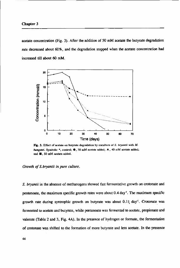
Chapter 3
acetate concentration (Fig. 3). After the addition of 50 mM acetate the butyrate degradation
rate decreased about 60%, and the degradation stopped when the acetate concentration had
increased till about 60 mM.
60 70 20 30 40 50
Time (days) Fig. 3. Effect of acetate on butyrate degradation by coculture of S. bryantii with M.
hungatei. Symbols: *, control; • , 30 mM acetate added; A , 40 mM acetate added;
and • , 50 mM acetate added.
Growth ofS.bryantii in pure culture.
S. bryantii in the absence of methanogens showed fast fermentative growth on crotonate and
pentenoate, the maximum specific growth rates were about 0.4 day"1. The maximum specific
growth rate during syntrophic growth on butyrate was about 0.11 day"1. Crotonate was
fermented to acetate and butyrate, while pentenoate was fermented to acetate, propionate and
valerate (Table 2 and 3, Fig. 4A). In the presence of hydrogen or formate, the fermentation
of crotonate was shifted to the formation of more butyrate and less acetate. In the presence
66

Butyrate degradation
Table 2. Degradation of pentenoate by S. bryantii in the presence and absence of butyrate
and M. hungatei
Substrates Substrate degraded(mmol/l) Products formed(mmol/l)
pentenoate butyrate valerate acetate propionate CH,
pentenoate
pentenoate+ butyrate
pentenoate+ butyrate + M. hungatei
8.7±0.5
19.4±1.S 6±0.l
3.3+0.1 4.8+0.1 5.2±0.5 0
17.8±0.7 17.5+0.1 5.3±0.2 0
17±1.6 7.2±0.6 13.1±0.1 22.6±1.7 8.1±0.2 2.7±0.2
butyrate + M. hungatei 16.5±0.9 0 33.3+0.4 0 13.9±0.1
Table 3. Effect of hydrogen and formate on crotonate conversion by S. bryantii
substrates (mM)
crotonate
crotonate + 20mM formate
crotonate + 0.6atm.H2
crotonate degraded (mM)
21.7+0.2
19.2±0.4
18.5±0. 3
butyrate produced (mM)
8.7+0.5
15.0+0.2
14.6+0.2
acetate produced (mM)
19.9 + 1.1
15.0+0.2
16.3+0.2
cell protein (ing/1)
63+5
56±5
53 ±5
degrading velocity (mM/h)
0.2
0.1
0.2
67

Chapter 3
of hydrogen or formate the fermentation
of pentenoate was shifted to the
formation of more valerate and less
propionate and acetate (data not shown).
The presence of these electron donors
led to a somewhat lower specific growth
rate on crotonate and lower molar cell
yields (Table 3). When S. bryantii was
grown on pentenoate plus butyrate, these
compounds were degraded simutaneously
(Fig. 4B). In the presence of butyrate,
more valerate and less propionate was
formed compared with the amounts
which had been formed after growth on
pentenoate alone (table 2). This indicates
2 3 4 5 6 7
Time (days)
Fig. 4. Degradation of pentenoate by S. bryantii in the absence (A) and presence (B) of butyrate, and butyrate degradation with pentenoate as electron acceptor (B) by crotonate-pregrown culture of S. bryantii. Symbols: O, pentenoate; A, valerate; • , butyrate; A, acetate; D propionate; • , formate.
that pentenoate can act as an electron acceptor during butyrate oxidation, and is thus able to
replace methanogens. Butyrate degradation stopped when the pentenoate and butyrate had
reached concentrations of about 3.5 mM, but pentenoate was degraded further (Fig. 4B).
Remarkably, about 0.3 mM formate was observed at day 4 of the incubation and this
concentration remained constant thereafter. In this culture, the hydrogen concentration
increased till about 300 Pa. When a coculture of S. bryantii and M. hungatei was grown on
pentenoate plus butyrate, methane was formed as well (Table 2). It can be calculated that
68
Butyrate degradation
under these conditions about 75 % of the electrons produced in the oxidation of butyrate to
acetate were transferred to the methanogen, whereas the remainder was used to reduce
pentenoate to valerate.
Hydrogenase and formate dehydrogenase activities.
The specific activity of methylviologen-coupled hydrogenase in cell-free extract of crotonate-
grown cells of S. bryantii was 59.4 /tmol.min'.mg"1 protein. The maximum specific activity
of formate dehydrogenase was 0.045 /tmol.mirï'.mg"1 protein. For the two enzymes an
apparent KM value of 0.21 mM H2 and 0.22 mM formate was determined in cell extracts.
Discussion
S. bryantii can grow on crotonate in pure culture (Zhao et al. 1989). This property allowed
us to investigate the role of hydrogen and formate transfer in reconstructed methanogenic
cocultures. Butyrate was degraded only in cocultures with methanogens which can use both
H2 and formate, like M. hungatei and M.formicicum, but not in cocultures with methanogens
which can use only H2, like M. arboriphilus nor which can only use acetate, like M.
soehngenii. This suggests that formate plays an important role in the syntrophic degradation
of butyrate. These findings give support to the hypothesis that in suspended cultures formate
transfer is more important than hydrogen transfer. Dwyer et al. (1988) pointed out that the
rate of hydrogen consumption during syntrophic degradation is largely determined by the
69
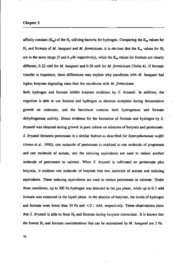
Chapter 3
affinity constant (K^ of the H2 utilizing bacteria for hydrogen. Comparing the KM values for
H2 and formate of M. hungatei and M. formicicum, it is obvious that the KM values for H2
are in the same range (5 and 6 /*M respectively), while the KM values for formate are clearly
different, 0.22 mM for M. hungatei and 0.58 mM for M. formicicum (Table 4). If formate
transfer is important, these differences may explain why cocultures with M. hungatei had
higher butyrate degrading rates than the cocultures with M. formicicum.
Both hydrogen and formate inhibit butyrate oxidation by S. bryantii. In addition, the
organism is able to use formate and hydrogen as electron acceptors during fermentative
growth on crotonate, and the bacterium contains both hydrogenase and formate
dehydrogenase activity. Direct evidence for the formation of formate and hydrogen by S.
bryantii was obtained during growth in pure culture on mixtures of butyrate and pentenoate.
S. bryantii ferments pentenoate in a similar fashion as described for Syntrophomonas wolfei
(Amos et al. 1990); one molecule of pentenoate is oxidized to one molecule of propionate
and one molecule of acetate, and the reducing equivalents are used to reduce another
molecule of pentenoate to valerate. When S. bryantii is cultivated on pentenoate plus
butyrate, it oxidizes one molecule of butyrate into two molecule of acetate and reducing
equivalents. These reducing equivalents are used to reduce pentenoate to valerate. Under
these conditions, up to 300 Pa hydrogen was detected in the gas phase, while up to 0.3 mM
formate was measured in the liquid phase. In the absence of butyrate, the levels of hydrogen
and formate were lower than 30 Pa and <0.1 mM, respectively. These observations show
that S. bryantii is able to form H2 and formate during butyrate conversion. It is known that
the lowest H2 and formate concentrations that can be maintained by M. hungatei are 3 Pa
70
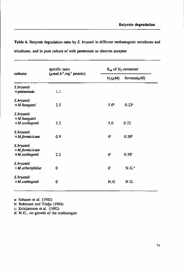
Butyrate degradation
Table 4. Butyrate degradation rates by S. bryantii in different methanogenic cocultures and
tricultures, and in pure culture of with pentenoate as electron acceptor
cultures specific rates (^mol.h'.mg*1 protein)
KM of H2-consumer
H2(/xM) formateO*M)
S. bryantii +penteonate 1.1
S. bryantii +M.hungatei 2.5
S.bryantii +M.hungatei +M.soehngenii 3.2
S. bryantii +M.formicicum 0.9
S. bryantii +M.formicicum +M.soehngenii 2.2
S. bryantii +M.arboriphilus 0
S. bryantii -+M.soehngenii 0
5.0"
5.0
N.G
0.22°
0.22
0.58*
0.58"
N.G.d
N.G.
a: Schauer et al. (1982) b: Robinson and Tiedje (1984) c: Kristjansson et al. (1982) d: N.G., no growth of the methanogen
71

Chapter 3
(Cord-Ruwisch et al. 1988) and 26 /xM (Schauer et al. 1982) respectively. Thus the
concentration difference of formate between S. btyantii and M. hungatei is about 150 times
higher than the concentration difference of H2 between the two species. The diffusion
coefficients of H2 and formate in water are 0.0045 and 0.0015 mm2.s\ respectively (Boone
et al. 1989). By using a simple Ficks diffusion equation (Schink and Thauer 1988), it can be
calculated that the flux of formate can be about 50 times as high as the flux of hydrogen,
indicating that formate transfer is more important than hydrogen transfer in our coculture.
Enzyme measurements by Wofford et al. (1986) showed that S. wolfei oxidized butyrate via
the crotonyl-CoA pathway. A similar pathway is present in S. bryantii (Schink 1992). Two
oxidation steps occur in this pathway, the butyryl-CoA oxidation to crotonyl-CoA and the 3-
hydroxybutyryl-CoA oxidation to acetoacetyl-CoA. Both reactions are thermodynamically
unfavorable when coupled to hydrogen and formate formation. The Gibbs free energy change
of the oxidation of butyryl-CoA to crotonyl-CoA and 3-hydroxybutyryl-CoA to acetoacetyl-
CoA are +75.2 and +29.3 kJ/mol when coupled to hydrogen formation, and +73.9 and 28
kJ/mol when coupled to formate formation (Thauer et al. 1977). Because S. bryantii is able
to produce hydrogen and formate, these calculations show that both hydrogen and formate
will inhibit butyrate oxidation, irrespectively whether hydrogen or formate transfer is most
important.
Although acetate is one of products in butyrate oxidation, cocultivation with the aceticlastic
methanogen M. soehngenii is not sufficient to allow butyrate degradation by S. bryantii.
However, methanogenic tricultures of S. bryantii, M. soehngenii and M. hungatei or M.
formicicum had a higher degrading rate than the cocultures without M. soehngenii. This
72
Butyrate degradation
indicates that a low acetate concentration is an additional requirement for achieving high
butyrate degradation rates in methanogenic habitats. Similar observations have been found
by Ahring and Westermann (1987, 1988) and Beaty and Mclnerney (1989).
Acknowledgement. This research was made possible by a grant of the Wageningen
Agricultural University.
References
Ahring BK, Westermann P (1987) Thermophilic anaerobic degradation of butyrate by a
butyrate-utilizing bacterium in coculture and triculture with methanogenic bacteria.
Appl Environ Microbiol 53:429-433
Ahring BK, Westermann P (1988) Product inhibition of butyrate metabolism by acetate and
hydrogen in a thermophilic coculture. Appl Environ Microbiol 54:2393-2397
Amos DA, Mclnerney MJ (1990) Growth of Syntwphomonas wolfei on unsaturated short
chain fatty acids. Arch Microbiol 154:31-36
Beaty PS, Mclnerney MJ (1987) Growth of Syntwphomonas wolfei in pure culture on
crotonate. Arch Microbiol 147:389-393
Beaty PS, Mclnerney MJ (1989) Effect of organic acid anions on the growth and metabolism
of Syntwphomonas wolfei in pure culture and in defined consortia. Appl Environ
Microbiol 55:977-983
73

Chapter 3
Boone DR, Johnson RL, Liu Y (1989) Diffusion of the interspecies electron carriers H2 and
formate in methanogenic ecosystems and its implications in the measurement of KM
for H2 or formate uptake. Appl Environ Microbiol 55:1735-1741
Bryant MP, Boone DR (1987) Isolation and characterization of Methanobacteriumformicicum
MF. Int J Syst Bacteriol 37:171
Cord-Ruwisch R, Seitz HJ, Conrad R (1988) The capacity of hydrogenotrophic anaerobic
bacteria to complete traces of hydrogen depends on the redox potential of the
terminal electron acceptors. Arch Microbiol 149:350-357
Dong X, Schyns PJYMJ, Stams AJM (1991) Degradation of galactomannan by a Clostridium
butyricum strain. Antonie van Leeuwenhoek 60:109-114
Dwyer DF, Weeg-Aerssens E, Shelton DR, Tiedje JM (1988) Bioenergetic conditions of
butyrate metabolism by a syntrophic, anaerobic bacterium in coculture with hydrogen-
oxidizing methanogenic and sulfidogenic bacteria. Appl Environ Microbiol 54:1354-
1359
Ferry JG, Smith PH, Wolfe RS (1974) Methanospirillum, a new genus of methanogenic
bacteria, and characterization of Methanospirillum hungatei sp. nov. Int J Syst
Bacteriol 24:465-469
Henson JM, Smith PH (1985) Isolation of a butyrate-utilizing bacterium in coculture with
Methanobacterium thermoautotrophicum from a thermophilic digester. Appl Environ
Microbiol 49:1461-1466
Houwen FP, Dijkema C, Schoenmakers CHH, Stams AJM, Zehnder AJB (1987) 13C-NMR
study of propionate degradation by a methanogenic coculture. FEMS Letters 41:269-
74

Butyrate degradation
274
Houwen FP, Cheng G, Folker GE, van den Heuvel WMJG and Dijkema C (1988) Pyruvate
and fumarate conversion by a methanogenic propionate-oxidizing coculture. p 62-70.
In: Lettinga G, Zehnder AJB, Grotenhuis JTC, Hulshoff Pol (eds). Granular
anaerobic sludge; Microbiology and Technology. Puduc, Wageningen, the
Netherlands.
Huser BA, Wuhrmann K, Zehnder AJB (1982) Methanothrix soehngenii gen. nov., a new
acetotrophic nonhydrogen-oxidizing methane bacterium. Arch Microbiol 132:1-9
Jetten MSM, Stams AJM, Zehnder AJB (1990) Acetate threshold values and acetate
activating enzymes in methanogenic bacteria. FEMS Microbiol Ecol 73:339-344
Kristjansson JK, Schönheit P, Thauer RK (1982) Different Ks valus for hydrogen of
methanogenic bacteria and sulfate reducing bacteria: an explanation for the apparent
inhibition of methanogenesis by sulfate. Arch Microbiol 131:278-282
Lamed R, Zeikus JG (1980) Glucose fermentation pathway of Thermoanaerobium brockii.
JBacteriol 141:1251-1257
Leonhardt U, Andreesen JR (1977) Some properties of formate dehydrogenase,
accumulation and incorporation of '85W-tungsten into proteins of Clostridium
formicoaceticum. Arch Microbiol 115:277-284
Lowry OH, Rosebrough NJ, Farr AL, Randau RJ (1951) Protein measurement with the folin
phenol reaction. J Biol Chem 193:265-275
Mah RA, Xun LY, Boone DR, Ahring B, Smith PH, Wilkie A (1990) Methanogenesis from
propionate in sludge and enrichment systems, p 99-111. In: Bélaich J-P, Bruschi M,
75
Chapter 3
Garcia J-L (eds) Microbiology and biochemistry of strict anaerobes involved in
interspecies hydrogen transfer. FEMS Symposium No.54. Plenum Press. New York.
Mclnerney MJ, Bryant M P, Pfennig N (1979) Anaerobic bacterium that degrades fatty acids
in syntrophic association with methanogens. Arch Microbiol 122:129-135
Mclnerney MJ, Bryant MP, Hespell RB, Costerton JW (1981a) Syntrophomonas wolfei gen.
nov., sp. nov., an anaerobic syntrophic, fatty acid-oxidizing bacterium. Appl
Environ Microbiol 41:1029-1039
Mclnerney MJ, Mackie RI, Bryant MP (1981b) Syntrophic association of a butyrate-
degrading bacterium and Methanosarcina enriched from bovine rumen fluid. Appl
Environ Microbiol 41:826-828
Robinson JA, Tiedje JM (1984) Competition between sulfate-reducing and methanogenic
bacteria for H2 under resting and growing conditions. Arch Microbiol 137:26-32
Schauer NL, Brown DP, Ferry JG (1982) Kinetics of formate metabolism in
Methanobacterium formicicum and Methanospirillum hungatei. Appl Environ
Microbiol 44:540-554
Schink B, Thauer RK (1988) Energetics of syntrophic methane formation and the influence
of aggregation, p 5-17. In: Lettinga G, Zehnder AJB, Grotenhuis JTC, Hulshoff Pol
(eds). Granular anaerobic sludge; Microbiology and Technology. Puduc,
Wageningen, the Netherlands.
Schink B (1992) Syntrophism among prokaryotes. p 276-299. In: Balows A, Trüper HG,
Dworkin M, Harder W, Schleifer K-H (eds). The Prokarotes (2°a ed). Springer-
Verlag, New York.
76

Butyrate degradation
Seitz HJ, Schink B, Conrad R (1988) Thermodynamics of hydrogen metabolism in
methanogenic cocultures degrading ethanol or lactate. FEMS Microbiol Letters
55:119-124
Stieb M, Schink B (1985) Anaerobic oxidization of fatty acids by Clostridium bryantii sp.
nov., a sporeforming, obligately syntrophic bacterium. Arch Microbiol 140:387-390
Thauer RK, Jungermann K, Decker K (1977) Energy conservation in chemotrophic anaerobic
bacteria. Bacteriol Rev 41:100-180
Thiele JH, Zeikus JG (1988) Control of interspecies electron flow during anaerobic digestion:
significance of formate transfer versus hydrogen transfer during syntrophic
methanogenesis in floes. Appl Environ Microbiol 54:20-29
Wofford NQ, Beaty PS, Mclnerney MJ (1986) Preparation of cell-free extract and the
enzymes involved in fatty acid metabolism in Syntrophomonas wolfei. J Bacteriol
167:179-185
Zeikus JG, Henning DL (1975) Methanobacterium arbophilium sp. nov. An obligate
anaerobe isolated from wetwood of living trees. Antonie van Leeuwenhoek 41:543-
552
Zhao H, Yang D, Woese CR, Bryant MP (1989) Assignment of the syntrophic, fatty acid-
degrading anaerobe Clostridium bryantii to Syntrophospora bryantii gen. nov.,comb.
nov. Int J Syst Bacteriol 40:40-44
77

Chapter 4
Evidence for H2 and formate formation during syntrophic
butyrate and propionate degradation
Xiuzhu Dong and Alfons J. M. Stams Anaerobe (Submitted)

Chapter 4
Abstract
Both H2 and formate were formed during butyrate oxidation by Syntrophospora bryantii with
pentenoate as electron acceptor and during propionate oxidation by a mesophilic propionate
oxidizing bacterium (MPOB) with fumarate as electron acceptor. H2 and formate levels were
affected by the bicarbonate concentration. S. bryantii and MPOB were also able to
interconvert formate and H2 + HC03"; the apparent KM values for formate were of 2.9 mM
and 1.8 mM, respectively. The conversion of H2 + HC03 to formate occurred only when
the H2 partial pressure was above 80 KPa. This interconversion seems rather unimportant
under conditions prevailing during syntrophic propionate and butyrate oxidation.
Key words: H2, formate, syntrophic degradation, butyrate oxidation, propionate oxidation,
interspecies electron transfer, methanogenesis
Introduction
Butyrate and propionate are two important intermediates in the mineralization of
organic matter under methanogenic conditions. These fatty acids are degraded by syntrophic
consortia of acetogenic bacteria and methanogenic bacteria [1, 2]. Complete mineralization
of butyrate and propionate into methane and C02 requires three different types of bacteria.
Acetogenic bacteria oxidize butyrate and propionate into acetate and H2/formate (Table 1,
equation 1, 2, 5 and 6). These products are further converted to methane by H2/formate-
80

H2 and formate formation
utilizing and aceticlastic methanogens (Table 1, equation 9, 10 and 11), respectively. Because
of unfavorable energetics of propionate and butyrate oxidation, the H2 and/or formate levels
have to be kept extremely low. At low H2 and formate concentrations the AG' values of
butyrate and propionate oxidation become negative. Interspecies H2 and formate transfer are
proposed as possible mechanisms for the syntrophic degradation by consortia [3-5]. Although
both H2 and formate were detected in the methanogenic cocultures when methanogenesis was
inhibited [4, 6], it is difficult to conceive whether H2 or formate is more important. This in
particular because methanogens are able to interconvert H2/C02 and formate [7, 8]. Up to
now, direct evidence is lacking that butyrate-degrading and propionate-degrading acetogens
can form both H2 and formate.
Syntrophomonas wolfei [9] is the first described acetogen which can oxidize butyrate. Since
then several butyrate-oxidizing bacteria were isolated and characterized, such as
Syntrophospora bryantii [10, 11], Syntrophomonas sapovorans [12] and strain SF-1 [13]. It
was demonstrated that S. wolfei and S. bryantii oxidized butyrate by 0-oxidation via the
crotonyl-CoA pathway [14, 1]. Reducing equivalents are released during the oxidations of
butyryl-CoA to crotonyl-CoA and of 3-hydroxybutyryl-CoA to acetoacetyl-CoA. Boone and
Bryant [15] described the first propionate oxidizer Syntrophobacter wolinii growing in a
defined coculture with Desulfovibrio G i l . Recently we enriched a mesophilic propionate
oxidizing-bacterium (MPOB) in coculture with Methanospirillwn strain, and obtained a
methanogen-free culture by growing it on fumarate [16]. Enzyme assays [17, 18] and
experiments of nC and 14C labelled propionate [19-22] have indicated that the methylmalonyl-
CoA pathway is involved in syntrophic propionate oxidation. Reducing equivalents are
81

Chapter 4
Table 1. Reactions involved in the syntrophic degradation of butyrate and propionate
Reactions AG°(kJ/reaction)'
1. Butyrate" + 2 H20
2. Butyrate" + 2 HC03
3. Pentenoate" + H2
4. Pentenoate" + HCOO +H20
— > 2 Acetate" + H+ + 2 H2
— > 2 Acetate" + H+ + 2 HCOO"
— > Valerate"
— > Valerate" + HC03"
48.1
45.5
-75.2"
-73.8"
5. Propionate" + 3 H20
6. Propionate" + 2 HC03"
7. Fumarate2" + H2
8. Fumarate2" + HCOO" + H20
> Acetate" + HC03" + 3 H2 + H+
-> Acetate" + 3HCOO" + H+
-> Succinate2"
-> Succinate2" + HC03
76
72
-86.2
-84.9
9. 4 H2 + HCO3- + H+
10. 4 HCOO" + H20 + H'
11. CH3COO + H20
> CH4 + 2H 20
-> CH4 + 3 HC03
•> CH4 + HC03"
-135.6
-130
-31
12. H, + HCO, - > HCOO" + H,0 -1.3
a, AG°' values were obtained from Thauer et al. [30].
b, It is assumed that the AG°' values of the reduction of pentenoate to valerate are similar to those of the reduction
of crotonate to butyrate.
82

H, and formate formation
formed in the oxidations of succinate to fumarate, malate to oxaloacetate and pyruvate to
acetate, respectively.
In our laboratory, it was found that S. bryantii degraded butyrate in pure culture in the
presence of pentenoate as electron acceptor [6], and a mesophilic propionate-degrading
bacterium (MPOB) [16] degraded propionate with fumarate as electron acceptor. By using
limiting amounts of electron acceptors we describe here that both H2 and formate are
produced by S. bryantii and MPOB. The ratio at which formate and hydrogen are formed
depends on the bicarbonate concentration.
Materials and methods
Organisms and cultivation:
Syntrophospora bryantii (DSM 3014B) in coculture with Methanospirillum hungatei was
obtained from the German Collection of Microorganisms (Braunschweig, Germany). 5.
bryantii was isolated from the coculture with crotonate as substrate as described by Zhao et
al. [11] in a medium with a composition as described previously [21]. Subcultivation was
done with 2.2 g/1 sodium crotonate under a gas phase of 162 KPa N2/C02. The mesophilic
propionate-oxidizing bacterium (MPOB) was described by Stams et al. [16]. Routinely, the
bacteria were grown in a mineral medium described previously [21] containing sodium
fumarate (6.5 g/1) under a gas phase of 162 KPa N2/C02.
83
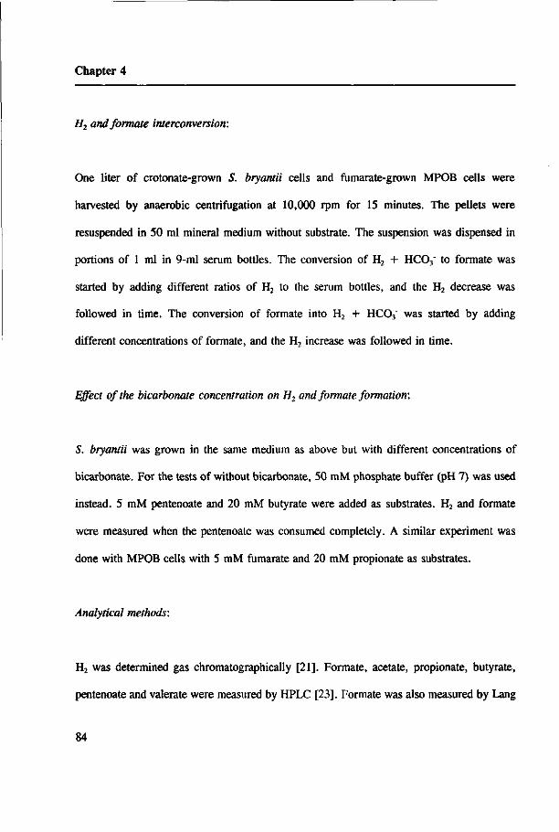
Chapter 4
H2 and formate interconversion:
One liter of crotonate-grown S. bryantii cells and fumarate-grown MPOB cells were
harvested by anaerobic centrifugation at 10,000 rpm for 15 minutes. The pellets were
resuspended in 50 ml mineral medium without substrate. The suspension was dispensed in
portions of 1 ml in 9-ml serum bottles. The conversion of H2 + HC03" to formate was
started by adding different ratios of H2 to the serum bottles, and the H2 decrease was
followed in time. The conversion of formate into H2 + HC03" was started by adding
different concentrations of formate, and the H2 increase was followed in time.
Effect of the bicarbonate concentration on H2 and formate formation:
S. bryantii was grown in the same medium as above but with different concentrations of
bicarbonate. For the tests of without bicarbonate, 50 raM phosphate buffer (pH 7) was used
instead. 5 mM pentenoate and 20 raM butyrate were added as substrates. H2 and formate
were measured when the pentenoate was consumed completely. A similar experiment was
done with MPOB cells with 5 mM fumarate and 20 mM propionate as substrates.
Analytical methods:
H2 was determined gas chromatographically [21]. Formate, acetate, propionate, butyrate,
pentenoate and valerate were measured by HPLC [23]. Formate was also measured by Lang
84

H, and formate formation
& Lang method [24]; where necessary the culture liquid was concentrated on Büchi RE121
Rotavapor at 470 Pa. at 50°C. Whole cell protein was measured by the Lowry method [25].
Results
H2 and formate production by butyrate- and propionate-oxidizing acetogens:
Theoretically, both H2 and formate can be produced during syntrophic oxidation of
propionate and butyrate (table 1). The maximal concentrations of H2 and formate that was
reached during butyrate oxidation by S. bryantii and during propionate oxidation by MPOB
are listed in table 2 and 3, respectively. In the presence of 17 mM butyrate plus 4 mM
pentenoate, S. bryantii degraded 2.1 mM butyrate. Up to 280 pM formate and 171 Pa H2
accumulated in the liquid and in the head space, respectively (Table 2). When the propionate-
oxidizing bacterium MPOB was grown on 14 mM propionate plus 4 mM fumarate, 1.6 mM
propionate was degraded with the available fumarate. In these cultures 24 /iM formate and
6.8 Pa H2 were measured in the liquid and in the head space, respectively (table 3). These
results show that butyrate- and propionate-oxidizing bacteria were indeed able to form both
H2 and formate.
85

Chapter 4
Table 2. H2 and formate formation during butyrate degradation by S. bryantii with pentenoate
as electron acceptor
days H2 formate pentenoate valerate butyrate acetate (Pa) (jM) (mM) (mM) (mM) (mM)
0
7
0
171
0
280
4.1
0
0
4.7
17.3
15.2
0.6
5.2
Table 3. H2 and formate formation during propionate degradation by MPOB using fumarate
as electron acceptor
days H2 formate fumarate succinate propionate acetate (Pa) OiM) (mM) (mM) (mM) (mM)
0
7
0
6.8
n.d
24
4.3
0
1.4
3.3
13.8
12.2
0
1.6
Effect of the bicarbonate concentration on H2 and formate formation:
Because both H2 and formate were produced, the effect of bicarbonate on the relative H2 and
86

H, and formate formation
formate formation by S. bryantii was investigated. Fig.l shows that the H2 and formate
production was dependent on the concentration of bicarbonate. The formate concentration
increased when the bicarbonate concentration was increased, while the H2 level decreased
slightly.
The effect of bicarbonate on H2 and formate production during propionate oxidation by
MPOB was determined as well. H2 accumulated to 44 Pa when the MPOB was grown on 20
mM propionate plus 5 mM fumarate in the absence of bicarbonate, whereas the formate
concentration was below the detection limit (<10 jtM). However, at a bicarbonate
concentration of 50 mM about 24 /*M formate was formed while the H2 partial pressure
amounted only till 6.8 Pa.
0.36 i 10.35
OmM buty. 20mM buty. 20mM buty. 20mM buty. 20mM buty. OmMHCCÇ OmMHCCÇ 5mM HCOs 20mMHCOJ SOmMHCOJ
Fig. 1. Hydrogen (solid bar) and formate (dotted bar) production during butyrate oxidation by S. bryantii at different HCO, concentration with limiting amount of pentenoate as electron acceptor.
87

Chapter 4
H2 and formate interconversion by S. bryantii and MPOB:
As the midpoint redox potentials of H+/H2 and HC037formate are in the same range (-414
mV and -432 mV, respectively), an interconversion between formate and H2/HC03" can be
expected. Fig.2A and 2B shows that S. bryantii and MPOB are indeed able to convert
formate into H2 + HC03". The maximum rate of formate conversion by S. bryantii was 3.8
nmol.min'.mg"1 cell protein, the KM value for formate was 2.9 mM, and the lowest
concentration of formate that could be reached was about 0.5 mM (table 4). The maximum
rate of formate conversion by MPOB was 4.9 nmol.min'.mg"1 cell protein, the KM value for
formate was 1.8 mM, and the lowest concentration of formate that could be reached was
about 0.9 mM (table 4). H2 + HC03' was also converted into formate by the two bacteria.
However, formate formation was detected only when the H2 partial pressure was above 80
KPa (results not shown).
Table 4. Conversion of formate to H2 by S. bryantii and MPOB
KM(mM) Vmax(/iM min'.mg1) lowestconcentration(mM)
S. bryantii 2.9 3.8*10"3 0.5
MPOB 1.8 4.9*10-3 0.9
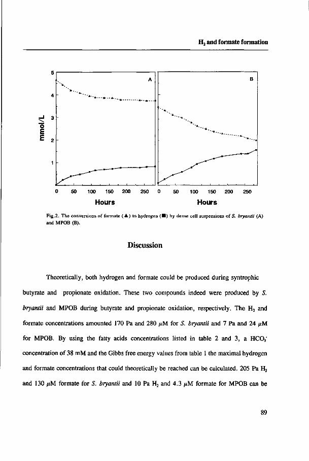
H, and formate formation
o E H 2
A
S^~^~^
B
" A , " - • *
~-A_
0 50 100 150 200 250 0 50 100 150 200 250
Hours Hours
Fig.2. The conversions of formate (A) to hydrogen ( • ) by dense cell suspensions of S. bryantii (A) and MPOB (B).
Discussion
Theoretically, both hydrogen and formate could be produced during syntrophic
butyrate and propionate oxidation. These two compounds indeed were produced by S.
bryantii and MPOB during butyrate and propionate oxidation, respectively. The H2 and
formate concentrations amounted 170 Pa and 280 pM for S. bryantii and 7 Pa and 24 pM
for MPOB. By using the fatty acids concentrations listed in table 2 and 3, a HC03"
concentration of 38 mM and the Gibbs free energy values from table 1 the maximal hydrogen
and formate concentrations that could theoretically be reached can be calculated. 205 Pa H2
and 130 j*M formate for S. bryantii and 10 Pa H2 and 4.3 ^M formate for MPOB can be
89

Chapter 4
deduced. This shows that the measured values are in the same range as what can be expected
from thermodynamical considerations.
By using the highest concentrations of formate and H2 that can be measured with the two
bacteria and the lowest formate and H2 concentrations that can be reached by methanogens,
the fluxes of formate and H2 between consortia in the syntrophic culture can be calculated
using the Fick's diffusion equation [26]. In these calculations diffusion constants of H2 and
formate in water of 4.5*10"5 and 1.5*10"5 cm2.sec"1., respectively, were used. The threshold
values for H2 and formate of M. hungatei are 2 Pa and 15 /xM, respectively [27, 28]. Table
5 shows that the calculated H2 flux in the coculture of MPOB with M. hungatei is too low
to account for the methane formation rate which had been measured [29]. However, the
formate flux is sufficiently high to account for the CH4 formation rate. In the coculture of
S. bryantii with M. hungatei both the calculated H2 flux and formate flux are much higher
than needed to explain the measured CH4 formation rate [6]. However, the formate fluxes
are about 100 times higher than the H2 fluxes in both cocultures. These calculations suggest
that formate transfer is more important in syntrophic butyrate and propionate degradation
than H2 transfer . This conclusion is in accordance with the observations in defined
methanogenic cocultures of the two acetogens, where H2/formate-utilizing but not H2-utilizing
methanogens can drive butyrate or propionate oxidation [6, 29].
The experiments of the bicarbonate effect on formate and H2 formation from butyrate and
propionate suggested that H2 and formate were produced simultaneously during butyrate and
propionate oxidation, i.e. reducing equivalents are used either to reduce protons or to reduce
HC03 . Although S. bryantii and MPOB are able to interconvert H2 + HC03" and formate,
90
H, and formate formation
this process seems to be relatively unimportant during syntrophic propionate and butyrate
oxidation. The concentrations at which this interconversion occurs are far higher than the H2
and formate concentrations which are required for syntrophic degradation. If this
interconversion would occur at low concentrations, H2-utilizing methanogens would have
been able to drive propionate and butyrate oxidation. Clearly, this was not the case [6, 29].
Acknowledgement
This research was made possible by a fellowship of the Wageningen Agricultural University.
91
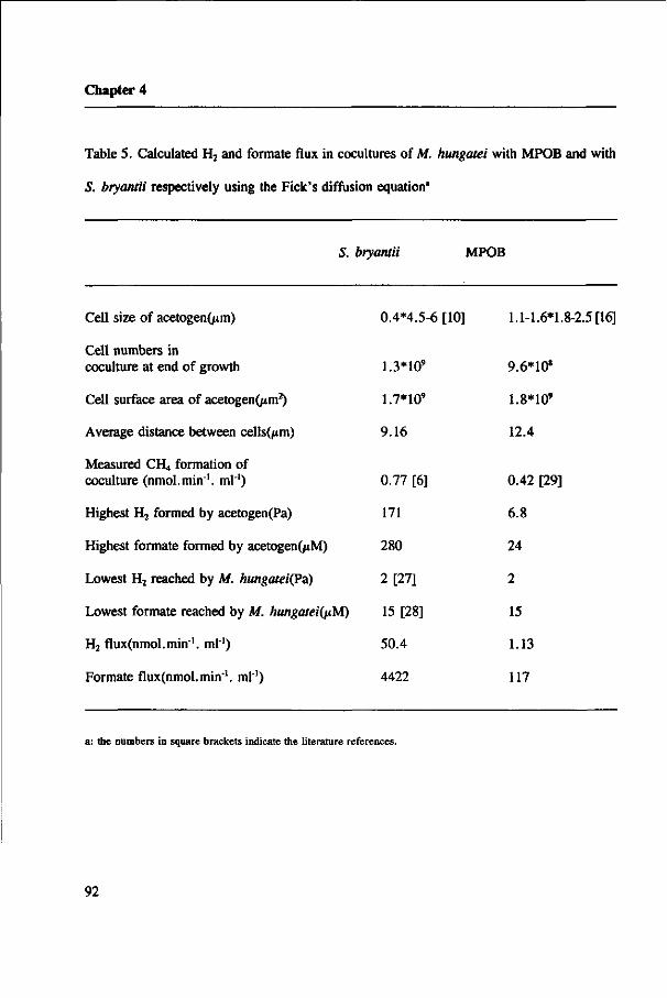
Chapter 4
Table 5. Calculated H2 and formate flux in cocultures of M. hungatei with MPOB and with
S. bryantii respectively using the Fick's diffusion equation"
S. bryantii MPOB
Cell size of acetogen(/*m)
Cell numbers in coculture at end of growth
Cell surface area of acetogen^m2)
Average distance between cells(/tm)
Measured CH4 formation of coculture (nmol.min1. ml"1)
Highest H2 formed by acetogen(Pa)
Highest formate formed by acetogen(/tM)
Lowest H2 reached by M. hungateiÇPd)
Lowest formate reached by M. hungatei(nM)
H2 fluxOimol.min"1. ml"1)
Formate flux(nmol.min'. ml"1)
0.4*4.5-6 [10]
1.3*10'
1.7*10'
9.16
0.77 [6]
171
280
2 [27]
15 [28]
50.4
4422
1.1-1.6*1.8-2.5 [16]
9.6*10*
1.8*10'
12.4
0.42 [29]
6.8
24
2
15
1.13
117
a: the numbers in square brackets indicate the literature references.
92
H2 and formate formation
References
1. Schink B. (1992) Syntrophism among prokaryotes. In Balows A. Trüper H.G. Dworkin
M. Harder W. and Schleifer K.-H. (eds.) The Prokaryotes 2nd ed., pp 276-299. Springer
Verlag, New York
2. Stams A.J.M. (1994) Metabolic interactions between anaerobic bacteria in methanogenic
environments. Antonie van Leeuwenhoek (In press)
3. Hungate R.E. (1967) Hydrogen as an intermediate in the rumen fermentation. Arch
Mikrobiol 59: 158-164
4. Thiele J.H. and Zeikus J.G. (1988) Control of interspecies electron flow during anaerobic
digestion: significance of formate transfer versus hydrogen transfer during syntrophic
methanogensis in floes. Appl Environ Microbiol 54: 20-29
5. Boone D.R. Johnson R.L. and Liu Y. (1989) Diffusion of the interspecies electron carriers
H2 and formate in methanogenic ecosystems, and implications in the measurement of KM for
H2 or formate uptake. Appl Environ Microbiol 55: 1735-1741
6. Dong X. Cheng G. and Stams A.J.M. (1994) Butyrate oxidation by Syrurophospora
bryantii in coculture with different methanogens and in pure culture with pentenoate as
electron acceptor. Appl Microbiol Biotechnol (In press)
7. Wu W.-M. Rickley R.F. Jain M.K. and Zeikus J.G. (1993) Energetics and regulations
of formate and hydrogen metabolism by Methanobacteriwnformicicwn. Arch Microbiol 159:
57-65
8. Bleicher K. and Winter J. (1994) Formate production and utilization by methanogens and
93

Chapter 4
by sewage sludge consortia - interference with the concept of interspecies formate transfer.
Appl Microbiol Biotechnol 40: 910-915
9. Mclnemey M.J. Bryant M.P. Hespell R.B. and Costerton J.W. (1981) Syntrophomonas
wolfei gen. nov. sp. nov., an anaerobic, syntrophic, fatty acid-oxidizing bacterium. Appl.
Environ Microbiol 41: 1029-1039
10. Stieb M. and Schink B. (1985) Anaerobic oxidation of fatty acids by Clostridium bryantii
sp. nov., a sporeforming, obligately syntrophic bacterium. Arch Microbiol 140: 387-390
11. Zhao H. Yang D. Woese C.R. and Bryant M.P. (1990) Assignment of Clostridium
bryantii to Syntrophospora bryantii gen. nov., comb. nov. on the basis of a 16S rRNA
sequence analysis of its crotonate-grown pure culture. Int J Syst Bacteriol 40: 40-44
12. Roy F. Samain E. Dubourguier H.C. and Albagnac G. (1986) Syntrophomonas
sapovorans sp. nov. a new obligately proton reducing anaerobe oxidizing saturated and
unsaturated long chain fatty acids. Arch Microbiol 145: 142-147
13. Shelton D.R. and Tiedje J.M. (1984) Isolation and partial characterisation of bacteria in
an anaerobic consortium that mineralizes 3-chlorobenzoic acid. Appl Environ Microbiol 48:
840-848
14. Wofford N.Q. Beaty P.S. and Mclnemey M.J. (1986) Preparation of cell-free extracts
and the enzymes involved in fatty acid metabolism in Syntrophomonas wolfei. J Bacteriol
167: 179-185
15. Boone D.R. and Bryant M.P. (1980) Propionate-degrading bacterium, Syntrophobacter
wolinii sp. nov. gen. nov., from methanogenic ecosystems. Appl Environ Microbiol 40:626-
632
94

H2 and formate formation
16. Stams A.J.M. van Dijk J. Dijkema C. and Plugge CM. (1993) Growth of syntrophic
propionate-oxidizing bacteria with fumarate in the absence of methanogenic bacteria. Appl
Environ Microbiol 59: 1114-1119
17. Houwen F.P. Plokker J. Dijkema C. and Stams A.J.M. (1990) Enzymatic evidence for
involvement of the methylmalonyl-CoA pathway in propionate oxidation by Syntrophobacter
wolinii. Arch Microbiol 155: 52-55
18. Plugge CM. Dijkema C. and Stams A.J.M. (1993) Acetyl-CoA cleavage pathway in a
syntrophic propionate oxidizing bacterium growing on fumarate in the absence of
methanogens. FEMS Microbiol letters 110: 71-76
19. Koch M. Dolfing J. Wuhrmann K. and Zehnder A.J.B. (1983) Pathway of propionate
degradation by enriched methanogenic cultures. Appl Environ Microbiol 45: 1411-1414
20. Robbins J.E. (1988) A proposed pathway for catabolism of propionate in methanogenic
cocultures. Appl Environ Microbiol 54: 1300-1301
21. Houwen F.P. Dijkema C. Schoenmakers C.H.H. Stams A.J.M. and Zehnder A.J.B.
(1987) "C-NMR study of propionate degradation by a methanogenic coculture. FEMS
Microbiol Letters 41: 269-274
22. Houwen F.P. Dijkema C Stams A.J.M. and Zehnder A.J.B. (1991) Propionate
metabolism in anaerobic bacteria; determination of carboxylation reaction with "C-NMR
spectroscopy. Biochim Biophys Acta 1056: 126-132
23. Dong, X. Schyns P.J.Y.M.J, and Stams A.J.M. (1991) Degradation of galactomannan
by a Clostridium butyricum strain. Antonie van Leeuwenhoek 60: 109-114
24. Lang E. and Lang H. (1972) Spezifische Farbreaktion zum direkten Nachweis der
95

Chapter 4
Ameisensäure. Fresenius Z Anal Chem 260: 8-10
25. Lowry O.H. Rosebrough N.J. Farr A.L. and Randau R.J. (1951) Protein measurement
with the folin phenol reaction. J Biol Chem 193: 265-275
26. Schink B. and Thauer R.K. (1988) Energetics of syntrophic methane formation and the
influence of aggregation. In Lettinga G. Zehnder A.J.B. Grotenhuis J.T.C, and Hulshoff Pol
L.W. (eds), Granular anaerobic sludge; microbiology and technology pp 5-17. Pudoc,
Wageningen
27. Seitz H.J. Schink B. and Conrad R. (1988) Thermodynamics of hydrogen metabolism
in methanogenic cocultures degrading ethanol or lactate. FEMS Microbiol Letters 55: 119-
124
28. Schauer N.L. Brown D.P. and Ferry J.G. (1982) Kinetics of formate metabolism in
Methanobacterium formicicum and Methanospirillum hungatei. Appl Environ Microbiol 44:
540-554
29. Dong X. Plugge CM. and Stams A.J.M. (1994) Anaerobic degradation of propionate
by a mesophilic acetogenic bacterium in coculture and triculture with different methanogens.
Appl Environ Microbiol (In press)
30. Thauer R.K. Jungermann K. and Decker K. (1977) Energy conservation in chemotrophic
anaerobic bacteria. Bacteriol Rev 41: 100-180
96
Chapter 5
Localization of the enzymes involved in H2 and formate
metabolism in Syntrophospora bryantii
Xiuzhu Dong and Alfons J. M. Stams
Antonie van Leeuwenhoek (Submitted)

Chapter 5
Abstract
Cell free extracts of crotonate-grown cells of the syntrophic butyrate-oxidizing bacterium-
Syntrophospora btyantii contained high hydrogenase activities (8.5-75.8 jtmol. min1, mg-1
protein) and relatively low formate dehydrogenase activities (0.04-0.07 /tmol. min1, mg"1
protein). The KM value and threshold value of the hydrogenase for H2 were 0.21 mM and
18 /xM, respectively, whereas the KM value and threshold value of the formate dehydrogenase
for formate were 0.22 mM and 10 ^M, respectively. Hydrogenase, butyryl-CoA
dehydrogenase and 3-OH-butyryl-CoA dehydrogenase were detected in cytoplasmic fraction.
Formate dehydrogenase and C02 reductase were membrane bound, likely located at the outer
aspect of the cytoplasmic membrane. Results suggest that during syntrophic butyrate
oxidation, H2 is formed intracellular^, while formate is formed at the outside of the cell.
Key words: anaerobic degradation, butyryl-CoA dehydrogenase, 3-OH-butyryl-CoA
dehydrogenase, electron transfer, formate dehydrogenase, hydrogenase, Syntrophospora
bryantii
98
Enzyme localization in S. bryantii
Introduction
Syntrophospora bryantü (Stieb & Schink 1985; Zhao et al. 1990) is a gram-positive spore-
forming anaerobic bacterium which oxidizes butyrate to acetate and H2 and/or formate.
Because of unfavorable energetics, S. bryantii can degrade butyrate only if H2 and/or formate
is efficiently removed by methanogens. However, S. bryantii is able to grow in pure culture
by fermentation of crotonate to butyrate and acetate (Zhao et al. 1990) and by oxidation of
butyrate to acetate with pentenoate as the terminal electron acceptor (Dong et al. 1994).
Research with S. bryantii and another butyrate oxidizer, Syntrophomonas wolfei, has
shown that butyrate is oxidized by ß-oxidation via the crotonyl-CoA pathway (Schink 1992;
Wofford et al. 1986). In this pathway, butyrate is activated to butyryl-CoA by a butyrate:
acetyl-CoA HSCoA transferase, which then is oxidized to two acetyl-CoA. One acetyl-CoA
is used to activate butyrate, while the other is converted to acetate via acetyl-phosphate,
resulting in the formation of 1 ATP. The reducing equivalents are released at the energetic
level of FADH2 and NADH during the oxidation of butyryl-CoA to crotonyl-CoA and of 3-
OH-butyryl-CoA to acetoacetyl-CoA, respectively. The oxidation of NADH and particularly
of FADHj coupled to H2 and formate formation (i.e. proton and C02 reduction) is
energetically unfavorable. NADH oxidation becomes possible at low H2 and formate
concentrations. However, for FADH2 oxidation the organism has to invest ATP to drive this
reaction even at low H2 and formate concentrations. A reversed electron flow mechanism has
been proposed for this (Thauer & Morris 1984; Schink 1992).
At present it is still not clear whether H2 transfer or formate transfer is most
important in the syntrophic degradation of butyrate by S. bryantii. In a previous study we
99

Chapter 5
have found that S. bryantii can oxidize butyrate in coculture with methanogens that can use
H2 and formate, but not in coculture with methanogens that can only use H2 (Dong et al.
1994). In addition, S. bryantii was able to produce both H2 and formate during butyrate
oxidation (unpublished results). The aim of this study was to elucidate how intracellularly
reducing equivalents released during butyrate oxidation are channelled to the terminal
electron acceptors H+ and HC03".
Materials and methods
Organism and cultivation
The biculture of Syntrophospora bryantii and Methanospirillum hungatei (DSM 3014B) was
obtained from the German culture collection of microorganisms (DSM, Braunschweig,
Germany). S. bryantii was isolated by us from the biculture by using media with sodium
crotonate (2.2 g/1) as carbon source. This was done as described by Zhao et al. (1990). The
bacterium was cultivated in 1-liter bottles in a medium as described previously (Dong et al.
1994).
Preparation of cell extracts and cell fractionation
Unless indicated otherwise, all the manipulations were done in an anaerobic glove box with
a N2/H2 (96:4) atmosphere. Traces of oxygen were removed by a palladium catalyst (BASF,
Arnhem, The Netherlands). Crotonate-grown cells were harvested in the late-log-phase by
100
Enzyme localization in S. bryantü
anaerobic centrifugation at 10,000 g for 15 min., and the cell pellet was suspended in 100
mM Tris-HCl buffer (pH 7.5) containing 250 mM sucrose, 20 mM KCl, 5 mM MgCl2 and
5 mM dithiothreitol (DTT). The cell suspension was passed twice through a French pressure
cell at 40 MPa. Unbroken cells and cell debris were removed by centrifugation at 11,000 g
at 4°C for 20 min.. The supernatant was designated as cell-free extract. For fractionation
into a cytoplasmic fraction and a membrane fraction, the cell-free extract was ultracentrifuged
at 140,000 g at 4°C for 1.5 h. The pellet designated as membrane fraction was resuspended
in the same buffer as described above.
Cell fractionation was also done after an osmotic shock treatment. For this 1.5 g cells
(wet weight) were suspended in 10 ml 50 mM Tris-HCl buffer (pH 7.6) containing 0.5 M
sucrose. A lysozyme solution was added (50 pg/ml, final concentration), and the suspension
was incubated for 30 min. at room temperature. Thereafter, the suspension was centrifuged
at 17,500 g at 4°C for 20 min.. The spheroplast pellet was resuspended in the same buffer
as described above but without sucrose to produce a "spheroplast lysate". A membrane
fraction and a cytoplasmic fraction were obtained by centrifuging at 17,500 g at 4°C for 20
min.. The preparations were stored under N2 in 9-ml serum bottles at 4°C. All the enzyme
activities were analyzed within 8 hours after preparation.
Enzyme assays
All the enzyme assays were performed anaerobically under N2 (H2 for hydrogenase assay)
at 37°C in 1-cm cuvettes sealed with rubber stoppers. The reduction of electron acceptors
and the oxidation of electron donors were followed spectrophotometrically using a Beekman
DU 7500 spectrophotometer. One unit of enzyme activity is defined as the amount of enzyme
catalyzing the conversion of 1 /*mol substrate per min.. Protein content was determined using
101

Chapter 5
the method of Bradford (1976).
Benzyl viologen-coupled hydrogenase and benzyl viologen-coupled formate
dehydrogenase activities were measured at pH 8.0 as described by Lamed & Zeikus (1980)
and Leonhart & Andreesen (1977), respectively. Benzyl viologen-coupled C02 reductase was
determined at pH 6.8 according to Thauer et al. (1973). MTT-coupled NADH dehydrogenase
was measured in 50 mM Hepes-KOH buffer (pH 7.5) as described by Bergsma et al. (1982).
Dichlorophenolindophenol (DCPIP)-coupled butyryl-CoA dehydrogenase and NADH-
dependent 3-OH-butyryl-CoA dehydrogenase were assayed as described by Wofford et al.
(1986). ATPase was measured by the method of Vogel & Steinhart (1976). NAD-, NADP-,
FMN-, FAD- and ferredoxin-dependent hydrogenase and formate dehydrogenase were
measured at pH 7.5 as described by Schauer & Ferry (1982). The following extinction
coefficients were used: NAD and NADP, e334=6.18 mM'.cm"1; FMN, e450=41 mM'.cm"';
FAD, e450=44.3 mM'.cm1; ferredoxin, e390=16 mM'.cm'. Benzyl viologen, e57g=9.2
mM'.cm'; DCPIP, e6oo=21 mM'.cm"' and MTT, e578=13 mM'.cm'.
Chemicals
Enzymes and cofactors for enzyme assays were obtained from Boehringer Mannheim
(Almere, The Netherlands). The chemicals which were at least of analytical grade were
obtained from Sigma Chemical Co. (Amsterdam, The Netherlands) or Merck (Darmstadt,
Germany). Butyryl-CoA and ô-acetoacetyl-CoA were purchased from Aldrich Chemie
(Brussel, Belgium). Phenazine methosulfate (PMS) was obtained from Janssen chimica
(Tilburg, The Netherlands) and dichlorophenolindophenol (DCPIP) from Fluka Chemika-
Biochemika (Breda, The Netherlands).
102

Enzyme localization in S. bryantü
Results
Hydrogenase and formate dehydrogenase activities in cell extract ofS. bryantü
Extracts of S. bryantii cells grown on crotonate contained very high hydrogenase activities
(Table 1). In the several experiments which had been done the hydrogenase activity was
never below 8.5 ftmol.min'.mg1. In contrast formate dehydrogenase activities were lower
(Table 1). The highest formate dehydrogenase activity measured in crude extracts was 0.07
ftmol. min'1. mg"1. The V,,,« values, the KM values and the threshold values of the two enzymes
are shown in table 1. The activities of hydrogenase and formate dehydrogenase of cells
grown on pentenoate plus butyrate and of cells grown on crotonate plus formate were in the
same range as the activities in crotonate-grown cells ( data not shown).
Table 1. Hydrogenase and formate dehydrogenase activités measured in cell extracts of S.
bryantii '
activity KM (mM) Vmax (/tmol.min'.mg1) Threshold valueb (JJM)
hydrogenase 0.21 8.5-75.8 18
formate dehydrogenase 0.22 0.04-0.07 10
a: All values refer to H2 and formate concentrations in the liquid. It was assumed that the soluble H2 concentration
was 0.59 mM when 105 Pa H2 was present in the gas phase (Boone et al. 1989).
b: These values refer to the lowest concentration at which activity could be measured in the standard assay.
103

Chapter 5
Enzyme activities of intact cells and of cell fractions prepared by French pressure lysis
The enzymatic activities in the different cell fractions prepared by French press disrupture
are listed in Table 2. Some activities could be measured with whole cells. Benzyl viologen-
coupled hydrogenase activity and MTT-coupled NADH dehydrogenase activity increased
upon cell lysis. However, the benzyl viologen-coupled formate dehydrogenase activity of
whole cells was the same as the activity in cell extracts. Upon fractionation of the crude
extracts, up to 95% of the hydrogenase activity and 75% the NADH dehydrogenase activity
were found in the soluble fraction, while up to 50% of the formate dehydrogenase activity
was recovered in the membrane fraction. About 90% of the DCPIP-coupled butyryl-CoA
dehydrogenase and the NAD-coupled 3-OH-butyryl-CoA dehydrogenase activities were
determined in the soluble fraction of the cell extract. Benzyl viologen-coupled CO2 reductase
activity was only found in the membrane fraction. Upon fractionation 88% of the presumably
membrane-bound ATPase was recovered in soluble fraction. This indicates that cell breakage
through French press releases some membrane-bound proteins into the soluble fraction.
Enzyme activities in membrane and cytoplasm fraction prepared by osmotic shock lysis
Table 3. shows that the ATPase activity was totally recovered in the membrane fraction when
the osmotic shock method was applied. This indicates that cell breakage by this method is
much milder than by the French pressure method. Butyryl-CoA dehydrogenase and 3-OH-
butyryl-CoA dehydrogenase activities again were mainly detected in the cytoplasmic fraction.
Hydrogenase and formate dehydrogenase activities were recovered in the membrane fraction
104
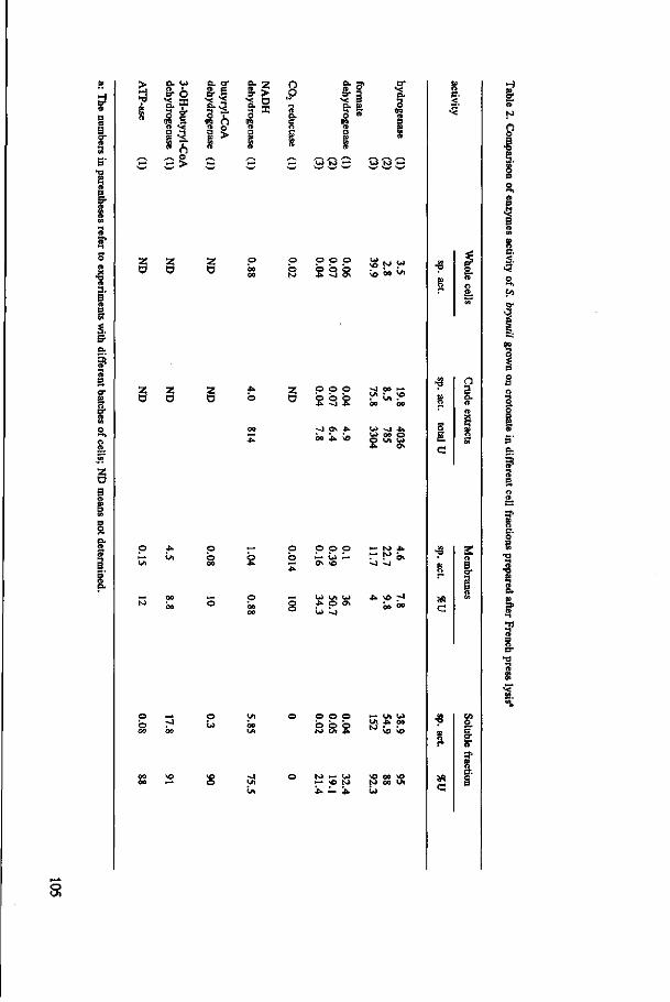
3 a-
> 5
»
,—s
£2
o. w 8-6 'S.? 5 o* <g I » *< 1 3
^5 ,-v o w >
o. a-a- S
1 S ft o
1* ft
,—s,
•̂ -
s- ^
IS 0
ft I ft ^-s
^
n
Q. C O
1 ft , ^ - y
^ .— w wr*
•g 3. 1 D S?
o i o ; io w
2! ö Z
O z ö o o o b o b
•u -4 5
o. p *• o
b o o O H - W >—
o — b r-»
H fr
n o B
o
•3
»
» c
g a R o-
S O
CT •O
SS C
O
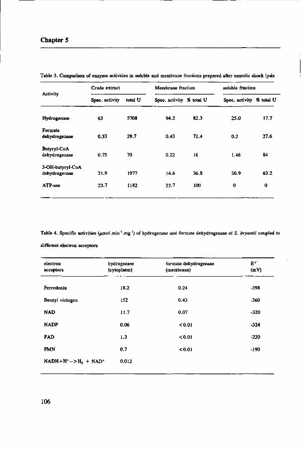
Chapter 5
Table 3. Comparison of enzyme activities in soluble and membrane fractions prepared after osmotic shock lysis
Activity
Hydrogenase
Formate dehydrogenase
Butyryl-CoA dehydrogenase
3-OH-butyryl-CoA dehydrogenase
ATP-ase
Crude extract
Spec, activity
63
0.33
0.75
21.9
23.7
total U
5708
29.7
70
1977
1182
Membrane fraction
Spec, activity
94.2
0.43
0.22
14.6
23.7
% total U
82.3
72.4
16
36.8
100
soluble fraction
Spec, activity
25.0
0.2
1.46
30.9
0
% total U
17.7
27.6
84
63.2
0
Table 4. Specific activities (/imol.inin'.mg "') of hydrogenase and formate dehydrogenase of S. bryantii coupled to
different electron acceptors
electron acceptors
Ferredoxin
Benzyl viologen
NAD
NADP
FAD
FMN
NADH+H+-->H2 + NAD+
hydrogenase (cytoplasm)
18.2
152
11.7
0.06
1.3
0.7
0.012
formate dehydrogenase (membrane)
0.24
0.43
0.07
<0.01
«C0.01
<0.01
E°' (mV)
-398
-360
-320
-324
-220
-190
106

Enzyme localization in S. bryantii
for 82% and 72%, respectively. The specific activities of hydrogenase and formate
dehydrogenase in the cell lysate after osmotic shock treatment were 1.5-8 times higher than
after French pressure lysis. Butyryl-CoA dehydrogenase and 3-OH-butyryl-CoA
dehydrogenase activities were also 3-5 times higher, both in the membrane fraction and in
the soluble fraction, than those prepared by French pressure. ATPase activity in the
membrane fraction was up to 160 times higher when cells were disrupted by osmotic shock.
Hydrogenase and formate dehydrogenase ofS. bryantii coupled to different electron acceptors
Different electron carriers could be used in the hydrogenase and the formate dehydrogenase
assays (Table 4). Hydrogenase could be assayed with NAD, FAD or FMN and ferredoxin.
However, the activities were much lower than with benzyl viologen. Formate dehydrogenase
could also be assayed with NAD and ferredoxin, also these activities were lower than when
benzyl viologen was used.
Discussion
Butyrate is degraded by acetogenic bacteria by ß-oxidation via the crotonyl-CoA pathway.
The key enzymes of this pathway have been demonstrated in Syntrophomonas wolfei
(Wofford et al. 1986) and in S. bryantii (Schink 1992). The enzyme activities found in this
study were in the same range as those reported by Dörner (1992) and by Mclnerney &
Wofford (1992). In butyrate-oxidizing acetogenic bacteria, reducing equivalents released in
the oxidation of butyryl-CoA to crotonyl-CoA and the oxidation of 3-OH-butyryl-CoA to
107
Chapter 5
acetoacetyl-CoA have to be channelled to protons and/or to bicarbonate, resulting in the
formation of H2 and formate. To get insight into the intracellular electron flow we
determined the localization of redox proteins in S. bryanrii. For this purpose we used two
different cell lysis methods, the French pressure method and the osmotic shock lysis method.
It should be mentioned that results of enzyme localization measurements have to be
interpreted with caution because membrane-associated enzymes can be recovered as soluble
proteins, as was the case with the membrane-bound ATPase using French pressure lysis
method. An additional complicating factor is that cells of J. bryantii lyse rather rapidly after
growth has ceased, causing that some intracellular enzymes apparently can be measured with
whole cells.
The butyryl-Co A dehydrogenase and 3-OH-butyryl-Co A dehydrogenase activities were
mainly found in the cytoplasmic fraction, and the specific activities of the two enzymes in
soluble fractions were higher than in the membrane fractions. These results suggest that the
two enzymes are located in the cytoplasm. However, the energetically highly unfavorable
oxidation of butyryl-CoA coupled to H2 or formate formation in one way or the other has to
be associated with the membrane. The butyryl-CoA dehydrogenase was found to be only
partly membrane bound in S. wolfei (Dörner 1992; Wallrabenstein & Schink 1994). If the
butyryl-CoA dehydrogenase is a cytoplasmic protein, a membrane bound FADH2
dehydrogenase has to be present.
The benzyl viologen-coupled hydrogenase activity can be measured with whole cells.
However, the activity was increased 2-5 times upon cell breakage by French press lysis.
Because benzyl viologen is almost membrane-impermeable, this indicates that the enzyme is
mainly intracellularly located. Moreover, the majority of the hydrogenase activity was
recovered in the soluble fraction of a French pressure lysate, which also suggests that the
108

Enzyme localization in S. bryantü
hydrogenase of S. bryantii is likely located in the cytoplasm. The majority of the
hydrogenase was found in the membrane fraction of an osmotic shock lysate, which might
indicate that the hydrogenase is partly membrane bound as was also observed in S. wolfei
(Dörner 1992; Wallrabenstein & Schink 1994). Taking all these observations into account,
H2 most likely is formed intracellularly in the cytoplasm or at the inner aspect of the
cytoplasmic membrane.
Formate dehydrogenase of S. bryantii most likely is a membrane-bound protein which
is localized at the outer aspect of the membrane. This conclusion is based on the observation
that: i) the formate dehydrogenase activity in the membrane fraction was about 8 times
higher than in the cytoplasm fraction, ii) the benzyl viologen-coupled formate dehydrogenase
activity was not increased by cell breakage, and iii) the majority of the formate
dehydrogenase and all the C02 reductase were recovered in the membrane fractions prepared
either by French pressure lysis or osmotic shock lysis. Additional evidence was obtained in
experiments with the ionophores. S. bryantii is able to convert formate into H2 + C02. This
conversion by cell suspensions was not affected by the addition of valinomycin and
nigériane, or the ATP-ase inhibitor, N,N-dicyclohexyl-carbodiimide (DCCD) (unpublished
results). This indicates either that formate uptake is not energy-dependent or that formate is
not taken up at all.
Based on our findings a model is proposed how during syntrophic butyrate oxidation
in S. bryantii electrons from butyryl-CoA and 3-OH-butyryl-CoA can be channelled to
protons to form H2 inside the cell (Fig. la) or to bicarbonate to form formate outside the cell
(Fig. lb). The redox potential of crotonyl-CoA/butyryl-CoA couple is -126 mV (Gustafson
et al. 1986). Therefore, the oxidation of butyryl-CoA requires the investement of energy
whenever the reaction is coupled to proton reduction (AE°'H+ /H2= -414 mV) or to HC03"
109
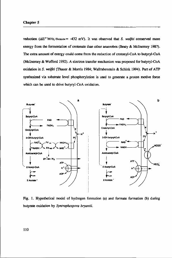
Chapter 5
reduction (AE0,HCO,7formate= -432 mV). It was observed that S. wolfei conserved more
energy from the fermentation of crotonate than other anaerobes (Beaty & Mclnerney 1987).
The extra amount of energy could come from the reduction of crotonyl-CoA to butyryl-CoA
(Mclnerney & Wofford 1992). A electron transfer mechanism was proposed for butyryl-CoA
oxidation in S. wolfei (Thauer & Morris 1984; Wallrabenstein & Schink 1994). Part of ATP
synthesized via substrate level phosphorylation is used to generate a proton motive force
which can be used to drive butyryl-CoA oxidation.
Butyrate'
r\ Butyryl-CoA
FAD
- * . FADH,
Crotonyt-CoA
3-OH-butyryl-CoA
L - N A o t » _ F d m -^s- NADHxr
|*-NADH - A * . Fd M A . MAD
Acetoacetyl-CoA
V 2 Acatyl-CoA
P 2 Acetate *
Butyrate'
r\ Butyryl-CoA
'K »-FADH,
Crotonyl-CoA
3-OH-butyryl-CoA
NAD*
• - NADH
Acetoacetyl-CoA
2 Acetyl-CoA
f t . 2 Acetate "
Fig. 1. Hypothetical model of hydrogen formation (a) and formate formation (b) during
butyrate oxidation by Syntrophospora bryantii.
110

Enzyme localization in S. bryantü
Acknowledgement
This research was made possible by a fellowship of the Wageningen Agricultural University.
Reference
Beaty PS & Mclnerney MJ (1987) Growth of Syntrophomonas wolfei in pure culture on
crotonate. Arch. Microbiol. 147: 389-393
Bergsma J, Dongen van MBM & Konings WN (1982) Purification and characterization of
NADH dehydrogenase from Bacillus subtilis. Eur. J. Biochem. 128: 151-157
Boone DR, Johnson RL & Liu Y (1989) Diffusion of the interspecies electron carriers H2 and
formate in methanogenic ecosystems, and its implications in the measurement of K,,,
for H2 or formate uptake. Appl. Environ. Microbiol. 55: 1735-1741
Bradford MM (1976) A rapid and sensitive method for the quantification of microgram
quantities of protein utilizing the principle of protein-dye binding. Anal. Biochem. 72:
248-254
Dong X, Cheng G & Stams AJM (1994) Butyrate oxidation by Syntrophospora bryantii with
different methanogens and in pure culture with pentenoate as electron acceptor. Appl.
Microbiol. Biotechnol. (In press)
Dörner C (1992) Biochemie und Energetik der Wasserstoff-Freisetzung in der syntrophen
Vergärung von Fettsäuren und Benzoat. PhD thesis, University of Tübingen, Germany
Gustafson WG, Feinberg BA & McFarland JT (1986) Energetics of ß-oxidation, reduction
potentials of general fatty acyl-CoA dehydrogenase, electron transfer flavoprotein, and
111

Chapter 5
fatty acyl-CoA substrates. J. Biological Chemist. 261: 7733-7741
Lamed R & Zeikus JG (1980) Glucose fermentation pathway of Thermoanaerobium brockii.
J. Bacterid. 141: 1251-1257
Leonhardt U & Andreesen JR (1977) Some properties of formate dehydrogenase,
accumulation and incorporation of 185W-Tungsten into proteins of Clostridium
formicoaceticum. Arch. Microbiol. 115: 277-284
Mclnerney MJ & Wofford NQ (1992) Enzymes involved in crotonate metabolism in
Syntrophomonas wolfei. Arch. Microbiol. 128: 344-349
Schauer NL & Ferry JG (1982) Properties of formate dehydrogenase in Methanobacterium
formicicum. J. Bacteriol. 150: 1-7
Schink B (1992) Syntrophism among prokaryotes. In: Balows A, Triiper HG, Dworkin M,
Harder W & Schleifer K-H (eds) The Prokaryotes (pp 276-279). Springer Verlag,
Heidelberg
Stieb M & Schink, B (1985) Anaerobic oxidation of fatty acids by Clostridium bryantii sp.
nov., a sporeforming, obligately syntrophic bacterium. Arch. Microbiol. 140: 387-390
Thauer RK, Fuchs G, Schnitker U & Jungermann K (1973) CCyreductase from Clostridium
pasteurianum: molybdenum dependence of synthesis and inactivation by cyanide.
FEBS letters 38: 45-48
Thauer RK & Morris JG (1984) Metabolism of chemotrophic anaerobes: old views and new
aspects. In: Kelly DP & Carr NG (eds) The microbe 1984: part 2. Prokaryotes and
eukaryotes (pp 123-168). Cambridge University press, Cambridge
Vogel G & Steinhart R (1976) ATPase of Escherichia coli: purification, dissociation, and
reconstitution of the active complex from the isolated subunits. Biochemistry 15: 208-
216
112

Enzyme localization in S. bryantii
Wallrabenstein C & Schink B (1994) Evidence of reversed electron transport in syntrophic
butyrate or benzoate oxidation by Syntrophomonas wolfei and Syntrophus buswellii.
Arch. Microbiol. 162: 136-142
Wofford NQ, Beaty PS & Mclnerney MJ (1986) Preparation of cell-free extracts and the
enzymes involved in fatty acid metabolism in Syntrophomonas wolfei. J. Bacteriol.
167: 179-185
Zhao H, Yang D, Woese CR & Bryant MP (1990) Assignment of Clostridium bryantii to
Syntrophospora bryantii gen. nov., comb, nov., on the basis of a 16S rRNA sequence
analysis of its crotonate-grown pure culture. Int. J. Syst. Bacteriol. 40: 40-44
113
Chapter 6
Localization of the enzymes involved in the H2 and formate
metabolism of a mesophilic propionate-oxidizing bacterium
Xiuzhu Dong, and Alfons J. M. Stams

Chapter 6
1. Summary
The mesophilic propionate-oxidizing bacterium (MPOB) contained both hydrogenase
and formate dehydrogenase activities, enabling the bacterium to form H2 and formate during
syntrophic growth on propionate, and to consume H2 and formate for the reduction of
fumarate. The V,^ and KM value of hydrogenase for H2 in cell extracts were 1.5-3.7
/imol.muv'.mg"1 protein and 0.05 mM, respectively, and the Wm!kX and KM value of formate
dehydrogenase for formate were 0.2-0.4 /xmol.min'.mg"1 protein and 0.3 mM, respectively.
The localization of the redox enzymes involved in propionate oxidation was determined after
cell breakage by French press and by lysozyme treatment. Fumarate reductase was membrane
bound and situated at the inner aspect of the membrane, while both malate dehydrogenase
and pyruvate dehydrogenase were located in the cytoplasm. Hydrogenase was detected in the
cytoplasm, periplasm as well as the membrane fraction; while formate dehydrogenase was
detected in the periplasm and the membrane fraction. A model is proposed to explain how
intracellularly electrons are channelled to the terminal electron acceptors H+ and HC03".
2. Introduction
Bacteria which grow on propionate in syntrophic association with methanogens
catabolize propionate to acetate and C02 via the methylmalonyl-CoA pathway (Koch et al.
1983; Houwen et al. 1990; Plugge et al. 1993). In this pathway propionate is first activated
116

Enzyme localization of MPOB
to propionyl-CoA by a propionate kinase and phosphotranspropionylase as in Syntrophobacter
wolinii (Houwen et al. 1990) or by a propionate:acetyl-CoA HS-CoA transferase as in MPOB
(Plugge et al. 1993). Propionyl-CoA is then carboxylated to methylmalonyl-CoA; this
reaction is coupled to the decarboxylation of oxaloacetate by means of a transcarboxylase.
The methylmalonyl-CoA is further degraded to acetate through several dehydrogenations and
decarboxylations. 1 mole of ATP is synthesized via substrate level phosphorylation by
conversion of acetyl-CoA to acetate (S. wolinii) or by conversion of succinyl-CoA to
succinate (MPOB). Reducing equivalents are released from the oxidation of succinate to
fumarate, malate to oxaloacetate and pyruvate to acetate, respectively. These reducing
equivalents are ultimately transferred to H+ or HC03" to form H2 or formate.
The oxidations of succinate to fumarate and of malate to oxaloacetate are energetically
unfavorable reactions (AG°' is 86.2 and 47.7 kJ/mol) when coupled to H+ or HC03~
reduction. Therefore, H2 or formate have to be removed to "pull" these oxidation reactions.
Up to now it is not clear whether H2 or formate is more important during propionate
oxidation, and how reducing equivalents are intracellularly channelled to H+ or HC03"
reduction. To elucidate how reducing equivalents in MPOB are coupled to the reduction of
H+ or HC03', we determined the localizations of the hydrogenase, formate dehydrogenase
and other redox enzymes involved in propionate oxidation.
3. Materials and methods
3.1. Organisms and cultivation
111

Chapter 6
The mesophilic propionate-oxidizing bacterium (MPOB) was isolated from a
methanogenic propionate-oxidizing culture. It was cultivated in a mineral medium containing
sodium fumarate (6.5 g/1) as described previously (Stams et al. 1993).
3.2. Preparation of cell fractions
Cells (2 liters) in the late-log-phase were harvested by anaerobic centrifugation at
10,000 g for 15 min. and suspended in 100 raM Tris-HCl buffer (pH 7.5) with 0.25 M
sucrose, 20 mM KCl, 5 mM MgCl2 and 5 mM dithiothreitol (DTT). This suspension was
passed twice through a French pressure cell at 40 MPa and the obtained crude extract was
centrifuged at 11,000 g at 4°C for 20 min.. The supernatant designated as cell-free extract
was ultracentrifuged at 140,000 g for 1.5 h. to get the cytoplasm fraction in the supernatant
and the membrane fraction in the pellet. The membrane fraction was resuspended in the same
buffer as described above.
Fractionation of cell extract after an osmotic shock treatment was done as described
by Odom and Peck (1981). For this, 1.5 g cells (wet weight) were suspended (1:10 wt/vol)
in the buffer as described above, but containing 0.5 M sucrose instead of 0.25 M sucrose.
This was done in stoppered serum bottles under a N2 gas phase. A lysozyme solution was
added (200 pg/ml, final concentration). The mixture was shaked for 0.5-1 h. at 37°C and
then centrifuged at 11,000 g at 4°C for 15 min.. The supernatant was designated as the
periplasmic fraction. To get a spheroplast lysate, the spheroplast pellet was resuspened in the
same buffer but without sucrose. After 10 min. at room temperature the suspension was
centrifuged at 18,000 g at 4°C for 15 min.. The pellet was designated as the membrane
118

Enzyme localization of MPOB
fraction and the supernatant as the cytoplasm fraction.
3.3 Enzyme assays
Unless indicated otherwise, all of the enzymes were determined anaerobically at
37°C. The reduction of electron acceptors and the oxidation of electron donors were followed
spectrophotometrically using a Beekman DU 7500 spectrophotometer. One unit of enzyme
activity is defined as the amount of enzyme catalyzing the conversion of 1 /*mol substrate per
min.. Protein content was determined using the method of Bradford (1976).
Benzyl viologen-coupled hydrogenase and formate dehydrogenase were measured at
pH 8.0 as described by Lamed and Zeikus (1980), and Leonhart and Andreesen (1977),
respectively. Benzyl viologen-coupled C02 reductase was determined at pH 6.8 according
to the method described by Thauer et al. (1973). Benzyl viologen-coupled fumarate reductase
was measured as described by Boonstra et al. (1975), malate dehydrogenase was determined
according to the method described by Stams et al. (1984), pyruvate dehydrogenase was
measured as described by Odom and Peck (1981), and ATPase was measured as described
by Vogel and Steinhart (1976).
3.4 Cytochrome spectra
The difference spectra of air-oxidized and dithionite-reduced cytoplasmic and
periplasmic fractions were scanned in a 1-cm light path Quartz cuvette on Beekman DU7500
spectrophotometer.
1 1 9

Chapter 6
4. Results
4.1 Hydrogenase, formate dehydrogenase and C02 reductase activities in cell-free extracts
ofMPOB:
The activities of hydrogenase, formate dehydrogenase and C02 reductase were
determined in cell extracts of MPOB. The V^,,, KM values and the lowest concentration
reached by each of the enzymes are listed in table 1. The hydrogenase activity was about 10
times higher than the formate dehydrogenase activity. The C02 reductase activity was in the
same range as the formate dehydrogenase activity. The KM value of the C02 reductase for
HCOj- was 0.43 mM at pH 6.8.
Table 1. Hydrogenase, formate dehydrogenase and C02 reductase activities in cell extract of
MPOB"
activity KM(mM) VmM(/tmol.min"l.rng"1.) lowest concentration(/tmol)
Hydrogenase 0.05 1.5-3.7 6
Formate dehydrogenase 0.3 0.2-0.4 70
C02 reductase 10.5 0.15-0.23 430
a: The values refer to H2 concentration in liquid for hydrogenase, formate concentration for formate dehydrogenase
and bicarbonate concentration for C02 reductase, respectively.
1 2 0

Enzyme localization of MPOB
4.2 Enzyme activities of intact cells and of cell fractions prepared by French pressure
disrupture:
Fumarate reductase (succinate dehydrogenase), malate dehydrogenase and pyruvate
dehydrogenase catalyze the dehydrogenation reactions involved in propionate oxidation. Table
2 shows that their activities were significantly increased by cell lysis, indicating their
intracellular localization. Upon fractionation of the cell extract, the majority of the fumarate
reductase activity was measured in membrane fraction. While both the malate dehydrogenase
and the pyruvate dehydrogenase were determined as cytoplasmic enzymes.
Benzyl viologen-coupled hydrogenase activities were increased 1.2 to 2 times upon
cell breakage, while the benzyl viologen-coupled formate dehydrogenase activity in cell
extracts was the same as that of whole cells. After fractionation of the cell extracts the
majority of the hydrogenase and the formate dehydrogenase was recovered in the soluble
fraction, while only up to 18% and 31% of the activities were detected in the membrane
fraction. However, 30% of the ATPase activity, a presumably membrane bound protein, was
determined in the soluble fraction as well. This indicates that membrane bound proteins could
be partly released from the membrane with the French pressure method. Treatment of the
membrane fraction with 0.1 % triton increased the formate dehydrogenase activity by 30%
(data not shown).
4.3 Enzyme activities in different cell fractions prepared after osmotic shock lysis:
Osmotic shock lysis is a milder method than the French pressure method as indicated
by the fact that both the activity and the distribution of the ATPase are higher in the
121
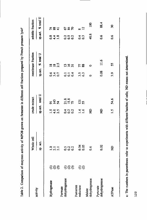
o
PU
x>
! o» S O. en e o
I
s OD m o
O H
O
N C
O , E o U
g
c o
I JU 2
2
S
1 •_ X l E <u E
E3
3
ss
-*-> g o. f/i
8 X <L>
<D
•o
3 s
<u o
o
• >
T t o o - H
r~ oo •*
oo ö
T t oo
r~ • * o vo oo r-
es m (S o ö o
CN OO r-
O o §
0 0 0 0
VO
ö
o P I
»o o
00 00 " i - H ^ H ( N
vo • * r-© c-> ©
c«-> V O —
»-H — T t
O O O
f» 00 r- oo
in co m vo
VO
oo O
m i n
ei
e i — • * TJ-o \ - i m
in o in - H ' • * N
' H i n m
•* IN (S O ö ö
cN i n - * i n
Tt vo O
~ - Z
VO
i n
2
—< «s c i i n t N t N
O ö ö SS ö ö
VO
Ö tN O Q
Z
- H t N m —i t N
0 0
2 •o
.24 s o u.
<D V I I « C <u 01)
s •o n (i)
T3
cd C
CO
"c3
0D O C -a s-*
x: (D -o
•» et) > 3
>> O.
on o V-
•a >-. JS <l>
•o
CS
eu H <
1 Ë i-t (U
•o o c Vi
E Q Z
. C
a e
c <u Ë •c 8. <u
2 Ja
<y (U
S
X>
Ê 3 C (U
J 3
H IN tN

H
O"
3
3 8>
'S 3. 3
tu
ar
EL
> H T) P
13
c < p m CL ft 3"
CL a o CTQ
°! 3 P
sa P CD D. ri 3-
o era 3 P O
Tl C
3
ft
8. c o
TJ O > - l
3 P ST D . a> 3" •< D .
0>
3 S p C&
tO In
K> OS
M p bo
K) O o s Os
p p 00 4^
4*- l*> * - OS
4^ '~n to ö
4*- 00
4*.
S i
OS OO
O so
4*- 4^ N> I^l
O — K) ts)
H - 0 0 SO K>
OO 4*
U> Os
4i. 00
s o Os O - 0
U) t-n >— w
bo bo p >- p p O bo
u> SO
u> tO
•O. ^* - J
K) •— 0 0 H -
(O >— t o 0 0
ts> U)
O
TJ
3. "H. P
&
3 3 a"
43 P o
ÎS o £
o >< b TJ S 3
5»
a. c
I < S' (g
• o o c T>
g. S N v; 3 CD P O Cf. < B.
3
I 5" 3
•8
S. P
a
o «3

Chapter 6
membrane fraction after the osmotic shock treatment than after the French pressure
treatment. Table 3 shows that up to 38 and 36 percent of hydrogenase and formate
dehydrogenase activities were determined in the periplasm fraction, respectively; while the
percentage of the two enzymes in the membrane fraction was higher after the osmotic shock
treatment. Only 16 and 10 percent of the fumarate reductase and ATPase, which are
presumbly membrane-bound proteins, were measured in the periplasmic fraction. However,
a certain amount of the malate dehydrogenase and the pyruvate dehydrogenase, which are
thought to be cytoplasmic proteins, was also found in the periplasmic fraction.
4.4 Cytochromes in cytoplasm and periplasm:
Reduced (dithionite) versus oxidized spectra of cytoplasmic and periplasmic fractions
of MPOB showed spectra which are typical for cytochrome C3; maxima were found at 420,
522 and 553 nm.
5. Discussion
Enzyme assays showed that MPOB contained all the enzyme activities of the
methylmalonyl-CoA pathway involved in propionate oxidation (Plugge et al. 1993). In this
pathway, three oxidation reactions release 3 couples of electrons which ultimately have to
be coupled to the reduction of H+ or HC03 leading to the formation of H2 or formate,
respectively. To know how the electrons are channelled to the terminal electron acceptors,
we have determined the localization of the redox enzymes involved in propionate oxidation.
124

Enzyme localization of MPOB
Fumarate reductase most likely is a membrane bound protein located at the inner aspect of
the membrane. Malate dehydrogenase and pyruvate dehydrogenase are detected as
cytoplasmic enzymes, upon fractionation of the cell extract prepared by French pressure
disrupture. However, a relative high percentage of their activities was also detected in the
membrane fractions prepared by osmotic shock. It is unclear whether they are indeed partly
membrane-bound.
From the results it is difficult to conceive where the hydrogenase and the formate
dehydrogenase are located. MPOB is a gram negative anaerobe, belonging to the sulfate-
reducing bacteria based on 16S rRNA sequence analysis (Harmsen, personnal communication
). As in Desulfovibrio gigas (George et al. 1974), up to 38 percent of the hydrogenase
activity of MPOB was detected in the periplasmic fraction upon fractionation of the cell
extract prepared by osmotic shock treatment, indicating that the hydrogenase of MPOB is
partly located in periplasm. However, the benzyl viologen coupled-hydrogenase activity
increased up to 2 times upon cell disrupture using the French pressure method, suggesting
its intracellular location because benzyl viologen is thought to be membrane-impermeable.
Upon fractionation of the cell extract prepared either by French pressure or by osmotic
shock, the majority of the hydrogenase was recovered in the soluble fraction and only a low
percentage was found in the membrane fraction, indicating that the hydrogenase might be
largely located in the soluble fractions, both in the periplasm and the cytoplasm. The benzyl
viologen-coupled formate dehydrogenase activity was not affected by cell lysis. Although up
to 84 percent of the activity is recovered in the soluble fraction upon fractionation of the cell
extract prepared by the French pressure method, a high percentage of the formate
125

Chapter 6
dehydrogenase was recovered in the membrane fraction and the periplasm fraction when the
osmotic shock treatment was applied. Moreover, treatment of the membranes with a
detergent increased the formate dehydrogenase activity to some extent. These observations
suggest that the formate dehydrogenase is partly located in the periplasm and partly
membrane bound, most likely situated on the outer aspect of the membrane. Some evidence
for the external localization of the formate dehydrogenase can be obtained from formate
uptake experiments. MPOB is able to convert formate into H2 + C02 . This conversion was
not inhibited by the addition of the ionophores, valinomycin or nigericine, and the ATPase
inhibitor, N,N-dicyclohexyl carbodiimide (DCCD) (unpublished results). These observations
indicate that formate uptake is either an energy-independent process in MPOB, or that
formate is not taken up at all.
Based on the localization of the redox enzymes, a hypothetical model how
intracellularly electrons are channelled to H+ and HC03" can be given (Fig. 1). Our
hypothesis is that formate is formed at the outer aspect of the membrane, while where H2 is
formed is unclear yet. It is assumed that the periplasmic hydrogenase of Desulfovibrio sp.
responsible for H2 oxidation, while the cytoplasmic hydrogenase is responsible for H2
production from lactate or ethanol oxidation (Badziong and Thauer 1980; George et al. 1974;
Odom and Peck 1981). In analogy with this it seems likely that MPOB forms H2 from
NADH and reduced ferredoxin intracellularly.
The oxidation of succinate to fumarate is highly endergonic (E°'ftuniinite/succinatt = +33
mV) when the reaction is coupled to H+ or HC03" reduction ( E °V /H , = -414mV and E° '
Hco.mcoa = -432 mV). Therefore, metabolic energy has to be invested to pull this oxidation.
126

Enzyme localization of MPOB
HCOO"
ATP
ADP
- Succinate . Propionate
- » • Fumarate
t Malate
• NAD .— * - Fd red . 2 H '
' ^^» t lADH-^V- . Fd ox - » ^ » - H Oxaloacetate
Pyruvate
Acetate
Periplasm Membrane Cytoplasm
Fig. 1. A hypothetical reversed electron transport mechanism during syntrophic propionate oxidation by MPOB. The oxidation of succinate is completed by a membrane bound fumarate reductase; the electrons are transducted either to formate dehydrogenase, which is situated at the outer aspect of the membrane or to hydrogenase, which might be located at the inner aspect or outer aspect of the membrane. In the scheme, NADH and reduced ferredoxin oxidations are coupled to the intracellular formation of H2. However, it might also be that the oxidation of these electrons is linked to a membrane-bound C02 reductase in an unknown fashion.
127

Chapter 6
A reversed electron transport mechanism was postulated (Schink 1992; Stams 1994); part of
the ATP synthesized at substrate level is spent to generate a proton motive force which can
be used to drive succinate oxidation. A study with Wolinella succinogenes revealed that the
fumarate reduction with H2 and formate as electron donors is a membrane-associated process
(Kroger 1978). The externally electrons released from H2 or formate are channelled to the
intracellular fumarate reductase, thereby creating a proton motive force. ATP is formed from
the H+ gradient across the membrane by means of the ATPase. It has been shown that this
bacterium forms 2/3 ATP per fumarate.
Reference
Badziong, W. and Thauer, R.K. (1980). Vectorial electron transport in Desulfovibrio vulgaris
(Marbury) growing on hydrogen plus sulfate as sole energy source. Arch. Microbiol.
125, 167-174.
Boonstra, J., Huttunen, M.T., and Konings, W.N. (1975) Anaerobic transport in Escherichia
coli membrane vesicles. J. Biol. Chem. 250, 6792-6798.
Bradford, M.M. (1976) A rapid and sensitive method for the quantification of microgram
quantities of protein utilizing the principle of protein-dye binding. Anal. Biochem. 72,
248-254.
George, R.B., LeGall, J. and Peck, JR. H.D. (1974) Evidence for the periplasmic location
of hydrogenase in Desulfovibrio gigas. J. Bacteriol. 120, 994-997.
128

Enzyme localization of MPOB
Houwen, F.P., Plokker, J., Stams, A.J.M. and Zehnder, A.J.B. (1990) Enzymatic evidence
for involvement of the methylmalonyl-CoA pathway in propionate oxidation by
Syntropkobacter wolinii. Arch. Microbiol. 155, 52-55.
Koch, M.E., Dolfing, J., Wuhrmann K. and Zehnder, A.J.B. (1983) Pathways of
propionate degradation by enriched methanogenic cultures. Appl. Environ. Microbiol.
45, 1411-1414.
Kroger, A. (1978) Fumarate as terminal acceptor of phosphorylative electron transport.
Biochim. Biophys. Acta 505, 129-145.
Lamed, R. and Zeikus, J.G. (1980) Glucose fermentation pathway of Thermoanaerobiwn
brockii. J. Bacteriol. 141, 1251-1257.
Leonhardt, U. and Andreesen, J.R. (1977) Some properties of formate dehydrogenase,
accumulation and incorporation of 185W-Tungsten into proteins of Clostridium
formicoaceticwn. Arch. Microbiol. 115, 277-284.
Odom, J.M. and Peck, H.D. (1981) Localization of dehydrogenase, reductases and
electron transfer components in the sulfate-reducing bacterium Desulfovibrio gigas.
J. Bacteriol. 147, 161-169.
Plugge, CM. Dijkema, C. and Stams, A.J.M. (1993) Acetyl-CoA cleavage pathway in a
syntrophic propionate oxidizing bacterium growing on fumarate in the absence of
methanogens. FEMS Microbiol. Let. 110, 71-76.
Schink, B. (1992) Syntrophism among prokaryotes. In: The Prokarotes (Balows, A., Triiper,
H.G., Dworkin, M., Harder, W. and Schleifer, K.-H. Eds.), pp. 276-299. Springer
Verlag, Heidelberg.
129

Chapter 6
Stams, A.J.M. (1994) Metabolic interactions between anaerobic bacteria in methanogenic
environments. Antonie van Leenwenhoek (In press).
Stams, A.J.M., van Dijk J., Dijkema, C. and Plugge, CM. (1993) Growth of syntrophic
propionate-oxidizing bacteria with fumarate in the absence of methanogenic bacteria.
Appl. Environ. Microbiol. 59, 1114-1119.
Stams, A.J.M., Kremer, D.R., Nicolay, K., Weenk, G.H. and Hansen, T.A. (1984)
Pathway of propionate formation in Desulfobulbus propionicus. Arch. Microbiol. 139,
167-173.
Thauer, R.K., Fuchs, G., Schnitker, U. and Jungermann, K. (1973) CC^-reductase from
Clostridium pasteurianum: molybdenum dependence of synthesis and inactivation by
cyanide. FEBS letters 38, 45-48.
Vogel, G. and Steinhart, R. (1976) ATPase of Escherichia coli: purification, dissociation,
and reconstitution of the active complex from the isolated subunits. Biochem. 15,
208-216.
130

Chapter 7
Summary
Chapter 7
Under methanogenic conditions, complex organic matter is mineralized by fermentative,
acetogenic and methanogenic bacteria. Propionate and butyrate are two important
intermediates; they account for 35% and 8% of the total methane formation, respectively.
Propionate and butyrate are converted to CH4 and C02 by the syntrophic consortia of
acetogenic and methanogenic bacteria. Both H2 transfer and formate transfer were proposed
as the possible mechanisms by which reducing equivalents are transferred from the acetogens
to the methanogens. Since extreme low levels of H2 and formate prevail in anaerobic
ecosystems and cocultures, it is not known which of the two is more important.
The aim of this research was to clarify the relative importance of H2 and formate transfer
during syntrophic degradation of propionate and butyrate by means of physiological and
biochemical approaches.
After succeeding in obtaining pure cultures of propionate oxidizer MPOB and butyrate
oxidizer Syntrophospora bryantii by growing them on fumarate and crotonate, respectively,
this study became possible. By constructing defined methanogenic cocultures, it was observed
that propionate oxidation by MPOB and butyrate oxidation by S. bryantii can only be driven
by H2/formate- but not by H2-utilizing or aceticlastic methanogens (Chapter 2 and 3),
indicating that formate transfer might be prevailing in these cultures. However, a triculture
of MPOB with aDesulfovibrio strain, which converts formate into H2/C02, and a H2-utilizing
methanogen also degraded propionate, confirming that H2 transfer is possible provided that
low formate concentrations are maintained (Chapter 2).
MPOB is able to ferment fumarate to succinate and to couple propionate oxidation to
fumarate reduction; while S. bryantii is able to couple butyrate oxidation to the reduction of
132
Summary
pentenoate (Chapter 3). With a limiting amount of fumarate in a propionate degrading
culture, the maximum H2 and formate levels produced from propionate by MPOB were 6.8
Pa and 24 /xmol, respectively; and with a limiting amount of pentenoate in a butyrate
degrading culture, the maximum H2 and formate levies produced by S. bryantii were 170 Pa
and 280 /*mol, respectively (Chapter 4). These results showed that both H2 and formate
indeed were formed and that the values which were reached were in the range which could
be expected from thermodynamical consideration. Using a diffusion model it could be
calculated that formate fluxes can be 100 times higher than H2 fluxes in the methanogenic
cocultures of MPOB and S. bryantii, suggesting that formate transfer is more important
during syntrophic propionate and butyrate degradation.
Enzyme measurments (Chapter 5) showed that S. bryantii contained very high hydrogenase
activity and low formate dehydrogenase activity. The K,,, values of the two enzymes are about
the same, 0.21 mM and 0.22 mM, respectively. Butyryl-CoA dehydrogenase and 3-
hydroxybutyryl-CoA dehydrogenase, which are involved in the two oxidation reactions during
butyrate oxidation, were determined as soluble enzymes. The hydrogenase was also
determined as a cytoplasmic enzyme while formate dehydrogenase and C02-reductase were
membrane bound, likely located at the outer aspect of the cytoplasmic membrane. These
observations suggest that during butyrate oxidation H2 is formed intracellularlly, while
formate is formed extracellularly. A reversed electron transfer mechanism was postulated to
drive the endergonic oxidation of butyryl-CoA to crotonyl-CoA. Similar experiments were
done with MPOB (Chapter 6). MPOB also contained relative high hydrogenase activities
(1.5-3.7 jamol.min'.mg1) and low formate dehydrogenase activities (0.2-0.4 ̂ mom.min'.mg"
133

Chapter 7
'). The K,,, values are 0.05 mM of H2 for hydrogenase and 0.3 mM of formate for formate
dehydrogenase. Localization of the redox enzymes involved in propionate oxidation indicated
that the fumarate reductase was membrane bound, likely located at the inner aspect of
membrane; while malate dehydrogenase and pyruvate dehydrogenase were located in the
cytoplasm. The hydrogenase is located partly in the periplasm, partly in the cytoplasm and
partly in the membrane; while the formate dehydrogenase was determined partly in the
periplasm and partly in the membrane. These results indicate that, during propionate
oxidation, formate is formed outside of cytoplasmic membrane, However, where H2 is
produced is not clear yet. As with butyrate oxidation, a reversed electron transport
mechanism was postulated to drive the oxidation of succinate (AG°'= +86.2 kJ/mol).
Although acetate is also a product of propionate and butyrate oxidation, removal of acetate
by aceticlastic methanogen alone does not drive the oxidations by the acetogens MPOB and
S. bryantii. However, addition of the aceticlastic methanogen Methanothrix (Methanosaeta)
soehngenii increased the degradation rates of propionate and butyrate by the cocultures of
MPOB and S. bryantii with H2/formate-trophic methanogens (Chapter 2 and 3). The addition
of 50 mM acetate decreased the butyrate degradation rate of the coculture of S. bryantii and
Methanospirillum hungatei by about 60% (Chapter 3). These results indicate that a low
acetate concentration is beneficial for syntrophic degradation of propionate and butyrate.
134

Chapter 8
Samenvatting

Chapter 8
Complex organisch materiaal wordt onder anaërobe methanogene omstandigheden
afgebroken door consortia van fermentatieve, acetogene en methanogene bacteriën.
Propionaat en butyraat zijn twee belangrijke intermediairen; zij dragen respectievelijk
voor 35% en 8% bij aan de totale hoeveelheid gevormd methaan. Propionaat en butyraat
worden omgezet naar CH4 en C02 door syntrofe consortia van acetogene en methanogene
bacteriën. Zowel de overdracht van H2 als van formiaat worden genoemd als mogelijke
mechanismen waarmee reductie-equivalenten worden overgedragen van acetogene
bacteriën naar methanogene bacteriën. Omdat in anaërobe ecosystemen en cocultures zéér
lage H2 en formiaat concentraties aanwezig zijn, is het niet bekend welke van de twee
moleculen belangrijker is.
Het doel van dit onderzoek was om het relatieve belang van zowel H2- als formiaatover-
dracht op te helderen tijdens de syntrofe omzetting van propionaat en butyraat; hierbij
gebruikmakend van een fysiologische en biochemische benadering.
Deze studie werd mogelijk nadat reincultures van de propionaatoxideerder MPOB en de
butyraatoxideerder Syntrophospora bryantii waren verkregen door ze respectievelijk op
fumaraat en crotonaat te kweken. Door gedefinieerde methanogene cocultures te maken,
werd waargenomen dat propionaatoxidatie door MPOB en butyraatoxidatie door S.
bryantii slechts dan optrad wanneer een H2 + formiaat gebruikende methanogeen
aanwezig was, en niet wanneer een H2-gebruikende of aceticlastische methanogeen
aanwezig was (Hoofdstuk 2 en 3). Dit kan er op duiden dat formiaatoverdracht overheerst
in deze cultures. Evenwel, een triculture van MPOB en een Desulfovibrio stam, die
formiaat in H2/C02 omzet, samen met een H2-gebruikende methanogeen kon wel
136
propionaat omzetten. Dit bevestigt dat H2-overdracht mogelijk is op voorwaarde dat de
formiaatconcentraties laag worden gehouden (Hoofdstuk 2).
MPOB is in staat om fumaraat naar succinaat om te zetten én om propionaat-oxidatie aan
fumaraatreductie te koppelen. S. bryantii is in staat om butyraatoxidatie te koppelen aan
pentenoaat-reductie (Hoofdstuk 3). Met een beperkende hoeveelheid fumaraat in een
propionaatoxiderende culture van MPOB konden maximale hoeveelheden van 6.8 Pa H2
en 24 fM formiaat gevormd worden; met een beperkende hoeveelheid pentenoaat in een
butyraatoxiderende culture van S. bryantii waren deze hoeveelheden respectievelijk 170
Pa voor H2 en 280 ßM voor formiaat (Hoofdstuk 4). Deze resultaten tonen aan dat zowel
H2 als formiaat daadwerkelijk gevormd worden en dat de waarden die gemeten worden
binnen het bereik liggen dat verwacht mag worden op grond van thermodynamische
berekeningen. Als een diffusiemodel gebruikt wordt, kan berekend worden dat formiaat-
fluxen 100 keer hoger zijn dan H2-fluxen in de methanogene cocultures van MPOB en 5.
bryantii. Dit suggereert dat formiaatoverdracht belangrijker is dan H2-overdracht tijdens
syntrofe propionaat- en butyraatafbraak.
Uit enzymmetingen bleek (Hoofdstuk 5) dat S. bryantii een zeer hoge hydrogenase en een
lage formiaatdehydrogenase activiteit bezat. De K^waarden van deze twee enzymen zijn
ongeveer gelijk: respectievelijk 0.21 mM en 0.22 mM. Butyryl-CoA dehydrogenase en 3-
hydroxybutyryl-CoA dehydrogenase, die betrokken zijn bij de twee oxidatie-reacties
tijdens de butyraatoxydatie bleken oplosbare enzymen te zijn. Het hydrogenase bleek een
cytoplasmatisch enzym te zijn, terwijl formiaatdehydrogenase en C02-reductase mem
braangebonden bleken, vermoedelijk gelokaliseerd aan de buitenkant van het cytoplasma
tisch membraan. Deze waarnemingen suggereren dat H2 intracellulair wordt gevormd
137

Chapter 8
tijdens de butyraatoxydatie, terwijl formiaat extracellulair gevormd wordt. De endergone
oxidatie van butyryl-CoA naar crotonyl-CoA wordt verondersteld te worden gedreven
door een omgekeerd elektronentransport mechanisme. Vergelijkbare experimenten werden
gedaan met MPOB. Ook MPOB bevatte relatief hoge hydrogenase activiteiten (1.5-3.7
/tmol.min'.mg') en lage formiaatdehydrogenase activiteiten (0.2-0.4 jtmol.min'.mg"1).
De Kj.-waarden zijn 0.05 mM H2 voor hydrogenase en 0.3 mM formiaat voor formiaatde
hydrogenase. Na lokalisatie van de redoxenzymen die betrokken zijn bij de propionaat-
oxydatie bleek fumaraatreductase membraangebonden, vermoedelijk aan de binnenkant
van de membraan, terwijl malaatdehydrogenase en pyruvaatdehydrogenase in het
cytoplasma zaten. Het hydrogenase was gedeeltelijk in het periplasma, gedeeltelijk in het
cytoplasma en gedeeltelijk in de membraan gelokaliseerd, terwijl formiaatdehydrogenase
gedeeltelijk in het periplasma en gedeeltelijk in de membraan werd aangetroffen. Deze
resultaten geven aan dat gedurende propionaatoxidatie, formiaat aan de buitenkant van de
cytoplasmatische membraan wordt geproduceerd. Echter, waar H2 wordt geproduceerd is
nog niet duidelijk. Net als bij butyraatoxidatie, wordt verondersteld dat de oxydatie van
succinaat (AG°'= +86.2 kJ/mol) wordt gedreven door een omgekeerd elekronentransport
mechanisme.
Hoewel acetaat ook een eindprodukt van propionaat- en butyraatoxydatie is, leidde de
verwijdering van acetaat door aceticlastische methanogenen alleen niet tot het laten
verlopen van de oxydatiereacties door MPOB en S. bryantü. Echter, wanneer de
aceticlastische methanogeen Methanothrix (Methanosaeta) soehngenii toegevoegd werd
aan cocultures van MPOB en S. bryantü samen met een H2 + formiaat gebruikende
138

methanogeen, leidde dit wel tot verhoogde omzettingssnelheden van propionaat en
butyraat (Hoofdstuk 2 en 3). Toevoeging van 50 mM acetaat aan een coculture van S.
bryantii en Methanospirillum hungatei verminderde de butyraatomzettingssnelheid met
60% (Hoofdstuk 3). Deze resultaten geven aan dat ook een lage acetaatconcentratie
bevorderlijk is voor de syntrofe omzetting van propionaat en butyraat.
139

4ÄM(Syntrophospora bryantii)Ôd^^#%, mmikMWÏÏMVMft. ilafflJ&ESDfô
wmmMmmmrmL, mmmmnmmmrwmicmji m^m\
mxmwmn (Desuifovibrio) n&A^mmmPWifàtGE^m&!tiïïnm
% 6.8Pa m 24umol; &mMT&&m&ïïM£&m%%®*MA]mi&penterioate, ä
rmimm%rmmi^mmm*imw% i70Pa m iz^mu^nm. &m%m
loom, mmm&mmmwBim*, mmmm.
ff^fllttKiitaËttBH, UifcHA0.21mMfôH2, mWOMBM%0.22âmwm&. THSfcttHAJfâ
W*l*lffl^^çtlTK»l|A|«fcAESSt«l|A)ati!5t6SÈ. £iPift£&lil£MP0B4'iafT(» A*). «W**^W#AW*ftllfôij(1.5-3.7uMl/iin/K)iBlffitt^iMtt*llî5* (0.2-0.4umol/min/ing). mteSlfôKmttA0.05niMÔt>H2, i?lM£WttKa|l!ft0.3Wfttfl|Rft.
140
srwmmm, m-^mmmmmmmkw '=86.2Kj/moD.
(Methanothrixsoehngenü), TifK«ÄIÄWi»m*I^ttlWW*(»=*Bl»H»). &
141

Acknowlegements
I greatly appreciate Alex Zehnder, my promotor, to give me the opportunity to do research
at the department, and to support me to pursuit a Ph.D. degree in Wageningen Agricultural
University.
I am grateful to Fons Stams, my co-promotor, for the most important support, inspiring
guidance and thoroughly review of this thesis, which would not be accomplished without his
help.
I indeed enjoyed the time to work amidst the colleagues in the anaerobic group during a few
years passed. The working atmosphere was not only active but also friendly. Among them,
firstly I would like to thank Caroline Plugge, she has given me a lot of help in many aspects;
Sever Kengen, he gave me many valuable ideas during the research, especially in enzyme
assays; Philippe Schyns, my computer tutor, Mike Jetten and Steef Biesterveld, they kindly
help me in my early period of the research; Stefanie Oude Elferink, Ine van Kuijk, Hans
Scholten and Karin Maarsen, are appreciated for their worthy discussion and warm
companionship. I thank Ans Geerling for her pleasant friendship during all of these years.
I also would like to thank Wim Roelofson and Nees Slotboom for their help during the
chromatography determination and other analysis.
The fellowship of Wageningen Agricultural University made this research possible. I thank
the Institute of Microbiology in Beijing, China to give me the chance to work in the
Netherlands, and Prof. Guangsheng Cheng to support me for doing partly research in his
Lab.
I would like to give my special thanks to Zhengang for his constant encouragement and
support that were of fundamental importance for completing this work.
142

Curriculum vitae
The author of this thesis, Xiuzhu Dong was born in Hebei, China. She graduated from
Agricultural University of Beijing, China in 1982, and obtained her master degree in Science
at Institute of Microbiology, scientific Academy of China in 1984. The topic of the master
thesis was on taxonomy of acetic acid bacteria. From 1985 till 1989, she was a research
member of the department of bacteriology, Institute of Microbiology, Beijing. Since 1990,
she was admitted to be a Ph.D. student through the "sandwich" program in the department
of microbiology, Wageningen Agricultural University. She will return to China at the end
of 1994 to continue the research in China.
143