rodolfo r. llinás - i of the vortex
TRANSCRIPT

Preface
This essay arose out of a set of talks given at The University of St. An-drews in Scotland, where Professor Glen Cottrell had graciously invitedme to give the American Alumni Lectures in 1989. Little did I know thenthat St. Andrews would be back in my life, when, in 1998, my son Alex-ander obtained his Ph.D. there during a break in his medical studies atNew York University.
The generation of this essay owes much to Michael Kistler, to whom Idictated much of this manuscript, so giving me a leg up into getting thematerial into a form that I could work with. Dr. Jean Jacoby helped withthe editing. My son Rafael, presently a junior staff neurologist at Har-vard’s Beth Israel Hospital, took the time to read and criticize this effort,as did my wife, Dr. Gillian Kimber, from her perspective as a philosopherof mind. I would also like to thank a special friend, Dr. AntonioFernandez de Molina, and my colleague Dr. Kerry Walton for specialcomments and additions.
This book presents a personal view of neuroscience aimed toward ageneral audience, as well as toward students and those of my colleagueswho might enjoy an attempt at synthesis. This general view is offeredfrom the perspective of a single-cell physiologist interested in neuronalintegration and synaptic transmission. Such a position is privileged,

because it lies between the realms of the molecular and the systemic, asthey relate to brain function.
Single large neurons have physical dimensions observable at low opti-mal magniªcation, that of a tenth of a millimeter. That is big enough tobe dissected by hand with pins, using a good magnifying glass (Deiters1856). Moving just two orders of magnitude down to the micrometerlevel, which requires a good microscope, one is at the scale of synaptictransmission. One may observe synapses at the union between nerve andmuscle, for example. Two orders of magnitude further down, at tens ofnanometers, with the aid of electron microscopy, we ªnd the realm of sin-gle ion channels and of signal transduction and molecular biology.
If, on the other hand we wish to roam orders of magnitude above thephysiology of single cells, we ªnd at two orders of magnitude above, andin the centimeter realm, the world of systems that is the scale of pennies,buttons, and ªngernails. At a further two orders of magnitude up, wecome to meters and to the world of motricity and cognition that charac-terizes human beings. That is, we arrive at the realm of chairs and tele-phones and other objects that one can hold in one’s hand or under one’sarm.
Most neuroscientists feel that two orders of magnitude above and be-low one’s central focus is “horizon enough,” and that anyone attemptingfour orders above and below is reckless. However, there are some whoattempt such a dangerous dynamic range. They probably know that therisk of failure is the price of synthesis, without which there are only ªeldsof dismembered parts.
viii Preface

Motor Primacy and the
Organization of Neuronal
Networks: Thinking as
Internalized Movement
A fundamental ªrst step in exploring the nature of mind, from a scientiªcpoint of view, is to reject the premise that the mind appeared suddenly asa result of spectacular intervention. The nature of mind must be under-stood on the basis of its origin, the process of its becoming, by the biolog-ical mechanism of trial and error endlessly at work. The mind, or what Ishall refer to as the “mindness state,” is the product of evolutionary pro-cesses that have occurred in the brain as actively moving creatures devel-oped from the primitive to the highly evolved. Therefore, a trueexamination of the scientiªc basis for mindness requires a rigorous evolu-tionary perspective, as it is through this process that mindness came tobe. How the mind came to us (or we to it, as we shall see) is a rich andbeautiful story that is over 700 million years old—and, like all things bio-logical, is still being written.
A prerequisite for grasping the nature of mind is, ªrst and foremost,the appropriate perspective. Just as Western society, steeped in dualisticthinking, must re-orient in order to grasp the elemental tenets ofnondualistic philosophy, so there must be a fundamental reorientation ofperspective in order to approach the neurobiological nature of mind. Anattempt at such reorienting was the task in the American Alumni Lec-tures at St. Andrews; this book will proceed in that vein.

Charles Sherrington, in his Gifford Lectures at Edinburgh in 1937, en-titled Man on his Nature (1941, chapter 12), hinted at the possibility thatif human beings ever came face to face with their true natures that knowl-edge might trigger the demise of human civilization. To him, evidently,humans prefer to consider themselves the lowest of angels rather than thehighest of beasts. I am of the opinion that if we were to comprehend fullythe awesome nature of mindness, we would, in fact, respect and admireeach other all the more.
x Introduction

R. Vario, Sueño, 1958. Pencil/paper, 23 × 16 cm.

1 Setting Mind to Mind
Mindness, Global Function Brain States, and Sensorimotor Images
There are some basic guidelines to be considered when taking a scientiªcapproach to the mind. Because this book is not supposed to be a detectivestory, let me offer some demarcating/clarifying deªnitions of the mind or“mindness state” that will be used here. From my monist’s perspective,the brain and the mind are inseparable events. Moreover, the mind, ormindness state, is but one of several global functional states generated bythe brain. Mind or the mindness state, is that class of all functional brainstates in which sensorimotor images, including self-awareness, are gener-ated. When using the term sensorimotor image, I mean something morethan visual imagery. I refer to the conjunction or binding of all relevantsensory input to produce a discreet functional state that ultimately mayresult in action. For instance, imagine that you have an itch on your back,at a place that you cannot see but which generates an internal “image”giving you a location within the complex geography of your body as wellas an attitude to take: SCRATCH! That is a sensorimotor image. Thegeneration of a sensorimotor image is not a simple input/output re-sponse, or a reºex, because it occurs within the context of what the ani-mal is presently doing. For obvious reasons, a dog wouldn’t want to

scratch with one leg while another one is up in the air. So, context is asimportant as content in the generation of sensorimotor images andpremotor formulation.
There are other states that occupy the same space in the brain mass butwhich may not support awareness. These include being asleep, beingdrugged or anesthetized, or having a grand mal epileptic seizure. Whenone’s brain is in these states, consciousness is lost; all memories and feel-ings melt into nothingness; yet the brain continues to function, requiringits normal supply of oxygen and nutrients. During these states, the braindoes not generate awareness of any kind, not even of one’s own existence(self-awareness). It does not generate our worries, our hopes, or ourfears—all is oblivion.
By contrast, I consider the global brain state known as dreaming to bea cognitive state, but not with respect to co-existing external reality be-cause it is not directly modulated by one’s senses (Llinás & Pare 1991).Rather, this state draws from the past experiences stored in our brain orfrom the intrinsic workings of the brain itself. Yet another global brainstate would be that known as “lucid dreaming” (LaBerge & Rheingold1990), where one is actually aware that one is dreaming.
In short then, the brain is more than the one and a half liters of inertgrayish matter occasionally seen pickled in a jar atop some dusty labora-tory shelf. One should think of the brain as a living entity that generateswell-deªned electrical activity. This activity could be described perhaps as“self-controlled” electrical storms, or what Charles Sherrington (1941,p. 225), one of the pioneers of neuroscience, refers to as the “enchantedloom.” In the wider context of neuronal networks, this activity is themind.
This mind is co-dimensional with the brain; it occupies all of thebrain’s nooks and crannies. But as with an electrical storm, the mind doesnot represent at any given time all possible storms, only those isomorphicwith (re-enacting, a transformed recreation of) the state of the local sur-rounding world as we observe it when we are awake. When dreaming, aswe are released from the tyranny of our sensory input, the system gener-ates intrinsic storms that create “possible” worlds—perhaps—very muchas we do when we think.
2 Chapter 1

Living brains and their electrical storms are descriptors for different as-pects of the same thing, namely neuronal function. These days, one hearsmetaphors for central nervous system function that are derived from theworld of computers, such as “the brain is hardware and the mind, soft-ware” (see discussion by Block 1995). I think this type of language usageis totally misleading. In the working brain, the “hardware” and the “soft-ware” are intertwined in the functional units, the neurons themselves.Neurons are both “the early bird” and “the worm,” because mindnesscoincides with functional brain states.
Before returning to our discussion of mindness, think about the itch onyour back again, and in particular the moment of the sensorimotor im-age—before you put into action the motor event of scratching the itch.Can you recognize the sense of future inherent to sensorimotor images,the pulling toward the action to be performed? This is very important,and a very old part of mindness. From the earliest dawning of biologicalevolution it was this governing, this leading, this pulling by predictivedrive, intention, that brought sensorimotor images—indeed, the mind it-self—to us in the ªrst place.
Let us shore up the discussion with a bit more precision. I propose thatthis mindness state, which may or may not represent external reality (thelatter as with imagining or dreaming), has evolved as a goal-oriented de-vice that implements predictive/intentional interactions between a livingorganism and its environment. Such transactions, to be successful, re-quire an inherited, prewired instrument that generates an internal imageof the external world that can then be compared with sensory-transducedinformation from the external environment. All of this must be sup-ported in real time. The functional comparison of internally generatedsensorimotor images with real-time sensory information from an organ-ism’s immediate environment is known as perception. Underlying theworkings of perception is prediction, that is, the useful expectation ofevents yet to come. Prediction, with its goal-oriented essence, so very dif-ferent from reºex, is the very core of brain function.
Setting Mind to Mind 3

Why Is Mindness So Mysterious?
Why is mindness so mysterious to us? Why has it always been this way?The processes that generate such states as thinking, consciousness, anddreaming are foreign to us, I fancy, because they always seem to be gener-ated with no apparent relation to the external world. They seem impalpa-bly internal.
At New York University School of Medicine, in a lecture in honor ofthe late Professor Homer Smith, entitled, “Unity of Organic Design:From Goethe and Geoffrey Chaucer to Homology of Homeotic Com-plexes in Anthropods and Vertebrates,” Stephen J. Gould mentioned thewell-known evolutionary hypothesis that we vertebrates may be regardedas crustaceans turned inside out. We are endoskeletal, with an internalskeleton; crustaceans are exoskeletal, with an external skeleton.
This idea led me to consider what would have happened if we had re-mained exoskeletal? If we had an external skeleton, the concept of howmovement is generated might be just as incomprehensible to us as is theconcept of thinking or mindness. Having an internal skeleton means thatwe become quite aware of our muscles from birth. We can see theirmovement and feel their contractions and clearly understand, in a veryintimate way, their relation to the movement of our different body parts.Unfortunately, we do not have such direct knowledge concerning theworkings of our brain. Why not? Because from a cerebral mass point ofview, we are crustaceans—our brains and spinal cord are covered byexoskeleton! (ªgure 1.1).
If we could observe or feel the brain at work, it would be immediatelyobvious that neuronal function is as related to how we see, interpret, andreact, as muscle contractions are related to the movements we make. Asfor our crustacean friends, who lack the luxury of direct knowledge ofthe relationship of muscle contraction to movement, their movementability, if they could consider it, might seem as inexplicable to them asthinking or mindness is to us. The essential point is that we do under-stand about muscles and tendons; in fact, we revel in them. We go so faras to hold world competitions for the comparison of symmetrically hy-pertrophied muscle mass produced by obsessively “pumping iron” (andoccasionally popping steroids), even though, as physical strength for size
4 Chapter 1

goes in the animal kingdom, we are way down near the bottom of theheap. The more analytically probing among us employ measuring tapes,scales, and force transducers in an effort to describe the properties ofthese precious organs of movement. However, no such paraphernalia areavailable for directly assessing the working of the brain (IQ tests notwithstanding). Perhaps this is why, in the ªeld of neuroscience, such dif-fering concepts have arisen about how the brain is functionallyorganized.
The central generation of movement and the generation of mindnessare deeply related; they are in fact different parts of the same process. Inmy view, from its very evolutionary inception mindness is the internaliza-tion of movement.
Historical Views of Motor Organization in the Brain
Around the turn of this century, there arose two strong opposing viewson the subject of the execution of movement. The ªrst, championed by
Setting Mind to Mind 5
Figure 1.1Detail showing the upper body and head from a life drawing by Leonardo daVinci, with an image of the brain superimposed.

William James (1890), viewed the working organization of the centralnervous system as fundamentally reºexological. From this perspectivethe brain is essentially a complex input/output system driven by the mo-mentary demands of the environment. Production of movement must bedriven by sensation, and the generation of movement is fundamentally aresponse to a sensory cue. This basic idea was very inºuential in thegroundbreaking studies of Charles Sherrington and his school (1948). Itprovided the impetus for the study of central reºexes—their function andhow they were organized—and ultimately for the study of central synap-tic transmission and neuronal integration. All of these have played cru-cial roles in present-day neuroscience.
A second inºuential approach was championed by Graham Brown(1911, 1914, 1915). Brown believed that the spinal cord was not orga-nized reºexologically. He viewed this system as organized on a self-referential basis by central neuronal circuits that provided the drive forthe electrical pattern generation required for organized movement. Thisconclusion was based on his studies of locomotion in deafferented ani-mals, that is, animals in which the pathways bringing sensation fromthe legs to the spinal cord are severed. Under these conditions animalscould still produce an organized gait (Brown 1911). This led Brown topropose that movement, even organized movement, is intrinsically gener-ated in the absence of sensory input. He viewed reºex activity as requiredonly for the modulation of, rather than being the driving force for, theproduction of gait. So, for example, while locomotion (one step after theother) is organized intrinsically, not requiring input from the externalworld, sensory input (e.g., a slippery spot on the ground) reºexively re-sets the rhythm so that we don’t fall, but it does not generate walkingitself.
Brown went on to propose that locomotion is produced in the spinalcord by reciprocal neuronal activity. In very simpliªed terms, autono-mous neuronal networks on one side of the spinal cord activate the mus-cles of the limb on the same side while preventing activity by the oppositelimb. He described this reciprocal organization as “half-paired centers”(Brown 1914), as their mutual interaction generated the left/right limbpacing that is locomotion (see ªgure 2.5, below).
6 Chapter 1

In this context, the function of the sensory input giving rise to reºex ac-tivity during locomotion is there to modulate the ongoing activity of thespinal cord motor network in order to adapt the activity (the output sig-nal) to the irregularities of the terrain over which the animal moves. Wenow know that such ongoing activity born of the intrinsic electrical activ-ity of neurons in the spinal cord and brain stem forms the basis for bothbreathing (Feldman et al. 1990) and locomotion (Stein et al. 1986; Cohen1987; Grillner and Matsushima 1991; Lansner et al. 1998) in verte-brates. A similar dynamic organization, but supported by a quite differ-ent anatomical arrangement, is found in invertebrates (Marder 1998). Inboth vertebrates and invertebrates, the neuronal activity being transmit-ted and modiªed between different levels by synaptic connectivity hascomparable dynamic properties.
Brown’s views remain highly regarded by many of us and have beenseminal to our understanding of the intrinsic activity of central neurons(Llinás 1974, 1988; Stein et al. 1984). This conceptual view of spinalcord function may be extended to the workings of the brainstem and ar-eas of higher brain function, such as the thalamus and forebrain—areaswhere mindness is ultimately generated in our brain.
The Intrinsic Nature of Brain Function
A working hypothesis related to Brown’s ideas is that nervous systemfunction may actually operate on its own, intrinsically, and that sensoryinput modulates rather than informs this intrinsic system (Llinás 1974).Let me hasten to say that being disconnected from sensory input is notthe normal operational mode of the brain, as we all know from child-hood, when ªrst we observed the behavior of a deaf or blind person. Butthe exact opposite is equally untrue: the brain does not depend on contin-uous input from the external world to generate perceptions (see The LastHippie, by Oliver Sacks), but only to modulate them contextually. If oneaccepts this view, it follows that the brain, like the heart, operates as aself-referential, closed system in at least two different senses: one, assomething separated from our direct inquiry by implacable bone; andtwo, as a system that is mostly self-referential, only able to know univer-sals by means of specialized sense organs. Evolution suggests that these
Setting Mind to Mind 7
sense organs specify internal states that reºect neuronal circuit selectionderived from ancestral trial and error. Such circuits become geneticallypredetermined (for example, we can see color primarily without havingto learn to do so). Once we are born, these ancestral circuits (comprisingthe inherited, functional architecture of the brain) are further enriched byour own experiences as individuals and thus constitute our own particu-lar memories, indeed, our selves.
We can look to the world of neurology for support of the concept thatthe brain operates as a closed system, a system in which the role of sen-sory input appears to be weighted more toward the speciªcation of on-going cognitive states than toward the supply of information—contextover content. This is no different than sensory input modulating a patternof neural activity generated in the spinal cord to produce walking, exceptthat here we are talking of a cognitive state generated by the brain andhow sensory input modulates such a state. The principle is the same. Forexample, prosopagnosia is a condition in which individuals, due to neu-rological damage, cannot recognize human faces. They can see and rec-ognize the different parts of a face, as well as subtle facial features, butnot the face as a whole entity (Damasio et al. 1982; De Renzi andPellegrino, 1998). Moreover, the people that inhabit the dreams ofprosopagnostics are faceless (Llinás and Pare 1991) (we shall return tothis issue later in the book).
The signiªcance of sensory cues is expressed mainly by their incorpora-tion into larger, cognitive states or entities. In other words, sensory cuesearn representation via their impact upon the pre-existing functional dis-position of the brain (Llinás 1974, 1987). This concept, that thesigniªcance of incoming sensory information depends on the pre-existingfunctional disposition of the brain, is a far deeper issue than one gathersat ªrst glance—particularly when we look into questions of the nature of“self.”
Intrinsic Electrical Properties of Neurons: Oscillation, Resonance, Rhythmicity,
and Coherence
How, then, do central neurons organize and drive bodily movement, cre-ate sensorimotor images, and generate our thoughts? Having grown in
8 Chapter 1

our knowledge from the days of Brown, we may paraphrase the abovequestion today to read: How do the intrinsic oscillatory properties ofcentral neurons relate to the information-carrying properties of the brainas a whole? Before attempting to answer this question, there are still afew more terms to cover. Let me start by describing what is meant by theintrinsic oscillatory electrical properties of the brain, from a relativelynontechnical point of view. This concept is at the heart of all we shall dis-cuss in this book.
Oscillation
When one thinks of the word “oscillation,” one thinks of a rhythmicback-and-forth event. Pendulums oscillate, as do metronomes; they areperiodic oscillators. The sweeping motion of a lamprey’s tail, back andforth, as it swims (Cohen 1987; Grillner and Matsushima 1991) is awonderful example of an oscillatory movement.
Many of the types of neurons in the nervous system are endowed withparticular types of intrinsic electrical activity that imbue them with par-ticular functional properties. Such electrical activity is manifested as vari-ations in the minute voltage across the cell’s enveloping membrane(Llinás 1988). This voltage may oscillate in a manner similar to the trav-eling, sinusoidal waves that we see as gentle ripples in calm water, andare weakly chaotic (Makarenko and Llinás 1998). As we will see later,this confers a great temporal agility to the system. These oscillations ofvoltage remain in the local vicinity of the neuron’s body and dendrites,and have frequencies ranging from less than one per second to more thanforty per second. On these voltage ripples, and in particular on theircrests, much larger electrical events known as action potentials may beevoked; these are powerful and far reaching electrical signals that formthe basis for neuron-to-neuron communication. Action potentials are themessages that travel along neuronal axons (conductive ªbers that com-prise the information pathways of the brain and the peripheral nerves ofthe body). Upon reaching the target cell, these electrical signals generatesmall synaptic potentials. Such local changes in the voltage across themembrane of a target cell add or subtract voltage to the intrinsic oscilla-tion of the target cell receiving the signal. Intrinsic oscillatory propertiesand modifying synaptic potentials are the coinage that a neuron uses to
Setting Mind to Mind 9

arrive at the generation of its own action potential message, which it willsend on to other neurons or to muscle ªbers. And so, in the case of mus-cle, all possible behaviors in us arise from activation of the motor neu-rons that activate the muscles that ultimately orchestrate our movements.These motor neurons in turn receive messages from other neurons lo-cated “up stream” from them (ªgure 1.2).
The peaks and valleys of the electrical oscillations of neurons can dic-tate the waxing and waning of a cell’s responsiveness to incoming synap-tic signals. It may determine at any moment in time whether the cellchooses to “hear” and respond to an incoming electrical signal or ignoreit altogether. As will be discussed in more depth in chapter 4, this oscilla-tory switching of electrical activity is not only very important in neuron-to-neuron communication and whole network function, it is the electricalglue that allows the brain to organize itself functionally and architectur-ally during development. Indeed, simultaneity of neuronal activity is themost pervasive mode of operation of the brain, and neuronal oscillationprovides the means for this simultaneity to occur in a predictable, if notcontinuous, manner.
Coherence Rhythmicity and Resonance Neurons that display rhythmic os-cillatory behavior may entrain to each other via action potentials. The re-sulting, far-reaching consequence of this is neuronal groups that oscillatein phase—that is, coherently, which supports simultaneity of activity.
Consider the issue of coherence from the perspective of communica-tion, for coherence is what communication rides on. Imagine a soft sum-mer night in a rural setting. Amidst the rich quietude, you hear ªrst onecicada, then another. Soon, there are many chirping. More importantly,they may chirp in rhythmic unison (note that to chirp in unison theymust all have a similar internal clock that tells them when to chirp next—such a mechanism is known as an intrinsic oscillator). The ªrst cicadamay be calling out to see if there are any kin about. But this unison ofmany cicadas chirping rhythmically becomes a bonding, literally a con-glomerated functional state. In the subtle ºuctuations of this rhythmicitycomes the transfer of information, at the whole community level, to avast number of remotely located individuals. Similar events occur in
10 Chapter 1

Setting Mind to Mind 11
Figure 1.2Evolution of nervous systems. An interneuron, in the strict sense, is any nerve cellthat does not communicate directly with the outside world either as a sensing de-vice (a sensory neuron) or by means of a motor terminal on a muscle (a motorneuron). Interneurons, therefore, receive and send information to other nervecells exclusively. Their evolution and development represent the basis for theelaboration of the central nervous system. The diagrams above represent stagesof development present in early invertebrates. In (A), a motile cell (in black) froma primitive organism (a sponge), responds to direct stimulation with a wave ofcontraction. In (B), in more evolved primitive organisms (e.g., the sea anemone),the sensory and contractile functions of the cell in A have been segregated intotwo elements; “r” is the receptor or sensory cell and “m” is the muscle or con-tractile element. The sensory cell responds to stimuli and serves as a motor neu-ron in the sense that it triggers muscle-cell contraction. However, this sensory cellhas become specialized so that it is incapable of generating movement (contrac-tion) on its own. Its function at this stage is the reception and transmission of in-formation. In (C), a second neuron has been interposed between the sensoryelement and the muscle (also from a sea anemone). This cell, a motor neuron,serves to activate muscle ªbers (m) but responds only to the activation of the sen-sory cell (r) (Parker 1919). In (D), as the evolution of the central nervous systemprogresses (this example is the vertebrate spinal cord), cells become interposedbetween the sensory neurons (A) and motor neurons (B). These are theinterneurons, which serve to distribute the sensory information (arrow in A) bytheir many branches (arrows in C) to the motor neurons or to other neurons inthe central nervous system. (Adapted from Ramón y Cajal, 1911.)

some types of ªreºies, which synchronize their light ºash activity andmay illuminate trees in a blinking fashion like Christmas tree lights.
This effect of oscillating in phase so that scattered elements may worktogether as one in an ampliªed fashion is known as resonance—and neu-rons do it, too. In fact, a local group of neurons resonating in phase witheach other may then resonate with another group of neurons that arequite far from the ªrst group (Llinás 1988; Hutcheon and Yarom 2000).Electrical resonance, a property supported by direct electrical connectiv-ity among cells (as occurs in the heart, allowing it to function as a pumpby the simultaneous contraction of all of its component muscle ªbers) isperhaps the oldest form of communication among neurons. The deli-cately detailed nuances of chemical synaptic transmission come later inevolution to enhance and embellish neuronal communication.
Not all neurons resonate at all times. It is the crucial property of neu-rons to be able to switch in and out of oscillatory modes of electrical ac-tivity that allows resonance to occur transiently among differing groupsof neurons at different times. If they were not able to do this, they wouldnot be able to represent the ever-changing reality that surrounds us.When differing groups of neurons capable of displaying oscillatory be-havior “perceive” or encode different aspects of the same incoming sig-nal, they may join their efforts by resonating in phase with each other.This is known as neuronal oscillatory coherence. Simultaneity ofneuronal activity, brought into existence not by chance but by intrinsicoscillatory electrical activity, resonance, and coherence are, as we shallsee, at the root of cognition. Indeed, such intrinsic activity forms the veryfoundation of the notion that there is such a thing called our “selves.”
Returning to the original question of intrinsic properties, one may pro-pose the following: that intrinsic electro-responsiveness of the brain’s ele-ments, the neurons and the networks they weave together, generateinternal representations (connections) that engender functional states.These states are speciªed in detail, but not in context, by incoming sen-sory activity. That is, brain function is proposed to have two distinctcomponents. One is the private or “closed” system that we have dis-cussed and that is responsible for qualities such as subjectivity and se-mantics; the other is an “open” component responsible for sensory-motor transformations dealing with the relations between the private
12 Chapter 1

component and the external world (Llinás 1974, 1987). Because thebrain operates for the most part as a closed system, it must be regarded asa reality emulator rather than a simple translator.
Acknowledging this, we might go on to say that the intrinsic electricalactivity of the brain’s elements (its neurons and their complex connectiv-ity) must form an entity, or a functional construct. Furthermore, this en-tity must efªciently handle the transformation of sensory input arisingfrom the external world into its motor output counterpart. How can westudy such a complicated functional construct as this? First we mustmodel it, make some assumptions concerning how the brain may be im-plementing such transformational properties, and for this we must bevery clear about what the brain actually does. If we decide, as a workinghypothesis, that this functional brain construct must bestow reality emu-lating properties, we may then consider what types of models could sup-port such a function.
Let us begin with a simple sensory-motor transformation. The motoraspect is implemented by muscle force (contractile) exercised on boneslinked to each other by hinges (joints). In order to study our assumedtransformational properties, we may describe the contractile aspect asperforming a given movement in space (or in mathematical terms, a vec-tor), and so the set of all muscle contractions contributing to this move-ment (or any type of behavior) will be enacted in a “vectorial coordinatespace.” With this approach, the electrical activity patterns that each neu-ron generates in the formation of a motor pattern, or any other internalpattern in the brain, must be represented in an abstract geometric space.This is the vectorial coordinate space where sensory input and its trans-formation into a motor output take place (Pellionisz and Llinás 1982). Ifthis sounds a bit like double-talk to you, please read the contents ofbox 1.1.
How Did the Mind Arise from Evolution?
Let us go back to the very ªrst point made at the beginning of this chap-ter, that the mind did not just suddenly appear at some point fullyformed. With some forethought and a little educated digging, we can ªndin biological evolution a quite convincing trail of clues as to the brain’s
Setting Mind to Mind 13

14 Chapter 1
Box 1.1Abstract Representation of Reality
Let us imagine a cube of electrically conductive material, a gelatin-like sub-stance, held in a spherical glass aquarium. Let’s imagine that the surface ofthe container has small electrical contacts that can allow electricity to passbetween one contact and any other through the gelatin. Finally, let’s saythat the gelatin condenses into thin conductive ªlaments if current passesbetween the electrical contacts often, but returns to amorphous gel if nocurrent ºows for a while.
If we now pass current among some contacts connected to one or moresensory systems that transform a complex external state (let’s say playingsoccer) and other contacts related to a motor system, a condensed set ofwirelike paths will grow that allows the sensory inputs to activate a motoroutput. (Keep in mind that these wires do not interact with each other—they are insulated, just as for the most part are the ªber pathways of thebrain, and therefore there are no short circuits. These wires can, however,branch to generate a complex connectivity matrix). As we proceed to gen-erate more complex sensory inputs they will in turn generate more complexmotor outputs. In short, a jungle of “wires” grows inside the ªshbowl, ormelts, if stimuli are not repeated for a time. This veritable mess of wireswould be the embedding that relates certain sensory inputs (in principleany thing that can be transduced by the senses, what we may call univer-sals) to given motor outputs. As an example, this contraption could be usedhypothetically to control a soccer-playing robot (backpropagation algo-rithms have this general form).
Looking at the ªshbowl we can understand that there, somewhere in thecomplex geometry of wires, are the rules for playing soccer, but in a verydifferent geometry from the playing of soccer itself. One cannot under-stand by direct inspection that the particular wiring represents such athing. “Soccer” is being represented in a different geometry from that ofsoccer in external reality, and in an abstract geometry at that—no legs orreferees or soccer balls, only wires. So the system is isomorphic (can enactsoccer playing) although not homomorphic with soccer playing (does notlook like soccer playing). This is analogous to the tape inside a videocas-sette, which despite close inspection offers no clues as to the details of themovie embedded in its magnetic code. Here we have a representation of theexternal world in which intrinsic coordinate systems operate to transforman input (a sensory event) into the appropriate output (a motor response)using the dynamic elements of the sensory organs and motor “plant,” theset of all muscles and joints, or their equivalent. This sensory-motor trans-formation is the core of brain function, that is, what the brain does for aliving.

origin. If one agrees that the mind and brain are one, then the evolutionof this unique mindness function must certainly have coincided with theevolution of the nervous system itself. It should also be obvious that theforces driving the evolution of the nervous system shaped and determinedthe emergence of mind as well. The questions to ask here are clear. Howand why did the nervous system evolve? What critical choices did naturehave to make along the way?
It Began at a Critical Time
The ªrst issue is whether a nervous system is actually necessary for all or-ganized life beyond that of a single cell. The answer is no. Living organ-isms that do not move actively, including sessile organisms such as plants,have evolved quite successfully without a nervous system. And so wehave landed our ªrst clue: a nervous system is only necessary formulticellular creatures (not cell colonies) that can orchestrate and expressactive movement—a biological property known as “motricity.” It is in-teresting to note that plants, which have well-organized circulatory sys-tems but no hearts, appeared slightly later in evolution than did mostprimitive animals; it is as if sessile organisms had, in effect, chosen not tohave a nervous system. Although this seems a rather strange statement tomake, the facts are quite irrefutable—the Venus Flytrap, Mimosa, andother locally moving plants not withstanding.
Where does the story begin? What type of creature can we look to forsupport of this important connection between the early glimmerings of anervous system and the actively moving, versus sessile, organism? A goodplace to begin is with the primitive Ascidiacea, tunicates or “sea squirts,”which represent a fascinating juncture in our own early chordate (truebackbone) ancestry (ªgure 1.3).
The adult form of this creature is sessile, rooted by its pedicle to a sta-ble object in the sea (ªgure 1.4, left) (Romer 1969; Millar 1971; Cloney1982). The sea squirt carries out two basic functions in its life: it feeds byªltering seawater, and it reproduces by budding. The larval form isbrieºy free-swimming (usually a day or less) and is equipped with abrainlike ganglion containing approximately 300 cells (Romer 1969;Millar 1971; Cloney 1992). This primitive nervous system receives sen-sory information about the surrounding environment through a statocyst
Setting Mind to Mind 15

16 Chapter 1
Figure 1.3A simpliªed diagram of chordate evolution. The tunicates, or sea squirts(Ascidiaceae; see ªgure 1.4) represents a stage in which the gill apparatus has be-come highly evolved in the sessile adult, while the larval stage in some species isfree-swimming, exhibiting the advanced features of a notochord and nerve cordassociated with the motile behavior. See text for more details. (Adapted fromRomer, 1969, p. 30.)

(organ of balance), a rudimentary, light-sensitive patch of skin, and anotochord (primitive spinal cord) (ªgure 1.4, right). These features allowthis tadpole-like creature to handle the vicissitudes of the ever-changingworld within which it swims. Upon ªnding a suitable substrate (Svaneand Young 1989; Young 1989; Stoner 1994), the larva proceeds to buryits head into the selected location and becomes sessile once again (Cloney1982; Svane and Young 1989; Young 1989). Once reattached to astationary object the larva absorbs—literally digests—most of its ownbrain, including its notochord. It also digests its tail and tail musculature,thereupon regressing to the rather primitive adult stage: sessile and lack-ing a true nervous system other than that required for activation of thesimple ªltering activity (Romer 1969; Millar 1971; Cloney 1982). Thelesson here is quite clear: the evolutionary development of a nervous sys-tem is an exclusive property of actively moving creatures.
Setting Mind to Mind 17
Figure 1.4Sea squirts (Ascidiaceae) or tunicates, which have a sessile, ªlter-feeding adultstage attached to the substratum (left), and in many cases a brief free-swimminglarval stage (right). (Bottom left) Diagram of a generalized adult solitary seasquirt. The black outer portion is its protective “tunic.” (Bottom right) Diagramof a typical free-swimming sea squirt larva or tadpole. A gut, gills and branchialstructure are present, but are neither functional nor open. See text for details.(From website www.animalnetwork.com/ªsh/aqfm/1997/)

We have now derived a basic concept—namely, that brains are an evo-lutionary prerequisite for guided movement in primitive animals—andthe reason for this becomes obvious. Clearly, active movement is danger-ous in the absence of an internal plan subject to sensory modulation. Trywalking any distance, even in a well-protected, uncluttered hallway, withyour eyes closed. How far can you go before opening your eyes becomesirresistible? The nervous system has evolved to provide a plan, one com-posed of goal-oriented, mostly short-lived predictions veriªed by mo-ment-to-moment sensory input. This allows a creature to move activelyin a direction according to an internal reckoning—a transientsensorimotor image—of what may be outside. The next question in ourpursuit of the evolution of mind should now be clear. How did thenervous system evolve to be able to perform the sophisticated task ofprediction?
18 Chapter 1

Tennis pro Gabriella Sabatini returns a shot. Photo reprinted courtesy of AlanCook, [email protected], http://alancook.50mpegs.com.

2 Prediction Is the Ultimate
Function of the Brain
Why Must the Brain Predict?
In chapter 1, we argued that a nervous system is only necessary for livingcreatures that move actively. If so, how has a nervous system contributedto their evolutionary success? Clearly, such creatures must move intelli-gently in order to survive, to procure food and shelter, and to avoid be-coming food for someone else. I use the word “intelligently” to implythat a creature must employ a rudimentary strategy, or at the very leastrely upon a set of tactical rules regarding the basic properties of the exter-nal world through which it moves. Otherwise, movement would be pur-poseless and necessarily dangerous. The creature must anticipate theoutcome of a given movement on the basis of incoming sensory stimuli. Achange in its immediate environment must evoke a movement (or lack ofit) in response to ensure survival. The capacity to predict the outcome offuture events—critical to successful movement—is, most likely, the ulti-mate and most common of all global brain functions.Before proceeding, it is important to have a clear sense of what is
meant by “prediction.” Prediction is a forecast of what is likely to occur.For example, we predict common outcomes—such as walking bare-footed on the hot pavement will hurt, or that not turning the car when
approaching a dead-end will result in something probably harmful to youand the car. When one runs to hit a tennis ball, one must predict where intime and space the ball and the face of one’s racket can successfully meet.Consider two mountain rams squaring to ªght. As they eye one an-
other, they slowly rise onto their hind legs and look for the tiniest of cluesthat will provide a hint of when the other is about to shift its weight for-ward and charge. Because even a half-step lead in momentum can changethe outcome of the contest, a ram (or any creature for that matter) mustbe able to anticipate the attack in order to counter strike, that is, it mustbe able to predict that a blow is coming before it arrives.The ability to predict is critical in the animal kingdom—a creature’s
life often depends upon it. Still, the mechanism of prediction is far moreubiquitous in the brain’s control of body function than the examples sofar described. Consider the simple act of reaching for a carton of milk inthe refrigerator. Without giving much focused thought to our action, wemust predict the carton’s weight, its slipperiness, its degree of fullness,and, ªnally, the compensatory balance we must apply for a successfullysmooth trajectory of the carton to our glass. Once movement is initiated,we adjust our movement and the compensatory balance as we receive di-rect sensory information coming in. However, before even reaching, wehave made a ballpark, premotor prediction of what will be involved.The brain’s ability to predict is not only generated from our awareness
of its operation; prediction is a far older evolutionary function than that.Consider this: have you ever found yourself blinking just before a buglands in your eye? You did not see the bug, at least not on a consciouslevel, yet you anticipated the event and blinked appropriately to ward offits entry into your eye. Prediction is at the heart of this basic protectivemechanism. Prediction, almost continually operative at conscious andreºex levels, is pervasive throughout most, if not all, levels of brainfunction.In the beginning of this book, I mentioned that the mindness state,
which may or may not necessarily represent external reality, has evolvedas a goal-oriented device to guide the interactions between a living organ-ism and its environment. Success in a goal-oriented, moving system is en-hanced by an innate mechanism for prediction. Furthermore, we canassume that prediction must be grounded, that there can be only one pre-
22 Chapter 2

dictive organ. It would make little sense if the head predicted one thingand the tail another. Predictive functions must be centralized.
Prediction and the Origin of “Self”
Although prediction is localized in the brain, it does not occur at only onesite in the brain. These predictive functions must be brought together intoa single understanding or construct; otherwise, the end result would beno different than if prediction were grounded in any number of differentorgans. What pulls these functions together? What is the repository ofpredictive function? I believe the answer lies in what we call the self: selfis the centralization of prediction. The self is not born out of the realm ofconsciousness, only the noticing of it is (i.e., self-awareness). Accordingto this view, the self can exist without awareness of its own existence.Even in we as self-aware individuals, self-awareness is not continuouslypresent. In the middle of a difªcult challenge, such as swimming awayfrom a shark, you will try to get to shore and be quite aware of what ishappening, but you will probably not be thinking to yourself, “Here I amswimming away from a shark.” You will think about it only when youget to shore and safety.The concept of self-awareness will be discussed in later chapters, but I
wanted to point to the issue of self now. Understanding that the brainperforms prediction on the basis of an assumed self “entity” will lead usto how the brain generates the mindness state.Why this predictive ability arose is clear: it is critical to survival, guid-
ing it at the level of both the single animal (moment-to-moment) and thespecies (in fact, of all actively moving species throughout evolution).How did the ability to predict arise from evolution? The answer can befound with a little thoughtful digging. First, however, we must under-stand how the nervous system actually performs prediction; once weknow that, we will ªnd the answer to how nature evolved this amazingfunction.As we will see later in this chapter, for the nervous system to predict, it
must perform a rapid comparison of the sensory-referred properties ofthe external world with a separate internal sensorimotor representationof those properties. For the prediction to be useful, the nervous systemmust then transform or utilize this premotor solution into ªnely timed
Prediction Is the Ultimate Function of the Brain 23
and executed movement. Once a pattern of neural activity acquires inter-nal signiªcance (sensory content gains internal context), the brain gener-ates a strategy of what to do next—another pattern of neural activity.This strategy can be considered an internal representation of what is tocome. Such premotor patterns of neural activity must then be trans-formed into the neuronal activity that sets into motion the appropriatebodily movement: These transformations require an internal representa-tion of what is to come, in order for them to become actualized in the ex-ternal world context (ªgure 2.1).
Prediction Saves Time and Effort Prediction is crucial to brain function notonly for the successful execution of goal-oriented, active movement, butalso as a basic functional operation in order to conserve time and energy.This may sound a bit strange, since the nervous system—particularly thehuman nervous system—being the most sophisticated and capable “pro-cessor” yet known, might be expected to be above such trivialconsiderations. Nevertheless, when the brain deals with the vicissitudesof the external world (and the internal as well) its activity does not paral-lel reality in its continuity; it just feels that way to us. In real life the brainoperates in a discontinuous manner from a processing perspective. It isnot possible to take in all of the information available to our senses fromthe external world and then arrive at the correct decision quickly in acontinuous fashion. Neurons are fast, but they are not that fast. Note, Iam still only speaking of the premotor phase of processing. Rememberthat a successful interaction with the external world also necessitates thesubsequent timely execution of the brain’s given decision throughmovement.It seems that the brain must compartmentalize incoming information
and implement its attention on a need to know basis in order to fuel itsmomentary decision-making ability without overloading. The brain mustleave itself enough time to implement a movement decision so that it re-mains in step with what is happening in the external world at a given mo-ment. It must also be able to skip to the next moment’s need forprocessing without being encumbered by the previous moment’s process-ing. In other words, the brain cannot be stuck doing one thing when itneeds to move on to the next task. This mode of operation derives fromwhat is known as a look ahead function, which is an inherent property of
24 Chapter 2
neuronal circuits. Indeed, prediction begins at the single neuron level. Wecan address this issue with an example: the control of movement.
Prediction and the Control of Movement
Because the ability to predict evolved in tandem with increasingly com-plex movement strategies, we must look at movement control in order tounderstand prediction. Let us return to the refrigerator for a carton ofmilk. The appropriate pattern of contraction must be speciªed for an ex-tension/grasping sequence to be executed properly (add to this the correctuse of postural muscles for support of the body while bending over dur-ing the reach). Now consider what the brain must do to pull off this sim-ple movement sequence. Each muscle provides a direction of pull (avector). Each muscle vector is composed of individual muscle ªbers thatare operated in pre-established groups based on their common inner-vation by the same motor neuron. This is called a motor unit (a singlemotor neuron innervates tens to hundreds of muscle ªbers). A given mus-cle may be composed of hundreds of such individual motor units. Thenumber of muscles multiplied by the number of motor units may then beviewed as the total number of degrees of freedom for any given move-ment. A movement such as reaching into the refrigerator is considered a
Prediction Is the Ultimate Function of the Brain 25
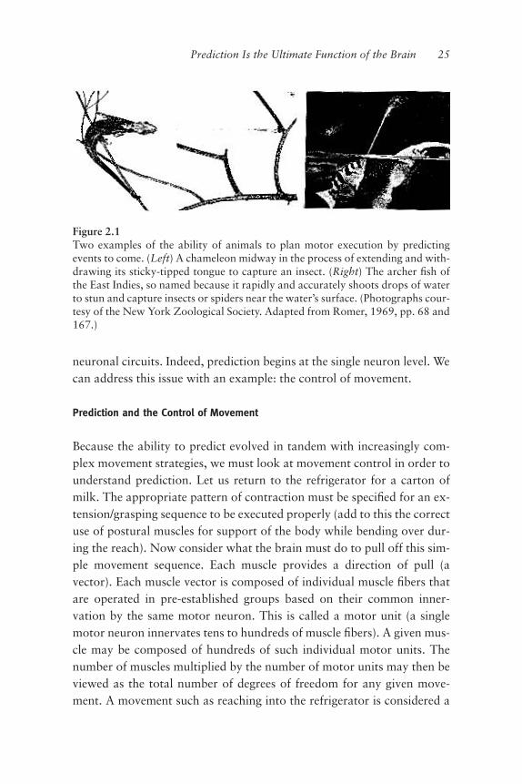
Figure 2.1Two examples of the ability of animals to plan motor execution by predictingevents to come. (Left) A chameleon midway in the process of extending and with-drawing its sticky-tipped tongue to capture an insect. (Right) The archer ªsh ofthe East Indies, so named because it rapidly and accurately shoots drops of waterto stun and capture insects or spiders near the water’s surface. (Photographs cour-tesy of the New York Zoological Society. Adapted from Romer, 1969, pp. 68 and167.)

simple one (as compared to, say, a good tennis return). However, from afunctional perspective, even a simple movement often engages most ofthe body’s muscles, resulting in an astronomical number of possiblesimultaneous and/or sequential muscle contractions and degrees of free-dom. With the milk carton example, your arm may be brought towardthe carton from any number of initial positions and postures (maybeyour back hurts today and so you bend into your reach from a stilted,atypical stance).All of this potential complexity exists before the load is actually placed
on your arm and body; you have yet to pick up the carton and can onlyguess its weight during your initial reaching motion.So this simple movement is not simple when we break it down and try
to understand how the brain handles it all. However, the dimensionalityof the problem of motor control does not derive solely from the numberof muscles involved, the differing degrees of pull force and angle, and soforth. The real dimensionality of the problem stems from the complicatedinteraction between the possible directions of muscle pull and their se-quence of activation in time.Much of motor control occurs in real time, “on-line,” as it were. Our
movements seldom take place under stimulus-free conditions. Considerthe following scenarios: running down a steep, winding forest path; steer-ing your car while holding a cup of coffee; jumping up and stretching toreturn a serve in tennis. The combination of muscles one contracts at anygiven moment is often determined as a movement sequence and executedin response to teleceptive stimuli (stimuli at a distance taken in mainlythrough the senses of hearing and vision), kinesthetic feedback (the feel-ing of one’s body moving), or thought.It is generally assumed that the optimal controller is one that produces
the smoothest possible movement. This idea implies the continuous mon-itoring (that is to say, a sampling rate of every millisecond or faster) offeed-forward and feedback inºuences on the selected activation se-quences in order to minimize the accelerative transients that produce jer-kiness in movement. Although this sounds right, we need to evaluatewhether it is computationally plausible for the brain to control move-ment in such a continuous, on-line manner.From the heuristic formula described above, and, given that there are
50 or so key muscles in the hand, arm, and shoulder that one uses to
26 Chapter 2

reach for the milk carton, over 1015 combinations of muscle contractionsare possible—a staggering number to say the least. If during every milli-second of this reaching/grasping sequence the single best of the 1015 com-binations is chosen after an evaluation of all of the possibilities, then 1018
decisions would have to be made every second. This would mean that thebrain, if it were a computer, would need a 1-exahertz (1 million giga-hertz) processor to choose the correct muscle combinations to executeappropriately this relatively simple reaching/grasping sequence. In reality,even the above scenario is an over simpliªcation (Welsh et al. 1995). Thedimensionality of the problem of motor control is increased many ordersof magnitude when one also considers that there is a bare minimum of100 motor units for every muscle, and that each muscle pull may, andmost likely will, involve differing sets of motor neurons.The brain does not seem to have evolved to deal with the control of
movement in this fashion—especially when one considers that there areon the order of 1011 neurons in the entire brain. Of these, only a fractionare in the cerebellum, the area of brain where most of the movement con-trol processing would take place for the movement sequence we havebeen discussing (Llinás and Simpson 1981).An alternative solution for the continuous control of movement might
be a scheme where each muscle in the body is somehow controlled inde-pendently through time. Metaphorically, the motor system could be con-sidered a bank of discrete representations (or parallel processors, withone for each muscle). This set-up would signiªcantly ease the functionalburden for the control of any single muscle, and render trivial the prob-lem of how to control a highly artiªcial and rare movement involvingonly one or two muscles. This scenario presents signiªcant difªculties forthe control of complicated muscle synergies, however. A muscle synergyis a set of muscles working in tandem to bring about a given movement.This synergy operates on the stretch reºex, that is, the relation betweenºexors and extensors (ªgure 2.2). For instance, our reaching for the milkcarton sequence is a muscle synergy, as are the associated muscles in-volved in the ensuing grasping movement of our hand and the reºexproperties of the spinal cord circuits. As the number of muscles involvedin a movement sequence increases, there would be a greater reliance onan absolutely precise and infallible synchronizing element to ensure thatthe muscle activations occur cohesively in time.
Prediction Is the Ultimate Function of the Brain 27
This solution seems more ªtting for a digital computer than a nervoussystem. However, unlike the elements of a digital system, neurons are an-alog: they have nonlinear response properties, and do not ªre their actionpotentials with sufªcient temporal precision to control continuously intime such parallel processing machinery.At this point it should be clear that the continuous control of move-
ment through time demands an extremely high computational overhead.This is true whether the movement is controlled by regulating the activityof every muscle discretely in parallel, or by choosing and implementingcombinations of muscles. We do, of course, make complicated move-
28 Chapter 2
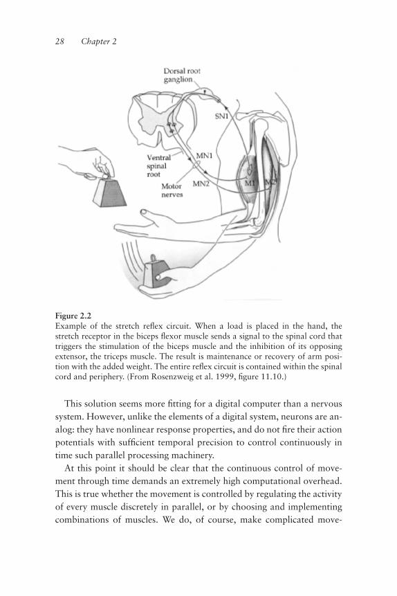
Figure 2.2Example of the stretch reºex circuit. When a load is placed in the hand, thestretch receptor in the biceps ºexor muscle sends a signal to the spinal cord thattriggers the stimulation of the biceps muscle and the inhibition of its opposingextensor, the triceps muscle. The result is maintenance or recovery of arm posi-tion with the added weight. The entire reºex circuit is contained within the spinalcord and periphery. (From Rosenzweig et al. 1999, ªgure 11.10.)

ments, and quite often. To delve further into this issue, we must ask thefollowing:
1. How might the dimensionality problem of motor control, this incredi-ble functional overhead for the brain, be reduced without signiªcantlydegrading the quality of movement sequences?2. Which well-established aspects of brain function can provide clues forhow to solve this problem?
The Discontinuous Nature of Movement
A relatively straightforward approach to reducing the dimensionality ofmotor control for the brain is to decrease the temporal resolution of thecontrolling system, that is, remove it from the burden of being continu-ously on-line and processing. This can be accomplished by breaking upthe time line of the motor task into a series of smaller units over whichthe controller must operate. Control would be discontinuous in time andthus the operations of such a system would occur at discrete intervals of a“dt” (literally, intervals of a discrete passage of time). We must here con-sider an important consequence, that movements controlled by this typeof pulsatile system would not be executed continuously, demonstratingobligatorily smooth kinematics, but rather would be executed in a dis-continuous fashion as a linked series of muscle twitches. Motor physiolo-gists have known this fact for over a century: movements are notexecuted continuously, but are discontinuous in nature. E. A. Schafer sur-mised this as early as 1886:
The curve of a voluntary muscular contraction . . . invariably shows, both at thecommencement of the contraction and during its continuance, a series of undula-tions that succeed one another with almost exact regularity, and can, as it wouldseem, only be interpreted to indicate the rhythm of the muscular response to thevoluntary stimuli which provoke the contraction. . . . The undulations . . . areplainly visible and are sufªciently regular in size and succession to leave no doubtin the mind of any person who has seen a graphic record of muscular tetanic con-traction produced by exciting the nerve about 10 times in the second, that thecurve . . . is that of a similar contraction. (9)
A tetanic contraction, or tetanus, is the maximum force that a musclecan generate when activated at high frequency. Schafer realized that aclearly deªned rhythmicity in the range of 8–12 Hz exists in volitional
Prediction Is the Ultimate Function of the Brain 29

muscular contraction. Following Schafer’s initial report, the pheno-menon of an 8–12 Hz periodicity to voluntary movement, termed“physiological tremor,” became a topic of intense research. In 1894,Harris measured the frequency of the “voluntary tetanus” (literally, thevoluntary driving of a muscle or muscle synergy to its maximum rhyth-mic speed, as in ºexing and extending one’s ªnger as quickly as possible,or the maximum rate at which one can voluntarily shake a foot, etc.) ina variety of muscles, including those of the arm, hand, ªngers, andtongue. Discontinuities of 8–12 Hz were observed in all of the muscles hestudied.Harris went on to state that “the average rate of single voluntary mus-
cle twitches is 10 or 11 per second—a ªgure sufªciently near to that ofthe rate of the voluntary tetanus as to be reckoned identical with it.” Inessence, what is seen at the single muscle level is reºected in the overtmovement. In 1910, Sherrington noted that the scherzo of Schubert’s Pi-ano Quartet No. 8 requires repetitive hand movements at approximately8 Hz, which approaches the upper limit for ªnger movements by profes-sional pianists. He also observed that the syllable “la” cannot be repeatedmore than 11 times per second and went on to state that “the limit set tothe frequency of repetition of the same one movement seems to be 11 persecond.”Some years later, Travis (1929) demonstrated that voluntary move-
ment initiated from a holding position was almost always initiated inphase with physiological tremor. He reported that “a voluntary move-ment is, in most instances, a continuation of tremor . . . [it] does not in-terrupt the tremor rhythm . . . and ªts into the kinetic melody”determined by the brain. Travis went on to suggest that the maximumrate of a repeated voluntary movement could not exceed the rate of phys-iological tremor. More recently, the study of physiological tremor hasbrought to light what may be a very close relationship between the closeto 10-Hz rhythmic discontinuity and the actual onset of movement itself.The work of Travis was advanced in 1956, when Marshall and Walshdemonstrated that the reºection of external movements in humans doesindeed start at the phase of the physiological tremor corresponding to thedirection of the intended movement.
30 Chapter 2

These researchers also noted that the physiological discontinuities involuntary movements were independent of both the velocity of the move-ment and the load imposed on the limb. In essence, although the maxi-mum rate for a voluntary repetitive movement cannot exceed the rate ofphysiological tremor in the muscle, the tremor rhythm exists, unchangedin its periodicity, regardless of the speed of the overt movement orwhether or not there is any force acting on the muscle. In the last 15 yearsor so, it has become clear that the 8–12 Hz rhythmicity of physiologicaltremor is observed not only during voluntary movement, but also, andperhaps to a greater extent, during maintained posture and in supportedlimbs at rest (Marsden et al. 1984).Most recently, Wessberg, Vallbo, and colleagues (1995; Vallbo and
Wessberg 1993; Wicklund Fernstrom et al. 1999) found prominent 8–10Hz discontinuities in slow and “smooth” ªnger movements (ªgure 2.3).As the latencies of the stretch reºex contributing to these movementswere incompatible with the timing of the observed discontinuities of themovements, they suggested (Wessberg and Vallbo 1995) that such dis-continuities were most likely generated from brain levels above the spinalcord. The stretch reºex is a simple, negative feedback mechanism involv-ing a muscle ªber and its associated segmental spinal cord circuitry; whena muscle is passively stretched this compensatory reºex causes a subse-quent contraction. From the latency of this reºex (from stretch to con-traction) that these authors calculated, they were able to conclude thatthe reºex could not explain the timing of the tremor components seen inthe above study. Hence, Wessberg and Vallbo (1995) suggested that thedrive causing these periodic components must derive from brain struc-tures higher than the spinal cord.N. A. Bernstein asked more than 30 years ago (1967), “Is there no rea-
son to suppose that this [tremor] frequency marks the appearance ofrhythmic oscillations in the excitability of all, or of the main elements ofthe . . . motor apparatus, in which a mutual synchronization throughrhythm is doubtless necessary?”We see that the underlying nature of movement is not as smooth and
continuous as our voluntary movements appear; rather, the execution ofmovement is a discontinuous series of muscle twitches, the periodicity of
Prediction Is the Ultimate Function of the Brain 31

which is highly regular. Furthermore, this physiological tremor is appar-ent even at rest (when we are not actively making movements). Indeed,the tremor is highly associated with movement onset and movement di-rection. For instance, upward movements are initiated during the ascend-ing phase of physiological tremor (Goodman and Kelso 1983).What do these rhythmic discontinuities represent? What might be their
functional signiªcance? To understand this, we may invoke the principleof parsimony (Occam’s Razor). So what is the simplest answer that willªt the data?Perhaps the most parsimonious explanation is one that takes into ac-
count the unbelievably high functional overhead the brain must handle inthe control of movement. From the example above, it appears that theserhythmic discontinuities are not an inherent property of muscle tissue it-self, but rather that this physiological tremor might be a reºection, at themusculoskeletal level, of a descending command from the forebrain thatis pulsatile in nature. If the control system operates discontinuously (toavoid high computational overhead), a pulsatile nature is ideal. Although
32 Chapter 2
Figure 2.3Examples of tremor. (A, B) Tracing of wrist ºexion and extension in a normaladult showing movement rhythmicity at 10 Hz (Schäfer 1886). (C) Sample re-cords to demonstrate a subject’s performance in voluntary ramps of varying ve-locities. Upper records in (A) show angular displacement and lower records thecorresponding angular velocities. The track speeds were 4, 10, 25, and 62 de-grees/second. (D) Power spectra from 160 records of the same subject whentracking the same four track speeds. Single peak is at 8–10 Hz. (From Vallbo andWessberg, 1993, ªgure 4, p. 680).

this is a step in the right direction for lowering our functional overhead,without gaining something in return, the risk of running motor controldiscontinuously could easily lead to choppy movements, with the uncer-tainty of whether muscle groups will synchronize appropriately in timethrough the execution of a given movement. What else might be gainedby pulsatile control through time, apart from easing up on the brain’sworkload?
Motor Binding in Time and the Centralization of Prediction
A pulsatile input into motor neurons from a control system, as opposedto a command system, may prepare a population of independent motorneurons for a descending command by uniformly biasing these motorneurons into their linear range of responsivity (Greene 1972). To clarify,a pulsatile control input would serve to “linearize” a population of non-linear and independent neuronal elements in order to ensure a uniformpopulation response to a command signal. The motor neurons that needto be recruited for a given movement are often separated by many spinallevels; this pulsatile mechanism may serve as a cueing function to syn-chronize such motor neuronal activities.A pulsatile control system might also allow for brief periods of move-
ment acceleration in order to provide an inertial break mechanism toovercome frictional forces and the viscosity of muscles (Goodman andKelso 1983). For example, when we rock a snowbound car, this type ofmovement helps to extract it.Finally, a periodic control system may allow for input and output to be
bound in time. In other words, this type of control system might enhancethe ability of sensory inputs and descending motor commands to be inte-grated within the functioning motor apparatus as a whole.What then is the difference between a controller system and a com-
mand system? A controller system sends only the necessary (need toknow) orders for each one of the elements of the system to execute aglobal command (it micromanages). A command system, on the otherhand, gives the same global instructions at the same time to all involved(“Get it done—I don’t care how”). It is clear that these systems mustwork together: everybody standing around with no idea of what to do is
Prediction Is the Ultimate Function of the Brain 33

like a union without work; a well-deªned project with none of the keyworkers on hand is a project undone. If you want to understand theworkings of the brain, the division of labor metaphor is often the most il-luminating. The only difference is the time frame. The brain operates inthe millisecond domain, requiring an agility that provides self-reorganiz-ing of focus at the drop of a hat.For right now, I want to focus on the control system. Later, we shall
deal with the command system, although we have already indicated thatthe command system is the self (i.e., the centralization of prediction).We have begun to describe conceptually the way the brain lessens the
work it must do in motor control. We now understand that operatingcontinuously online, which we might have thought was the only way thebrain could bring about smoothly executed movement, is simply not pos-sible physiologically. Instead, the brain has relegated the rallying of themotor troops to the control of a pulsatile, discontinuous-in-time signal,which is reºected in the musculoskeletal system as physiological tremor.Other than just saving the brain from being computationally over-whelmed, a pulsatile control input also serves to bring the neurons, mus-cles, or limbs closer to a threshold for some action, be it ªring,integration, or movement. The possible risks of operating discontinu-ously in time are beautifully minimized by the synchronizing effect thispulsatile signal has on the independent elements, at all levels, of the mo-tor apparatus. Let us remember the words of Bernstein: “a mutual syn-chronization through rhythm is doubtless necessary” for the motorapparatus as a whole.Before pressing on with this conceptual investigation of how the func-
tional overhead of motor control may be lessened, I would like to touchon something else. Today we know that a physiological tremor is areºection, at the musculoskeletal level, of a descending control signal (thenature and source of which we shall discuss shortly). Yet, during earlystages of development, the tremor is not just a reºection. In fact, thetremor is a property inherent to and exclusive of muscle tissue (Harrisand Whiting 1954). This is known as the myogenic moment of motricity,which occurs during development before motoneurons have even madecontact with the muscles they will later drive. In the next chapter, weshall see how this tremor is “handed” from the muscles to the
34 Chapter 2
motoneurons that innervate them, and then to the upper motoneuronsthat drive them, and further and further “inward” to become the control-ler system and, ultimately the command system. I shall state again what Ihave said from the outset: that which we call thinking is the evolutionaryinternalization of movement.
Synergies Save Time
Let us return again to the issue of reducing the dimensionality of theproblem of motor control for the brain. We understand now that thiscontroller system operates discontinuously in time and eases the networkburden brought about by being continuously on-line. Would it not alsohelp in this regard to have the brain control muscles as discrete collectivesinstead of individually? A muscle collective is a group of muscles that areactivated simultaneously—as in our grasping movement for the carton ofmilk. A muscle collective, or a time series of muscle collectives, that issuccessful in achieving some purpose is a muscle synergy. I referred tosynergies earlier, but consider this: if the target units controlled by thebrain are collectives or synergies rather than the individual muscles them-selves, the brain’s functional load underlying their control will be greatlyreduced. The extent of this reduction will be proportional to the degreeto which subsets of muscles are activated simultaneously in a given move-ment execution. Controlling muscle collectives rather than single musclesreduces the number of degrees of freedom and thereby simpliªes the un-derlying computation needed for control.The early studies of complex reºexes helped to explain that the brain
may control movement through muscle collectives rather than throughthe control of individual muscles. One such example is the vestibulo-spinal reºex, the mechanism by which you automatically correct yourbody position when you begin to lose your equilibrium (if riding a bicy-cle, lean when turning!). Such reºexes engage collections of muscles thatspan multiple joints and are innervated by motoneurons from many dif-ferent spinal levels (Brooks 1983). The stereotyped and time-locked per-formance of multiple, clearly independent muscles in these reºexessuggested to early researchers that the muscles were activated by a singlecommand and controlled as a single, functional entity. The idea implied
Prediction Is the Ultimate Function of the Brain 35
that a relatively invariant coupling of muscle activities underlies the per-formance of certain complex movements.Actually, when we think of all the movements we make—or are capa-
ble of making—most are not composed of such stereotypic, hard-wiredpatterns of muscular activation. Furthermore, most complex voluntary(as opposed to reºex) movements can be executed successfully in a greatnumber of ways—ways that may involve different combinations of mus-cles. For instance, maybe you will grab the carton of milk from its leftside on one occasion and from the opposite side on another. This, how-ever, does not invalidate the claim that muscle collectives need to savetime and energy. Considering that muscle synergies and collectives maywell underlie movement may help us reorient, heuristically, our views onthe neuronal organization of motor control.We see that muscles are often used in combinations, that ªxed or hard-
wired synergies are not the only rule, and that muscle combinationsclearly change dynamically—as they must—during the execution of acomplex movement. If muscle collectives, not individual muscles, are theunits to be controlled, then what does this ask of the central process un-derlying the control of movement? It demands that as a complex move-ment proceeds, the control system must be able to reconªgure itselfdynamically so that these collectives are cast temporarily, quickly dis-solved, and rearranged as required. Because the central nervous systemhas many possible solutions for a given motor task, it follows that anygiven functional synergy organized by the brain must be a ºeeting,dissipative construct. Furthermore, such constructs may not be easily rec-ognized in behavior as an invariant pattern of muscle activation such asthose we recognize in many overt, stereotypical reºexes.If one postulates an “over complete” system of muscle collectives, this
would ensure a degree of versatility and ºexibility in choices that the con-trol system could make. If we think of all the different ways we can reachfor the milk carton, the idea of over-completeness is clear. If the motorcontrol system may select from an over complete pool of similar func-tional synergies, any number of which get the job done reasonably well,then this would certainly lower the burden for the control system. Itwould ease the demand for precision, for having to make the right choiceevery time.
36 Chapter 2

Doing the Two-Step
To continue with this train of thought, one may propose that the execu-tion of rapid voluntary movements consists of two components with dif-fering forms of operation. The ªrst component is a feedforward, ballistic(no modulation en route) approximation of the movement’s endpoint(get your hand close to the carton of milk) in which only advance sensoryinformation can be used to shape the initial trajectory of a movement(open loop). In other words, we see the milk carton before we reach forit, and this sensory information is fed forward to the premotor controlsystem to help it choose an appropriate reaching movement we shouldthen make. The second component ªne tunes the movement. This com-ponent operates “closed loop,” meaning that it allows for sensory feed-back to reªne the movement as it is being executed, using tactile,kinesthetic, vestibular (balance), or visual cues (get a hold of the carton).Feedback ªne tuning allows us to alter the trajectory of our reaching mo-tion if we happen to hit, mistakenly, the door or the ketchup bottle withour hand, and similarly to make motion adjustments once we havegrasped the carton based on its now known weight and slipperiness. Forthese reasons, feedforward control is sometimes referred to as predictive,while feedback control is sometimes referred to as reºective.Greene (1982) suggests that the synergy underlying the feedforward
component of a complex voluntary movement is selected from a varietyof ballpark estimates that will approximate, but not precisely render, thedesired endpoint. In this scheme, the magnitude of the feedback adjust-ment of the movement is inversely proportional to the precision withwhich the feedforward contribution can achieve the desired endpoint. Se-lecting a more optimal muscle synergy for the task will reduce theamount of follow-up effort required to correct any deviation producedby the feedforward component. Keep in mind, however, that if only onemuscle synergy can approximate the desired movement endpoint, an er-roneous selection would require a large correction with loss of time andof movement coordination. But as I said before, because there are manysynergies to choose from that will approximate the desired movement,due to their over completeness, this reduces the necessity for an abso-lutely precise selection. As long as the selection is within the ballpark, the
Prediction Is the Ultimate Function of the Brain 37

savings from operating in a feedforward mode will pay for the minimalfollow-up effort based on feedback.Lastly, the dimensionality of the problem of motor control can be re-
duced yet further if the motor system selects away from muscle collec-tives that are meaningless or maladaptive to prevent pollution of its poolof choices. Meaningless muscle synergies impose a great deal of extrawork on the motor system, as feedback modulation to bring the move-ment to its desired endpoint would be at a premium. The selection of use-ful synergies requires an innate mechanism for biasing the motor systemtoward meaningful muscle collectives. In chapter 3, when we look intohow the brain’s ability to predict arose from evolution, the formation ofmeaningful muscle collectives will be as clear as why there is only oneseat of prediction: survival is not random.
Summarizing So Far
Before looking more deeply into the control of movement and its inti-mate relationship with the predictive properties of the brain, let us lookback and summarize to this point.We have discussed two fundamental reasons why the brain must per-
form prediction. First, at the behavioral level any actively moving crea-ture must have predictive abilities in order to interact with the externalworld in a meaningful way. Second, without intelligent and rapid interac-tion with the external world via active movement, life for such creatureswould be necessarily more dangerous than it already is.We understood that prediction at this level occurs by the formation—
indeed, formulation—of a sensorimotor image, a contextualization of theexternal world. This internal, premotor image of what is to come is refer-enced to the properties of the external world as reported by sensorymechanisms such as hearing, vision, or touch. The solution to this com-parison of internal and external worlds is then externalized: an appropri-ate action is taken, a movement is made. By this process a spectaculartransference has occurred: an “upgrading” of the internal image of whatis to come to its actualization into the external world.We also came to understand that prediction must be centralized so that
the premotor/sensorimotor images formed by the predictive propertiesare understood as a single construct. This is actually the issue of cognitivebinding, and the neural mechanisms that formulate single, cognitively
38 Chapter 2

bound constructs are the same as those that generate, as a single con-struct, the subconscious sensorimotor image that says close your eye, abug is coming.The second reason that the brain must operate by prediction is to con-
serve energy for lessening the enormous burden of movement control. Ithas become clear that although movements seem smooth and continuousto us in their execution, they are not: they are generated and controlleddiscontinuously through time, in a pulsatile fashion, at discrete intervalsof a dt look-ahead function. This highly periodic control signal isreºected in our muscles as the 8–12 Hz physiological tremor, which oc-curs both during movement and at rest. This pulsatile, premotor controlsignal saves time and computational overhead by not being continuouslyonline. It also serves to synchronize all the elements of the motor appara-tus so that these elements can all hear the command signal and thus oper-ate as a single construct, the timely execution of a given movement.We saw that the brain also saves time by using muscle collectives rather
than individual muscles as the target units to control. Computational en-ergy is further conserved by an over-complete pool of functional syner-gies to choose from for the initial stage of a rapid, voluntary movement,thus keeping costly feedback control to a minimum. The controller sys-tem has many functional muscle synergies that will reasonably approxi-mate a movement endpoint, and this saves the system time by not forcingit to choose the precise functional synergy. Lastly, the motor system onlyallows for the formation of synergies that will get the job done asefªciently as possible, further lowering computational overhead.A common thread emerges from all we have discussed so far. In order
for the brain to predict, as when I move my tennis racket in space andtime to successfully hit the ball, the brain must be capable of rapid anddramatic reorganization of focus. Moreover, when we think in suchterms, it becomes clear that at any point in time the brain operates on awhat-is-important-to-know-at-this-moment-only basis. There is nochoice really; the brain has no time for anything else! In similar fashionthe brain operates as a reality emulator, a generator of conscious experi-ence. In order to deliver a reconstruction of the external world that is aseamless, dreamlike movie ºowing continuously through time, it mustforever be anticipating or looking ahead, operating and orienting its fo-cus discontinuously in time, piecing it all together while jumping in
Prediction Is the Ultimate Function of the Brain 39

discrete intervals of time. One can see that prediction drives thisdissipative, fast reorganizing of focus. How the brain calls up, employs,and then dissolves muscle synergies on its need-to-know (or use) basisand how this reorganizing of focus occurs at the conscious level are oneand the same: it would be a strange brain if it used different global strate-gies for motion and cognition. In the last sections of this chapter, I aim tode-mystify why this is so.
How Can a Neuronal Circuit Predict? A More Detailed View
Taking what we have learned thus far, it is time to turn up the magniªcat-ion of our microscope and look a bit closer. We must investigate how ourmotor controller system does what it does: how it actually controls ourmovements discontinuously through time. From there we will be able tounderstand the nature of consciousness, and that its generation is discon-tinuous in time as well. First, however, we must understand how it is thata neural circuit actually predicts.Two decades ago, Andras Pellionisz and I tried to determine how a
neuronal circuit predicts (Pellionisz and Llinás 1979). We came to theconclusion that the brain predicts by taking advantage of the differencesin electrical behavior among individual nerve cells. Because some neu-rons are highly sensitive to stimuli while others are less so, a dt look-ahead function might be implemented by neuronal circuits through aprocess analogous to a mathematical function known as the Taylor SeriesExpansion. This is the same dt we have been discussing all along. Whatthis means in general terms is that those neurons that are very sensitivewill tend to want to “jump the gun” or anticipate a given stimulus by re-sponding to it before it is fully reformed. Rather than measuring howhigh a stimulus may be, these neurons respond to how fast a stimulus ischanging. These “Nervous Nelly” neurons, by responding to the speed ofchange of a stimulus, implement something similar to a mathematical dif-ferentiation: they respond faster than whatever it is that is changing inthe outside world.In the business world today, computer programs are beginning to pre-
dict ºuctuations in the stock market by doing something very similar towhat we proposed these highly responsive neurons do in the brain to gen-
40 Chapter 2

erate prediction. As soon as a monitored stock begins to fall in value at acertain rate, the program implements a sell order; it does not wait forveriªcation of what is happening. If the trend is reversed, it buys. If sev-eral computers are monitoring many different stocks, and they are carry-ing out this operation very quickly, they will make money. They respondnot to the value but to the rate of change in value. Even though the pro-grams allow gains of only very small increments, these small gains addup, and, what is more important, this way of operating will never let youcrash.Imagine that there are many computers in parallel, each simulta-
neously taking different measurements of an event in the external world.If some are extrapolating quickly, some doing so at intermediate veloci-ties, and ªnally, some measuring the event in real time, what one ends upwith is a reconstruction of the event ahead of its time of completion. Thisis the dt look-ahead function. Neuronal circuits do this. For example, onecloses one’s eye before the insect can land in it by extrapolating that at itsestimated speed of approach, the insect will land in the open eye veryshortly. The result: the eye is shut before the insect gets to it.The same mechanism operates when you are playing tennis: you swing
the racket to where you extrapolate the ball will be when the racket getsthere, not to where it actually is at the time you see it. We may infer thesame mechanism is at work when we consider an event that will occur ata slightly longer time in the future, such as when one puts on the brakessoftly as soon as the car in front begins to slow down. One plans his/herfuture by extrapolating what he/she thinks might happen if things con-tinue in a certain way. The farther one extrapolates into the future, themore errors one is likely to make.This dt look-ahead process as performed by neuronal circuits, amaz-
ing though it may seem, is not some clever invention by the brain tobe somehow better or quicker at what it already does adequately. Rather,it grows out of the tumblings of natural selection, the process thatevolved the brain to operate in a predictive fashion to generate suchpremotor images. This is the only way the brain can keep up with what itneeds to do: emulate reality as quickly and efªciently as possible,dissipatively, so that we may negotiate the external world from momentto changing moment.
Prediction Is the Ultimate Function of the Brain 41

But how does the brain emulate reality, and how does it generate themindness state? We must further our understanding of how the braingenerates/controls movement. It is time for us to remember our cursorydiscussion from chapter 1 concerning electrotonic coupling of neuronsand the generation of oscillatory and resonant states; such concepts arefundamental to the understanding of the pulsatile and synergistic organi-zation of movement.
The Discontinuous Control of Movement: Organizing the System
Let us recall chapter 1 for a moment and bring to mind the importantwork of Graham Brown. Brown, you will remember, viewed activities ofthe motor system as organized not on a reºexological basis, but rather ona self-referential basis. The reºexological view held that movement exe-cution is (and must be) driven initially by a sensory cue arriving from theexternal world. Brown, by contrast, suggested that movement executionis driven initially by central neuronal circuits whose functional propertiescan and would generate the appropriate patterns of activity to “will” thebody into organized movement. This view is referred to as self-referentialbecause although information arising from the external world may be asufªcient reason for organized movement (behavior), such information isnot necessary for its actual physiological genesis. Brown demonstratedthat although sensory input is necessary for the modulation of ongoingmovement, it is not required for its generation (ªgure 2.4). These pointsmay be related back to the two fundamental control components of vol-untary movement. The internally generated initial feedforward compo-nent does not require sensory feedback during execution. The laterfeedback component requires sensory input from the periphery for ªnetuning a voluntary movement.Following Brown’s work, the question became one of mechanism. If
not reºex/sensory input, then just what is at the heart of such central cir-cuits that could provide the intrinsic drive to generate organized move-ment? Although the answers to these inquiries have been slow in coming,the self-referential approach to the organization of motor control hasbeen given a shot in the arm by the discovery of intrinsic neuronal oscilla-tions and the speciªc ionic currents necessary for their generation (Llinás1988).
42 Chapter 2

Prediction Is the Ultimate Function of the Brain 43
Figure 2.4An example of a self-referential, stereotypical behavior pattern triggered in theabsence of its appropriate external stimulus. In a small pond, a cardinal feedsminnows, which rose to the surface looking for food. Over several weeks the birdfed them, probably because her nest had been destroyed. Having lost her nest-lings, the bird was most likely responding inappropriately to a dominant parentalinstinct, that is, the sight of stimuli similar to her nestling (small open mouths)elicited an inborn and stereotypic behavior (known as a “ªxed action pattern,”or FAP; see chapter 7). This behavior is genetically determined and presents acomplex interaction with environmental stimuli. The Dutch ethologist NikoTinbergen was one of the ªrst to study such behaviors in vertebrates. (FromN. Tinbergen, Animal Behavior. New York: Time Inc., 1966.)

It should be clear that oscillatory neuronal behavior relates to the orga-nizational operation of the motor apparatus as a whole. Oscillatory be-havior underlies, or at least is associated with, the generation of an overt,rhythmic activity, as we see in the movements of walking or scratching,and in involuntary rhythmic movements such as the physiological tremor.Regarding tremor and its relationship to the initiation of voluntarymovement, we arrived at the understanding that this 8–12 Hz periodicactivity seen in muscle represented the reºection of a supraspinal motorcontrol system that operates in a pulsatile, discontinuous fashion. More-over, this system is believed to synchronize the elements of the motor ap-paratus as a whole to facilitate the combination of the different premotorsignals required for the generation of a meaningful movement (ªgure2.5). It is amazing that this control system operates in a noncontinuousway when one considers that the conduction velocities (speed of nervoussignaling) of the differing neuronal pathways that must exercise controlover movement most likely vary widely. Because different neuronal ele-ments (which relate to a movement) cannot be physiologically informedahead of time of the activities simultaneously generated by other possible(neuronal) contributors to the ªnal movement, the control system musttherefore have a clock or a timing device. This device allows for certainevents, correct choices, to be more likely than others. At the very core ofthe clock or timing device is its oscillatory, periodic behavior.Where does such a control system reside, to synchronize motor control
signals so that movement is executed in an organized, expeditious fash-ion? It must be centrally located (it does not reside in the muscle or spinalcord). The spinal cord is more than capable of sustaining a rhythmicmovement—like the proverbial decapitated chicken!—but it does nothave the wherewithal to organize and generate a directed movement onits own. Central timing must display oscillatory neuronal behavior andalso oscillatory ensemble behavior (one or two neurons here and thereare not enough to time the organized execution of movement); it oughtalso to show an overt and deep association with movement itself.
The Inferior Olive
Several groups of central neurons (nuclei) such as the inferior olivary nu-cleus (IO) play a fundamental role in movement coordination. In the case
44 Chapter 2

Prediction Is the Ultimate Function of the Brain 45
Figure 2.5Half-paired center. Stereotypical oscillatory behavior underlying directed move-ment is driven by circuits such as those illustrated here. Activation of the motorneuron innervating the ºexor muscle in the left limb is accompanied by the simul-taneous inhibition of its counterpart in the right limb, and vice versa. This alter-nating paired activation/inhibition is controlled by a central pattern generatororiginating higher up in the neuraxis. (Drawing by R. Llinás, unpublished.)

of the IO neurons, their axons group to form nerve ªber bundles thattravel together into a portion of the brain known as the cerebellum(ªgure 2.6). This structure is located behind the main brain mass, the ce-rebrum, and controls motor coordination. The ªbers arising from the in-ferior olive end by branching on to the main neurons of the cerebellarcortex are called the Purkinje cells. These are the largest nerve cells in thebrain, and the ends of the IO axons, termed climbing ªbers, literallyclimb up over the Purkinje cells’ branching dendrites (ªngerlike projec-tions providing additional surface), where neurons receive input fromother neurons (ªgure 2.6). As mentioned earlier, most movement controlprocessing occurs in the cerebellum, and the climbing ªbers, some of themost powerful synaptic inputs in the vertebrate central nervous system,play an important role in motor control (Eccles et al. 1966), as Purkinjecells are inhibitory onto their target neurons (Ito 1984, p. 61). Damage tothe IO or to the climbing ªbers causes immediate, severe, and irreversibleabolition of many aspects of motor coordination, both in the timing ofmovements and in the correct negotiation of movement through three-di-mensional space: wrong timing, wrong placement. The IO plays such animportant role in timing that animals with damage to this system haveproblems learning new motor behaviors (Welsh et al. 1995; Welsh 1998).However, this does not mean that the cerebellum is the seat of motorlearning, as some contemporary scientists believe.Axons of the IO (inferior olive) cells give rise to the cerebellar climbing
ªbers (ªgure 2.6). Intracellular recording from such cells (see chapter 4)has demonstrated that the transmembrane voltage in these cells oscillatesspontaneously (at 8–12 Hz). IO cells ªre action potentials (spikes) at afrequency of 1–2 Hz (spikes per second) (Llinás 1981), and although theydo not ªre on every oscillation, when they do, it occurs at the peak of thewave. It should be pointed out that this is not an isolated phenomenon,but that such IO activity is seen across many species.Today, we know that IO cells ªre their action potentials in a rhythmic
fashion, and we also know a great deal about the intricate interplay ofmembrane conductances (ionic ºow) that underlies the generation of thisoscillatory activity. This rhythmic activity is sometimes referred to as re-generative ªring, for such cells are capable of generating action potentialswithout the help of excitatory input converging on them (Llinás andYarom 1981a, b).
46 Chapter 2

Prediction Is the Ultimate Function of the Brain 47
Figure 2.6(A) Circuit diagram showing the connections between the inferior olive (IO),cerebellar cortex and cerebellar nuclei (CN). (Modiªed from Llinás 1987, ªgure23.5) (B) Detail of the IO-cerebellum-CN loop. The climbing ªber axons of theIO terminate on the Purkinje cell, while Purkinje cell axons project to and termi-nate in the CN. The CN cells project their axons in turn back to the IO.(Modiªed from Llinás and Welsh, 1993, ªgure 1)

Imagine that the pattern of electrical activity occurring simultaneouslyin many cells that sense each other electrically is imparted to the Purkinjecells of the cerebellar cortex by the climbing ªbers and to cells in the cere-bellar nuclei that can drive movement (see ªgure 2.6, above). If we re-member that the cerebellum is the neuronal area where most of thecontrol of movement coordination is processed, then we are gettingcloser, physiologically speaking, to our timing signal for the control ofmovement. The issue here is that oscillation of the inferior olive results ina slight tremor that we all have at close to 10 Hz, even when we are notmoving (Llinás et al. 1975). This slight movement (known as physiologi-cal tremor) serves to time movements, like a metronome does when weare learning to play a musical instrument. Interestingly, no one can movefaster than they can tremble. Indeed, one more echo of Bernstein’s wordsis in order: “Is there no reason to suppose that this [tremor] frequencymarks the appearance of rhythmic oscillations in the excitability of all, orof the main elements of the . . . motor apparatus, in which a mutual syn-chronization through rhythm is doubtless necessary?” (Bernstein 1967).We have discussed physiological tremor and have surmised that it is a
reºection at the musculoskeletal level of a central timing mechanism. Thestudies of IO anatomy and function are all consistent with the idea thatthe IO is in fact operating as a timing mechanism for the rhythmic or-chestration of such premotor signals required for the genesis of coordi-nated movement. But the proof is in the pudding: we have chased tremorup into the brain, can we chase it back down again?As detailed in box 2.1, there is strong scientiªc evidence to indicate the
relationship of the IO to tremor. An important question to ask is whetherthe IO is rhythmically active when a voluntary rhythmic movement isperformed. We know that the initiation of a voluntary movement ishighly associated with the phase of tremor. In one study, by use of multi-ple, simultaneous microelectrode recordings of Purkinje cell activity dur-ing pulsatile protrusions of the tongue in the free-moving, unanesthetizedrat, a clear and robust pattern of activity of the IO was observed (Welsh1998). These ªndings, which are much too involved to detail here, givesigniªcant support to the idea that the pulsate organization of movementmay well be related to rhythmic, ensemble output of the IO (Smith 1998;Welsh 1998).
48 Chapter 2

Prediction Is the Ultimate Function of the Brain 49
Box 2.1Harmaline
A particular pharmacological agent known as harmaline, when applied di-rectly to the IO, can enhance the membrane potential oscillations of IOneurons, locking them into a powerful, 10-Hz, synchronous discharge.When administered systematically, harmaline induces widespread rhythmicactivation of muscles throughout the body; this is observed behaviorally asa strong and highly stereotypic 10-Hz tremor (Villablanca and Riobo1970; Lamarre et al. 1971; de Montigny and Lamarre 1973). Furthermore,when set up for intracellular recording as well, this preparation revealedthat the highly repetitive and rhythmic activity of single olivary cells,reºected at the Purkinje cell level, was tightly time-locked to the 10-Hztremor and remained so through long-term recording (ªgure 2.7) (Llinásand Volkind 1973; Llinás 1981). Upon experimental destruction of the IO,regardless of the presence of harmaline, the behavioral tremor was abol-ished (Llinás et al. 1975).
Figure 2.7Either systemic or localized application of harmaline induces the IO to generate a10-Hz synchronous discharge. See text for details.

About Motor Control: Principles and Ideas
We have spent a good deal of time and focus on the issue of motor con-trol. At this point, it may seem that we have drifted quite far from our ex-pedition into how the mind arose from evolution, but we are actuallymuch closer. Before we move onward, let us recapitulate what we havelearned about how the brain controls movement. We will need theseideas and principles as we press toward the evolutionary/physiologicalinfrastructure of the mind. The brain’s control of organized movementgave birth to the generation and nature of the mind.Continuous-through-time control of movement, combined with simul-
taneous but independent control of individual muscles, leads to a physio-logically untenable functional overhead for the brain, even if all neuronscontained within it were used, which they are not. Most motor process-ing is handled by the cerebellum and its associated incoming and outgo-ing systems. Motor control therefore must operate as a process wherebymotor output is restricted (controlled) to an optimally small subset of allpossible movements. Clearly such optimization must be born out of prin-ciples of neural function that simplify the enormity of the underlyingcomputation.Heuristically speaking, this optimization/simpliªcation process would
be discontinuous through time and the target of such pulsatile controlwould be functional collectives of muscles, synergies “expected” or pre-disposed to work together for a given aspect of a movement sequence.This control, which is necessarily pulsatile, must operate often enough tominimize accelerative transients so that jerkiness in a movement issmoothed out. Finally, this control must be labile. It must be able toreconªgure itself readily, allowing for a nearly inªnite ability to appropri-ate need-to-use-this-moment-only combinations and recombinations ofmuscle synergies. The ability of this control system to do so should mir-ror in time the transience of muscle conªgurations as they are recruitedand discarded during a voluntary movement sequence.From a physiological standpoint, we see thorough and quite convinc-
ing evidence that the olivocerebellar system is the prime candidate for aneural assembly capable of optimizing and simplifying motor control: it
50 Chapter 2

is temporally pulsatile and rapidly, dynamically self-reorganizing in thespatial domain.It is clear that prediction is possible when well-deªned segments of
time can be calculated. These small fragments of time must be welldeªned so that they may be properly operated on, properly controlled ifyou will. A well-behaved oscillatory movement can be well controlled be-cause one generates movement at short time steps. One does not investmore than a certain amount of movement when one is not certain. This iscalled moving cautiously or tentatively. Such a movement strategy is notalways conducive to survival. A boxer can deliver a punch in 100 milli-seconds. If you don’t see the punch coming you will not be able to dodgeit (tentative dodging is not an alternative). So what does “see it coming”mean? You must know that if the shoulder is moving forward, a punchmay follow 200 milliseconds later. Boxing requires rapid prediction andrapid execution, a bit like playing the stock market. They are both excel-lent examples of the need for prediction based on information input andof coordinated execution requiring temporal coherence in neuronal activ-ity. But all of this is important even when reaching for a carton of milk inthe refrigerator. We know because often we misjudge the carton’s weightand hit the upper shelf!
Prediction Is the Ultimate Function of the Brain 51

Anamorphic picture of a butterºy with catoptic cone, c. 1848. Science Museum/Science & Society Picture Library, London

3 The Embedding of Universals
through the Embedding of
Motricity
A Synthesis of Mind
It is fair to say that present-day science can be characterized by a strongertendency toward isolated analysis than synthesis. The study of neurosci-ence is no exception; our research often fails to reach past the objectivedescription of the properties of neurons and of the networks they weave.For those of us who believe in the necessity of a seamless, contextual con-tinuum of analysis from the molecular to the psychophysical level, it isheartening to note that this global approach has been implemented incertain areas, most particularly in the study of sensory systems.
In the visual system for example, it is generally agreed that by trans-forming lightwave fronts into images, the geometric and refractive prop-erties of the eye provide the context for the organization of the visualsystem at all levels of evolution. This trend toward a unifying synthesis isalso reºected in our thinking about other sensory organs. The acceptedview today is that the resonant properties of the basilar membrane in thecochlear organ of Corti in the inner ear (ªgure 3.1) and the spatial ar-rangement of the semicircular canals of the vestibular organ provide thenecessary coordinate systems for the neural representation of sound andangular acceleration of the head, respectively. However, as we move

away from the peripheral sensory systems, it becomes increasinglydifªcult to keep a clear focus on the overall context into which each levelof analysis must ultimately be woven. And so, in this chapter it is my aimto attempt a context-dependent analysis and to explain how the brain’sability to understand is intimately related to the evolution of its organiza-tional structure.
From chapters 1 and 2, we saw that for any actively moving creature,predictive ability, and therefore a central nervous system, is indispens-able. Such creatures must have a robust strategy for internally referencingthe consequences of their comings and goings in a world ruled by simpleyet relentless natural selection. This referencing or understanding of theexternal world comes about through the functional juxtaposing of inter-nally generated sensorimotor images with the sensory-referred proper-ties, or “universals” present in the world outside. In chapter 2, weconsidered that the generation of sensorimotor images occurs throughthe intrinsic properties of the brain and that such properties are in every
54 Chapter 3
Figure 3.1Diagram of the auditory system, including the outer, middle, and inner ear, con-taining the cochlear organ of Corti. See text for other details. (From Bear et al.1996, ªgure 11.3)
way similar to those that rapidly construct and employ muscle synergiesfor movement execution. Such premotor constructs, ºeetingly gatheredand dissolved functional patterns of neuronal activity, must emulate ex-ternal reality in order to determine the consequences of their movements.The properties of this external world, universals, must somehow be em-bedded into the functional workings or neuronal circuitry of the brain.Such internalization, the embedding of universals into an internal func-tional space, is one of the essentials of brain function.
Can we describe such complex and ºeeting entities from a physiologi-cal point of view? Yes, and we shall do so later in this chapter; at thispoint we shall begin the process by referring to the functional geometryof the brain.
The brain does not actually compute anything, not in the sense of thealgorithmic handling of ones and zeros that characterizes Alan Turing’sdigital “universal computer” (Turing 1947; Millican and Clark 1996).Our reality emulator acts primarily as the prerequisite for coordinated,directed motricity; it does so by generating a predictive image of an eventto come that causes the creature to react or behave accordingly. Such animage may be considered a premotor template that serves as a planningplatform for behavior or purposeful action. It may also be considered asthe basis from which consciousness, in all living forms, is generated.
The brain did not just pop into existence out of nowhere, without anyevolutionary trace, so it is probable that it already possesses at birth asmuch of an a priori order to its intricate organization as does the rest ofthe body. All the bones, joints, and muscles, and most of what they arecapable of doing, are in principle already inscribed in the geometry of thesystem when we are born. We also possess at birth the quality of plastic-ity, the ability to adapt to the changing circumstances of the world we livein by changing those biological parameters that have been predesigned tobe malleable. Human language is a good example. We hone our ability torecognize inherently human phonemes by discarding those we do nothear during our developing years (Kuhl et al. 1997; Kuhl 2000). In thisprocess, we enhance our ability to acquire one human language as op-posed to another. But we must keep in mind that plasticity can occur onlywithin clearly deªned constraints. As we saw in chapter 2, regardless oftraining or personal effort we cannot make movements much faster than
The Embedding of Universals through the Embedding of Motricity 55

10 Hz (Llinás 1991). We can greatly increase muscle mass with speciªcexercise, but we cannot alter the number of individual muscle ªbers inthe body, only their mass, and even that has an upper limit. One can ap-ply this rule to the brain as well: the intrinsic organization of nervous sys-tems can be enriched through plasticity and learning, but only up to apredetermined point.
One may then ask, if the perceptual properties of the brain are notlearned de novo, where and how do they originate? Well, they must arisethrough evolution, of course. We can actually observe in detail these in-nate capabilities of nervous system function if, for instance, we look atthe visual system at birth. From the ªrst moment that light hits the retina,the ability to assign some meaning to visual images is present in most ani-mals, including primates (Wiesel and Hubel 1974; Sherk and Stryker1976; Ramachandran et al. 1977; Hubel and Wiesel 1979). This bringsus to the concept of a neurological a priori. The foundation concept itselfis by no means new, as it has been a philosophical issue since the days ofImmanuel Kant (Kant 1781). The only difference today is that by virtueof what we understand of the functional properties of nerve cells and thebrain, we may move the issue of a neurological a priori from one solely ofepistemological concern to that of a developmental, phylogeneticconcern.
Before we head into the deeper questions of how the brain came to usepredictive, sensorimotor representations, and then to extract and embeda set of universals that may represent the external world, I must bring upthe issue of the brain as a closed system modulated by the senses. Anopen system, if you will recall, is one that accepts inputs from the envi-ronment, processes them, and returns them to the world reºexively re-gardless of their complexity.
A logical extension of this view is that the central nervous system couldbe initially in a close to tabula rasa conªguration at birth. If so, it shouldbe fundamentally a learning machine. This view is still pervasive and issupported mostly by a branch of science known as neural networks. Ithas also given tremendous impetus to the electronics industry because thepractical results are very real and add an important control technique.However, this approach, while useful in other applications, explains verylittle concerning the actual functioning of the nervous system itself and,
56 Chapter 3
moreover, has no reply to the observation that there are clear areas ofinvariance in nervous system function among species. On this latterpoint, neurobiology and neuroscience hold that a basic similarity of phe-notype (observable physical traits) within species and even among speciesis related to similar neuronal function. Thus these disciplines assume thatto a large extent brain structure is under the control of genetic determin-ism—far removed from the tabula rasa idea.
The closed-system hypothesis (Llinás 1974, 1987), on the other hand,argues for a primarily self-activating system, one whose organization isgeared toward the generation of intrinsic images. We saw in chapter 2that given the nature of the thalamocortical system, sensory input fromthe external world only gains signiªcance via the preexisting functionaldisposition of the brain at that moment, its internal context. It followsthat such a self-activating system is capable of emulating reality (generat-ing emulative representations or images) even in the absence of inputfrom such reality, as occurs in dream states or daydreaming (which weshall discuss in later chapters). From this one may draw a very importantconclusion. This intrinsic order of function represents the fundamental,core activity of the brain. This core activity may be modiªed (to a point!)through sensory experience and through the effects of motor activity, thelatter in response to the external world or to internally generated imagesor concepts. We may view emotions in this light as excellent examples ofinternally generated intrinsic events and as such, they are also excellentexamples of premotor templates in primitive forms. Such templates arealso evident in higher vertebrates, if the motor suppression that oftenconstrains the behavioral manifestation of emotion is also consideredpart of the motor realm, as in “curb your anger!” These issues will behandled more thoroughly in ensuing chapters, mainly chapter eight.
To return to our hypothesis: as a closed system, the central nervoussystem must have developed over evolutionary time as a neuronal net-work that initially handled very simple connectivity relations betweensensory and motor systems. As the nervous system evolved, the con-straints generated by the coordinate systems that describe the body wereslowly embedded into a functional space within the nervous system. Thisprovided a natural, activity dependent understanding that a creaturewould have of its own body, an easily agreed upon prerequisite for
The Embedding of Universals through the Embedding of Motricity 57

purposeful movement (as in the play behavior displayed by most younganimals, really an exploration of the properties of internal functionalspace). Furthermore, as with those genetically selected-for aspects of thebody, such embedding of coordinate systems into a functional spacewithin the nervous system slowly became genetically determined as well(Pellionisz and Llinás 1982). And so we see, following straight Darwin-ism, that a neurological a priori was developed over the hundreds of mil-lions of years of vertebrate and invertebrate phylogeny. From this comesthe global message of the ªrst section of this book: that we may considercognition to be not only a functional state, but an intrinsic property ofthe brain and a neurological a priori, as well. The ability to cognate doesnot have to be learned; only the particular content of cognition as it spe-ciªcally relates to the particulars around us must be learned.
The Embedding of Single Cell Motricity to Form a System
Let us now turn to an examination of how the brain came to embed theproperties of the external world, how it carries this out, and the evolu-tionary relationship of this phenomenon to the generation of such anamazing functional space as mindness.
Let us begin with some general thoughts on how our brain negotiatesthe immense differences between the properties of the external world anda neuronally generated representation of those properties. Suppose youwish to draw the face of someone you once knew, and you are conve-niently able to draw quite well. Consider the numerous input/outputbrain events that must be involved in successfully carrying out this de-sired task. With delicately executed movements, you must reproduce—externalize—your internal image of that person, an image that wasformed by your brain from sensory inputs arising from the externalworld. What is the nature of the functional space we have referred to thatallows the brain to construct and evaluate such an internal image andthen to externalize it? It is not difªcult to understand that the externaliza-tion of any internal image can only be carried out through movement:drawing, speaking, gesturing with one’s arms. What I must stress here isthat the brain’s understanding of anything, whether factual or abstract,arises from our manipulations of the external world, by our moving
58 Chapter 3
within the world and thus from our sensory-derived experience of it. Ishould like to propose how, from the electrotonic and oscillatory cou-pling concepts we have discussed at length, the internalization process,the “embedding,” could have occurred.
A good preliminary example: how did the heart evolve? The answer,although not at ªrst obvious, is really quite simple. It evolved through theprocess of organizing single-cell motricity so that macroscopic motricitycame about. In mechanistic terms it meant coordinating the bioelectricproperties of single cells so that they added together to generate a“system.”
Single-cell motricity is derived from the activation of contractile ma-chinery often rhythmically modulated by intrinsic voltage oscillations ofthe cell’s surface membrane driven by transmembrane ionic concentra-tion differences. Experimentally, we can grow individual cardiac cells in adish, and they will beat on their own (for reviews, see DeHaan and Sacks1972; Mitcheson et al. 1998). Once these single cardiac cells come intocontact with each other, they become electrotonically coupled, one cell tothe next, whereupon they begin to beat together. What one sees at thisstage are waves of contraction, driven and determined by impedance-matched connectivity. As this sheath proceeds to fold on itself and makepockets, or heart chambers, the contractility is not equally distributed(isotropic) in space. Rather than simple contraction waves, the added ge-ometry of a pocket transforms a contracting cellular sheath into a pump.And so single-cell motricity and its intrinsic oscillatory properties havegenerated, through speciªc topological reorganization, a macroscopicevent by the coupling of such properties through connectivity. This is thebasis of movement of all types, and will permeate our discussion: the or-ganization and function of our brains are based on the embedding ofmotricity over evolution (cf. Llinás 1986).
From Contractile Muscle Tissue to the Organization of Brain
Let us consider a beautifully illustrative case of the general organizationof brain development, that of the elasmobranchs (sharks). The embryo ofthese sharks develops in an egg case that allows oxygen through. In orderfor oxygen to be distributed appropriately throughout the tissues of the
The Embedding of Universals through the Embedding of Motricity 59
developing embryo, there must be a continuous movement of the ºuid in-side the egg (ooplasm), and so the embryo undulates rhythmically, in a si-nusoidal fashion. Now here we have a very signiªcant point, for at thisstage of development, the movement is not generated by nervous systemactivity (Harris and Whiting 1954). In fact, the muscle cells comprisingthe musculature that generates this rhythmic movement have yet to beinnervated by their respective motoneurons! How then are the musclesoperative? At this stage of development, the muscle cells are all electri-cally coupled (Blackshaw and Warner 1976; Kahn et al. 1982; Arm-strong et al. 1983). It is very similar to the heart example above, exceptthat this is not a heart—this is an animal. Being coupled in this fashion,the electrical signal that causes contraction of one muscle cell spreadsrapidly from cell to cell; thus it sets up the rhythmic undulations seen atthe whole animal level. Because this movement is born purely from themuscle cells themselves, this event is referred to as the “myogenic” stageof motricity (Harris and Whiting 1954). This myogenic stage of motricityserves many important physiological functions; for one, it begins the or-ganization of the creature’s eventual direction of forward movement inthe ocean.
In the next stage of development a very signiªcant functional transfor-mation occurs. The spinal cord starts putting out the axons of the motorneurons that travel or “migrate” to their target muscles. At this point, themotor neurons are also electrotonically coupled (O’Donovan 1987;Walton and Navarrete 1991; Mazza et al. 1992; Kandler and Katz 1995).As the growing axons begin to contact and innervate their target musclecells, forming electrochemical synapses (that we spoke of in chapter 2),the muscle cells cease being electrotonically coupled (Armstrong et al.1983); this is the end of the so-called myogenically derived motricity.What we see is that the ability to make undulatory movements has beendisplaced from the muscle cells to the interior of the spinal cord (ªgure3.2). In other words, the motility properties of the muscular mass havebeen embedded into the connectivity and intrinsic electrical properties ofthe spinal cord neuronal circuits. This is now the stage known asneurogenic motricity.
Thus, there is a close impedence matching (having the same dynamicproperties) of neurogenic movement to the properties of the muscle. The
60 Chapter 3

upshot of this is that the external properties of the animal have begun tobe internalized in the brain. The motoneurons stay electrotonically cou-pled until the upper part of the system, the brain stem (also at this pointelectrotonically coupled), starts making its synaptic connections with themotor neurons (Armstrong et al. 1983; Bleasel and Pettigrew 1992;Welsh and Llinás 1997; Chang et al. 2000). At that point, the motor neu-rons become electrotonically decoupled, but the upper part of the systemremains coupled. In addition to becoming electrotonically decoupled atthis stage of development, motor neurons also begin to receive synapticinputs from other parts of the nervous system that do not speciªcally re-late to the activation of given muscle groups. These additional inputs re-late more to the global movement of the total mass of the animal, and
The Embedding of Universals through the Embedding of Motricity 61
Figure 3.2Diagram of the central pathways for somatosensory input and voluntary motorexcitation from the cerebral cortex. (A) Cerebral cortex. (B) Spinal cord. (C)Muscle ªbers receiving input from spinal cord motor neurons. (D) Afferent ªbersfrom the periphery via the dorsal root ganglion. (From Ramón y Cajal, 1911,ªgure 27).

involve the vestibular system, the organ of equilibrium that informs themotor neuronal network (thus the musculoskeletal system) about holisticproperties of motricity. Am I swimming right side up, or upside-down? Ithelps the animal organize its motricity with respect to a larger frame ofreference than its own body, such as the gravitational pull of the Earthand the inertial consequences of movement at right angles to gravita-tion—to “think” left and right, and up and down. Then encephalizationappears (the formation of the mature aspects of the brain, its shape, andits connectivity). Animals that are elongated tend to move along the pathof least resistance, in the direction of their long axis. This in turn stagesthe direction of most common movement, let’s call it forward, and it isthe forward end through which the creature will encounter whatever isnew in its environment more often than the other extreme (which tendsto be naturally selected out, for obvious reasons). It follows that here atthe front end is the smartest place to put the telereceptive sensory systemssuch as olfaction and sight. Here at the leading end will also be the jaws,and here a head develops with a brain, protected by exoskeleton, theskull, which fortuitously serves, among other things, as a bumper. Bythe same token, the unwanted products of digestion are expelled out theback end, hopefully never to be encountered again.
The Internalization of the Body Plant
And so we see that the property of motricity is being internalized—thebeast is literally pulling itself up by its bootstraps! It is the only way to ex-plain the incorporation of external motricity inside. The only way is bypulling it in upwards; the system takes properties from the outside andpulls them immediately inside. Through intrinsic oscillatory propertiesand electrical coupling, these properties are pulled up the neuraxis andinto the encephalization of the brain. So what do we have? The ability tothink, which arises from the internalization of movement. This was pro-posed in chapter 1 when I mentioned that thinking was a central eventborn out of an increasing number of successful possible motor strategies.The issue is that thinking ultimately represents movement, not just ofbody parts or of objects in the external world, but of perceptions andcomplex ideas as well.
62 Chapter 3

How is this embedding of motricity actually accomplished and whyshould we want to know? Perhaps in understanding how this was/is ac-complished, we will understand something about our very own nature, asthe mechanism for internalization must be very closely related to how weprocess our own thinking, and to the nature of mind and of learning byexperience. The short answer to the question, as we have just discussed,is that we do this by the activation and transfer of intrinsic, oscillatoryelectrical properties. Basically, motor neurons ªre intrinsically, musclescontract rhythmically, and the receptors in muscles and joints respond tothe movement and inform motor neurons about their success in produc-ing movement and the direction of such movement in body coordinates.In other words, when I activate motor neurons, I get a sensory echo—andthe echo somehow seems to be related to my body’s response to the mo-tor order. In fact, during development, embryos generate continuousbouts of muscle tremor not unlike small epileptic ªts (Hamburger andBalaban 1963; Bekoff et al. 1975). This is more than a metaphor; epilep-tic activity may in fact be among the most primitive of all functionalstates, given that it is so very similar across differing species and differentpeople, and independent of social and environmental factors. It is a bitlike coughing or sneezing in this regard.
Cephalization
This upward march of cephalization is seen not only in embryonic devel-opment (ontogeny), but also (and much more slowly) in phylogenetic de-velopment. For instance, the phylogenetic stage of the adult lamprey isthe stage of neurogenic motricity in which motor neurons are still electri-cally coupled (Ringham 1975; Christensen 1976; Shapovalov 1977;Batueva and Shapovalov 1977; Batueva 1987). And so, in lampreys, as inthe case of the heart, motricity (swimming for them) has to do with in-trinsic neuronal activity and with how these neurons are electrically cou-pled with each other. This moving forward or backward may be thoughtof as equivalent to a set of heartbeats. The difference is that the animalmoves front to back, and it is not the muscles but rather the neurons driv-ing the muscles that are electrically coupled. The neurons move the mus-cles, and the muscles return a message to the neurons about the
The Embedding of Universals through the Embedding of Motricity 63
outside world by taking into consideration—simply experiencing—theunevenness of the terrain the animal is moving in or on. For example, thechanging currents it swims through or the bottom it crawls over.
This brings up an important, rather critical evolutionary point. The ad-vantage of separating the movement generating property from the move-ment pattern generator is that specialized pattern generators (motorneurons and their connectivity) can produce, by combinatorial propertiesamong themselves, far more complex types of motricity. For example,leaving the issue of walking to the intrinsic properties of muscles them-selves will not get us very far down the sidewalk. To walk, we need theintrinsic properties of the neuronal circuitry of the spinal cord (and far-ther up the neuraxis toward the head, as we saw in chapter 2, for its initi-ation) to generate the coordinated, rhythmic drive to the appropriate,alternating muscle synergies involved in gait. Similarly, although theheart will beat rhythmically on its own, it is the brainstem that modulatesup or down the periodicity of the intrinsic rhythm.
Internal Functional Space: The External World and the Internal World Have
Different Coordinate System Reference Frames
Now the reader should have a grasp of how evolution has employed cell-biological rules to embed properties of the external world into the verynature of nervous system organizational structure. The next step is to ex-amine how this embedding process is recapitulated in nervous systemfunction. I began this chapter by pointing out that perhaps the most im-portant issue in brain research today is that of the internalization or em-bedding of the universals of the external world into an internal functionalspace. Let me now address what an internal functional space is and howit must work, given that the brain operates as a closed system.
It should be understood now that for an organism to successfully nego-tiate the external world, the nervous system must be able to handle expe-ditiously (process and understand) the universals of this external worldarriving via sensory input. The processed information must be convertedsubsequently into well-executed motor output delivered back into the ex-ternal world. This conceptually, but not physiologically, amorphous area
64 Chapter 3

of transformation between sensory input and motor output is what ismeant by an internal functional space. It is clear that the properties ofthis space and the properties of the external world are not the same; andyet, for motor output to have usefully expressed meaning, there must be acontinuity of similarity. This internal functional space that is made up ofneurons must represent the properties of the external world—it mustsomehow be homomorphic with it. Just as a translator must operate withconceptual continuity between the two different languages he/she istranslating, so too must this internal functional space preserve concep-tual continuity.
How then does this internal functional space operate? Well, we mustask what the translational—indeed transformational—properties of thisspace need to subserve in order to provide this homomorphic continuitybetween the sensory-derived properties of the external world and subse-quent motor output. It is a serious question of the differences in coordi-nate system reference frames between the external and internal worldsand how continuity between perception and execution may/must exist.A. Pellionisz and I addressed these questions in a series of papers span-ning well over a decade of research (see, for example, Pellionisz andLlinás 1979, 1980, 1982, 1985).
A helpful example, at the intuitive level, that sensorimotor transforma-tion truly is coordinate system independent, would be the following. Letus return to our drawing example from the beginning of this chapter(p. 58), but this time you draw two versions of the face of the person youonce knew. The ªrst version is a large drawing, using mainly your shoul-der and elbow joints and an appropriately large drawing charcoal (ªgure3.3 A, left). For the second version, you draw the face with your forearmheld rigid and you only use ªnger movements to generate the drawing(ªgure 3.3A, right). Obviously, this drawing will be much smaller. If wethen photographically compensate for size (ªgure 3.3 B) and superim-pose the two pictures (ªgure 3.3 C), the two faces will be remarkablysimilar (if of course you can draw well! The drawing shown in ªgure 3.3was made by a well-known artist). What does this say? It says that the in-ternal representation of the face can be externalized using entirely differ-ent sensory and motor coordinate systems—that the internal vector
The Embedding of Universals through the Embedding of Motricity 65

representing the face may be transformed into a motor-execution space ina fashion independent of the coordinate-system. This is a clear exampleof the fact that tensorial properties of the brain operate in sensorimotortransformations.
Functional Geometry
A second organizing principle may be equally important—one that isbased on temporal rather than spatial relationships among neurons. Thistemporal mapping may be viewed as a type of functional geometry(Pellionisz and Llinás 1982). This mechanism has been difªcult to studyuntil recently because it requires the simultaneous measurement of activ-ity from large numbers of neurons and is not a parameter usually consid-ered in neuroscience. The central tenet of the temporal mappinghypothesis can be summarized simply. Spatial mapping creates a ªniteuniverse of possible representations. Adding the component of time tothe spatial mapping generates an immensely larger set of possible repre-sentations as categorization is achieved by the superposition of spatialand temporal mapping via thalamocortical resonant iteration. It is the
66 Chapter 3
Figure 3.3Externalization using different sets of motor and sensory coordinates. (A) Com-posite showing a large drawing made by the artist Arnold Gross of Budapestwhen allowing full movement of his elbow and shoulder, and a small version ofthe same drawing made when he restricted movement to the hand alone (bottomright). (B) Enlargement of the small drawing in (A). (C) Superimposition of (A)and (B) showing the great similarity between the two drawings. (From Llinás,1987, ªgure 23.6, p. 355).

temperospatial dialogue between the thalamus and the cortex that gener-ates subjectivity.
The case of the CNS (central nervous system) is comparable to taking apicture of a moving object, not with an instantaneous ºash, but replacingthe light with a set of lights (in the CNS, axons), each having a differentconduction time. Creating an internal “picture” of the external reality inthe CNS in such a manner, through differently delayed neuronal signals,means that simultaneous external events will not be represented in theCNS as simultaneous. Conversely, simultaneous onset of ªring of a groupof neurons with different conduction velocities will not produce a set ofsimultaneous external events, either. “Simultaneous occurrence” can bedetected by a clock only where an instantaneous (or otherwise synchro-nous) access is available to it from the “timed” events. Because in theCNS the difference between the speed of the controlled events (e.g.,movements) and that of the controller signals (slowly propagatingneuronal ªrings) is not great enough to allow for instantaneous and syn-chronous access to a clock through the axons, simultaneity cannot be es-tablished. Therefore, this concept is not applicable to the internalfunctioning of neuronal systems. (To give a vivid example of the problemof space-timing by a central clock, consider an attempt to coordinate theposition of speeding battle tanks from military headquarters, not with in-stantaneous radio signals, but by cavalry messengers).
It is clear that if no superfast command signal is available, an alterna-tive mode of space-timing must be found that does not rely on the con-cept of simultaneity. In the same vein, because simultaneity of eventswithin the CNS could not be established by the use of any “brain clock,”even if such a device were to exist, then the brain must be using an alter-native mode of space-timing.
The Embedding of Universals through the Embedding of Motricity 67

Picture of a typical neuron.

4 Nerve Cells and Their
Personalities
Single Cells Give Rise to Single Minds
As mindness is one of many global functional states generated by thebrain, we may also then say that mindness is one of many states gener-ated by a society of neurons. We have reviewed the concept of mindness,and how its acquisition was driven by the evolutionary embedding ofmotricity. Using principles underlying the long-accepted disciplines ofelectrophysiology and biophysics, let us now ground these views in ªrmscience.To address issues concerning the workings of the mind, we can de-
scribe the cell-biological rules that evolution must have employed to pro-duce the nervous system, namely the evolutionary trial and errorapproach of natural selection, at both the single cell and whole animallevel.One must ªrst identify the property of nerve cells that allows them to
be organized into the networked society capable of representing univer-sals and their meaningful interaction with the outside world in real time.This property is electrical activity at the microscopic level, the electricalproperties of neurons provided by their intrinsic electrical excitability,their synaptic connectivity, and the architecture of the networks they

weave. The macroscopic architecture can be easily understood. For ex-ample, consider that the interconnected neurons in the retina must form athin layer of transparent tissue spread across the black inner surface ofthe eye cup so that the lenses at the front of the eye can project a light im-age onto its surface. The elements that bring about this connectivity mustinsure that the retina transmits by electrical means a meaningful repre-sentation of the image to the brain.It is the difference in the electrical properties and connectivity of neu-
rons that allows networks to internalize the external world images intoour brain and to transform such images into motor behavior. Such net-works also generate the rapidly moving electrical storms that represent,internally, the fast and ever-changing external reality. These dissipativeelectrical events of the brain, rich enough to represent all that we can ob-serve or imagine, constitute the mind. These electrical events in our net-works constitute “us.”Those of us who do electrophysiological recording routinely will argue
that there are few events as wondrous and exciting as listening to thesound of a live neuron speaking its own particular language and seeingthis language as bursting electrical patterns ºickering across the oscillo-scope screen. Such intracellular recording techniques can also tell us whatelectrical patterns of activity are coming into a neuron from other neu-rons (box 4.1) When I speak of the varying electrical properties of indi-vidual neurons, this is one of the ways that we have to characterize thoseproperties. There are other electrophysiological techniques, such asextracellular recording of electrical potentials and patch clamping mem-branes (high-resistance seals between the electrode and membrane sur-face to record the movement of tiny currents carried by ions through
70 Chapter 4
Box 4.1How Does One Study the Electrical Activity of Single Cells?
When one studies the intrinsic electrical properties of nerve cells, let’s saythose of the inferior olive, we see that these cells demonstrate a tuning fork-like oscillatory rhythmicity in their membrane potential. The frequency ofthis electrical oscillation ranges from 4–12 Hz, or 4–12 oscillations persecond when recorded in vitro (Llinás and Yarom 1981a, b). The evidenceof such electrical properties comes from direct recordings obtained fromthe interior of individual neurons by means of a microelectrode.

Nerve Cells and Their Personalities 71
Intracellular recording microelectrodes are glass tubes of 1–1.5 millime-ter diameter, ªlled with conductive ºuid, the microelectrode’s ªne, sharp-ened tips can penetrate the membrane of a single cell with sometimesminimal damage to cell function. Microelectrodes are fashioned from melt-ing and pulling very thin micropipettes. Mechanical tension is applied us-ing a type of microrack that clamps onto and simultaneously pulls bothends of the micropipette with equal force in opposite directions; the middleof this tug-o-war is heated by an electric ªlament. As the glass melts, thetension causes the pipette to stretch. When it is pulled to a thin thread, theglass separates in the middle, and we now have the beginnings of twomicroelectrodes. After stretching, the tip of each microelectrode remainspatent, and the diameter of the opening can be as small as one-ªftieth of amicron—less than one-thousandth the width of a human hair, and not visi-ble to the naked eye.After pulling, the electrode is back-ªlled with an ionic solution from a
syringe to provide an electrically conducting medium. The microelectrodeis then ªxed to a mechanical manipulator that very precisely controls animpaling movement (a micrometer screw), which allows the microelectrodeto harpoon single cells. A wire, usually silver, is inserted into this medium-ªlled electrode from the back end. The wire is attached, via connectors, toa very sensitive ampliªer. The output signal from the ampliªer is ªlteredand further ampliªed, and the signal is then digitized and stored on com-puter, as well as displayed on an oscilloscope. The signal is often also sentto a loudspeaker so that one may “hear” each electrical event.The small diameter of the electrode tip makes the microelectrodes very
sharp. This is important, for when the electrode tip penetrates the thinmembrane of a neuron it is critical not to tear the membrane. If the mem-brane is torn, the delicately balanced ionic solution will be immediately al-tered, as the extracellular ºuid—the ionic solution outside a neuron—willbe in direct continuity with the cell’s intracellular ºuid. This severely com-promises the fragile electrochemical environment inside the cell—if notkilling the cell altogether.I will not detail a tedious description of exactly how the electrode tip is
successfully coaxed to impale the cell membrane of a neuron (whose cellbody diameter is typically on the order of 20 microns) that resides in a par-ticular nucleus in a particular region of the brain. One is aided by the volt-age deºections on the oscilloscope, which are used as a compass todetermine where the tip of the electrode is, and where it is going. Sufªce itto say that the job of impaling such neurons, and the subsequent effort in-volved in monitoring and recording their electrical activity, is no small feat,but requires as much patience and diligence as tirelessly searching for aparticular piece of straw in a barn.
individual ion channels spanning the membrane); we shall discuss themas needed. Single-cell recordings are routinely obtained from the brainsof animals or humans (during neurosurgery) and can give importantclues to brain function and to the diagnosing of neurological diseases.Recordings can also be obtained from tissue slices kept alive in oxygen-ated ionic solutions or in cultured cells in vitro.
Why Study Cells?—The Unlikeliness of Organized, Multicellular Life
It is the present view of geologists that the Earth is approximately 4.5 bil-lion years old. Paleobiologists estimate that probably 400–500 millionyears after this, the ªrst life as we know it arose, either from extraterres-trial seeding (transferring the process to another location) or by a sort ofprotracted spontaneous generation from assemblages of organic poly-mers in the inanimate ooze (Margulis and Sagan 1985). It also appearsthat life may have started shortly after the Earth cooled. These primor-dial life forms, called “prokaryotes,” were essentially single-celled organ-isms related to the bacteria and bacteria-like organisms we know today.Prokaryotes are believed to have changed very little from these originalstrains; therefore they are said to have been “highly conserved” over evo-lution. A good example is Escherichia coli (ªgure 4.1, left), which be-nignly inhabits our intestines. The carcasses of E. coli comprise much ofthe solid waste that we excrete. In other parts of the body these bacteriaare far from benign and can cause serious infection.These early living organisms are essentially small compartments or
bags covered by two or three types of layers. The innermost is a mem-brane, a thin fatty wrapping known as a lipid bilayer (double layer). Ex-ternal to this is generally a protective outer proteoglycan cell wall, whichis sometimes covered by a third covering external to the cell wall, called acapsule (Margulis and Sagan 1985; Margulis and Olendzenski 1992;Cole et al. 1992; Lengeler et al. 1999). Inside this bag is the internal ºuidor cytoplasm. Scattered within this ºuid are found DNA, RNA, ribo-somes, and the necessary enzymatic machinery to read evolution’s geneticcode and to make the proteins necessary for cellular function.Prokaryotes, however, do not separate the genetic material into a nucleusas do the more modern eukaryotic cells (“true cells”).
72 Chapter 4

Over the next 600 million years or so, some of these prokaryoteslearned initially to parasitize others, eventually conjoining with otherprokaryotes in a mutually beneªcial manner. This is the biological mean-ing of the term “symbiosis.” Over time, prokaryotic symbiosis led to theformation of more elaborate single cell types; this process in evolutionprobably gave rise to the ªrst eukaryotic cell (see, for example,Margulis and Olendzenski 1992; Ridley 1996). Eukaryotic cells arelarger than prokaryotic cells, have a well-deªned outer membrane and aricher internal architecture with membrane surrounded compartmentssuch as the nucleus, and well-deªned small internal organs, the organelles(ªgure 4.1, middle, right). As is the case with prokaryotes, eukaryotes
Nerve Cells and Their Personalities 73
Figure 4.1Examples of prokaryotic (left) and eukaryotic (middle, right) unicellularlifeforms. (Left) E. coli. (Middle) Euglena, typical unspecialized protist, showinga nucleus, mitochondria, and choloroplasts. (Right) Epidinium, a single cellshowing extensive specialization with a permanent mouth structure and digestivesystem including esophagus, rectum, and anus. Retractile muscle-like ªbers movethe mouth and esophagus, under control of a nerve-like network. Organs of loco-motion are also controlled by a nerve-like network. These ªbers all converge andconnect in a single motor mass, similar to a command center or central patterngenerator. (Left and middle ªgures from Gould and Gould, 1989, p. 10; rightªgure from Simpson et al., 1957, ªgure 3-13, p. 54.)

internally manufacture proteins that the cell needs to survive. Some ofthese manufactured proteins are specialized to perforate and embedthemselves in the outer cell membrane. These proteins function to regu-late the exchange of materials in and out of the cell, as well as to signalthe regulation of many self-speciªc events within the cell.So we have little islands of life contained inherently within “walls,” the
cellular membranes of lipid, and they are for the most part closed to theexternal world. One may also consider these compartments of life asclosed systems in that they only communicate—and can only communi-cate—with the outside world by way of specialized, transmembranegates. These are composed mostly of one or more long amino acid chainsfolded in complicated yet orderly tangles. By embedding themselves inand across the lipid membrane these proteins function as signalingsystems that serve as speciªc receptors, ion channels, or pumps. Theseprimordial compartments of life make up all life as we know it. The oper-ation of closed systems began a long time ago; life is compartments, as isthe mind.It is illuminating that once these single-celled eukaryotic organisms
ªrst appeared, evolution took a further 2 billion years to join them intocooperating colonies of cells and what we would call the ªrst multicellu-lar life forms (for general references, see Margulis and Olendzenski 1992;Ridley 1996). It seems as though these eukaryotic cells had chosen not tomake closely aggregated cellular societies. Two billion years is a very longperiod of time, particularly when one considers that once nature suc-ceeded in making the ªrst simple “animal,” the rest of the entire animalkingdom emerged (to the present day) within 700 or so million years!
What Took So Long?
Why such an inordinate amount of time for life to move from single-celled to multicellular forms? Well, we must look at what it means for na-ture to actually make an “animal,” a highly organized society of cells. In-deed, we must look at what is required for any successful society. Theremust be an agreed commonality and communication among the partici-pants and a set of global rules that are adhered to by at least the majorityof the participants. And here is the key to the mystery: evolution found
74 Chapter 4

that the task of imbuing single cells with the ability to communicate withone another to exchange information in a biologically meaningful man-ner was far more complicated than making the ªrst single-celled life!Although we are not really in a position to judge, still, 2 billion years
does seem like a very, very long time for evolution to invent cell-to-cellcommunication, and we can only speculate on the reasons why. Perhapsthe answer is that before certain specializations, cell aggregation simplydid not provide any survival advantage: a bit selªsh, but perhaps pro-foundly true. But when the transition from single-cell life to a multi-cellular society—an animal—occurred, a completely new approach to lifeevolved that has been with us ever since.This approach is one that stresses total commitment to a cellular soci-
ety (the “group” as self) as opposed to a total commitment to the singlecell (the “individual” as self). When an animal ªnally evolved, true pro-grammed “corporate death” was created. Although single-cell organismscan certainly be destroyed, we may toy with the thought that they do notnormally “die” per se, but just divide. Any amoeba alive today, one maysay, has never really died, just divided in two, many times, over the mil-lennia—an immortal entity until the last (the ªrst) is ªnally stepped on.By contrast, the death of a particular group of cells within a multicellularsociety may result in the demise of all, regardless of how healthy theywere to begin with (as when a person dies of a gunshot wound to theheart or to the brain). This represents the forfeiture of a very importantprinciple: the ability (and purpose) of a single cell to maintain and protectits own life. This commitment to cellular societies is at the core of whatwe are as multicellular beings, where the individual cell replaces its ownsurvival principles with that of the society. In a multicellular organism,individual cells cannot break their ties to the group and swim away whenthe going gets tough; this ability has been cashed in.By becoming multicellular societies, cells exchange one set of freedoms
for another: the freedom to negotiate singly and face life’s perils alone isexchanged for the freedom to “unionize” and group negotiate, and in sodoing, to lose or win as a group.A second prerequisite for animal evolution was the development of
a system for delivering high-energy fuels to hungry cells trapped andpacked into immobile arrangements. Essential steps included oxidative
Nerve Cells and Their Personalities 75

metabolism and a digestive system capable of supplying high-levelnutrients to very densely packed cell groups via a circulatory system.Thus single cells devoid of an exoskeleton (unlike plants) cannot surviveoutside of a watery environment having certain nutrients. Eukaryotescannot survive for long without oxygen. Animals have solved the prob-lem by trapping this all-important ºuid environment within, and carryingtheir internal ocean (blood and extracellular ºuid) with them.A third likely reason why evolution took so long to develop animals is
cellular complexity. Think of what must actually be entailed in bringingtrue cell-to-cell communication into existence. Initially, cells of differentgenetic lineage develop a biomolecular language, giving rise to a ruledcommonality, an all life encompassing bio-politic. In essence, cells had toacquire the ability through trial and error to receive, interpret, and sendclear signals among themselves. They did this ªrst, perhaps, as daughtercells of a mutual cell division, held together by the retention of cytoplas-mic “bridges” from incomplete separation, or a mucopolysaccharide“glue” on their surfaces (as seen in the primitive cell colony volvox; forreference, see Kirk 1998). Later, they were together as somewhat moredistant but genetically close relatives, as heterogeneous cell groups, thenas a colony, and ªnally, as a homogeneous group having the same geneticcode but each expressing only part of it, and so allowing the cellular spe-cialization mentioned above.The question arises: what is gained, from an evolutionary perspec-
tive, from this new philosophy of order? And the answer is obvious; cellgroups have emergent properties that single cells cannot attain. Amongthese is the ability of individual cells in the group to differentiate, that is,to become specialized for speciªc tasks (at the expense of their own au-tonomy) to an extent not possible in a single-cell life form where all re-quirements for survival must be present in the single element.
Early “Fire”—Single Cells Began to Use Intracellular and Extracellular “Tools”
A great advance in cell-to-cell communication came from the ability thatcells evolved to control the concentration of intracellular calcium ion(Kretsinger 1996, 1997; Pietrobon et al. 1990; Williams 1998). Calciumis one of the most reactive elements in the periodic table; it is an ex-
76 Chapter 4

tremely difªcult ion to tame. At the salinity of the ocean, one ªnds thatcalcium is buffered at 10 millimolar or 0.4 grams/liter (that is, the con-centration of dissolved calcium ion is 0.4 grams/liter of seawater). Abovethat concentration, the high reactivity of calcium results in crystal forma-tion (calcium phosphate and carbonate), leading to such things as marbleor shells. Thus calcium cannot attain concentrations in water as high assodium or potassium (in the 100s of millimolar range) without making“rock,” and rocks and other crystals are the antithesis of life.And yet nature has evolved calcium as a requirement for life—and has
learned to regulate it with great precision. How did this intensely reactiveelement weave its way into the fabric of eukaryotic life? It did so as a con-sequence of its dangerous love affair with another element, phosphorus(Kretsinger 1996, 1997).Phosphorus is critical to eukaryotic life. Eukaryotic organisms that
must support energetically expensive tasks such as muscular contractionand the activity of nerve cells require a means of obtaining the highestlevels of usable energy from fuel molecules, and oxygen is essential forthis task through the process of oxidative phosphorylation. But in orderto keep phosphorus on board for oxidative phosphorylation, eukaryoticlife had to learn to fend off the highly reactive calcium. Otherwise, cal-cium would steal phosphorus from the cell and crystallize, thus stealinglife. It is the best of Shakespearean tragedies: the charged event has to beprevented. Eukaryotes dealt with the threat of calcium by developingmolecules that recognize and bind calcium, thus preventing it from mov-ing freely within the cell and protecting against potential, dangerous liai-sons with phosphorus.We may come to see that calcium was to the eukaryotic cell what ªre
was to early humans. Our ancestors needed to control ªre, and their rela-tion had to be “neither too close nor too far.” The control of ªre made usa very powerful animal indeed. Just as we learned to control ªre,eukaryotes learned to control calcium. Evolution took advantage of thecalmodulin harness on calcium and began to use the reactivity of this ele-ment for exceedingly useful purposes.Once phosphorus was left safely alone to carry out its role in
oxidative phosphorylation, oxygen could then be carried efªciently andutilized by eukaryotic cells. With the development of calmodulin, the
Nerve Cells and Their Personalities 77

calcium/calmodulin complex became a serious intracellular tool as a verysophisticated signaling system, and the normally very low concentrationof free calcium inside the cell allowed it to be exploited for what we nowterm “second messenger roles.” These roles are of critical importance inconveying information that regulates the triggering of the rapid and lo-calized enzymatic reactions leading to many events such as muscle cellcontraction, axon elongation, synaptic transmission, and programmedcell death. This paramount event in eukaryotic evolution provided the bi-ological necessities that allowed for cells to be part of an organized, inter-communicating society.
Neurons Arose within the Space between Sensing and Moving; This Space
Mushroomed to Become the Brain
For the next stage in this drama we may revisit the sea squirt, a sessile seaplant rooted to the bottom of the ocean and ªlter-feeding its life away.When the time has come to reproduce itself, the sea squirt does so bybudding, casting off larvae that are, for a brief time, free-swimming tuni-cates. As I said before, these little fellows are well equipped to negotiatethe demands of swimming, as they have on board a primitive “brain”(one or more ganglia), a rudimentary “eye,” and an organ for balance(Romer 1969). Once free from the sessile adult, the tadpole-like larvaswims off, ªnds a nook or cranny, parks its head end into it, literally di-gests most of its own brain, and reverts to the more primitive, sessileadult form of the species. The take-home lesson for us here is that a brainis necessary only for actively moving creatures.Now consider the evolutionary pressure that would bring about a
nervous system. We can look at a primitive, brainless creature such as thePortuguese man-o’-war and see the basic, driving concept already in play(ªgure 4.2). A man-o’-war is what is known as a “super-organism,”which means that it is actually a colony of interdependent, yet geneticallyunrelated organisms. These organisms are actually themselves cell colo-nies made up of genetically related cells. Some of these are specialized forbuoyancy, some for protection of the colony as a whole, still others forthe purposes of reproduction and feeding. But there is cellular communi-cation amongst these elements or individual colonies and so, although
78 Chapter 4

brainless—having no centralized neural networkings—the man-o’-war iscapable of considerable coordination.How can this be? Because the distinct cell colonies comprising the
man-o’-war have organized themselves in such a way that “sensory” ele-ments (cells that sense differing forms of energy—mechanical, thermal,etc.) and “motor” elements (cells that generate retraction of the tentacles)are related to one another—even in the absence of a nervous system. Suchconnectedness of sensory and motor function can be seen every few min-utes as the sides of the jellyªsh’s bright blue ºoat are drawn under thesurface of the water to re-moisten the tissues of the gas chamber; the sen-sory cue here is presumably a degree of tissue “dryness.”
Nerve Cells and Their Personalities 79
Figure 4.2Portuguese man-o’-war. Found typically in tropical and subtropical regions of thePaciªc and Indian Oceans, and Gulfstream of the Northern Atlantic. Its buoy-ancy at the surface is due to a gas-ªlled bladder-like ºoat, 3–12 inches long. Un-der this ºoat are clusters of polyps from which hang tentacles. The tentacles maybe as much as 30 feet long (in some species, 165 feet). The tentacles bear stingingstructures that paralyze small ªsh and other prey. (From websites www.best5.net/animal/Viewlmg/ and www.britannica.com/seo/p/portuguese-man-of-war)
If we look at the sensorimotor transformations in a very primitivemulticellular organism, say a sponge, we see that the motile cells (cellsthat generate movement) also respond directly to sensory stimulationwith a wave of contraction: touch it, it moves, and in the same way eachand every time. The size of the response is basically proportional to thesize of the stimulus. We see this form of direct sensorimotor coupling inmany land plants as well. Moving up the evolutionary ladder, if we lookat the cellular mechanisms of a sea anemone, the sensory and contractilefunctions that were combined into one type of cell, as in the sponge, havenow begun their evolutionary segregation into two distinct elements.One cell, serving the sensory function, responds to stimuli by generatingan electrical impulse (or series of impulses, depending on the size of thestimulus); that impulse triggers the motor element or the contractile cellto do its job. The interesting thing to note here is that the sensory cellhas become specialized in its function: it is no longer capable of gen-erating movement on its own, but rather has taken on the select role ofthe reception and transmission of information. In this regard, it is alsosimilar to a motor neuron in that it serves only to drive muscle or motorcell contraction. The next evolutionary step is to insert a motor neuronbetween the sensory cells and the motor cells. Here, the motor neuronserves to activate muscle ªbers, but responds only to the activation of thesensory cell.
The Interneuron
As the evolution of the central nervous system progresses, we see the ap-pearance and juxtaposition of another neuron between the sensory neu-rons and the motor neurons. These cells are called “interneurons.” In astrict sense one may deªne an interneuron as any nerve cell that does notcommunicate directly with the world outside the nervous system.Interneurons send and receive signals by means of synaptic contacts toand from other neurons exclusively, and serve to reroute and distributesensory input to different components of the motor system (the motorneurons and the muscle cells they innervate). If we look at the vertebratespinal cord, we see interneurons in place, distributing the sensory infor-mation they receive to the motor neurons or to other neurons in the cen-
80 Chapter 4

tral nervous system. The great advantage provided by such often widelybranching interneurons is the ability to “steer with multiple reins.” Thesensory stimuli activating a few sensory cells may activate a small set ofinterneurons, which may in turn and, through many spinal segments ofconnectivity, evoke a complex motor response involving a large numberof contractile elements. Through this profusely branching forward con-nectivity, the animal becomes capable of performing well-deªned grossmovements that involve many muscles along its body.One should not think of interneurons as a species of neuron that only
lives in the spinal cord. Interneurons are found throughout the centralnervous system, and there may be many, not just one, juxtaposed be-tween the sensory cell and the motor neuron. If we stick to our deªnitionof an interneuron as any nerve cell that does not communicate directlywith the outside world, then the thalamocortical cells (and cortico-thalamic for that matter) are interneurons as well. In fact, in that sense,the overwhelming majority of neurons comprising our brain are inter-neurons (ªgure 4.3). In modern terms, interneurons are deªned as neu-rons that do not project their axons outside the realm (brain region) inwhich they live, and so they are also called local circuit neurons. Thosethat contact other brain regions are known as projection neurons. Whenthought of in this light, it becomes even clearer that for the most part thebrain functions as a closed system most of the time. This intricately wo-ven mass of neurons operates as a closed system to perform sensorimotortransformations. Information is fed into this system from the externalworld, and the results of the operations are put back out into the externalworld as active, purposeful movement that is necessary for survival.These neurons are the functional space where movement strategies aregenerated and then implemented; these neurons are where we think.
Nerve Cells and Their Personalities
So what is a neuron then? Neurons or nerve cells constitute a remarkablespecialization of the eukaryotic cells that allowed the evolution of natural“computation” by cellular ensembles. Once evolved, they became thecentral structure in all brains of all animal forms: the carriers of informa-tion, the constructors, supporters, and memorizers of an internal
Nerve Cells and Their Personalities 81

world—an internal world composed of neurons that simulates the exter-nal reality, stealing from it its principles of operation and injecting backinto the external world the product of cognition.Neurons came into existence in order to facilitate and orchestrate the
ever-growing complexity of sensorimotor transformations. But how dothey do this? How does a neuron work?A neuron is essentially a battery, and like a battery, it can generate a
voltage (ªgure 4.4). This voltage is known as the “membrane potential.”The separation of ionic species (positively and negatively charged atomssuch as sodium and potassium, and also charged large impermeable mol-ecules) inside relative to outside of the neuron that sets up and maintainsthe voltage difference. This charge separation occurs because of the
82 Chapter 4
Figure 4.3Drawings of neurons of the brain and spinal cord. (A) Pyramidal cell in the cere-bral cortex of a rabbit. (B) Basket cell of the cerebellum, with axons that termi-nate in a basket-like conªguration on the cell bodies of many Purkinje cells. (C)Purkinje cell from the human cerebellum. (D) Inferior olive cell. (E) Fetal cat spi-nal cord motor neuron with an axon exiting the spinal cord through the ventralroot to terminate on a muscle. (After Ramón y Cajal, 1911.)

Figure 4.4Basic electrical properties of the membrane of excitable cells, including neuronsand muscle ªbers. (A) The membrane can be modeled as a battery, with a resistorand capacitor in parallel. Embedded in the membrane are ligand- and voltage-activated ion channels that selectively allow current ºow across the membrane.The direction of current ºow through a channel when open depends on the po-tential across the membrane, the charge of the permeable ion, and the concentra-tion gradient of that ion between the inside and outside of the cell. (B) Examplesof ion channels. The Na+ and Ca2� channels shown are voltage-activated, whilethe K� and Cl� channels are ligand-gated (Ca2� and the neurotransmitter GABA,respectively). (C) Example of the time course of membrane potential change of anexcitatory postsynaptic potential (EPSP, arrow, mV scale at right), which is pro-duced by a short-lasting current (dashed line, scale at left). The rate of fall of thevoltage across the membrane depends on the resistive and capacitative propertiesof the membrane “battery.” (D) Action potential (bottom trace) triggered by thesynaptic potential (upper trace) from “threshold” level. The resting membranepotential for each trace is the level before the onset of the synaptic potential.

existence of large, charged molecules that cannot traverse the cell mem-brane (are “impermeant”), and the presence of tiny channels in theneuronal membrane, each speciªc for the passage of only certain ions.Some channels are always open; some only open transiently. There aremany factors that determine an individual channel’s state at a given mo-ment. This is why one refers to the neuronal membrane as “semi-perme-able.” Neurons also actively “pump” certain ions in and certain otherions out of the cell against their electrochemical gradients (see below),maintaining the charge separation of inside with respect to outside of thissemi-permeable membrane (for general review of ion channels, see Hille1992).Given the different ionic environment inside versus outside of the cell,
when a channel opens the speciªc ionic type that is allowed through thechannel may ºow across. The rate of movement of a charged particle(here through the channel) creates an electrical current and so, when onespeaks of a “potassium membrane current,” one refers to that electricalcurrent carried by potassium ions as they move through a transientlyopen “potassium channel” that spans a neuron’s membrane. Such a cur-rent may be inward or outward, depending on the direction of the “driv-ing force” acting on a given ion. This driving force is set up by anelectrochemical gradient.Opposites attract, and so positively charged ions seek a negative envi-
ronment, and negatively charged ions seek a positive environment (i.e.,are more likely to move toward a positive environment): they movetoward electrical neutrality. This, then, is the electrical part of the gradi-ent. Ions also prefer equal concentrations: if the concentration of, say, so-dium ions is higher on one side of the membrane, sodium ions will, ifgiven access, cross the membrane and even up the distribution. Let mehasten to say that ions do not “will” their movement; all they can do ismove by the simple random walk known as diffusion. The net effect oftheir random movement is to eliminate concentration differences be-tween the regions. If there are more ions in one region of space, the prob-ability that any ion in the region of higher concentration will move into aregion of lower concentration is greater than the probability of the lessnumerous ions in the lower concentration region moving into regions ofgreater concentration. This is the chemical part of the gradient. And so,the combined electrical drive and concentration differences inside with
84 Chapter 4
respect to outside of the cell determine ionic direction—if it can ºow.Whether or not the ion can ºow at all brings up the issue of permeability.
Ionic Channels
If an ionic channel across the cell’s membrane “opens,” it represents apath for given ions to move. This channel is said to be “permeable” to aparticular type or types of ion (ionic species differ in their size andcharge, and so an open sodium channel will not pass the slightly largerpotassium ion); if the channel is closed, it becomes impermeable to thatparticular ion. The size of a membrane current, then, is determined by therate of movement of ions through their respective channel. This rate isbased on three things: ªrst, that the respective channel be open; second,that the appropriate ion species for the open channel be present (this iscalled the channel selectivity); and third, that there be a driving force act-ing on the given ion (it will move down its electrochemical gradient). Ifthere is no driving force acting to move an ion in or out of the cell, thereis no net movement of charge and thus no current ºows.And so we can see that membrane voltage is due to a maintained
charge separation by a semipermeable membrane that allows passage ofonly certain ions down their electrochemical gradients via ion-selectivechannels. There are other ion-moving proteins (enzymes) in this mem-brane that actively pump certain ions in and certain ions out of the cell (aprocess differing from the ªrst in that it takes place against an ion’s elec-trochemical gradient, and thus requires work). In the so-called “restingcell”—that is when a neuron is not ªring its all-or-none signal, the actionpotential—this voltage is referred to as the “resting membrane poten-tial.” The resting potential is typically on the order of −70 millivolts (mV)with respect to the outside of the cell, which is arbitrarily given as 0 mV.Due to certain electrical events in and about the membrane, a neuron canbecome depolarized, meaning that its membrane potential changes in apositive direction toward 0 mV, or that its negative potential decreases.A cell can also become hyperpolarized, the membrane polarizationincreasing to values more negative than �70 mV. There is a particularvoltage level known as “threshold,” which occurs at values positive tothe resting potential, somewhere close to �55 mV. When the membranebecomes depolarized to �55 mV, due to movement of positive chargeinto the neuron (which adds up over time and membrane area), a set of
Nerve Cells and Their Personalities 85

voltage-dependent channels opens for a very short time (activated only atparticular membrane voltages). These newly opened channels cause morepositive charge to ºow in (a chain reaction), which results in the genera-tion of an action potential: a short-lasting (one-thousandth of a second)single depolarizing wave that grows in amplitude very fast until it cangrow no more, as the driving force for ion movement is exhausted. Theaction potential is about 100 mV in amplitude (�70 mV to a positive po-tential around �30 mV with respect to the outside) that travels down theaxon in a self-regenerating fashion.
The Action Potential
The action potential is termed “all-or-none” because like any expulsion iteither occurs or fails. This character is due to its chain reaction nature,which forces it to be as large as possible given the driving force. Once ac-tivated, it travels down the axon without changing in size, and once initi-ated, it is difªcult to stop. This action potential, or “spike,” is a soliton,an electric wave that travels (as a wave in a whip) from the cell body at asite known as the “axon hillock” down the axon to the “axon termi-nals.” These terminals each contact other neurons, forming “synapses.”Although nerve terminals may form synapses with any region of the tar-get neuron, the most common point of contact is with the target cell’sdendrites. Dendrites are branching projections that also serve as theantennae that receive impulses from other neurons. The number of indi-vidual synapses on a given dendritic “tree” can easily be in the tens ofthousands. The dendrites conduct these incoming synaptic currents,which are either positive or negative, down the dendritic “branches,” toits “trunk” and into the cell body. There, these potentials, positive andnegative, are summed over time across the membrane, and if positive out-weighs negative, enough to depolarize the cell to �55 mV, this cell willªre its action potential (ªgure 4.4D).One should not get the impression that this all-or-none action potential
stays that way from neuron to neuron; this is not the case. When the ac-tion potential reaches the axon terminals of the presynaptic neuron, thisdepolarization causes (again, through voltage-dependent mechanisms)the release of neurotransmitters. As mentioned previously, these specialmolecules are intercellular messengers that diffuse across the space be-tween the two cells, the “synaptic cleft” (ªgure 4.5) (neurons are not
86 Chapter 4

Nerve Cells and Their Personalities 87
Figure 4.5Electron micrograph showing a motor neuron axon terminal forming aneuromuscular junction on a muscle ªber. The axon terminal contains synapticvesicles, small packets ªlled with neurotransmitter molecules, that release theircontents into the space between the presynaptic (axonal) and postsynaptic (mus-cle) elements during synaptic transmission. The neurotransmitter molecules dif-fuse across the interstitial space, or synaptic cleft, and bind to receptors on themuscle membrane, triggering the opening of ion channels that depolarize themembrane, generating a synaptic potential. If the synaptic potential results in alarge enough depolarization, an action potential is triggered that initiates the cas-cade of events leading to muscle contraction. The hexagonally arranged dots seenin the muscle are cross-sectional views of myoªbrils, which contain the contrac-tile machinery of the muscle ªber. (Kindly provided by Dr. T. Reese.)

actually physically connected; the ºuid-ªlled space between them isapproximately 20 nanometers, or 20 � 10�9 meters) and bind to speciªcreceptors on the membrane of the receiving or postsynaptic neuron.When activated in this fashion, these receptors cause changes in the dy-namics of associated ion channels in the membrane of the postsynapticcell, usually at a dendrite. These changes alter the ºow of ions throughthe dendritic membrane, thereby generating small currents that causesmall voltage shifts—called “synaptic potentials”—in the local area ofthe membrane.
Synaptic Potentials
Synaptic potentials are also called graded potentials. Unlike the actionpotential, synaptic potentials vary in size from fractions of a millivolt totens of millivolts. They are produced by the release of small amounts oftransmitter from vesicles (quanta), each of which is known as a miniaturepotential (ªgure 4.6, upper left). The sum of many of these small mem-brane polarizations make up the synaptic potentials that are subject todecay along the postsynaptic membrane, in that they are not observed ashort distance away from the synaptic junction (ªgure 4.6, upper right).They are local events that can trigger chain reaction events while lackingsuch properties themselves.Indeed, while synaptic potentials are individually small, they will sum
together to make larger potentials. If there are enough of these eventswithin a short time period to outweigh the decay processes as they areconducted toward the cell body (such processes are called the cable prop-erties of the membrane), they may add to depolarize the cell to �55 mV,and off goes an action potential in the receiving cell (ªgure 4.6A).The transfer of signals from one neuron to another is ªrst electrical, in
the action potential, then chemical, in synaptic transmission, and thenelectrical again with the generation of the next action potential. This iswhy neuronal communication is called “electrochemical coupling” or“electrochemical signaling.”
Electrotonic Coupling
We spoke of electrotonic coupling in chapter 2; it was shown that thisform of neuronal communication allows the inferior olivary nucleus tooscillate in phase as a singular ensemble of neurons. As opposed to the
88 Chapter 4
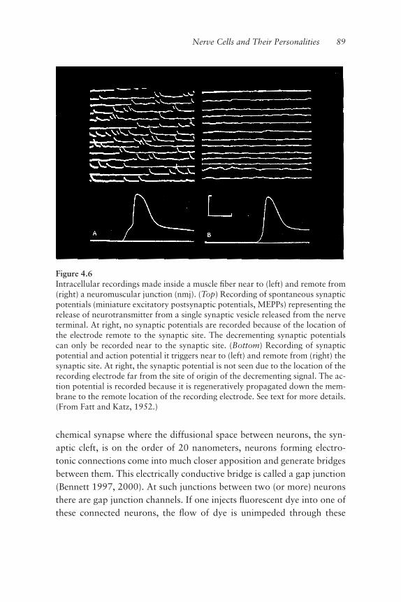
chemical synapse where the diffusional space between neurons, the syn-aptic cleft, is on the order of 20 nanometers, neurons forming electro-tonic connections come into much closer apposition and generate bridgesbetween them. This electrically conductive bridge is called a gap junction(Bennett 1997, 2000). At such junctions between two (or more) neuronsthere are gap junction channels. If one injects ºuorescent dye into one ofthese connected neurons, the ºow of dye is unimpeded through these
Nerve Cells and Their Personalities 89
Figure 4.6Intracellular recordings made inside a muscle ªber near to (left) and remote from(right) a neuromuscular junction (nmj). (Top) Recording of spontaneous synapticpotentials (miniature excitatory postsynaptic potentials, MEPPs) representing therelease of neurotransmitter from a single synaptic vesicle released from the nerveterminal. At right, no synaptic potentials are recorded because of the location ofthe electrode remote to the synaptic site. The decrementing synaptic potentialscan only be recorded near to the synaptic site. (Bottom) Recording of synapticpotential and action potential it triggers near to (left) and remote from (right) thesynaptic site. At right, the synaptic potential is not seen due to the location of therecording electrode far from the site of origin of the decrementing signal. The ac-tion potential is recorded because it is regeneratively propagated down the mem-brane to the remote location of the recording electrode. See text for more details.(From Fatt and Katz, 1952.)

channels and thus between cells. These gap junction channels allow con-duction of ions (electric current) directly between the cells, thus makingthem electrically coupled. There are no neurotransmitters in electrotonicºow and so there is very little, if any, delay in the voltage shifts impartedby the ºow of electricity from one cell to another. The process of electro-chemical signaling has associated with it a small delay due to the manysteps involved in the release of transmitter, diffusion time across the cleft,subsequent transmitter binding, and then activation of the associated ionchannels to allow current ºow in and out of the local area of the mem-brane. This all takes anywhere from 1 to 5 milliseconds. Withelectrotonic connectivity, however, the ionic ºow is through channelsthat are already open (although they can close), and current ºows be-tween cells directly as the interior of the cells are interconnected. Conse-quently, when one neuron ªres an action potential, any cellelectrotonically coupled to it is compelled to receive part of the signal,virtually simultaneously, and if the signal is large enough, it will itself ªreaction potentials in the coupled cells.Is such electrical ºow bidirectional? It turns out that many gap junc-
tions regulate current ºow in an even, bidirectional manner (Bennett2000). Bidirectionality is seen in the electrical connections of the inferiorolive (IO) cells, which allows such cells to function, electrically, almost asone big cell (for review, see Welsh and Llinás 1997). But not all gap junc-tions operate in such a bidirectional fashion. Some gap junctions allowionically carried current ºow in only one direction and thus send theirsignal simultaneously to the next neuron but do not receive signals backfrom that cell (see Furshpan and Potter 1959).And so, because of this direct ºow of current from cell to cell by means
of electrotonic coupling, the result is rapid and synchronous ªring of in-terconnected cells. This is how “ensemble” signaling occurs. These typesof signaling allow groups of neurons to convey to widely scattered andperhaps distantly located neurons a concise and synchronous signal pat-tern. The simultaneity produced by electrical coupling gives this signalthe “roar of the masses” as many cells ªre together, rather than the“voice in the wilderness” of a single cell. It is a choir as opposed to a soloperformance. This issue of temporal binding relates also to the seamless-ness we sense in consciousness.
90 Chapter 4

Electrotonic coupling plays a signiªcant role in developmental pro-cesses as well. We saw in chapter 3 the advantage of simultaneous signal-ing in order to internalize motricity from myogenically rhythmic musclesto the function of the oscillatory resonance of the brain itself. Such cou-pling during development also takes advantage of the fact that the diame-ters of the gap junction channels are comparatively large (Bennett 2000).In addition to ions, gap junctions allow ºow of relatively large moleculesthat play important, internal regulatory roles in development and cellfunction, and which might have a harder time getting into the cells other-wise, given their size. The gap junction channels are roughly 1.5 nm in di-ameter, allowing small peptides and also critical molecules such as cyclicadenosine monophosphate (cAMP) to pass from cell to cell (see Simpsonet al. 1977; Pitts and Sims 1977; Kam et al. 1998; Bevans et al. 1998).Such larger molecules cannot pass into cells by way of the ion channels inthe cell’s membrane, as these channels are simply much too small. Onemay look at this as a form of regulatory simultaneity between developingneurons.
And so, the basic functional unity for the neuron is electrical, withelectrotonic interactions and action potentials serving as the temporalbinding element giving the neuron its integrative backbone. Chemicaland electrical synaptic transmission are, on the other hand, the basiccoinage that binds the different cellular elements into single multicellularfunctional states.
Nerve Cells and Their Personalities 91

Alfred Jensen, Square Beginning—Cyclic Ending, Per I–V, 1960. Oil on canvas,50 � 250 in., overall; ªve panels, 50 � 50 in each; panel 4. Courtesy ofPaceWildenstein. Photograph by Ellen Page Wilson.

5 Lessons from the
Evolution of the Eye
The Invention of Sensory Organs
What is the evolutionary drive to form an animal? How is such a spectac-ular and complex cellular-based architecture generated? And what canone say of organs and their vast diversity of form and function within ananimal? We think of our organs from a perspective of physiological divi-sion of labor, as specialized components of our bodies whose functionalroles are unique, often singularly so. In most cases they are vital to the or-ganism as a whole, in the short run of a given lifetime and in the long runfor the perpetuation of the species. The heart, the eyes, the liver—they aremodules, individual local devices. In many ways, like the brain, a givenorgan may be considered a specialized, closed society within an animal.Among these many similarities, however, there is an exceptional differ-ence. The brain is basically closed in its nature and operation. It com-pletely escapes direct examination by any of our senses. We cannot see it,we cannot hear it, we do not feel it beating, it does not heave up anddown, it can feel no pain if we strike it. And more, it seems capable of be-ing remote from its corporeal anchor—as when we empathize with thepain of others or observe with admiration the universe around us.

This closed organic system we call the brain has the further advantageof not being limited by the properties of the senses. Consider that thewaking state is a dreamlike state (in the same sense that dreaming is awakelike state) guided and shaped by the senses, whereas regular dream-ing does not involve the senses at all. Although the brain may use thesenses to take in the richness of the world, it is not limited by thosesenses; it is capable of doing what it does without any sensory inputwhatsoever. The nature of the brain and what it does makes the nervoussystem a very different type of entity from the rest of the universe. It is, asstated repeatedly, a reality emulator. Suggesting that the system is closed,and so very different, means that it must be another way of expressing“everything.” In other words, brain activity is a metaphor for everythingelse. Comforting or disturbing, the fact is that we are basically dreamingmachines that construct virtual models of the real world. It is probably asmuch as we can do with only one and a half pounds of mass and a “dim”power consumption of 14 watts (Erecinska and Silver 1994).Perhaps more puzzling still is that this unique and speciªc functional
architecture, this closed cellular system, forms without knowing a prioriwhat it or its function is to be! How can this be? It turns out that this isone of the curiosities of the evolutionary process that the brain has incommon with all organs. They have all developed into their complex andunique structures, their speciªc and capable functions, without any a pri-ori ªnal plan. “But wait, what about genetics and the developmental planpreprogrammed into our DNA?” you ask. Well, of course genetics are in-volved, but only as an accumulated result of each generation’s storytell-ing of the great epic, with no overarching plot and certainly no end.There are plenty of characters, a bang of a beginning, but then only atumbling, churning, never-ending middle. Curious indeed.How can one understand the process by which the nervous system,
the mind, came to us if its ancestry started out in evolution without acompass or a map? Well, all other organs (as the animals they inhabit)developed in the same way, by trial and error, and in a process that hasno end.In order to develop the nervous system nature had to learn from the
properties of the external universe and it had to incorporate them inbeneªcial ways. In chapter 3, the deeply important issue of embedding
94 Chapter 5

was discussed. Now it is time to ask what the ªrst step is in this process.How do we take in fractionalized properties of the external world andput them in the context of a single whole? Evolution had to “invent” sen-sory organs, specialized relay mechanisms between the universals of theexternal world and the inherently closed system that is the brain. It is per-haps easier to understand that this “taking in” of external properties isprecisely what nature has had life do over all of evolution, when we un-derstand the sensory relay or transformation process at the momentarylevel. Momentary level is, for example, how one may take in patterns oflight, right now as you read; these patterns make up the words on thispage, which come out somehow as a voice in your head. Rolling throughthe millennia, the system embeds, “remembers,” and what is the result?Primitive photoreceptors functioning to detect light have evolved into thewondrously complex organs that are our eyes. The primitive statocyst,that at one stage was (and still is) the rudimentary balancing mechanismfor the sea squirt, has evolved into the enormously important and intri-cately networked subsystem of the brain we call the vestibular system.Sensory systems have evolved as exceedingly sophisticated tools for thebrain, honed over the eons to ever increase and perfect the proªciency ofpredictive movement and thus survival.Our descriptions imply a slant toward sensory organs. This is the case,
but it needn’t be so. The learning from the embedding of the universalproperties of the external world drove the formation of all organs, as wellas feet and hands, tails and feathers. But with sensory organs, this processis easier to understand because sensory organs are direct conduits fromthe external world to the internal world of the brain, and the brain is theattracting focus of this book. The liver, for example, evolved under thesesame principles of evolutionary pressure but in ways somewhat less di-rect. The liver evolved to help rid our systems of toxins that we unwit-tingly (or some times wittingly, as with the fondness for wine) take in. Buttoxicity is mainly relative across species; it is not a universal. What makesus deathly ill is a happy snack for a rat or a beetle’s daily routine. When Ispeak of universal properties, or simply universals, I am referring to thoseproperties arising from the external world that are invariant across all oflife. Light waves, for example, have a universal property, as does temper-ature or the force of gravity. These are among the very ªrst, powerful,
Lessons from the Evolution of the Eye 95

unchanging phenomena that life had to confront and by which life hasbeen shaped. But can all this be understood? Well, let us think of onefunction—seeing—and one set of organs—the eyes.
Why Eyes?
As mentioned above, I am slanting toward sensory organs, and in thischapter eyes are my central example, because of the variety of forms thathave evolved for the transduction (conversion) of light into useful spe-ciªcation of internal brain states. Basically, in all cases and for all thesenses, the brain only accepts those speciªc properties of the externalworld that stimulate sense organs (we do not sense radio or TV electro-magnetic waves directly) and these inputs are conveyed only as neuronalelectrical activity.
So to Begin, What Is Vision?
What does it mean to “see”? Why is the visual apparatus the way it is andwhat can it teach us about the brain—the mind? (For an excellent reviewof vision consult Zeki 1993.)The eye (ªgure 5.1A), and in particular the retina (the light-sensitive
part), is a true extension of the central nervous system (ªgure 5.1B). Theneurons in the retina form an extraordinarily compact and beautiful cir-cuit that sends the electrical messages that the brain interprets as light. Inchapter 4, we came to understand that it is not just the ensemble proper-ties of neuronal circuits that give rise to a particular circuit’s unique andspeciªc behavior and function; it is the architecture of the ensemble thatgives the circuit its macroscopic properties (as in the case of the heart)and through such architectures both context and intentionality. These ar-chitectures, or modules, are generally formed by neuronal elements ofdifferent types and of different intrinsic electrical properties. Some areexcitatory and some inhibitory. In the case of the eye, the retina providesan excellent example of this strategy. So with our eyes and our mind’seye, let’s take a look into the evolution of the eye, our eye, and someother fascinating eyes as well.It all began with organisms taking advantage of the energy from the
sun. Solar energy is absolutely essential for life. We are on earth courtesy
96 Chapter 5
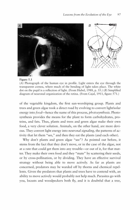
of the vegetable kingdom, the ªrst sun-worshiping group. Plants andtrees and green algae took a direct road by evolving to convert light/solarenergy into food—hence the name of this process, photosynthesis. Photo-synthesis provides the means for the plant to form carbohydrates, pro-teins, and fats. Thus, plants and trees and green algae make their ownfood, a very clever solution. Animals, on the other hand, are more devi-ous. They convert light energy into neuronal signaling, the patterns of ac-tivity that let them “see,” and then they eat the plants (and each other).Why don’t plants and green algae “see”? As pointed out before, it
stems from the fact that they don’t move, or in the case of the algae, notat a rate that could get them into any trouble—or out of it, for that mat-ter. They make their own food and they “mate” by scattering their seeds,or by cross-pollination, or by dividing. They have an effective survivalstrategy without being able to move actively. As far as plants areconcerned, predators may be warded off by thorns and chemical repel-lents. Given the predators that plants and trees have to contend with, anability to move actively would probably not help much. Parasites go withyou, locusts and woodpeckers both ºy, and it is doubtful that a tree,
Lessons from the Evolution of the Eye 97
Figure 5.1(A) Photograph of the human eye in proªle. Light enters the eye through thetransparent cornea, where much of the bending of light takes place. The whitedot on the pupil is a reºection of light. (From Hubel, 1988, p. 35.) (B) Simpliªeddiagram of neuronal organization of the retina. (From Cajal, 1911, ªgure 571.)
A B

given the miraculous ability to ºy or move at all, would be able to outruneither.So trees do not run or ºy and they cannot beat off enemies with their
branches. The closest that trees come to moving would be what is calledphototropism, the tendency for such photophilic life forms to bend to-ward light, or drift, as the case may be for the algae. But this is more ac-curately thought of in terms of the Jamesian reºexology we discussed inchapter 2, rather than any form of self-referential movement. Trees donot move actively, and that is why they neither need, nor have, a brain:their survival does not depend on prediction.As for actively moving creatures, “teleception,” or remote sensing such
as seeing, hearing, or smelling, extends a creature’s predictive capacity innegotiating the external world. It is nice to be able to see that a threat iscoming, as opposed to having to feel it through one’s outer being ªrst, orperhaps to have to taste it, in order to register belatedly its arrival. In-deed, we sense these latter modes of sensory transduction, informationthrough feeling and tasting, as being, in the context of an oncoming pred-ator, too close. For us, too close in these cases usually means too late.And so teleception, remote sensing, arose to buy more time for predictionand the predictive properties of the brain to be useful.The earliest aspects of prediction that gave rise to the magniªcent or-
gans we know as eyes started with the need of very early creatures tosense light. In the ocean, the need was to predict which way was up to thesurface and which down to the depths. In terrestrial forms, the need wasfor safety, as one knows from seeing a speeding cockroach disappear intothe woodwork as one turns on the light at night when looking for asnack. Hiding in the dark usually works out better than hiding in the sun.Originally these basic but clear needs had nothing to do with seeing animage, or even of forming one; they were simply the need to sensewhether or not there was light. At this stage, the ability to sense the direc-tion of a light source, although incredibly helpful, required an organ yetto evolve. To understand this transition of organ capability—the detec-tion of light versus the detection of the direction of a light source—wehave to look into the phototransduction process itself.Light is made up of events called photons. The argument continues as
to whether photons are particles that act like waves or waves that act likeparticles or neither. From a physiological perspective, we consider them
98 Chapter 5

to be energy packets (quanta) that activate a receptor, but the wavelengthof these electromagnetic quanta emitted by the sun is critical as well; dif-fering wavelengths of photons translate to us as different colors.These wavelike particles travel in straight lines at close to 300,000
kilometers/second. Because photons interact with each other and rein-force or cancel one another (Richard Feynman’s “Sum Over Histories” inFeynman and Hibbs, 1965), even if light reºects off something, it never-theless continues to travel straight. Light can also be refracted, whichmeans that as it passes from one medium to another (say from air to wa-ter or glass), its angle of trajectory can be changed as it passes through.After the light is set on this new path, it remains straight as long as themedium does not change or as long as the space in which it travels is notwarped by a big gravitational ªeld. The angular degree to which a me-dium bends the trajectory of light is known as its “refractive index.” Thehigher the degree of bending, the higher the index.Thus light can bounce off things, as in reºection, and it can pass
through things, as in refraction, and so light can inform us of the opticalproperties of the universe around us. Light can be caught or absorbed.The fact that light travels in a straight line is particularly important, as itmeans that it is easy to work with light and easy to know its source. Thefacts that light follows a straight trajectory and is abundant, were essen-tial in the evolution of vision, enabling photons striking the eye to func-tion as accurate and faithful messengers communicating to us the remotelandscape of the external world.Photons are “caught” by pigmented matter. Or rather, matter is pig-
mented (colored) because it absorbs particular photons. Pigments arewhat give color to things, and it works as follows. As mentioned before,light photons come in different wavelengths. A traveling light beam has asinusoidal character, like waves in water, a certain distance from peak topeak (or trough to trough) known as wavelength. For the range of colorswe can see, called the visible spectrum, these wavelengths are in the hun-dreds of nanometers (nms) or 10−9 meters. For instance, what we see asblue is light in the 420-nm wavelength range; red is in the longer 550-nmwavelength range.One may also refer to the frequency of light. Frequency is inversely re-
lated to wavelength: the longer the wavelength, the lower the frequency.Frequency is a measure of the number of individual wave cycles in a
Lessons from the Evolution of the Eye 99
second. If the space between peaks is greater, that is, the wavelength islonger, then it stands to reason that fewer wave cycles will pass by somegiven point in a second, and thus the frequency will be lower. Frequencyis not the same as speed. The speed of light, for the purposes at hand,does not change. It is a constant.Natural sunlight is a mixture of all the light frequencies of the entire
color spectrum. And so, is it my colleague’s blue book in front of me thatis absorbing blue light, or is it my eye? It is the eye that is catching theblue light. If it were the book that absorbed the blue light, how would theblueness information reach my brain? The pigment in the dictionarycover has absorbed, stopped, or caught the color frequencies other thanblue. Blue has been reºected off, and in a straight line; the photons of thisfrequency have made it to my eye. However, keep in mind that bluenessdoes not exist in the external world. Blueness is a brain interpretationgiven to a particular wavelength (420 nm) range.It is my eye that is absorbing—stopping—these photons of light whose
frequency says “blue.” How is this done? The photons were absorbed byneurons called photoreceptors. In these photoreceptors are found a classof very old proteins called opsins, a component of the visual pigment.These opsins interact closely with a second molecule called a chromo-phore (the actual photon catcher), triggering the activation of the recep-tor cell when light strikes. In the case of my colleague’s book, as thehigher proportion of blue photons were reºected toward me, a greaterproportion of blue photon-catching photoreceptors were activated.Now let’s go back to our primitive creature who is just trying to sense
light photons, a patch of photosensitive skin, photoreceptors, reºection,and refraction
Photon Sensing and the Direction of Light
The photoreceptor neuron determines light intensity by “counting” thenumber of photons it catches. Each time a photon is caught, the mem-brane potential of the photoreceptor changes slightly, so light is measuredin incremental membrane potential steps. In fact, we humans can detectat the single photon level (Hubel 1988). The pattern of electrical activitygiven by a group of photoreceptors counting light photons gives the mag-
100 Chapter 5

nitude of the light received. The changes in the patterns of photoreceptorcounting can give a measure of the ºuctuation of the light source(shadow, predator). And so we have a primitive creature with a patch ofphotosensitive skin or “eye spot.”The ability to sense day or night, and perhaps that light meant warmth,
was certainly helpful to survival. But can one improve on this? How doesone do a better job of catching light? Well, the photosensitive patch ofskin, which is actually composed of ciliated ectodermal cells containingsome 100 photoreceptors (Land and Fernald 1992), cannot generate animage of what is out there. As this photoreceptor “patch” begins to en-large in area, however, it tends naturally to form a cup shape or pit. Thusif light comes directly from the front, the back of the cup is activated.Light coming from any other angle will tend to activate the cup asymmet-rically, giving rise to a very crude directionality allowing, at most, up anddown differentiation, and maybe left and right as well. Many of thesepitlike eyes actually only respond to rapid changes in light (movingshadows).Then a wondrous event occurred in evolution. This cupped patch of
skin, containing 100 photoreceptors or so, enlarged further and reducedits aperture (diameter of the opening at the front), closing over to form acystlike structure. This gave rise to what is known as the “cup eye.” Thecup eventually became closed completely except for a small hole, and thisgave rise to the “pinhole eye,” where the pinhole serves as a lens. An in-verted real image of the world was projected on to the photoreceptivesurface. A better image was achieved when lenses in the form of transpar-ent epithelia ªnally evolved (Ali 1984) (ªgure 5.2).
Some Eyes Are Weird
Now we enter the truth-is-stranger-than-ªction realm. My friends EnricoNasi and Maria del Pilar Gomez, who work during the summers at theWoods Hole Marine Biological Laboratory, are experts on photorecep-tion and study the eyes of scallops. These delicious mollusks have, at thepoint of contact of their two shells, a large number of beautifullyrounded blue eyes that peer in all directions and are, as I learned fromEnrico and Maria, quite strange. Unlike our eyes, which have a retina
Lessons from the Evolution of the Eye 101

102 Chapter 5
Figure 5.2Evolution of the single-chambered eye. Arrows indicate structural developmentsrather than speciªc evolutionary sequences. (a) Pit eye. (b) Pinhole eye, found inNautilus. (c) Eye with lens. (d) Homogeneous lens. (e) Inhomogeneous,“Matthiesen” lens. (f) Multiple lens eye of male Pontella. (g) Two-lens eye ofCopilia (see ªgure 5.4). Solid arrow shows image position. Open arrow, themovement of the scanning second lens. (h) Human eye with cornea and lens. Ic,image formed by cornea alone; Ir, ªnal image on retina. (i) Mirror eye of the scal-lop Pecten. (From Land and Fernald, 1992, ªgure 1, p. 6).

against the back end of the eye cup, scallops have two back-to-back reti-nas that hang in an up-down direction like a projection screen near thecenter of the eye. This screen divides the eye cup into a front and a backhalf. These retinas, like two opposing drum skins, are organized so thatthe front one measures light intensity as the light enters the eye. The lightpasses through this retina and hits the back of the eye, which is a mir-rored surface! Upon hitting the mirror, as in a Newtonian telescope,the light is focused back on to the second retina located at the back ofthe ªrst one. This second retina receives the light image made by theback mirror. Both retinas send their messages out through an optic nervethat radiates from the eye cup at the equator (if we consider that thenorth and south poles are respectively the front and back of the eye)(ªgure 5.3).
They Get Weirder
In some other marine invertebrates such as the ostracod crustaceanGigantocypris, the eye consists not of a lens, but a parabolic mirror(ªgure 5.4). The mirror collects light and focuses it on a match head-likeretina (a bit like the front light of a car, if one exchanges the light bulb fora bulb-shaped retina) (Land 1980). And, weirder still, in heteropod seasnails and in jumping spiders, among others, the retina is a strip that isonly a few receptors across and several hundred receptors long; the eyescans by tilting the retina through a 90-degree arc (Land and Fernald1992). But the real weirdo in the eye world is that of Copilia, a marine in-vertebrate found in the Mediterranean near Naples (ªgure 5.5). Thiscreature has a transparent head-bound immobile lens (like an airplanewindow). Inside the head there is a second lens and a small group ofphotoreceptors that scan the horizon ªve times a second, not unlike thesweep of a television set.What does all this say? It says that a surface that originally only ab-
sorbed light can evolve through a set of intermediate steps that do not yetmake images, into one that can support image making! This bit of skinhas become a highly specialized module of function: it has become an or-gan, the eye.
Lessons from the Evolution of the Eye 103

104 Chapter 5
Figure 5.3The scallop eye. (Top) A cross section through this eye shows that, unlike the lenseyes of vertebrates, which have a clear zone between the lens and the retina acrosswhich light rays are focused, the lens of the scallop eye is in contact with a cres-cent-shaped retina. Behind the retina is the extremely thin mirror (not discernibleat this magniªcation) and then a thick layer of dark pigment. At right, a diagramshows the path of light entering this eye. The light, only weakly refracted by thelens, passes through the retina to the hemispherical reºector, which focuses thelight back to the distal photoreceptor cells in the upper layer of the retina. Be-cause the light passes through the retina once before it is detected, the contrastacuity of the scallop eye is poor. (Bottom) The eyes of the scallop Pecten are seenin this photograph as small globules (highlighted by circles) along the mollusk’smantle, which is exposed through the gaping opening of the two valves, or shells.Within each eye, about a millimeter in diameter, is a shiny hemispherical mirror.The eyes detect the movement of a shadow or a dark edge across the animal’s ªeldof view, allowing the scallop to discern the approach of a predator. These low-contrast-acuity eyes may also play a role in phototaxis: movement toward areasof darkness or light. (From Land 1978, pp. 127, 130.)

From Cells Come Systems
And so, from the complexity inherent to cells, the complexity of systemsarises. It has done so without knowing a priori what it is going to be-come. These are emergent properties; the systems are not all or none,but evolve. Yet intuitively, it would seem that something, some attrac-tor, some process, must lead the eye to become what it is, given itsunbelievable complexity. But that is exactly what the process is not.There is no intention to become an eye. With the eye, almost every possi-ble solution has been implemented in the wake of its emergence. The
Lessons from the Evolution of the Eye 105
Figure 5.4Another mirror eye. The deep-sea crustacean Gigantocypris has large reºectingeyes that enable it to concentrate the extremely faint light available at a depth of1,000 meters in the ocean (primarily from the light organs of luminescent crusta-ceans and ªshes). About a centimeter long, this scallop’s head makes up abouthalf of its body. The two reºecting eyes are covered by transparent windows inthe orange carapace that completely encloses the animal. Sir Alister Hardy of theUniversity of Oxford likened its eyes to the “headlamps of a large car.” Hardywas the ªrst to speculate that the mirrors serve to focus light. (From Land, 1978,p. 131)

106 Chapter 5
Figure 5.5(Left) copepod Copilia (Right) Diagram of the eyes. Retinae subtend only 3� ofarc, and scan through a total of 14� of arc. (From website nmnhwww.si.edu/iz/copepod/_borders/).
system did not know to do a ºat retina or a Galilean lens; it simply triedeverything that was feasible structurally, and for which there was somefunctional advantage at the time. What do all the solutions have in com-mon? They share exploitation of the properties of light, namely that itgoes straight and generates an image through reºection or refraction.Once we understand that, we can probably understand every conceivableperipheral visual system, whether here or on another planet.How is it that we know that evolution has indeed tried almost every-
thing in terms of making images with light? Well, in truth we do not, butit is likely simply because the photoreceptor has been around for such along time. Or perhaps evolution has not yet tried everything, but let’slook at it this way. Nature does not design, yet spectacular things happen.Let me give you another example.Ask yourself, how much does a bag of marbles cost? A few dollars? But
people need jobs to make a living, so the actual cost of producing the bagfull of marbles must probably be a few pennies. How do you make a mar-ble so perfect, so totally seamless and absolutely spherical, at such a tinyprice? And remember, marbles have beautiful patterns in them as well. Sohow do you do it?Well, probably for most people, the idea of the shot tower comes to
mind. With a shot tower, molten lead is sprayed out and falls into a cool-ing liquid. As the lead falls, it congeals into more or less spherical bits.Any liquid if tossed from a sufªcient height will congeal into spheres. Butwith marbles, this would be a very expensive approach to their produc-tion and you would not be able to incorporate, in any controllable way,the patterns you want the marbles to have. Furthermore, the marbleswould not be perfectly round. Instead, the solution is to melt glass, shapeit into a rod while it is still soft, put in your patterns, and cut into shortcylinders. Then you tumble them together in a tumbling drum with abra-sives. After a period of tumbling time, the marbles come out perfectlyround, or, at least they approach perfect roundness.This is precisely what happens in biology, over evolution: if you let
something tumble long enough, it comes out almost perfect. Such is thepower of random collisions and patience, and that constitutes the sum to-tal of nature’s intelligence. All the rough edges, the ºaws, the things thatdon’t work are systematically dispatched by natural selection. What
Lessons from the Evolution of the Eye 107
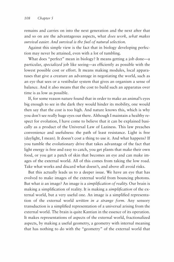
remains and carries on into the next generation and the next after thatand so on are the advantageous aspects, what does work, what makessurvival easier. And survival is the fuel of natural selection.Against this simple view is the fact that in biology developing perfec-
tion may never be attained, even with a lot of tumbling.What does “perfect” mean in biology? It means getting a job done—a
particular, specialized job like seeing—as efªciently as possible with thelowest possible cost or effort. It means making modules, local appara-tuses that give a creature an advantage in negotiating the world, such asan eye that sees or a vestibular system that gives an organism a sense ofbalance. And it also means that the cost to build such an apparatus overtime is as low as possible.If, for some reason nature found that in order to make an animal’s eyes
big enough to see in the dark they would hinder its mobility, one wouldthen say that the cost is too high. And nature knows this, which is whyyou don’t see really huge eyes out there. Although I maintain a healthy re-spect for evolution, I have come to believe that it can be explained basi-cally as a product of the Universal Law of Laziness. This law preachesconvenience and usefulness: the path of least resistance. Light is free(daylight, I mean). It doesn’t cost a thing to use it. And what happens? Ifyou tumble the evolutionary drive that takes advantage of the fact thatlight energy is free and easy to catch, you get plants that make their ownfood, or you get a patch of skin that becomes an eye and can make im-ages of the external world. All of this comes from taking the low road.Take what works and discard what doesn’t, and above all avoid risks.But this actually leads us to a deeper issue. We have an eye that has
evolved to make images of the external world from bouncing photons.But what is an image? An image is a simpliªcation of reality. Our brain ismaking a simpliªcation of reality. It is making a simpliªcation of the ex-ternal world, but a very useful one. An image is a simpliªed representa-tion of the external world written in a strange form. Any sensorytransduction is a simpliªed representation of a universal arising from theexternal world. The brain is quite Kantian in the essence of its operation.It makes representations of aspects of the external world, fractionalizedaspects, by making a useful geometry, a geometry with internal meaningthat has nothing to do with the “geometry” of the external world that
108 Chapter 5

gave rise to it. This is the vector/vector transformational capability of thebrain that is independent of the coordinate systems utilized to measure it.Consider colors, which are just the particular way we transduce energy
at a particular frequency. A snake sees infrared, which is actually heat. Itis very clear that the images in our head are only a representation of theworld.Eyes are neurons that embed geometries of bouncing light, and the
brain is a set of coordinate systems that measure or recognize abstract ge-ometries that do not exist in the external world. The smell of the forest isan internal abstraction that does not exist as an external geometry.Here is a further thought to consider: language is an eye, but an ab-
stract one, an internal abstraction.Just as a patch of photosensitive skin became an eye, language as we
know it followed the same journey. Both are specialized apparatuses forthe geometrical internalization of external, fractionalized properties.Thus we have the following streams: patch/wrinkle/cup/pinhole/eye;the generation of protonetworks to the mature functional system, analo-gous to protolanguage, developing to language (perhaps it still isprotolanguage!)I say that I am a closed system but not a solipsist. I can’t be, because of
the way I was built by evolution by internalizing the properties of theoutside world.
Lessons from the Evolution of the Eye 109
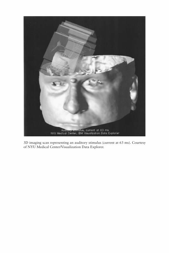
3D imaging scan representing an auditory stimulus (current at 63 ms). Courtesyof NYU Medical Center/Visualization Data Explorer.

6 The I of the Vortex
Consciousness and the Nature of Subjectivity
Having touched briefly on the cell biological rules that evolution has em-ployed to internalize fractured components of the external world (chap-ters 1–3), we come now to the question of synthesis: how do all thesediffering components come together into a singular, global, internal con-struct? Because these neurons of varying “personalities” are by deªnitionrelatively specialized (chapter 4), no particular cell’s activity can repre-sent more than a small component of such reality. In chapter 5 we sawthat photoreceptors are cells specialized for capturing photons and trans-ducing (converting) this electromagnetic energy into electrical activity.Similarly, in our skin we have what are known as mechanoreceptors, spe-cialized cells that transduce mechanical energy into a pattern of neuronalactivity (ªgure 6.1). That you can feel this book in your hands is medi-ated in part by an array of different mechanoreceptors that tell of pres-sure, pressure changes, and pressure differences on/to the skin. Workingin parallel with these are the joint receptors and muscle sensors known as“muscle spindles” that give you a sense of limb position in space. Inshort, you don’t need to be able to see your hands holding this book toknow that you are doing so.
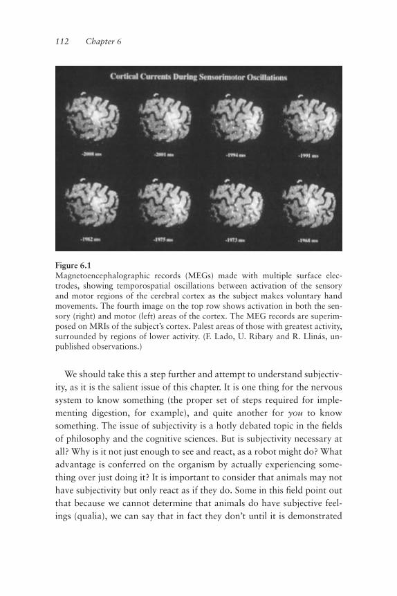
We should take this a step further and attempt to understand subjectiv-ity, as it is the salient issue of this chapter. It is one thing for the nervoussystem to know something (the proper set of steps required for imple-menting digestion, for example), and quite another for you to knowsomething. The issue of subjectivity is a hotly debated topic in the ªeldsof philosophy and the cognitive sciences. But is subjectivity necessary atall? Why is it not just enough to see and react, as a robot might do? Whatadvantage is conferred on the organism by actually experiencing some-thing over just doing it? It is important to consider that animals may nothave subjectivity but only react as if they do. Some in this ªeld point outthat because we cannot determine that animals do have subjective feel-ings (qualia), we can say that in fact they don’t until it is demonstrated
112 Chapter 6
Figure 6.1Magnetoencephalographic records (MEGs) made with multiple surface elec-trodes, showing temporospatial oscillations between activation of the sensoryand motor regions of the cerebral cortex as the subject makes voluntary handmovements. The fourth image on the top row shows activation in both the sen-sory (right) and motor (left) areas of the cortex. The MEG records are superim-posed on MRIs of the subject’s cortex. Palest areas of those with greatest activity,surrounded by regions of lower activity. (F. Lado, U. Ribary and R. Llinás, un-published observations.)

otherwise. It may be argued, however, that the burden of proof is onthose who deny subjectivity in animals. For myself, I suspect that subjec-tivity is what the nervous system is all about, even at the most primitivelevels of evolution. As an obvious corollary to that suspicion, I alsosuspect that consciousness as the substrate for subjectivity does not existoutside the realm of nervous system function or its nonbiological equiva-lent, if there is any.We know that single-cell “animals” are capable of irritability, that is,
they respond to external stimuli with organized, goal-directed behavior.It is difªcult to ignore that such cellular property is probably the ancestryfor the irritability and motricity displayed by sensory and muscle cells, re-spectively. And so we are left with the nagging feeling that irritability andsubjectivity, in a very primitive sense, are properties originally belongingto single cells. If so, that primitive subjectivity may be built into the con-sciousness and subjectivity displayed by the nervous system as cells orga-nize into the ensembles that we know as nerve cell circuits. But do takenote that only certain architectures are capable of supporting and en-hancing such primitive “feelings.” Also note, however, that such a primi-tive form of subjectivity and the notion of “grandmother cells,” whichwill be discussed further in this chapter and in chapter 10, are totally dis-tinct concepts.The goal of this chapter is to add further substance to the basic posi-
tion of this book, namely that the issue of cognition is ªrst and foremostan empirical problem, not a philosophical one. This issue has been ad-dressed by some of the most distinguished biologists of this century(Crick 1994; Crick and Koch 1990; Changeux 1996; Changeux andDeheane 2000; Edelman 1992, 1993; Mountcastle 1998).We have spoken of the embedding of universals and of the coordinate
system transformations that the brain performs to do this (chapter 3). Wealso saw that as we trace the ºow of activity into the brain (from the pe-ripheral sensory receptors or the output organs of the body, the musclesand glands), the geometric transformations that provide functional com-munication between sensory and motor frames of reference become in-creasingly more abstract. Given this, when addressing such remotefunctional sites it is not surprising that terms such as “higher computa-tional levels” have become commonplace in neuroscience. However, one
The I of the Vortex 113

may ask what is actually being computed when a neuron ªres an actionpotential. This is not an insigniªcant issue. Such language implies under-lying assumptions that may not apply to the brain and that may encour-age misguided inquiry.
Sensory Representation
At the very forefront of brain theory is the goal of transforming knowl-edge relating to the properties of the nervous system into an understand-ing of global brain function. And so, for instance, in the study of sensorysystems or pathways, we see the tendency to favor constructions that de-scribe how cells with general sensory properties (e.g., photoreceptors)connect to cells with more speciªc receptive properties, and so on. Theoverall process, so it is supposed, is just a matter of the handing over ofduty to components with successively better expertise in sensory analysis.But how can one have better expertise as one moves away from the levelof direct observation provided by sensory input and into the realm ofmore abstract and recondite corners of brain function? Well, the fact isthat reality is elaborated by brain function, and it does not come easily.The study of neurological conditions tells us that the ability to recognizereality and to respond to it may be altered in an enormous number ofways, giving us a glimpse into the amazingly cooperative nature of brainfunction. In this particular instance all the parts are important, eventhose that are silent at any particular time. As in music, in brain functionsilence is as important as sound.Although this line of thinking has been central to our attempts at un-
derstanding brain function, it can also lead to misperceptions. For in-stance, it has lead to a belief in the appropriateness of such terms as the“face cell” or the “grandmother cell,” a cell whose ªring denotes the rec-ognition or recollection of one’s grandmother (see, for example, Grossand Sergent 1992; Rolls 1992). The fundamental problem with theseconcepts, and the line of thinking that produced them, is easily exposed.If the operation of each neuron represents knowledge of only a veryspeciªc component of reality, to whom does this information get commu-nicated in the rest of the brain? That is, how could these neurons makethemselves understood by other cells not “in the know”? What sort of
114 Chapter 6

cognitive deªcits might we observe if such cells are damaged? WouldGrandma disappear from our cognitive world if that set of cells were todie? A strong physiological argument opposing such ideas is the soberingfact that the truly galactic number of possible representations that thebrain can make clearly exceeds the number of available neurons! (Tononiet al. 1992).It is not difªcult to understand how such views came about, however.
In the 1950s, Wilder Penªeld and Herbert Jasper made seminal contribu-tions toward an understanding of the functional organization of thebrain. Their ªndings were subsequently extended (inappropriately) be-yond their original well-deªned limit.In those early studies (Penªeld and Rasmussen 1950), patients with in-
tractable epilepsy were treated by surgically removing the site of origin ofthe deleterious electrical activity. During these surgeries, in which pa-tients were fully awake and only the scalp was anaesthetized (a painlessprocedure), the surgeon electrically stimulated various areas of the cor-tex. Within a certain cortical region, stimulation elicited musculartwitches in the ªngers, toes, arms, shoulders, and so on. Similarly, in an-other nearby cortical area, stimulation produced sensations in variousspeciªc body parts, as reported by the patient. Through diligent work, amap of what we now call motor cortex and somatosensory cortex wasconstructed. Many of these cortical areas are somatotopically organized,in that the layout of the body is represented, in faithful point to pointmatching, by the cells of each of these cortical structures. For example,the ªngers of the left hand are represented as a neuronal map of the hand(and its ªngers in correct place and order) in the motor cortex for theºow of motor information to the periphery, and in the somatosensorycortex for tactile information coming from the periphery. These somato-topic mappings are distorted versions of the body, however, and for goodreason. The various body parts are represented in different proportions.For instance, there are many times more neurons involved in the motorand sensory aspects of the functioning of the tongue than there are for,say, the heel. The tongue is far more articulate in all that it does than theheel. Similarly, more cortical real estate is dedicated to the skin of the in-dex ªnger than is provided for a similar area of skin on the back. The tac-tile sensitivity of the index ªnger is vastly greater than that of the back,
The I of the Vortex 115
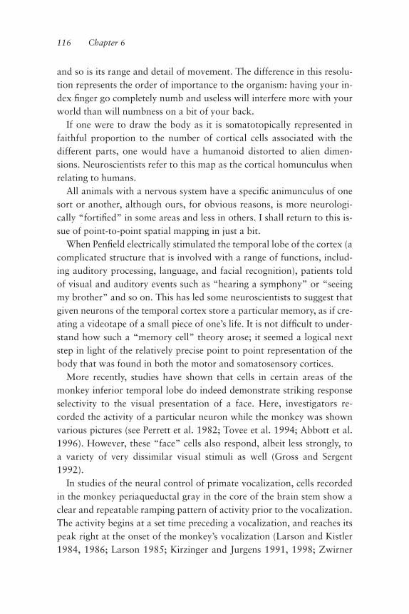
and so is its range and detail of movement. The difference in this resolu-tion represents the order of importance to the organism: having your in-dex ªnger go completely numb and useless will interfere more with yourworld than will numbness on a bit of your back.If one were to draw the body as it is somatotopically represented in
faithful proportion to the number of cortical cells associated with thedifferent parts, one would have a humanoid distorted to alien dimen-sions. Neuroscientists refer to this map as the cortical homunculus whenrelating to humans.All animals with a nervous system have a speciªc animunculus of one
sort or another, although ours, for obvious reasons, is more neurologi-cally “fortiªed” in some areas and less in others. I shall return to this is-sue of point-to-point spatial mapping in just a bit.When Penªeld electrically stimulated the temporal lobe of the cortex (a
complicated structure that is involved with a range of functions, includ-ing auditory processing, language, and facial recognition), patients toldof visual and auditory events such as “hearing a symphony” or “seeingmy brother” and so on. This has led some neuroscientists to suggest thatgiven neurons of the temporal cortex store a particular memory, as if cre-ating a videotape of a small piece of one’s life. It is not difªcult to under-stand how such a “memory cell” theory arose; it seemed a logical nextstep in light of the relatively precise point to point representation of thebody that was found in both the motor and somatosensory cortices.More recently, studies have shown that cells in certain areas of the
monkey inferior temporal lobe do indeed demonstrate striking responseselectivity to the visual presentation of a face. Here, investigators re-corded the activity of a particular neuron while the monkey was shownvarious pictures (see Perrett et al. 1982; Tovee et al. 1994; Abbott et al.1996). However, these “face” cells also respond, albeit less strongly, toa variety of very dissimilar visual stimuli as well (Gross and Sergent1992).In studies of the neural control of primate vocalization, cells recorded
in the monkey periaqueductal gray in the core of the brain stem show aclear and repeatable ramping pattern of activity prior to the vocalization.The activity begins at a set time preceding a vocalization, and reaches itspeak right at the onset of the monkey’s vocalization (Larson and Kistler1984, 1986; Larson 1985; Kirzinger and Jurgens 1991, 1998; Zwirner
116 Chapter 6

and Jurgens 1996). Furthermore, these cells are active only preceding vo-calizations of a very speciªc pitch. Clearly, all we have to do is ªnd thecells that encode for vocalizations of each and every speciªc pitch and wehave mapped out the cells responsible for a monkey’s vocalization reper-toire, right? Tempting, perhaps, but a proportion of these very speciªcvocalization cells also respond to general auditory stimuli and some arestrikingly well correlated with eye movements of certain directions(Larson and Kistler 1984). To ignore secondary responses when inter-preting the data for a particular cell would be to misrepresent intention-ally the complexity of the system, leading to misconceptions about how itactually works.Returning to our face or grandmother cell, ªndings in many systems
suggest that such a categorical representation is achieved by the activityof populations of cells and not by the electrical activity of any one cell. Itshould be clear that the grandmother cell concept implies that the multi-ple sensory input associated with grandmother, and the multiple statesthat grandmother can have in real life, would make, de facto, such agrandmother cell a tremendous connectivity puzzle. What mechanismcan one ªnd that will bind processed information from disparate sensorysources so that, for momentary practical purposes, the resulting internalrepresentation or sensorimotor image means the same thing? This mech-anism should also associate memories and/or thoughts with this internalconstruct, such as imagining/remembering a voice reading to you fromthe book you are holding. Just as easily you may read this book aloud,adding a deªnitive motor component. Yet the holding and seeing of thebook, your reading aloud from it, and the fact that you may be doing sowithout your shoes on are all still bound seamlessly in time as a singleevent. Something quite different seems to be at work here than if each in-dividual neuron represented a single, predetermined, highly speciªc as-pect of this event. Note that in creating this experience, you are bringingtogether elements that are truly yours (your hand representation andyour shoeless feet) and elements that are truly foreign (the contents of thebook you are reading)This raises the issue of whether the brain handles body representation
in the same way that it handles the representation of objects and events itwas not born with—those of the external world. Is there a single solutionto the set of questions that have been posed here? Let us see.
The I of the Vortex 117

Perceptual Unity of Consciousness—Content and Context
At the outset of this chapter we asked: Given that neurons have evolvedinto performing differing and speciªed functions capable only of repre-senting fragments of reality, how then does the brain manage to make asingular, useful construct from these pieces? The integration of speciªcsensory occurrences into single percepts, or rather, the neuronal mecha-nisms underlying this feat, are even more amazing (and thus experimen-tally more challenging) when one keeps in mind that the brain does thisin a context-dependent fashion. We touched on this issue in chapter 1.The integration of speciªc sensory occurrences, the content of our per-
cept, is dependent on the internal context of the brain that we generallyrefer to as attention (a momentary functional disposition), and is mosteasily recognized when comparing the functional states of wakefulnessand sleep. If, while you are awake, someone whispers to you that there isa bee in your hair, you will most likely do something about it. If, on theother hand, you are asleep when they whisper, you most likely won’t. Ifthis same scenario of comparisons were under experimental conditionswhere it was possible to monitor the ºow of auditory information fromyour ear into your brain, we would see that this sensory signal is trans-duced peripherally, in full regalia, in both circumstances (waking andsleep). Why don’t you hear it when asleep? Because the signal reachedonly a certain stage of processing, after which point the brain ignoredit—and the brain ignored it because in the sleeping state it does not incor-porate sensory input into the prevailing internal context of the moment.The internal context of the brain during sleep is one that does not grantsigniªcance to the meaning of those whispered words or much of any au-ditory information save for the very loud (Llinás and Pare 1991). Duringthe waking state, however, the internal context does give signiªcance tothese words, this auditory stimulus, capturing your momentary atten-tion, if not eliciting an overt behavioral response.
The Issue of Attention
Consider the example of trying to remain attentive to a public speakerwhen someone seated behind you will not keep quiet. Eventually, you
118 Chapter 6
phase them out and only give internal signiªcance to the words you wishto hear. I use this example because I believe it lends focus to the nuancesof different internal contexts and how subtly, at a moment’s notice, theymay and do change. The task for the theory presented in this chapter is tosuggest how the fragmented representations of individual stimulus prop-erties observed in the primary sensory areas of the brain are linked orgathered to obtain a complete pattern (a singular percept). It is also thetask of this chapter to suggest how such reconstituted patterns are giveninternal signiªcance within the prevailing momentary context. We shalldeal with content and context at length later in this chapter. For now, letus address the ªrst part of the hypothesis: how it is that the brain takesthe sensory-referred fragments of reality and puts them together in timeas a single cognitive construct.It has long been accepted in the world of neurological research that in
humans the general circuitry of the brain present at birth is not funda-mentally modiªed during normal maturation, but rather is only ªnetuned. The neural circuitry for us to move our ªngers is with us at birth;we don’t have to learn this. By contrast, to play the violin, particularlywith any degree of esthetic value, takes practice; this is the experientialªne tuning of synaptic connections that I spoke of in chapter 3. Both theskill and especially the musicality with which a person is able to performa piece is clearly dependent on/limited by a complex of capabilities pres-ent at birth, but can be uncovered or facilitated to varying degrees bypractice. The ability to have language, as another example, is geneticallydetermined and thus the neuronal circuitry ascribed for this ability iswith us at birth; which particular language becomes our mother tongue isthe “nurture” aspect of this equation. The view and understanding ofsuch structural a priories began with the identiªcation of the corticalspeech area (Broca 1861) and continued with the anatomical work ofRamón y Cajal. Cajal demonstrated that neurons were the basic units forbrain organization, the “neuronal doctrine,” and went on to describe theexistence of well-speciªed neuronal circuits present in all normal brains.This functional connectivity was supported later on by Penªeld’s point-to-point somatotopic mapping, which applies to all normal humanbrains. The degree of variability observed for such point-to-point connec-tions among humans is that which characterizes anatomical variability
The I of the Vortex 119

from person to person (height, distance between eyes, etc.), but surelyhaving two eyes, a nose between them, and a mouth underneath is a con-stant that we all expect. And so a thalamus, a cortex, and the speciªcconnectivity between them are not learned but inherited.This structural a priori of point-to-point neuronal connectivity, re-
ferred to as “spatial mapping,” is produced by the links that related neu-rons make with each other. Within a group of functionally relatedneurons there is certainly a ªnite number of neurons, and thus a ªnitenumber of possible connections among them. It follows that there is alsoa ªnite universe of possible representations to be created by these neu-rons, their connectedness, and their individual activities. However, thevariations and permutations of neuronal activity with 10 billion cells in abrain are for all practical purposes limitless, especially given the humanlife span.And so, we are brought to a very important question: Is it possible that
the hierarchical organization produced by neuron to neuron communica-tion is capable of putting together the shards of reality given by oursenses into a single internal percept by generating all-knowing cells? Theanswer, as one might suspect, is probably not. A purely hierarchical con-nectivity alone is simply too slow and unwieldy to keep pace with theever-changing aspects of the external world. There must be anothermechanism at work.
Timeness is Consciousness
This mechanism is most likely temporal coherence. It has been stated thatin brain development, “neurons that ªre together wire together.” A varia-tion on this statement might be “neurons that ªre together conspire to-gether” or “timeness is consciousness.” Mapping connectedness in thetime domain, superimposed on top of the limited possibilities of spatialconnectedness, creates a vastly larger set of possible representationsthrough the almost inªnite possibilities of combination. This is the con-cept of perceptual unity based on spatial and temporal conjunction.Building on physical connectivity, the nerve cells of the brain have createdan “interlocking” solution: the synchronous binding in the time domainof those individual neuronal activities. By making different time-inter-locking patterns, neurons can represent a unity of reality by combining
120 Chapter 6

the individual, fractionalized aspects of reality that each neuron carries.This time-interlocking phenomenon is temporal coherence. If wholemodules of neurons (whose activities represent fractionalized aspects ofthe external world) electrically oscillate in phase or resonate, as we sawin chapter 2, a global activity pattern is formed. This activity patternshould have all of the components necessary for a transiently useful, in-ternal construct of the external world in the given, present moment. Andso, as an analogy, one asks how many melodies (tonal sequences) can beplayed on a piano. Given the permutations and combinations of simulta-neously played keys, the answer is clearly in the realm of the inumerablylarge.Temporal coherence is believed to be the neurological mechanism that
underlies perceptual unity, the binding together or conjunction of inde-pendently derived sensory components, called “cognitive binding.” It isthought to be implemented by temporal conjunction—the physiologicallinking in the time domain of the independently operating neural mecha-nisms subserving the processing of sensory and interoceptive stimuli.Similarly produced and easier to understand is “motor binding,” where,as we saw with the inferior olive, motricity requires precise temporal acti-vation of muscles in order to implement even the simplest movementscorrectly.Until recently, temporal mapping in the brain has been more difªcult to
understand and to study than spatial mapping. This is because its studyrequires an understanding of the dynamics of brain function. Basically,the electroanatomy (who inhibits, or excites, whom) has been the prevail-ing philosophy in our studies, but it is simply not enough. Even as theconcept of temporal mapping is in general becoming more accepted, inneuroscience it has been a neglected parameter. This has been so primar-ily for technical reasons, because it necessitates simultaneous electricalrecordings from a sufªciently large number of neurons to be statisticallysigniªcant. That is, one must demonstrate the simultaneity of neuronalªring by a time series analysis (e.g., cross-correlation), and this correla-tion must be causally linked with a sensory or motor event that it suppos-edly generates.Synchronous activation of neurons that are spatially disparate is a
likely mechanism to increase the efªciency of the brain. We have knownfor some time about such simultaneity for motricity and for motor-
The I of the Vortex 121
derived brain activity. Examples include the electrical shock produced byªsh such as the electric eel or by the elasmobranch Torpedo marmorata(Bennett 1971). The “electric organ” of ªsh, the electroplaques (thecomponents of the electrical whacking system) must operate at the sametime. The small currents produced by each of the tiny electroplaquesmust sum into the electrically stunning blow these can generate by “let-ting go” in unison, and this requires that they activate at the same time.How is this synchronicity achieved, as the electroplaques are located atdifferent distances from the central command nucleus? Well, the neuronsin the command nucleus ªre synchronously, and the conduction time(time for a neuronal signal to travel) to the electroplaques is uniform be-cause the conduction velocities of the different motor neurons vary di-rectly with the distance to the individual electroplaques they activate.That is to say, the axons of the motor neurons are of differing lengths; thelonger axons conduct their signals faster, and the shorter axons moreslowly, so the signals reach their targets, whether far or near, simulta-neously. The issue here is that nature deals with the issue of simultaneitywith great care, and will go the extra distance by tuning conduction ve-locities to assure synchronicity, without which the ªsh may change froma “stunner” to a “tickler.” Similarly, such isochronic activation in theface of wide spatial disparity is seen in the mammalian olivocerebellarsystem described in chapter 2. Purkinje cells across a wide expanse of cer-ebellar cortex are activated synchronously by direct activation of the in-ferior olive (Sugihara et al. 1993; De Zeeuw et al. 1996). This is againachieved by the varying conduction velocities with the length of the inputaxons.Is there evidence for synchronous neuronal activation during sensory
input? This is an important issue, for if perceptual unity of objects andevents of the external world is to occur through the conjunction of spatialand temporal mapping in the brain, one would expect to observe syn-chronous activation of neurons related to sensory input and processing—and this is what we see. In the visual system, the volley of neural activityentering the optic nerve following activation of the entire population ofretinal ganglion cells is close to synchronous on to the thalamus (Stan-ford 1987). Stationary and moving light stimuli also evoke oscillatory re-sponses in retinal ganglion cells that are synchronized across the nasal
122 Chapter 6

and temporal halves of the retina, and which evoke synchronous re-sponses in the thalamus (Neuenschwander and Singer 1996). This meansthat activity from the central and peripheral aspects of the retina havesimilar conduction times, despite the fact that the distance the sensoryaxons must travel from the most peripheral ganglion cells to thethalamus may be twice that of axons arising from ganglion cells near theoptic nerve. This is another case of temporal tuning to attainsynchronicity.More centrally, Wolf Singer and his colleague Charlie Gray have re-
ported widespread synchronicity in the mammalian cerebral cortex(Eckhorn et al. 1988; Gray and Singer 1989; Gray et al. 1989). This syn-chronous activity from cells in a given column of the visual cortex is ob-served when light bars of optimal dimension, orientation, and velocityare presented. Moreover, the components of a visual stimulus that are re-lated to a singular cognitive object (such as a line in a visual ªeld) pro-duce temporally coherent “gamma oscillations” (close to 40 Hz) (Grayand Singer 1989; Gray et al. 1989). These oscillations are in regions ofthe cortex separated by as much as 7 mm (as far as neural real estate isconcerned, this is basically a different county). There is also a high corre-lation with 40-Hz oscillatory activity between related cortical columns.We have discussed such oscillatory resonance mechanisms before (chap-ter 2), when looking at the motor control signal emanating from the infe-rior olivary nucleus. With reference to the visual cortex, we are seeingspatially disparate ensembles of neurons whose activities are bound to-gether in time by a distinct rhythm that oscillates at 40 Hz (Llináset al. 1991; Nunez et al. 1992; Lutzenberger et al. 1995; Sokolov et al.1999).How the brain actually orchestrates simultaneity in the (neural) orga-
nization of perception in a physiological sense is as fascinating as it iscomplicated. Before turning to the neuronal nuts and bolts of this organi-zational mechanism, I would like to introduce the reader to the overt,global brain mechanism that I consider the prime candidate for imple-menting this essential cognitive binding event. The brain mechanism inquestion is as much a product of the evolutionary embedding of motricityas are the intrinsic oscillatory electrical properties of the neurons thatcombine to produce this mechanism.
The I of the Vortex 123

Global 40 Hz: The Signal that Binds
Studies indicate that 40-Hz coherent neuronal activity large enough to bedetected from the scalp is generated during cognitive tasks. Furthermore,some propose that this 40-Hz activity reºects the resonant properties ofthe thalamocortical system, which is itself endowed with intrinsic 40-Hzoscillatory activity (Llinás 1990; Llinás et al. 1991; Pedroarenas andLlinás 1998; Steriade et al. 1991; Whittington et al. 1995; Steriade andAmzica 1996; Steriade et al. 1996; Molotchnikoff and Shumikhina 1996)(ªgure 6.2). The 40-Hz coherent activity has been a candidate for thegeneration of unitary perceptual entities out of many sensory and motorvector components, which represents the details of the perceived world.What does it mean? We are confronted with a system that addresses theexternal world not as a slumbering machine to be awoken by the entry ofsensory information, but rather as a continuously humming brain. Thisactive brain is willing to internalize and incorporate into its intimate ac-tivity an image of the external world, but always within the context of itsown existence and its own intrinsic electrical activity.If we posit that the 40-Hz coherent waves are related to consciousness,
we may conclude that consciousness is a noncontinuous event deter-mined by simultaneity of activity in the thalamocortical system (Llinásand Pare 1991). A 40-Hz oscillation displays a high degree of spatial or-ganization and thus may be a candidate mechanism for the production ofthe temporal conjunction of rhythmic activity over a large ensemble ofneurons. Global temporal mapping generates cognition. The binding ofsensory information into a single cognitive state is implemented throughthe temporal coherence of inputs from speciªc and nonspeciªc thalamicnuclei at the cortical level. This coincidence detection is the basis of tem-poral binding.
The Thalamocortical System and the Generation of “Self”
I have already discussed the proposition that the brain operates as aclosed system: it is not surprising that the thalamic input from the cortexis far larger than from the peripheral sensory systems. This suggests thatthe thalamocortical iterative activity is a main mechanism of brain func-
124 Chapter 6

The I of the Vortex 125
Figure 6.2Simpliªed diagram to illustrate the generation of temporal binding due to theconjunction of coincident 40-Hz bursting activity within separate, but conver-gent thalamocortical pathways. Cell at left represents speciªc sensory or motornuclei projecting to the cerebral cortex (layer IV), while the right hand cell repre-sents nonspeciªc intralaminary nuclei projecting to the most superªcial layer ofthe cortex (layer I). See text for details. (Adapted from Llinás et al. 1998, ªgure 6,p. 1847.)

tion. In addition, neurons with intrinsic oscillatory capabilities that re-side in this (thalamocortical) complex synaptic network allow the brainto self-generate dynamic oscillatory states that shape the functionalevents elicited by sensory stimuli. The switching of ªring modes inthalamic neurons can trigger macroscopic changes in (global) functionalstates as dramatic as the difference between sleep and arousal. In myview, the thalamocortical system has evolved as the most efªcient solu-tion for the implementation of temporal coherence across areas of thebrain that not only subserve differing roles in reality emulation, butwhich also are physically very distant from each other. How? Thethalamocortical system, by its hublike organization, allows radial com-munication of the thalamic nuclei with all aspects of the cortex. Thesecortical regions include the sensory, motor, and associational areas. Thelatter is the largest part of the cerebral cortex in Homo sapiens. It receivesinput from the association nuclei of the thalamus and also from the sen-sory cortex. These areas subserve a feedforward/feedback, reverberatingºow of information.The thalamocortical system is a close to isochronic sphere that syn-
chronously relates the sensory-referred properties of the external worldto internally generated motivations and memories. This temporally co-herent event that binds, in the time domain, the fractured components ofexternal and internal reality into a single construct is what we call the“self.” It is a convenient and exceedingly useful invention on the part ofthe brain. It binds, therefore I am! Temporal coherence not only gener-ates the self as a composite, singly perceived construct, but creates a sin-gle seat or centralization from which the predictive functions of thebrain, so critical to survival, may operate in coordinated fashion. Thus,subjectivity or self is generated by the dialogue between the thalamus andthe cortex; or to put it in other words, the binding events comprise thesubstrate of self.Is the binding event actually the substrate or scaffolding of self? Fol-
lowing damage to the intralaminar or nonspeciªc thalamic nuclei (cellgroups in the thalamus that receive ascending input from a region of thebrain stem called the reticular formation), patients are not aware of theinputs conveyed from the thalamus to the cortex by the intact speciªcthalamocortical circuit (see Llinás and Pare 1991). Although inputs from
126 Chapter 6

the speciªc thalami are received, the injured individual cannot perceive orrespond to them. In essence, the individual no longer exists, from a cogni-tive point of view, and although speciªc sensory inputs to the cortex re-main intact, they are completely ignored. These results argue that the“nonspeciªc system” is required to achieve binding; that is, to place therepresentation of speciªc sensory images into the context of ongoingactivities.
Prediction Must Be Centralized—It Leads to Self
Given that prediction is the ultimate and most pervasive of all brain func-tions, one may ask how this function is grounded so that there evolvedonly one predictive organ. Intuitively, one can imagine the timing mis-matches that would occur if there were more than one seat of predictionmaking judgement calls for a given organism’s interaction with theworld; it would be most disadvantageous for the head to predict onething and the tail to predict another! For optimum efªciency it wouldseem that prediction must function to provide an unwavering residencyand functional connectedness: it must somehow be centralized to themyriad interplays of the brain’s strategies of interaction with the externalworld. We know this centralization of prediction as the abstraction wecall the “self.”
The Concept of “I”
“I” has always been the magniªcent mystery; I believe, I say, I whatever.But one must understand that there is no such tangible thing. It is just aparticular mental state, a generated abstract entity we refer to as “I” or“self.” Consider for a moment that your brachial plexus (nerve networkcarrying sensory and motor innervation of the arm) is damaged. You maylook at your (limp and numb) arm and say, “That’s not me”—becauseyou can’t feel it. Well, it is you and yours. Somehow we have developed astrange, almost solipsistic physiological cosmology: “I only possess thatwhich I innervate,” or, “I am only that which I innervate.” Odd, but per-haps that is what we actually do. So we have developed this simple rulethat sort of brings together everything into one single entity we call the
The I of the Vortex 127

self. It stands on the vestibular nucleus and pokes its head into thebrain—it has an up and a down to it, it has a visual component, a soundcomponent, and so on.So what is the self then? Well, it is a very important and useful con-
struct, a complicated eigen (self) vector. It exists only as a calculated en-tity. Consider the following two examples of what I mean. First we havethe concept of Uncle Sam. When one reads in the newspapers, “UncleSam bombards Belgrade,” everyone understands that the U.S. ArmedForces have been deployed against that country. However, there is nosuch entity as Uncle Sam. It is a convenient symbol and even a convenientconcept that implies existence, but it is a category without elements. The“I” of the vortex, that which we work for and suffer for, is just a conve-nient word that stands for as global an event as does the concept of UncleSam vis à vis the reality of a complex, heterogeneous United States. A sec-ond even more interesting example, given the decline of patriotism or theidea of “country as self,” is sports advocacy. Consider the European orSouth American riots associated with football (soccer) matches. It is in-teresting to note that to such fanatical sports fans the team they root foris an extension of themselves; so much so, that, as one might do for one’sloved ones or sometimes for one’s ideas, they will ªght and risk bodilyharm defending “their team.”
Secondary Qualities of the Senses as Inventions/Constructs
It should be clear that the secondary qualities of our senses such as col-ors, identiªed smells, tastes, and sounds are but inventions/constructs ofan intrinsic CNS (central nervous system) semantic (c.f. Llinás 1987).This semantic allows placing sensory inputs into an internal context sothat the brain may then interact with the external world in a predictivemanner. As stated above, the generated abstraction called “self” is funda-mentally no different from these secondary qualities of the senses; self isthe invention of an intrinsic CNS semantic. It exists inside the closed sys-tem of the CNS as an attractor, a vortex without true existence otherthan as the common impetus of otherwise unrelated parts. It is an orga-nizer of extrinsically and intrinsically derived percepts: the loom thatweaves the relation of the organism to its internal representation of theexternal world.
128 Chapter 6

In actuality, however, the above philosophical discussions concerningthe extent to which our perception of reality and “actual” reality overlapor match are truly of little practical importance. All that is required isthat the predictive properties of the computational states generated bythe brain meet the requirements for successful interactions with the exter-nal world. How this is managed by the brain, given the fractured natureof sensory input, is at the core of neurocognitive study today.
Dreaming and Wakefulness
Was it a vision, or a waking dream?Fled is that music:—do I wake or sleep?
—John Keats, Ode to a Nightingale
If cognition is an intrinsically generated state, what if any distinction isthere between dreaming and wakefulness? If we propose that cognition isa function of the 40-Hz thalamocortical resonance discussed above, whathappens to this oscillatory rhythm during sleep, particularly dream orREM sleep? In a series of experiments, my colleague Urs Ribary and Istudied the 40-Hz resonance during wakefulness and sleep, employing
The I of the Vortex 129
Figure 6.3Oscillations in brain activity of 40 Hz recorded during sleep (�-wave and REMsleep) and wakefulness associated with an external stimulus (bottom trace), illus-trating the lack of resetting during sleep. See text for details. (Adapted fromLlinás and Ribary 1993, ªgure 1, p. 2079.)

the technique of magnetoencephalography and using a 37-channel sensorarray distributed over the scalps of ªve normal adults. We found that 40-Hz coherent magnetic activity was spontaneously present in the awakeand in rapid-eye-movement (REM) sleep states, but greatly reduced dur-ing delta sleep (deep sleep characterized by delta waves in the electroen-cephalogram) (Llinás and Ribary 1993) (ªgure 6.3). On the other hand,and in agreement with previous studies (Ribary et al. 1991; Galamboset al. 1981; Pantev et al. 1991), an auditory stimulus produced well-deªned 40-Hz oscillations in the wakefulness state, but no re-setting ofthe 40-Hz oscillation was observed during either delta or REM sleep(ªgure 6.3). There were two salient ªndings from these studies. One wasthat the waking and REM sleep states are electrically very similar with re-spect to the presence of 40-Hz oscillations. A second signiªcant ªndingwas that 40-Hz oscillations are not reset by sensory input during REMsleep, even though other studies have clearly shown that the thalamocor-tical system is accessible to sensory input during sleep (Llinás and Pare1991; Steriade 1991). We consider this to be the central difference be-tween dreaming and wakefulness: we do not perceive the external worldduring REM sleep because the intrinsic activity of the nervous systemdoes not place sensory input within the context of the functional state be-ing generated by the brain (Llinás and Pare 1991).In more speciªc terms, we interpret these ªndings to mean that sensory
input that occurs during REM sleep is not correlated temporally with on-going thalamocortical activity (i.e., is not put into the context of thala-mocortical “reality”), so it does not exist as a functionally meaningfulevent.Although wakefulness and REM sleep can both generate cognitive ex-
periences, the above ªndings corroborate what is commonly known, thatthe external environment is for the most part excluded from the imagingthat is characteristic of the sleep states. In other words, it may be restatedthat the dreaming brain is characterized by an increased attentiveness toits intrinsic state and that external stimuli do not usually perturb thisactivity.By contrast, if the responsiveness generated during the waking state is
duplicated in the absence of the appropriate sensory input by virtue of ac-tivity generated via intrinsic thalamocortical interactions, then reality
130 Chapter 6

emulating states such as hallucinations may be generated. The implica-tions of this proposal are of some consequence, for if consciousness is aproduct of thalamocortical activity, as it appears to be, it is the dialoguebetween the thalamus and the cortex that generates subjectivity in hu-mans and in higher vertebrates.
The I of the Vortex 131

William Wegman, Man Ray Contemplating the Bust of Man Ray, 1978. Silvergelatin print, 14 � 11 in.
7 Fixed Action Patterns: Automatic
Brain Modules that Make
Complex Movements
Complex Movements
So now we have a wondrous biological “machine” that is intrinsically ca-pable of the global oscillatory patterns that literally are our thoughts,perceptions, dreams—the self and self-awareness. The next level of func-tional organization is again one of functional efªciency. The self, the cen-tralization of prediction (chapters 2 and 6), cannot, however, orchestrateevery feat the body must accomplish from moment to moment in theever-changing world in which we live. Fixed action patterns (FAPs) aresets of well-deªned motor patterns, ready-made “motor tapes” as itwere, that when switched on produce well-deªned and coordinatedmovements: the escape response, walking, swallowing, the prewired as-pects of bird songs, and the like.
These motor patterns are called “ªxed” because they are quite stereo-typed and relatively unchanging not only in the individual, but in all indi-viduals within a species. Such ªxedness can be seen from the simplest tothe most complex of motor patterns. For the execution of the very simplespinal reºexes, a central nervous system may not even be required. If oneirritates a patch of skin on the back of a frog, a reºex to scratch is set intomotion. The hind leg will swing out and up in a very stereotypical fashion,

circling around to land the foot on the distressed area; this is readily re-peatable and is the same across all frogs. Furthermore, this reºex may beactivated and runs exactly the same in the absence of the brain and brainstem, proof that the upper central nervous system is not required for theoperation of some of the more simple, rudimentary motor reºexes (Ostryet al. 1991; Schotland and Rymer 1993). In the decerebrate case above, ifone impedes the trajectory of the hind leg once this reºex is activated, thehind leg will simply stop where it is obstructed from its goal. The leg willnot swing out further or pull in closer to bypass the impediment, nor willit stray from its stereotypical trajectory of motion; it is quite ªxed in whatit can do and be. At this stage the rest of the brain would be required tointercede in order to resolve this motor impasse.
Fixed Action Patterns and the Usefulness of Stereotypical Behavior
Fixed action patterns (FAPs) are somewhat more elaborated reºexes thatseem to group lower reºexes into synergies (groups of reºexes capable ofmore complex goal-oriented behavior) (ªgure 7.1). The rhythm of ourwalking, once initiated by the upper motor system with minor adjust-ments to the terrain upon which we walk, is handled largely by nervouscircuitry in the spinal cord. However, it requires the activity of more thanthe cord to put it into context (see Bizzi et al. 1998). Neuronal networks,which specify stereotypical, often rhythmic, and relatively unchangingmovements of the body when activated, are known as central patterngenerators (CPGs), for this is precisely what they do. They generate theneuronal patterns of activity that drive overt FAPs such as walking (forreview, see Cropper and Weiss 1996; Arshavsky et al. 1997).
We may look at FAPs as modules of motor activity that liberate the selffrom unnecessarily spending time and attention on every aspect of an on-going movement, or indeed on the movement at all. Thus we ªnd our-selves having walked miles of city sidewalks or wooded paths almostblindly while engrossed in deep conversation with a friend. Looking backon it, what tends to come to mind most is the content of the conversationand how it made us feel. Our visual memory may hold only those detailsof walking that required our attention, as when we perhaps stumbledbrieºy on a root or rock, regaining balance and resetting our gait. At such
134 Chapter 7

a moment, consciousness of what we are thinking and talking about isbeing brieºy refocused to that of walking. That is, as a result of stum-bling on the root our senses bring the focus of consciousness from the in-terior to the exterior, from our thoughts to our body and the world it ismoving within. After changing our gait—stepping over the rock orroot—walking again becomes the FAP and our consciousness goes rightback to the conversation in which we were previously engrossed. Thiswalking FAP, like all others, liberates the self to spend time and attentionwhere it would rather be. Put quite plainly, if one had to focus on everymuscle and joint and their mechanics throughout each phase of the walk-ing cycle, consciously willing their drive, none of us would ever havethose pleasant conversations through the woods on a fall day. FAPs allowus the time to do other things with our minds.
The example above also highlights another related and very importantissue: the restraining properties of the senses on the ever-whirringthalamocortical system that we spoke of in chapter 6. In the case above,stumbling on a root momentarily brought us out of the conversation wewere having. The senses remind us that there is a world outside, but weforget sometimes because the internal world generated by the intrinsicproperties of the thalamocortical system can be so rich. As human beings,we differ from one another in the extent to which we pay attention to theexternal versus the internal world. We shall discuss that example in moredepth throughout this chapter, for the restraining capabilities of thesenses on the thalamocortical system also allow this system to change orªnely hone FAPs as needed to interact successfully with the ever-changing
Fixed Action Patterns 135
Figure 7.1Examples of common aggressive responses in three different vertebrate species.

world around us. But ªrst, we must broaden our understanding of FAPsthemselves—and into what amazing expressions they have evolved overthe millennia.
The central nervous system is required for FAPs more complicatedthan locomotion, which can be elicited by the brain stem and spinal cordalone (Jankowska and Edgley 1993; Nichols 1994; Whelan 1996). Thecurrent evolutionary residence of FAPs is in the brain (see Arashavskyet al. 1997). The evolution of this process has followed the same biologi-cal imperative that we saw for the internalization of movement as the ba-sis for mindness; in fact, it is exactly the same. The scratch reºex is purelya spinal mechanism (see Deliagina et al. 1983; Stein 1983, 1989; Mortinand Stein 1989; Jankowska and Edgley 1993). Because of its simplicity,natural selection has not found a need to move this module of functionup the neuraxis and into the more sophisticated processing capabilities ofthe central nervous system. These capabilities are needed for more elabo-rate (motor) events, such as the perfectly honed, complicated ªnger artic-ulations that bring to us, for instance, the beauty of Jascha Heifetzplaying Tchaikovsky’s Violin Concerto in A minor. As we watch him playthis from memory, eyes closed, smiling as if far removed from his task, wewonder: a FAP? Can playing a violin concerto be a FAP? Well, not all ofit, but a large portion. Indeed, the unique and at once recognizable styleof play Mr. Heifetz brings to the instrument is a FAP, enriched and modu-lated by the speciªcs of the concert, generated by the voluntary motorsystem. We shall look further into this issue later in the chapter when wediscuss the relation of FAPs to the origins of human creativity.
The Basal Ganglia as the Origin of FAPs
In the case of these more complex FAPs, it is believed that they are gener-ated centrally by the basal ganglia (Saint-Cyr et al. 1995; Hikosaka1998), a set of huge subcortical nuclei intimately related to the brain’smotor systems (see Savander et al. 1996). For many years, neurosciencehas held the basal ganglia to be the storehouse of motor programs, owingto their intrinsic circuitry. But in actuality these nuclei represent some ofthe least understood areas of the brain, particularly in regards to theirfunctional organization and architecture. We know that the expression of
136 Chapter 7

FAPs is supported by the interplay among a number of vastly differingparts of the nervous system and the basal ganglia (Greybiel 1995). Thesenuclei are localized in the center of the brain. They connect synapticallywith the thalamus and receive input from both the cortex and thethalamus (for review, see Smith et al. 1998; Redgrave et al. 1999). Aswith the cerebellum, the majority of connections within the basal gangliaare inhibitory, with many reciprocal contacts (see Berardelli et al. 1998;Kropotov and Etlinger 1999). This is to say that neurons terminate di-rectly on each other, so that cell A projects to cell B and B back to A, thusgenerating very complicated, inhibitory electrical patterns that in essencerepresent the negation of activity. If these circuits within the basal gangliarepresent, when activated, the motor tapes that run FAPs, then the inac-tive state (momentarily not engaging via central and peripheral connec-tivity the muscle synergies that would execute and thus fully express thegiven FAP) is a condition of intrinsic mutual inhibition (ªgure 7.2). Onemay remember from Dante’s Inferno a section where some of the damnedsouls are kept in a cauldron, but there is no demon keeping watch tomake sure none of them escape their incarceration. The question arises asto why this is so. The answer is that the souls inside this cauldron are so
Fixed Action Patterns 137
Figure 7.2Diagram of the basal ganglia and their connections with the frontal cortex.Synapses marked with a plus (�) are excitatory; those with a minus (�) are inhib-itory. (From Bear et al. 1996, ªgure 14.12, p. 390).

envious of one another (the sin for which they are being punished) thatwhen one does manage to escape, the others pull him/her back in! And sothe cauldron closes itself. It is the same with the basal ganglia: the intrin-sic, reciprocal inhibitory activity keeps all the potential FAPs from be-coming, expressing what they are supposed to be. Therefore, when a FAPis actually executed, we say that it has been “liberated” into action. Thebasal ganglia are the doors that when unlocked may release into actionvery large functions outside of the basal ganglia.
There is a wealth of neurological/physiological evidence suggestingthat the basal ganglia represent or embody the neural circuitry of motortapes. This is particularly evident if we look at the results of damage tothe basal ganglia or to parts of the nervous system that heavily inºuencethe basal ganglia (see Saint-Cyr et al. 1995; Wenk 1997; Berardelli et al.1998). Let us take as an example a well-studied FAP, the generation ofsong in birds. This is particularly important here because birds sing ac-cording to their genotype (nature) and according to their phenotype(Nottebohm 1981a; Doupe and Konishi 1991; Vicario 1994; Whalinget al. 1997; MacDougal et al. 1998). Genotypically speaking, this meansthat a particular type of robin would have a speciªc song that character-izes the family. The male sings and the female recognizes the song, choos-ing a male on the quality of the song; but females do not sing. Theancestral song is expressed even in birds that have lost the ability to hearthe sound of the song they sing. They would still sing, although the lackof auditory feedback would result eventually in abnormal song patterns(Nordeen and Nordeen 1992; Heaton et al. 1999).
This is the base song of that given species only. In normal animals thissong has regional embellishments or dialects. A properly trained orni-thologist can recognize the origin of a particular bird in a big city or evena particular borough by the song’s signature dialect—this type of song isouter Brooklyn and not lower Manhattan. And so, in a normal bird, ge-neric singing is modiªed by a learning experience when young and by theintrinsic properties of that particular animal (all brains are not exactlythe same from bird to bird, for example) (Scharff and Nottebohm 1991;Nordeen and Nordeen 1993). It turns out that within a particular group-ing some birds are better singers than are others, and thus have a betterchance of reproduction (Tchernichovski and Nottebohm 1998). In addi-
138 Chapter 7
tion to employing the brain, singing is quite a motor performance, andthus a good measure of the state of health of the animal, as well as ameasure of originality of brain activity. In fact, there is brain competition,as birds will invent, copy, and steal variations of songs from each other.The songs vary in duration and complexity; the longer and the morecomplex, the better. Ornithologists have pieced together how a particularsong is developed before reproduction occurs and preceding mating.They have described how song comes to fruition and maturity at matingtime and then is reinvented the following year with different variations(Nottebohm 1981b; Nottebohm et al. 1986; DeVoogd 1991; Johnsonand Bottjer 1993; Clayton 1997; Nordeen and Nordeen 1997; Smithet al. 1997; Mooney 1999; Iyengar et al. 1999). Next season the malewill need a new song because he won’t do so well with the old song. It isnature’s planned obsolescence. The females recognize the males’ songfrom last season and that that sperm may not be so good anymore! It isthe same as any other champion we know of: the rise, the peak, and thefall is pervasive throughout biology. Here, a bird’s rise and fall is denotedby the newness or oldness of his song (ªgure 7.3).
But is the song of birds actually a FAP, and how does this relate to thebasal ganglia? If we look at what happens in the brain of a male birdwhen you remove the male hormone testosterone, we see that the basalganglia are reduced and that in some bird species there is no song produc-tion (see Nottebohm 1980) or song production is reduced (Arnold1975a, b). If a female, who was never meant to sing, is given testosterone,she will start singing for the ªrst time in her life (Nottebohm and Arnold1976; Kling and Stevenson-Hinde 1977; Nottebohm 1980; DeVoogd andNottebohm 1981; Schlinger and Arnold 1991; Rasika et al. 1994;Nespor et al. 1996), and in bird species with singing females, females im-planted with testosterone developed male-like song (Gahr and Garcia-Segura 1996)! This singing in either males or females is based on the ad-vent of new neurons and connections within the basal ganglia (Nordeenet al. 1992; Rasika et al. 1999 for normal male development; Schlingerand Arnold 1991 for changes in females given testosterone). So, thereyou have a prototypical, indeed an archetypal FAP that is intrinsic in ori-gin. This female may never have heard the song (testosterone-inducedsinging still happens to females raised in isolation). Yet she is fully
Fixed Action Patterns 139

Figure 7.3Song in birds as modiªable FAPs. (Top) A female (left) and male (right) zebraªnch, Taeniopygia guttata. (Bottom) A schematic of a male bird’s brain and songcircuit, representing a sagittal section, or slice through the brain along its longaxis, showing its full rostrocaudal extent. Indicated are the nuclei that form themotor pathway for song production, descending from the HVc (higher vocal cen-ter) through RA (robust nucleus of archistriatum) to nXIIts (hypoglossal nerve)and thence to the syrinx. Also shown are nuclei involved with song learning: HVcthrough X (area X), DLM (medial nucleus of the dorsolateral thalamus), andLMAN (lateral magnocellular nucleus of the anterior neostriatum) to RA (pathnot shown). Also shown: DM, dorsomedial nucleus of the intercollicular nucleusof the midbrain; Uva, uvaeform nucleus of the thalamus; Nif, interfacial nucleusof the neostriatum; AVT, ventral area of Tsai of the midbrain. HVc, RA, and Xare not present in closely related species that do not produce complex vocaliza-tions. (Diagram courtesy of Heather Williams.)

capable of generating a well-deªned pattern of motricity that coordinatesvery speciªc muscle synergies that relate to the laryngeal musculature, theabdominal musculature, the intercostals, and in short the whole of themusculature necessary and sufªcient to generate song. The expression ofthe complete FAP, the basic song of that bird’s species, appears when theproper hormone is introduced. Damage to the avian basal ganglia ren-ders both the intact male and the female given testosterone irreversiblyincapable of generating normal song (Doupe and Konishi 1991; Scharffand Nottebohm 1991). And so in the case of the female, we see the liber-ation of an otherwise phenotypically dormant, but genotypically com-plete and complex FAP. This module of motor function is hardwired atbirth and is activated by testosterone, naturally in the male and experi-mentally in the female. Just why it is that only males sing naturally andyet the FAP persists in the female is not clear; what is clear is that there isa lot of “old stuff” left around in the brain. Evidently the causal sequencethat gave rise to this organization cannot easily be retraced and/or iso-lated in order to modify the female to eliminate an unnecessary, energeti-cally costly component. Apparently, it is cheaper and easier just to leave itthere.
Disorders of the basal ganglia provide clues to their relationship to FAPs
In the realm of human neurology, we see events that relate FAPs to thebasal ganglia. Neuropathology of these nuclei may be viewed as eitherproducing an excess of FAPs, as in Tourette’s syndrome, or as a defectwith the eventual loss of them, as seen in Parkinson’s syndrome. In thecase of people with Tourette’s syndrome, where there is diagnosed partialdestruction of the basal ganglia, there is an abnormal, continuous libera-tion of very particular types of FAPs (Coffey et al. 1994; Saint-Cyr et al.1995; Robertson and Stern 1997; Saba et al. 1998). These patients arecharacterized by continuous drumming of their ªngers, continuous talk-ing, continuous arm movement, and the continuous inability to stayquiet; in a word, the typical hyperkinetic individual. These nervous,ªdgety-type people are also typically quite intellectual, often athletic, andrespond very quickly to sensory stimuli that relate to motricity (eye-handcoordination for example). They are witty and quick tempered; no calm
Fixed Action Patterns 141
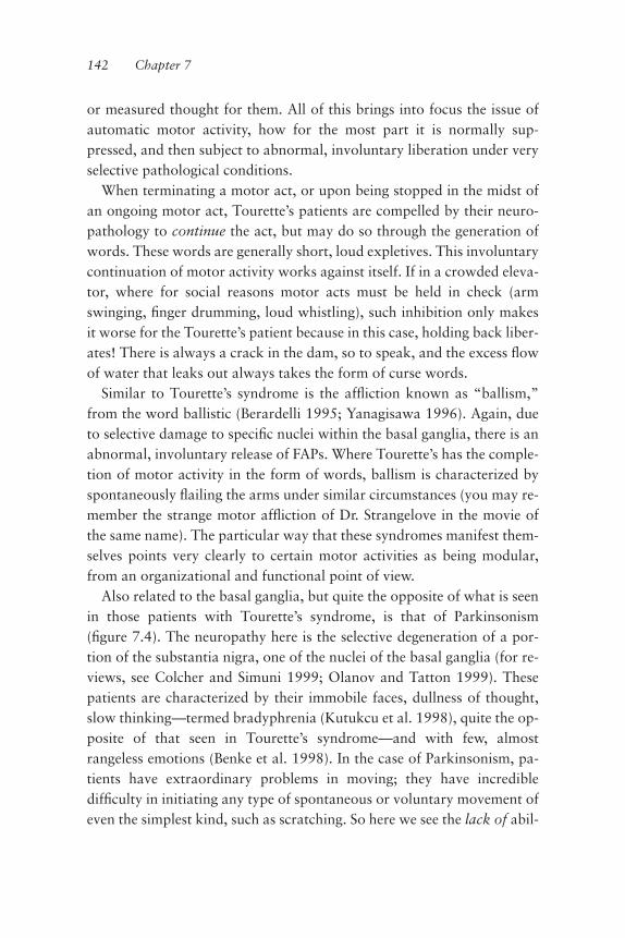
or measured thought for them. All of this brings into focus the issue ofautomatic motor activity, how for the most part it is normally sup-pressed, and then subject to abnormal, involuntary liberation under veryselective pathological conditions.
When terminating a motor act, or upon being stopped in the midst ofan ongoing motor act, Tourette’s patients are compelled by their neuro-pathology to continue the act, but may do so through the generation ofwords. These words are generally short, loud expletives. This involuntarycontinuation of motor activity works against itself. If in a crowded eleva-tor, where for social reasons motor acts must be held in check (armswinging, ªnger drumming, loud whistling), such inhibition only makesit worse for the Tourette’s patient because in this case, holding back liber-ates! There is always a crack in the dam, so to speak, and the excess ºowof water that leaks out always takes the form of curse words.
Similar to Tourette’s syndrome is the afºiction known as “ballism,”from the word ballistic (Berardelli 1995; Yanagisawa 1996). Again, dueto selective damage to speciªc nuclei within the basal ganglia, there is anabnormal, involuntary release of FAPs. Where Tourette’s has the comple-tion of motor activity in the form of words, ballism is characterized byspontaneously ºailing the arms under similar circumstances (you may re-member the strange motor afºiction of Dr. Strangelove in the movie ofthe same name). The particular way that these syndromes manifest them-selves points very clearly to certain motor activities as being modular,from an organizational and functional point of view.
Also related to the basal ganglia, but quite the opposite of what is seenin those patients with Tourette’s syndrome, is that of Parkinsonism(ªgure 7.4). The neuropathy here is the selective degeneration of a por-tion of the substantia nigra, one of the nuclei of the basal ganglia (for re-views, see Colcher and Simuni 1999; Olanov and Tatton 1999). Thesepatients are characterized by their immobile faces, dullness of thought,slow thinking—termed bradyphrenia (Kutukcu et al. 1998), quite the op-posite of that seen in Tourette’s syndrome—and with few, almostrangeless emotions (Benke et al. 1998). In the case of Parkinsonism, pa-tients have extraordinary problems in moving; they have incredibledifªculty in initiating any type of spontaneous or voluntary movement ofeven the simplest kind, such as scratching. So here we see the lack of abil-
142 Chapter 7

Fixed Action Patterns 143
Figure 7.4Neural basis of Parkinsonian tremor illustrated using magnetic ªeld tomography(MFT) imaging. MFT was used to deªne the temporospatial distribution of corti-cal activity during a single resting tremor (one contraction of the ºexor digitorumsuperªcialis muscle of the hand). The results conªrm rhythmic bursting in thethalamus and the sensorimotor cortex associated with tremor. For each tremorcycle, the suggested pattern of activation is as follows: Tremor is initiated 30–40ms before muscle activation by thalamic activity. Approximately 5–10 ms later,activity is seen in the premotor cortex, followed by activation in the primarysensorimotor cortex, whose output drives the contraction of the ºexor muscle,initiating the tremor. (A) The image at left is a coronal MRI brain slice, with theregion studied (5-cm wide cylinder) indicated by a rectangle. (B, C, left): two setsof data, one representing recorded activity in a virtual slice at the outer edge ofthe 5-cm rectangle (top) and the other activity in a slice at the inner edge of therectangle (as indicated, bottom). Each circle represents a time point. �40 ms indi-cates an event recorded 40 ms before muscle activation, and 2 ms indicates timeelapsed after onset of muscle activation. Moving from left to right, each columnrepresents activity associated with subsequent tremors. (B, C right) 3D MRI re-constructions of the regions corresponding to the outermost and innermost slicestudied. Th, thalamus. (D) Simultaneous representation of activity pattern withinthe entire 5-cm cylinder over time, displayed left to right, illustrating therhythmicity of the tremor. (From Volkmann et al., 1996, ªgure 6, p. 1367).

ity to release FAPs. The existence of these two syndromes, along withother motor syndromes related to disorders of the basal ganglia, suggestthat FAPs are most probably implemented at the level of the basal gangliaand put into context by the reentry of the basal ganglia output into theever-cycling thalamocortical system.
In the beginning of this chapter I said that the basal ganglia send to andreceive information from the thalamus. In fact, the intralaminar complexof the thalamus (recall chapter 6) projects with a veritable vengeanceonto the basal ganglia, which should suggest to the reader the idea of thephysiological interplay between the self and FAPs. This is a very impor-tant issue. It is, in fact, the central issue of this chapter, and not an easyone to grasp in its full detail. So let us press on and we will get there.
FAPs and the Economizing of Choices
Having (hopefully) clariªed the idea of what FAPs are, I would like to re-turn to the issue of the intrinsic self-containment of motor programs.This intrinsic containment makes intuitive sense if we recall from chap-ter 2 the vast overcompleteness of the motor system. We have alreadyseen that through the inherent architecture of this system, it may imple-ment a given movement in an almost inªnite number of ways (recall thedifferent ways we reached for the milk carton). From a central nervoussystem perspective, one may ask how an animal is able to execute partic-ular desires or goals given that those goals are often executable in a stag-gering number of ways. How are choices, correct choices, made? Clearly,making the correct motor choice can be tantamount to survival, so onesuspects that, at the very least, natural selection has somehow ªnely pol-ished and engrained into the nervous system a mechanism for the reduc-tion of possible choices. Let us look into this further.
It must be understood that in theory, the nervous system can designtwo types of overall strategies. One is to leave the system completely free;the other is to have a built-in mechanism for the reduction of choices. Byfree I mean that if a gazelle sees a tiger coming, it may decide to run in ahopping fashion or with only three of four legs or to have two legs run-ning forwards and two running backwards. The problem with a com-pletely free system, one of almost inªnite possibilities, is that if allowed to
144 Chapter 7

operate it would be very expensive. We know that the system is vastlyovercomplete; so an efªcient mechanism for the reduction of its degreesof freedom, its choices, is therefore critical. Taking too much time choos-ing how to escape from the tiger is not only inefªcient, but also poten-tially lethal. A system that permits implementation of an inappropriateway of escaping the tiger, such as attempting ªrst to make swimming mo-tions while on land, is also ill advised and potentially lethal.
And so we see that reduction of choice is the mode of operation forwhich the system has been naturally selected. The motor system, given itsrichness, its overcompleteness, has to have such a global strategy for theappropriate implementation of an effective motor execution. It is simplydue to the imperative of time. Of importance as it relates to FAPs, thesepatterns respond somewhat selectively to an urgent event in the externalworld requiring a well-deªned, overt strategy such as attack and defense,ªnding food, reproduction, and the like, and in a timely and appropriatefashion. A set of clear constraints must be superimposed on a system thatis so extraordinarily rich and predictive, and they must be very powerful.Thus birds have evolved so that they never waste time trying to ºy by in-effectually beating only one wing. Of course, a bird can and will need tomodify the FAP of ºying once in ºight, but, at the very origin of this“hardwired” FAP, as it is liberated into expression, there is the clear con-straining by natural selection to do the right thing: beat both wings ratherthan one at activation time. That a bird is capable of ºapping only onewing is obvious if one watches a bird washing itself in a bath. But this isnot ºying. The motor system is constrained, carved down out of itsovercompleteness into (among many) this particular FAP, so that whenneeded it is activated at once and perfectly so.
From a physiological standpoint, the process of FAPs reduces the im-mense degrees of freedom of the system. In chapter 2 we spoke of musclesynergies, the co-activation of speciªc muscle groups in coordinated com-bination in order to carry out a given motor task. In that chapter, we usedthe example of grasping something with our hand. The beating of a bird’swings is no different: this FAP also requires the synchronous and coordi-nated activation of a number of different and very speciªc muscle syner-gies. Driving this motor event is the synchronous and coordinated ªringof very speciªc motor neurons with functionally speciªc ªring patterns,
Fixed Action Patterns 145

frequencies, and durations. To achieve this quite amazing feat of weedingout the extraneous, almost inªnite number of other possible motor neu-ron activation patterns, the system has been carved by evolution’s trialand error into FAPs, relatively speciªc modules of motor function.
FAPs Have Two Parts: Strategy and Its Implementation in Tactics
There are two very important aspects to this constraining of the motorsystem as it relates to FAPs. One is of strategy. This of course has to dowith some global issue, as we have just said, a large level categoricalchoice such as ªght or ºight; one cannot do both simultaneously. But agiven strategy must also be put into the context of whatever is happeningat the time to the animal in the surrounding world, and so FAPs have twocomponents. One is the strategic component as mentioned; the other isthe context-dependent implementation of such strategies—the tactics(ªgure 7.5). These two components are intimately intertwined and bothmust be considered premotor events (chapter 2). That we are runningfrom a tiger does not create the need to be doing so. The need to runcomes from a sense of urgency perceived within the momentary contextof the external world. Such urgency is processed within the premotorrealm of prediction (I must run), and then the tactical solution, the ap-propriate FAP, is implemented: I run. Within this global decision, tacticalevaluation determines that it is best to move my legs in a fashion that al-lows me to run my fastest. It is also strategically best to run away fromthe tiger rather than toward it. This may seem obvious, but the brainmust implement every aspect correctly for survival. It is at all times atwo-level decision: the appropriate strategy and the appropriate tacticswithin the strategy. A frog may decide to jump when the headlights come.This is clearly a good tactic, but given the number of frogs one unfortu-nately sees squashed on the road, the right strategy—to jump away fromthe car—is not always employed correctly or in time. Natural selection’swork is never done.
It is important to clarify fully the difference between strategy and tac-tics as they relate to FAPs. Let us say that one is a jaguar. There is an en-emy and one’s strategy is to stay and ªght. But which enemy is critical indeªning the contextual tactics: ªghting a snake, from a motor FAP per-
146 Chapter 7
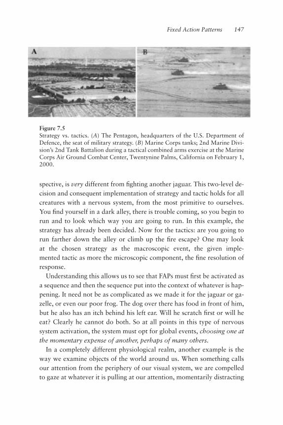
spective, is very different from ªghting another jaguar. This two-level de-cision and consequent implementation of strategy and tactic holds for allcreatures with a nervous system, from the most primitive to ourselves.You ªnd yourself in a dark alley, there is trouble coming, so you begin torun and to look which way you are going to run. In this example, thestrategy has already been decided. Now for the tactics: are you going torun farther down the alley or climb up the ªre escape? One may lookat the chosen strategy as the macroscopic event, the given imple-mented tactic as more the microscopic component, the ªne resolution ofresponse.
Understanding this allows us to see that FAPs must ªrst be activated asa sequence and then the sequence put into the context of whatever is hap-pening. It need not be as complicated as we made it for the jaguar or ga-zelle, or even our poor frog. The dog over there has food in front of him,but he also has an itch behind his left ear. Will he scratch ªrst or will heeat? Clearly he cannot do both. So at all points in this type of nervoussystem activation, the system must opt for global events, choosing one atthe momentary expense of another, perhaps of many others.
In a completely different physiological realm, another example is theway we examine objects of the world around us. When something callsour attention from the periphery of our visual system, we are compelledto gaze at whatever it is pulling at our attention, momentarily distracting
Fixed Action Patterns 147
Figure 7.5Strategy vs. tactics. (A) The Pentagon, headquarters of the U.S. Department ofDefence, the seat of military strategy. (B) Marine Corps tanks; 2nd Marine Divi-sion’s 2nd Tank Battalion during a tactical combined arms exercise at the MarineCorps Air Ground Combat Center, Twentynine Palms, California on February 1,2000.

us from our previous visual purpose. This is accomplished easily by mov-ing our eyes, along with, in varying cases, moving our heads, necks, hips,even ankles and feet. In orienting our bodies, we get into the ballparkarea of our goal. Once we are in the ballpark, we have to implement acompletely different type of activity. Now we must attain the target—it istactical now. In the case of eye movements, the procedure is to move theeyes so that the object of interest is shifted into the center of one’s visualªeld and then to begin to foveate (if you happen to be a foveating ani-mal). Foveating means switching to a higher acuity of vision, using al-most exclusively the cone-type photoreceptors. In looking at somethingwe like, such as an interesting mural, we may wish to look at this part orthat part, or yet another part or area ªrst. Here we have to decide, givenour momentary level of enthusiasm, the degree of detail at which to look,for after all, looking is a subtle form of touching. And so again, we seethe strategy, and the tactics within the strategy, at work.
The last example brings us to a crucial issue: the tedious balance be-tween the system’s deep need for an operative that greatly (andbeneªcially) reduces the degrees of freedom, the choices the system canmake/implement, and the clearly critical need for the freedom to be ableto make choices. This is yet a further difference between strategy and thetactics within a given strategy; it is the difference between reºexive re-sponse and volitional choice.
When something in our peripheral vision captures our attention, wemove our eyes to roughly center the object in our ªeld of vision. This isthe global strategy the system adopts. It is clearly a FAP, for it is a reºexand a well constrained one; otherwise this reºex would have our eyesconstantly overshooting and undershooting. Once we center this particu-larly fabulous painting in our ªeld of vision, tactics now come into play:we decide which part we want to look at and foveate (ªgure 7.6). Thistactic is not a FAP; it is voluntary and therefore demands consciouschoice. Which part will we look at? A further and very salient point isthat the tactic inhibits the FAP; it breaks free from its ªxedness. If onecorner of the painting is particularly engaging visually, I guarantee theevents now in your peripheral vision will be left there. What would typi-cally cause a reºex glance to another ballpark (strategy) has been volun-tarily inhibited. Sorry, I am foveating on a given detail now.
148 Chapter 7
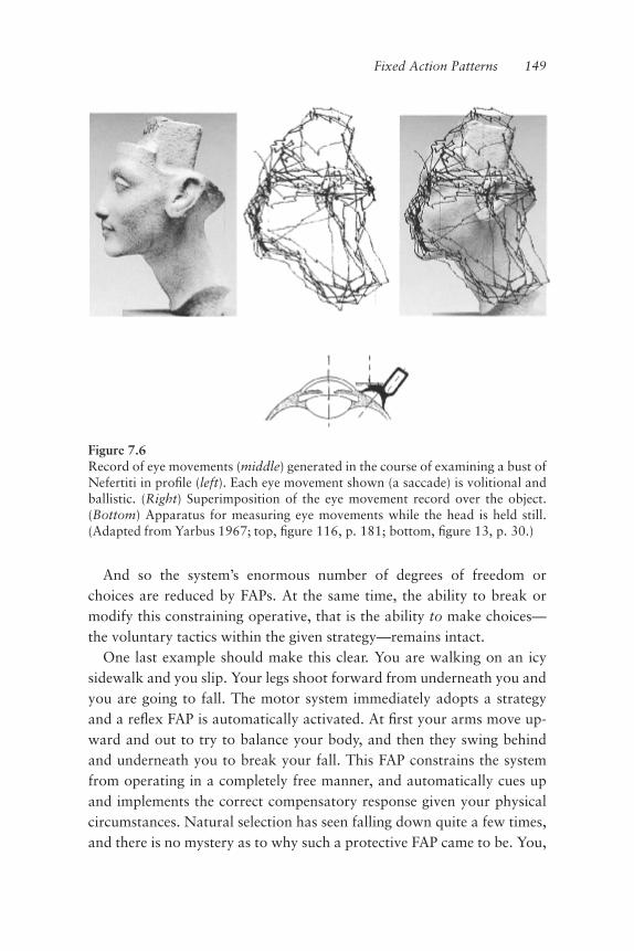
And so the system’s enormous number of degrees of freedom orchoices are reduced by FAPs. At the same time, the ability to break ormodify this constraining operative, that is the ability to make choices—the voluntary tactics within the given strategy—remains intact.
One last example should make this clear. You are walking on an icysidewalk and you slip. Your legs shoot forward from underneath you andyou are going to fall. The motor system immediately adopts a strategyand a reºex FAP is automatically activated. At ªrst your arms move up-ward and out to try to balance your body, and then they swing behindand underneath you to break your fall. This FAP constrains the systemfrom operating in a completely free manner, and automatically cues upand implements the correct compensatory response given your physicalcircumstances. Natural selection has seen falling down quite a few times,and there is no mystery as to why such a protective FAP came to be. You,
Fixed Action Patterns 149
Figure 7.6Record of eye movements (middle) generated in the course of examining a bust ofNefertiti in proªle (left). Each eye movement shown (a saccade) is volitional andballistic. (Right) Superimposition of the eye movement record over the object.(Bottom) Apparatus for measuring eye movements while the head is held still.(Adapted from Yarbus 1967; top, ªgure 116, p. 181; bottom, ªgure 13, p. 30.)

the self, haven’t the time to think through and willingly drive the musclesynergies needed to invent de novo this protective motor event. And sonatural selection saw what helps when you fall down in this fashion andover the eons honed it into a speciªc module that is activated by, for themost part, very speciªc motor circumstances: my legs are over my headand I am going down. FAPs are very good friends of the self.
But just how ªxed are FAPs? Let’s rewind the tape: you are just begin-ning to slip and your legs are sliding out from underneath you. But thistime you are holding your mother’s priceless Etruscan vase.
If the FAP that we have been speaking of were truly ªxed, like that ofthe poor toad with the itch on his back, you would still break your fall.And, most likely, about half a second later, the vase would break too,ending its fall from where you had, as part of the damned FAP, involun-tarily tossed it into the air.
But the vase is priceless, and what’s more it belongs to your mother.The FAP does not know this, but you do. The FAP notwithstanding, weknow exactly what the true outcome of this situation is likely to be. Youfall straight on your duff and the vase, still in your hands and probablycentered on your lap, is ªne.
So what happened? Did the “appropriate” FAP not get released/acti-vated? It most certainly did—as I said before, this is automatic, very fast.Ah, but so are the predictive properties of the brain. And your slippingand falling is nothing new to the thalamocortical system, either (remem-ber from chapter 3 I said that we must move within the world in order toembed?), so predicting the consequences is both easy and automatic.There is the predictive sensorimotor image: if I break my fall, the vasewill shatter into a million pieces. The voluntary solution? Don’t break thefall. That is, tactically inhibit the FAP, don’t let it run its stereotypicalcourse, consciously override it: hold on to that vase at all costs! If you arenot convinced that the FAP that helps you break your fall is not activatedªrst and then overridden, just recall similar situations you have been in,and the difªculty of adjusting through the fall—to hold on to that vase orcup of coffee.
Let us brieºy review. We have a motor system that when driven byglobal strategies implements contextually appropriate FAPs; the appro-priateness comes from the immediate reduction of possible choices by the
150 Chapter 7

given strategy the system adopts. These FAPs are relatively hard wired attheir origin and so may be considered as reºex at the time they are acti-vated. As modules of automatic motor function, they have been formedand honed by evolution to save (computational) time as an efªcient anti-dote to a vastly overcomplete motor system; with their timeliness of con-textual activation and their dependability of execution, FAPs thus savetime for the self, the seat of prediction. Once activated, however, mostFAPs may be tactically modiªed from their stereotypical motor expres-sion, as the given context requires. This “breaking out” or overriding of agiven motor event that is constrained by the FAP being executed is ac-complished by the thalamocortical system, the self, making volitionalchoices that arise from weighing the information and predicting conse-quences of the unfolding context of the given situation. It is the necessaryadvent of consciousness to an otherwise responsively ªxed repertoire ofmovement.
Language as a Premotor FAP
To conclude this chapter, I would like to touch on something we willhandle more fully in chapter 10, but for very different reasons. We canlearn something very interesting from the Tourette’s Syndrome we talkedof earlier. It is worthwhile to note that the clear symptoms of Tourette’soccur across people of all languages; this suggests something very fasci-nating about how the organization of the brain subserves language itself.That is, that language itself is a FAP. It is a premotor FAP at that, andmost likely very intimately related to the activity of the basal ganglia, assuggested by the clinical symptoms of at least one patient whom I haveseen and reported on in a collaborative study entitled “Words withoutMind” (Schiff et al. 1999). This study was conducted with my colleaguesFred Plum and Nicholas Schiff, distinguished neurologists from CornellMedical School, and my friend and collaborator Urs Ribary, who is, as Iam, from NYU Medical School. In the patient studied, a massive strokehad left almost nothing functional save for the basal ganglia and a part ofthe cortex known as Broca’s area. This area is responsible for the genera-tion or motor aspects of language. The stroke also left intact parts of the
Fixed Action Patterns 151

thalamus in such a way that these parts, along with the basal ganglia andthe cortical Broca’s area, shared some interconnected circuitry betweenthem. This person is in a coma and has been so for the last 20 years, andevery measurement performed objectively and by means of noninvasiveimaging has indicated that most of this patient’s brain is functionallydead. And yet this person, in a vegetative state, will occasionally generatewords (ªgure 7.7).
So here we have someone who has lost all other abilities and the onlyability left intact is the ability to generate words. This again reafªrms thatthe nervous system appears very much to be organized in functionalmodules. In this case, word generation is an intrinsic property of the
152 Chapter 7
Figure 7.7Language centers of the brain. The left hemisphere comprises word and sentenceimplementation structures and mediation structure for various lexical items andgrammar. The collections of neural structures that represent the concepts them-selves are distributed across both right and left hemispheres in many sensory andmotor regions. (From Damasio and Damasio, 1992, p. 92.)

brain. This circumstance, the random emitting of words with no con-sciousness behind the FAP that produced them, is very sad. However, theopposite and equally possible scenario is perhaps more harrowing: if youdamage the system, the individual may be capable of understanding lan-guage, of understanding prosody, of seeing and hearing and interactingwith the external world, except that he/she will be incapable of generat-ing words. But again, the point here is that these cases clearly point to amodular organization of function in the nervous system.
FAPs are subject to modiªcation; they can be learned, remembered, andperfected. How does the brain learn and remember anything? The self?We shall look into these issues in chapter 9.
Fixed Action Patterns 153

R. Varo, Capillar Locomotion, 1959. Oleo/masonite, 83 � 61 cm.

8 Emotions as FAPs
It is always healthy to approach the issue of emotions with a good meas-ure of trepidation. There are few topics of inquiry as thorny as our affec-tive world. The fact that emotions are for the most part irrational, andthat they can enslave our rationality (according to Hume) is probably atthe root of their thorniness. On the plus side, they are the reason for ourwanting to survive, and our inspiration. Indeed, the properties and vicis-situdes of our emotional selves are often referred to as our “humanity.”These issues have a long and distinguished history, with misunderstand-ing of human motive at its helm. The “face that launched a thousandships” in Homer’s legendary Iliad is a case in point. It was not only theface, nor probably any of Helen’s other anatomical characteristics thatmobilized Agamemnon’s armada against Troy. The main culprit mostprobably was the loss of love, injured pride.So how shall we address such a complex topic? I propose that we con-
sider that emotions are elements in the class of “ªxed action patterns,” orFAPs, where the actions are not motor but premotor. Further, we mayconsider that, as with muscle tone that serves as the basic platform forthe execution of our movements, emotions represent the premotor plat-form as either drives or deterrents for most of our actions. However,

unlike muscle tone where the level of muscle activation is the parameterregulated, emotions are notorious for their variety.Reºecting on the history of humanity we ªnd emotional states well
characterized in the Western, Judeo-Christian tradition in the form of“the cardinal Sins” (pride, rage, greed, lust, envy, sloth, and gluttony)and the somewhat lesser known and not easily reconciled with true emo-tional states, the “cardinal Virtues” (justice, prudence, temperance, forti-tude, faith, charity, and hope). According to a modern dictum the latterseven have the quality of being their own reward (truly intrinsic), an ideathat may derive from, or at least reºect, practical needs surrounding theorigins of settled, agricultural society—in essence, the notion of “enlight-ened self-interest.”That issue aside, emotions in general are among the very oldest of our
brain properties. They are enacted by the rhinencephalon (see Velascoet al. 1988, 1989), whose activity supports and generates not only ouremotional feelings, but also a host of motor, autonomic, and endocrinepostures that probably evolved as states of readiness to action, as well asmodes of social signaling of intentionality. From a neuroscience point ofview the question of the neurological basis for our affective world is thetopic of classical and contemporary research (Brown and Schafer 1888;Bard 1928; Kluver and Bucy 1939; Hess and Rugger 1943; Hess 1957;Weiszcrantz 1956; Hunsperger 1956; Fernandez de Molina andHunsperger 1959, 1962; Downer 1961; Geschwind 1965; Fernandez deMolina 1991; Damasio 1994, 1999; LeDoux 1996, 1998; Rolls 1999).One would not be surprised if emotional states were to be simple stereo-typical responses; the cardinal entities (in particular the sins) may betriggered by peptide modulators to the point that the universal character-ization may be recognized by most human cultures.
Sensations Are Intrinsic Events and Emotions, Global Sensation FAPs
The relationship of emotional states to actions, and indeed to motricity, isall important, for under normal conditions it is an emotional state thatprovides the trigger and internal context for action. But the underlyingemotional state, the “premotor FAP,” does not only trigger the action asa FAP; it is also expressed in the form of another accompanying motor
156 Chapter 8

FAP (such as a facial expression), which telegraphs to others the context(motivation) for the action and possibly the imminence of the action it-self. Examples of such motor patterns can be artiªcially generated byelectrical stimulation of the motor nerve branches in the face, without theemotional content (ªgure 8.1). If one inadvertently touches a very hotpan on the stove, the rapid pulling back of the hand (a FAP) is generallypreceded and accompanied by a grimace (a motor FAP) and an outburst(another motor FAP) (Darwin 1872). As with other FAPs, however, theexpression of an emotional state can be and often is suppressed. G.Gordon Liddy, of Watergate fame and more recently a radio talk-showhost, used to impress people at Washington parties by holding his handover a ºame. “What’s the trick?” someone once asked. “Not minding”he replied.One need not invoke the very sophisticated emotional world of hu-
mans to see the inextricable relationship between emotion and action, foreven the motor FAPs of relatively primitive animals are accompanied by awell-deªned emotional component. This emotive element is also relatedto the issue of “qualia,” which we shall discuss in detail a few chaptersfrom now.In order for a motor FAP to be generated, the input that triggers it must
be ampliªed and put into context some time before the FAP is activated.There’s a ªre! Run! It makes intuitive sense as well; one does not want to
Emotions as FAPs 157
Figure 8.1Examples of facial expressions generated by the selective stimulation of differentcombinations of muscles in the face. (From Duchenne de Boulogne 1862 [repub-lished 1990] plates 13, 31, 65.)

just start triggering FAPs without good reason. But the input that acti-vates a FAP need not be so alarming. It may be as simple as an itch, whichfor the most part is a very small stimulus. Small indeed, if one considersthat all of one’s skin is innervated and thus generating background activ-ity almost continuously from all over the surface of the body. Now theskin is one of the largest organs in the body, and yet we react aggressivelyto a mosquito bite by slapping ourselves in an attempt to kill the offender.Input, in the form of sound or the small sting, generates a brief emotionalreaction and thus a motor FAP, the slap, is activated. If it happens to be aspider crawling on you and you have a bit of arachnophobia, well, theregoes your mother’s vase! Something as insigniªcant as a mosquito bite ora spider wandering over your surface may evoke not just a sensory re-sponse, but actually a brief emotional state. Even a benign itch, as thosewho have spent time with a limb in a cast will know, can be ampliªed tocause maniacal behavior. We will go to any length to stop the itch, stick-ing anything we can ªnd down inside the cast to that wicked place ourªngers cannot reach. And when we simply can’t reach the spot thatitches, an even larger emotional state may be unleashed. What we under-stand from this situation (wearing a cast) is the importance of this kind ofampliªcation of sensory input into an emotional state, for emotionalstates set clear contexts for and from which the thalamocortical systemmay operate.So what do we mean by an “emotional state”? Well, I would like to re-
late such states to non-motor FAPs and beyond, as we shall see. Let’s be-gin by agreeing that emotional states give context to motor behavior. Inthis sense, both pain and the next step, fear, are emotional states. Thereader may believe that the feeling of pain relates to given emotionalstates, but is pain itself an emotional state? I would argue that yes, it is. Aperson who has a malfunctioning frontal lobe in the cerebral cortex, sayin the cingulate cortex (Brodmann’s area 24), can receive a sensation thatactivates specialized pain (nociceptive) brain pathways, but the activa-tion of the pathways will not generate pain (see Devinsky et al. 1995;Kuroda et al. 1995; Sierra and Berrios 1998; Heilman and Gillmore1998). The patient may recognize that the stimulus should lead to pain,but it is also clear to him that the sensation is not stressful in the way wetypically know “pain” to be. If you ask, the person may say, “Yes, I feel
158 Chapter 8

the pain but it doesn’t hurt.” This is, however, not the usual response to apainful stimulus. You may want to use the argument that they are separa-ble to support the view that pain and the emotion it generates are sepa-rate events. Well, that would be so if by pain we mean the purely sensoryexperience and not the unpleasantness associated with pain. Most of usactually consider the unpleasantness the pain, and not whatever else wasassociated with it. “It hurt like hell, and I also felt the squeezing of thethumb as the hammer hit it?”The cingulate is activated mostly when you are in pain of the intracta-
ble, long-term type, such as that from cancer (Devinsky et al. 1995;Rainville et al. 1997; Casey 1999) (ªgure 8.2). Interestingly, the cingulatecortex is also activated when one makes an error. Oh no! Not the vase!When you think of it, a mistake or error or oversight leads to a pain ofsorts, certainly a distinct emotional state that we all know only too well.Very importantly, it is a pain that is not localizable but nevertheless isvery profound, as profound as the feeling of pain we feel when those wecare for are hurt or in distress. Related to this is a similar form of “deeppain” described by psychiatric patients; whether one can call it “psycho-logical pain” or not, it is often the reason why many of these patientscommit suicide. The issue is that it is not localizable. In fact, no pain islocalizable. The cutting of a ªnger and the pain it brings seems localized,but it is simply a co-activation of pain, the emotional state, and generaltactile stimulation. The unpleasantness of pain is an emotional state gen-erated by the brain (Tolle et al. 1999; Treede et al. 1999), not an eventthat somehow resides at a particular body location (Greenªeld 1995).Let’s put it this way. The peripheral receptors and their neural path-
ways leading to the central perception of pain, that is, to the sensation,deªne the process underlying the generation of pain (ªgure 8.2). Sensa-tions are intrinsic events, a product of the ongoing activity of the nervoussystem that ªnds a way onto the stage of consciousness. Sensations aretruly intrinsic, in that they can also be attained or obtained in the absenceof activation of sensory pathways. During dreaming we feel many differ-ent sensations (Zadra et al. 1998), yet none of the things we feel in ourdreams comes via the pathways that convey such sensations during thewaking state. You will remember from chapter 6 that although these sen-sory pathways are capable of transducing stimuli from the external
Emotions as FAPs 159
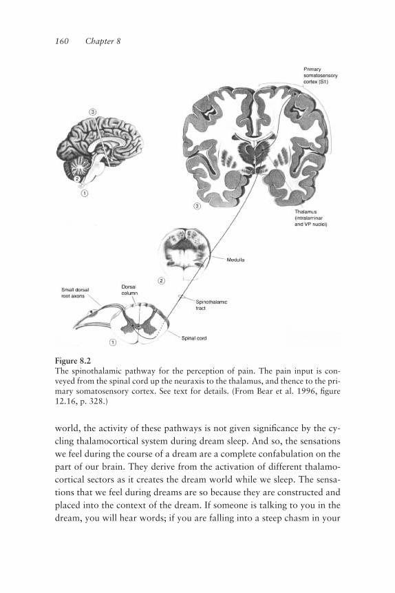
world, the activity of these pathways is not given signiªcance by the cy-cling thalamocortical system during dream sleep. And so, the sensationswe feel during the course of a dream are a complete confabulation on thepart of our brain. They derive from the activation of different thalamo-cortical sectors as it creates the dream world while we sleep. The sensa-tions that we feel during dreams are so because they are constructed andplaced into the context of the dream. If someone is talking to you in thedream, you will hear words; if you are falling into a steep chasm in your
160 Chapter 8
Figure 8.2The spinothalamic pathway for the perception of pain. The pain input is con-veyed from the spinal cord up the neuraxis to the thalamus, and thence to the pri-mary somatosensory cortex. See text for details. (From Bear et al. 1996, ªgure12.16, p. 328.)

dream, you will feel as if you are falling. Yet your body is actually undis-turbed and unmoving, asleep in your bed. Further proof that sensationsare an intrinsic event of the nervous system comes from the fact that wemay ºy in our dreams, with arms outstretched or at our sides. This expe-rience may come complete with full sensations of swooping, gliding, andhovering, feeling the wind and chill as we ºy, and even the rain in ourface. To ºy unaided is something that very few of us have ever actuallyfelt in waking reality through our bodies, through the activation of oursensory pathways. So in thinking on these matters, it may be a good ideato separate the carriers of sensory activity from the actual executors ofsensation.The sensory pathways do not execute sensations; they only serve to in-
form the internal context about the external world; during dream sleepthey do not even do this. In both states, sensation is a construct given bythe intrinsic activity of the brain, within the momentary internal contextgiven by the thalamocortical system.In this regard, we may look at emotions as the global sensation aspect
of FAPs, if not as the FAPs themselves. They are clearly different from themotor aspect of FAPs but are nonetheless intimately related; they are of-ten inseparable at both the intuitive and the physiological level. Emotionsrelate very clearly to areas distinct from the basal ganglia but nonethelessclosely associated with them (Saper 1996; Heilman and Gilmore 1998).Emotions are linked to the motor aspects of FAPs (the workings of thebasal ganglia) by access through the amygdala and the hypothalamus andtheir associated connectivity with the brain stem (Bernard et al. 1996;Beckmans andMichiels 1996). Though they are intimately related, I shalladdress the hypothalamus (or hypothalamic complex, for it is a group ofrelated nuclei) and the amygdala (or amygdaloid complex, also a collec-tive of related nuclei) separately.
FAPs and the Generation of Emotional States
The Hypothalamus
Today it is well established that the FAPs that are related to vegetativeand emotional events are triggered by hypothalamic activation. Thehypothalamus is a key structure in the generation of emotions and the
Emotions as FAPs 161

vegetative and endocrine activities of the body. This is important becausethe FAPs that generally accompany emotional states require that thenervous system, in addition to generating coordinated motion, must alsomodify other parameters/systems of the body (see Spyer 1989 for re-view). Thus, when a bird is threatened and it must respond with ºight,the rapid, synchronized activation of the chest muscles necessary for thegeneration of winged force is preceded slightly by an enormous increasein blood ºow to those muscles. Without this increase in local circulation,the muscles would not have available the oxygen necessary to sustain theincreased contractile force, and the animal would not be able to over-come inertia and become airborne. There is likewise a commensurate in-crease in heart output and respiratory activity (an ongoing motor FAPitself!) just before the actual execution of the motor FAP that we some-what glibly recognize as ºying. And so we see that the generation of amotor FAP is accompanied by an equally complicated and well-orches-trated control and activation of many body functions that are required tosuccessfully execute a given FAP.The hypothalamus is the system that regulates, as a master switch, the
generation of all of these components without which the motor or thecognitive component, the conscious, emotional component, of the re-sponse could not be triggered (Sudakov 1997) (ªgure 8.3). The hypothal-amus provides the physiological link between the emotional state—in thebird’s case fear—and the motor FAP that is the appropriate response:ºight.Now in the case of animals where the amygdala has been lesioned
(Weiskrantz 1956) or the forebrain is damaged but the hypothalamus isleft intact, motor FAPs accompanied by what we would clearly recognizeas emotional expression may be activated. Experimental stimulation ofcertain areas of the hypothalamus produces a response that appears asrage (see Smith and deVito 1984; Schwartz-Giblin and Pfaff 1985–86).This rage is, however, termed “sham” if the brain is not fully intact. Suchemotional display is only the outward manifestation of what we associatein our minds with particular internal/emotional states—but the actualcontext of this state is absent (a bit like the issue illustrated in ªgure 8.1).The animal may produce the sounds and gestures associated with whatwe recognize as fear or pain or rage, such as hissing and the showing of
162 Chapter 8

teeth, but in the sham condition. These sounds and gestures are emittedin the absence of the emotional state, the context that normally generatesthese outward manifestations. Similarly, sham emotion is also present inhumans, such as when someone sheds tears of the crocodile variety. Ac-tors learn to weep like that. And so again we see that emotions, like theirmotor counterpart, are FAPs in their own right. They are associated withthe motor counterpart, but the above points to their somewhat clean andcomplete removal, implying that they are in fact discrete physiologicalentities.
The Amygdala
From the above discussion it follows that we should ask, “Where doesthe neuronal substrate for feeling emotions, as opposed to their outwardmanifestation, reside?” Experimental activation of the amygdala (ªg.8.3) by electrical stimulation (Fernandez de Molina and Hunsperger1959; Velasco et al. 1989) or application of excitatory amino acid
Emotions as FAPs 163
Figure 8.3Diagram of the brain regions making up the limbic system, including olfactorybulb and tract, hypothalamus (mammillary body indicated here is part of the hy-pothalamus), and hippocampus. See text for more details. (From PSYweb.com.)

transmitters in animals elicits physiological and behavioral signs consis-tent with the emotional states of fear and/or distress (see LeDoux 1998).Long-term stimulation of the amygdala produces stress-related illnessessuch as gastric ulcers (Morrow et al. 1993; Ray et al. 1993). We see thesame in human beings who have abnormal activity in this nucleus due toepilepsy. This abnormal activity is clearly correlated with the feeling ofemotional content but without the correct or appropriate context, as inbeing very frightened when there is nothing to be frightened about(Charney and Deutch 1996).Likewise, lesions of this nucleus will produce the well-known Klüver-
Bucy syndrome (Klüver and Bucy 1939; Weiskrantz 1956; see for reviewsHorel et al. 1975; Trimble et al. 1997; Hayman et al. 1998), in which ani-mals (and humans as well) are unusually placid and emotionallyunengaged. Animals or humans with damage to the amygdala are typi-cally unable to muster the vehemence required for the initiation and com-pletion of even the simplest of acts. They also seem incapable ofgenerating the general emotional feelings that normally accompanythreatening or even painful situations. Perhaps even more interesting,Klüver-Bucy individuals are also unable to recognize the context of dan-ger even though otherwise unimpaired from a sensory or motor perspec-tive. Outwardly, this appears very similar to the Parkinson condition wespoke of earlier. But neurologically, the origin of these outward symp-toms is very different. A Parkinson patient cannot express the emotionalstate because their associated motor FAPs are no longer physiologicallyaccessible; the Klüver-Bucy patient has the motor FAPs available butnot the emotional or contextual ampliªcation necessary to liberatethese FAPs into expression. In a classic paper on this subject, the issueof the “disconnection syndrome” postulated by Geschwind (1965), deaf-ferentation of amygdalar input from the cortex resulted in an inabilityto trigger either the forward amygdalar activation pattern towardthe thalamus, responsible for engendering the cognitive component,or the down going input to the hypothalamus, medial gray substanceof the brain stem or the pons and medula, which are responsible forthe motor expression of emotions (Fernandez de Molina and Hunsper-ger 1962). Disconnection syndrome became the accepted view, as itcould explain experiments such as those of Downer (1961), who showed
164 Chapter 8
that damage to the connectivity between visual cortex and amygdala,while not producing blindness or other visual impairments, resulted in amodality speciªc Klüver-Bucy syndrome—that is, in the inability to gen-erate emotional conditions by visual stimuli but not via other sensoryinputs.
The Rhinencephalon
Associated with the amygdala is a set of cortical regions collectivelyknown as the rhinencephalon; these are believed to have evolved fromthe olfactory system. This rhinencephalic system primarily consists of thepart of the brain that directly surrounds the basal ganglia. This “circle”contains the olfactory epithelium and olfactory bulb, the cortex immedi-ately behind the olfactory bulb called the pre-piriform cortex, thepiriform cortex itself (so named because it is shaped like a pear), as wellas the entorhinal cortex and the hippocampus. In front of this structure isthe cingulate that we have already talked about. All of these areas seemto be deeply related to the feelings of emotions and the activation or re-lease of FAPs, once the emotional functional states are contextually in-stated. The activity of the cingulate cortex is of special interest becausedamage to this structure may interfere with the feeling of pain and otheremotional components (Rainville et al. 1997). This part of the cortex isdeeply related to emotions by means of its connectivity with theamygdala, as well as the thalamus, basal ganglia, and hypothalamus.These interconnections form the neuronal bases for the acquisition andmotor expression of emotional states (See Adrianov 1996; Saper 1996;Heilman and Gilmore 1998; Davis 1998).
Olfaction and Emotion
From the above, the reader may be wondering what olfaction—the senseof smell—has to do with emotion. In human beings, the olfactory system,compared with vision or hearing, offers a rather limited amount of infor-mation about the external world. Of course, we recognize quite a largenumber of odoriferous substances, but the categorizations that mostlycome to mind are very global and simple: is it a pleasant smell or an un-pleasant one? Precious little else is noticed, unless one happens to be a
Emotions as FAPs 165

wine connoisseur, a superb cook, or a perfume chemist. It is probably thistype of grossly divided and categorized experience—yea or nay—thatmost clearly describes what consciousness would be for very primitiveanimals. But here again we see the macroscopic strategy of the brain, thereduction of choice: this smells awful, so don’t eat it. This smells right,mate with it (Doty 1986; Shipley et al. 1996). This mechanism is clunky,big, overseeing, and seemingly not very vast in scope—and on the otherhand extraordinarily powerful for smells very closely linked to emotionalstates that release the appropriate motor FAPs necessary for survival.Even animals who have an olfactory apparatus as exquisite as that ofbloodhounds seem to use olfaction as a “go to” or “away from” cue, inspite of all the clues they are given to make more reªned evaluations. Thismay be the product of what is being measured. Olfaction analyzes thechemistry of things; that is, it is a molecular probe and as such can sayvery little about the macroscopic nature of things, unlike touch or sight.Although “this smells right, mate with it” sounds a bit crude, it is also
to a large extent how things happen for most species. As interesting asthis attracting ability is, the issue of olfaction (ªgure 8.3) being related toemotion brings to light a very thorny issue that is strongly debated atthis time. It is the issue of sensory inputs that may deeply modify ourbehavior without those inputs ever reaching the level of consciousness.At present, the topic of the vomeronasal system in humans (inºuence ofpheromones) carries much weight in discussions about consciousness.There have been clearly questionable suggestions, given the lack of cen-tral connectivity of this vestigial sensory system, that this secondary ol-factory system may activate attitudes of like or dislike between people.Furthermore, it apparently does so without reaching consciousness. Inother words, the information driving such decisions has arisen from a pe-ripheral receptor organ located in the anterior part of the nose that is re-sponsive to pheromones. According to such views, we may ªnd ourselvesquite attracted to or repelled by someone without so much as seeing ortalking to that person.A more likely scenario to explain irrational likes and dislikes is that
other “subliminal” inputs are responsible. It is unquestionable that cer-tain attitudes or emotional states of like or dislike may be implementedlong before the inquisitive power of consciousness. The well-known sev-
166 Chapter 8

enteenth century verse by Thomas Brown about his teacher John Fell,bishop of Oxford, puts it succinctly.
I do not love thee Dr. FellThe reason why I cannot tellBut this I know and know full wellI do not love thee Dr. Fell
This raises a very important issue, for it relates to the possibility thatbehavior, in its incredible range of detail and expression, may be deeplymodulated by physiological events that are not experienced at a con-scious level. These events nonetheless exert a deep inºuence on the ulti-mate goals implemented by the thalamocortical system at any given time,and it may be that some animals operate only in this mode. That is, if thesystem is properly organized, the proper behavioral response to incomingstimuli may simply not require consciousness.Let us think about this for a moment. Under consideration is the hy-
pothesis that certain attitudes and intentions are related to subcon-sciously received stimuli. Some behavior is subconsciously activated, butslow in expressing itself; this system does not have the immediacy to ad-dress stimuli requiring more urgent action, such as reacting to a hard ob-ject ºying straight at you. Thus, the emotional states engendered by thissystem may be analogous to the feelings of well-being that we experienceafter having a meal, where most of the ingestive steps are treated by thenervous system as not requiring the intervention of the conscious self.What does this say about consciousness? It tells us that consciousness
really represents the solution for an over complete system, very much likeFAPs. If FAPs represent modules of ºeeting but well-deªned functionwithin the motor domain, used and forgotten as needed, consciousnessrepresents a similar module of function, but one of focus, also ºeeting,utilized within the context of the moment and discarded. Again we seethe reduction of choice, choice of where focus is best used, because wecannot focus on everything that is continuously occurring inside and out-side of our bodies. The physiological events that operate over longer timeframes, such as the vegetative functions of digestion or wound healing,for the most part don’t reach consciousness because they don’t require itspredictive, decision-making properties. Consciousness is discontinuous;global strategies of focus mandate that it must be so.
Emotions as FAPs 167

Consciousness and Emotions
In terms of FAPs and the emotional states that are associated with theirliberation, there is a glimmer here of something very interesting and verydifferent. Although the thalamocortical system is capable of activatingcognition and consciousness, cognition and consciousness probablyevolved from the emotional states that trigger FAPs.As we have discussed, the thalamocortical system is extraordinarily
rich in its predictive capabilities. In order for the system to be able tomake a decision once prediction has occurred, the system must be able tofocus on what are probably the best solutions to the problem, on reduc-tion of choice. What I am going to do about this prediction deªnes mystrategy, and the execution of the solution deªnes the tactics or FAPs.Strategy is the reduction of choice, the quick entrance into the right ball-park. Do you like it or not? Will you attack or defend? It is what we alldo at one level or another before we act (execute a given movement). Theimportant point here is that the system has to decide which overall strat-egy is going to be implemented. One strategy will always supplant theother; the system is organized to prioritize momentary emotional states,choose one as the most important, and then act on it.Consider this example: two individuals get into an argument and are
about to kill each other, but something unexpected happens that is sofunny that they both cannot help but laugh aloud. In an instant this meltsaway all of the strategic implementation that would have been war; it isan entirely different strategic operative now. Strategy is an either/or con-struct. Will the dog scratch the itch or eat his food? One or the other butnever both. Given the complexity of the decisions and the speed at whichthe nervous system must implement a given global strategy, the only solu-tion that will make this work is one in which the animal is conscious ofthe particular emotional state. Why? Because consciousness has the greatability to focus—this is why consciousness is necessary. It is necessary be-cause it underlies our ability to choose.The reduction of all possible choices to a useful set of the most prob-
able solutions for the particular situation is a necessary prerequisite to ef-fective behavior. The strategy of reducing choice by picking any solution,regardless of its potential feasibility just to save time, is counterproduc-
168 Chapter 8
tive, so natural selection has weeded it out. Another example: what if thecontrol panel of a modern ªghter jet had, instead of all those complicatedinstruments, a little face that tells you how everything is, at that moment,by its expression? You are in a heated battle; there is no way that youwould be able to look at every instrument and gauge under such condi-tions. So you have a face—if the face is smiling, it means “Do what youmust! Don’t worry about the condition of the plane, you don’t havetime!” You need an apparatus that can direct one to focus and choose—and that is consciousness! The face is transforming all the incoming in-formation into one coherent event. Because operating from a single eventis always easier, it is far more powerful than continuously having to takeinto consideration an ever changing set of variables within an ever chang-ing point of governance. This is why there is but one seat of prediction,and thus consciousness. A system with only one or two possible stateswould not require consciousness. The issue of over-completeness then be-comes absolutely central, as is the issue of the speed of execution, fromboth the perceptual and motor points of view.So what we have is a system that has evolved to be able to acquire in-
puts, to put those inputs into the context of what is going on internallyand into the context of what is going on externally (chapter 3). But howdo we combine FAPs, emotions, and consciousness into one directed out-put? As I said earlier, the thalamocortical system, especially the non-speciªc intralaminar system, projects extremely aggressively to the basalganglia— and thus, as expected, there is no perception that is ever sepa-rated from a possible, functional, motor implementation.In chapter 7 I spoke of Jascha Heifetz and the complicated repertoire
of ªngering articulations necessary to play the violin at such a level ofproªciency (or to play the violin at all). Although it seems intuitively im-possible that something as complicated and exacting in detail as theªnger movements involved in playing Tchaikovsky’s Violin Concertowould be a FAP, it is an automatic module of discrete motor function. Itmust be. Think of it this way. When a soloist such as Heifetz plays with asymphony orchestra accompanying him, by convention the concerto isplayed purely from memory. Such playing implies that this highly speciªcmotor pattern is stored somewhere and subsequently released at the timethe curtain goes up. It also says something about the rich emotional state
Emotions as FAPs 169

the soloist must be in, to be able to focus on, to release into expression aFAP as complicated and lovely. From this example it is evident that a FAPcan be learned. Better still, a human FAP can be modiªed by experience(Graybiel 1995).Let me pursue this further. There are and will be other great violinists,
but Jascha Heifetz was a unique talent. Can we speak of such talentscientiªcally? Can the issue of human creativity be put into biologicalterms? Yes, I believe so. And whereas we can talk quite rationally aboutcreativity and the human brain, the neural processes underlying thatwhich we call creativity have nothing to do with rationality. That is tosay, if we look at how the brain generates creativity, we will see that it isnot a rational process at all; creativity is not born out of reasoning.Let us think again of our motor tapes in the basal ganglia. I should like
to suggest to you that these nuclei do not always wait for a tape to becalled up for use by the thalamocortical system, the self (see, for example,Persinger and Makarec 1992). In fact, the activity in the basal ganglia isrunning all the time, playing motor patterns and snippets of motor pat-terns amongst and between themselves—and because of the odd, re-entrant inhibitory connectivity amongst and between these nuclei, theyseem to act as a continuous, random, motor pattern noise generator.Here and there, a pattern or portion of a pattern escapes, without its ap-parent emotional counterpart, into the context of the thalamocorticalsystem—and suddenly you hear a song in your head or out of seeminglynowhere ªnd yourself anxious to play tennis. Things sometimes justcome to us. For some, the things that come are truly unique; Mozart saidhis music came to him, uninterrupted.
170 Chapter 8

Art Medieval, Gui de Pavie, “Liber notiablium Phillipi Septimi, francorum regis,a libris Galieni extractus.” Médicin découpant la boite cranienne d’un malade,Italy, 1345. Miniature. Chantilly, Musée Conde. Lauros-Giraudon.

9 Of Learning and Memory
Biology’s Need to Learn and Remember
Although ªxed action patterns (FAPs) constitute an extremely useful setof tools that the nervous system has evolved, we know that by their verynature they are also quite limited; there are boundaries to what a givenFAP can do. Given the changing world that all actively moving organismslive in, FAPs must therefore be capable of being modiªed in their rangeand thus in their circuitry. Suppose that all FAPs were as rigidly wired intheir function (and range of functions) as is the scratch reºex that we sawin chapter 7. The wonders of such an event as human language and itsnecessary adaptability to the complexity of human communication andthought simply would not have happened. The automatic motor patternsthat FAPs truly are must remember and become adaptable to thesechanges for survival. In an ontogenetic sense they do so by embedding thechanges in the body and its parts as a child grows; the nervous systemmodiªes to match the change (see Edelman 1993; Singer 1995). Thisºexibility is seen as well in the phylogenetic sense, in that such adaptabil-ity must be available and the results capable of internalization—not, ofcourse, that we will inherit our parents’ speciªc motor memories, in aLamarckian sense.

In the ontogenetic sense, a tightrope walker must learn to modify par-ticular FAPs that relate to balance and the reºexes of balance compensa-tion. For the right foot/left foot spinally generated FAP of walking—walking on a line in the sidewalk and walking on a tightrope or wire arenot much different at all: simply ask the step cycle to follow a straightline. The sensory feedback, however, is drastically different between thetwo. There is much more need for compensatory balance adjustment inthe case of tightrope walking because the area for foot placement is solimited and because the rope moves in response to the body’s movements,whereas a line in the sidewalk does not. The major difference, however, isa contextual one, and a serious one at that. With tightrope walking, ifyou lose your balance, you may die. Under such circumstances, “easydoes it” is the watchword: the goal is to have the rope move as little aspossible. This is an example of on-line modiªcation (remember that aFAP can only be modiªed once it has been activated) of a relatively hardwired FAP (all balance reºexes are fairly hard wired, being very old). It isnot at all dissimilar to the modiªcation we employed in saving Mother’sEtruscan vase, with the exception that here, the wrong move could befatal.
Repetition
There is another difference in the modiªcation of these two FAPs thathighlights an important aspect of learning and memory that we will dis-cuss throughout this chapter: the issue of repetition. Extremely containedand controlled balance reºexes are honed with practice by tightropewalkers, just as the incredible ªnger dexterity of a violin player is honedslowly over time, with practice and repetition. This is an on-linemodiªcation, but these efforts to modify also add up over time, throughrepetition. Later we will look at the neuronal mechanisms that listen toand make note of frequently occurring patterns of activity, and how theseneuronal mechanisms may alter the level of translated signiªcance that agiven pattern of activity has within the internal context. If a particularpattern of activity means “shadow,” which usually means “predator,”which then usually means “run away,” intrinsic properties of the associ-ated neurons may streamline this circuit so that the composite signi-ªcance of these associations is elevated and hastened: shadow now means
174 Chapter 9

immediately release the FAP of “run.” A change in internal signiªcancemay also be understood in other ways. I see a person for the ªrst time,and his face is represented by a particular pattern of activity of certain,speciªed neurons. Over the years, this individual becomes my best friend.The internal signiªcance of the pattern of activity that represents this per-son’s face has changed quite a bit from the ªrst time I saw him, eventhough the pattern of activity representing his face has not changed muchat all.
Repetition and practice are not the only route by which the nervoussystem becomes modiªed or learns. It is possible to embed properties andevents of the body, indeed properties and events of the external world—in a single trial fashion. As the reader might guess, this form of learninghas everything to do with the prevailing internal context at the time, theparticular emotional status. This is an extremely important aspect oflearning and we shall discuss this more deeply later in the chapter.
Not unexpectedly, learning, remembering, and adapting FAPs can beseen at the phylogenetic level as well. For instance, over the millennia, incertain species, the FAP of swimming became modiªed to that of crawl-ing. In many species, the FAPs of breathing and swallowing slowly be-came modiªed in their functional range to work together, producing theextremely important FAP known as vocalization. When we ponder theconcepts of learning and memory, however, what seem most often tocome to mind are the wonders of human capability. The immenseamount of knowledge some people acquire from years of education, orthe ability to recall a singular event from one’s childhood decades later, asclearly as if one were living it again, is what generally comes to mindwhen thinking of human memory. But one should keep in mind that theneuronal mechanisms subserving these fantastic capacities came to us, asdo all things physiological, by the long evolutionary processes of trial anderror. For our nervous systems—for us—to be able to learn and remem-ber means that evolution not only had to learn and remember, but that ithad to learn and remember how to learn and remember. That we maylearn is the unplanned but rather thoroughly born out product of naturalselection. What we or any creature may learn, however, is a product ofthe myriad needs and events experienced during development, a richdream called our personal lives that vanishes leaving no immediate bio-logical legacy. Our memories die with us.
Of Learning and Memory 175

Jumping to Conclusions Over the Millennia: Biological Being and Becoming
At this period in neuroscience, the issues of learning and memory are cen-tral. Indeed, the ability to learn is viewed as critical for bettering our-selves within the practical world in which we live. And although thisarguably may be true, to some within the ªeld of neuroscience memoryitself is the basis for the functioning of the nervous system. This tabularasa perspective posits that although the brain is fully wired at birth, ripewith the potential to learn, it has not learned anything yet. In fact, it stillneeds to learn everything, a feat that presumably occurs during thosevague, shimmering days of infancy. A good example of this posture is theview that speech is fundamentally developed purely from reinforcementand feedback (Skinner 1986). The brain is thus viewed as a “learning ma-chine,” a machine that, from its blank slate beginning, simply acquiresand accrues experience as memory ªles upon memory ªles. This viewcontrasts with another that I feel reºects a more accurate and circum-spect grasping of brain function, one that recognizes the ability of thenervous system to modify itself on the basis of experience. It also recog-nizes that we basically learn what, at some level, we already know (see,for example, Hadders-Algra et al. 1997, on the development of posturalcontrol in infants, and Jusczyk and Bertoncini 1988; Locke 1990; Wexler1990, on the development of language).
In other words, we are born with a well-wired brain and an incredibleamount of knowledge derived from the genetic wiring of our brains. Thisis easily demonstrated by the existence of such professions as neurologyor psychiatry, which de facto expect that brain damage in one person willproduce similar symptoms in all patients with a similar lesion. In otherwords, people never learn so much that their neurology becomes funda-mentally different from that of a completely uneducated person.
Phylogenetic Memory: “Basic Connectivity”
It was evolution’s task to learn and slowly ªne tune the appropriateforms, the structural morphologies that added to the survivability of agiven species. By so doing, it brought together the world of our externalbodies with our brains. The result was the opposable thumb, the tail of a
176 Chapter 9
rat, the nose of a kitten, even the shape of the brain. This kind of memorywould be considered phylogenetic; these structural forms are present atbirth and do not have to be learned during development, within the shortperiod known as a single lifetime. Such structural memory determinesspecies form, the whole animal, and organ architectures that we seeechoed across the millennia (ªgure 9.1).
But the phylogenetic memory of structural form that expresses itself atbirth is not enough. Although the dancing eyes of a newborn child maybe beguiling, if these eyes do not function, one is justiªed in asking whatthe purpose is of the phylogenetic memory of form. Another type ofmemory must be resident in a creature at birth: that which uniquely mar-ries form with function. A muscle cell is nothing if it cannot contract (un-less one is an electric ªsh in which some muscles have been modiªed tobecome electricity generating organs; Bennett and Pappas 1983). Aneuron, with its beautiful and intricate dendritic and axonal branchings,
Of Learning and Memory 177
Figure 9.1Photograph of a caiman hatchling, exhibiting many of the same capabilities atbirth as its fully grown parent.

is nothing if it does not communicate with other neurons or a muscle or agland (unless one is another type of electric ªsh in which the nerve termi-nal themselves became another type of electricity generating organ;Bennett et al. 1989).
Dynamic Memory: “Circuit in Action”
We see intertwined into the organ architecture (the plant) a second typeof memory, as phylogenetically old as that providing structure, the elec-trochemical dynamic structures (the basic intrinsic activity of the brainprior to experience) that inhabit our brains and that deªne “us.”Neuronal connectivity and the electrochemical “music” it supports is theevolutionary memory that allows the intrinsic oscillatory properties ofexcitable cells to be present and to represent external reality in a differentgeometry. These properties allow precise impedance matching that sup-ports electrochemical neuronal communication, the critical functionalglue and the basic coinage that assembles together speciªc functionalmodules present at birth and resident from generation to generation.
Combined, these two types of memory provide the structural and func-tional a prioris of the body and brain. These are, for example, that wehave legs and that these legs work, and that neurons weave themselvesduring development into the speciªc functional modules that we call thelobes, ªber bundles, and nuclei of the brain. Regardless of what we maydo in our lifetimes, we cannot unlearn the vastly intricate circuitry of theoccipital lobe, or what we call visual cortex, the central area that pro-cesses the perception of vision unless, of course, our eyes fail us totallywhen we are very young. We may come to learn the word that humansagree denotes “green,” but that we perceive “green-ness” is not learnedontogenetically; it has been learned and remembered phylogenetically.This perception is hardwired, and barring damage to the CNS, is an abil-ity we can neither learn nor unlearn—it is no longer within our biologicalcapabilities to do so.
Working in tandem, structural and electrical memory thus provide forthe body and brain at birth a beautiful biological state of being and be-coming—being, in the sense that functional structures, hands, mouths,
178 Chapter 9
FAPs, and the like are present at birth; becoming, in that the whole sys-tem may adapt to the scaling changes imposed on our bodies as we growduring development, or deconstruct with aging. The nervous system andits machinations must be able to innervate and adapt to any type of body.The nervous system during development does not know how tall you willbe or how wide apart your eyes will end up being: it must adapt function-ally to a body it has never “seen” before. And yet, at all stages of develop-ment, the nervous system adapts so that the functioning of any module isunimpaired as it is scaled through the changes in the body’s size and rela-tive proportions. Your feet are growing, but your legs grow faster. Whatdoes this do to the efªciency of the FAP of walking? Not much!
The functional geometry of the nervous system discussed in chapter 3must adapt to the functional geometry of an unseen body, and must do socontinuously as this functional geometry of the body changes during de-velopment. This functional adaptation is necessarily activity-dependent;at all times the dialogue between the neurons and their target muscles (ororgans) during ontogeny is actively driven and the embedding processhas everything to do with the repetition of activity patterns that we spokeof earlier.
The Prewireness of Brain Function
Let us compare this phylogenetic memory with the tabula rasa viewpointthat the nervous system learns how to learn after birth, during develop-ment, and that no learning could have occurred prior to birth. Considerfor a moment the well known wild life ªlm clip where we see a herd ofwildebeests milling around contentedly on a hot African veldt. We see avery pregnant female moments away from birth. The camera drifts to theperiphery and shows three or four lions approaching the herd, clearly onthe hunt. The pregnant female, frozen to where she stands, is in midbirth,and one lion, still a long way away, spots her. Within ªve seconds of thecalf’s birth, the lion strikes. The mother protects, warding off the ªrst at-tempt. The terriªed calf, still in the process of righting itself on wet, wob-bly, and spindly legs, awkwardly begins to run away from the lion,darting frantically and successfully evading the attacker, but not for long.
Of Learning and Memory 179
The lion quickly closes the distance and with a single, teeth-bared lungebrings the calf down by its throat; within seconds the calf’s short life isover.
This little ªlm clip of the brutal realities of life in the wild highlights afew speciªc and salient points. First of all, we see that the FAP of running(walking, gait, etc.) is very much in place, prewired and functional, atbirth. It is possible, from the tabula rasa point of view, to argue that thoseªve seconds between birth and actual running were enough to allow on-togeny to teach the newborn calf how to run, but this is not only uncon-vincing from an intuitive standpoint, but from a physiological one aswell. Even a single working synapse would have problems stabilizing thisquickly. Second, the tabula rasa perspective argues that learning only oc-curs in the face of sensory experience. In that case, our newborn calfwould have had to learn how to run through trial and error. But it ran,granted not as well as an adult, and it did so at its ªrst need to do so. Thetrial and error of learning how to run must have occurred over phylog-eny. This ability and the ability to modify the underlying FAP once it is setinto action (the ability to dart this way then that) was evidently instilledas a functional module at birth. Running is simply too critical to survivalfor this animal to have to learn and embed its functionality de novo dur-ing ontogeny, generation after generation. Phylogeny has put this FAPinto place in the same way as the calf begins breathing regularly at birth.
A second aspect of the ªlm brings to light another serious differencewith the tabula rasa perspective of learning and memory. In chapter 8 wecame to understand that motor FAPs are only released into action by ap-propriately associated emotional states (though not always “appropri-ate,” as we saw in the case of Tourette’s Syndrome). In the case of thenewborn calf, the emotional state was that of fear, and thus fear drovethe release of the FAP of running. What does this tell us? It tells us thatparticular emotional states, as is the case for FAPs, are also prewired andoperative at birth. Within ªve seconds of being born, this calf is capableof taking in the speciªc content of his external world (a lion is coming).He contextually sizes up this content (lion means danger), he implementsan appropriate emotional state (danger means fear), and then acts on thisinternal contextual construct, this sensorimotor image that says fearmeans “run!” But it doesn't only mean “run,” it means attempt to evade
180 Chapter 9

the attacker by dodging, i.e., abruptly changing the direction of ºight.This sequence indicates that the internal functional matrix we spoke of inchapter 3, the internal functional space that tensorially relates the con-tent of the external world with the ongoing context of the internal world,is itself prewired and ready to do what it does at birth. This also meansthat the capacity to have consciousness is a phylogenetic, functionala priori.
A Third Type of Memory: “Learning from Experience,” Practice, Practice, and
More Practice
So what can one say about memory and learning? Let’s formally intro-duce a third type of memory, one we have already spoken of at length in-directly: that of “referential memory,” the type of memory we mostcommonly think of when we think of the term “memory.” Let’s be clear:this third type is based on the other two (i.e., body architecture and basicfunctional brain wiring) and functions by subtly modifying the structuraland dynamic properties of brain connectivity, but it is also fundamentallydifferent. It embeds the external world and its properties. This is thefunctional capability of the brain that allows us to remember the particu-lar world each one of us lives in, as opposed to the “all possible worlds”prewired at birth. In other words, the ªrst two types of memory com-bined provide for the third type, embedding our body properties into theneuronal networkings (internal functional space) of our brains and theexpected projections of this internal reference to the external world. In-deed, this third type of memory allows embedding the properties of theexternal world into this internal functional matrix that is generated bythe ªrst two types of memory (ªgure 9.2).
Where the ªrst two types represent the memory accrued and prunedover many lifetimes, as an organism’s qualities and characteristics thathave been naturally selected for, the referential memory represents thatwhich has accrued during development and throughout a single lifetime.It is an intrinsic capability that aids the predictive properties of the brainand thus contributes fundamentally to an organism’s survival. These dif-fering constructs must resonate for a singular, predictive sensorimotorimage to be useful: “move toward light if you are selecting a key from
Of Learning and Memory 181

182 Chapter 9
Figure 9.2Working memory. A delayed response task tests the functioning of the prefrontalcortex in the functioning of working memory. A monkey brieºy views a targetstimulus, which in this case is a morsel of food. After some delay, the animal is al-lowed to retrieve the food. The experimenter randomly varies the location of thefood between trials, so that each response tests only the animal’s short-term re-tention of visual and spatial information. The relevant information is not presentat the time the response is elicited. Behavior is guided by the internal representa-tion of the rewarded location. (From Goldman-Rakic 1992, p. 112.)

your key-chain.” It is the ability to hold the detail of signiªcant contentof the external world within the momentary internal context generatedby the thalamocortical system. It is also the ability to bring constructs ofsigniªcance from their moorings in memory should the predictive de-mands of the prevailing internal context need to do so. Something may becommitted to memory and then it may be retrieved or remembered.“Lions typically go there, and so I will remember not to go there if I don’tabsolutely have to.” And so the whole social ecology of, let’s say, a water-ing hole, is based on such memories by the participants, with well-deªnedaccess times and a well-deªned social order.
And so it is that this third type of referential memory embeds, into thefunctional matrix generated by the ªrst two types of memory, the proper-ties of the particular world within which a given organism survives. Thisis so because the system, as we saw in chapter 3, makes attractors basedon repetition. If the system has “seen” something before (a particularpattern of electrical activity), it will recognize this pattern better andbetter each time it is presented; the system will then also coactivate famil-iar, associated patterns of already embedded patterns of activity. More-over, the pattern of ensemble ªring in a given sensory cortex eventuallyassociates and resonates with neurons in the cortical area that deal withrelated subjects (visual thalamocortical sites with face recognitionthalamocortical sites, for example). Such and such a construct patterngains signiªcance as it comes to mean, say, the face that is my grandson’s.Through repetition and simultaneity, this conglomerate activity patternbecomes associated with particular activity in the language-generatingareas of the brain and soon it will be impossible not to have the associa-tion of my grandson’s face with the internal “hearing” of his name. Quitesimilarly, a particular snippet of a song is randomly released from thebasal ganglia; this fragment of a FAP then brings with it the internal vi-sual of when you heard this song last or perhaps where you were andwhat you were doing when you ªrst heard it.
This third type of memory can be exempliªed very easily. A friend Ihad not seen for a while came to stay in my house for a few days. After heleft, the images I recalled were of his face and some of the conversationsthat we had. It seems apparent that this type of recall will be more vividimmediately after a conversation, as it is a form of working memory (see
Of Learning and Memory 183

below; Goldman-Rakic 1987) or current event memory, no differentfrom that used in remembering where you left the book you recentlypurchased. This memory may vanish in a few days, leaving, strangelyenough, the nagging feeling that you can remember where you left it. If,on the other hand, your friend accidentally sets ªre to your house, youwill remember his visit for the rest of your life. This is known as a memo-rable occasion, and one that is clearly referential. It becomes transferredinto long-term memory as “things we will never forget.” And so, referen-tial memory can be short term (kept for a short time, “where in the park-ing lot is my car?”) as opposed to long term, the friend who burned myhouse down.
Implicit and Explicit Memory
Referential memory of the long term variety can be subdivided furtherinto implicit and explicit types (Milner et al. 1998). Anyone who has at-tempted to learn to play a musical instrument knows that “practicemakes perfect.” The old story of the tourist asking the New Yorker,“How do you get to Carnegie Hall?” and being told “Practice” tells it all.During a single lifetime, we have seen with the likes of a Jascha Heifetzthat such perfection rests largely with the nurture aspect of the nature/nurture equation; however, it also depends on the natural talents of theindividual. That is something many have learned the hard way.
Explicit memory, also known as declarative or conscious memory, gen-erally refers to the memory underlying the conscious recollection ofthings, such as faces, names of objects, past experiences. It has also beenfurther subdivided into two possibly distinct aspects of the retrieval pro-cess (Schacter 1987): the voluntary, intentional retrieval of a memory,and the subjective, conscious awareness of having remembered it(Tulving 1983). Implicit memory, nondeclarative or nonconscious mem-ory, is the unconscious, unintentional retrieval of memory for perfor-mance of a learned activity or skill. One carries out such a task unawareof that which one has learned and “retrieved” from memory (for recentreviews of explicit and implicit memory, see, for example: Estevez-Gonzalez et al. 1997; Verfaellie and Keane 1997; Milner et al. 1998;Schacter and Buckner 1998; Schachter et al. 1998; Wagner and Gabrieli
184 Chapter 9

1998; see also Rovee-Collier 1997, on memory studies in infants, for aninteresting challenge to the limiting deªnition of explicit memory asconªned exclusively to “conscious” recollection).
For Jascha Heiªtz, knowing how to draw the bow across the string toproduce an exquisite tone is an example of this implicit memory. It is ahighly reªned, learned motor skill in which the memory of that learningis applied with each stroke of the bow, but to which the artist has no con-scious access at that moment.
That these two types of memory are functionally separate and separa-ble from one another in more than a semantical sense emerged from stud-ies begun in the 1950s with amnesic patients who had in most casesundergone medial temporal lobectomy for intractable epilepsy. These pa-tients had suffered a loss of the ability to acquire new memories of places,names, events, or people. Although they could hold an image in theirminds for a short time, as soon as attention was diverted that image wasirretrievably lost, until re-experienced as something entirely novel andunfamiliar. Most famous among these was “HM,” studied extensivelyover many years (Scoville 1954; Scoville and Milner 1957; Penªeld andMilner 1958).
Surprising to the investigators at that time, such patients could betaught motor skills that they retained quite well, usually along a normallearning curve, yet having no conscious recollection of ever havingdone the task before (Milner 1962) (see ªgure 9.2). Thus the patientwith obliterated explicit memory for recently acquired information hadintact motor skill learning, the ªrst type of implicit memory clearly dis-tinguishable as mediated by neural substrates different from those of “re-membering.” At the end of 30 trials spread over three days, the patientHM was able to attain and retain a fairly difªcult drawing skill (ªgure9.3), yet when presented with the task after the three-day learning periodhe had no idea that he had ever done the task before. It was “unfamiliar”to him.
A more curious and astounding form of implicit memory is revealedwith amnesic patients asked to identify line drawings of common objectsmade “sketchy” and difªcult to identify by removing many of the con-tours of the object. After successive presentations, patients became moreadept at perceiving and identifying what they were shown, and this
Of Learning and Memory 185

Figure 9.3(A) Improvement in a task that involved the learning of skilled movements. In thistest, the patient HM was taught to trace a line between the two outlines of a star,beginning at point S, while viewing his hand and the star in a mirror. His drawingimproved steadily over the three days of testing, even though at each ensuing ses-sion he had no idea that he had ever done the task before. The graph in (B) plotsthe number of times during each trial that HM strayed outside the boundaries ashe drew the star. (From Milner et al. 1998, ªgure 2, p. 449; after Milner 1962).

ability was retained over many weeks of testing. But again, when pre-sented with the tasks, patients did not remember ever performing the testbefore. This “perceptual learning,” as it was called by Brenda Milner, iswhat we now refer to as priming (Milner et al. 1968; Warrington andWeiskrantz 1968; see Milner et al. 1998).
Other types of implicit memory include emotional learning such as fearconditioning (LeDoux 1996, 1998; Davis et al. 1996) and category learn-ing, the ability to learn how to identify and classify objects according toextractable characteristics or features they possess (Weiskrantz 1990;Tulving and Schachter 1990; Reed et al. 1999). From lesion studies in an-imals and the studies of amnesic patients, and more recently with the ad-vent of neuroimaging including PET (positron emission tomography) andfMRI (functional magnetic resonance imaging), it has been possible to lo-calize the brain regions most closely associated with these types of mem-ory (for review, see Schacter and Buckner 1998). Brain regions that seemto underlie implicit memory include most prominently the amygdala,whereas aspects of explicit memory clearly involve the hippocampus andprefrontal cortex.
So how does this apply to the real world of human behavior? What isthe role of explicit versus implicit memory in the learning and then execu-tion of a musical composition in a concert, for example? The previouslymastered and memorized composition represents a learned motor skill,and is largely implicit memory. There is simply no time to think aboutwhat is the next note you must play. But the process of mastering a newpiece, although it represents motor skill learning, nevertheless requiresthe interplay of explicit and implicit memory. How so? Without intactexplicit memory, Jascha Heiªtz would not remember from day to daywhich piece he had chosen to work on previously, or that he had everworked on that piece before. Nor would he recall what he had accom-plished the day before or by analysis of past experience what particularproblems in execution should be a focus of today’s practice session. Infact, it would not occur to him to have a practice session at all; withoutclose direction from someone else he would be effectively incapable ofundertaking the process of learning any new piece, irrespective of hisconsiderable technical skills.
Over 40 years of study have determined that implicit memory underly-ing the learning of tasks and skills can be completely dissociated from
Of Learning and Memory 187

explicit memory, both functionally and anatomically, but the truth is thatwithout explicit memory to guide us virtually all learning would be ex-clusively “reactive” and primitive. Explicit memory, deªned as consciousrecollection, the subjective awareness that one is retrieving a memory,provides the crucial context and direction for volitional learning, and byextension all that we “create” external to ourselves. The mutual depend-ence of these two separate memory processes suggests that they musthave evolved together, for the most part, and that they may also share acommon developmental timeline as well (see Rovee-Collier 1997; Ger-hardstein et al. 2000).
On the Mechanisms for the Acquisition of Memories and for Remembering
Speculations about the neural substrate for learning and memory haveabounded for more than a hundred years. At the beginning of this cen-tury, Ramon y Cajal, one of the true intellectual and experimental pio-neers in neuroscience, introduced the so-called neuron doctrine, that allbrains are the wiring product of individual cells, the neurons. He alsoproposed that long-term learning occurs by strengthening synaptic con-nections (chapter 4) and through the generation of new connectionsamong neurons (Ramón y Cajal 1911).
As mentioned above, in recent times investigators have proposed thatshort-term or “working” memory is supported by ongoing activity thatre-enters a neuronal loop (Goldman-Rakic et al. 1990; Goldman-Rakic1996; Paulesu et al. 1993). This is a bit like continuously repeating aphone number to oneself in order to remember it while dialing the phone,a somewhat risky substitute for writing the number down. This may besupported by ongoing electrical activity produced by synaptic feedback,or by the persistent activation of neurons by their intrinsic properties(Camperi and Wang 1998). Good examples have been proposed forworking memory (Chelazzi et al. 1998; Glassman 1999) and for themaintenance of eye position during gaze (Hayhoe et al. 1998; McPeeket al. 1999). Since the time of Donald O. Hebb (1953); associative mem-ory has been equated with long-term potentiation (LTP) and long-termdepression (LTD) (see Goldman-Rakic et al. 1990, and Goldman-Rakic1996, for reviews). These mechanisms are described by the ability thatsynapses have to modify the amount of transmitter released by a
188 Chapter 9

Figure 9.4The cellular mechanisms of habituation, as illustrated with the gill-withdrawalreºex of the marine snail, Aplysia. (A) A dorsal view of Aplysia illustrates the re-spiratory organ (gill), which is normally covered by the mantle shelf. The mantleshelf ends in the siphon, a ºeshy spout used to expel seawater and waste. A tactilestimulus to the siphon elicits the gill-withdrawal reºex. Repeated stimuli lead tohabituation. (B) This simpliªed circuit shows key elements involved in the gill-withdrawal reºex as well as sites involved in habituation. In this circuit about 24mechanoreceptors in the abdominal ganglion innervate the gill and with severalgroups of excitatory and inhibitory interneurons that synapse on the motor neu-rons. (Only one of each type of neuron is shown here.) Repeated stimulation ofthe siphon leads to a depression of synaptic transmission between the sensory andmotor neurons as well as between certain interneurons and the motor cells.(From Kandel et al. 2000, ªgure 63-1, p. 1248).

presynaptic action potential, or the ability that the postsynaptic cell hasto produce postsynaptic receptors that make the receiving cell more(LTP) or less (LTD) sensitive. Indeed, much of the detail concerning howsuch synaptic modiªcations are implemented was ªrst demonstrated ininvertebrates (for a review, see Kandel et al. 2000). Moreover, many ofthe molecular steps involved in such modiªcations are beginning to beunderstood in detail (Kandel et al. 2000).
Regardless of the sweat and toil we may put in during our lives, the de-tails of the electrical current ºow through neuronal membranes that com-prises our learning (or the memory of what we have learned) is somethingwe cannot pass on to our offspring. We cannot pass it on in the way thatwe may pass on the family genes that give that signature nose, the colorof eyes, or the predisposition to corpulence. Why is it that what is learnedduring ontogeny does not ªnd its way through the familial conduit ofDNA to the next generation? Why indeed, particularly if repetition seemsto have so solidly engrained something, such as our native language, oursense of self, something we are with every day of our lives?
Phylogenetic and Referential Memory
The explanation for why we cannot pass on our memories lies in ourfathoming of time and our deªnition of signiªcance, phylogenetic versusreferential. What we deem signiªcant, which can only be determinedfrom the perspective of the seat of prediction, the self, and what the selfhas experienced and juxtaposed over a single lifetime, are not what natu-ral selection considers noteworthy or preservable. What do we hold, inour lifetimes, as signiªcant? We run through the memories—graduation,a particular Christmas morning, the birth of a child—as if they are ªlednicely in some sort of an emotional Rolodex, and certain events from ourchildhood may stick with us all our lives.
But although these events clearly highlight our lives, indeed are ourlives, they mean very little to biological evolution, for they are too vari-able, too case-speciªc at the individual level, and have little or no impacton the species as a whole. Moreover, they are not repetitive, consistent,or frequent enough in the time frames that phylogeny, natural selection,requires for incorporation into the genomic blueprint. As short-termmemory is to individuals, so is long-term memory in individuals justshort term to the species. The difference lies in that the long-term mem-
190 Chapter 9

ory of individuals can only be kept in place by social culture. Geneticmemory (long term in species terms) is present at birth, as memory thatoccurs in the absence of sensory experience. Such memories were writtendirectly into our genetic code by the myriad of small mutations occurringin our genome over time and brought to light by natural selection. Bene-ªcial adaptations that become genomic are not so because of practice.Our teeth are inherited, and even though we may take care to conscien-tiously eat only harder foods throughout life to make our teeth strongerand more capable, this effort will have no impact on the teeth that ourchildren inherit. That’s about it for one lifetime, unless, over the millen-nia, you’ve proven a case for stronger teeth increasing productiveªtness—in other words, if there was a positive selection for those randomevent mutations that aided one’s ability to consume foods facilitating sur-vival for the successful reproduction of viable offspring. Thus we haveour teeth, thus their shape (see, for example, Brown 1983; Krishtalkaet al. 1990; Plavcan 1993; Stock et al. 1997).
We see the same difference between phylogenetic and ontogeneticmemory if we look at human language. Clearly, any form of intraspeciescommunication aids in survival, and there is no exception in the case ofhumans. How and from where human language developed will be han-dled in detail in chapter 10. For our purposes here, sufªce it to say thatthe fact that we can have language is a property of the nervous systempresent at birth, a phylogenetic a priori. During ontogeny, the perceptionof phonemes a child has at birth is very quickly reduced to the perceptionof only those phonemes used in his/her native language. We may notproperly recognize the sounds speciªc to those languages we are not ex-posed to during our childhood (Kuhl et al. 1997). The acquisition of ournative language is implemented via linguistically prespeciªed rules(Chomsky 1980) through a process of selective use and repetition(Jusczyk and Bertoncini 1988; Locke 1990; Wexler 1990; Greenªeld andSavage-Rumbaugh 1993; Werker and Tees 1999), but it is not throughthe process of practice and repetition that this language ability is with us,but rather only the particular way we use it. If phylogenetic memory wereLamarckian, children in France would be born predisposed to speakFrench, and there is no such genetic predisposition to human language.“Culture” is simply not old enough or consistent enough for natural se-lection to pay attention to it.
Of Learning and Memory 191

Knowledge in the Absence of Experience
As far as the brain and body are concerned, we must work with what wehave, and what we are. For the most part, this very speciªc connectivity isacquired in the absence of experience. This means that during ontogeny,functionally capable and correct brain circuits are generated in the totalabsence of sensory input. For example, in the mammalian visual system,the eye itself and all of the functional connectivity ultimately capable ofsupporting vision is built completely in the absence of light input; this in-trinsic connectivity is formed while the animal is still in the womb. Manyyears ago, David Hubel and Torsten Wiesel described how neurons in thevisual cortex of a newborn monkey respond selectively to lines of partic-ular orientation and movement direction even though the animal hadnever seen lines before (Hubel and Wiesel 1963, 1974, 1977; Wiesel andHubel 1974; Hubel et al. 1976). In this case it is not reasonable to saythat the brain “learned,” but rather that the neuronal connectivity of thebrain must have been speciªed and driven by factors other than (experi-ential) learning. These factors most likely derived from the intrinsic elec-trical properties of the relevant cells (recall motricity climbing up theneuraxis in chapters 2 and 3), timed associated neural growth factors, in-teractions among developing and migrating axonal ªbers, and the respec-tive neurons receiving the terminals of these migrating ªbers. Thesereceiving cells may then accept or deny access to their receptors by allow-ing or denying certain cell-to-cell adhesion events. Very importantly, allof these events are an ontogenetic prelude to sensory input-evoked synap-tic transmission.
This scenario is a clear strike against the tabula rasa concept of brainfunction, which argues that the neural connectivity that supports speciªcgiven functions is driven by sensory experience. It is not possible for sen-sory derived experience in the brain or its neuronal circuits to occur inthe absence of sensory evoked synaptic transmission. This must be distin-guished from the spontaneous electrical activity that is present in oursense organs in the absence of sensory input, as happens for instance inthe eye, where retinal neurons ªre spontaneously prior to birth. This ac-tivity, which is required for the establishment of normal wiring in the vi-sual system (Penn et al. 1998; Cook et al. 1999; Eglen 1999), is notdriven by the presence or absence of speciªc external stimuli.
192 Chapter 9

What Changes When We Learn
During our lifetimes, what we add to these preordained patterns are themodiªcations that underlie learning and memory, the new connections,the changes at the protein level that inºuence speciªc synaptic efªciency.Measurable modiªcations and additions such as changes in muscle toneand density from exercise and use are minimal, however, when comparedto what we do behaviorally with those modiªcations. Variation in bothneuronal architecture and composition at the molecular level of cells thatmake up the cortical language areas of a person who speaks only Frenchversus one who speaks only Spanish may be so small that one could notdetect any differences. Yet each of these languages represents entirely dif-ferent worlds to a person trained in the other. Notwithstanding the recentevidence of ongoing neurogenesis (generation of new neurons) in the ma-ture primate brain (Gould et al. 1999), ontogenetically speaking, learningand memory constitute only very slight modiªcations of elements ormodules within the functional architecture already determined by phy-logeny, already present at birth.
And so, if one were asked to what extent the brain is prewired bynature and to what extent it is modiªed by the nurturing provided byexperience and by learning, my view would have to be that the systemis, for the most part, genetically determined. There are powerful argu-ments for this line of thinking, supported by the aforementioned workof Hubel and Wiesel in vision, Mountcastle in somatosensory systems(Mountcastle 1979, 1997, 1998), and Chomsky in language (Chomsky1959, 1964, 1986).
The basic brain circuitry for these functions is not acquired throughlearning. If central connectivity were seriously modiªed during develop-ment by learning, neurology as such would be impossible. This is so be-cause the normative functioning of the brain would ultimately modify thestructure of the individual’s brain to such a great extent that it would beimpossible to know where the visual cortex is or the particular functionof any part of the brain from one person to another. Moreover, considerthat our eyes are for the most part not perfectly aligned because the twosides of our heads are not precisely symmetrical and that the morphologyof the eyes and their associated muscles may not be precisely the same.Thus, when the eyes are relaxed they may well not be parfocal (see
Of Learning and Memory 193
Braddick 1996 for discussion of binocularity development in infants).And yet when we look at something, our eyes generally align quite beau-tifully, and realign when we look at an object that is near to us as op-posed to one that is far away (see Miles 1999, for discussion of visualstabilization mechanisms). We also know that certain substances thatalter the function of the nervous system, such as alcohol, may generatedouble vision when taken in sufªcient quantities (Miller 1992). This dou-ble vision (diplopia) occurs because the eyes are no longer perfectlyaligned.
The nervous system is clearly capable of correcting for such deviations,but only up to a point. For reasons of peripheral trauma or congenitalcentral miswiring, a person may demonstrate a squint (strabismus). Thenervous system may not be capable of correcting for it if the deviation istoo large, as evidenced by the many children who effectively lack depthperception (stereopsis) due to congenital strabismus (Archer et al. 1986;Weinstock et al. 1998). This means then that the inherent ability to learnor the ability to correct errors has a rather clear range. The system mightbe able to modify this range slightly but always at a cost of some otherfunction being lost. It is very similar to athletes, where by necessity, ad-vanced specialization in one sport ultimately results in the loss of the abil-ity to become competitive in another type of sport at the same advancedlevel. For the same reasons there are very few virtuosi in more than oneinstrument and none that are virtuoso in all instruments.
Here again, perhaps the best example that we know of is the acquisi-tion of language, in particular, the acquisition of the phonemes that char-acterize a certain language (See Winkler et al. 1999). Unless these arelearned within a particular period of time, the possibility of acquisitionor retention of these speciªc phonemes will be irreversibly lost (Kuhlet al. 1997; Kuhl 2000). The bottom line then, is that one can learn a par-ticular language, but only at the expense of the ability to learn other lan-guages to the same extent (Logan et al. 1991). This is a particularlycontentious issue, as many people may consider themselves articulate, tothe point of eloquence, in more than one language. Generally, upon closescrutiny, that is rarely, if ever, the case. Similarly, consider that languagecan be understood and executed at only certain speeds. That is to say, ifthe rate at which words are spoken were increased as little as ten-fold, we
194 Chapter 9

would never be able to understand the speech or execute it as such (Llináset al. 1998). This means that within the realm of the possible, not only islanguage very much already preset, but the boundaries of language, out-side of which one can no longer learn or adapt to it, are also preset. Fromthis point of view, we are really quite limited to what we already know(are phylogenetically prewired to express) and the range of adaptabilityof a particular function. Such limits of adaptability apply to everythingwe do or learn.
The Requirements for Learning Given by Nature
We may feel a touch slighted by the fact that the self is fundamentally justa convenient construct on the part of the nervous system to centralize andthus coordinate its predictive properties. We, our egos, also may feel a lit-tle deºated by the fact that learning, and subsequently what goes intomemory, comes only from the honing of properties that are already pres-ent in our nervous systems at birth.
Furthermore, this honing operation, which we can measure as changesin the number of synaptic contacts within a given circuit, as well as theefªciency of given synaptic contacts, is very, very small when we comparesuch measured events to what functionally comes from them. These arethe languages we learn, the people and places we have committed tomemory, and the speciªc education we attained long ago and continue toutilize every day. Yet although our inferior olivary nuclei and visual path-ways look alike, our visual memories are totally different. One would ex-pect that in the face of all these sensorimotor images available to us, moreserious physiological modiªcations would have occurred within the nerv-ous system, but the actual synaptic modiªcations that produce thesememories are very slight. The range limits of learning, in the physiologi-cal sense, help deªne and dictate our commonality. If there were not pre-set physiological limits within which the speed of perceivable languageproduction is constrained, the very thread of commonality providing forhuman to human language development may never have been selectedfor. Phylogeny determines that green is pretty much green to all our visualsystems. These limitations to learning and memory are as valuable in ourconsensus dealings with the external world as is the ability to learn.
Of Learning and Memory 195

We learn to facilitate nervous system function in order to adapt to therequirements given by nature, by the world in which we live. Althoughthe details of the external world seem to be of the ontogenetic realm, theon-line, “what’s happening right now” realm, the signiªcance of such de-tail may be given by the phylogenetically preset characteristics of theorganism. For example, if an animal does not digest grass and wants tosurvive, it may have to learn to hunt other animals. An excellent exampleof such required learning is the acquisition of hunting skills by carnivo-rous animals.
The lion who successfully attacked the newborn wildebeest was youngonce, too. The foundations of the hunting skills this lion employed wereinnate, but the details, the context-dependent tactics of carnivorous pre-dation, had to be learned.
As cubs, many of the skills of hunting are learned through interactionwith littermates during development. Here, through the rough and tum-ble of play, the cubs learn the parameters of successful pouncing, pawing,and teething, the ins and outs of how to subdue another creature. Theyalso learn the boundaries of these individual skills, when play is no longerplay, when biting or pawing hurts and/or frightens.
The above is mainly direct learning, through mostly tactile routes ofexperience. But the cubs also learn more teleceptively as well, by watch-ing examples shown to them by mother. Mother, or as likely a group ofmothers, will take the cubs along on a real hunt, having them watch theaction from afar, letting them in closer, perhaps letting the cubs partici-pate if the safety of the situation permits. This is where the cubs learn thevery differing tactics of hunting: creeping up and pouncing on a bird isvery different from chasing and pulling down a wildebeest. Here is wherethe cub learns indirectly about the way another animal’s brain works:about the typical running patterns of a gazelle; that a wild boar will likelyturn and launch its own attack; about the body posture and attentivenessof a snake that says it will strike right now. The cubs are developing theirpredictive skills, the critical skills needed for survival.
This leads to a very interesting conclusion about the evolution of thenervous system that the reader may well have already realized. We spokeat length in chapter 3 of the need for organisms with nervous systems tomove actively within the world in order to embed into their internal func-
196 Chapter 9
tional space the salient properties of the external world. If we look at thedevelopment of the cortical mantle across species, we see that the moresophisticated circuitry lies with those creatures that are carnivorousrather than those that graze for their food, the herbivores. This makesperfect phylogenetic and ontogenetic sense: those animals that must seri-ously compete for their food must have a far greater repertoire of foodprocuring tactics at their momentary disposal, not to mention that inmost cases the food is not obtained without a species-speciªc stereotypi-cal ªght. These animals interact in a far more sophisticated way withtheir world and this must be reºected in the neural systems underlyingthese elevated interactions. Again, the ability, the prewired circuitry, isprovided phylogenetically and is honed through ontogeny.
Imprinting
Another wonderful example of the properties of learning is that of awidespread phenomenon crucial to survival called “imprinting” (Lorenz1935, 1937; Tinbergen 1951; Bateson 1966) (ªgure 9.5), which has alsobeen referred to as “perceptual learning” (Bateson 1966). Imprinting, es-pecially in birds, has been studied in detail. Here we have a situationwhere particular properties of the external world come to deªne intrinsiccentral connectivity so that a particular sound and a particular visual cluein conjunction may be deªned to a duckling as meaning, and forevermeaning, “mother.” That “mother” can be imprinted only once makesintuitive sense; it does not serve a duckling well if it has seven differentevents in the external world all of which mean mother. A duckling needsits mother and it needs this construct of mother to be its real mother.That only one combination of stimuli comes to mean mother is an intrin-sic property of the circuits that embed this maternal representation. Anattractor-like property through repetition of a given sequence of stim-uli—hear quack, orient to quack, see this big duck—ultimately comes toembed the construct of mother. There is a sensitive period of time duringwhich the repetition of such sequences or combinations of stimuli willcome to be embedded. If a duckling is put into isolation directly afterbirth and kept away from sensory input for a deªned period of time,no mother construct will be learned. Even if the actual mother of this
Of Learning and Memory 197

duckling is presented after the sensitive period has passed, no motherconstruct will be embedded: even the real mother will never mean mother(Hess 1972).
The attractor-like properties of the pertinent neuronal circuits are verystrong during this critical period for learning—as they must be: a duck-ling needs to learn mother ªrst and foremost in this world to survive.During the critical period, if the actual mother is taken away, the duck-ling will imprint as mother such things as a shoe, a bucket, or any objectthat is repeatedly presented. And so, under these (experimental) condi-tions, even in the face of its real, urgently quacking mother, the ducklingwill follow around a shoe pulled by a string, because as far as the duck-ling knows, this is mother, and mother is to be followed.
We may ask why ducklings in the wild don’t inadvertently imprintother sequences of stimuli—other things—as mother. Well, it can happen,but the likelihood of the real mother duck being imprinted as mother is
198 Chapter 9
Figure 9.5Konrad Lorenz out for a swim with geese that were imprinted on him as goslings.See text for more details.

very, very high. Right after birth, the repertoire of sensory events fromthe external world that a duckling is exposed to is quite narrow (theydon’t move around much unless mother is nearby). Within that limitedamount of sensory exposure, the majority of sensory detail arises frominteractions with the actual mother. And so, for the most part, naturedoes its job and ducklings typically end up with the mothers they are sup-posed to have.
A very interesting aspect to this is the attractor-like functioning ofthese circuits that embed the combination of sensory stimuli to meanmother. This is in every way the same as the streamlining of synaptic con-nectivity mentioned earlier, when shadow meant predator, which meantrun away, followed by over-repetition to more beneªcially hasten pro-cessing to just: “shadow means activate the FAP of running.” This is alsowhat we spoke of in chapter 6 concerning the attracting and binding ofvaried sensory stimuli into a single, uniªed perceptual construct.Learning the mother construct was no different. The fascinating aspect ofall this is that the mother construct, once embedded, can be activated infull by a single sensory component of the complete construct. That is, themother’s quack, when the duckling cannot see the mother, neverthelessmeans “mother”: go to her.
Imprinting is not limited to mother. For example, in the wild, ducks ina brood will imprint to their peers as well as to their mother (Dyer et al.1989; Dyer and Gottlieb 1990). Imprinting is something that we all do,or rather our nervous systems do. A friend and colleague of mine, a well-known and respected physician, told of a particular incident he experi-enced during World War II. While serving on a ship in the Navy, therewas a particular smell to the paint used throughout the ship. Years andyears later, if he smells this particular paint—anywhere, under any condi-tions—he hears the whirring of the ship’s engines. It is the attractor as-pects of the circuits that have embedded the construct: if your sensesallow in one aspect, the system resonates all of the other aspects, (re-)cre-ating the full, internal sensorimotor image or construct. These differentsensory related components of a given construct reside in very disparateparts of our cortices and it is neuronal resonance that recombines themfor us.
Of Learning and Memory 199

Kiki Smith, My Blue Lake, 1994. Photogravure and monoprint, 42-1/2 � 53-1/2�(108 � 135.9 cm); edition of 41. Photograph by Ellen Page Wilson. Courtesy ofPaceWildenstein.

10 Qualia from a Neuronal
Point of View
Exorcising the Ghost in the Machine
“Qualia” refers to the quality of entities. The philosopher Willard Quineused the term to denote the feeling character of sensation. I shall use theterm qualia to denote subjective experience of any type generated by thenervous system (Smart 1959), be it pain (Benini 1998), the color green(Churchland and Churchland, 1998), or the speciªc timbre of a musicalnote (see, for general discussions, Gregory 1988, 1989; Leeds 1993;Sommerhoff and MacDorman 1994; Banks 1996; Hubbard 1996;Feinberg 1997). This issue has been discussed at great length from aphilosophical point of view (Churchland 1986; Searle 1992, 1998;Dennett 1993; Chalmers 1996, among others).
There are today two similar beliefs concerning the nature of qualia.The ªrst is that qualia represent an epiphenomenon that is not necessaryfor the acquisition of consciousness (Davis 1982). Second and somewhatrelated is the belief that while being the basis for consciousness, qualiaappeared only in the highest life forms, suggesting that qualia represent arecently evolved central function that is present in only the more ad-vanced brains (Crook 1983). This view relegates the more lowly animals,

for example ants, to a realm characterized by the absence of subjectiveexperiences of any kind. It implies that these animals are wired with setsof automatic, reºexively organized circuits that provide for survival bymaintaining a successful, albeit purely reactive interaction with the ongo-ing external world. Although primitive creatures such as ants and cock-roaches may be wildly successful, for all practical purposes they arebiological automatons.
For those of the elitist camp who believe that qualia are limited to bepart of the brain functioning of higher lifeforms, there is another qualia-damning caveat to this view as well: qualia arose accidentally, as an unex-pected, perhaps emergent property of the brain’s complex circuitry but itis not necessary for properly organized behavior. Those who adhere tothis position typically point out that even in the case of qualia-endowedhumans the great majority of what goes on in the brain is not part ofqualia, nor are qualia a part of those events. Rather, they propose thatmost brain activity has been involved with the more preconscious func-tions and/or the neuronal goings-on that support motor coordination.Further, they often point out that even these rather infrequently em-ployed aspects of brain function that can in principle subserve sensoryexperience simply may not always do so, particularly if one is momen-tarily distracted. While watching a tennis match your wallet is stolen—you remember only later that perhaps you did feel something brush yourhip or breast pocket. According to these views, qualia are not necessarycomponents or products of brain function, and even if they occasionallyare, they are essentially rather ºeeting and unreliable.
To me, these views lack a proper evolutionary perspective, which isperhaps why qualia are given so little overall emphasis in the study ofbrain function. We clearly understand that the functional architecture ofthe brain is a product of the slow tumblings of evolution and that brainfunction implements what natural selection has found to be the mostbeneªcial in terms of species survivability. What is not often understoodis how deeply related qualia truly are to the evolutionary, functionalstructure of the brain. My argument is that sensory experience leading toactive movement (motricity) through the function of prediction is the ul-timate reason for the very existence of the central nervous system. If one
202 Chapter 10

takes into consideration the fact that perception itself—through anysense modality—has become the elaborate process we see now throughthe course of evolution, then the most parsimonious view is that sensoryexperience, qualia, must be primordial in the global organization of nerv-ous system function. In fact, qualia must represent a signiªcant andinºuential drive throughout evolution. Allow me to elaborate on thispoint.
We know from developmental biology that as the nervous system ma-tures, given functions may migrate from one site in the brain to another.A given function may evolve away from a given location during ontogenyand/or over great expanses of evolutionary time. This migration of func-tion can only be accomplished by the migration of the complete neuronalembodiment of that function.
Ontogenetically speaking, the best example of this migration of func-tion is how elasmobranchs oxygenate themselves during embryogenesis(Harris and Whiting 1954). The reader will recall that intrinsic tremor inthe musculature itself leads, via electrotonic coupling, to rhythmic, oscil-latory movement, thus allowing water ºow though the gills and oxygenexchange with the external world and, so vital to life, throughout the eggsack. This form of motricity is “myogenic,” for it represents movementborn purely from the intrinsic properties of the muscle cells.
How does the migration of function relate to qualia? We have men-tioned that over phylogeny the head (rostral) end or pole of an animalhas become richer in sense organs, as opposed to their migrating to, say,the foot or the tail. Why? Because of an animal’s choice of what will be its“forward” direction of movement. It makes selective sense that sensoryorgans would migrate to a body location where they are most beneªciallyutilized, particularly when used in combination.
And so, from the need to sensorially monitor the world within whichan animal may move, the head pole becomes richer in sense organs be-cause of the forward movement of the animal. Further, not only do theseindividual sense organs become richer in their capability of monitoringthe external world, the nerve centers associated with and supporting thisrostral pole in turn specialize to perform the rapid, predictive decisionmaking that underlies and maintains the holistic behaviors crucial for
Qualia from a Neuronal Point of View 203

survival. But more fundamentally, the experience serves to contextualizeand to arouse the unity of sensory activation into one global functionalstate—something akin to “I feel” that acts to mediate decision making. Itis clear from this that the head becomes the seat of qualia, having movedfrom more caudal regions to be supported by and to drive a richer neuralconnectivity. Understanding this, it seems to me that qualia, sensory ex-perience, must certainly be one of the fundamental neuronal ensembleproperties that gave rise to the evolutionary development of the centralnervous system. And if qualia play such a crucial role in the phylogenicdevelopment of the central nervous system, then it is hard to believe thatthey play no role, or that the role they play is sketchy or unimportant asmany say, in the functioning of our brains during our lifetimes. We shalldiscuss the importance of qualia—their critical necessity—in the latterparts of this chapter. It is now time, however, to address qualia, whatthey are, what they must be, from an objective, physiological point ofview.
Localizing Qualia
While performing surgery for intractable epilepsy, Wilder Penªeld electri-cally stimulated various aspects of the brains of epileptic patients andasked them to report what sort of sensory experience was elicited by thestimulation. This technique exposes the cerebral cortex with little or nodiscomfort to the patient, and the patients are fully awake and capable ofreporting on what may be occurring to them. What Penªeld found (apartfrom making limbs, ªngers, and lips twitch from electrical stimulation ofvarious parts of the motor homunculus) was that very speciªc sensory ex-periences could also be elicited by electrical stimulation of somatosensoryand associational cortices (Penªeld and Rasmussen 1950) (ªgure 10.1).Such experiences included “hearing” parts of familiar songs or voicesand “seeing” a relative or a vista from the past. These were perhaps notas complete as a sensory experience generated by the activation of thesensory pathways themselves, the experiencing of something arising fromthe external world, or the conscious recollection of a memory, but thesimple pulsing of electricity into relatively tiny areas of the cortex gener-
204 Chapter 10
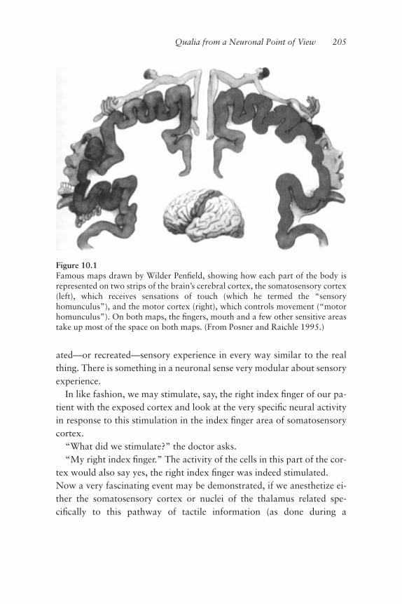
ated—or recreated—sensory experience in every way similar to the realthing. There is something in a neuronal sense very modular about sensoryexperience.
In like fashion, we may stimulate, say, the right index ªnger of our pa-tient with the exposed cortex and look at the very speciªc neural activityin response to this stimulation in the index ªnger area of somatosensorycortex.
“What did we stimulate?” the doctor asks.“My right index ªnger.” The activity of the cells in this part of the cor-
tex would also say yes, the right index ªnger was indeed stimulated.Now a very fascinating event may be demonstrated, if we anesthetize ei-ther the somatosensory cortex or nuclei of the thalamus related spe-ciªcally to this pathway of tactile information (as done during a
Qualia from a Neuronal Point of View 205
Figure 10.1Famous maps drawn by Wilder Penªeld, showing how each part of the body isrepresented on two strips of the brain’s cerebral cortex, the somatosensory cortex(left), which receives sensations of touch (which he termed the “sensoryhomunculus”), and the motor cortex (right), which controls movement (“motorhomunculus”). On both maps, the ªngers, mouth and a few other sensitive areastake up most of the space on both maps. (From Posner and Raichle 1995.)

procedure known as a Wada Test to locate the site for the speech centerin neurosurgery). When we again stimulate our patient’s ªnger and askwhat was stimulated, we inevitably hear: “You didn’t stimulate anythingyet.” We have created with our barbiturate application a complete andimmediate disappearance of sensation, of this speciªc sensory experience,without any change in connectivity or anatomy of any type. As this mayalso be done with local anaesthesia, we must conclude that qualia are in-deed fundamentally related to the electrical activity of the brain. All wehave done by the application of anesthesia is to modify one single aspectof the function of the nerve cells: the ability to generate particular electri-cal patterns of activity.
That qualia must be subserved by electrical events at nerve cells makesenormous theoretical sense as well. Think of the unbelievable rapiditywith which any sensory stimulus may be analyzed and woven into astream of consciousness. Bringing to mind the 40-Hz oscillatory activa-tion of the brain and its relation to intrinsic thalamocortical activity, wesaw that a quantum of cognition can be measured to be a well deªned12–15 millisecond time epoch. This means that the perceptual capabili-ties of the central nervous system are such that for two sensory stimuli tobe perceived as two distinguishable sensory events, there must be a mini-mum of 12.5 milliseconds separating these events—otherwise, the brainwill register them as a single sensory event (Kristofferson 1984; Llinásand Pare 1991; Llinás and Ribary 1993; Joliet et al. 1994). Such a quan-tum of cognition requires the patterned activation of millions or evenhundreds of millions of cells. Given this, the only way that cells wouldever be able to generate an event of this coherent nature would be to useelectricity as the connecting mode of information ºow between them—no other medium we know (that exists within the brain) is even remotelyfast enough! If we look at the possible biological events in and abouteven a single neuron, a 12–15 millisecond time frame of ensemble activityencompassing or employing hundreds of millions of cells poses very seri-ous limitations in terms of the plausible carriers of this type of necessaryinformation ºow. Diffusion is far, far too slow and its range of effectis far too short. In these time frames, a given molecule could not get veryfar from a cell, or very far into a cell for that matter, if diffusion were
206 Chapter 10

the carrier of ensemble information ºow. Electricity is the only mediumfast enough and far reaching enough to support the rapid and widespreadensemble activity underlying sensory experience within perceptual timeframes envisioned by Charles Sherrington as “the enchanted loom”(1941, p. 225). So we must accept that qualia are triggered by electricalactivity in the brain and are made up of events very close in time tothe electrical structures that skate over the surfaces of neuronal mem-branes. These electrical vortices dart this way and that, like sheet light-ning that ºickers and passes, leaving behind only a faint, short-lastingglow—a sensation to be lit up again as the next sheet of lightning strikesand spreads, forming to us the perception of a continuous web of sensa-tion. Qualia truly are ºeeting and discontinuous cellular events for thesame physiological reasons that consciousness itself is a ºeeting anddiscontinuous event. That qualia and the self—speciªcally self-aware-ness—are related is a topic we shall look into toward the end of thechapter.
Having related qualia to the electrical events of neurons, we can sayeven more. When considering the large functional events of the centralnervous system, such as waking or sleep, not only is electrical activitynecessary but electrical activity at particular frequencies, as was dis-cussed in detail in chapter 6 when examining thalamocortical connectiv-ity and function. When we fall into dreamless sleep, non-REM sleep as itis called, we see that this functional state is characterized by slow wave,synchronous delta wave activity (Llinás and Ribary 1993). This wholebrain, rhythmic wave pattern is in the .5–4 Hz frequency range and hasthe largest amplitude of EEG or MEG (magnetoencephalogram) moni-tored brain activity. Recall from chapter 6 that when in this deep sleepstate, sensory input of all types (modalities)is for the most part rejectedby the thalamocortical system. Sensory pathways carry their modality-speciªc information, but this information is not given internalsigniªcance; there is in fact no sensory experience whatsoever. Qualiahave temporarily ceased to exist!
Similarly, qualia also cease to be during a petit mal seizure where sim-ply, because of the epileptic state, the prominent frequency of brain activ-ity is lowered, without otherwise affecting much or any of the
Qualia from a Neuronal Point of View 207

connectivity or the basic, ongoing functioning of the nervous system. Allsensory experience—indeed the “person”—is gone. So it is not just elec-trical activity of neurons that determine qualia, but particular frequencyranges of whole brain activity where qualia may appear and disappear. Insimple terms, there are particular types of electrical patterns, global andlocal, that must be coactivated for feelings to be evoked.
The Functional Geometry of Qualia: Internalized FAPs
What may we say then is the neuronal basis for qualia? To pursue this, Iwould like to address the subject of qualia from a somewhat theoreticalperspective, starting with the motor point of view and based on much ofwhat we have already learned from earlier chapters. Ultimately, motricityis always the product of muscle contraction; we can make no movementthrough any other means. From this we immediately come to the conclu-sion that the nervous system works within the context of a ªnal motoreffector that is capable of transforming the electrical activity of motorneurons into actual muscular contraction. We may ask by analogy whatis the effector, the apparatus for the endpoint expression of sensory expe-rience. This is the central question of neuroscience at the present time.The answer is that physiologically we do not know what the effectors ofsensory experience are or how they work. We do, however, know therealms of operation of such effectors, that they require electrical,neuronal activity of a particular type and in particular parts of the centralnervous system. These effectors also require that other parts of the nerv-ous system be quiescent. Looking at it from this angle, we may come tothe conclusion that the effectors of qualia are very similar in theirneuronal bases to the neuronal bases of motor FAPs—except that theyappear to be internalized FAPs. Internally, motor FAPs are silent in termsof expression until they are liberated into action; the outward expressionof this is a stereotypical movement. By contrast, what I shall refer to assensory FAPs ªnd their endpoint or overt expression internally; this ex-pression is what we call subjective experience. Sensory FAPs are accom-panied by subjective experience whether they are produced by activationfrom stimuli of the external world via our sensory systems, by direct, ex-
208 Chapter 10

perimental electrical (or chemical) stimulation of various areas of thebrain, or through internally derived activation, as in dreams. It is quiteclear that sensory components evoked by direct experimental activationof the brain mostly produce small fragments of sensation rather than thecomplete sensory events that are generated by normal, physiological acti-vation of the brain. This is not surprising. Compared to the normal phys-iological activation of the brain, with the complex intricacies of itselectrical detail, exogenously applied electrical stimulation must be ex-traordinarily gross at best, limited in its articulation, range of effect, andthe like.
We can demonstrate that electrical stimulation of a certain brain areaproduces a sensation and that disruption of the electrical activity withinor leading to the same area is accompanied by a disappearance of suchsensations. Thus we can say again that qualia most certainly relate toelectrical activity and location. At this point there are a few possible sce-narios that we can consider. One, as many people believe, is that qualiarepresent a very profound event in neuronal function dealing with quan-tum mechanical structures of neurons that include the detailed organiza-tion of microtubules and microªlaments. This of course opens up a new,previously unexplored area of neuroscience that I won’t explore here, forI sincerely doubt it is a plausible scenario worth pursuing at any seriouslevel. The reason for its dismissal is that the neuronal elements that sup-port sensory activation seem to be quite similar to those that support mo-tor activity. Qualia seem to be related not only to particular neuronsper se, but also more to the geometrical, electrical patterns of activityneurons are capable of supporting.
To me the evolutionary reason for qualia is straightforward. Qualiarepresent an ultimate bottom line, because sensations themselves are geo-metric, electrically triggered events. At this time we cannot reduce it anyfurther. If this geometric, functional state is sensation itself, a seriousphilosophical problem immediately arises. By this deªnition are notqualia just another example of “that which we have yet to understand”?Or are they perhaps something of a qualitatively different character alto-gether, something transcending the neurological substrate of neurons andtheir electrical activity we attempt to hide them behind? I don’t think so,
Qualia from a Neuronal Point of View 209

for I believe that patterned electrical activity in neurons and their molecu-lar counterparts are sensations.
“The Hard Problem”: Is It True that Science Will Never Understand Feelings?
So, as for the neurobiological basis of qualia, one could leave the issue atthe level of the observation that qualia are functional electrobiologicalevents supported by particular sets of neuronal circuits and related to theactivation of some neurons and the silence of others within such a net-work. While this basic description may appear trite it is the only possiblebasis for a scientiªc approach to this problem. Ultimately, we will need toknow much more about the intricate workings of the nervous system be-fore we can begin to understand what feelings are all about. However,what can we say about qualia today?
For all intents and purposes, the question of qualia or feelings is thequestion of conscious experience. The feasibility of our ever understand-ing such an elusive phenomenon in scientiªc terms is a subject of continu-ous debate—even as to whether any hypothetical explanation groundedin physical/neural processes can be fully satisfying and complete (seeChalmers 1995, 1997; Shear 1997, for a discussion of various ap-proaches to this issue). While an answer to these questions may be inac-cessible at the present time, we can at least attempt to frame the questionin a useful way.
David Chalmers, one of the prominent recent voices in this debate, setsup the problem by ªrst noting that consciousness is an ambiguous termthat has been used to refer to a collection of distinguishable phenomena,including what he terms the “easy problems” and also the “hard prob-lem” of conscious experience itself:
The easy problems of consciousness are those that seem directly susceptible to thestandard methods of cognitive science, whereby a phenomenon is explained interms of computational or neural mechanisms. The hard problems are those thatseem to resist those methods. The easy problems of consciousness include thoseof explaining the following phenomena:
the ability to discriminate, categorize, and react to environmental stimuli;the integration of information by a cognitive system;the reportability of mental states;the ability of a system to access its own internal states;
210 Chapter 10
the focus of attention;the deliberate control of behavior;the difference between wakefulness and sleep. (Chalmers 1995)
and:
For these phenomena, once we have explained how the relevant functions areperformed, we have explained what needs to be explained. The hard problem, bycontrast, is not a problem about how functions are performed. For any givenfunction that we explain, it remains a nontrivial further question: why is the per-formance of this function associated with conscious experience? The sort of func-tional explanation that is suited to answering the easy problems is therefore notautomatically suited to answering the hard problem. (Chalmers 1997)
Chalmers collects all these “easy” phenomena associated with con-sciousness under the umbrella term “awareness” (functional, reportablephenomena), and then argues that there is an inextricable linkage be-tween awareness and experience, almost as cause and effect:
To a ªrst approximation, the contents of awareness are the contents that are di-rectly accessible and potentially reportable, at least in a language-using system.
Awareness is a purely functional notion, but it is nevertheless intimately linkedto conscious experience. In familiar cases, wherever we ªnd consciousness, weªnd awareness. Wherever there is conscious experience, there is some corre-sponding information in the cognitive system that is available in the control ofbehavior, and available for verbal report. Conversely, it seems that whenever in-formation is available for report and for global control, there is a correspondingconscious experience. Thus, there is a direct correspondence between conscious-ness and awareness. (Chalmers 1995)
We return to the essential issue as Chalmers roots this mapping ofawareness to conscious experience within the physical mechanisms of thebrain:
In general, any information that is consciously experienced will also becognitively represented. . . . This principle reºects the central fact that eventhough cognitive processes do not conceptually entail facts about conscious expe-rience, consciousness and cognition do not ºoat free of one another but cohere inan intimate way. (Chalmers 1995)
The linkage Chalmers provides us may be correct, but it may also besecondary to the fundamental origin of qualia as arising from the veryproperties of physical mechanisms present in the living organism, andmore ancient than the cognitive processing of a complex brain. Causeand effect driven from the other end, at least conceptually. In this regard,
Qualia from a Neuronal Point of View 211
I feel there is a possible hypothesis that can be offered concerning thevery nature of qualia. This view may be considered as coming from leftªeld, so let me prepare the scenario just a bit.
We have known for a long time that single cells are capable of irritabil-ity, that is, the ability to respond to stimuli with a behavioral responseconsisting of either moving away from, or approaching an object or an-other cell. In the latter case the relation may be that of hunting for foodor ºeeing from deleterious or threatening conditions. These observationsshould remind us that there are in single cells certain abilities related in aprimitive way to intentionality, and thus to what may be considered aprimitive sensory function. If we are allowed to consider that qualia rep-resent a specialization of such primitive sensorium, then it is a reasonableconceptual journey from there to the multicellular phenomenon of “cor-porate feelings” manifested by higher organisms. If this is something wecan live with, then we will understand that qualia must arise from, funda-mentally, properties of single cells (ªgure 10.2), ampliªed by the organi-zation of circuits specialized in sensory functions.
This means that only those circuits having the necessary numbers ofsensory cells organized with a particular architecture will be able tosupport such a function. This can be seen with muscle cells, where thecontractile property that characterizes each one is nothing other than aspecialization of ªlamentous interactions found in all cells, a specializa-tion prominent because of architecture. In muscles, actin and myosinmolecules are organized parallel to each other to support the sliding ªla-ment network and are also anchored to the endoskeletal system, so theforce generated by interaction among ªlaments may add vectorially tosupport cell contraction (Huxley 1980). If many cells can sum the forcegenerated by their simultaneous contraction, in this case by convergingon to a common point (a tendon), then a macroscopic force capable ofproducing movement is generated. A corporate motor has been evolved.Something similar may be happening with sensory cells. Their summingproperties can and have been studied, as we will see presently. Thatwhich is summed (single cell primitive qualia-like property) is what mustbe understood, but the problem then seems far more tractable. We arenot searching for a spook.
212 Chapter 10

The Issue of Qualia as a Single-Cell Property
So what can we say concerning the role of single cells in the generation ofqualia? The simplest way to address the issue is to consider other cellularproperties that might be, even remotely, related to the problem at hand.What other systems do we know in which electrical signaling is the trig-ger for coherent cell action? The best analog may be, as mentioned be-fore, muscle contraction.
The following properties are common for muscle contraction andqualia:
1. They are both triggered by electrical activation of the cell.2. In both, the cellular event of interest is separable from the electricalevent that triggers it, and follows in time the electrical activation.
Qualia from a Neuronal Point of View 213
Figure 10.2Drawing of the one-celled organism Codonella companella, illustrating the highdegree of structural specialization possible in a single cell. (From Villee-Dethier,1971, ªgure 3-2, p. 33.)

3. The “corporate event” of muscle contraction or qualia has summingproperties relating to the numbers of elements activated and to the fre-quency of activation.
• In muscle the product of cellular activation, force, is the sum (linear) ofthe pull of each cell, onto a common tendon (a geometric property) at agiven time.• In qualia the product of cellular activation “sensation” is the sum (log-arithmic) of each cell activation on to a common coherent event (a geo-metric property), at a given time.
4. Drugs can affect muscle contraction and qualia.
• Modiªcation of electrical activity by sodium conductance block (e.g.,TTX) will prevent both muscle contraction and qualia.• Drugs can modulate muscle contraction by acting on membrane recep-tors that modify particular molecular events inside the cell(glibencamide) (Light et al. 1994). Similarly, drugs can modulate qualiaby acting on membrane receptors that modify particular molecularevents inside the cell (psychotropic drugs like marijuana).
The following properties are different as they relate to muscle contrac-tion and qualia:
1. In muscle contraction and qualia the ultimate products are quitedifferent.
• Force is an old physical concept that relates fundamentally to the rapidexchange of virtual force carrying particles among molecules inside themuscle cell (sliding ªlament theory).• Qualia (subjective sensation) is an old “natural philosophy” conceptthat relates fundamentally to nothing that we know about at this mo-ment, inside a cell.2. Muscle cells are easily recognizable by their very speciªc internalstructure. Neurons that may support qualia are not presently distinguish-able in form from those that do not. What is more, it is not clear thatthere are any differences, at this time.3. Muscle cells can contract in in vitro conditions. Sensory neurons can-not be demonstrated to generate qualia in conditions other than in an in-tact animal. Thus electrical stimulation of neurons may generate asensation as reported by a behavioral response such as the statement “yesI felt that” from a human.
Note that the similarities outweigh the differences and that the areas ofdifference belong to a single category, lack of speciªc knowledge.
214 Chapter 10

Can Qualia Be Quantiªed?
If the brain attempts at all times to decrease the functional overhead ofmotor control, it is difªcult to believe that it does not do the same for sen-sory systems. So of what are qualia a simpliªcation? We cannot experi-ence everything all at once, all the time, so qualia provide a constructbased on what the thalamocortical system deems worthy on a moment-to-moment basis of focus/attention/signiªcance.
Is there a particular way in which perception, through any sense mo-dality, can be understood to work? Is there an underlying pattern that cangive us some insight into the functional architecture supporting qualia?In other words, is there a measure for qualia that illuminates the natureof the functional architecture that stages qualia? There is. The measurefor all qualia can be given mathematically by the Weber-Fechner law(Cope 1976), governing the relationship between the intensity of sensoryactivation and perception:
s = kln A/Ao
Where s is sensory experience, k is a proportionality constant, ln is thenatural logarithm, and A is sensory activation. Ao is the level of sensoryactivation at which there is no sensory experience; that is, the stimulus re-mains just under the threshold of perception. One can see that as the am-plitude of A increases, the sensory experience increases in a constantratio, in a geometrical progression based on the value of e, 2.17, the basefor the natural logarithm.
We may intuit quite easily this mathematical progression that dividessensory experience into discretely perceived events in terms of musicalpitch. Perceptual differences in pitch are detected by humans in steps assmall as a few thousandths of a percent change in sound frequency. If welook at musical notation, we realize that the notes corresponding to givenincrements (or decrements) in sound frequency have a certain propor-tional change from a center point frequency, or interval. For example, anoctave corresponds to twice the frequency of this center sound, and sothe increase in the octave frequency corresponds to twice for the next oc-tave, four times for two octaves up, eight times for three, and so on
Qualia from a Neuronal Point of View 215

regardless of which note we choose to start with. Likewise, the musicalstaff, the ªve parallel, horizontal lines used in musical notation sinceGuido de Arezzo introduced this system 1000 years ago, denotes the log-arithm of sound frequency expressed vertically, with time denoted fromleft to right (ªgure 10.3). In fact, ethnomusicologists recognize that theseven basic note structure of the Western system is by no means unique.The notes of the Indian musical system, sa, ri, ga, ma, pa, dha and ni, areidentical in every respect to the do, re, mi, fa, sol, la, si, notes of the West-ern system. It may be more than a coincidence that we also recognizeseven colors; the structure of the seven color bands of a rainbow is con-stant to our qualia. The order is always the same; the thickness is of a cer-tain type. This suggests that the number seven plays perhaps animportant, pervasive demarcating role in sensory experience; this is sup-ported by the thesis, “seven, plus or minus two,” the magic number thatGeorge Miller has so elegantly described.
With respect to this geometry, I think the basic structure of qualia mostlikely consists of a center point for any sensory experience, with a num-ber of levels above and below as ratios of the central value (two to fourlevels in systems based on ªve to nine primary points, respectively). Bycenter point I refer to the level at which most receptors for a given sensemodality operate, their most common ªring rate or pattern. Firing ratesabove or below this central point would trigger a modulation back to-ward it. If one thinks of body temperature, the center point around whichthe system operates is 36.5 degrees Celsius. Shifting above or below thisvalue sets into motion events that will bring the system back to its natu-rally selected set point. Similarly, in the human vestibular system, the sys-tem that manages our sense of balance, the center point of neural activityis based on the body being upright and erect. The vestibular neurons thatªre most frequently do so during the least amount of body movement, in-dicating the importance of being upright as the center point for balanceto operate around.
In considering how a functional geometry such as this may have cometo be, it is worth noting that natural growth, when unimpeded, occurs atthe logarithmic base e, as described by John Napier in 1614. This univer-sal constant, the solution to lnx�1, rules all growth (see Thompson, On
216 Chapter 10

Qualia from a Neuronal Point of View 217
Figure 10.3Musical notation illustrating the seven-tone western music system. As illustratedin this famous piano composition by Robert Schumann, the pitch of each note isspeciªed by its position on the treble (top) or bass (bottom) clef.

Growth and Form), and is wonderfully evident in the shell curvature ofthe mollusk Nautilus, which exempliªes one of the beautiful and ubiqui-tous geometric structure in nature. It would not be surprising to ªnd thatqualia derive from electrical architectures embedded in neuronal circuitscapable of such logarithmic order. If sensations conform to the geometrydescribed by the Weber-Fechner law, it seems quite possible that the elec-trical patterns of neurons that represent qualia would operate on a simi-lar, logarithmically geometric basis.
So What Are Qualia Good For?
Given our knowledge today, we seem to have come as close as we can tounderstanding qualia. Those who reject the reduction of qualia to theelectrical activity and geometry of neuronal circuits perhaps do so be-cause they lack any understanding of functional geometries; qualia arenot some mysterious events that, “residing between,” manage miracu-lously to change the nature of electrical activity into “feelings.” After all,we must remember that, as stated above, qualia are soluble in local anes-thetics. Here the ghost in the machine is responsive to surgery or even awhack on the head. Since when are transcendent properties so fragile andclose to the biological process? Parsimony and serious science clearly in-dicate that “the bridge,” “the mysterious transformation” of electro-chemical events into sensations is an empty set. It does not exist:neuronal activity and sensation are one and the same event.
Indeed, if a single cell is not capable of having a modicum of qualia,how then can a group of cells generate something that does not belong toa given individual? We would have to say that asking if qualia areproperties of a single cell is similar to asking if movement is a property ofa single cell. As stated above, movement, as in the case of a limb, is pro-duced by the summed contractile properties of many muscle cells. A sin-gle muscle cell cannot produce macroscopical movement of a limb. Tofollow the argument through, nerve cells must be capable of a “proto-qualia.” An organized sensation requires the activation of many neuronsin a particular pattern, that is, it requires the generation of a neuronal ar-chitecture capable of supporting macroscopic qualia in the same way that
218 Chapter 10

the musculoskeletal apparatus requires a certain architecture to producemovement.
Coming back to the issue of sensory FAPs, there is a concept that hasbeen lurking in the halls of neuroscience about as long as that disciplinehas been around. It is the concept of “labeled lines,” and it may furtherhelp us theoretically remove the ghost in the machine once and for all.The concept of labeled lines states that sensory pathways of all sense mo-dalities encode the speciªc properties of the world they convey by veryspeciªc ªring patterns, and that each line or pathway only carries infor-mation of that speciªc modality. In literal terms, these speciªc patternsare the speciªc sense modality messages from the outside world.
It makes intuitive sense that the perception of high frequency sound re-quires receptors that convert sound waves into neural energy. These arethe hair cells of the auditory apparatus, which respond to high frequencysound with a correspondingly high rate of ªring. Similarly, hair cells re-spond with a low rate of ªring when presented with low-frequencysound. Pacinian corpuscles, receptors of the skin that respond to mechan-ical compression, ªre their labeled line message of low frequency pulsingin response to light compression of the skin, and correspondingly higherfrequencies of ªring for increased mechanical compression. And so it isthat the initial message carried by a given sensory pathway faithfully “la-bels” its outer world counterpart. This frequency coding property, andthe fact that each sensory pathway only carries information about its spe-ciªc sense modality, has led to the concept of labeled lines.
But let us follow one of these labeled lines a little farther, right into thecentral nervous system. The high frequency ªring of the auditory appara-tus in response to sound of high frequency does not remain as such. Aswe follow this labeled line, the high frequency activity is translated intolow frequency activity by the time it reaches its end point (auditory corti-cal neurons). This tells us something very important: it is not the code ormessage coming from the outside world that is being transmitted, butrather it is the neuronal element that responds to the message from theoutside that is itself the message! It is the sensation born of an internallyactivated sensory FAP—one may justiªably say that the labeled line car-ries de facto the frequency because it ªred!
Qualia from a Neuronal Point of View 219

Are we getting any closer to a deªnable functional architecture forqualia? Let us pursue a bit further this concept of the effectors of sensa-tion as being sensory FAPs. From what we learned in chapters 2 and 7,we came to understand that motor FAPs represent a naturally selectedfunctional organization of the central nervous system, one slanted to-ward computational efªciency. These “plug and play” modules, when ac-tivated or released, automatically call to order the various muscle groupsand synergies required for a stereotypical movement execution, from thesimple to the complex. The computational efªciency, the reader will re-call, is attained by the preset automaticity of these modules of function:the brain does not have to reinvent the wheel, from a neuronal circuit(connectivity) perspective, each and every time a particular routine move-ment is required of the body by circumstance. This allows the centralnervous system to put its mind to other things, so to speak. The effectorsof FAPs are the motor neurons and the muscles whose contractions themotor neurons drive. Put another way, generated from the internal func-tional geometry within the basal ganglia is a translation into expressionthrough the functional geometry of how the body can and needs to move,given the momentary (internal and/or external) context.
Can we think of qualia—sensations or sensory experience—in thesame way? We can, and the key here is the brain’s innate drive towardreducing overhead. A few moments ago I spoke of high-frequency audi-tory signals being translated to low-frequency activity once this sensorypathway enters more deeply into the central nervous system; this is quiteconsistent with what we have been saying about an economy of transmis-sion. Rather than the elements being generated from simple to complex,each element carries its own signiªcance and the whole is assembled bythe pre-existent presence of signiªcant activity and the absence of othersigniªcant activity.
This point is right in line with what we came to understand about thefunctional organization of sensory systems described in chapter 5. By thevery nature of the translation of the geometry of the properties of the ex-ternal world into the geometry of the internal functional space, reality isat all times simpliªed. It has to be so; it is the only way the brain can keepup with reality. It must simplify at all times.
220 Chapter 10

The Necessity for Qualia
Why is it so important to address the question of qualia in animals? Mostpeople are apparently not convinced that qualia are necessary for any an-imal. One could, it is supposed, perform exactly the same actions withoutqualia or feelings. Life would be exactly the same without it! A cat coulddo everything it does as an automation without qualia: there is no addedadvantage, there is no reason to have qualia. Should we then deny thefact that they exist? To me, by contrast, qualia, from the perspective ofthe workings of the brain, constitute the ultimate bottom line. Qualia arethat part of self that relates (back) to us! It is a fantastic trick! One can-not operate without qualia; they are properties of mind of monumentalimportance. Qualia facilitate the operation of the nervous system by pro-viding well-deªned frameworks, the simplifying patterns that implementand increase the speed of decision and allow such decisions to re-enter(the system) and become part of the landscape of perception. Not onlywere you pricked on the side by the thorn and you moved, but now youhave also been sensitized to thorns in general—you can either stay awayfrom them or you can tame them, that is, use them as weapons. And so,qualia become exceedingly important tools in perceptual integration; it isthe repository of the binding event.
In chapter 5 we took a closer look at the evolution of the eye and sawthat nature generates very complex functional architectures. Along withthe eye, the heart, and so on, FAPs and even language may be consideredas organs, local modules of function with very specialized capabilitiesand duties. We may, and I think we must come to understand qualia as asort of master organ, one that allows for the individual senses to operateor co-mingle in an ensemble fashion. Qualia make simplifying, momen-tary judgements about this ensemble activity, allowing these judgementsto be re-entered into the system for the predictive needs of the organism(self). Qualia represent judgements or assessments at the circuit level ofthe information carried by sensory pathways, or sensations. And thesesensations, the integration product of the activation of internal sensoryFAPs, represent the ultimate predictive vectors that recycle/re-enter intothe internal landscape of the self. They are the “ghost” in the machine
Qualia from a Neuronal Point of View 221

and represent the critically important space between input and output,for they are neither, yet are a product of one and the drive for the other.And all the while they are simpliªed constructs on the part of the intrinsicproperties of the neuronal circuits of our brains.
222 Chapter 10

Kiki Smith, Tongue and Hand, 1985. Painted plaster, 5-1/2 � 3-1/2 � 3� (14 �8.9 � 7.6 cm) tongue unit; 5-1/2 � 3 � 3-1/2� (14 � 7.7 � 8.9 cm) hand unit.Photograph by Ellen Page Wilson. Courtesy of PaceWildenstein.

11 Language as the Child of
Abstract Thought
The Beginnings of Abstraction
Let us begin by agreeing on a consensus deªnition of abstraction and/orabstract thinking. An abstraction generally refers to something that onlyexists in the mind: an idea, a conception, a mental representation ofsomething that may (or may not) exist in the outside world. Abstraction,or the collection of neural processes that generate abstraction, is a funda-mental principle of nervous system function. The nature of these pro-cesses owes its origin to the phylogenetic wiring patterns acquired by thenervous system on its evolutionary course. As such, it is more than likelythat abstract thinking probably began long ago in very primitive nervoussystems. This view emerges from considering the nervous system as beinggeared toward predictive movement. In order to place movement withinthe context of the whole animal, the animal must ªrst be capable of gen-erating some type of internal “image” or description of itself as a whole,and this image must support the strategy around which to organize thetactics of what the animal will do.
At ªrst glance, the generation of a voluntary internal sensory-motortransformation (see chapter 7) does not correspond directly to the moreobvious neuronal connectivity required to transform, for example, a
stubbing of the toe into a leg ºexion. The newer type of wiring representsmore than the segmental reºex. It is metasegmental, representing a globalfunction such as the coordinated walking of an elongated multisegmentalanimal rather than just the stepping supported by a given local segment.By “elongated animal,” I refer to any encephalized creature with a headend and a tail or foot end with a column or chain of nervous tissue tra-versing (and subserving) the length of the body. This description encom-passes a broad range from the more lowly creatures with very primitivenotochords to those with quite sophisticated spinal cords. That the nerv-ous system was selected in evolution to be organized in a segmented fash-ion was probably driven by the neurobiological practicality of optimizingbody surface area to volume, in order to minimize the distance a nervesignal must travel either to or from the external world. Elongated ani-mals are basically made of a horizontal stack of “coins,” where the neu-ral wherewithal subserving each coin is organized to know about itsrespective segment and relatively little else. In order to make a completeworking animal out of these segments, there must be a portion of thenervous system that is not exclusively segmental in its organization. Thisportion of the nervous system can put the many segments together intosomething that beforehand did not exist: a uniªed whole. As statedabove, we may consider this the beginning of abstract function as thisportion of the nervous system does not relate directly to the connectivityof the nervous system at any particular, segmental level. The central nerv-ous system abstracts the fact that the animal is composed of a series ofunit segments; ipso facto, the process of intersegmental integration is anabstraction, and represents the beginning of abstraction as a naturally se-lected biological process. That this is the evolutionary direction is sup-ported by the observation that the central nervous system mushroomsout in front of the spinal cord, polarizing encephalization. We see some-thing important happening: from the animal’s very neurological becom-ing is the fact that the animal can have an internal representation of itselfnot only as a set of parts but as a whole entity. It is here, from this germi-nal metaevent, that abstraction begins and the self emerges.
How does this relate to prediction? Well, beyond the description of theanimal itself and the description of the input that comes to this animal,the intrinsic circuits of the nervous system are capable of generating a
226 Chapter 11

premotor representation of what is going on outside. From this, self-referentially in the presence of that motor image, the animal is capable ofdeciding what to do. The animal is capable of prediction. Run, ªght, ªndfood or whatever, functionally the animal is the circuit that represents itssensory motor attributes, and this central event is an abstract entity.
Now of great importance is the event that happens between the stimu-lus that evokes movement and the liberated motor FAP. The promptingstimulus may be of external origin (a ferret is climbing up my pants!) orinternal (I left the stove on at home!). Either way, these stimuli, if grantedappropriate internal signiªcance (by and within the momentary status orcontext of the cycling thalamocortical system), are ampliªed into anemotional state. We have already seen that the nervous system is wired insuch a way that under normal conditions FAPs are only liberated into ac-tion by the generated emotional states that precede them. Such internalevents, emotions, are then by deªnition premotor states.
We can take this further. Emotions or emotional states are events thatdo not exist in the outside world; they are purely internal events andwould remain completely hidden to us (as observers of others) were it notfor motricity. Precisely which emotion may be occurring is inferred onlythrough the expression of the FAP that is liberated by that given emotion.The dog is snarling and baring its teeth at me; what is going on inside ofthis dog is most likely not that he is happy to see me. Just how it is that Icame to know or infer this is another important issue that we will discussshortly. The point here is that emotions, being themselves purely internalevents, are simply invented states on the part of the central nervous sys-tem and as such are clearly abstractions. It is fair to say that just as emo-tions are intrinsic products of central nervous system function, so areabstractions.
Intentionality
Returning to prediction, it is clear that prediction must have a goal, oth-erwise it is not referentially based on anything; purposeless movement isnot only wasteful, but can be quite dangerous as well. The goal or objectof movement must be well deªned, and we may deªne it here as thatwhich one intends to do in relation to that object or goal. Also an
Language as the Child of Abstract Thought 227

abstraction, intentionality is the premotor detail of the desired result ofmovement through which a particular emotional state is expressed: thechoice of what to do before the doing of it.
Consider the following: if our brains are capable of planning move-ment strategies that are implementable if desired, then they should alsobe able to outwardly express intentionality as a motor representation ofwhat is happening inside our heads. I am suggesting that the outward ex-pression of premotor activity precedes and predicts the activation of spe-ciªc motor patterns. An example of such a process is shouting “run!”when in danger before we actually begin to run. This raises a cruciallyimportant point about language itself. I suggest that our ability to vocal-ize the different aspects of intentionality developed ªrst as the ability toseparate the properties of things from the things themselves. This processof abstraction would over time engender what we may consider to be amental catalog, much like an alphabet, that would allow us to generateinside our heads events that would be reentered admixtures of the pri-mary events that went into the generation of language to begin with. Wehave arrived at our ªrst corollary: even before language was sufªcientlywell structured to be communicable, its genesis must have had as a pre-requisite foundation the nervous system’s capacity to generate thepremotor imagery required to abstract the properties of things from thethings themselves. That is, it required the premotor imagery to make ab-stractions of universals.
And so we see that there are two very important issues we must bear inmind when considering the evolution of language. One, that abstractthinking must have preceded language during evolution, and two, thatthe premotor events leading to expression of language are in every waythe same as those premotor events that precede any movement that is ex-ecuted for a purpose. From these two points, which are so similar as to bealmost the same, we may gather that language is simply an elementwithin a much larger, more general category of function.
Prosody: The First Cracklings of Language
Let us now dig into the plausible origins of language and how this indis-pensable tool must have evolved. Just as in the case of the rather mean-
228 Chapter 11
dering evolution of the eye, language may be difªcult to trace cleanlybackwards through evolutionary time (see, for discussions and concepts,MacNeilage 1994, 1998; Verhaegen 1995; Gordon 1996; Ujhelyi 1996;Aboitiz and Garcia 1997 a, b; Honda and Kusakawa 1997; Ganger andStromswold 1998; Gannon et al. 1998; Kay et al. 1998; Doupe and Kuhl1999; Nowak and Krakauer 1999). As in previous examples, evolution-ary steps prior to the emergence of a specialized organ may not necessar-ily function or appear in any fashion similar to what we see in the presentday organ. As we learned from the lifetime of lifetimes that brought usthe eye, we may perhaps expect an unexpected genealogical path in theancestry of language.
Now before proceeding further, we must gather a few clarifyingdeªnitions. Just what do we mean by “language”? The ªrst thing thattypically comes to mind is human language, its wide variety of types, thatit is often written as well as spoken, and that languages other than ourown are at once fascinating and opaque. If you feel that language is ex-clusively a human capability or that we humans invented language, then Imust tell you that I wholeheartedly disagree, although you may not bealone in this thinking. The reasons for which I disagree are straightfor-ward: language clearly exists in many species far, far older in the evolu-tionary sense than we Homo sapiens; furthermore it is too general a traitthroughout the animal kingdom ever to be seriously considered as exclu-sive to humans. Although it is most likely true that we exhibit the richestand most complex of languages, we are neither the origin nor sole pos-sessor of language.
Let us deªne language as the given methodology by which one animalmay communicate with another. In this regard, language is a rather large,generic category, for this deªnition says nothing about the intent to com-municate, only that some level of communication is ultimately achieved.We have so far been saying that language is a logical product of the in-trinsic abstracting properties of the central nervous system, or simply ofabstract thought. But this I would say is a subcategory within languagethat I would call biological “prosody.” Prosody is a more generalizedform of motor behavior, an outward gesturing of an internal state, anoutward expression of a centrally generated abstraction that meanssomething to another animal. For us, smiling, laughter, frowning, the
Language as the Child of Abstract Thought 229

lifting of one’s eyebrows are all forms of prosody, for they convey one’sinternal, momentary state in a way that is recognizable and understand-able to someone else. Prosody is language, but it is not spoken language.Nonetheless, it is purposeful communication. Prosody is by no meansconªned to humans; it is widespread throughout the animal kingdom,and in the evolutionary sense is very old. Darwin, in his brilliant text onfacial expression, studied prosody in animals in terms of moods and facesand how faces and postures represent the momentary internal states of ananimal. These are representations of the internal abstractions such asemotions and intentions. So a prosodic event is an abstraction coupledwith a motor expression that conveys to another animal what its internalstate is like at that moment.
If prosody represents a subcategory within language, what would bean example of language without prosody? Well, there are types of lan-guage that are extraordinarily speciªc and although they carry very sim-ple messages, they are nevertheless essential for the survival of thespecies. The pheromone delivery and reception systems of a moth areknown to be effective for distances of several miles, and so they clearlyrepresent communication at a distance. The pheromone released by thefemale is recognized speciªcally and exclusively by the male of the samespecies and is communicatively effective enough for the couple to ªndeach other in an otherwise crowded niche (Willis and Arbas 1991;Hildebrand 1995; Roelofs 1995; Baker et al. 1998). But although thislanguage is critical for the species’ survival, it has nothing to do with theoutward expression of an internally generated abstraction. This is there-fore not prosody, but rather simply a behavior-modifying event throughthe release and reception of a speciªc molecule.
In most cases, however, language circumscribes a prosodic event. Weªnd that language as such may be observed at many levels in evolution,where it serves very different functions. One of the ªrst nonhuman lan-guages to be understood sufªciently by people, one that communicatessimple orders, is the language of bees. This language is basically a dance,a rhythm and orientation performed in space. These dances, each onespeciªc to bees of a particular species, give information about the quan-tity and location of food with respect to the beehive. In this way all beesof the colony can know about and help procure the food (von Frisch1994; Gould 1976, 1990; Hammer and Menzel 1995; Menzel and Mul-
230 Chapter 11

ler 1996; Waddington et al. 1998). Languages such as these have alsobeen studied in vertebrates and other invertebrates. By deªnition and inall cases, these forms of communication require social order so that theinformation conveyed may be used to some purpose by the receivingorganism.
There are variations of language that convey information quite differ-ent from just the issue of food for one’s family. For instance, when mostanimals are attacked, they generally posture in a manner that is clearlyrecognizable as a defensive stance by the attacker or as a counter-punchready to occur. Such posturing may be as simple as a puffer ªsh increas-ing its size to appear more formidable, or the very common showing ofteeth and growling we see in most vertebrates. Animals with horns, suchas the rhinoceros or buffalo, take a stance where the horns are directed atthe threatening or attacking animal. These are all languages, grantedwith very limited repertoire, but this form of prosody must be at the veryfoundation of all types of purposeful communication between and withinspecies.
Moving higher up the evolutionary scale, we can look at languagesthat convey a higher level of organization. The language of wolves is anexcellent example where by means of prosody, wolf packs express rela-tively complex, socially structured attack and defense behaviors. In thiscase, the relations between the differing animals are not only of simpleprosody but also represent a (social) context within which this prosodiclanguage is exercised.
This form of prosody in wolves is quite sophisticated, utilizing a num-ber of different motor avenues for overall expression, including vocaliza-tion, eye contact, head gesturing, and whole body communication. Forinstance, the establishment of dominance, or which wolf is to be the al-pha male, is by means of communication not just of one’s physical might,but by those males subordinate to the alpha male showing their social po-sition by expressing submission. They will roll over onto their backs andoffer the alpha male their neck. This form of language leads to an estab-lishment of social hierarchy that is central to the strategies of the pack asa whole. However, during hunting other factors arise, and so:
True leadership is not strongly apparent in a wolf pack, as any animal may initi-ate its movement. However, the pack is highly cooperative in hunting and in thecare of the young. Dominance organization is not strongly apparent in wild wolf
Language as the Child of Abstract Thought 231

packs, but in the captive packs in zoos, where the principle occupation of huntingis made unnecessary by artiªcial feeding, wolves spend a large amount of theirtime threatening one another and enforcing dominance. (Dewsbury andRethlingshafer, 1973)
Notwithstanding this caution about the risks of using behavior ob-served in captivity to make sweeping generalizations about behavior inthe wild, the social organization of wolves does have a hierarchy, so thelanguage or the prosodic events would be understood differently at itsdifferent levels. The prosody that cues one adult male to know that it ishis turn within an attack sequence may to a young pup be simply thelearning of the global differences between attacking and defending. Themain point here is that the social hierarchy itself could not exist of therewere no elements of commonality existing at all levels. In this case we seethat language is developed in the context of a particular social order as ameans of binding animals into a single working entity for the beneªt ofall.
Similarly, there are very interesting hunting behaviors that one sees inwild dogs of Africa. They often attack where there is tall grass, for that isusually where the smaller animals of prey are found. These dogs havewhite-tipped tails that they keep straight up in the air. They move theirtails back and forth in particular patterns that stimulate peripheral vi-sion, so each dog, without having to look around that much, can seewhere the other tails are, and thus receive a constantly updated status re-port on the structure of the pack as it ºushes out and/or corners its prey.It is fascinating to ponder here the reasonable conclusion that anatomyhas evolved to be related to the language of strategy.
These prosodic, ensemble properties of the canine species have madepossible the very particular relationship that dogs have with man. Thisrelation is especially clear in situations where dog and man form a work-ing team, as in shepherding or hunting. Here the dog is simply expressingproperties (abstraction and prosody) that were genetically determinedand is imprinting the relations of the hierarchy it already knows—savefor the fact that here, the hierarchy is with an animal other than anotherdog.
232 Chapter 11

Mimicry: The Origin of Meaning Between Organisms
What seems to be emerging is that language must have evolved from aprelinguistic type of attribute, mostly concerning prosody, punctuated byeither particular sounds or particular gestures. But we must not overlooka very crucial element to language. If prosody is the outward expressionof a momentary internal state, what is its purpose unless it is understoodby another animal? Communication without consensus meaning simplyis not communication. So what we are really asking is: How did themeaning part come to communication?
Here is something I would have to refer to as the infectious nature ofbrain activity. Laughter is the perfect example—it is infectious betweenpeople. Someone begins laughing, you hear (and/or see) this, and soonyou cannot help but laugh. Put another way, laughter is generated andwhen you receive it, you create the similar state in your head. It is as if theabstraction itself is infectious—an intrinsic property of neuronal circuitsthat seems to get outside of itself. We may think that if laughter in infec-tious, and so is yawning, then maybe showing your teeth and growling isinfectious as well. Let’s examine this point further.
Consider the kookaburra, a bird of Australia. These birds hang out to-gether, usually spotting the branches of two or three neighboring trees.From the silence, one of the birds starts making the characteristic kooka-burra sound, which happens to sound, to us, exactly like distorted hu-man laughter. Another kookaburra joins in, mimicking the same soundas the ªrst, and within seconds, the whole ºock is “laughing.” We see asimilar event with ªreºies: one male lights up, the others entrain. And sothe female, far off in the distance, gets a real ºash!
What does this mimicry tell us about language? One can imagine thatif the nervous system were to acquire by accident the ability to recognize,through sensory input, the FAPs displayed by others, this would be a veryuseful property if the animals are going to live in groups. In fact, thisproperty makes the group de facto birds of a feather! And so animalswith the ability to mimic each other would immediately tend to form afamily, because what has been engendered is indeed a sense of familiarity:Hey! You’re one of us!
Language as the Child of Abstract Thought 233

Recognition of one’s kin is as old as time. Fair enough, but this recog-nition is fostered through mimicry, through a creature’s repeating theFAPs of others that it gleans through its senses (ªgure 11.1). But what canone say of communicative meaning across species? I know that the dogstill snarling at me from a few pages back means trouble for me. It meanstrouble to another dog as well. How does it know? How do I know?Rather, how did we come to know? Let’s wind evolution backward andthink it through.
An animal bares its teeth at another animal; the other animal does notrecognize this FAP. Nonetheless, this FAP evokes a “show teeth back” ora “run” FAP. Why? Because all of those who didn’t at once recognize thebaring of teeth FAP died! They became lunch, and thus over time theywere naturally selected out of the gene pool! But those that remained, inthe natural selection sense, evolved to have such FAP recognition becomean intrinsic a priori. Thus, out of phylogeny comes the ability to recog-nize certain things as dangerous without necessarily memorizing them inreal time, in ontogenetic time. For instance, ªsh know from birth that
234 Chapter 11
Figure 11.1Ant ªshing learned by mimicry. (Photo by T. Nishida, from Whiten et al. 1999.)

very brightly colored (other) ªsh are dangerous. At birth, they just knowit. Those brightly colored ªsh over there are poisonous; they advertise it!And this is as wired as is the fact that ªsh have tails!
The above is the evolutionary downstream endpoint, if you will, butmimicry is at its origin. Of course we agree that language would make nosense if the receiver did not understand what language means. How doesunderstanding occur? The easiest way for the receiver to understandwhat language means is for the receiving animal to somehow associate itsown production of the motor event with the sensory reception of the mo-tor event. That is, an outward motor expression of the internal sensor-imotor image is matched with that which the receiving animal is actually,sensorially, experiencing: monkey see, monkey do. It can be donethrough learning, but it has to be more powerful than that. It would haveto be done by understanding in a sensory way the consequences of thismimicked motor behavior. This is a bit complicated; there is a hurdle toget over.
Let’s say I am an alligator now and I bare my teeth. Simple enough, buthere’s the problem: I most likely will never see myself baring my teeth.However, I might hear my own growling and so when a growling occurs Imay recognize it as what I do when I am in a certain mood. If I see thatgrowling is often associated with teeth baring I will soon associate thetwo. This is why when another alligator bares its teeth at me, I don’t justignore it, or simply look the other way as if nothing happened. We are atan evolutionary crux. Coming to know universals must have come fromnot knowing universals. This can happen in only one of two ways. Eitherthe nervous system absolutely knows in advance what it is doing or itdoesn’t, in which case it is natural selection that has determined my re-sponse: when another alligator bares its teeth at me, I bare mine rightback. The answer must be, of course, the latter, natural selection. Howdo I know? Think of how people act when they see themselves on TV forthe ªrst time, or in a ªlm. They are always amazed at how silly they ap-pear to themselves. Something internal does not match with the externalpresentation of the same event, so just the (sensorimotor image of the)doing of it is not enough for a complete understanding. There must bean understanding, in a sensory way, of the consequences of the motorbehavior, and the only way is through mimicry: I do not see myself baring
Language as the Child of Abstract Thought 235

my teeth but I know that I do that when I am angry and/or ready to at-tack. That is my emotional state and that is the FAP it liberates. Now Isee this other animal baring its teeth at me: it could be another alligator,or it could be any animal that bares its teeth. Do I put it together that thatFAP is the outward expression of the same internal state of anger that Iknow, or do I not put it together? Hopefully I do put it together—my lifedepends on it—but the point here is that this is the only way in which ananimal can come to recognize and know what the momentary internalstate of another animal may be. Furthermore, understanding must be ac-quired by trial and error and it must be acquired through mimicry, whichmeans attempting to replicate the motor behavior one notices being ex-pressed by another. Such understandings are generalized by trial and er-ror. It may take me forever to understand that a particular gesture byanother animal means danger, because I may not know that. Instead Imay simply have lived around those alligators that do know, longenough, and may simply have mimicked the behaviors displayed by themafter something bared their teeth and snarled at them. They walked awayand so I walk away with them—with not so much as an inkling of an ideaas to what the precipitating event might have been! But my chance of sur-vival is increased because I receive the beneªts of the knowledge of myfellow alligators. I am naturally selected because I behave as if I knowthat if something bares its teeth at me it means danger. This is what Imean by the generalization of these understandings and that they too areat the behest of trial and error. We may look at these as providing eachanimal’s speciªc informational niche, what those around me do and whatthese things come to mean to me.
It is pattern recognition, of course, but this pattern recognition is to-tally context-dependent. This is why you can’t take an alligator from theNile and put it in the Amazon and expect it to survive. There is no longerany familiarity of patterns, any matching of the internal with the per-ceived external. The alligator won’t recognize anything in its environ-ment, and its system expects a set of external features that areunfortunately completely different from what surrounds it! In otherwords, abstraction has been bamboozled!
A brief footnote. It appears as if abstraction is seeking a match up ofthe internal with the external. The system accepts as recognizable pat-
236 Chapter 11
terns that are only close at best, if that. Recall our discussion about im-printing. Clearly this phenomenon employs pattern recognition and thusabstraction to engender and associate understanding or meaning. An-other example: recently there has been the creation of an artiªcial beefrom a microchip. It does the dance, communicates with the other, realbees, and off they all race together looking for the food (Montague et al.1995). And so it doesn’t really have to be a bee: just a reasonably close,dynamic, four-dimensional geometric pattern!
Role of Mimicry in Language Development
Let us return to mimicry and its role in the development of language.What routes might natural selection have taken to allow for mimicry tobe an intrinsic property? The path of least resistance—what else? We aremoving from the ability to mimic to the desire to mimic, a crucial nextstep. The instinct to mimic is ampliªed by the fact that for most animals,mimicry is very easily accomplished through the auditory system. Why?Because if you hear a sound you can produce your own sound over andover until it matches. The visual system is more difªcult, as I pointed outbefore, because one usually does not see oneself doing something. So youget mimicry evolving fastest and running rampant in systems where it isvery easy for it to ºourish.
Mimicry of Sounds
Clearly animals can make sounds. The nice thing about making sounds isthat the animal can hear the sounds it makes. The FAP of vocalizationmakes a huge difference, because the animal can now match from a mo-tor point of view whatever sensory tape it may have, that is, whateversounds it may be attempting to mimic (remember the bird song issues).
Vocalization is quite a fascinating subject. We tend, for some reason, tolump vocalization and language together, typically in the sense of humanlanguage, but vocalization is much older. The true richness of vocaliza-tion as we know it evolved once intention or prosody was coupled withthat effector. But that effector, vocalization, came ªrst, probably as somemotor accident that caused something beneªcial and was thus naturallyselected for.
Language as the Child of Abstract Thought 237

Vocalization was itself part of a rather sophisticated FAP to begin with.Which one, you ask? When you hurt, you cry out. And you cry out be-cause it hurts, but this vocalization is also a form of defense. When youcry out you tend to startle for an instant the attacking animal, whetherhuman or other, and this distracts it momentarily from its offensive. Ifyou scream, the attacker may stop and possibly even go away. You haveincreased your chances of survival. Now think in the evolutionary sense.You scream when you are bitten, when you are attacked. From this itevolves to be that you also may scream when you think you are being bit-ten and/or attacked. Next you simply scream whenever you feel pain, andnow it has nothing to do with being attacked. A sharp pang in your stom-ach, you get a cramp in your leg, you stub your toe on a rock. It general-izes from that.
Vocalizing moves from just representing the responses to external ac-tivity to that of internal activity. Vocalization is a motor reºection ofarousal. Now one should not think that arousal is only a response tosomething from the outside world. To be sure, your alarm clock is a veryarousing object. But arousal is an internal state and is generated fromwithin as much as from without. Some hours later you realize that youlocked your keys inside your car by mistake: Oh no! This is arousal, ab-solutely, and this state is generated purely from an internal stimulus, thatthe concept of locking the keys in the car is a disturbing one. The conceptis the stimulus. Or a dream may be so intense that it actually rouses youfrom sleep. This is again arousal purely from the inside. Stimuli of the ex-ternal world are beating on the nervous system and stimuli from the in-ternal world are beating on the system; they are in this regard quiteequivalent. Thus it is that an animal cries out both because it has beenbitten and because it has been bitten from the inside.
And so an animal hears noises, makes noises, hears its own noises be-ing made, and comes to learn what the noises mean when it makes thenoise and when it hears another making the (same) noise. This form ofmimicry is probably the best way to associate things, as we have said, be-cause when you are angry and you make an angry noise, you will come torecognize as “angry” these noises in other animals. You come to recog-nize that they are having the same sort of internal experience that youhad when you made that particular noise. It is the same as the laughter
238 Chapter 11

we spoke of earlier. You come to hear and recognize laughter because youassociate it with your own.
Mimicry ºourished quite well through these systems because we canhear the sounds we make and make the sounds we hear. If you also pos-sess an apparatus capable of generating sufªciently complex sound andsound patterns, as we have with our sophisticated laryngeal mechanism(Hirose and Gay 1972; Passingham 1981; Doupe 1993; Zhang et al.1994; Davis et al. 1996; Jurgens and Zwirner 1996; Jurgens 1998;Doupe and Kuhl 1999) and as birds have with their broad-ranged soundshaping syrinx (Goller and Suthers 1996a,b; Goller and Larsen 1997a,b;Wild 1997a,b; Suthers 1997; Suthers et al. 1999), then the complexitiesof internal meaning and their outward expression will ºourish and growas well. A cow, on the other hand, can “moo,” and that’s about it; itdoesn’t have many other voices, so to speak. In humans and many birds,the range of the phonating apparatus is enormous and so naturally be-comes selected as a very good medium for communication.
Visual Mimicry
Also quite common in the animal kingdom but not so easy to bring aboutis mimicry through the visual system. Consider the ºounder; althoughperhaps not the most elegant and beautiful of creatures, the ºounder isnonetheless fascinating in its intent. Think of a ºounder: it has its twoeyes on one side of the body, the dark side. The other side, the underbelly,is light colored. A ºounder on the bottom of the ocean settles in and dis-appears into an image of the surrounding external world. What is so in-triguing here is that in completing an image of its universe, this particularanimal is creating with its body a pattern over a patch of sea bottom that,by deªnition, it will never see. Its eyes, facing upward, are not positionedto see the terrain the body is covering. The image, made for others to see,is an attempt to mimic the visual context of whatever else is surroundingthe ªsh where it has come to rest. The ºounder is creating a pattern in itsskin of what the sea ºoor underneath it should look like to others giventhe surroundings. This is an odd animal. Obviously, if one covers its eyes,it cannot make the (given) pattern. If a ºounder is placed on top of achessboard, it will attempt to copy the pattern to ªt in visually and con-tinue the pattern. The important thing is that this ªsh has to be able to
Language as the Child of Abstract Thought 239

generalize, to abstract. It won’t manage a great likeness to the chessboardpattern—one can see the clear problems with the optical system of thisªsh—but it certainly gives it a sporting try. This is a very beautiful case ofmimicry from a visual system point of view. The animal has the ability tocreate a bit of reality that does not exist but that is nonetheless closeenough to the terrain for another animal to not be able to discern a dis-continuity in what it is seeing (ªgure 11.2). This camouºaging behavioron the part of the ºounder can only be explained by the fact that abstrac-tion must be involved in this type of pattern generation.
Another animal in which we see relatively more evolved mimicrythrough the visual system is the cuttleªsh Sepia. These cephalopodshave specialized cells in the skin called chromophores that can expand orcontract by nervous activation and thus appear to change from whiteto black (Ferguson et al. 1994; Loi et al. 1996; Shasher et al. 1996).Through the use of the chromophores, the cuttleªsh will make all sorts ofneonlike patterns, lines and geometries of lines that go this way and thatacross their bodies. A quite striking effect, this is another example of aprobably very complex prosodic language, in this case a purely visualone, a semaphoric type of language, if you will. This is also a very richlanguage, because the cuttleªsh can make these dissipating patterns veryrapidly and with a great range of complexity. That there is meaning con-
240 Chapter 11
Figure 11.2Photographs of ºounders, illustrating their capacity for camouºaging themselvesby changing their appearance to mimic the color and texture of the background.The adaptive changes take place within 2–8 seconds. Left and middle images areof the same ªsh. (Left from website www.richmond.edu/�ed344/webunits/verte-brates/camouºage.html; middle and right from Ramachandran et al., 1996,ªgure 1, p. 816.)

veyed by this language between the two animals is clear; from the exact-ness and speed with which they mimic each other’s patterns and thepatterns of patterns, it is obvious more is going on than idle reºex. Some-thing must drive the necessary sophistication and control to be able tosignal with such an incredible amount of complexity, and that somethinghas intention to do written all over it.
Let us summarize brieºy. Language must clearly be born of abstractionor abstract thinking, or, to put it another way, the processes generatingthe abstracting properties of the nervous system must have precededwhat we call language, most particularly prosodic language. We havedeªned prosody as the outward expression of a momentary internal statethat by way of this outward expression means something to another crea-ture. Such internal states, emotions and intentions alike, do not exist inthe outside world and so by deªnition must be abstractions. How suchinternal states have come to mean the same thing—or close enough to thesame thing to be useful between animals—must have evolved through theconduit of mimicry. Mimicry provides the commonality of same behaviorso that the associations between internal states and perceived behavior inothers may occur. I do this when I feel this way; I now see you doing thisand so maybe you feel the same way when you are doing it. And so, overeons of trial and error, meaning between organisms evolved.
Mimicry has occurred in two primary ways, both of which arereºections of internal abstraction, though of differing types. There ismimicry that is copying, such as the “I hear this sound, I make this sounduntil it matches” method, and there is extrapolation, as the ºounder doesby abstracting itself into a visual pattern that others see. Globally speak-ing, for meaningful communication, copying sets the stage of commonal-ity by which the nuances of extrapolation may be discerned betweencreatures.
Of Human Language
From all of the above, it should be clear that language of any kind couldnot have materialized suddenly out of nowhere, a lightning bolt of ad-vancement in biological evolution. This will not sit well with those who
Language as the Child of Abstract Thought 241

feel that there is no form of true language except for that found in hu-mans, but it is reasonable to consider that abstraction and prosody,whose intra- and interspecies meaning developed slowly out of mimicry,must be the prerequisite elements, the evolutionary preamble, for thatwhich we know as human language.
Language, and in particular human language, arose as an extension ofpremotor conditions, namely those of the increasing complexities ofintentionality as abstract thinking grew richer. It simply became neces-sary for us humans to do more with what we had, with our motor capa-bilities, and by increasing the sophistication of purposeful movementthrough modifying and overriding the existing FAPs. The expansion intoa much wider range of motor expression probably occurred at the sametime as the ability to override FAPs.
The reader will recall from chapter 7 that the ability to override a liber-ated FAP is an ability born out of an increasingly elaborate thalamo-cortical system. It is also most clearly supported by the development ofcorticospinal connectivity (the so-called pyramidal tract). In a word, wesee the addition of increasingly sophisticated intentionality behind pur-poseful movement. The evolution of such specialized systems as the pyra-midal tract, which is related to toe and ªnger movements, as well as tothe activation of the cranial nerves activating lips, tongue, and pharynx-larynx, supported the ability to overide particular FAPs, removing inher-ent constraints. It allowed for the incredible dexterity we see in highermammals, particularly in simians and humans. It is fair to say that theevolution and enrichment of the cortical mantle, as occurred in the motorsystem, is the most important overall message to be gathered from theevolution of the cortex. It has been the ability of the nervous system to in-crease the number of possible functional states without violating theFAPs upon which voluntary movements must ultimately ride. We cannotoverride FAPs to the point where they simply do not exist or are com-pletely dormant. We have already discussed in depth the critically impor-tant need they ªll in reducing the nervous system’s computationaloverhead. Rather, it is the ªnely achieved evolutionary balance betweenautomatic, computational efªciency and the ability to generate necessarynuance to our movements that characterizes our brain as the most exqui-sitely capable of all brains. These issues are so standard, so taken for
242 Chapter 11
granted that we hardly ever realize the incredible coordination of FAPsthat is necessary for something so commonplace as giving a publicspeech. One must be able to maintain an erect, vertical stance, or, per-haps, depending on circumstances, a constant pacing movement, whileseamlessly executing the very complicated synergies that allow for respi-ratory, laryngeal, and orofacial mechanisms to act synchronously to pro-duce even one recognizable word! Add to this prosodic movements suchas gesturing with the arms and facial expressions, and we are now de-scribing a rather complex motor event. Yet, for most, it is considered asjust walking and talking at the same time.
Let us take the example of the evolution of increased eye/hand coordi-nation, where the new sophistication of cortical connectivity produces awider variety of possible movements, and yet does so without violatingthe FAPs that underlie all voluntary movements. We may say here thatthis new system interlaces with the present FAPs and uses them as col-umns or buttresses on which to support the very new. Metaphoricallyspeaking, the richer intentionality demands a stage upon which to enactparticular plays that have never been played before. This should soundfamiliar with respect to the processes underlying abstraction, for they areone and the same. In fact, this is precisely where abstract thinking getsparticularly puriªed, as discussed in chapter 6. But, take note, vocaliza-tion became with usage a FAP! The new system, bringing increasedintentionality and prosodic capabilities, used and expanded the FAP ofvocalization, taking advantage of its wide range of (sound) pattern gener-ation. And so, just as the increased complexity of intentionality de-manded an increase in the richness of eye/hand coordination, so too didthe increased need to express internal abstractions take advantage of andincrease the dexterity of the vocal mechanism of the animal. It did so notonly with the laryngeal and orofacial structures that shape the differentsounds, but also with the entire respiratory apparatus, so necessary ingenerating the patterned airºow required to make these very speciªcsounds (Wild 1994; Davis et al. 1996). These sounds, known to us allthrough the processes of mimicry and repetition, are the phonemes thatcomprise our language. They represent the very granularity of our lan-guage and they are the basis for human language independent of the par-ticular language used. In complicated eye/hand coordination in which the
Language as the Child of Abstract Thought 243

numbers of motor patterns activated are very large (Jeannerod 1986;Miall 1998), the possible patterns are nevertheless ªnite in number. It isthe same with phonemes or the letters of an alphabet: much can be doneas these ªnite grains of motricity become mixed into incredible mosaicsof expression. When we add our already rich prosodic capabilities to thislanguage, it is difªcult to argue that we, as a species, have evolved to ex-press our internal states more thoroughly than any other species.
With respect to linguistic theory, one major theoretical proposal hasbeen a source of much controversy in this century. This is the concept ofmodularity as it relates to brain function. As originally stated byChomsky in his book on the neurological basis of language (Chomsky1972), it was his view that the unique ability of the human nervous sys-tem to generate complex language was produced by a very special func-tion in the brain, probably subserved by a very specialized region. This isnot necessarily so. Certainly the presence of Wernicke’s area (languagecomprehension, or auditory association area) and Broca’s speech area,and the problems caused by lesions to these areas (alexia, inability toread, anomia, word-ªnding difªculties, aphasias, speech disorders) pro-vided a strong impetus and justiªcation for this particular view. And yetthis answer is unsatisfying, because of the nervous system’s limited abilityto reorganize itself beyond a certain point, the imprecise localization ofthese areas, and the possibility of the migration of such functions fromone part of the brain to another (for instance as in epilepsy). Theseªndings question the very simplistic view of a phrenological type of mod-ular organization that permeated neurology for many years and thatagain seemed to be supported by the misuse of noninvasive imaging tech-niques in what may be called neophrenology. But the fact that it may bedifªcult to pinpoint a brain event to less than a few cubic centimeters oftissue is not sufªcient evidence to discard modularity entirely, especially ifsuch modularity is considered a functional structure, even if a dissipativeone.
My own particular reason for accepting this view has to do with pa-tients such as those described in chapter 7 and the issue of FAPs. Not onlyphonemes but also particular words can be generated by an individualwhose brain has been damaged to such an extent that only the most mini-mal neurological correlate to the module remains (ªgure 11.3). This indi-
244 Chapter 11

cates that the motor FAP is necessary and sufªcient for the behavior ofthat particular module but not sufªcient for other aspects of speech, suchas being able to produce the thought required behind the words or eventhe appropriate context within which those words should be used.
Language as the Child of Abstract Thought 245
Figure 11.3Positron emission tomography (PET) scan of a woman who has been in a comafor more than 20 years in a permanent vegetative state. She spontaneously but in-frequently produces isolated words unrelated to any external stimulation. Hercondition developed as a result of a succession of three massive strokes that col-lectively destroyed all but the basal ganglia, some parts of the thalamus, and a re-gion of the cortex known as Broca’s area that controls the motor generation ofspeech. The highlighted areas indicate those few brain regions of signiªcant meta-bolic activity. (See chapter 7, p. 152.) This case history indicates that selected ce-rebral circuits can support “modular” motor expression. In the case of spokenwords, this requires proper phonological articulation, and the properly timed ac-tivation of various muscles (including the diaphragm) in addition to the vocalcords. (Adapted from Schiff et al. 1999, ªgure 4).

Jasper Johns, Gray Alphabets, 1956. Encaustic on newsprint and pencil on can-vas, 168 � 124 cm. © Jasper Johns/Licensed by VAGA, New York, NY. TheMenil Collection, Houston.

12 The Collective Mind?
The Issue of Communication
In the last chapter, abstraction was described as an element of a very gen-eral category of intrinsic brain function, deriving from the global organi-zation of the nervous system that has been naturally selected for overbiological history. As I pointed out, there is a lot of nervous system thatdoes not deal with segmental function. The binding of segmental func-tion into a composite is an abstraction that kinesthetically images the ani-mal as a whole to itself, thus affording the animal the ability to placeitself in the context of the external world. Further, this evolutionary pathof encephalization has as perhaps its most signiªcant characteristic theever-increasing enrichment of the thalamocortical system, of which wehumans are the best and most extreme example. If one were to ask whatis the primary beneªt of this enrichment, the answer would have to be theability to override FAPs. When this ability is coupled with the ability toabstract “self,” we see in many (relatively higher) animals escape behav-iors consisting of complex wriggling movements that help the animal freeitself from unexpected problems or from being inadvertently stuck in aconstraining position. Such behavior requires that the animal have an im-age of self within the context of its entrapment, and also that it employ a

set of motor solutions to this problem of constraint that go beyond theroutine and inadequate FAPs for walking, scratching, or chewing. Con-sider, for example, the problem faced by an albatross walking about ver-sus ºying. The body size and geometry will be totally different, and sodifferent self-images must be deployed.Both the image of self and the overriding of FAPs demand the use of
the abstractive properties of the nervous system. These abstractions es-cape into the external world, for they may be understood and learned—thus communicated—through mimicry. (If wriggling like that helped mysibling get away, maybe it will help me, too.)But here we must make an important distinction between ourselves
and, say, the squirrel that twists and turns its way loose from a predator.Although the squirrel may show another squirrel or animal that violentjerking and wriggling may help it escape a foe, it cannot tell another thatthis is so. With all due respect to squirrels, the message could be conveyedmore effectively with the use of spoken language, which can facilitate thecommunication of internal abstraction(s), both in detail and accuracy.Spoken language, as opposed to prosodic body or facial gesturing, in
many ways extends the range of communication beyond measure, andalso, it extends the range of the senses. How? Almost any example willdo. A friend stands on my shoulders to look over a very high wall.“What do you see?” I ask. And he tells me.Spoken language here clearly allows me to “see” where I cannot.Or my friend extends his arm over and touches an object on the other
side. “What’s in there, what do you feel?” Now I can touch what I couldnot touch. He sticks his head over the wall; now it is my sense of smellthat has been extended in range.This raises two points. First, while it is possible that what my friend
saw, felt, and smelled could be communicated to me by means of pros-ody—body and facial gesturing—this would most likely suffer in detailand clarity and thus speed, if the desired end result is to transmit the in-formation accurately. This is usually the case with communication by anymeans, including deception through words or actions (the opossum whoplays dead). For deception of any kind to achieve its desired result de-mands the clear outward expression of the internal abstraction; how
248 Chapter 12

truly this abstraction represents external reality is irrelevant. Only theclarity and accuracy with which the intent is conveyed is important.A second point regarding the extension of the senses by means of spo-
ken language concerns boundaries. The range of this extension isbounded by the range of the combination of the vocal and auditory ele-ments of (this) communication. We can only yell so loudly and we canonly hear from so far away. Thus, there are clear boundaries to this formof communication—and they seem rather constraining. But are they?Let us say my friend is on a ladder now, looking over the same wall. I
am just barely within earshot, and he yells to me what it is he sees. I turnand yell to another person just within ear shot, and so on and so forth.This chain of communication can allow someone far away to “see” withmy friend’s eyes as he peers over the wall. Now consider having cellphones.There is no doubt this extension in the range of communication ex-
tends the range of the senses. There is also no doubt that early manlearned this and used it to advantage, by sending messengers on foot andon horseback and by means of semaphores such as ºags, smoke signals,and reºective surfaces added between nodes in this chain where word-to-word communication kept it moving. Here was a technique that con-veyed information across great distances in segmented or nodal fashion,not at all unlike the conduction of the action potential signal.But unlike the unfailing, unchanging action potential, the form of com-
munication described above is limited. Although language increases therange of communication (and thus the theoretical range of the senses), itdoes so at the loss of both speed and accuracy. If you have noticed the dis-tortion of information through gossip you will know what I mean. By thetime the story cycles back, it has undergone a noticeable transformationand is distorted to the point of barely resembling the original at all. Al-though such distortion is often hilarious, in conditions where “pass theword” is important, it is not quite so funny when “one if by land, two ifby sea” gets mixed up. The old adage that too many links makes thechain weak is very true in regards to spoken language, with “weak” heremeaning deªcient in detail, accuracy, and speed. Even if somehow thetransmission of information is reliable and unchanging at each node from
The Collective Mind? 249

source to end in all chains of ºow (pathway of communication), distor-tion of the overall signal will occur because of differences in the timing ofreception on the part of the receiving elements. For appropriate impact, agiven signal (message) almost always needs to reach many destinationsrather than just one.Let us look at information ºow from a broader evolutionary context.
Just as it took a long time for single cells to become animals, it has takena long time for humans to evolve into a closely knit society, and the rea-son or reasons for this are basically the same. In the case of single cells,we saw in chapter 4 that cell grouping into multicellular animals requiredcommunication—meaning—between cells. This took a tremendousamount of time to develop. In even the most primitive cell colonies, theimportance of simultaneity in signal reception is clear when we think ofthe combination of motor elements that must act in synchrony to perfor-m successfully even the simplest of movements, such as the organizedFAP of swimming in the lamprey. As the nervous system developed overevolution into its own society of cells, simultaneity of activation as amodular form underlying function was not only conserved, but increasedin capability. As the need for more complicated movement increased, syn-chronous activation of widely differing muscle synergies became neces-sary. This was accomplished through both a coordinating timing signalfrom such sites as the inferior olivary nucleus, and the varied speed ofconduction of ªbers of different lengths assuring simultaneous arrival ofthe signal at target destinations across a wide range of distances.At the evolutionary present, the most profound example of natural se-
lection’s conservation, embellishment, and incorporation of simultaneityof activation is the brain’s solution to the problem of perceptual bindingand its byproduct, cognition. Chapter 6, devoted solely to this issue, de-scribed the thalamocortical system as a close to isochronic (synchro-nized) sphere of function, binding in time the fractured elements ofinternal and external reality as represented by the neural activities of spa-tially disparate regions of the brain. According to this view, simultaneityof activation within this system results in perceptual unity: this book youfeel in your hands, this voice that seems to be reading to you, the sense ofthe chair around you, all seem as one event, occurring now.
250 Chapter 12

Imagine the problems with perceptual truth if simultaneous activationdid not occur. Even within one sensory modality there would be trouble.If we could not bind in perceptual time what the tongue feels, the chang-ing pressures the teeth feel, the sense of the roof of the mouth and innercheeks, we would quickly destroy this multicomponent apparatus so im-portant to us for the ingestion of food, among other things. If the timingof perception of these different tactile sensations were off even a little, thesimple act of chewing one’s dinner would result in a bitten tongue andlacerated cheeks.Without simultaneity of activation the problems are compounded if
there is an attempt to orchestrate more than one sense modality. Wecould never play a musical instrument, because what we hear and whatwe feel in our ªngers would never match. We would be unable to enunci-ate words, or ride a bicycle. In short, without coordinated simultaneity ofactivation, the binding of activity of the various sensory systems into per-ceptual unity would be impossible, and without that the self would be leftfragmented. Had evolution not solved the binding problem, we wouldnot be discussing it now. Out of time, out of mind—literally.Similarly, we can see that early in the formation of human society there
was a need to solve the binding problem for information transfer. Mes-sages were distorted by the fact that they were distributed at differentspeeds among different elements in the society and thus were not receivedsimultaneously by everyone. Things change; what is important one daymay not be the next, and so conºicting messages occur. The result is thatconsensus truth about the global—even local—state of affairs is neithercomplete nor stable.Just as the evolution of the brain has solved the perceptual binding
problem by its incorporation and use of simultaneity of activation, it isabstraction, a product of intrinsic brain activity, that has tightened thecommunicative fabric binding society together, in the sense of consensualtruth of information. Beginning with communication through picturesand then the written word, abstract thinking has lead to a series of tech-nological advances resulting in successively more accurate, detailed, andtoday virtually simultaneous communication between individuals sepa-rated by great distance. “One small step for a man, one giant leap for
The Collective Mind? 251

mankind” may sound banal, but as a riveting, historic moment it couldbe experienced by all of us on Earth at the same time, if not at the exactmoment of its utterance (ªgure 12.1).In the decade of the 1990s we have experienced the latest event in this
series of communicative advancements: the World Wide Web. In all seri-ousness, it is fair to say that the Web represents a breakthrough in com-munication, perhaps second only in importance to the invention ofwritten language itself. The ªrst great advance altered the course of hu-man civilization, and this second one may too. Just in its infancy, the
252 Chapter 12
Figure 12.1One small step for a man, one giant leap for mankind. Astronaut Edwin “Buzz”Aldrin’s footprint in the lunar soil, made during the Apollo 11 mission in 1969, aspart of an experiment to study the nature of lunar dust and the effects of pressureon the surface. (From website nssdc.gsfc.nasa.gov).
Web’s presence has already profoundly reshaped the most developed so-cieties, and will continue to do so in ways that are difªcult to imaginenow.
The Web: A Hub Perhaps, but a Collective Mind?
Other than the Web, let us consider what communication systems wehave. A television signal can reach millions, as can newspapers, albeit agood deal more slowly. Neither is interactive. A given message or opinionis stated, we receive it, make our judgements, and there it stops. We maydiscuss it with friends, but we are not contributing in any real sense tothat unidirectional ºow of information. We may write to an editor, butthis is in all likelihood just pebbles at an elephant, and worse still is thatthis interaction is painfully slow. Compared to a simple conversation atlunch, this is hardly interactive at all.Telephone and certain forms of radio have the range and speed to al-
low virtually instantaneous transmission, but bidirectional communica-tion ºow quickly turns unidirectional when the number of usersincreases, even by a few. Any taxi driver will tell you that although onecan hear the activity on a particular channel, as the number of users onthat channel increases one can hardly get a word in edgewise. The fre-quency bandwidth is full and that happens, unfortunately, rather easily.Telephones can connect you with just about anyone, anywhere, with
negligible delay. But how many people can you interact with in this fash-ion? One could, in theory, ªll an auditorium and address this crowdthrough an ampliªed speaker or conference phone, but if more than eventwo people respond, the result would be unintelligible noise. And so,again, bidirectional communication ºow quickly becomes unidirectionalas the number of users increases and interaction is reduced to listening ortalking, but not both.Much has changed since the time of Paul Revere, in that issues of de-
tail, accuracy, range (serial range, anyway), and speed of communicationno longer pose serious limitations to individual communication needs.The outer boundary of our capabilities becomes exposed only when weinsist on bidirectional or interactive information ºow that is virtually si-multaneous over vast ranges and over vast numbers of sending and re-
The Collective Mind? 253

ceiving parties. When we ask that communication ºow as it does in thebrain, the limitations of global information transfer are exposed.But these limitations are disappearing. At least in theory, the Web is a
nervous system-like structure in that its functioning seems to be solving,to a certain extent, society’s binding problem (ªgure 12.2).Already the Web provides communicative simultaneity of activation
unlike anything the world has ever seen, allowing for a single person topost a message to thousands, hundreds of thousands, even millions ofother people almost instantaneously. Moreover, interaction remainsbidirectional at these numbers: any or all of these recipients may sendtheir reply right back, the only delay being the amount of time it takes toframe one’s thoughts and compose the reply. In other words, the delay isnow for the most part no longer a technical one.We see that the ºow of information through the Web is similar to, and
perhaps best analogized by, the ºow of information between and amongneurons, but will the Web demonstrate some sort of intrinsic embeddingas well? If so, what will be embedded? We learned in chapters 3 and 8that repetitious patterns of neural activity become recognized and incor-porated into the nervous system’s overall mode of operation (memories,FAPs, and the like), which at all times attempts to increase its computa-tional efªciency while lowering its computational overhead. Intuitively,the increased speed and volume of information ºow we see on the Webshould feed well into this concept of embedding . . . but is this analogyreal? And if so, will the results be beneªcial?If neurons beget mind, can the people—the minds—that represent each
nodal point in the Web generate or become a collective mind? Can theWeb support a consciousness of man, and if so, what on earth would thisbe like? On the surface, the Web and the brain are very different. Thebrain is alive and the Web is not. Can something nonbiological have amind?
254 Chapter 12
Figure 12.2The World Wide Web.

This last question is neither rhetorical nor limited to discussions con-cerning the Web. It is of potentially great importance to human society,and is one demanding thorough and concerted attention across manydisciplines.At ªrst glance, the workings of the Web do appear to have some fea-
tures in common with the workings of the brain, but even this pseudo-analogy falls apart rather quickly on closer scrutiny. At all times through-out this book, I have stressed a perspective of functional architecture, andviewed from this perspective the Web is awkward at best. In practice, theWeb as it is would not be able to support a consciousness of many. Forone, it is a very noisy system. Also, although very fast in tasks such as get-ting a message from here to there, it is not fast enough in its integrativeparameters to support consciousness the way the nervous system does(which is still our best, if not only reference or standard). The nervoussystem, you should recall, increases its own efªciency through modulari-zation of function (see Miklos 1993). The Web as it presently exists is notmodular. Among all nervous systems it might be most closely analogizedto that of a coelenterate—a hydra or jellyªsh. And if there is indeed con-sciousness present in a jellyªsh, it is not of a quality that would be capa-ble of supporting, en masse, a collective mind. Ultimately what one needsis a subsystem to collect and another subsystem to distribute, with thesimplest of interactions at the node where these two would cometogether.The concept of a collective consciousness is not a new one. The out-
come of an election is taken as a mandate of the people, representing acollective decision by the populace. For embedding, the beneªts of inter-acting with a greater number of other minds and the experiences thateach represent should be obvious, as the nervous system pays particularattention to new stimuli, and it embeds, for the most part, based on repe-tition. If one person in your life cautions, “Do not play with any blackspider that has an hourglass on its belly,” but then goes on to say, “Youwon’t believe this, but I once saw a ºying whale,” you might rememberthe spider warning at about the time you are being bitten. On the otherhand, if you were to hear this spider advice numerous times from friends,parents, teachers, and doctors, all warning you about what could happen
The Collective Mind? 255

to you if are bitten, you would probably steer clear of these spiders theªrst time you saw one. What sticks in the mind is the repetition, and thesense that this knowledge evolved from the repetitive swirling of the in-formation between and across other minds before you.But wait: collective knowledge and collective mind are not one and the
same. Although there may be many deªnitions of collective mind, one weall may agree upon is that the elements comprising the whole combine insuch a way that when confronted as a whole, a singular decision aboutwhat to do is formed and implemented. The decision that is formedmay not and most likely will not be representative of each element’sperspective or opinion, but rather is a consensus that serves to beneªt thegroup overall. This is the same as the sacriªces made and beneªts gainedwhen single cells opt to socialize, leading eventually to multicellular or-ganisms. This process culminates in the formation of a collective struc-ture that assumes the role of the decision maker for the animal, namelythe nervous system.If we think seriously about what constitutes a collective mind, the Web
is a promising candidate in terms of what it potentially might take to sup-port a consciousness of many. It is certainly arguable that the Web hasbeen created out of man’s desire to create a collective mind.Is the Web a nervous system composed of nervous systems, a mind
composed of minds? Not yet, as I’ve said, not in the classical sense of col-lective mind. It is communicating, true, but it isn’t thinking. A very simi-lar form of global decision-making process is taking shape nonetheless,which is beginning—and will continue—to affect everyone, for better orworse.
“Eat Garbage: One Trillion Flies Cannot Be Wrong”
Should we believe that we should eat garbage? Is this logical? Is it true?This is the problem with numbers. The tyranny of the majority has al-ways been an issue, and as most of us know, the document known as theU.S. Constitution was designed in part to protect the citizens from thisthreat. Nevertheless, if enough people want to have creationism taughtalongside evolution in their schools as equally viable and unproven (andimplicitly unprovable) theories, then so shall it be. The ability of the massmedia to affect public opinion has been demonstrated many times, al-
256 Chapter 12

though, as we have said, mass media is not interactive like the Web or asresponsive to one’s ideas as the Web can be. So far, the public has beenlargely a passive receiver of information. However, for the ªrst time, be-cause of the incredible speed, range, and volume of communication theWeb brings, public opinion could truly become public—with its inherentadvantages and disadvantages.Here is where the problem with numbers arises. The Web, precisely be-
cause of its speed and volume of information ºow, may perpetuate thenotion of weighting the value of ideas or beliefs based simply on the num-ber of people reporting that they adhere to them. Not just tyranny of themajority, but of a biased, self-selected one at that! Two hundred thou-sand people contributing their opinions on an issue on the Web think thisor that, ergo it must be true. Ultimately, this inertia of numbers developsa life of its own and determines whether we like or believe something ornot, thus giving rise to the self-fulªlling prophecy. And this phenomenonwill undoubtedly be accelerated by the machinations of the Web.As individuals, we know that generalizations based on popular opin-
ion are unreliable. But at the individual level, if you disagree with the in-ertia you will become an outsider and will therefore suffer theconsequences of not being part of the group. Indeed, if anything one sayscan immediately be criticized by millions of people, it will quickly be-come very difªcult to separate one’s self from the beliefs and feelings ofothers. Under such pressures, homogenization of thinking will, of neces-sity, take place. As the Web becomes more intelligent, these machinationswill have a strong inºuence on self-perception, and the very concept ofself will become redeªned. The concept of ideas as belonging to oneselfwill be diluted by the fact that any idea given to the Web will either gainacceptance, immediately being commonplace, or will be immediately re-jected. This will whittle away our ability to discern individual identity,our possession of ideas—in essence, what forms the backbone of our be-liefs in and of self. A homogenization of thought cannot help but occur,and this will, by further feeding the inertia of numbers, cycle against itselfin a very implosive fashion.Homogenization of thought will lead to the homogenization of society,
a sobering prospect for the future. When traveling as a youngster, Ialways loved to see the richness of differences in cultures, beliefs, andviewpoints. Not so much today; for example, you ªnd that children in
The Collective Mind? 257

Asia or Europe or Africa all want the same consumer products, in partbecause they are bombarded by similar images from whatever mediareaches them. The trend toward sameness is everywhere apparent, aseverything, the good and the trite, is being copied—and in general, thetrite is easier to copy than something that takes some thought. We arefast approaching a world culture of sameness not only in the externaltrappings, but also the character and values of societies. The strength ofpublic media and its inºuence has made it almost impossible to buck thistrend, and there is no reason to believe that the Web will not acceleratethis process.The true downside to homogenization is a decrease in variation, and
variation is the key to survival. So the system becomes more brittle sim-ply by the fact that options are reduced if everyone feels exactly the sameabout any event or given set of values. Against a background of same-ness, vulnerabilities are more easily exposed—and more likely. We willlook into this a bit later on.A ªnal point on the generation of a collective mind is that, as in evolu-
tion, trial and error must of necessity come into play. It might take asmuch time as it took nerve cells to make brains for us to learn how to im-plement such an extended awareness. If properly used this could be anextraordinary development. But as it is right now, the Web needs a seri-ous overhaul in functional architecture to even hope to approach a col-lective event of the nature we have discussed.Is it reasonable to consider the world order as being at all like that of
the brain? Yes. What we observe is a similarity of order expressed at dif-ferent levels, at all levels from cells to animals and from animals to societ-ies. One wonders if this is perhaps a universal law. The way the systemorganizes itself may reºect, for example, its solution to the tyranny of thesecond law of thermodynamics, “order will decrease with time.” Theremay be a deeper message here. One of the few ways in which local ordercan increase is through the generation of such things as a nervous systemthat employs modularization of function. If modularization is indeed auniversal to combat disorder, such a geometric and architectural solutionmay have happened at other levels as well. Chances are high that theweak anthropomorphic principle, namely that we are here because theuniversal laws make it likely to the point of inevitability, is the underlying
258 Chapter 12

universal tendency, rather than the other way around (the strong anthro-pomorphic principle)—that a predetermined event in the distant pastformed the universe in the way it did, so that we could “become.”
Can I Stay Inside and Play?
The spawn of the technology behind the Web presents an ominous eventif not properly modulated. If allowed to expand out of all control, itcould become a danger, perhaps the most serious threat that society hasever encountered, eclipsing that of war, disease, famine, or drug prob-lems. The event we should fear most is the possibility that as we developbetter forms of communication with one another, we may cease to desireinteraction with the external world. If one considers the problems for so-ciety of mind-altering drugs, then imagine if people could realize theirdreams, any dreams, by means of virtual communication with other realor imaginary human beings. And not just via the visual system, butthrough all sensory systems. Keep in mind that the only reality that existsfor us is already a virtual one—we are dreaming machines by nature!And so virtual reality can only feed on itself, with the risk that we canvery easily bring about our own destruction.If you consider how many hours a day people now watch TV, the
amount of time that will be spent in virtual worlds can only be more be-cause it is not just watching but interacting. You can play the musicyou are hearing. You can ºy a plane, hunt an elephant, experience inti-mate sexual contact, virtually. Whatever you wish. The possibility ofdisrupting society is virtually boundless. It could be the ultimate intellec-tual dependency, because the true boundaries that reality deªnes woulddisappear. The hard facts of life could be seriously questioned. Here is thepossibility of creating a totally hedonistic state, a decadent sybaritic soci-ety rushing headlong into self-destruction and oblivion. We all know thatpleasure must be titrated; it must not be inhaled too deeply. Ideally,pleasure is not an end in itself but rather the means to an end. If we areapproaching some form of collective consciousness, it may be a danger-ously narcissistic one, one that could precipitate the unraveling of a soci-ety already undermined by the ominously anti-intellectual climate inwhich we live.
The Collective Mind? 259

Brain research has known this for years. Place a stimulating electrodewithin a rat’s medial forebrain bundle, the pleasure center of the brain.Now allow the rat free control to activate this area by pressing an electri-cally connected lever, and the rat will forego all food, sleep, and water tostay in a state of constant bliss. And it will stay there until it is dead(ªgure 12.3). Humans will lethally titrate cocaine intake in the same way.Virtual reality will be a lever in this regard, possibly greater and morepowerfully addicting than any we have seen yet. Life itself is not a dream;it is about physical survival and continuation. Virtual reality will not ªllthe need.
260 Chapter 12
Figure 12.3Uncontrolled self-stimulation. A Skinner-box apparatus is used to study the be-havioral effects of brain reward. A metal electrode is implanted in the reward sys-tem of the rat, and the animal is allowed to trigger an electric stimulus to its brainby pressing the treadle. The curve on the oscilloscope screen indicates the deliveryof the stimulus. If the stimulating electrode is implanted in the medial forebrainbundle of the hypothalamus, the rat will stimulate itself nearly continuously fordays, neglecting food, water, and sleep. Other parts of reward system give rise toless dramatic effects. (From Routtenberg 1978.)

Hopefully, the wisdom of human nature will ultimately recognize thisvirtual realm as nonreal: that somehow the evolutionary event would bysome quirk have known that something like this was possible. Thatsomehow our brains evolved not to allow us to act out our REM-drivendreams lest we hurt ourselves. More realistically, one can hope that evo-lution might resolve the problem much as it resolves great natural catas-trophes, through variation and selection. A small subset of people may befound who say, “Don’t give me any of that two-dimensional sex, I wantthe real thing.” The culling of society through natural selection mightproduce a different, more thoughtful human being. It may be all that wecan hope for.
Mindness Is not Necessarily a Property of Biology Alone
Whether the Web is alive in a biological sense or not is probably irrele-vant. If we consider each opinion, belief, or message from an individualas a stimulus, then the Web acts much as consciousness does. It makesquick, yea or nay consensus decisions about incoming stimuli and gener-ates a solution; there simply is no time for anything else.Discussions of this nature suggest an obvious, ultimate question: Is
mindness a property that can reside only within the realm of the biologi-cal, or living ºesh and blood?Let us think for a moment about the case of ºight. If it were the thir-
teenth or fourteenth century, we might conclude that ºight is a propertyof biology, perhaps exclusively so from the fact that the only objects thatare heavier than air and that can ºy are living creatures. By contrast,every person living at the end of the twentieth century now knows thatºying is not a property exclusive to biology. Similarly, one may wonder ifmindness is exclusively a biological property. Computers as we knowthem today do not seem ready to have a mind, but that may be due moreto limitations in our choice of design architecture than to any theoreticalconstraint on artiªcially created mindness. In the case of ºight, special-ized skin, cuticle tissue, and feathers have all proven their worth as mate-rials in the composite that conquers gravity—as have plastic, dead wood,and various metals. It is not just the materials, but the design that deªnesfeasibility here.
The Collective Mind? 261

So is “mind” a property of biology alone, or is it actually a physicalproperty that may in theory be supported by some nonbiological archi-tecture? Put another way, is there any serious reason to believe that biol-ogy is separate from physics? The scientiªc knowledge gathered over thelast hundred years or so suggests that biology, in all its amazing complex-ity, is no different from anything else that obeys the laws of physics. Thusit should be possible for consciousness to be implemented by a physicalorganism, which in our case happens to be what we call a biologicalsystem.The question that people generally ask is somewhat different: whether
devices of an other than biological nature are capable of supporting con-sciousness, qualia, memory, and awareness, that which we consider to bethe serious properties of nervous system function. That is, would a com-puter ever really be able to think?The easy answer is yes, we think they can and will. But the more rele-
vant question is: What would the physical system have to be like or looklike before it can do the same as the brain? Or, perhaps, as some still feel,is there something spooky or otherwise indeªnable, not knowable, inbrain, that which in philosophy has been called the “hard problem”? Itseems to me that the issue is most likely one of physical degrees of free-dom of functional architecture, rather than the aliveness of biology ver-sus the deadness of physics.Having been a vertebrate physiologist all my life, with some forays into
the invertebrate world, I have presented in this book an image of con-sciousness that is embodied by a particular type of neural network or cir-cuit. But I must tell you one of the most alarming experiences I’ve had inpondering brain function. This was the realization, from discussions withRoger Hanlin at the Marine Biological Laboratory at Woods Hole, thatthe octopus is capable of truly extraordinary feats of intelligence. I haveread of experiments in octopus by J. Z. Young (1989), where these inver-tebrates have solved problems as complicated as opening a jar to removea crab kept inside. Operating with nothing but the visual image and ol-factory clues indicating the presence of the crab inside and the tactile ma-nipulation of the jar, the creature ªnally found that the top could beopened by applying force. And after having done so, when presentedagain with the same problem, the animal was immediately capable of
262 Chapter 12
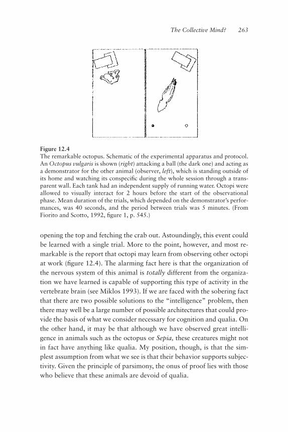
opening the top and fetching the crab out. Astoundingly, this event couldbe learned with a single trial. More to the point, however, and most re-markable is the report that octopi may learn from observing other octopiat work (ªgure 12.4). The alarming fact here is that the organization ofthe nervous system of this animal is totally different from the organiza-tion we have learned is capable of supporting this type of activity in thevertebrate brain (see Miklos 1993). If we are faced with the sobering factthat there are two possible solutions to the “intelligence” problem, thenthere may well be a large number of possible architectures that could pro-vide the basis of what we consider necessary for cognition and qualia. Onthe other hand, it may be that although we have observed great intelli-gence in animals such as the octopus or Sepia, these creatures might notin fact have anything like qualia. My position, though, is that the sim-plest assumption from what we see is that their behavior supports subjec-tivity. Given the principle of parsimony, the onus of proof lies with thosewho believe that these animals are devoid of qualia.
The Collective Mind? 263
Figure 12.4The remarkable octopus. Schematic of the experimental apparatus and protocol.An Octopus vulgaris is shown (right) attacking a ball (the dark one) and acting asa demonstrator for the other animal (observer, left), which is standing outside ofits home and watching its conspeciªc during the whole session through a trans-parent wall. Each tank had an independent supply of running water. Octopi wereallowed to visually interact for 2 hours before the start of the observationalphase. Mean duration of the trials, which depended on the demonstrator’s perfor-mances, was 40 seconds, and the period between trials was 5 minutes. (FromFiorito and Scotto, 1992, ªgure 1, p. 545.)

But is there something in principle quite different from the types of em-bodiments that we have in modern day computers and in the nervous sys-tem itself? That is a very serious and important question to ask. One mayconsider, as did Alan Turing (Turing 1947; Millican and Clark 1996),whether it is in principle possible to make a universal machine out of adigital type of device if the appropriate algorithms are implemented. Canalgorithmic computation ever be sufªciently extensive, fast, and conciseenough to implement the totality of properties that a 14-watt entity suchas our brain can implement with 1.5 kilograms of mass? And what do wemake of the intelligence of an ant that as a robot demonstrates incrediblecomputational agility with mere milligrams of neuronal mass, a brainwith less mass than a single microchip? The fundamental issue is thatbrains are nothing like digital computers; they operate as analog devicesand thus utilize physics directly in their measurements, as opposed to theabstracted measures of zeros and ones that are cleansed of the ele-ments that generated them. Is the computation of digital physical com-puters truly comparable to that performed by analog devices? It has beenstated that for a digital computer to be able to support the equivalentcomputational properties (capabilities) of the brain, the mass requiredmight be many orders of magnitude larger and the power supply equallyas large.There is another argument to consider in terms of the differences be-
tween brains and computers. Warren McCulloch wondered long agohow it was that reliability could arise from nonreliable systems (McCul-loch 1965). The reader should know by now how unreliable nerve cellsare as computational entities. First of all, they have intrinsic activity, andthus as conveyors and relayers of information may be extremely noisy.McCulloch’s answer was rather intriguing: he felt that reliability could beattained if neurons were organized in parallel so that the ultimate mes-sage was the sum of activity of the neurons acting simultaneously. He fur-ther explained that a system where the elements were unreliable to thepoint that their unreliabilities were sufªciently different from one an-other would in principle be far more reliable than a system made out oftotally reliable parts. Here, a reliable system is one with unreliability ineach element as low as possible but still present.This may sound almost paradoxical, but in what is considered a reli-
able system, the elements are reliable to about the same extent. And even
264 Chapter 12

if this reliability is 99.99 percent, the problem is that the elements arealso all the same in their unreliability, meaning that what is unreliable iscommon to all the elements. It therefore becomes an issue of probabili-ties. In such reliable or redundant systems then, whatever tiny problem orunreliability they do have will add up. In nonreliable systems, however,the elements are not redundant and are therefore slightly different in theirunreliability. Because they are all slightly different in their unreliability,there will never be the possibility of this unreliability adding up! Theseunreliable systems are therefore far more reliable than reliable systems.The ºip side of this is that in a system with elements of differing unrelia-bilities, what they have in common are the reliable aspects! This is funda-mental. It means that for an instrument to be totally reliable it mustultimately be made up of unreliable—varied—parts!Herein lies precisely the fragility of society we may experience from
the homogenizing effect of the Web on our thoughts, ideas, beliefs,and the like. As variation decreases, things become increasingly redun-dant and unreliability becomes the prevailing commonality across theelements—and in the case of society, we are the elements.Returning to the implementation of consciousness and so-called
artiªcial (nonbiological) intelligence, it is possible that until we can un-derstand the issue of unreliability and the probabilistic nature of compu-tation in analog systems, we will not be able to generate the requiredarchitecture. With the proper functional architecture we could probablygenerate consciousness in a very large set of nonbiological entities.The second issue is one of knowledge of self. Suppose a potential em-
bodiment of consciousness is allowed the necessary freedom to exploreand internalize the external world such that an image of self, howeverprimitive it may be, is implemented. While this embodiment may meas-ure external reality, the possibility of having an entity that is aware in thesense we mean most probably will not ever arise. We know that this isfundamental in the functioning of the nervous system. It can be seen inindividuals who are given inverted prisms that make the world appear vi-sually upside down. These individuals will learn to use the visual imageas if right side-up only if allowed to interact in a motor sense with thatimage. They must move within it to adjust. Ultimately we see that the ar-chitecture capable of generating cognition must relate to the motricityupon which such cognition was developed. To be conscious, computers
The Collective Mind? 265

must move and manipulate—they must be robots. Without such self-reference, the issue of syntax versus semantics will always come up (seethe Chinese room paradigm; Searle 1992) as consciousness is ultimatelysimply context-dependent.When the architectures for generating cognition are ªnally realized, we
may have thinking/feeling machines. However, our ability to design andbuild them may not ultimately be that useful in understanding brainfunction, in the same way that understanding airplanes may not tell us allabout how the physiology of bats or birds enables them to ºy.
266 Chapter 12

References
Abbott, L. F., Rolls, E. T., and Tovee, M. J. (1996). Representational capacity offace coding in monkeys. Cereb. Cortex. 6: 498–505.
Abel, T., and Kandel, E. (1998). Positive and negative regulatory mechanismsthat mediate long-term memory storage. Brain Res. Rev. 26: 360–378.
Aboitiz, F., and Garcia, R. (1997a). The anatomy of language revisited. Biol. Res.30: 171–183.
Aboitiz, F., and Garcia, R. (1997b). The evolutionary origin of the language areasin the human brain. A neuroanatomical perspective. Brain Res. Rev. 25: 381–396.
Adrianov, O. S. (1996). Cerebral interrelationships of cognitive and emotionalactivity: Pathways and mechanisms. Neurosci. Behav. Physiol. 26: 329–339.
Ali, M. A., ed. (1984). Photoreception and Vision in Invertebrates. New York:Plenum.
Archer, S. M., Helveston, E. M., Miller, K. K., and Ellis, F. D. (1986). Stereopsisin normal infants and infants with congenital esotropia. Am. J. Ophthalmol. 101:591–596.
Armstrong, D. L., Turin, L., and Warner, A. E. (1983). Muscle activity and theloss of electrical coupling between striated muscle cells in Xenopus embryos.J. Neurosci. 3: 1414–1421.
Arnold, A. P. (1975a). The effects of castration on song development in zebraªnches (Poephila guttata). J. Exp. Zool. 191: 261–278.
Arnold, A. P. (1975b). The effects of castration and androgen replacement onsong, courtship, and aggression in zebra ªnches (Poephila guttata). J. Exp. Zool.191:309–326.
Arshavsky, Y. I., Deliagina, T. G., and Orlovsky, G. N. (1997). Pattern genera-tion. Curr. Opin. Neurobiol. 7: 781–789.
Bailey, C. H., Bartsch, D., and Kandel, E. R. (1996). Toward a moleculardeªnition of long-term memory storage. Proc. Natl. Acad. Sci. USA. 93: 13445–13452.
Baker, T. C., Cosse, A. A., and Todd, J. L. (1998). Behavioral antagonism in themoth Helicoverpa zea in response to pheromone blends of three sympatricheliothine moth species is explained by one type of antennal neuron. Ann. NYAcad. Sci. 855: 511–513.
Banks, W. P. (1996). How much work can a quale do? Conscious Cogn. 5: 368–380.
Bard P. (1928). A diencephalic mechanism for the expression of rage with specialreference to the sympathetic nervous system. Amer. J. Physiol. 84: 490–515.
Bateson, P. P. G. (1966). The characteristics and context of imprinting. BiologicalReview 41: 177–220.
Batueva, I. V. (1987). Efªciency of electrical transmission in reticulo-motoneuronal synapses of lamprey spinal cord. Exp. Brain Res. 69: 131–139.
Batueva, I. V., and Shapovalov, A. I. (1977). Electrotonic and chemical EPSPs inlamprey motor neurons following stimulation of the descending tract and poste-rior root afferents. Neiroªziologiia 9: 512–517. [Article in Russian]
Bear, M. F., Conners, B. W., and Paradiso, M. A. (1996). Neuroscience: Ex-ploring the Brain. Baltimore: Williams and Wilkins.
Beeckmans, K., and Michiels, K. (1996). Personality, emotions and thetemporolimbic system: A neuropsychological approach. Acta Neurol. Belg. 96:35–42.
Bekoff, A., Stein, P. S. G., and Hamburger, V. (1975). Co-ordinated motor outputin the hindlimb of the 7-day chick embryo. Proc. Natl. Acad. Sci. USA 72: 1245–1248.
Benini, A. (1998). Pain as a biological phenomenon of consciousness. SchweizRundsch Med. Prax. 87: 224–228.
Benke, T., Bosch, S., and Andree, B. (1998). A study of emotional processing inParkinson’s disease. Brain Cogn. 38: 36–52.
Bennett, M. V. L. (1971). Electric organs. In: Fish Physiology, W. S. Hoar, D. J.Randall, eds. New York: Academic Press, pp. 347–391.
Bennett, M. V. L. (1997). Gap junctions as electrical synapses. J. Neurocytol. 26:349–366.
Bennett, M. V. L. (2000). Electrical synapses, a personal perspective (or history).Brain Res. Brain Res. Rev. 32: 16–28.
Bennett, M. V. L, and Pappas, G. D. (1983). The electromotor system of thestargazer: A model for integrative actions at electrotonic synapses. J. Neurosci. 3:748–761.
268 References
Bennett, M. V. L, Sandri, C., and Akert, K. (1989). Fine structure of the tuberouselectroreceptor of the high-frequency electric ªsh, Sternarchus albifrons(gymnotiformes). J. Neurocytol. 18: 265–283.
Berardelli, A. (1995). Symptomatic or secondary basal ganglia diseases andtardive dyskinesias. Curr. Opin. Neurol. 8: 320–322.
Berardelli, A., Rothwell, J. C., Hallett, M., Thompson, P. D., Manfredi, M., andMarsden, C. D. (1998). The pathophysiology of primary dystonia. Brain 121:1195–1212.
Bernard, J. F., Bester, H., and Besson, J. M. (1996). Involvement of the spino-parabrachio-amygdaloid and -hypothalamic pathways in the autonomic and af-fective emotional aspects of pain. Prog. Brain Res. 107: 243–255.
Bernstein, N. A. (1967). The Coordination and Regulation of Movements. Ox-ford: Pergamon Press.
Bevans, C. G., Kordel, M., Rhee, S. K., Harris, A. L. (1998). Isoform compositionof connexin channels determines selectivity among second messengers and un-charged molecules. J. Biol. Chem. 273: 2808–16.
Bizzi, E., Saltiel, P., and Tresch, M. (1998). Modular organization of motor be-havior. Z. Naturforsch. 53: 510–517.
Blackshaw, S. E., and Warner, A. E. (1976). Low resistance junctions betweenmesoderm cells during development of trunk muscles. J. Physiol. (Lond.) 255:209–230.
Bleasel, A. F., and Pettigrew A. G. (1992). Development and properties of sponta-neous oscillations of the membrane potential in inferior olivary neurons in therat. Brain Res. De. Brain Res. 65: 43–50.
Block, N. (1995). The Mind as the software of the brain. In An Invitation to Cog-nitive Science, ed. D. Osheron, L. Gleitman, S. Kosslyn, E. Smith andS. Steinberg, ed. Cambridge: MIT Press.
Braddick, O. (1996). Binocularity in infancy. Eye. 10: 182–188.
Broca, P. (1861). Memoire sur le cerveau de l’homme. Paris: Reinwald.
Brooks, V. B. (1983) Motor control: How posture and movements are governed.Phys. Ther. 63: 664–673.
Brown, G. (1911). The intrinsic factors in the act of progression in the mammal.Proc. Roy. Soc. Lond. B. 84: 308–319.
Brown, G. (1914). On the nature of the fundamental activity of nervous centersJ. Physiol. 48: 18–46.
Brown, G. (1915). On the activities of the central nervous system; together withan analysis of the conditioning of rhythmic activity in progression and a theory ofthe evolution of the nervous system. J. Physiol. 49: 18–46.
Brown, K. S. (1983). Evolution and development of the dentition. Birth DefectsOrig. Artic. Ser. 19: 29–66.
References 269

Brown, S., and Schafer, A. (1888). An investigation into the functions of the oc-cipital and temporal lobes of the monkey’s brain. Phil. Trans. R. Soc. London B.Biol. Sci. 179: 303–327.
Camperi, M., and Wang, X. J. (1998). A model of visuospatial working memoryin prefrontal cortex: Recurrent network and cellular bistability. J. Comput.Neurosci. 5: 383–405.
Casey, K. L. (1999). Forebrain mechanisms of nociception and pain: Analysisthrough imaging. Proc. Natl. Acad. Sci. USA. 96: 7668–7674.
Chalmers, D. J. (1995). Facing up to the problem of consciousness. J. Conscious-ness Studies 2: 200–219.
Chalmers, D. J. (1996). The Conscious Mind: In Search of a Fundamental The-ory. New York: Oxford University Press.
Chalmers, D. J. (1997). Moving forward on the problem of consciousness.J. Consciousness Studies 4: 3–46.
Chang, Q., Pereda, A., Pinter, M. J., and Balice-Gordon R. J. (2000). Nerve in-jury induces gap junctional coupling among axotomized adult motor neurons.J. Neurosci. 20: 674–684.
Changeux, J. P. (1996) The Neuronal Man. Princeton: Princeton University Press.
Changeux, J. P. and Deheane, S. (2000). Hierarchical modeling of cognitive func-tions: From synaptic transmission to the Tower of London. Int. J. Psychol.Physiol. 35: 179–187.
Charney, D. S., and Deutch, A. (1996). A functional neuroanatomy of anxietyand fear: Implications for the pathophysiology and treatment of anxiety disor-ders. Crit. Rev. Neurobiol. 10: 419–446.
Chelazzi, L., Duncan, J., Miller, E. K., and Desimone, R. (1998). Responses ofneurons in inferior temporal cortex during memory-guided visual search.J. Neurophysiol. 80: 2918–2940.
Chomsky, N. (1964). The development of grammar in child language: Formaldiscussion. Monogr. Soc. Res. Child Dev. 29: 35–39.
Chomsky, N. (1967). Recent contributions to the theory of innate ideas. Synthese17: 2–11.
Chomsky, N. (1968) Language and Mind. New York: Harcourt, Brace andWorld.
Chomsky, N. (1972). Language and Mind. New York: Harcourt, Brace andWorld.
Chomsky, N. (1980). Rules and representations. Behav. and Brain Sci. 3: 1–16.
Chomsky, N. (1986). Analytic study of the Tadoma method: Language abilitiesof three deaf-blind subjects. J. Speech Hear. Res. 29: 332–347.
Christensen, B. N. (1976). Morphological correlates of synaptic transmission inlamprey spinal cord. J. Neurophysiol. 39: 197–212.
270 References

Churchland, P. M. and Churchland, P. S. (1998). Recent work on consciousness:Philosophical, theoretical, and empirical. In On the Contrary: Critical Essays,1987–1997. Cambridge: MIT Press.
Churchland, P. S. (1986). Neurophilosophy: Toward a Uniªed Understanding ofthe Mind-Brain. Cambridge: MIT Press.
Clayton, D. F. (1997). Role of gene regulation in song circuit development andsong learning. J. Neurobiol. 33: 549–571.
Cloney, R. A. (1982). Ascidian larvae and the events of metamorphosis. Am.Zool. 22: 817–826.
Coffey, B. J., Miguel, E. C., Savage, C. R., Rauch, S. L. (1994). Tourette’s disor-der and related problems: A review and update. Harv. Rev. Psychiatry. 2: 121–132.
Cohen, A. (1987). Effects of oscillator frequency on phase-locking in the lampreycentral pattern generator. J. Neurosci. Methods. 21: 113–125.
Colcher, A., and Simuni, T. (1999). Clinical manifestations of Parkinson’s dis-ease. Med. Clin. North Am. 83: 327–347.
Cole, J. A., Mohan, S., and Dow, C. (eds.) (1992). Prokaryotic Structure andFunction: A New Perspective. Society for General Microbiology Symposium,no. 47. Cambridge: Cambridge University Press.
Cook, P. M., Prusky, G., and Ramoa, A. S. (1999). The role of spontaneous reti-nal activity before eye opening in the maturation of form and function in theretinogeniculate pathway of the ferret. Vis. Neurosci. 16: 491–501.
Cope, F. W. (1976). Derivation of the Weber-Fechner law and the Loewensteinequation as the steady-state response of an Elovich solid state biological system.Bull. Math. Biol. 38: 111–118.
Crick, F. C. H. (1991). The Astonishing Hypothesis: The Scientiªc Search for theSoul. New York: Touchstone Books.
Crick, F. C. H., and Koch C. (1990). Towards a neurological theory of conscious-ness. Semin. Neurosci. 2: 263–275.
Crook, J. H. (1983). On attributing consciousness to animals. Nature 303: 11–14.
Cropper, E. C., and Weiss, K. R. (1996). Synaptic mechanisms in invertebratepattern generation. Curr. Opin. Neurobiol. 6: 833–841.
Damasio, A. (1994). Descartes’ Error. New York: Putnam.
Damasio, A. (1999). The Feeling of What Happens. New York: Harcourt Braceand Co.
Damasio, A. R., Damasio, H., and Van Hoesen, G. W. (1982). Prosopagnosia:Anatomic basis and behavioral mechanisms. Neurology. 32: 331–341.
D’arcy W. Thompson. (1992).On growth and form. Cambridge, UK: CambridgeUniversity Press.
References 271

Darwin, C. (1872). The Expression of Emotions in Man and Animals. London:John Murray, Publisher.
Davis, L. (1982). What is it like to be an agent? Erkenntnis 18: 195–213.
Davis, M. (1998). Anatomic and physiologic substrates of emotion in an animalmodel. J. Clin. Neurophysiol. 15: 378–387.
Davis, P. J., Zhang, S. P., Winkworth, A., and Bandler, R. (1996). Neural controlof vocalization: Respiratory and emotional inºuences. J. Voice. 10: 23–38.
DeHaan, R. L., and Sachs, H. G. (1972). Cell coupling in developing systems:The heart-cell paradigm. Curr. Top. Dev. Biol. 7: 193–228.
Deiters O. (1865). In M. Schultze, ed. Unter schungen über Gehim undRückenmark des Menschen und der Sängethiere. Braunschweig Wieweg.
Deliagina, T. G., Orlovsky, G. N., and Pavlova, G. A. (1983). The capacity forgeneration of rhythmic oscillations is distributed in the lumbosacral spinal cordof the cat. Exp. Brain Res. 53: 81–90.
Dennet, D. C. (1993). Consciousness Explained. New York: Penguin.
De Renzi E. de Pellegrino G. (1998). Prosopagnosia and alexia without objectagnosia. Cortex 34: 403–15.
Devinsky, O., Morrell, M. J., and Vogt, B. A. (1995). Contributions of anteriorcingulate cortex to behaviour. Brain. 118: 279–306.
DeVoogd, T. J. (1991). Endocrine modulation of the development and adult func-tion of the avian song system. Psychoneuroendocrinology.16: 41–66.
DeVoogd, T. J., and Nottebohm, F. (1981). Sex differences in dendritic morphol-ogy of a song control nucleus in the canary: A quantitative Golgi study. J. Comp.Neurol. 196: 309–316.
Dewsbury, D. A., and Rethlingshafer, D. A. (1973). Comparative psychology: Amodern survey. New York: McGraw-Hill, pp. 125–127.
De Zeeuw, C. I., Lang, E. J., Sugihara, I., Ruigrok, T. J., Eisenman, L. M.,Mugnaini, E., and Llinás, R. (1996). Morphological correlates of bilateral syn-chrony in the rat cerebellar cortex. Neurosci. 16: 3412–3426.
Doty, R. L. (1986). Odor-guided behavior in mammals. Experientia. 42: 257–271.
Doupe, A. J. (1993). A neural circuit specialized for vocal learning. Curr. Opin.Neurobiol. 3: 104–111.
Doupe, A. J., and Konishi, M. (1991). Song-selective auditory circuits in the vo-cal control system of the zebra ªnch. Proc. Natl. Acad. Sci. USA 88: 11339–11343.
Doupe, A. J., and Kuhl, P. K. (1999). Birdsong and human speech: Commonthemes and mechanisms. Annu. Rev. Neurosci. 22: 567–631.
272 References
Downer J. C. D. (1961). Changes in the visual gnostic function and emotional be-havior following unilateral temporal lobe damage in the “split-brain” monkey.Nature 191: 50–51.
Duchesne de Boulogne, G.-B. (1862). [Mecanisme de la Physionomie Humaine].The Mechanism of Human Facial Expression. Edited and translated by R. A.Cuthbertson, 1990. Cambridge UK: Cambridge University Press.
Dyer, A. B., and Gottlieb, G. (1990). Auditory basis of maternal attachment inducklings (Anas platyrhynchos) under simulated naturalistic imprinting condi-tions. J. Comp. Psychol. 104: 190–194.
Dyer, A. B., Lickliter, R., and Gottlieb, G. (1989). Maternal and peer imprintingin mallard ducklings under experimentally simulated natural social conditions.Dev. Psychobiol. 22: 463–475.
Eccles, J. C., Llinás, R., and Sasaki, K. (1966). The excitatory synaptic action ofclimbing ªbres on the Purkinje cells of the cerebellum. J. Physiol. 182: 268–296.
Eckhorn, R., Bauer, R., Jordan, W., Brosch, M., Kruse, W., Munk, M., andReitbock, H. J. (1988). Coherent oscillations: A mechanism of feature linking inthe visual cortex? Biol. Cybern. 60: 121–130.
Edelman, G. M. (1992). Bright Air, Brilliant Fire: On the Matter of the Mind.New York: Basic Books.
Edelman, G. M. (1993). Neural Darwinism: Selection and reentrant signaling inhigher brain function. Neuron. 10: 115–125.
Eglen, S. J. (1999). The role of retinal waves and synaptic normalization inretinogeniculate development. Philos. Trans. R. Soc. Lond. B Biol. Sci. 354: 497–506.
Elsen F. P., and Ramirez, J. M. (1998). Calcium currents of rhythmic neurons re-corded in the isolated respiratory network of neonatal mice. J. Neurosci. 18:10652–10662
Erecinska, M., and Silver, I. A. (1994). Ions and energy in the mammalian brain.Prog. Neurobiol. 43: 37–71.
Estevez-Gonzalez, A., Garcia-Sanchez, C., and Barraquer-Bordas, L. (1997).Memory and learning: “experience” and “skill” of the brain. Rev. Neurol. 25:1976–1988. [Article in Spanish]
Fatt, P., and Katz, B. (1952). Spontaneous subthreshold activity at motor nerveendings. J. Physiol. (Lond.) 117: 109–128.
Feinberg, T. E. (1997). The irreducible perspectives of consciousness. Semin.Neurol. 17: 85–93.
Feldman, J. L., Smith, J. C., Ellenberger H. H., Connelly C. A., Liu G., Greer J. J.,Lindsay, A. D., and Otto, M. R. (1990). Neurogenesis of respiratory rhythm andpattern-emerging concepts. Am. J. Physiol. 259: 879–886.
References 273

Ferguson, G., Messenger, J., and Budelmann, B. (1994). Gravity and lightinºuence the countershading reºexes of the cuttleªsh Sepia ofªcinalis. J. Exp.Biol. 191: 247–256.
Fernandez de Molina, A. (1991). El cambio cerebral en la emoción. Anales RealAcad. Nac. Medicina Madrid, 1–109.
Fernandez de Molina, A., and Husperger R. W. (1959). Central representation ofaffective reactions in forebrain and brainstem: Electrical stimulation ofamygdala, stria terminalis, and adjacent structures. J. Physiol. 145: 251–265.
Fernandez de Molina, A., and Husperger R. W. (1962). Organization of thesubcortical system governing defence and ºight reactions in the cat. J. Physiol.160: 200–213.
Feynman R. P., and Hibbs, A. R. (1965). Quantum mechanics and path integralsNew York: McGraw-Hill.
Fiorito, G., Scotto, P. (1992). Observational learning in Octopus vulgaris.Science. 256: 545–547.
Furshpan, E. J., and Potter, D. D. (1959). Transmission of giant motor synapsesof the cray ªsh. J. Physiol. 145: 289–325.
Fuster, J. M. (1998). Distributed memory for both short and long term.Neurobiol. Learn. Mem. 70: 268–274.
Fuster, J. M., and Uyeda, A. A. (1971). Reactivity of limbic neurons of the mon-key to appetitive and aversive signals. EEG Clin. Neurophys. 30: 281–293.
Gahr, M., and Garcia-Segura, L. M. (1996). Testosterone-dependent increase ofgap-junctions in HVC neurons of adult female canaries. Brain Res. 712: 69–73.
Galambos, R., Makeig, S., and Talmachoff, P. J. (1981). A 40-Hz auditory poten-tial recorded from the human scalp. Proc. Natl. Acad. Sci. 78: 2643–2547.
Ganger, J., and Stromswold, K. (1998). Innateness, evolution, and genetics oflanguage. Hum. Biol. 70: 199–213.
Gannon, P. J., Holloway, R. L., Broadªeld, D. C., and Braun, A. R. (1998).Asymmetry of chimpanzee planum temporale: Humanlike pattern of Wernicke’sbrain language area homolog. Science. 279: 220–222.
Gerhardstein, P., Adler, S. A., and Rovee-Collier, C. (2000). A dissociation in in-fants’ memory for stimulus size: Evidence for the early development of multiplememory systems. Dev. Psychobiol. 36: 123–135.
Geschwind, N. (1965). The disconnexion syndrome in animals and man. Brain88: 237–294.
Glassman, R. B. (1999). A working memory “theory of relativity”: Elasticity intemporal, spatial, and modality dimensions conserves item capacity in radialmaze, verbal tasks, and other cognition. Brain Res. Bull. 48: 475–489.
Goldman-Rakic, P. S. (1987). Circuitry of primate prefrontal cortex and regula-tion of behavior by representational memory. In F. Plum, V. B. Mountcastle (eds).
274 References

Handbook of Physiology. Sect. 1, The Nervous System. Vol. 5, Higher Functionsof the Brain, Part 1, pp. 373–417. Bethesda, MD: Am. Physiol. Society.
Goldman-Rakic, P. S. (1992). Working memory and the mind. Sci. Am. 267:111–117.
Goldman-Rakic, P. S. (1996). Memory: Recording experience in cells and cir-cuits: Diversity in memory research. Proc. Natl. Acad. Sci. USA 93: 13435–13437.
Goldman-Rakic, P. S., Funahashi, S., and Bruce, C. J. (1990). Neocortical mem-ory circuits. Cold Spring Harb. Symp. Quant. Biol. 55: 1025–1038.
Goller, F., and Larsen, O. N. (1997a). A new mechanism of sound generation insongbirds. Proc. Natl. Acad. Sci. USA 94: 14787–14791.
Goller, F., and Larsen, O. N. (1997b). In situ biomechanics of the syrinx andsound generation in pigeons. J. Exp. Biol. 200: 2165–2176.
Goller, F., and Suthers, R. A. (1996a). Role of syringeal muscles in controlling thephonology of bird song. J. Neurophysiol. 76: 287–300.
Goller, F., and Suthers, R. A. (1996b). Role of syringeal muscles in gating airºowand sound production in singing brown thrashers. J. Neurophysiol. 75: 867–876.
Goodman, D., and Kelso, J. A. (1983). Exploring the functional signiªcance ofphysiological tremor: A biospectroscopic approach. Exp. Brain Res. 49: 419–431.
Gordon, N. Speech, language, and the cerebellum. 1996. Eur. J. Disord.Commun. 31: 359–367.
Gould, E., Reeves, A. J., Graziano, M. S. A., and Gross, C. G. (1999).Neurogenesis in the neocortex of adult primates. Science 286: 548–552.
Gould, J. L. (1976). The Dance-language controversy. The quarterly review of bi-ology. Q. Rev. Biol. 51: 211–244.
Gould, J. L. (1990). Honey bee cognition. Cognition. 37: 83–103.
Gould, J. L., and Gould, C. G. (1989). Life at the Edge. Readings from ScientiªcAmerican Magazine. New York: W.H. Freeman and Co.
Gould, S. J. (199?) Lecture entitled: Unity of Organic Design: From Goethe andGeoffroy Chaucer to Homology of Homeotic Complexes in Arthropods and Ver-tebrates, presented at New York University in honor of Homer Smith.
Gray, C. M., Konig, P. L., Engel, A. K., and Singer, W. (1989). Oscillatory re-sponses in cat visual cortex exhibit inter-columnar synchronization which reºectsglobal stimulus properties. Nature 338: 334–337.
Gray, C. M., and Singer, W. (1989). Stimulus-speciªc neuronal oscillations in ori-entation columns of cat visual cortex. Proc. Nat. Acad. Sci. USA. 86: 1698–1702.
Graybiel, A. M. (1995). Building action repertories: Memory and learning func-tions of the basal ganglion. Curr. Opin. Neurobiol. 5: 733–741.
References 275

Greene, P. H. (1972). Problems of organization of motor systems. In R. Rosenand F. M. Snell (eds.) Progress in Theoretical Biology, Vol. 2. New York: Aca-demic, pp. 303–338.
Greene, P. H. (1982). Why is it easy to control your arms? J. Motor Behav. 14:260–286.
Greenªeld, P. M., and Savage-Rumbaugh, E. S. (1993). Comparing communica-tive competence in child and chimp: The pragmatics of repetition. J. Child. Lang.20: 1–26.
Greenªeld, S. A. (1995). Journey to the Centers of the Mind. New York: W. H.Freeman.
Gregory, R. L. (1988). Questions of quanta and qualia: Does sensation makesense of matter—or does matter make sense of sensation? Part 1. Perception 17:699–702.
Gregory, R. L. (1989). Questions of quanta and qualia: Does sensation makesense of matter—or does matter make sense of sensation? Part 2. Perception. 18:1–4.
Grillner, S., and Matsushima, T. (1991). The neural network underlying locomo-tion in lamprey synaptic and cellular mechanisms. Neuron 7: 1–15.
Gross, C. G., and Sergent, J. (1992). Face recognition. Curr. Opin. Neurobiol. 2:156–161.
Hadders-Algra, M., Brogren, E., and Forssberg, H. (1997). Nature and nurture inthe development of postural control in human infants. Acta. Paediatr. Suppl. 422:48–53.
Hamburger, V., and Balaban, M. (1963). Observations and experiments on spon-taneous rhythmical behavior in the chick embryo. Dev. Biol. 7: 533–545.
Hammer, M., and Menzel, R. (1995). Learning and memory in the honeybee.J. Neurosci. 15: 1617–1630.
Harris, D. F. (1894). The time-relations of the voluntary tetanns in man.J. Physiol. (Lond.) 17: 315–330.
Harris, J. E., and Whiting, H. P. W. (1954a). Structure and function in thelocomotory system of the dogªsh embryo. The myogenic stage of movement.J. Physiol. 501–524.
Harris, J. E., and Whiting, H. P. W. (1954b). Structure and functional feedback inthe control of movement. Trends Neurosc. 7: 253–257.
Hartshorn, K., Rovee-Collier, C., Gerhardstein, P., Bhatt, R. S., Klein, P. J.,Aaron, F., Wondoloski, T. L., and Wurtzel, N. (1998). Developmental changes inthe speciªcity of memory over the ªrst year of life. Dev. Psychobiol. 33: 61–78.
Hayhoe, M. M., Bensinger, D. G., and Ballard, D. H. (1998). Task constraints invisual working memory. Vision Res. 38: 125–137.
276 References

Hayman, L. A., Rexer, J. L., Pavol, M. A., Strite, D., and Meyers, C. A. (1998).Kluver-Bucy syndrome after bilateral selective damage of amygdala and its corti-cal connections. J. Neuropsychiatry Clin. Neurosci. 10: 354–358.
Heaton, J. T., Dooling, R. J., and Farabaugh, S. M. (1999). Effects of deafeningon the calls and warble song of adult budgerigars (Melopsittacus undulatus).J. Acoust. Soc. Am. 105: 2010–2019.
Hebb, D. O. (1953). Heredity and environment in mammalian behavior. Brit.J. Animal Behav. 1: 43–47.
Heilman, K. M., and Gilmore, R. L. (1998). Cortical inºuences in emotion.J. Clin. Neurophysiol. 15: 409–423.
Hess, E. H. (1972). The natural history of imprinting. Ann. NY Acad. Sci. 193:124–136.
Hess, R. W. (1957). The Functional Organization of the Diencephalon. NewYork: Grune and Stratton.
Hess R. W., and Rugger, M. (1943). Das subkortikale Zentrum der affektivenAbwehr-reaktion. Helv. Physiol. Acta. 1: 33–52.
Hikosaka, O. (1998). Neural systems for control of voluntary action—a hypoth-esis. Adv. Biophys. 35: 81–102.
Hildebrand, J. G. (1995). Analysis of chemical signals by nervous systems. Proc.Natl. Acad. Sci. USA 92: 67–74.
Hille, B. (1992). Ionic Channels of Excitable Membranes, 2d ed. New York:Sinauer Associates.
Hirose, H., and Gay, T. (1973). Laryngeal control in vocal attack. Anelectromyographic study. Folia Phoniatr (Basel) 25: 203–213.
Honda, K., and Kusakawa, N. (1997). Compatibility between auditory andarticulatory representations of vowels. Acta. Otolaryngol. Suppl. (Stockh.) 532:103–105.
Hubbard, T. L. (1996). The importance of a consideration of qualia to imageryand cognition. Conscious Cogn. 5: 327–358.
Hubel, D. H. (1988). Eye, Brain, and Vision. Scientiªc American Library Series,#22. New York: Freeman and Company.
Hubel, D. H., and Wiesel, T. N. (1963). Receptive ªelds of cells in striate cortexof very young, visually inexperienced kittens. J. Neurophysiol. 26: 994–1002.
Hubel, D. H., and Wiesel, T. N. (1974). Sequence regularity and geometry of ori-entation columns in the monkey striate cortex. J. Comp. Neurol. 158: 267–294.
Hubel, D. H., andWiesel, T. N. (1977). Ferrier lecture: Functional architecture ofmacaque monkey visual cortex. Proc. Roy. Soc. Serv. B. 198: 1–59.
Hubel, D. H., and Wiesel, T. N. (1979). Brain mechanisms of vision. Sci. Am.241: 150–162.
References 277
Hubel, D. H., Wiesel, T. N., and LeVay, S. (1976). Functional architecture of area17 in normal and monocularly deprived macaque monkeys. Cold Spring Harb.Symp. Quant. Biol. 40: 581–589.
Hunsperger R. W. (1956). Role of substantia gricea centralis mesencephali inelectrically induced rage reactions. Folia Psychiat. (Amst.). 19: 289–294.
Hutcheon, B., and Yarom, Y. (2000). Resonance, oscillation and intrinsic fre-quency preferences of neurons. TINS 23: 216–222.
Huxley A. (1980). Reºections on muscle, Princeton University Press, Princeton.
Ito, M. (1984). The Cerebellum and Neural Control. New York: Raven Press.
Iverson, P. and Kuhl, P. K. (1996). Inºuences of phonetic identiªcation and cate-gory goodness on American listeners’ perception of /r/ and /l/. J. Acoust. Soc. Am.99: 1130–1140.
Iyengar, S., Viswanathan, S. S., and Bottjer, S. W. (1999). Development of topog-raphy within song control circuitry of zebra ªnches during the sensitive periodfor song learning. J. Neurosci. 19: 6037–6057.
James, W. (1950 [1890]). Principles of Psychology. New York: Dover.
Jankowska, E., and Edgley, S. (1993). Interactions between pathways controllingposture and gait at the level of spinal interneurons in the cat. Prog. Brain Res. 97:161–171.
Jeannerod, M. (1986). Mechanisms of visuomotor coordination: A study in nor-mal and brain-damaged subjects. Neuropsychologia. 24: 41–78.
Johnson, F., and Bottjer, S. W. (1993). Hormone-induced changes in identiªedcell populations of the higher vocal center in male canaries. J. Neurobiol. 24:400–418.
Joliot, M., Ribary, U., and Llinás, R. (1994). Human oscillatory brain activitynear 40 Hz coexists with cognitive temporal binding. Proc. Natl. Acad. Sci. USA91: 11748–11751.
Jurgens, U. (1998). Neuronal control of mammalian vocalization, with specialreference to the squirrel monkey. Naturwissenschaften. 85: 376–388.
Jurgens, U., and Zwirner, P. (1996). The role of the periaqueductal grey in limbicand neocortical vocal fold control. Neuroreport. 7: 2921–2923.
Jusczyk, P. W., and Bertoncini, J. (1988). Viewing the development of speech per-ception as an innately guided learning process. Lang. Speech. 31: 217–238.
Kahn, J. A., Roberts, A., and Kashin, S. M. (1982). The neuromuscular basis ofswimming movements in embryos of the amphibian Xenopus laevis. J. Exp. Biol.99: 175–184.
Kam, Y., Kim, D. Y., Koo, S. K., and Joe, C. O. (1998). Transfer of second mes-sengers through gap junction connexin 43 channels reconstituted in liposomes.Biochim. Biophys. Acta. 1372: 384–388
Kandel, E. R., Schwartz, J. H., and Jessell, T. M. (eds.) (2000). Principles of Neu-ral Science, 4th ed. New York: McGraw-Hill.
278 References

Kandler, K., and Katz, L. C. (1995). Neuronal coupling and uncoupling in the de-veloping nervous system. Curr. Opin. Neurobiol. 5: 98–105.
Kant, I. (1781). Critique of Pure Reason. J. M. Meiklejohn (translator); NormanK. Smith, ed. St. Martin’s Press, Inc. 1990
Kay, R. F., Cartmill, M., Balow, M. (1998). They hypoglossal canal and the originof human vocal behavior. Proc Natl Acad Sci USA. 95: 5417–5419.
Kirk, D. L. (1998). Volvox: Molecular-Genetic Origins of Multicellularity andCellular Differentiation. (Development and cell biology series). Cambridge: Cam-bridge University Press.
Kirzinger, A., and Jurgens, U. (1991). Vocalization-correlated single-unit activityin the brain stem of the squirrel monkey. Exp. Brain Res. 84: 545–560.
Kling, J. W., and Stevenson-Hinde, J. (1977). Development of song and reinforc-ing effects of song in female chafªnches. Anim. Behav. 25: 215–220.
Klüver, H., and Bucy, P. (1939). Preliminary analysis of functions of the temporallobes in monkeys. Arch. Neurol. Psychiat. (Chic.). 42: 979–1000.
Konishi, M. (1989). Birdsong for neurobiologists. Neuron 3: 541–549.
Kretsinger R. H. (1997). EF-hands embrace. Nat. Struct. Biol. 4: 514–516.
Kretsinger, R. H. (1996). EF-hands reach out. Nat. Struct. Biol. 3: 12–15.
Krishtalka, L., Stucky, R. K., and Beard, K. C. (1990). The earliest fossil evidencefor sexual dimorphism in primates. Proc. Natl. Acad. Sci. USA. 87: 5223–5226.
Kristofferson, A. B. (1984). Quantal and deterministic timing in human durationdiscrimination. Ann. NY Acad. Sci. 423: 3–15.
Kropotov, J. D., and Etlinger, S. C. (1999). Selection of actions in the basal gan-glia-thalamocortical circuits: Review and model. Int. J. Psychophysiol. 31: 197–217.
Kuhl, P. K., Andruski, J. E., Christovich, I. A., Chistovich, L. A., Kozhevnikova,E. V., Ryskina, V. L., Stolyarova, E. I., Sundberg, U., Lacerda, F. (1997). Cross-language analysis of phonetic units in language addressed to infants. Science.277: 684–686.
Kuhl, P. K. (2000) Language, mind, and brain: Experience alters perception. InM. S. Gazzaniga (ed.), The New Cognitive Neurosciences, 2nd edition, pp. 99–115. Cambridge, MA: MIT Press.
Kuroda, R., Yorimae, A., Yamada, Y., Furuta, Y., and Kim, A. (1995). Frontalcingulotomy reconsidered from a WGA-HRP and c-Fos study in cat. Acta.Neurochir. Suppl. (Wien). 64: 69–73.
Kutukca, Y., Marks, W. J. Jr, Goodin, D. S., and Aminoff, M. J. (1998). Cerebralaccompaniments to simple and choice reaction tasks in Parkinson’s disease. BrainRes. 799: 1–5.
LaBerge, S. and Rheingold, H. (1990). Exploring the World of Lucid Dreaming.New York: Ballantine.
References 279

Lamarre, Y., Montigny, C. de, Dumont, M., andWeiss, M. (1971). Harmaline-in-duced rhythmic activity of cerebellar and lower brain stem neurons. Brain Res.32: 246–250.
Land, M. F. (1978). Animal eyes with mirror optics. Sci. Am. 239: 126–135.
Land, M. F. (1980). Compound eyes: Old and new optical mechanisms. Nature.287: 681–686.
Land, M. F., and Fernald, R. D. (1992). The evolution of eyes. Annu. Rev.Neurosc. 15: 1–29.
Lang, E. J., Sugihara, I., and Llinás, R. (1996). GABAergic modulation of com-plex spike activity by the cerebellar nucleoolivary pathway in rat.J. Neurophysiol. 76: 255–275.
Lansner, A., Kotaleski, J. H., and Grillner, S. (1998). Modeling of the spinalneuronal circuitry underlying locomotion in a lower vertebrate. Ann. NY Acad.Sci. 860: 239–49.
Larson, C. R. (1985). The midbrain periaqueductal gray: A brainstem structureinvolved in vocalization. J. Speech Hear. Res. 28: 241–249.
Larson, C. R., Kistler, M. K. (1984). Periaqueductal gray neuronal activity asso-ciated with laryngeal EMG and vocalization of the awake monkey. NeurosciLett. 46: 261–266.
Larson, C. R., and Kistler, M. K. (1986). The relationship of periaqueductal grayneurons to vocalization and laryngeal EMG in the behaving monkey. Exp. BrainRes. 63: 596–606.
Laurent, G. (1996). Dynamical representation of odors by oscillating and evolv-ing neural assemblies. Trends Neurosci. 19: 489–496.
LeDoux, J. (1996) The Emotional Brain. New York: Simon and Schuster.
LeDoux, J. (1998). Fear and the brain: Where have we been, and where are wegoing? Biol. Psychiatry. 44: 1229–1238.
Leeds, S. (1993). Qualia, awareness, sellars. Nous. 27: 303–330.
Lehky S. R., and Sejnowski T. J. (1999). Seeing white: Qualia in the context ofdecoding population codes. Neural Comput. 11: 1261–1280.
Lengeler, J. W., Drews, G., and Schlegel, H. G. (eds.). (1999) Biology of theProkaryotes. Boston: Blackwell Science Inc.
Llinás, R. (1974). La forme et la fonction des cellules nerveuses. La Recherche. 5:232–240.
Llinás, R. (1981). Microphysiology of the cerebellum. In: Handbook of Physiol-ogy, vol. II, The Nervous System, part II, ed. V. B. Brooks. Bethesda, MD: Ameri-can Physiology Society, pp. 831–976.
Llinás, R. (1987). “Mindness” as a functional state of the brain. In:Mind Waves,ed. C. Blakemore, S. A. Greenªeld. Oxford: Basil Blackwell, pp. 339–358.
280 References

Llinás, R. (1988). The intrinsic electrophysiological properties of mammalianneurons: Insights into central nervous system function. Science 242: 1654–1664.
Llinás R. (1990). Intrinsic electrical properties of mammalian neurons and CNSfunction. In Fidia Research Foundation Neuroscience Award Lectures, Vol. 4.New York: Raven Press, pp. 175–194.
Llinás, R. (1991). The noncontinuous nature of movement execution. In: Motorcontrol: Concepts and Issues, ed. D. R. Humphrey, H. J. Freund. New York: JohnWiley & Sons, pp. 223–242.
Llinás, R., Grace, A. A., and Yarom, Y. (1991). In vitro neurons in mammaliancortical layer 4 exhibit intrinsic oscillatory activity in the 10-Hz to 50-Hz fre-quency range. Proc. Natl. Acad. Sci. USA 88: 897–901.
Llinás, R., and Pare, D. (1991). Of dreaming and wakefulness. Neuroscience 44:521–535.
Llinás, R., and Ribary, U. (1993). Coherent 40-Hz oscillation characterizesdream state in humans. Proc. Natl. Acad. Sci. USA 90: 2078–2081.
Llinás, R., Ribary, U., and Tallal P. (1998). Dyschronic language-based learningdisability. In Basic Mechanisms in Cognition and Language, ed. Von Euler et al.New York: Oxford.
Llinás, R., Ribary, U., Contreras, D., and Pedroarena, C. (1998). The neuronalbasis for consciousness. Phil. Trans. R. Soc. Lond. B 353: 1841–1849.
Llinás, R. R., and Simpson, J. I. (1981). Cerebellar control of movement. In Mo-tor Coordination, vol. 5. Handbook of Behavioral Neurology, ed. A. L. Toweand E. S. Luschel. New York: Plenum Press, pp. 231–302.
Llinás R., and Volkind, R. A. (1973). The olivo-cerebellar system: Functionalproperties as revealed by harmaline-induced tremor. Exp. Brain Res. 18: 69–87.
Llinás R., Walton, K., Hillman, D. E., and Sotelo, C. (1975). Inferior olive: Itsrole in motor learning. Science 190: 1230–1231.
Llinás, R. and Welsh, J. P. (1993). On the cerebellum and motor learning. Curr.Opinion Neurobiol. 3: 958–965.
Llinás R., and Yarom, Y. (1981a). Electrophysiology of mammalian inferiorolivary neurons in vitro. Different types of voltage-dependent ionic conduc-tances. J. Physiol. (Lond.). 315: 549–567.
Llinás R., and Yarom, Y. (1981b). Properties and distribution of ionic conduc-tances generating electroresponsiveness of inferior olivary neurons in vitro.J. Physiol. (Lond.) 315: 569–584.
Locke, J. L. (1990). Structure and stimulation in the ontogeny of spoken lan-guage. Dev. Psychobiol. 23: 621–643.
Logan, J. S., Lively, S. E., and Pisoni, D. B. (1991). Training Japanese listeners toidentify English /r/ and /l/: A ªrst report. J. Acoust. Soc. Am. 89: 874–886.
References 281

Loi, P., Saunders, R., Young, D., and Tublitz, N. (1996). Peptidergic regulation ofchromatophore function in the European cuttleªsh Sepia ofªcinalis. J. Exp. Biol.199: 1177–1187.
Lorenz, K. (1935). Der kumpan in der umwelt des vogels. J. Ornithol. 83: 137–213.
Lorenz, K. (1937). Uber die bildung des instinktbegriffes. Naturwiss Enschaften.25: 289–300.
Lutzenberger, W., Pulvermuller, F., Elbert, T., and Birbaumer, N. (1995). Visualstimulation alters local 40-Hz responses in humans: An EEG study. Neurosci.Lett. 183: 39–42.
MacDougall-Shackleton, S. A., Hulse, S. H., and Ball, G. F. (1998). Neural corre-lates of singing behavior in male zebra ªnches (Taeniopygia guttata).J. Neurobiol. 36: 421–430.
MacNeilage, P. F. (1994). Prolegomena to a theory of the sound pattern of theªrst spoken language. Phonetica 51: 184–194.
MacNeilage, P. F. (1998). The frame/content theory of evolution of speech pro-duction. Behav. Brain Sci. 21: 499–511.
Makarenko, V., and Llinás, R. (1998). Experimentally determined chaotic phasesynchronization in a neuronal system. Proc. Natl. Acad. Sci. USA 95: 15747–15752.
Marder, E. (1998). From biophysics to models of network function. Annu. Rev.Neurosci. 21: 25–45.
Marder, E., Abbott, L. F., Turrigiano, G. G., Liu, Z., and Golowasch, J. (1996).Memory from the dynamics of intrinsic membrane currents. Proc. Natl. Acad.Sci. USA. 93: 13481–13486.
Margulis, L., and Olendzenski, L. (eds.). (1992). Environmental Evolution: Ef-fects of the Origin and Evolution of Life on Planet Earth. Cambridge: MIT Press.
Margulis, L., and Sagan, D. (1985). Order amidst animalcules: The Protoctistakingdom and its undulipodiated cells. Biosystems. 18: 141–147.
Marsden, C. D., Rothwell, J. C., and Day, B. L. (1984). The use of peripheralfeedback in the control of movement. Trends Neurosci. 7: 253–257.
Marshall, J., and Geoffrey-Walsh, E. (1956). Physiological tremor. J. Neurol.Neurosurg. Psychiat. 19: 260–267.
Mazza, E., Nunez-Abades, P. A., Spielmann, J. M., and Cameron, W. E. (1992).Anatomical and electrotonic coupling in developing genioglossal motoneurons ofthe rat. Brain Res. 598: 127–137.
McCulloch, W. S. (1965). Embodiments of Mind. Cambridge, MA: MIT Press.
McPeek, R. M., Maljkovic, V., and Nakayama, K. (1999). Saccades require focalattention and are facilitated by a short-term memory system. Vision Res. 39:1555–1566.
282 References

Menzel, R., and Muller, U. (1996). Learning and memory in honeybees: From be-havior to neural substrates. Annu. Rev. Neurosci. 19: 379–404.
Miklos, G. L. (1993). Molecules and cognition: The latterday lessons of levels,language, and lac. Evolutionary overview of brain structure and function in somevertebrates and invertebrates. J. Neurobiol. 24: 842–890.
Miles, F. A. (1999). Short-latency visual stabilization mechanisms that help tocompensate for translational disturbances of gaze. Ann. NY Acad. Sci. 871: 260–271.
Millar, R. H. (1971). The biology of ascidians. Adv. Mar. Biol. 9: 1–100.
Miller, R. J. (1992). Ingested ethanol as a factor in double vision. Ann. NY Acad.Sci. 654: 489–491.
Millican, P., and Clark A., (eds). (1996). Machines and Thought: The Legacy ofAlan Turing (Mind Association Occasional Series), vol. 1. Oxford: ClarendonPress.
Milner, B. (1962). Les troubles de la mémoire accompagnant les lésionshippocampiques bilatérales. In: Physiologie de l’Hippocampe, ColloquesInternationaux No. 107 (Paris, C.N.R.S.), pp. 257–272. [English translation(1965). In: Cognitive Processes and the Brain, ed. P. M. Milner and S. Glickman,pp. 97–111. Princeton, NJ: Van Nostrand]
Milner, B., Squire, B. R., and Kandel, E. R. (1998). Cognitive neuroscience andthe study of memory. Neuron 20: 445–468.
Mitcheson, J. S., Hancox, J. C., and Levi, A. J. (1998). Cultured adult cardiacmyocytes: Future applications, culture methods, morphological and electro-physiological properties. Cardiovasc. Res. 39: 280–300.
Molotchnikoff, S., and Shumikhina, S. (1996). The lateral posterior-pulvinarcomplex modulation of stimulus-dependent oscillations in the cat visual cortex.Vision Res. 36: 2037–2046.
Montague, P. R., Dayan, P., Person, C., and Sejnowski, T. J. (1995). Bee foragingin uncertain environments using predictive hebbian learning. Nature. 377: 725–728.
de Montigny, C., and Lamarre, Y. (1973). Rhythmic activity induced byharmaline in the olivo-cerebellar-bulbar system of the cat. Brain Res. 53: 81–95.
Mooney, R. (1999). Sensitive periods and circuits for learned birdsong. Curr.Opin. Neurobiol. 9: 121–127.
Moray, N. (1972). Visual mechanisms in the copepod Copilia. Perception. 1:193–207.
Morrow, N. S., Grijalva C. V., Geiselman, P. J., and Novin, D. (1993). Effects ofamygdaloid lesions on gastric erosion formation during exposure to activity-stress. Physiol. Behav. 53: 1043–1048.
References 283

Mortin, L. I., and Stein, P. S. (1989). Spinal cord segments containing key ele-ments of the central pattern generators for three forms of scratch reºex in the tur-tle. Neuroscience 9: 2285–2296.
Mountcastle, V. B. (1979). An organizing principle for cerebral function: Theunit module and the distributed system. In: The Neurosciences. Fourth StudyProgram. Cambridge: MIT Press, pp. 21–42.
Mountcastle, V. B. (1997). The columnar organization of the neocortex. Brain120: 701–722.
Mountcastle, V. B. (1998). Perceptual Neuroscience. The Cerebral Cortex. Cam-bridge: Harvard University Press.
Nespor, A. A., Lukazewicz, M. J., Dooling, R. J., and Ball, G. F. (1996). Testos-terone induction of male-like vocalizations in female budgerigars (Melopsittacusundulatus). Horm. Behav. 30: 162–169.
Neuenschwander, S., and Singer, W. (1996). Long-range synchronization of oscil-latory light responses in the cat retina and lateral geniculate nucleus.Nature 379:728–732.
Nichols, T. R. (1994). A biomechanical perspective on spinal mechanisms of co-ordinated muscular action: An architecture principle. Acta. Anat. (Basel). 151: 1–13.
Nordeen, E. J., Grace, A., Burek, M. J., and Nordeen, K. W. (1992). Sex-depend-ent loss of projection neurons involved in avian song learning. J. Neurobiol. 23:671–679.
Nordeen, K. W., and Nordeen, E. J. (1992). Auditory feedback is necessary forthe maintenance of stereotyped song in adult zebra ªnches. Behav. Neural Biol.57: 58–66.
Nordeen, K. W., and Nordeen, E. J. (1993). Long-term maintenance of song inadult zebra ªnches is not affected by lesions of a forebrain region involved insong learning. Behav. Neural. Biol. 59: 79–82.
Nordeen, K. W., and Nordeen, E. J. (1997). Anatomical and synaptic substratesfor avian song learning. J. Neurobiol. 33: 532–548.
Nottebohm, F. (1980). Testosterone triggers growth of brain vocal control nucleiin adult female canaries. Brain Res. 189: 429–436.
Nottebohm, F. (1981a). Gonadal hormones induce dendritic growth in the adultavian brain. Science 214: 202–204.
Nottebohm, F. (1981b). A brain for all seasons: Cyclical anatomical changes insong control nuclei of the canary brain. Science 214: 1368–1370.
Nottebohm, F., and Arnold, A. P. (1976). Sexual dimorphism in vocal control ar-eas of the songbird brain. Science 194: 211–213.
Nottebohm, F., Nottebohm, M. E., and Crane, L. (1986). Developmental andseasonal changes in canary song and their relation to changes in the anatomy ofsong-control nuclei. Behav. Neural. Biol. 46: 445–471.
284 References

Nowak, M. A., and Krakauer, D. C. (1999). The evolution of language. Proc.Natl. Acad. Sci. USA 96: 8028–8033.
Nunez, A., Amzica, F., and Steriade, M. (1992). Voltage-dependent fast (20–40Hz) oscillations in long-axoned neocortical neurons. Neuroscience 51: 7–10.
O’Donovan, M. J. (1987). Developmental approaches to the analysis of verte-brate central pattern generators. J. Neurosci. Methods 21: 275–286.
Olanow, C. W., and Tatton, W. G. (1999). Etiology and pathogenesis of Parkin-son’s disease. Annu. Rev. Neurosci. 22: 123–144.
Ono, T., Fukuda, M., Nishino, H., Sasaki, K., and Muramoto, K. I. (1983).Amygdaloid neuronal responses to complex visual stimuli in an operant feedingsituation in the monkey. Brain Res. Bull. 11: 515–518.
Ostry, D. J., Feldman, A. G., and Flanagan, J. R. (1991). Kinematics and controlof frog hindlimb movements. J. Neurophysiol. 65: 547–562.
Pankesepp, J. (1998). Affective Neuroscience. Oxford: Oxford University Press.
Pantev, C., Makeig, S., Hoke, M., Galambos, R., Hampson, S., and Gallen, C.(1991). Human auditory evoked gamma-band magnetic ªelds. Proc. Natl. Acad.Sci. USA 88: 8996–9000.
Parker, G. H. (1919). The Elementary Nervous System. Philadelphia: Lippincott.
Passingham, R. E. (1981). Broca’s area and the origins of human vocal skill.Philos. Trans. R. Soc. Lond. B. Biol. Sci. 292: 167–175.
Paulesu, E., Frith, C. D., and Frackowiak, R. S. (1993). The neural correlates ofthe verbal component of working memory. Nature. 362: 342–345.
Pedroarena, C. M., and Llinás, R. (1998). Dendritic calcium conductance gener-ate high frequency oscillations in thalamocortical neurons. Proc. Natl. Acad. Sci.USA 94: 724–728.
Pellionisz, A., and Llinás, R. (1979). Brain modeling by tensor network theoryand computer simulation. The cerebellum: Distributed processor for predictivecoordination. Neuroscience 4: 323–348.
Pellionisz, A., and Llinás, R. (1980). Tensorial approach to the geometry of brainfunction: Cerebellar coordination via metric tensor.Neuroscience 5: 1125–1136.
Pellionisz, A., and Llinás, R. (1982). Space-time representation in the brain. Thecerebellum as a predictive space-time metric tensor. Neuroscience 7: 2949–2970.
Pellionisz, A., and Llinás, R. (1985). Tensor network theory of themetaorganization of functional geometries in the CNS. Neuroscience 16: 245–273.
Penªeld, W., and Milner, B. (1958). Memory deªcits induced by bilateral lesionsin the hippocampal zone. Am. Med. Assoc. Arch. Neurol. Psychiatry 79: 475–497.
Penªeld, W., and Rasmussen, T. (1950). The Cerebral Cortex of Man.New York:MacMillan.
References 285

Penn, A. A., Riquelme, P. A., Feller, M. B., and Shatz, C. J. (1998). Competitionin retinogeniculate patterning driven by spontaneous activity. Science 279: 2108–2112.
Perrett D. I., Rolls, E. T., and Caan, W. (1982). Visual neurons responsive tofaces in the monkey temporal cortex. Exp. Brain Res. 47: 329–342.
Persinger, M. A., and Makarec, K. (1992). The feeling of a presence and verbalmeaningfulness in context of temporal lobe function: Factor analytic veriªcationof the muses? Brain Cogn. 20: 217–226.
Pietrobon, D., Di Virgilio, F., and Pozzan, T. (1990). Structural and functional as-pects of calcium homeostasis in eukaryotic cells. Eur. J. Biochem. 193: 599–622.
Pitts, J. D., and Simms, J. W. (1977). Permeability of junctions between animalcells: Intercellular transfer of nucleotides but not of macromolecules. Ex. CellRes. 104: 153–163.
Plavcan, J. M. (1993). Canine size and shape in male anthropoid primates. Am.J. Phys. Anthropol. 92: 201–216.
Plum, F., Schiff, N., Ribary, U., and Llinás, R. (1998). Coordinated expression inchronically unconscious persons. Philos. Trans. R. Soc. Lond. B Biol. Sci. 353:1929–1933.
Posner, M. I., and Raichle, M. (1995). Images of Mind. New York: W. H.Freeman.
Rainville, P., Duncan, G. H., Price, D. D., Carrier, B., and Bushnell, M. C. (1997).Pain affect encoded in human anterior cingulate but not somatosensory cortex.Science 277: 968–971.
Ramachandran, V. S., Clarke, P. G., and Whitteridge, D. (1977). Cells selective tobinocular disparity in the cortex of newborn lambs. Nature 268: 333–335.
Ramachandran, V. S., Tyler, C. W., Gregory, R. L., Rogers-Ramachandran, D.,Duensing S., Pillsbury, C., and Ramachandran, C. (1996). Rapid adaptivecamouºage in tropical ºounders. Nature 379: 815–818.
Ramón y Cajal (1911). Histologie du systeme nervex de l’homme et desvertebres. Paris: Maloine.
Rasika, S., Alvarez-Buylla, A., and Nottebohm, F. (1999). BDNF mediates the ef-fects of testosterone on the survival of new neuronsin an adult brain. Neuron 22:53–62.
Rasika, S., Nottebohm, F., and Alvarez, Buylla, A. (1994). Testosterone increasesthe recruitment and/or survival of new high vocal center neurons in adult femalecanaries. Proc. Natl. Acad. Sci. USA. 91: 7854–7858.
Ray, A., Henke, P. G., Gulati, K., and Sen, P. (1993). The amygdaloid complex,corticotropin releasing factor and stress-induced gastric ulcerogenesis in rats.Brain Res. 624: 286–290.
Redgrave, P., Prescott, T. J., and Gurney, K. (1999). The basal ganglia: A verte-brate solution to the selection problem? Neuroscience 89: 1009–1023.
286 References

Reed, J. M., Squire, L. R., Patalano, A. L., Smith, E. E., and Jonides, J. (1999).Learning about categories that are deªned by impaired declarative memory.Behav. Neurosci. 113: 411–419.
Ribary, U., Ioannides, A. A., Singh, K. D., Hasson, R., Bolton J. P. R., Lado, R.,Mogilner, A., and Llinás, R. (1991). Magnetic ªeld tomography (MFT) of coher-ent thalamo-cortical 40-Hz oscillations in humans. Proc. Natl. Acad. Sci. USA.88: 11037–11041.
Ridley, M. (1996). Evolution, 2d ed. Boston: Blackwell Science.
Ringham, G. L. (1975). Localization and electrical characteristics of a giant syn-apse in the spinal cord of the lamprey. J. Physiol. (Lond.) 251: 395–407.
Robertson, M. M., and Stern, J. S. (1997). Gilles de la Tourette syndrome. Br. J.Hosp. Med. 58: 253–256.
Roelofs, W. L. (1995). Chemistry of sex attraction. Proc. Natl. Acad. Sci. USA.92: 44–49.
Rolls, E. T. (1992). Neurophysiological mechanisms underlying face processingwithin and beyond the temporal cortical visual areas. Phil. Trans. R. Soc. Lond.B Biol. Sci. 335: 11–20.
Rolls E. T. (1999). The Brain and Emotion. Oxford: Oxford University Press.
Romer, A. S. (1969). Vertebrate history with special reference to factors relatedto cerebellar evolution. In Neurobiology of Cerebellar Evolution and Develop-ment, ed. R. Llinás. Chicago: Amer. Med. Assn., pp. 1–18.
Rosenzweig, M. R., Leiman, A. L., and Breedlove, S. M. (1999). Biological Psy-chology: An Introduction to Behavioral, Cognitive and Clinical Neuroscience.Sunderland, Mass.: Sinauer.
Routtenberg, A. (1978). The Reward System of the Brain. Readings from Scien-tiªc American Magazine. New York: W.H. Freeman.
Rovee-Collier, C. (1997). Dissociations in infant memory: Rethinking the devel-opment of implicit and explicit memory. Psychol. Rev. 104: 467–498.
Saba, P. R., Dastur, K., Keshavan, M. S., and Katerji, M. A. (1998). Obsessive-compulsive disorder, Tourette’s syndrome, and basal ganglia pathology on MRI.J. Neuropsychiatry Clin. Neurosci. 10: 116–117.
Sacks, O. (1996). The last hippie. In: An Anthropologist on Mars: Seven Para-doxical Tales. New York: Vintage Books.
Saint-Cyr, J. A., Taylor, A. E., and Nicholson, K. (1995). Behavior and the basalganglia. Adv. Neurol. 65: 1–28.
Saper, C. B. (1996). Role of the cerebral cortex and striatum in emotional motorresponse. Prog. Brain Res. 107: 537–550.
Savander, V., Go, C. G., Ledoux, J. E., and Pitkanen, A. (1996). Intrinsic connec-tions of the rat amygdaloid complex: Projections originating in the accessorybasal nucleus. J. Comp. Neurol. 374: 291–313.
Schacter, D. L. (1987). Implicit memory: History and current status. J Exp.Psychol. Learning, Memory, and Cogn. 13: 501–518.
References 287

Schacter, D. L., and Buckner, R. L. (1998). On the relations among priming, con-scious recollection, and intentional retrieval: Evidence from neuroimaging re-search. Neurobiol. Learn. Mem. 70: 284–303.
Schacter D. L., Buckner, R. L., and Koutstaal, W. (1998). Memory, consciousnessand neuroimaging. Philos. Trans. R. Soc. Lond. B Biol. Sci. 353: 1861–1878.
Schäfer, E. A. (1886). On the rhythm of muscular response to volitional impulsesin man. J. Physiol. (Lond.) 7: 111–117.
Scharff, C., and Nottebohm, F. (1991). A comparative study of the behavioraldeªcits following lesions of various parts of the zebra ªnch song system: Implica-tions for vocal learning. J. Neurosci. 11: 2896–2913.
Schiff, N., Ribary, U., Plum, F., and Llinás, R. (1999). Words without mind.J. Cogn. Neurosci. 11: 650–656.
Schlinger B. A., and Arnold, A. P. (1991). Androgen effects on the development ofthe zebra ªnch song system. Brain Res. 561: 99–105.
Schotland, J. L., and Rymer, W. Z. (1993). Wipe and ºexion reºexes of the frog.II. Response to perturbations. J. Neurophysiol. 69: 1736–1748.
Schwartz-Giblin, S., and Pfaff, D. W. (1985–86). Hypothalamic output control-ling reticulospinal and vestibulospinal systems important for emotional behavior.Int. J. Neurol. 19–20: 89–110.
Scoville, W. B. (1954). The limbic lobe in man. J. Neurosurg. 11: 64–66.
Scoville, W. B., and Milner, B. (1957). Loss of recent memory after bilateralhippocampal lesions. J. Neurol. Neurosurg. Psychiatry 20: 11–21.
Searle, J. R. (1992). The Rediscovery of Mind. Cambridge: MIT Press.
Searle, J. R. (1998). How to study consciousness scientiªcally. Philos. Trans. R.Soc. Lond. B Biol. Sci. 353: 1935–1942.
Shapovalov, A. I. (1977). Interneuronal synapses with electrical and chemicalmechanisms of transmission and the evolution of the central nervous system. Zh.Evol. Biokhim. Fiziol. 13: 621–632. [Article in Russian]
Shashar, N., Rutledge, P., and Cronin, T. (1996). Polarization vision in cuttleªshin a concealed communication channel? J. Exp. Biol. 199: 2077–2084.
Sherk, H. and Stryker, M. P. (1976). Quantitative study of cortical orientation se-lectivity in visually inexperienced kitten. J. Neurophysiol. 39: 63–70.
Sherrington, C. S. (1910). In: Tendon Phenomenon and Spasm in System of Med-icine, ed. T. C. Allbut and H. D. Rolleston, pp. 290–304. London: MacMillan.
Sherrington, C. S. (1941). Man on his nature. In: Gifford Lectures at Edinburghin 1937, chapter 12. TheMacMillan Co., Cambridge, England, University Press.
Sherrington, C. S. (1948). The Integrative Action of the Nervous System. NewYork: Yale University Press.
Shipley, M. T., Murphy, A. Z., Rizvi, T. A., Ennis, M., and Behbehani, M. M.(1996). Olfaction and brainstem circuits of reproductive behavior in the rat.Prog. Brain Res. 107: 355–377.
288 References

Shors, T. J., and Matzel, L. D. (1997). Long-term potentiation: What’s learninggot to do with it? Behav. Brain Sci. 20: 597–614; discussion, 614–655.
Sierra, M., and Berrios, G. E. (1998). Depersonalization: Neurobiological per-spectives. Biol. Psychiatry 44: 898–908.
Simpson, I., Rose, B., and Loewenstein, W. R. (1977). Size limit of molecules per-meating the junctional membrane channels. Science 195: 294–296.
Singer, W. (1995). Development and plasticity of cortical processing architec-tures. Science 270: 758–764.
Skinner B. F. (1986) The evolution of verbal behavior. J. Exp. An. Behav. 45:115–122.
Smart J. J. C. (1959) Sensations and brain processes. Philos. Rev. 68: 141–156.
Smith, G. T., Brenowitz, E. A., and Wingªeld, J. C. (1997). Roles of photoperiodand testosterone in seasonal plasticity of the avian song control system.J. Neurobiol. 32: 426–442.
Smith, O. A., and deVito, J. L. (1984). Central neural integration for the controlof autonomic responses associated with emotion. Annu. Rev. Neurosci. 7: 43–65.
Smith, S. S. (1998). Step cycle-related oscillatory properties of inferior olivaryneurons recorded in ensembles. Neuroscience 82: 69–81.
Smith, Y., Bevan, M. D., Shink, E., and Bolam, J. P. (1998). Microcircuitry of thedirect and indirect pathways of the basal ganglia. Neuroscience 86: 353–387.
Sokolov, A., Lutzenberger, W., Pavlova, M., Preissl, H., Braun, C., andBirbaumer, N. (1999). Gamma-band MEG activity to coherent motion dependson task-driven attention. NeuroReport 10: 1997–2000.
Sommerhoff, G., and MacDorman, K. (1994). An account of consciousness inphysical and functional terms: A target for research in the neurosciences. Integr.Physiol. Behav. Sci. 29: 151–181.
Spyer, K. M. (1989). Neural mechanisms involved in cardiovascular control dur-ing affective behaviour. Trends Neurosci. 12: 506–513.
Stanford, L. R. (1987). Conduction velocity variations minimize conduction timedifferences among retinal ganglion cell axons. Science 238: 358–360.
Stein, P. S. (1983). The vertebrate scratch reºex. Symp. Soc. Exp. Biol. 37: 383–403.
Stein, P. S. (1989). Spinal cord circuits for motor pattern selection in the turtle.Ann. NY Acad. Sci. 563: 1–10.
Stein, P. S. G. (1984). Central pattern generators in the spinal cord. In:Handbookof the Spinal Cord, vols. 2 and 3: Anatomy and Physiology, ed. R A Davidoff.New York: Marcel Dekker, pp. 647–672
Stein P. S. G., Mortin L. I., and Robertson G. A. (1986). The forms of a task andtheir blends. In: Neurobiology of Vertebrate Locomotion, ed. S. Grillner, P. S. G.Stein, D. G. Stuart, H. Forssberg, R. M. Herman. London: Macmillan Press.
References 289

Steriade, M. (1991). Alertness, quiet sleep, dreaming. In: Cerebral Cortex, ed.A. Peters and E. G. Jones. eds. Vol. 9, Normal and Altered States of Function,New York: Plenum, pp. 279–357.
Steriade, M., Dossi, R. C., Pare, D., and Oakson, G. (1991). Fast oscillations(20–40 Hz) in thalamocortical systems and their potentiation by meopontinecholinergic nuclei in the cat. Proc. Natl. Acad. Sci. USA. 88: 4396–4400.
Steriade, M., and Amzica, F. (1996). Intracortical and corticothalamic coherencyof fast spontaneous oscillations. Proc. Natl. Acad. Sci. USA. 93: 2533–2538.
Steriade, M., Contreras, D., Amzica, F., and Timofeev, I. (1996). Synchronizationof fast (30–40 Hz) spontaneous oscillations in intrathalamic and thalamocorticalnetworks. J. Neurosci. 16: 2788–2808.
Stock, D. W., Weiss, K. M., and Zhao, Z. (1997). Patterning of the mammaliandentition in development and evolution. Bioessays 19: 481–490.
Stoner, D. S. (1994). Larvae of a colonial ascidian use a non-contact mode of sub-stratum selection on a coral reef. Mar. Biol. 121: 319–326.
Stryer, L. (1987). The molecules of visual excitation. Scientiªc American 257:42–50.
Sudakov, K. V. (1997). Effects of acute emotional stress on the brain and auto-nomic variables. Baillieres Clin. Neurol. 6: 261–274.
Sugihara, I., Lang, E. J., and Llinás, R. (1993). Uniform olivocerebellar conduc-tion time underlies Purkinje cell complex spike synchronicity in the rat cerebel-lum. J. Physiol. (Lond.) 470: 243–271.
Sussman, J. E., and Lauckner-Moreno, V. J. (1995). Further tests of the “percep-tual magnet effect” in the perception of (i). J. Acoust. Soc. Amer. V. 34 Abs,129th Annual Meeting.
Suthers, R. A. (1997). Peripheral control and lateralization of birdsong.J. Neurobiol. 33: 632–652.
Suthers, R. A., Goller, F., and Pytte, C. (1999). The neuromuscular control ofbirdsong. Philos. Trans. R. Soc. Lond. B Biol. Sci. 354: 927–939.
Svane, I. B., and Young, C. M. (1989). The ecology and behaviour of ascidian lar-vae. Ocean Mar. Biol. Annu. Rev. 27: 45–90.
Tchernichovski, O., and Nottebohm, F. (1998). Social inhibition of song imita-tion among sibling male zebra ªnches. Proc. Natl. Acad. Sci. USA 95: 8951–8956.
Tinbergen, N. (1951). The Study of Instinct. Oxford: Oxford University Press.
Tinbergen, N. (1966). Animal Behavior. New York: Time Life.
Tolle, T. R., Kaufmann, T., Siessmeier, T., Lautenbacher, S., Berthele, A., Munz,F., Zieglgansberger, W., Willoch, F., Schwaiger, M., Conrad, B., and Bartenstein,P. (1999). Region-speciªc encoding of sensory and affective components of painin the human brain: A positron emission tomography correlation analysis. Ann.Neurol. 45: 40–47.
290 References

Tonomi, G., Sporns, O., and Edelman, G. M. (1992). Reentry and the problem ofintegrating multiple cortical areas: Simulation of dynamic intgration in the visualsystem. Cerebral Cortex 2: 310–335.
Tovee, M. J., Rolls, E. T., and Azzopardi, P. (1994). Translation invariance in theresponses to faces of single neurons in the temporal visual cortical areas of thealert macaque. J. Neurophysiol. 72: 1049–1060.
Travis, C. E. (1929). Excitation as the physiological basis for tremor: A biophysi-cal study of the oscillatory properties of mammalian central neurones in vitro. In:Movement Disorders: Tremor, ed. L. J. Findley and R. Capildeo. London:MacMillan, pp. 165–182.
Treede, R. D., Kenshalo, D. R., Gracely, R. H., and Jones, A. K. (1999). The cor-tical representation of pain. Pain 79: 105–111.
Trimble, M. R., Mendez, M. F., and Cummings, J. L. (1997). Neuropsychiatricsymptoms from the temporolimbic lobes. J. Neuropsychiatry Clin. Neurosci. 9:429–438.
Tulving, E. (1983). Elements of Episodic Memory. Oxford: Clarendon Press.
Tulving, E., and Schacter, D. L. (1990). Priming and human memory systems.Science 247: 301–306.
Turing, A. M. (1947). Lecture to the London mathematical society on 20 Febru-ary 1947. MD Comput. 12: 390–397.
Ujhelyi, M. (1996). Is there any intermediate stage between animal communica-tion and language? J. Theor. Biol. 180: 71–76.
Vallbo Å. B., and Wessberg, J. (1993). Organization of motor output in slowªnger movements in man. J. Physiol. (Lond.), 469: 673–691.
Velasco, J. M., and Fernandez de Molina, A. (1988). Unitary activity in thesuprarhinal cortex of the rat and its modulation after lateral amygdala stimula-tion. Exp. Neurol. 99: 447–453.
Velasco, J. M., Fernandez de Molina, A., and Perez, D. (1989). Suprarhinal cor-tex response to electrical stimulation of the lateral amygdala nucleus in the rat.Exp. Brain Res. 74: 168–172.
Verfaellie, M., and Keane, M. M. (1997). The neural basis of aware and unawareforms of memory. Semin. Neurol. 17: 153–161.
Verhaegen, M. (1995). Aquatic ape theory, speech origins, and brain differenceswith apes and monkeys. Med. Hypotheses 44: 409–413.
Vicario, D. S. (1994). Motor mechanisms relevant to auditory-vocal interactionsin songbirds. Brain Behav. Evol. 44: 265–278.
Villablanca, J., and Riobo, F. (1970). Electroencephalographic and behavioral ef-fects of harmaline in intact cats and in cats with chronic mesencephalictransection. Psychopharmacologia 17: 302–313.
Villee, C. A., and Dethier, V. G. (1971). Biological Principles and Processes. Phil-adelphia: W.B. Saunders.
References 291
Volkmann, J., Joliot, M., Mogilner, A., Ioannides A. A., Lado, F., Fazzini, E.,Ribary, U., and Llinás, R. (1996). Central motor loop oscillations inparkinsonian resting tremor revealed by magnetoencephalography. Neurology46: 1359–1370.
Von Frisch, K. (1994). [The “language” of bees and its utilization in agriculture.1946] Experientia 50: 406–413.
Waddington, K. D., Nelson, C. M., and Page, R. E. (1998). Effects of pollen qual-ity and genotype on the dance of foraging honey bees. Anim. Behav. 56: 35–39.
Wagner, A. D., Gabrieli, J. D. (1998) On the relationship between recognition fa-miliarity and perceptual ºuency: evidence for distinct mnemonic processes. ActaPsychol (Amst). 98: 211–230.
Ward, J. M. (1994). The auditory-vocal-respiratory axis in birds. Brain Behav.Evol. 44: 192–209.
Walton, K. D., and Navarrete, R. (1991). Postnatal changes in motoneuroneelectrotonic coupling studied in the in vitro rat lumbar spinal cord. J. Physiol.(Lond.) 433: 283–305.
Weinstock, V. M., Weinstock, D. J., and Kraft, S. P. (1998). Screening for child-hood strabismus by primary care physicians. Can. Fam. Physician 44: 337–343.
Weiskrantz, L. (1956). Behavioral changes associated with ablation of theamygdaloidcomplex in monkeys. J. Comp. Physiol. Psychol. 49: 381–391.
Weiskrantz, L. (1990). Some contributions of neuropsychology of vision andmemory to the problem of consciousness. In: Consciousness and ContemporaryScience, ed. A. Marcel and E. Bisiach. (New York: Oxford University Press),pp. 183–197.
Welsh, J. P. (1998). Systemic harmaline blocks associative and motor learning bythe actions of the inferior olive. Eur. J. Neurosci. 10: 3307–3320.
Welsh, J. P., Lang, E. J., Sugihara, I., and Llinás, R. (1995). Dynamic organizationof motor control within the olivocerebellar system. Nature 374: 453–457.
Welsh, J. P., and Llinás, R. (1997). Some organizing principles for the control ofmovement based on olivocerebellar physiology. Prog. Brain Res. 114: 449–461.
Wenk, G. L. (1997). The nucleus basalis magnocellularis cholinergic system: Onehundred years of progress. Neurobiol. Learn. Mem. 67: 85–95.
Werker, J. F., and Tees, R. C. (1999). Inºuences on infant speech processing: To-ward a new synthesis. Annu. Rev. Psychol. 50: 509–535.
Wessberg, J., and Vallbo, Å. B. (1995). Human muscle spindle afferent activity inrelation to visual and control in precision ªnger movements. J. Physiol. (Lond.)482: 225–233.
Wexler, K. (1990). Innateness and maturation in linguistic development. Dev.Psychobiol. 23: 645–660.
Whaling, C. S., Solis, M. M., Doupe, A. J., Soha, J. A., and Marler, P. (1997).Acoustic and neural bases for innate recognition of song. Proc. Natl. Acad. Sci.USA. 94: 12694–12698.
292 References
Whelan, P. J. (1996). Control of locomotion in the decerebrate cat. Prog.Neurobiol. 49: 481–515.
Whiten, A., Goodall, J., McGrew, W. C., Nishida, T., Reynolds, V., Sugiyama, Y.,Tutin, C. E. G., Wrangham, R. W., and Boesch, C. (1999). Cultures in chimpan-zees. Nature 399: 682–685.
Whittington, M. A., Traub, R. D., and Jefferys, J. G. (1995). Synchronized oscil-lations in interneuron networks driven by metabotropic glutamate receptor acti-vation. Nature. 373: 612–615.
Wiesel T. N., and Hubel, D. H. (1974). Ordered arrangement of orientation col-umns in monkeys lacking visual experience. J. Comp. Neurol. 158: 307–318.
Wiklund Fernstrom, K., Wessberg, J., Olausson, H., and Vallbo, A. (1999). Oursecond touch system: Receptive ªeld properties of unmyelinated tactile afferentsin man. Acta Physiol. Scand. 167: A26.
Wild, J. M. (1997a). Neural pathways for the control of birdsong production.J. Neurobiol. 33: 653–670.
Wild, J. M. (1997b). Functional anatomy of neural pathways contributing to thecontrol of song production in birds. Eur. J. Morphol. 35: 303–325.
Williams, R. J. (1998). Calcium: Outside/inside homeostasis and signaling.Biochim. Biophys. Acta. 1448: 153–165.
Willis, M. A., and Arbas, E. A. (1991). Odor-modulated upwind ºight of thesphinx moth, Manduca sexta L. J. Comp. Physiol. 169: 427–440.
Winkler, I., Kujala, T., Tiitinen, H., Sivonen, P., Alku, P., Lehtokoski, A., Czigler,I., Csepe, V., Ilmoniemi, R. J., and Naatanen, R. (1999). Brain responses revealthe learning of foreign language phonemes. Psychophysiology. 36: 638–642.
Wong, C. W. (1997). A brain model with the circuit to convert short-term mem-ory into long-term memory. Med. Hypotheses. 48: 221–226.
Yanagisawa, N. (1996). Historical review of research on functions of basal gan-glia. Eur. Neurol. 36 (suppl 1): 2–8.
Yarbus, A. L. (1967). Eye Movements and Vision (translated from Russian byB. Haigh). New York: Plenum Press.
Young, C. M. (1989). Selection of predator-free settlement sites by larval ascid-ians. Ophelia 30: 131–140.
Young, J. Z. (1989). The Bayliss-Starling lecture. Some special senses in the sea.J. Physiol. (Lond.) 411: 1–25.
Zadra, A. L., Nielsen, T. A., and Donderi, D. C. (1998). Prevalence of auditory,olfactory, and gustatory experiences in home dreams. Percept. Mot. Skills. 87:819–826.
Zeki, S. (1993). A Vision of the Brain. Boston: Blackwell Scientiªc Publications.
Zhang, S. P., Davis, P. J., Bandler, R., and Carrive, P. (1994). Brain stem integra-tion of vocalization: Role of the midbrain periaqueductal gray. J. Neurophysiol.72: 1337–1356.
References 293

Index
Abstraction, 14, 242, 243, 247,248
beginnings of, 225–227deªned, 225Action potentials, 9–10, 86–89Aggressive responses, 135Algae, green, 97Amino acid chains, 74Amnesia, 185Amoeba, 75Amygdala, 163–165Anesthesia, 206, 218Aplysia, 189Ascidiacea. See Sea squirtsAttention, 118–120Attractor-like properties of neuronal
circuits, 198, 199Auditory system, 53, 54high-frequency ªring, 220Awareness, 211–212. See also
ConsciousnessAxon and axon terminal, 86, 87
Ballism, 142Basal ganglia, 170
Basal ganglia disorders, and connec-tions to frontal cortex, 137
Basket cell of cerebellum, 82Behavior, unconscious modulation of,
166–167Brain, 2, 163. See also speciªc topicscore activity, 57as “learning machine,” 176mind and, 2–3organization, 59–62Brain function. See also speciªc topicsintrinsic nature, 7–8prewireness, 179–181
Caiman hatchling, 177Calcium, 76–77Calcium channels, 83Calcium ion, control over concentra-
tion of intracellular, 76–78Calmodulin, 77–78Capsule, 72Cardiac cells, 59Center point, 216, 218Central nervous system (CNS), 67,128reason for existence of, 202–203

Central pattern generators (CPGs),134
Cephalization, 63–64Cerebellar nuclei (CN), 47Cerebellum, 44, 46–48Choices, making, 144–145, 147, 148,
168Chordate evolution, 16Cingulate, 159Closed loop, 37Closed-system hypothesis, 57–58, 93–
94Codonella companella, 213Cognitive binding, 121Coherence rhythmicity, 10, 12Coincidence ªring and detection, 124,
125Collective knowledge/mind, 253–
258Colors, 109Command system, 33Communication, 247–254Consciousnessawareness and, 211–212capacity to have, 181collective, 253–258and the nature of subjectivity, 111–113
perceptual unity, 118timeness as, 120–123Contractile cells, 80Control(ler) system, 33–35, 39, 44Copilia, 103, 106Copying, 241Creativity, 170Crustaceans, 4Cuttleªsh, 240–241Cytoplasm, 72
Dendrites, 86Depolarization and hyperpolarization,
85–88Digital system, 27–28Disconnection syndrome, 164
Dogs, 232Dreaming and wakefulness, 129–130.
See also Sleep, REMdt (discrete passage of time), 29, 40,
41
Elasmobranchs (sharks), 59–60Electrical memory, 178–179Electrochemical coupling/signaling,
88, 90Electrochemical dynamic structures,
178Electrochemical gradient, 84–85Electrotonic coupling, 88–91Elongated animals, 226Emotional learning, 187Emotional states, 156, 158FAPs and the generation of, 161–165, 168
Emotionsconsciousness and, 167–170as global sensation FAPs, 155–161olfaction and, 165–167scientiªc understanding of, 210–213Encephalization, 62Endoskeletal vs. exoskeletal animals,
4, 76Enzymes, 85Epidinium, 73Escherichia coli, 72, 73Euglena, 73Eukaryotic cells, 72–74, 76–78, 81Evolution. See Life; speciªc topicsExcitable postsynaptic potential
(EPSP), 83Exoskeletal vs. endoskeletal animals,
4, 76Explicit/declarative memory, 184–188Externalization, 58–59, 65, 66External worldaccuracy of perception of, 128embedding of properties of, 58, 64–65, 95, 181, 183
how we examine objects in, 146–148
296 Index

negotiation of differences betweenrepresentations of and the actual,58–59
Extrapolation, 241Eye/hand coordination, evolution of
increased, 243Eye movement, 149Eye(s), 97evolution from cells to systems, 105,107–109
evolution of single-chambered, 101,102
purpose, 96unusual, 101, 103–105“Eye spot,” 101
Face cells, 116, 117Facial expressions, and emotional
states, 156–157Familiarity, 233–234, 236Feedback (reºective) component of
movement, 37, 42, 174Feedforward (predictive) component
of movement, 37, 42Fixed action patterns (FAPs), 133–
134, 153, 155, 180, 236, 237,242, 243
ability to override, 247, 248basal ganglia as origin of, 136–141basal ganglia disorders and, 141–144economizing of choices and, 144–146
emotional states that trigger, 168emotions as global sensation, 155–161
and generation of emotional states,161–165, 168
global sensation aspect, 161how ªxed they are, 150learning, memory, and, 173–174motor, 156–158, 162, 209, 220, 227,245
premotor, 151–153, 156qualia as inside-out, 208–210
sensory, 209, 219, 220strategy and its implementation intactics, 146–151
usefulness of stereotypical behaviorand, 134–136
Flight, 263Flounder, 239–240Focus, 168Functional geometry, 66–67
Gap junction channels, 90, 91Genetic memory. See Phylogenetic
memoryGigantocypris, 103, 105Global functional brain states, 1Goal orientation, 3Graded potentials. See Synaptic
potentialsGrandmother cells. See Face cells
Habituation, 188, 189Hand/eye coordination, evolution of
increased, 243Harmaline, 49Heart, evolution of the, 59Hierarchical connectivity, 120Hunting behaviors, 232Hyperpolarization. See Depolarization
and hyperpolarizationHypothalamus, 161–163
“I.” See also “Self”concept of, 127–128of the vortex, 128Images, 108–109. See also Self-imagepredictive, 55premotor/sensorimotor, 38sensorimotor, 1–3, 54–56visual, 108Implicit/nondeclarative memory, 184–
188Imprinting, 197–199. See also Percep-
tual learningInferior olivary cell, 44, 46–49
Index 297
Inferior olivary nucleus (IO), 44, 46–49
Information transfer, 251. See alsoCommunication; Language
Intelligence, 21Intention, 3Intentionality, 212, 227–228Internal functional space, 64–67Internalization, 59Internet. SeeWorld Wide WebInterneurons, 11, 80–81deªned, 11, 80, 81Intrinsic central connectivity, 198Ion(ic) channels, 83–86rate of movement of ions through, 85Ions, 84
Jellyªsh, 78–79
Klüver-Bucy syndrome, 164Knowledge, in the absence of experi-
ence, 192Kookaburra, 233
Labeled lines, 219–220Lampreys, 63Language, 119, 194deªned, 229extension of senses by means of spo-ken, 250–251
origins and evolution, 228–232,241–245
as premotor FAPs, 151–153Language centers of brain, 152Layers of organisms, 72Learningemotional, 187from experience, 181–184need for, 173–174requirements for, given by nature,195–197
requires sensory experience, 180of skilled movements, 185, 186Learning process, changes that occur
during, 193–195
Liddy, G. Gordon, 157Lifeorganized, multicellularreasons it took so long to evolve,74–76unlikeliness of, 72–74
Light, 99wavelength and frequency, 99–100
Light source, detection of the direc-tion of a, 98–99
Lipid bilayer, 72Lipid membranes, 74Liver, 95Local circuit neurons. See
InterneuronsLocomotion, 6–7Long-term depression (LTD), 188Long-term potentiation (LTP), 188
Magnetoencephalographic records(MEGs), 112
Man-o’-war, 78–79Meaning, 250Membrane current, 85Membrane potential, 82–84resting, 85Membranes of excitable cells, electri-
cal properties of, 82, 83“Memory cell” theory, 116Memory(ies)dynamic, 178–179mechanisms for acquisition of, 188–191
need for, 173–174ontogenetic, 191phylogenetic, 176–178, 190–191referential, 181–188, 190–191implicit and explicit, 184–188short-term (seeWorking memory)Microelectrodes, intracellular record-
ing, 70–71Migration of function, 203, 204Mimicry, 233–241through visual system, 239–241
298 Index

Mind/mindness, 3, 69evolution of, 13–18, 50is not a problem of biology alone,261–266
mysteriousness of, 4–5synthesis of, 53–58Mindness state, ix, 1Miniature excitable postsynaptic po-
tential (MEPPs), 89Modularity, brain function and, 244–
245Modularization of function, 258Momentary level, 95Motile cells, 80Motor binding, 121Motor control, 24–29. See also Con-
trol(ler) systemdimensionality, 29, 35, 37optimization/simpliªcation processin, 50–51
principles and ideas regarding, 50–51Motor neurons, 11, 61, 80evolution and, 80, 203and their connectivity, 64Motor organization in brain, histori-
cal views of, 5–7Motor patterns. See Fixed action pat-
terns (FAPs)Motor unit, 25Motricity, 15, 60–62, 156, 202, 208,
265embedding of, 62–63to form a system, 59internalization and, 62, 63, 203–204myogenic, 60, 203neurogenic, 60, 203rhythmic, 203–204single-cell, 59Movement(s)complex, 133–134control of, 38–40continuous, 26–28, 50discontinuous, 42–44prediction and, 24–29discontinuous nature of, 29–34
execution of, 5–6feedforward and feedback compo-nents, 37, 42, 174
Muscle cells, 60Muscle collectives, 35, 36, 38, 39Muscle spindles, 111Muscle synergies, 27, 38, 39saving time, 35–36Muscle tissue, contractile, 60Muscular contraction, 208qualia and, 208, 214–215Musical notation, 216, 217Musical performance, 136, 169–170
Negation of activity, 137Nerve cells. See Neuron(s)Nervous system. See also speciªc
topicsmaturation and migration of func-tions of, 203, 204
need for, 15Neurogenic movement/motricity, 60–
61, 63. See alsoMotricityNeurological a priori, 56Neuronal circuitry, ªne tuned during
maturation, 119Neuronal circuits, prediction by, 40–
42Neuronal communication, 88Neuronal connectivity, structural a
priori of point-to-point, 119–120“Neuronal doctrine,” 119Neuronal networks, 134Neuronal oscillatory coherence, 11Neuronal signaling, 11Neuron doctrine, 188Neuron(s)arose within space between sensingand moving, 78–80
as batteries, 82–84electrical properties, 8–14, 69–71, 91depolarization andhyperpolarization, 85–88
network organization and connectiv-ity, 69–70
Index 299

Neuron(s) (cont.)“personalities” of, 81–85, 111picture of typical, 68studying single, 70–71
Octopus, 264, 265Olfaction, emotions and, 165–167Olivocerebellar system, 50, 122. See
also Inferior olivary nucleus (IO)Open loop, 36–37Oscillation (electrical), 9–10, 59, 63,
70gamma/40-Hz, 124–125, 129, 207Oscillatory movement, 51Oscillatory neuronal behavior, 42–44,
59
Pain, 157–160Parkinson’s disease, 141–143, 164Perception, 3. See also External
world; speciªc topicsPerceptual abilities, origin/evolution
of, 56Perceptual binding problem, 253Perceptual learning, 185–186, 197–
198Periodic control system, 33Permeability of ionic channels, 84, 85Pheromones, 230Phosphorus, 77Photons, 98Photon sensing, and the direction of
light, 100–101Photoreceptors, 100, 101Photosynthesis, 97Phylogenetic memory, 176–178, 190–
191Plants, 96–98Plasticity, 55–56Postsynaptic potential, 83, 89Potassium channel, 83, 84Potassium membrane current, 84Practice, 174–175, 181–184. See also
Repetition
Prediction, 3, 37, 38, 169, 202, 227and control of movement, 24–29deªned, 21–22motor binding in time and the cen-tralization of, 33–34
needs to be centralized, 127by neuronal circuits, 40–42and origin of “self,” 23–24saves time and effort, 24Predictive ability, 25, 98evolution, 22, 23, 98importance, 22–23, 38–39Predictive images, 55Predictive sensorimotor stage, 150Premotor constructs, 54–55, 228Premotor/sensorimotor images, 38Premotor templates, 57Processing, mental, 25Prokaryotes, 72, 73Prosody, 229–233, 241, 244Proteins, manufactured, 74“Proto-qualia,” 219Pulsatile control, 33–34, 39, 50Pulsatile input, 33Purkinje cells, 46–49, 82, 122Pyramidal cell of cerebral cortex,
82Pyramidal tract, 242
Qualiabasic structure, 216beliefs concerning the nature of,201–202
conception and meaning, 201, 215,220
and exorcising the ghost in the ma-chine, 201–204
feelings and, 210–212functional geometry and architecture,208–210, 220
localizing, 204–208migration of function and, 203necessity for, 221–222purpose/functions, 218–220
300 Index

as quantiªable, 215–218as single-cell property, 213–214
Rapid eye movement (REM) sleep,129–130
Reality. See External worldReciprocal neuronal activity, 6Reºex activity, 6, 35, 36, 134. See
also Fixed action patterns (FAPs)Reºexological view, 42Refraction, 99Regenerative ªring, 46Remembering, 185, 188–191. See also
Memory(ies)Remote sensing, 98Repetition, 174–175, 198, 199. See
also PracticeRepresentations, 108–109. See also
AbstractionResonance, electrical, 12Resting cell, 85Retina, 70, 96, 103Rhinencephalon, 165Rhythmicity, coherence, 10, 12Rhythmic movement and motricity,
48, 203–204
Scallop eye, 101, 103, 104Sea anemone, 80Sea animals, 101, 103. See also spe-
ciªc animalsSea squirts, 15–17, 78“Self,” 8, 12, 23concept of, 127–128, 247knowledge of, 267thalamocortical system and the gen-eration of, 124, 126
Self-activating system, 57Self-awareness, 23Self-image/self-representation, 226,
247–248, 265Self-referential behavior, 42, 43, 227Self-stimulation, uncontrolled, 259,
260
Sensations, as intrinsic events, 156–161
Senses. See also speciªc sensesextension of, by means of spokenlanguage, 248–249
restraining properties, 135–136secondary qualities of, as inventions/constructs, 128
Sensorimotor images, 1–3, 54–56deªned, 1Sensorimotor transformation(s), 65–
66, 80Sensory cells, 11specialization, 80Sensory cues, 8Sensory experience(s), 202center point for, 216, 218learning, memory, and, 180, 190–192
Sensory feedback, 37Sensory input, 7Sensory organs, invention of, 93–
96Sensory pathways, 161Sensory representation, 114–117Sepia, 240, 263Sessile, 15. See also Sea squirts“Shadow,” 174–175, 199Sharks, 59–60Simpliªcation of reality, 108Simultaneity, 67, 121–123Single-cell motricity, embedded to
form a system, 58–59Single-cell organisms, 75, 113, 213Single cells, 213, 219as giving rise to single models, 69–72
use of intracellular and extracellular“tools,” 76–78
Skin, receptors in, 111, 112Sleep, 208. See also Dreaming and
wakefulnessREM, 129–130Song of birds, generation of, 138–141
Index 301
Space-timing, 67Spinal cord, 6, 7, 80Spinal cord motor neurons, 82Squirrels, 250Stereotypical movement/behavior, 43,
45, 220FAPs and the usefulness of, 134–136Stretch reºex, 31Stretch reºex circuit, 27, 28Structural memory, 176–179Subconscious modulation of behavior,
166–167Subjectivity, 112–113“Super-organism,” 78Symbiosis, 73Synaptic cleft, 88Synaptic contacts, 195Synaptic potentials, 87–89Synaptic transmission, 86–88Synchronous activation of spatially
disparate neurons, 121–123
Tabula rasa perspective, 56, 57, 176,179–180, 192
Teleception, 98Temporal binding, 124Temporal coherence, 121, 124, 126Temporal mapping, 121Temporal mapping hypothesis, 66Tetanus/tetanic contraction, 29voluntary, 30Thalamocortical activity, 130Thalamocortical system, 135, 169,
181, 242, 247Thinking, 34homogenization of, 257–258, 265Tightrope walking, 174Time. See dt; Temporal bindingTime-interlocking patterns, 120–121Tourette’s syndrome, 141–142, 151Trees, 98Tremor (physiological), 29–31, 34,
63, 203. See also Parkinson’sdisease
examples of, 31, 32IO and, 48, 49Tunicates. See Sea squirts
Unconscious modulation of behavior,166–167
“Universal computer,” 55“Universals,” 54–56
Vectorial coordinate space, 13Vertebrates vs. invertebrates, 7Vestibular nucleus, 127Vision, 96–100. See also speciªc
topicsVisual images, 108Visual system, 53, 56mimicry through, 239–241Vocalization, 116–117, 175, 237,
243–244. See also Song of birdsas form of defense, 237–238Voltage, 9. See alsoMembrane
potentialVoluntary movement, 30, 48two components of the execution ofrapid, 36–38
Vortex, 128
Weber-Fechner law for qualia, 215–216, 218
Words, generation of, 153Working memory, 181, 182, 185, 188World Wide Web, 252, 253–256, 257as collective mind, 253–258, 265impact on people’s beliefs, 256–258
302 Index