rnase-sensitive dna modification(s) initiates s. pombe mating-type
TRANSCRIPT
RNase-sensitive DNA modification(s)initiates S. pombe mating-type switchingSonya Vengrova and Jacob Z. Dalgaard1
Marie Curie Research Institute (MCRI), The Chart, Oxted, Surrey RH8 0TL, UK
Mating-type switching in fission yeast depends on an imprint at the mat1 locus. Previous data showed thatthe imprint is made in the DNA strand replicated as lagging. We now identify this imprint as anRNase-sensitive modification and suggest that it consists of one or two RNA residues incorporated into themat1 DNA. Formation of the imprint requires swi1- and swi3-dependent pausing of the replication fork.Interestingly, swi1 and swi3 mutations that abolish pausing do not affect the use of lagging-strand primingsite during replication. We show that the pausing of replication and subsequent formation of the imprintoccur after the leading-strand replication complex has passed the site of the imprint and after lagging-strandsynthesis has initiated at this proximal priming site. We propose a model in which a swi1- andswi3-dependent signal during lagging-strand synthesis leads to pausing of leading-strand replication and theintroduction of the imprint.
[Keywords: DNA modification; imprint; replication pausing; mating-type switching; Schizosaccharomycespombe]
Supplemental material is available at http://www.genesdev.org.
Received October 25, 2003; revised version accepted February 27, 2004.
Cellular differentiation mediated by DNA recombina-tion has been described in several biological systems in-cluding Saccharomyces cerevisiae, Schizosaccharomy-ces pombe, and mammalian B-cell and T-cell differentia-tion. Several different mechanisms are used for initiationof these DNA recombination events. In S. cerevisiae, thesite-specific endonuclease HO introduces a double-stranded break (DSB) at the MAT locus (Kostriken et al.1983; for review, see Haber 1998). In B and T cells, V(D)Jrecombination is initiated by DSBs, introduced in thetarget DNA sequence by RAG1 and RAG2 proteins (Mc-Blane et al. 1995; for review, see Gellert 2002). The com-bined action of the cytidine deaminase and uracil-DNAglycosylase is thought to generate abasic sites, leading toclass switch recombination in B cells (Di Noia and Neu-berger 2002; Rada et al. 2002). Here we show that S.pombe relies on RNase-sensitive modifications in theDNA double helix for the initiation of the replication-coupled recombination event that leads to mating-typeswitching in this yeast.
The S. pombe mating-type region consists of threegene cassettes, mat1, mat2P, and mat3M, each flankedby the homology boxes H1 and H2 (Fig. 1A). The donorloci mat2P and mat3M, containing P or M information,respectively, are transcriptionally silenced (Kelly et al.
1988). The mat1, which contains either P or M informa-tion, is transcriptionally active and determines the mat-ing type of the cell. Switching between mating typesoccurs via a recombination event between mat1M andmat2P or mat1P and mat3M cassettes, which replacesthe information at mat1 with the information of the op-posite mating type (Fig. 1A; Beach 1983; Beach and Klar1984; Arcangioli and de Lahondes 2000; for review, seeDalgaard and Klar 2001b).
Pedigree analysis of a mitotically dividing newlyswitched cell shows that one of the two daughter cellshas a potential to switch (cells labeled � in Fig. 1B), andone of four granddaughter cells switches mating type (Min Fig. 1B; Miyata and Miyata 1981). Switchable cellscarry a strand-specific imprint at the mat1 (Egel and Eie1987; Klar 1987; Klar and Bonaduce 1993). Later studiesshowed that the imprint is converted into a DSB duringstandard methods of DNA purification (Arcangioli 1998;Dalgaard and Klar 1999). This break was mapped to aposition close to the border of the homology domain H1within the mat1 cassette on the strand, which containsthe imprint (Fig. 1B; Nielsen and Egel 1989). The imprintwas characterized either as an alkali-labile modification(Dalgaard and Klar 1999) or a nick (Arcangioli 1998). Ge-netic experiments show that imprinting occurs onlywhen mat1 is replicated in a centromere-proximal direc-tion (Fig. 1C; Dalgaard and Klar 1999, 2000, 2001a). Apolar terminator of replication, RTS1, is located proxi-mal to mat1 (Fig. 1C). This element ensures, by direc-tion-specific replication termination, that the mat1 lo-
1Corresponding author.E-MAIL [email protected]; FAX 44-0-1883-714375.Article published ahead of print. Article and publication date are athttp://www.genesdev.org/cgi/doi/10.1101/gad.289404.
794 GENES & DEVELOPMENT 18:794–804 © 2004 by Cold Spring Harbor Laboratory Press ISSN 0890-9369/04; www.genesdev.org
Cold Spring Harbor Laboratory Press on March 31, 2018 - Published by genesdev.cshlp.orgDownloaded from
cus is always replicated in the correct orientation for theimprinting and mating-type switching (Dalgaard andKlar 2000, 2001a).
We previously proposed a model, termed the direction-of-replication model, which explains the switching pat-tern of S. pombe (Dalgaard and Klar 1999; line drawing,Fig. 1B). The mat1 locus is replicated by a distal origin,and the imprint is made only in the sister chromatidreplicated as the lagging strand. The imprint is main-tained in the DNA during the cell cycle until the next
S-phase, when the imprinted strand is a template forleading-strand replication. There, it is thought that thereplication fork encounters the modification and a DSBis formed for the initiation of the DNA recombinationevent, leading to mating-type switching (Dalgaard andKlar 2001b).
Imprinting depends on the trans-acting factors swi1,swi3, and swi7 (Egel et al. 1984). 2D-gel analysis of rep-lication intermediates detects a pause of the replicationfork in the proximity of the mat1 imprint. Interestingly,trans-acting factors swi1 and swi3 are required for thisreplication pause (Dalgaard and Klar 2000). swi1p is ahomolog of S. cerevisiae Tof1p and TIM1/Timeless pro-teins in higher eukaryotes (Park and Sternglanz 1999;Lakin-Thomas 2000). Tof1p and swi1p have been impli-cated in MEC1- and cds1-dependent replication check-points in S. cerevisiae and S. pombe, respectively (Foss2001; Noguchi et al. 2003). In addition, increased recom-bination is observed at hydroxyurea-stalled replicationforks in swi1 mutants (Noguchi et al. 2003). The identi-fication of swi3 gene has not yet been published. Impor-tantly, swi7 was shown to be polymerase � which ispredominantly involved in lagging-strand replication(Singh and Klar 1993). In addition, two cis-acting se-quences, SAS1 and SAS2, which are located distal tomat1 (Fig. 1A), are essential for imprinting. SAS1 andSAS2 are thought to play a role in maintenance of theimprint (Arcangioli and Klar 1991). The DNA-bindingprotein sap1p was shown to bind to SAS1 and SAS2 invitro (Arcangioli and Klar 1991). A 263-bp deletion,named smt-0 (Figs. 1A, 3A, below), has been identified.This deletion removes SAS1 and SAS2 and abolishes theimprint and switching (Styrkarsdottir et al. 1993). Inter-estingly, neither the swi7 nor the smt-0 mutations affectreplication pausing at mat1 (Dalgaard and Klar 2000).Thus, replication pausing at mat1 is necessary but notsufficient for the formation and/or the maintenance ofthe imprint.
In this study, we first address how mating-type switch-ing is initiated, when the leading-strand replication pro-gression is blocked by the presence of the imprint in thetemplate strand. A 2D gel of mat1 replication interme-diates detects a signal, characteristic of “chicken foot”structure, formed when leading-strand replication en-counters the imprint. Second, we show that the imprintis one or two RNase-sensitive nucleotides, incorporatedinto the mat1 DNA. Last, we characterize the spatial andtemporal relationship between the replication pause nec-essary for imprinting and the formation of the imprint bymapping the ends of the nascent strands at the pausedreplication fork. The 3�-end of the leading strand and the5�-end of the last Okazaki fragment map 354–369 and342–345 nucleotides centromere-proximal to the im-print, respectively. Furthermore, the identified lagging-strand priming site is used with similar frequency inwild-type and swi7 strains, where replication pausing oc-curs, and in swi1 and swi3 strains, where replicationpausing is abolished. Thus, the leading-strand replica-tion pause at mat1 occurs after the synthesis of the lag-ging strand has initiated.
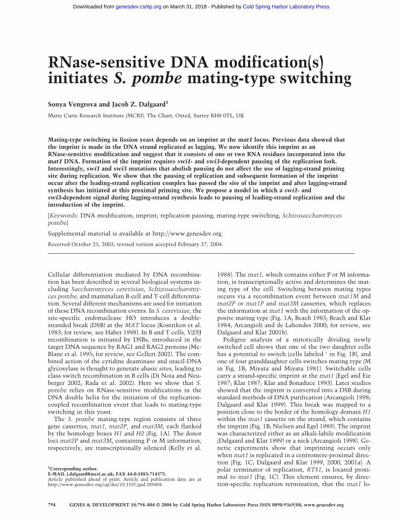
Figure 1. mat1 locus and the direction-of-replication model.(A) The mating-type region of S. pombe. mat1, containing eitherP or M information; mat2P and mat3M cassettes are shown.Each cassette is flanked by homology domains H1 and H2(boxes). The positions of the centromere (circle) and the cis-acting sequences SAS1 and SAS2 (bracket) relative to mat1 areindicated. The recombination event, which replaces the cas-sette at mat1 with the information of the opposite mating type,is shown by black arrows. The silencing of mat2 and mat3 isindicated by the gray line. (B) The direction-of-replicationmodel. The mating-type pedigree of a newly switched cell isshown. The cells are shown in yellow, and the cell generation isindicated to the right. Next to each cell is a diagram of the sisterchromatids, present in the corresponding cell type. The strands,replicated as leading and lagging, are shown as red and bluearrows, respectively. The imprint is indicated by a yellow circle,and the diagram of initiation of the recombination event is dis-played in brackets. (C) Replication at the mat1 locus. mat1replication by a distal origin is secured by a polar replicationterminator RTS1, shown as a gray triangle. Nascent leading andlagging strands are indicated by continuous red and discontinu-ous blue arrows, respectively. Homology domains H1 and H2are indicated by gray boxes. The white arrows below representthe direction of replication at mat1. The swi1- and swi3-depen-dent replication pause is shown as a gray shadow.
Replication pausing and imprinting
GENES & DEVELOPMENT 795
Cold Spring Harbor Laboratory Press on March 31, 2018 - Published by genesdev.cshlp.orgDownloaded from
On the basis of these and previous observations, wepropose the following additions to our earlier model forthe mechanism of the imprinting: (1) The leading-strandreplication complex replicates past the site of imprint-ing; (2) lagging-strand synthesis of the Okazaki fragmentthat is going to span over the site of the imprint is ini-tiated at this proximal priming site; (3) a swi1p- andswi3p-dependent signal during lagging-strand elonga-tion causes pausing of the leading-strand replication; and(4) the imprint is formed either via incorporation of theribonucleotides during polymerization, or postreplica-tively by an oxidation of the DNA backbone.
Results
Imprint in the template strand acts as a blockfor leading-strand replication
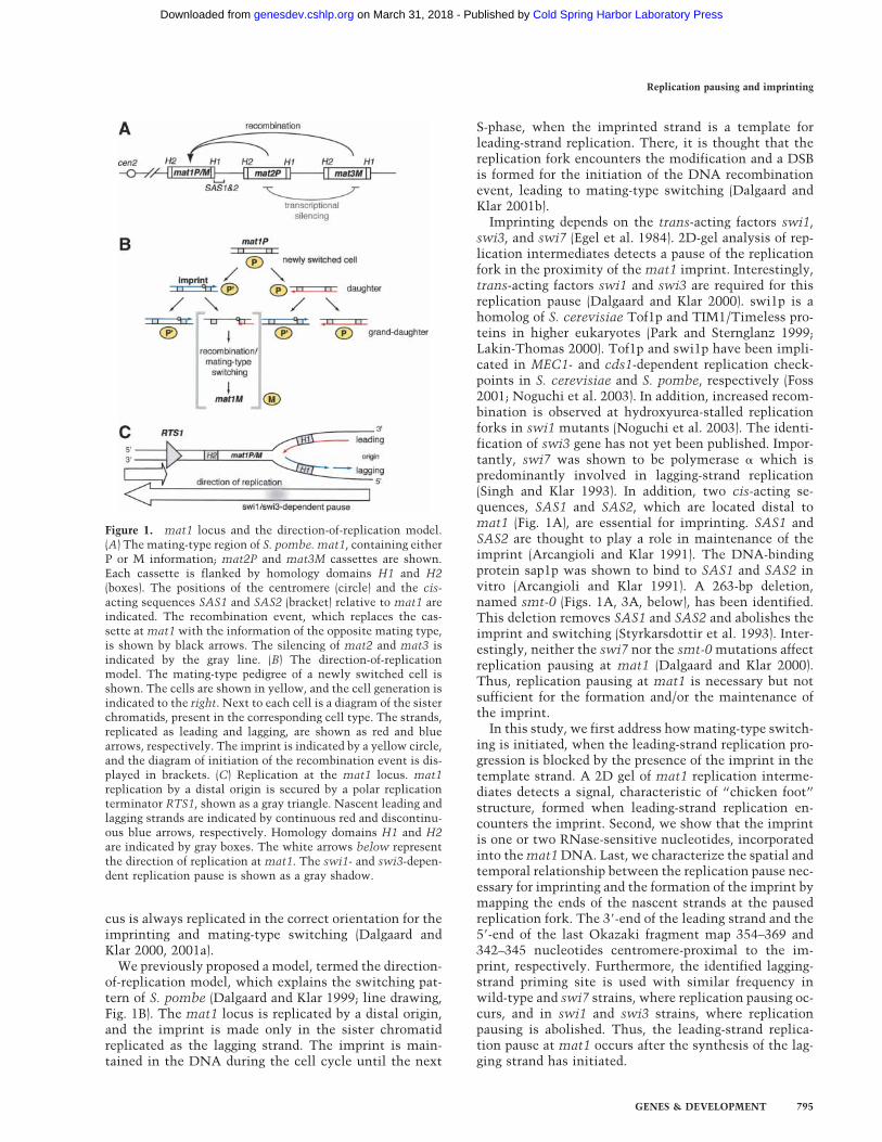
Mating-type switching has been proposed to initiatewhen the leading-strand replication complex encountersthe imprint in the template strand (Arcangioli and deLahondes 2000; Dalgaard and Klar 2001b; Fig. 1B, brack-ets). We analyzed the progression of the leading strandthrough the site of the imprint by applying to replicationintermediates a high-resolution Southern blot technique(Nielsen and Egel 1989). The replication and switchingintermediates were purified (Huberman et al. 1987) fromJZ105 (wild-type mat1M) and SV1 (swi3) strains, eachcontaining a deletion of the donor loci mat2P andmat3M. The deletion of mat2 and mat3 prevents thesequence homology at the donor cassettes from interfer-ing with the analysis. In strains deleted for donor loci,the imprint at mat1 is still formed, but it is thought thatinstead of switching, homologous recombination occurswith the sister chromatid (Klar and Miglio 1986). In thisexperiment, the swi3 mutant strain acts as a control, asit lacks the mat1 imprint and therefore any switching-related intermediates. Replication intermediates weredigested with SspI, purified from nonreplicating DNA byusing benzoylated naphthoylated DEAE (BND) cellulose(Kiger and Sinsheimer 1969), and separated on a poly-acrylamide gel. After subsequent electrotransfer, the fil-ter was hybridized to the strand-specific probe that de-tects the lower strand. Using this technique, we mappedthe 3�-end of the leading strand, formed when the repli-cation fork encounters the imprint (Figs. 1B, 2B), exactlyto the position where the imprint/break had beenmapped on the other strand (Fig. 2A; Nielsen and Egel1989). This result indicates that the leading-strand pro-gression is blocked at the site of the imprint. Becauserecombination frequently occurs at stalled replicationforks (Michel et al. 2001), the data support the modelthat the switching event is initiated when leading-strandreplication encounters the imprint in the templatestrand.
There are fewer replication forks proximal to the siteof the imprint in the strain that contains the imprint
According to the direction-of-replication model (Fig. 1B),approximately half of the cells in the population should
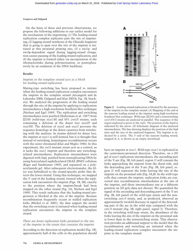
have an imprint at mat1. Wild-type mat1 is replicated inthe centromere-proximal direction. Therefore, on a 2Dgel of mat1 replication intermediates, the ascending partof the Y-arc (Fig. 3B, left panel, region 1) will contain theforks approaching the imprint from the distal side, andthe descending part of the Y-arc (Fig. 3B, left panel, re-gion 2) will represent the forks leaving the site of theimprint on the proximal side (Fig. 3A,B). In the wild-typecells that contain the imprint, replication forks are con-verted into recombination intermediates at the site ofthe imprint, and these intermediates run at a differentposition on 2D gels (data not shown). We quantified thesignal of the ascending and descending parts of the Y-arc(Fig. 3B, left panel) of the wild-type strain and the non-switching smt-0 strain, and, as predicted, observed anapproximately twofold decrease in signal of the descend-ing part of the arc in the wild type compared with thesmt-0 mutant (Fig. 3B, right panel, signal 2). Therefore,in the switching wild-type strain, the abundance of theforks leaving the site of the imprint on the proximal sideis lower than in the nonswitching strain. This observa-tion provides an additional support to the model that therecombination and switching are initiated when theleading-strand replication complex encounters the im-print in the template strand.
Figure 2. Leading-strand replication is blocked by the presenceof the imprint in the template strand. (A) Mapping of the end ofthe nascent leading strand at the imprint using high-resolutionSouthern blot technique. Wild-type (JZ105) and a nonswitchingswi3 (SV1) strains are analyzed in parallel. The sequence of theregion analyzed is given to the right. The position of the band isindicated by the arrow. (B) Schematic diagram of the analyzedintermediates. The line drawing displays the position of the SspIsites and the size of the analyzed fragment. The imprint is in-dicated by a circle. The 3�-end of the nascent leading strand,mapped in A, is shown as an empty arrowhead.
Vengrova and Dalgaard
796 GENES & DEVELOPMENT
Cold Spring Harbor Laboratory Press on March 31, 2018 - Published by genesdev.cshlp.orgDownloaded from
A ‘chicken foot’ is formed when the leading-strandreplication encounters the imprint in thetemplate strand
Our data, published earlier, suggest that replicationpausing at mat1 is prerequisite to imprinting (Dalgaard
and Klar 2000). However, mat1 pausing and imprintingcan be genetically separated: both swi7 and smt-0 mu-tants display a wild-type level of replication pausing atmat1, but lack the imprint (Dalgaard and Klar 2000).When we compared mat1 replication intermediates ofwild-type and smt-0 strains by means of 2D gels, wedetected a cone-shaped signal at the apex of the wild-type arc (Fig. 3A, wt). Importantly, this cone signal isabsent on a 2D gel of the smt-0 strain (Fig. 3A, smt-0),suggesting that it represents the structure formed whenthe replication fork encounters the imprint in the tem-plate strand. These structures display a mobility charac-teristic of regressing replication forks similar to thoseformed when the leading-strand replication encountersDNA damage (Sogo et al. 2002; Courcelle et al. 2003).Indeed, both the replication fork that is blocked at thesite of the imprint and the reversed fork generated fromthe blocked fork (Fig. 4B, right diagram) have the samemolecular weight. Therefore, both fork structures shouldnot separate in the first-dimension gel, but only in thesecond, where the regressed forks are retarded due to amore branched structure. Thus, on a 2D gel, the conesignal representing the regressing forks from the site ofthe imprint can only be positioned above the region ofthe Y-arc that corresponds to the forks blocked at theimprint.
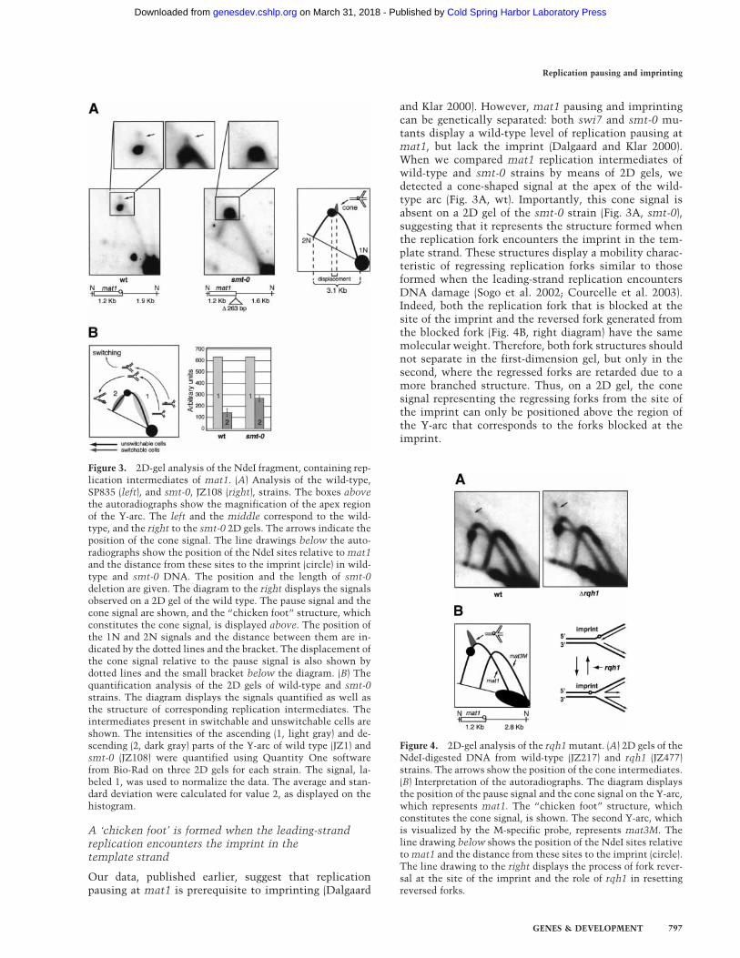
Figure 4. 2D-gel analysis of the rqh1 mutant. (A) 2D gels of theNdeI-digested DNA from wild-type (JZ217) and rqh1 (JZ477)strains. The arrows show the position of the cone intermediates.(B) Interpretation of the autoradiographs. The diagram displaysthe position of the pause signal and the cone signal on the Y-arc,which represents mat1. The “chicken foot” structure, whichconstitutes the cone signal, is shown. The second Y-arc, whichis visualized by the M-specific probe, represents mat3M. Theline drawing below shows the position of the NdeI sites relativeto mat1 and the distance from these sites to the imprint (circle).The line drawing to the right displays the process of fork rever-sal at the site of the imprint and the role of rqh1 in resettingreversed forks.
Figure 3. 2D-gel analysis of the NdeI fragment, containing rep-lication intermediates of mat1. (A) Analysis of the wild-type,SP835 (left), and smt-0, JZ108 (right), strains. The boxes abovethe autoradiographs show the magnification of the apex regionof the Y-arc. The left and the middle correspond to the wild-type, and the right to the smt-0 2D gels. The arrows indicate theposition of the cone signal. The line drawings below the auto-radiographs show the position of the NdeI sites relative to mat1and the distance from these sites to the imprint (circle) in wild-type and smt-0 DNA. The position and the length of smt-0deletion are given. The diagram to the right displays the signalsobserved on a 2D gel of the wild type. The pause signal and thecone signal are shown, and the “chicken foot” structure, whichconstitutes the cone signal, is displayed above. The position ofthe 1N and 2N signals and the distance between them are in-dicated by the dotted lines and the bracket. The displacement ofthe cone signal relative to the pause signal is also shown bydotted lines and the small bracket below the diagram. (B) Thequantification analysis of the 2D gels of wild-type and smt-0strains. The diagram displays the signals quantified as well asthe structure of corresponding replication intermediates. Theintermediates present in switchable and unswitchable cells areshown. The intensities of the ascending (1, light gray) and de-scending (2, dark gray) parts of the Y-arc of wild type (JZ1) andsmt-0 (JZ108) were quantified using Quantity One softwarefrom Bio-Rad on three 2D gels for each strain. The signal, la-beled 1, was used to normalize the data. The average and stan-dard deviation were calculated for value 2, as displayed on thehistogram.
Replication pausing and imprinting
GENES & DEVELOPMENT 797
Cold Spring Harbor Laboratory Press on March 31, 2018 - Published by genesdev.cshlp.orgDownloaded from
The “chicken foot” structures accumulate in therqh1 mutant
Fork reversal is well characterized in bacterial systems.There, the helicase RecG can actively drive fork reversal,while another helicase, RecQ, is thought to counteractit, converting the X-shaped intermediate back into theY-shaped replication fork. However, fork reversal canalso happen spontaneously, without involvement of en-zymes (for review, see Michel et al. 2001). Although theS. pombe homolog of RecG has not yet been identified, S.pombe rqh1p is thought to possess activities similar toRecQ (Fig. 4B; Murray et al. 1997; Doe et al. 2000, 2002).To confirm that the cone signal observed on 2D gels ofmat1 represents fork regression, we analyzed the rqh1mutant strain. Because rqh1 is involved in resetting re-versed replication forks, we expected to see an increasein intensity of the cone signal, due to accumulation ofthe reversed forks. Indeed, the cone signal is more promi-nent on the 2D gel of the rqh1 mutant than on the gel ofthe wild-type strain (Fig. 4A). This result supports thesuggestion that the observed signal represents fork re-gression.
The analyzed strains contain the S. cerevisiae LEU2marker inserted distal to mat1. This insertion does notaffect pausing or imprinting (data not shown), but be-cause of the different length of the analyzed fragment,the pause site and the site of the imprint are displacedalong the Y-arc compared with that shown in Figure 3A.Importantly, the cone signal is also displaced, indicatingthat the “chicken foot” is linked to the position of theimprint in the DNA.
Imprint is one or two ribonucleotides
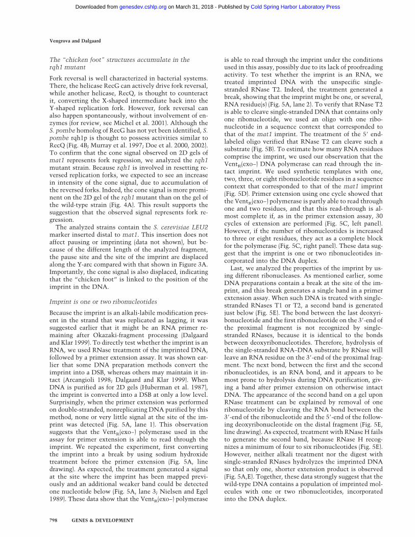
Because the imprint is an alkali-labile modification pres-ent in the strand that was replicated as lagging, it wassuggested earlier that it might be an RNA primer re-maining after Okazaki-fragment processing (Dalgaardand Klar 1999). To directly test whether the imprint is anRNA, we used RNase treatment of the imprinted DNA,followed by a primer extension assay. It was shown ear-lier that some DNA preparation methods convert theimprint into a DSB, whereas others may maintain it in-tact (Arcangioli 1998; Dalgaard and Klar 1999). WhenDNA is purified as for 2D gels (Huberman et al. 1987),the imprint is converted into a DSB at only a low level.Surprisingly, when the primer extension was performedon double-stranded, nonreplicating DNA purified by thismethod, none or very little signal at the site of the im-print was detected (Fig. 5A, lane 1). This observationsuggests that the VentR(exo−) polymerase used in theassay for primer extension is able to read through theimprint. We repeated the experiment, first convertingthe imprint into a break by using sodium hydroxidetreatment before the primer extension (Fig. 5A, linedrawing). As expected, the treatment generated a signalat the site where the imprint has been mapped previ-ously and an additional weaker band could be detectedone nucleotide below (Fig. 5A, lane 3; Nielsen and Egel1989). These data show that the VentR(exo−) polymerase
is able to read through the imprint under the conditionsused in this assay, possibly due to its lack of proofreadingactivity. To test whether the imprint is an RNA, wetreated imprinted DNA with the unspecific single-stranded RNase T2. Indeed, the treatment generated abreak, showing that the imprint might be one, or several,RNA residue(s) (Fig. 5A, lane 2). To verify that RNase T2is able to cleave single-stranded DNA that contains onlyone ribonucleotide, we used an oligo with one ribo-nucleotide in a sequence context that corresponded tothat of the mat1 imprint. The treatment of the 5� end-labeled oligo verified that RNase T2 can cleave such asubstrate (Fig. 5B). To estimate how many RNA residuescomprise the imprint, we used our observation that theVentR(exo−) DNA polymerase can read through the in-tact imprint. We used synthetic templates with one,two, three, or eight ribonucleotide residues in a sequencecontext that corresponded to that of the mat1 imprint(Fig. 5D). Primer extension using one cycle showed thatthe VentR(exo−) polymerase is partly able to read throughone and two residues, and that this read-through is al-most complete if, as in the primer extension assay, 30cycles of extension are performed (Fig. 5C, left panel).However, if the number of ribonucleotides is increasedto three or eight residues, they act as a complete blockfor the polymerase (Fig. 5C, right panel). These data sug-gest that the imprint is one or two ribonucleotides in-corporated into the DNA duplex.
Last, we analyzed the properties of the imprint by us-ing different ribonucleases. As mentioned earlier, someDNA preparations contain a break at the site of the im-print, and this break generates a single band in a primerextension assay. When such DNA is treated with single-stranded RNases T1 or T2, a second band is generatedjust below (Fig. 5E). The bond between the last deoxyri-bonucleotide and the first ribonucleotide on the 3�-end ofthe proximal fragment is not recognized by single-stranded RNases, because it is identical to the bondsbetween deoxyribonucleotides. Therefore, hydrolysis ofthe single-stranded RNA–DNA substrate by RNase willleave an RNA residue on the 3�-end of the proximal frag-ment. The next bond, between the first and the secondribonucleotides, is an RNA bond, and it appears to bemost prone to hydrolysis during DNA purification, giv-ing a band after primer extension on otherwise intactDNA. The appearance of the second band on a gel uponRNase treatment can be explained by removal of oneribonucleotide by cleaving the RNA bond between the3�-end of the ribonucleotide and the 5�-end of the follow-ing deoxyribonucleotide on the distal fragment (Fig. 5E,line drawing). As expected, treatment with RNase H failsto generate the second band, because RNase H recog-nizes a minimum of four to six ribonucleotides (Fig. 5E).However, neither alkali treatment nor the digest withsingle-stranded RNases hydrolyzes the imprinted DNAso that only one, shorter extension product is observed(Fig. 5A,E). Together, these data strongly suggest that thewild-type DNA contains a population of imprinted mol-ecules with one or two ribonucleotides, incorporatedinto the DNA duplex.
Vengrova and Dalgaard
798 GENES & DEVELOPMENT
Cold Spring Harbor Laboratory Press on March 31, 2018 - Published by genesdev.cshlp.orgDownloaded from
No RNA primers, synthesized during lagging-strandreplication, map to the site of the imprint
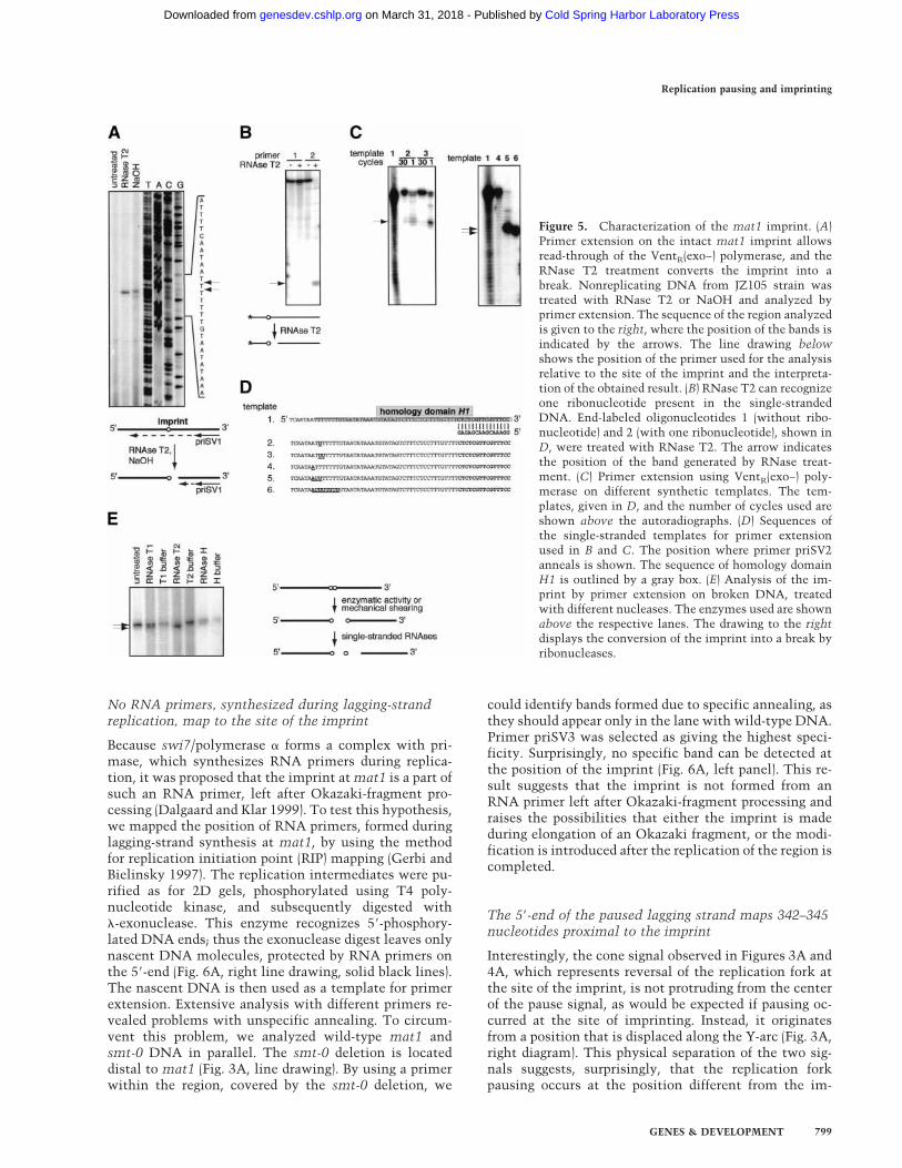
Because swi7/polymerase � forms a complex with pri-mase, which synthesizes RNA primers during replica-tion, it was proposed that the imprint at mat1 is a part ofsuch an RNA primer, left after Okazaki-fragment pro-cessing (Dalgaard and Klar 1999). To test this hypothesis,we mapped the position of RNA primers, formed duringlagging-strand synthesis at mat1, by using the methodfor replication initiation point (RIP) mapping (Gerbi andBielinsky 1997). The replication intermediates were pu-rified as for 2D gels, phosphorylated using T4 poly-nucleotide kinase, and subsequently digested with�-exonuclease. This enzyme recognizes 5�-phosphory-lated DNA ends; thus the exonuclease digest leaves onlynascent DNA molecules, protected by RNA primers onthe 5�-end (Fig. 6A, right line drawing, solid black lines).The nascent DNA is then used as a template for primerextension. Extensive analysis with different primers re-vealed problems with unspecific annealing. To circum-vent this problem, we analyzed wild-type mat1 andsmt-0 DNA in parallel. The smt-0 deletion is locateddistal to mat1 (Fig. 3A, line drawing). By using a primerwithin the region, covered by the smt-0 deletion, we
could identify bands formed due to specific annealing, asthey should appear only in the lane with wild-type DNA.Primer priSV3 was selected as giving the highest speci-ficity. Surprisingly, no specific band can be detected atthe position of the imprint (Fig. 6A, left panel). This re-sult suggests that the imprint is not formed from anRNA primer left after Okazaki-fragment processing andraises the possibilities that either the imprint is madeduring elongation of an Okazaki fragment, or the modi-fication is introduced after the replication of the region iscompleted.
The 5�-end of the paused lagging strand maps 342–345nucleotides proximal to the imprint
Interestingly, the cone signal observed in Figures 3A and4A, which represents reversal of the replication fork atthe site of the imprint, is not protruding from the centerof the pause signal, as would be expected if pausing oc-curred at the site of imprinting. Instead, it originatesfrom a position that is displaced along the Y-arc (Fig. 3A,right diagram). This physical separation of the two sig-nals suggests, surprisingly, that the replication forkpausing occurs at the position different from the im-
Figure 5. Characterization of the mat1 imprint. (A)Primer extension on the intact mat1 imprint allowsread-through of the VentR(exo−) polymerase, and theRNase T2 treatment converts the imprint into abreak. Nonreplicating DNA from JZ105 strain wastreated with RNase T2 or NaOH and analyzed byprimer extension. The sequence of the region analyzedis given to the right, where the position of the bands isindicated by the arrows. The line drawing belowshows the position of the primer used for the analysisrelative to the site of the imprint and the interpreta-tion of the obtained result. (B) RNase T2 can recognizeone ribonucleotide present in the single-strandedDNA. End-labeled oligonucleotides 1 (without ribo-nucleotide) and 2 (with one ribonucleotide), shown inD, were treated with RNase T2. The arrow indicatesthe position of the band generated by RNase treat-ment. (C) Primer extension using VentR(exo−) poly-merase on different synthetic templates. The tem-plates, given in D, and the number of cycles used areshown above the autoradiographs. (D) Sequences ofthe single-stranded templates for primer extensionused in B and C. The position where primer priSV2anneals is shown. The sequence of homology domainH1 is outlined by a gray box. (E) Analysis of the im-print by primer extension on broken DNA, treatedwith different nucleases. The enzymes used are shownabove the respective lanes. The drawing to the rightdisplays the conversion of the imprint into a break byribonucleases.
Replication pausing and imprinting
GENES & DEVELOPMENT 799
Cold Spring Harbor Laboratory Press on March 31, 2018 - Published by genesdev.cshlp.orgDownloaded from
printing site. Because mat1 is replicated in a unidirec-tional manner, the position of the cone signal on theapex and the pause signal on the descending part of theY-arc shows that the pause occurs within the mat1 cas-sette at the centromere-proximal side of the imprint.
In the primer extension experiments described earlier,the most prominent band observed on a gel mapped ∼350nucleotides upstream of the imprint, also on a centro-mere-proximal side (Fig. 6A, left panel). To test whetherthis band represents the mat1 pause site, we comparedwild-type, swi1, swi3, and swi7 strains on the gel: inoriginal 2D-gel analysis, the pause site was shown to bepresent in wild type and swi7 and absent in swi1 andswi3 mutants (Dalgaard and Klar 2000). Surprisingly, theRIP-mapping analysis detected this prominent band withequal intensity in all four strains (Fig. 6A, right panel).We therefore repeated the experiment with paused inter-mediates purified from the agarose gels, omitting the�-exonuclease treatment, and here we observed a signifi-cant difference between wild-type and swi3 strains. Thesignal that appears as two bands at positions 342 and 345is much stronger in the wild type than in swi3 and ab-sent in smt-0 (Figs. 6B, 7A). We speculated that when thereplication fork pauses,
Okazaki-fragment processing takes place, and RNase Hremoves the RNA primers from the long-lived pausedmolecules detected by 2D gels. �-Exonuclease, used inthe first experiment, removes the processed paused in-termediates, which are not protected by an RNA primer,in the wild-type DNA. Thus, there is no difference infrequency the lagging-strand priming site is used in thestrains that lack pausing, but swi1p- and swi3p-depen-dent pausing allows accumulation of Okazaki frag-ments where the RNA primers have been processed byRNase H.
The 3�-end of the paused leading strand maps closeto the 5�-end of the lagging strand
To map the end of the leading strand in the paused rep-lication fork, we used the high-resolution Southern blotmethod described earlier. A similar approach was usedpreviously to map the blocked replication fork in therDNA repeats of S. cerevisiae (Gruber et al. 2000), where150 copies of rDNA repeats per genome are present.However, for a single-copy mat1 locus, it proved to bevery difficult. The balance between analyzing enoughDNA to detect the signal and using a thin enough gel to
Figure 6. Analysis of replication intermedi-ates at mat1. (A) Primer extension on Oka-zaki fragments from the wild-type (JZ105),smt-0 (JZ108), swi1 (SV5), swi3 (SV1), andswi7 (SP469) strains. The left panel showsthe analysis of the wild-type and smt-0strains. The position of the homology do-main H1 and the site of the imprint are givento the right of the autoradiograph. The draw-ing below shows the annealing position ofthe primer used for the assay on the wild-typeDNA and lack of specific annealing to smt-0DNA. The right panel shows the analysis ofswi1, swi3, and swi7 mutants. Black lines in-dicate the position of the analyzed regionrelative to the left panel. The drawing belowindicates that in this experiment the parentalDNA, shown by dotted lines, was digestedaway by �-exonuclease before primer exten-sion. (B) Mapping of the pause site on thelagging strand on gel-purified paused inter-mediates. Strains are given in A. The se-quence of the region (upper strand) is shownto the right, and the arrows indicate theposition of the bands observed. The drawingbelow shows that RNase H removes RNAprimers at 5�-ends of pausing intermediatesof the lagging strand. The drawing also indi-cates that in this experiment primer cananneal both to nascent and to parental DNA.(C) Mapping of the pause site on the lead-ing strand. The nonswitching strain smt-0(JZ108) was used for clarity of the analysis, to avoid detecting imprinting and switching intermediates. The sequence of the analyzedregion is shown to the right. The position of the BstNI restriction site on the upper strand is labeled, and the region of pausing isdesignated by the brackets. The line drawing below displays the position of the BstNI and SfaNI sites and the size of the full-lengthfragment of the lower strand. The strands, visualized on the gel, are shown as thick lines, and the mapped 3�-end of the nascent leadingstrand is shown as an empty arrowhead.
Vengrova and Dalgaard
800 GENES & DEVELOPMENT
Cold Spring Harbor Laboratory Press on March 31, 2018 - Published by genesdev.cshlp.orgDownloaded from
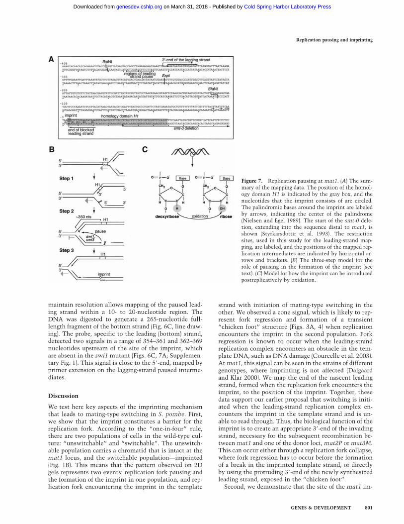
maintain resolution allows mapping of the paused lead-ing strand within a 10- to 20-nucleotide region. TheDNA was digested to generate a 265-nucleotide full-length fragment of the bottom strand (Fig. 6C, line draw-ing). The probe, specific to the leading (bottom) strand,detected two signals in a range of 354–361 and 362–369nucleotides upstream of the site of the imprint, whichare absent in the swi1 mutant (Figs. 6C, 7A; Supplemen-tary Fig. 1). This signal is close to the 5�-end, mapped byprimer extension on the lagging-strand paused interme-diates.
Discussion
We test here key aspects of the imprinting mechanismthat leads to mating-type switching in S. pombe. First,we show that the imprint constitutes a barrier for thereplication fork. According to the “one-in-four” rule,there are two populations of cells in the wild-type cul-ture: “unswitchable” and “switchable”. The unswitch-able population carries a chromatid that is intact at themat1 locus, and the switchable population—imprinted(Fig. 1B). This means that the pattern observed on 2Dgels represents two events: replication fork pausing andthe formation of the imprint in one population, and rep-lication fork encountering the imprint in the template
strand with initiation of mating-type switching in theother. We observed a cone signal, which is likely to rep-resent fork regression and formation of a transient“chicken foot” structure (Figs. 3A, 4) when replicationencounters the imprint in the second population. Forkregression is known to occur when the leading-strandreplication complex encounters an obstacle in the tem-plate DNA, such as DNA damage (Courcelle et al. 2003).At mat1, this signal can be seen in the strains of differentgenotypes, where imprinting is not affected (Dalgaardand Klar 2000). We map the end of the nascent leadingstrand, formed when the replication fork encounters theimprint, to the position of the imprint. Together, thesedata support our earlier proposal that switching is initi-ated when the leading-strand replication complex en-counters the imprint in the template strand and is un-able to read through. Thus, the biological function of theimprint is to create an appropriate 3�-end of the invadingstrand, necessary for the subsequent recombination be-tween mat1 and one of the donor loci, mat2P or mat3M.This can occur either through a replication fork collapse,where fork regression has to occur before the formationof a break in the imprinted template strand, or directlyby using the protruding 3�-end of the newly synthesizedleading strand, exposed in the “chicken foot”.
Second, we demonstrate that the site of the mat1 im-
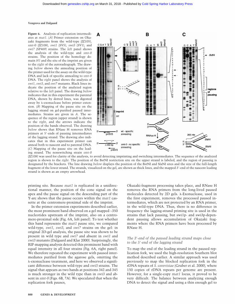
Figure 7. Replication pausing at mat1. (A) The sum-mary of the mapping data. The position of the homol-ogy domain H1 is indicated by the gray box, and thenucleotides that the imprint consists of are circled.The palindromic bases around the imprint are labeledby arrows, indicating the center of the palindrome(Nielsen and Egel 1989). The start of the smt-0 dele-tion, extending into the sequence distal to mat1, isshown (Styrkarsdottir et al. 1993). The restrictionsites, used in this study for the leading-strand map-ping, are labeled, and the positions of the mapped rep-lication intermediates are indicated by horizontal ar-rows and brackets. (B) The three-step model for therole of pausing in the formation of the imprint (seetext). (C) Model for how the imprint can be introducedpostreplicatively by oxidation.
Replication pausing and imprinting
GENES & DEVELOPMENT 801
Cold Spring Harbor Laboratory Press on March 31, 2018 - Published by genesdev.cshlp.orgDownloaded from
print/”chicken foot” structure and the site of replicationpause, prerequisite to imprinting and mating-typeswitching, are separated on a 2D gel (Figs. 3A, 4). We alsomap the 3�-end of the nascent leading strand in thepaused replication complex 354–369 nucleotides up-stream of the site of the imprint, using high-resolutionSouthern blot, and the 5�-end of the nascent laggingstrand 342–345 nucleotides upstream of the site of theimprint, using primer extension (Fig. 7A). This meansthat the leading-strand replication complex has alreadypassed the region where the imprint is to be introduced(Fig. 7B, Step 1). Primer extension on Okazaki fragmentsshows that wild type, swi1, swi3, and swi7 display asimilar amount of fragments, the DNA part of whichstarts 342–345 nucleotides from the imprint. However,when primer extension is performed on purified pausedintermediates in the wild type, the signal is greatly in-creased. These data suggest that pausing of lagging-strand replication occurs after polymerase � has initiatedthe synthesis of the last Okazaki fragment, and thatRNase H is able to remove the RNA primers at thestalled fork (Fig. 7B, Step 2).
We do not know what the signal for the imprintingevent is. Because the 5�-end of the lagging-strand tem-plate becomes exposed earlier than the 3�-end, it wouldremain single-stranded for a longer period of time. Thereis evidence from experiments with trinucleotide repeattracts in S. cerevisiae that this allows formation of thesecondary structure in the DNA, thus affecting lagging-but not leading-strand synthesis (Maurer et al. 1998). Atthe mat1 locus of S. pombe, the sequence around theimprint has symmetric properties (Nielsen and Egel1989) and the homology domain H1 (Fig. 1A) is impor-tant for replication pausing as well as for the formationof the imprint (Styrkarsdottir et al. 1993; Kaykov et al.2004). Alternatively, a trans-acting protein might bindthe exposed single-stranded DNA at the implicated H1sequence. Therefore, the lagging-strand replicationmight encounter difficulties during elongation throughthis region, either due to the secondary DNA structure,or due to involvement of a trans-acting factor. Such bar-riers can constitute the signal that leads to imprinting(Fig. 7B, Step 2).
Last, we show that the mat1 imprint is sensitive toRNases, and we estimate that it consists of one or tworibonucleotides. The assay used here could not detect anRNA primer at the position of the mat1 imprint, whichsuggests that the imprint is not a part of a primer that isnormally synthesized to initiate a new Okazaki frag-ment. We see at least three possible mechanisms for im-print introduction (Fig. 7B, Step 3). (1) It could be thatmistakes happen during lagging-strand synthesis, whenthe lagging-strand replication complex attempts to es-cape from the stall site. In S. cerevisiae trinucleotiderepeat tracts, this leads to tract expansion (Pelletier et al.2003). Ribonucleotide(s) at mat1 can be introduced whenthe lagging-strand replication is overcoming the barrierpresent in the template DNA. (2) The studies using theSV40 DNA replication system showed that synthesis ofthe first ∼30 nucleotides of an Okazaki fragment by poly-
merase � is followed by polymerase switch, and the restof the fragment is completed by polymerase � (Waga andStillman 1998). Because of the signal during lagging-strand progression through the site of the imprint, a re-verse change of polymerases may take place, and theribonucleotides are incorporated in a swi7/pol�-depen-dent manner. (3) Alternatively, such ribonucleotidescould be formed postreplicatively by an oxidation of theDNA backbone (Fig. 7C). The difference between riboseand deoxyribose is one oxygen atom at the 2� position.The enzyme ribonucleotide-reductase converts ribo-nucleotides into deoxyribonucleotides using NADPH asa source of energy. We imagine that the reverse processcan be catalyzed by an oxidating enzyme that has a struc-ture to bind the DNA duplex and access the 2� positionof the deoxyribose, and the signal from the lagging-strand synthesis might designate the site for the modifi-cation.
The presence of ribonucleotides in the newly repli-cated DNA was previously shown by RNase sensitivityassays in mitochondrial DNA, where ribonucleotides arepresent in both nascent strands, but are much moreabundant in the newly synthesized lagging strand (Yanget al. 2002). The data presented here is the first evidenceof a site-specific RNase-sensitive modification in thenuclear eukaryotic DNA. Such imprints could act as amark for other cellular processes. Indeed, the imprintingfunction of swi1, swi3, and swi7 is important for alkyla-tion damage repair in S. pombe (E. Sommariva, T. Pellny,S. Vengrova, T. Eydmann and J. Dalgaard, in prep.).There, such imprints are proposed to mark the siteswhere the replication forks were stalled by damage to thetemplate DNA. swi1p, swi3p, and swi7p proteins, in-volved in imprinting in S. pombe, are conserved fromyeast to metazoans, suggesting that similar mechanismsmay play a role in other organisms.
Materials and methods
S. pombe strains
Strains were constructed as described elsewhere (Moreno et al.1991). The genotypes of the strains used are as follows: JZ105,h90 mat1M �mat 2,3�LEU2, ade6-210, leu1-32, ura4-D18, his2;JZ108, mat1M smt-0 �mat 2,3�LEU2, ade6-210, leu1-32, his2;JZ217, h90 mat1 �1.5 Kb distal�LEU2, ade6-210, leu1-32, ura4-D18, swi6 mod+; JZ477, h90 mat1 �1.5 Kb distal�LEU2, ade6-210, leu1-32, ura4-D18, swi6 mod+, rqh1��ura4; SV1, h90
mat1M �mat 2,3�LEU2, ade6-216, his2, swi3; SV5, h90 mat1M�mat 2,3�LEU2, ade6-210, leu1-32, ura4-D18, his2,swi1��ura4; SP469, h90 mat1M �mat 2,3�LEU2, ade6-216,leu1-32, his2, swi7 (A. Klar, National Cancer Institute, Freder-ick, MD); SP835, h90 mat1P �mat2,3�LEU2 ade6-216 leu1-32ura4-D18 his2 (A. Klar); SP976, h90 ade6-210, leu1-32, ura4-D18, swi6 mod+ (A. Klar).
DNA preparation
Yeast chromosomal DNA was purified form logarithmicallygrowing culture as described by Huberman et al. (1987). Repli-cation intermediates were separated from nonreplicating DNAby BND-cellulose chromatography (Kiger and Sinsheimer 1969).
Vengrova and Dalgaard
802 GENES & DEVELOPMENT
Cold Spring Harbor Laboratory Press on March 31, 2018 - Published by genesdev.cshlp.orgDownloaded from

2D gels
2D gels were performed as described by Brewer and Fangman(1987), except that the first- and second-dimension gels con-tained 0.5% and 1.2% agarose, respectively.
High-resolution Southern blot
Replication intermediates were digested with SspI or BstNI/SfaNI and enriched using BND-cellulose. The DNA was precipi-tated overnight with isopropanol, redissolved in loading buffer,and loaded on a 6% denaturing polyacrylamide gel. Sequencingreactions, made using primers mat1M-SspI (GCACTCCCTACCATAATATACTCACTAAT) or mat1M-SfaNI (CTAAAACAGTATGGCATTAC) and a bacterial plasmid containing the EcoRIfragment of mat1M (pBZ85), were run in parallel as a marker.The DNA was electroblotted on a GeneScreen membrane(NEN). For Figure 2A, the membrane was probed with a strand-specific RNA probe (Nielsen and Egel 1989). For Figure 6C, theprobe was synthesized by linear primer extension, using primermat1M-BstNI (TGGTTATAAGTACCAGCCTAG), on a 268-bpBstNI/SfaNI fragment (Ruven et al. 1994).
Primer extension
NaOH treatment of nonreplicating DNA was performed as fol-lows: DNA was incubated in 40 mM NaOH, 2 mM EDTA at25°C for 2 h, neutralized with equimolar HCl, and precipitatedbefore primer extension. RNase treatment of nonreplicatingDNA was performed by incubating heat-denatured DNA with10 units of RNase T2 or 1000 units of RNase T1 (both from LifeTechnologies) for 30 min to 2 h at 37°C, followed by phenol/chloroform extraction and precipitation before primer exten-sion. For RNase H (Life Technologies) digest, 4 units of theenzyme were used and the DNA was not denatured. NascentDNA was purified using �-exonuclease as for RIP mapping(Gerbi and Bielinsky 1997). For Figure 6B, the replication inter-mediates were digested with NdeI and separated as for 2D gelson a first-dimension gel. The region from 3.5 to 6 kb was ex-cised, and the DNA was purified using a Qiagen gel-purificationkit. Primer extension was performed using primers priSV1 (ATCTCGTTAGAGGGAAGGGGAAGGT) and priSV3 (AATATTAGTGAGTATATTATGGT) and VentR(exo−) DNA polymerase(New England Biolabs). Sequencing reactions using correspond-ing primers and a bacterial pBZ85 plasmid were run in parallelas a marker. Synthetic templates were obtained from HelenaBiosciences. Primer priSV2 and template 1 were end-labeledwith �32P-ATP (Amersham Pharmacia) and T4 polynucleotidekinase (New England Biolabs). Extension reactions were per-formed as for RIP mapping.
Acknowledgments
We thank our colleagues at MCRI for helpful suggestions anddiscussions. Special thanks to Dr. Robert Cross for suggestionsand help with the manuscript. This research was sponsored bythe Association of International Cancer Research (J.Z.D.) andthe Marie Curie Cancer Care.
The publication costs of this article were defrayed in part bypayment of page charges. This article must therefore be herebymarked “advertisement” in accordance with 18 USC section1734 solely to indicate this fact.
References
Arcangioli, B. 1998. A site- and strand-specific DNA break con-fers asymmetric switching potential in fission yeast. EMBOJ. 17: 4503–4510.
Arcangioli, B. and de Lahondes, R. 2000. Fission yeast switchesmating type by a replication-recombination coupled process.EMBO J. 19: 1389–1396.
Arcangioli, B. and Klar, A.J. 1991. A novel switch-activating site(SAS1) and its cognate binding factor (SAP1) required for ef-ficient mat1 switching in Schizosaccharomyces pombe.EMBO J. 10: 3025–3032.
Beach, D.H. 1983. Cell type switching by DNA transposition infission yeast. Nature 305: 682–687.
Beach, D.H. and Klar, A.J. 1984. Rearrangements of the trans-posable mating-type cassettes of fission yeast. EMBO J. 3:603–610.
Brewer, B.J. and Fangman, W.L. 1987. The localization of repli-cation origins on ARS plasmids in S. cerevisiae. Cell 51:463–471.
Courcelle, J., Donaldson, J.R., Chow, K.H., and Courcelle, C.T.2003. DNA damage-induced replication fork regression andprocessing in Escherichia coli. Science 299: 1064–1067.
Dalgaard, J.Z. and Klar, A.J. 1999. Orientation of DNA replica-tion establishes mating-type switching pattern in S. pombe.Nature 400: 181–184.
———. 2000. swi1 and swi3 perform imprinting, pausing, and ter-mination of DNA replication in S. pombe. Cell 102: 745–751.
———. 2001a. A DNA replication-arrest site RTS1 regulatesimprinting by determining the direction of replication atmat1 in S. pombe. Genes & Dev. 15: 2060–2068.
———. 2001b. Does S. pombe exploit the intrinsic asymmetryof DNA synthesis to imprint daughter cells for mating-typeswitching? Trends Genet. 17: 153–157.
Di Noia, J. and Neuberger, M.S. 2002. Altering the pathway ofimmunoglobulin hypermutation by inhibiting uracil-DNAglycosylase. Nature 419: 43–48.
Doe, C.L., Dixon, J., Osman, F., and Whitby, M.C. 2000. Partialsuppression of the fission yeast rqh1(-) phenotype by expres-sion of a bacterial Holliday junction resolvase. EMBO J. 19:2751–2762.
Doe, C.L., Ahn, J.S., Dixon, J., and Whitby, M.C. 2002. Mus81-Eme1 and Rqh1 involvement in processing stalled and col-lapsed replication forks. J. Biol. Chem. 277: 32753–32759.
Egel, R. and Eie, B. 1987. Cell lineage asymmetry for Schizosac-charomyces pombe: Unilateral transmission of a high-fre-quency state of mating-type switching in diploid pedigrees.Curr. Genet. 12: 429–433.
Egel, R., Beach, D.H., and Klar, A.J. 1984. Genes required forinitiation and resolution steps of mating-type switching infission yeast. Proc. Natl. Acad. Sci. 81: 3481–3485.
Foss, E.J. 2001. Tof1p regulates DNA damage responses during Sphase in Saccharomyces cerevisiae. Genetics 157: 567–577.
Gellert, M. 2002. V(D)J recombination: RAG proteins, repairfactors, and regulation. Annu. Rev. Biochem. 71: 101–132.
Gerbi, S.A. and Bielinsky, A.K. 1997. Replication initiationpoint mapping. Methods 13: 271–280.
Gruber, M., Wellinger, R.E., and Sogo, J.M. 2000. Architectureof the replication fork stalled at the 3� end of yeast ribosomalgenes. Mol. Cell. Biol. 20: 5777–5787.
Haber, J.E. 1998. Mating-type gene switching in Saccharomycescerevisiae. Annu. Rev. Genet. 32: 561–599.
Huberman, J.A., Spotila, L.D., Nawotka, K.A., el-Assouli, S.M.,and Davis, L.R. 1987. The in vivo replication origin of theyeast 2 microns plasmid. Cell 51: 473–481.
Kaykov, A., Holmes, A.M., and Arcangioli, B. 2004. Formation,maintenance and consequences of the imprint at the mating-type locus in fission yeast. EMBO J 23: 930–938.
Kelly, M., Burke, J., Smith, M., Klar, A., and Beach, D. 1988.Four mating-type genes control sexual differentiation in thefission yeast. EMBO J. 7: 1537–1547.
Replication pausing and imprinting
GENES & DEVELOPMENT 803
Cold Spring Harbor Laboratory Press on March 31, 2018 - Published by genesdev.cshlp.orgDownloaded from
Kiger Jr., J.A. and Sinsheimer, R.L. 1969. Vegetative lambdaDNA. IV. Fractionation of replicating lambda DNA on ben-zoylated-naphthoylated DEAE cellulose. J. Mol. Biol. 40:467–490.
Klar, A.J. 1987. Differentiated parental DNA strands confer de-velopmental asymmetry on daughter cells in fission yeast.Nature 326: 466–470.
Klar, A.J. and Bonaduce, M.J. 1993. The mechanism of fissionyeast mating-type interconversion: Evidence for two types ofepigenetically inherited chromosomal imprinted events.Cold Spring Harb. Symp. Quant. Biol. 58: 457–465.
Klar, A.J. and Miglio, L.M. 1986. Initiation of meiotic recombi-nation by double-strand DNA breaks in S. pombe. Cell 46:725–731.
Kostriken, R., Strathern, J.N., Klar, A.J., Hicks, J.B., and Heffron,F. 1983. A site-specific endonuclease essential for mating-type switching in Saccharomyces cerevisiae. Cell 35: 167–174.
Lakin-Thomas, P.L. 2000. Circadian rhythms: New functionsfor old clock genes. Trends Genet. 16: 135–142.
Maurer, D.J., O’Callaghan, B.L., and Livingston, D.M. 1998.Mapping the polarity of changes that occur in interruptedCAG repeat tracts in yeast. Mol. Cell. Biol. 18: 4597–4604.
McBlane, J.F., van Gent, D.C., Ramsden, D.A., Romeo, C.,Cuomo, C.A., Gellert, M., and Oettinger, M.A. 1995. Cleav-age at a V(D)J recombination signal requires only RAG1 andRAG2 proteins and occurs in two steps. Cell 83: 387–395.
Michel, B., Flores, M.J., Viguera, E., Grompone, G., Seigneur,M., and Bidnenko, V. 2001. Rescue of arrested replicationforks by homologous recombination. Proc. Natl. Acad. Sci.98: 8181–8188.
Miyata, H. and Miyata, M. 1981. Mode of conjugation in ho-mothallic cells of Schizosaccharomyces pombe. J. Gen.Appl. Microbiol. 27: 365–371.
Moreno, S., Klar, A., and Nurse, P. 1991. Molecular geneticanalysis of fission yeast Schizosaccharomyces pombe. Meth-ods Enzymol. 194: 795–823.
Murray, J.M., Lindsay, H.D., Munday, C.A., and Carr, A.M.1997. Role of Schizosaccharomyces pombe RecQ homolog,recombination, and checkpoint genes in UV damage toler-ance. Mol. Cell. Biol. 17: 6868–6875.
Nielsen, O. and Egel, R. 1989. Mapping the double-strand breaksat the mating-type locus in fission yeast by genomic se-quencing. EMBO J. 8: 269–276.
Noguchi, E., Noguchi, C., Du, L.L., and Russell, P. 2003. Swi1prevents replication fork collapse and controls checkpointkinase Cds1. Mol. Cell. Biol. 23: 7861–7874.
Park, H. and Sternglanz, R. 1999. Identification and character-ization of the genes for two topoisomerase I-interacting pro-teins from Saccharomyces cerevisiae. Yeast 15: 35–41.
Pelletier, R., Krasilnikova, M.M., Samadashwily, G.M., Lahue,R., and Mirkin, S.M. 2003. Replication and expansion of tri-nucleotide repeats in yeast. Mol. Cell. Biol. 23: 1349–1357.
Rada, C., Williams, G.T., Nilsen, H., Barnes, D.E., Lindahl, T.,and Neuberger, M.S. 2002. Immunoglobulin isotype switch-ing is inhibited and somatic hypermutation perturbed inUNG-deficient mice. Curr. Biol. 12: 1748–1755.
Ruven, H.J., Seelen, C.M., Lohman, P.H., Mullenders, L.H., andvan Zeeland, A.A. 1994. Efficient synthesis of 32P-labeledsingle-stranded DNA probes using linear PCR; application ofthe method for analysis of strand-specific DNA repair. Mu-tat. Res. 315: 189–195.
Singh, J. and Klar, A.J. 1993. DNA polymerase-alpha is essentialfor mating-type switching in fission yeast. Nature 361: 271–273.
Sogo, J.M., Lopes, M., and Foiani, M. 2002. Fork reversal and
ssDNA accumulation at stalled replication forks owing tocheckpoint defects. Science 297: 599–602.
Styrkarsdottir, U., Egel, R., and Nielsen, O. 1993. The smt-0mutation which abolishes mating-type switching in fissionyeast is a deletion. Curr. Genet. 23: 184–186.
Waga, S. and Stillman, B. 1998. The DNA replication fork ineukaryotic cells. Annu. Rev. Biochem. 67: 721–751.
Yang, M.Y., Bowmaker, M., Reyes, A., Vergani, L., Angeli, P.,Gringeri, E., Jacobs, H.T., and Holt, I.J. 2002. Biased incor-poration of ribonucleotides on the mitochondrial L-strandaccounts for apparent strand-asymmetric DNA replication.Cell 111: 495–505.
Vengrova and Dalgaard
804 GENES & DEVELOPMENT
Cold Spring Harbor Laboratory Press on March 31, 2018 - Published by genesdev.cshlp.orgDownloaded from
10.1101/gad.289404Access the most recent version at doi: 18:2004, Genes Dev.
Sonya Vengrova and Jacob Z. Dalgaard switching
mating-typeS. pombeRNase-sensitive DNA modification(s) initiates
Material
Supplemental
http://genesdev.cshlp.org/content/suppl/2004/04/02/289404.DC1
References
http://genesdev.cshlp.org/content/18/7/794.full.html#ref-list-1
This article cites 47 articles, 15 of which can be accessed free at:
License
ServiceEmail Alerting
click here.right corner of the article or
Receive free email alerts when new articles cite this article - sign up in the box at the top
Cold Spring Harbor Laboratory Press
Cold Spring Harbor Laboratory Press on March 31, 2018 - Published by genesdev.cshlp.orgDownloaded from