rnai handbook - thermo fisher scientific · glossary of common rnai terms rnai ribonucleic acid...
TRANSCRIPT
RNAi Handbook
thermofisher.com
Quick start guide
1. Decide on effector molecule: siRNA or vector—Chapter 1
2. Find your gene online: www.thermofisher.com/findyourgene
(shown at right)
3. Choose delivery vehicle: transfection reagent, mechanical
method, or viral delivery—Chapter 3
4. Decide on controls—Chapter 2
5. Validate to measure loss of function—Chapter 11
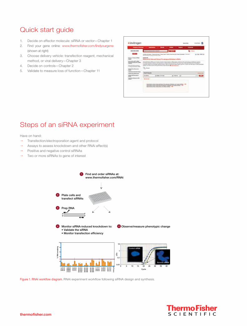
Figure 1. RNAi workflow diagram. RNAi experiment workflow following siRNA design and synthesis.
Steps of an siRNA experiment
Have on hand:
→ Transfection/electroporation agent and protocol
→ Assays to assess knockdown and other RNAi effect(s)
→ Positive and negative control siRNAs
→ Two or more siRNAs to gene of interest
Find and order siRNAs at:www.thermofisher.com/RNAi
Monitor siRNA-induced knockdown to:• Validate the siRNA• Monitor transfection efficiency
Observe/measure phenotypic change
Plate cells and transfect siRNAs
1
2
Prep RNA3
4 4
10
100 5 15
Cycle
Control siRNA
Survivin siRNA
20 25 30 35 40
1
0.1
0.010
20
40
60
80
100
NEK
4-1
NEK
4-2
NEK
4-3
TGFB
R2-
1TG
FBR
2-2
TGFB
R2-
3
STK
6-1
STK
6-2
STK
6-3
RP
S6K
A4-
1R
PS
6KA
4-2
RP
S6K
A4-
3
MA
P2K
1-1
MA
P2K
1-2
MA
P2K
1-3
MA
P2K
4-1
MA
P2K
4-2
MA
P2K
4-3
WEE
1-1
WEE
1-2
WEE
1-3
PK
428-
1P
K42
8-2
PK
428-
3
STK
12-1
STK
12-2
STK
12-3
PC
NA
gold
% R
NA
rem
aini
ng
30
t5'3'
3'5'
Rn

Glossary of common RNAi terms
RNAiRibonucleic acid interference (first used by A. Fire and C. Mello et al., 1998).
siRNAShort interfering RNA. siRNAs are 21–25 bp dsRNA with dinucleotide 3’ overhangs that are processed from longer dsRNA by Dicer
in the RNA interference pathway. Introduction of synthetic siRNAs can induce RNA interference in mammalian cells. siRNAs can also
originate from endogenous precursors.
shRNAShort hairpin RNA; also short interfering hairpin. shRNAs are used in vector-based approaches for supplying siRNA to cells to produce
stable gene silencing. A strong Pol III type promoter is used to drive transcription of a target sequence designed to form hairpins and
loops of variable length, which are processed by cellular siRNA machinery. Once in the cell the shRNA can decrease the expression
of a gene with complementary sequences by RNAi.
miR RNAiVectors that express microRNAs for RNAi. miRNAs are 19–23 nt single-stranded RNAs, originating from single-stranded precursor
transcripts that are characterised by imperfectly base-paired hairpins. miRNAs function in a silencing complex that is similar, if not
identical, to RISC.
Chemically modified siRNAsiRNA molecules which have chemical modifications.
RISCRNA-induced silencing complex (RISC). A nuclease complex, composed of proteins and siRNA, that targets and cleaves endogenous
mRNAs complementary to the siRNA within the RISC complex.
Off-target effectsEffects that occur when one or a few nontarget genes not specifically targeted show loss of gene function following the introduction of
an siRNA or d-siRNA pool. The effect may be mediated by the sense strand of an siRNA, which may initiate a loss-of-function response
from an unrelated gene. Off-target effects can also occur as a secondary effect of the antisense strand of a specific siRNA if it has
sufficient homology to knock down the expression of a nontarget gene.
i
Contents
thermofisher.com
SECTION I—RNA INTERFERENCE
CHAPTER 1
Introduction to RNAi . . . . . . . . . . . . . . . . . . . . . . . . . . . . . . . . . . . . . . . . . . . . . . . . . . 2
Make your RNA interference experiments simple, stress-free, and successful . . . . . . . . . . . . . . . . . . . . . . . 2
A brief history of RNAi . . . . . . . . . . . . . . . . . . . . . . . . . . . . . . . . . . . . . . . . . . . . . . . . 2
How RNAi works . . . . . . . . . . . . . . . . . . . . . . . . . . . . . . . . . . . . . . . . . . . . . . . . . . . 2
Eight tips for a successful siRNA experiment . . . . . . . . . . . . . . . . . . . . . . . . . . . . . . . . . . . . . . 3
Considerations . . . . . . . . . . . . . . . . . . . . . . . . . . . . . . . . . . . . . . . . . . . . . . . . . . . . 3
Interview with Gregory Hannon . . . . . . . . . . . . . . . . . . . . . . . . . . . . . . . . . . . . . . . . . . . . 4
CHAPTER 2
Controls for RNAi experiments. . . . . . . . . . . . . . . . . . . . . . . . . . . . . . . . . . . . . . . . . . . . . . 7
Transfection controls . . . . . . . . . . . . . . . . . . . . . . . . . . . . . . . . . . . . . . . . . . . . . . . . . 7
Negative controls. . . . . . . . . . . . . . . . . . . . . . . . . . . . . . . . . . . . . . . . . . . . . . . . . . . 8
Positive controls . . . . . . . . . . . . . . . . . . . . . . . . . . . . . . . . . . . . . . . . . . . . . . . . . . . 8
Downstream controls . . . . . . . . . . . . . . . . . . . . . . . . . . . . . . . . . . . . . . . . . . . . . . . . . 9
Interferon controls . . . . . . . . . . . . . . . . . . . . . . . . . . . . . . . . . . . . . . . . . . . . . . . . . . 9
Use multiple siRNA sequences per target to verify results . . . . . . . . . . . . . . . . . . . . . . . . . . . . . . . . 9
Titrate siRNA. . . . . . . . . . . . . . . . . . . . . . . . . . . . . . . . . . . . . . . . . . . . . . . . . . . . . 9
Rescue experiments . . . . . . . . . . . . . . . . . . . . . . . . . . . . . . . . . . . . . . . . . . . . . . . . . 9
CHAPTER 3
Delivering RNAi to cells—transfection and viral delivery . . . . . . . . . . . . . . . . . . . . . . . . . . . . . . . . 10
Methods to achieve high transfection efficiency . . . . . . . . . . . . . . . . . . . . . . . . . . . . . . . . . . . . 10
Importance of minimising transfection-mediated cytotoxicity . . . . . . . . . . . . . . . . . . . . . . . . . . . . . . 10
Significance of reducing off-target effects . . . . . . . . . . . . . . . . . . . . . . . . . . . . . . . . . . . . . . . 11
Cell health . . . . . . . . . . . . . . . . . . . . . . . . . . . . . . . . . . . . . . . . . . . . . . . . . . . . . 14
Culture conditions . . . . . . . . . . . . . . . . . . . . . . . . . . . . . . . . . . . . . . . . . . . . . . . . . 14
Passage number . . . . . . . . . . . . . . . . . . . . . . . . . . . . . . . . . . . . . . . . . . . . . . . . . . 14
siRNA quality . . . . . . . . . . . . . . . . . . . . . . . . . . . . . . . . . . . . . . . . . . . . . . . . . . . 15
siRNA quantity . . . . . . . . . . . . . . . . . . . . . . . . . . . . . . . . . . . . . . . . . . . . . . . . . . . 15
Choice of transfection agent . . . . . . . . . . . . . . . . . . . . . . . . . . . . . . . . . . . . . . . . . . . . . 15
Volume of transfection agent . . . . . . . . . . . . . . . . . . . . . . . . . . . . . . . . . . . . . . . . . . . . 15
Exposure to transfection agent/siRNA complexes . . . . . . . . . . . . . . . . . . . . . . . . . . . . . . . . . . . 15
Presence of serum in the medium during transfection . . . . . . . . . . . . . . . . . . . . . . . . . . . . . . . . . 16
Optimising transfection experiments . . . . . . . . . . . . . . . . . . . . . . . . . . . . . . . . . . . . . . . . . 16
ContentsX
Contents

ii
Section IIIRNAi and Epigenetics Sourcebook
CHAPTER 4
Vector-based RNAi technologies. . . . . . . . . . . . . . . . . . . . . . . . . . . . . . . . . . . . . . . . . . . . 17
Introduction to adenoviral and lentiviral RNAi vectors for delivery . . . . . . . . . . . . . . . . . . . . . . . . . . . . 17
Lentiviral and adenoviral RNAi vectors—choose any cell type for RNAi. . . . . . . . . . . . . . . . . . . . . . . . . . 18
CHAPTER 5
In vivo RNAi . . . . . . . . . . . . . . . . . . . . . . . . . . . . . . . . . . . . . . . . . . . . . . . . . . . . . 19
RNAi molecules for in vivo applications . . . . . . . . . . . . . . . . . . . . . . . . . . . . . . . . . . . . . . . . 19
Choosing RNAi effector molecules . . . . . . . . . . . . . . . . . . . . . . . . . . . . . . . . . . . . . . . . . . 19
Choosing RNAi purity . . . . . . . . . . . . . . . . . . . . . . . . . . . . . . . . . . . . . . . . . . . . . . . . 20
Tracking delivered duplexes . . . . . . . . . . . . . . . . . . . . . . . . . . . . . . . . . . . . . . . . . . . . . 21
In vivo RNAi protocols . . . . . . . . . . . . . . . . . . . . . . . . . . . . . . . . . . . . . . . . . . . . . . . 22
Measuring RNA concentration . . . . . . . . . . . . . . . . . . . . . . . . . . . . . . . . . . . . . . . . . . . . 23
Harvesting tissue—RNA extraction from tissue . . . . . . . . . . . . . . . . . . . . . . . . . . . . . . . . . . . . 24
Sectioning tissue . . . . . . . . . . . . . . . . . . . . . . . . . . . . . . . . . . . . . . . . . . . . . . . . . . 24
Protein extraction from tissues. . . . . . . . . . . . . . . . . . . . . . . . . . . . . . . . . . . . . . . . . . . . 25
Frequently asked questions about in vivo RNAi . . . . . . . . . . . . . . . . . . . . . . . . . . . . . . . . . . . . 27
CHAPTER 6
siRNA screening . . . . . . . . . . . . . . . . . . . . . . . . . . . . . . . . . . . . . . . . . . . . . . . . . . . 29
Selecting an siRNA library . . . . . . . . . . . . . . . . . . . . . . . . . . . . . . . . . . . . . . . . . . . . . . 29
Screening with siRNA libraries . . . . . . . . . . . . . . . . . . . . . . . . . . . . . . . . . . . . . . . . . . . . 31
siRNA library screening workflow (5 steps) . . . . . . . . . . . . . . . . . . . . . . . . . . . . . . . . . . . . . . 32
Toxicity . . . . . . . . . . . . . . . . . . . . . . . . . . . . . . . . . . . . . . . . . . . . . . . . . . . . . . 34
Secondary screening: confirmation of candidates . . . . . . . . . . . . . . . . . . . . . . . . . . . . . . . . . . . 35
SECTION II—PRODUCTS FOR RNA INTERFERENCE
CHAPTER 7
siRNA technologies . . . . . . . . . . . . . . . . . . . . . . . . . . . . . . . . . . . . . . . . . . . . . . . . . . 40
Invitrogen™ Silencer™ Select siRNA and Stealth RNAi™ siRNA . . . . . . . . . . . . . . . . . . . . . . . . . . . . 40
siRNA selection guide. . . . . . . . . . . . . . . . . . . . . . . . . . . . . . . . . . . . . . . . . . . . . . . . 48
Invitrogen™ Silencer™ Select siRNA libraries . . . . . . . . . . . . . . . . . . . . . . . . . . . . . . . . . . . . . 50
Invitrogen™ Silencer™ Pre-designed and Validated siRNAs. . . . . . . . . . . . . . . . . . . . . . . . . . . . . . . 52
CHAPTER 8
Vector-based RNAi technologies. . . . . . . . . . . . . . . . . . . . . . . . . . . . . . . . . . . . . . . . . . . . 53
How vector-mediated RNAi works . . . . . . . . . . . . . . . . . . . . . . . . . . . . . . . . . . . . . . . . . . 56
Invitrogen™ BLOCK-iT™ Pol II miR RNAi expression vectors—versatile Pol II vector-based RNAi technology . . . . . . . . 57

iii
Contents
thermofisher.com
Combine Invitrogen™ BLOCK-iT™ vector kits with lentivirus for stable, long-term expression . . . . . . . . . . . . . . . 58
Invitrogen™ Gateway™ compatibility for expanded experimental options . . . . . . . . . . . . . . . . . . . . . . . . 62
Invitrogen™ BLOCK-iT™ miR RNAi Select . . . . . . . . . . . . . . . . . . . . . . . . . . . . . . . . . . . . . . 64
Guaranteed results with Invitrogen™ BLOCK-iT™ miR RNAi Select 4 Sets . . . . . . . . . . . . . . . . . . . . . . . . 65
Invitrogen™ BLOCK-iT™ RNAi Entry Vector Kits. . . . . . . . . . . . . . . . . . . . . . . . . . . . . . . . . . . . 66
Lentiviral and adenoviral RNAi vectors . . . . . . . . . . . . . . . . . . . . . . . . . . . . . . . . . . . . . . . . 68
Powerful shRNA delivery with Invitrogen™ BLOCK-iT™ viral vectors. . . . . . . . . . . . . . . . . . . . . . . . . . . 69
Invitrogen™ BLOCK-iT™ Lentiviral RNAi System . . . . . . . . . . . . . . . . . . . . . . . . . . . . . . . . . . . 70
Invitrogen™ BLOCK-iT™ Adenoviral RNAi Expression System . . . . . . . . . . . . . . . . . . . . . . . . . . . . . 71
CHAPTER 9
RNAi delivery . . . . . . . . . . . . . . . . . . . . . . . . . . . . . . . . . . . . . . . . . . . . . . . . . . . . . 72
Introduction to RNAi delivery . . . . . . . . . . . . . . . . . . . . . . . . . . . . . . . . . . . . . . . . . . . . 72
Invitrogen™ Lipofectamine™ RNAiMAX Transfection Reagent— unmatched gene silencing with reduced cytotoxicity for siRNA experiments . . . . . . . . . . . . . . . . . . . . . . . . . . . . .73
Invitrogen™ Lipofectamine™ 2000 and LTX Transfection Reagents—ideal for delivery of shRNA and miR RNAi vectors . . . 75
Invitrogen™ Oligofectamine™ Transfection Reagent—potent internalization of RNA oligos . . . . . . . . . . . . . . . . . 76
Invitrogen™ Neon™ Transfection System . . . . . . . . . . . . . . . . . . . . . . . . . . . . . . . . . . . . . . . 76
CHAPTER 10
RNA interference controls . . . . . . . . . . . . . . . . . . . . . . . . . . . . . . . . . . . . . . . . . . . . . . . 79
Controls for RNAi experiments . . . . . . . . . . . . . . . . . . . . . . . . . . . . . . . . . . . . . . . . . . . . 79
Invitrogen™ BLOCK-iT™ Alexa Fluor™ Red Fluorescent Control and green BLOCK-iT™ Fluorescent Oligo . . . . . . . . . 80
BLOCK-iT™ Transfection Kit—simple optimisation of Lipofectamine 2000 transfection conditions . . . . . . . . . . . . . 81
Invitrogen™ Silencer™ Select positive and negative control siRNAs . . . . . . . . . . . . . . . . . . . . . . . . . . . 82
Invitrogen™ Stealth RNAi™ siRNA negative controls—ideal negative controls for every experiment . . . . . . . . . . . . . 83
Invitrogen™ Stealth RNAi™ siRNA reporter controls . . . . . . . . . . . . . . . . . . . . . . . . . . . . . . . . . . 84
Invitrogen™ Stealth RNAi™ siRNA positive controls . . . . . . . . . . . . . . . . . . . . . . . . . . . . . . . . . . 84
Invitrogen™ BLOCK-iT™ Transfection Optimisation Kit (Human). . . . . . . . . . . . . . . . . . . . . . . . . . . . . 84
Optimise RNAi experiments with the BLOCK-iT Fluorescent Oligo . . . . . . . . . . . . . . . . . . . . . . . . . . . . 85
Positive controls for BLOCK-iT Pol II miR RNAi vectors . . . . . . . . . . . . . . . . . . . . . . . . . . . . . . . . . 85
Silencer negative control siRNAs. . . . . . . . . . . . . . . . . . . . . . . . . . . . . . . . . . . . . . . . . . . 86
Silencer positive control siRNAs . . . . . . . . . . . . . . . . . . . . . . . . . . . . . . . . . . . . . . . . . . . 87
CHAPTER 11
Measuring knockdown . . . . . . . . . . . . . . . . . . . . . . . . . . . . . . . . . . . . . . . . . . . . . . . . 88
Functional validation following RNAi knockdown. . . . . . . . . . . . . . . . . . . . . . . . . . . . . . . . . . . . 88
Applied Biosystems™ TaqMan™ Gene Expression Assays . . . . . . . . . . . . . . . . . . . . . . . . . . . . . . . 88
Custom TaqMan™ Gene Expression Assays . . . . . . . . . . . . . . . . . . . . . . . . . . . . . . . . . . . . . 89
Custom TaqMan™ Probes and Primers. . . . . . . . . . . . . . . . . . . . . . . . . . . . . . . . . . . . . . . . 89
qRT-PCR directly from cells . . . . . . . . . . . . . . . . . . . . . . . . . . . . . . . . . . . . . . . . . . . . . 90
Protein separation and western blotting. . . . . . . . . . . . . . . . . . . . . . . . . . . . . . . . . . . . . . . . 91
ContentsX
Contents
iv
Section IIIRNAi and Epigenetics Sourcebook
Antibodies and immunodetection . . . . . . . . . . . . . . . . . . . . . . . . . . . . . . . . . . . . . . . . . . 91
Nucleic acid purification and quantification . . . . . . . . . . . . . . . . . . . . . . . . . . . . . . . . . . . . . . 91
Gene expression microarray analysis . . . . . . . . . . . . . . . . . . . . . . . . . . . . . . . . . . . . . . . . . 91
CHAPTER 12
RNAi services . . . . . . . . . . . . . . . . . . . . . . . . . . . . . . . . . . . . . . . . . . . . . . . . . . . . 92
RNAi services . . . . . . . . . . . . . . . . . . . . . . . . . . . . . . . . . . . . . . . . . . . . . . . . . . . 92
RNAi design services . . . . . . . . . . . . . . . . . . . . . . . . . . . . . . . . . . . . . . . . . . . . . . . . 93
Stealth RNAi, Silencer Select, and Silencer siRNA custom synthesis. . . . . . . . . . . . . . . . . . . . . . . . . . . 93
Vector-based RNAi services . . . . . . . . . . . . . . . . . . . . . . . . . . . . . . . . . . . . . . . . . . . . . 93
Delivery optimisation services . . . . . . . . . . . . . . . . . . . . . . . . . . . . . . . . . . . . . . . . . . . . 94
Viral production . . . . . . . . . . . . . . . . . . . . . . . . . . . . . . . . . . . . . . . . . . . . . . . . . . 95
RNAi functional validation services . . . . . . . . . . . . . . . . . . . . . . . . . . . . . . . . . . . . . . . . . . 96
Phenotypic assay development and high-throughput screening services. . . . . . . . . . . . . . . . . . . . . . . . . 97
RNAi custom collaborative research . . . . . . . . . . . . . . . . . . . . . . . . . . . . . . . . . . . . . . . . . 97
CHAPTER 13
Overview of miRNAs . . . . . . . . . . . . . . . . . . . . . . . . . . . . . . . . . . . . . . . . . . . . . . . . . 98
Which miRNA profiling or validation system should I use?. . . . . . . . . . . . . . . . . . . . . . . . . . . . . . . . 99
Genome-wide small RNA discovery and profiling . . . . . . . . . . . . . . . . . . . . . . . . . . . . . . . . . . .100
MicroRNA: a new frontier in biology . . . . . . . . . . . . . . . . . . . . . . . . . . . . . . . . . . . . . . . . .100
Fundamental questions in microRNA research . . . . . . . . . . . . . . . . . . . . . . . . . . . . . . . . . . . .100
Capture the sequence and expression level of every small RNA in your sample . . . . . . . . . . . . . . . . . . . . . .101
Genome-wide small RNA discovery and profiling workflow . . . . . . . . . . . . . . . . . . . . . . . . . . . . . . .101
Robust small RNA discovery and profiling using the leading next-generation sequencing platform . . . . . . . . . . . . .102
Profile microRNA expression in a single day using gold standard TaqMan assay technology . . . . . . . . . . . . . . . .103
Detect and quantify specific microRNAs . . . . . . . . . . . . . . . . . . . . . . . . . . . . . . . . . . . . . . .106
Analyse microRNA function . . . . . . . . . . . . . . . . . . . . . . . . . . . . . . . . . . . . . . . . . . . . .108
Sample preparation tailored for microRNA experiments . . . . . . . . . . . . . . . . . . . . . . . . . . . . . . . . 112
CHAPTER 14
Optimised miRNA profiling. . . . . . . . . . . . . . . . . . . . . . . . . . . . . . . . . . . . . . . . . . . . . . . 115
Invitrogen™ NCode™ platform—for optimised miRNA profiling . . . . . . . . . . . . . . . . . . . . . . . . . . . . . 115
Efficient isolation of total RNA (including miRNA) using Invitrogen™ TRIzol™ Reagent . . . . . . . . . . . . . . . . . . . 116
Reliable recovery of total RNA (including miRNA) from FFPE samples with the Invitrogen™ RecoverAll™ Total Nucleic Acid Isolation Kit . . . . . . . . . . . . . . . . . . . . . . . . . . . . . . . 116
Simplified miRNA fluorescent labeling . . . . . . . . . . . . . . . . . . . . . . . . . . . . . . . . . . . . . . . . 118
Ultrasensitive miRNA amplification . . . . . . . . . . . . . . . . . . . . . . . . . . . . . . . . . . . . . . . . . . 119
Comprehensive miRNA expression profiling . . . . . . . . . . . . . . . . . . . . . . . . . . . . . . . . . . . . . .120
Invitrogen™ NCode™ Non-coding RNA Arrays . . . . . . . . . . . . . . . . . . . . . . . . . . . . . . . . . . . .124
Genome-wide analysis of long non-coding RNA (ncRNA) expression in solid tumours using the NCode Non-coding RNA Microarray platform. . . . . . . . . . . . . . . . . . . . . . . . . . . . . . . .126

v
Contents
thermofisher.com
SECTION III—miRNA PROFILING
CHAPTER 15
Introduction to DNA methylation . . . . . . . . . . . . . . . . . . . . . . . . . . . . . . . . . . . . . . . . . . . .134
Overview of DNA methylation . . . . . . . . . . . . . . . . . . . . . . . . . . . . . . . . . . . . . . . . . . . .134
DNA methylation analysis . . . . . . . . . . . . . . . . . . . . . . . . . . . . . . . . . . . . . . . . . . . . . .134
Methylated DNA enrichment . . . . . . . . . . . . . . . . . . . . . . . . . . . . . . . . . . . . . . . . . . . . .134
Enrichment of differentially methylated regions with Applied Biosystems™ MethylMiner™ fractionation and deep sequencing with the Applied Biosystems™ SOLiD™ System. . . . . . . . . . . . . . . . . . .136
Locus-specific DNA methylation analysis . . . . . . . . . . . . . . . . . . . . . . . . . . . . . . . . . . . . . . .142
Genome-wide methylation. . . . . . . . . . . . . . . . . . . . . . . . . . . . . . . . . . . . . . . . . . . . . .142
Invitrogen™ PureLink™ reagents—reliable isolation of genomic DNA . . . . . . . . . . . . . . . . . . . . . . . . . .143
Invitrogen™ MethylCode™ bisulfite treatment—fast and complete DNA conversion . . . . . . . . . . . . . . . . . . . .144
Detection of bisulfite-modified DNA . . . . . . . . . . . . . . . . . . . . . . . . . . . . . . . . . . . . . . . . .145
High-fidelity reagents for amplifying bisulfite-treated DNA and methylation-specific PCR (MSP)* . . . . . . . . . . . . . .145
Simplify your preparation for bisulfite genomic sequencing . . . . . . . . . . . . . . . . . . . . . . . . . . . . . . .146
Top-rated sequencing vectors . . . . . . . . . . . . . . . . . . . . . . . . . . . . . . . . . . . . . . . . . . . .146
SECTION IV—HISTONE MODIFICATIONS AND CHROMATIN REMODELLING
CHAPTER 16
Histone modifications and chromatin remodelling. . . . . . . . . . . . . . . . . . . . . . . . . . . . . . . . . . . .150
Investigating histone modifications and chromatin remodelling . . . . . . . . . . . . . . . . . . . . . . . . . . . . . 151
Applied Biosystems™ MAGnify™ ChIP . . . . . . . . . . . . . . . . . . . . . . . . . . . . . . . . . . . . . . . . 151
Applied Biosystems™ MAGnify™ Chromatin Immunoprecipitation System . . . . . . . . . . . . . . . . . . . . . . . .152
Antibodies qualified for chromatin immunoprecipitation . . . . . . . . . . . . . . . . . . . . . . . . . . . . . . . .154
Applied Biosystems™ DynaMag™-PCR . . . . . . . . . . . . . . . . . . . . . . . . . . . . . . . . . . . . . . .154
Quantitative PCR (qPCR) of ChIP-ready DNA . . . . . . . . . . . . . . . . . . . . . . . . . . . . . . . . . . . . .155
ContentsX
Contents


thermofisher.com
Section I—RNA Interference
Chapter 1—Introduction to RNAi . . . . . . . . . . . . . . . . . . . . . 2
Chapter 2—Controls for RNAi experiments . . . . . . . . . . . . . . . . 7
Chapter 3—Delivering RNAi to cells—transfection and viral delivery . . . . 10
Chapter 4—Vector-based RNAi technologies . . . . . . . . . . . . . . . 17
Chapter 5—In vivo RNAi . . . . . . . . . . . . . . . . . . . . . . . . . 19
Chapter 6—siRNA screening . . . . . . . . . . . . . . . . . . . . . . . 29
2 Important Licensing Information: These products may be covered by one or more Limited Use Label Licenses (See Thermo Fisher Scientific Catalogue or our website, www.thermofisher.com). By use of these products you accept the terms and conditions of all applicable Limited Use Label Licenses. All products are for research use only. CAUTION: Not intended for human or animal diagnostic or therapeutic uses.
Section I RNA Interference
CHAPTER 1
Introduction to RNAi
Make your RNA interference experiments simple, stress-free, and successful
RNA interference (RNAi) is one of the most important technological breakthroughs in modern biology, allowing us to directly
observe the effects of the loss of function of specific genes in mammalian systems. Once viewed as a technique used only by select
laboratories, RNAi is now considered essential for studying gene function. It has become a prominent tool for protein knockdown
studies, phenotype analysis, function recovery, pathway analysis, in vivo knockdown, and drug target discovery.
A brief history of RNAi
In the early 1990s, scientists first observed that RNA inhibited protein expression in plants and fungi. In 1998, Andrew Fire and Craig
Mello, working with Caenorhabditis elegans, discovered that double-stranded RNA (dsRNA) was the source of the inhibition, and they
called this phenomenon RNA interference (RNAi). While studies in C. elegans were encouraging, the use of RNAi was limited to lower
organisms because delivering long dsRNA for RNAi was nonspecifically inhibitory in mammalian cells. In 2001, shorter RNAs (siRNA)
were shown to directly trigger RNAi in mammalian cells, without evoking the nonspecific effects observed with longer dsRNAs. In
2006, just 8 years after the discovery of siRNA, the Nobel Prize in Physiology or Medicine was awarded to Fire and Mello for their
discovery, underscoring the importance of RNAi as an investigative tool.
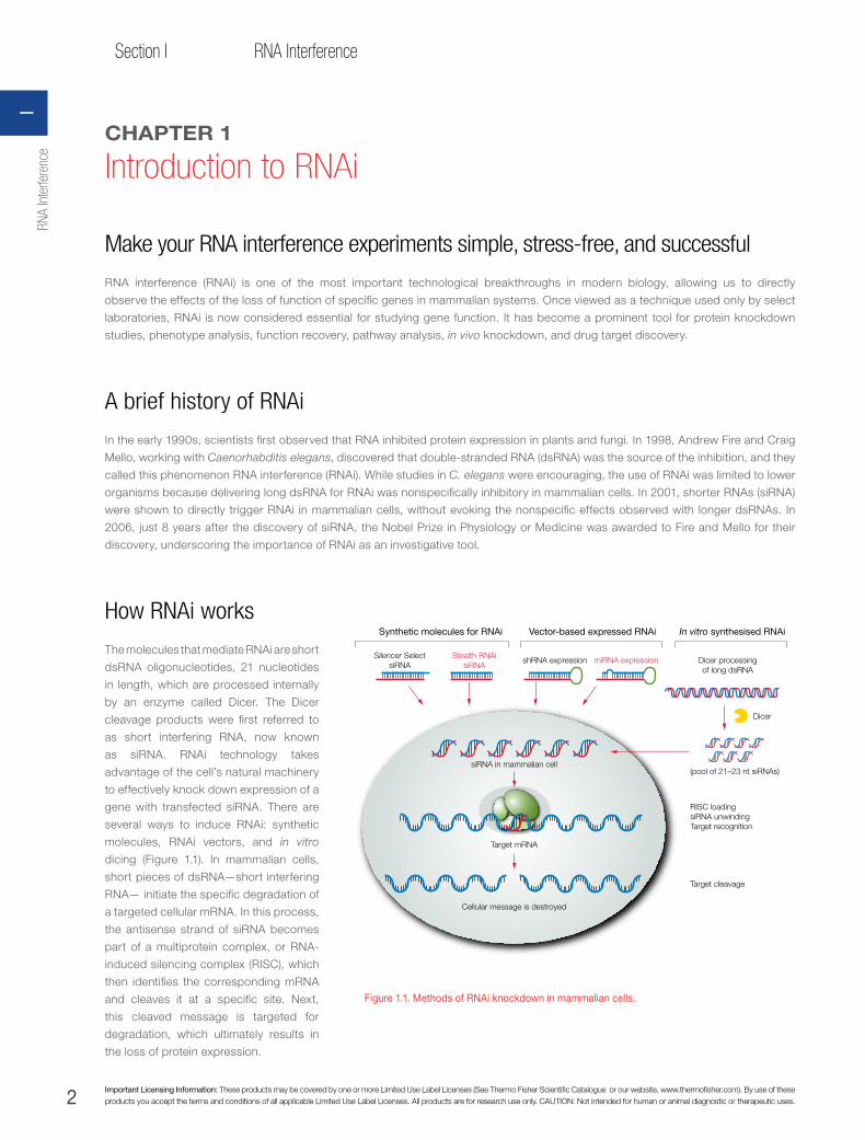
How RNAi works
The molecules that mediate RNAi are short
dsRNA oligonucleotides, 21 nucleotides
in length, which are processed internally
by an enzyme called Dicer. The Dicer
cleavage products were first referred to
as short interfering RNA, now known
as siRNA. RNAi technology takes
advantage of the cell’s natural machinery
to effectively knock down expression of a
gene with transfected siRNA. There are
several ways to induce RNAi: synthetic
molecules, RNAi vectors, and in vitro
dicing (Figure 1.1). In mammalian cells,
short pieces of dsRNA—short interfering
RNA— initiate the specific degradation of
a targeted cellular mRNA. In this process,
the antisense strand of siRNA becomes
part of a multiprotein complex, or RNA-
induced silencing complex (RISC), which
then identifies the corresponding mRNA
and cleaves it at a specific site. Next,
this cleaved message is targeted for
degradation, which ultimately results in
the loss of protein expression.
Silencer Select siRNA
Stealth RNAisiRNA
shRNA expression
siRNA in mammalian cell
Target mRNA
Cellular message is destroyed
miRNA expression Dicer processing of long dsRNA
(pool of 21–23 nt siRNAs)
Target cleavage
RISC loading siRNA unwindingTarget recognition
Dicer
Synthetic molecules for RNAi Vector-based expressed RNAi In vitro synthesised RNAi
Figure 1.1. Methods of RNAi knockdown in mammalian cells.
RNA
Inte
rfere
nce
I

3
Chapter 1Introduction to RNAi
thermofisher.com
Eight tips for a successful siRNA experiment
1. Go to www.thermofisher.com/rnai and utilise the best-in-class design algorithms to design your siRNAs. Do not attempt to
design siRNAs on your own.
2. Avoid RNases! Trace amounts of ribonucleases can sabotage siRNA experiments. Since RNases are present throughout the
laboratory environment on your skin, in the air, on anything touched by bare hands or on anything left open to the air, it is
important to take steps to prevent and eliminate RNase contamination. We offer a complete line of products designed to detect
and eliminate RNases.
3. Maintain healthy cell cultures and strict protocols for good transfection reproducibility. In general, healthy cells are transfected
at higher efficiency than poorly maintained cells. Routinely subculturing cells at a low passage number ensures that there will
be minimal instability in continuous cell lines from one experiment to the next. When performing optimization experiments we
recommend transfecting cells within 50 passages, since transfection efficiency drops over time.
4. Avoid antibiotic use. Avoid the use of antibiotics during plating and up to 72 hours after transfection. Antibiotics have been
shown to accumulate to toxic levels in permeabilised cells. Additionally, some cells and transfection reagents require serum-free
conditions for optimal siRNA delivery. We suggest you perform a pilot transfection experiment in both normal growth media and
serum-free media to determine the best condition for each transfection.
5. Transfect siRNAs using optimised reagents. Use an optimised siRNA transfection reagent and protocol for your cell type. The choice of
transfection reagent is critical for success in siRNA experiments. It is essential to use transfection reagents formulated to deliver small RNAs
(most commercially available transfection reagents were designed for large plasmid DNA, not small RNA molecules). Also, some reagents
have been developed for the transfection of specific cell lines while others have broader specificity.
NOTE: Lipofectamine RNAiMAX (Cat. No. 137780-75) is our best-performing transfection reagent for siRNA. See page 73 for more information.
6. Use an appropriate positive control to optimise transfection and assay conditions. Housekeeping genes are suitable positive
controls for most cell types. To optimise conditions, transfect target cells with several concentrations of an siRNA specific to
your chosen positive control and to your experimental target siRNA. Measure the reduction in the control protein or mRNA level
compared to untransfected cells 48 hours after transfection. Too much siRNA can lead to cell toxicity and death. For maximum
convenience, we offer positive control siRNAs against a variety of gene targets.
7. Use a negative control siRNA to distinguish nonspecific effects. Negative controls should be designed by scrambling the
nucleotide sequence of the most active siRNA. However, be sure to perform a homology search to ensure that your negative
control sequence lacks homology to the genome of the organism being studied.
8. Use labelled siRNAs for protocol optimisation. Fluorescently labelled siRNA can be used to analyse siRNA stability and transfection
efficiency. Labelled siRNA is also useful to study siRNA subcellular localisation and in double label experiments (with a labelled antibody) to
visualise cells that receive siRNA during transfection and to correlate transfection with down-regulation of the target protein.
Considerations
siRNA vs. vector approachesBoth siRNA and vector-based RNAi can be
extremely effective at producing loss of function
phenotypes. In general, most researchers choose
siRNA because they can start quickly and there
are no special preparations needed other than
basic cell culture techniques. However, there are
a number of reasons why a researcher might
choose either siRNA or a vector-based RNAi.
Table 1.1 contains criteria that will help you make
the decision.
Table 1.1. Synthetic siRNA vs. vector-expressed siRNA.
siRNARNAi Vectors miR RNAi &
shRNA
Long-term stable knockdown •
Inducible knockdown •
Delivery to hard-to-transfect cells •
Least hands-on time •
Most immediate effect •
Higher potency likely •
Design more efficient •
Introduction to RNAi1

4 Important Licensing Information: These products may be covered by one or more Limited Use Label Licenses (See Thermo Fisher Scientific Catalogue or our website, www.thermofisher.com). By use of these products you accept the terms and conditions of all applicable Limited Use Label Licenses. All products are for research use only. CAUTION: Not intended for human or animal diagnostic or therapeutic uses.
Section I RNA Interference
CHOOSING siRNA FOR TARGET OF INTEREST—IMPROVED SPECIFICITYWhen choosing siRNA for a target of interest it is very important to use a state-of-the-art algorithm for choosing your siRNA.
For example, Silencer Select siRNA’s five-step bioinformatic filtering process allows for elimination of siRNAs that are predicted
to elicit off-target effects.
www.thermofisher.com/silencerselect
With Stealth Select RNAi siRNA, the work of designing highly effective sequences has been done for you. The development
of these molecules begins with identifying all human, rat, and mouse transcripts in the Unigene database. In four steps we
select highly effective in silico-designed sequences. www.thermofisher.com/stealthrnai
See page 40 for Silencer Select siRNA and page 44 for Stealth Select RNAi siRNA.
Interview with Gregory Hannon
Q. Research into RNA interference (RNAi) and its emerging use as a tool to explore gene function has taken the research community by storm. Can you tell us how you first became interested in RNAi?Dr. Hannon: It’s sort of interesting, actually. I was at a Pew Scholars Meeting; it was my first year.
Craig Mello was also a Pew Scholar, and he gave a small “chalk talk” at the meeting—although we were in the bowels of Mexico
somewhere, so there was no chalk. He presented this really interesting phenomenon. It was either just before or just after the first
RNAi paper was published. It really excited me and we chatted a couple times about it at the meeting. But we didn’t really do anything
about it, not being C. elegans biologists. His theory just sort of percolated for a year.
Then the next year, again at the Pew Scholars Meeting, Rich Carthew showed that RNAi worked in Drosophila. He was doing
this by embryo injection. And that’s what really pulled me in. Here was something that was not just a biological oddity of C. elegans (I
was unaware of the plant work at that time so I hadn’t thought about it very deeply). The notion that this phenomenon was going to be
universal really captivated me. This was partly because I was spending a lot of effort trying to do forward genetics in cultured mammalian
cells using things like retroviral libraries. I saw the RNAi phenomenon as, one day long down the road, something that could complement
over expression approaches, and that would give us the loss-of-function tool that mammalian systems have always lacked.
Q. Wow, “…one day long down the road…”, I bet you were surprised at how quickly this field has progressed.Dr. Hannon: Well, that’s what got us started in RNAi. We started thinking about the cell culture models from Drosophila. Our initial goal
had been to try to use S2 cells as a model for studying gene function. And so I actually called the lab from that Pew Meeting and said,
“…You know what? Get out those Drosophila cells and see if they do RNAi…” And they did.
Q. So how did your interest in using RNAi to study gene function morph into your work focusing on the mechanism of RNAi?Dr. Hannon: We got hooked on this technique in part because I have a background in RNA processing—I worked on trans-splicing
with Tim Nilsen as a graduate student—so there was the possibility that we would have just run with the whole notion of doing gene
function in S2 cells. As it turns out, for most of the things that we were interested in—cell cycle control, and such—S2 cells are a terrible
system. But we said, okay, since nobody understands much about the RNAi mechanism yet, let’s play for a little while and see if we
can generate an in vitro system from cultured cells that we can use to try to figure out how this all works. And we sort of got sucked
down the mechanism path from there.
Q. Your lab, Mello’s lab, and others published research indicating that Dicer is the nuclease that digests long dsRNA into siRNAs which in turn mediate RNAi. Can you describe some of the experiments that led you to this conclusion?Dr. Hannon: We had approached this purely from a biochemical standpoint, where we had taken a “candidate gene approach”. We
tagged all of these proteins, did IPs (immunoprecipitations), and looked for activity. It led to a beautiful biochemical correlation between
Dicer activity and siRNA. In other words, we had an enzyme that did basically what it was supposed to do. But any time you have a
biochemical result, you have to be somewhat concerned about whether or not the story you’ve derived based on in vitro experiments
has any basis in reality. To really know that something is involved in any kind of specific pathway you need genetics. Ultimately the
proof is always in the genetics. And we had done a really crappy experiment, which was to use RNAi to knock out Dicer—that’s sort
of become de rigueur in the field—so I don’t know if I should still be embarrassed about or not. But our primary motivation was just to
verify the hypothesis that Dicer was the initiating enzyme.
RNA
Inte
rfere
nce
I

5
Chapter 1Introduction to RNAi
thermofisher.com
Q. Now we know that Dicer is also important in a gene regulatory pathway involving short temporal RNA (stRNA) and your lab’s work helped to demonstrate that. How did you tie Dicer to the stRNA pathway?Dr. Hannon: Shortly after Emily left, Plasterk’s lab finally found a C. elegans Dicer mutant. It was initially somewhat worrying that there
weren’t somatic RNAi defects. Ronald tried to be reassuring and talked about maternal effects. But ultimately when they looked at
germ line transgenes, silencing of germ line transgenes was defective. And this was sort of a nice confirmation. I think at least now
there’s no doubt that Dicer really is the initiating enzyme for the process.
Q. What do you think will be the single greatest outcome of research into RNA silencing?Dr. Hannon: It’s really an impossible question to answer. One of the things that makes this field very hot at the moment is that in a way
it’s all things to all people. You have very interesting biology. For example, scientists are studying transposon silencing, the evolution
of the relationship between repetitive and mobile genetic elements, DNA parasites and their hosts, and the interaction between
viruses and their hosts. That group is interested because RNAi is involved, at least in plants. And there is also a definitive relationship
in C. elegans. RNAi seems to be involved in somehow controlling these kinds of nucleic acid parasites. But there are also people
looking at these microRNAs, at least for endogenous gene regulation, and saying, look, here’s a whole new regulatory paradigm
where there are hundreds of, what you might call orphan hairpins, running around out there. We don’t know what they do, we don’t
know who they regulate, we don’t know how prevalent this is, how general it is, or even really at what regulatory levels these different
things act. We know about two that act at the level of protein synthesis, but there’s nothing to say that others don’t act at the level of
message stability, or even at the level of directing the modification of chromosome structure. Here we may find that this is something
that is as important as the discovery of enhancer sequences, in terms of controlling gene expression itself.
You’ve also got a group of people who are interested in the basic mechanics of RNAi and what it means. Everybody is intrigued
about a mechanism where a worm can eat a piece of RNA and knock out a gene in its progeny. There’s something intrinsically
appealing about that—understanding the mechanism of that bit of biology. And there’s a whole group of people who want to
understand the biological ramifications of the system—that it may regulate development in plants, maybe stem cell identity, or
maintenance of stem cell character in both plants and animals.
And then, a much broader group of people, who don’t really care that much about mechanism, are just interested in harnessing
this phenomenon as a tool. So, it’s really impossible to predict the single greatest outcome of this research. And I think if you ask that
question of ten different people, you’d get ten different answers. The reason I can’t give you just one is because we’re interested in
all of them.
Q. Your lab and other labs have developed expression vectors that express siRNA long term. Do you see the use of siRNA expression vectors as a replacement to transfection of siRNAs for inducing RNAi, or are these techniques complementary?Dr. Hannon: Oh, I think they’re complementary. Very much so. It’s still early in terms of trying to understand the power of each of
these technologies. What seems to be true, at least this early on, is that siRNAs can get into cells at very low concentrations to
provoke a very good effect. I think the jury is still out on whether it’s easier to get an effect with an siRNA on a sort of per cell or whole
population basis. We don’t really have that much information on it. And I suspect eventually these things will run even because of
advances in different transfection technologies. But in terms of ease of use, nothing beats typing in a sequence, having a couple
oligos show up, and then dumping them onto cells, right? If you’re looking for a quick answer, and you only have one or two genes
that you want to look at, nothing is going to beat the idea that you can chemically synthesise these things, just in terms of ease.
Q. What are some of the advantages of expression vectors over chemically synthesised siRNAs?Dr. Hannon: The expression constructs are going to be powerful for a different reason. And maybe more appropriate for specific
sorts of experiments that involve much more long term analysis of phenotype, or biochemical studies that involve larger cell numbers
that might be more difficult or more expensive to do by transfecting each cell that you want to analyse.
Q. Cell logistic problems, right?Dr. Hannon: Right. There are a lot of phenotypes that you want to look at over long time scales or in mosaic animals. That’s where
the power of these kinds of expression constructs are going to be. Now, another advantage of the expression constructs is the fact
that they are propagatable—you make one and validate it and you have it forever. You never have to remake it. You never have to
reorder it—it sits in the freezer and you can trot it out and use it any time you want. We are finding that you can marry these sorts of
Introduction to RNAi1

6 Important Licensing Information: These products may be covered by one or more Limited Use Label Licenses (See Thermo Fisher Scientific Catalogue or our website, www.thermofisher.com). By use of these products you accept the terms and conditions of all applicable Limited Use Label Licenses. All products are for research use only. CAUTION: Not intended for human or animal diagnostic or therapeutic uses.
Section I RNA Interference
modular cassettes with pretty much any gene transfer technology that you want to talk about—viruses, etc. They will be useful with model
systems like tissue slices, where it might be more difficult to get siRNAs [inside cells] in good numbers.
Q. I’d like to ask you about your laboratory. How many people do you have working with you currently, and how are they split between postdocs, grad students, etc.?Dr. Hannon: We are at around 16 at the moment; 6 graduate students, a couple of visitors, about 4 or 5 postdocs, me, a few technicians,
and a research associate who is semi-independent who’s also working with me.
Q. We know that you’re the founder of the biotech company, Genetica. And you’re obviously busy as a professor at the Cold Spring Harbor labs, too. Do you still find time to work at the bench? And, if so, do you think that’s unusual for someone in your position?Dr. Hannon: Yeah, oh, definitely. I try to work at the bench every day. I’m not sure that I do anything useful, but I try to work. I think that
Cold Spring Harbor has, not necessarily a tradition, but a lot of the faculty here do work at the bench. And, if you think about it, it’s sort of
what got us involved in this whole RNAi work in the beginning. I find that it keeps me engaged much more in the day to day activities of
the lab. It also keeps me grounded in the reality of doing experiments and makes me much more understanding about students’ ligations
failing occasionally, because mine fail right alongside theirs. And I think that the work moves faster because I’m there. I’m available. We talk
about things more—everything from the biology of RNAi to the nitty gritty details of lab work, like, “Gee, this isn’t working,” and “Maybe I’ve
seen that before…” That helps us troubleshoot and move things along a little bit more quickly. I like being in the lab, and I like interacting
with the people in my lab.
Q. What’s next for Gregory Hannon?Dr. Hannon: I’m going to go do minipreps for one of my students, that’s what’s next.
About Dr. Hannon
Gregory J. Hannon received his PhD from Case Western Reserve University in 1992 and was a 1997 Pew Scholar. He is currently an
Associate Professor at the Cold Spring Harbor Laboratory, where his research is focused in part on determining the mechanism of RNAi,
as well as using RNAi as a tool in the study of cancer development and investigating the potential of siRNAs as cancer therapeutic agents.
Dr. Hannon and his colleagues have been at the forefront of many of the important discoveries in the RNAi area. In 2000 they
identified the nuclease activity responsible for dsRNA-guided mRNA degradation, now known as the RNA-induced silencing complex
(RISC). They identified one of the protein components of RISC, Argonaute2, as well as the enzyme (“Dicer”) that begins the RNAi process
by cutting long dsRNAs into siRNAs. Most recently they have shown the effectiveness of short hairpin RNAs (shRNAs) at gene silencing,
providing a longer-lasting alternative to siRNAs for much-needed functional studies.
Dr. Hannon is one of the founders of Genetica, Inc., a biotechnology company using RNAi and other tools to link genetic data with
biologic function via genetic manipulation of mammalian cells. Such genetic manipulations include RNAi-mediated stable silencing of
gene expression for the elucidation of disease pathways such as cancer development and subsequent drug target validation.
References 1. Paddison PJ, Hannon GJ (2002) Cancer Cell 2(1):17–23.
2. Hannon GJ (2002) Nature 418(6894):244–251.
3. Paddison PJ, Caudy AA, Bernstein E, Hannon GJ, Conklin DS (2002) Genes Dev 16(8):948–958.
4. Paddison PJ, Caudy AA, Hannon GJ (2002) Proc Natl Acad Sci U S A 99(3):1443–1448.
5. Bernstein E, Denli AM, Hannon GJ (2001) RNA 7(11):1509–1521.
6. Hammond SM, Boettcher S, Caudy AA, Kobayashi R, Hannon GJ (2001) Science 293(5532):1146–1150.
7. Hammond SM, Caudy AA, Hannon GJ (2001) Nat Rev Genet 2(2):110–119.
8. Bernstein E, Caudy AA, Hammond SM, Hannon GJ (2001) Nature 409(6818):363–366.
9. Hammond SM, Bernstein E, Beach D, Hannon GJ (2000) Nature 404(6775):293–296.
RNA
Inte
rfere
nce
I
7
Chapter 2Controls for RNAi experiments
thermofisher.com
CHAPTER 2
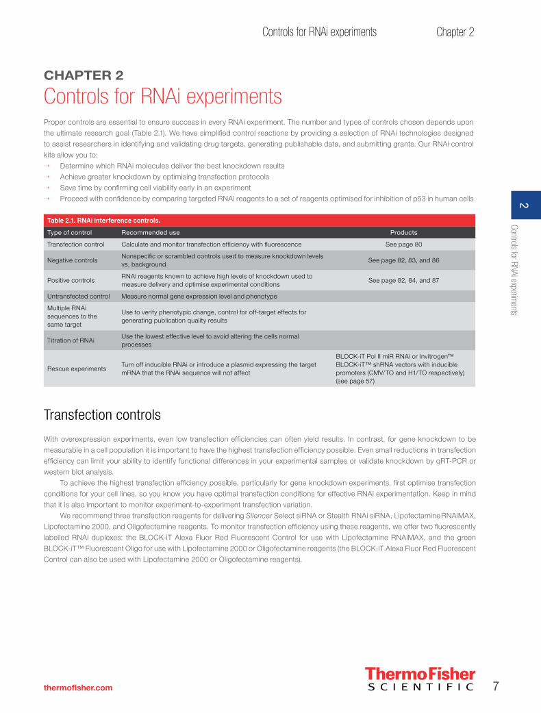
Controls for RNAi experimentsProper controls are essential to ensure success in every RNAi experiment. The number and types of controls chosen depends upon
the ultimate research goal (Table 2.1). We have simplified control reactions by providing a selection of RNAi technologies designed
to assist researchers in identifying and validating drug targets, generating publishable data, and submitting grants. Our RNAi control
kits allow you to:
→ Determine which RNAi molecules deliver the best knockdown results
→ Achieve greater knockdown by optimising transfection protocols
→ Save time by confirming cell viability early in an experiment
→ Proceed with confidence by comparing targeted RNAi reagents to a set of reagents optimised for inhibition of p53 in human cells
Table 2.1. RNAi interference controls.
Type of control Recommended use Products
Transfection control Calculate and monitor transfection efficiency with fluorescence See page 80
Negative controls Nonspecific or scrambled controls used to measure knockdown levels vs. background
See page 82, 83, and 86
Positive controls RNAi reagents known to achieve high levels of knockdown used to measure delivery and optimise experimental conditions
See page 82, 84, and 87
Untransfected control Measure normal gene expression level and phenotype
Multiple RNAi sequences to the same target
Use to verify phenotypic change, control for off-target effects for generating publication quality results
Titration of RNAi Use the lowest effective level to avoid altering the cells normal processes
Rescue experiments Turn off inducible RNAi or introduce a plasmid expressing the target mRNA that the RNAi sequence will not affect
BLOCK-iT Pol II miR RNAi or Invitrogen™ BLOCK-iT™ shRNA vectors with inducible promoters (CMV/TO and H1/TO respectively) (see page 57)
Transfection controls
With overexpression experiments, even low transfection efficiencies can often yield results. In contrast, for gene knockdown to be
measurable in a cell population it is important to have the highest transfection efficiency possible. Even small reductions in transfection
efficiency can limit your ability to identify functional differences in your experimental samples or validate knockdown by qRT-PCR or
western blot analysis.
To achieve the highest transfection efficiency possible, particularly for gene knockdown experiments, first optimise transfection
conditions for your cell lines, so you know you have optimal transfection conditions for effective RNAi experimentation. Keep in mind
that it is also important to monitor experiment-to-experiment transfection variation.
We recommend three transfection reagents for delivering Silencer Select siRNA or Stealth RNAi siRNA, Lipofectamine RNAiMAX,
Lipofectamine 2000, and Oligofectamine reagents. To monitor transfection efficiency using these reagents, we offer two fluorescently
labelled RNAi duplexes: the BLOCK-iT Alexa Fluor Red Fluorescent Control for use with Lipofectamine RNAiMAX, and the green
BLOCK-iT™ Fluorescent Oligo for use with Lipofectamine 2000 or Oligofectamine reagents (the BLOCK-iT Alexa Fluor Red Fluorescent
Control can also be used with Lipofectamine 2000 or Oligofectamine reagents).
Controls for RNAi experiments
2

8 Important Licensing Information: These products may be covered by one or more Limited Use Label Licenses (See Thermo Fisher Scientific Catalogue or our website, www.thermofisher.com). By use of these products you accept the terms and conditions of all applicable Limited Use Label Licenses. All products are for research use only. CAUTION: Not intended for human or animal diagnostic or therapeutic uses.
Section I RNA Interference
For more information on controls to use for optimising transfection with synthetic RNAi or a table listing the contents for each of
the transfection control kits, visit www.thermofisher.com/RNAicontrols.
We also recommend two transfection reagents for delivering RNAi vectors to cells: Lipofectamine 2000 and Lipofectamine LTX
reagents. We also recommend transfecting a vector that expresses a fluorescent protein to monitor cellular uptake of the RNAi vector
(BLOCK-iT Pol II miR RNAi Expression Vector with EmGFP).
For more information on controls to use for optimising transfection with RNAi vectors or a table listing the contents for each of the
transfection control kits, visit www.thermofisher.com/RNAivectorcontrols.
Negative controls
Negative control siRNAs—siRNAs with sequences that do not target any gene product—are essential for determining the effects
of siRNA delivery and for providing a baseline to compare siRNA-treated samples. There are two Silencer Select Negative Control
siRNAs. These siRNAs include the same modifications for reducing off-target effects as found in other Silencer Select siRNAs and
have no significant sequence similarity to mouse, rat, or human gene sequences. These negative control siRNAs have been tested
by microarray analysis and shown to have minimal effects on gene expression. In addition, these two controls have been tested in
multiparametric cell-based assays and are proven to have no significant effect on cell proliferation, viability, or morphology in the cell
lines tested.
By using the Invitrogen™ BLOCK-iT™ RNAi Designer, you can choose scrambled controls for any Stealth RNAi siRNA sequence.
We also provide a negative control in our RNAi vector cloning kits. The negative control should be the same chemical structure as the
target RNAi molecules. For example, if you are using shRNA vectors, the negative control should have the same vector backbone, but
a different RNAi sequence.
For experiments using Stealth RNAi siRNA, we have three predesigned negative controls with the following features:
→ Three levels of GC content to match that of the experimental Stealth RNAi siRNA
→ No homology to any known vertebrate gene
→ Tested sequences, which do not induce a stress response
We recommend using one or more negative control in every RNAi experiment.
Positive controls
Positive controls provide more confidence in your RNAi experiments by ensuring that the experimental conditions were met to achieve
robust data. Positive controls are RNAi molecules that are known to achieve high levels of knockdown (>70%). A positive control
should be used to optimise RNAi delivery conditions and to reconfirm high levels of delivery in each RNAi experiment. When a positive
control fails to produce the anticipated phenotype, carefully evaluate your experimental conditions and decide if some factors need to
be adjusted. Examples of positive controls are genes expressed at easily detectable levels, such as p53, lamin or GAPDH. However,
if you are looking at a particular phenotype such as apoptosis, you will most likely want to choose a positive control known to elicit
apoptosis, such as EG5.
RNA
Inte
rfere
nce
I
9
Chapter 2Controls for RNAi experiments
thermofisher.com
Downstream controls
Before transfecting cells and performing qRT-PCR and western blots to measure mRNA and protein levels, we recommend validating your
downstream reagents. Validating qRT-PCR primers or antibodies for your positive control and target genes before performing knockdown
experiments in your cell line ensures that your reagents are sensitive enough to detect changes in expression of your target gene due to
knockdown. Without sufficient sensitivity, it can be difficult to interpret knockdown results from genes or proteins with low expression levels.
Interferon controls
The introduction of RNAi reagents to cells can induce cellular stress response pathways, such as the interferon response. Activation
of these stress response pathways can lead to translational arrest, growth inhibition, and cellular toxicity. These events make it difficult
to assess whether observed cellular phenotypes are due to nonspecific stress responses or the loss of function of the targeted gene.
Validated qRT-PCR primers for PKR, IFIT-1 and 5’OAS stress response genes provide a specific and sensitive way to monitor whether
toxic cellular effects are complicating the interpretation of your RNAi experimental data.
Use multiple siRNA sequences per target to verify results
siRNA sequences with partial homology to other targets may contribute to off-target activity. Gene profiling experiments have shown that
duplexes with partial homology to other transcripts can cleave the target or act like a micro RNA (miRNA), inhibiting translation of the target mRNA.
Specificity studies have revealed that siRNA duplexes can have varying activities depending on the number, position, and base pair composition
of mismatches with respect to the target RNA. To ensure that knockdown of the intended gene causes a particular siRNA phenotype, the
phenotypic results should be confirmed by at least two siRNA molecules that target non-overlapping regions of the target mRNA. Thus, if one
siRNA sequence produces a particular phenotype, but the second siRNA sequence (designed to target the same gene) produces a different
phenotype, then you cannot conclude that the gene of interest was successfully knocked down.
Titrate siRNA
Silencer Select siRNA and Stealth RNAi siRNA can be very effective even at low concentrations. Titrating down the dose of the Silencer Select
siRNA and Stealth RNAi siRNA duplex enables you to reduce any off-target or nonspecific effects while achieving robust knockdown.
Rescue experiments
RNAi rescue experiments are performed to ensure that the observed effect is due to knocking down the target gene of interest. If you are using
an inducible RNAi vector system, turn off the RNAi expression by removing tetracycline from the medium. If you are using Silencer Select siRNA
or Stealth RNAi siRNA there are two main methods used to rescue the phenotype. The first method involves designing RNAi sequences to the
3’UTR and then transfecting the cells after knockdown with a vector expressing the the open reading frame (ORF) of the gene of interest. If the
RNAi sequences were designed to the ORF, you can use a mutagenesis kit to create one or more silent third-codon point mutations within the
region targeted by the RNAi sequence, preferably the seed and cleavage regions on the antisense strand (bases 2–12).
Controls for RNAi experiments
2
10 Important Licensing Information: These products may be covered by one or more Limited Use Label Licenses (See Thermo Fisher Scientific Catalogue or our website, www.thermofisher.com). By use of these products you accept the terms and conditions of all applicable Limited Use Label Licenses. All products are for research use only. CAUTION: Not intended for human or animal diagnostic or therapeutic uses.
Section I RNA Interference
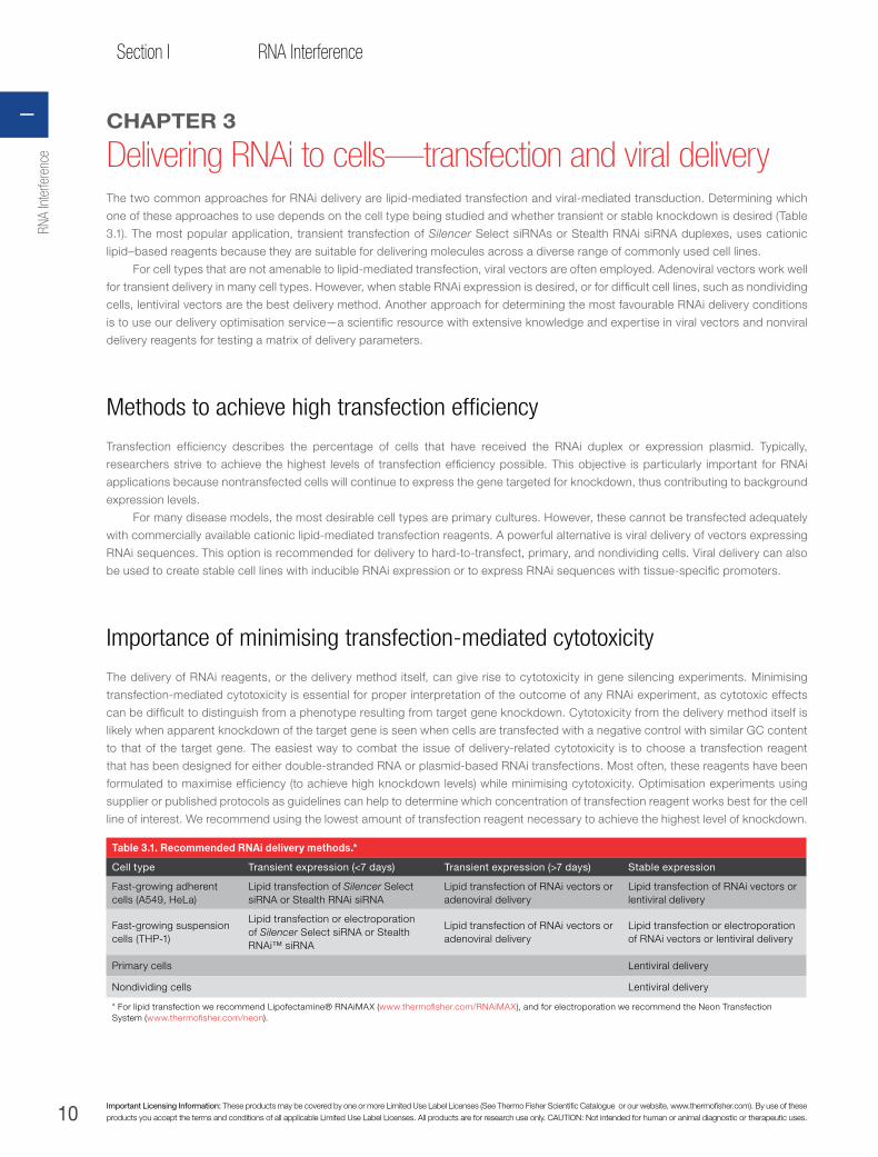
Table 3.1. Recommended RNAi delivery methods.*
Cell type Transient expression (<7 days) Transient expression (>7 days) Stable expression
Fast-growing adherent cells (A549, HeLa)
Lipid transfection of Silencer Select siRNA or Stealth RNAi siRNA
Lipid transfection of RNAi vectors or adenoviral delivery
Lipid transfection of RNAi vectors or lentiviral delivery
Fast-growing suspension cells (THP-1)
Lipid transfection or electroporation of Silencer Select siRNA or Stealth RNAi™ siRNA
Lipid transfection of RNAi vectors or adenoviral delivery
Lipid transfection or electroporation of RNAi vectors or lentiviral delivery
Primary cells Lentiviral delivery
Nondividing cells Lentiviral delivery
* For lipid transfection we recommend Lipofectamine® RNAiMAX (www.thermofisher.com/RNAiMAX), and for electroporation we recommend the Neon Transfection System (www.thermofisher.com/neon).
CHAPTER 3
Delivering RNAi to cells—transfection and viral deliveryThe two common approaches for RNAi delivery are lipid-mediated transfection and viral-mediated transduction. Determining which
one of these approaches to use depends on the cell type being studied and whether transient or stable knockdown is desired (Table
3.1). The most popular application, transient transfection of Silencer Select siRNAs or Stealth RNAi siRNA duplexes, uses cationic
lipid–based reagents because they are suitable for delivering molecules across a diverse range of commonly used cell lines.
For cell types that are not amenable to lipid-mediated transfection, viral vectors are often employed. Adenoviral vectors work well
for transient delivery in many cell types. However, when stable RNAi expression is desired, or for difficult cell lines, such as nondividing
cells, lentiviral vectors are the best delivery method. Another approach for determining the most favourable RNAi delivery conditions
is to use our delivery optimisation service—a scientific resource with extensive knowledge and expertise in viral vectors and nonviral
delivery reagents for testing a matrix of delivery parameters.
Methods to achieve high transfection efficiency
Transfection efficiency describes the percentage of cells that have received the RNAi duplex or expression plasmid. Typically,
researchers strive to achieve the highest levels of transfection efficiency possible. This objective is particularly important for RNAi
applications because nontransfected cells will continue to express the gene targeted for knockdown, thus contributing to background
expression levels.
For many disease models, the most desirable cell types are primary cultures. However, these cannot be transfected adequately
with commercially available cationic lipid-mediated transfection reagents. A powerful alternative is viral delivery of vectors expressing
RNAi sequences. This option is recommended for delivery to hard-to-transfect, primary, and nondividing cells. Viral delivery can also
be used to create stable cell lines with inducible RNAi expression or to express RNAi sequences with tissue-specific promoters.
Importance of minimising transfection-mediated cytotoxicity
The delivery of RNAi reagents, or the delivery method itself, can give rise to cytotoxicity in gene silencing experiments. Minimising
transfection-mediated cytotoxicity is essential for proper interpretation of the outcome of any RNAi experiment, as cytotoxic effects
can be difficult to distinguish from a phenotype resulting from target gene knockdown. Cytotoxicity from the delivery method itself is
likely when apparent knockdown of the target gene is seen when cells are transfected with a negative control with similar GC content
to that of the target gene. The easiest way to combat the issue of delivery-related cytotoxicity is to choose a transfection reagent
that has been designed for either double-stranded RNA or plasmid-based RNAi transfections. Most often, these reagents have been
formulated to maximise efficiency (to achieve high knockdown levels) while minimising cytotoxicity. Optimisation experiments using
supplier or published protocols as guidelines can help to determine which concentration of transfection reagent works best for the cell
line of interest. We recommend using the lowest amount of transfection reagent necessary to achieve the highest level of knockdown.
RNA
Inte
rfere
nce
I
11
Chapter 3Delivering RNAi to cells–transfection and viral delivery
thermofisher.com
FEATURED PROTOCOL
Transfecting Stealth RNAi siRNA or Silencer Select siRNA into A549 cells using Lipofectamine RNAiMAX ReagentIntroductionLipofectamine RNAiMAX Reagent is a proprietary formulation specifically developed for highly efficient delivery of Stealth RNAi siRNA or
Silencer Select siRNA to mammalian cells for RNAi analysis. This reference provides a recommended procedure to transfect Stealth RNAi
siRNA or Silencer Select siRNA into human A549 lung carcinoma cells (ATCC, Cat. No. CCL-185) using Lipofectamine RNAiMAX Reagent
(Cat. Nos. 13778-075, 13778-150). Lipofectamine RNAiMAX Reagent has a broad range of activity, enabling maximal knockdown levels
with a minimum of optimization required.
Important guidelines for transfectionFollow these important guidelines when transfecting Stealth RNAi siRNA or Silencer Select siRNA into A549 cells using Lipofectamine
RNAiMAX:
→ Both Reverse transfection and Forward transfection protocols (page 12) can be used for transfecting A549 cells.
→ To assess transfection efficiency, we recommend using a KIF11 Stealth Select RNAi siRNA, as described in Assessing transfection
efficiency (page 12).
→ We recommend using 10 nM of the siRNA duplex and indicated procedures. However, the efficacy of the RNAi sequence chosen,
the transcription rate of the target gene, and the stability of the resulting protein influence the degree of target gene knockdown
observed. You may need to adjust the RNAi concentration (1–50 nM can be used) and assay time (up to 72 hours) to establish
optimal knockdown of your target gene.
→ We recommend Gibco™ Opti-MEM™ I Reduced Serum Medium (Cat. No. 31985-062) to dilute RNAi duplexes and Lipofectamine
RNAiMAX Reagent before complexing.
→ Do not add antibiotics to media during transfection as this causes cell death.
→ Test serum-free media for compatibility with Lipofectamine RNAiMAX Reagent.
→ Lipofectamine RNAiMAX Reagent has a broad peak of activity; for a range of cell densities and volumes of transfection reagent
suitable for use, see Acceptable range for maximal activity (page 13).
Materials neededHave the following reagents on hand before beginning:
→ A549 cells maintained in DMEM (Cat. No. 11965-092) supplemented with 10% fetal bovine serum (Cat. No. 26140-079), 2 mM
glutamine (Cat. No. 25030-149), and penicillin/streptomycin (Cat. No. 15070-063)
Note: Use low-passage cells (<50 passages); make sure that cells are healthy and greater than 90% viable before transfection.
→ Silencer Select siRNA or Stealth RNAi siRNA of interest
→ Lipofectamine RNAiMAX Reagent (store at +2–8°C until use)
→ Opti-MEM I Reduced Serum Medium
→ Appropriate tissue culture plates and supplies
Significance of reducing off-target effects
As well as being a source of cytotoxicity, a suboptimal delivery reagent or excess reagent can result in apparent off-target effects. One cause of
off-target effects is the up- or down-regulation of genes due to the gene delivery procedure. However, with appropriate controls, these effects can
be identified and diminished. Again, use the lowest amount of transfection reagent that provides the best gene silencing activity.
The potential also exists for off-target effects due to knockdown from the siRNA duplex itself. To determine the most favourable conditions,
vary the concentration of siRNA while holding the concentration of transfection reagent constant at the lowest concentration previously identified.
Utilise the lowest siRNA concentration that gives the desired level of knockdown in RNAi experiments. Keep in mind that the specificity of the
siRNA will have an impact on potential off-target effects. The use of exceptionally specific reagents, such as Silencer Select siRNA or
modified Stealth RNAi siRNA duplexes, can help alleviate these concerns.
Delivering RNAi to cells–transfection and viral delivery3
12 Important Licensing Information: These products may be covered by one or more Limited Use Label Licenses (See Thermo Fisher Scientific Catalogue or our website, www.thermofisher.com). By use of these products you accept the terms and conditions of all applicable Limited Use Label Licenses. All products are for research use only. CAUTION: Not intended for human or animal diagnostic or therapeutic uses.
Section I RNA Interference
FEATURED PROTOCOL, CONTINUED
Reverse transfectionUse this procedure to reverse-transfect Stealth RNAi siRNA or Silencer Select siRNA into A549 cells in a 24-well format (for other formats,
see Scaling up or down transfections, page 13). In reverse transfections, the complexes are prepared inside the wells, after which cells
and medium are added. Reverse transfections are faster to perform than forward transfections, and are the method of choice for high-
throughput transfection. All amounts and volumes are given on a per well basis.
1. For each well to be transfected, prepare RNAi duplex–Lipofectamine RNAiMAX Reagent complexes as follows:
a. Dilute 6 pmol RNAi duplex in 100 µL Opti-MEM I Medium without serum in the well of the tissue culture plate. Mix gently.
Note: If the volume of your RNAi duplex solution is too small to dispense accurately (less than 1 µL), and you cannot pool
dilutions, predilute your RNAi duplex 10-fold in 1X RNA Annealing/Dilution Buffer (or the dilution buffer recommended by your
RNAi duplex manufacturer), and dispense a 10-fold higher amount (should be at least 1 μL per well). For example, to get 6
pmol of RNAi duplex from a 20 μM RNAi duplex stock solution, dilute your RNAi duplex 10-fold to a concentration of 2 μM, and
dispense 3 µL.
b. Mix Lipofectamine RNAiMAX Reagent gently before use, then add 1 µL Lipofectamine RNAiMAX Reagent to each well
containing the diluted RNAi molecules. Mix gently and incubate for 10–20 minutes at room temperature.
4. Dilute A549 cells in complete growth medium without antibiotics so that 500 µL contains 30,000 cells (cell density should be
30–50% confluent 24 hours after plating).
5. To each well with RNAi duplex–Lipofectamine RNAiMAX Reagent complexes, add 500 µL of the diluted cells. This gives a final
volume of 600 µL and a final RNA concentration of 10 nM. Mix gently by rocking the plate back and forth.
8. Incubate the cells 24–72 hours at 37°C in a CO2 incubator until you are ready to assay for gene knockdown.
Forward transfectionUse this procedure to forward-transfect Stealth RNAi siRNA or Silencer Select siRNA into A549 cells in a 24-well format (for other formats,
see Scaling up or down transfections, page 13). In forward transfections, cells are plated in the wells, and the transfection mix is
generally prepared and added the next day. All amounts and volumes are given on a per well basis.
1. One day before transfection, plate 30,000 cells in 500 µL of growth medium without antibiotics. The cell density should be 30–50%
confluent at the time of transfection.
2. For each well to be transfected, prepare RNAi duplex–Lipofectamine RNAiMAX Reagent complexes as follows:
a. Dilute 6 pmol RNAi duplex in 50 µL Opti-MEM I Reduced Serum Medium without serum. Mix gently.
Note: If the volume of your RNAi duplex solution is too small to dispense accurately (less than 1 µL), and you cannot pool
dilutions, predilute your RNAi duplex 10-fold in 1X RNA Annealing/Dilution Buffer (or dilution buffer recommended by your RNAi
duplex manufacturer), and dispense the proper higher amount (should be at least 1 µL per well). For example, to get 6 pmol of
RNAi duplex from a 20 μM RNAi duplex stock solution, dilute your RNAi duplex 10-fold to a concentration of 2 μM, and dispense
3 µL.
b. Mix Lipofectamine RNAiMAX Reagent gently before use, then dilute 1 µL in 50 µL Opti-MEM I Reduced Serum Medium. Mix gently.
d. Combine the diluted RNAi duplex with the diluted Lipofectamine RNAiMAX Reagent. Mix gently and incubate for 10–20 minutes
at room temperature.
5. Add the RNAi duplex–Lipofectamine RNAiMAX Reagent complexes to each well containing cells. This gives a final volume of 600 µL
and a final RNA concentration of 10 nM. Mix gently by rocking the plate back and forth.
8. Incubate the cells 24–48 hours at 37°C in a CO2 incubator until you are ready to assay for gene knockdown. Medium may be
changed after 4–6 hours, but this is not required.
Assessing transfection efficiencyTo qualitatively assess transfection efficiency, we recommend using a KIF11 Stealth Select RNAi™ siRNA (available through
www.thermofisher.com/rnaiexpress; for human cells, oligo HSS105842 is a good choice). Adherent cells in which KIF11/Eg5 is knocked
down exhibit a “rounded-up” phenotype after 24 hours due to a mitotic arrest [1]; slow growing cells may take up to 72 hours to display
the rounded phenotype. Alternatively, growth inhibition can be assayed after 48–72 hours.
Note: The BLOCK-iT Fluorescent Oligo (Cat. No. 2013) is optimised for use with Lipofectamine®2000 reagent, and is not recommended
for use with Lipofectamine RNAiMAX Reagent.
RNA
Inte
rfere
nce
I

13
Chapter 3Delivering RNAi to cells–transfection and viral delivery
thermofisher.com
FEATURED PROTOCOL, CONTINUED
Acceptable range for maximal activityDue to the broad range of maximal activity exhibited by Lipofectamine RNAiMAX Reagent, a range of cell densities and volumes
of Lipofectamine RNAiMAX Reagent can be used for transfection. For transfecting A549 cells in 24-well format, 0.5–1.25 µL
Lipofectamine RNAiMAX Reagent and 20,000–50,000 cells per well is suitable. For extended time course experiments (72
hours), consider using the lower cell number; for short-term experiments (24 hours), consider the higher cell number. The final
concentration of RNAi duplex can be varied between 1 and 50 nM. A concentration of 10 nM RNAi duplex is suitable to knock
down many target genes. However, the optimal concentration of RNAi duplex will vary depending on the efficacy of the duplex,
and should be determined empirically.
Scaling up or down transfections To transfect A549 cells in different tissue culture formats, vary the amounts of Stealth RNAi siRNA or Silencer Select siRNA,
Lipofectamine RNAiMAX Reagent, cells, and medium used in proportion to the relative surface area, as shown below.
Note: 20 μM Stealth RNAi™ siRNA or siRNA = 20 pmol/µL.
Recommended reagent amounts and volumes.
Cul
ture
ves
sel
Rel
ativ
e su
rfac
e ar
ea*
Vo
lum
e o
f p
latin
g m
ediu
m
Cells plated per well Dilution medium RNAi duplex amount
Final RNAi duplex concentration
Lipofectamine
RNAiMAX Reagent†
Sta
rt p
oin
t
Acc
epta
ble
ran
ge
Rev
erse
tra
nsf
ecti
on
(µL)
Fo
rwar
d t
ran
sfec
tio
n (µ
L)
Sta
rt p
oin
t (p
mo
l)
Acc
epta
ble
ran
ge
(pm
ol)
Sta
rt p
oin
t (n
M)
Acc
epta
ble
ran
ge
(nM
)
Sta
rt p
oin
t (µ
L)
Acc
epta
ble
ran
ge
(µL)
96-well 0.2 100 µL 7,500 5,000–10,000 20 2 x 10 1.2 0.12–6 10 1–50 0.2 0.1–0.25
48-well 0.4 200 µL 15,000 10,000–20,000 40 2 x 20 2.4 0.24–12 10 1–50 0.4 0.25–0.5
24-well 1 500 µL 30,000 20,000–50,000 100 2 x 50 6 0.6–30 10 1–50 1 0.5–1.25
6-well 5 2.5 mL 150,000 100,000–250,000
500 2 x 250 30 3–150 10 1–50 5 2.5–6.25
*Surface areas may vary depending on the manufacturer. †If the volume of Lipofectamine RNAiMAX Reagent is too small to dispense accurately, and you cannot pool dilutions, predilute Lipofectamine RNAiMAX Reagent 10-fold in Opti-MEM I Reduced Serum Medium, and dispense a 10-fold higher amount (should be at least 1.0 µL per well). Discard any unused diluted Lipofectamine RNAiMAX Reagent.
Want transfection protocols? Go to www.thermofisher.com/rnaitransfectionprotocol.
Delivering RNAi to cells–transfection and viral delivery3
14 Important Licensing Information: These products may be covered by one or more Limited Use Label Licenses (See Thermo Fisher Scientific Catalogue or our website, www.thermofisher.com). By use of these products you accept the terms and conditions of all applicable Limited Use Label Licenses. All products are for research use only. CAUTION: Not intended for human or animal diagnostic or therapeutic uses.
Section I RNA Interference
Cell health
In general, healthy cells take up nucleic acids better than poorly maintained cells. Many cells undergo expression profile changes that
can adversely affect your experiments when they are stressed by culture conditions. Routinely subculturing cells before they become
overcrowded or unhealthy will minimise instability in continuous cell lines. Information on basic cell culture technique can be found in
Culture of Animal Cells, a Manual of Basic Technique [2].
Culture conditions
Overly crowded or sparse cultures are not conducive for healthy cells. As a rule, cells should be replated before the medium becomes
depleted. As cell cultures approach confluence, they typically contain some number of either unhealthy or dead cells, which make cell
counts inaccurate. In addition, cells that have grown in depleted medium between subculturing events have been deprived of nutrients
and may have experienced pH shifts that are detrimental to health and viability. Therefore, avoid overgrowing cells and and subjecting
them to frequent pH and temperature shifts.
Some adherent cell lines are sensitive to trypsin exposure and to shear forces from vigorous pipetting or high-speed centrifugation.
(For most cell lines, trypsinization should be kept shorter than 10 minutes.) In addition to treating cells gently, maintaining strict
protocols, including harvesting cells for experiments at similar confluencies and maintaining consistent time intervals between plating
and transfecting cells, will improve experimental reproducibility.
Mycoplasma contamination is another common stress to cells in culture that can deleteriously effect experimental results.
Mycloplasma are small, free-living prokaryotes that are not observable by light microscopy. Because they grow as filamentous or
coccal forms without cell walls, they are not sensitive to antibiotics that interfere with cell wall production. Mycoplasmas can alter cell
growth characteristics, inhibit cell metabolism, and disrupt nucleic acid synthesis, causing chromosomal abnormalities, and altering
transfection or infection rates. In most situations, mycoplasmas from an infected cell line spread to other cultures in a laboratory via
aerosolisation during routine pipetting and handling or from shared reagents (e.g., medium, serum) that become contaminated. The
best prevention and control requires good aseptic technique (including working with cultures in order of clean to untested to infected
during the work day or week) and routine testing. Many commercial kits (PCR-, ELISA-, fluorescence-, luminescence-, and culture-
based assays) are available to test cultures for mycoplasma contamination. Cultures infected with mycoplasmas are usually discarded
and replaced, but for irreplaceable cultures, treatment options are available to inhibit or eliminate mycoplasmas.
TIPS→ Let freshly thawed cells recover for at least 48 hours. Do not perform analyses on freshly thawed cells within 48 hours of plating.
→ Optimisation of siRNA delivery for different phenotypic assays. Similar to balancing siRNA-induced knockdown and cell
viability, there may also be a balance between siRNA delivery and downstream assay conditions. It may be necessary to
reoptimise siRNA delivery conditions for different downstream assays that are used in siRNA screening passes.
Passage number
Because some cell lines may gradually change in culture, we recommend using normal or primary cell types within 10 passages
of determining optimal siRNA delivery conditions. If transfection or electroporation efficiency begins to drop, thaw fresh cells for
subsequent experiments. If frozen stocks are not available for reculturing, it may be necessary to reoptimize the transfection conditions
using existing stocks. Passage number is usually not as critical for immortalized or transformed cell lines.
RNA
Inte
rfere
nce
I
15
Chapter 3Delivering RNAi to cells–transfection and viral delivery
thermofisher.com
siRNA quality
The quality of siRNA can significantly influence RNAi experiments. siRNAs must be free of reagents carried over from synthesis, such as ethanol,
salts, and proteins. Also, dsRNA contaminants longer than 30 bp are known to alter gene expression by activating the nonspecific interferon
response and causing cytotoxicity [3]. Therefore, we recommend using siRNAs that are greater than 80% full length (standard purity siRNAs).
siRNA quantity
The optimal amount of siRNA and its capacity for gene silencing are influenced in part by properties of the target gene products,
including the following: mRNA localisation, stability, abundance, as well as target protein stability and abundance. Although many
siRNA experiments are still performed by transfecting cells with 100 nM siRNA, published results indicate that transfecting lower
siRNA concentrations can reduce off-target effects exhibited by siRNAs [4,5]. For lipid-mediated reverse transfections, 10 nM of
siRNA (range 1–30 nM) is usually sufficient. For siRNA delivery using electroporation, siRNA quantity has a less pronounced effect,
but typically 1 μg/50 µL cells (1.5 μM) of siRNA (range, 0.5–2.5 μg/50 µL cells or 0.75–3.75 μM) is sufficient.
Keep in mind that while too much siRNA may lead to off-target or cytotoxic effects, too little siRNA may not reduce target gene
expression effectively. Because there are so many variables involved, it is important to optimize the siRNA amount for every cell line
used. In addition, the amount of nontargeting negative control siRNA should be the same as the experimental siRNAs.
Choice of transfection agent
It is important to select the appropriate transfection agent for the cell line being used. Different cell types vary in their response to different
transfection agents; thus, the best transfection agent for a particular cell type must be determined experimentally. Lipofectamine RNAiMAX
Reagent is known to be the best performing siRNA transfection reagent on the market. For more information, see Chapter 9.
Volume of transfection agent
The volume of transfection agent is a critical parameter to optimise because too little can limit transfection, and too much can be toxic.
The overall transfection efficiency is influenced by the amount of transfection agent complexed to the siRNA. To optimise, titrate the
transfection agent over a broad dilution range, and choose the most dilute concentration that still gives good gene knockdown. This
critical volume should be determined empirically for each cell line.
While cell density is important for traditional, pre-plated transfection experiments, cell density is less critical and requires little to no
optimisation, when siRNAs are delivered by reverse transfection. However, if too many cells are used, and the amount of siRNA is not
increased proportionally, the concentration of siRNA in the sample may be too low to effectively elicit gene silencing. When cell density
is too low, cultures can become unstable. Instability can vary from well to well because culture conditions (e.g., pH, temperature) may
not be uniform across a multiwell plate and can differentially influence unstable cultures.
Exposure to transfection agent/siRNA complexes
Although most transfection agents are designed to induce minimal cytotoxicity, exposing cells to excessive amounts of transfection
agent or for an extended time can be detrimental to the overall health of the cell culture. Sensitive cells may begin to die from exposure
to the transfection agent after a few hours. If transfection causes excessive cell death with your cells, remove the transfection mixture
and replenish with fresh growth medium after 8–24 hours.
Delivering RNAi to cells–transfection and viral delivery3
16 Important Licensing Information: These products may be covered by one or more Limited Use Label Licenses (See Thermo Fisher Scientific Catalogue or our website, www.thermofisher.com). By use of these products you accept the terms and conditions of all applicable Limited Use Label Licenses. All products are for research use only. CAUTION: Not intended for human or animal diagnostic or therapeutic uses.
Section I RNA Interference
Presence of serum in the medium during transfection
Complex formation between transfection agents and siRNA should be performed in reduced-serum or serum-free medium, so that
serum components will not interfere with the reaction. However, once complex formation has occurred, some transfection agents will
permit transfection in serum-containing, normal growth medium (follow manufacturer’s instructions). No culture medium addition or
replacement is usually required following transfection, but changing the media can be beneficial in some cases, even when serum
compatible reagents are used. Be sure to check for serum compatibility before using a particular agent. Some transfection agents
require serum-free medium during the transfection and a change to complete growth media after an initial incubation with transfection
complexes.
Optimising transfection experiments
Maximising transfection efficiency while minimising cytotoxicity are crucial for optimal gene silencing. The best transfection efficiencies
are achieved for each cell type by identifying (in order of importance):
1. Choice of transfection reagent
2. Volume of transfection agent
3. Amount of siRNA
4. Cell density at the time of transfection
5. Length of exposure of cells to transfection agent/siRNA complexes
6. Transfection method: traditional transfection where cells are pre-plated or reverse transfection where cells are transfected as
they adhere to the plate
7. Presence or absence of serum
Once the conditions for maximal gene silencing are determined, keep them constant among experiments with a given cell type.
TIPS→ siRNA storage: Store siRNAs at –20°C or –80°C, but do not use a frost-free freezer. Our data indicate that up to 50 freeze/
thaw cycles are not detrimental to siRNAs in solution at 100 μM (as assessed by mass spectrometry and analytical HPLC).
However, we recommend that siRNAs that have been resuspended in RNase-free water or buffer be stored in small
aliquots to avoid potential contamination.
→ Nuclease resistance of siRNAs: Annealed, double-stranded siRNAs are much more nuclease resistant than single-
stranded RNA. However, stringent RNase-free techniques should be used during all RNAi experiments.
→ Checking siRNA for degradation: If you suspect that a preparation of siRNA may be degraded, check the integrity of
the siRNA by running ~2.5 μg on a nondenaturing 15–20% acrylamide gel. Visualise the RNA by staining with ethidium
bromide, and verify that it is the expected size and intensity. The siRNA should migrate as a tight band; smearing indicates
degradation.
Chapter references1. Weil D et al. (2002) Biotechniques 33:1244–1248.
2. Freshney RI (2000) 4th ed. New York (NY): Wiley-Liss.
3. Stark GR et al. (1998) Ann Rev Biochem 67:227–264.
4. Jackson AL et al.(2003) Nat Biotechnol 21(6):635–637.
5. Semizarov D et al. (2003) Proc Natl Acad Sci U S A 100(11):6347–6352.
RNA
Inte
rfere
nce
I

17
Chapter 4Vector-based RNAi technologies
thermofisher.com
No treatment Lenti-miR-negative
Lenti-miR-MAP2A
Figure 4.1. Lentiviral transduction of miR RNAi. Untreated (A, B) and transduced (C–F) samples were stained with MAP2 antibodies (orange) and DAPI (blue). E and F clearly show less target protein expression compared to the untreated neurons and those transduced with the Lenti-miR-negative control.
CHAPTER 4
Vector-based RNAi technologies
Introduction to adenoviral and lentiviral RNAi vectors for delivery
Having evolved to proficiently deliver nucleic acids to cells, viruses offer a means to reach hard-to-transfect cell types for protein
overexpression or knockdown. Adenoviral, oncoretroviral, and lentiviral vectors have been used extensively for delivery in cell culture
and in vivo (Table 4.1). Adenoviruses are DNA viruses that can transiently transduce nearly any mammalian cell type, including
nondividing primary and growth arrested cells. Adenoviruses enter target cells by binding to the Coxsackie adenovirus receptor (CAR).
After binding to the CAR, the adenovirus is internalised via integrin-mediated endocytosis, followed by active transport to the nucleus,
where its DNA is expressed episomally. Oncoretroviruses and lentiviruses are positive-strand RNA viruses that stably integrate their
genomes into host cell chromosomes. When pseudotyped with an envelope that has a broad tropism, such as vesicular stomatitis
virus glycoprotein (VSV-G), these viruses can enter virtually any mammalian cell type. However, the oncoretroviruses depend upon
nuclear membrane breakdown during cell division to transduce cells. In contrast, lentiviruses are more versatile tools, as they use an
active nuclear import pathway to transduce nondividing cells (Figure 4.1).
Table 4.1. Viral delivery strategies for RNAi.
Transient expression Stable expression
Dividingcells
Nondividingcells
Dividingcells
Neuronalcells
Drug- or growth-arrested cells
Contact-inhibited cells
Adenovirus • •
Lentivirus • • • • • •
Oncoretrovirus • •
Vector-based RNAi technologies4

18 Important Licensing Information: These products may be covered by one or more Limited Use Label Licenses (See Thermo Fisher Scientific Catalogue or our website, www.thermofisher.com). By use of these products you accept the terms and conditions of all applicable Limited Use Label Licenses. All products are for research use only. CAUTION: Not intended for human or animal diagnostic or therapeutic uses.
Section I RNA Interference
Store up to 1 year
Transduce mammaliancells, select for stableexpression, and assay
ViraPower 293FT producer cell line
Generate the pLenti expression construct containing the miR RNAi
or shRNA of interest
Combine with optimisedViraPower
Packaging Mix
Cotransfect the ViraPower 293FT producer cellline with the pLenti expression construct and
the optimised ViraPower Packaging Mix
Harvest viral supernatantand determine the titer
pLenti/BLOCK-iT
RNAiconstruct
Ampicillin SV40 pA
RRE
pUC ori
PSV40
PRSV
/5´ L
TR
ΔU3/
3´ L
TR
ψ
EM7
Selection
marker
Table 4.2. Choose a lentiviral or adenoviral RNAi system.
Viral system When to use Products
Lentiviral RNAi delivery systems
• Stable RNAi in any cell line, even nondividing cells
• Inducible or constitutive shRNA or miR RNAi expression
• Studies in animal models
• BLOCK-iT™ Lentiviral Pol II miR RNAi Expression System—a complete lentiviral system with all of the advantages of miR RNAi: multiple-target knockdown and a higher design success rate than conventional shRNA (contains pLenti6/V5-DEST™ vector)
• BLOCK-iT™ Lentiviral Pol II miR RNAi Expression System with EmGFP—a system with all of the benefits listed above, plus easy expression tracking with cocistronic EmGFP (contains pLenti6/V5-DEST™ vector)
• BLOCK-iT™ Inducible H1 Lentiviral RNAi System—complete lentiviral system for inducible or constitutive shRNA expression in any cell type (contains pLenti4/BLOCK-iT™-DEST vector)
• BLOCK-iT™ Lentiviral RNAi Expression System—complete lentiviral system for constitutive shRNA expression in any cell type (contains pLenti6/BLOCK-iT™-DEST vector)
BLOCK-iT RNAi Adenoviral System
• High-level transient shRNA expression
• Effective delivery to a wide range of human cell types
• Studies in animal models
• BLOCK-iT Adenoviral RNAi Expression System—complete system for high-level transient expression of shRNA
Lentiviral and adenoviral RNAi vectors—choose any cell type for RNAi
For many disease models, the most desirable cell types, such as immune system or primary cells, are not amenable to transfection.
Viral delivery of RNAi vectors is a powerful alternative to transfection for these cell types as well as for in vivo applications. To accurately
determine the efficacy of knockdown from an shRNA/miR RNAi molecule in a population of cells, it is critical to deliver the shRNA/
miR RNAi molecule to as many cells as possible. Otherwise, when knockdown is measured by quantitative real-time PCR (qRT-PCR)
or western blot analysis, the background of mRNA or protein in nontransfected cells will make the knockdown appear less effective
than it actually is. Viral delivery can be the best option in virtually any mammalian cell type, including hard-to-transfect, primary, and
even nondividing cells. Conveniently, lentiviral delivery systems are available for both shRNA and miR RNAi vectors, and an adenoviral
delivery system is available for shRNA vectors (Table 4.2).
The procedure for using both RNAi viral systems (Figure 4.2):
1. Clone the double-stranded DNA oligo encoding an shRNA or miR RNAi into one of the BLOCK-iT entry (shRNA) or expression
(miR RNAi) vectors.
2. Transfer the RNAi cassette into the adenoviral (shRNA only) or lentiviral destination vector by Gateway recombination.
3. Transfect vectors into the appropriate packaging cells (use Invitrogen™ ViraPower™ Packaging Mix for lentiviral systems only) to
produce viral stocks, which can be used immediately or stored at –80°C.
4. Harvest and (for adenovirus only) amplify the viral supernatant and use it for shRNA/miR RNAi delivery to any cell type.
Figure 4.2. How the BLOCK-iT™ lentiviral RNAi systems work.
RNA
Inte
rfere
nce
I

19
Chapter 5In vivo RNAi
thermofisher.com
21-mer siRNA Stealth RNAi siRNA 21-mer siRNA Stealth RNAi siRNA
Mouse serum Human serum
0 4 8 24 48 720 4 8 24 48 720 4 8 24 48 72 0 4 8 24 48 72
Figure 5.1. Chemical modification makes Stealth RNAi siRNA ideal for in vivo applications. Stealth RNAi siRNA duplexes are chemically modified to enhance stability against nucleases in serum. Unmodified 21-mer dsRNA sequences and corresponding Stealth RNAi siRNA sequences were analysed at 0, 4, 8, 24, 48, and 72 hr following incubation in either mouse or human 10% serum. Samples were separated on a Invitrogen™ Novex™ 15% TBE-urea polyacrylamide precast gel and stained with methylene blue.
CHAPTER 5
In vivo RNAi
RNAi molecules for in vivo applications
The obstacles and challenges for in vivo RNAi delivery are very different from those in in vitro settings. To achieve successful knockdown,
in vivo siRNA has to survive opsonisation and degradation by nucleases, target particular cells, and traffic into the appropriate cell
compartment. This chapter is set up to provide guidelines and protocols enabling successful in vivo RNAi experiments.
Choosing RNAi effector molecules
siRNA vs. RNAi vectorsRNAi can be delivered using two different approaches: siRNA synthetic duplexes, or siRNA expressed from plasmids or viral vectors
(shRNA, miR RNAi). siRNA is becoming the method of choice for the fast development of therapeutics. They are easy to design,
synthesise, and use. siRNA can be rapidly identified and multiple genes can be targeted at the same time. RNAi vectors offer steadier
expression because the vectors can target nondividing stem cells, lymphocytes, and neurons. Additionally, there is the possiblity
of stable integration of the siRNA plasmid into the genome of the targeted cell. The drawbacks of RNAi vectors include danger of
oncogenic transformation from insertional mutagenesis, and unanticipated toxicity from long-term silencing of human genes and/or
high amounts of siRNA inside the cell [1].
Chemically modified vs. unmodified siRNAsDelivered in vivo, standard siRNA is rapidly degraded and cleared from plasma with a half-life of minutes [2]. Chemical modifications that
prolong siRNA half-life, without jeopardising biological activity, are highly desirable for success in vivo. Stealth RNAi siRNAs are chemically
modified RNA duplexes are 25 base pairs in length with blunt ends. They offer higher stability in serum for longer-lasting knockdown
effects in cells (Figure 5.1). Modifications on the sense strand ensure that only the antisense strand is utilised by the RNA-induced silencing
In vivo RNAi5
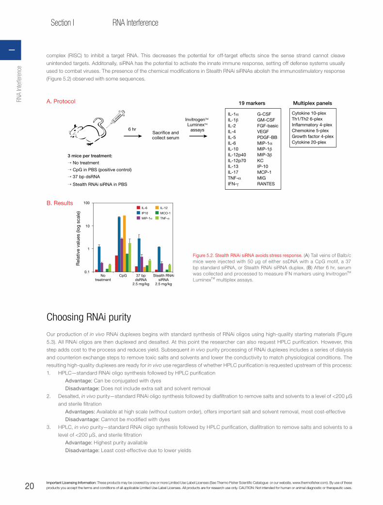
20 Important Licensing Information: These products may be covered by one or more Limited Use Label Licenses (See Thermo Fisher Scientific Catalogue or our website, www.thermofisher.com). By use of these products you accept the terms and conditions of all applicable Limited Use Label Licenses. All products are for research use only. CAUTION: Not intended for human or animal diagnostic or therapeutic uses.
Section I RNA Interference
Figure 5.2. Stealth RNAi siRNA avoids stress response. (A) Tail veins of Balb/c mice were injected with 50 μg of either ssDNA with a CpG motif, a 37 bp standard siRNA, or Stealth RNAi siRNA duplex. (B) After 6 hr, serum was collected and processed to measure IFN markers using InvitrogenTM LuminexTM multiplex assays.
→ No treatment
3 mice per treatment:
6 hrSacri�ce andcollect serum
InvitrogenTM LuminexTM
assays
→ CpG in PBS (positive control)
→ 37 bp dsRNA
→ Stealth RNAi siRNA in PBS
IL-1αIL-1βIL-2IL-4IL-5IL-6IL-10IL-12p40IL-12p70IL-13IL-17TNF-αIFN-γ
G-CSFGM-CSFFGF-basicVEGFPDGF-BBMIP-1αMIP-1βMIP-3βKCIP-10MCP-1MIGRANTES
Cytokine 10-plexTh1/Th2 6-plexIn�ammatory 4-plexChemokine 5-plexGrowth factor 4-plexCytokine 20-plex
19 markers Multiplex panelsA. Protocol
Rel
ativ
e va
lues
(log
sca
le)
0.1
1
10
100
37 bpdsRNA
2.5 mg/kg
Stealth RNAisiRNA
2.5 mg/kg
CpGNotreatment
IL-6
IP10
MIP-1α
IL-12
MCO-1
TNF-α
B. Results
Choosing RNAi purity
Our production of in vivo RNAi duplexes begins with standard synthesis of RNAi oligos using high-quality starting materials (Figure
5.3). All RNAi oligos are then duplexed and desalted. At this point the researcher can also request HPLC purification. However, this
step adds cost to the process and reduces yield. Subsequent in vivo purity processing of RNAi duplexes includes a series of dialysis
and counterion exchange steps to remove toxic salts and solvents and lower the conductivity to match physiological conditions. The
resulting high-quality duplexes are ready for in vivo use regardless of whether HPLC purification is requested upstream of this process:
1. HPLC—standard RNAi oligo synthesis followed by HPLC purification
Advantage: Can be conjugated with dyes
Disadvantage: Does not include extra salt and solvent removal
2. Desalted, in vivo purity—standard RNAi oligo synthesis followed by diafiltration to remove salts and solvents to a level of <200 µS
and sterile filtration
Advantages: Available at high scale (without custom order), offers important salt and solvent removal, most cost-effective
Disadvantage: Cannot be modified with dyes
3. HPLC, in vivo purity—standard RNAi oligo synthesis followed by HPLC purification, diafiltration to remove salts and solvents to a
level of <200 µS, and sterile filtration
Advantage: Highest purity available
Disadvantage: Least cost-effective due to lower yields
complex (RISC) to inhibit a target RNA. This decreases the potential for off-target effects since the sense strand cannot cleave
unintended targets. Additonally, siRNA has the potential to activate the innate immune response, setting off defense systems usually
used to combat viruses. The presence of the chemical modifications in Stealth RNAi siRNAs abolish the immunostimulatory response
(Figure 5.2) observed with some sequences.
RNA
Inte
rfere
nce
I

21
Chapter 5In vivo RNAi
thermofisher.com
Figure 5.3. We produce high-quality in vivo RNAi duplexes in three purity selections supporting Alexa Fluor dye and biotin modifications.
Standard synthesis
HPLCpuri�cation
in vivoIn vivo purity processes• Dia�ltration (<200 µS/cm)
• Sterile �ltration
• Endotoxin testing (<2 EU/mg)Desaltedin vivo
HPLCpuri�cation
Purity Modi�cations
• HPLC • Alexa Fluor 488
• Desalted in vivo • Alexa Fluor 546
• HPLC in vivo • Alexa Fluor 555
• Alexa Fluor 647
• Alexa Fluor 680
• Alexa Fluor 750
• Biotin
Raf
-1/G
AP
DH
rat
io
Raf-1 +BLOCK-iT
Raf-1 biotinRaf-1 Alexa Fluor 647
Raf-1Control
0.2
0
0.4
0.6
0.8
1.0
1.2
1.4
Figure 5.4. Labelled Stealth RNAi siRNA exhibits potent knockdown. MDA-MB-435 cells were transfected with 25 nmol unlabelled Stealth RNAi siRNA duplexes, or Stealth RNAi siRNA duplexes labelled with an Alexa Fluor dye or biotin, in each case the duplexes targeting Raf-1. Knockdown was assessed at >90% by qPCR; delivery is demonstrated through fluorescence imaging.
Figure 5.5. An alternate method to show knockdown. Mixing unlabelled duplex with labelled control duplexes.
Raf-1 RNAi–Alexa Fluor 647Raf-1 RNAi, biotinylatedAlexa Fluor 488–streptavidin
Raf-1 RNAi + BLOCK-iT™ Alexa Fluor 555
Tracking delivered duplexes
We have demonstrated that labeling Stealth RNAi siRNA duplexes does not hamper their knockdown potency (Figure 5.4). An
alternative approach is to mix unlabelled duplexes with labelled control duplexes (Figure 5.5); this method is more commonly used
when performing in vivo RNAi and allows the progression to clinical research to proceed unhindered by questions about the possible
effects of a dye label. Tracking options are:
→ Stealth RNAi siRNA can be combined with bright BLOCK-iT fluorescent controls (available in different scales, and colours shown
in Figure 5.3) without loss of activity
→ Stealth RNAi siRNA can be directly labeled with Alexa Fluor dyes, without loss of activity
→ Stealth RNAi siRNA can be biotinylated and combined with either streptavidin-Alexa Fluor or streptavidin-Invitrogen™ Qdot™
labels
In vivo RNAi5

22 Important Licensing Information: These products may be covered by one or more Limited Use Label Licenses (See Thermo Fisher Scientific Catalogue or our website, www.thermofisher.com). By use of these products you accept the terms and conditions of all applicable Limited Use Label Licenses. All products are for research use only. CAUTION: Not intended for human or animal diagnostic or therapeutic uses.
Section I RNA Interference
IN VIVO RNAi WITH VECTORSAlthough employing RNAi vector systems can be slightly more involved than using synthetic RNAi reagents, the flexibility of the
vector-based systems is compelling for many RNAi researchers conducting both in vitro and in vivo experiments. There are
two main types of RNAi expression vector technologies on the market: short hairpin (shRNA) expression vectors and artificial
microRNA (miRNA) expression vectors. Most RNAi vectors available today employ shRNA vector technology, which typically
involves shRNA expression from a Pol III promoter and may or may not employ viral delivery. These vectors express shRNA
sequences, typically from a U6 or H1 promoter, and some have inducible promoters (typically H1/TO—a tetracycline-inducible
promoter). While these vectors can be used for in vivo RNAi experiments, there are some drawbacks, including low design
success rate and inability to track shRNA expression or express the shRNA in a specific target tissue.
RNAi vector delivery methodsSimilar to RNAi vectors for in vitro applications, you can use either standard transfection techniques or a viral delivery method to
deliver RNAi vectors in vivo. Currently, the delivery of an RNAi expression vector in vivo without using a viral delivery system is similar
to delivering synthetic dsRNA in vivo. Typically, this would involve complexing the RNAi expression vector with a commonly used
lipid-based in vitro transfection reagent and injecting directly into the animal. While this may be the easiest approach for delivering
RNAi vectors into animals, it has quite a few limitations, including the inability to deliver systemically, and low transfection efficiencies.
For these reasons, most researchers choose to use a viral delivery method when employing RNAi vectors for in vivo experiments.
Regardless of whether one chooses an shRNA or miR RNAi vector system, viral delivery is a huge advantage for many in vivo
approaches. Most viral delivery approaches involve either adenoviral, retroviral (nonlentiviral), or lentiviral technology:
→ Adenovirus can be used for transient RNAi expression in either dividing or nondividing cells
→ Retrovirus can be employed for transient or stable expression, but can only be used to transduce dividing cells
→ Lentiviral delivery affords the most options, as it can be used for transient or stable expression in dividing or nondividing
cells, as well as neuronal cells, drug- or growth-arrested cells, or even primary cells
For protocols and further recommendations, see www.thermofisher.com/invivornai.
In vivo RNAi protocols
Refer to the following supplemental protocols for special consideration for in vivo experiments. For questions about a protocol, please
contact our Technical Support team to have a representative guide you through the procedure.
Protocols for successful in vivo RNAi experiments:
→ Resuspension of Stealth RNAi siRNA for in vivo applications
→ Measuring RNA concentration
→ Tissue harvest/RNA extraction
→ Tissue sectioning
→ Protein extraction from tissues
→ Tissue preparation for flow cytometry analysis
Table 5.1. Overview of products for in vivo applications. Explore our full line of in vivo RNAi resources at www.thermofisher.com/invivornai.
RNA Purification Scale Alexa Fluor dye Biotin
BLOCK-iT siRNA and Stealth RNAi siRNA
Desalted + in vivo purity 25 nmol, 100 nmol, 2 μmol NA NA
HPLC 5, 20, or 500 nmol 488, 546, 555, 647, 680, 750 Yes
HPLC + in vivo purity 5, 20, or 500 nmol 488, 546, 555, 647, 680, 750 Yes
BLOCK-iT Fluorescent Oligo ControlHPLC 5, 20, or 500 nmol 488, 546, 555, 647, 680, 750 Yes
HPLC + in vivo purity 5, 20, or 500 nmol 488, 546, 555, 647, 680, 750 Yes
RNA
Inte
rfere
nce
I

23
Chapter 5In vivo RNAi
thermofisher.com
In vivo–purity Stealth RNAi siRNA duplexes are specifically formulated for use in animals. Resuspend the RNA duplex in
Invitrogen™ UltraPure™ DNase/RNase-free distilled water or appropriate DNase/RNase-free buffer (e.g., PBS, Ringer’s solution, 0.9%
NaCl). A 5 mg/mL stock solution is recommended for in vivo RNAi experiments. Table 5.2 and Table 5.3 specify the recommended
resuspension volume for in vivo–purity Stealth RNAi siRNA and BLOCK-iT siRNA.
NOTE: The recommended resuspension volumes are for an RNAi molecule with 50% GC content. The molecular weight of RNAi molecules varies slightly depending on the
GC content, but these differences are negligible for in vivo RNAi experiments.
Table 5.2. Desalted in vivo purity: recommended resuspension volume for 5 mg/mL final concentration.
Desalted in vivo quantity Stealth RNAi siRNA resuspension volume—in vivo purity siRNA resuspension volume—in vivo purity
25 nmol 80 µL 67 µL
100 nmol 320 µL 260 µL
2 μmol 6.4 mL 5.4 mL
Table 5.3. HPLC purity: recommended resuspension volume for 5 mg/mL final concentration.
HPLC-purified delivered quantity
Stealth RNAi siRNA resuspension volume— in vivo purity siRNA resuspension volume—in vivo purity
5 nmol 16 µL 13 µL
20 nmol 64 µL 53 µL
500 μmol 1.6 mL 1.3 mL
Measuring RNA concentration
If desired, measure RNA concentration using UV absorbance at 260 nm (A260). Dilute the RNA solution in resuspension buffer or water,
mix well. Measure the A260 of the dilution in a spectrophotometer blanked against dilution buffer (using a cuvette with a 1 cm optical
path length). Calculate the RNA concentration using the appropriate formula:
Stealth RNAi siRNA formulaRNA concentration (μg/mL) = A260 (OD260 units) x 44 ((μg/mL)/OD unit) x dilution factor
siRNA formulaRNA concentration (μg/mL) = A260 (OD260 units) x 41 ((μg/mL)/OD unit) x dilution factor
NOTE: The formulas for Stealth RNAi siRNA and siRNA are slightly different due to chemical and size differences.
In vivo RNAi5

24 Important Licensing Information: These products may be covered by one or more Limited Use Label Licenses (See Thermo Fisher Scientific Catalogue or our website, www.thermofisher.com). By use of these products you accept the terms and conditions of all applicable Limited Use Label Licenses. All products are for research use only. CAUTION: Not intended for human or animal diagnostic or therapeutic uses.
Section I RNA Interference
Harvesting tissue—RNA extraction from tissue
1. Homogenize 50–100 mg of tissue in 1 mL Invitrogen™ TRIzol™ Plus Reagent using lysing matrix D on the FastPrep™-24
Instrument (MP Biomedical) at 4°C. A tissue homogenizer or rotor-stator can also be used.
2. For harder tissues (tumours, lungs), perform 3 cycles of 60 seconds each at 6 m/s using a tissue homogenizer or rotor stator. For softer
tissues (brain, liver), 1 cycle of 60 sec at 4.5 m/s is sufficient to completely dissociate the tissue. Add 0.2 mL of chloroform directly into
the tube and process following the protocol described in the Invitrogen™ PureLink™ RNA Mini Kit Purification System manual.
3. Determine the quantity and purity of the purified RNA using UV absorbance at 260 nm or Quant-iT™ RNA Assay Kit (Cat. No.
Q33140). To determine RNA quality, electrophorese on an Invitrogen™ E-Gel™ agarose gel or analyse on the Agilent 2100
Bioanalyzer.
4. After quantification, use 750 ng of total RNA for first-strand synthesis using the Invitrogen™ SuperScript™ VILO™ Kit and qPCR
analysis performed using Applied Biosystems™ TaqMan™ Gene Expression Assays to measure knockdown.
Sectioning tissue
Slide preparationIf you are preparing your own slides, precoat slides with Invitrogen™ HistoGrip™ Concentrate (Cat. No. 008050) or 0.1% poly-L-lysine
in water, then air-dry. Commercially available precoated glass slides are available and can be used to mount frozen or formalin-fixed,
paraffin-embedded tissue sections.
Frozen tissueA protocol for preparing frozen tissue samples is described below, but this is only a example. If your laboratory already has an
optimised protocol for your sample type, we recommend that you use that protocol.
1. Snap freeze fresh tissues in cryomolds containing Invitrogen™ OCT™ (Optimal Cutting Temperature) compound (a solution of
glycols and resins which provides an inert matrix for sectioning). Store frozen tissue blocks at –70°C until you are ready for tissue
sectioning.
2. For sectioning, allow the frozen tissue block to equilibrate to the cryostat temperature, cut 4–20 µm cryostat sections, and mount
on coated glass slides.
3. Dry tissue sections at room temperature for 30 minutes.
4. If desired, store slides at –70°C before fixing. If slides are stored at –70°C, warm the slides to room temperature before the fixing
step.
5. Place the slides in 100% acetone at 4°C for 10 minutes to fix the sections. Remove the slides from acetone and air dry for 10–30
minutes.
6. Circle each tissue section using the Mini PAP Pen. Store at –70°C until ready to use.
7. The slides should be washed in PBS for 10 minutes prior to staining or mounting.
Paraffin-embedded sections—deparaffinisation and rehydration To use the formalin-fixed, paraffin-embedded sections for immunohistochemical staining, deparraffinise slides with xylene and then
rehydrate in a graded series of alcohol.
1. To prepare sections, dry slides containing 4 µm formalin-fixed, paraffin-embedded sections in a 55°C oven for 2 hours or
overnight (do not allow the temperature to exceed 60°C) and store the slides at room temperature until needed.
2. Place the slides in xylene for 5 minutes at room temperature to deparraffinise.
3. Remove slides and place in a second xylene wash for 5 minutes.
4. Remove the slides and place in two successive 100% ethanol washes for 5 minutes each.
5. Next, place the slides in 95% ethanol for 5 minutes and then transfer to 80% ethanol for 5 minutes.
6. Remove the slides from the 80% ethanol and place in PBS for 10 minutes.
7. Drain any excess reagent by tapping the edge of the slide on paper towels and wipe the area near the tissue sections with a
laboratory wipe.
8. Circle each tissue section using the Mini PAP Pen. The slides are now ready to use.
RNA
Inte
rfere
nce
I

25
Chapter 5In vivo RNAi
thermofisher.com
Protein extraction from tissues
Sample preparationProper sample preparation is key to the success of a western blot analysis experiment. Various factors affect the design of the sample
preparation protocol. Due to the large variety of proteins present in different cells and tissues, it is not possible to have a single sample
preparation protocol that is suitable for all proteins. Based on the starting material and goal of the experiment, the sample preparation
protocol needs to be determined empirically. The sample preparation conditions may also be optimised based on your initial results.
General guidelines are provided below to prepare samples from various sources, and example procedures are provided.
Mammalian tissue samplesThe protocol below describes the preparation of a mammalian tissue lysate from 100 mg tissue using 1 mL Cell Extraction Buffer.
The following protocol is suitable for use with a variety of tissue types, although some optimisation may be necessary for some tissue
types.
1. If needed, cut the tissue into smaller pieces and place ~100 mg tissue in a microcentrifuge tube.
2. Add 1 mL Cell Extraction Buffer containing Protease Inhibitor Cocktail.
3. Homogenise the tissue using a pestle that fits snugly into the microcentrifuge tube.
4. Incubate the samples on ice for 10 minutes with intermittent vortexing.
5. Centrifuge the lysate at 10,000 x g for 5–10 minutes to remove any particulate material.
6. Transfer the supernatant to sterile microcentrifuge tubes and aliquot into single use tubes.
7. Proceed to Preparing Samples for SDS-PAGE after protein estimation, or store aliquots at –80°C. For more detailed information,
follow protocol described in the kit (Cat. No. WB7401).
Preparing tissue for flow cytometry analysis1. Following euthanasia, collect the tissue of interest and place it in cold PBS.
2. Mince the tissue into ~1 mm cubes using a razor blade in a 35 mm Petri dish containing 1–2 mL of Liver Digest Medium
(17703-034).
3. Incubate 15 to 45 minutes, depending on the degree of connective tissue, and pipette up and down every 15 minutes using a 5
mL pipettor to mix.
4. Following incubation, add 4–5 mL of PBS, and use a 100 µm strainer to gently force cells through the strainer into a 10 mL conical
tube.
5. Wash the isolated cells by adding 5 mL PBS and centrifuging at 1,000 x g for 5 minutes.
6. Discard the supernatant and repeat 1–2x for a total of 2–3 washes.
7. If the pellet is red, indicating a high number of red blood cells (RBCs), wash the pellet with Erythrocytes Lysis Buffer (L5) from the
Invitrogen™ PureLink™ Total RNA Blood Kit (K1560-01).
8. If you need to remove RBCs, add 500 µL of L5 and incubate for 10 minutes on ice.
9. Gently vortex the tube 2–3 times during the incubation step to promote complete lysis of erythrocytes. The solution should
become translucent.
10. Centrifuge the tube in a 4°C centrifuge at 400 x g for 10 minutes (to make sure cell debris is not retained supernatant). Carefully
remove the supernatant and discard it.
11. Gently resuspend the cell pellet in PBS and proceed with the wash. The cell pellet should be white with no traces of red.
12. Resuspend the pellet into 0.5% paraformaldehyde and force cells through strainer (50 µm) before flow cytometry analysis.
SPECIAL NOTE AND DISCLAIMER: Literature describing in vivo delivery of siRNA and modified siRNA has become more abundant. However, the applications and methods described often vary. To the extent we provide general guidelines for using siRNA in animals, the company
makes no guarantees concerning use of these products or guidelines in animal studies.
In vivo RNAi5

26 Important Licensing Information: These products may be covered by one or more Limited Use Label Licenses (See Thermo Fisher Scientific Catalogue or our website, www.thermofisher.com). By use of these products you accept the terms and conditions of all applicable Limited Use Label Licenses. All products are for research use only. CAUTION: Not intended for human or animal diagnostic or therapeutic uses.
Section I RNA Interference
FEATURED STUDY
Stealth RNAi siRNA in vivo delivery to mouse brain as a model of Alzheimer’s disease
Effective knockdown achieved in an Alzheimer’s disease model using Stealth RNAi™ siRNA. (A) Experimental design: A transgenic mouse model for Alzheimer’s (hAPP Tg) overexpressing the huAPP gene carrying London and Swedish mutations under the mThy1 promoter was used. A cannula was inserted into the lateral ventricle of the brain. Stealth RNAi APP siRNA solution (4.75 mg/mL in PBS) was infused via cannula at 6 μL/day for 2 weeks in the lateral ventricle using an osmotic mini pump. (B) Brain homogenates from Stealth RNAi siRNA–treated mice were separated into cytosolic and particulate fractions as described in Rockenstein et al. [3]. Western blots were probed with a mouse monoclonal anti-hAPP antibody, screened, and analysed. (C) The fixed brains were serially sectioned at 40 μm, and sections were immunolabelled with anti-hAPP antibody, followed by incubation with a FITC-labeled secondary antibody, and imaged with a Quantimet™ 570C system (Leica) or by LSCM as previously described. APP protein level is reduced in the neocortex treated with the Stealth RNAi APP siRNA but not when treated with the Stealth RNAi siRNA Control.
mThy-1 gene
APP mut
APP
Injection
Intracerebral
Stealth RNAi™ siRNA
Hippocampus
Cortex
120
Non-transgenic
Stealth RNAisiRNA Control
Stealth RNAi APP siRNA
42
hAPP
Actin
hAPP transgenic
Inte
nsity
leve
ls
NontransgenicStealth RNAiAPP siRNA
Stealth RNAi
siRNA Control
2,000
0
4,000
6,000
8,000hAPP levels
hAPP transgenic
Nontransgenic
hAPP transgenic
0
100
200
300
Inte
nsity
leve
ls
hAPP levels
Stealth RNAiAPP siRNA
Stealth RNAi
siRNA Control
hAPP
Stealth RNAi siRNA control Stealth RNAi APP siRNA
A
B
C
RNA
Inte
rfere
nce
I
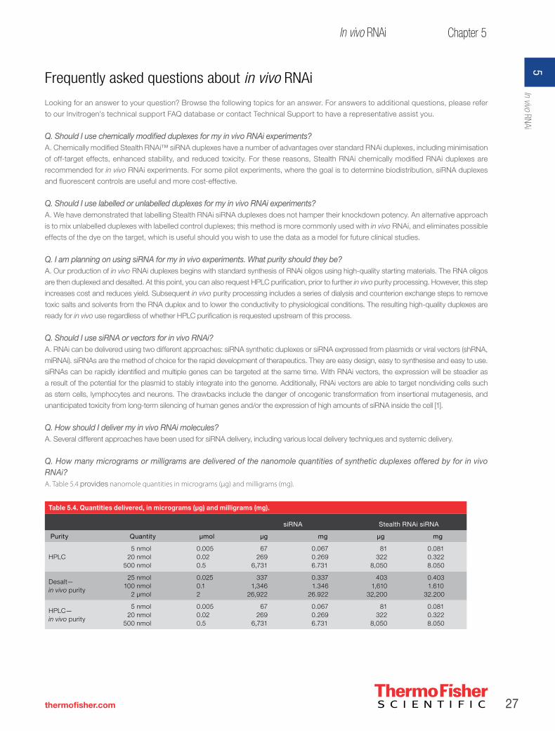
27
Chapter 5In vivo RNAi
thermofisher.com
Frequently asked questions about in vivo RNAi
Looking for an answer to your question? Browse the following topics for an answer. For answers to additional questions, please refer
to our Invitrogen’s technical support FAQ database or contact Technical Support to have a representative assist you.
Q. Should I use chemically modified duplexes for my in vivo RNAi experiments?A. Chemically modified Stealth RNAi™ siRNA duplexes have a number of advantages over standard RNAi duplexes, including minimisation
of off-target effects, enhanced stability, and reduced toxicity. For these reasons, Stealth RNAi chemically modified RNAi duplexes are
recommended for in vivo RNAi experiments. For some pilot experiments, where the goal is to determine biodistribution, siRNA duplexes
and fluorescent controls are useful and more cost-effective.
Q. Should I use labelled or unlabelled duplexes for my in vivo RNAi experiments?A. We have demonstrated that labelling Stealth RNAi siRNA duplexes does not hamper their knockdown potency. An alternative approach
is to mix unlabelled duplexes with labelled control duplexes; this method is more commonly used with in vivo RNAi, and eliminates possible
effects of the dye on the target, which is useful should you wish to use the data as a model for future clinical studies.
Q. I am planning on using siRNA for my in vivo experiments. What purity should they be?A. Our production of in vivo RNAi duplexes begins with standard synthesis of RNAi oligos using high-quality starting materials. The RNA oligos
are then duplexed and desalted. At this point, you can also request HPLC purification, prior to further in vivo purity processing. However, this step
increases cost and reduces yield. Subsequent in vivo purity processing includes a series of dialysis and counterion exchange steps to remove
toxic salts and solvents from the RNA duplex and to lower the conductivity to physiological conditions. The resulting high-quality duplexes are
ready for in vivo use regardless of whether HPLC purification is requested upstream of this process.
Q. Should I use siRNA or vectors for in vivo RNAi?A. RNAi can be delivered using two different approaches: siRNA synthetic duplexes or siRNA expressed from plasmids or viral vectors (shRNA,
miRNAi). siRNAs are the method of choice for the rapid development of therapeutics. They are easy design, easy to synthesise and easy to use.
siRNAs can be rapidly identified and multiple genes can be targeted at the same time. With RNAi vectors, the expression will be steadier as
a result of the potential for the plasmid to stably integrate into the genome. Additionally, RNAi vectors are able to target nondividing cells such
as stem cells, lymphocytes and neurons. The drawbacks include the danger of oncogenic transformation from insertional mutagenesis, and
unanticipated toxicity from long-term silencing of human genes and/or the expression of high amounts of siRNA inside the cell [1].
Q. How should I deliver my in vivo RNAi molecules?A. Several different approaches have been used for siRNA delivery, including various local delivery techniques and systemic delivery.
Q. How many micrograms or milligrams are delivered of the nanomole quantities of synthetic duplexes offered by for in vivo RNAi?A. Table 5.4 provides nanomole quantities in micrograms (µg) and milligrams (mg).
Table 5.4. Quantities delivered, in micrograms (µg) and milligrams (mg).
siRNA Stealth RNAi siRNA
Purity Quantity µmol µg mg µg mg
HPLC5 nmol
20 nmol500 nmol
0.0050.020.5
67269
6,731
0.0670.2696.731
81322
8,050
0.0810.3228.050
Desalt—in vivo purity
25 nmol100 nmol
2 µmol
0.0250.12
3371,346
26,922
0.3371.346
26.922
4031,610
32,200
0.4031.610
32.200
HPLC—in vivo purity
5 nmol20 nmol
500 nmol
0.0050.020.5
67269
6,731
0.0670.2696.731
81322
8,050
0.0810.3228.050
In vivo RNAi5

28 Important Licensing Information: These products may be covered by one or more Limited Use Label Licenses (See Thermo Fisher Scientific Catalogue or our website, www.thermofisher.com). By use of these products you accept the terms and conditions of all applicable Limited Use Label Licenses. All products are for research use only. CAUTION: Not intended for human or animal diagnostic or therapeutic uses.
Section I RNA Interference
Q. I want to order larger scales than the standard offering. Whom do I contact?A. Please contact [email protected] regarding large-scale in vivo RNAi
Q. Do I need endotoxin testing for my in vivo RNAi duplexes?A. Please check with the appropriate regulatory agency for guidance.
Q. How can I order in vivo RNAi duplexes?A. RNAi duplexes for in vivo experiments can be ordered using the simple Invitrogen™ BLOCK-iT™ RNAi Express.
Q. How much is the price per duplex of the in vivo RNAi duplex I wish to purchase?A. Pricing is calculated in the following manner:
Duplex price = (purity price) + (price per base for chosen scale x quantity of bases) + (optional 5’ sense strand label) + (optional endotoxin testing)
NOTE: siRNA has 42 bases. Stealth RNAi siRNA has 50 bases.
Chapter references 1. Grimm D et al. (2006) Nature 441:537–541.
2. Layzer JM et al. (2006) Biochem Biophys Res Commun 344:406–415.
3. Rockenstein E et al. (2001) J Neural Sci Res 66:573–558.
RNA
Inte
rfere
nce
I

29
Chapter 6siRNA screening
thermofisher.com
CHAPTER 6
siRNA screening
Selecting an siRNA library
Customer insights: what is important when selecting an siRNA library?Data generated from a large-scale siRNA screening experiment are invaluable when searching for gene targets involved in a particular
biological process, pathway, or network. However, obtaining a large-scale siRNA library and preparing to run the first screen can
involve a significant investment. Many factors should be considered prior to initial library purchase, including the quality of the reagents,
formatting needs, validation strategy, and support. Recently, we interviewed scientists from the top siRNA screening laboratories and
here we provide a summary of the factors they recommend you should consider before purchasing an siRNA library.
Key consideration: siRNA design and performanceLarge-scale siRNA screens can generate hundreds of potential “hits”—genes for which one or more siRNAs give a desired phenotype.
Unfortunately, in early screens with unmodified siRNA technologies and pooled siRNA approaches, many of these hits actually result from
off-target effects. Off-target effects can result from poor siRNA design, using an siRNA technology that is not designed to reduce off-target
effects, or using siRNAs with limited potency, which requires delivery of a large amount of siRNA to observe gene target knockdown. “The first
concern is the quality of the algorithm that is used to design the siRNAs,” says Anand Ganesan, MD, PhD, Assistant Professor, Department
of Dermatology and Biological Chemistry at the University of California, Irvine. Secondly, the number of siRNAs that have been validated
either by the company or as a part of other studies” should be considered. Silencer Select siRNAs incorporate the latest improvements in
siRNA design, off-target effect prediction algorithms, and chemistry. As a result, they enable unrivalled silencing consistency, potency, and
specificity, and fewer failed experiments, allowing cleaner, more consistent phenotypic data (Figures 6.1 and 6.2).
Silencer Select siRNAs are designed with a newly developed algorithm and include locked nucleic acid (LNATM) chemical
modifications, making them the best-performing siRNAs commercially available. Experiments designed to assess levels of knockdown
and observed phenotypes show that LNA-modified Silencer Select siRNAs consistently demonstrate the highest levels of knockdown
and the least off-target effects [1]. To date, more than 4,300 Silencer Select siRNAs targeted to the most popular gene targets have
been bench tested and validated at a concentration of 5 nM to provide ≥80% target mRNA knockdown as measured by qRT-PCR with
Applied BiosystemsTM TaqManTM Gene Expression Assays. This extensive level of validation provides enhanced confidence in both the
Silencer Select siRNA design algorithm and the siRNA library’s overall performance.
Figure 6.1. Silencer Select siRNAs provide up to 100x higher potency compared to other siRNAs. Silencer Select siRNAs to 10 different targets and siRNAs from two other suppliers to the same 10 different targets were individually transfected into HeLa cells in triplicate at the indicated siRNA concentrations. mRNA knockdown levels were tested 48 hr later as described in Figure 6.2. Average percent mRNA remaining is shown for each set of siRNAs.
Ave
rage
% m
RN
A r
emai
ning
100
80
60
40
20
00.03 nM 0.3 nM 3 nM 30 nM
80% Knockdown
siRNA concentration
D modi�ed (n = 40)Q unmodi�ed (n = 30)Silencer ® Select (n = 30)
Figure 6.2. Silencer Select siRNAs elicit expected phenotype at a higher rate than other siRNAs. siRNAs to 7 gene targets with well-understood RNAi-induced phenotypes were individually transfected at 3 nM and phenotypes measured 48 hr later. Each bar represents the percent of siRNAs that gave the expected silenced phenotype. siRNAs to BUB1B, AURKB, WEE1, and PLK1 were assessed using a multiparametric cell growth/apoptosis assay in U2OS human osteosarcoma cells. siRNAs to HMGCR, LDLR, and FDFT1 were assessed using an LDL uptake assay in HUH7 human hepatoma cells.
% s
iRN
As
with
exp
ecte
d p
heno
typ
e
100
80
60
40
20
0A
modi�edn = 28
B unmodi�ed
n = 21
Silencer siRNAn = 34
Silencer Select siRNAn = 43
siRNA screening6

30 Important Licensing Information: These products may be covered by one or more Limited Use Label Licenses (See Thermo Fisher Scientific Catalogue or our website, www.thermofisher.com). By use of these products you accept the terms and conditions of all applicable Limited Use Label Licenses. All products are for research use only. CAUTION: Not intended for human or animal diagnostic or therapeutic uses.
Section I RNA Interference
Key consideration: siRNA qualitysiRNA design and structure is important; however, siRNA manufacturing quality is equally critical. Dr. Hakim Djaballah, Director of the High
Throughput Screening Core Facility at Memorial Sloan Kettering Cancer Center, said, “Cellular viability is a major concern in our screening
experiments. We need to know that the contaminants that result from the production of siRNA are at extremely low levels. Often we dilute our
reagents and use them on a small scale with a small number of cells, and we cannot afford impurities to be present.” Silencer Select siRNAs
are manufactured under stringent control and are subjected to rigorous quality control procedures to ensure lot-to-lot consistency. Each
and every strand is subjected to MALDI-TOF mass spec, and each annealed strand is tested by nondenaturing PAGE to ensure efficient
annealing. The average purity of “standard purity” siRNA strands is approximately 90% full-length product.
Key consideration: completeness of the libraryAll screening scientists agree that a complete siRNA library is crucial to success. The library should contain siRNAs to as many relevant
genes as possible, and most researchers prefer to err on the side of screening too many, rather than too few targets. The Silencer Select
siRNA algorithm has been used to generate siRNAs to the human, rat, and mouse genomes, and an siRNA library for 21,585 unique genes
within the human genome is available for immediate shipment. Other species are covered on a custom basis. Predesigned siRNAs sets
are available for a number of important human gene families that include kinases, phosphatases, GPCRs, and several others. An extended
druggable genome set is available that targets 10,415 genes of potential therapeutic interest. Although prepared sets are very useful, more
and more researchers prefer to screen their own subsets of targets. To that end, we can provide a custom preparation of Silencer Select
siRNAs targeting your list of favourite genes. Finally, our bioinformatics team is standing by to help you generate a list of genes within a
functional class or biological pathway for your custom siRNA library.
Key consideration: customisationsiRNA screens are quite varied in nature. “Every experiment has different requirements. What is the scale of siRNA desired in each
well? How are the duplexes oriented? Can the supplier provide custom aliquotting? And most importantly, what are the turnaround
times for ordered libraries?” said Dr. Djaballah. All are critical factors in selecting an siRNA library vendor, both for the initial siRNA
library purchase and for hit confirmation follow-up studies.
Silencer Select siRNA libraries are available at scales of 0.1, 0.25, 1.0, 2.0, and 5.0 nmol per well. Custom aliquotting requests of all
types are easily handled, and we can supply siRNAs in your desired plate layouts to match your transfection and cell-based assay protocols.
Key consideration: validationOnce hits from a library screen are identified, validation is a crucial step prior to drawing conclusions. Typically, hits are validated by
repeating the experiment with the same siRNA sequences, using a secondary assay or additional siRNA sequences, or using qRT-
PCR, often followed by looking at protein levels to assess target knockdown. “We don’t think simply using another supplier’s siRNA is
a good enough method for validation if you are relying on the differences between the two suppliers’ algorithms. We prefer qRT-PCR
followed by protein level measurements. We sometimes look at rescue experiments using an expression vector, but these can be
tedious and there can be toxicity concerns,” said Dr. Djaballah. Dr. Ganesan remarked, “We validate hits by several different strategies.
We utilise multiple siRNAs to eliminate off targets. We do quantitative qRT-PCR to ensure that the siRNA impacts the expression of the
target gene, and we examine the impact of these siRNAs in several different cell lines. More recently, we have also been validating our
hits with shRNAs.” Dr. Ganesan went on to say, “The major cost of screens is usually not from the screen itself, but from the follow-
ups.” Keys to successful validation are having a defined strategy up front and having a streamlined method for obtaining additional
siRNAs for validation purposes. We can help you define an appropriate follow-up and hit validation strategy. Since 2002, we have
consulted with and helped thousands of scientists using siRNAs. Custom sets of Silencer Select siRNAs are available with as little as
0.1 nmol siRNA per well—arranged in plates any way you like. Of course, following up on large numbers of false positive results can
easily drain your hit validation budget. By performing our primary screen with Silencer Select siRNAs, which reduce off-target effects
by up to 90%, and by purchasing the lowest amount of siRNA you need, you can keep follow-up siRNA costs to a minimum.
Key consideration: communication and supportWe realise that purchasing the initial siRNA library, setting up, performing, and validating an siRNA screen can be a large endeavor.
Dr. Djaballah, who has directed many siRNA screening experiments, stated that “there must be access to open discussion” when
it comes to planning a screen. We certainly understand this need. To that end, we encourage open communication through siRNA
library user group meetings, we support major RNAi symposia, and offer free consultations on your siRNA screening project every
step of the way. Our scientists and award-winning technical support staff welcome the opportunity to support you in your siRNA
experiments.
For more information about Silencer Select siRNA libraries, please contact your local sales representative or visit
www.thermofisher.com/rnai.
RNA
Inte
rfere
nce
I

31
Chapter 6siRNA screening
thermofisher.com
DEFINITIONSsiRNA librariesSets of siRNAs, usually provided individually in 96-well or 384-well plates. siRNA libraries may target any number of genes, e.g.,
an entire genome, a gene family, a biological pathway, or a custom set of genes.
HitsFor this application guide, a “hit” is defined as a gene that triggers the phenotype being assayed in an initial siRNA library screen
when it is successfully targeted by an siRNA. In other words, hits (also called candidate or putative hits), are positive results from
an siRNA library screen—they may result in a phenotype that is similar to the positive control in the screening assay. Ideally,
candidate hits represent genes that directly or indirectly influence the cellular process under study. Confirmed hits are those
that have been validated using 2–3 methods to corroborate the initial screening results.
NegativesGenes for which none of the targeting siRNAs results in a hit.
False positivessiRNAs that give a positive result in the screening assay that are not confirmed by other distinct siRNAs targeting the same gene
or by secondary assays. The anomalous assay result from a false positive may be due to off-target effects of the siRNA or to
experimental error. siRNA screens should be designed to rule out all false positives.
False negativesTrue hits that are not detected. These siRNAs give a negative signal; however, use of other distinct siRNAs targeting the same
gene show that when this target is silenced it gives a positive assay result. False negative results can cause you to overlook
true positives and should be minimised.
Biological false negativesOccur when the RNAi reagents cannot silence a target sufficiently to render it rate-limiting—thus, no phenotype is observed.
For example, the target protein may have a long half-life, or extremely high activity, so that very little protein is needed to fulfill
its function. There may also be redundancy of function in the pathway, such that the function of the silenced gene product is
replaced by the product of a second gene. These types of false negatives are difficult if not impossible to avoid.
Technical false negativesAre caused by suboptimal screening reagents, conditions, or assay design. They can also result from experimental variability.
While, of course, one would like to detect as many potential hits as possible, allowing for a higher rate of technical false
negatives may be worth the significant decrease in overall costs and time required to perform the screen.
Screening with siRNA libraries
AdvantagesRNAi is currently the easiest and most cost-effective reverse genetics tool for studying gene function. Plate-based and cell-based
assays, including higher content multiparameter assays and the availability of libraries of effective siRNAs, allow researchers to quickly
evaluate dozens to hundreds of genes for their role in cellular processes. siRNA libraries can be obtained in a variety of formats,
including single-transfection, ready-to-use versions, making genome-wide or smaller RNAi screens possible in almost any laboratory.
siRNA screening6

32 Important Licensing Information: These products may be covered by one or more Limited Use Label Licenses (See Thermo Fisher Scientific Catalogue or our website, www.thermofisher.com). By use of these products you accept the terms and conditions of all applicable Limited Use Label Licenses. All products are for research use only. CAUTION: Not intended for human or animal diagnostic or therapeutic uses.
Section I RNA Interference
ChallengesWhile many laboratories are performing RNAi screens with siRNA libraries to determine gene function in mammalian cultured cells, the
technology is just now being broadly applied. In the past, three factors have limited the adoption of this technology: (1) the perceived
cost of siRNA libraries, (2) the belief that expensive robotic liquid handling systems are required, and (3) the anticipated difficulties in
performing transfections reproducibly in 96- or 384-well plates.
Tools are now available that address all of these issues, making siRNA library screening feasible for more labs than ever before. siRNA
libraries are available in formats suitable for a single transfection, and up to thousands of transfections. There are now extensive sets of
positive and negative controls and delivery optimisation reagents for developing optimal screening protocols. High-throughput delivery
reagents, instruments, and protocols yield easy and reproducible siRNA delivery in multiwell plates using either manual or robotic means.
Keep in mind that RNAi facilitates gene knockdown, not gene knockout; i.e., target gene expression is almost never completely
extinguished. Whether the level of knockdown is sufficient to generate a positive outcome in the screening assay (rendering expression
rate-limiting) will depend on the endogenous expression level of the gene, on the protein product’s activity and half-life within the
cell, and on redundancy of function within that biological pathway. Some proteins may require very little activity to affect a biological
process, and are, in general endogenously expressed at much higher levels than biologically necessary. Thus, their residual activity
after RNAi knockdown could still be sufficient to fulfill their cellular role. It might therefore be difficult, for example, to achieve sufficient
gene knockdown to extinguish the effects of certain highly active proteins.
GoalsDesign the siRNA screen for comprehensive coverage of the genes involved in the phenomenon being studied while minimising the
rate of technical false negatives (biological false negatives cannot be avoided; see Definitions on page 31). By careful design of
experimental controls and assessment of results, the screen should also strive to eliminate false positives.
These goals are best attained through sequential screening passes, ideally using independent assays for each as false positives. The
initial pass is focused on maximising sensitivity of detection, and therefore should use conditions that favour maximal silencing (e.g., use
multiple individual siRNAs per target at relatively high concentrations (30–100 nM)). Depending on the type of assay and the threshold used
(usually 2–3 standard deviations from the baseline), a relatively high percentage of targeted genes may show a positive phenotype from at
least one siRNA, warranting follow-up evaluation [2]. These candidate hits will, no doubt, include numerous false positives, which will be
filtered out using validation experiments.
siRNA library screening workflow (5 steps)
RNAi screening using an siRNA library involves the following five steps; they are briefly introduced here and are described in detail in
later chapters.
→ Step 1. Experimental design
→ Step 2. Optimise the siRNA delivery method
→ Step 3. Develop and optimise an effective screening assay(s)
→ Step 4. Perform screen and analyse data
→ Step 5. Validate screening results
Step 1. Experimental designAmong the most critical aspects of experimental design for siRNA screening are selection of siRNAs to screen and to use as controls,
the cell type(s) in which the experiment is to be performed, and the siRNA delivery strategy. These decisions should be based on the
biological pathway under study. For instance, to look at oncogenesis, you may wish to screen an siRNA library for differential effects
on cell proliferation in a cancer cell line compared to a noncancerous cell line. The choice of siRNA library will depend on the depth of
the screen and the overall design of the experiment. Do you want to analyse every possible target, or just potentially druggable targets,
such as kinases? siRNA libraries that target complete genomes (human, mouse, etc.), as well as sets of siRNAs that target specific
gene families or biological pathways are available. Libraries can also be custom designed; the possibilities are almost endless—it is up
to each individual researcher to determine how best to approach the experimental problem at hand.
Equally important in choosing an appropriate set of siRNAs for your screen, is to use well-designed, efficacious siRNAs with a high
probability of inducing successful knockdown. Effective siRNA design is key to avoid wasting both time and money. We use “intelligent” siRNA
design algorithms and validate a selection of our siRNAs. Ultimately the most important attribute of any algorithm is its performance when tested
experimentally. Therefore an algorithm that has been proven using many siRNAs targeting many different endogenous transcripts will produce
RNA
Inte
rfere
nce
I
33
Chapter 6siRNA screening
thermofisher.com
more effective libraries leading to more useful data. Additionally, algorithms that have been trained to predict the highest potency siRNAs will allow
you to use siRNAs in your experiments at lower concentrations, resulting in more effective screening performance.
Even with extensive validation of an siRNA design algorithm, the vast majority of siRNAs in almost all siRNA libraries have not been
experimentally proven to knock down their intended target. The use of multiple siRNAs per target is generally accepted to be the best approach
[3]. Employing three or more distinct, individual siRNAs, and performing replicate transfections, provides statistical significance and significantly
decreases both false positive and false negative rates as compared to screening with pools of siRNAs.
Another important part of the process of selecting siRNAs for screening is to choose appropriate positive and negative control siRNAs.
Positive control siRNAs are necessary to prove that the siRNAs in the screen are delivered efficiently and can induce silencing in the experimental
conditions used. Additional assay-specific positive control siRNAs are necessary to ensure that the screening assay yields sufficient signal for
accurate quantitation of the data. Nontargeting, negative control siRNAs are necessary to control for nonspecific effects due to siRNA delivery
and to screen assay background. Finally, in screens that include a treatment of some sort to induce the phenotypic effect, untreated controls
serve as a baseline for the screening assay.
Step 2. Optimise the siRNA delivery methodScreening siRNAs that target hundreds to thousands of genes requires conditions for high-throughput siRNA delivery. Furthermore, efficient,
reproducible siRNA delivery is critical for effective siRNA library screens. Using optimal siRNA delivery conditions eliminates the most common
causes of failed gene silencing experiments. Because it is so important, we highly recommend investing the time to select the best siRNA delivery
method and conditions for the cells that are to be used for siRNA library screening.
There are two basic methods employed for siRNA delivery: lipid-mediated transfection and electroporation. Lipid-mediated reverse
transfection can be used to transfect up to 1,000 wells in less than an hour without the need for robotics [4]. With automated liquid handling, the
pace can be even faster. Electroporation can also be used to deliver dozens to hundreds of siRNAs quickly when performed with a 96-well plate
electroporation chamber.
The first step in optimising siRNA delivery conditions for an RNAi screen is to identify a transfection reagent and plating conditions, or
electroporation conditions, that maximise uptake of active siRNA while maintaining high cell viability. We find it useful to measure both siRNA-
induced target knockdown and cell viability in cells transfected with 2–5 different transfection reagents or 5–10 electroporation conditions (e.g.,
varying cell concentration, voltage, pulse length, pulse number, siRNA concentration, etc.). Delivery can then be further optimised for the reagent
or electroporation condition that worked best among the conditions tested. Choice of delivery method and its optimisation will depend on the
cell type used.
Step 3. Develop and optimise an effective screening assayThe results of siRNA library screens are typically evaluated using phenotypic assays, such as reporter gene assays, cell-based assays, plate-
based assays, etc. rather than through direct monitoring of changes in target mRNA or protein levels. The success of an siRNA library screen
depends greatly on the quality of the phenotypic assay used for screening. Thus, it is worthwhile to invest the time and effort required to create an
assay with enough precision, signal-to-noise, and linear range to ensure identification of siRNAs that induce the desired phenotype in transfected
cells.
The diversity of cell functions that can be characterised using siRNA libraries is limited only by the range of phenotypic assays that can be
developed. Assays that are amenable to siRNA screening experiments range from microscopic assays that monitor cell size, cell cycle status, or
antibody staining; to enzymatic assays that assess the turnover of a specific substrate in a cell lysate; to direct measurements of biomolecules or
small molecules in lysates, on cells, or in medium.
Good quantitative assays will demonstrate a high signal-to-noise ratio; that is, they will exhibit a large difference in the results obtained from
assay-specific positive controls and nontargeting negative controls (which may serve as negative controls for both the phenotypic assay and for
silencing). Such assays will yield a wide range of results from siRNA screening experiments, and they will maximise the chance of identifying genes
with an interesting phenotype when silenced. Maximising the signal-to-noise ratio involves testing variables like assay time, assay components
(e.g., the reporter), cell type, and length of time between transfection and assay.
Step 4. Perform screen and analyse dataWith a robust phenotypic assay and optimised siRNA delivery conditions established, a library of individual siRNAs can be introduced
into cells. Triplicate transfections for each siRNA provide enough data for reasonable statistical analysis. Use of three siRNAs per
target helps to eliminate nonspecific effects caused by any single siRNA sequence (false positives) as well as any false negatives.
Positive control and negative controls siRNAs on each plate provide quantitative numbers to normalise data from different plates, and
to provide the range of phenotypes that can be used to identify siRNAs that yield a positive effect, designated “candidate hits” (see
Definitions on page 31) The data are collected and analysed to identify candidate hits. In many cases, we recommend reserving this
siRNA screening6
34 Important Licensing Information: These products may be covered by one or more Limited Use Label Licenses (See Thermo Fisher Scientific Catalogue or our website, www.thermofisher.com). By use of these products you accept the terms and conditions of all applicable Limited Use Label Licenses. All products are for research use only. CAUTION: Not intended for human or animal diagnostic or therapeutic uses.
Section I RNA Interference
designation for target genes that exhibit the expected phenotype when transfected with two of the three individual siRNAs. This should eliminate
false positives and false negatives [5]. Another approach is to follow up on all of the siRNAs to a given gene even when one gave a positive result.
This approach requires additional follow up and few potential hits are confirmed; however, it is a good approach to avoid missing targets.
Candidate hits are essentially a series of genes identified by the initial screen to be potentially interesting and that warrant further study. They
are typically confirmed using an independent method to eliminate “false positives” and to ensure that only legitimate genes are evaluated further
(Step 5).
Step 5: Validate screening resultsTargets identified as candidate hits in the intital screen must be validated. This is done by a second and often, a third screening pass. These
experiments will also typically shed light on the roles of the target genes in specific biological pathways. An obvious validation technique
used in secondary screens is to show a direct link between the observed phenotype and target gene silencing by documenting a reduction
in target mRNA or protein level. However, the specific goals of the library screening and the assay tools available will dictate the techniques
used for validation. A major goal of the second screening pass is to retest all candiate genes to identify interesting hits and eliminate false
positives. Since fewer genes are analysed, these validation experiments may also serve to further refine the relevance of candidate hits
with respect to the biological process of interest. It would not be uncommon for the second screen to eliminate up to 90% of candiate hits
identified in the first pass.
Those genes confirmed as positive in the second screening pass can be subjected to a final third pass wherein, if not shown by
previous experiments, the functional phenotypic assay is repeated in parallel with a direct measure of target gene silencing (usually by qRT-
PCR or branched DNA assays), thus directly linking the two. Targets that have been validated by two or more screens, in addition, to the
initial screening experiment, are excellent candidates for further studies (e.g., in vivo studies) designed to help independently confirm data
and expand upon results found in the published literature.
Toxicity
When siRNAs are introduced into cells via transfection, unexpected and/or toxic effects might be observed in addition to the expected phenotypes
involved in the screen.
Off-target effects are any gene silencing effects caused by siRNAs on nontarget mRNAs through the RNAi mechanism. They could come
from the guide (antisense) or passenger (sense) strand of siRNA. In the case of near-complete complementarity the strands will induce cleavage
of mRNA (act as siRNA), while the case of limited homology cause translation inhibition (act as miRNA). The SVM algorithm used to design our
siRNAs selects only the sequences that have no or little homology (<15 nt) to the transcriptome outside the intended mRNA target. Further, the
passenger strand of siRNA is inactivated by chemical modifications. However, a high proportion of ‘‘off-target’’ transcripts silenced by siRNAs
have been shown to have 3’-UTR sequence complementarity to the seed region of the siRNA, indicating the off-target effect is mediated through
an miRNA pathway. Since it is impossible to eliminate all 7–8-base matches of siRNAs to the transcriptome, it is difficult to achieve absolute
specificity, even when chemical modifications are introduced. Performing RNAi experiments with hyper-potent siRNAs at low concentrations (<5
nM) minimises off-target effects.
Another problem can arise from cellular responses to introduction of the foreign nucleic acid. Both non-immune and immune cells may
be activated by long dsRNA, leading to the activation of cytoplasmic receptors such as the dsRNA-dependent protein kinase R (PKR) and the
retinoic acid inducible gene-I (RIG-I). Once PKR is activated, it phosphorylates the eukaryotic translation initiation factor (EIF-2)-α, leading to global
suppression of protein synthesis and subsequent programmed cell death. PKR can also activate nuclear factor κB (NF-κB) with consequent
induction of type-I IFN production. A family of 2959-oligoadenylate synthetases (2959-OAS) can also be activated by dsRNA. This leads to the
activation of RNase L, which eventually triggers the nonspecific degradation of mRNA.
RIG-I is an intracellular dsRNA sensor capable of triggering IFN production. The RIG-I helicase recognises blunt-ended siRNAs, leading to
the activation of dsRNA signaling. RIG-I efficiently unwinds siRNAs containing blunt ends and the efficiency in duplex unwinding is translated into
downstream signalling to interferon regulatory factor 3 (IRF-3) and NF-κB activation. Therefore, introduction of long double-stranded RNA (>30
bp), especially blunt-ended siRNA, can induce severe antiviral responses in transfected cells. For these reasons, typical synthetic siRNAs are
shorter, 19 bp in length, and contain 2 nt overhangs at 3’-ends to reduce cellular responses. In most cases cellular responses to such molecules
are minimal.
Mammalian immune cells also express a family of Toll-like receptors (TLRs), which recognise pathogen-associated molecular patterns
including CpG motifs and viral dsRNA. For example, TLR7 and TLR8 were initially shown to mediate the recognition of RNA viruses. TLR7, TLR8,
and TLR9 are expressed in endosomes and require endosomal maturation for efficient signaling. siRNA recognition by TLR7, TLR8, and TLR9
results in activation of NF-kB and IRFs, which induce inflammatory cytokines and IFNs, respectively. TLR7 and TLR8 mediate the recognition of
RNA
Inte
rfere
nce
I
35
Chapter 6siRNA screening
thermofisher.com
siRNAs in a sequence-dependent manner: preferentially U- and G-rich siRNAs are recognised. Recognition of siRNAs by TLRs takes place in the
endosome, before the siRNAs enter the cytoplasm. Therefore, if siRNAs can enter the cytoplasm avoiding the endosome, they should bypass
the activation of immune systems but still mediate gene silencing.
Secondary screening: confirmation of candidatesThe goal of the secondary screen is to confirm that the phenotype observed is a specific effect of siRNA-induced target knockdown
and not an off-target effect dependent on the protocol or a reagent. Because the siRNA delivery conditions were optimised for a
positive control siRNA, the siRNAs that target candidate hits should be further tested for silencing efficiency in secondary screens [6],
using techniques described in Chapters 10 and 11. If there are a large number of candidate hits, optimisation experiments may help
prioritise which genes should be followed up first.
Confirming that phenotypes are due to reduced expression of the intended target
Test multiple siRNAs. To minimise false positive and false negative results, we recommend using two or more siRNAs that target
distinct regions of each gene and delivering each siRNA in triplicate, for the initial screening experiment. Although the number of
siRNAs targeting the same gene required to confirm a candidate hit can be determined statistically by estimating the probability that
any random siRNA from the library will score positively in the assay under the chosen conditions (i.e., the false positive rate), most
researchers consider an siRNA target a confirmed hit, if at least two different siRNAs that target the candidate produce the same
phenotype. If the initial screen was carried out using multiple individual siRNAs per target, you may want to prioritise your follow up
to first include only genes for which at least two distinct siRNAs give a phenotype outside the set thresholds of the assay. Note that
candidate hits where only one of the siRNAs gives the desired phenotype should not be ignored.
Reproduce the phenotype. For well-characterised target proteins, antibodies or small molecule inhibitors may exist and can be
used to mimic or counteract the effect of the siRNA [7]. To confirm that a hit is associated with a specific pathway, additional
phenotypes (e.g., up- or down-regulation of other genes, increase or decrease in protein modification), that are known to occur
when the pathway of interest is perturbed, can be assayed after siRNA treatment [8,9]. These additional assays should be based on
existing knowledge of the biological process. To further study complex cellular processes, some researchers have begun to develop
automated, microscopy-based assays to simultaneously analyse fluorescent markers linked to antibodies or ligands to assess relative
intensity and cellular localisation over time [6,10].
Perform a rescue experiment. Functional validation of RNAi experiments is supported by successful siRNA-refractory rescue
experiments. In these experiments overexpression from cDNA constructs of target mRNAs that are not recognised by gene-specific
siRNAs are used to restore the wild type phenotype. For example, a cDNA with a silent mutation(s) in the siRNA target sequence is
expected to rescue the phenotype caused by siRNA-induced reduction in endogenous gene expression. Alternatively, a phenotype
caused by siRNAs that recognizes 5’ or 3’ untranslated sequences can be rescued by expressing a cDNA of the target gene that
contains only coding sequences [11]. Recently, bacterial artificial chromosome transgenesis in cultured cells has been used to create
RNAi-resistant transgenes [12].
Measure mRNA and protein levelsAs mentioned above, confirmation of positive hits involves verifying that the observed phenotype is specifically due to reducing
the expression of the target gene. Whenever possible, determining siRNA-induced reduction in both mRNA and protein levels is
recommended to help interpret your data. In both cases, target mRNA or corresponding protein levels should be assessed relative to
that of samples treated with nontargeting negative control siRNAs.
If target mRNA levels are not appreciably reduced, yet the siRNA induces a positive result in the assay, it is possible that the
observed phenotype is caused by an off-target effect. However, in this scenario, the corresponding protein levels may be reduced
without observing a reduction in target mRNA levels. There are two possible reasons for this observation: the siRNA could be acting
as a microRNA and inhibiting translation rather than targeting its cognate mRNA for destruction, or the RNA assay may not accurately
measure target mRNA levels. An RNA assay may fail to accurately reflect mRNA knockdown if the target mRNA cleavage product
is relatively stable or if the assay is designed incorrectly (for example it does not measure one or more targeted splice variants or
qRT-PCR normalisation was inaccurate). In either case, it is a good idea to measure the reduction in target protein levels after siRNA
delivery. If knockdown is not observed at either the mRNA or protein level, yet the siRNA induces a distinct phenotype, it is more likely
that the phenotype is the result of an off-target effect.
siRNA screening6
36 Important Licensing Information: These products may be covered by one or more Limited Use Label Licenses (See Thermo Fisher Scientific Catalogue or our website, www.thermofisher.com). By use of these products you accept the terms and conditions of all applicable Limited Use Label Licenses. All products are for research use only. CAUTION: Not intended for human or animal diagnostic or therapeutic uses.
Section I RNA Interference
One note of caution: we recommend that cell viability (i.e., cell number) be monitored in addition to siRNA induced reduction
in target gene expression levels in case the target gene is essential for cell survival. Low cell viability may complicate mRNA and
protein measurements and lead to erroneous results. One way to study essential genes is to use reporter assays as described
below. Alternatively, it might be possible to monitor the effects of siRNA treatment at shorter timepoints before significant cell death
is observed in your cultures.
Assess mRNA levels. Assessing mRNA levels of the target gene is a direct way to monitor siRNA-induced gene silencing. Quantitative
RT-PCR, Northern blots, branched DNA assays, or microarray analyses can be used to measure mRNA levels. qRT-PCR is the most
common method, in part because of its high sensitivity and rapidity; however, replicates and normalisation controls need to be
carefully assessed to avoid missing slight changes in levels of gene expression.
Assess protein levels. Measuring protein levels offers another direct way to link reduced gene expression to the loss-of-function
phenotype. Remember that the optimal time for measuring protein levels is based on the half-life of the protein. Also, the magnitude
of the decrease in protein expression required to detect a loss-of-function phenotype will differ among proteins and may vary with
cell cycle. As described in protein based assays on page 91, there are many assays for quantitating protein levels. The choice of
method depends on the specific target and availability of appropriate equipment, assays (e.g., protocols to measure enzyme activity
or ion transport) or antibodies.
Reporter gene assays. Whenever possible, detection of endogenous protein levels is preferred because it provides a direct link
to changes that occur in your model system. However, reporter assays, which monitor exogenous proteins (expression or enzyme
activity, can be a useful alternative for validating candidate hits with relatively low expression or without available antibodies).
For example, cotransfection of siRNA with plasmids expressing the target transcript may be helpful for studying low abundance
mRNA targets [11]. Because transfection conditions are often not the same for plasmids and siRNA, cell lines stably transfected with
the plasmid may be required in some cases to test siRNA effects.
Reporter constructs facilitate the use of fluorescent (e.g., Green Fluorescent Protein or Red Fluorescent Protein) [12,3] or enzymatic
(e.g., luciferase or β-galactosidase) assays to monitor changes in target gene expression. This approach can involve expression of a
fusion protein containing reporter and target sequences [7]. Alternatively, the construct can be designed to generate a chimeric mRNA
in which a translation stop codon separates the open reading frame of the reporter gene from the siRNA target sequence so that only
the reporter gene is translated.
Instead of reporter genes, another approach involves fusing epitope tages (e.g., glutathione-S-transferase, Myc, hemagglutinin,
6-histidine, or Thermo ScientificTM FLAG12 pt epitope tags) to the target protein to enable detection with well-characterised commercially
available antibodies [7,11,14].
Chapter references 1. Puri N, Wang X, Varma R et al. (2008) Nuc Acids Symp Series No. 52. 25–26.
2. Echeverri C, Perrimon N (2006) Nat Rev Genet Apr 11, epub ahead of print.
4. (2003) Nature Cell Biology 5: 489–490.
5. (2005) Ambion Technotes™ 11.6:24–25. Also at www.ambion.techlib/tn/116/19.html.
6. Brown D, Byrom M, Krebs J et al. (2006) Ambion Technotes™ 12.1:23–25. Also at www.ambion.techlib/tn/121/11.html.
10. Sachse C, Krausz E, Kronke A et al. (2005) Methods Enzymol 392:242–277.
13. Brummelkamp TR, Nijman SM, Dirac AM et al. (2003) Nature 424(6950):797–801.
15. Aza-Blanc P, Cooper CL, Wagner K et al. (2003) Mol Cell 12(3):627–637.
18. Silva JM, Mizuno H, Brady A et al. (2004) Proc Natl Acad Sci U S A 101(17):6548–6552.
20. Sachse C, Echeverri CJ (2004) Oncogene 23(51):8384–8391.
21. Hsieh AC, Bo R, Manola J et al. (2004) Nucleic Acids Res 32(3):2333–2340.
23. Kittler R, Pelletier L, Ma C et al. (2005) Proc Natl Acad Sci U S A 102(7):2396–2401.
26. Kumar R, Conklin DS, Mittal V (2003) Genome Res 13(10): 2333–2340.
27. Sandy P, Ventura A, Jacks T (2005) Biotechniques 35(2): 215–224.
RNA
Inte
rfere
nce
I

37
Chapter 6siRNA screening
thermofisher.com
siRNA screening6

thermofisher.com
Section II—Products for RNA Interference
Chapter 7—siRNA technologies . . . . . . . . . . . . . . . . . . . . . . 40
Chapter 8—Vector-based RNAi technologies . . . . . . . . . . . . . . . 53
Chapter 9—RNAi delivery . . . . . . . . . . . . . . . . . . . . . . . . 72
Chapter 10—RNA interference controls . . . . . . . . . . . . . . . . . . 79
Chapter 11—Measuring knockdown . . . . . . . . . . . . . . . . . . . 88
Chapter 12—RNAi services. . . . . . . . . . . . . . . . . . . . . . . . 92
Chapter 13—Overview of miRNAs . . . . . . . . . . . . . . . . . . . . 98
Chapter 14—Optimised miRNA profiling . . . . . . . . . . . . . . . . . 115

40 Important Licensing Information: These products may be covered by one or more Limited Use Label Licenses (See Thermo Fisher Scientific Catalogue or our website, www.thermofisher.com). By use of these products you accept the terms and conditions of all applicable Limited Use Label Licenses. All products are for research use only. CAUTION: Not intended for human or animal diagnostic or therapeutic uses.
Section II Products for RNA Interference
CHAPTER 7
siRNA technologiesIntroduction to nonvector methods for transient RNAiFor transient knockdown experiments, synthetic, nonvector approaches offer significant advantages over vector-based methods for RNAi
delivery. In particular, nonvector experiments are typically easier to design and perform and can result in higher levels of transient knockdown.
In addition, recent improvements in RNAi design have increased the likelihood of achieving high-level knockdown after testing only a few RNAi
molecules. Consequently, using synthetically generated RNA duplexes is the most popular method for conducting RNAi experiments.
Concerns about nonspecific effects. Although the use of synthetic RNAi molecules is an easy, effective method of inhibiting gene expression,
there is some concern about the specificity of these molecules. The introduction of some siRNA duplexes into mammalian cells can result in
phenotypic changes unrelated to inhibition of the target gene, thereby generating false-positive results that are nonspecific. Nonspecific effects,
which are due to regulation of unintended gene targets, are most commonly referred to as off-target effects, and may be due to sequence-
specific motifs that generate aptamer-like effects, such as activation of stress response pathways. siRNA duplexes with partial homology to other
targets may also contribute to off-target activity. Gene profiling experiments have shown that duplexes with partial homology to other transcripts
can cleave the target or act like microRNA (miRNA) and inhibit translation [1–3]. Specificity studies have revealed that siRNA duplexes can have
varying activities depending on the number, position, and base-pair composition of mismatches with respect to the target RNA [4]. While these
studies have shown that duplexes with partial homology to a target can regulate gene expression, the rules for designing duplexes to eliminate
off-target effects are not completely understood.
Unwanted interferon responses. It is generally accepted that siRNA duplexes with fewer than 30 base pairs evade recognition by the
proteins mediating the mammalian antiviral response. However, mounting evidence suggests that some siRNAs can activate interferon
and stress response pathways. In mammalian cells, double-stranded RNAs (dsRNAs) are recognised by dsRNA-binding proteins and
Toll-like receptors, leading to global shutdown of protein synthesis and activation of the interferon response. Recently, some siRNAs
containing sequence-specific motifs that induce the interferon response have been identified [5,6]. Eliminating these sequence motifs
during the siRNA design process is one strategy to avoid inducing stress response pathways. Nonetheless, other sequence motifs
might exist, and methods to screen for inducing these motifs are largely uncharacterised.
Enhancing specificity. Advanced sequence design algorithms can be applied to reduce nonspecific effects due to off-target regulation
or sequence-specific activation of stress response pathways. This solution requires a detailed understanding of the rules for designing
highly precise duplexes. An alternative approach to enhancing specificity lies in the use of chemical modifications.
Silencer Select siRNA and Stealth RNAi siRNA
RNA interference, the biological mechanism by which double-stranded RNA (dsRNA) induces gene silencing by targeting
complementary mRNA for degradation, is revolutionising the way researchers study gene function. For the first time, scientists can
quickly and easily reduce the expression of a particular gene in mammalian cell systems, often by 90% or greater, to analyse the effect
of that gene on cellular function.
Proven siRNAs for in vitro and in vivo analyses. Silencer Select siRNAs are the best-performing siRNAs for in vitro studies, and are available
in a variety of formats including preplated collections and custom libraries to simplify screening experiments. Stealth RNAi siRNAs are the
gold standard for in vivo applications, and have been proven effective in helping to meet the demands of in vivo experiments. Together, these
tools allow your experiments to yield accurate data and conclusions—from hypothesis to demonstrated phenotype.
Silencer Select siRNAsThe best siRNAs for in vitro applications
→ Classical 21-mer siRNAs with enhanced chemical modifications and improved algorithm
→ Specific knockdown—novel chemical modifications reduce off-target effects by up to 90%
Prod
ucts
for R
NA In
terfe
renc
eII

41
Chapter 7siRNA technologies
thermofisher.com
→ Effective knockdown—designed with an all-new algorithm that typically improves prediction accuracy by more than 25%
→ Reliable results—demonstrated improvements in consistency and reliability of phenotypic results
→ Unparalleled potency—up to 100-fold more potent than currently available siRNAs
→ Guaranteed confidence—100% guaranteed to silence, the best guarantee in the industry
Cleaner, more consistent data. The Silencer Select siRNAs incorporate the latest improvements in siRNA design, off-target effect
prediction algorithms, and chemistry. As a result, they allow unrivalled silencing consistency, potency, and specificity, and fewer failed
experiments, enabling cleaner, more consistent phenotypic data.
Design algorithm improves siRNA effectiveness. Current siRNA design algorithms predict siRNAs that induce 70% target mRNA
knockdown with only ~80% confidence. Many RNAi applications demand better efficiency. The Silencer Select siRNA design algorithm
was developed using a powerful machine learning method. Performance data from thousands of siRNAs were analysed to better
understand the link between an siRNA’s sequence, target location, and thermodynamic properties and its silencing effi ciency (Figure
7.1). The result is more effective siRNAs (Figure 7.2).
The Silencer Select siRNA design algorithm. The design algorithm incorporates more than 90 different sequence and thermodynamic
parameters to increase predictive accuracy 28% over previous-generation siRNA design algorithms. The result is siRNAs that are up to
100-fold more potent than other siRNAs (modified and unmodified), allowing a higher percentage of “on-target” phenotypes.
Phenotype from
siRNA 1
Phenotype from
siRNA 2
Phenotype from
siRNA 3
Chemical modifications
Bioinformatic filters
Design algorithm
Silencer Select siRNAs Increased confidence
in results
More consistent knockdown Better potency
Reduced toxicity Better specificity
Figure 7.1 The concept behind Silencer Select siRNAs.
Figure 7.2. Silencer Select siRNA design algorithm significantly improves effective siRNA prediction accuracy. The Silencer Select siRNA design algorithm was used to design 155 siRNAs to 40 different targets. These siRNAs were tested side by side with siRNAs designed using the previous algorithm at 5 nM in HeLa cells. mRNA knockdown was measured 48 hr posttransfection via qRT-PCR using TaqMan Gene Expression Assays. Results are expressed as percent of mRNA remaining compared to Silencer Negative Control #1 siRNA–treated cells. The inset shows the percentage of siRNAs that elicited ≥70% and ≥80% mRNA knockdown.
1 6 11 16 21 26 31 36 41 46 51 56 61 66 71 76 81 86 91 96 101 106 111 116 121 126 131 136 141 146 151
100
90
80
70
60
50
40
30
20
10
0
94%
% e
ffect
ive
86% 87%
≥70% Knockdown
≥80% Knockdown
68%
Silencer® siRNA Silencer® Select Designs siRNA Designs
% m
RN
A r
emai
ning
7siRNA technologies

42 Important Licensing Information: These products may be covered by one or more Limited Use Label Licenses (See Thermo Fisher Scientific Catalogue or our website, www.thermofisher.com). By use of these products you accept the terms and conditions of all applicable Limited Use Label Licenses. All products are for research use only. CAUTION: Not intended for human or animal diagnostic or therapeutic uses.
Section II Products for RNA Interference
100
80
60
40
20
0 0.03 nM 0.3 nM 3 nM 30 nM
siRNA concentration
80% Knockdown
Ave
rage
% m
RN
A R
emai
ning
D modi�ed (n = 40) Q unmodi�ed (n = 30) Silencer Select (n = 30)
More potent knockdown, fewer off-target effects. Higher siRNA concentrations are known to increase off-target effects [1,7]. The
need for more potent siRNAs that can be used at lower concentrations was a key consideration in the siRNA design algorithm
improvement process. The Silencer Select siRNAs show increased potency and fewer off-target effects compared to first- and
second-generation siRNA designs:
→ Up to 100x more potent than competitor siRNAs (Figure 7.3)
→ Can be routinely transfected at ≤5 nM and retain their silencing power
→ Fewer off-target effects when used at these lower concentrations
→ Reduced cost per experiment than siRNAs used at higher concentrations
More consistent yield of silenced phenotypes. The main goal of an RNAi experiment is to examine the biological effect of knocking
down a target of interest, often with a cell-based assay. However, to elicit that phenotype, some minimum threshold level of knockdown
is required, and this threshold level will vary depending on the target. Silencer Select siRNAs:
→ More reliably elicit maximum knockdown levels (Figure 7.4)
→ More consistently reach the threshold level of knockdown required to see a loss-of-function phenotype
→ In side-by-side tests, result in a higher percentage of expected, silenced phenotypes than siRNAs from other vendors
Figure 7.3. Silencer Select siRNAs provide up to 100x higher potency compared to other siRNAs. Silencer Select siRNAs to 10 different targets and siRNAs from two other suppliers to the same 10 different targets were individually trans fected into HeLa cells in triplicate at the indicated siRNA concentration. mRNA knockdown lev-els were tested 48 hr later as described in Figure 7.4. Average percent mRNA remaining is shown for each set of siRNAs.
Figure 7.4. Silencer Select siRNAs elicit expected phenotype at a higher rate than other siRNAs. siRNAs to 7 gene targets with well-understood RNAi-induced phenotypes were individually transfected at 3 nM and phenotypes measured 48 hr later. Each bar represents the percent of siRNAs that gave the expected, silenced phenotype. siRNAs to BUB1B, AURKB, WEE 1, and PL K1 were assessed using a multiparametric cell growth/apoptosis assay in U2OS human osteo sarcoma cells. siRNAs to HMGCR, LDLR, and FDFT 1 were assessed using an LDL uptake assay in HUH7 human hepatoma cells.
SILENCER SELECT siRNA KNOCKDOWN GUARANTEE
→ Buy 2 Silencer Select Pre-designed siRNAs to a target; both are guaranteed to knock down target gene expression by ≥70%
→ Buy 3 Silencer Select Pre-designed siRNAs to a target; 2 of 3 are guaranteed to knock down by ≥80%
→ Silencer Select Validated siRNAs are guaranteed to knock down by ≥80%
For details, visit www.thermofisher.com/rnai.
% s
iRN
As
with
exp
ecte
d p
heno
typ
e
Silencer® Select siRNA
100
80
60
40
20
0 Q unmodi�ed
n = 21 Silencer®
siRNA n = 34 n = 43
D modi�ed n = 28
Prod
ucts
for R
NA In
terfe
renc
eII

43
Chapter 7siRNA technologies
thermofisher.com
Bioinformatic filtering improves siRNA specificity. Although using siRNAs at low concentrations decreases off-target effects, further
specificity gains can be made using bioinformatic filtering to predict and eliminate potentially “bad” siRNAs. The Silencer Select siRNA
design includes a 5-step bioinformatic filtering process (Figure 7.5), which:
→ Removes siRNAs with a high propensity for off-target effects
→ Uses the Silencer Select siRNA Toxicity Classifier, which eliminates sequences predicted to elicit an off-target apoptotic phenotype
→ Minimises miRNA pathway–related off-target effects by removing siRNAs with seed regions that resemble naturally occurring
miRNAs and selecting siRNAs with the fewest seed region matches in the 3’ UTRs of off-target transcripts
Chemical modifications enhance guide strand bias. Strong guide strand bias, where the guide strand of the siRNA is preferentially
taken up into the RISC over the passenger strand, is important both for maximizing siRNA silencing potency and for decreasing
passenger strand–related off-target effects (Figure 7.6). Although incorporating the right siRNA design parameters can help, siRNA
design alone is not sufficient to ensure strong guide strand bias. The Silencer Select siRNAs:
→ Consistently enhance guide strand bias, which correlates strongly to knockdown efficiency
→ Prevent the passenger strand from inducing silencing, which serves to reduce off-target effects
→ Result in no loss in siRNA silencing potency; in many cases an improvement is seen
Figure 7.5. Five-step bioinformatic filtering process that eliminates siRNAs predicted to elicit off-target effects.
% Luciferase activity remaining
0
1
2
3
4
5
6
7
8
9
0 10 20 30 40 50 60 70 80
Fold
diff
eren
ce b
etw
een
guid
e an
d p
asse
nger
str
and
Silencer SelectUnmodi�ed Q – unmodi�ed D – modi�ed
Mismatch Filter
Silencer Select siRNA Toxicity Classifier
Natural miRNA Seed Region Filter
Antiviral Response Motif Filter
siRNA Seed Region Ranking
Advanced Silencer Select siRNA
Silencer Select siRNA Design Algorithm
Figure 7.6. Silencer Select siRNAs show enhanced guide strand bias. Luciferase reporter gene constructs with siRNA targets cloned in either the sense (guide strand target) or antisense (passenger strand target) orientation were cotransfected with the corresponding siRNA and a β-galactosidase–encoding control vector. Luciferase and β-galactosidase assays were performed 72 hours later, and knockdown for each strand was calculated relative to negative control siRNA–transfected cells. The average ratio of guide to passenger strand knockdown is plotted against luciferase knockdown for 6 Silencer Select and 36 competitor siRNAs.
7siRNA technologies
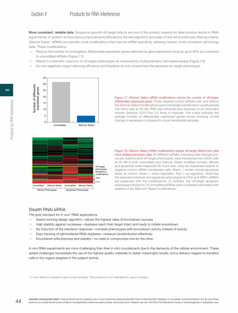
44 Important Licensing Information: These products may be covered by one or more Limited Use Label Licenses (See Thermo Fisher Scientific Catalogue or our website, www.thermofisher.com). By use of these products you accept the terms and conditions of all applicable Limited Use Label Licenses. All products are for research use only. CAUTION: Not intended for human or animal diagnostic or therapeutic uses.
Section II Products for RNA Interference
More consistent, reliable data. Sequence-specific off-target effects are one of the primary reasons for false positive results in RNAi
experiments. In addition to the potency improvements afforded by the new algorithm and state-of-the-art bioinformatic filtering criteria,
Silencer Select siRNAs incorporate novel modifications that improve siRNA specificity, allowing cleaner, more consistent cell biology
data. These modifications:
→ Reduce the number of nontargeted, differentially expressed genes detected by gene expression array by up to 90% as compared
to unmodified siRNAs (Figure 7.7)
→ Result in a dramatic reduction of off-target phenotypes as measured by multiparametric cell-based assays (Figure 7.8)
→ Do not negatively impact silencing efficiency and therefore do not compromise the expected on-target phenotypes
Figure 7.7. Silencer Select siRNA modifications reduce the number of off-target, differentially expressed genes. Three negative control siRNAs with and without the Silencer Select modifications were individually transfected in quadruplicate into HeLa cells at 30 nM. RNA was extracted and analysed on an Affymetrix Human Genome U133 Plus 2.0 Array in triplicate. The y-axis indicates the average number of differentially expressed genes—those showing ≥2-fold change in expression compared to mock-transfected samples.
30
25
15
10
5
0 Unmodi�ed Silencer Select
35
20
Num
ber
of d
iffer
ently
ex
pre
ssed
gen
es
Unmodi�ed Unmodi�ed
Mitosis Phenotypes
Silencer Select Silencer Select
Apoptosis Phenotypes
Off-target phenotypes elimated by modi�cations
WEE1
PLK1
* in vivo refers to research use in small animals. This product is not intended for use in humans.
Figure 7.8. Silencer Select siRNA modifications reduce off-target effects and yield more reliable phenotypic data. 53 different siRNAs, including older designs pre-viously noted to elicit off-target phenotypes, were transfected into U2OS cells at 30 nM in both unmodified and Silencer Select modified formats. Mitosis and apoptosis were measured 48 hours later. Data are expressed relative to negative control siRNA–transfected cells. Black = similar mitosis/apoptosis levels as control. Green = down-regulation. Red = up-regulation. Note that the expected mitotosis and apoptosis phenotypes for PLK and WEE1 siRNAs are preserved with the modifications. In contrast, the off-target apoptotic phenotypes elicited by 10 unmodified siRNAs were completely eliminated with addition of the Silencer® Select modifications.
Stealth RNAi siRNAThe gold standard for in vivo* RNAi applications
→ Award-winning design algorithm—allows the highest rates of knockdown success
→ High stability against nucleases—duplexes reach their target intact and ready to initiate knockdown
→ No induction of the interferon response—correlate phenotypes with knockdown activity instead of toxicity
→ Easy tracking of administered RNAi duplexes—measure biodistribution effectively
→ Knockdown effectiveness and stability—no need to compromise one for the other
In vivo RNAi experiments are more challenging than their in vitro counterparts due to the demands of the cellular environment. These
added challenges necessitate the use of the highest-quality materials to obtain meaningful results, and a delivery reagent to transfect
cells in the organs targeted in the subject animal.
Prod
ucts
for R
NA In
terfe
renc
eII

45
Chapter 7siRNA technologies
thermofisher.com
Stealth RNAi siRNA molecules are chemically modified, blunt-ended, 25-mer double-stranded duplexes that are recognised by
the RNA-induced silencing complex (RISC) to mediate inhibition of a target gene. Proprietary chemical modifications allow Stealth
RNAi™ siRNA to overcome many in vivo–specific obstacles, allowing effectiveness and stability in in vivo applications.
Specific and effective knockdown. In RNAi experiments, it is necessary to minimise off-target effects so that observed phenotypes
can be correctly attributed to target knockdown. With standard siRNA duplexes, both strands are able to participate in the knockdown
reaction, increasing the chances of off-target effects. With Stealth RNAi siRNA duplexes, the sense strand is chemically modified
to prevent it from contributing to RNAi activity. Stealth RNAi siRNA duplexes are designed with our award-winning BLOCK-iT RNAi
Designer (www.thermofisher.com/rnaidesigner), which uses Smith-Waterman alignment to compare the RNA duplex region to the
entire genome, including untranslated regions. This design step minimises the potential for off-target effects and allows a very high
percentage of experimentally successful duplexes (Figures 7.9 and 7.10).
lacZ
act
ivity
(% o
f con
trol
)
100
80
60
40
20
0
Contro
l
siRNA 1
siRNA 2
Contro
l
siRNA 1
siRNA 2
Stealt
h RNAi s
iRNA 1
Stealt
h RNAi s
iRNA 2
Stealt
h RNAi s
iRNA 1
Stealt
h RNAi s
iRNA 2
lacZ–target gene (forward) fusion lacZ–target gene (reverse) fusion
Sense strandSense strand
Antisense strand
Per
cent
(%) o
f tar
get
mR
NA
exp
ress
ion
rem
aini
ng (r
elat
ive
to c
ontr
ol) 1,000 different Stealth RNAi siRNA duplexes
171 human genes
4–12 Stealth RNAi siRNA duplexes per gene
2 nM Stealth RNAi siRNA duplexes transfected
0
20
40
60
80
100
≥97% knockdown
≥70% knockdown
Figure 7.9. Stealth RNAi siRNA exhibits increased target specificity. Sequences 1 and 2 were designed as either siRNA or Stealth RNAi siRNA duplexes against a target gene. Only the antisense strand of a Stealth RNAi siRNA duplex can enter the RISC for specific targeting of mRNA, whereas either strand of siRNA can enter the RISC, increasing the possibility of nonspecific silencing.
Figure 7.10. Success rate of Stealth RNAi siRNA design exceeds 90%. We have been generating data to evaluate the efficacy of a large number of Stealth RNAi siRNA duplexes since initiation of its extensive Validated Stealth RNAi™ siRNA programme in 2004. This data set was derived from 1,000 Stealth RNAi™ siRNA duplexes targeted to 171 genes generated using the BLOCK-iT™ RNAi Designer from 2004. Knockdown was measured using the RNAi target screening system, which assesses knockdown at the protein level. Over 90% of these duplexes (bounded by green box) resulted in ≥70% knockdown, and 30% (bounded by yellow box) resulted in ≥97% knockdown. These results were obtained using 2 nM Stealth RNAi siRNA duplexes. Typically, 10 to 100 nM is used for these types of experiments; the higher end of this range can lead to off-target effects. The data were analysed at each position to determine the differences between “good” and “bad” Stealth RNAi siRNA sequences. This information was then used to train the algorithm and has been incorporated into the latest version of the BLOCK-iT RNAi Designer (www.thermofisher.com/rnaidesigner).
7siRNA technologies

46 Important Licensing Information: These products may be covered by one or more Limited Use Label Licenses (See Thermo Fisher Scientific Catalogue or our website, www.thermofisher.com). By use of these products you accept the terms and conditions of all applicable Limited Use Label Licenses. All products are for research use only. CAUTION: Not intended for human or animal diagnostic or therapeutic uses.
Section II Products for RNA Interference
Stable in the presence of nucleases. Nucleases capable of degrading siRNA duplexes contribute to the challenging in vivo
experimental environment. Nuclease resistance helps ensure that siRNA duplexes reach their intended location intact and that they
are capable of initiating the knockdown reaction. Stealth RNAi siRNA duplexes have proprietary chemical modifications that confer
remarkable stability for in vivo studies (Figure 7.11); standard siRNAs lack such modifications, making them more susceptible to
degradation by nucleases.
No induction of the interferon response. The interferon response is a powerful immune reaction that can make analysis of knockdown
nearly impossible due to nonspecific toxic effects. Induction of the interferon response can lead to severely altered global gene activity
and make phenotypic assessment difficult. Standard siRNA has the potential to activate this innate response, setting off defense
systems usually used to combat viruses. Chemical modifications in Stealth RNAi siRNA molecules abolish the immunostimulatory
response observed with some sequences. Stealth RNAi siRNA complexes do not induce the interferon response (Figure 7.12),
providing confidence that observed phenotypes can be attributed to specific target knockdown and not to nonspecific toxicity.
Tracking delivered duplexes. Tracking the biodistribution of administered RNAi duplexes is necessary to ensure that the delivered
material has targeted the desired tissues. We use the industry-leading Alexa Fluor fluorescent dyes to label Stealth RNAi siRNA
duplexes for this purpose. These dyes are ideal for in vivo RNAi applications, due to their superior brightness and photostability.
Duplexes can be tracked in three different ways: a Stealth RNAi siRNA duplex can be directly labelled with an Alexa Fluor dye, it can
be biotinylated and detected with labelled streptavidin, or a labeled control duplex can be mixed with a target-specific Stealth RNAi
siRNA duplex (Figure 7.13). Direct labelling of Stealth RNAi siRNA duplexes does not affect their potency.
Together, the Silencer Select siRNA and the Stealth RNAi siRNA technologies provide a superior, complete, and robust solution for your
RNAi studies both in vitro and in vivo. For more information on these and other products for RNAi studies, visit www.thermofisher.com/rnai.
21-mer siRNA Stealth RNAi siRNA 21-mer siRNA Stealth RNAi siRNA
Mouse serum Human serum
0 4 8 24 48 720 4 8 24 48 720 4 8 24 48 72 0 4 8 24 48 72
Figure 7.11. Chemical modification makes Stealth RNAi siRNA more stable for in vivo applications. Stealth RNAi siRNA duplexes are chemically modified to enhance stability against nucleases in serum. Unmodified 21-mer dsRNA sequences and corresponding Stealth RNAi™ siRNA sequences were analysed at 0, 4, 8, 24, 48, and 72 hr following incubation in either 10% mouse or human serum. Samples were separated on a Novex 15% TBE-urea polyacrylamide precast gel and stained with methylene blue. These data demonstrate that Stealth RNAi siRNA duplexes remain intact, whereas unmodified 21-mer dsRNA sequences were susceptible to pronounced degradation.
Prod
ucts
for R
NA In
terfe
renc
eII

47
Chapter 7siRNA technologies
thermofisher.com
Figure 7.12. Stealth RNAi siRNA avoids the interferon stress response so that data can be confidently correlated with knockdown activity. Tail veins of Balb/c mice (three mice per treatment) were injected at 5 mg/kg and 10 mg/kg of Stealth RNAi siRNA duplexes. After 6 hr, serum was collected and processed to measure interferon markers using the Invitrogen Cytokine Mouse 20-Plex Panel for the Luminex instrument (Cat. No. LMC0006).
→ No treatment
3 mice per treatment:
6 hr
Luminex®
assays
→ CpG in PBS (pos. ctrl)→ 37 bp dsRNA→ Stealth RNAi siRNA in PBS
IL-1αIL-1βIL-2IL-4IL-5IL-6IL-10IL-12p40IL-12p70IL-13IL-17TNF-αIFN-γ
G-CSFGM-CSFFGF-basicVEGFPDGF-BBMIP-1αMIP-1βMIP-3βKCIP-10MCP-1MIGRANTES
Cytokine 10-plexTh1/Th2 6-plexIn�ammatory 4-plexChemokine 5-plexGrowth factor 4-plexCytokine 20-plex
19 markers Multiplex panels
Sacri�ce and collect serum
Rel
ativ
e va
lues
(log
sca
le)
0.1
1
10
100
37 bpdsRNA
2.5 mg/kg
Stealth RNAisiRNA
2.5 mg/kg
CpGNotreatment
IL-6
IP10
MIP-1α
IL-12
MCO-1
TNF-α
ProtocolR
af-1
/GA
PD
H r
atio
Raf-1 +BLOCK-iT
Raf-1 biotinRaf-1 Alexa Fluor 647
Raf-1Control
0.2
0
0.4
0.6
0.8
1.0
1.2
1.4
Raf-1RNAi–Alexa Fluor 647
Raf-1 RNAi, biotinylated Alexa Fluor 488–streptavidin
Raf-1 RNAi + BLOCK-iT™ Alexa Fluor 555
Figure 7.13. Labeled Stealth RNAi siRNA exhibits potent knockdown. MDA-MB-435 cells were transfected with 25 nmol unlabeled Stealth RNAi siRNA duplexes, or Stealth RNAi duplexes labeled with an Alexa Fluor dye or biotin. In each case the duplexes targeted Raf-1. Knockdown was assessed at >90% by real-time PCR; delivery is demonstrated through fluorescence imaging. Regardless of the labeling strategy, Stealth RNAi siRNA knockdown results were consistent.
7siRNA technologies

48 Important Licensing Information: These products may be covered by one or more Limited Use Label Licenses (See Thermo Fisher Scientific Catalogue or our website, www.thermofisher.com). By use of these products you accept the terms and conditions of all applicable Limited Use Label Licenses. All products are for research use only. CAUTION: Not intended for human or animal diagnostic or therapeutic uses.
Section II Products for RNA Interference
siRNA selection guidesiRNA type Description Technology Recommended use
Pre-designed/Validated (tubes)
Stealth Select RNAi siRNA and Vali dated Stealth RNAi siRNA
• Modified for serum stability and improved specificity
• Pre-designed for human, mouse, and rat genes• Validated for some human genes
Modified 25-mer
• Suitable for all applications• Particularly good for in vivo applications
InvitrogenTM SilencerTM Select Pre-designed and Validated siRNA
• Improved potency and guaranteed knockdown• Modified for improved specificity• Pre-designed for human, mouse and rat genes• Validated for some human genes
LNA modified 21-mer
• Suitable for all applications• Particularly good for cell culture and RNAi
screening work
InvitrogenTM SilencerTM Pre-designed and Validated siRNA
• Pre-designed for human, mouse, and rat genes• Validated for some human genes
Unmodified 21-mer
• Lower-cost alternative, but without some of the performance benefits of the newer technologies
• Suitable for all applications
Pre-made sets/collections
InvitrogenTM Made-to-Stock SilencerTM Select siRNA Libraries
• Pre-defined sets of Silencer Select siRNAs for the human genome and functional gene classes
• 0.25 nmol of each siRNA• Ready for immediate delivery
LNA modified 21-mer
• For screening of pre-defined Silencer Select siRNA sets such as human kinases, GPCRs, druggable and whole genome
InvitrogenTM Made-to-Stock SilencerTM siRNA Libraries
• Pre-defined sets of Silencer siRNAs for the human and mouse genomes and functional gene classes
• 0.25 nmol of each siRNA• Ready for immediate delivery
Unmodified 21-mer
• Lower-cost alternative to Silencer Select siRNA Libraries, but with out some of the performance enhancements
Plated
InvitrogenTM Made-to-Order SilencerTM Select siRNA Libraries
• Custom sets of Silencer Select siRNAs at 0.1 to 5 nmol
• Priced by quote and ordered offline
LNA modified 21-mer
• For screening 36 to >10,000 Silencer Select siRNAs
• Submit a gene list or choose one of our functional sets
• Multiple sizes and plating options
InvitrogenTM Made-to-Order SilencerTM siRNA Libraries
• Custom sets of Silencer siRNAs at 1 to 5 nmol• Priced by quote and ordered offline
Unmodified 21-mer
• Lower-cost alternative to Silencer Select siRNA Libraries, but without some of the performance benefits
InvitrogenTM PlateSelect™ RNAi w/ Stealth Select RNAi™ siRNA
• Stealth Select RNAi siRNA in plated format at 1 nmol size
• Configure plates with Pre-designed siRNA or siRNAs designed online with the BLOCK-iT™ RNAi Designer
Modified 25-mer
• For Stealth Select RNAi™ siRNA experiments in plates with 1 to >96 siRNAs
• Specify a set siRNAs using online interface• Made to order
InvitrogenTM PlateSelect™ RNAi w/ BLOCK-iT™ siRNA
• BLOCK-iT siRNA in plated format at 1 nmol size• Configure plates with siRNAs designed online
with the BLOCK-iT™ RNAi Designer
Unmodified 21-mer
• For unmodified siRNA experiments in plates with 1 to >96 siRNAs at 1 nmol
• Specify a set of siRNAs using online interface• Made to order
Design-your-own siRNAs
InvitrogenTM Stealth RNAi™ siRNA
• Chemically modified siRNA designed using the BLOCK-iT™ RNAi Designer
Modified 25-mer
• For genes that fall outside of our Pre-designed siRNA collection
• Suitable for all applications• Particularly good for in vivo applications
InvitrogenTM Block-iT™ siRNA • Traditional, unmodified siRNA designed using the
BLOCK-iT RNAi Designer Unmodified 21-mer
• For genes that fall outside of our Pre-designed siRNA collection
• Lower cost alternative, but without some of the performance benefits of Stealth RNAi™ siRNAs
Prod
ucts
for R
NA In
terfe
renc
eII
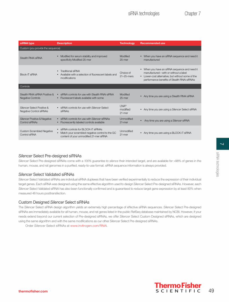
49
Chapter 7siRNA technologies
thermofisher.com
siRNA type Description Technology Recommended use
Custom (you provide the sequence)
Stealth RNAi siRNA• Modified for serum stability and improved
specificity Modified 25-mer Modified 25-mer
• When you have an siRNA sequence and need it manufactured
Block-iT siRNA • Traditional siRNA• Available with a selection of fluorescent labels and
modifications
Choice of 21–25-mers
• When you have an siRNA sequence and need it manufactured—with or without a label
• Lower-cost alternative, but without some of the performance benefits of Stealth RNAi siRNAs
Controls
Stealth RNAi siRNA Positive & Negative Controls
• siRNA controls for use with Stealth RNAi siRNA• Fluorescent labels available with some
Modified 25-mer
• Any time you are using a Stealth RNAi siRNA
Silencer Select Positive & Negative Control siRNAs
• siRNA controls for use with Silencer Select siRNAs
LNA® modified 21-mer
• Any time you are using a Silencer Select siRNA
Silencer Positive & Negative Control siRNAs
• siRNA controls for use with Silencer siRNAs• Fluorescently labeled controls available
Unmodified 21-mer
• Any time you are using a Silencer siRNA
Custom Scrambled Negative Control siRNA
• siRNA controls for BLOCK-iT siRNAs• Match your scrambled negative control to the GC
content of your unmodified 21-mer siRNA
Unmodified 21-mer
• Any time you are using a BLOCK-iT siRNA
Silencer Select Pre-designed siRNAsSilencer Select Pre-designed siRNAs come with a 100% guarantee to silence their intended target, and are available for >98% of genes in the
human, mouse, and rat genomes in a purified, ready-to-use format. siRNA sequence information is always provided.
Silencer Select Validated siRNAsSilencer Select Validated siRNAs are individual siRNA duplexes that have been verified experimentally to reduce the expression of their individual
target genes. Each siRNA was designed using the same effective algorithm used to design Silencer Select Pre-designed siRNAs. However, each
Silencer Select Validated siRNA has also been functionally confirmed and is guaranteed to reduce target gene expression by at least 80% when
measured 48 hours posttransfection.
Custom Designed Silencer Select siRNAsThe Silencer Select siRNA design algorithm yields an extremely high percentage of effective siRNA sequences. Silencer Select Pre-designed
siRNAs are immediately available for all human, mouse, and rat genes listed in the public RefSeq database maintained by NCBI. However, if your
needs extend beyond our current selection of Pre-designed siRNAs, we offer Silencer Select Custom Designed siRNAs, which are designed
using the same algorithm and with the same modifications as our other Silencer Select Pre-designed siRNAs.
Order Silencer Select siRNAs at www.invitrogen.com/RNAi.
7siRNA technologies
50 Important Licensing Information: These products may be covered by one or more Limited Use Label Licenses (See Thermo Fisher Scientific Catalogue or our website, www.thermofisher.com). By use of these products you accept the terms and conditions of all applicable Limited Use Label Licenses. All products are for research use only. CAUTION: Not intended for human or animal diagnostic or therapeutic uses.
Section II Products for RNA Interference
Silencer Select siRNA libraries
Pre-defined siRNA libraries plated to orderPre-defined, made-to-order Silencer Select siRNA Libraries are available for human:
→ Genome
→ Druggable genome
→ Extended druggable genome
→ Kinases
→ Phosphatases
→ GPCRs (non-olfactory)
→ Ion channels
→ Nuclear hormone receptors
→ Proteases
Human Genome siRNA Library (Consists of Part Numbers: 4397922 + 4397924 + 4397923)
Increasingly, researchers are performing genome-scale surveys of gene function. The InvitrogenTM SilencerTM Select Human Genome
siRNA Library V4 is ideal for this purpose. Three unique, non-overlapping siRNAs are provided for each of 21,585 human targets. The
siRNAs targeting the “druggable” portion of this library are arranged by gene functional class to enable easy screening of important
gene subsets such as kinase, phosphatase, GPCR, etc. This siRNA library is supplied in 384-well plates.
Human Druggable Genome and Human Extended Druggable Genome siRNA LibrariesWith siRNAs targeting the most therapeutically relevant human genes and conveniently grouped by target gene class, the InvitrogenTM
SilencerTM Select Human Druggable Genome siRNA Library V4 and InvitrogenTM SilencerTM Select Human Extended Druggable Genome
siRNA Library V4 are popular choices for many researchers. The druggable genome set targets 9,032 genes, whereas the extended
druggable genome collection targets the same 9,032 genes plus the “Extension Set” for a total of 10,415 genes, including transcription
factors. Please contact us for the complete list of targets. These libraries are available in both 96- and 384-well plate formats.
Human Kinase siRNA LibraryBecause of their importance in cell signalling and many biological pathways, kinases are key drug targets and subjects of intense
scrutiny. The InvitrogenTM SilencerTM Select Human Kinase siRNA Library V4 targets 710 human kinases with three individual Silencer
Select siRNAs per gene. This siRNA library enables systematic yet cost-effective RNAi studies of these key cell regulators. Validated
siRNAs are included where available. The Silencer Select Human Kinase siRNA Library V4 is provided in 96-well plates.
Human Phosphatase siRNA LibraryAlong with kinases, phosphatases are important regulators of biological pathways and cell signaling cascades. The InvitrogenTM
SilencerTM Select Human Phosphatase siRNA Library V4 targets 298 human phosphatases with three individual siRNAs per gene
supplied in 96-well plates. This library is the ideal companion to the InvitrogenTM SilencerTM Select Kinase siRNA Library V4, enabling
more detailed studies of biological pathways.
Human GPCR siRNA LibraryG-protein–coupled receptors, or GPCRs, are key components of many signal transduction pathways and also represent an important
class of druggable genes. The InvitrogenTM SilencerTM Select Human GPCR siRNA Library V4 targets 380 non-olfactory human GPCRs
with three individual Silencer Select siRNAs per target supplied in 96-well plates.
Prod
ucts
for R
NA In
terfe
renc
eII

51
Chapter 7siRNA technologies
thermofisher.com
Human Protease, Ion Channel, and Nuclear Hormone Receptor siRNA LibrariesFocused siRNA sets to human proteases, ion channels, and nuclear hormone receptors provide a cost effective means of studying
these important regulators of cellular function. These siRNA libraries are each supplied in 96-well plates. The InvitrogenTM SilencerTM
Select Human Protease siRNA Library V4 features 3 siRNAs per target for 494 human proteases. The InvitrogenTM SilencerTM Select
Human Ion Channel siRNA Library V4 features 3 siRNAs per target for 338 human ion channels. The InvitrogenTM SilencerTM Select
Human Nuclear Hormone Receptor siRNA Library V4 includes 3 siRNAs per target for 47 nuclear hormone receptors.
To create these siRNA libraries, our bioinformatics team uses PANTHER (www.pantherdb.org) and Gene Ontology™ annotation
information to create up-to-date target gene sets. siRNAs targeting the collection are then plated to order (minimum 0.25 nmol), giving
you not only the most up-to-date siRNA libraries, but also the freedom to customise your library so that it is most convenient for your
particular experimental setup. Want to add siRNAs to your collection? No problem! We can also provide siRNAs in 96-well or 384-
well plates, deliver multiple aliquots of each plate, arrange siRNAs in custom configurations, and provide siRNAs individually and/or
in pools.
(Our most popular collections are also available as pre-plated, ready-to-ship libraries with 0.25 nmol of each siRNA.)
Custom siRNA libraries—the ultimate in flexibilityDo you have a favourite list of human genes you would like to target with a collection of siRNAs? We can prepare a custom Silencer
Select siRNA Library to any set of human genes. The minimum order is only 36 siRNAs. All you need to supply is a list of genes or
transcripts identified by NCBI Entrez Gene ID or RefSeq mRNA accession number. With this user-friendly option, you can specify the
siRNA layout on either 96- or 384-well plates. For a 96-well plate, our standard format includes your choice of 0.1, 0.25, 1, 2, or 5 nmol
of each siRNA, with three individual siRNAs provided per target. For a 384-well plate, our standard format includes your choice of 1, 2,
or 5 nmol of each siRNA, with three individual siRNA provided per target. Multiple aliquots, pooling of siRNAs, and even custom siRNA
design requests can all be accomodated.
Information to accelerate discovery. All Silencer Select siRNA Libraries, whether custom, pre-defined, or pre-plated, are supplied with
full siRNA sequence information, and when available, with siRNA validation data. Also provided on the CD accompanying the siRNA
library are gene annotation information, which includes gene symbol, gene name, aliases, NCBI Entrez Gene ID, associated RefSeq
mRNA accession numbers, and other accession numbers. Plates are individually barcoded with unique identifiers; this information is
also provided with the siRNA and gene information.
Individual siRNAs for enhanced data reliabilityAll Silencer Select siRNA Libraries feature three individual siRNAs for each target. Screening with three siRNAs per gene significantly
decreases both false positive and false negative rates, increasing confidence in RNAi screening data, reducing the risk of missing
important genes, and decreasing time spent following up on false positive hits from the screen.
Silencer Select siRNA Libraries contain 0.25 nmol of each siRNA, sufficient for 500 transfections (at 5 nM siRNA, 100 µL
transfection volume). Preloaded 96-well plates are supplied with lyophilized siRNAs. One column of each 96-well plate is left empty to
allow for the addition of controls at the time of use. The libraries are delivered with full siRNA sequence information, as well as the exon
targeted and each targeted gene’s symbol, full name, aliases, RefSeq number, and Entrez Gene ID, plus results from any validation
experiments done using the included siRNAs.
Silencer Select siRNA Libraries to these or any other list of human genes can also be provided with 1, 2, or 5 nmol of each siRNA.
Custom formatting is available with these larger sizes.
If you would like a gene list, additional information, or would like to discuss your research with one of our technical experts,
contact your local sales representative or e-mail us at [email protected].
7siRNA technologies

52 Important Licensing Information: These products may be covered by one or more Limited Use Label Licenses (See Thermo Fisher Scientific Catalogue or our website, www.thermofisher.com). By use of these products you accept the terms and conditions of all applicable Limited Use Label Licenses. All products are for research use only. CAUTION: Not intended for human or animal diagnostic or therapeutic uses.
Section II Products for RNA Interference
Silencer Pre-designed and Validated siRNAs
Traditional, unmodified siRNAsFor human, mouse, and rat:
→ Cost effective, unmodified siRNAs designed for human, mouse and rat genes
→ Guaranteed silencing
→ Provided with free sequence information
Silencer siRNAs are remarkably effective and guaranteed to silenceSilencer Pre-designed siRNAs—chemically synthesised, unmodified siRNAs available for human, mouse and rat genes—are designed
with a rigorously tested siRNA design algorithm (see Silencer® Select Pre-designed siRNAs on page 40 for chemically modified
siRNAs designed with an even more effective algorithm). We guarantee that when three Silencer Pre-designed siRNAs are obtained
to the same target, at least two will reduce target mRNA levels by 70% or more.
Silencer Validated siRNAs are individual siRNA duplexes that have already been verified experimentally to reduce the expression
of their individual target genes. Each siRNA was designed using the same effective algorithm used to design Silencer Pre-designed
siRNAs. However, each one has also been functionally proven and is guaranteed to reduce target gene expression by at least 70%
48 hours posttransfection.
High-quality synthesisWe synthesise and purify each siRNA in state-of-the-art facilities to meet the highest quality standards. As part of our rigorous quality
control procedures, we analyse the mass of each RNA oligonucleotide by MALDI-TOF mass spectrometry and assess the purity of all
HPLC-purified oligonucleotides by HPLC. Finally, we analyse each annealed siRNA by gel electrophoresis to confirm that the strands
annealed properly. The result is premium quality siRNA that is purified and ready to use. See page 20 for a description of siRNA
purity grades.
Silencer siRNAs are easy to obtainOur searchable online siRNA database makes it easy to obtain effective, guaranteed-to-work siRNAs. Simply visit www.thermofisher.
com/siRNA to find siRNAs for your human, mouse, or rat gene of interest. Interested in other organisms? Invitrogen provides Custom
Designed Silencer and Silencer Select siRNAs (see page 49).
Order Silencer siRNAs at www.thermofisher.com/RNAi.
Chapter references 1. Jackson AL et al. (2003) Nat Biotechnol 21:635–637.
2. Saxena S et al. (2003) J Biol Chem 278:44312–44319.
3. Scacheri PC et al. (2004) Proc Natl Acad Sci U S A 101:1892–1897.
4. Du Q et al. (2005) Nucleic Acids Res 33:1671–1677.
5. Hornung V et al. (2005) Nat Med 11:263–270.
6. Judge AD et al. (2005) Nat Biotechnol 23:457–462.
7. Semizarov D et al. (2003) Proc Natl Acad Sci U S A 100:6347–6352.
Prod
ucts
for R
NA In
terfe
renc
eII
53
Chapter 8Vector-based RNAi Technologies
thermofisher.com
CHAPTER 8
Vector-based RNAi technologiesRNAi vectors for increased RNAi experimental optionsInvitrogen™ BLOCK-iT™ RNAi vectors offer a variety of options for long-term or transient RNAi expression.
With Invitrogen™ BLOCK-iT™ RNAi viral delivery systems, RNAi experiments can be performed in the
most biologically relevant cell type rather than in cell lines that are easy to transfect. The Invitrogen™
BLOCK-iT™ inducible systems permit RNAi initiation and duration to be controlled, while the BLOCK-iT
Pol II miR RNAi vectors limit knockdown to specific tissues or cells. There is a suitable BLOCK-iT RNAi
vector for every RNAi application.
We have developed two distinct vector-based RNAi systems for gene knockdown experiments:
BLOCK-iT Pol II miR RNAi Expression Vectors and Invitrogen™ BLOCK-iT™ shRNA Vectors. Both Pol II
miR RNAi and shRNA vector approaches include the capability for stable and inducible expression and
viral delivery.
The powerful new BLOCK-iT Pol II miR RNAi Expression Vectors have significant advantages over
the short-hairpin RNA (shRNA) vector technology currently used for RNAi vector applications (Table 8.1).
These new vectors include flanking and loop sequences from an endogenous miRNA that directs the
excision of the engineered miRNA from a longer Pol II transcript (pri-miRNA). When present in the nucleus,
these vectors efficiently use the endogenous cellular machinery to process knockdown sequences that
are specifically designed to have 100% homology to the target mRNA and will result in target cleavage. In
addition, the loop sequence has a unique restriction site, so that it can be linearised to allow for trouble-
free sequencing, as sequencing standard shRNA hairpins is sometimes a challenge.
See Table 8.2 for a comprehensive overview of available vectors for your RNAi experiments.
Table 8.1. Comparison of standard shRNA vectors and BLOCK-iT Pol II miR RNAi expression vectors.
BLOCK-iT shRNA vector technologies shRNA miR RNAi
Typical knockdown success rate † ~50% >75%
Expression tracking No Yes
Multiple-target knockdown No Yes
Tissue-specific expression No Yes
Gateway vector compatibility with most DEST vectors No Yes
† Rate at which constructs reduce target mRNA expression >70%
8Vector-based RNAi Technologies

54 Important Licensing Information: These products may be covered by one or more Limited Use Label Licenses (See Thermo Fisher Scientific Catalogue or our website, www.thermofisher.com). By use of these products you accept the terms and conditions of all applicable Limited Use Label Licenses. All products are for research use only. CAUTION: Not intended for human or animal diagnostic or therapeutic uses.
Section II Products for RNA Interference
Table 8.2. BLOCK-iT vector technologies.
VectorRNAi vector technology
Transient or stable expression
Selection marker Delivery method
Constitutive or inducible expression Reporter Kits Page
BLOCK-iT Pol II miR RNAi expression vector kits
pcDNA™6.2- GW/miR
miR RNAiTransient and stable
Blasticidin Transfection
Constitutive Pol II (CMV) Inducible and tissue-specific options
NoneBLOCK-iT Pol II miR RNAi Expression Vector Kit (also included in BLOCK-iT Lentiviral Pol II miR RNAi Expression System)
57
pcDNA™6.2- GW/EmGFP- miR
miR RNAiTransient and stable
Blasticidin Transfection
Constitutive Pol II (CMV) Inducible and tissue-specific options
Cocistronic Emerald GFP (EmGFP)
BLOCK-iT Pol II miR RNAi Expression Vector Kit with EmGFP (also included in BLOCK-iT Lentiviral Pol II miR RNAi Expression System)
57
BLOCK-iT™ Pol II miR RNAi lentiviral expression systems
pLenti6/V5-DEST miR RNAiTransient and stable
BlasticidinViral transduction (or transfection)
Constitutive Pol II (CMV)
NoneBLOCK-iT Lentiviral Pol II miR RNAi Expression System (includes BLOCK-iT Pol II miR RNAi Expression Vector Kit)
58
pLenti6/V5-DEST miR RNAiTransient and stable
BlasticidinViral transduction (or transfection)
Constitutive Pol II (CMV)
Cocistronic Emerald GFP (EmGFP)
BLOCK-iT Lentiviral Pol II miR RNAi Expression System with EmGFP (includes BLOCK-iT Pol II miR RNAi Expression Vector Kit with EmGFP)
58
pLenti6.4/ R4R2/ V5-DEST
miR RNAiTransient and stable
BlasticidinVital transduction (or transfection)
Constitutive (CMV or EF-1α) or other
Cocistronic Emerald GFP (EmGFP)
BLOCK-iT Lentiviral Pol II miR RNAi Expression System with EmGFP (includes BLOCK-iT™ Pol II miR RNAi Expression Vector Kit with EmGFP)
58
BLOCK-iT™ shRNA entry vectors
pENTR™/U6 shRNA Transient None Transfection Constitutive None
Invitrogen™ BLOCK-iT™ U6 RNAi Entry Vector Kit (also included in the BLOCK-iT RNAi Lentiviral Expression System and the BLOCK-iT Adenoviral RNAi Expression System)
66
pENTR™/H1/TO shRNATransient and stable
Zeocin™ TransfectionInducible or constitutive
None
Invitrogen™ BLOCK-iT™ Inducible H1 RNAi Entry Vector Kit (also included in the BLOCK-iT™ Inducible H1 Lentiviral RNAi System)
66
BLOCK-iT™ shRNA destination vectors
pLenti6/BLOCK-iT™-DEST
shRNATransient and stable
BlasticidinViral transduction (or transfection)
Constitutive NoneInvitrogen™ BLOCK-iT™ RNAi Lentiviral Expression System Invitrogen™ BLOCK-iT™ Lentiviral RNAi Gateway™ Vector Kit
69
pLenti4/BLOCK-iT™-DEST
shRNATransient and stable
Zeocin™Viral transduction (or transfection)
Inducible or constitutive
None
Invitrogen™ BLOCK-iT™ Inducible H1 Lentiviral RNAi System Invitrogen™ BLOCK-iT™ Lentiviral RNAi Zeo Gateway™ Vector Kit
71
pLenti6/TR (for expression of tetracycline repressor only)
shRNA or miR RNAi
Transient and stable
BlasticidinViral transduction (or transfection)
Constitutive NonepLenti6/TR Vector Kit BLOCK-iT™ Inducible H1 Lentiviral RNAi System
70
pAd/BLOCK-iT™-DEST
shRNA Transient NoneViral transduction (or transfection)
Inducible or constitutive
NoneInvitrogen™ BLOCK-iT™ Adenoviral RNAi Expression System pAd/BLOCK-iT™-DEST RNAi Gateway™ Vector
69
Invitrogen™ pBLOCK-iT™ 6-DEST
shRNATransient and stable
Blasticidin Transfection Constitutive None pBLOCK-iT 6-DEST 69
Invitrogen™ pBLOCK-iT™ 3-DEST
shRNATransient and stable
Geneticin TransfectionInducible or constitutive
None pBLOCK-iT 3-DEST 71
Continued on next page
Prod
ucts
for R
NA In
terfe
renc
eII

55
Chapter 8Vector-based RNAi Technologies
thermofisher.com
Table 8.2. BLOCK-iT vector technologies, continued.
VectorRNAi vector technology
Transient or stable expression
Selection marker Delivery method
Constitutive or inducible expression Reporter Kits Page
Selected InvitrogenTM GatewayTM DEST vectors compatible with miR RNAi vector technology*
Invitrogen™ pT-REx™-DEST30
miR RNAiTransient and stable
Geneticin™ TransfectionInducible (CMV)
Cocistronic EmGFP
No kit available. Vector Cat. No. 12301-016. Needs to be used in a T-REx™ cell line or in cells that express the Tet repressor protein.
62
Invitrogen™ pLenti4/TO/V5-DEST
miR RNAiTransient and stable
Zeocin™Viral transduction (or transfection)
Inducible (CMV)
Cocistronic EmGFP or none †
Invitrogen™ ViraPower™ T-REx™ Lentiviral Expression System. Needs to be used in a T-REx™ cell line or in cells that express the Tet repressor protein.
62
Invitrogen™ pEF-DEST51
miR RNAiTransient and stable
Blasticidin TransfectionConstitutive (EF-1α)
Cocistronic EmGFP or none †
No kit available. Vector Cat. No. 12285-011 62
Invitrogen™ pDEST™ R4-R3
miR RNAi Transient None Transfection Your choiceCocistronic EmGFP or none †
Invitrogen™ MultiSite Gateway™ Three-Fragment Vector Construction Kit
62
Invitrogen™ pLenti6/R4R2/V5-DEST
miR RNAiTransient and stable
BlasticidinViral transduction (or transfection)
Your choiceCocistronic EmGFP or none †
Invitrogen™ ViraPower™ Promoterless Lentiviral Invitrogen™ Gateway™ Expression System with MultiSite Invitrogen™ Gateway™ Technology
62
Invitrogen™ pLenti6/TR (for expression of tetracycline repressor only)
shRNA or miR RNAi
Transient and stable
BlasticidinViral transduction (or transfection)
Constitutive None pLenti6/TR Vector Kit 62
Invitrogen™ pcDNA™6/TR (for expression of tetracycline repressor only)
shRNA or miR RNAi
Transient and stable
Blasticidin TransfectionConstitutive (CMV)
NoneInvitrogen™ T-REx™ Core Kit or Invitrogen™ T-REx™ Complete Kit
62
Invitrogen™ pSCREEN-iT™/lacZ-DEST
shRNA or miR RNAi
Transient None Transfection Constitutive lacZ
Invitrogen™ BLOCK-iT™ RNAi Target Screening System; Invitrogen™ BLOCK-iT™ RNAi Target Screening Kit; Invitrogen™ pSCREEN-iT™/lacZ-DEST Gateway™ Vector Kit
64
Ready-to-use vectors: cloned and sequence verified
miR RNAi or shRNA
Transient and stable
Blasticidin, Zeocin™, or Geneticin
Transfection or viral transduction
Inducible or constituitive
Custom RNAi Services 93
* Additional components, such as Invitrogen™ Clonase™ enzyme mixes and Invitrogen™ Gateway™ donor vectors, are needed to transfer the miR RNAi cassette into these DEST vectors. The additional components are included in the BLOCK-iT™ Lentiviral Pol II miR RNAi Expression System but are not included in BLOCK-iT Pol II miR RNAi expression vector kits and must be purchased separately to transfer the miR RNAi expression cassette to alternative DEST vectors.
† Cocistronic EmGFP reporter can be transferred from the original BLOCK-iT Pol II miR RNAi expression vector.
8Vector-based RNAi Technologies
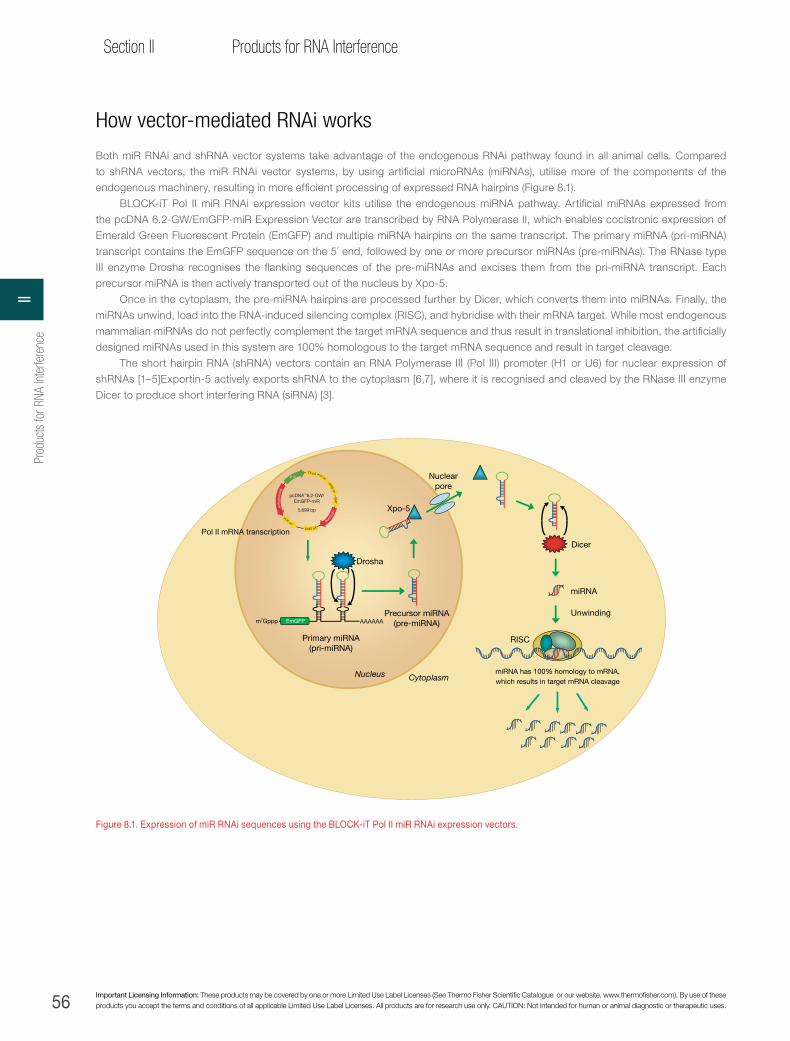
56 Important Licensing Information: These products may be covered by one or more Limited Use Label Licenses (See Thermo Fisher Scientific Catalogue or our website, www.thermofisher.com). By use of these products you accept the terms and conditions of all applicable Limited Use Label Licenses. All products are for research use only. CAUTION: Not intended for human or animal diagnostic or therapeutic uses.
Section II Products for RNA Interference
How vector-mediated RNAi works
Both miR RNAi and shRNA vector systems take advantage of the endogenous RNAi pathway found in all animal cells. Compared
to shRNA vectors, the miR RNAi vector systems, by using artificial microRNAs (miRNAs), utilise more of the components of the
endogenous machinery, resulting in more efficient processing of expressed RNA hairpins (Figure 8.1).
BLOCK-iT Pol II miR RNAi expression vector kits utilise the endogenous miRNA pathway. Artificial miRNAs expressed from
the pcDNA 6.2-GW/EmGFP-miR Expression Vector are transcribed by RNA Polymerase II, which enables cocistronic expression of
Emerald Green Fluorescent Protein (EmGFP) and multiple miRNA hairpins on the same transcript. The primary miRNA (pri-miRNA)
transcript contains the EmGFP sequence on the 5´ end, followed by one or more precursor miRNAs (pre-miRNAs). The RNase type
III enzyme Drosha recognises the flanking sequences of the pre-miRNAs and excises them from the pri-miRNA transcript. Each
precursor miRNA is then actively transported out of the nucleus by Xpo-5.
Once in the cytoplasm, the pre-miRNA hairpins are processed further by Dicer, which converts them into miRNAs. Finally, the
miRNAs unwind, load into the RNA-induced silencing complex (RISC), and hybridise with their mRNA target. While most endogenous
mammalian miRNAs do not perfectly complement the target mRNA sequence and thus result in translational inhibition, the artificially
designed miRNAs used in this system are 100% homologous to the target mRNA sequence and result in target cleavage.
The short hairpin RNA (shRNA) vectors contain an RNA Polymerase III (Pol III) promoter (H1 or U6) for nuclear expression of
shRNAs [1–5]Exportin-5 actively exports shRNA to the cytoplasm [6,7], where it is recognised and cleaved by the RNase III enzyme
Dicer to produce short interfering RNA (siRNA) [3].
i
miRNA has 100% homology to mRNA,which results in target mRNA cleavage
Drosha
AAAAAA
Primary miRNA(pri-miRNA)
Precursor miRNA(pre-miRNA)
Dicer
Nuclearpore
Xpo-5
miRNA
Unwinding
RISC
Nucleus Cytoplasm
EmGFP
Pol II mRNA transcription
m7Gppp
P CMV
SV40 pA
TK pA
Blastic
idin
Spectinom
ycin
f1 oriSV40 ori
EM7
pUC ori
pcDNA™6.2-GW/EmGFP-miR
5,699 bp
Figure 8.1. Expression of miR RNAi sequences using the BLOCK-iT Pol II miR RNAi expression vectors.
Prod
ucts
for R
NA In
terfe
renc
eII

57
Chapter 8Vector-based RNAi Technologies
thermofisher.com
BLOCK-iT Pol II miR RNAi expression vectors—versatile Pol II vector-based RNAi technology
BLOCK-iT Pol II miR RNAi expression vectors combine the advantages of traditional RNAi vectors—stable expression and the ability
to use viral delivery—with capabilities for tissue-specific expression and multiple-target knockdown from the same transcript. The
pcDNA6.2-GW/miR and pcDNA6.2-GW/EmGFP-miR vectors (Figure 8.2), included in the BLOCK-iT Pol II miR RNAi expression vector
kits and the BLOCK-iT Lentiviral Pol II miR RNAi Expression System, are designed for expressing artificial miRNAs that have been
engineered to have 100% homology to a target sequence and will result in its cleavage. The BLOCK-iT Pol II miR RNAi Expression
Vector technology offers many benefits:
→ Over 75% knockdown success—screen fewer clones because design predictability is higher than that of other vector-based
RNAi methods
→ Easy expression tracking—assess knockdown of target miRNA simply and reliably with cocistronic expression with EmGFP
→ Multiple-target knockdown—knock down multiple targets simultaneously or generate synthetic phenotypes
→ Gateway vector compatibility—use a wide selection of Invitrogen™ destination vectors, including lentiviral vectors, for stable
expression in any cell type, such as primary and nondividing cells
→ Tissue-specific experimental options—select from a variety of destination vectors or MultiSite Gateway vector applications to add
different promoters for cellular applications or in vivo knockdown
→ Inducible expression—regulation of the RNAi response permits the study of changes over time and loss-of-function experiments,
and provides an excellent control system to measure phenotypic changes during recovery of gene function
→ BLOCK-iT miR RNAi Select—target the majority of annotated human, mouse, and rat genes with predesigned BLOCK-iT miR
RNAi Select hairpins, ready to anneal and clone into either of the BLOCK-iT Pol II miR RNAi expression vectors
P CMV
SV40 pA
TK pA
Blastic
idin
Spectinom
ycin
f1 oriSV40 ori
EM7
pUC ori
pcDNA™6.2-GW/EmGFP-miR
5,699 bp
attB1 EmGFP 5´ miR�anking region ACGA attB23´ miR
�anking regionCAGG
P CMV
SV40 pA
TK pA
Blastic
idin
Spectinom
ycin
f1 oriSV40 ori
EM7
pUC ori
pcDNA™6.2-GW/miR
4,944 bp
attB1 5´ miR�anking region ACGA attB23´ miR
�anking regionCAGG
Figure 8.2. The BLOCK-iT Pol II miR RNAi expression vectors. The pcDNA 6.2-GW/miR vector is driven by the CMV promoter, has the blasticidin resistance marker, and is available with or without cocistronic EmGFP expression as a reporter.
NOTE: Find information on miR RNAi expression vectors online at www.thermofisher.com/mir.
8Vector-based RNAi Technologies

58 Important Licensing Information: These products may be covered by one or more Limited Use Label Licenses (See Thermo Fisher Scientific Catalogue or our website, www.thermofisher.com). By use of these products you accept the terms and conditions of all applicable Limited Use Label Licenses. All products are for research use only. CAUTION: Not intended for human or animal diagnostic or therapeutic uses.
Section II Products for RNA Interference
Combine BLOCK-iT vector kits with lentivirus for stable, long-term expressionInvitrogen™ BLOCK-iT™ and Invitrogen™ BLOCK-iT™ HiPerform™ Lentiviral Pol II miR RNAi Expression Systems combine all of
the advantages of the BLOCK-iT Pol II miR RNAi Expression Vector Kit with Invitrogen™ ViraPower™ Lentiviral Expression Vectors
(Figure 8.3), enabling stable delivery of engineered miRNAs into nondividing, primary, and hard-to-transfect cells. For example,
ViraPower Lentiviral Expression Vectors have been used to deliver miR RNAi sequences into rat primary cortical neurons to knock
down microtubule-associated protein 2 (Figure 8.4). These vectors deliver miRNA sequences into HT1080 and HeLa cells to knock
down lamin A/C or lacZ (Figure 8.5), establishing long-term, stable expression of miRNAs in these cell model systems.
The BLOCK-iT HiPerform Lentiviral Pol II miR RNAi Expression System with EmGFP is the most powerful and flexible RNAi vector
offered to date. This technology combines high titers and maximum expression. The ability to incorporate custom promoters makes
the system suitable for in vivo applications.
The HiPerform vector contains an mRNA-stabilising sequence (WPRE) and a nuclear import sequence (cPPT) that can generate
up to 5-fold higher virus titers and EmGFP expression levels in many cell lines. Additionally, MultiSite technology allows you to express
the EmGFP/miR RNAi cassette from CMV, EF-1α, your own tissue-specific promoter, or another appropriate in vivo promoter:
→ Achieve up to 5x higher titers (measured by GFP), allowing more cells to be transduced or higher multiplicities of infection (MOIs)
to be employed
→ Incorporate your own tissue-specific or another appropriate in vivo promoter
→ Track miRNA expression through cocistronic expression with EmGFP
→ Knock down more than one gene simultaneously through expression of multiple miRNAs from a single transcript
Figure 8.3. pLenti6/V5-DEST vector. A. The BLOCK-iT Lentiviral Pol II miR RNAi Expression Systems utilise the pLenti6/V5-DEST vector for constitutive expression from the CMV immediate-early promoter, and the blasticidin selection marker for compatibility with a tetracycline-regulated inducible system. B. Images taken 4 days following transduction of Invitrogen™ GripTite™ 293 MSR cells at an MOI of 3 with lentiviral particles generated using the indicated vectors.
pLenti6.4/CMV orEF-1α/MSGW/EmGFP-miR
8,875 bp
PR
SV/5
´ LTR
RRE
∆E3/3´ L
TRB
last
icid
in
SV40 pA
Ampicillin
pUC
ori
PPGK
EM7
attB1 attB2attB4 EmGFP 5´ miR�anking region
3´ miR�anking regionPCMV
cPPT WPRE
A
pLenti6-GW/EmGFP-miR-negpLenti6.4/CMV/V5- MSGW/EmGFP-miR-negB
Prod
ucts
for R
NA In
terfe
renc
eII

59
Chapter 8Vector-based RNAi Technologies
thermofisher.com
No treatment
Lenti-miR-Neg
Lenti-miR-MAP2A
Figure 8.4. Target gene knockdown in primary neurons. Unlike standard retroviruses, lentiviruses can transduce nondividing cells, such as these rat primary cortical neurons. Here, microtubule-associated protein 2 (MAP2A) was targeted, and its expression was dramatically decreased. No treatment (A and B), Lenti-miR-Neg (C and D), and Lenti-miR-MAP2 (E and F) were stained for anti-MAP2 and DAPI. Images were acquired with a Nikon E800 microscope and the Omega XF-32 filter cube for the AF555 channel and the Omega XF136-2 filter cube for DAPI. Panels A, C, and E: 20x magnification. Panels B, D, and F: 40x magnification. There is a visible difference between the transfected control and the lenti control vs. lentiviral delivery of an RNAi knockdown reagent.
Mag
icM
ark™
Lam
in
Day33
80 kDa Lamin A/C
β-actin
60 kDa
50 kDa
40 kDa
Day48
Day64
Day85
Day99
lacZ
Lam
in
lacZ
Lam
in
lacZ
Lam
in
lacZ
Lam
in
lacZ
Figure 8.5. Stable lentiviral transduction of miRNA expression cassettes. Western blot analysis of HeLa cell populations stably transduced (MOI = 20) for 33 to 99 days with lamin or lacZ miRNA–expressing lentiviral particles. The blot was probed with anti–lamin A/C (top half) or anti–β-actin (bottom half) antibodies.
8Vector-based RNAi Technologies
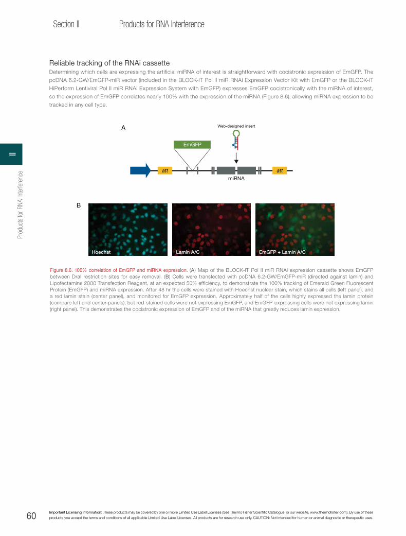
60 Important Licensing Information: These products may be covered by one or more Limited Use Label Licenses (See Thermo Fisher Scientific Catalogue or our website, www.thermofisher.com). By use of these products you accept the terms and conditions of all applicable Limited Use Label Licenses. All products are for research use only. CAUTION: Not intended for human or animal diagnostic or therapeutic uses.
Section II Products for RNA Interference
Reliable tracking of the RNAi cassetteDetermining which cells are expressing the artificial miRNA of interest is straightforward with cocistronic expression of EmGFP. The
pcDNA 6.2-GW/EmGFP-miR vector (included in the BLOCK-iT Pol II miR RNAi Expression Vector Kit with EmGFP or the BLOCK-iT
HiPerform Lentiviral Pol II miR RNAi Expression System with EmGFP) expresses EmGFP cocistronically with the miRNA of interest,
so the expression of EmGFP correlates nearly 100% with the expression of the miRNA (Figure 8.6), allowing miRNA expression to be
tracked in any cell type.
Figure 8.6. 100% correlation of EmGFP and miRNA expression. (A) Map of the BLOCK-iT Pol II miR RNAi expression cassette shows EmGFP between DraI restriction sites for easy removal. (B) Cells were transfected with pcDNA 6.2-GW/EmGFP-miR (directed against lamin) and Lipofectamine 2000 Transfection Reagent, at an expected 50% efficiency, to demonstrate the 100% tracking of Emerald Green Fluorescent Protein (EmGFP) and miRNA expression. After 48 hr the cells were stained with Hoechst nuclear stain, which stains all cells (left panel), and a red lamin stain (center panel), and monitored for EmGFP expression. Approximately half of the cells highly expressed the lamin protein (compare left and center panels), but red-stained cells were not expressing EmGFP, and EmGFP-expressing cells were not expressing lamin (right panel). This demonstrates the cocistronic expression of EmGFP and of the miRNA that greatly reduces lamin expression.
miRNA
Web-designed insert
EmGFP
att att
A
Lamin A/CHoechst EmGFP + Lamin A/C
B
Prod
ucts
for R
NA In
terfe
renc
eII
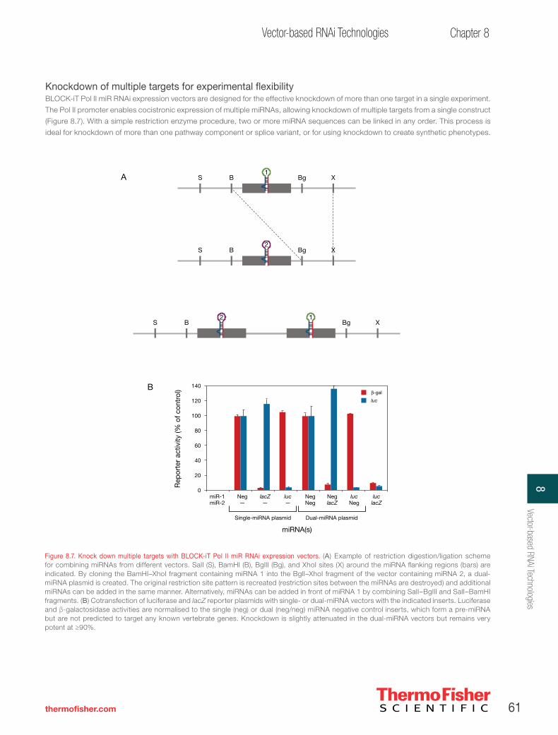
61
Chapter 8Vector-based RNAi Technologies
thermofisher.com
Knockdown of multiple targets for experimental flexibilityBLOCK-iT Pol II miR RNAi expression vectors are designed for the effective knockdown of more than one target in a single experiment.
The Pol II promoter enables cocistronic expression of multiple miRNAs, allowing knockdown of multiple targets from a single construct
(Figure 8.7). With a simple restriction enzyme procedure, two or more miRNA sequences can be linked in any order. This process is
ideal for knockdown of more than one pathway component or splice variant, or for using knockdown to create synthetic phenotypes.
Figure 8.7. Knock down multiple targets with BLOCK-iT Pol II miR RNAi expression vectors. (A) Example of restriction digestion/ligation scheme for combining miRNAs from different vectors. SalI (S), BamHI (B), BglII (Bg), and XhoI sites (X) around the miRNA flanking regions (bars) are indicated. By cloning the BamHI–XhoI fragment containing miRNA 1 into the BglI–XhoI fragment of the vector containing miRNA 2, a dual-miRNA plasmid is created. The original restriction site pattern is recreated (restriction sites between the miRNAs are destroyed) and additional miRNAs can be added in the same manner. Alternatively, miRNAs can be added in front of miRNA 1 by combining SalI–BglII and SalI–BamHI fragments. (B) Cotransfection of luciferase and lacZ reporter plasmids with single- or dual-miRNA vectors with the indicated inserts. Luciferase and β-galactosidase activities are normalised to the single (neg) or dual (neg/neg) miRNA negative control inserts, which form a pre-miRNA but are not predicted to target any known vertebrate genes. Knockdown is slightly attenuated in the dual-miRNA vectors but remains very potent at ≥90%.
S B Bg X
S B Bg X
S B Bg X
1
2
2 1
A
β-gal
luc
NegNeg
NeglacZ
lucNeg
luclacZ
Neg—
Single-miRNA plasmid
lacZ—
luc—
miR-1miR-2
Rep
orte
r ac
tivity
(% o
f con
trol
) 140
100
40
120
60
80
20
0
miRNA(s)
Dual-miRNA plasmid
B
8Vector-based RNAi Technologies

62 Important Licensing Information: These products may be covered by one or more Limited Use Label Licenses (See Thermo Fisher Scientific Catalogue or our website, www.thermofisher.com). By use of these products you accept the terms and conditions of all applicable Limited Use Label Licenses. All products are for research use only. CAUTION: Not intended for human or animal diagnostic or therapeutic uses.
Section II Products for RNA Interference
Gateway vector compatibility for expanded experimental options
Gateway technology is a fast and efficient way to transfer miR RNAi cassettes into a variety of vectors by homologous recombination.
Gateway recombination eliminates tedious and time-consuming subcloning procedures, and the resulting expression cassettes can
be easily moved into any of the Gateway DEST vectors. See Table 8.3 for a listing of Gateway destination (DEST) vectors that have
been functionally tested for compatibility with the BLOCK-iT™ Pol II miR RNAi Vector Kits.
With the BLOCK-iT Pol II miR RNAi Expression Vector Kits, miRNAs are cloned directly into Gateway expression vectors, as
opposed to Gateway entry vectors. As a result, there are two key differences between the pcDNA 6.2-GW/miR and pcDNA 6.2-GW/
EmGFP-miR expression vectors and typical Gateway entry vectors:
1. The miRNA expression vectors include the cytomegalovirus (CMV) promoter. After miRNA sequences are cloned, they are
immediately ready for transfection and miRNA expression.
2. attB sites flank the miRNA (and EmGFP sequences if using pcDNA 6.2-GW/EmGFP-miR). For expression in different DEST
vectors, the inserts must first be transferred to a pDONR™ (donor) vector and then to a DEST vector of choice by a dual Clonase
enzyme reaction. The pDONR 221 vector, BP and LR Clonase II enzyme mixes, and pLenti6/V5-DEST vector are included in the
BLOCK-iT Lentiviral Pol II miR RNAi Expression System.
Pol II promoter versatility allows tissue-specific expressionMost shRNA vectors are driven by Pol III promoters, significantly limiting the types of promoters that can be used in RNAi experiments
and making tissue-specific expression in an in vivo system impossible. Other vector approaches use specially modified Pol II promoters,
which cannot be easily exchanged for other Pol II promoters. The BLOCK-iT Pol II miR RNAi Expression Vectors include the CMV
immediate-early Pol II promoter and are compatible with virtually any other Pol II promoter (Figure 8.8), providing a flexible system to
regulate knockdown or to use promoters that are active in specific tissues for in vivo studies.
Inducible expression for control over your experimentThe Invitrogen™ BLOCK-iT™ Inducible Pol II miR RNAi Expression Vector Kit with EmGFP enables customers to regulate RNAi
experiments (see Figure 8.8B). Now you can observe changes over time by controlling the initiation of the RNAi response with an
inducible system. The kit contains the pT-REx-DEST30 Gateway vector which, after simple cloning and shuttling techniques, produces
a miR RNAi expression vector suitable for inducible knockdown. The pT-REx-DEST30 Gateway vector contains the CMV promoter with
two copies of the tetracycline operator (TetO2) sequence, allowing high-level and regulated expression (Figure 8.9). This permits the
study of loss of function in a stably transfected cell line even if the gene of interest is essential. Also, induction of miR RNAi expression
can be halted so phenotypic changes can be measured during recovery of gene function.
Table 8.3. Compatibility of Gateway destination vectors.
Promoter or system Compatibility Cat. no.
ViraPower lentiviral vectors, including MultiSite Gateway technology
Bench-tested with pLenti6/V5-DEST and pLenti6/UbC/V5-DEST
V496-10 V499-10
EF-1α promoter Bench-tested with pEF-DEST51 12285-011
Invitrogen™ T-REx™ vector Bench-tested with pT-REx DEST30 12301-016
Invitrogen™ Flp-In™ cell line Bench-tested with pEF5/FRT/V5-DEST V6020-20
N-terminal reporter tags Bench-tested with pcDNA 6.2/N-YFP/DEST V358-20
MultiSite Gateway technologyBench-tested with pDEST/R4-R3 using thymidine kinase (TK) poly(A) 3´ element and various 5´ promoter elements
12537-023
Prod
ucts
for R
NA In
terfe
renc
eII

63
Chapter 8Vector-based RNAi Technologies
thermofisher.com
β-gal
luc
Neg AAK1 lacZ luc2AAK1
300 ng CMV
lacZ luc2Neg
Rep
orte
r en
zym
e ac
tivity
(% o
f neg
con
trol
s)
100
40
120
140
60
80
20
0
300 ng EF-1α
A
luc unind.
luc ind.
β-gal unind.
β-gal ind.
lacZ luc2AAK1Neg
Rep
orte
r en
zym
e ac
tivity
(% o
f neg
con
trol
s)
120
100
80
60
40
20
0
30 ng CMV/TO
BR
epor
ter
activ
ity (%
of c
ontr
ol)
(1 x
1,0
00 R
FU)
luc2lacZNeg
200
0
400
600
800
1,000
1,200
β-gal
Rep
orte
r ac
tivity
(% o
f con
trol
)(R
FU)
luc2lacZNeg0
5,000
10,000
15,000
20,000
25,000
luc
C
Figure 8.8. BLOCK-iT Pol II miR RNAi Expression Vectors are compatible with multiple promoters. (A) Normalised reporter activities from lysates of GripTite 293 cells cotransfected with 100 ng each of luciferase and lacZ reporter plasmids and 300 ng/well of either pcDNA 6.2-GW/EmGFP-miR (CMV) or pEF-GW/ EmGFP-miR (EF-1α) expression vectors. (B) Knockdown of pSCREEN-iT/lacZ-AAK1 vector and luciferase reporters in T-REx 293 cells with and without induction with 1 μg/mL tetracycline at 30 ng of pT-REx-GW/EmGFP-miR (CMV/TO) plasmid per well. (C) Knockdown of cotransfected lacZ and luciferase reportergenes in HepG2 cells by MultiSite Gateway EmGFP-miR constructs with the anti-trypsin promoter as the 5´ element and the poly(A) signal from the HSV thymidine kinase gene as the 3´ element.
Repressed state
Derepressed state
H1 TetO2 TATA TetO2 miR RNAi
H1 TetO2 TATA TetO2 miR RNAi
Figure 8.9. Inducible RNAi via tetracycline regulation of the CMV/TO Pol II promoter. When the tetracycline repressor (TR) protein is present in the cell, it tightly binds the two TetO2 sites within the H1 promoter, essentially blocking initiation of shRNA transcription. When tetracycline is added to the culture medium, it binds to, and changes, the conformation of the TR protein, causing the TR protein to release from the two TetO2 sites, allowing transcription to occur.
8Vector-based RNAi Technologies

64 Important Licensing Information: These products may be covered by one or more Limited Use Label Licenses (See Thermo Fisher Scientific Catalogue or our website, www.thermofisher.com). By use of these products you accept the terms and conditions of all applicable Limited Use Label Licenses. All products are for research use only. CAUTION: Not intended for human or animal diagnostic or therapeutic uses.
Section II Products for RNA Interference
BLOCK-iT miR RNAi Select
Now it is easier than ever to harness the power of the BLOCK-iT Pol II miR RNAi Expression Vectors with predesigned BLOCK-iT miR
RNAi Select hairpins, which target the majority of the human, rat, and mouse genes in the RefSeq database. The BLOCK-iT miR RNAi
hairpins, designed in silico using the award-winning BLOCK-iT RNAi Designer, are provided as DNA oligos that are ready to anneal
and clone into one of the BLOCK-iT Pol II miR RNAi Expression Vectors. Ready-to-order BLOCK-iT miR RNAi Select hairpins provide
significant advantages:
→ Easy access to BLOCK-iT Pol II miR RNAi Expression Vector technology, which combines the high design success rate of artificial
miRNAs with tissue-specific, inducible, multiple gene–targeting, and GFP reporter options
→ Effortless ordering using the BLOCK-iT RNAi Express search engine
→ Allows maximum reduction of t due to combined power of BLAST and the advanced Smith-Waterman alignment specificity
search
→ Four BLOCK-iT miR RNAi Select hairpin designs directed to nonoverlapping regions of the target mRNA sequence.
With Invitrogen™ BLOCK-iT™ miR RNAi Select 4 Sets, you can avoid the work of designing RNAi hairpin sequences in-house.
Product Quantity Cat. no.
Invitrogen™ miR RNAi vector kits
BLOCK-iT Pol II miR RNAi Expression Vector Kit 20 rxns K4935-00
BLOCK-iT Pol II miR RNAi Expression Vector Kit With EmGFP 20 rxns K4936-00
BLOCK-iT Lentiviral Pol II miR RNAi Expression System 20 rxns K4937-00
BLOCK-iT Lentiviral Pol II miR RNAi Expression System With EmGFP 20 rxns K4938-00
BLOCK-iT Pol II miR-lacZ Validated miRNA Control Vector 10 µg V49350-00
BLOCK-iT Pol II miR-luc Validated miRNA Control Vector 10 µg V49351-00
BLOCK-iT Pol II miR-LMNA Validated miRNA Control Vector 10 µg V49352-00
BLOCK-iT miR RNAi Select 1 hairpin †
BLOCK-iT miR RNAi Select 4 Sets* 4 hairpins †
BLOCK-iT HiPerform Lentiviral Pol II miR RNAi Expression System With EmGFP 20 rxns K4934-00
BLOCK-iT Inducible Pol II miR RNAi Expression Vector Kit With EmGFP 20 rxns K4939-00
Recommended destination vector kits
Gateway pT-REx-DEST30 Vector 6 µg 12301-016
ViraPower T-REx Lentiviral Expression System 20 rxns K4965-00
Gateway pEF-DEST51 Vector 6 µg 12285-011
MultiSite Gateway Three-Fragment Vector Construction Kit 20 rxns 12537-023
ViraPower Promoterless Lentiviral Gateway Expression System with MultiSite Gateway Technology 20 rxns K5910-00
* Go to www.thermofisher.com/rnaicentral for details on the 100% performance guarantee.
† Visit www.thermofisher.com/rnaiexpress for ordering information on the latest BLOCK-iT miR RNAi Select, BLOCK-iT miR RNAi Select 4 Sets, and BLOCK-iT Pol II miR Validated miRNA Vector DuoPaks that are available.
Prod
ucts
for R
NA In
terfe
renc
eII

65
Chapter 8Vector-based RNAi Technologies
thermofisher.com
Guaranteed results with BLOCK-iT miR RNAi Select 4 Sets
The purchase of a BLOCK-iT miR RNAi Select 4 Set includes 100% guaranteed results: two of the four designs will result in at least
70% transcript knockdown, given a transfection efficiency of at least 80%. If, under these conditions, three or more of the designs fail
to knock down the target mRNA by at least 70%, we will design and ship two additional target-specific BLOCK-iT miR RNAi hairpins
at no expense.*
* Please contact our Technical Services to take advantage of this guarantee. Be prepared to fax or email your order reference number, BLOCK-iT miR RNAi Select sequences, and data showing transfection efficiency and level of knockdown. This guarantee can be used only once per target gene.
With BLOCK-iT miR RNAi Select 4 Sets, you can avoid the work of designing RNAi hairpin sequences yourself. The development of
these molecules begins with identification of the entire human, rat, and mouse transcripts in the Unigene database. We then select
exceptionally effective in silico–designed sequences by a four-step process:
→ BLAST search to remove sequences within the target mRNA that are homologous to other mRNA sequences within the target
species
→ BLOCK-iT miR RNAi Select hairpin design using the BLOCK-iT RNAi Designer, which includes both standard and proprietary
rules based on testing thousands of RNA duplex and RNAi vector sequences
→ Smith-Waterman mismatch alignment to rigorously screen sequences for potential off-target effects
→ Selection of the top four artificial miRNA hairpin designs targeting nonoverlapping regions in all major splice variants found in
RefSeq for the target mRNA sequence; these designs are offered independently as BLOCK-iT miR RNAi Select hairpins or as a
complete BLOCK-iT miR RNAi Select 4 Set
8Vector-based RNAi Technologies
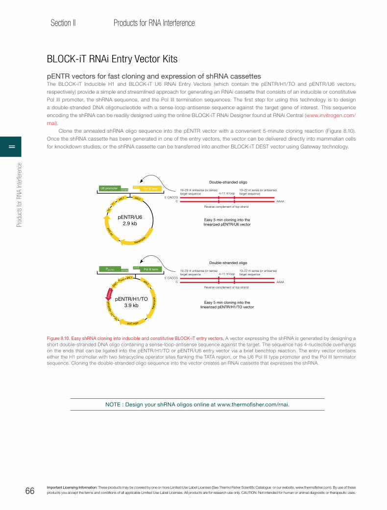
66 Important Licensing Information: These products may be covered by one or more Limited Use Label Licenses (See Thermo Fisher Scientific Catalogue or our website, www.thermofisher.com). By use of these products you accept the terms and conditions of all applicable Limited Use Label Licenses. All products are for research use only. CAUTION: Not intended for human or animal diagnostic or therapeutic uses.
Section II Products for RNA Interference
BLOCK-iT RNAi Entry Vector Kits
pENTR vectors for fast cloning and expression of shRNA cassettesThe BLOCK-iT Inducible H1 and BLOCK-iT U6 RNAi Entry Vectors (which contain the pENTR/H1/TO and pENTR/U6 vectors,
respectively) provide a simple and streamlined approach for generating an RNAi cassette that consists of an inducible or constitutive
Pol III promoter, the shRNA sequence, and the Pol III termination sequences. The first step for using this technology is to design
a double-stranded DNA oligonucleotide with a sense-loop-antisense sequence against the target gene of interest. This sequence
encoding the shRNA can be readily designed using the online BLOCK-iT RNAi Designer found at RNAi Central (www.invitrogen.com/
rnai).
Clone the annealed shRNA oligo sequence into the pENTR vector with a convenient 5-minute cloning reaction (Figure 8.10).
Once the shRNA cassette has been generated in one of the entry vectors, the vector can be delivered directly into mammalian cells
for knockdown studies, or the shRNA cassette can be transferred into another BLOCK-iT DEST vector using Gateway technology.
Double-stranded oligo
4–11 nt loop19–22 nt sense (or antisense)target sequence
19–29 nt antisense (or sense)target sequence
Reverse complement of top strand
AAAA5´ CACCG
C
Easy 5 min cloning into thelinearized pENTR/U6 vector
Easy 5 min cloning into thelinearized pENTR/H1/TO vector
Double-stranded oligo
4–11 nt loop19–22 nt sense (or antisense)target sequence
19–29 nt antisense (or sense)target sequence
Reverse complement of top strand
AAAA5´ CACCG
C
pENTR/U62.9 kb
U6 promoter Pol III termGTGGTTTT
PH1/TO Pol III termGTGGTTTT
pENTR/H1/TO3.9 kb
T2
T1
SV
40 p
A
Zeo
cin
™
EM
7 P SV40 a
ttL1 attL2
pUC orgin
Kan
amyc
in
T2
T1
attL1 attL2
pUC
ori Kanamycin
Figure 8.10. Easy shRNA cloning into inducible and constitutive BLOCK-iT entry vectors. A vector expressing the shRNA is generated by designing a short double-stranded DNA oligo containing a sense-loop-antisense sequence against the target. The sequence has 4-nucleotide overhangs on the ends that can be ligated into the pENTR/H1/TO or pENTR/U6 entry vector via a brief benchtop reaction. The entry vector contains either the H1 promoter with two tetracycline operator sites flanking the TATA region, or the U6 Pol III type promoter and the Pol III terminator sequence. Cloning the double-stranded oligo sequence into the vector creates an RNAi cassette that expresses the shRNA.
NOTE : Design your shRNA oligos online at www.thermofisher.com/rnai.
Prod
ucts
for R
NA In
terfe
renc
eII
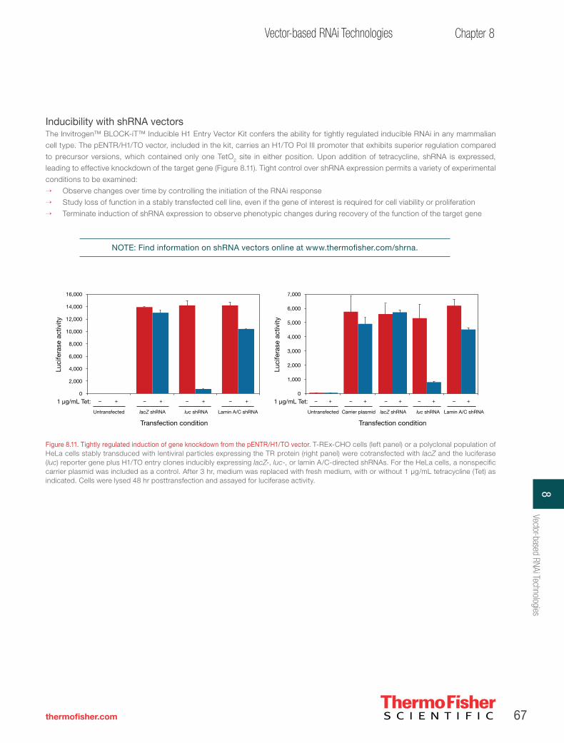
67
Chapter 8Vector-based RNAi Technologies
thermofisher.com
Inducibility with shRNA vectorsThe Invitrogen™ BLOCK-iT™ Inducible H1 Entry Vector Kit confers the ability for tightly regulated inducible RNAi in any mammalian
cell type. The pENTR/H1/TO vector, included in the kit, carries an H1/TO Pol III promoter that exhibits superior regulation compared
to precursor versions, which contained only one TetO2 site in either position. Upon addition of tetracycline, shRNA is expressed,
leading to effective knockdown of the target gene (Figure 8.11). Tight control over shRNA expression permits a variety of experimental
conditions to be examined:
→ Observe changes over time by controlling the initiation of the RNAi response
→ Study loss of function in a stably transfected cell line, even if the gene of interest is required for cell viability or proliferation
→ Terminate induction of shRNA expression to observe phenotypic changes during recovery of the function of the target gene
NOTE: Find information on shRNA vectors online at www.thermofisher.com/shrna.
+–+–+–+–1 µg/mL Tet:
Luci
fera
se a
ctiv
ity
16,000
14,000
10,000
12,000
8,000
6,000
4,000
2,000
0
Transfection condition
Untransfected lacZ shRNA lacZ shRNAluc shRNA Lamin A/C shRNA
+–1 µg/mL Tet:
Luci
fera
se a
ctiv
ity
7,000
5,000
6,000
4,000
3,000
2,000
1,000
0
Transfection condition
Untransfected Carrier plasmid luc shRNA Lamin A/C shRNA
+– +– +–+–
Figure 8.11. Tightly regulated induction of gene knockdown from the pENTR/H1/TO vector. T-REx-CHO cells (left panel) or a polyclonal population of HeLa cells stably transduced with lentiviral particles expressing the TR protein (right panel) were cotransfected with lacZ and the luciferase (luc) reporter gene plus H1/TO entry clones inducibly expressing lacZ-, luc-, or lamin A/C-directed shRNAs. For the HeLa cells, a nonspecific carrier plasmid was included as a control. After 3 hr, medium was replaced with fresh medium, with or without 1 μg/mL tetracycline (Tet) as indicated. Cells were lysed 48 hr posttransfection and assayed for luciferase activity.
8Vector-based RNAi Technologies

68 Important Licensing Information: These products may be covered by one or more Limited Use Label Licenses (See Thermo Fisher Scientific Catalogue or our website, www.thermofisher.com). By use of these products you accept the terms and conditions of all applicable Limited Use Label Licenses. All products are for research use only. CAUTION: Not intended for human or animal diagnostic or therapeutic uses.
Section II Products for RNA Interference
Lentiviral and adenoviral RNAi vectors
Choose any cell type for RNAiFor many disease models, the most appropriate cell types, such as immune system or primary cells, are not amenable to transfection.
Viral delivery of RNAi vectors is a powerful alternative to transfection for these cell types, as well as for in vivo applications. To
accurately determine the efficacy of knockdown from an shRNA/miR RNAi molecule in a population of cells, it is critical to deliver the
shRNA/miR RNAi molecule to as many cells as possible. Otherwise, when knockdown is measured by quantitative real-time PCR
(qRT-PCR) or western blot analysis, the background of mRNA or protein in nontransfected cells will make the knockdown appear
less effective than it actually is. Viral delivery can be the best option in virtually any mammalian cell type, including hard-to-transfect,
primary, and even nondividing cells. Conveniently, lentiviral delivery systems are available for both shRNA and miR RNAi vectors, and
an adenoviral delivery system is available for shRNA vectors (Table 8.4).
The procedure for using both RNAi viral systems (Figure 8.12):
1. Clone the double-stranded DNA oligo encoding an shRNA or miR RNAi into one of the BLOCK-iT entry (shRNA) or expression
(miR RNAi) vectors.
2. Transfer the RNAi cassette into the adenoviral (shRNA only) or lentiviral destination vector by Gateway recombination.
3. Transfect vectors into the appropriate kit-provided packaging cells (use ViraPower Packaging Mix for lentiviral systems only) to
produce viral stocks, which can be used immediately or stored at –80°C.
4. Harvest and (for adenovirus only) amplify the viral supernatant and use it for shRNA/miR RNAi delivery to any cell type.
Store up to 1 year
Transduce mammaliancells, select for stableexpression, and assay
ViraPower 293FT producer cell line
Generate the pLenti expression construct containing the miR RNAi
or shRNA of interest
Combine with optimisedViraPower
Packaging Mix
Cotransfect the ViraPower 293FT producer cellline with the pLenti expression construct and
the optimised ViraPower Packaging Mix
Harvest viral supernatantand determine the titer
pLenti/BLOCK-iT
RNAiconstruct
Ampicillin SV40 pA
RRE
pUC ori
PSV40
PRSV
/5´ L
TR
ΔU3/
3´ L
TR
ψ
EM7
Selection
marker
Figure 8.12. How the BLOCK-iT lentiviral system works.
Table 8.4. Choose a lentiviral or adenoviral RNAi system.
Viral system When to use Products
Lentiviral RNAi delivery systems
• Stable RNAi in any cell line, even nondividing cells
• Inducible or constitutive shRNA or miR RNAi expression
• Studies in animal models
• BLOCK-iT Lentiviral Pol II miR RNAi Expression System—a complete lentiviral system with all of the advantages of miR RNAi: multiple-target knockdown and a higher design success rate than conventional shRNA (contains pLenti6/V5-DEST vector)
• BLOCK-iT Lentiviral Pol II miR RNAi Expression System with EmGFP—a system with all of the benefits listed above, plus easy expression tracking with cocistronic EmGFP (contains pLenti6/V5-DEST vector)
• BLOCK-iT Inducible H1 Lentiviral RNAi System—complete lentiviral system for inducible or constitutive shRNA expression in any cell type (contains pLenti4/BLOCK-iT-DEST vector)
• BLOCK-iT Lentiviral RNAi Expression System—complete lentiviral system for constitutive shRNA expression in any cell type (contains pLenti6/BLOCK-iT™-DEST vector)
BLOCK-iT RNAi Adenoviral System
• High-level transient shRNA expression
• Effective delivery to a wide range of human cell types
• Studies in animal models
• BLOCK-iT Adenoviral RNAi Expression System—complete system for high-level transient expression of shRNA
Prod
ucts
for R
NA In
terfe
renc
eII

69
Chapter 8Vector-based RNAi Technologies
thermofisher.com
Powerful shRNA delivery with BLOCK-iT viral vectors
The viral delivery systems include these vectors, which are also available separately (Figure 8.13):
→ pLenti4/BLOCK-iT-DEST—lentiviral vector with the Zeocin™ selection marker for compatibility with a tetracycline-regulated
inducible system
→ pLenti6/BLOCK-iT-DEST—lentiviral vector with the blasticidin selection marker for fast, efficient selection for constitutive shRNA
expression
→ pAd/BLOCK-iT-DEST—adenoviral vector expediently produced without the need for a shuttle vector or for performing other
labor-intensive methods
Product Quantity Cat. no.
BLOCK-iT Lentiviral Pol II miR RNAi Expression System 20 rxns K4937-00
BLOCK-iT Lentiviral Pol II miR RNAi Z´-factor Expression System with EmGFP 20 rxns K4938-00
BLOCK-iT Inducible H1 Lentiviral RNAi System 20 rxns K4925-00
BLOCK-iT Lentiviral RNAi Expression System 20 rxns K4944-00
BLOCK-iT Adenoviral RNAi Expression System 20 rxns K4941-00
pLenti4/BLOCK-iT™-DEST
8.0 kb
attR1 attR2ccdB CmR
pLenti6/BLOCK-iT™-DEST
8.7 kb
pAd/BLOCK-iT™-DEST
3.5 kb
attR1 attR2CmR ccdBattR1 attR2CmR ccdB
PR
SV/5
´ LTR
RRE
wt A
d5 (∆
E3)
Am
pici
llin5´ ITR
pU
C ori
3´ ITR3´ LTR (∆
E3)
Bla
stic
idin
SV40 pA
Am
picillin
pU
C ori
PSV40
EM7
3´ LTR (∆E3
)Z
eoci
n™
SV40 pA
Am
picillin
pU
C ori
PR
SV/5
´ LTR
RRE PSV40
EM7
Figure 8.13. BLOCK-iT-DEST viral vectors. The BLOCK-iT Inducible H1 Lentiviral RNAi System can generate long-term inducible knockdown results whether a clonal population is used or individual clones are selected for analysis. The promoter configuration of the H1/TO vector ensures tight regulation; thus, unwanted background from shRNA expression will not interfere with results.
8Vector-based RNAi Technologies

70 Important Licensing Information: These products may be covered by one or more Limited Use Label Licenses (See Thermo Fisher Scientific Catalogue or our website, www.thermofisher.com). By use of these products you accept the terms and conditions of all applicable Limited Use Label Licenses. All products are for research use only. CAUTION: Not intended for human or animal diagnostic or therapeutic uses.
Section II Products for RNA Interference
BLOCK-iT Lentiviral RNAi System
Stable RNAi expression in nondividing mammalian cellsThe RNAi cassette from the Invitrogen™ BLOCK-iT™ U6 entry vector can also be transferred into the pLenti6/BLOCK-iT-DEST or
pLenti4/BLOCK-iT-DEST lentiviral RNAi vectors. First, generate lentiviral stocks with the 293FT cell line and associated ViraPower™
reagents, which are available separately or with the BLOCK-iT Lentiviral RNAi System. Then, prepare a lentiviral stock, transduce cells,
and assay for the RNAi response (Figures 8.14 and 8.15). The BLOCK-iT lentiviral RNAi vectors possess all the required components
for efficient packaging of the RNAi shRNA to dividing, hard-to-transfect, and nondividing cells, as well as for in vivo delivery to animal
models.
Mag
icM
ark™
Mag
icM
ark™
Mag
icM
ark™
Mag
icM
ark™
Day 6Day 3 Day 60Day 23 Day 37Day 16
– + – + – + – + – + – + – + – + – + – +– + – +lamAC lacZlamAC lacZ lamAC lacZ lamAC lacZ lamAC lacZ lamAC lacZshRNA:
1 µg/ml Tet:
Lamin A/C
β-actin
120100
80
60
50
40
30
120100
80
60
50
40
30
120100
80
60
50
40
30
120100
80
60
50
40
30
Figure 8.15. Long-term inducible knockdown in stably transduced T-REx HeLa cells. T-REx HeLa cells were transduced with lentiviral particles expressing lamin A/C (lamAC)-directed or control lacZ-directed shRNA from the tetracycline (Tet)-inducible H1/TO promoter at an effective MOI of <1. Stable populations of cells were selected with Zeocin reagent for 3 weeks and plated with or without Tet at 1 μg/mL for the number of days indicated. Whole cell lysates were separated by western blotting and probed with antibodies against lamin A/C and β-actin as a loading control.
Store up to 1 year
Transduce mammaliancells, select for stableexpression, and assay
ViraPower 293FT producer cell line
Generate the pLenti expression construct containing the
gene of interest
Combine with optimisedViraPower
Packaging Mix
Cotransfect the ViraPower 293FT producer cellline with the pLenti expression construct and
the optimised ViraPower™ Packaging Mix
Harvest viral supernatantand determine the titer
Pol III promoter Termination sequenceshRNA sequence
pLenti/BLOCK-iT
RNAiconstruct
Ampicillin SV40 pA
RRE
pUC ori
PSV40
PRSV
/5´ L
TR
ΔU3/
3´ L
TR
ψ
EM7
Selection
marker
Figure 8.14. Generating BLOCK-iT lentiviral RNAi stock.
Prod
ucts
for R
NA In
terfe
renc
eII

71
Chapter 8Vector-based RNAi Technologies
thermofisher.com
pAd/BLOCK-iT-DEST
34.9 Kb
Generate the adenoviralRNAi vector containing the
shRNA of interest. Digest thepuri�ed plasmid with Pac I to expose the inverted terminal repeats (ITRs).
Transfect the 293Aproducer cell line with
the adenoviral RNAi vector.Harvest cells and prepare
a crude viral lysate.
Amplify the adenovirusby infecting 293A producer
cells with the crude virallysate. Determine the titerof the adenoviral stock.
Add the viral supernatantto the mammalian cell
line of interest.
Assay for knockdown of gene on interest
293A cell line Target mammalian cell line
Promoter shRNA sequence
Pac I
Pac I
Pol III promoter Termination sequenceshRNA sequence
pUC
ori
3´ ITR
Am
pici
llin
5´ ITR + 3´ ITR
wt Ads ( ΔE3)
Figure 8.16. How the BLOCK-iT adenoviral RNAi system works.
Product Quantity Cat. no.
BLOCK-iT Inducible H1 RNAi Entry Vector Kit 20 constructions K4920-00
BLOCK-iT U6 RNAi Entry Vector Kit 20 constructions K4945-00
Invitrogen™ T-REx™-Jurkat Cell Line 3 x 10⁶ cells R722-07
T-REx-CHO Cell Line 3 x 10⁶ cells R718-07
T-REx-HeLa Cell Line 3 x 10⁶ cells R714-07
T-REx-293 Cell Line 3 x 10⁶ cells R710-07
pLenti6/TR Vector Kit 1 kit V480-20
pcDNA 6/TR 20 µg V1025-20
BLOCK-iT Adenoviral RNAi Expression System 20 rxns K4941-00
pAd/BLOCK-iT-DEST RNAi Gateway® Vector 6 µg V492-20
293A Cell Line 3 x 10⁶ cells R705-07
BLOCK-iT Adenoviral RNAi Expression System
Efficient transient delivery and shRNA expression in mammalian cellsAdenoviral systems are popular platforms for reliable gene delivery and superior expression of transient shRNA in nearly any mammalian
cell type. The key advantage of the BLOCK-iT Adenoviral RNAi Expression System is Gateway recombination technology, which
simplifies the cloning and generation of an adenoviral vector, eliminating the tedious and time-consuming manipulations, screening,
and multiple transformations that other adenoviral systems require.
Adenoviruses enter target cells by binding to the Coxsackie adenovirus receptor (CAR) [8]. After binding to CAR, the adenovirus
is internalised via integrin-mediated endocytosis [9], followed by active transport to the nucleus where the shRNA is expressed. High-
level transient shRNA expression is easily achieved with the BLOCK-iT Adenoviral RNAi System (Figure 8.16). Gateway recombination
provides a way to quickly generate an adenoviral RNAi construct. For the first time, high-throughput cloning into adenoviral vectors is
possible due to the efficacy of this recombination reaction.
1. Brummelkamp TR et al. (2002) Science 296:550–553.
2. McManus MT et al. (2002) RNA 8:842–850.
3. Paddison PJ et al. (2002) Genes Dev 16:948–958.
4. Sui G et al. (2002) Proc Natl Acad Sci U S A 99:5515–5520.
5. Yu JY et al. (2002) Proc Natl Acad Sci U S A 99:6047–6052.
6. Gwizdek C et al. (2003) J Biol Chem 278:5505–5508.
7. Yi R et al. (2003) Genes Dev 17:3011–3016.
8. Bergelson JM et al. (1997) Science 28:1320–1323.
9. Russell WC (2000) J Gen Virol 81:2573–2604.
Chapter references
8Vector-based RNAi Technologies

72 Important Licensing Information: These products may be covered by one or more Limited Use Label Licenses (See Thermo Fisher Scientific Catalogue or our website, www.thermofisher.com). By use of these products you accept the terms and conditions of all applicable Limited Use Label Licenses. All products are for research use only. CAUTION: Not intended for human or animal diagnostic or therapeutic uses.
Section II Products for RNA Interference
CHAPTER 9
RNAi delivery
Introduction to RNAi delivery
The two most common approaches for RNAi delivery are lipid-mediated transfection and virus-mediated transduction. Determining which of
these approaches to use depends on the cell type being studied and whether transient or stable knockdown is desired (Table 9.1). The most
popular application, transient transfection of unmodified siRNAs, modified siRNA duplexes, or RNAi vectors, utilises cationic lipid–based
reagents because they are suitable for delivering these molecules across a diverse range of commonly used cell lines. For cell types not
amenable to lipid-mediated transfection, viral vectors are often employed. Adenoviral vectors work well for transient delivery in many cell
types; however, for stable delivery in dividing and nondividing cells, lentiviral vectors are best. To determine the most favourable RNAi delivery
conditions, we offer you our delivery optimisation service (see Chapter 12), a resource backed by extensive knowledge and expertise in viral
vectors and nonviral delivery reagents for testing a matrix of delivery parameters.
Methods to achieve high transfection efficiencyTransfection efficiency is expressed as the percentage of cells that have received the RNAi duplex or expression plasmid. Typically, researchers
strive to achieve the highest efficiencies possible. This objective is particularly important for RNAi applications, because nontransfected cells
will continue to express normal levels of the targeted gene, which will result in background expression levels. With the cationic Lipofectamine
RNAiMAX Transfection Reagent, transfection efficiency can be optimised and verified easily using the BLOCK-iT Alexa Fluor Red Fluorescent
Control. In the case of Lipofectamine 2000 Transfection Reagent, either the BLOCK-iT Alexa Fluor Red Fluorescent Control or the original green
BLOCK-iT Fluorescent Oligo can be used.
For some disease models, primary cultures or nondividing cells, which are often the most appropriate cell types to use, cannot be
transfected adequately with commercially available transfection reagents. A powerful alternative to cationic lipid–mediated transfection is viral
delivery of vectors containing RNAi cassettes. This option is best for delivery to hard-to-transfect, primary, and nondividing cells. Viral delivery can
also be used to create stable cell lines with inducible RNAi or to express RNAi sequences with tissue-specific promoters. See Chapter 4 for more
information about using viral vectors in RNAi experiments.
Importance of minimising transfection-mediated cytotoxicityThe delivery of siRNAs, or the method of delivery, can result in cytotoxicity in gene silencing experiments. Minimising transfection-mediated
cytotoxicity is essential for proper interpretation of the outcome of any RNAi experiment. Cytotoxic effects can be difficult to distinguish from
the expected phenotype resulting from target gene knockdown. Cytotoxicity due to the delivery method is often seen as apparent knockdown
Table 9.1. Selecting transfection reagents and viral delivery methods.
Product Key advantages
Lipofectamine RNAiMAX Transfection Reagent
• Designed and manufactured for siRNA delivery• Superior efficiencies, allowing low concentrations of siRNA to be used• Wide range of compatibility with diverse cell lines• Optimised protocols available for many common cell lines
Lipofectamine 2000 Transfection Reagent• Designed for optimal expression when delivering plasmids, including shRNA and miR RNAi vectors• Robust cotransfection of vectors and synthetics (siRNA or Stealth RNAi siRNA duplexes)
Lipofectamine LTX Reagent• Designed for improved transfection performance in hard-to-transfect and primary cells• High transfection efficiency and significantly lower toxicity levels for a wide range of cell
lines
Oligofectamine Transfection Reagent• Expressly formulated for delivery of antisense oligos• Dependable delivery of siRNA
BLOCK-iT Adenoviral RNAi Expression System • Ideal system for long-term transient expression of RNAi vectors in a wide range of cell lines
BLOCK-iT Lentiviral Pol II miR RNAi Expression SystemBLOCK-iT Lentiviral Pol II miR RNAi Expression System with EmGFPBLOCK-iT Lentiviral RNAi Expression System BLOCK-iT Inducible H1 Lentiviral RNAi System
• Stable expression of RNAi vectors in difficult-to-transfect cell lines• Suitable methods for in vivo applications• Inducible systems available
Prod
ucts
for R
NA In
terfe
renc
eII

73
Chapter 9RNAi delivery
thermofisher.com
in cells transfected with several negative controls, relative to no treatment. The easiest way to combat transfection-mediated cytotoxicity is to
choose a transfection reagent designed for either double-stranded RNA or plasmid-based RNAi transfections. Most often, these reagents have
been formulated to maximise knockdown efficiency while minimising cytotoxicity. Optimisation experiments, when using published or suppliers’
protocols as guidelines, can help to determine the concentration of transfection reagent that works best for the cell line of interest. Using the
lowest amount of transfection reagent needed to give the highest level of knockdown is recommended.
Significance of reducing off-target effectsAs well as being a source of cytotoxicity, a suboptimal transfection reagent or excess reagent can result in apparent off-target effects.
One cause of off-target effects is the up- or down-regulation of genes due to the gene delivery procedure. However, with appropriate
controls, these effects can be identified and diminished (see Chapter 2 for a discussion of controls for RNAi experiments). Again, the
lowest amount of transfection reagent that provides the best gene silencing activity is advised.
The potential also exists for off-target effects caused by the siRNA duplex itself. To determine the most favourable conditions
for transfection, the concentration of siRNA should be varied while holding the concentration of transfection reagent constant, using
the lowest concentration (identified previously) to limit cytotoxicity. Keep in mind that the specificity of the siRNA will impact potential
off-target effects. The use of exceptionally specific reagents, such as modified Stealth RNAi siRNA duplexes, can help alleviate these
concerns (see Chapter 7 for detailed information about the specificity advantage of Stealth RNAi siRNA).
Lipofectamine RNAiMAX Transfection Reagent—unmatched gene silencing with reduced cytotoxicity for siRNA experimentsLipofectamine RNAiMAX Transfection Reagent is a proprietary, siRNA-specific cationic lipid formulation that offers the highest
transfection efficiencies with the widest variety of cell types for siRNA gene knockdown experiments:
→ Superior transfection efficiency enables use of lower siRNA concentrations and leads to more successful gene knockdown with
a minimum of nonspecific effects (Figure 9.1A)
→ Easy optimisation due to low cytotoxicity across a 10-fold range of transfection reagent concentrations (Figure 9.1B)
→ Versatile approach compatible with a wide range of cell types
→ Simple and rapid protocol for consistent results
Stealth RNAi siRNA concentration
0.5 nM50 nM 1 nM10 nM1 nM0.5 nM 10 nM 50 nM Cells only
Rel
ativ
e ex
pre
ssio
n,p
53 v
s. n
ontr
ansf
ecte
d
2.0
1.5
1.0
0.5
0
Stealth RNAi siRNA Negative Control Medium GC
Human p53 Stealth RNAi siRNA
p53 knockdown % viable cells
Lipofectamine RNAiMAX reagent volume
00.1 µL0.25 µL0.5 µL1.0 µL 00.1 µL0.25 µL0.5 µL1.0 µL
Rel
ativ
e ex
pre
ssio
n,p
53 v
s. n
ontr
ansf
ecte
d
Per
cent
via
ble
cel
ls
1.4
1.0
0.6
0.2
1.2
0.8
04
0
120
80
60
20
100
40
0
1 nM Stealth RNAi siRNA NegativeControl Medium GC
1 nM p53 Stealth RNAi siRNA
Figure 9.1. Optimal gene silencing and minimal cytotoxicity with Lipofectamine RNAiMAX Transfection Reagent. (A) Superior knockdown levels were observed with as little as 0.5 nM Stealth RNAi siRNA with Lipofectamine RNAiMAX Transfection Reagent. Transfection complexes containing 0.3 μL of Lipofectamine RNAiMAX Transfection Reagent were prepared in 48-well plates. A549 cells were added to each well to give final Stealth RNAi siRNA concentrations of 0.5 to 50 nM. Twenty-four hours after addition of cells, p53 knockdown was measured by qRT-PCR with LUX™ primers and normalized to GAPDH expression. The control duplex was the Stealth RNAi Negative Control Medium GC. (B) Minimal cytotoxicity over a 10-fold range of Lipofectamine RNAiMAX Transfection Reagent concentrations. Indicated volumes of transfection reagent were mixed with 10 nM p53 Validated Stealth RNAi siRNA or Stealth RNAi Negative Control Medium GC duplexes in 48-well plates. A549 cells were added to each well for a final siRNA concentration of 1 nM. Knockdown of p53 was measured as described in (A). Over a 10-fold concentration range of Lipofectamine RNAiMAX Transfection Reagent, high levels of gene silencing were obtained without a dramatic increase in transfection-mediated cytotoxicity.
A B
9RNAi delivery

74 Important Licensing Information: These products may be covered by one or more Limited Use Label Licenses (See Thermo Fisher Scientific Catalogue or our website, www.thermofisher.com). By use of these products you accept the terms and conditions of all applicable Limited Use Label Licenses. All products are for research use only. CAUTION: Not intended for human or animal diagnostic or therapeutic uses.
Section II Products for RNA Interference
Product Quantity Cat. no.
Lipofectamine RNAiMAX Transfection Reagent 0.75 mL1.5 mL
13778-07513778-150
BLOCK-iT Alexa Fluor Red Fluorescent Control 2 x 125 μL 14750-100
RNA interferenceSimple protocol. The procedure for using Lipofectamine RNAiMAX Transfection Reagent consists of mixing it with siRNA, adding
cells, incubating, and measuring gene knockdown (Figure 9.2). The simplicity and speed, combined with superior transfection
efficiency (Figure 9.3), make Lipofectamine RNAiMAX Transfection Reagent the best choice for high-throughput siRNA transfections.
Transfection conditions can be readily established for automated or robotic systems used in such applications. Protocols for using the
Lipofectamine RNAiMAX Transfection Reagent can be found at www.thermofisher.com/rnaimax.
Figure 9.3. Assessing transfection efficiency of the Lipofectamine RNAiMAX Transfection Reagent with the BLOCK-iT Alexa Fluor Red Fluorescent Control. Lipofectamine RNAiMAX Transfection Reagent was used to transfect HeLa cells with the BLOCK-iT Alexa Fluor Red Fluorescent Control (50 nM) (A). Twenty-four hours after transfection, growth medium was removed and replaced with PBS containing 10 mg/mL Hoechst 33342 for visualisation of cell nuclei (B). Nuclear localisation of the red-fluorescent control oligo is seen (A). The bright-field image (C) shows that the cells retain a normal morphology after transfection.
A B C
HASMC NTERA-2 HUVECHREHEKa ADSC Control
100
20
15
10
5
0
GAPDH
CSNK2A1
Rel
ativ
e ex
pre
ssio
n
Figure 9.2. Superior knockdown of Silencer Select siRNA with Lipofectamine RNAiMAX Transfection Reagent. (Email [email protected] for protocol details).
NOTE: Lipofectamine RNAiMAX Transfection Reagent is for use with siRNA duplexes, and not for transfecting vectors expressing RNAi sequences.
Prod
ucts
for R
NA In
terfe
renc
eII

75
Chapter 9RNAi delivery
thermofisher.com
NOTE : The BLOCK-iT Fluorescent Oligo (Cat. No. 2013) is optimised for use with Lipofectamine 2000 Transfection Reagent and does not work
well with Lipofectamine RNAiMAX Transfection Reagent.
Figure 9.4. Delivery of BLOCK-iT Pol II miR RNAi expression vectors. Cells were transfected with pcDNA 6.2-GW/EmGFP-miR (directed against lamin) and Lipofectamine 2000 Transfection Reagent, at an expected 50% efficiency, to demonstrate the 100% tracking of Emerald Green Fluorescent Protein (EmGFP) and miRNA expression. After 48 hr, cells were stained with Hoechst nuclear stain, which stains all cells (A), and a red lamin stain (B), and monitored for GFP expression. Approximately half of the cells highly expressed the lamin protein, but red-stained cells were not expressing EmGFP, and EmGFP-expressing cells were not expressing lamin (C). This demonstrates the cocistronic expression of EmGFP and the miRNA that greatly reduces lamin expression.
Product Quantity Cat. no.
Lipofectamine LTX Reagent 1 mL 15338100
PLUS Reagent 0.85 mL 11514015
Lipofectamine 2000 Transfection Reagent 0.75 mL1.5 mL
11668-02711668-019
BLOCK-iT Fluorescent Oligo for lipid transfection 2 x 125 μL 2013
BLOCK-iT Transfection Kit* 1 kit 13750070
* The BLOCK-iT Transfection Kit contains Lipofectamine 2000 Transfection Reagent and the BLOCK-iT Fluorescent Oligo.
A. Hoechst B. Lamin C. EmGFP + lamin
Lipofectamine 2000 and LTX Transfection Reagents—ideal for delivery of shRNA and miR RNAi vectorsLipofectamine 2000 Transfection Reagent can be used for delivery of assorted RNAi agents, including shRNA and miR RNAi vectors
and synthetic molecules such as siRNA, Stealth RNAi siRNA, and Dicer-generated siRNA pools. Lipofectamine LTX Reagent, used
in combination with Invitrogen™ PLUS™ Reagent, is the choice reagent for hard-to-transfect cell lines, including primary cells, for
delivery of assorted RNAi molecules. Lipofectamine 2000 and Lipofectamine LTX reagents for RNAi transfection offer many benefits:
→ Effective transfection for shRNA and miR RNAi vectors (Figure 9.4) and synthetics (siRNA and Stealth RNAi siRNA); also works
well for cotransfections of synthetics and vectors
→ Easy-to-follow protocols; media changes not required
→ Convenient optimisation of transfection conditions and efficiency with the BLOCK-iT Fluorescent Oligo
→ Excellent performance in a wide variety of cell types
9RNAi delivery

76 Important Licensing Information: These products may be covered by one or more Limited Use Label Licenses (See Thermo Fisher Scientific Catalogue or our website, www.thermofisher.com). By use of these products you accept the terms and conditions of all applicable Limited Use Label Licenses. All products are for research use only. CAUTION: Not intended for human or animal diagnostic or therapeutic uses.
Section II Products for RNA Interference
Oligofectamine Transfection Reagent—potent internalisation of RNA oligos
Oligofectamine Transfection Reagent is designed to deliver antisense oligos and is also useful for transfecting RNAi reagents into
eukaryotic cells. To optimise transfection conditions, Oligofectamine Transfection Reagent can be used in conjunction with the
BLOCK-iT Fluorescent Oligo or the BLOCK-iT RNAi Basic Control Kit. In the first published demonstration of siRNA-mediated
knockdown in a mammalian cell line, Elbashir et al. reported their use of Oligofectamine™ Transfection Reagent to transfect HeLa cells
with siRNAs [1]. This work was continued by Harborth et al., who used a similar transfection protocol with Oligofectamine Transfection
Reagent to demonstrate the knockdown of 20 additional essential and nonessential target genes [2]. These early studies established
the general applicability of using siRNA for RNAi.
Product Quantity Cat. no.
Oligofectamine Transfection Reagent 1.0 mL 12252-011
The Neon Transfection System
High transfection efficiency and high cell viability in a broad range of cell lines
The company that continues to bring you transfection reagents on the cutting edge
now introduces the Neon Transfection System.
→ Efficiency—up to 90% in many cell types, including difficult-to-transfect,
primary, and stem cells (Table 9.2)
→ Flexibility—easily transfect from 2 x 10⁴ cells to 6 x 10⁶ cells per reaction
→ Simplicity—single reagent kit for all cell types
→ Versatility—open system allows electroporation parameters to be optimised freely
The Neon Transfection System is a novel benchtop electroporation device that employs an electroporation technology using a pipette
tip as an electroporation chamber to efficiently transfect mammalian cells, including primary and immortalised hematopoietic cells,
stem cells, and primary cells (Figure 9.5). The Neon Transfection System efficiently delivers nucleic acids, proteins, and siRNA into all
mammalian cell types with a high cell survival rate. The transfection is performed using as few as 2 x 10⁴ or as many as 8 x 10⁶ cells
per reaction, in a sample volume of 10 μL or 100 μL, in a variety of cell culture formats (60 mm, 6-well, 48-well, and 24-well).
The Neon Transfection System uses a single transfection kit (Neon Kit) that is compatible with various mammalian cell types,
including primary and stem cells, thereby avoiding the need to determine an optimal buffer for each cell type.
The Neon Transfection System offers open and transparent protocols that are optimised for ease of use and simplicity. The Neon
device is preprogrammed with one 24-well optimisation protocol to optimise conditions for your nucleic acid/siRNA and cell type, or
you can programme and store up to 50 cell-specific protocols in the Neon device database. Optimised protocols for many commonly
used cell types are also available at www.thermofisher.com/neon for your convenience, to maximize transfection efficiencies for your
cell types.
Neon system pipette tip design vs. standard electroporation cuvetteUnlike standard cuvette-based electroporation chambers, the Neon system uses a patented biologically compatible pipette tip
chamber that generates a more uniform electric field (Figure 9.6). This design allows better maintenance of physiological conditions,
resulting in very high cell survival compared to conventional electroporation [3].
Prod
ucts
for R
NA In
terfe
renc
eII

77
Chapter 9RNAi delivery
thermofisher.com
Figure 9.6. Standard electroporation cuvette compared to Neon system pipette tip. The design of the electrode in the pipette has been shown to produce a more uniform electric field. The result is less toxicity to the cells and higher transfection efficiencies.
Table 9.2. Examples of cell lines successfully transfected using the Neon Transfection System.
Cell line Cell type Transfection efficiency (%)* Viable cells (%)
MEF primary Embryonic fibroblast 80 75
293A Kidney 90 90
3T3-L1 Mouse adipose 85 80
A549 Lung 75 92
Macrophages Human (peritoneal) 60 60
MCF-7 Breast 70 80
HeLa Cervical carcinoma 90 87
HL-60 Blood 55 70
PBMC Blood 23 95
Primary rat cortical cells Brain, cortical 42 98.5
Primary rat hippocampal cells Brain, hippocampal 37 77
Raw 264.7 Blood 74 80
* Transfection efficiency is calculated from total population of live and dead cells. Protocols and reference information for a large number of cell lines are available in the Neon cell database at www.thermofisher.com/neon.
Figure 9.5. High transfection efficiency of Jurkat cells with the Neon Transfection System. Intracelluar uptake of reporter vector encoded with EGFP at 24 hr following transfection of Jurkat cells using the Neon Transfection System. B is the corresponding fluorescence image of A.
A B
9RNAi delivery
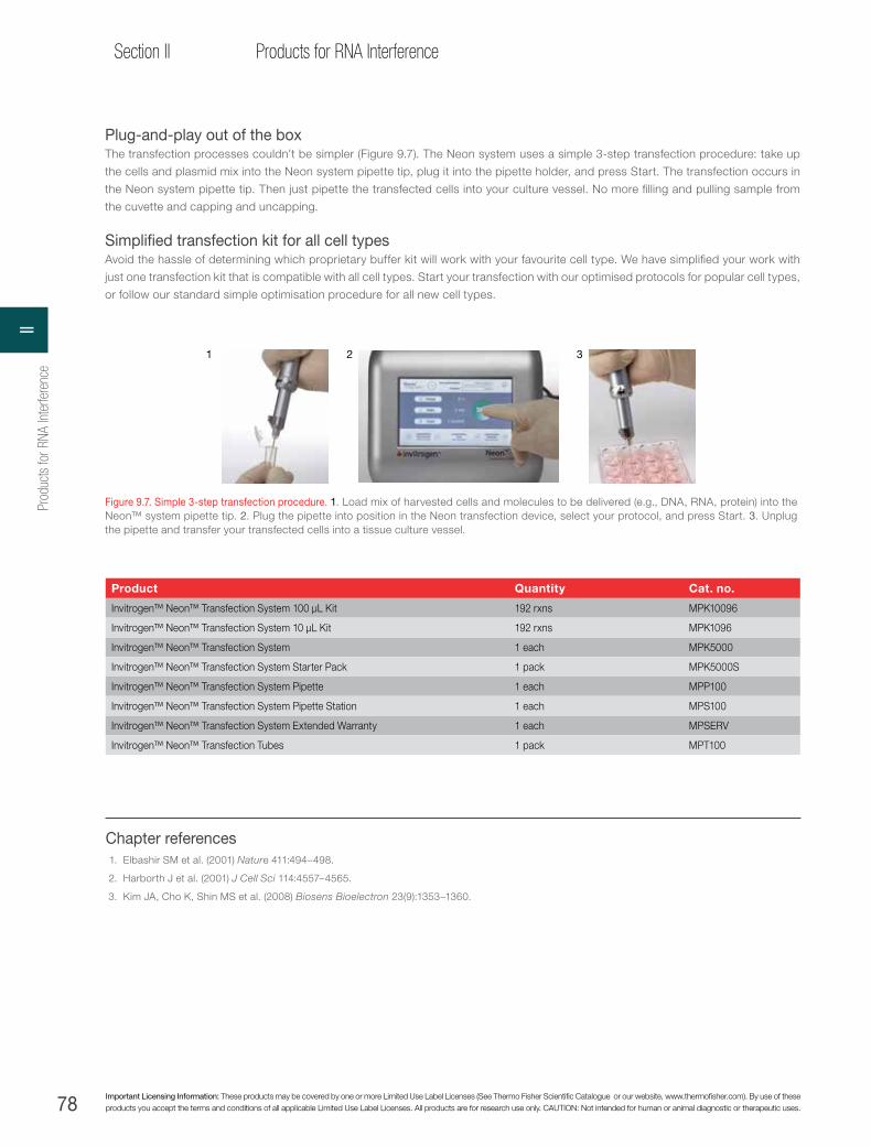
78 Important Licensing Information: These products may be covered by one or more Limited Use Label Licenses (See Thermo Fisher Scientific Catalogue or our website, www.thermofisher.com). By use of these products you accept the terms and conditions of all applicable Limited Use Label Licenses. All products are for research use only. CAUTION: Not intended for human or animal diagnostic or therapeutic uses.
Section II Products for RNA Interference
Chapter references1. Elbashir SM et al. (2001) Nature 411:494–498.
2. Harborth J et al. (2001) J Cell Sci 114:4557–4565.
3. Kim JA, Cho K, Shin MS et al. (2008) Biosens Bioelectron 23(9):1353–1360.
1 2 3
Figure 9.7. Simple 3-step transfection procedure. 1. Load mix of harvested cells and molecules to be delivered (e.g., DNA, RNA, protein) into the Neon™ system pipette tip. 2. Plug the pipette into position in the Neon transfection device, select your protocol, and press Start. 3. Unplug the pipette and transfer your transfected cells into a tissue culture vessel.
Plug-and-play out of the boxThe transfection processes couldn’t be simpler (Figure 9.7). The Neon system uses a simple 3-step transfection procedure: take up
the cells and plasmid mix into the Neon system pipette tip, plug it into the pipette holder, and press Start. The transfection occurs in
the Neon system pipette tip. Then just pipette the transfected cells into your culture vessel. No more filling and pulling sample from
the cuvette and capping and uncapping.
Simplified transfection kit for all cell typesAvoid the hassle of determining which proprietary buffer kit will work with your favourite cell type. We have simplified your work with
just one transfection kit that is compatible with all cell types. Start your transfection with our optimised protocols for popular cell types,
or follow our standard simple optimisation procedure for all new cell types.
Product Quantity Cat. no.
Invitrogen™ Neon™ Transfection System 100 µL Kit 192 rxns MPK10096
Invitrogen™ Neon™ Transfection System 10 µL Kit 192 rxns MPK1096
Invitrogen™ Neon™ Transfection System 1 each MPK5000
Invitrogen™ Neon™ Transfection System Starter Pack 1 pack MPK5000S
Invitrogen™ Neon™ Transfection System Pipette 1 each MPP100
Invitrogen™ Neon™ Transfection System Pipette Station 1 each MPS100
Invitrogen™ Neon™ Transfection System Extended Warranty 1 each MPSERV
Invitrogen™ Neon™ Transfection Tubes 1 pack MPT100
Prod
ucts
for R
NA In
terfe
renc
eII
79
Chapter 10RNA interference controls
thermofisher.com
CHAPTER 10
RNA interference controls
Controls for RNAi experiments
Overview of controls for RNAi experimentsAppropriate controls are essential to success in every RNAi experiment. The number and types of controls chosen depend upon
the ultimate research goal [1]. With our RNAi technologies, performing the appropriate control reactions has been simplified (Table
10.1). RNAi control kits are designed to assist researchers in identifying and validating drug targets, generating publishable data, and
accomplishing the following:
→ Determining which RNAi reagents deliver the best knockdown results
→ Achieving greater knockdown by optimising transfection protocols
→ Saving time by confirming cell health early in an experiment
→ Proceeding confidently with RNAi experiments by comparing targeted RNAi reagents to a set of reagents optimised for inhibition
of positive control genes in human cells
→ Using negative controls that match the GC content of the target molecule
Table 10.1. Control kits ensure successful gene inhibition experiments.
Products/kits
Components
Fluorescent oligo
Transfection reagent
Dead-cell stain
Nuclear stain
RNAi positive controls
RNAi negative controls
qRT-PCR primers
BLOCK-iT Alexa Fluor Red Fluorescent Control
•
BLOCK-iT Fluorescent Oligo •
BLOCK-iT Transfection Kit• •
Silencer Select Positive Control siRNA
•
Silencer Select Negative Control siRNA
•
Stealth RNAi siRNA Negative Control
•
Stealth RNAi siRNA Positive Control(actin, GAPDH, cyclophilin B)
•
Stealth RNAi siRNA Reporter Control
•
BLOCK-iT Transfection Optimisation Kit (Human)
• • • •
BLOCK-iT™ Pol II miR Validated miRNA Control Vector
• •
Applications
Track transfectionMonitor experimental variation
Measure the effect of experimental knockdown vs. background
Assess knockdownOptimise transfection conditions
Assess cell viability
10RNA interference controls

80 Important Licensing Information: These products may be covered by one or more Limited Use Label Licenses (See Thermo Fisher Scientific Catalogue or our website, www.thermofisher.com). By use of these products you accept the terms and conditions of all applicable Limited Use Label Licenses. All products are for research use only. CAUTION: Not intended for human or animal diagnostic or therapeutic uses.
Section II Products for RNA Interference
BLOCK-iT Alexa Fluor Red Fluorescent Control and green BLOCK-iT Fluorescent OligoTo achieve significant levels of specific gene inhibition, the Stealth RNAi siRNA or siRNA duplexes must be taken up by target
cells. Factors such as poor cell health and high passage number can negatively impact transfection efficiency. The green BLOCK-iT
Fluorescent Oligo and the BLOCK-iT Alexa Fluor Red Fluorescent Control allow you to easily visualise transfection results and,
thus, provide key controls for Stealth RNAi siRNA or standard siRNA transfection experiments. These stabilised RNA duplexes help
determine transfection efficiency by providing:
→ Strong, easily detectable signal that indicates transfection efficiency (Figure 10.1)
→ Clear, persistent signal that exceeds the intensity of other labelled RNA duplexes
→ Predominant nuclear localisation to confirm that the control has been internalised
→ Unique sequences that have no homology to any known gene, avoiding induction of nonspecific or off-target effects
The green BLOCK-iT Fluorescent Oligo and the BLOCK-iT Alexa Fluor Red Fluorescent Control strongly correlate with transfection
efficiency with Stealth RNAi, Silencer Select, and Silencer siRNAs, as well as Invitrogen™ Pre-miR™ miRNA mimics and Invitrogen™
Anti-miR™ miRNA inhibitors.
Figure 10.1. BLOCK-iT Alexa Fluor Red Fluorescent Control for clear visualisation of transfection results. HeLa cells were transfected with the BLOCK-iT Alexa Fluor Red Fluorescent Control using either Lipofectamine 2000 (A–C) or Lipofectamine RNAiMAX Transfection Reagent (D–F). The recommended HeLa cell transfection protocol was used for each reagent, and the final control concentration was 50 nM. Twenty-four hours after transfection, growth medium was removed and replaced with PBS containing 10 µg/mL Hoechst 33342 for visualisation of cell nuclei (B and E). Nuclear localisation of the control is seen with both transfection reagents (A and D). Almost 100% of the cells take up the control, and cells retain a normal morphology, as seen in the bright-field images (C and F).
A
D
B
E
C
F
Prod
ucts
for R
NA In
terfe
renc
eII

81
Chapter 10RNA interference controls
thermofisher.com
BLOCK-iT Transfection Kit—simple optimisation of Lipofectamine 2000 transfection reagent conditionsThe BLOCK-iT Transfection Kit enables you to shorten the process of optimising RNAi transfection conditions and monitoring
transfection variation in every experiment. To optimise transfection parameters, the BLOCK-iT Transfection Kit contains Lipofectamine
2000 Transfection Reagent and the green BLOCK-iT Fluorescent Oligo (Figure 10.2).
NOTE: The BLOCK-iT Fluorescent Oligo is optimised for use with Lipofectamine 2000 Transfection Reagent and is not recommended for use with Lipofectamine RNAiMAX Transfection Reagent.
Product Quantity Cat. no.
BLOCK-iT Alexa Fluor Red Fluorescent Control (lipid transfection) 2 x 125 µL 14750-100
BLOCK-iT Fluorescent Oligo for lipid transfection 2 x 125 µL 2013
BLOCK-iT Fluorescent Oligo for electroporation 75 µL (1 mM) 13750-062
BLOCK-iT Transfection Kit 1 kit 13750-070
Lipofectamine RNAiMAX Transfection Reagent 1.5 mL0.75 mL
13778-15013778-075
Lipofectamine 2000 Transfection Reagent 1.5 mL0.75 mL
11668-01911668-027
A B
Figure 10.2. Strong fluorescence signal to optimise transfection. A549 cells were transfected with green BLOCK-iT Fluorescent Oligo or standard fluorescently labelled RNA, using Lipofectamine 2000 Transfection Reagent. Twenty-four hours later, the fluorescent signal had persisted in the cells transfected with the green BLOCK-IT Fluorescent Oligo (A). In contrast, the fluorescent signal was largely undetectable in cells transfected with the standard labelled siRNA (B).
10RNA interference controls

82 Important Licensing Information: These products may be covered by one or more Limited Use Label Licenses (See Thermo Fisher Scientific Catalogue or our website, www.thermofisher.com). By use of these products you accept the terms and conditions of all applicable Limited Use Label Licenses. All products are for research use only. CAUTION: Not intended for human or animal diagnostic or therapeutic uses.
Section II Products for RNA Interference
Product Quantity Cat. no.
Invitrogen™ Silencer™ Select GAPDH Positive Control siRNA 5 nmol 4390849
Invitrogen™ Silencer™ Select GAPDH Positive Control siRNA 40 nmol 4390850
Invitrogen™ Silencer™ Select GAPDH Positive Control siRNA, In Vivo Ready 250 nmol 4404024
Invitrogen™ Silencer™ Select Negative Control #1 siRNA 40 nmol 4390844
Invitrogen™ Silencer™ Select Negative Control #1 siRNA 5 nmol 4390843
Invitrogen™ Silencer™ Select Negative Control #1 siRNA, In Vivo Ready 250 nmol 4404020
Invitrogen™ Silencer™ Select Negative Control #2 siRNA 5 nmol 4390846
Invitrogen™ Silencer™ Select Negative Control #2 siRNA 40 nmol 4390847
Silencer Select positive and negative control siRNAs
→ Validated siRNA controls for optimising siRNA experiments
→ GAPDH Positive Control functionally tested in several common cell lines
→ Negative Controls functionally proven to have minimal effects on cell proliferation and viability
→ Include Silencer Select siRNA modifications for enhanced specificity
→ For use in human, mouse, and rat cells
Silencer Select GAPDH Positive Control siRNAOur extensively validated positive control siRNA to human, mouse, and rat GAPDH serves multiple functions. First, it is an ideal “test”
siRNA for those just beginning siRNA experiments, because it is validated to work in multiple cell lines. In addition, because it targets
GAPDH mRNA, which is commonly used as an internal control, its effects are easy to assay, and thus it provides an excellent tool to
monitor siRNA transfection efficiency by real-time RT-PCR as well as with the Invitrogen™ KDalert™ GAPDH Assay Kit. The Invitrogen™
Silencer™ Select GAPDH siRNA includes the same modifications for reducing off-target effects as found in other Silencer Select siRNAs.
Silencer Select Negative Control siRNAsNegative control siRNAs—siRNAs with sequences that do not target any gene product—are essential for determining the effects
of siRNA delivery on the cell and for providing a baseline to compare siRNA-treated samples. There are two extensively tested
Silencer Select Negative Control siRNAs for this purpose. These siRNAs include the same modifications for reducing off-target effects
found in other Silencer Select siRNAs and have no significant sequence similarity to mouse, rat, or human gene sequences. These
negative control siRNAs have been tested by microarray analysis and shown to have minimal effects on gene expression. In addition,
multiparametric cell-based assays have confirmed they have no significant effect on cell proliferation, viability, or morphology in the
cell lines tested.
Prod
ucts
for R
NA In
terfe
renc
eII

83
Chapter 10RNA interference controls
thermofisher.com
Stealth RNAi siRNA negative controls—ideal negative controls for every experimentStealth RNAi siRNA negative controls provide the means to measure the effect of a Stealth RNAi siRNA duplex targeted to a specific
gene, compared to background. These controls have the following features:
→ Three levels of GC content (low, medium, high), with three sequences available for matching GC content to that of experimental
Stealth RNAi siRNA duplexes. Multiple sequences in each GC range give even more certainty of results.
→ No homology to any known vertebrate genes
→ Tested sequences do not induce stress response
The Stealth RNAi siRNA Negative Control Kit contains all three controls (low, medium, and high GC content, excluding sequences #2
and #3), and each is also available separately.
Product GC contentSuitable for use with Stealth RNAi siRNA duplexes with the following GC content Quantity Cat. no.
Invitrogen™ Stealth RNAi™ siRNA Negative Control Kit
1 kit 12935-100
Invitrogen™ Stealth RNAi™ siRNA Negative Control Lo GC
36% 35–45% 250 µL 12935-200
Invitrogen™ Stealth RNAi™ siRNA Negative Control Lo GC Duplex #2
36% 35–45% 250 µL 12935-110
Invitrogen™ Stealth RNAi™ siRNA Negative Control Lo GC Duplex #3
36% 35–45% 250 µL 12935-111
Invitrogen™ Stealth RNAi™ siRNA Negative Control Med GC
48% 45–55% 250 µL 12935-300
Invitrogen™ Stealth RNAi™ siRNA Negative Control Med GC Duplex #2
48% 45–55% 250 µL 12935-112
Invitrogen™ Stealth RNAi™ siRNA Negative Control Med GC Duplex #3
48% 45–55% 250 µL 12935-113
Invitrogen™ Stealth RNAi™ siRNA Negative Control Hi GC
68% 55–70% 250 µL 12935-400
Invitrogen™ Stealth RNAi™ siRNA Negative Control Hi GC Duplex #2
68% 55–70% 250 µL 12935-114
Invitrogen™ Stealth RNAi™ siRNA Negative Control Hi GC Duplex #3
68% 55–70% 250 µL 12935-115
10RNA interference controls

84 Important Licensing Information: These products may be covered by one or more Limited Use Label Licenses (See Thermo Fisher Scientific Catalogue or our website, www.thermofisher.com). By use of these products you accept the terms and conditions of all applicable Limited Use Label Licenses. All products are for research use only. CAUTION: Not intended for human or animal diagnostic or therapeutic uses.
Section II Products for RNA Interference
Stealth RNAi siRNA reporter controls
Stealth RNAi siRNA reporter controls are ideal for RNAi experiments to optimise your transfection conditions in any vertebrate cell line.
These controls are:
→ Designed to efficiently knock down their intended targets
→ Not homologous to anything in the vertebrate transcriptome
Product Quantity Cat. no.
Invitrogen™ Stealth RNAi™ siRNA GFP Reporter Control 250 µL 12935-145
Invitrogen™ Stealth RNAi™ siRNA Luciferase Reporter Control 250 µL 12935-146
Invitrogen™ Stealth RNAi™ siRNA lacZ Reporter Control 250 µL 12935-147
Invitrogen™ Stealth RNAi™ siRNA β-Lactamase Reporter Control 250 µL 12935-148
Stealth RNAi siRNA positive controls
Stealth RNAi siRNA positive housekeeping control duplexes (human) are ideal controls for assessing knockdown and optimizing RNAi
experiments. These controls are:
→ Designed to efficiently knock down their intended targets
→ Bench-tested and supplied in a ready-to-use format
Product Quantity Cat. no.
Invitrogen™ Stealth RNAi™ siRNA GAPDH Positive Control (human) 250 µL 12935-140
Invitrogen™ Stealth RNAi™ siRNA Actin Positive Control (human) 250 µL 12935-141
Invitrogen™ Stealth RNAi™ siRNA Cyclophilin B Control (human) 250 µL 12935-142
BLOCK-iT Transfection Optimisation Kit (Human)
The BLOCK-iT Transfection Optimisation Kit is designed to optimise RNAi transfection with Lipofectamine 2000 Transfection Reagent,
through the use of controls for transfection and cell viability. Each kit contains a BLOCK-iT Fluorescent Oligo, a dead-cell stain, a
positive control Stealth RNAi siRNA duplex directed at human p53, and a scrambled Stealth RNAi siRNA duplex as a negative control.
These reagents, used together, provide the means to optimise transfection conditions with Lipofectamine 2000 Transfection Reagent,
and a functional positive control for every RNAi experiment.
Product Quantity Cat. no.
BLOCK-iT Transfection Optimisation Kit (human) 1 kit 13750047
Prod
ucts
for R
NA In
terfe
renc
eII

85
Chapter 10RNA interference controls
thermofisher.com
Optimise RNAi experiments with the BLOCK-iT Fluorescent Oligo
Saving time and reagents by not requiring separate assays to be performed in different wells, the BLOCK-iT Fluorescent Oligo allows you
to obtain a clear visual appraisal of RNAi transfection efficiency. The dead-cell stain, nuclear stain, and BLOCK-iT Fluorescent Oligo emit
signals at different wavelengths, permitting them to be detected together in a single well. The nuclear stain (Hoechst dye) and dead-cell
reagent (ethidium homodimer-1) allow assessment of cell death and quantitation of cell number, making sample-to-sample and experiment-
to-experiment comparisons more accurate. The simple addition of two dyes to an extra cell culture sample shows the physiological state of
cells. The BLOCK-iT Fluorescent Oligo for transfection optimisation and monitoring, used in conjunction with two cell-staining dyes, helps
achieve the best RNAi results possible (Figure 10.3).
Transfection efficiencies of at least 70% can be confirmed by this procedure:
1. Transfect human cell line with target-specific Stealth RNAi siRNA or siRNA duplexes, positive and negative controls, and BLOCK-iT
Fluorescent Oligo.
2. Incubate a portion of the cells with dead-cell stain and nuclear stain.
3. Visualise the BLOCK-iT Fluorescent Oligo and stained cells to assess transfection efficiency and cell health; if more than 70% of
the cells are transfected (green) and cell death is less than 5% to 10%, proceed to step 4.
4. Measure RNAi-mediated inhibition of targeted genes using Applied Biosystems™ TaqMan™ Gene Expression Assays (see Chapter 11)
Product Size Cat. no.
BLOCK-iT Fluorescent Oligo for Lipid Transfection 2 x 125 µL 2013
BLOCK-iT Fluorescent Oligo for Electroporation 2 x 125 µL 13750062
Positive controls for BLOCK-iT Pol II miR RNAi vectors
The Invitrogen™ BLOCK-iT™ Pol II miR Validated miRNA Control Vectors are valuable positive controls for initial optimisation and validation
of experiments that use the BLOCK-iT Pol II miR RNAi vectors (see Chapter 8 for more information on these vectors). Three validated
positive controls are available, and each control targets a different commonly used reporter: lacZ, luciferase, or lamin A/C. These controls
are cloned into the BLOCK-iT miR RNAi vectors with Emerald GFP (EmGFP) and are ready to use for the following applications:
→ Optimising transfection conditions and monitoring experimental variation for robust results
→ Identifying the most potent reagent for optimal RNAi knockdown
→ Validating miR RNAi expression in commonly used reporter systems
Figure 10.3. Early cell visualisation to assess quality of RNAi experiments. HeLa cells were transfected with 100 nM BLOCK-iT Fluorescent Oligo (green) and 2.5 mg/mL Lipofectamine 2000 Transfection Reagent. Twenty-four hours posttransfection, cells were stained with dead-cell stain (ethidium homodimer-1, red) or nuclear stain (Hoechst 33342, blue). Cells were evaluated at 40x magnification by bright-field microscopy or with appropriate filters. The blue Hoechst dye stains the nuclei of all cells, while the red ethidium homodimer-1 stains only dead or dying cells. Transfection efficiency is determined by comparing the BLOCK-iT Fluorescent Oligo transfection control and nuclear-stained cells; cell viability is determined by comparing the dead cells and nuclear-stained cells. If cell viability is low, further transfection optimisation may be required to reduce cell death.
Bright-field Fluorescent labeled oligoDead-cell stain Nuclear stain
10RNA interference controls
86 Important Licensing Information: These products may be covered by one or more Limited Use Label Licenses (See Thermo Fisher Scientific Catalogue or our website, www.thermofisher.com). By use of these products you accept the terms and conditions of all applicable Limited Use Label Licenses. All products are for research use only. CAUTION: Not intended for human or animal diagnostic or therapeutic uses.
Section II Products for RNA Interference
Silencer negative control siRNAs
→ These nontargeting siRNAs have limited sequence similarity to known genes
→ Validated for use in human, mouse, and rat cells
→ Functionally proven to have minimal effects on cell proliferation and viability
→ Available individually and as a panel of 7 negative control siRNAs
→ HPLC-purified, duplexed, and ready to use
Negative controlsNegative control siRNAs—siRNAs with sequences that do not target any gene product—are essential for determining transfection
efficiency and to control for the effects of siRNA delivery. In siRNA screening applications, negative control siRNAs are also critical for
setting the “hit” threshold that determines whether an siRNA is considered to have a positive, negative, or neutral effect in a particular
assay.
We designed and tested seven Negative Control siRNAs that have no significant sequence similarity to mouse, rat, or human gene
sequences. They have all been tested extensively in cell-based screens to ensure they have no significant effect on cell proliferation, viability,
or morphology. Negative Controls #1 and #2, our most popular negative control siRNAs, are available in 40 nmol and 5 x 40 nmol sizes, in
addition to the standard 5 nmol size.
In Vivo Ready Negative ControlOur “In Vivo Ready” Invitrogen™ Silencer™ Negative Control #1 siRNA is subjected to an extra level of purification and testing required for the
introduction of siRNAs into animals. After HPLC purification and annealing, each siRNA is further purified utilising a process that removes excess
salt via a semipermeable membrane. The result is highly pure siRNA with minimal salt content, suitable for in vivo applications. In Vivo Ready
siRNAs are then filter-sterilized, and tested for the presence of endotoxin. At concentrations of 50 µM in the presence of deionized water, In Vivo
Ready siRNAs contain <0.6 mM Na+, <2.0 mM K+, and <0.1 mM Mg2+.
Silencer siRNA Screening Control PanelThe Invitrogen™ Silencer™ siRNA Screening Control Panel includes all seven negative control siRNAs and is intended for laboratories
performing siRNA screens that employ hundreds or thousands of siRNAs per experiment. Because of the importance of such screens
and the fact that some negative control siRNAs may have unintended effects in a particular assay, most researchers carefully test their
negative control siRNAs before choosing 2–6 control siRNAs for inclusion in their siRNA library screen. The Silencer siRNA Screening
Control Panel simplifies this process. As an added benefit, a positive control siRNA to human, mouse, and rat KIF11 (Eg5) is included
in the panel. Knockdown of KIF11, which encodes a kinesin family motor protein, leads to mitotic arrest, so effective delivery of KIF11
siRNA can be assessed visually; it can also be measured by several cell-based assays.
The Silencer siRNA Screening Control Panel contains 1 nmol of each of the seven negative control siRNAs plus the KIF11 siRNA
in a dried format. Interested in larger amounts of any Negative Control siRNA? Contact us at [email protected] for a
quote.
Product Quantity Cat. no.
Invitrogen™ Silencer™ Negative Control #1 siRNA 5 x 40 nmol AM4636
Invitrogen™ Silencer™ Negative Control #1 siRNA 40 nmol AM4635
Invitrogen™ Silencer™ Negative Control #1 siRNA 5 nmol (50 µM) AM4611
Invitrogen™ Silencer™ Negative Control #1 siRNA, In Vivo Ready 250 nmol 4404021
Invitrogen™ Silencer™ Negative Control #2 siRNA 5 x 40 nmol AM4638
Invitrogen™ Silencer™ Negative Control #2 siRNA 40 nmol AM4637
Invitrogen™ Silencer™ Negative Control #2 siRNA 5 nmol (50 µM) AM4613
Invitrogen™ Silencer™ Negative Control #3 siRNA 5 nmol (50 µM) AM4615
Invitrogen™ Silencer™ Negative Control #4 siRNA 5 nmol (50 µM) AM4641
Invitrogen™ Silencer™ Negative Control #5 siRNA 5 nmol (50 µM) AM4642
Invitrogen™ Silencer™ Negative Control #7 siRNA 5 nmol (50 µM) AM4644
Invitrogen™ Silencer® siRNA Screening Control Panel (includes negative control siRNAs #1–7, plus KIF11 siRNA) 8 x 1 nmol AM4640
Prod
ucts
for R
NA In
terfe
renc
eII

87
Chapter 10RNA interference controls
thermofisher.com
Silencer positive control siRNAs
→ Validated siRNA controls for optimising siRNA experiments
→ Gene-specific control siRNAs provided with scrambled negative controls
→ Functionally tested in several common cell lines
→ HPLC-purified, duplexed, and ready to use
Premade, gene-specific siRNA controlsInvitrogenTM SilencerTM control siRNAs, which target mRNAs frequently used as internal controls in applications designed to monitor
gene expression, such as RT-PCR, northern blot, and RPA, are ideal for developing and optimising siRNA experiments. Each Silencer
control siRNA is validated for use in human cell lines, including the GAPDH siRNA, which has also been optimised for mouse and
rat. (The cyclophilin siRNA has also been validated in mouse cell lines.) For measuring silencing at the protein level, a selection of
antibodies are available, including antibodies against β-actin, cyclophilin, GAPDH, and Ku proteins. Each Silencer control siRNA
contains 5 nmol of ready-to-use chemically synthesised siRNA. A scrambled negative control siRNA (2 nmol) is included with most of
these siRNAs. The popular InvitrogenTM SilencerTM GAPDH siRNAs are also available in 40 nmol and 5 x 40 nmol sizes.
In Vivo Ready Positive ControlOur “In Vivo Ready” InvitrogenTM SilencerTM GAPDH Positive Control siRNA is subjected to an extra level of purification and testing
required for the introduction of siRNAs into animals. After HPLC purification and annealing, each siRNA is further purified utilising a
process that removes excess salt via a semipermeable membrane. The result is highly pure siRNA with minimal salt content, suitable
for in vivo applications. In Vivo Ready siRNAs are then filter-sterilised and tested for the presence of endotoxin. At concentrations of 50
µM in the presence of deionized water, In Vivo Ready siRNAs contain <0.6 mM Na+, <2.0 mM K+, and <0.1 mM Mg2+.
Product Quantity Cat. no.
Invitrogen™ Silencer™ c-Myc siRNA 5 nmol (dried) AM4250
Invitrogen™ Silencer™ Firefly Luciferase (GL2 + GL3) siRNA 5 nmol + 2 nmol Neg Control (50 µM) AM4629
Invitrogen™ Silencer™ GAPDH Positive Control siRNA, In Vivo Ready 250 nmol 4404025
Invitrogen™ Silencer™ GAPDH siRNA (Human) 5 nmol + 2 nmol Neg Control (50 µM) AM4605
Invitrogen™ Silencer™ GAPDH siRNA (Human) 40 nmol AM4633
Invitrogen™ Silencer™ GAPDH siRNA (Human) 5 x 40 nmol AM4634
Invitrogen™ Silencer™ GAPDH siRNA (Human, Mouse, Rat) 5 nmol + 2 nmol Neg Control (50 µM) AM4624
Invitrogen™ Silencer™ GAPDH siRNA (Human, Mouse, Rat) 5 x 40 nmol AM4632
Invitrogen™ Silencer™ GAPDH siRNA (Human, Mouse, Rat) 40 nmol AM4631
Invitrogen™ Silencer™ GFP (eGFP) siRNA 5 nmol + 2 nmol Neg Control (50 µM) AM4626
Invitrogen™ Silencer™ KIF11 (Eg5) siRNA (Human, Mouse, Rat) 5 nmol + 2 nmol Neg Control (50 µM) AM4639
Invitrogen™ Silencer™ Renilla Luciferase siRNA 5 nmol + 2 nmol Neg Control (50 µM) AM4630
Chapter references1. Editors (2003) Whither RNAi? Nat Cell Biol 5:489–490.
10RNA interference controls

88 Important Licensing Information: These products may be covered by one or more Limited Use Label Licenses (See Thermo Fisher Scientific Catalogue or our website, www.thermofisher.com). By use of these products you accept the terms and conditions of all applicable Limited Use Label Licenses. All products are for research use only. CAUTION: Not intended for human or animal diagnostic or therapeutic uses.
Section II Products for RNA Interference
CHAPTER 11
Measuring knockdown
Functional validation following RNAi knockdown
For RNAi results to be considered valid, relevant levels of target-specific knockdown must be demonstrated. Ideally, measurement showing
a substantial decrease of both the cellular mRNA and the targeted protein would be demonstrated. Knowing the level of knockdown is
critical for interpretation of functional and phenotypic RNAi effects. Typically, at least 70% knockdown, as measured by quantitative reverse
transcription PCR (qRT-PCR), is considered the minimum knockdown required for a successful RNAi experiment.
A knockdown level equal to or exceeding 70% might give a negative phenotypic result (i.e., no change in phenotype relative to control),
but this outcome does not necessarily mean that the gene of interest is not functionally relevant. Some genes may not show a phenotypic
response even at greatly reduced levels of mRNA. This lack of an observable response can be due to the protein having a long half-life
(thus, measuring knockdown by western blotting might be desirable) or due to a very small amount of protein being sufficient to achieve
normal cellular function. If the latter is the case, it is imperative to generate the highest possible level of knockdown to see an effect.
TaqMan Gene Expression Assays
→ Gene-specific Applied BiosystemsTM TaqManTM probe and primer sets for quantitative gene expression studies in human, mouse,
rat, and 16 other species
→ Convenient single-tube format and 20X formulation
→ Universal thermal cycling conditions
TaqMan Gene Expression Assays (Figure 11.1) are a comprehensive collection of over 1.2 million pre-designed primer and TaqMan probe
sets designed to quickly and easily perform quantitative gene expression studies on human, mouse, rat, and 16 other species. Each
gene expression assay consists of a FAM™ dye–labeled TaqMan™ MGB probe and a pair of PCR primers designed to amplify the target
of interest from cDNA. Assays are supplied in a preformulated 20X mix in ready-to-use single tubes. Every assay has been optimised to
run under universal thermal cycling conditions at a final reaction concentration of 250 nM for the probe and 900 nM for each primer. We
have combined a comprehensive assay selection with a streamlined approach to provide you with a convenient, standardised process for
quantitative gene expression.
Approximately 60,000 TaqMan Gene Expression Assays for human, mouse, rat, and seven other species are available as inventoried,
off-the-shelf products. This collection includes an average of 1 assay for each RefSeq mRNA transcript. Approximately 1.1 million additional
assays are available on a made-to-order basis, and cover most exon-exon junctions of mRNA transcripts for human, mouse, rat, C.
elegans, Drosophila, Arabidopsis, and 13 other species.
Our extensive online catalog of TaqMan Gene Expression Assays can be accessed at www.thermofisher.com. You can search for
your assay using public ID numbers (including RefSeq ID, Entrez Gene ID, or Unigene ID), common gene names, symbols, or aliases, and
functional categories and groups (such as kinases, cytokines, transcription factors, etc.). For a complete list of TaqMan Gene Expression
Assays in single-tube format, or preloaded in 96- and 384-well plates and micro fluidic cards, visit www.allgenes.com.
Figure 11.1. TaqMan Gene Expression Assays are delivered with a compact disc containing an electronic assay information file.
Prod
ucts
for R
NA In
terfe
renc
eII

89
Chapter 11Measuring knockdown
thermofisher.com
Custom TaqMan Gene Expression Assays
→ Available for any species or organism
→ Use the target sequence of your choice
→ Provided in a convenient single-tube format
Applied BiosystemsTM Custom TaqManTM Gene Expression Assays are available for any species, any splice variant, or any novel gene.
Simply use the Applied BiosystemsTM Custom TaqManTM Assay Design Tool (www.thermofisher.com/cadt) to format and submit your target
sequence, or to search for specific sequences or pre-designed assays for your gene of interest. The software easily guides you through the
ordering process, from selecting the assay size, formatting your target sequence to identify the ideal location of the probe, and submitting
your order via email. All file submissions are done in a secure format to ensure that your target sequences and the associated assays that
are designed remain confidential. With Custom TaqMan Gene Expression Assays, you benefit from our proprietary software algorithms for
primer and probe design, which enable you to obtain optimal assays for each target sequence. Assays are delivered in a single-tube, ready-
to-use format, along with the primer and probe sequences designed from your submitted sequence.
Description Concentration # of 20 µL reactions Dye labels Universal
formulation Delivery time Cat. no.
TaqMan Gene Expression Assays
Inventoried 20X 250 FAM™ dye Yes 3–5 days 4331182
Made-to-Order 20X 360 FAM™ dye Yes 5–10 days 4351372
Custom TaqMan Gene Expression Assays
20X 360 FAM™ dye Yes 10–14 days 433134843320784332079
20X 750
60X 2,900
Applied BiosystemsTM TaqManTM Endogenous Controls
Not primer-limited FAM™ dye Yes Various: see pages 90, 107Primer-limited VIC™ dye Yes
Custom TaqMan Probes FAM™ dyeVIC™ dyeTET™ dyeNED™ dye
No 4–7 daysVarious: see page 90
Custom TaqMan Probes and Primers
→ Choice of dye labels, quenchers, and synthesis scale
→ Available for any species or organism
→ For use in quantitative gene expression, SNP genotyping, other allelic discrimination applications, and pathogen detection
When you know the exact sequences you need for your TaqMan probes and primers, we can synthesise them for you (Table 11.1). As
the market leader in real-time PCR, our high-quality custom products can be used in all your real-time and end-point applications. These
products offer you the ideal in flexibility if you prefer to optimise your own reaction formulation or if you simply prefer to buy in bulk.
Table 11.1. TaqMan probe usage chart for gene expression. For gene expression, the minimum number of reactions obtained from our TaqMan probe products were calculated based on universal assay conditions, primer concentrations of 900 nM, and probe concentrations of 250 nM. The numbers are shown for 50 μL and 20 μL reaction volumes.
TaqMan probe quantity Number of 50 µL reactions (96-well plates) Number of 20 µL reactions (384-well plates)
6,000 pmol 480 1,200
20,000 pmol 1,600 4,000
50,000 pmol 4,000 10,000
11M
easuring knockdown

90 Important Licensing Information: These products may be covered by one or more Limited Use Label Licenses (See Thermo Fisher Scientific Catalogue or our website, www.thermofisher.com). By use of these products you accept the terms and conditions of all applicable Limited Use Label Licenses. All products are for research use only. CAUTION: Not intended for human or animal diagnostic or therapeutic uses.
Section II Products for RNA Interference
qRT-PCR directly from cells
Cells-to-Ct family of kitsReady to perform. Right out of the box.
→ Complete, pre-optimised expression workflows in a single box are robust and reliable
→ Prepare samples at room temperature in 10 minutes or less, including DNase treatment
→ Results equivalent to purified RNA with validated accuracy, reproducibility, and sensitivity
→ Validated with hundreds of primers sets and TaqMan Gene Expression Assays
Invitrogen™ Cells-to-Ct™ Kits enable you to quickly and easily transform cultured cells into real-time PCR results. A breakthrough
cell lysis and RNA stabilisation technology eliminates the need for RNA purification. Because samples can be processed directly in
culture plates (96- or 384-well), sample handling and the potential for sample loss or transfer error are minimised, resulting in higher
reproducibility. Cells-to-Ct Kits, however, don’t stop at sample preparation. The lysis technology is integrated into a complete workflow
that includes reverse transcription reagents and high-performance TaqMan- or Applied Biosystems™ SYBR™ Green–based PCR
master mixes (Figure 11.2).
For more information, visit www.thermofisher.com/cellstoct.
Product Delivery time Quantity Cat. no.
Applied Biosystems™ TaqMan™ TAMRA™ Probes
4–5 days 6,000 pmol* 450025
4–5 days 20,000 pmol* 450024
4–5 days 50,000 pmol* 450003
TaqMan Probes are available with a choice of 5' fluorescent label—6-FAM, VIC, or TET dye*—and 3' Quencher TAMRA probe. All probes are HPLC-purified and sequence-verified by mass spectrometry.
Applied Biosystems™TaqMan™ MGB Probes
6–7 days 6,000 pmol* 4316034
6–7 days 20,000 pmol* 4316033
6–7 days 50,000 pmol* 4316032
TaqMan MGB probes are available with a choice of 5' fluorescent label—6-FAM, VIC, TET,* or NED dye**—and a 3' minor groove binder (MGB)/nonfluorescent quencher (NFQ). All probes are HPLC-purified and sequence-verified by mass spectrometry. For Research Use Only. Not for use in diagnostic procedures.
*Please note that filter-based instruments such as the Applied Biosystems™ ABI PRISM™ 7000 Sequence Detection System and Applied Biosystems™ 7300/7500/7500 Fast Real-Time PCR Systems are not supplied with calibration plates for TET dye. These instruments may be custom calibrated to use TET dye in a singleplex reaction, but TET dye should not be used in a multiplex reaction with either FAM or VIC dyes, as the TET dye will not be distinguished by these instruments. **Please note that the Applied Biosystems™ 7500 Real-Time PCR System is optimised for use with NED dye–labeled probes. Probes labeled with NED dye will give lower signal intensities on other real-time instrument systems than probes labelled with 6-FAM, VIC, or TET dye. 3' label: MGBNFQ (minor groove binder/nonfluorescent quencher).
Figure 11.2. Cells-to-Ct Kits provide a complete workflow from cells to qRT-PCR. Cells-to-Ct Kits require 10 min or less to release nucleic acids into a cell lysate solution at room temperature that is compatible with the included reverse transcriptase and real-time PCR reagents.
1. Wash cells with PBS2. Add Lysis Solution and incubate for 5 minutes3. Add Stop Solution and incubate for 2 minutes
1. Mix lysate with RT master mix2. Run the RT thermal cycle for 65 minutes
1. Mix real-time PCR master mix with cDNA2. Run the PCRs in the appropriate standard or Fast real-time PCR instrument
5'
5'
5'3'
3'3'
cDNA5'3'
mRNART primer
7 min total
1. Cell Lysis 2. Reverse Transcription (RT) 3. Real-Time PCR
Prod
ucts
for R
NA In
terfe
renc
eII
91
Chapter 11Measuring knockdown
thermofisher.com
Protein separation and western blotting
To verify how knockdown of a gene results in phenotypic differences, analyse protein from RNAi-treated cells by protein separation
and western blotting techniques. We supply kits, reagents, and services for protein separation, stains, standards, blotting, and
detection methods. Popular products include the Invitrogen™ NuPAGE™ gel system, Invitrogen™ SimplyBlue™ SafeStain, Invitrogen™
MagicMark™ XP Western Protein Standard, and Invitrogen™ WesternBreeze™ Immunodetection Kits.
The NuPAGE™ Novex™ precast gel system is a revolutionary high-performance polyacrylamide gel system. The NuPAGE system
consists of NuPAGE™ Novex™ Bis-Tris gels and buffers for small to mid-sized proteins, and NuPAGE™ Novex™ Tris-acetate gels for
larger proteins. The unique formulation and neutral operating pH of NuPAGE gels during electrophoresis offer significant advantages
over other gel systems:
→ Longest shelf life—up to 1 year
→ Best resolution, sharpest bands
→ Fastest run times and most efficient transfers
→ Highest protein capacity and greatest protein stability
For a complete list of products or custom protein services, visit www.thermofisher.com/proteomics.
Antibodies and immunodetection
Our immunodetection offerings include research and pathology antibodies and flow cytometry, cytokine and signaling, and labelling
and detection products. Find high-quality antibodies, assays, and kits for immunodetection at www.thermofisher.com/antibodies.
Nucleic acid purification and quantification
Our nucleic acid purification technologies, formats, and kits help meet the challenges posed by particular nucleic acid types, sample
sources and volumes, throughput levels, and downstream applications. Featured products include the TRIzol line of products and
Invitrogen™ PureLink™ DNA and RNA purification systems. For quantifying DNA, RNA, and proteins, Invitrogen™ Quant-iT™
technology is the most accurate means. Quant-iT technology uses sensitive dyes that become highly fluorescent upon binding to
their targets. Unlike assays that rely solely on absorbance, assays using these dyes are extremely selective for the molecule being
quantified. Our products for nucleic acid purification and quantification comprise one of the most comprehensive collections available:
→ Technologies—reagents, filter columns, plates, and beads (magnetic and nonmagnetic)
→ Formats—manual, high-throughput, and automatable
→ Sample types—cells, blood, and tissue, including formalin-fixed, paraffin-embedded (FFPE) forensic samples and more
→ Macromolecules—purification and quantification of DNA, RNA, and protein
Visit www.thermofisher.com/nap for a complete description of nucleic acid purification and quantification technologies.
Gene expression microarray analysis
DNA microarrays are the most commonly used method for detecting global changes in gene expression across the transcriptome. We
supply a complete line of sample labelling, microarray content, quality control, and hybridisation products. Extensively cited products
include the Invitrogen™ SuperScript™ Plus Direct and Indirect cDNA Labeling Systems, the Invitrogen™ SuperScript™ RNA Amplification
Systems, and the Invitrogen™ SuperScript™ One-Cycle cDNA Kit (for Affymetrix® One-Cycle assays). For more information, visit
www.thermofisher.com/microarray.
11M
easuring knockdown

92 Important Licensing Information: These products may be covered by one or more Limited Use Label Licenses (See Thermo Fisher Scientific Catalogue or our website, www.thermofisher.com). By use of these products you accept the terms and conditions of all applicable Limited Use Label Licenses. All products are for research use only. CAUTION: Not intended for human or animal diagnostic or therapeutic uses.
Section II Products for RNA Interference
CHAPTER 12
RNAi servicesRNAi molecules for in vivo applicationsThe obstacles and challenges for in vivo RNAi delivery are very different from those in in vitro settings. To achieve successful knockdown,
in vivo siRNA has to survive opsonisation and degradation by nucleases, target particular cells, and traffic into the appropriate cell
compartment. This chapter is designed to provide you with the guidelines and protocols for successful in vivo RNAi experiments.
RNAi services
Access years of scientific experienceStrategic outsourcing can dramatically increase the pace of discovery and decrease the cost of product development. An outsourcing
partner contributes specialised scientific expertise and state-of-the-art technology—whether in molecular biology, RNAi delivery,
cloning and viral production, or development of cell-based or biochemical assays. Allowing researchers to focus their efforts on
determining the best targets and compounds to pursue, our scientists provide the expertise required to take a project from beginning
to end in a timely, professional manner.
With a broad portfolio of products and high-quality services, we are the industry leader in the powerful new technology of RNAi.
Our Custom RNAi Services utilise synthetic and vector RNAi technologies and downstream assays to help customers achieve robust
knockdown and to further the overall breadth of knowledge of complex biological systems. RNAi Services offers many advantages:
→ State-of-the-art technologies, equipment, and high-throughput capabilities
→ Quality RNAi reagents with proven performance—Stealth RNAi siRNA, BLOCK-iT siRNA, BLOCK-iT shRNA vectors, and
BLOCK-iT™ Pol II miR RNAi vectors
→ An expert scientific team with years of experience in gene knockdown studies
→ Fast, reliable results that are competitively priced
→ Professional customer support and consulting
Each RNAi Services project can be customised to fit specific scientific goals (Figure 12.1). Whether you need assistance in designing
RNAi reagents or you desire a more expansive service such as high-throughput cloning, screening, or lentiviral production, we have
the resources to accelerate research.
Cloning of RNAi vectors miR RNAi or shRNA
RNAi synthesis Stealth RNAi or siRNA
Functional validation
Functional validation
Sequence transferor chaining
Project consultation Experimental design
Sequence design
Lentiviral and adenoviral production
Phenotypic assay development and
screening
miRNA pro�ling
Delivery optimization
Figure 12.1. RNAi Services workflow.
Prod
ucts
for R
NA In
terfe
renc
eII
93
Chapter 12RNAi services
thermofisher.com
RNAi design services
Successful RNAi experiments start with well-designed molecules. Our RNAi Design Services uses a proprietary algorithm to generate
highly effective Stealth RNAi siRNA, BLOCK-iT siRNA, BLOCK-iT Pol II miR RNAi, and BLOCK-iT™ shRNA sequences. This service
can accommodate a variety of sequence requests:
→ Design of large numbers of RNAi sequences for one or numerous genes
→ Design of sequences to target different splice variants or multiple species
→ Conversion of siRNA to Stealth RNAi siRNA sequences, and of Stealth RNAi siRNA to BLOCK-iT Pol II miR RNAi sequences
→ Design of sequences for creating reporter vectors
→ Design of LUX Fluorogenic Primers for evaluating RNAi reagents by quantitative reverse transcription PCR (qRT-PCR)
Stealth RNAi, Silencer Select, and Silencer siRNA custom synthesis
Synthetically generated RNAi molecules such as Stealth RNAi, Silencer Select, and Silencer siRNA are the most popular RNAi
molecules used in experiments. These duplexes are easily synthesised within just a few days. We offer Custom RNAi Synthesis
Services for Stealth RNAi, Silencer Select, and Silencer siRNA molecules, but we recommend the use of chemically modified, blunt-
ended 25-mer Stealth RNAi double-stranded duplexes. Stealth RNAi siRNA reduces off-target effects, avoids induction of stress
response pathways, and has enhanced nuclease stability without a loss in potency.
Vector-based RNAi services
DNA-based or vector-mediated RNAi is often used for long-term expression, hard-to-transfect cell lines, tissue-specific RNAi
expression, and inducible RNAi. The DNA vectors express RNA sequences that are processed by Dicer, an endogenous enzyme, into
functional RNAi duplexes that lead to knockdown of the target message. Vector technologies help accomplish these goals:
→ Achieve transient or stable target knockdown
→ Perform RNAi in any cell type—even hard-to-transfect, primary, and nondividing cells
→ Regulate gene inhibition with inducible RNAi
→ Enable study of long-term gene knockdown
→ Track expression of RNAi sequences with Emerald Green Fluorescent Protein (EmGFP)
→ Target multiple genes for knockdown using one vector
Our RNAi vector technologies take advantage of the Gateway system for easy cloning, and include both BLOCK-iT™ Pol II miR RNAi
vectors and BLOCK-iT shRNA vectors. Chapter 8 contains more details on the features and benefits of the two vector technologies
and the Gateway system.
Cloning of BLOCK-iT™ Pol II miR RNAi vectorsBLOCK-iT Pol II miR RNAi vectors combine the benefits of traditional shRNA vectors—stable expression and the ability to use viral
delivery—with capabilities for tissue-specific expression and multiple-target knockdown from the same transcript. These BLOCK-iT
Pol II miR RNAi sequences can be designed, synthesised, and then cloned into one of the following miR RNAi entry vectors: pcDNA
6.2-GW/miR or pcDNA 6.2-GW/EmGFP-miR (see Chapter 8 for more information on these vectors). Each miR RNAi entry vector
has a Pol II cytomegalovirus (CMV) promoter to drive expression, attB sites for compatibility with Gateway technology and simplified
cloning, and the ability to be excised from a long Pol II transcript for expression of more than one miR RNAi knockdown sequence
from the same vector. In addition, the pcDNA 6.2-GW/EmGFP-miR vector features cocistronic expression of the EmGFP reporter,
so that EmGFP expression can be correlated with the knockdown activity of the miR RNAi vector expression cassette. High-quality
sequenced BLOCK-iT Pol II miR RNAi vectors are delivered as glycerol stocks and are ready for transfection.
12RNAi services

94 Important Licensing Information: These products may be covered by one or more Limited Use Label Licenses (See Thermo Fisher Scientific Catalogue or our website, www.thermofisher.com). By use of these products you accept the terms and conditions of all applicable Limited Use Label Licenses. All products are for research use only. CAUTION: Not intended for human or animal diagnostic or therapeutic uses.
Section II Products for RNA Interference
Chaining BLOCK-iT Pol II miR RNAi sequencesThe BLOCK-iT Pol II miR RNAi vectors are capable of expressing more than one miR RNAi sequence on the same transcript, allowing
for the knockdown of multiple genes simultaneously. Using our restriction enzyme procedure, we can link, in any order, two or more
cloned miR RNAi sequences from the pcDNA 6.2-GW/miR or pcDNA 6.2-GW/EmGFP-miR entry vector. We deliver the final vectors
containing the chained miR RNAi sequences as glycerol stocks that can be prepared for transfecting cells or subcloning into a
destination vector for a variety of downstream applications.
Subcloning BLOCK-iT Pol II miR RNAi vector sequencesThrough Gateway recombination, experimental possibilities are greatly increased. The miR RNAi cassette, which contains EmGFP
(pcDNA 6.2-GW/EmGFP-miR vector only), miR flanking regions, and miR RNAi sequence homologous to the target of interest, can be
transferred to many destination vectors. Available destination options include the following:
→ Lentiviral vectors for stable transduction of dividing and nondividing cells
→ Vectors with tissue-specific or regulated promoters
→ Vectors with alternative reporter fusion constructs
→ Flp-In system for single-site chromosomal integration
The Invitrogen™ BLOCK-iT™ Pol II miR RNAi Subcloning Service includes the Gateway BP and subsequent LR recombination
reactions to move the miR RNAi cassette from the miR RNAi entry vector into the Gateway destination vector of choice. The resulting
vectors are delivered as glycerol stocks that can be prepared for transfection or used to produce lentiviral particles. For a list of
Invitrogen™ destination vectors, please visit www.thermofisher.com/gateway.
Cloning BLOCK-iT shRNA vectorsIt is important to begin knockdown experiments with high-quality, error-free shRNA vectors. BLOCK-iT shRNA sequences can
be designed, synthesised, and cloned into one of two shRNA entry vectors: pENTR/U6 or ENTR/H1/TO (see Chapter 8 for more
information on these vectors). Each shRNA entry vector has a Pol III promoter to drive expression and attL sites for compatibility with
Gateway technology and simplified cloning. We can also move the RNAi expression cassette to a number of other vector backbones
for expanded experimental choices. The sequenced, verified BLOCK-iT shRNA vectors are delivered as glycerol stocks ready for
transfection or adenoviral or lentiviral production.
Subcloning BLOCK-iT shRNA sequencesFor increased experimental flexibility, the shRNA cassette containing the Pol III promoter and an shRNA sequence homologous to the
target of interest can be transferred to many BLOCK-iT destination vectors through a Gateway LR recombination reaction. Currently,
five BLOCK-iT DEST vectors, including lentiviral and adenoviral vectors, are available. The resulting vectors are delivered as glycerol
stocks that can be prepared for transfection or used to produce lentiviral or adenoviral particles.
Delivery optimisation services
Achieving robust gene inhibition begins with efficient delivery of RNAi reagents while minimising toxicity. Optimising the RNAi
transfection conditions for a particular cell line is the first step in any RNAi experiment, and we have been optimising transfection
in a broad range of cell lines for over a decade. For easy-to-transfect cell lines, we typically use cationic lipid transfection reagents.
For difficult-to-transfect cell lines, we recommend viral delivery. In the Delivery Optimisation Services, we use our knowledge of and
expertise with viral vectors and nonviral reagents to test a matrix of delivery parameters and find the optimal delivery conditions for
the cell line of interest.
Prod
ucts
for R
NA In
terfe
renc
eII

95
Chapter 12RNAi services
thermofisher.com
Viral production
Many mammalian cell types and experimental situations create challenges for standard transfection experiments. Lentiviral and
adenoviral systems allow viral delivery of BLOCK-iT Pol II miR RNAi vectors and BLOCK-iT shRNA vectors to virtually any cell type:
→ Postmitotic and nondividing cells
→ Primary cells
→ Stem cells
→ Growth-arrested cells
BLOCK-iT™ lentiviral production servicesLentiviral particles containing BLOCK-iT Pol II miR RNAi or BLOCK-iT shRNA sequences are actively imported into the nuclei of cells by
cis-acting elements. Expression of RNAi sequences of interest can occur transiently, but after integration into the genome, expression
of sequences is stable, resulting in continuous high yields of expression. Our Viral Production Services creates replication-incompetent
lentiviral particles that efficiently transduce nearly any dividing or nondividing cell type. Crude lentiviral titers typically range from 104 to 106
transducing units per milliliter (TU/mL) as measured by a blasticidin resistance assay, and the working lentiviral stocks are available in two
scales to meet individual experimental needs: 50 or 100 mL. Larger sizes may be available. The resulting titered lentiviral stocks are shipped
ready for use in RNAi knockdown experiments.
When experiments require lentiviral stocks with titers above 104–106 TU/mL, we can concentrate 50–100 mL of crude lentivirus
down to 1–2 mL of concentrated viral stock. This process will increase the concentration of the stock 25- to 100-fold. The final lentiviral
stocks are shipped ready for use in RNAi knockdown experiments.
BLOCK-iT adenoviral production servicesAdenoviral systems are popular platforms for reliable delivery and high-level transient expression of BLOCK-iT Pol II miR RNAi or
BLOCK-iT shRNA sequences in any mammalian cell type. We use a 293A producer cell line to generate high titers of replication-
incompetent (E1- and E3-deleted) adenoviral particles that deliver and express RNAi sequences. Crude adenoviral titers typically
range from 108 to 109 plaque-forming units per milliliter (PFU/mL), and the working adenovirus stocks are available in three scales: 10,
50, or 100 mL. Larger sizes may be available. The resulting titered adenoviral stocks are shipped ready for use in shRNA knockdown
experiments. For some applications, it might be necessary to start with adenoviral titers above the normal 108 to 109 PFU/mL that are
typically achieved with the BLOCK-iT Adenoviral Production Services. With the BLOCK-iT Adenoviral Concentration Services, 10–100
mL of crude adenovirus can be concentrated to achieve titers in the 1010–1011 PFU/mL range. The final adenoviral stocks are shipped
ready for use in RNAi knockdown experiments.
12RNAi services

96 Important Licensing Information: These products may be covered by one or more Limited Use Label Licenses (See Thermo Fisher Scientific Catalogue or our website, www.thermofisher.com). By use of these products you accept the terms and conditions of all applicable Limited Use Label Licenses. All products are for research use only. CAUTION: Not intended for human or animal diagnostic or therapeutic uses.
Section II Products for RNA Interference
RNAi functional validation services
We can rapidly identify effective RNAi reagents that can be used in further functional studies. Understanding that the goal of each
RNAi experiment is unique, we offer a wide array of approaches to screen for the most effective RNAi reagents.
Quantitative RT-PCR (qRT-PCR)—measure mRNA knockdownThis method sensitively measures endogenous mRNA levels. If the target gene is expressed at a reasonable level in the specified cell
line, qRT-PCR is fast and reliable.
RNAi target screening system—use a reporter-based system for preliminary testsThis method is preferred for high-throughput applications or to screen for effective RNAi reagents before moving experiments
into a difficult cell line. The Stealth RNAi siRNA or siRNA duplex is cotransfected with the pSCREEN-iT/lacZ reporter vector, and
β-galactosidase levels are used to quantitatively determine the effectiveness of the RNAi duplex.
Western blot analysis—measure protein knockdownThis method is best for clearly showing that loss of function is due to specific endogenous protein knockdown, and removes concerns
that protein half-life is affecting phenotype.
Dose response/IC50 assays—determine optimal conditions for a chosen systemDose response data show the dynamic range of RNAi suppression and enable the most appropriate siRNA concentration to be
selected for a chosen cell type. IC50 is the concentration of the siRNA duplex that is required for 50% inhibition of the target and is
commonly used as a measure of drug effectiveness. The dose response is also a good way to determine conditions that show optimal
knockdown and minimal toxicity associated with the delivery method. We can evaluate siRNA duplexes at various time points in a
particular cell line.
Inducible RNAi—screen with RNAi expression “on” and “off”Both BLOCK-iT Pol II miR RNAi and BLOCK-iT shRNA vectors can be cloned for inducible RNAi with tight tetracycline regulation.
Prod
ucts
for R
NA In
terfe
renc
eII

97
Chapter 12RNAi services
thermofisher.com
Phenotypic assay development and high-throughput screening services
The ultimate goal of gene knockdown is to observe a change in phenotype. Having many years of experience with RNAi experiments and
phenotypic assays, we will use this experience and knowledge to help develop and conduct assays tailored to specific research projects.
The RNAi services focus on delivering the most reliable data utilising the most appropriate RNAi technology for phenotypic assays. A
wide range of phenotypic assays can be performed for a variety of target classes—kinases, phosphatases, proteases, nuclear receptors,
G-protein–coupled receptors (GPCRs), ion channels, and cytochrome P450s—including assays of the following types:
→ Morphological
→ Enzymatic
→ Biochemical
→ Cell-based
→ Customised
Custom stable cell line generationOur Custom Services can transform a cell line to stably express an RNAi knockdown sequence or gene of interest at a constant level,
indefinitely. The homogeneous cell population expressing the knockdown sequence or target gene can be used for many experiments,
including differentiation studies, inducible expression studies, and in vivo transfer. Our scientists will assist you in creating a stable cell line,
and perform quality control testing to ensure that the cell line meets requirements. These are some of the advantages of having us create
your stable cell line:
→ Skilled scientists with specialised training in creating stable cell lines, along with expert customer support and consulting
→ Wide range of quality control testing assays
→ BLOCK-iT Pol II miR RNAi and BLOCK-iT shRNA vector technologies and services
→ Generation of T-REx host cell lines for expression that can be regulated
→ Gateway cloning services for a gene of interest
We can create cell lines by transfection of a DNA plasmid and subsequent selection, or by using lentiviral particles. Lentiviral particles
can integrate DNA efficiently into the recipient cell genome and can reduce the timeline for creating the stable cell line. The gene of
interest or RNAi sequence will be randomly integrated into the cell’s genome. To find at least one clone that meets your expression
requirements, we will expand and test many stable clones. The final stable cell line will be tested for mycoplasmas and supplied ready
for further experiments.
RNAi custom collaborative research
Using a fully collaborative approach, we can work with you to custom-design and execute an RNAi approach to fit your needs. From
simple custom assays to long-term arrangements for target discovery and validation, we have the history, experience, and technology
to help you succeed. Give us a call today to discuss your vision.
NOTE: Find information on RNAi services online at www.thermofisher.com/customservices.
12RNAi services

98 Important Licensing Information: These products may be covered by one or more Limited Use Label Licenses (See Thermo Fisher Scientific Catalogue or our website, www.thermofisher.com). By use of these products you accept the terms and conditions of all applicable Limited Use Label Licenses. All products are for research use only. CAUTION: Not intended for human or animal diagnostic or therapeutic uses.
Section II Products for RNA Interference
CHAPTER 13
Overview of miRNAsAs the complexity of a genome increases, so does the ratio of non-coding to coding RNAs [1]. Non-coding RNAs appear to be involved
in a variety of cellular roles, ranging from simple housekeeping functions to complex regulatory functions. Of the various subclasses
of non-coding RNAs, microRNAs (miRNAs) are the most thoroughly characterised. These single-stranded RNAs are typically 19 to 22
nucleotides long and are thought to regulate gene expression posttranscriptionally by binding to the 3´ untranslated regions (UTRs)
of target mRNAs and inhibiting their translation [2]. Recent experimental evidence suggests that the number of unique miRNAs in
humans could exceed 1,000 [3], though several groups have hypothesised that there may be up to 20,000 [4,5] non-coding RNAs that
contribute to eukaryotic complexity.
miRNA function and regulationBoth RNA polymerase II and III transcribe miRNA-containing genes, generating long primary transcripts (pri-miRNAs) that are
processed by the RNase III–type enzyme Drosha, yielding hairpin structures 70 to 90 base pairs in length (pre-miRNAs). Pre-miRNA
hairpins are exported to the cytoplasm, where they are further processed by the RNase III protein Dicer into short double-stranded
miRNA duplexes 19 to 22 nucleotides long. The miRNA duplex is recognised by the RNA-induced silencing complex (RISC), a multiple-
protein nuclease complex, and one of the two strands, the guide strand, assists this protein complex in recognising its cognate mRNA
transcript. The RISC-miRNA complex often interacts with the 3´ UTR of target mRNAs at regions exhibiting imperfect sequence
homology, inhibiting protein synthesis by a mechanism that has yet to be fully elucidated (Figure 13.1).
Plant miRNAs can bind to sequences on target mRNAs by exact or near-exact complementary base pairing and thereby direct
cleavage and destruction of the mRNA [6,7]. Similar to the mechanism employed in RNA interference (RNAi), the cleavage of a single
phosphodiester bond on the target mRNA occurs between bases 10 and 11 [8]. In contrast, nearly all animal miRNAs studied so far
do not exhibit perfect complementarity to their mRNA targets, and seem to inhibit protein synthesis while retaining the stability of the
mRNA target [2]. It has been suggested that transcripts may be regulated by multiple miRNAs, and an individual miRNA may target
numerous transcripts. Research suggests that as many as one-third of human genes may be regulated by miRNAs [9]. Although
hundreds of miRNAs have been discovered in a variety of organisms, little is known about their cellular function. Several unique
physical attributes of miRNAs, including their small size, lack of polyadenylated tails, and tendency to bind their mRNA targets with
imperfect sequence homology, have made them elusive and challenging to study.
iDrosha
AAAAAA
Primary miRNA(pri-miRNA)
Precursor miRNA(pre-miRNA)
Dicer
Nuclearpore
Xpo-5
miRNA
Unwinding
Nucleus
CytoplasmmiRNA has 100% homology to mRNA,which results in target mRNA cleavage
RISC
Pol II and Pol IIImRNA transcription
miRNA gene
m7Gppp
miRNA binds 3´ UTR with imperfect homology, inhibiting translation
RISC
3´ UTR
Figure 13.1. Biogenesis and function of miRNA. MicroRNA transcripts, generated by RNA polymerases II and III, are processed by the RNase III enzymes Drosha (nuclear) and Dicer (cytoplasmic), yielding 19–22 nucleotide miRNA duplexes. One of the two strands of the duplex is incorporated into the RISC complex,which regulates protein expression.
Prod
ucts
for R
NA In
terfe
renc
eII

99
Chapter 13Overview of miRNAs
thermofisher.com
Which miRNA profiling or validation system should I use?
Figure 13.2. Which miRNA profiling system should I use?
Figure 13.3. Which miRNA validation system should I use?
TaqMan Assays or NCode platform?
I have a small amount of sample
I need precise quantitation of levels
of miRNAs
Do you have access to an Applied Biosystems
7900HT?
Do you have access to an Applied Biosystems
7900HT?
I am not working in human, mouse,
or rat
We recommend NCode miRNA
Microarrays
We recommend the Megaplex primer pool
work�ow utilising TaqMan
Assay technology
We recommend the Megaplex primer pool
work�ow utilising TaqMan
Assay technology
YES YES
TaqMan Assays or NCode platform?
I want to be very speci�c in my analysis
I need precise quantitation of levels
of miRNAs
I want to quantitate mRNA and miRNA in
the same sample
Do you have access to an AB instrument?
Do you have access to an AB instrument?
We recommend NCode miRNA
qRT-PCR Assays
We recommend TaqMan MicroRNA Assays or
Custom TaqMan Small RNA Assays
We recommend TaqMan MicroRNA Assays or
Custom TaqMan Small RNA Assays
YES YES
13Overview of m
iRNAs

100 Important Licensing Information: These products may be covered by one or more Limited Use Label Licenses (See Thermo Fisher Scientific Catalogue or our website, www.thermofisher.com). By use of these products you accept the terms and conditions of all applicable Limited Use Label Licenses. All products are for research use only. CAUTION: Not intended for human or animal diagnostic or therapeutic uses.
Section II Products for RNA Interference
Genome-wide small RNA discovery and profiling
Use the SOLiD System to discover novel small RNAs and simultaneously determine which miRNAs are expressed, and where and
when they are expressed.
MicroRNA Profiling Using Applied BiosystemsTM TaqManTM Chemistry-Based TechnologyUse Applied Biosystems™ Megaplex™ Primer Pools and Applied Biosystems™TaqMan™ MicroRNA Arrays to find out which miRNAs
are expressed, and where and when they are expressed.
Targeted MicroRNA QuantitationUse individual TaqMan MicroRNA Assays to get specific information about where and when miRNAs are expressed.
MicroRNA Functional AnalysisUse synthetic miRNA mimics and inhibitors for gain- and loss-of-function studies to learn which cell processes are regulated by
miRNAs. Further investigate miRNA targets and effects on protein expression to determine which genes miRNAs control.
MicroRNA: a new frontier in biology
MicroRNAs (miRNAs) are small, non-coding RNA molecules that direct posttranscriptional suppression of gene expression. The
mechanisms by which miRNAs exert their regulatory effects are still being investigated, but it is well established that they interact
with messenger RNAs (mRNA) based on sequence homology and ultimately affect translation and/or stability of the targeted mRNA.
Each miRNA may regulate multiple genes, and it is likely that more than one third of all human genes may be regulated by miRNA
molecules [10]. First referred to as the “biological equivalent of dark matter” [11], miRNAs have been shown to act as key regulators
in many basic biological processes such as development, cell proliferation, differentiation, and the cell cycle. Emerging evidence also
implicates miRNAs in the pathogenesis of human diseases such as cancers, metabolic diseases, neurological disorders, infectious
diseases, and other illnesses [12].
Fundamental questions in microRNA research
Researchers have only just begun to understand how miRNAs work and what roles they play in biology. Thus, even fundamental
questions about miRNAs have yet to be answered. Laboratories around the world are examining the effects of miRNAs in various
experimental systems. Initial inquiries typically investigate which miRNAs are present in a particular cell type and the differences in
miRNA expression between different tissues, developmental stages, or disease states. Additionally, many efforts to discover previously
unreported miRNAs are underway. Once miRNAs of interest are identified, subsequent studies may be designed to artificially perturb
miRNA expression levels in cultured cells while measuring resulting mRNA, protein, or phenotypic changes. Ultimately, the goal of
many projects is to identify the effects of miRNAs on cellular processes, and as a result, assess their biological impact and significance.
Invitrogen and Applied Biosystems products lead the way in developing microRNA research toolsActive, mature miRNAs are typically 17–24 nucleotide, single-stranded RNA molecules that are excised from larger precursors.
Because of their small size, the study of mature miRNA and other classes of small RNA requires specialised techniques. Invitrogen and
Applied Biosystems products provide innovative tools developed specifically for miRNA research, from isolation through discovery,
profiling, quantitation, validation, and functional analysis. These tools enable scientists to not only address the fundamental questions
about miRNA, but to use these advances to realise the full potential of this new and exciting chapter in biological science.
Prod
ucts
for R
NA In
terfe
renc
eII

101
Chapter 13Overview of miRNAs
thermofisher.com
Capture the sequence and expression level of every small RNA in your sampleThe discovery of new small RNAs is ongoing; 2,000 new sequences have been added to the miRBase Registry in the last two years
[13]. The Applied Biosystems™ SOLiD™ RNA Analysis Solution includes a suite of products that enable rapid discovery of previously
unreported small RNAs, sensitive quantitation of expression levels, and for the first time, the ability to assess allele-specific expression
patterns.
Unlike other detection methods that only assay known or predicted small RNA sequences, the SOLiD System provides a digital
readout of the small RNA molecules in a particular sample without prior sequence information. This capability enables both the
discovery of novel small RNA molecules, and the detection of different isoforms generated by alternative processing [11].
Genome-wide small RNA discovery and profiling workflow
Features of the workflow:
→ Ability to discover novel small RNA molecules and isoforms
→ Sensitivity to detect small RNAs present at less than one copy per cell
→ Orientation information is preserved for strand-specific expression analysis
→ Enable cost-effective analysis of multiple samples in a single run using barcodes
How it worksSmall RNAs are isolated from the sample and reverse transcribed to make a cDNA library of the small RNA population. The cDNA in the
library is then amplified and sequenced, generating sequence fragments, commonly called “tags”, corresponding to the cDNA of each
small RNA. The tags are then mapped back to reference sequence databases to identify both new and known small RNA species.
Relative expression levels can be calculated based on the number of tags obtained for each small RNA (Figure 13.4).
• Ambion mirVana miRNA Isolation Kit • SOLiD Small RNA Expression Kit • SOLiD ePCR Kits• Thermal Cyclers• SOLiD Sequencing Kits• SOLiD System
• Ambion Pre-miR Precursors and Anti-miR Inhibitors• TaqMan MicroRNA Assays • Custom TaqMan Small RNA Assays (Early Access)
~18-100bp
inserts
MicroRNA
P1 P2IA2
Barcode
ATAA GA CA AATATT CT GT TT CA
GTGCCG
3'
1 2 3 4 5 6 7 ... (n cycles)Ligation cycle
Perform Genome-Wide Sequencing Validate ResultsPrepare Small RNA LibraryIsolate RNA
Q F
Reverse Transcription
Real-Time PCR
Stem-loop RT primer
Target Sequence
Forward Primer
TaqMan® ProbeReverse Primer
Figure 13.4. Genome-wide small RNA discovery and profiling workflow.
13Overview of m
iRNAs

102 Important Licensing Information: These products may be covered by one or more Limited Use Label Licenses (See Thermo Fisher Scientific Catalogue or our website, www.thermofisher.com). By use of these products you accept the terms and conditions of all applicable Limited Use Label Licenses. All products are for research use only. CAUTION: Not intended for human or animal diagnostic or therapeutic uses.
Section II Products for RNA Interference
Robust small RNA discovery and profiling using the leading next-generation sequencing platformThe SOLiD RNA Analysis Solution is a robust method for global discovery and expression profiling of RNA. The Applied Biosystems™
SOLiD™ Small RNA Expression Kit provides a streamlined protocol for library construction enabling strand-specific expression
analysis from as little as 10 ng of total RNA. It provides a simple means to convert RNA into a library of double-stranded DNA
molecules using a robust one-day procedure (Figure 13.5). This unique process preserves the orientation information of the original
molecule and enables strand-specific expression analysis. The small RNA libraries are then fed into the emulsion PCR (ePCR) step of
the SOLiD system sample preparation workflow.
The SOLiD System generates over 400M mappable reads per run. This level of throughput allows researchers to analyse multiple
samples per run while maintaining the sensitivity necessary for quantitative analysis of molecules present at low levels. The ability to
multiplex multiple samples, due to the integration of barcode sequences during library preparation, dramatically reduces the cost,
time, and labour associated with downstream ePCR and sequencing reactions.
The SOLiD System is a hypothesis-generating method for global small RNA discovery and quantitation. Figure 13.6 shows the
proportions of data from a SOLiD System sequencing experiment that mapped to four known sequence databases. Approximately
51% of the tags mapped to known miRNAs, and more than 40% of the tags mapped to genomic regions but not to any known RNA
species. Some of this latter set could represent previously uncharacterised miRNAs.
Data from the sequence tags that mapped to known miRNAs were evaluated to determine the relative expression of the
corresponding miRNAs in human lung compared to placenta. MicroRNAs that showed significantly different expression in the two
tissues were selected for differential expression analysis using TaqMan MicroRNA Assays. Figure 13.7 shows a comparison of miRNA
expression data from the two methods. With an R value of 0.9, this analysis suggests that the SOLiD platform is a valid profiling tool
for gene expression analysis.
4. Small RNA Library Amplification (1–1.5 hr)
PCR Primer
PCR Primer
Barcode
5. Amplified Small RNA Library Cleanup and Size Selection by PAGE (3–4 hr)
Excise~105–150 bp
6. SOLiD Sample Preparation
SOLiD System: Sample is ready for emulsion PCR
(templated bead preparation)
1. Isolate RNA
Small RNAs Adaptor mix A or B
+
2. Hybridization/ligation (2 hr)
Ligation junctions
3. Reverse transcription and RNase H digestion (1 hr)
cDNA
Figure 13.5. SOLiD Small RNA Expression Kit protocol.
Prod
ucts
for R
NA In
terfe
renc
eII

103
Chapter 13Overview of miRNAs
thermofisher.com
Ribosomal RNA, tRNA, repeats, 5754136, 6%
RefSeq, 2856052, 3%
miRNA, 47648764, 51%
Human Genome, 38262313, 40%
Figure 13.6. Distribution of 94 million mappable sequence reads to known sequence databases. Four human RNA samples (three from placenta and one from lung) were prepared and sequenced using a single SOLiD System run in the quad slide configuration. A total of 173M sequence tags were generated and 94M were mapped to the indicated databases with 0–1 mismatches. The small percentage (9%) of tags that mapped to known rRNAs, tRNAs, repeat regions, and the RefSeq database indicate that library preparation using the SOLiD Small RNA Expression Kit successfully enriched for the small RNA fraction. A subset of the sequence tags that mapped to the human genome, but not to known miRNAs, could represent previously uncharacterised small RNAs. These species are currently being validated as novel small RNAs using Custom TaqMan Small RNA Assays.
Figure 13.7. Good correlation between microRNA expression data from SOLiD System sequencing and analysis with TaqMan MicroRNA Assays. The log2 of the fold-change between differentially expressed miRNAs in human lung and placenta samples obtained by SOLiD system sequencing was compared to the same data obtained by analysis using TaqMan MicroRNA Assays. The resulting R value of 0.9 indicates that there is good correlation between relative miRNA expression data from the two platforms.
log2 (fold change) SOLiD™ system sequence analysis
–10
–15
–10
–5
0
5
R=0.9
–5 0 5 10
log 2
(fold
cha
nge)
Ta
qM
an® M
icro
RN
A A
ssay
s
Profile microRNA expression in a single day using gold standard TaqMan assay technologyProfiling the differences in global miRNA expression among samples is a useful first step in identifying specific miRNAs that influence
a biological process. For example, a researcher might compare the miRNA profiles in diseased vs. healthy tissue, compound-treated
vs. untreated samples, or different organs from a single subject with the intention of identifying those miRNAs that are expressed at
different levels between the sample types. Since miRNAs can influence cellular function, even at very low concentrations, and can be
expressed over an extremely wide range, miRNA quantitation requires tools with high sensitivity and a broad dynamic range. Megaplex
Primer Pools, in conjunction with Applied Biosystems™ TaqMan™ Array MicroRNA Cards, are ideal for such experiments. Using these
tools, researchers can generate an expression profile for 754, 518, or 303 miRNAs from human, mouse, or rat, respectively, in a single
working day from as little as 1 ng of input total RNA. Taking full advantage of the gold standard sensitivity, specificity, and dynamic
range afforded by TaqMan Assay chemistry, and incorporating our innovative stem-loop RT primer design for PCR of tiny targets,
Megaplex Primer Pools provide significant benefits over microarrays, which require several days and hundreds of nanograms of input
RNA to generate data.
13Overview of m
iRNAs

104 Important Licensing Information: These products may be covered by one or more Limited Use Label Licenses (See Thermo Fisher Scientific Catalogue or our website, www.thermofisher.com). By use of these products you accept the terms and conditions of all applicable Limited Use Label Licenses. All products are for research use only. CAUTION: Not intended for human or animal diagnostic or therapeutic uses.
Section II Products for RNA Interference
MicroRNA profiling using TaqMan assay technologyFeatures of the workflow:
→ Results the same day—complete an experiment profiling hundreds of miRNAs in as little as 5 hours (Figure 13.8)
→ Ideal for human, mouse, and rat profiling
→ Requires only minute sample amounts—as little as 1 ng of total RNA input—making it suitable for FFPE, FACS, biopsy, and other
very small samples
→ Comprehensive coverage of known miRNAs consistent with Sanger miRBase v14 and v10, for human and mouse, respectively
How it worksUp to 381 miRNAs are reverse transcribed in a single reaction using Invitrogen™ Megaplex™ RT Primers, a mixture of miRNA-
specific stem-loop primers. Next, an optional amplification step can be performed using Invitrogen™ Megaplex™ PreAmp Primers.
This unbiased amplification step significantly increases the concentration of miRNAs in the sample enabling maximum sensitivity and
detection using real-time PCR. For the final quantitation step, Applied Biosystems™ TaqMan™ Universal PCR Master Mix is added to
each sample and the mixtures are pipetted into the sample loading ports of a TaqMan Array MicroRNA Card—a preconfigured micro
fluidic card containing 384 TaqMan MicroRNA Assays. The real-time PCR is run on the Applied Biosystems 7900HT Fast Real-Time
PCR System.
Amplify cDNA(optional)
Load TaqMan® Array MicroRNA Card
Run TaqMan® Array MicroRNA Card Analyze Data
Reverse Transcribe
• Ambion® mirVana™ miRNA Isolation Kit
• Ambion® RecoverAll™ Total
Nucleic Acid Isolation Kit for FFPE
• TaqMan® MicroRNA Cells-to-CT™ Kit
• Megaplex™ RT Primers
• TaqMan® MicroRNA Reverse Transcription Kit
• Megaplex™ PreAmp Primers
• TaqMan® PreAmp Master Mix
• RT product & TaqMan®
Universal PCR Master Mix
• Applied Biosystems® 7900HT
Fast Real-Time PCR System
• Instrument Software (included)
• RQ Manager Enterprise Software (optional)
• Real-Time StatMiner® Software (optional)
Prepare Sample
10–30 min 150 min 90 min 10 min 120 min
134567 28
Figure 13.8. MicroRNA profiling workflow using TaqMan technology.
Megaplex Primer PoolsWhether your profiling experiment requires ultimate sensitivity, broad coverage, or both, Invitrogen™ Megaplex™ Primer Pools offer
the flexibility to enable you to accomplish your research goals (Figure 13.9). Providing comprehensive coverage of Sanger miRBase
v14 and v10, for human and mouse, respectively, when used with TaqMan MicroRNA Arrays, Megaplex Primer Pools deliver the ideal
miRNA profiling solution for both human and rodent species.
→ Megaplex RT Primers—Designed to streamline miRNA profiling, Megaplex RT Primers are pools of RT primers identical to
those found in individual TaqMan MicroRNA Assays. Megaplex RT Primers are ideal for global miRNA expression profiling;
just two pools provide comprehensive coverage of the Sanger miRBase v10 database content, and their content matches the
corresponding Applied Biosystems™ TaqMan™ MicroRNA Array Human or Rodent MicroRNA Card. However, they can also be
used to prepare cDNA for individual TaqMan MicroRNA Assays.
→ Megaplex PreAmp Primers—When samples are limiting or assay sensitivity is of utmost importance, Megaplex PreAmp Primers
significantly help enhance the ability to detect human and rodent miRNAs with low expression levels or from limited amounts of
sample. Megaplex PreAmp Primers enable the generation of a comprehensive miRNA expression profile, using as little as 1 ng
of input total RNA (Figure 13.10). With content matched to the Megaplex RT Primers and the TaqMan Array Human or Rodent
MicroRNA Cards, cDNA prepared by reverse transcription with Megaplex RT Primers can be preamplified using Megaplex
PreAmp Primers and TaqMan PreAmp Master Mix to uniformly amplify all miRNAs that were present in the original sample.
Prod
ucts
for R
NA In
terfe
renc
eII

105
Chapter 13Overview of miRNAs
thermofisher.com
Figure 13.9. Megaplex Primer Pools bring the power of real-time PCR to microRNA profiling experiments.
Megaplex PreAmp Primers TaqMan Array MicroRNA Cards Megaplex RT Primers
• Pre-configured arrays correspond to Megaplex Primer Pools A and B
• Up to 381 miRNA targets per array
• Comprehensive coverage of the Sanger miRBase v14 or v10 (for human or rodent, respectively)
Pools of RT primers from TaqMan MicroRNA Assays
Pools of forward and reverse PCR primers from TaqMan MicroRNA Assays
Optional
TaqMan MicroRNA Assays
• Up to 381 multiplexed forward and reverse assay primer pairs
• Preamplification of cDNA prior to real-time PCR when working with small samples (<350 ng)
• Single tube RT reaction with up to 381 Megaplex RT Primers
• Generate cDNA in single reactions for all assays on a TaqMan Array MicroRNA Card
• Each assay contains a target-specific RT primer and a primer/probe set for real-time PCR
• Assay availability is regularly updated with new human, mouse, and rat sequences in the miRBase database
TaqMan MicroRNA Assay
1 3 4 5 6 72 8 1 3 4 5 6 72 8
TaqMan Array MicroRNA CardsCARD A CARD B
F
R
F Q
F
R
F Q
F
R
F Q
F
R
F Q
F
R
F Q
miRNA-specificstem-loop RT primer
PCR primer pair& TaqMan probe
Megaplex RT PrimersPOOL A
Megaplex RT PrimersPOOL B
Megaplex™
PreAmp PrimersPOOL A
Megaplex PreAmp PrimersPOOL B
F
R
F
R
F
R
F
R
F
R
F
R
F
R
F
R
F
R
F
R
Megaplex Primer Pools
Figure 13.10. MicroRNA detection using Megaplex Primer Pools and TaqMan® Array MicroRNA Cards. Synthetic artificial targets (10 pM) for each assay represented on the A and B TaqMan Array MicroRNA Cards were spiked into a complex total RNA background (10 ng/μL). The mixture was then serially diluted across a range of 6 or 4 logs, and detection of the artificial targets was tested using the Megaplex primer pools workflow with and without the preamplification step. In addition, no-template control (NTC) reactions were performed to confirm assay specificity. Subsequently, real-time PCR quantitation is performed using corresponding TaqMan Array Human or Rodent MicroRNA Cards, on an Applied Biosystems 7900HT Fast Real-Time PCR System.
Concentration of template Concentration of template
Detection Rate of Megaplex Human A Pool Content Detection Rate of Megaplex Human B Pool Content
Det
ectio
n %
RT PreAmp
0NTC 0.1 fM 1 fM 10 fM 100 fM 1 pM 10 pM 100 pM
20
40
60
80
100RT PreAmp
0NTC 0.1 fM 1 fM 10 fM 100 fM 1 pM 10 pM 100 pM
20
40
60
80
100
13Overview of m
iRNAs

106 Important Licensing Information: These products may be covered by one or more Limited Use Label Licenses (See Thermo Fisher Scientific Catalogue or our website, www.thermofisher.com). By use of these products you accept the terms and conditions of all applicable Limited Use Label Licenses. All products are for research use only. CAUTION: Not intended for human or animal diagnostic or therapeutic uses.
Section II Products for RNA Interference
TaqMan Array MicroRNA CardsIdeal for human or rodent profiling, Applied Biosystems™ TaqMan™ Array MicroRNA Cards provide all the advantages of TaqMan
MicroRNA Assays in a convenient, preconfigured micro fluidic card. The content of each card is matched to the respective Megaplex
Primer Pools and contains up to 381 unique TaqMan MicroRNA Assays, reducing setup time and experimental variability. Following
reverse transcription of miRNA targets using Megaplex RT Primers, and optional preamplification with Megaplex PreAmp Primers, TaqMan
Universal Master Mix II is simply combined with each reaction and pipetted into each of the TaqMan Array cards eight sample loading
ports. This simplifies sample handling and helps increase sample throughput. A set of two TaqMan MicroRNA Arrays for each species
provides assays to cover Sanger miRBase v14 or v10 ( for human or rodent, respectively) database content. When used in conjunction
with Megaplex RT Primers and optional Megaplex PreAmp Primers, a comprehensive dataset can be generated in as little as five hours.
Integromics StatMiner SoftwareReal Time StatMiner™ Software from Integromics is a powerful, easy-to-use bioinformatics tool for quality control and differential expression
analysis of real-time PCR data. With just a single click, the software imports raw data or CT values from any Applied Biosystems™ Real-
Time PCR System, and then guides you though every step of analysis to enable accurate results in minutes. The software is ideal for
medium- and high-throughput PCR experiments and is compatible with TaqMan Array Cards, and TaqMan Assays in plate format.
TaqMan MicroRNA Reverse Transcription (RT) KitThe Applied Biosystems™ TaqMan™ MicroRNA RT Kit provides all the necessary components for optimal TaqMan MicroRNA Assay
performance. Components of this kit are used with the single RT primer provided with each individual TaqMan MicroRNA Assay, or
with Megaplex RT Primers, to convert miRNA to cDNA prior to real-time PCR quantitation.
Detect and quantify specific microRNAs
Detailed studies on specific miRNAs are often conducted to validate previous results or to learn more about these miRNAs. Through the
use of novel adaptations in assay design, we bring the benefits of TaqMan Assays and quantitative real-time PCR—unparalleled sensitivity,
specificity, and dynamic range—to miRNA detection and quantitation. TaqMan MicroRNA Assays incorporate a target-specific stem-loop,
reverse transcription primer. This innovative design addresses a fundamental problem in miRNA quantitation: the short length of mature
miRNAs (~22 nt). The stem-loop structure provides specificity for only the mature miRNA target and forms an RT primer/mature miRNA
chimera that extends the 3’ end of the miRNA. The resulting, longer RT product presents a template amenable to standard TaqMan real-time
PCR assay. To facilitate accurate results, every TaqMan MicroRNA Assay is functionally validated under laboratory conditions. We offer a
comprehensive collection of TaqMan MicroRNA Assays covering a broad collection of species, including but not limited to human, mouse,
rat, Arabidopsis, C. elegans, and Drosophila. In addition, new assays are added on a regular basis in accordance with updates to the Sanger
miRBase Registry.
Targeted microRNA quantitation workflowFeatures of the workflow (Figure 13.11):
→ Ideal for quantitating individual miRNAs from a broad assortment of species, including human, mouse, rat, Drosophila, C. elegans,
and Arabidopsis
→ Each kit includes the TaqMan Assay and reverse transcription primer specific for the mature miRNA target of interest
→ Assays are continuously added for human, mouse, and rat species to stay aligned with the Sanger miRBase Registry
How it worksTaqMan MicroRNA Assays employ an innovative target-specific stem-loop reverse transcription primer to address the challenge of
the short length of mature miRNA. The primer extends the 3’ end of the target to produce a template that can be used in standard
TaqMan Assay-based real-time PCR (Figure 13.12). Also, the stem-loop structure in the tail of the primer confers a key advantage to
these assays: specific detection of the mature, biologically active miRNA.
Individual TaqMan MicroRNA AssaysTwo-step Applied Biosystems™ TaqMan™ MicroRNA Assays, containing the TaqMan Assay and reverse transcription primer specific
for the target of interest, are designed for individual miRNA targets found in human, mouse, rat, Drosophila, C. elegans, and Arabidopsis
species. We are continuously increasing the number of assays for these species to remain aligned with the Sanger miRBase Registry. Due
to the high degree of miRNA conservation between species, coverage is observed to extend well beyond these core species.
Prod
ucts
for R
NA In
terfe
renc
eII

107
Chapter 13Overview of miRNAs
thermofisher.com
Set Up Singleplex TaqMan® MicroRNA
Assay Reaction
Perform Real-Time PCRAmplification
Analyse DataReverse
Transcribe RNA
• Ambion mirVana™ miRNA Isolation Kit
• RecoverAll Total Nucleic Acid Isolation Kit for FFPE
• TaqMan MicroRNA Cells-to-CT Kit
• TaqMan MicroRNA Reverse Transcription Kit
• TaqMan MicroRNA Assays
• TaqMan Universal Master Mix II
• TaqMan MicroRNA Assays
• Applied Biosystems Real-Time PCR System
• Instrument Software (included)
• RQ Manager Enterprise Software (optional)
• RealTime StatMiner Software (optional)
Prepare Sample
30 min 65 min 25 min 90 min
Stem-loop primer
microRNAmicroRNA
Figure 13.11. Targeted microRNA quantitation workflow.
Q F
Reverse Transcription
Real-Time PCR
Stem-loop RT primer
Target Sequence
Forward Primer
TaqMan ProbeReverse Primer
Figure 13.12. TaqMan MicroRNA Assay approach. A simple two-step process brings the advantages of real-time PCR to miRNA research.
TaqMan MicroRNA Assay Endogenous ControlsThis selection of endogenous control assays for human, mouse, rat, Arabidopsis, C. elegans, and Drosophila simplifies data
normalisation. Designed against carefully selected small non-coding RNAs that are unrelated to miRNAs, these controls are expressed
at consistent levels across a wide variety of cell types, tissues, and experimental conditions.
TaqMan MicroRNA Reverse Transcription (RT) KitThe Applied Biosystems™ TaqMan™ MicroRNA RT Kit provides all the necessary components for optimal TaqMan MicroRNA Assay
performance. Components of this kit are used with the single RT primer provided with each individual TaqMan MicroRNA Assay, or
with Megaplex RT Primers, to convert miRNA to cDNA prior to real-time PCR quantitation.
Custom TaqMan Small RNA Assays (Early Access)The novel adaptations in TaqMan Assay design developed for the study of miRNAs using TaqMan MicroRNA Assays are ideal for analysis
of any small nucleic acid less than 200 bases long. With Applied Biosystems™ Custom TaqMan™ Small RNA Assays, the benefits of
these assays are available for any small RNA, from any species. This includes newly discovered miRNAs that are not yet in the registry
and other classes of small RNAs, such as piwi-interacting RNA (piRNA), small nuclear RNA (snRNA), and small nucleolar RNA (snoRNA).
13Overview of m
iRNAs

108 Important Licensing Information: These products may be covered by one or more Limited Use Label Licenses (See Thermo Fisher Scientific Catalogue or our website, www.thermofisher.com). By use of these products you accept the terms and conditions of all applicable Limited Use Label Licenses. All products are for research use only. CAUTION: Not intended for human or animal diagnostic or therapeutic uses.
Section II Products for RNA Interference
Figure 13.14. TaqMan MicroRNA Assays provide wide dynamic range. This wide dynamic range enables miRNA targets that vary in abundance from a few copies to millions of copies to be accurately quantitated in the same experiment—an important factor given the wide range of miRNA concentrations within and across different cells, tissue types, and disease states. To illustrate the dynamic range and sensitivity of TaqMan MicroRNA Assays, a synthetic lin-4 miRNA was serially diluted and amplified using the lin-4 TaqMan MicroRNA Assay. (A) Amplification plot of synthetic lin-4 miRNA over seven orders of magnitude. Synthetic RNA input ranged from 1.3 x 10⁻³ fM (equivalent to 7 copies per reaction) to 1.3 x 10⁴ fM (equivalent to 7 x 10⁷ copies per reaction) in PCR; (B) Standard curve of synthetic lin-4 miRNA amplification.
Ampli�cation Plot
Cycle Quantity
Standard Curve
1.0
39
37
35
33
31
29
27
25
23
21
19
17
15
13
11
0.1∆ R
n
CT
0.01
0 5 10 15 20 25 30 35 40 10 100 1,000 104 105 106 107 108 109
Slope = –3.36R2 = 0.999
7copies
70Mcopies
miRNA Assays
Mature miRNAs Precursors
Perfectly
matched CT
Mismatched
CT
∆CT (mismatch
vs. match)
CT ∆CT (Precursor
vs. mature)
let-7aLooped 16.5 33.1 16.6 29.5 13.0
Linear 23.6 38.3 14.7 30.4 6.8
miRNA
Stem-loopRT primer
miRNA
LinearRT primer
Figure 13.13. The stem-loop primer strategy for reverse transcription in TaqMan MicroRNA Assays confers specificity for biologically active mature microRNA. An off-the-shelf TaqMan MicroRNA Assay for let-7a, containing a stem-loop RT primer, was compared with a comparable-sequence linear RT primer/PCR primer/TaqMan probe set. Next, 1.5 x 10⁸ copies of synthetic miRNA mimicking mature let-7a, mature let-7e (a closely related miRNA differing at only two base positions), and the stem-loop let-7a precursor were added to RT reactions primed with either the stem-loop TaqMan MicroRNA Assay RT primer or linear RT primer of comparable sequence. The cDNA was then amplified using real-time PCR. The data indicate that the stem-loop RT primer confers better discrimination between mature and precursor miRNAs and closely related targets.
TaqMan technology goes small with big benefits for miRNA research→ Highly specific—quantitate only the biologically active mature miRNAs, not precursors—with single-base discrimination of
homologous family members (Figure 13.13)
→ Sensitive—requires only 1–10 ng of total RNA or equivalent to conserve limited samples
→ Wide dynamic range—up to 9 logs—detect high and low expressors in a single experiment (Figure 13.14)
→ Fast, simple, and scalable—two-step real-time RT-PCR assay quickly helps provide high-quality results
→ Custom assays available—you specify the sequence, and we will design an assay
Analyse microRNA function
Analyses of miRNA function are performed using strategies that are similar to those used for protein-encoding genes. Transfecting
cultured cells with miRNA mimics can help identify gain-of-function phenotypes; down-regulation or inhibition experiments using
miRNA inhibitors can be conducted to identify loss-of-function phenotypes. The combination of up-regulation and down-regulation
can be used to identify genes and cellular processes that are regulated by specific miRNAs. Further investigation of miRNA function
includes studies of miRNA-mRNA target interaction and the impact on target mRNA levels and concomitant protein expression.
A B
Prod
ucts
for R
NA In
terfe
renc
eII

109
Chapter 13Overview of miRNAs
thermofisher.com
mRNA
5'Cap AAAAAAAAAAAAA3'
5'Cap AAAAAAAAAAAAA3' No translation
Translation
3' 5'
Sequence-speci�c loading into RISC-like complex
Pre-miR
Precursor
Complementary binding of speci�c miRNAs—no RISC-like complex association
Anti-miR
Inhibitor
Pre-miR miRNA Precursors
Anti-miR miRNA Inhibitors
3' 5'
3' 5'
3' 5'
3' 5'
3' 5'
Figure 13.15. Activity of Pre-miR miRNA Precursors and Anti-miR miRNA Inhibitors.
Cel
l num
ber
0
20
40
60
80
100
120
140
160
180
200Negative controlmiR-31 inhibitormiR-24 miR-21
miR-190 miR-218
Figure 13.16. Identification of miRNAs involved in cell proliferation by screening with Anti-miR miRNA Inhibitors. HeLa cells were transfected with individual Anti-miR miRNA Inhibitors. After 72 hours, cells were fixed, stained with propidium iodide, and treated with an antibody to detect β-actin. Cell number was analyzed using an Acumen Explorer (TTP LabTech) plate reader. The horizontal shaded area represents the normal range of cell number for this type after treatment with Anti-miR Negative Control #1. The right inset shows the morphology of these control cells. The left inset shows the morphology of cells transfected with an Anti-miR Inhibitor to miR-31. Artificial up-regulation of miR-31 resulted in elongated cells and reduced cell numbers.
Pre-miR miRNA Precursors and Anti-miR mimics and inhibitors and the means to deliver them to cellsInvitrogen™ Pre-miR™ miRNA Precursors and Anti-miR™ miRNA Inhibitors are designed to mimic or inhibit specific miRNAs for gain-of-function
or loss-of-function studies, respectively (Figure 13.15). Both can be introduced into cells using transfection or electroporation parameters similar
to those used for siRNAs.
Pre-miR miRNA Precursors are small, chemically-modified double-stranded RNA molecules that are similar to, but not identical to, siRNAs,
and are designed to mimic endogenous mature miRNAs. The chemical modifications ensure that the correct strand, representing the desired
mature miRNA, is taken up into the RNA-induced silencing complex (RISC)-like responsible for miRNA activity. Thanks to their small size, they
are easier to transfect than vectors, and can be delivered using conditions similar to those used for siRNAs by transfection or electroporation. In
contrast to miRNA expression vectors, these synthetic molecules can be used in dose response studies.
Anti-miR miRNA Inhibitors are chemically modified, single-stranded nucleic acids designed to specifically bind to and inhibit endogenous
miRNAs. When tested using individual reporter constructs containing the appropriate miRNA binding site, these inhibitors induced, on average,
an approximately 4-fold increase in the expression of the reporter relative to cells cotransfected with a negative control Anti-miR miRNA inhibitor,
indicating their strong inhibitory properties.
There is a suite of products based on this technology that includes positive and negative controls, as well as Precursor and Inhibitor Libraries
available as premade or custom sets. Figure 13.16 shows an experiment in which a library of Anti-miR miRNA Inhibitors was screened to identify
miRNAs that are involved in cell proliferation. Note that Pre-miR miRNA Precursors and Anti-miR miRNA Inhibitors keep pace with new additions
to the miRBase database, and for sequences that are not published, customer-defined mimics and inhibitors are available.
Order Pre-miR and Anti-miR products at www.thermofisher.com/ordermirna.
13Overview of m
iRNAs
110 Important Licensing Information: These products may be covered by one or more Limited Use Label Licenses (See Thermo Fisher Scientific Catalogue or our website, www.thermofisher.com). By use of these products you accept the terms and conditions of all applicable Limited Use Label Licenses. All products are for research use only. CAUTION: Not intended for human or animal diagnostic or therapeutic uses.
Section II Products for RNA Interference
Pre-miR miRNA Precursor LibrariesThe Invitrogen™ Pre-miR™ miRNA Precursor Library-Human V3 consists of 470 miRNA mimics corresponding to 470 human mature
miRNAs cataloged in version 9.2 of the miRBase Sequence Database. This collection of miRNA mimics enables rapid study of miRNA
function (Figure 3) and can be used to simplify identification of miRNAs that interact with a particular target transcript. Each Pre-miR
miRNA Precursor is in a quantity of 0.25 nmol, dried in individual wells of a 96-well plate. This amount is sufficient for 250 transfections
when used at 10 nM in 96-well plates.
The Invitrogen™ Pre-miR™ miRNA Precursor Library–Mouse V3 consists of 379 miRNA mimics corresponding to 379 mouse
mature miRNAs catalogued in version 9.2 of the miRBase Sequence Database. This collection of miRNA mimics enables rapid study of
miRNA function and can be used to simplify identification of miRNAs that interact with a particular target transcript. Each Pre-miR miRNA
Precursor is supplied dried at 0.25 nmol in individual wells of a 96-well plate. This amount is sufficient for 250 transfections when used
at 10 nM in 96-well plates.
Invitrogen™ Custom Pre-miR™ miRNA Precursor Libraries, which include 50 or more Pre-miR miRNA Precursors of your choice in
the amount you specify, are also available. Contact us at [email protected] for additional details.
Anti-miR miRNA Inhibitor LibrariesThe Invitrogen™ Anti-miR™ miRNA Inhibitor Library-Human V2 includes 327 miRNA inhibitors (0.25 nmol each) that correspond to 327
miRNAs in miRBase Sequence Database version 8.0.
The Anti-miR miRNA Inhibitor Library–Mouse V3 consists of 379 miRNA inhibitors corresponding to 379 mouse mature miRNAs
catalogued in version 9.2 of the miRBase Sequence Database. This collection of miRNA inhibitors enables rapid study of miRNA function
and can be used to simplify identification of miRNAs that interact with a particular target transcript. Each Anti-miR miRNA Inhibitor is
supplied dried at 0.25 nmol in individual wells of a 96-well plate. This amount is sufficient for 50 transfections when used at 50 nM in
96-well plates.
Invitrogen™ Custom Anti-miR™ miRNA Inhibitor Libraries, which include 50 or more Anti-miR miRNA Inhibitors of your choice in the
amount you specify, are also available. Contact us at [email protected] for additional details.
Order at www.thermofisher.com/epi.
Reporter vector and combined chemiluminescent assay systemsTo evaluate the interaction between miRNAs and their target sites, the Invitrogen™ pMIR-REPORT™ miRNA Expression Reporter Vector
System provides a simple solution. This validated reporter gene system contains two mammalian expression vectors, Invitrogen™ pMIR-
REPORT™ Luciferase and Invitrogen™ pMIR-REPORT™ β-Galactosidase Control Vector. pMIR-REPORT Luciferase features the firefly
luciferase reporter gene under the control of a CMV promoter and a cloning region for miRNA target sequences or a 3’ UTR with one
or more putative miRNA binding sites downstream of the luciferase coding sequence. After cloning, pMIR-REPORT Luciferase can
be cotransfected into mammalian cells with the pMIR-REPORT β-Galactosidase Control Vector to evaluate the effects of endogenous
miRNA expression on the target.
In addition, the pMIR-REPORT System can be used to monitor down-regulation or up-regulation of reporter gene expression after
transfection with Pre-miR miRNA Precursors or Anti-miR miRNA Inhibitors, respectively. With a Pre-miR miRNA Precursor Library, pMIR-
REPORT Luciferase is an ideal screening tool to study miRNA-mediated regulation of a target gene.
The resulting luciferase and β-galactosidase activity can be measured using the Tropix™ Dual-Light™ Luciferase and β-Galactosidase
Reporter Gene Assay System. With this innovative system, both assays are performed in less than one hour using a single extract for
greater convenience and precision. Moreover, the wide dynamic range of this dual assay enables accurate measurement of luciferase
and β-galactosidase concentrations over seven orders of magnitude, with 100- to 1,000-fold greater sensitvity than colourimetric and
fluorescent assays for β-galactosidase.
Protein expression analysismiRNA functional studies may require simultaneous analyses of RNA and protein expression. The Western-SuperStar™ Immunodetection
System is a highly sensitive chemiluminescent immunodetection system, providing an ideal solution for measuring the effect of miRNA
function on the expression of specific proteins.
Prod
ucts
for R
NA In
terfe
renc
eII

111
Chapter 13Overview of miRNAs
thermofisher.com
TaqMan Gene Expression AssaysA number of miRNAs have been reported to directly affect target mRNA levels. Others are thought to target the expression of transcription
factors, indirectly affecting the expression levels of many genes. Either way, Applied Biosystems™ TaqMan™ Gene Expression Assays can
be used with miRNA gain-of-function and loss-of-function experiments to quantitate effects on target mRNA expression.
We offer more than 1,000,000 TaqMan Gene Expression Assays for 19 species, the most comprehensive set of pre-designed,
real-time PCR assays available. All TaqMan Gene Expression Assays are designed using our validated bioinformatics pipeline, and run
with the same PCR protocol, eliminating the need for primer design or PCR optimisation.
MicroRNA functional analysis workflowFeatures of the workflow (Figure 13.17):
→ Gain- and loss-of-function phenotypes identify genes and processes regulated by miRNAs
→ Western blotting and/or real-time RT-PCR are used to validate miRNA targets
→ Target site interaction and impact on protein expression can be evaluated using reporter gene and immunodetection systems
How it worksPre-miR miRNA Precursors and Anti-miR miRNA Inhibitors are designed to mimic or inhibit specific miRNAs for artificial up-regulation
and down-regulation of target mRNA translation, respectively. miRNA targets are validated by quantitating target protein and/or
messenger RNA levels in reponse to miRNA up-regulation or down-regulation. Western blot analysis is used to investigate the impact
on protein expression.
Assess target mRNA levels(optional)
assess target protein levelsor other biological effect
Transfect cells
• Ambion Anti-miR miRNA Inhibitors• Ambion Pre-miR miRNA Precursors
• Ambion Pre-miR miRNA Starter Kit• Lipofectamine RNAiMAX
• TaqMan Gene Expression Cells-to-CT Kit• TaqMan Gene Expression Assays• Applied Biosystems Real-Time PCR Systems
• Western-SuperStar Immunodetection System• Ambion pMIR-REPORT miRNA Expression Reporter Vector System• Dual-Light Combined Reporter Gene Assay System
Select miRNA inhibitoror mimics and controls
Pre-miR
miRNA PrecursortAnti-miR
miRNA Inhibitor
3' 5'3' 5'
Figure 13.17. MicroRNA functional analysis workflow.
13Overview of m
iRNAs

112 Important Licensing Information: These products may be covered by one or more Limited Use Label Licenses (See Thermo Fisher Scientific Catalogue or our website, www.thermofisher.com). By use of these products you accept the terms and conditions of all applicable Limited Use Label Licenses. All products are for research use only. CAUTION: Not intended for human or animal diagnostic or therapeutic uses.
Section II Products for RNA Interference
Sample preparation tailored for microRNA experiments
Most RNA isolation kits were developed to recover messenger RNA, and eliminate smaller molecules such as miRNAs. We offer a
range of products that were designed for optimal recovery of miRNA and other small RNAs from a wide variety of sample types (Figure
13.18). The selection guide (Table 13.1) is designed to help researchers decide which product best fits their needs. The following pages
provide more detailed information on the sample preparation products mentioned in the workflows for miRNA discovery, profiling,
targeted analysis, and functional analysis
MicroRNA Sample Preparation KitsWe offer a variety of kits for the isolation and analysis of miRNA and other small RNAs. Each kit is ideal for use in miRNA analysis
because they are optimized to enable:
→ Quantitative recovery of small RNA (<200 nt)
→ Maintenance of representative amounts of small RNA (eliminating experimental bias)
mirVana™ miRNA Isolation KitSamples are lysed in a denaturing lysis solution that both stabilises RNA and inactivates RNases. The lysate is then extracted with
acid-phenol/chloroform, yielding a semipure RNA sample. The RNA is further purified over a glass-fiber filter to yield either total RNA or
a size fraction enriched in miRNAs. The kit reagents are specifically formulated for miRNA retention to avoid the loss of small RNAs that
is typically seen with standard glass-fiber filter methods. This quick and easy procedure is compatible with virtually all cell and tissue
types, and can be used for efficient isolation of small RNA-containing total RNA, or for enrichment of the small RNA fraction (<200 nt),
to increase sensitivity in downstream analyses.
MicroRNAs Are you isolating RNA from blood?
RecoverAll
Kit
Yes
No Are you isolating RNA from FFPE tissue?
Yes
No
Blood types
LeukoLOCK
Kit
Human
Mouse RiboPure
Blood Kit
Mouse
Would you like to isolate
both RNA & protein?
mirVana
PARIS KitYes
No Isolated RNA needs to...
Contain microRNA
Be enriched for microRNA
mirVana
miRNA Isolation
Kit
>10 mg tissue>105 cells
<10 mg tissue<105 cells
≤105 cells
How much starting material?
mirVana
miRNA Isolation
Kit
Would you like to skip the
RNA puri�cation step?
Yes
No
TaqMan
MicroRNACells-to-CT
Kit
RNAqueous
Micro Kit
RNAqueous
Micro Kit
Figure 13.18. MicroRNA sample preparation selection guide.
Prod
ucts
for R
NA In
terfe
renc
eII
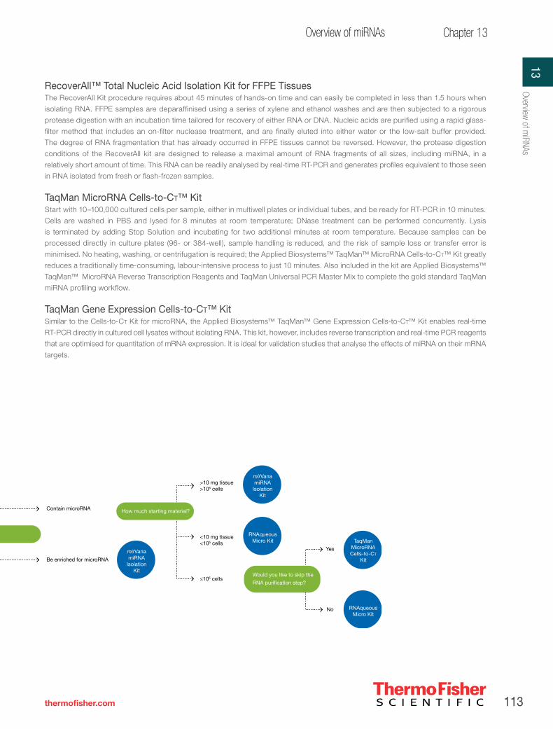
113
Chapter 13Overview of miRNAs
thermofisher.com
MicroRNAs Are you isolating RNA from blood?
RecoverAll
Kit
Yes
No Are you isolating RNA from FFPE tissue?
Yes
No
Blood types
LeukoLOCK
Kit
Human
Mouse RiboPure
Blood Kit
Mouse
Would you like to isolate
both RNA & protein?
mirVana
PARIS KitYes
No Isolated RNA needs to...
Contain microRNA
Be enriched for microRNA
mirVana
miRNA Isolation
Kit
>10 mg tissue>105 cells
<10 mg tissue<105 cells
≤105 cells
How much starting material?
mirVana
miRNA Isolation
Kit
Would you like to skip the
RNA puri�cation step?
Yes
No
TaqMan
MicroRNACells-to-CT
Kit
RNAqueous
Micro Kit
RNAqueous
Micro Kit
RecoverAll™ Total Nucleic Acid Isolation Kit for FFPE TissuesThe RecoverAll Kit procedure requires about 45 minutes of hands-on time and can easily be completed in less than 1.5 hours when
isolating RNA. FFPE samples are deparaffinised using a series of xylene and ethanol washes and are then subjected to a rigorous
protease digestion with an incubation time tailored for recovery of either RNA or DNA. Nucleic acids are purified using a rapid glass-
filter method that includes an on-filter nuclease treatment, and are finally eluted into either water or the low-salt buffer provided.
The degree of RNA fragmentation that has already occurred in FFPE tissues cannot be reversed. However, the protease digestion
conditions of the RecoverAll kit are designed to release a maximal amount of RNA fragments of all sizes, including miRNA, in a
relatively short amount of time. This RNA can be readily analysed by real-time RT-PCR and generates profiles equivalent to those seen
in RNA isolated from fresh or flash-frozen samples.
TaqMan MicroRNA Cells-to-Ct™ KitStart with 10–100,000 cultured cells per sample, either in multiwell plates or individual tubes, and be ready for RT-PCR in 10 minutes.
Cells are washed in PBS and lysed for 8 minutes at room temperature; DNase treatment can be performed concurrently. Lysis
is terminated by adding Stop Solution and incubating for two additional minutes at room temperature. Because samples can be
processed directly in culture plates (96- or 384-well), sample handling is reduced, and the risk of sample loss or transfer error is
minimised. No heating, washing, or centrifugation is required; the Applied Biosystems™ TaqMan™ MicroRNA Cells-to-Ct™ Kit greatly
reduces a traditionally time-consuming, labour-intensive process to just 10 minutes. Also included in the kit are Applied Biosystems™
TaqMan™ MicroRNA Reverse Transcription Reagents and TaqMan Universal PCR Master Mix to complete the gold standard TaqMan
miRNA profiling workflow.
TaqMan Gene Expression Cells-to-Ct™ KitSimilar to the Cells-to-Ct Kit for microRNA, the Applied Biosystems™ TaqMan™ Gene Expression Cells-to-Ct™ Kit enables real-time
RT-PCR directly in cultured cell lysates without isolating RNA. This kit, however, includes reverse transcription and real-time PCR reagents
that are optimised for quantitation of mRNA expression. It is ideal for validation studies that analyse the effects of miRNA on their mRNA
targets.
Figure 13.18. MicroRNA sample preparation selection guide.
13Overview of m
iRNAs

114 Important Licensing Information: These products may be covered by one or more Limited Use Label Licenses (See Thermo Fisher Scientific Catalogue or our website, www.thermofisher.com). By use of these products you accept the terms and conditions of all applicable Limited Use Label Licenses. All products are for research use only. CAUTION: Not intended for human or animal diagnostic or therapeutic uses.
Section II Products for RNA Interference
1. Mattick JS (2001) EMBO Rep 2:986–991.
2. Ambros V (2004) Nature 431:350–355.
3. Bentwich I et al. (2005) Nat Genet 37:766–770.
4. Okazaki Y et al. (2002) Nature 420:563–573.
5. Imanishi T et al. (2004) PLoS Biol 2:e162.
6. Rhoades MW et al. (2002) Cell 110:513–520.
7. Chen X (2005) FEBS Letters 579:5923–5931.
8. Elbashir S et al. (2001) Genes Dev 15:188–200.
9. Lim LP et al. (2003) Science 299:1540.
10. Kim VN, Nam JW (2006) Trends Genet 22:165–173.
11. Bartel DP (2004) Cell 116:281–297.
12. Soifer HS, Rossi JJ, Saetrom P (2007) Mol Ther 15:2070–2079.
13. Griffiths-Jones S, Saini HK, van Dongen S et al. (2008) Nucleic Acids Res 36:D154–D158.
Table 13.1. MicroRNA sample preparation kits: quantitative recovery of small RNAs from a variety of sample types.
TaqMan MicroRNA Cells-to-Ct™ Kit
Invitrogen™ mirVana™ miRNA Isolation Kit
Invitrogen™ mirVana™ PARIS™ Kit
RecoverAll Total Nucleic Acid Isolation Kit for FFPE Tissues
MagMAX-96 for Microarrays Total RNA Isolation Kit
Technology Cells-to-Ct technology, with optional DNase treatment for preparation of cultured cell lysates that can be used in real-time RT-PCR without RNA isolation
Acid-phenol and rapid, enhanced glass-fiber filter purification
Cell Disruption Buffer combined with acid-phenol/chloroform extraction and glass-fiber filter purification
Deparaffinisation, protease digestion, and glass-fiber filter purification
Cell lysis using Invitrogen™ TRI REAGENT™ and magnetic bead purification
Sample input amounts
10 to 105 cultured cells 103–107 cultured cells or 0.5–250 mg tissue
100–107 cultured cells or up to 100 mg tissue
Up to four 20 μm FFPE sections
Up to 5 x 10⁶ cultured cells or up to 100 mg tissue
Features • Go from cells in culture to RT-PCR, typically in 10 min at room temperature
• Simple procedure with no sample transfers, no centrifugation, and no vacuum manifold needed
• For real-time RT-PCR• Superior results
when used with TaqMan MicroRNA Assays and Arrays
• Fast, easy isolation of small RNA from cultured cells and most tissues (including tissues with high levels of ribonucleases)
• Ideal for miRNA profiling experiments and other gene expression applications
• Simple, 30 minute procedure
• Protein can also be recovered
• Optional small RNA enrichment procedure
• Ideal for correlating mRNA, miRNA, and/or siRNA with protein levels
• Isolate total nucleic acids, including DNA and microRNAs, from FFPE samples
• No overnight proteinase K digestion required—deparaffinise in the morning and perform qRT-PCR in the afternoon
• Routinely obtain yields of >50% that of unfixed tissue from the same sample source
• For real-time RT-PCR and PCR, mutation screening, and microarray analyses
• Highly consistent results from experiment to experiment
• Streamlined RNA purification
• Requires less hands-on time than competitor kits
• Walk away—integrate with established robotic platforms
• Modified protocol to recover small RNAs
Kit sizes and part numbers
100 lysis rxns/500 PCRs P/N 4391848400 lysis rxns/2,000 PCRs P/N 4391996
Up to 40 purifications P/N AM1560
Up to 40 purifications P/N AM1556
40 purifications P/N AM1975
96 purifications P/N AM1839
Chapter references
Prod
ucts
for R
NA In
terfe
renc
eII

115
Chapter 14Optimised miRNA profiling
thermofisher.com
CHAPTER 14
Optimised miRNA profiling
NCode platform—for optimised miRNA profiling
miRNA expression profiling came into prominence in part because of the expectation that a highly expressed miRNA for a given tissue
or cell type (or developmental stage) is likely to play a regulatory role. Since the first published article to report on miRNA profiling
using an oligonucleotide microarray [1], microarray analysis has become the preferred tool for profiling miRNA expression patterns
to gain insight into their relevance in development and disease. The Invitrogen™ NCode™ platform is optimised to investigate global
miRNA expression patterns. From sample preparation to array analysis to qRT-PCR validation, the NCode platform provides optimised
reagents for every step of the miRNA profiling workflow (Figure 14.1).
Total RNA isolationTRIzol Reagent
miRNA labelingNCode miRNA Ampli�cation System
NCode Rapid miRNA Labeling System
miRNA data analysisNCode Pro�ler software
miRNA qRT-PCRNCode SYBR Green and SYBR GreenER qRT-PCR Kits
NCode miRNA First-Strand cDNA Synthesis Kit
Preprinted miRNA microarray
NCode Human miRNA Microarray V3NCode Multi-Species miRNA Microarray V2
In-house printed miRNA microarray
NCode Human miRNA Microarray Probe Set V3NCode Multi-Species miRNA Microarray Probe Set V2
miRNA microarray controlsNCode Multi-Species miRNA
Microarray Control V2
miR
NA
mic
roar
ray
sam
ple
pre
par
atio
nm
iRN
A h
ybrid
izat
ion
Dat
a va
lidat
ion
Dat
a an
alys
is
Functional toolsAmbion Pre-miR miRNA precursorsAmbion Anti-miR miRNA inhibitors
Figure 14.1. NCode platform workflow. During sample preparation, total RNA is isolated and quantified. Amplification of the small RNA molecules may be necessary when there is insufficient starting material. Next, a direct labelling method polyadenylates the enriched small RNA molecules and ligates a fluorescently conjugated, branched DNA structure to the tailed RNA. Tagged and tailed miRNAs are subsequently hybridised to the array. Bound miRNAs are detected by the hybridization of branched DNA structures containing dye molecules. The miRNA expression profile is calculated from relative signal intensity detected by a microarray scanner for each spot on the array. Data validation is accomplished using qRT-PCR.
14Optim
ised miRNA profiling

116 Important Licensing Information: These products may be covered by one or more Limited Use Label Licenses (See Thermo Fisher Scientific Catalogue or our website, www.thermofisher.com). By use of these products you accept the terms and conditions of all applicable Limited Use Label Licenses. All products are for research use only. CAUTION: Not intended for human or animal diagnostic or therapeutic uses.
Section II Products for RNA Interference
Efficient isolation of total RNA (including miRNA) using TRIzol Reagent
Isolating high-quality total RNA from your sample is a step crucial to the success of your research. Invitrogen™ TRIzol™ Reagent purifies all
RNAs, including those in the 10–200 nucleotide range, and is thus recommended for isolating total RNA for studying miRNAs. Extraction
procedures involve ready-to-use monophasic solutions of phenol and guanidine isothiocyanate, and are based upon improvements to
the single-step RNA isolation method developed by Chomczynski and Sacchi [2]. Using TRIzol Reagent enables:
→ More effective purification of total RNA than column-based purification, without depletion of small RNAs
→ A simple, flexible protocol, completed in less than 1 hr
→ High-purity RNA from a wide range of animal tissues and cells
Reliable recovery of total RNA (including miRNA) from FFPE samples with the RecoverAll Total Nucleic Acid Isolation Kit→ Optimised for isolation of total nucleic acids, including microRNAs, from FFPE tissue
→ No overnight proteinase K digestion required—deparaffinise in the morning and perform qRT-PCR in the afternoon
→ Obtain typical yields of >50% that of unfixed tissue from the same sample source
→ Recovered nucleic acids are suitable for qRT-PCR, qPCR, mutation screening, and microarray analyses
The Invitrogen™ RecoverAll™ Total Nucleic Acid Isolation Kit is designed to extract total nucleic acid from formalin or paraformalin-
fixed, paraffin-embedded (FFPE) tissues (Figure 14.2). Up to four 20 µm sections, or up to 35 mg of unsectioned core samples, can
be processed per reaction.
The ability to isolate nucleic acid from archived tissue samples that is suitable for molecular analysis enables retrospective studies
of diseased tissue at both the genomic and gene expression level. While standard preservation techniques that employ formalin are
ideal for maintaining tissue structure and preventing decomposition, formalin preservation creates protein–protein and protein–nucleic
acid crosslinks that make it difficult to perform molecular analyses. In addition, RNA (and to some extent DNA) is often fragmented and
chemically modified to such a degree that it is incompatible with many molecular analysis techniques.
The degree of RNA fragmentation that has already occurred in FFPE tissues cannot be reversed. However, the protease digestion
conditions of the RecoverAll Kit are designed to release a maximal amount (Figure 14.3) of RNA fragments of all sizes, including
microRNA, in a relatively short amount of time.
The RecoverAll Total Nucleic Acid Isolation Kit procedure requires about 45 minutes of hands-on time and can easily be completed
in less than 1 day when isolating RNA. FFPE samples are deparaffinised using a series of xylene and ethanol washes. Next, they are
subjected to a rigorous protease digestion with an incubation time compatible for recovery of either RNA or DNA. The nucleic acids
are purified using a rapid glass-filter methodology that includes an on-filter nuclease treatment and are eluted into either water or the
low-salt buffer provided.
The recovered nucleic acids are suitable for downstream applications such as microarray analyses, qRT-PCR (see Figure 14.4),
and mutation screening. However, as is the case with all FFPE tissue, sample fixation and storage typically cause nucleic acid
fragmentation and modification. Therefore, downstream applications, such as microarray analysis, which require more pristine RNA
than does qRT-PCR, may require modification for best results. Although DNA tends not to fragment as easily as RNA, DNA is more
reactive to the formalin and requires a longer (2-day) protease digestion time to release substantial amounts of DNA. The recovered
DNA can typically be used for PCR and other downstream applications.
Product Quantity Cat. No.
TRIzol Reagent 100 mL 15596026
Prod
ucts
for R
NA In
terfe
renc
eII

117
Chapter 14Optimised miRNA profiling
thermofisher.com
Product Quantity Cat. no.
RecoverAll Total Nucleic Acid Isolation Kit for FFPE 40 purifications AM1975
Figure 14.2. Overview of the RecoverAll Total Nucleic Acid Isolation Kit procedure.
Figure 14.3. Yield of RNA from archived human FFPE tissue samples: RecoverAll kit vs. two competitor systems. A 10–20 µm section from each of the above archived human tissue blocks was isolated using each of three kits: Competitor A kit, Competitor B kit, and the RecoverAll kit. Colon, lung, and breast samples were 1–2 years old; kidney was 3–5 years old; and bladder was 10–15 years old. Once the RNA was isolated, the concentration was determined via OD260, and the amount of RNA recovered in micrograms was calculated. The RecoverAll™ Kit yielded the highest recovery of the three systems for all tissue types.
Figure 14.4. Ct values from real-time RT-PCR of frozen and FFPE mouse tissues. Four different tissues (brain, kidney, heart, and liver) were dissected from four different mice. Each tissue was split in half—one half was flash-frozen in liquid nitrogen and then stored at –80°C, while the other half was fixed and embedded using standard hospital protocol. RNA was isolated from one 20 µm section from each FFPE sample using the RecoverAll Kit. RNA from the frozen controls was isolated using the mirVana™ miRNA Isolation Kit. The RNA (400 ng) was then subjected to two-step real-time RT-PCR. The cDNA was generated using the Invitrogen™ RetroScript® Kit. Invitrogen™ SuperTaq™ polymerase and other necessary reagents, plus an aliquot of the RT reaction, were used for the PCR. All gene targets had an amplicon ranging from 80 to 100 nt. The Ct values for all reactions of the same tissue were averaged together, then the Ct values for the frozen controls were subtracted from the Ct values for the FFPE samples to generate the graph above.
0
1
2
3
4
5
6
7
8
TBP GAPDH UBC PKC Recc1 RNAPol II FAS
Ct D
iffer
ence
Heart
Brain
Kidney
Liver
Deparaffinization
Protease Digestion
Nucleic Acid Isolation
Nuclease Digestion and Final Purification
20 min
3 hr or48 hr
10 min
45 min
1. Assemble FFPE sections equivalent to <–80 µm or <–35 mg unsectioned core
2. Add 1 mL 100% xylene, mix, and incubate for 3 min at 50°C
3. Centrifuge for 2 min at maximum speed, and discard the xylene
4. Wash the pellet twice with 1 mL 100% ethanol and air dry
1. Add Digestion Buffer and Protease
2. Incubate at 50°C for 3 hr for RNA isolation and 48 hr for DNA isolation
1. Add 480 µL Isolation Additive and vortex
2. Add 1.1 mL 100% ethanol and mix
3. Pass the mixture through a Filter Cartridge
4. Wash with 700 µL of Wash 1
5. Wash with 500 µL of Wash 2/3
1. Add DNase or RNase mix to each Filter Cartridge and incubate for 30 min
2. Wash with 700 µL of Wash 1
3. Wash twice with 500 µL of Wash 2/3
4. Elute nucleic acid with 2 x 30 µL Elution Solution or nuclease-free water
0
1
2
3
4
5
6
7
8
9
10
Colon Lung Breast Kidney Bladder
Tota
l am
ount
of R
NA
(µg) RecoverAll
Competitor B
Competitor A
14Optim
ised miRNA profiling

118 Important Licensing Information: These products may be covered by one or more Limited Use Label Licenses (See Thermo Fisher Scientific Catalogue or our website, www.thermofisher.com). By use of these products you accept the terms and conditions of all applicable Limited Use Label Licenses. All products are for research use only. CAUTION: Not intended for human or animal diagnostic or therapeutic uses.
Section II Products for RNA Interference
5´
Poly(A) tailing of miRNA
Ligation of detection reagent to
3´ poly(A) tail
Hybridisation tomiRNA microarray
Final wash and scanning
3´ total RNA
5´ 3´-tailed miRNA
Single reagent
Alexa Fluor detection reagent
miRNA probe
Tagged miRNA
5´ 3´-labelled miRNA
Number of positive calls
Mean SNR in positive calls
Num
ber
of p
ositi
ve fe
atur
es
NCode™ RapidCompetitor E
5
0
10
15
20
25
30
35
Labelling method
Figure 14.5. NCode Rapid miRNA Labeling System titration. The NCode Rapid miRNA Labeling System is recommended when starting with 250 ng to 5 μg total RNA. When starting with less than 50 ng of total RNA, we recommend using the Invitrogen™ NCode™ miRNA Amplification System to increase the quantity of miRNA that can be detected and provide sufficient material for replicate experiments.
Figure 14.6. Sensitivity of the NCode Rapid miRNA Labeling System compared to competitor “E”. Starting with 100 ng pooled total human RNA, samples were labelled with the NCode Rapid or labeling system from “E”. Replicate samples were arrayed on either Invitrogen™ NCode™ microarrays or competitor microarrays in both red and green channels. Images scanned via an Axon scanner and data acquired in a GenePix Pro™ scanner. Data were background-corrected and compared across common human features for signal-to-noise ratios (SNR) and positive-call designation. Positive call is determined as SNR >3 in all replicate features across all replicate arrays. Mean SNR was calculated from positive calls. The NCode Rapid miRNA Labeling System is brighter and detects more features at low input (100 ng of total RNA).
Simplified miRNA fluorescent labeling
The Invitrogen™ NCode™ Rapid miRNA Labeling System is a fast, reliable method to label endogenous miRNAs with fluorescent
tags. Using this system, miRNAs from total RNA samples are polyadenylated and labelled with fluorescent Alexa Fluor dyes, and
hybridised to microarrays printed with species-specific antisense miRNA probes. This system has been optimised to enable sensitive
and accurate profiling of miRNA expression patterns from minimal RNA input. Starting with 250 ng to 5 μg of total RNA, miRNAs are
tagged directly in a quick and easy protocol with the NCode Rapid miRNA Labeling System (Figure 14.5). Lower starting amounts (100
ng) may be used with similar sensitivities, depending on the type of scanner and hybridisation method used. Dynamic hybridisation is
required to achieve maximum sensitivity at lower input RNA levels. Maximum sensitivities are typically seen in the 500 ng to 1 μg range,
depending on sample type. The protocol takes approximately 1 hour to complete and consists of just two steps prior to hybridisation:
poly(A) tailing, and ligation of a fluorescent molecule. After 8–16 hours of hybridisation, the microarrays are ready to scan and analyse.
The fluorescent dendrimer—a branched structure of single- and double-stranded DNA conjugated with Alexa Fluor dyes—appends
approximately 15 fluorophores to the RNA strand. The high sensitivity achieved with this method is due to the signal amplification effect
of the labelled branched DNA structure [1], enabling maximum signal-to-background ratios and strong signal correlation for increased
sensitivity (Figure 14.6, Figure 14.7).
Using the NCode Rapid miRNA Labeling System enables:
→ Less time at the bench—hybridise in one step, with no miRNA enrichment required
→ Increased sensitivity—reliably detect attomolar levels of miRNA
→ Reliable miRNA profiling results
→ Rapid turnaround—complete your experiments in a single day using the 8 hr hybridisation protocol with dynamic hybridisation
1. This method uses 3DNATM Reagent, manufactured under exclusive license from Genisphere, Inc.
Prod
ucts
for R
NA In
terfe
renc
eII

119
Chapter 14Optimised miRNA profiling
thermofisher.com
>100
>1,000
>10,000
Num
ber
of p
ositi
ve fe
atur
es
NCode™ RapidCompetitor A
50
0
100
150
200
250
Labelling method
Figure 14.7. Sensitivity of the NCode Rapid miRNA Labeling System compared to competitor “A”. Starting with 100 ng pooled total human RNA, samples were labelled with the NCode Rapid or labeling system from “A”. Replicate samples were arrayed on either NCode microarrays or competitor microarrays in the green channel. Images were scanned via an Axon scanner and data acquired in a GenePix™ scanner. Data were background-corrected and compared across common human features for signal-to-noise ratios (SNR) and positive-call designation. Positive call is determined as SNR >3 in all replicate features across all replicate arrays. Mean SNR was calculated from positive calls. The number of features having signals greater than 10,000 FU (purple bars), 1,000 FU (dark blue bars), and 100 FU (red bars) are shown above. The NCode Rapid miRNA Labeling System is brighter and detects more features at low input.
Product Quantity Cat. no.
NCode Rapid miRNA Labeling System 20 rxns MIRLSRPD-20
Invitrogen™ NCode™ Rapid Alexa Fluor™ 3 miRNA Labeling System 20 rxns MIRLSA3-20
Ultrasensitive miRNA amplification
In many instances, RNA samples derived from laser capture microdissection (LCM), flow-sorted (FACS) samples, or needle biopsies do
not yield sufficient starting amounts of RNA for standard profiling methods or for the experimental replicates needed for statistical reliability.
In such cases, it is useful to apply linear miRNA amplification methods to the enriched miRNA population, thereby increasing the relative
target abundance levels. The Invitrogen™ NCode™ miRNA Amplification System, based on mRNA amplification methods, enables robust
and efficient linear amplification and microarray profiling of small amounts of RNA species, such as miRNA, from as little as 50 ng of total
RNA or the enriched equivalent. The NCode miRNA Amplification System enables:
→ Increased sensitivity, with consistent 2,000- to 5,000-fold amplification of miRNA
→ Amplified product that faithfully represents the initial miRNA sample (Figure 14.8)
→ Precise amplification from reaction to reaction
Product Quantity Cat. No.
NCode™ miRNA Amplification System 20 rxns MIRAS20
Directly labeled
Ampli�ed
let7
a
let7
e
mir
143
mir
145
mir
200b
mir
21
mir
223
mir
30a
0
0.5
1.0
1.5
2.0
2.5
3.0
3.5
Tum
our/
norm
al e
xpre
ssio
n ra
tio
Cell/tissue types
Figure 14.8. Ultrasensitive and accurate miRNA microarray profiling from small samples. Total RNA from large cell carcinomas and adjacent normal tissue was isolated from multiple patients and enriched for miRNA with the Invitrogen™ PureLink™ miRNA Isolation Kit. Enriched miRNA from 300 ng of total RNA was amplified using the NCode miRNA Amplification System and labelled for array analysis with the NCode miRNA Labeling System. Additionally, the miRNA from 10 μg of total RNA from the same source was labeled with the NCode miRNA Labeling System without amplification and hybridised to Invitrogen™ NCode™ Multi-Species miRNA Microarrays for analysis. The ratios of miRNA expression in tumor samples vs. adjacent normal tissues were calculated for all human miRNA.
14Optim
ised miRNA profiling

120 Important Licensing Information: These products may be covered by one or more Limited Use Label Licenses (See Thermo Fisher Scientific Catalogue or our website, www.thermofisher.com). By use of these products you accept the terms and conditions of all applicable Limited Use Label Licenses. All products are for research use only. CAUTION: Not intended for human or animal diagnostic or therapeutic uses.
Section II Products for RNA Interference
Comprehensive miRNA expression profiling
Whether you prefer to profile miRNAs using in-house printed arrays or preprinted arrays, the NCode platform allows you to
comprehensively screen most known miRNAs in a variety of species (Figure 14.9). Enjoy the advantages of a leading preprinted miRNA
profiling microarray, or gain the flexibility of a probe set and controls for array fabrication in your lab. The NCode microarrays offer:
→ Arrays spotted in triplicate with positive and negative controls throughout for monitoring hybridisation specificity
→ Increased sensitivity from maximum hybridisation intensities, and normalised melting temperatures for uniform hybridisation
→ Easy normalisation of signal intensities during scanning, using Invitrogen™ Alexa Fluor™ dye control probes
→ Detection and identification of miRNAs conserved in other species but not yet validated for your model
NCode™ Human miRNA MicroarrayThe NCode™ Human miRNA Microarray V3 consists of Corning® epoxide–coated glass slides printed with optimised probe sequences
that target nearly all known human miRNAs in the Sanger miRBase Sequence Database, Release 10.0, plus 373 novel putative human
miRNAs (Table 14.1) discovered through deep sequencing and validated by both microarray hybridizations and qPCR analysis. Each
microarray slide comes fully blocked and ready to use. Access to this putative novel content enables researchers to access and profile
biologically relevant small non-coding RNAs (putative miRNAs) that are not available on other platforms (Figure 14.10). This content is
not derived from in silico predictions.
NCode Multi-Species miRNA MicroarrayThe Invitrogen™ NCode™ Multi-Species miRNA Microarray V2 consists of optimised probe sequences targeting nearly all of the known and
predicted mature miRNAs in the Sanger miRBase Sequence Database, Release 9.0, for human, mouse, rat, D. melanogaster, C. elegans,
and zebrafish sequences (Figure 14.11, Table 14.1). These species-specific, unmodified oligonucleotides are 34 to 44 bases in length, and
each microarray slide comes fully blocked and ready to use. Interrogation of samples with the NCode Multi-Species miRNA Microarray
allows researchers to compare cross-species miRNA expression, and validate the existence of predicted miRNA species (or those that are
conserved but not yet validated in a particular organism).
ControlsThe NCode Multi-Species miRNA Microarray Control V2 is a synthetic 22-nucleotide miRNA positive control that is used to assess
the efficiency of array labelling and hybridisation. This control does not exhibit any detectable cross-hybridisation or interference with
endogenous miRNAs from any sequenced organism. The NCode Multi-Species miRNA Microarray and Invitrogen™ NCode™ miRNA
Microarray Probe Sets include oligonucleotide probes that are complementary to the control.
Undifferentiated huESC
Liver
Ovary
Heart
Lung
Thymus
Stomach
Breast
Colon
Brain
Tissue classi�cation
Fold change
–6 0 +6
Figure 14.9. Profiling and cluster analysis of total RNA from various tissues on the NCode Human miRNA Microarray V3. Both known and novel content show tissue-specific differential expression.
Prod
ucts
for R
NA In
terfe
renc
eII
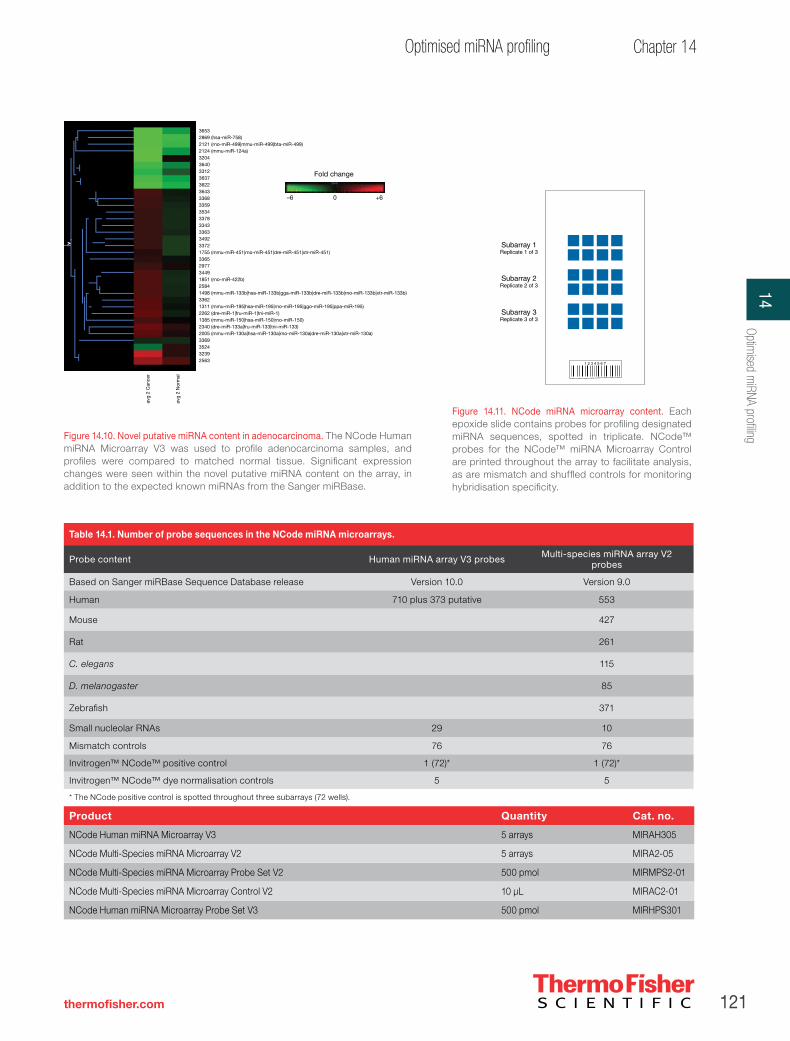
121
Chapter 14Optimised miRNA profiling
thermofisher.com
36532869 (hsa-miR-758)2121 (rno-miR-499|mmu-miR-499|bta-miR-499)2124 (mmu-miR-124a)320436403312363736223643336833593534337833433363349233721755 (mmu-miR-451|rno-miR-451|dre-miR-451|xtr-miR-451)3365297734491851 (rno-miR-422b)25841498 (mmu-miR-133b|hsa-miR-133b|gga-miR-133b|dre-miR-133b|rno-miR-133b|xtr-miR-133b)33621311 (mmu-miR-195|hsa-miR-195|rno-miR-195|ggo-miR-195|ppa-miR-195)2262 (dre-miR-1|fru-miR-1|tni-miR-1)1385 (mmu-miR-150|hsa-miR-150|rno-miR-150)2340 (dre-miR-133a|fru-miR-133|tni-miR-133)2005 (mmu-miR-130a|hsa-miR-130a|rno-miR-130a|dre-miR-130a|xtr-miR-130a)3369352432392563
Fold change
–6 0 +6
avg
2 C
ance
r
avg
2 N
orm
al
Figure 14.10. Novel putative miRNA content in adenocarcinoma. The NCode Human miRNA Microarray V3 was used to profile adenocarcinoma samples, and profiles were compared to matched normal tissue. Significant expression changes were seen within the novel putative miRNA content on the array, in addition to the expected known miRNAs from the Sanger miRBase.
Subarray 1Replicate 1 of 3
Subarray 2Replicate 2 of 3
Subarray 3Replicate 3 of 3
Table 14.1. Number of probe sequences in the NCode miRNA microarrays.
Probe content Human miRNA array V3 probes Multi-species miRNA array V2 probes
Based on Sanger miRBase Sequence Database release Version 10.0 Version 9.0
Human 710 plus 373 putative 553
Mouse 427
Rat 261
C. elegans 115
D. melanogaster 85
Zebrafish 371
Small nucleolar RNAs 29 10
Mismatch controls 76 76
Invitrogen™ NCode™ positive control 1 (72)* 1 (72)*
Invitrogen™ NCode™ dye normalisation controls 5 5
* The NCode positive control is spotted throughout three subarrays (72 wells).
Product Quantity Cat. no.
NCode Human miRNA Microarray V3 5 arrays MIRAH305
NCode Multi-Species miRNA Microarray V2 5 arrays MIRA2-05
NCode Multi-Species miRNA Microarray Probe Set V2 500 pmol MIRMPS2-01
NCode Multi-Species miRNA Microarray Control V2 10 μL MIRAC2-01
NCode Human miRNA Microarray Probe Set V3 500 pmol MIRHPS301
Figure 14.11. NCode miRNA microarray content. Each epoxide slide contains probes for profiling designated miRNA sequences, spotted in triplicate. NCode™ probes for the NCode™ miRNA Microarray Control are printed throughout the array to facilitate analysis, as are mismatch and shuffled controls for monitoring hybridisation specificity.
14Optim
ised miRNA profiling

122 Important Licensing Information: These products may be covered by one or more Limited Use Label Licenses (See Thermo Fisher Scientific Catalogue or our website, www.thermofisher.com). By use of these products you accept the terms and conditions of all applicable Limited Use Label Licenses. All products are for research use only. CAUTION: Not intended for human or animal diagnostic or therapeutic uses.
Section II Products for RNA Interference
ProbesFor researchers planning to spot their own miRNA microarrays, we have probes from the NCode Human miRNA Microarray V3 and
NCode Multi-Species miRNA Microarray V2 available in 384-well plates. Each well contains 500 pmol of lyophilised oligonucleotide
probe, ready for resuspension and printing.
Simplified data analysis using the NCode ProfilerUse of Latin squares loop design/dye swap methodology. There are a number of methods used to analyse expression profiling
microarray data, but some are not ideal for miRNA microarrays due to the reduced number of features present and the dynamic
levels of miRNA expression across samples. We recommend using the Latin squares normalisation and loop design/dye swap model
described by Kerr et al. [3], because this method provides reliable statistical data with the smallest number of microarray chips. This
experimental design generates a loop without identical chip replicates, though each tissue will be replicated twice, once with each
Alexa Fluor dye. Using this array layout enables comparison of each test sample with both the reference sample and comparison of
each test sample with any other test sample. This normalisation method attempts to reduce or eliminate any general miRNA effect,
array effect, dye effect, overall tissue effect, array–miRNA interaction, and dye–miRNA interactions from the data (Figure 14.12).
Experimental design. Invitrogen™ NCode™ Profiler software enables simple design and analysis of miRNA microarray data using loop
design and dye swap normalisation methods that support sample-to-sample comparison within the experiment. They are designed
with statistical considerations to reduce or eliminate any general miRNA effect, array effect, dye effect, overall tissue effect, array–
miRNA interaction, and dye–miRNA interaction from the data.
Data analysis. miRNA researchers now have a tailored tool to identify differentially expressed miRNA markers on microarrays. NCode
Profiler software is an advanced experimental design and analysis solution designed for two-dye expression profiling microarray
experiments. Once an experiment has been conducted, the raw data can be imported and analysed using the statistical methods
designed into the array. For each tissue on the array, pairwise differential expression is determined and the test statistics, fold
expression changes, and p-values are generated. Additionally, the ranking of each miRNA with respect to the other miRNAs in terms
of overall expression level within the tissue is given. Using NCode Profiler software enables:
→ Simplified experimental design steps
→ Confidence interpreting results with proven statistical analysis using dye swap or loop design normalisation models
→ Output includes test statistic, fold expression changes for each pair-wise comparison, and p-values and rankings of miRNA
markers for all samples
→ Easy export of normalised data to visualisation software for clustering (tree) and heat map analysis
Visit www.thermofisher.com/ncode to download a free copy. NCode Profiler software runs on Microsoft Windows™ XP and Windows
Vista™operating systems.
Figure 14.12. The NCode Profiler software for miRNA microarray data analysis has a simple user interface and provides both data analysis using Latin squares normalisation and experimental design guidance.
Prod
ucts
for R
NA In
terfe
renc
eII

123
Chapter 14Optimised miRNA profiling
thermofisher.com
Robust data validation using the NCode miRNA qRT-PCR KitsQuantitative RT-PCR (qRT-PCR) is the standard for validating microarray data and is an invaluable tool for highly sensitive and accurate
profiling of miRNA population subsets. Most commercially available miRNA qRT-PCR systems use proprietary, predesigned, miRNA-
specific primers for real-time PCR and/or reverse transcription. Unfortunately, such primers require that the miRNA sequence is
publicly available, and that a commercial qRT-PCR assay has been developed for that specific sequence. This limits the availability
of qRT-PCR assays for many model organisms, recently discovered miRNAs or non-coding RNAs, or proprietary miRNA sequences.
The Invitrogen™ NCode™ VILO™ miRNA cDNA Synthesis and Invitrogen™ NCode™ EXPRESS SYBR™ GreenER™ miRNA
qRT-PCR Kits overcome these limitations by combining a carefully optimised combined polyadenylation and reverse transcription step
using the Invitrogen™ SuperScript™ VILO™ enzyme and a universal primer. The miRNA-specific amplification occurs during the PCR
reaction, in which the sequence of the miRNA of interest is used as the target-specific PCR primer.
Use the NCode EXPRESS SYBR GreenER miRNA qRT-PCR Kits to:
→ Amplify using minimal total RNA input (no additional enrichment needed, conserve precious samples) (Figure 14.13)
→ Detect miRNA, ncRNA, or mRNA sequences from the same universal cDNA
→ Archive cDNA for later validation—the use of the cDNA for multiple experiments will enable more reliable comparison and allow
future interrogation of new sequences
→ Obtain excellent sensitivity over a broad dynamic range of miRNA abundance
→ Profile closely related miRNAs with single-nucleotide discrimination
→ Use multiple real-time PCR instrument platforms, including standard and fast- cycling modes
Figure 14.13. miRNA detection from total RNA or enriched miRNA starting materials. cDNA was generated from brain reference RNA or an equivalent volume of enriched miRNA. Average Ct values for 10 ng/µL total RNA or equivalent per qPCR reaction are shown.
MicroRNA Total RNA Enriched RNA
26a 16.53 16.53
195 17.19 17.28
1 24.97 25.36
Match the NCode qPCR tool to your research needs
NCode EXPRESS SYBR GreenER miRNA qRT-PCR Kits—incorporate proven Invitrogen™ enzymes and the newest SYBR™
GreenER™ dye specifically tailored for superior results on high-throughput, fast-cycling instruments. These kits provide:
→ Fast-activating, antibody-mediated Invitrogen™ Platinum™ Taq DNA Polymerase in association with the brighter and less PCR-
inhibitory SYBR GreenER dye—complete, rapid activation that enables the best sensitivity and reproducibility
→ Flexible format with premixed or separate ROX—designed to work with default instrument protocols on a wide range of high-
throughput fast instruments (e.g., Applied Biosystems™ 7500 FAST, Applied Biosystems™ 7900 HT Fast, Applied Biosystems™
StepOne™ System, Roche LightCyler™ 480, Corbett Rotor-Gene™
→ Significantly reduced carryover contamination with UDG carryover protection—uracil-DNA-glycosylase (UDG)/dUTP incorporated
in all kits
3020100 40
miR-26a
14Optim
ised miRNA profiling
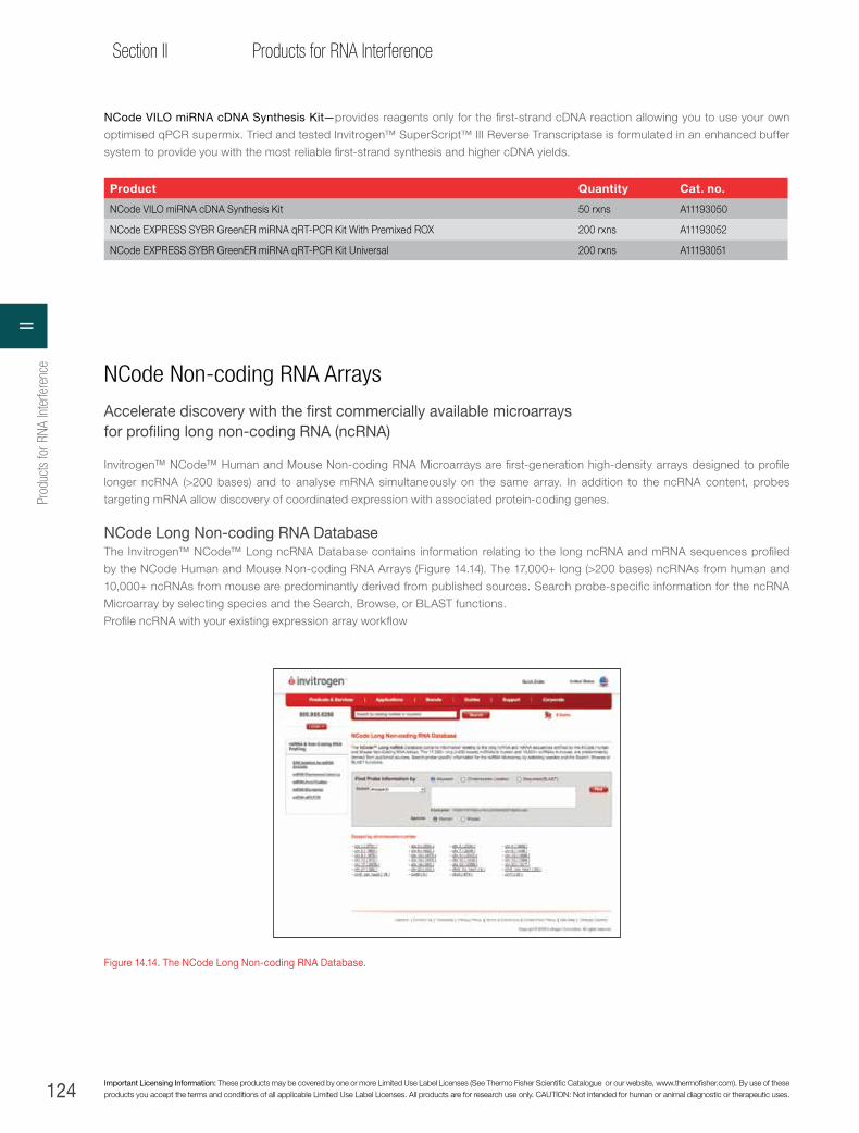
124 Important Licensing Information: These products may be covered by one or more Limited Use Label Licenses (See Thermo Fisher Scientific Catalogue or our website, www.thermofisher.com). By use of these products you accept the terms and conditions of all applicable Limited Use Label Licenses. All products are for research use only. CAUTION: Not intended for human or animal diagnostic or therapeutic uses.
Section II Products for RNA Interference
NCode VILO miRNA cDNA Synthesis Kit—provides reagents only for the first-strand cDNA reaction allowing you to use your own
optimised qPCR supermix. Tried and tested Invitrogen™ SuperScript™ III Reverse Transcriptase is formulated in an enhanced buffer
system to provide you with the most reliable first-strand synthesis and higher cDNA yields.
Product Quantity Cat. no.
NCode VILO miRNA cDNA Synthesis Kit 50 rxns A11193050
NCode EXPRESS SYBR GreenER miRNA qRT-PCR Kit With Premixed ROX 200 rxns A11193052
NCode EXPRESS SYBR GreenER miRNA qRT-PCR Kit Universal 200 rxns A11193051
Figure 14.14. The NCode Long Non-coding RNA Database.
NCode Non-coding RNA Arrays
Accelerate discovery with the first commercially available microarrays for profiling long non-coding RNA (ncRNA)
Invitrogen™ NCode™ Human and Mouse Non-coding RNA Microarrays are first-generation high-density arrays designed to profile
longer ncRNA (>200 bases) and to analyse mRNA simultaneously on the same array. In addition to the ncRNA content, probes
targeting mRNA allow discovery of coordinated expression with associated protein-coding genes.
NCode Long Non-coding RNA DatabaseThe Invitrogen™ NCode™ Long ncRNA Database contains information relating to the long ncRNA and mRNA sequences profiled
by the NCode Human and Mouse Non-coding RNA Arrays (Figure 14.14). The 17,000+ long (>200 bases) ncRNAs from human and
10,000+ ncRNAs from mouse are predominantly derived from published sources. Search probe-specific information for the ncRNA
Microarray by selecting species and the Search, Browse, or BLAST functions.
Profile ncRNA with your existing expression array workflow
Prod
ucts
for R
NA In
terfe
renc
eII
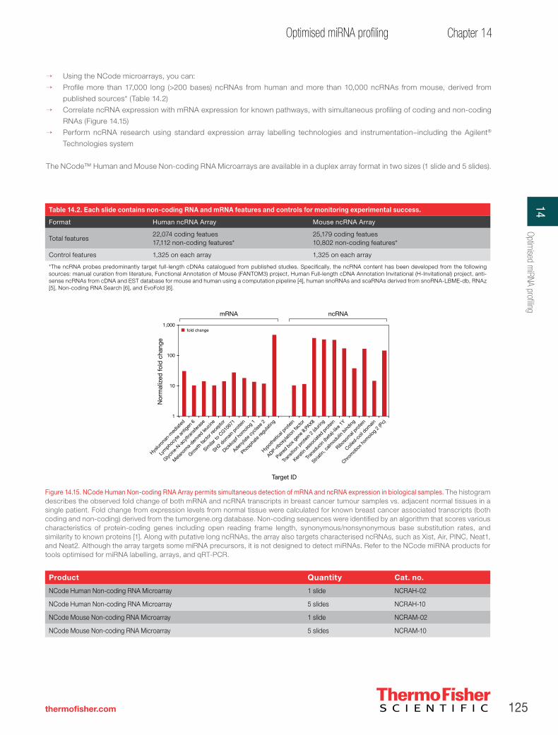
125
Chapter 14Optimised miRNA profiling
thermofisher.com
→ Using the NCode microarrays, you can:
→ Profile more than 17,000 long (>200 bases) ncRNAs from human and more than 10,000 ncRNAs from mouse, derived from
published sources* (Table 14.2)
→ Correlate ncRNA expression with mRNA expression for known pathways, with simultaneous profiling of coding and non-coding
RNAs (Figure 14.15)
→ Perform ncRNA research using standard expression array labelling technologies and instrumentation–including the Agilent®
Technologies system
The NCode™ Human and Mouse Non-coding RNA Microarrays are available in a duplex array format in two sizes (1 slide and 5 slides).
Figure 14.15. NCode Human Non-coding RNA Array permits simultaneous detection of mRNA and ncRNA expression in biological samples. The histogram describes the observed fold change of both mRNA and ncRNA transcripts in breast cancer tumour samples vs. adjacent normal tissues in a single patient. Fold change from expression levels from normal tissue were calculated for known breast cancer associated transcripts (both coding and non-coding) derived from the tumorgene.org database. Non-coding sequences were identified by an algorithm that scores various characteristics of protein-coding genes including open reading frame length, synonymous/nonsynonymous base substitution rates, and similarity to known proteins [1]. Along with putative long ncRNAs, the array also targets characterised ncRNAs, such as Xist, Air, PINC, Neat1, and Neat2. Although the array targets some miRNA precursors, it is not designed to detect miRNAs. Refer to the NCode miRNA products for tools optimised for miRNA labelling, arrays, and qRT-PCR.
Table 14.2. Each slide contains non-coding RNA and mRNA features and controls for monitoring experimental success.
Format Human ncRNA Array Mouse ncRNA Array
Total features22,074 coding featues17,112 non-coding features*
25,179 coding featues10,802 non-coding features*
Control features 1,325 on each array 1,325 on each array
*The ncRNA probes predominantly target full-length cDNAs catalogued from published studies. Specifically, the ncRNA content has been developed from the following sources: manual curation from literature, Functional Annotation of Mouse (FANTOM3) project, Human Full-length cDNA Annotation Invitational (H-Invitational) project, anti-sense ncRNAs from cDNA and EST database for mouse and human using a computation pipeline [4], human snoRNAs and scaRNAs derived from snoRNA-LBME-db, RNAz [5], Non-coding RNA Search [6], and EvoFold [6].
Product Quantity Cat. no.
NCode Human Non-coding RNA Microarray 1 slide NCRAH-02
NCode Human Non-coding RNA Microarray 5 slides NCRAH-10
NCode Mouse Non-coding RNA Microarray 1 slide NCRAM-02
NCode Mouse Non-coding RNA Microarray 5 slides NCRAM-10
1
10
100
1,000
Nor
mal
ized
fold
cha
nge
Hyalur
onan
-med
iated
Lym
phocy
te an
tigen
6
Glycine
-N-a
cyltr
ansfe
rase
Mela
nom
a-der
ived le
ucine
Growth
fact
or re
cepto
r
Simila
r to
CG1067
1
SH2 dom
ain p
rote
in
Dickko
pf hom
olog
1
Adenyla
te cy
clase
2
Phosp
hate
regu
lating
Hypot
hetic
al pro
tein
ADP-ribos
ylatio
n fa
ctor
Paired
box
gen
e 8;P
AX8
Trans
ition
prote
in 2
(dur
ing
Kerat
in as
socia
ted p
rote
in
Trans
ducin
(bet
a)-lik
e 1Y
Ribosom
al pro
tein
Coiled
-coil
dom
ain
Striat
in, ca
lmod
ulin
binding
Chrom
obox
hom
olog
2 (P
c)
fold change
mRNA ncRNA
Target ID
14Optim
ised miRNA profiling

126 Important Licensing Information: These products may be covered by one or more Limited Use Label Licenses (See Thermo Fisher Scientific Catalogue or our website, www.thermofisher.com). By use of these products you accept the terms and conditions of all applicable Limited Use Label Licenses. All products are for research use only. CAUTION: Not intended for human or animal diagnostic or therapeutic uses.
Section II Products for RNA Interference
Genome-wide analysis of long non-coding RNA (ncRNA) expression in solid tumors using the NCode Non-coding RNA Microarray platformNumerous studies have demonstrated that in higher organisms, most of the nonrepetitive genome is transcribed in a developmentally
regulated fashion. Interestingly, only a small percentage (<20%) of these transcripts are associated with genes that encode proteins [1].
Non-coding transcripts come in a wide range of sizes (18 nt to 100 kb), and, in stark contrast to what is known of protein-coding transcripts,
very few non-coding transcripts have experimentally derived functions. Recent evidence suggests that thousands of long non-coding RNAs
are expressed whose functions have yet to be studied but which appear to play important roles in gene regulation. Microarray studies have
demonstrated that a majority of the mammalian genome is transcribed [8], often on both strands, to produce overlapping and interlacing
transcripts, many of which are cell- and tissue-specific. While most are as yet unstudied, increasing evidence suggests these non-coding
transcripts affect many of the developmental and regulatory processes [9] that are critical in cancer research investigations. More recent
data on large intervening non-coding RNAs, or lincRNAs [10], further define a group of functional ncRNAs that are conserved and implicated
in diverse biological processes. Here we describe the utility of the NCode Non-coding RNA Microarray platform for simultaneously profiling
coding and non-coding transcripts from three types of solid tumours compared to adjacent normal tissue.
New microarrays to examine ncRNAsWith the maturation of next-generation sequencing technologies, more ncRNA transcripts, especially the longer classes, are becoming
the subject of gene expression research. In 2008, two novel microarrays were launched to assist researchers in the study of ncRNA
biology: the Invitrogen™ NCode™ Human and Mouse Non-coding RNA Microarrays. These two high-density arrays contain content
developed by John Mattick (University of Queensland, Australia), using algorithms that identified non-coding transcripts present in public
cloning and sequencing databases like FANTOM3. The human array contains probes targeting 17,000 unique non-coding transcripts from
various databases and 22,000 unique protein-coding transcripts from the RefSeq collection, printed in duplicate along with thousands of
standard positive and negative controls. The mouse array contains features for 10,800 unique non-coding transcripts and 23,000 unique
protein-coding transcripts from the RefSeq collection, also printed in duplicate with the standard positive and negative controls (Figure
14.16).
NCode Human and Mouse ncRNA MicroarraysData presented here were obtained using the Invitrogen™ NCode™ Human Non-coding RNA Microarray platform (Cat. No. NCRAH10).
This high-density microarray is the first commercially available tool capable of simultaneously profiling both long non-coding RNA (ncRNA)
expression and messenger RNA (mRNA) expression from a single sample (Table 14.3). The NCode Human Non-coding RNA Microarray is
manufactured with Agilent SurePrint™ technology, and is printed in a 2 x 10⁵K format, which allows for two individual sample hybridisations
per slide. Non-coding sequences were identified by a proprietary algorithm that scores various characteristics of protein-coding genes,
including open reading frame length, synonymous/nonsynonymous base substitution rates, and similarity to known proteins [1]. Characterised
ncRNAs manually curated from literature as well as from public database repositories are also included. In addition to the ncRNA content,
probes targeting mRNA content from the RefSeq database and other collections are also included, allowing discovery of coordinated
expression with associated protein-coding genes.
Data analysisThe microarrays were scanned with an Agilent DNA Microarray Scanner, and Feature Extraction Software was used to generate raw data
files. Raw data were uploaded into GeneSpring GX 7.3.1 Software (Agilent) for normalisation and analysis. Normalisation was performed
using the default parameters for Agilent one-colour array data. Expression levels of differentially expressed transcripts, both coding and
non-coding, were validated using the Invitrogen™ EXPRESS SYBR™ GreenER™ qPCR SuperMix (Cat. No. 11784-200). Threshold cycle (Ct)
values were normalised to the GAPDH housekeeping gene.
NCode Human Non-coding RNA ArrayVersion 1.0 Content
NCode Mouse Non-coding RNA ArrayVersion 1.0 Content
mRNA-associated22,074
Array controls1,325
ncRNA-associated17,112
mRNA-associated25,179
Array controls1,325
ncRNA-associated10,802
Figure 14.16. Charts representing content of the Human and Mouse Non-coding RNA Microarrays, highlighting the ncRNA and mRNA percentages.
Prod
ucts
for R
NA In
terfe
renc
eII

127
Chapter 14Optimised miRNA profiling
thermofisher.com
Evaluation of labeling strategies for NCode ncRNA array analysis As a validation of the recommended RNA labelling approaches, we compared RNA expression profiles from a lung tumour sample labelled
using cDNA synthesis primed with either an oligo(dT) primer, similar to the primer used in the Invitrogen™ SuperScript™ Indirect RNA
labelling method, or random primers, or a combination of both. This allowed us to examine the efficacy of using a poly(A) tail–targeted
priming method to analyse ncRNA transcripts, many of which are thought not to be poly(A)-tailed. As can be seen in Figure 14.17, more
ncRNA features were detected with oligo(dT) priming than with random priming or with random priming in combination with oligo(dT)
priming. Additionally, our comparison revealed very high Pearson correlations (r2 = 0.87 and 0.90, respectively) between poly(A)-primed
samples and randomly primed, or randomly primed in addition to oligo(dT)-primed samples. These data indicate that although many of
these transcripts may not have poly(A) tails, the majority are still detectable by poly(A)-specific priming due to internal poly(A) sequence
stretches. This is not surprising given that the non-coding content was mined from public expression library databases that were generated
using oligo(dT)-primed cDNA synthesis methods. We also examined the normalised signal intensities of a subset of transcripts (Figure
14.18) and observed that both the oligo(dT)-primed cDNA probes and oligo(dT)-primed plus randomly primed cDNA probes provided
similar signal intensities.
Table 14.3. Non-coding and coding-associated transcripts targeted in the NCode microarrays.
Number of unique sequences (60-mer, printed in duplicate)
Number of putative long non-coding RNAs (>200 nt) targeted
Number of coding-associated transcripts targeted
NCode Human Non-coding RNA Microarray*
39,186 17,112 22,074
NCode Mouse Non-coding RNA Microarray*
35,981 10,802 25,179
* 1,325 Agilent positive control features are also included in each array, and are used for generating automated QC reports.
Non
-cod
ing
RN
A fe
atur
es d
etec
ted
Oligo(dT)/randomhexamer
Randomhexamer
Oligo(dT)0
750
1,500
3,000
4,500
6,000
2,250
3,750
5,250
Priming method
3,934
2,534
3,148
Oligo(dT) Random hexamer
Oligo(dT)/ random hexamer
Oligo(dT) 1.00
Random hexamer 0.87 1.00
Oligo(dT)/ random hexamer
0.90 0.94 1.00
Figure 14.17. Comparison of RNA labelling strategies. The histogram shows the overall number of transcripts detected using cDNA-labelled probes generated with oligo(dT)-primed vs. randomly primed cDNA templates. Additionally, the table shows the Pearson correlation between data sets from each priming method.
6,000
5,000
4,000
3,000
2,000
1,000
0
ncRNA
AK123891 AY927614 BX538013 BC071805AF086201 uc001vxa BC127836 uc003ixr
Nor
mal
ized
inte
nsity
Oligo(dT)
Random hexamer
Oligo(dT) + random hexamer
Figure 14.18. Relative signal intensities of different cRNA labelling strategies. The histograms above show the relative signal intensities of a number of ncRNA transcripts, detected as significantly above background, with each priming method.
14Optim
ised miRNA profiling

128 Important Licensing Information: These products may be covered by one or more Limited Use Label Licenses (See Thermo Fisher Scientific Catalogue or our website, www.thermofisher.com). By use of these products you accept the terms and conditions of all applicable Limited Use Label Licenses. All products are for research use only. CAUTION: Not intended for human or animal diagnostic or therapeutic uses.
Section II Products for RNA Interference
Figure 14.19 shows the generalised workflow from RNA isolation to results validation. cDNA probes were generated using the
Invitrogen™ SuperScript™ Indirect RNA Amplification System (Cat. No. L1016-02) according to the manufacturer’s protocol, using 1 μg of
total RNA in each labelling reaction. Three matched pairs of human primary tumours and adjacent normal tissue were profiled. Total RNA
isolated from breast, colon, and lung tissue (obtained from BioChain, Hayward, CA) was extracted with TRIzol reagent (Cat. No. 15596026).
A 1,000-fold dilution of Agilent One-Color RNA Spike-In Mix (containing exogenous mRNA transcripts that are specific to the Agilent control
features found on the Agilent array) was added to 1 μg of total RNA sample in each tube. The RNA samples were amplified and labelled
using the SuperScript Indirect RNA Amplification System following the suggested protocol. The resulting aminoallyl cRNA was purified
and indirectly labeled with Alexa Fluor 555 reactive dye (Invitrogen Cat. No. A32756) using the supplied protocol. The labelled cRNA was
quantitated using a NanoDrop instrument. Additional RNA from each sample was labelled using the Invitrogen™ SuperScript™ Indirect
cDNA labeling method, primed with either oligo(dT) or random primers. The Gene Expression Hybridization Kit (Agilent) was used to prepare
1.5 µg of labeled cRNA following the suggested protocol. Hybridisation buffer (2X) was added to the fragmented samples, and carefully
loaded onto the array using Hybridization Gaskets (Agilent Part Number G2534-60002) and SureHyb chambers (both from Agilent). After an
overnight hybridisation at 65°C, the arrays were processed using Agilent wash buffers.
Profiling mRNA and ncRNA expression in solid tumours with the SuperScript Indirect RNA Amplification SystemAll of the primary solid tumour comparisons yielded specific relative expression changes (compared to adjacent normal tissue) that
can be associated with tumorigenesis, as well as distinctly tissue-specific expression profiles not associated with disease state but
rather tissue type. Solid tumour–specific transcripts, both coding and non-coding, were detected from many genomic loci in both the
sense and anti-sense orientations (Figure 14.20). The annotation files supplied with the microarray allowed complex genomic location
analysis of specific chromosomal locations that could be associated with relative changes in expression.
RNA isolation HybridisationLabellingScanning and
data analysis
qRT-PCR
validation
<5 µgtotal RNA
TRIzol reagent
ncRNA arrayAgilent Spike-In ControlAgilent Hybridization/
Wash Buffer Kit
Agilent scanner, software of choice
EXPRESS SYBR GreenER qPCR
SuperMixSuperScript Indirect RNA
Amplification System
Figure 14.19. Recommended workflow from RNA isolation to qRT-PCR validation of microarray data.
Figure 14.20. Profiling mRNA and ncRNA expression in three tumor types. The heat maps and genomic maps depict changes and location of both coding and non-coding transcription on chromosome 17 associated with each tissue and tumour type. The specific genome tracts displayed compare publicly available transcriptome annotations to our observed expression profiles.
Prod
ucts
for R
NA In
terfe
renc
eII

129
Chapter 14Optimised miRNA profiling
thermofisher.com
To gain a more detailed picture of global changes in gene expression associated with primary tumours, efforts were focused on
gene expression profiles in primary breast tumour tissue compared to adjacent normal tissue. The scatter plot in Figure 14.21 shows
global expression of ncRNAs and mRNAs in breast tumor compared with adjacent normal breast epithelial cells. As can be seen from
this graph, the majority of both coding and non-coding transcripts show equivalent levels of expression between the two samples;
however, there is clear evidence of differential expression for both types of transcripts, both up- and down-regulated, in the tumour
sample. Overall, 1,155 non-coding transcripts and 2,281 mRNA transcripts showed statistically significant differences in expression
levels in the breast tumour sample (Figure 14.22).
Furthermore, differential expression of a known breast cancer–specific mRNA marker, HER2, was seen. Specifically, a 3-fold
up-regulation of HER2 transcript in the tumor samples was observed, which is consistent with the previously reported observation
that HER2 is up-regulated in ~15–20% of breast cancer types [8]. Interestingly, we also detected significant changes in a previously
unidentified ncRNA transcript in the vicinity of the HER2 gene (array probe ID: IVGNh21558) (Figure 14.23).
Adjacent normal breast tissue(normalised intensity)
Bre
ast
prim
ary
tum
our
tissu
e(n
orm
alis
ed in
tens
ity)
0.01 0.10 1 10 1,0001000.01
0.10
1
10
100
ncRNA (2SD > BG)mRNA (2SD > BG)ncRNA (±2-fold vs. normal)
Figure 14.21. Differentially expressed transcripts in breast tumours. The scatter plot above depicts (1) ncRNA expression within at least two standard deviations above background in primary breast tumour and/or adjacent normal breast tissue (black), (2) mRNA expression within at least two standard deviations above background in primary breast tumour and/or adjacent normal breast tissue (blue), and (3) changes in ncRNA expression greater than 2-fold up-regulated (red) and down-regulated (green). This genome browser view represents the relative change and genomic location for the Her2/neu gene and surrounding areas.
Figure 14.22. Differentially expressed transcripts in breast tumours. The histogram depicts the coding and non-coding transcripts exhibiting >2-fold up- or down-regulation in breast tumour vs. normal breast tissue.
Num
ber
of p
ositi
ve fe
atur
es
mRNAncRNA
500
0
1,000
1,500
2,000Up-regulated features
Down-regulated features
Feature type
388
767 736
1,545
Figure 14.23. Differentially expressed genes in the vicinity of HER2. This figure depicts the relative expression and genomic location for the HER2 gene and surrounding areas. The levels of up-regulation of HER2 expression and the adjacent ncRNA transcript are shown.
14Optim
ised miRNA profiling

130 Important Licensing Information: These products may be covered by one or more Limited Use Label Licenses (See Thermo Fisher Scientific Catalogue or our website, www.thermofisher.com). By use of these products you accept the terms and conditions of all applicable Limited Use Label Licenses. All products are for research use only. CAUTION: Not intended for human or animal diagnostic or therapeutic uses.
Section II Products for RNA Interference
As shown in Figures 14.24 and 14.25, differentially expressed transcripts, both coding and non-coding, were validated using the
EXPRESS SYBR GreenER qPCR SuperMix (Cat. No. 11784-200). Most importantly, differential expression of the HER2 gene and the
adjacent ncRNA transcript was confirmed to be up-regulated in breast tumours.
ConclusionsThe importance of non-coding RNA (ncRNA) in cellular functions, development, and disease has only been recently appreciated, but
is now widely accepted. One of the critical issues hindering the elucidation of ncRNA function is the lack of research tools to study
them. Here we describe the use of a high-density microarray to profile the expression patterns of 17,000 ncRNAs in humans, which
should help expedite this functional investigation. In addition, this array contains features for 22,000 mRNAs, allowing researchers to
simultaneously profile both coding and non-coding RNA expression in their model systems.
We describe the profiling of three solid tumours from three separate patients and show the power of this array for identifying
differentially expressed transcripts, both mRNAs and ncRNAs associated with these cancers. In one primary breast tumour, we
confirm the overexpression of the HER2 gene, which has been described previously. Interestingly, we also identify a novel ncRNA
transcript downstream of the HER2 gene that is also up-regulated in this breast tumour. Although the function of this previously
unknown ncRNA transcript and its relatedness to HER2 expression and function are not known, researchers using standard mRNA
expression arrays would not have been able to even discover this transcript. This demonstrates the power of this novel array product.
Subsequent steps in this workflow might include the examination of function by up- and down-regulation of specific sequences to
study endogenous or reporter gene regulation and phenotypic response.
mRNAuc001qfd
mRNACR592336
mRNAAK125544
mRNAuc002fwd
mRNABC068477
ncRNAAK091224
1,000
100
10
1
4.6 5.0
164.5
54.0
2.33.4
39.5
3.4
12.1
4.2 4.5 3.8
qPCR
Array
ERBB2/HER2
Fold
cha
nge
rela
tive
to n
orm
al
Figure 14.24. NCode qRT-PCR validation of NCode ncRNA array results. The figure above shows the relative change (compared to the donor’s normal breast tissue) of mRNA and ncRNA targets, measured by qRT-PCR and microarray.
Prod
ucts
for R
NA In
terfe
renc
eII
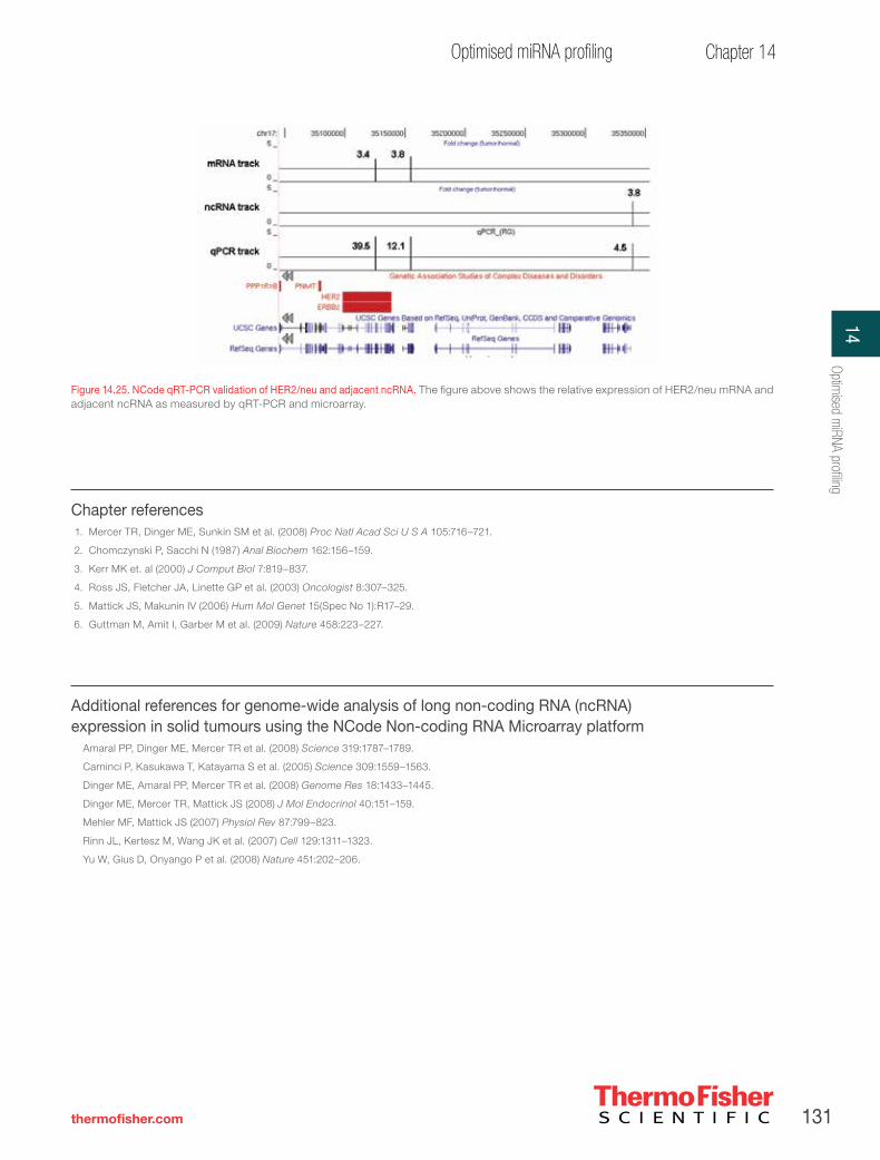
131
Chapter 14Optimised miRNA profiling
thermofisher.com
Chapter references1. Mercer TR, Dinger ME, Sunkin SM et al. (2008) Proc Natl Acad Sci U S A 105:716–721.
2. Chomczynski P, Sacchi N (1987) Anal Biochem 162:156–159.
3. Kerr MK et. al (2000) J Comput Biol 7:819–837.
4. Ross JS, Fletcher JA, Linette GP et al. (2003) Oncologist 8:307–325.
5. Mattick JS, Makunin IV (2006) Hum Mol Genet 15(Spec No 1):R17–29.
6. Guttman M, Amit I, Garber M et al. (2009) Nature 458:223–227.
Additional references for genome-wide analysis of long non-coding RNA (ncRNA) expression in solid tumours using the NCode Non-coding RNA Microarray platform
Amaral PP, Dinger ME, Mercer TR et al. (2008) Science 319:1787–1789.
Carninci P, Kasukawa T, Katayama S et al. (2005) Science 309:1559–1563.
Dinger ME, Amaral PP, Mercer TR et al. (2008) Genome Res 18:1433–1445.
Dinger ME, Mercer TR, Mattick JS (2008) J Mol Endocrinol 40:151–159.
Mehler MF, Mattick JS (2007) Physiol Rev 87:799–823.
Rinn JL, Kertesz M, Wang JK et al. (2007) Cell 129:1311–1323.
Yu W, Gius D, Onyango P et al. (2008) Nature 451:202–206.
Figure 14.25. NCode qRT-PCR validation of HER2/neu and adjacent ncRNA. The figure above shows the relative expression of HER2/neu mRNA and adjacent ncRNA as measured by qRT-PCR and microarray.
14Optim
ised miRNA profiling


thermofisher.com
Section III—miRNA Profiling
Chapter 15—Introduction to DNA methylation . . . . . . . . . . . . . . 134
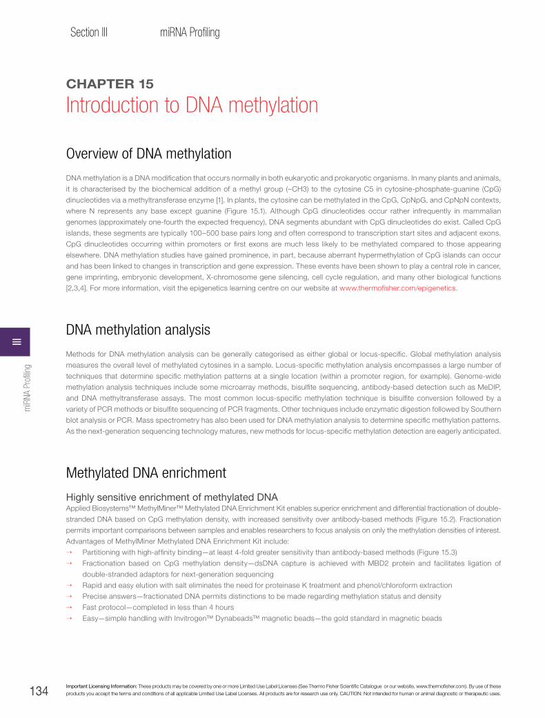
134 Important Licensing Information: These products may be covered by one or more Limited Use Label Licenses (See Thermo Fisher Scientific Catalogue or our website, www.thermofisher.com). By use of these products you accept the terms and conditions of all applicable Limited Use Label Licenses. All products are for research use only. CAUTION: Not intended for human or animal diagnostic or therapeutic uses.
Section III miRNA Profiling
CHAPTER 15
Introduction to DNA methylation
Overview of DNA methylation
DNA methylation is a DNA modification that occurs normally in both eukaryotic and prokaryotic organisms. In many plants and animals,
it is characterised by the biochemical addition of a methyl group (–CH3) to the cytosine C5 in cytosine-phosphate-guanine (CpG)
dinucleotides via a methyltransferase enzyme [1]. In plants, the cytosine can be methylated in the CpG, CpNpG, and CpNpN contexts,
where N represents any base except guanine (Figure 15.1). Although CpG dinucleotides occur rather infrequently in mammalian
genomes (approximately one-fourth the expected frequency), DNA segments abundant with CpG dinucleotides do exist. Called CpG
islands, these segments are typically 100–500 base pairs long and often correspond to transcription start sites and adjacent exons.
CpG dinucleotides occurring within promoters or first exons are much less likely to be methylated compared to those appearing
elsewhere. DNA methylation studies have gained prominence, in part, because aberrant hypermethylation of CpG islands can occur
and has been linked to changes in transcription and gene expression. These events have been shown to play a central role in cancer,
gene imprinting, embryonic development, X-chromosome gene silencing, cell cycle regulation, and many other biological functions
[2,3,4]. For more information, visit the epigenetics learning centre on our website at www.thermofisher.com/epigenetics.
DNA methylation analysis
Methods for DNA methylation analysis can be generally categorised as either global or locus-specific. Global methylation analysis
measures the overall level of methylated cytosines in a sample. Locus-specific methylation analysis encompasses a large number of
techniques that determine specific methylation patterns at a single location (within a promoter region, for example). Genome-wide
methylation analysis techniques include some microarray methods, bisulfite sequencing, antibody-based detection such as MeDIP,
and DNA methyltransferase assays. The most common locus-specific methylation technique is bisulfite conversion followed by a
variety of PCR methods or bisulfite sequencing of PCR fragments. Other techniques include enzymatic digestion followed by Southern
blot analysis or PCR. Mass spectrometry has also been used for DNA methylation analysis to determine specific methylation patterns.
As the next-generation sequencing technology matures, new methods for locus-specific methylation detection are eagerly anticipated.
Methylated DNA enrichment
Highly sensitive enrichment of methylated DNAApplied Biosystems™ MethylMiner™ Methylated DNA Enrichment Kit enables superior enrichment and differential fractionation of double-
stranded DNA based on CpG methylation density, with increased sensitivity over antibody-based methods (Figure 15.2). Fractionation
permits important comparisons between samples and enables researchers to focus analysis on only the methylation densities of interest.
Advantages of MethylMiner Methylated DNA Enrichment Kit include:
→ Partitioning with high-affinity binding—at least 4-fold greater sensitivity than antibody-based methods (Figure 15.3)
→ Fractionation based on CpG methylation density—dsDNA capture is achieved with MBD2 protein and facilitates ligation of
double-stranded adaptors for next-generation sequencing
→ Rapid and easy elution with salt eliminates the need for proteinase K treatment and phenol/chloroform extraction
→ Precise answers—fractionated DNA permits distinctions to be made regarding methylation status and density
→ Fast protocol—completed in less than 4 hours
→ Easy—simple handling with Invitrogen™ Dynabeads™ magnetic beads—the gold standard in magnetic beads
miR
NA P
rofili
ngIII
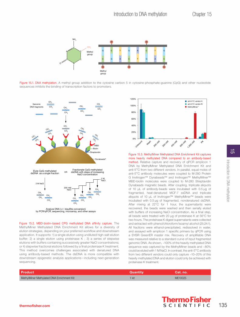
135
Chapter 15Introduction to DNA methylation
thermofisher.com
Product Quantity Cat. no.
MethylMiner Methylated DNA Enrichment Kit 1 kit ME10025
Figure 15.3. MethylMiner Methylated DNA Enrichment Kit captures more heavily methylated DNA compared to an antibody-based method. Relative capture and recovery of qPCR amplicon 1 DNA by MethylMiner Methylated DNA Enrichment Kit and anti-5mC from two different vendors. In parallel, equal moles of anti-5mC antibody molecules were coupled to M-280 Protein G Invitrogen™ Dynabeads™ and Invitrogen™ MethylMiner™ MBD-biotin molecules were coupled to M-280 Streptavidin Dynabeads magnetic beads. After coupling, triplicate aliquots of 10 µL of antibody-beads were incubated with 0.5 µg of fragmented, heat-denatured MCF-7 ssDNA and triplicate aliquots of 10 µL of Invitrogen™ MethylMiner™ beads were incubated with 0.5 µg of fragmented, nondenatured dsDNA. After mixing at 22°C for 1 hour, the supernatants were recovered, the beads were washed and then serially eluted with buffers of increasing NaCl concentration. As a final step, all beads were treated with 20 µg of proteinase K at 56°C for two hours. The proteinase K digest supernatants were collected and extracted with phenol/chloroform/isoamyl alcohol (25:24:1). All fractions were ethanol-precipitated, redissolved in water, and assayed with amplicon 1 specific primers by qPCR using a SYBR GreenER master mix. Recovery of amplifiable DNA was measured relative to a standard curve of input fragmented genomic DNA. As shown, ~100% of the heavily methylated DNA sequence was captured by the MethylMiner beads and ~80% could be eluted with 1 M NaCl. In contrast, the anti-5mC antibody from two different vendors could only capture ~10–20% of this heavily methylated DNA and elution could only be achieved with proteinase K treatment.
Figure 15.2. MBD-biotin–based CPG methylated DNA affinity capture. The MethylMiner Methylated DNA Enrichment Kit allows for a diversity of elution strategies, depending on your preferred workflow and downstream application. It supports: 1) a single elution using undiluted high-salt elution buffer; 2) a single elution using proteinase K ; 3) a series of stepwise elutions with buffers containing successively greater NaCl concentrations; or 4) stepwise fractional elutions followed by a final proteinase K treatment. This method overcomes challenges associated with denatured DNA using antibody-based methods. The dsDNA is more compatible with downstream epigenetic analysis applications—including next-generation sequencing.
GenomicDNA fragments
Elute CpG-methylateddsDNA as a single fraction
Fractionate CpG-methylateddsDNA with steps of increasing
NaCl concentration
Analyze DNA (+/– bisul�te conversion)by PCR/qPCR, sequencing, microarray, and other assays
2 M NaCl
OR
200 mMNaCl
CH3
CH3
CH3
CH3
CH3
CH3
CH3
CH3
CH3
CH3
CH3
CH3
2 MNaCl
CH3
CH3
CH3
CH3
CH 3
CH 3
CH 3
CH 3
Dynabeads®
Streptavidin
Biotin
MBD
CH3
CH3
CH3
CH3CH3
CH3
CH3
CH3
CH3
CH3
Unbou
ndW
ash PK
1,00
0 m
M
600
mM
450
mM
350
mM
200
mM
120%
100%
80%
60%
40%
20%
0%
anti-5mC vendor A
anti-5mC vendor B
MethylMiner™
Figure 15.1. DNA methylation. A methyl group addition to the cytosine carbon 5 in cytosine-phosphate-guanine (CpG) and other nucleotide sequences inhibits the binding of transcription factors to promoters.
N
C
CN
C C
O
NH2
CH3
Methyl group
Methyl group
G
C
C
G
T
A
G
C
A
T
C
G
T
A
G
C
M M
M M M
Introduction to DNA methylation
15
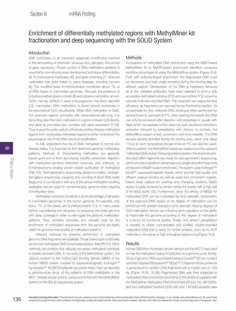
136 Important Licensing Information: These products may be covered by one or more Limited Use Label Licenses (See Thermo Fisher Scientific Catalogue or our website, www.thermofisher.com). By use of these products you accept the terms and conditions of all applicable Limited Use Label Licenses. All products are for research use only. CAUTION: Not intended for human or animal diagnostic or therapeutic uses.
Section III miRNA Profiling
Enrichment of differentially methylated regions with MethylMiner kit fractionation and deep sequencing with the SOLiD System
IntroductionDNA methylation is an important epigenetic modification involved
in the remodelling of chromatin structure and, ultimately, the control
of gene expression. Proper control of DNA methylation patterns is
essential for normal embryonic development and tissue differentiation
[5], X-chromosome inactivation [6], and gene imprinting [7]. Aberrant
methylation has been linked to many diseases, including cancers
[8]. The modified base 5-methylcytosine constitutes about 1% of
all DNA bases in mammalian genomes. Although the presence of
5-hydroxymethylcytosine in brain [8] and cytosine methylation at non-
CpGs, namely CpNpG in early embryogenesis, has been reported
[10], mammalian DNA methylation is found almost exclusively in
the symmetrical CpG dinucleotide. While DNA methylation in CpG-
rich promoter regions correlates with transcriptional silencing, it is
becoming clear that CpG methylation in regions of lower CpG density
and distal to promoters also correlate with gene expression [11,12].
Thus, it would be quite useful to effectively partition densely methylated
regions from moderately methylated regions to better understand the
physiological role of this DNA chemical modification
To fully understand the role of DNA methylation in normal and
disease states, it is important to first determine genomic methylation
patterns. Methods of characterising methylation are generally
based upon one of three techniques: bisulfite conversion, digestion
with methylation-sensitive restriction enzymes, and antibody- or
5-methylcytosine binding protein–based purification of methylated
DNA [13]. Next-generation sequencing platforms enable ultrahigh-
throughput sequencing, mapping, and counting of short DNA reads
(tags) and, in combination with any of the above methylation profiling
strategies, can be used for comprehensive, genome-wide mapping
of methylation sites.
Methylated cytosines constitute a small percentage of all bases
in mammalian genomes. In the human genome, for example, only
about 1% of the bases are 5-methylcytosine. It is in many cases
neither cost-effective nor necessary to sequence the entire genome
with deep coverage in order to interrogate the genomic methylation
patterns. Thus, sensitive, accurate, and versatile tools for the
enrichment of methylated sequences from the genome are highly
useful for genome-wide studies of methylation patterns.
Different methods for selective enrichment of methylated
genomic DNA fragments are available. Those based upon antibodies
are termed methylated DNA immunoprecipitation (MeDIP) [14]. Other
methods use proteins that naturally recognise methylated cytosines
in double-stranded DNA. In the case of the MethylMiner system, the
capture medium is the methyl-CpG binding domain (MBD) of the
human MBD2 protein coupled to superparamagnetic Invitrogen™
Dynabeads™ M-280 Streptavidin via a biotin linker. Here we describe
a genome-scale study of the patterns of DNA methylation in the
MCF-7 breast cancer cell line, using enrichment with the MethylMiner
system on the SOLiD sequencing system.
MethodsComparison of methylated DNA enrichment using the MBD-based
MethylMiner kit to MeDIP-based enrichment identified numerous
workflow advantages to using the MethylMiner system (Figure 15.4).
First, with antibody-based enrichment, the fragmented DNA must
be denatured and kept single-stranded during the binding step for
efficient capture. Denaturation of the DNA is necessary because
all of the validated antibodies have been selected to bind a fully
accessible methylated cytosine (5mC) and cannot bind 5mC occurring
naturally in double-stranded DNA. This constraint can reduce binding
efficiency, as fragments can reanneal during the binding reaction. To
compensate for this, antibody–DNA binding is often performed for
several hours to overnight at 4°C. After washing the beads, the DNA
can only be recovered after digestion with proteinase K, usually with
heat, which necessitates further clean-up, such as phenol/chloroform
extraction followed by precipitation with ethanol. In contrast, the
MethylMiner system is fast, convenient, and more versatile. The DNA
remains double-stranded during the binding step, which only takes
1 hour at room temperature (longer times at 4°C can also be used).
After incubation, the MethylMiner beads are washed and the captured
methylated DNA eluted. Following adapter ligation, the eluted double-
stranded DNA fragments are ready for next-generation sequencing,
which provide a significant advantage over single-stranded fragments
produced in MeDIP-based enrichment. The system uses Invitrogen™
Dynal™ superparamagnetic beads, which provide high-quality and
efficient capture kinetics as well as rapid and convenient magnet-
based bead capture for washing and changing solutions. Batch
elution is easily achieved by simply mixing the beads with a high-salt
(2 M NaCl) buffer [15]. Furthermore, since the affinity of MBD2 for
methylated DNA can be modulated by ionic strength, fractionation
of the captured DNA based on its degree of methylation can be
performed with graded changes in ionic strength. Varying degrees of
CpG methylation density can influence gene regulation so the ability
to fractionate the genome according to the degree of methylation
is a bonus for functional studies. Finally, only ethanol precipitation
is needed to obtain concentrated and purified double-stranded
methylated DNA that is ready for further analysis, such as by PCR
methods or microarray or high-throughput sequencing (Figure 15.4).
ResultsHuman DNA from the breast cancer–derived cell line MCF-7 was used
to map the methylation status of CpG sites on a genome scale. Briefly,
50 μg of genomic DNA was sheared using a Covaris™ S2 non-contact
sonicator (Applied Biosystems™ SOLiD™ 3 fragment library protocol)
to generate short random DNA fragments with a median size of ~150
bp (Figure 15.5A, 15.5B). Fragmented DNA was then subjected to
methylated DNA enrichment according to the protocol supplied with
the MethylMiner Methylated DNA Enrichment Kit (Cat. No. ME10025),
and two methylated fractions (500 mM and 1 M NaCl eluates) were
miR
NA P
rofili
ngIII
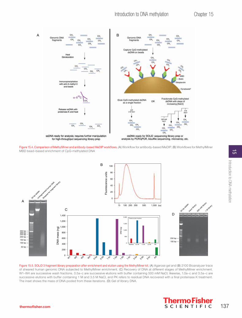
137
Chapter 15Introduction to DNA methylation
thermofisher.com
Figure 15.4. Comparison of MethylMiner and antibody-based MeDIP workflows. (A) Workflow for antibody-based MeDIP. (B) Workflows for MethylMiner MBD bead–based enrichment of CpG-methylated DNA
Figure 15.5. SOLiD 3 fragment library preparation after enrichment and elution using the MethylMiner kit. (A) Agarose gel and (B) 2100 Bioanalyzer trace of sheared human genomic DNA subjected to MethylMiner enrichment. (C) Recovery of DNA at different stages of MethylMiner enrichment. W1–W4 are successive wash fractions. 0.5a–c are successive elutions with buffer containing 500 mM NaCl; likewise, 1.0a–c and 3.5a–c are successive elutions with buffer containing 1 M and 3.5 M NaCl, and PK refers to residual DNA recovered with a final proteinase K treatment. The inset shows the mass of DNA pooled from these iterations. (D) Gel of library DNA.
50 b
p ladder
Unfra
gmen
ted h
uman
DNA
Shear
ed fo
r fra
gmen
t libra
ry
350 bp —300 bp —250 bp —200 bp —
150 bp —
100 bp —
50 bp —
15 300200100 1,500500 [bp]
Fluo
resc
ence
uni
ts
0
20
40
60
80
100
1,400
1,200
1,000
800
600
400
200
0W
10.
5b 1.0aW
4W
21.
0c3.
5b PK0.
5a1.
5cW3
1.0b 3.
5a3.
5c
DN
A m
ass
(ng)
DN
A (n
g)
PK3.5 M1.0 M0.5 MWashes0
500
1,000
1,500
2,000Pooled elution fractions
50 b
p ladder
Input
libra
ry
500
mM
libra
ry
1 M
libra
ry
250 bp —
150 bp —
A
B
CD
A B
Introduction to DNA methylation
15
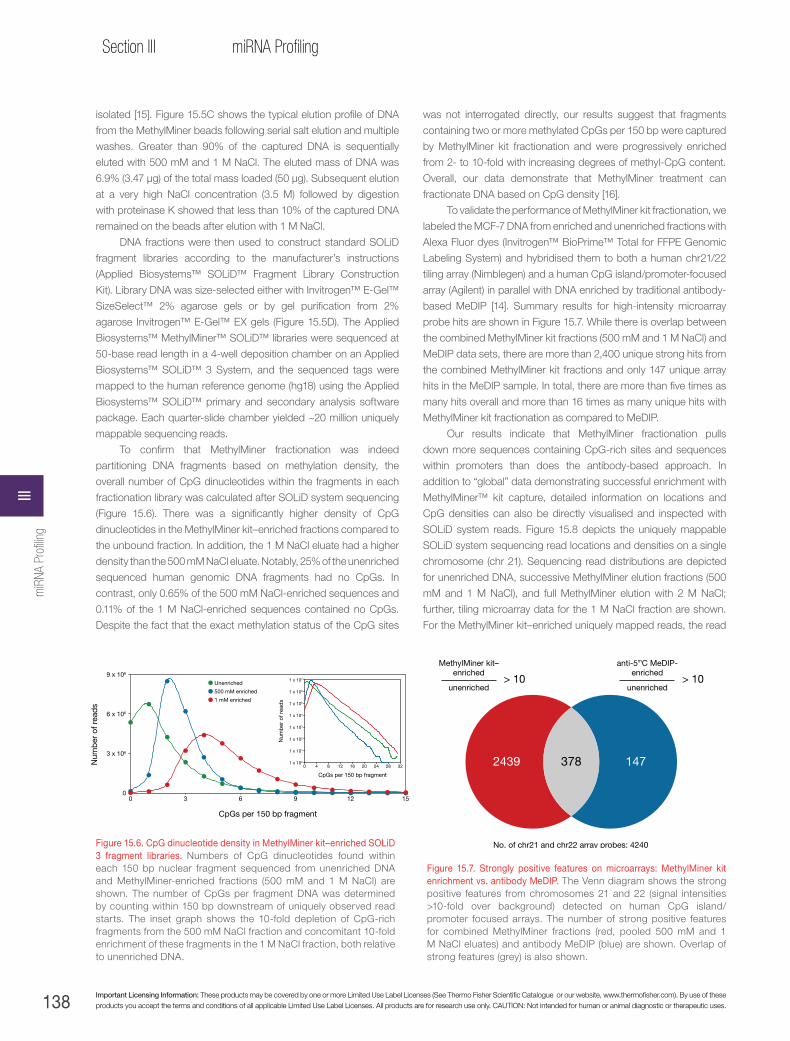
138 Important Licensing Information: These products may be covered by one or more Limited Use Label Licenses (See Thermo Fisher Scientific Catalogue or our website, www.thermofisher.com). By use of these products you accept the terms and conditions of all applicable Limited Use Label Licenses. All products are for research use only. CAUTION: Not intended for human or animal diagnostic or therapeutic uses.
Section III miRNA Profiling
isolated [15]. Figure 15.5C shows the typical elution profile of DNA
from the MethylMiner beads following serial salt elution and multiple
washes. Greater than 90% of the captured DNA is sequentially
eluted with 500 mM and 1 M NaCl. The eluted mass of DNA was
6.9% (3.47 μg) of the total mass loaded (50 μg). Subsequent elution
at a very high NaCl concentration (3.5 M) followed by digestion
with proteinase K showed that less than 10% of the captured DNA
remained on the beads after elution with 1 M NaCl.
DNA fractions were then used to construct standard SOLiD
fragment libraries according to the manufacturer’s instructions
(Applied Biosystems™ SOLiD™ Fragment Library Construction
Kit). Library DNA was size-selected either with Invitrogen™ E-Gel™
SizeSelect™ 2% agarose gels or by gel purification from 2%
agarose Invitrogen™ E-Gel™ EX gels (Figure 15.5D). The Applied
Biosystems™ MethylMiner™ SOLiD™ libraries were sequenced at
50-base read length in a 4-well deposition chamber on an Applied
Biosystems™ SOLiD™ 3 System, and the sequenced tags were
mapped to the human reference genome (hg18) using the Applied
Biosystems™ SOLiD™ primary and secondary analysis software
package. Each quarter-slide chamber yielded ~20 million uniquely
mappable sequencing reads.
To confirm that MethylMiner fractionation was indeed
partitioning DNA fragments based on methylation density, the
overall number of CpG dinucleotides within the fragments in each
fractionation library was calculated after SOLiD system sequencing
(Figure 15.6). There was a significantly higher density of CpG
dinucleotides in the MethylMiner kit–enriched fractions compared to
the unbound fraction. In addition, the 1 M NaCl eluate had a higher
density than the 500 mM NaCl eluate. Notably, 25% of the unenriched
sequenced human genomic DNA fragments had no CpGs. In
contrast, only 0.65% of the 500 mM NaCl-enriched sequences and
0.11% of the 1 M NaCl-enriched sequences contained no CpGs.
Despite the fact that the exact methylation status of the CpG sites
was not interrogated directly, our results suggest that fragments
containing two or more methylated CpGs per 150 bp were captured
by MethylMiner kit fractionation and were progressively enriched
from 2- to 10-fold with increasing degrees of methyl-CpG content.
Overall, our data demonstrate that MethylMiner treatment can
fractionate DNA based on CpG density [16].
To validate the performance of MethylMiner kit fractionation, we
labeled the MCF-7 DNA from enriched and unenriched fractions with
Alexa Fluor dyes (Invitrogen™ BioPrime™ Total for FFPE Genomic
Labeling System) and hybridised them to both a human chr21/22
tiling array (Nimblegen) and a human CpG island/promoter-focused
array (Agilent) in parallel with DNA enriched by traditional antibody-
based MeDIP [14]. Summary results for high-intensity microarray
probe hits are shown in Figure 15.7. While there is overlap between
the combined MethylMiner kit fractions (500 mM and 1 M NaCl) and
MeDIP data sets, there are more than 2,400 unique strong hits from
the combined MethylMiner kit fractions and only 147 unique array
hits in the MeDIP sample. In total, there are more than five times as
many hits overall and more than 16 times as many unique hits with
MethylMiner kit fractionation as compared to MeDIP.
Our results indicate that MethylMiner fractionation pulls
down more sequences containing CpG-rich sites and sequences
within promoters than does the antibody-based approach. In
addition to “global” data demonstrating successful enrichment with
MethylMiner™ kit capture, detailed information on locations and
CpG densities can also be directly visualised and inspected with
SOLiD system reads. Figure 15.8 depicts the uniquely mappable
SOLiD system sequencing read locations and densities on a single
chromosome (chr 21). Sequencing read distributions are depicted
for unenriched DNA, successive MethylMiner elution fractions (500
mM and 1 M NaCl), and full MethylMiner elution with 2 M NaCl;
further, tiling microarray data for the 1 M NaCl fraction are shown.
For the MethylMiner kit–enriched uniquely mapped reads, the read
Figure 15.6. CpG dinucleotide density in MethylMiner kit–enriched SOLiD 3 fragment libraries. Numbers of CpG dinucleotides found within each 150 bp nuclear fragment sequenced from unenriched DNA and MethylMiner-enriched fractions (500 mM and 1 M NaCl) are shown. The number of CpGs per fragment DNA was determined by counting within 150 bp downstream of uniquely observed read starts. The inset graph shows the 10-fold depletion of CpG-rich fragments from the 500 mM NaCl fraction and concomitant 10-fold enrichment of these fragments in the 1 M NaCl fraction, both relative to unenriched DNA.
Figure 15.7. Strongly positive features on microarrays: MethylMiner kit enrichment vs. antibody MeDIP. The Venn diagram shows the strong positive features from chromosomes 21 and 22 (signal intensities >10-fold over background) detected on human CpG island/promoter focused arrays. The number of strong positive features for combined MethylMiner fractions (red, pooled 500 mM and 1 M NaCl eluates) and antibody MeDIP (blue) are shown. Overlap of strong features (grey) is also shown.
2439 378 147
No. of chr21 and chr22 array probes: 4240
MethylMiner kit–enriched
unenriched> 10
anti-5mC MeDIP-enriched
unenriched> 10
0
3 x 10⁶
6 x 10⁶
9 x 10⁶
0 3 6 9 12 15
CpGs per 150 bp fragment
Num
ber
of r
ead
s
Unenriched
500 mM enriched
1 mM enriched
1 x 100
1 x 101
1 x 10²
1 x 10³
1 x 10⁴
1 x 10⁵
1 x 10⁶
1 x 10⁷
0 4 8 12 16 20 24 28 32
CpGs per 150 bp fragment
Num
ber
of r
ead
s
miR
NA P
rofili
ngIII

139
Chapter 15Introduction to DNA methylation
thermofisher.com
Figure 15.8. MethylMiner kit enrichment and SOLiD system sequencing across chromosome 21. Tracks illustrating sequencing reads from unenriched MCF-7 DNA, MethylMiner kit–enriched 500 mM and 1 M NaCl elution fractions, and full MethylMiner kit enrichment with 2 M NaCl elution are shown. Sequencing read depth of coverage for the enriched fractions is shown in red relative to the more uniform low coverage obtained from unenriched DNA shown in blue. Positive features for the MethylMiner 1 M NaCl-enriched fraction on a tiling array are also shown. Locations of annotated genes and CpG islands are indicated. Zoomed-in views of 50 kb and 1 kb regions are discussed in the text.
Introduction to DNA methylation
15

140 Important Licensing Information: These products may be covered by one or more Limited Use Label Licenses (See Thermo Fisher Scientific Catalogue or our website, www.thermofisher.com). By use of these products you accept the terms and conditions of all applicable Limited Use Label Licenses. All products are for research use only. CAUTION: Not intended for human or animal diagnostic or therapeutic uses.
Section III miRNA Profiling
coverage without enrichment is superimposed on the enriched
read coverage to illustrate the relative enrichment obtained for
equivalent amounts of sequencing time and expense; each
sequencing reaction comprised ~20 million uniquely mappable
reads from one chamber of a 4-well slide. The annotated genes
and CpG islands are also shown for reference. The unenriched
track suggests that there has been amplification of chromosome
21 DNA in this validated cancer cell line, and the enriched tracks
indicate that the distal end of the long arm of the chromosome
is densely methylated. This is not surprising since widespread
methylation of large chromosomal segments has been seen
in other cancer genome studies and since the vast majority of
annotated genes lie within this region of chromosome 21 [17,18].
At this high level, good concordance can be seen between
the data sets, and the profiles of the serially eluted 1 M NaCl
fraction reads and the batch-eluted 2 M NaCl reads are
remarkably similar, indicating good reproducibility of sequence
capture and significant sequence overlap between these two
different elution schemes. The greater depth of coverage seen
in the 1 M NaCl fraction attests to the decomplexing power of
fractionation. Methylated sequences that resist elution with 500
mM NaCl are better enriched in a subsequent 1 M NaCl elution
fraction than in the more complex sample that is eluted as a
whole with 2 M NaCl.
For a more detailed view, the analysis focused on a 50
kb region in chromosome 21 (middle of Figure 15.7), and at
this level of resolution, a broad peak of enrichment that spans
the neighbouring genes PFKL and C21orf2 can be seen in
the SOLiD™ system data from the 1 M NaCl elution fraction.
Closer inspection of the 1 kb region that encompasses one of
the PFKL gene’s intronic CpG islands clearly demonstrates the
enrichment in this fraction (lower portion of Figure 15.8). The
average depth of coverage is less than 1 in both the unenriched
and 500 mM NaCl elution fractions; in contrast, coverage depth
ranges from 5 to 15 in the 1 M NaCl elution fraction. Bisulfite
cloning and sequencing of this CpG island verified that its CpGs
are 94% methylated (Figure 15.8, bottom row). Notably, this
CpG island failed to be identified as methylated in our MeDIP-
microarray experiments. To further corroborate the MethylMiner
SOLiD system data with the methylation status of the genomic
DNA, we focused on a 200 kb region harbouring the ADAMTS1
and ADAMTS5 genes on chromosome 21 (Figure 15.9). The
differential enrichment between the MethylMiner fractions is
evident at this localised view. Sequences spanning the body
and the upstream promoter region of the ADAMTS1 gene are
exclusively enriched in the 500 mM NaCl fraction. With respect
to sequences covering the Methylated CpG Unmethylated
CpG island in the promoter of the ADAMTS5 gene, however,
there is preferential enrichment in the 1 M NaCl fraction (Figure
15.8). We further carried out bisulfite cloning and sequencing
across a portion of the CpG island in the ADAMTS5 promoter
to determine the exact methylation pattern, since this locus,
like the PFKL CpG island shown in Figure 15.8, had also been
identified in our microarray experiments as significantly enriched
(>10-fold) with MethylMiner treatment but not captured by
MeDIP. As shown in Figure 15.8, the bisulfite-sequenced region
shows an intermediate degree (57%) of CpG methylation.
In all other loci tested (5 regions that were similarly detected
on microarrays with MethylMiner kit but missed with MeDIP),
bisulfite sequencing has confirmed the presence of >80% CpG
methylation (data not shown). These results further indicate that
MethylMiner™ kit enrichment permits the isolation of fractions
of genomic DNA that harbour differing degrees of methylation.
This capacity to capture moderately methylated regions, and
direct compatibility with SOLiD system sequencing, are notable
advantages over antibody-based methods.
ConclusionsWhen it comes to whole-genome methylation analysis,
MethylMiner enrichment provides several significant advantages
over traditional antibody-based enrichment. The DNA binding
step is rapid, efficient, and more sensitive to low levels of CpG
methylation with the biotinylated MBD protein in MethylMiner
kit fractionation. In addition, the incorporation of Dynal
superparamagnetic beads facilitates effective, high-quality
capture kinetics. Furthermore, with the MethylMiner kit method,
genomic DNA can be fractionated according to its methylation
density. Finally, the capacity to capture and release double-
stranded DNA greatly expedites library construction for current
generation high-throughput sequencing. The multiplexing
capabilities of SOLiD system sequencing permits bar coding
of numerous samples that can be sequenced simultaneously.
Moreover, samples can be further divided into distinct fractions
according to their methylation density with MethylMiner
kit fractionation. In summary, MethylMiner kit fractionation
combined with SOLiD system sequencing is a seamless, easy
workflow that enables robust enrichment and deep coverage
across focal areas for cost-effective sequencing of methylated
sequences.
Product Quantity Cat. no.
MethylMiner Methylated DNA Enrichment Kit 1 kit ME10025
miR
NA P
rofili
ngIII

141
Chapter 15Introduction to DNA methylation
thermofisher.com
Figure 15.9. Annotated view of a 200 kb region of chromosome 21. Tracks of SOLiD system reads from unenriched and MethylMiner kit–enriched 500 mM and 1 M NaCl fractions. For MethylMiner kit–enriched sequences, the read distribution from an unenriched sequence data set of the same size has been subtracted to highlight regions of enrichment. Locations of annotated genes and CpG islands are also shown. Results of bisulfite sequencing of a small segment in the promoter CpG island of ADAMTS5 are also shown. Open circles: nonmethylated CpGs; filled circles: methylated CpGs.
Methylated CpG
Unmethylated CpG Introduction to DNA methylation
15

142 Important Licensing Information: These products may be covered by one or more Limited Use Label Licenses (See Thermo Fisher Scientific Catalogue or our website, www.thermofisher.com). By use of these products you accept the terms and conditions of all applicable Limited Use Label Licenses. All products are for research use only. CAUTION: Not intended for human or animal diagnostic or therapeutic uses.
Section III miRNA Profiling
Sam
ple
pre
par
atio
n
Genomic DNA isolation and puri�cationPureLink Genomic DNA Mini Kit
Bisul�te conversionMethylCode Bisul�te Conversion Kit
Amplifying bisul�te-modi�ed DNAPlatinum Taq Polymerase
Det
ectio
n CloningTOPO cloning kits for sequencing
Plasmid puri�cation (optional)PureLink Quick Plasmid Miniprep Kit
Standard DNA sequencing
Bis
ul�t
e co
nver
sion
Methylation-speci�c PCRPrimers
Figure 15.11. Streamlined MethylCode method produces better results. The most commonly used technique for detecting methylation in specific regions of DNA is the sodium bisulfite conversion method.
Locus-specific DNA methylation analysis
Since the first published article on the sequencing of bisulfite-converted DNA in 1992 [19], the bisulfite conversion method has become
a preferred tool for methylation analysis. In this method, DNA is denatured and treated with sodium bisulfite, causing unmethylated
cytosines to be converted to uracils while methylated cytosines remain unchanged [19]. Converted DNA can be PCR-amplified and
analysed by DNA sequencing or restriction endonuclease digestion and the methylation patterns determined by comparison to
untreated DNA (Figure 15.10, Figure 15.11).
Genome-wide methylation
Methylation of cytosines in mammalian genomic DNA is now a recognised epigenetic phenomenon with wide-ranging implications in basic
gene regulation and medical research. In particular, changes in DNA methylation, including genome-wide losses and hypermethylation of
promoter regions, are strongly correlated with the onset and progression of cancer. One common strategy for monitoring the methylation
status of the genome, and particular loci within it, involves fragmenting the DNA, and then capturing the methylated fragments with an anti–
5-methyl cytosine antibody. The MeDIP (methylated DNA immunoprecipitation) assay has been used in conjunction with bisulfite sequencing
to determine the precise distribution of methylated cytosines in three human chromosomes and in the Arabidopsis genome. Variations on
this assay (MCIP and MIRA) have been described that utilise methyl-binding domain (MBD-family) proteins in lieu of an antibody.
Genomic DNA isolation and purification for methylation analysis
Successful results begin with effective genomic DNA isolation and purification. We offer several options for genomic DNA isolation:
→ Invitrogen™ PureLink™ reagents—silica spin-column technology
→ Invitrogen™ ChargeSwitch™- magnetic bead based purification, amenable to high-throughput isolation
Visit www.thermofisher.com/oligos for information on primer design software to help with low-complexity DNA.
Original DNA with methylated CmpG
DNA sequencing after CT conversion
151413121110987654321
CCGTCACTCGCmGTTG
TTGTTATTTGCGTTG
Figure 15.10. Complete conversion of unmethylated cytosines into uracils after bisulfite treatment. DNA with methylated CpG (position 5) was processed using the Applied Biosystems™ MethylCode™ Bisulfite Conversion Kit. The recovered DNA was amplified by PCR, cloned, and sequenced. Following bisulfite treatment, the methylated cytosine at position 5 remained intact while the unmethylated cytosines (positions 7, 9, 11, 14, and 15) were converted into uracils and detected as thymines following PCR.
miR
NA P
rofili
ngIII

143
Chapter 15Introduction to DNA methylation
thermofisher.com
PureLink reagents—reliable isolation of genomic DNA
Invitrogen™ PureLink™ genomic DNA isolation reagents enable high-yield, high-purity DNA extractions from a wide variety of sample
types, including blood, tissue, cells, bacteria, swabs, and blood spots, in a familiar silica spin-column or semi-skirted 96-well plate
format. Genomic DNA Kits allow you to:
→ Obtain higher gDNA yields and purity, with optimised spin-column membrane and buffer formulations
→ Save time and money—use with a large range of sample sizes (e.g., 100 μL to 1 mL blood); 96-well versions do not require special
equipment
→ Increase flexibility—one kit works with a variety of sample types
Product Quantity Cat. no.
PureLink Genomic DNA Mini Kit 10 preps K1820-00
PureLink Genomic DNA Mini Kit 50 preps K1820-01
PureLink Genomic DNA Mini Kit 250 preps K1820-02
PureLink 96 Genomic DNA Kit 4 x 96 preps K1821-04
PureLink Genomic Digestion Buffer 70 mL K1823-01
PureLink Genomic Lysis/Binding Buffer 80 mL K1823-02
PureLink Genomic Wash Buffer I 100 mL K1823-03
PureLink Genomic Wash Buffer 2 75 mL K1823-04
PureLink Genomic Elution Buffer 160 mL K1823-05
PureLink Pro 96 Genomic DNA Purification Kit 4 x 96 preps K1821-04A
Invitrogen™ EveryPrep™ Universal Vacuum Manifold 1 each K2111-01
PureLink Genomic Plant DNA Purification Kit 50 preps K1830-01
Introduction to DNA methylation
15

144 Important Licensing Information: These products may be covered by one or more Limited Use Label Licenses (See Thermo Fisher Scientific Catalogue or our website, www.thermofisher.com). By use of these products you accept the terms and conditions of all applicable Limited Use Label Licenses. All products are for research use only. CAUTION: Not intended for human or animal diagnostic or therapeutic uses.
Section III miRNA Profiling
Figure 15.12. Effects of bisulfite treatment. Unmethylated cytosines are converted to uracils, while methylated cytosines remain unchanged.
Original DNA with methylated CpG: G T T G Cm G C T C A C T G C C Same DNA after bisul�te treatement: G T T G Cm G U T U A U T G U U
Figure 15.13. Streamlined MethylCode™ bisulfite conversion protocol is complete in just 3 hr. A. Sample DNA is temperature-denatured and con-verted, and the converted DNA is precipitated using an in-column desulfonation technology and then eluted. B. The DNA can then be PCR amplified, cloned, sequenced, and analysed for methylation patterns.
TOPO
TOPO
TOPO®
cloningvector
PCR ProductCACC
Design conversion-speci�c primers and PCR amplify
Clone PCR product for sequencing
Sequence and analyse methylation status
Elute
Mix sample DNA and CT conversion reagent
Incubate at 98°C for 10 min, then 64°C for 2.5 hr
Clean up and desulfonate
PCR-ready DNA
A
B
Product Quantity Cat. no.
MethylCode™ Bisulfite Conversion Kit 50 rxns MECOV-50
MethylCode bisulfite treatment—fast and complete DNA conversion
The most commonly used technique for DNA methylation analysis consists of treating DNA with sodium bisulfite, which causes
unmethylated cytosines to be converted to uracils while methylated cytosines remain unchanged (Figure 15.12). The MethylCode™
Bisulfite Conversion Kit improves upon this approach, increasing conversion efficiency, while reducing the number of steps and
overall time required for results. The streamlined protocol (Figure 15.13) integrates the DNA denaturation and bisulfite conversion
processes into one convenient step, replacing chemical denaturation using sodium hydroxide with a simple temperature denaturation.
In addition, cumbersome postconversion DNA precipitation steps are eliminated by an innovative in-column desulfonation technology.
This minimises template degradation and DNA loss so that as little as 500 pg of starting material is required. Recovered DNA is
ready for PCR amplification and downstream analysis applications including restriction endonuclease digestion, sequencing, and
microarrays. Complete conversion of unmethylated cytosines, as well as DNA purification, can be achieved in less than 3 hours,
compared to traditional methods that require overnight incubation. The MethylCode™ Bisulfite Conversion Kit offers many benefits:
→ Complete conversion of unmethylated cytosines—allows you to eliminate false positives
→ Minimal template degradation and DNA loss during treatment and clean-up
→ High yield of bisulfite-converted DNA
→ Less time at the bench—integrated one-step DNA denaturation and bisulfite conversion completed in 3 hr
→ Simplified procedure—enables elimination of cumbersome DNA precipitation steps using novel in-column desulfonation
miR
NA P
rofili
ngIII

145
Chapter 15Introduction to DNA methylation
thermofisher.com
Detection of bisulfite-modified DNA
Detection of bisulfite-modified DNA can be facilitated through conventional PCR, methylation-specific PCR, and bisulfite genomic
sequencing after PCR amplification, with or without cloning.
High-fidelity reagents for amplifying bisulfite-treated DNA and methylation-specific PCR (MSP)*Following bisulfite conversion, the modified DNA template is distinguishable from the original template at methylated cytosines. This
results in a pool of DNA fragments with altered nucleotide sequences due to differential methylation status. The methylation-specific
PCR reaction involves amplifying the altered nucleotide sequences using methylation-specific primers for the CpG islands of interest
(Figure 15.14). Our PCR products are tailored to address the persistent challenges associated with amplifying bisulfite-modified DNA—
such as a small initial sample of highly fragmented DNA. Not all polymerases are effective at amplifying converted DNA, due to
variation in specificity and fidelity of the particular enzymes used. We have validated the use of Platinum Taq DNA polymerases for
amplification of bisulfite-converted DNA. Using the Platinum PCR kits, you can expect:
→ Faithful amplification based on reliable and consistent single-nucleotide discrimination
→ High sensitivity and high yield of bisulfite-converted DNA
→ Room-temperature reaction assembly
* Methylation-specific PCR may be covered by one or more U.S. patents—nos. 5,786,146; 6,017,704; 6,200,756, and 6,265,171, and patents based on foreign counterpart applications. No license or rights under these patents to perform methylation-specific PCR is conveyed expressly or by implication to the purchase of our product. Users of our products subject to this label license should determine whether they have all the appropriate licenses in place. Further, no warranty is provided that the use of these products will not infringe on the patents referred to above.
Product Quantity Cat. No.
Taq DNA Polymerase, native 100 units 18038-018
Taq DNA Polymerase, native 500 units 18038-042
Taq DNA Polymerase, recombinant 100 units 10342-053
Taq DNA Polymerase, recombinant 500 units 10342-020
Platinum Taq DNA Polymerase 100 rxns 10966-018
Platinum Taq DNA Polymerase 250 rxns 10966-026
Platinum Taq DNA Polymerase 500 rxns 10966-034
Estrogen receptor α (ERα) Unmethylated CpG Methylated CpG
MCF-7
MDA-MB-231
CpG 1 CpG 2 CpG 3 CpG 4 CpG 5 CpG 6 CpG 7 CpG 8 CpG 9 CpG 10 CpG 11 CpG 12 CpG 13 CpG 14 CpG 15 CpG 16
Figure 15.14. Methylation patterns of ERα as analysed using the MethylCode kit. Genomic DNA from MCF-7 and MDA-MB-231 cell lines was isolated and treated with the MethylCode Bisulfite Conversion Kit. Bisulfite-converted DNA was then used as template in PCR to amplify a portion on the estrogen receptor alpha (ERα) gene. Next, the PCR product was cloned into a sequencing vector and sequenced. Finally, the sequences were analysed to show a differential methylation pattern of the ERα gene between the MDA-MB-231 and MCF-7 cell lines (locus—X03635 6450 bp mRNA linear PRI 11-JUN-2003; definition— Homo sapiens mRNA for estrogen receptor; accession—X03635 M11457; version—X03635.1 GI:31233).
Introduction to DNA methylation
15
146 Important Licensing Information: These products may be covered by one or more Limited Use Label Licenses (See Thermo Fisher Scientific Catalogue or our website, www.thermofisher.com). By use of these products you accept the terms and conditions of all applicable Limited Use Label Licenses. All products are for research use only. CAUTION: Not intended for human or animal diagnostic or therapeutic uses.
Section III miRNA Profiling
Simplify your preparation for bisulfite genomic sequencing
Combine effective plasmid DNA purification with quick and efficient cloning. The Invitrogen™ PureLink™ Quick Plasmid Miniprep Kit
provides efficient and reliable purification of plasmid DNA for cloning with Invitrogen™ TOPO™ technology. TOPO technology presents
the fastest, most efficient way to clone PCR products for sequencing. The key to TOPO cloning is the enzyme DNA topoisomerase I,
which functions as both a restriction enzyme and a ligase that cleaves and rejoins DNA during replication. To harness the religating
activity of topoisomerase, we provide over 30 linearised vectors with topoisomerase I covalently bound to each 3’ phosphate. Typically,
after only 5 minutes at room temperature, ligation of DNA sequences with compatible ends ligations is complete and your DNA is ready
for transformation into E. coli.
Top-rated sequencing vectors
The Invitrogen™ TOPO™ TA Cloning Kits for Sequencing contain a vector with a minimised multiple cloning site that positions the T7
and T3 priming sites only 33 bp away from the PCR product insertion site. This allows you to sequence more of your insert and less of
the vector, saving you time and money. It is designed for fast and efficient results from Taq-amplified PCR products.
Product Quantity Cat. no.
TOPO TA Cloning Kit for Sequencing with One Shot™ MAX Efficiency™ DH5α-T1R E. coli 1 kit K4595-01
TOPO TA Cloning Kit for Sequencing with One Shot™ MAX Efficiency™ DH5α-T1R E. coli 40 rxns K4595-40
TOPO TA Cloning Kit for Sequencing (with pCR 4-TOPO) with One Shot™ TOP10 Chemically Competent E. coli and PureLink™ Quick Plasmid Miniprep Kit
20 preps K4575-02
TOPO TA Cloning Kit for Sequencing with One Shot™ TOP10 Chemically Competent E. coli 10 rxns K4575-J10
TOPO TA Cloning Kit for Sequencing with One Shot™ TOP10 Electrocomp™ E. coli 20 rxns K4580-01
TOPO TA Cloning Kit for Sequencing with One Shot™ TOP10 Electrocomp™ E. coli 40 rxns K4580-40
TOPO TA Cloning Kit for Sequencing 40 rxns K4575-40
TOPO TA Cloning Kit for Sequencing 20 rxns K4575-01
TOPO TA Cloning for Sequencing with One Shot™ Mach1™ T1 Phage-Resistant Chemically Competent E. coli 20 rxns K4530-20
TOPO TA Cloning Kit Dual Promoter (with pCR II-TOPO vector) with One Shot™ Mach1™ T1 Phage-Resistant Chemically Competent E. coli
20 rxns K4610-20
TOPO XL PCR Cloning Kit with One Shot™ Mach1™ T1 Phage-Resistant Chemically Competent E. coli 20 rxns K7030-20
TOPO TA Cloning Kit (with pCR 2.1-TOPO vector) with One Shot™ Mach1™ T1 Phage-Resistant Chemically Competent E. coli
20 rxns K4510-20
Chapter references
1. Adams RL (1995) Bioessays 17:139–145.
2. Ozanne SE et al. (2007) Nat Clin Pract Endocrinol Metab 3:539–546.
3. Dolinoy DC et al. (2007) Reprod Toxicol 23:297–307.
4. Verma M et al. (2006) Crit Rev Oncol Hematol 60:9–18.
5. Morgan HD, Santos F, Green K et al. (2005) Hum Mol Genet 14:R47–R58.
6. Reik W, Lewis A (2005) Nat Rev Genet 6(5):403–410.
7. Feinberg AP (2007) Nature 447(7143):433–440.
8. Jones PA, Baylin SB (2007) Cell 128(4):683–692.
9. Kriaucionis S, Heintz N (2009) Science 324(5929):929–930.
10. Ramsahoye BH, Biniszkiewicz D, Lyko F et al. (2000) Proc Natl Acad Sci U S A 97(10):5237–5242.
11. Schmidl C, Klug M, Boeld TJ et al. (2009) Genome Res 19(7):1165–1174.
12. Irizarry RA, Ladd-Acosta C, Wen B et al. (2009) Nat Genet 41(2):178–186.
13. Zilberman D, Henikoff S (2007) Development 134(22):3959–3965.
14. Weber M, Davies JJ, Wittig D et al. (2005) Nat Genet 37(8):853–862.
15. MethylMiner™ manual; follow links from bottom of the following page: www.thermofisher.com/methylminer.
16. MethylMiner™ for Methylated DNA Enrichment webinar: http://thermofisher.cnpg.com/Video/flatFiles/1080/index.aspx.
17. Clark S (2007) Hum Mol Genet 16:R88–R95.
18. Gardiner K, Horisberger M, Kraus J et al. (1990) EMBO J 9(1):25–34.
19. Frommer M et al. (1992) Proc Natl Acad Sci U S A 89:1827–1831.
miR
NA P
rofili
ngIII

147
Chapter 15Introduction to DNA methylation
thermofisher.com
Introduction to DNA methylation
15


thermofisher.com
Section IV—Histone Modifications and Chromatin Remodelling
Chapter 16—Histone modifications and chromatin remodelling . . . . . . . . . 150

150 Important Licensing Information: These products may be covered by one or more Limited Use Label Licenses (See Thermo Fisher Scientific Catalogue or our website, www.thermofisher.com). By use of these products you accept the terms and conditions of all applicable Limited Use Label Licenses. All products are for research use only. CAUTION: Not intended for human or animal diagnostic or therapeutic uses.
Section IV Histone Modifications and Chromatin Remodelling
CHAPTER 16
Histone modifications and chromatin remodellingDNA is compacted and organized within organised in the nuclei of eukaryotic cells. As the fundamental subunits of chromatin,
nucleosomes form a chain of ellipsoidal beads of histone proteins around which the DNA is wound. Four histone types (H2A, H2B, H3,
and H4) form the octamer centre of the nucleosome, with 147 nucleotide pairs wrapped 1¾ turns around the centre [1]. Each histone
has an amino-terminal tail region consisting of 25–40 amino acid residues that protrude beyond the nucleosome surface. H1 histone
plays a role in linking the nucleosome structures together to condense the chromatin.
Chromatin exists in many configurations and undergoes dynamic structural changes. These range from local changes necessary
for transcriptional regulation to global changes necessary for chromosome segregation. Several epigenetic mechanisms introduce
variation to chromatin structure, including covalent histone modifications, histone variant composition, DNA methylation, and non-
coding RNA. Changes to the chromatin structure, called chromatin remodelling, can facilitate gene transcription by loosening the
histone-DNA complex, allowing other proteins such as transcription factors to access the DNA. Alternatively, chromatin remodelling
where histones assume a more closed conformation, blocks transcription factor access to the DNA, resulting in loss of gene
expression. Relatively little is known about how remodeling factors change nucleosome structure and how different factors work
together to promote chromatin remodelling.
Chromatin remodelling is typically initiated by posttranslational modification of the amino acids that make up the histone proteins,
as well as through methylation of neighboring DNA (Figure 16.1).
ARTKQTARKSTGGKAPRKQLATKAARKSAPATGGVKKPH Histone H3 135 aa2 4 9 10 14 1718 23 26 27 28 36 79
S F R F K G G K G L G K G G A K R H R K V L R D N I Q G I T K PA I R R L A R Histone H4 102 aa1 3 5 8 12 16 20
SGRGKQGGKARAKAKSRSSRAGLQFPVGRVHRLLRKGNY Histone H2A 129 aa1 5 9 119
PEPAKSAPAPKKGSKKAVTKAQKKDSKKRKRSRKESYSV Histone H2B 125 aa5 12 14 120
Acetylation
Methylation(arginine)
Methylation(active lysine)
Methylation(repressive lysine)
Ubiquitination
Phosphorylation
Figure 16.1. Histone modifications and variants. Posttranslational modifications to histone amino-terminal tails include acetylation (lysine and arginine), phosphorylation (serine and threonine), ubiquitination (lysine), and sumoylation, which are associated with a wide variety of biological processes including transcriptional activation and repression, mitosis, and apoptotic regulation.
Hist
one
Mod
ificat
ions
and
Chr
omat
in R
emod
ellin
gIV
151
Chapter 16Histone modifications and chromatin remodelling
thermofisher.com
Investigating histone modifications and chromatin remodelling
Histones are subject to a wide variety of posttranslational modifications, including but not limited to lysine acetylation, lysine and
arginine methylation, serine phosphorylation, and lysine ubiquitination. These modifications occur primarily within the histones’
amino-terminal tails and are thought to affect chromosome function. Posttranslational modifications of histones create an epigenetic
mechanism for regulating a variety of normal and disease-related processes. Current research has focused on investigating the role of
epigenetics in transcription, cell proliferation, differentiation, senescence, apoptosis, and the DNA damage response.
MAGnify ChIP
To date, the most widely used and powerful method to identify regions of the genome associated with specific proteins is the
chromatin immunoprecipitation (ChIP) assay. The ChIP assay has been widely used to study both histone and nonhistone proteins,
such as transcription factors, within the context of the cell. Antibodies that recognise a protein of interest are used to determine the
relative association of that antigen in the context of chromatin at one or more loci in the genome.
In the ChIP assay, chromatin is sheared and immunoprecipitated with a highly specific antibody directed against a component
of the chromatin complex. Because transcription factors and other DNA-binding proteins have a weaker affinity for chromatin than
histones, a crosslinking step is incorporated before immunoprecipitation to avoid dissociation of nonhistone proteins from the
chromatin-binding site.
Histone modifications and chrom
atin remodelling
16
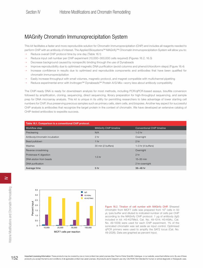
152 Important Licensing Information: These products may be covered by one or more Limited Use Label Licenses (See Thermo Fisher Scientific Catalogue or our website, www.thermofisher.com). By use of these products you accept the terms and conditions of all applicable Limited Use Label Licenses. All products are for research use only. CAUTION: Not intended for human or animal diagnostic or therapeutic uses.
Section IV Histone Modifications and Chromatin Remodelling
MAGnify Chromatin Immunoprecipitation System
This kit facilitates a faster and more reproducible solution for Chromatin Immunoprecipitation (ChIP) and includes all reagents needed to
perform ChIP with an antibody of interest. The Applied Biosystems™ MAGnify™ Chromatin Immunoprecipitation System will allow you to:
→ Reduce overall ChIP protocol time by one day (Table 16.1)
→ Reduce input cell number per ChIP experiment (10,000–300,000 cells required) (Figures 16.2, 16.3)
→ Decrease background caused by nonspecific binding through the use of Dynabeads
→ Improve reproducibility due to optimised magnetic DNA purification (avoid columns and phenol/chloroform steps) (Figure 16.4)
→ Increase confidence in results due to optimised and reproducible components and antibodies that have been qualified for
chromatin immunoprecipitation
→ Easily increase throughput with small volumes, magnetic protocol, and magnet compatible with multichannel pipetting
→ Reduce experimental error with Invitrogen™ Dynabeads™ Protein A/G Mix—worry less about antibody compatibility
The ChIP-ready DNA is ready for downstream analysis for most methods, including PCR/qPCR-based assays, bisuflite conversion
followed by amplification, cloning, sequencing, direct sequencing, library preparation for high-throughput sequencing, and sample
prep for DNA microarray analysis. This kit is unique in its utility for permitting researchers to take advantage of lower starting cell
numbers for ChIP, thus preserving precious samples such as primary cells, stem cells, and biopsies. Another key aspect for successful
ChIP analysis is antibodies that recognize the target protein in the context of chromatin. We have developed an extensive catalog of
CHiP-tested antibodies to expedite success.
Table 16.1. Comparison to a conventional ChIP protocol.
Workflow step MAGnify ChIP timeline Conventional ChIP timeline
Preclearing N/A 1–2 hr
Antibody/chromatin incubation 2 hr Overnight
Bead pulldown 1 hr 2 hr
Washes 30 min (2 buffers) 1–3 hr (4 buffers)
Reverse crosslinking
1.5 hr
Overnight
Proteinase K digestion 2 hr
DNA elution from beads 15–30 min
DNA purification 2 hr–overnight
Average time 5 hr 36–48 hr
Figure 16.2. Titration of cell number with MAGnify ChIP. Sheared chromatin from MCF7 cells was prepared from 106 cells in 50 μL lysis buffer and diluted to indicated number of cells per ChIP according to the MAGnify ChIP protocol. 1 μg of antibody (IgG included in kit: H3-K27Me3, Cat. No. 49-1014; H3-K9Ac, Cat. No. 49-1009) were used for each ChIP experiment. 1% of the sonicated chromatin was set aside as input control. Optimised qPCR primers were used to amplify the SAT2 locus (Cat. No. 49-2026). Data are graphed as percent input.
Per
cent
inp
ut
50,000 100,00025,00010,000
0.5
0
1.0
1.5
2.0
2.5
3.5
3.0
IgG
H3-K9Ac
H3-K27Me3
MCF7 cells per reaction
Hist
one
Mod
ificat
ions
and
Chr
omat
in R
emod
ellin
gIV

153
Chapter 16Histone modifications and chromatin remodelling
thermofisher.com
Per
cent
inp
ut
H3-K9Ac
MCF7
MDA-MB-231
H3-K27Me3IgG RNA Pol II H3-K9AcH3-K27Me3IgG RNA Pol II
0.5
0
1.0
1.5
2.5
3.5
4.5
2.0
3.0
4.0
RARb1 promoter ER-α promoter
Figure 16.3. 10,000 cell input with MAGnify ChIP. Sheared chromatin from MCF7 or MDA-MB231 cells was prepared from 106 cells in 50 μL lysis buffer and diluted to 10,000 cells per ChIP, according to the MAGnify ChIP protocol. 1 μg of antibody (IgG included in kit: H3-K27Me3, Cat. No. 49-1014; H3-K9Ac, Cat. No. 49-1009) or 3 μL RNA Pol II (Cat. No. 49-1033) were used for each ChIP experiment. 1% of the sonicated chromatin was set aside as input control. Optimised qPCR primers were used to amplify to different loci: RARb1 promoter (Cat. No. 49-2027) or ER-α promoter (Cat. No. 49-2028). Data are graphed as percent input.
Figure 16.4. MAGnify ChIP vs. conventional protocols. Sheared chromatin from 293Gt cells was prepared and 50,000 cells used for ChIP (MAGnify ChIP) and 106 cells per ChIP used for the conventional ChIP protocol. 1 μg of antibody (IgG included in kit: H3-K9Me3, Cat. No. 49-1008) was used for each ChIP experiment. 1% of the sonicated chromatin was set aside as input control. Optimised qPCR primers were used to amplify the SAT2 locus (Cat. No. 49-2026), a known target of heterochromatin-associated H3-K9Me3. Key elements of a conventional protocol use an overnight immunoprecipitation step of antibody and chromatin, followed by enrichment with Protein A sepharose beads and extensive washes with buffers containing variable salt concentrations. Reverse crosslinking was done overnight at 65°C and DNA purification performed by phenol/chloroform extraction.
Product Quantity Cat. no.
MAGnify Chromatin Immunoprecipitation System 1 kit 49-2024
5
0
10
15
20
K9Me3IgGIgG K9Me3
Per
cent
inp
ut S
AT2
locu
s
MAGnify™ ChiP
Conventional protocol
Histone modifications and chrom
atin remodelling
16
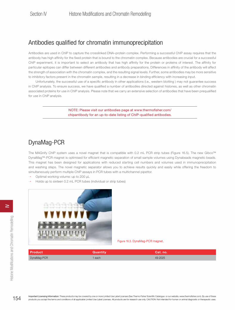
154 Important Licensing Information: These products may be covered by one or more Limited Use Label Licenses (See Thermo Fisher Scientific Catalogue or our website, www.thermofisher.com). By use of these products you accept the terms and conditions of all applicable Limited Use Label Licenses. All products are for research use only. CAUTION: Not intended for human or animal diagnostic or therapeutic uses.
Section IV Histone Modifications and Chromatin Remodelling
Antibodies qualified for chromatin immunoprecipitation
Antibodies are used in ChIP to capture the crosslinked DNA–protein complex. Performing a successful ChIP assay requires that the
antibody has high affinity for the fixed protein that is bound to the chromatin complex. Because antibodies are crucial for a successful
ChIP experiment, it is important to select an antibody that has high affinity for the protein or proteins of interest. The affinity for
particular epitopes can differ between different antibodies and antibody preparations. Differences in affinity of the antibody will affect
the strength of association with the chromatin complex, and the resulting signal levels. Further, some antibodies may be more sensitive
to inhibitory factors present in the chromatin sample, resulting in a decrease in binding efficiency with increasing input.
Unfortunately, the successful use of a specific antibody in other applications (i.e., western blotting ) may not guarantee success
in ChIP analysis. To ensure success, we have qualified a number of antibodies directed against histones, as well as other chromatin
associated proteins for use in ChIP analysis. Please note that we carry an extensive selection of antibodies that have been prequalified
for use in ChiP analysis.
NOTE: Please visit our antibodies page at www.thermofisher.com/chipantibody for an up-to-date listing of ChIP-qualified antibodies.
DynaMag-PCR
The MAGnify ChIP system uses a novel magnet that is compatible with 0.2 mL PCR strip tubes (Figure 16.5). The new Gibco™
DynaMag™-PCR magnet is optimised for efficient magnetic separation of small sample volumes using Dynabeads magnetic beads.
This magnet has been designed for applications with reduced starting cell numbers and volumes used in immunoprecipitation
and washing steps. The novel magnetic separator allows you to achieve results quickly and easily while offering the freedom to
simultaneously perform multiple ChIP assays in PCR tubes with a multichannel pipettor.
→ Optimal working volume: up to 200 µL
→ Holds up to sixteen 0.2 mL PCR tubes (individual or strip tubes)
Product Quantity Cat. no.
DynaMag-PCR 1 each 49-2025
Figure 16.5. DynaMag-PCR magnet.
Hist
one
Mod
ificat
ions
and
Chr
omat
in R
emod
ellin
gIV

155
Chapter 16Histone modifications and chromatin remodelling
thermofisher.com
Quantitative PCR (qPCR) of ChIP-ready DNA
Good primer design is critical for high-quality ChIP data. In general, primers should be 20 to 30 bases long with a Tm between 55° and
60°C. Most primers do not require purification or special treatment prior to PCR. Primers should be designed so that amplification
targets are 75 to 350 bp in length, as amplification efficiency is reduced substantionally with increasing length. In general, a final
primer concentration of 1 μM works well for most primer sets, but in some instances, improved product specificity may be obtained
by lowering the final primer concentration 5- to 10-fold.
High-quality primer pairs should result in ~1.9-fold amplification/cycle (this can be determined from quantitative analysis of raw
fluorescence data for each cycle, which is generally available on commercial instruments). Amplified material at the completion of the
PCR should contain only one product (as assayed on high-percentage agarose or polyacrylamide gels). Specificity information can
also be obtained by running dissociation curves on reactions following the conclusion of the qPCR run.
In general, individual samples should be run in triplicate. For each primer pair examined, the input DNA samples should be run
alongside the immunoprecipitated samples. Amplification efficiencies among different primer pairs vary slightly on a per-cycle basis,
and these slight variations in efficiency can translate into substantially different amounts of amplified material in the cycle range used
for analysis. Precise quantitation of relative binding cannot be accurately performed without a primer pair–specific input signal.
We have validated ChIP primer pairs for various promoter regions for validation of ChIP-qPCR. Primers were validated by testing
5-point dilutions of input DNA from MCF7 and PC3 cells, with primer efficiency and slope assessed for each pair.
Product Quantity Cat. no.
MAGnify SAT2 Primers 100 reactions 49-2026
MAGnify RARβ1 Primers 100 reactions 49-2027
MAGnify ERα Primers 100 reactions 49-2028
MAGnify c-Fos Primers 100 reactions 49-2029
Chapter reference
1. Luger K et al. (1997) Nature 389:251–260.
Histone modifications and chrom
atin remodelling
16

156
Section IIIRNAi and Epigenetics Sourcebook
Aacetylation . . . . . . . . . . . . . . . . . . . . . . . . . . . . . . . . . . . . . . . . . . . . . . . . . . . . . . . . . . . . . 151adenocarcinoma . . . . . . . . . . . . . . . . . . . . . . . . . . . . . . . . . . . . . . . . . . . . . . . . . . . . . . . . . . 121adenoviral RNAi vectors. . . . . . . . . . . . . . . . . . . . . . . . . . . . . . . . . . . . . . . . . . . . . . . . . . . . . . . 18adenovirus . . . . . . . . . . . . . . . . . . . . . . . . . . . . . . . . . . . . . . . . . . . . . . . . . . . . . . . . . . . 17, 22Alexa Fluor dyes . . . . . . . . . . . . . . . . . . . . . . . . . . . . . . . . . . . . . . . . . . . . . . 21–22, 46, 118, 120, 138Pre-miR miRNA Precursors . . . . . . . . . . . . . . . . . . . . . . . . . . . . . . . . . . . . . . . . . . . . . . . . . . 109, 111Silencer Select siRNA. See Silencer Select siRNAAnti-miR mimics and inhibitors . . . . . . . . . . . . . . . . . . . . . . . . . . . . . . . . . . . . . . . . . . . . . . . . . . . 109Anti-miR miRNA Inhibitors . . . . . . . . . . . . . . . . . . . . . . . . . . . . . . . . . . . . . . . . . . . . . . . . . . 109–111
Bbisulfite conversion . . . . . . . . . . . . . . . . . . . . . . . . . . . . . . . . . . . . . . . . . . . . . . . . . 136, 142, 144–145bisulfite sequencing . . . . . . . . . . . . . . . . . . . . . . . . . . . . . . . . . . . . . . . . . . . . . . . . . . . 134, 140–142BLOCK-iT Adenoviral RNAi Expression System . . . . . . . . . . . . . . . . . . . . . . . . . . . . . . . . . 18, 54, 68–69, 71, 72BLOCK-iT Alexa Fluor Red Fluorescent Control . . . . . . . . . . . . . . . . . . . . . . . . . . . . . . . . . . . . . . 7, 72, 79–81BLOCK-iT fluorescent controls . . . . . . . . . . . . . . . . . . . . . . . . . . . . . . . . . . . . . . . . . . . . . . . . . . . 21BLOCK-iT Fluorescent Oligos . . . . . . . . . . . . . . . . . . . . . . . . . . . . . . . . . . . . . . . . . . 12, 22, 75, 79–81, 85BLOCK-iT HiPerform Lentiviral Pol II miR RNAi Expression System . . . . . . . . . . . . . . . . . . . . . . . . . . . . . 58, 60, 64BLOCK-iT Inducible H1 Lentiviral RNAi System . . . . . . . . . . . . . . . . . . . . . . . . . . . . . . . . . . . . . . . 68–69, 72BLOCK-iT Inducible H1 RNAi Entry Vector Kit. . . . . . . . . . . . . . . . . . . . . . . . . . . . . . . . . . . . . . . . . . . . 54BLOCK-iT Lentiviral Pol II miR RNAi Expression System . . . . . . . . . . . . . . . . . . . . .18, 54–55, 57–58, 62, 64, 68–69, 72BLOCK-iT Lentiviral RNAi Systems . . . . . . . . . . . . . . . . . . . . . . . . . . . . . . . . . . . . . . . . . . . . . 18, 70, 72BLOCK-iT miR RNAi Select . . . . . . . . . . . . . . . . . . . . . . . . . . . . . . . . . . . . . . . . . . . . . . . . . 57, 64–65BLOCK-iT Pol II miR RNAi . . . . . . . . . . . . . . . . . . . . . . . . . . . . . . . . . . . . . . . . . . . . . . . . . . . 93, 95BLOCK-iT Pol II miR RNAi expression vectors . . . . . . . . . . . . . . . . . . . . . . . . . . . . . . . .53–54, 56–57, 61–65, 75BLOCK-iT Pol II miR RNAi vectors . . . . . . . . . . . . . . . . . . . . . . . . . . . . . . . . . . . . . . . . . . . . . . . 92–93BLOCK-iT Pol II miR Validated miRNA Control Vectors . . . . . . . . . . . . . . . . . . . . . . . . . . . . . . . . . . . . . 79, 85BLOCK-iT Pol II Validated miRNA Control Vectors . . . . . . . . . . . . . . . . . . . . . . . . . . . . . . . . . . . . . . . . . 64BLOCK-iT RNAi Adenoviral System . . . . . . . . . . . . . . . . . . . . . . . . . . . . . . . . . . . . . . . . . . . . . . . 18, 68BLOCK-iT RNAi Basic Control Kit . . . . . . . . . . . . . . . . . . . . . . . . . . . . . . . . . . . . . . . . . . . . . . . . . . 76BLOCK-iT RNAi Designer . . . . . . . . . . . . . . . . . . . . . . . . . . . . . . . . . . . . . . . . . . . . . . . . . 8, 45, 64–66BLOCK-iT RNAi Entry Vector Kits . . . . . . . . . . . . . . . . . . . . . . . . . . . . . . . . . . . . . . . . . . . . . . . . . . 66BLOCK-iT RNAi Express . . . . . . . . . . . . . . . . . . . . . . . . . . . . . . . . . . . . . . . . . . . . . . . . . . . . . . 28BLOCK-iT RNAi Express search engine. . . . . . . . . . . . . . . . . . . . . . . . . . . . . . . . . . . . . . . . . . . . . . . 64BLOCK-iT RNAi vectors . . . . . . . . . . . . . . . . . . . . . . . . . . . . . . . . . . . . . . . . . . . . . . . . . . . . . . 53BLOCK-iT shRNA vectors. . . . . . . . . . . . . . . . . . . . . . . . . . . . . . . . . . . . . . . . . . . . . . . . 53–54, 92, 94BLOCK-iT siRNA . . . . . . . . . . . . . . . . . . . . . . . . . . . . . . . . . . . . . . . . . . . . . . . . . . . . .22–23, 48–49BLOCK-iT Transfection Kit . . . . . . . . . . . . . . . . . . . . . . . . . . . . . . . . . . . . . . . . . . . . . . . . . 75, 79, 81BLOCK-iT Transfection Optimization Kit. . . . . . . . . . . . . . . . . . . . . . . . . . . . . . . . . . . . . . . . . . . . . 79, 84BLOCK-iT U6 RNAi Entry Vectors. . . . . . . . . . . . . . . . . . . . . . . . . . . . . . . . . . . . . . . . . . . . . . . . 54, 66
Ccancer . . . . . . . . . . . . . . . . . . . . . . . . . . . . . . . . . . . . 6, 30, 32, 100, 125, 126, 129–130, 134, 136, 140, 142cell cycle regulation . . . . . . . . . . . . . . . . . . . . . . . . . . . . . . . . . . . . . . . . . . . . . . . . . . . . . . . . . 134Cells-to-Ct Kits . . . . . . . . . . . . . . . . . . . . . . . . . . . . . . . . . . . . . . . . . . . . . . . . . . . . . . . . . 90, 113chromatin immunoprecipitation (ChIP). . . . . . . . . . . . . . . . . . . . . . . . . . . . . . . . . . . . . . . . . . . . . 151–154chromatin remodeling . . . . . . . . . . . . . . . . . . . . . . . . . . . . . . . . . . . . . . . . . . . . . . . . . . . . . 150–155cloning
TOPO technology . . . . . . . . . . . . . . . . . . . . . . . . . . . . . . . . . . . . . . . . . . . . . . . . . . . . . . . 146CpG dinucleotides . . . . . . . . . . . . . . . . . . . . . . . . . . . . . . . . . . . . . . . . . . . . . . 134, 136, 138–141, 145custom siRNA libraries . . . . . . . . . . . . . . . . . . . . . . . . . . . . . . . . . . . . . . . . . . . . . . . . . . . . . . . 51

157
Index
thermofisher.com
DDicer . . . . . . . . . . . . . . . . . . . . . . . . . . . . . . . . . . . . . . . . . . . . . . . . . . . . . . . . . . . . . . . . 4–6DNA methylation . . . . . . . . . . . . . . . . . . . . . . . . . . . . . . . . . . . . . . . . . . . . . . . . . . 134, 142, 144, 150DNA polymerases. . . . . . . . . . . . . . . . . . . . . . . . . . . . . . . . . . . . . . . . . . . . . . . . . . . . . . . . . . 145dsRNA . . . . . . . . . . . . . . . . . . . . . . . . . . . . . . . . . . . . . . . . . . . . . . . . . . . . . . . . . . . . . 15, 40dye swap. See loop design/dye swapDynaMag-PCR . . . . . . . . . . . . . . . . . . . . . . . . . . . . . . . . . . . . . . . . . . . . . . . . . . . . . . . . . . . 154
Eelectroporation . . . . . . . . . . . . . . . . . . . . . . . . . . . . . . . . . . . . . . . . . . . . . . . . . . . . . . . . . . . 15embryonic development . . . . . . . . . . . . . . . . . . . . . . . . . . . . . . . . . . . . . . . . . . . . . . . . . . . 134, 136epigenetics. See DNA methylation, chromatin remodeling, noncoding RNAs
FFastPrep-24 Instrument. . . . . . . . . . . . . . . . . . . . . . . . . . . . . . . . . . . . . . . . . . . . . . . . . . . . . . . 24
GGateway technology . . . . . . . . . . . . . . . . . . . . . . . . . . . . . . . . . . . . . . . . . . . . . . 55, 62, 64, 66, 93–94gene imprinting . . . . . . . . . . . . . . . . . . . . . . . . . . . . . . . . . . . . . . . . . . . . . . . . . . . . . . . . 134, 136genomic DNA isolation . . . . . . . . . . . . . . . . . . . . . . . . . . . . . . . . . . . . . . . . . . . . . . . . . . . . 142–143
HHistoGrip . . . . . . . . . . . . . . . . . . . . . . . . . . . . . . . . . . . . . . . . . . . . . . . . . . . . . . . . . . . . . . 24histone modifications . . . . . . . . . . . . . . . . . . . . . . . . . . . . . . . . . . . . . . . . . . . . . . . . . . . . . . . . 150
Iimmunoprecipitation . . . . . . . . . . . . . . . . . . . . . . . . . . . . . . . . . . . . . . . . . . . . . . . . . . . . . 152–153Inducible Pol II miR RNAi Expression Vector Kit . . . . . . . . . . . . . . . . . . . . . . . . . . . . . . . . . . . . . . . . . . . 62inducible RNAi . . . . . . . . . . . . . . . . . . . . . . . . . . . . . . . . . . . . . . . . . . . . . . . 7, 9, 10, 63, 67, 72, 93, 96interferon responses . . . . . . . . . . . . . . . . . . . . . . . . . . . . . . . . . . . . . . . . . . . . . . . . . . . . . . . . 40in vivo RNAi . . . . . . . . . . . . . . . . . . . . . . . . . . . . . . . . . . . . . . . . . . . . . . . . . . . . . 19–28, 44, 46, 92
Llabeling . . . . . . . . . . . . . . . . . . . . . . . . . . . . . . . . . . . . . . . . . . . . . . . . . . . . . . . . . . . . . . . 118Latin squares normalization . . . . . . . . . . . . . . . . . . . . . . . . . . . . . . . . . . . . . . . . . . . . . . . . . . . . . 122lentiviral delivery. . . . . . . . . . . . . . . . . . . . . . . . . . . . . . . . . . . . . . . . . . . . . . . . . . . . . . . . . . . 22Lentiviral Pol II miR RNAi Expression System. See BLOCK-iT Lentiviral Pol II miR RNAi Expression Systemlentiviral RNAi delivery. . . . . . . . . . . . . . . . . . . . . . . . . . . . . . . . . . . . . . . . . . . . . . . . . . 17–18, 68, 70Lipofectamine 2000 Transfection Reagent . . . . . . . . . . . . . . . . . . . . . . . . . . . . . . . 7–8, 12, 60, 72, 75, 81, 84–85Lipofectamine LTX Reagent . . . . . . . . . . . . . . . . . . . . . . . . . . . . . . . . . . . . . . . . . . . . . . . . . . . . 8, 72Lipofectamine RNAiMAX Transfection Reagent . . . . . . . . . . . . . . . . . . . . . . . . . . . . . . 7, 10–13, 15, 72–75, 80–81location analysis . . . . . . . . . . . . . . . . . . . . . . . . . . . . . . . . . . . . . . . . . . . . . . . . . . . . . . . . . . 128loop design/dye swap . . . . . . . . . . . . . . . . . . . . . . . . . . . . . . . . . . . . . . . . . . . . . . . . . . . . . . . 122
MMAGnify Chromatin Immunoprecipitation (ChIP) System . . . . . . . . . . . . . . . . . . . . . . . . . . . . . . . . . . . 152–154MeDIP . . . . . . . . . . . . . . . . . . . . . . . . . . . . . . . . . . . . . . . . . . . . . . . . . . . . . . . . . 138, 140, 142.
See also methylated DNA immunoprecipitationMegaplex primers. . . . . . . . . . . . . . . . . . . . . . . . . . . . . . . . . . . . . . . . . . . . . . . . . . . . . . . 104, 106methylated DNA immunoprecipitation. . . . . . . . . . . . . . . . . . . . . . . . . . . . . . . . . . . . . . . . . . 136–138, 142methylation . . . . . . . . . . . . . . . . . . . . . . . . . . . . . . . . . . . . . . . . . . . . . . . . . . . . . . . . . . 144–145methylation-specific PCR . . . . . . . . . . . . . . . . . . . . . . . . . . . . . . . . . . . . . . . . . . . . . . . . . . . . . . 145MethylCode Bisulfite Conversion Kit . . . . . . . . . . . . . . . . . . . . . . . . . . . . . . . . . . . . . . . . . . 142, 144–145
IndexX
Index
158
Section IIIRNAi and Epigenetics Sourcebook
methyl-CpG binding domain. See DNA methylationMethylMiner Methylated DNA Enrichment Kit . . . . . . . . . . . . . . . . . . . . . . . . . . . . . . . . . . . . 134–135, 138, 140microarrays . . . . . . . . . . . . . . . . . . . . . . . . . . . . . . . . . . . 91, 103, 114, 118, 120, 122, 124–127, 138, 140, 144miRNA . . . . . . . . . . . . . . . . . . . . . . . . . . . . . . . . . . . . . . . . . . . . . . . . . . . . . .9, 34, 57–58, 98–114
amplification . . . . . . . . . . . . . . . . . . . . . . . . . . . . . . . . . . . . . . . . . . . . . . . . . . . . . . . 108, 119function . . . . . . . . . . . . . . . . . . . . . . . . . . . . . . . . . . . . . . . . . . . . . . . . . . . . . . . . . . 98–114microarrays . . . . . . . . . . . . . . . . . . . . . . . . . . . . . . . . . . . . . . . . . . . . . . . . . . . . . . . . . . 122profiling . . . . . . . . . . . . . . . . . . . . . . . . . . . . . . . . . . . . . . . . . . . . . . . . . . . . . . . . . 115–131qRT-PCR . . . . . . . . . . . . . . . . . . . . . . . . . . . . . . . . . . . . . . . . . . . . . . . . . . . . . . . . . . . 123
miRNA inhibitor library . . . . . . . . . . . . . . . . . . . . . . . . . . . . . . . . . . . . . . . . . . . . . . . . . . . . . . . 110miR RNAi cassettes. . . . . . . . . . . . . . . . . . . . . . . . . . . . . . . . . . . . . . . . . . . . . . . . . . . . . . . . . 62mirVana miRNA Isolation Kit. . . . . . . . . . . . . . . . . . . . . . . . . . . . . . . . . . . . . . . . . . . . . . . . . . . . . 112MultiSite Gateway Three-Fragment Vector Construction Kit . . . . . . . . . . . . . . . . . . . . . . . . . . . . . . . . . . . . . 64
NNCode EXPRESS SYBR GreenER miRNA qRT-PCR Kit . . . . . . . . . . . . . . . . . . . . . . . . . . . . . . . . . . . . . . 123NCode miRNA Amplification System . . . . . . . . . . . . . . . . . . . . . . . . . . . . . . . . . . . . . . . . . . . . . 118–119NCode miRNA microarrays . . . . . . . . . . . . . . . . . . . . . . . . . . . . . . . . . . . . . . . . . . . . . . . . . . 120–122NCode ncRNA array analysis . . . . . . . . . . . . . . . . . . . . . . . . . . . . . . . . . . . . . . . . . . . . . . . . . 124–130NCode platform. . . . . . . . . . . . . . . . . . . . . . . . . . . . . . . . . . . . . . . . . . . . . . . . . . . . . . . . 115, 120NCode Profiler software. . . . . . . . . . . . . . . . . . . . . . . . . . . . . . . . . . . . . . . . . . . . . . . . . . . . . . . 122NCode Rapid miRNA Labeling System . . . . . . . . . . . . . . . . . . . . . . . . . . . . . . . . . . . . . . . . . . . . 118–119NCode VILO miRNA cDNA Synthesis Kit . . . . . . . . . . . . . . . . . . . . . . . . . . . . . . . . . . . . . . . . . . . . . . 123Neon Transfection System . . . . . . . . . . . . . . . . . . . . . . . . . . . . . . . . . . . . . . . . . . . . . . . . . 10, 76–78next-generation sequencing . . . . . . . . . . . . . . . . . . . . . . . . . . . . . . . . . . . . . . . . . . . . . . . . . . . . 126non-coding RNAs. . . . . . . . . . . . . . . . . . . . . . . . . . . . . . . . . . . . . . . . 120, 123–125, 150. See also miRNAnucleic acid purification . . . . . . . . . . . . . . . . . . . . . . . . . . . . . . . . . . . . . . . . . . . . . . . . . . . . . . . 91NuPAGE Novex gels . . . . . . . . . . . . . . . . . . . . . . . . . . . . . . . . . . . . . . . . . . . . . . . . . . . . . . . . 91
Ooff-target effects . . . . . . . . . . . . . . . . . . . . . . . . . . . 4, 7–8, 11, 15, 20, 29–31, 34, 40, 42–45, 64–65, 73, 80, 82, 93Oligofectamine Transfection Reagent . . . . . . . . . . . . . . . . . . . . . . . . . . . . . . . . . . . . . . . . . . . . . 7, 72, 76oncoretrovirus. . . . . . . . . . . . . . . . . . . . . . . . . . . . . . . . . . . . . . . . . . . . . . . . . . . . . . . . . . . . 17
PPCR . . . . . . . . . . . . . . . . . . . . . . . . . . . . . . . . . . . . . . . . . . . . . . . . . . . . . . . . . . . . . . 18, 116phosphorylation. . . . . . . . . . . . . . . . . . . . . . . . . . . . . . . . . . . . . . . . . . . . . . . . . . . . . . . . . . . 151PlateSelect RNAi . . . . . . . . . . . . . . . . . . . . . . . . . . . . . . . . . . . . . . . . . . . . . . . . . . . . . . . . . . 48Platinum Taq DNA Polymerase . . . . . . . . . . . . . . . . . . . . . . . . . . . . . . . . . . . . . . . . . . . . . . . . 123, 145PLUS Reagent . . . . . . . . . . . . . . . . . . . . . . . . . . . . . . . . . . . . . . . . . . . . . . . . . . . . . . . . . . . 75pMIR-REPORT miRNA Expression Reporter Vector System . . . . . . . . . . . . . . . . . . . . . . . . . . . . . . . . . . . . 110Pol II miR RNAi expression vectors. See BLOCK-iT Pol II miR RNAi expression vectorsPol II miR RNAi vectors . . . . . . . . . . . . . . . . . . . . . . . . . . . . . . . . . . . . . . . . . . . . . . . . . . . 85, 94–95pre-miRNAs. . . . . . . . . . . . . . . . . . . . . . . . . . . . . . . . . . . . . . . . . . . . . . . . . . . . . . . . . 56, 98–114Pre-miR miRNA Precursors . . . . . . . . . . . . . . . . . . . . . . . . . . . . . . . . . . . . . . . . . . . . . . . . . . 109, 110protein separation. . . . . . . . . . . . . . . . . . . . . . . . . . . . . . . . . . . . . . . . . . . . . . . . . . . . . . . . . . 91pSCREEN-iT/lacZ-DEST Gateway Vector Kit . . . . . . . . . . . . . . . . . . . . . . . . . . . . . . . . . . . . . . . . . . . . 55PureLink reagents and kits . . . . . . . . . . . . . . . . . . . . . . . . . . . . . . . . . . . . . . . . . . . . . . . 142–143, 146
QqPCR . . . . . . . . . . . . . . . . . . . . . . . . . . . . . . . . . . . . . . .21, 24, 120, 123–124, 126, 130, 135, 152–153, 155qRT-PCR . . . . . . . . . . . . . . . . . . . . . . . 9, 29–30, 34–36, 41, 68, 73, 79, 88–91, 93, 96, 114, 115–116, 123–125, 130Quant-iT RNA Assay Kit. . . . . . . . . . . . . . . . . . . . . . . . . . . . . . . . . . . . . . . . . . . . . . . . . . . . . . . 24

159
Index
thermofisher.com
Rreal-time PCR/RT-PCR . . . . . . . . . . . . . . . . . . . . . . . . . . . . . .18, 47, 68, 82, 89–90, 104–106, 108, 111, 117, 113RecoverAll Total Nucleic Acid Isolation Kit . . . . . . . . . . . . . . . . . . . . . . . . . . . . . . . . . . . . . . . . . . . . . . 113retrovirus . . . . . . . . . . . . . . . . . . . . . . . . . . . . . . . . . . . . . . . . . . . . . . . . . . . . . . . . . . . . . . 22RISC. See RNA-induced silencing complexRNAi . . . . . . . . . . . . . . . . . . . . . . . . . . . . . . . . . . . . . . . . . . . . . . . 40–52, 53–71, 72–78, 79–87, 88–91
cassette . . . . . . . . . . . . . . . . . . . . . . . . . . . . . . . . . . . . . . . . . . . . . . . . . . . . . . . . 18, 55, 60controls . . . . . . . . . . . . . . . . . . . . . . . . . . . . . . . . . . . . . . . . . . . . . . . . . . . . . . . . . . 79–87delivery . . . . . . . . . . . . . . . . . . . . . . . . . . . . . . . . . . . . . . . . . . . . . . . . . . . . . . . . 68, 72–78functional validation . . . . . . . . . . . . . . . . . . . . . . . . . . . . . . . . . . . . . . . . . . . . . . . . . . . . . . 96measuring knockdown. . . . . . . . . . . . . . . . . . . . . . . . . . . . . . . . . . . . . . . . . . . . . . . . . . . 88–91services . . . . . . . . . . . . . . . . . . . . . . . . . . . . . . . . . . . . . . . . . . . . . . . . . . . . . . . . . . 92–96transfection . . . . . . . . . . . . . . . . . . . . . . . . . . . . . . . . . . . . . . . . . . . . . . . . . . . . . . . . . . 84vectors . . . . . . . . . . . . . . . . . . . . . . . . . . . . . . . . . . . . . . . . . . . . . . . . . . . . 68, 70, 72–78, 85
RNAi Designer. See BLOCK-iT RNAi DesignerRNAi, in vivo. See also DNA methylation, chromatin remodeling, noncoding RNAsRNA-induced silencing complex (RISC) . . . . . . . . . . . . . . . . . . . . . . . . . . . . . . . . . . . . . . 2, 6, 19, 56, 98, 109RNA interference. See RNAiRNAi siRNA, BLOCK-iT siRNA . . . . . . . . . . . . . . . . . . . . . . . . . . . . . . . . . . . . . . . . . . . . . . . . . . . 92RNA Polymerase II . . . . . . . . . . . . . . . . . . . . . . . . . . . . . . . . . . . . . . . . . . . . . . . . . . . . . 56, 98–114RT-PCR . . . . . . . . . . . . . . . . . . . . . . . . . . . . . . . . . . . . . . . . . . . . . . . . . . . . . 82, 108, 111, 113, 117
SSanger miRBase Sequence Database . . . . . . . . . . . . . . . . . . . . . . . . . . . . . . . . . . . . . . . . . . . . 120–121sequencing . . . . . . . . . . . . . . . . . . . . . . . . . . . . . . . . . . . . . .53, 102, 120, 126, 136, 138–142, 144–146, 152
bisulfite . . . . . . . . . . . . . . . . . . . . . . . . . . . . . . . . . . . . . . . . . . . . . . . . . . . . . . . . . . . . 146services
assay development . . . . . . . . . . . . . . . . . . . . . . . . . . . . . . . . . . . . . . . . . . . . . . . . . . . . . . 97BLOCK-iT RNAi design . . . . . . . . . . . . . . . . . . . . . . . . . . . . . . . . . . . . . . . . . . . . . . . . . . . . 48BLOCK-iT RNAi Designer . . . . . . . . . . . . . . . . . . . . . . . . . . . . . . . . . . . . . . . . . . . . . . . . . . . 66custom RNAi . . . . . . . . . . . . . . . . . . . . . . . . . . . . . . . . . . . . . . . . . . . . . . . . . . . . . . . . . 55DNA methylation. . . . . . . . . . . . . . . . . . . . . . . . . . . . . . . . . . . . . . . . . . . . . . . . . . . . . 134, 136miRNA. . . . . . . . . . . . . . . . . . . . . . . . . . . . . . . 22, 40, 43, 53, 56, 60–62, 64–65, 75, 80, 102, 110, 111, 116miRNA Control Vectors . . . . . . . . . . . . . . . . . . . . . . . . . . . . . . . . . . . . . . . . . . . . . . . . . . . . 85miRNAi . . . . . . . . . . . . . . . . . . . . . . . . . . . . . . . . . . . . . . . . . . . . . . . . . . . . . . . . . . . . 27RNAi. . . . . . . . . . . . . . . . . . . . . . . . . . . . . . . . . . . . . . . . . . . . . . . . . . . . . . . . . . . . . . 85RNAi design . . . . . . . . . . . . . . . . . . . . . . . . . . . . . . . . . . . . . . . . . . . . . . . . . . . . . 8, 40, 45, 93RNAi functional validation . . . . . . . . . . . . . . . . . . . . . . . . . . . . . . . . . . . . . . . . . . . . . . . . . . . 96RNAi subcloning . . . . . . . . . . . . . . . . . . . . . . . . . . . . . . . . . . . . . . . . . . . . . . . . . . . . . . . . 94RNAi transfection . . . . . . . . . . . . . . . . . . . . . . . . . . . . . . . . . . . . . . . . . . . . . . . . . . . . . 10, 75RNAi vector cloning kits . . . . . . . . . . . . . . . . . . . . . . . . . . . . . . . . . . . . . . . . . . . . . . . . . . . . . 8stable cell line generation . . . . . . . . . . . . . . . . . . . . . . . . . . . . . . . . . . . . . . . . . . . . . . . . . . . 97viral production . . . . . . . . . . . . . . . . . . . . . . . . . . . . . . . . . . . . . . . . . . . . . . . . . . . . . . 92, 95
shRNA . . . . . . . . . . . . . . . . . . . . . . . . . . . . 3, 6, 7–8, 18, 19, 22, 27, 30–34, 55–56, 62–63, 66–71, 72, 75, 93–97shRNA expression . . . . . . . . . . . . . . . . . . . . . . . . . . . . . . . . . . . . . . . . . . . . . . . . . . . 18, 68–69, 71shRNA vectors. See BLOCK-iT RNAi vectors; See BLOCK-iT shRNA vectorsSilencer Pre-designed siRNA . . . . . . . . . . . . . . . . . . . . . . . . . . . . . . . . . . . . . . . . . . . . . . . . 48–49, 52Silencer Select . . . . . . . . . . . . . . . . . . . . . . . . . . . . . . . . . . . . . . . . . . . . . . . . . . . . . . . . . . . 82Silencer Select Negative Control siRNA . . . . . . . . . . . . . . . . . . . . . . . . . . . . . . . . . . . . . . . . . . . . . . . 79Silencer Select Positive Control siRNA . . . . . . . . . . . . . . . . . . . . . . . . . . . . . . . . . . . . . . . . . . . . . . . 79Silencer Select Pre-designed siRNA . . . . . . . . . . . . . . . . . . . . . . . . . . . . . . . . . . . . . . . . . . . . 48–49, 52Silencer Select siRNA . . . . . . . . . . . . . . . . . . . . . . . . . . . . . . . . . . . . . . . 4, 8–9, 10–13, 29–30, 40–44, 46, 49Silencer Select siRNA libraries . . . . . . . . . . . . . . . . . . . . . . . . . . . . . . . . . . . . . . . . . . . . . . . . . 50–51Silencer Select Validated siRNA . . . . . . . . . . . . . . . . . . . . . . . . . . . . . . . . . . . . . . . . . . . . . . . . . 48–49Silencer siRNA . . . . . . . . . . . . . . . . . . . . . . . . . . . . . . . . . . . . . . . . . . . . . . . . . . . . . . . . . 52, 86Silencer Validated siRNA . . . . . . . . . . . . . . . . . . . . . . . . . . . . . . . . . . . . . . . . . . . . . . . . . . . . 48–49
IndexX
Index
160
Section IIIRNAi and Epigenetics Sourcebook
siRNA. . . . . . . . . . . . . . 2–6, 7–9, 10–16, 19, 20, 23, 25, 27–28, 29–37, 40–52, 72–76, 80–82, 85–87, 92–93, 96, 109, 114pools . . . . . . . . . . . . . . . . . . . . . . . . . . . . . . . . . . . . . . . . . . . . . . . . . . . . . . . . . . . 51, 75
siRNA in vivo delivery . . . . . . . . . . . . . . . . . . . . . . . . . . . . . . . . . . . . . . . . . . . . . . . . . . . . . . . . 26siRNA libraries . . . . . . . . . . . . . . . . . . . . . . . . . . . . . . . . . . . . . . . . . . . . . . . . . . . . . 29–32, 48, 50siRNA reporter controls . . . . . . . . . . . . . . . . . . . . . . . . . . . . . . . . . . . . . . . . . . . . . . . . . . . . . . . 84SOLiD RNA Analysis Solution . . . . . . . . . . . . . . . . . . . . . . . . . . . . . . . . . . . . . . . . . . . . . . . . . 101–102SOLiD System . . . . . . . . . . . . . . . . . . . . . . . . . . . . . . . . . . . . . . . . . . . . . . . . . . . 100–103, 136–141Stealth RNAi siRNA . . . . . . . . . . . . . . . . . . . . . . . . . 8–9, 10–13, 19–20, 22–23, 26–28, 40, 44–49, 73, 75, 83–85, 93Stealth RNAi siRNA controls . . . . . . . . . . . . . . . . . . . . . . . . . . . . . . . . . . . . . . . . . . . . . . . . . . 79, 84Stealth Select RNAi siRNA . . . . . . . . . . . . . . . . . . . . . . . . . . . . . . . . . . . . . . . . . . . . . . . . . . . . . . 4stress response pathways . . . . . . . . . . . . . . . . . . . . . . . . . . . . . . . . . . . . . . . . . . . . . . . . . . 9, 40, 93sumoylation . . . . . . . . . . . . . . . . . . . . . . . . . . . . . . . . . . . . . . . . . . . . . . . . . . . . . . . . . . . . . 150SuperScript One-Cycle cDNA Kit . . . . . . . . . . . . . . . . . . . . . . . . . . . . . . . . . . . . . . . . . . . . . . . . . . 91SuperScript Plus Direct . . . . . . . . . . . . . . . . . . . . . . . . . . . . . . . . . . . . . . . . . . . . . . . . . . . . . . . 91SuperScript RNA Amplification Systems . . . . . . . . . . . . . . . . . . . . . . . . . . . . . . . . . . . . . . . . . . . . . . 91SuperScript VILO Kit . . . . . . . . . . . . . . . . . . . . . . . . . . . . . . . . . . . . . . . . . . . . . . . . . . . . . . . . 24SYBR Green–based PCR master mixes. . . . . . . . . . . . . . . . . . . . . . . . . . . . . . . . . . . . . . . . . . . . . . . 90synthetic RNAi . . . . . . . . . . . . . . . . . . . . . . . . . . . . . . . . . . . . . . . . . . . . . . . . . . . . . . . . 8, 22, 40
TTaq DNA Polymerase . . . . . . . . . . . . . . . . . . . . . . . . . . . . . . . . . . . . . . . . . . . . . . . . . . . . . . . . 123TaqMan Array MicroRNA Cards . . . . . . . . . . . . . . . . . . . . . . . . . . . . . . . . . . . . . . . . . . . . . . . . . . . 106TaqMan Assay . . . . . . . . . . . . . . . . . . . . . . . . . . . . . . . . . . . . . . . . . . . . . . . . . . . . . . . . . . . 103TaqMan Gene Expression Assays. . . . . . . . . . . . . . . . . . . . . . . . . . . . . . . . . . . . . . . . . . . . 24, 88–91, 111TaqMan Gene Expression Cells-to-Ct Kit . . . . . . . . . . . . . . . . . . . . . . . . . . . . . . . . . . . . . . . . . . . . . . 113TaqMan MGB Probes . . . . . . . . . . . . . . . . . . . . . . . . . . . . . . . . . . . . . . . . . . . . . . . . . . . . . . . . 90TaqMan MicroRNA Arrays. . . . . . . . . . . . . . . . . . . . . . . . . . . . . . . . . . . . . . . . . . . . . . . . . . . 100, 104TaqMan MicroRNA Assays . . . . . . . . . . . . . . . . . . . . . . . . . . . . . . . . . . . . . . . . . . . . . . . 102, 106–107TaqMan MicroRNA Cells-to-Ct Kit. . . . . . . . . . . . . . . . . . . . . . . . . . . . . . . . . . . . . . . . . . . . . . . . . . 113TaqMan probes . . . . . . . . . . . . . . . . . . . . . . . . . . . . . . . . . . . . . . . . . . . . . . . . . . . . . . . . . 88–90TOPO technology. . . . . . . . . . . . . . . . . . . . . . . . . . . . . . . . . . . . . . . . . . . . . . . . . . . . . . . . . . 146traditional siRNA . . . . . . . . . . . . . . . . . . . . . . . . . . . . . . . . . . . . . . . . . . . . . . . . . . . . . . . . . . 49transfection . . . . . . . . . . . . . . . . . . . . . . . . . . . 3, 5, 7–8, 10–16, 18, 22, 30–34, 52, 68, 79–87, 93–94, 97, 109–110
conditions . . . . . . . . . . . . . . . . . . . . . . . . . . . . . . . . . . . . 7, 14, 32, 36, 67, 72–73, 75–76, 79, 84–85, 94controls . . . . . . . . . . . . . . . . . . . . . . . . . . . . . . . . . . . . . . . . . . . . . . . . . . . . . . . . . . . . . 7efficiency. . . . . . . . . . . . . . . . . . . . . . . . . . . . . . . . 3, 7, 10, 11–12, 15, 16, 65, 72, 74, 76–77, 80, 82, 85–86reagents . . . . . . . . . . . . . . . . . . . . . . . . . . . . . . . . . . . 3, 7–8, 10–12, 11, 22, 33, 73, 75, 79–81, 84–85, 94
TRIzol Plus Reagent . . . . . . . . . . . . . . . . . . . . . . . . . . . . . . . . . . . . . . . . . . . . . . . . . . . . . . . . 24TRIzol Reagent . . . . . . . . . . . . . . . . . . . . . . . . . . . . . . . . . . . . . . . . . . . . . . . . . . . . . . . . 116, 128
Uubiquitination . . . . . . . . . . . . . . . . . . . . . . . . . . . . . . . . . . . . . . . . . . . . . . . . . . . . . . . . . . . . 151
VValidated Stealth RNAi siRNA . . . . . . . . . . . . . . . . . . . . . . . . . . . . . . . . . . . . . . . . . . . . . . . . . . . . 48viral delivery . . . . . . . . . . . . . . . . . . . . . . . . . . . . . . . . . . . . . . . . . . 10–16, 17–18, 22, 57, 68–69, 72, 93–95ViraPower T-REx Lentiviral Expression System . . . . . . . . . . . . . . . . . . . . . . . . . . . . . . . . . . . . . . . . . . . 64
Wwestern blotting. . . . . . . . . . . . . . . . . . . . . . . . . . . . . . . . . . . . . . . . . . . . . . . . . . 70, 88–91, 111, 154
XX-chromosome gene silencing . . . . . . . . . . . . . . . . . . . . . . . . . . . . . . . . . . . . . . . . . . . . . . . . . . . 134

Find out more at thermofisher.com/RNAi
For Research Use Only. Not for use in diagnostic procedures. © 2015 Thermo Fisher Scientific Inc. All rights reserved. All trademarks are the property of Thermo Fisher Scientific and its subsidiaries unless otherwise specified. Covaris is a trademark of Covaris, Inc. Trizol and TRI Reagent are registered trademarks of Molecular Research Center, Inc. TaqMan is a registered trademark of Roche Molecular Systems, Inc. Luminex is a registered trademark of Luminex Corporation. GenePix is a registered trademark of Molecular Devices, Inc. CO37205 1115