rna sequencing of transplant-stage idiopathic pulmonary ... · rna sequencing of transplant-stage...
TRANSCRIPT

RNA sequencing of transplant-stageidiopathic pulmonary fibrosis lungreveals unique pathway regulation
Pitchumani Sivakumar1, John Ryan Thompson2, Ron Ammar2, Mary Porteous3,Carly McCoubrey3, Edward Cantu III4, Kandasamy Ravi5, Yan Zhang5, Yi Luo6,Denis Streltsov1, Michael F. Beers3,7, Gabor Jarai1 and Jason D. Christie3,7
Affiliations: 1Fibrosis Translational Research and Development, Bristol-Myers Squibb Research andDevelopment, Princeton NJ, USA. 2Translational Bioinformatics, Bristol-Myers Squibb Research andDevelopment, Princeton NJ, USA. 3Pulmonary and Critical Care Medicine, University of Pennsylvania,Philadelphia PA, USA. 4Surgery Dept, University of Pennsylvania, Philadelphia PA, USA. 5Integrated Genomics,Bristol-Myers Squibb Research and Development, Princeton NJ, USA. 6Clinical Biomarkers, Bristol-MyersSquibb Research and Development, Princeton NJ, USA. 7PENN Center for Pulmonary Biology, University ofPennsylvania, Philadelphia PA, USA.
Correspondence: Pitchumani Sivakumar, Fibrosis Translational Research, Bristol-Myers Squibb, 311Pennington Rocky Hill Road, Pennington, NJ 08534, USA. E-mail: [email protected]
ABSTRACT Idiopathic pulmonary fibrosis (IPF), the scarring of lung parenchyma resulting in the loss oflung function, remains a fatal disease with a significant unmet medical need. Patients with severe IPF oftendevelop acute exacerbations resulting in the rapid deterioration of lung function, requiring transplantation.Understanding the pathophysiological mechanisms contributing to IPF is key to develop novel therapeuticapproaches for end-stage disease.
We report here RNA-sequencing analyses of lung tissues from a cohort of patients with transplant-stageIPF (n=36), compared with acute lung injury (ALI) (n=11) and nondisease controls (n=19), that reveal arobust gene expression signature unique to end-stage IPF. In addition to extracellular matrix remodellingpathways, we identified pathways associated with T-cell infiltration/activation, tumour development, andcholesterol homeostasis, as well as novel alternatively spliced transcripts that are differentially regulated inthe advanced IPF lung versus ALI or nondisease controls. Additionally, we show a subset of genes that arecorrelated with percent predicted forced vital capacity and could reflect disease severity.
Our results establish a robust transcriptomic fingerprint of an advanced IPF lung that is distinct frompreviously reported microarray signatures of moderate, stable or progressive IPF and identifies hithertounknown candidate targets and pathways for therapeutic intervention in late-stage IPF as well asbiomarkers to characterise disease progression and enable patient stratification.
@ERSpublicationsAn RNA-Seq-based transcriptomic fingerprint of severe IPF enriched in pathways of T-cellinfiltration/activation, tumour development and cholesterol homeostasis highlights novel splicevariants, candidate targets and biomarkers in advanced IPF http://bit.ly/2YbTOv8
Cite this article as: Sivakumar P, Thompson JR, Ammar R, et al. RNA sequencing of transplant-stage idiopathic pulmonary fibrosis lung reveals unique pathway regulation. ERJ Open Res 2019; 5:00117-2019 [https://doi.org/10.1183/23120541.00117-2019].
Copyright ©ERS 2019. This article is open access and distributed under the terms of the Creative Commons AttributionNon-Commercial Licence 4.0.
This article has supplementary material available from openres.ersjournals.com
Received: 13 May 2019 | Accepted after revision: 15 June 2019
https://doi.org/10.1183/23120541.00117-2019 ERJ Open Res 2019; 5: 00117-2019
ORIGINAL ARTICLEINTERSTITIAL LUNG DISEASE
IntroductionIdiopathic pulmonary fibrosis (IPF) is a fatal disease of unknown aetiology characterised by the scarring ofthe lung parenchyma, resulting in the progressive loss of lung function and eventual death [1]. Althoughtwo recently approved medications for IPF (pirfenidone (Esbriet) and nintedanib (Ofev)) modestly reducelung function decline in moderate IPF, they do not halt or reverse fibrosis, and do not significantlyimprove quality of life [2–4]. Lung transplant still remains the only option to prolong survival in patientswith severe IPF [5]. Therapeutic approaches to IPF targeting numerous inflammatory and tissueremodelling pathways have consistently failed in the clinic, in part due to limited disease understanding,and lack of predictive diagnostic/prognostic biomarkers. Several studies in the past have utilised microarrayprofiling [6–9] and more recently single-cell RNA sequencing [10, 11] of IPF patient-derived lung tissue toidentify genes and/or pathways differentially regulated in comparison with controls or patients with otherlung diseases, providing signatures for disease classification. Peripheral blood profiling across small cohortsof patients with IPF has also identified potential biomarkers of disease [12–15]. Where available, gene/protein expression profiles have been associated with clinical diagnosis, disease severity and measures oflung function [8]. While these studies have shed light on pathways that could contribute to early, stable orprogressive IPF, our knowledge of the pathways and mechanisms that contribute to severe/end-stage IPFremains very limited. Importantly, therapies targeting pathways identified to be dysregulated in patientswith early/stable/progressive IPF have not been effective in the treatment of advanced IPF. Patients withsevere IPF often develop additional lung complications including acute exacerbations, lung cancer andrapid decline in lung function, requiring lung transplantation [16]. The diagnosis of IPF often occurs verylate in the clinical course, when the disease has progressed significantly. Thus, understanding of molecularmechanisms in severe IPF could help develop targeted therapies and personalised medicine approaches forthis deadly disease.
We hypothesised that the molecular signature of severe IPF would be different from that of ALI andhealthy controls and therefore, could help differentiate and stratify patients with advanced disease andidentify novel therapeutic targets and biomarkers. Accordingly, we performed RNA sequencing on lungtissues from a cohort of patients with severe IPF that underwent lung transplantation (n=36) andcompared this with tissues from nondiseased controls (n=19) and patients with clinical and pathologicalacute lung injury (ALI) (n=11). Furthermore, we used regression analyses to identify genes most stronglyassociated with lung function. Additionally, we identified alternative splicing of a large number of genes inadvanced IPF. Using these complementary approaches, we established a robust transcriptomic fingerprintof severe IPF, revealed several key pathways (T-cell infiltration, immune response, host defence, cholesterolhomeostasis and prostaglandin synthesis) that are differentially regulated and highlighted candidatebiomarkers and targets as well as alternative isoform regulation. Our work therefore is important inexpanding the knowledge of uniquely altered pathways that could lead to lung function decline inend-stage IPF and the identification of new biomarkers to predict organ failure and potential targets totreat advanced IPF.
Materials and methodsHuman subjects and lung tissue acquisitionAll human subject sample acquisitions (described in supplementary methods) and experiments wereconducted with the appropriate approval from the Institutional Review Board (IRB 806468, IRB 813685).
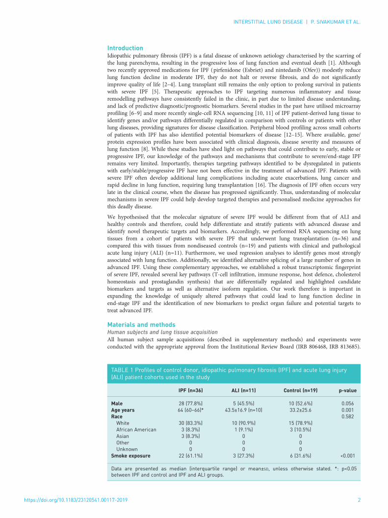
TABLE 1 Profiles of control donor, idiopathic pulmonary fibrosis (IPF) and acute lung injury(ALI) patient cohorts used in the study
IPF (n=36) ALI (n=11) Control (n=19) p-value
Male 28 (77.8%) 5 (45.5%) 10 (52.6%) 0.056Age years 64 (60–66)* 43.5±16.9 (n=10) 33.2±25.6 0.001Race 0.582White 30 (83.3%) 10 (90.9%) 15 (78.9%)African American 3 (8.3%) 1 (9.1%) 3 (10.5%)Asian 3 (8.3%) 0 0Other 0 0 0Unknown 0 0 0
Smoke exposure 22 (61.1%) 3 (27.3%) 6 (31.6%) <0.001
Data are presented as median (interquartile range) or mean±SD, unless otherwise stated. *: p<0.05between IPF and control and IPF and ALI groups.
https://doi.org/10.1183/23120541.00117-2019 2
INTERSTITIAL LUNG DISEASE | P. SIVAKUMAR ET AL.

The clinical profile and demographics of IPF, ALI and control subjects are listed in table 1. Details of theacquisition protocol are provided in the supplementary material.
RNA sequencingIllumina TruSeq Stranded Total RNA Library Prep Kit with Ribo-Zero (cat. No RS-122-2203, IlluminaInc., San Diego, CA) was used to generate sequencing libraries per manufacturer’s recommendation. Geneexpression was determined via RNA-Seq libraries run on an Illumina HiSeq 25000 platform producing75 bp paired-end (PE) reads. We generated on average 40 million PE reads for each sample. Reads werealigned to the human genome (GRch38) with the Omicsoft Sequence Aligner [17]. Gene and transcriptabundance was determined using Ensembl release 90 human gene models [18] using RSEM [19]. All thegene expression data were deposited in the National Center for Biotechnology Information GeneExpression Omnibus (GSE 134692).
RNA-Seq data analysesAll RNA-Seq data were processed in R with the Bioconductor packages [20]. RNA-Seq samples wereTMM (Trimmed Mean of M Values) normalised [21] with the edgeR package [22]. Outlier detection wasperformed using t-distributed stochastic neighbour embedding (t-SNE) [23]. Contributions of knownvariables (disease state, age, sex) to the data variance were assessed by principal variance componentanalyses. Differential gene expression contrasts between treatment groups was performed using the limmapackage [24]. Pathway enrichment was computed using Genego (Metacore) and MetaBase (ClarivateAnalytics) version 6.34.69200. We also performed ensemble gene set enrichment analyses using themolecular signature database (MSigDB), a comprehensive database from the Broad Institute encompassingnumerous curated public gene sets and pathways [25].
Alternative isoform regulationWe used JunctionSeq software for detecting the differential usage of exons and splice junctions fromRNA-Seq data [26]. This enabled the determination of alternative isoform regulation. Testable lociidentified through JunctionSeq were further filtered by excluding those with mean counts ⩾10 and anadjusted p-value ⩽0.05.
Statistical analysesTotal gene expression data were analysed using a fold change/false discovery rate (FDR) cut-off of 2X/0.001 (for comparing IPF and control) or 1.5X/0.1 (for comparing IPF versus control and ALI versuscontrol signatures) to generate gene lists for pathway analyses. Individual gene expression was comparedacross groups by one-way ANOVA followed by Tukey’s post-test with differences considered statisticallysignificant at p⩽0.05.
0.4
b)
0.2
0.0
We
igh
ted
ave
rag
e
pro
po
rtio
n v
ari
an
ce
Dis
ea
se
sta
tus:
Ag
e
Dis
ea
se
sta
tus:
Se
x
Se
x:a
ge
Ag
e
Se
x
Dis
ea
se
sta
tus
Re
sid
a)
Disease status
ALI IPF Normal
0.5
0.35
0.04 0.03 0.03 0.03 0.03
FIGURE 1 Quality control analyses of RNA-sequencing data from idiopathic pulmonary fibrosis (IPF) (n=36),acute lung injury (ALI) (n=10) and control (n=20) lung tissue. a) Principal component analyses ofbatch-corrected normalised expression data using the t-scholastic neighbourhood enrichment (t-SNEmethod). b) Principal variance component analyses were used to assess the relative contribution of patientvariables to variance in the normalised expression across the samples.
https://doi.org/10.1183/23120541.00117-2019 3
INTERSTITIAL LUNG DISEASE | P. SIVAKUMAR ET AL.
Resultst-SNE variance analysesWe used the t-SNE method to visualise the distribution of the expression data across control and diseasedsamples and identify outliers. As shown in figure 1a, there was a clear separation of the IPF samples fromboth the control and ALI samples. To assess the contribution of donor variables to gene expression, weperformed variance partition analyses. Figure 1b shows that the largest contributor to the variance amongknown variables was the IPF or control disease state reaffirming that the difference in normalised geneexpression was driven primarily by the IPF or control disease state (“Residual” represents a residual sum ofunknown variables and could include polypharmacology, medications, and genetic factors among manyothers). Other known parameters such as age (although significantly higher in IPF), sex or ethnicity didnot appear to significantly contribute towards the changes in expression.
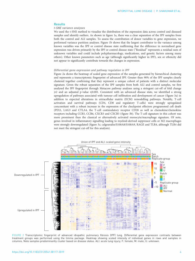
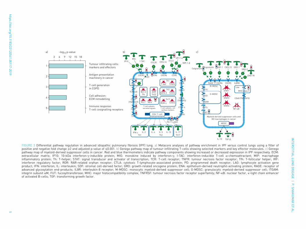
Differential gene expression and pathway regulation in IPFFigure 2a shows the heatmap of scaled gene expression of the samples generated by hierarchical clusteringand represents a transcriptomic fingerprint of advanced IPF. Greater than 90% of the IPF samples clearlyclustered together confirming that they represent a unique cohort of patients with a distinct molecularsignature. Given the robust separation of the IPF samples from both ALI and control samples, we firstanalysed the IPF fingerprint through Metacore pathway analyses using a stringent cut-off of fold change⩾2 and an adjusted p-value ⩽0.001. Consistent with an advanced disease state, we identified a strongupregulation of pathways associated with tumour cell infiltration and development of cancer (figure 3a) inaddition to expected alterations in extracellular matrix (ECM) remodelling pathways. Notably, T-cellactivation and survival pathways (CD4, CD8 and regulatory T-cells) were strongly upregulatedconcomitant with a robust increase in the expression of the checkpoint effectors programmed cell death(PD)1, LAG3 and CTLA4, the T-cell costimulatory receptor CD28 as well as chemokine/chemokinereceptors including CCR5, CCR6, CXCR3 and CXCR5 (figure 3b). The T-cell signature in this cohort wasmore prominent than the classical or alternatively activated monocyte/macrophage signature. Of note,genes involved in inflammatory signalling leading to myeloid-derived suppressor cells or M2 macrophageswere strongly downregulated (figure 3c; calgranulin/S100A8/S100A9, RAGE and TLR4, although TLR4 didnot meet the stringent cut-off for this analysis).
Downregulated in IPF
Union of IPF and ALI: scaled gene intensity
F
ALIIPFNormal
Replicate group
Replicate group
Batch
Batch
Sex
Sex
15
10
5
0
–5
–10
M
12
U
Upregulated in IPF
FIGURE 2 Transcriptomic fingerprint of advanced idiopathic pulmonary fibrosis (IPF) lung. Differential gene expression contrasts betweentreatment groups was performed using the limma package. Heatmap showing scaled intensity of individual genes in rows and samples incolumns. Note samples predominantly cluster based on disease status. ALI: acute lung injury; F: female; M: male; U: unknown.
https://doi.org/10.1183/23120541.00117-2019 4
INTERSTITIAL LUNG DISEASE | P. SIVAKUMAR ET AL.
-log10 p-valuea) b) c)
3 6 9 12 1815
1
2
3
4
5
Tumour infiltrating cells:
markers and effectors
Antigen presentation
machinery in cancer
T-cell generation
in COPD
Cell adhesion:
ECM remodelling
Immune response:
T-cell cosignalling receptors
RAGE CXCR4
XSDF-1
CXCR5 IL8RB
M-MDSC
STAT1
CD40 (TNFRSF5)
CD14
G-MDSCCD33
ITGAM
FUT4
CD67
STAT3
STAT6
NF-κB
Myeloid-derived suppressor cells and
M2 macrophages in cancer
Low
MHC class II
B
CogranulinCalgranulin
IL-10 TGF-β
B
CXCL13
B
GRO-1GRO-2
ENA-78
B BB
B
MDSC
CXCR3 CCR6
PD-1 LAG3
CD244CD2
CD89CTLA-4
CD40L
(TNFSF5)
CD161
CD28
CD3
T-cell subsets: cell surface markers
T-cell subsets:secreted signals
IFN-γTh1
IL-2 IL-4 IL-5
Th2
IL-13
Th17
IL-17
IP10
MIGI-TAC
CCL20 CXCL16CCL5
UP+1-α
MIP-1-β
B
B B B B BB
B
B
STAT4
Th1
T-bet
STAT6
Th2
GATA-3
STAT3
Th17
ROR-γIRF4
XX9
PU.1
CD4+ T-cell
CD27
(TNFRS57)
CXCR6 CCR5
TCR/α/β
CD4
Bcl-6
Tfh
AHR
Th22
FIGURE 3 Differential pathway regulation in advanced idiopathic pulmonary fibrosis (IPF) lung. a) Metacore analyses of pathway enrichment in IPF versus control lungs using a filter ofpositive and negative fold change ⩾2 and adjusted p-value of ⩽0.001. b) Genego pathway map of tumour-infiltrating T-cells showing selected markers and key effector molecules. c) Genegopathway map of myeloid-derived suppressor cells in cancer. Red and blue thermometers indicate pathway components showing increased or decreased expression in IPF respectively. ECM:extracellular matrix; IP10: 10-kDa interferon-γ-inducible protein; MIG: monokine induced by interferon-γ; I-TAC: interferon-inducible T-cell α-chemoattractant; MIP: macrophageinflammatory protein; Th: T-helper; STAT: signal transducer and activator of transcription; TCR: T-cell receptor; TNFR: tumour necrosis factor receptor; Tfh: T-follicular helper; IRF:interferon regulatory factor; ROR: RAR-related orphan receptor; CTLA: cytotoxic T-lymphocyte-associated protein; PD: programmed death receptor; LAG: lymphocyte activation geneproduct; IFN: interferon; IL: interleukin; SDF: stromal cell-derived factor; GRO: growth-related oncogene protein; ENA: epithelium-derived neutrophil-activating protein; RAGE: receptor ofadvanced glycosylation end-products; IL8R: interleukin-8 receptor; M-MDSC: monocytic myeloid-derived suppressor cell; G-MDSC: granulocytic myeloid-derived suppressor cell; ITGAM:integrin subunit αM; FUT: fucosyltransferase; MHC: major histocompatibility complex; TNFRSF: tumour necrosis factor receptor superfamily; NF-κB: nuclear factor, κ-light chain enhancerof activated B-cells; TGF: transforming growth factor.
https://doi.org/10.1183/23120541.00117-20195
INTER
STITIALLU
NGDISEA
SE|P.SIVA
KUMARET
AL.
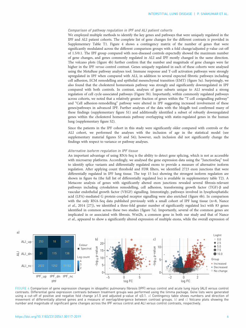
Comparison of pathway regulation in IPF and ALI patient cohortsWe employed multiple methods to identify the key genes and pathways that were uniquely regulated in theIPF and ALI patient cohorts. The complete list of gene changes for the different contrasts is provided inSupplementary Table T1. Figure 4 shows a contingency matrix of the number of genes that weresignificantly modulated across the different comparison groups with a fold change/adjusted p-value cut-offof 1.5/0.1. The IPF group compared with non-diseased controls expectedly showed the maximum numberof gene changes, and genes commonly regulated in ALI and IPF mostly changed in the same direction.The volcano plots (figure 4b) further confirm that the number and magnitude of gene changes were farhigher in the IPF versus control contrast. Genes uniquely regulated in each of these cohorts were analysedusing the MetaBase pathway analyses tool. Immune response and T-cell activation pathways were stronglyupregulated in IPF when compared with ALI, in addition to several expected fibrotic pathways includingcell adhesion, ECM remodelling and epithelial mesenchymal transition (EMT) (figure 5a). Surprisingly, wealso found that the cholesterol homeostasis pathway was strongly and significantly downregulated in IPFcompared with both controls. In contrast, analyses of gene subsets unique to ALI revealed a strongregulation of cell cycle-associated pathways (Figure 5b). Importantly, within commonly regulated pathwaysacross cohorts, we noted that a relatively greater fraction of genes within the “T-cell cosignalling pathway”and “Cell adhesion-remodelling” pathway were altered in IPF suggesting increased involvement of thesegenes/pathways in advanced IPF. Further analyses of the data with the Msigdb tool confirmed many ofthese findings (supplementary figure S1) and additionally identified a subset of robustly downregulatedgenes within the cholesterol homeostasis pathway overlapping with statin-regulated genes in the humanlung (supplementary figure S2).
Since the patients in the IPF cohort in this study were significantly older compared with controls or theALI cohort, we performed the analyses with the inclusion of age in the statistical model (seesupplementary material figures S3 and S4); however, such inclusion did not significantly change thefindings with respect to variance or pathway analyses.
Alternative isoform regulation in IPF tissueAn important advantage of using RNA-Seq is the ability to detect gene splicing, which is not as accessiblewith microarray platforms. Accordingly, we analysed the gene expression data using the “JunctionSeq” toolto identify splice variants and differentially regulated exons to provide a measure of alternative isoformregulation. After applying count threshold and FDR filters, we identified 2723 exon junctions that weredifferentially regulated in IPF lung tissue. The top 15 loci showing the strongest isoform regulation areshown in figure 6a (the full list of differentially regulated loci is available in supplementary table T2). AMetacore analysis of genes with significantly altered exon junctions revealed several fibrosis-relevantpathways including cytoskeleton remodelling, cell adhesion, transforming growth factor (TGF)-β andvascular endothelial growth factor (VEGF) signalling. Interestingly, pathways involved in lysophosphatidicacid (LPA)-mediated G protein-coupled receptor signalling were also enriched (figure 6b). In comparisonwith the only RNA-Seq data published previously with a small cohort of IPF lung tissue (n=8, Nanceet al., 2014 [27]), we identified a three-fold greater number of significantly regulated loci with 83 genesidentified in common across these two studies (figure 7a). Importantly, several of the common genes areimplicated in or associated with fibrosis. Wnt2b, a common gene in both our study and that of Nanceet al., appeared to show a significantly altered expression of multiple exons, while the overall expression of
20
15
10
5
0
-lo
g1
0 p
-va
lue
log FC–5 0 5IPF_ncIPF_dnIPF_up
ALI_nc
ALI_dn
ALI_up
a)
b)
10
5
0
-lo
g1
0 p
-va
lue
log FC–2 0 2
c)
IPF
AL
I
Colour
253 18 247
21 115 138
2022 1252 11152
Log count
4
3
2
Spectral
LogInt
Group
Increased
Decreased
No change
0.0
2.5
5.0
7.5
10.0
FIGURE 4 Comparison of gene expression changes in idiopathic pulmonary fibrosis (IPF) versus control and acute lung injury (ALI) versus controlcontrasts. Differential gene expression contrasts between treatment groups was performed using the limma package. Gene lists were generatedusing a cut-off of positive and negative fold change ⩾1.5 and adjusted p-value of ⩽0.1. a) Contingency table shows numbers and direction ofmovement of differentially altered genes and a measure of overlap/divergence between contrast groups. b) and c) Volcano plots showing thenumber and magnitude of significant gene changes across the IPF versus control and ALI versus control contrasts, respectively.
https://doi.org/10.1183/23120541.00117-2019 6
INTERSTITIAL LUNG DISEASE | P. SIVAKUMAR ET AL.

the gene was unchanged (figure 7b). Moreover, the altered exonic expression appears to map to a specifictranscript, suggesting this transcript is expressed differentially in patients with IPF.
Association between gene expression and lung functionWe correlated gene expression to % predicted forced vital capacity (FVC), as a marker of disease severity,using Spearman’s correlation analyses for nonlinear association studies. We identified nearly 300 genesthat were significantly correlated with % predicted FVC (supplementary table T3). The top 15 genes thatcorrelated positively with a decline in lung function are shown in figure 8a, and a Metacore pathwayanalyses that indicated that the gene subsets were enriched in fibrosis-relevant pathways are shown infigure 8b. The correlation of 2 of the top 15 genes (secretogranin-2 and semaphorin-3C) presented infigure 8c–e demonstrates that their expression in IPF tissue was also significantly elevated.
n=3692, abs fold change >1.5, FDR <0.1a)
19/21
39/67
33/56
25/39
31/53
30/52
Cholesterol biosynthesis
Immune response: T-cell cosignalling
Cell adhesion: ECM remodelling
Immune response: T-cell subsets
Colorectal cancer
Immune response: IL-4 type 2 immunity
Fraction of genes
n=611, abs fold change >1.5, FDR <0.1b)
18/37
18/53
18/87
10/26
10/31
13/56
Cell cycle: metaphase checkpoint
Cell cycle: APC regulation of
Cell cycle: spindle assembly
Cell cycle: chromosome condensation
DNA damage: ATM/ATR: G2/M checkpoint
Cell adhesion: ECM remodelling
Fraction of genes
Downregulated
Upregulated
Not detected
No change
FIGURE 5 Comparison of pathway enrichment in a) idiopathic pulmonary fibrosis (IPF) and b) acute lung injury (ALI) cohorts. Gene lists weregenerated using a cut-off of positive and negative fold change ⩾1.5 and adjusted p-value of ⩽0.1. Left panels: pathways uniquely enriched in theIPF or ALI cohort. Numbers against each bar represent number of differentially altered genes compared with the total number of genes definingthat pathway. Right panels: fraction of genes within the pathway that show increase, decrease or no alteration. ECM: extracellular matrix; FDR:false discovery rate.
3
2
1
4
5
6
7
8
9
Gene name Exon or junction ID
Gene description
HOPXHOPXMYLKMYLKHOPXMYLK
EPB41L2ARMC9
EPB41L2ARMC9WINT2BWNT2BHOPX
E014
J037
E014
E013
E013
E015
E028
E028
J105
E023
E001
E002
E015
HOP homeobox
HOP homeobox
Myosin light chain kinase
Myosin lght chain kinase
HOP homeobox
Myosin light chain kinase
Erythrocyte membrane protein band 4.1
Armadillo repeat containing 9
Erythrocyte membrane protein band 4.1
Armadillo repeat containing 9
Wnt family member 2B
Wnt family member 2B
HOP Homeobox
Cytoskeleton remodelling: Ephrin B reverse signallingCell adhesion: integrin-mediated cell adhesion and migration
Cytoskeleton remodelling: Rho GTPase
Glucocorticoid-induced elevation of ocular pressure: glaucoma riskChemotaxis: lysophosphatidic acidsignalling via GPCRsDevelopment: role of IL-8 in angiogenesis
CHDI: correlations from discovery data: causal network (positive)Development: VEGF2 signalling via VEGFR2 genetic cascadesHypertrophy of asthma airway smooth muscle cellsStimulation of TGF-β signalling in lung cancer10
1
-log p-value
a) b)
2 3 4 5 6 87
FIGURE 6 Alternative isoform regulation in advanced idiopathic pulmonary fibrosis (IPF) lung. a) List of top loci that show alternative splicing inthe IPF lung analysed with the ‘JunctionSeq’ tool. b) Metacore analyses of pathways enriched by genes that show significant alternative isoform/exon usage in the IPF lung (adjusted p-value ⩽0.05). GPCR: G-protein coupled receptor; IL: interleukin; VEGF: vascular endothelial growth factor;VEGFR: vascular endothelial growth factor receptor; TGF: transforming growth factor.
https://doi.org/10.1183/23120541.00117-2019 7
INTERSTITIAL LUNG DISEASE | P. SIVAKUMAR ET AL.
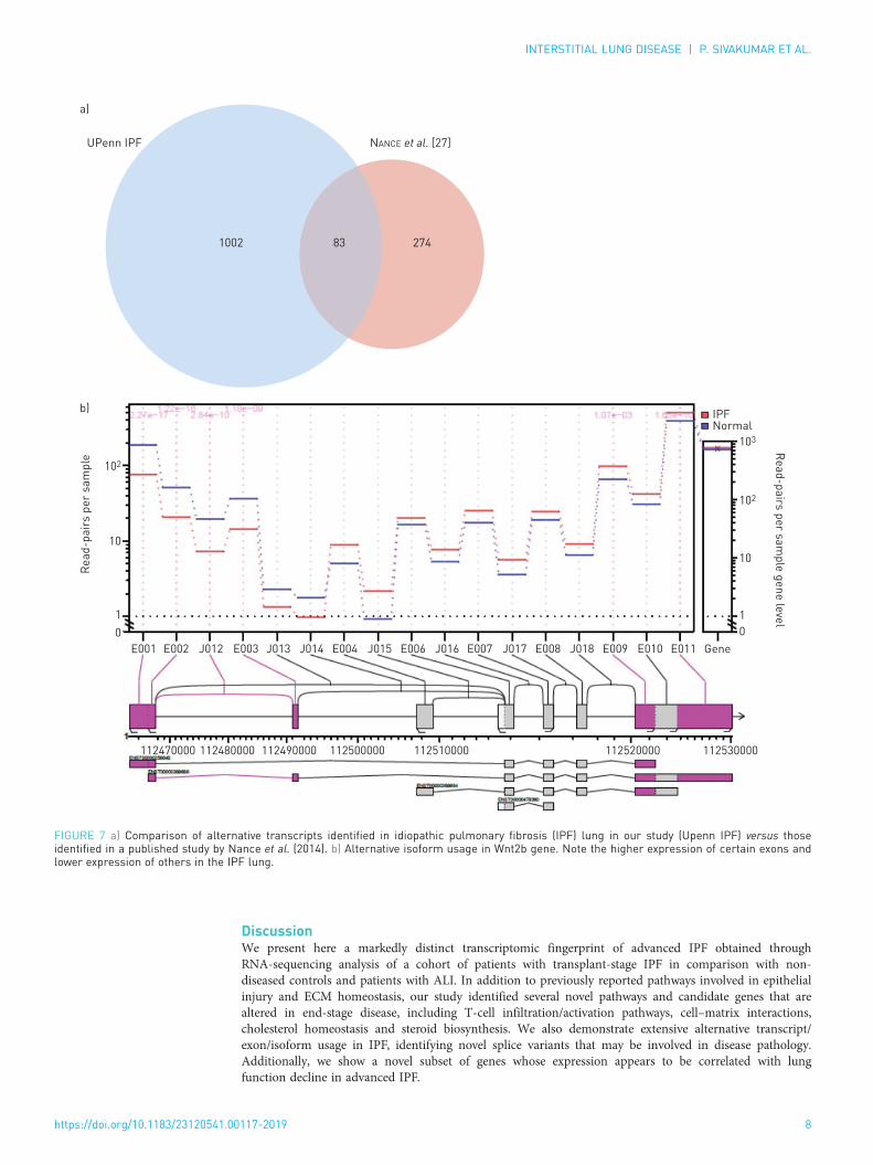
DiscussionWe present here a markedly distinct transcriptomic fingerprint of advanced IPF obtained throughRNA-sequencing analysis of a cohort of patients with transplant-stage IPF in comparison with non-diseased controls and patients with ALI. In addition to previously reported pathways involved in epithelialinjury and ECM homeostasis, our study identified several novel pathways and candidate genes that arealtered in end-stage disease, including T-cell infiltration/activation pathways, cell–matrix interactions,cholesterol homeostasis and steroid biosynthesis. We also demonstrate extensive alternative transcript/exon/isoform usage in IPF, identifying novel splice variants that may be involved in disease pathology.Additionally, we show a novel subset of genes whose expression appears to be correlated with lungfunction decline in advanced IPF.
10
102
b)
a)
1
0
Re
ad
-pa
irs p
er
sa
mp
leR
ea
d-p
airs
pe
r sa
mp
le g
en
e le
vel
UPenn IPF NANCE et al. [27]
102
10
1
0
103
IPFNormal
E001 E002 J012 E003 J013 J014 E004 J015 E006 J016 E007 J017 E008 J018 E009 E010 E011
112530000112520000112510000112500000112490000112480000112470000
Gene
1002 83 274
FIGURE 7 a) Comparison of alternative transcripts identified in idiopathic pulmonary fibrosis (IPF) lung in our study (Upenn IPF) versus thoseidentified in a published study by Nance et al. (2014). b) Alternative isoform usage in Wnt2b gene. Note the higher expression of certain exons andlower expression of others in the IPF lung.
https://doi.org/10.1183/23120541.00117-2019 8
INTERSTITIAL LUNG DISEASE | P. SIVAKUMAR ET AL.

a)
b)
c)
e)
Gene symbol Description Correl. coeff. p-value
SLC44A1SCG2GLO1USP53TPST2VGLL3XPOTPI15PTPRGERFESACSBAG2KIAA1324LTSPAN5SEMA3C
Solute carrier family 44 member 1
Secretogranin II
Glyoxalase I
Ubiquitin-specific peptidase 53
Tyrosylprotein sulfotransferase 2
Vestigial-like family member 3
Exportin for tRNA
Peptidase inhibitor 15
Protein tyrosine phosphatase, receptor type G
Erythroferrone
Sacsin molecular chaperone
BCL2-associated athanogene 2
KIAA1324 like
Tetraspanin 5
Semaphorin 3C
–0.62479102
–0.588154207
–0.532567317
–0.528075448
–0.522179868
–0.518109111
–0.514459467
–0.511090565
–0.510809823
–0.503370163
–0.494947907
–0.48175304
–0.480349331
–0.479366734
–0.477822654
6.02081×10⁻⁵
0.000202637
0.000989844
0.001112177
0.001292849
0.001432208
0.001568197
0.001703649
0.001715386
0.002053369
0.002504995
0.003386762
0.003494772
0.003572144
0.003696731
6
4
2
0
log
2 C
PM
FVC % predicted
30 5040 60 70
Corr coeff=–0.588
p=0.000203
Grey indicates 95% conf.
1
1 2 3 4
2
3
4
5
Chemotaxis: lysophosphatidic acid signalling
via GPCRs
TGF-β-induced fibroblast/myofibroblast
migration and ECM production in asthmatic
airways
Cell adhesion: integrin-mediated cell adhesion
and migration
TGF-β signalling via kinase casades in breast
cancer
Gene log fold change(IPF versus healthy)
Adjusted p-value
SCG2SEMA3C
1.740
0.934
0.0000827063
0.0000013766
Development: BMP signalling
d)8
4
4
2
0
log
2 C
PM
FVC % predicted
30 5040 60 70
Corr coeff=–0.478
p=0.003697
Grey indicates 95% conf.
FIGURE 8 Correlation of idiopathic pulmonary fibrosis (IPF) lung gene expression to lung function. a) List oftop genes that show significant inverse correlation to % predicted forced vital capacity (FVC) as assessed bySpearmen’s correlation analyses. b) Metacore analyses of pathways enriched by genes that show significantcorrelation with % predicted FVC (adjusted p-value ⩽0.05). c) and d) Correlation plots of gene expression ofSCG2 and SEMA3C, two candidate genes identified through Spearman’s analyses as significantly correlatedwith % predicted FVC. e) Gene expression intensity of SCG2 and SEMA3C confirming significant increase ofthese genes in IPF lung compared with control lung. tRNA: transfer RNA; GPCR: G-protein coupled receptor;TGF: transforming growth factor; ECM: extracellular matrix; BMP: bone morphogenetic protein; FVC: forcedvital capacity.
https://doi.org/10.1183/23120541.00117-2019 9
INTERSTITIAL LUNG DISEASE | P. SIVAKUMAR ET AL.

The molecular signature of our IPF cohort is distinctly different from past studies that have used lungtissues from patients with IPF with mild-to-moderate disease. Yang et al. [9] investigated thetranscriptomic profile of IPF/usual interstitial pneumonia from the Lung Tissue Research Consortium andidentified subgroups of patients with alterations in cilium-associated genes and epithelial injury pathways,further validating their findings with samples from the National Jewish Health cohort. Notably, the mean% FVC in their test cohort was ⩾59%. In contrast, the mean % FVC of patients in our study was 44%,with a majority of patients at ⩽30%. Likewise, microarray studies by Depianto et al., with a cohort of 40patients with IPF, identified two primary clusters of coregulated genes representing bronchiolar epitheliumand lymphoid aggregates [28]. Worsening IPF pathology remains a significant risk factor for acuteexacerbations, development of lung cancer and additional lung complications that warrant a lungtransplant. Konishi et al. [6] showed that the gene expression profile of acute exacerbations of IPF(AE-IPF) was mostly similar to that of stable IPF with only a few genes significantly altered between thetwo groups. Interestingly, these authors also observed a downregulation of AGER (receptor for advancedglycation end products) as we noted in our cohort of advanced IPF. However, defensin a3 (DEFA3) wasstrongly reduced in our cohort in contrast with Konishi’s study as well as to a separate study by Yang et al.[14] on the peripheral blood transcriptome of patients with IPF that identified DEFA3 to be increased inprogressive disease. The apparent differences may be due to the different patient profiles and measures ofdisease severity. The AE-IPF cohort in Konishi’s study had a mean % FVC of 55% and a lower DLCO(diffusing capacity of the lung for carbon monoxide) of 36%, and Yang’s study used DLCO as the diseaseclassifier.
Consistent with advanced disease, pathways involved in T-cell activation and cancer/tumour developmentwere strongly enriched in the IPF molecular signature in our study. Notably, several checkpoint effectormolecules including PD1 receptor, LAG3 and CTLA4 were shown to be upregulated in IPF tissue,indicating the possibility of immune exhaustion in end-stage IPF. The role of T-lymphocytes in humanIPF and animal models of fibrosis has been recognised before [29]. Abnormal PD1 levels have beenidentified in human IPF, and human mesenchymal stem cell therapy was protective in a humanised mousemodel of fibrosis through suppression of CD8+ T-cell infiltration and activation by a PD1/programmeddeath ligand (PD-L)1 axis [30]. It was recently reported that the increased expression of PD1 on CD4+
T-cells in IPF lung and plasma promotes a T helper (Th)-17 mediated response and induces active TGF-βsecretion from stimulated T-cells [31]. Interestingly, the authors also show that knockout of PD1 as well asblockade of PD-L1 signalling ameliorates the fibrotic responses induced by bleomycin in mice. Our dataemphasise the role of T-cell activation in advanced IPF and highlight the potential utility of this pathwayas a disease classifier as well as a candidate therapeutic target. However, while the T-cell phenotype andlineage appear to be critically important in the context of IPF and particularly in severe IPF, more work isneeded to understand their relative contribution to disease pathology.
While we noted increases in T-cell activation pathways, we also observed a reduction in genes involved inthe activation of myeloid-derived suppressor cells and M2 macrophages (calgranulin, AGER and TLR4).Myeloid-derived suppressor cells are elevated in the peripheral blood of patients with IPF and inverselycorrelate with lung function [32]. Alternatively activated (M2) macrophages, described to be abundant inboth murine and human fibrosis are known to drive anti-inflammatory and profibrotic responsessimultaneously [33]. These macrophages also produce chemokines that recruit T-lymphocytes into theinjured lung. Our observation of reduction in signals that activate M2 differentiation, together with anupregulation of chemokines involved in T-cell recruitment may imply that T-cell-mediated pathwayscontribute to late-stage disease.
Biopsies of early IPF are not in standard clinical practice and would be very difficult to procure insufficient numbers to include in the study. Therefore, in our study design, we specifically included ALI asan additional comparator because both IPF and ALI represent severe lung pathologies, albeit with differentaetiologies. Only cell adhesion and ECM remodelling pathways were commonly regulated in both thesecohorts in comparison with non-disease controls. The IPF signature was rich in T-cell/immune responsewhile the ALI signature was enriched in genes/pathways involved in cell cycle regulation. Thus, the IPFsignature that we present here could be uniquely representative of end-stage lung fibrosis. Intriguingly, thecholesterol homeostasis pathway was significantly downregulated in our patients with advanced IPF withthe signature overlapping with published statin-responsive genes in the lung [34], a finding that should befurther explored and perhaps validated in independent cohorts. In addition, genes within lipoproteinsteroid and fatty acid biosynthesis pathways were also strongly under-expressed in the IPF cohort. Giventhat lipid-lowering therapy and reduced lipoprotein levels have been shown to be profibrotic in preclinicaland clinical studies [35, 36], our data lend further credence to the hypothesis that lipoprotein levels areinversely correlated with the pathogenesis of interstitial lung disease, although the mechanismscontributing to this effect remain to be investigated.
https://doi.org/10.1183/23120541.00117-2019 10
INTERSTITIAL LUNG DISEASE | P. SIVAKUMAR ET AL.
To date, there is only one study investigating alternative splicing in the IPF lung obtained through RNAsequencing of a small cohort of eight lungs from patients with IPF [27]. In that study, the authors usedDEXseq to demonstrate alternative exon usage in known fibrosis-related genes including periostin andcollagen 6. Our study identifies, for the first time, a larger subset of genes that exhibit alternativetranscript/isoform usage in advanced IPF, opening up hitherto unknown targets and pathways fortherapeutic intervention or as biomarkers of disease severity. Top pathways enriched in differentiallyspliced genes were found to be involved in fibrotic signalling (TGF, VEGF, ephrin B and LPA pathways)and cytoskeletal/ECM remodelling. Interestingly, we found HOPX (homeobox protein x), a known markerof alveolar Type I cells [37] as the top alternatively spliced gene in our study. A recent study showed thatHOPX impacts alveolar epithelial injury and fibrosis and is decreased in human IPF tissue [38]contributing to lung function decline potentially through a failure of epithelial cell regeneration. Ourexpression data (downregulation of HOPX in IPF versus control) confirm these findings and suggest thatalternative splicing of HOPX could play a role in the development of fibrosis.
We note that previously reported biomarkers (MMP7) and genes implicated in alveolar epithelial cell(DIO2) and surfactant functions (SP-A, SP-C) and lineage-negative lung progenitor cell expansion (KRT5)changed in total expression, validating previous findings [12, 39, 40]. However, exploring differentialexpression of exons can help identify genes that may not change in total expression but could bealternatively spliced in disease, a mechanism that is likely to be missed with microarray studies. Forexample, we identified Wnt2b, a molecule that is antifibrotic in hepatic stellate cells [41] as a majoralternatively spliced gene although the overall expression was not changed in IPF tissue.
Prognostic and/or predictive biomarkers of disease progression in IPF are critical for the early diagnosisand treatment of the disease. In our cohort, we investigated the correlation of gene expression with clinicalmeasures of disease severity, using % predicted FVC (recorded prior to transplantation) as the measure oflung function. Through Spearman’s correlation analyses, we identified several genes whose expression waspositively or negatively correlated with % predicted FVC. Many of the identified associations are novel.Interestingly, many do not appear to significantly change when comparing overall expression, but theirexpression level increased or decreased with disease severity. Our studies thus may have identified novelbiomarkers of disease severity that can be validated in other independent cohorts.
In summary, we have described for the first time an RNA-Seq-based transcriptomic fingerprint of severeIPF and a molecular signature that can be further evaluated as a potential disease classifier. We chose notto directly compare this signature with previously described signatures from microarray platforms, as thereare significant differences in patient disease profiles and the methods used to derive the molecularsignatures. However, we note that as opposed to previous studies that have largely described epithelialinjury, cell–matrix remodelling pathways, our cohort of patients with advanced IPF exemplify other uniquepathways that could contribute to the disease and potentially present interesting targets and biomarkers.Recently, in an approach that complements ours, Reyfman et al. [11] used single-cell sequencing ofadvanced IPF lungs to generate a single-cell atlas of IPF and identify distinct subpopulations of alveolarmacrophages and epithelial cells that could drive fibrosis. Similar to our observations, their work alsoidentifies immune response, ECM organisation and Wnt signalling pathways as regulated in advancedfibrosis. Although we did not use a single-cell approach, which would have been cumbersome given thelarge number of samples involved, we believe that the whole-organ signature of advanced IPF is importantand informative, since the disease phenotype in IPF is a collective consequence of the heterogeneity in celltypes within the organ. RNA-Seq coupled with single-cell analyses could provide a new step forward in theelusive quest for biomarkers and targets for the diagnosis and treatment of IPF.
Conflict of interest: P. Sivakumar has nothing to disclose. J.R. Thompson has nothing to disclose. R. Ammar hasnothing to disclose. M. Porteous has nothing to disclose. C. McCoubrey has nothing to disclose. E. Cantu III hasnothing to disclose. K. Ravi has nothing to disclose. Y. Zhang has nothing to disclose. Y. Luo has nothing to disclose.D. Streltsov has nothing to disclose. M.F. Beers reports grants from Bristol-Myers Squibb during the conduct of thestudy. G. Jarai has nothing to disclose. J.D. Christie reports grants from the NIH and Bristol-Myers Squibb during theconduct of the study and grants from the NIH, GlaxoSmithKline and Bristol-Myers Squibb outside the submitted work.
References1 Martinez FJ, Collard HR, Pardo A, et al. Idiopathic pulmonary fibrosis. Nat Rev Dis Primers 2017; 3: 17074.2 George PM, Wells AU. Pirfenidone for the treatment of idiopathic pulmonary fibrosis. Expert Rev Clin Pharmacol
2017; 10: 483–491.3 Rogliani P, Calzetta L, Cavalli F, et al. Pirfenidone, nintedanib and N-acetylcysteine for the treatment of idiopathic
pulmonary fibrosis: A systematic review and meta-analysis. Pulm Pharmacol Ther 2016; 40: 95–103.4 Tomioka H, Takada H. Treatment with nintedanib for acute exacerbation of idiopathic pulmonary fibrosis.
Respirol Case Rep 2017; 5: e00215.
https://doi.org/10.1183/23120541.00117-2019 11
INTERSTITIAL LUNG DISEASE | P. SIVAKUMAR ET AL.
5 Caminati A, Cassandro R, Torre O, et al. Severe idiopathic pulmonary fibrosis: what can be done? Eur Respir Rev2017; 26: 170047.
6 Konishi K, Gibson KF, Lindell KO, et al. Gene expression profiles of acute exacerbations of idiopathic pulmonaryfibrosis. Am J Respir Crit Care Med 2009; 180: 167–175.
7 Kusko RL, Brothers JF 2nd, Tedrow J, et al. Integrated genomics reveals convergent transcriptomic networksunderlying chronic obstructive pulmonary disease and idiopathic pulmonary fibrosis. Am J Respir Crit Care Med2016; 194: 948–960.
8 Steele MP, Luna LG, Coldren CD, et al. Relationship between gene expression and lung function in idiopathicinterstitial pneumonias. BMC Genomics 2015; 16: 869.
9 Yang IV, Coldren CD, Leach SM, et al. Expression of cilium-associated genes defines novel molecular subtypes ofidiopathic pulmonary fibrosis. Thorax 2013; 68: 1114–1121.
10 Xu Y, Mizuno T, Sridharan A, et al. Single-cell RNA sequencing identifies diverse roles of epithelial cells inidiopathic pulmonary fibrosis. JCI Insight 2016; 1: e90558.
11 Reyfman PA, Walter JM, Joshi N, et al. Single-cell transcriptomic analysis of human lung provides insights intothe pathobiology of pulmonary fibrosis. Am J Respir Crit Care Med 2018; 198: 440–446.
12 Bauer Y, White ES, de Bernard S, et al. MMP-7 is a predictive biomarker of disease progression in patients withidiopathic pulmonary fibrosis. ERJ Open Res 2017; 3: 00074-2016.
13 Rosas IO, Richards TJ, Konishi K, et al. MMP1 and MMP7 as potential peripheral blood biomarkers in idiopathicpulmonary fibrosis. PLoS Med 2008; 5: e93.
14 Yang IV, Luna LG, Cotter J, et al. The peripheral blood transcriptome identifies the presence and extent of diseasein idiopathic pulmonary fibrosis. PLoS One 2012; 7: e37708.
15 Herazo-Maya JD, Sun J, Molyneaux PL, et al. Validation of a 52-gene risk profile for outcome prediction inpatients with idiopathic pulmonary fibrosis: an international, multicentre, cohort study. Lancet Respir Med 2017;5: 857–868.
16 Lipsi R, Mazzola D, Caminati A, et al. Severe idiopathic pulmonary fibrosis: A clinical approach. Eur J Intern Med2018; 50: 20–27.
17 Hu J, Ge H, Newman M, et al. OSA: a fast and accurate alignment tool for RNA-Seq. Bioinformatics 2012; 28:1933–1934.
18 Zerbino DR, Achuthan P, Akanni W, et al. Ensembl 2018. Nucleic Acids Res 2018; 46: D754–D761.19 Li B, Ruotti V, Stewart RM, et al. RNA-Seq gene expression estimation with read mapping uncertainty.
Bioinformatics 2010; 26: 493–500.20 Huber W, Carey VJ, Gentleman R, et al. Orchestrating high-throughput genomic analysis with Bioconductor. Nat
Methods 2015; 12: 115–121.21 Robinson MD, Oshlack A. A scaling normalization method for differential expression analysis of RNA-Seq data.
Genome Biol 2010; 11: R25.22 Robinson MD, McCarthy DJ, Smyth GK. edgeR: a Bioconductor package for differential expression analysis of
digital gene expression data. Bioinformatics 2010; 26: 139–140.23 Van der Maaten L, Hinton G. Visualizing data using t-SNE. J Mach Learn Res 2008; 9: 2579–2605.24 Ritchie ME, Phipson B, Wu D, et al. Smyth GK. limma powers differential expression analyses for
RNA-sequencing and microarray studies. Nucleic Acids Res 2015; 43: e47.25 Subramanian A, Tamayo P, Mootha VK, et al. Gene set enrichment analysis: a knowledge-based approach for
interpreting genome-wide expression profiles. Proc Natl Acad Sci USA 2005; 102: 15545–15550.26 Hartley SW, Mullikin JC. Detection and visualization of differential splicing in RNA-Seq data with JunctionSeq.
Nucleic Acids Res 2016; 44: e127.27 Nance T, Smith KS, Anaya V, et al. Transcriptome analysis reveals differential splicing events in IPF lung tissue.
PLoS One 2014; 9: e97550.28 DePianto DJ, Chandriani S, Abbas AR, et al. Heterogeneous gene expression signatures correspond to distinct
lung pathologies and biomarkers of disease severity in idiopathic pulmonary fibrosis. Thorax 2015; 70: 48–56.29 Luzina IG, Todd NW, Iacono AT, et al. Roles of T lymphocytes in pulmonary fibrosis. J Leukoc Biol 2008; 83:
237–244.30 Ni K, Liu M, Zheng J, et al. PD-1/PD-L1 pathway mediates the alleviation of pulmonary fibrosis by human
mesenchymal stem cells in humanized mice. Am J Respir Cell Mol Biol 2018; 58: 684–695.31 Celada LJ, Kropski JA, Herazo-Maya JD, et al. PD-1 up-regulation on CD4(+) T cells promotes pulmonary fibrosis
through STAT3-mediated IL-17A and TGF-β1 production. Sci Transl Med 2018; 10: eaar8356.32 Fernandez IE, Greiffo FR, Frankenberger M, et al. Peripheral blood myeloid-derived suppressor cells reflect disease
status in idiopathic pulmonary fibrosis. Eur Respir J 2016; 48: 1171–1183.33 Braga TT, Agudelo JS, Camara NO. Macrophages during the fibrotic process: M2 as friend and foe. Front
Immunol 2015; 6: 602.34 Lane J, van Eeden SF, Obeidat M, et al. Impact of statins on gene expression in human lung tissues. PLoS One
2015; 10: e0142037.35 Podolanczuk AJ, Raghu G, Tsai MY, et al. Cholesterol, lipoproteins and subclinical interstitial lung disease: the
MESA study. Thorax 2017; 72: 472–474.36 Xu JF, Washko GR, Nakahira K, et al. Statins and pulmonary fibrosis: the potential role of NLRP3 inflammasome
activation. Am J Respir Crit Care Med 2012; 185: 547–556.37 Liebler JM, Marconett CN, Juul N, et al. Combinations of differentiation markers distinguish subpopulations of
alveolar epithelial cells in adult lung. Am J Physiol Lung Cell Mol Physiol 2016; 310: L114–L120.38 Ota C, Ng-Blichfeldt JP, Korfei M, et al. Dynamic expression of HOPX in alveolar epithelial cells reflects injury
and repair during the progression of pulmonary fibrosis. Sci Rep 2018; 8: 12983.39 Kinder BW, Brown KK, McCormack FX I×JH, et al. Serum surfactant protein-A is a strong predictor of early
mortality in idiopathic pulmonary fibrosis. Chest 2009; 135: 1557–1563.40 Yu G, Tzouvelekis A, Wang R, et al. Thyroid hormone inhibits lung fibrosis in mice by improving epithelial
mitochondrial function. Nat Med 2018; 24: 39–49.41 Yuan Y, Han Q, Li S, et al. Wnt2b attenuates HSCs activation and liver fibrosis through negative regulating TLR4
signaling. Sci Rep 2017; 7: 3952.
https://doi.org/10.1183/23120541.00117-2019 12
INTERSTITIAL LUNG DISEASE | P. SIVAKUMAR ET AL.