rna interference for apoptosis signal-regulating kinase … · rna interference for apoptosis...
TRANSCRIPT

RNA interference for apoptosis signal-regulating kinase-1 (ASK-1)rescues photoreceptor death in the rd1 mouse
Daisuke Sekimukai, Shigeru Honda, Akira Negi
Department of Surgery, Division of Ophthalmology, Kobe University Graduate School of Medicine, Kobe, Japan
Purpose: To evaluate whether RNA interference against apoptosis signal-regulating kinase-1 (ASK-1), a gene involvedin stress-induced apoptosis, inhibits photoreceptor death in retinal degeneration 1 (rd1) mice.Methods: Retinal explants from rd1 mice were subjected to organ cultures on postnatal day 9 (P9). Short interfering RNA(siRNA) for ASK-1 was transfected into cultured retinas at the onset of experiments. Real-time PCR was performed toevaluate the natural expression of ASK-1 mRNA and its inhibition with siRNA. Retinal explants were fixed at P13 andP16, and consecutive cryosections were prepared. Histological and immunohistochemical examinations including TUNELassays were performed.Results: In preliminary experiments, the incorporation of fluorescent siRNA was found in cells in the outer nuclear andinner nuclear layers on the day following transfection. The expression of ASK-1 mRNA increased with time, which wassuppressed more than 70% by siRNA. ASK-1 immunopositive cells were found mostly in the outer nuclear layers, andthe number of immunopositive cells was remarkably reduced in retinas treated with siRNA for ASK-1 compared tountreated controls. The thickness of outer nuclear layers of control retinas decreased with time, while the thickness ofsiRNA transfected retinas was significantly preserved compared to control at P16 (p=0.0021). In TUNEL assays, siRNAfor ASK-1 significantly decreased TUNEL-positive cells (49% and 42% of controls at P13 and 16, p=0.039 and 0.0028,respectively).Conclusions: RNA interference against ASK-1 may provide a benefit by inhibiting photoreceptor apoptosis in rd1 mice.
Retinitis pigmentosa (RP) is a major retinal hereditarydisease characterized by progressive degeneration ofphotoreceptors and retinal pigment epithelial cells (RPEs). RPcauses night blindness, visual field contraction, andeventually, severe visual disturbance [1]. Studies havedisclosed several mutations in photoreceptor and RPE genesthat associate with its pathogenesis. At least 30 genes havebeen identified, many of which encode photoreceptor-specificproteins such as peripherin [2], rod outer segment membraneprotein 1 [3], rod cyclic GMP (cGMP) phosphodiesterase[4], and rhodopsin [5]. Animal models of retinal degenerationare widely used to investigate genetic mechanisms forphotoreceptor apoptosis, a feature common to all cases ofhuman RP. In the retinal degeneration 1 (rd1) mouse, amutation located in the gene encoding the beta-subunit of rodcGMP phosphodiesterase [6] causes accumulation of cGMPfollowed by an increase in calcium ion (Ca2+) influx to thecytoplasm, which activates Ca2+ dependent proteases, such ascalpain and cathepsin D, or causes mitochondrial stress, whichinduces apoptosis of photoreceptors early in postnataldevelopment [7,8]. Since mutations in this gene are the mostcommon cause of autosomal recessive RP in humans [4], it is
Correspondence to: Shigeru Honda, Department of Surgery,Division of Ophthalmology, Kobe University Graduate School ofMedicine, 7-5-2 Kusunoki-cho, Chuo-ku, Kobe 650-0017, Japan;Phone: +81-78-382-6048, Fax +81-78-382-6059; email:[email protected]
a particularly relevant model. Research has demonstrated thatmultiple pathways exist in the process of photoreceptorapoptosis in the rd1 mouse [9-11].
Apoptosis signal-regulating kinase 1 (ASK-1) is amitogen-activated protein (MAP) kinase family member,which is one of the activators of the p38-JNK apoptosissignaling cascade [12,13]. ASK-1 is activated in response tovarious cytotoxic stresses, including TNF, Fas, and reactiveoxygen species (ROS) such as hydrogen peroxide (H2O2)[13-16]. Overexpression of wild-type or constitutively activeASK1 induces apoptosis in various cells throughmitochondria-dependent caspase activation [13,15,16], andASK1 is required for apoptosis induced by oxidative stress,TNF and endoplasmic reticulum (ER) stress [17,18].Moreover, reports showed that Ca2+ signaling regulates theASK1–p38-MAP kinase cascade [19]. Ca2+ influx evoked bymembrane depolarization in primary neurons andsynaptosomes induced activation of p38, which was impairedin samples derived from ASK1-deficient mice. However, todate, no study has addressed the role of ASK-1 inphotoreceptor apoptosis in the rd1 mouse. We investigated theexpression and distribution of ASK-1 in retinas from rd1 mice.
RNA interference is a technology that can knock downthe expression of specific genes using a few copies of shortRNA [20]. This is known to be quite effective in suppressinggenes in vitro, but few reports have been published about theuse of RNA interference in vivo or in organ cultures [21-23].In this study, we evaluated whether RNA interference for
Molecular Vision 2009; 15:1764-1773 <http://www.molvis.org/molvis/v15/a186>Received 23 May 2009 | Accepted 28 August 2009 | Published 2 September 2009
© 2009 Molecular Vision
1764

ASK-1 works in retinal organ cultures and inhibitsphotoreceptor death in rd1 mice.
METHODSRetinal organ cultures: The study adhered to the Associationfor Research in Vision and Ophthalmology (ARVO)Statement for the Use of Animals in Ophthalmic and VisionResearch. Retinal explant cultures of C3H/HeN (rd1) miceand C57Bl/6 (Bl6) mice were prepared as describedpreviously [24]. The mice were purchased from (CLEA JapanInc., Tokyo, Japan) and kept in mouse cages with free accessof solid food and water. Light-dark cycle was 12:12 h. Briefly,mice were euthanized (intraperitoneal injection of 15 mg ofpentobarbital per mouse for euthanasia) and neural retinaswere extracted on postnatal day (P) 8 or 9, depending on theexperiment. Each retina was submerged in HBSS andextended to make whole flat-mounts on 30 mm diametermicroporous membranes (Millicell-CM; Millipore, Bedford,MA) with ganglion cell layers facing up. Millicell-CMcontaining retinal explants were soaked in 1 ml of culturemedium (Opti-MEM, Invitrogen, Carlsbad, CA) with orwithout 25% heat inactivated horse serum in six-well platesand incubated, allowing an air interface with the ganglion cellside of the retina in a humidified atmosphere of 5% CO2 and95% air at 34 °C. Culture media was changed every other day.Transfection of siRNA: For preliminary experiments, toconfirm that siRNA was properly incorporated into retinalexplants, 100 pmol of fluorescently labeled dsRNA (Block-itTM Alexa Fluor Red Fluor. Oligo, Invitrogen) was bound with5 μl lipofectamine (RNAiMAX, Invitrogen) in a total 250 μlof serum free culture media for 20 min and applied to retinalorgan cultures. After incubation for 4 h at 34 °C, thetransfection media was replaced with Opti-MEM culturemedia containing 25% serum. After 24 h, organ culturecryosections were prepared with DAPI staining and observedwith a Keyence Biozero fluorescence microscope (BZ-8000;Keyence, Osaka, Japan). For silencing ASK-1, Stealth RNAi,which included two different sequences of siRNA for ASK-1(Table 1), were obtained from Invitrogen. Each siRNA wastransfected into the retina with lipofectamine RNAiMAXusing the same method as described above. Medium GCcontent scrambled control siRNA (Invitrogen; proprietarysequence) was also applied to examine off-target effects.Retinal explants were either transfected or untransfected withscrambled siRNA and cultured for 72 h before RNAextractions.
Real-time PCR: Total RNA was extracted using RNeasy Plus(Qiagen, Valencia, CA) from organ cultures for 0, 1, 3, 5, and7 days according to the manufacturer’s recommendations.Total RNA was eluted from columns in 50 μl RNase-freewater. The purity and concentration of RNA was determinedby measuring the absorbance at 260 nm and 280 nm. RNAwas reverse-transcribed into cDNA (High Capacity cDNAReverse Transcription Kit, Applied Biosystems, Foster City,CA) in a total volume of 100 μl according to themanufacturer’s instructions. Real-time PCRs were performedin 96 well plates on an ABI Prism 7500 Sequence DetectionSystem (Applied Biosystems StepOneTM and StepOnePlusTM
Real Time PCR System and Taqman Fast Universal PCRMaster Mix, Applied Biosystems) according to themanufacturer’s instructions. Final reaction volumes were25 µl. Each sample was analyzed in triplicate. Thermal cyclerconditions were as follows: 2 min at 50 °C, 10 min at 95 °C,followed by 40 cycles of 15 s at 95 °C and 1 min at 60 °C.Sequence Detector Software (Applied Biosystems) was usedto extract PCR data, which were exported into Excel 2003(Microsoft Corporation, Redmond, WA) for further analyses.The amount of targeted gene expressed was normalized to anendogenous reference and relative to a calibrator. β-Actin wasused as an endogenous reference in these experiments. Theformula ΔΔCT was used to calculate the amount of target geneexpression normalized to the endogenous control and relativeto the calibrator.Histological procedures: After transfections and organcultures for 0, 4, and 7 days (P9, 13 and 16, respectively),retinal explants were fixed with 4% paraformaldehyde.Cryosections were prepared and stained with DAPI so thethickness of the outer nuclear layer (ONL) could be measured.For immunohistochemistry, sections were blocked with 5%normal goat serum in 0.1 M phosphate buffered saline (PBS;137 mM NaCl, 2.7 mM KCl, 10 mM Sodium Phosphatedibasic, 2 mM Potassium Phosphate monobasic and a pH of7.4), and incubated in 1:200 diluted rabbit anti-human ASK-1antibody (catalog# sc-7931; Santa Cruz Biotech, Santa Cruz,CA), with normal goat serum overnight at 4 °C. This rabbitASK-1 antibody is compatible with mouse ASK-1. Forsecondary antibody, incubation with 1:1,000 diluted sheepanti-rabbit IgG (whole molecule), F(ab′)2 fragment–Cy3antibody (Sigma-Aldrich, Tokyo, Japan) was applied for 30min at room temperature. Nuclei were stained with 1:1,500diluted TO-PRO-3 iodide (642/661; Invitrogen), and sectionswere examined using a LSM 5 Pascal confocal imagingsystem (Carl Zeiss Inc., Tokyo, Japan).
TABLE 1. SEQUENCES OF STEALTH SIRNAS FOR ASK-1 USED IN THE STUDY.
Title Lot No. SequencesiRNA-1 MSS218535 5′-AAUUGCAGUCUGCACAGCCUUUCGG-3′siRNA-2 MSS218536 5′-AAAUGCGUAAUGAAACUUCACGUGG-3′
Molecular Vision 2009; 15:1764-1773 <http://www.molvis.org/molvis/v15/a186> © 2009 Molecular Vision
1765
Terminal dUTP Nick End Labeling: Cryosections wereincubated in 50 μl of reaction buffer containing terminaldeoxynucleotidyl transferase (TdT; Promega, Southampton,UK) and fluorescein-12-dUTP (Roche, Basel, Switzerland)according to the manufacturer’s instructions. Sections wereincubated at 37 °C for 1 h in a humidified chamber. Afterseveral washes in PBS and staining with DAPI, sections wereobserved with a Keyence Biozero fluorescence microscope.Under masked conditions, at least 500 cells in the ONL werecounted using digital images at 200× magnification for eachcondition and the proportion of TUNEL-positive cells wasdetermined.
Statistical analyses: Unpaired two-tailed Student’s t-testswere used for all statistical analyses in the present study. A pvalue less than 0.05 was considered statistically significant.
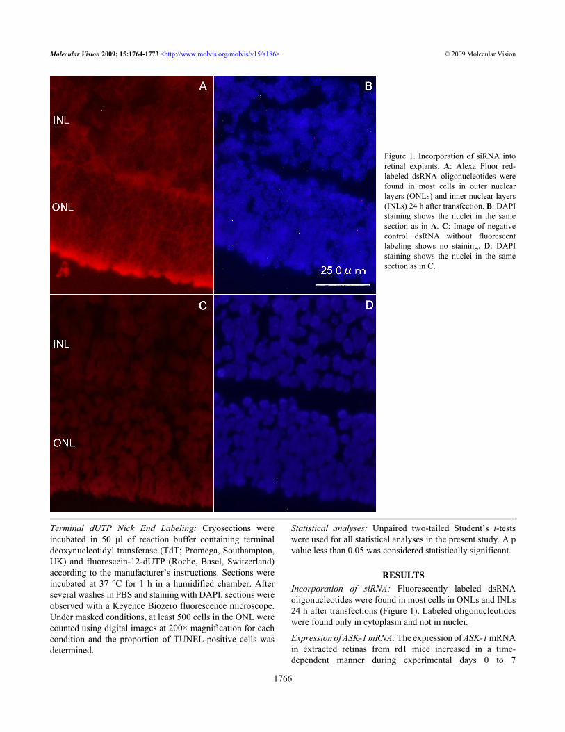
RESULTSIncorporation of siRNA: Fluorescently labeled dsRNAoligonucleotides were found in most cells in ONLs and INLs24 h after transfections (Figure 1). Labeled oligonucleotideswere found only in cytoplasm and not in nuclei.
Expression of ASK-1 mRNA: The expression of ASK-1 mRNAin extracted retinas from rd1 mice increased in a time-dependent manner during experimental days 0 to 7
Figure 1. Incorporation of siRNA intoretinal explants. A: Alexa Fluor red-labeled dsRNA oligonucleotides werefound in most cells in outer nuclearlayers (ONLs) and inner nuclear layers(INLs) 24 h after transfection. B: DAPIstaining shows the nuclei in the samesection as in A. C: Image of negativecontrol dsRNA without fluorescentlabeling shows no staining. D: DAPIstaining shows the nuclei in the samesection as in C.
Molecular Vision 2009; 15:1764-1773 <http://www.molvis.org/molvis/v15/a186> © 2009 Molecular Vision
1766

(corresponds to P8 to P15). Upregulation was significant ondays 3 and 7 (Figure 2). The expression of ASK-1 mRNA inretinas from Bl6 mice was low until day 3 but significantlyincreased by day 7.
RNA interference for ASK-1: Since the effect of RNAinterference is usually evaluated within 72 h [25-27], wedetermined the relative expression of ASK-1 mRNA in rd1mice retinas at 24 h (P9) and 72 h (P11) after siRNAtransfection. The expression of ASK-1 mRNA was suppressedmore than 70% with each of two siRNA at both time points(Figure 3). Because of the equal inhibitory effect of bothsiRNAs, we used siRNA-2 for the subsequent experiments. Inthe control experiment, retinal explants treated withscrambled siRNA showed higher expression of ASK-1 (3.5fold compared to starting point, n=3) than untreated subjects(threefold compared to starting point, n=3) at 72 h ofincubation although the difference was not significant.
Immunoreactivity of ASK-1: Immunoreactivity of ASK-1 wasfound in the outer part of ONLs of control retinas (day 4corresponding to P12; Figure 4). However, theimmunoreactivity was very weak for the same time periodwith transfected siRNA-2.
Histological findings: ONL thickness decreased markedlyduring P9 to P16 in retinal explants from rd1 mice (Figure 5).The transfection of siRNA-2 markedly preserved the ONLthickness in rd1 mice. In semiquantitative analyses, ONLthickness in retinas transfected for siRNA-2 was preservedsignificantly compared with untreated control retinas from rd1mice (p=0.012 at P13, and p=0.0021 at P16).
Apoptotic cells in retinas: Most TUNEL-positive cells werefound in the ONL, while few were in the INL in untreatedcontrols and siRNA-2 transfected retinas from rd1 mice.There were fewer TUNEL-positive cells in siRNA-2transfected retinas than in untreated controls (Figure 6). Themean ratio of TUNEL-positive cells to total ONL cell countsin untreated control retinas of rd1 mice was 5.0% at P13 and3.3% at P16. Ratios in retinas transfected with siRNA-2 were2.4% at P13 and 1.4% at P16. The ratio of TUNEL positivecells to total ONL counts in siRNA-2-transfected retinas wasabout 48% of that seen in untreated retinas at P13, decreasingfurther to 42% at P16, which was statistically significant(p=0.039 at P13 and p=0.0028 at P16).
DISCUSSIONIn this study, we confirmed that RNA interference is possiblein a retinal explant. Further, we demonstrated that ASK-1 ismainly expressed in ONL cells in rd1 mice and plays animportant role in inducing the apoptosis of photoreceptors.
Photoreceptor degeneration is known to occur due tovarious changes to molecules in signal transduction cascades,dysfunctions in energy metabolism, oxidative stress in outerretinas, or disturbances of phagocytic processes by RPE [28].In the rd1 mouse, a mutation in the enzyme cGMPphosphodiesterase causes a remarkable elevation ofcytoplasmic Ca2+ [6], which activates multiple pathways ofapoptosis including the activation of Ca2+ dependentproteases, mitochondrial stress accompanied with thegeneration of ROS, and ER stress [7-11]. These events arefollowed by the activation of the ASK-1-JNK-c-Jun pathway(Figure 1) [12,16,18,19,29-31]. The continuous elevation of
Figure 2. ASK-1 expression in the retinaof rd1 mice. The expression of ASK-1mRNA in rd1 mice increased graduallyduring days 0–7 (corresponds to P8–15).The expression of ASK-1 mRNA in theretina of Bl6 mice was low until P11 andmildly increased by P15. Values arepresented as averages of 3 independentexperiments. Asterisk (*) indicatesp<0.005, and double asterisk (**)represents a p<0.0005 compared withbaseline.
Molecular Vision 2009; 15:1764-1773 <http://www.molvis.org/molvis/v15/a186> © 2009 Molecular Vision
1767

Ca2+ levels in retinas from rd1 mice from P9 to P13 waspreviously reported [10] to correspond with a gradual increasein the expression of ASK-1 mRNA, as found in the presentstudy. Moreover, the expression of ASK-1 mRNA wasincreased in retinal explants of Bl6 mice after P11, three daysfrom the start of organ culture. It is likely that the oxygentension in normal atmosphere (about 21%) is high enough to
cause oxidative stress in cultured retinal explants and induceASK-1 [32].
RNA interference techniques are used in vitro to silencespecific genes; Palfi et al. [21] applied RNA interference toknock down rds-peripherin in retinal organotypic culture.Zacks et al. [22] demonstrated that RNA interference for Fascan work in retinal organ cultures to rescue photoreceptors
Figure 3. Suppression of ASK-1 mRNAexpression by siRNA. The expression ofASK-1 mRNA in the retina of rd1 micewas inhibited at 24 and 72 h after thetransfection of siRNA. The expressionof β-actin was used as an internalcontrol. Values are presented as theaverage±SEM of three independentexperiments.
Figure 4. Immunohistochemistry ofASK-1 on experimental day 4. A:Immunoreactivity of ASK-1 was foundin the outer part of ONL from controlretina in rd1 mice (arrows). B:Immunoreactivity was weakly found(arrows) in retina transfected withASK-1 siRNA. Images are shown at200× magnification.
Molecular Vision 2009; 15:1764-1773 <http://www.molvis.org/molvis/v15/a186> © 2009 Molecular Vision
1768

from apoptosis. Lingor et al. [23] applied anti-Apaf-1 and anti-c-Jun siRNA into vitreous cavities of rats and successfullyrescued retinal ganglion cells from axotomy-inducedapoptosis. Since c-Jun is directly downstream of the ASK-1-JNK apoptosis pathway, the aforementioned findings areconsistent with our results. In our study, siRNA wassuccessfully incorporated in retinal explants, and siRNA forASK-1 silenced ASK-1 mRNA expression. Considering thatprevious reports of RNA interference in vivo or ex vivoresulted in about 50%–80% suppression of target genes [30,33-35], it was likely sufficient that we achieved more than70% suppression of target genes in retinal explants. Theimmunoreactivity of ASK-1 was predominantly found inseveral ONL cells that represented nuclei of photoreceptorcells, which suggests several stresses are present inphotoreceptors from rd1 mice to activate ASK-1. It is not clearwhy only the most distal cells in ONL expressed ASK-1, butit is possible that the increased oxygen tension in the ONLaccelerates the expression of ASK-1 [36]. In contrast, ASK-1mRNA was found predominantly in inner retinal cells inischemic retinal injury models that cause strong oxidativestress in inner retinas [29]. ASK-1 immunoreactivity was
undetectable in retinas treated with siRNA for ASK-1, whichmeans that ASK-1 immunoreactivity in cultured retinas fromrd1 mice was possibly regulated by RNA interference.Although the ONL thickness in untreated retinas from rd1mice was markedly decreased at P16 compared with that atP9, the ONL thickness of retinas transfected with siRNA forASK-1 was preserved significantly over the same course.However, all retinal explants tended to decrease in thicknessincluding ONLs in the present study, probably due to theinfluence of organ culture conditions [37]. This finding wassupported by TUNEL assays showing that TUNEL-positivecells were significantly fewer in retinas transfected withsiRNA for ASK-1 at four days and seven days of incubationcompared with untransfected control retinas. These findingssuggest that ASK-1 is an important molecule for the apoptosisof photoreceptors in the rd1 mouse. However, the distributionof TUNEL-positive cells was not matched to that of ASK-1-positive cells, which suggested the existence of otherapoptosis pathways independent of ASK-1.
Kim et al. [38] attempted to rescue photoreceptors fromrd1 mice by inhibiting calcium ion dependent proteases oroxygen radicals, but this approach failed to halt apoptosis of
Figure 5. Histological changes in retinalexplants. A: The morphology of retinalexplant at baseline (p9) is shown. B: Inuntreated retinas from rd1 mice (P16),ONL thickness was remarkablydecreased. C: ONLs were preserved inretinas (P16) transfected for ASK-1siRNA-2. D: The thickness of ONLs inuntreated retinas of rd1 mice decreasedwith time while the ONLs from retinastransfected for siRNA-2 weresignificantly preserved. Filled circlesindicate control rd1 mouse retina, andopen circles represent rd1 mouse retinatransfected for siRNA-2. Data arepresented as the average±SD of threeindependent experiments. Asterisk (*)indicates p<0.05, and double asterisk(**) represents p<0.005 compared withcontrol.
Molecular Vision 2009; 15:1764-1773 <http://www.molvis.org/molvis/v15/a186> © 2009 Molecular Vision
1769

Figure 6. TUNEL-positive cells. A: Inuntreated retinas from rd1 mice, almostall TUNEL-positive cells were found inONLs while few were found in INLs.B: There were few TUNEL-positivecells in retinas transfected for ASK-1siRNA-2. C: The transfection of ASK-1siRNA-2 significantly decreasedTUNEL-positive cells compared withuntransfected retinas from rd1 mice onday 4 (P13) and day 7 (P16). Filledcolumn denotes control rd1 mouseretina, and open column indicates rd1mouse retina transfected for siRNA-2.Data are presented as the average±SD ofthree independent experiments.
Molecular Vision 2009; 15:1764-1773 <http://www.molvis.org/molvis/v15/a186> © 2009 Molecular Vision
1770

photoreceptors. Other studies using p53 or p75NTR knockoutrd1 mice did not stop photoreceptor apoptosis [39,40]. Incontrast, increasing glutathione transferase levels, addinggrowth factors such as CNTF with BDNF, or inhibiting poly(ADP-ribose) polymerase promotes rescued rd1photoreceptors [41-43]. These facts indicate that themechanism of apoptosis in the rd1 mouse is quite complicatedand multiple pathways may work simultaneously. Althoughapoptosis may occur by several signaling cascades, ASK-1may be a good target, since it is involved in most stress-induced apoptosis pathways [29-31]. However, downstreamsignals of ASK-1 are unclear in photoreceptor apoptosis in therd1 mouse. Harada et al. reported that p38 MAP kinaseexpression was suppressed in the retina of ASK-1 knockoutmice compared with control mice after ischemic retinal injury,while JNK expression was unchanged [29,44]. Furtherinvestigations will be needed to determine the completemechanism of photoreceptor apoptosis in the rd1 mouse
Of course, possible side effects from inhibiting ASK-1should be considered. A previous report demonstrated thatretinal structures and some cell death during developmentwere normal in ASK-1 knockout mice [29]. Interestingly, theknockouts were less susceptible to ischemic injury, and thenumber of surviving retinal neurons was significantlyincreased compared with that in wild-type mice. Anotherproblem requiring a solution is the discovery of effective invivo retinal drug delivery systems for siRNA. Since the invivo duration of a single administration of siRNA is thoughtto be one to four weeks [45,46], repeated treatment is requiredto maintain healthy photoreceptors over time. Chemicalmodification of siRNA may improve the stability andlengthen in vivo effects [46].
In conclusion, RNA interference of ASK-1 may becomea preventative strategy for retinal degeneration associatedwith mutations in the cGMP phosphodiesterase beta-subunit.However, further investigations are needed to define thecomplete effects of ASK-1 inhibition in vivo.
ACKNOWLEDGMENTSThis work was supported by a grant-in-aid from the JapaneseRetinitis Pigmentosa Society (SH).
REFERENCES1. Boughman JA, Conneally PM, Nance WE. Population genetic
studies of retinitis pigmentosa. Am J Hum Genet 1980;32:223-35. [PMID: 7386458]
2. Farrar GJ, Kenna P, Jordan SA, Kumar-Singh R, HumphriesMM, Sharp EM, Sheils D, Humphries P. Autosomal dominantretinitis pigmentosa: a novel mutation at the peripherin/RDSlocus in the original 6plinked pedigree. Genomics 1992;14:805-7. [PMID: 1427912]
3. Bascom RA, Manara S, Collins L, Molday RS, Kalnins VI,McInnes RR. Cloning of the cDNA for a novel photoreceptormembrane protein (rom-1) identifies a disk rim protein familyimplicated in human retinopathies. Neuron 1992;8:1171-84. [PMID: 1610568]
4. McLaughlin ME, Sandberg MA, Berson EL, Dryja TP.Recessive mutations in the gene encoding the beta-subunit ofrod phosphodiesterase in patients with retinitis pigmentosa.Nat Genet 1993; 4:130-4. [PMID: 8394174]
5. Farrar GJ, McWilliam P, Bradley DG, Kenna P, Lawler M,Sharp EM, Humphries MM, Eiberg H, Conneally PM,Trofatter JA, Humphries P. Autosomal dominant retinitispigmentosa: linkage to rhodopsin and evidence for geneticheterogeneity. Genomics 1990; 8:35-40. [PMID: 2081598]
6. Bowes C, Li T, Danciger M, Baxter LC, Applebury ML, FarberDB. Retinal degeneration in the rd mouse is caused by a defectin the beta subunit of rod cGMP-phosphodiesterase. Nature1990; 347:677-80. [PMID: 1977087]
7. Lolley RN, Rong H, Craft CM. Linkage of photoreceptordegeneration by apoptosis with inherited defect inphototransduction. Invest Ophthalmol Vis Sci 1994;35:358-62. [PMID: 8112981]
8. Portera-Cailliau C, Sung CH, Nathans J, Adler R. Apoptoticphotoreceptor cell death in mouse models of retinitispigmentosa. Proc Natl Acad Sci USA 1994; 91:974-8.[PMID: 8302876]
9. Rohrer B, Pinto FR, Hulse KE, Lohr HR, Zhang L, Almeida JS.Multidestructive pathways triggered in photoreceptor celldeath of the rd mouse as determined through gene expressionprofiling. J Biol Chem 2004; 279:41903-10. [PMID:15218024]
10. Doonan F, Donovan M, Cotter TG. Activation of multiplepathways during photoreceptor apoptosis in the rd mouse.Invest Ophthalmol Vis Sci 2005; 46:3530-8. [PMID:16186330]
11. Doonan F, Donovan M, Cotter TG. Caspase-independentphotoreceptor apoptosis in mouse models of retinaldegeneration. J Neurosci 2003; 23:5723-31. [PMID:12843276]
12. Takeda K, Matsuzawa A, Nishitoh H, Ichijo H. Roles ofMAPKKK ASK1 in stress-induced cell death. Cell StructFunct 2003; 28:23-9. [PMID: 12655147]
13. Ichijo H, Nishida E, Irie K, ten Dijke P, Saitoh M, MoriguchiT, Takagi M, Matsumoto K, Miyazono K, Gotoh Y. Inductionof apoptosis by ASK1, a mammalian MAPKKK that activatesSAPK/JNK and p38 signaling pathways. Science 1997;275:90-4. [PMID: 8974401]
14. Chang HY, Nishitoh H, Yang X, Ichijo H, Baltimore D.Activation of apoptosis signal-regulating kinase 1 (ASK1) bythe adapter protein Daxx. Science 1998; 281:1860-3. [PMID:9743501]
15. Saitoh M, Nishitoh H, Fujii M, Takeda K, Tobiume K, SawadaY, Kawabata M, Miyazono K, Ichijo H. Mammalianthioredoxin is a direct inhibitor of apoptosis signal-regulatingkinase (ASK) 1. EMBO J 1998; 17:2596-606. [PMID:9564042]
16. Hatai T, Matsuzawa A, Inoshita S, Mochida Y, Kuroda T,Sakamaki K, Kuida K, Yonehara S, Ichijo H, Takeda K.Execution of ASK1-induced apoptosis by the mitochondria-dependent caspase activation. J Biol Chem 2000;275:26576-81. [PMID: 10849426]
17. Tobiume K, Matsuzawa A, Takahashi T, Nishitoh H, Morita K,Takeda K, Minowa O, Miyazono K, Noda T, Ichijo H. ASK1is required for sustained activations of JNK/p38 MAP kinasesand apoptosis. EMBO Rep 2001; 2:222-8. [PMID: 11266364]
Molecular Vision 2009; 15:1764-1773 <http://www.molvis.org/molvis/v15/a186> © 2009 Molecular Vision
1771

18. Nishitoh H, Matsuzawa A, Tobiume K, Saegusa K, Takeda K,Inoue K, Hori S, Kakizuka A, Ichijo H. ASK1 is essential forendoplasmic reticulum stressinduced neuronal cell deathtriggered by expanded polyglutamine repeats. Genes Dev2002; 16:1345-55. [PMID: 12050113]
19. Takeda K, Matsuzawa A, Nishitoh H, Tobiume K, Kishida S,Ninomiya-Tsuji J, Matsumoto K, Ichijo H. Involvement ofASK1 in Ca2+-induced p38 MAP kinase activation. EMBORep 2004; 5:161-6. [PMID: 14749717]
20. Corey DR. Chemical modification: the key to clinicalapplication of RNA interference? J Clin Invest 2007;117:3615-22. [PMID: 18060019]
21. Palfi A, Ader M, Kiang AS, Millington-Ward S, Clark G,O'Reilly M, McMahon HP, Kenna PF, Humphries P, FarrarGJ. RNAi-based suppression and replacement of rds-peripherin in retinal organotypic culture. Hum Mutat 2006;27:260-8. [PMID: 16419083]
22. Zacks DN, Boehlke C, Richards AL, Zheng QD. Role of theFas-signaling pathway in photoreceptor neuroprotection.Arch Ophthalmol 2007; 125:1389-95. [PMID: 17923548]
23. Lingor P, Koeberle P, Kügler S, Bähr M. Down-regulation ofapoptosis mediators by RNAi inhibits axotomy-inducedretinal ganglion cell death in vivo. Brain 2005; 128:550-8.[PMID: 15659426]
24. Hatakeyama J, Kageyama R. Retrovirus-mediated gene transferto retinal explants. Methods 2002; 28:387-95. [PMID:12507456]
25. Murphy N, Bonner HP, Ward MW, Murphy BM, Prehn JH,Henshall DC. Depletion of 14–3–3 zeta elicits endoplasmicreticulum stress and cell death, and increases vulnerability tokainate-induced injury in mouse hippocampal cultures. JNeurochem 2008; 106:978-88. [PMID: 18466333]
26. Comes N, Borrás T. Functional delivery of synthetic nakedsiRNA to the human trabecular meshwork in perfused organcultures. Mol Vis 2007; 13:1363-74. [PMID: 17768383]
27. Davies JA, Ladomery M, Hohenstein P, Michael L, Shafe A,Spraggon L, Hastie N. Development of an siRNA-basedmethod for repressing specific genes in renal organ cultureand its use to show that the Wt1 tumour suppressor is requiredfor nephron differentiation. Hum Mol Genet 2004;13:235-46. [PMID: 14645201]
28. Stone J, Maslim J, Valter-Kocsi K, Mervin K, Bowers F, ChuY, Barnett N, Provis J, Lewis G, Fisher SK, Bisti S, GarginiC, Cervetto L, Merin S, Peér J. Mechanisms of photoreceptordeath and survival in mammalian retina. Prog Retin Eye Res1999; 18:689-735. [PMID: 10530749]
29. Harada C, Nakamura K, Namekata K, Okumura A, MitamuraY, Iizuka Y, Kashiwagi K, Yoshida K, Ohno S, MatsuzawaA, Tanaka K, Ichijo H, Harada T. Role of apoptosis signal-regulating kinase 1 in stress-induced neural cell apoptosis invivo. Am J Pathol 2006; 168:261-9. [PMID: 16400028]
30. Matsukawa J, Matsuzawa A, Takeda K, Ichijo H. The ASK1-MAP kinase cascades in mammalian stress response. JBiochem 2004; 136:261-5. [PMID: 15598880]
31. Matsuzawa A, Nishitoh H, Tobiume K, Takeda K, Ichijo H.Physiological roles of ASK1-mediated signal transduction inoxidative stress- and endoplasmic reticulum stress-inducedapoptosis: advanced findings from ASK1 knockout mice.Antioxid Redox Signal 2002; 4:415-25. [PMID: 12215209]
32. Bernardini C, Fantinati P, Zannoni A, Forni M, Tamanini C,Bacci ML. Expression of HSP70/HSC70 in swine blastocysts:effects of oxidative and thermal stress. Mol Reprod Dev 2004;69:303-7. [PMID: 15349842]
33. Gorbatyuk M, Justilien V, Liu J, Hauswirth WW, Lewin AS.Suppression of mouse rhodopsin expression in vivo by AAVmediated siRNA delivery. Vision Res 2007; 47:1202-8.[PMID: 17292939]
34. Wang J, Jiang S, Kwong JM, Sanchez RN, Sadun AA, Lam TT.Nuclear factor-kappaB p65 and upregulation of interleukin-6in retinal ischemia/reperfusion injury in rats. Brain Res 2006;1081:211-8. [PMID: 16530172]
35. O'Reilly M, Palfi A, Chadderton N, Millington-Ward S, AderM, Cronin T, Tuohy T, Auricchio A, Hildinger M, Tivnan A,McNally N, Humphries MM, Kiang AS, Humphries P, KennaPF, Farrar GJ. RNA interference-mediated suppression andreplacement of human rhodopsin in vivo. Am J Hum Genet2007; 81:127-35. [PMID: 17564969]l
36. Yu DY, Cringle SJ. Oxygen distribution in the mouse retina.Invest Ophthalmol Vis Sci 2006; 47:1109-12. [PMID:16505048]
37. Johnson TV, Martin KR. Development and characterization ofan adult retinal explant organotypic tissue culture system asan in vitro intraocular stem cell transplantation model. InvestOphthalmol Vis Sci 2008; 49:3503-12. [PMID: 18408186]
38. Kim JH, Kim JH, Yu YS, Jeong SM, Kim KW. Delay ofphotoreceptor cell degeneration in rd mice by systemicallyadministered phenyl-N-tert-butylnitrone. Korean JOphthalmol 2005; 19:288-92. [PMID: 16491819]
39. Wu J, Trogadis J, Bremner R. Rod and cone degeneration in therd mouse is p53 independent. Mol Vis 2001; 7:101-6. [PMID:11344337]
40. Nakamura K, Harada C, Okumura A, Namekata K, MitamuraY, Yoshida K, Ohno S, Yoshida H, Harada T. Effect ofp75NTR on the regulation of photoreceptor apoptosis in therd mouse. Mol Vis 2005; 11:1229-35. [PMID: 16402023]
41. Ahuja P, Caffé AR, Ahuja S, Ekström P, van Veen T. Decreasedglutathione transferase levels in rd1/rd1 mouse retina:replenishment protects photoreceptors in retinal explants.Neuroscience 2005; 131:935-43. [PMID: 15749346]
42. Azadi S, Johnson LE, Paquet-Durand F, Perez MT, Zhang Y,Ekström PA, van Veen T. CNTF+BDNF treatment andneuroprotective pathways in the rd1 mouse retina. Brain Res2007; 1129:116-29. [PMID: 17156753]
43. Paquet-Durand F, Silva J, Talukdar T, Johnson LE, Azadi S,van Veen T, Ueffing M, Hauck SM, Ekström PA. Excessiveactivation of poly (ADP-ribose) polymerase contributes toinherited photoreceptor degeneration in the retinaldegeneration 1 mouse. J Neurosci 2007; 27:10311-9. [PMID:17881537]
44. Harada C. Role of apoptosis signal-regulating kinase 1 (ASK1)-mediated signaling pathway during ischemic retinal injury(Japanese). Nippon Ganka Gakkai Zasshi 2008;112:965-74. [PMID: 19069379]
45. Takabatake Y, Isaka Y, Mizui M, Kawachi H, Takahara S, ImaiE. Chemically modified siRNA prolonged RNA interferencein renal disease. Biochem Biophys Res Commun 2007;363:432-7. [PMID: 17880921]
46. Bartlett DW, Davis ME. Effect of siRNA nuclease stability onthe in vitro and in vivo kinetics of siRNA-mediated gene
Molecular Vision 2009; 15:1764-1773 <http://www.molvis.org/molvis/v15/a186> © 2009 Molecular Vision
1772

silencing. Biotechnol Bioeng 2007; 97:909-21. [PMID:17154307]
Molecular Vision 2009; 15:1764-1773 <http://www.molvis.org/molvis/v15/a186> © 2009 Molecular Vision
The print version of this article was created on 28 August 2009. This reflects all typographical corrections and errata to thearticle through that date. Details of any changes may be found in the online version of the article.
1773