risk stratification of patients with acute pulmonary …cdn.intechopen.com/pdfs/32200.pdfrisk...
TRANSCRIPT

2
Risk Stratification of Patients with Acute Pulmonary Embolism
Calvin Woon-Loong Chin National Heart Center Singapore
Singapore
1. Introduction
Acute pulmonary embolism (PE) is an under-diagnosed but potentially fatal condition. This condition presents with a wide clinical spectrum, from asymptomatic small PE to life-threatening one causing cardiogenic shock. Depending on the estimated risk of an adverse outcome, treatment with thrombolysis or embolectomy may be indicated in high-risk individuals. Conversely, early hospital discharge or even home treatment with anti-coagulation may be considered in low risk PE. Thus, a systematic approach to risk stratification is essential in guiding the management of patients diagnosed with acute PE. Evidence-based prognostic tools such as clinical scores, echocardiography, computed tomography scans, and cardiac biomarkers will be discussed.
2. Hemodynamic consequences of acute pulmonary embolism
Anatomically massive PE has been defined as having more than 50% obstruction of the pulmonary vasculature or the occlusion of two or more lobar arteries (Urokinase Pulmonary Embolism Study Group, 1970). In a unique situation, a large embolus may lodge at the bifurcation of the main pulmonary artery, i.e. saddle embolus. Although it was once regarded as a severe form of PE, a saddle PE shares a similar clinical course with a non-saddle PE, and low in-hospital mortality (Pruszczyk et al., 2003; Kaczyńska et al., 2005; Ryu et al., 2007). An anatomically massive PE in a patient with adequate cardiopulmonary reserve and a submassive PE in a patient with poor reserve may manifest similar hemodynamic outcomes. The hemodynamic response to an acute PE depends not only the size of the embolus and the degree of pulmonary vasculature obstruction, but also on the physiologic reaction to the neurohumoral factors released and the underlying cardiopulmonary status of the patient. Normally, the RV faces low resistance as it empties into a low-pressure system of the pulmonary vasculature. In acute PE, both mechanical obstruction and hypoxic vasoconstriction increase pulmonary vascular resistance, and this initiates a series of hemodynamic derangements leading to RV dysfunction (Figure 1). The release of humoral factors, such as serotonin from platelets, thrombin from plasma and histamine from tissue also contribute to pulmonary artery vasoconstriction. As a consequence of the elevated pulmonary resistance, the highly compliant RV dilates acutely. Initially, compensatory maintenance of cardiac output is achieved by catecholamine-driven tachycardia and vasoconstriction. The left atrial contraction also contributes more than usual to
www.intechopen.com

Pulmonary Embolism
20
left ventricular filling. Eventually, with persistent pressure overload and wall stress, RV systolic function begins to fall. Cardiac output is decreased further by impaired distensibility of the left ventricle (LV) from the leftward shift and flattening of the interventricular septum during systole/early diastole, and impaired LV filling during diastole. Myocardial ischemia also worsens RV function by increased oxygen demands due to elevated wall stress and decreased oxygen supply from elevated right-sided pressures (Goldhaber et al., 2003; Wood, 2002). The hemodynamic cascade provides an appreciation in understanding the roles the various imaging modalities and biomarkers play in the risk assessment of patients with acute PE.
Fig. 1. Hemodynamic consequences due to acute pulmonary embolism and mechanism of biomarkers detection (PA, pulmonary artery; RV, right ventricle; LV, left ventricle; BNP, brain natriuretic peptide; NT-proBNP, NT-pro brain natriuretic peptide; H-FABP, heart-type fatty acid binding protein)
3. Classification of risk
The prognosis of acute PE correlates most directly with the degree of hemodynamic
compromise and RV dysfunction.
The European Society of Cardiology recommends an individual risk assessment of early PE-related deaths (Torbicki et al, 2008). Based on the clinical presentation, presence of RV dysfunction and elevated biomarkers, high-risk PE has a short-term (in-hospital or 30-day)
www.intechopen.com

Risk Stratification of Patients with Acute Pulmonary Embolism
21
mortality risk of > 15%. Non high-risk patients are more heterogenous and are further stratified into intermediate risk (short term mortality risk of 3 to 15%) and low risk (short term mortality risk of less than 1%) (Figure 2).
Fig. 2. Risk stratification based on pulmonary embolism-related adverse outcomes
4. Risk assessment based on clinical parameters and risk models
The presence of co-morbidities increases the risk of adverse events, even with a small
PE. Advanced age (more than 70 years old), congestive heart failure, cancer, or chronic
lung disease were identified as independent predictors of 3-month mortality from PE
(Goldhaber, 1999).
The clinical manifestations of acute PE are non-specific and often overlap with other cardiac
and pulmonary conditions. Chest pain is one of the most frequent presentations of PE.
Pleuritic chest pain, with or without dyspnea, is usually caused by pleural irritation due to
distal emboli which may be associated with pulmonary infarction. Individuals may also
present with retrosternal angina-like chest pain, reflecting right ventricular ischemia.
Isolated dyspnea of a rapid onset is suspicious of a more central and hemodynamically
significant PE. Occasionally, the onset of dyspnea is more insidious especially in patients
with co-existing heart failure or pulmonary disease.
Cardiogenic shock occurs in less than 5% of acute PE, and these patients have a high risk of death. Conversely, patients with non-massive PE present with stable blood pressure and have a lower risk of death. In the International Cooperative Pulmonary Embolism Registry,
www.intechopen.com

Pulmonary Embolism
22
the death rate was about 58% in hemodynamically unstable patients and about 15% in patients who were hemodynamically stable (Goldhaber et al., 1999). Despite the limited sensitivity and specificity of individual symptoms, and signs, clinical risk models consisting of a combination of clinical variables makes it possible to identify patients with suspected PE into risk categories. The Geneva prognostic index and the Pulmonary Embolism Prognostic Index (PESI) are two standardized prognostic scores that incorporated systolic blood pressure, amongst other clinical parameters, to predict risk of PE-related adverse outcomes. These scores have been well validated to identify low-risk, clinically stable patients for outpatient treatment. The Geneva prognostic index is based mainly on findings from the past medical history and the clinical examination (Table 1). Risk stratification was performed using the score with a maximum of 8 points. Patients with a score of 2 or less are considered at low risk for PE-related adverse events. Of the 180 low risk patients identified, only 4 experienced an adverse outcome at 3 months (Wicki et al., 2000). The PESI score uses 11 weighted clinical parameters commonly available on presentation (Table 2). Patients are stratified by their scores into five classes of increasing risk of death and adverse outcomes. Patients classified as low risk (score of 85 or less corresponding to PESI Class I or II) have a 30-day mortality of 1.0% (Aujesky et al., 2006). Of the two, the PESI score appears to be more accurate at predicting low-risk patients. In a
head-to-head comparison, the two models were retrospectively applied in a cohort of 599
patients with PE. The 30-day mortality in the Geneva low-risk patients was 5.6% compared
to the PESI low-risk mortality rate of 0.9%. The PESI score classified fewer patients as low-
risk than the Geneva model (36% vs. 84%), but the area under the receiver operating curve
was higher for the PESI (0.76 vs. 0.61) (Jiménez et al., 2007).
Unfortunately, the major limitation of the PESI is the difficulty to apply in a busy clinical
environment. There are many variables to be considered, each with its own weight. To
address this limitation, a simplified PESI has been developed with similar prognostic
accuracy (Jiménez et al., 2010). However, prospective validation of the simplified PESI is
lacking.
Risk Factor Geneva Risk Scale
(Points)
Active cancer 2
Systolic blood pressure < 100mmHg 2
Concomitant deep venous thrombosis at diagnosis
1
History of venous thromboembolism 1
Congestive heart failure 1
Hypoxia (arterial PaO2 < 60mmHg) 1
Geneva Risk Categories Low risk: 2 or fewer points; High risk: 3 or more points
Table 1. Geneva Pulmonary Embolism Prognostic Index
www.intechopen.com

Risk Stratification of Patients with Acute Pulmonary Embolism
23
Variable Points
Age 1 point/year
Male gender 10
Cancer 30
Congestive heart failure 10
Chronic lung disease 10
Heart rate > 110/min 20
Systolic blood pressure < 100mmHg 30
Respiratory rate ≥ 30/min 20
Body temperature < 36° 20
Disorientation, lethargy, stupor or coma 60
Oxygen saturation < 90%(pulsoximetry) 20
Risk category Points 30-day mortality risk
Class I < 65 0 %
Class II 66 to 85 1.0 %
Class III 86 to 105 3.1 %
Class IV > 125 24.4 %
Table 2. Pulmonary Embolism Severity index (Low risk = Class I and II)
5. Risk assessment based on presence of right ventricular dysfunction
The majority of patients with acute PE are stable at time of diagnosis, but this may not
necessarily imply a benign course. Patients may appear stable initially because the
development of RV failure and cardiogenic shock can be delayed as the vicious cycle of
elevated pulmonary resistance, RV dilatation, and the RV hypokinesis unfolds. In stable
patients with acute PE, the presence of RV dysfunction is associated with a high mortality
rate (Sanchez et al., 2008). In addition, RV dysfunction in acute PE predicts recurrent thromboembolic events. During a mean follow-up of three years, patients with persistent RV dysfunction were more likely to have a recurrent PE, deep venous thrombosis or higher PE-related deaths compared with patients without RV dysfunction or had RV dysfunction that resolved at discharge (Grifoni et al., 2006).
5.1 Echocardiography Echocardiography is non-invasive and able to provide very useful information promptly. However, it is not recommended as a routine imaging test to diagnose PE because an echocardiogram can appear normal in about 50% of the patients with suspected PE. Despite its limitations, a bedside echocardiogram in a hemodynamically unstable patient is an
www.intechopen.com
Pulmonary Embolism
24
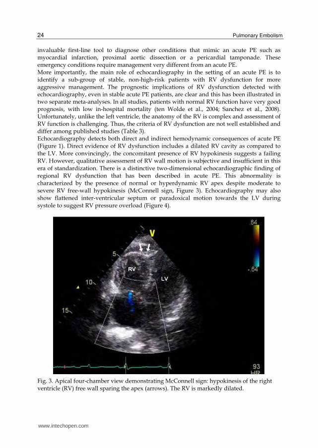
invaluable first-line tool to diagnose other conditions that mimic an acute PE such as myocardial infarction, proximal aortic dissection or a pericardial tamponade. These emergency conditions require management very different from an acute PE. More importantly, the main role of echocardiography in the setting of an acute PE is to identify a sub-group of stable, non-high-risk patients with RV dysfunction for more aggressive management. The prognostic implications of RV dysfunction detected with echocardiography, even in stable acute PE patients, are clear and this has been illustrated in two separate meta-analyses. In all studies, patients with normal RV function have very good prognosis, with low in-hospital mortality (ten Wolde et al., 2004; Sanchez et al., 2008). Unfortunately, unlike the left ventricle, the anatomy of the RV is complex and assessment of RV function is challenging. Thus, the criteria of RV dysfunction are not well established and differ among published studies (Table 3). Echocardiography detects both direct and indirect hemodynamic consequences of acute PE (Figure 1). Direct evidence of RV dysfunction includes a dilated RV cavity as compared to the LV. More convincingly, the concomitant presence of RV hypokinesis suggests a failing RV. However, qualitative assessment of RV wall motion is subjective and insufficient in this era of standardization. There is a distinctive two-dimensional echocardiographic finding of regional RV dysfunction that has been described in acute PE. This abnormality is characterized by the presence of normal or hyperdynamic RV apex despite moderate to severe RV free-wall hypokinesis (McConnell sign, Figure 3). Echocardiography may also show flattened inter-ventricular septum or paradoxical motion towards the LV during systole to suggest RV pressure overload (Figure 4).
Fig. 3. Apical four-chamber view demonstrating McConnell sign: hypokinesis of the right ventricle (RV) free wall sparing the apex (arrows). The RV is markedly dilated.
www.intechopen.com

Risk Stratification of Patients with Acute Pulmonary Embolism
25
Authors Definition of RV dysfunction
Goldhaber et al, 1993, 1999 Ribeiro et al, 1997, Jerjes-Sanchz et al, 2001, Kucher et al, 2003, 2005
RV hypokinesis by qualitative assessment of the RV wall motion
Kasper et al, 1997
Dilated RV cavity (qualitative assessment of RV compared to left ventricle) or RVEDD > 30mm; or when 2 of the following were present: 1. TR velocity > 2.8m/s 2. TR velocity > 2.5m/s in the absence of inspiratory
collapse of the IVC 3. Dilated RPA (> 12mm/m2) 4. RV wall thickness > 5mm 5. Loss of inspiratory collapse of the IVC
Grifoni et al, 2000, 2001
Presence of any 1 of the following:1. Dilated RV (RVEDD/LVEDD > 1 or RVEDD >
30mm) 2. Septal dyskinesis 3. Pulmonary hypertension (Doppler PAT <90ms or
RV-RA gradient >30mmHg) 4. Absence of RV hypertrophy (thickness > 7mm)
Pieralli et al, 2006
Presence of any 1 of the following:1. Dilated RV (RVEDD/LVEDD > 1 or RVEDD >
30mm) 2. Septal dyskinesis 3. Pulmonary hypertension (Doppler PAT <90ms or
RV-RA gradient >30mmHg)
Vieillar-Baron et al, 2001 RVEDA/LVEDA > 0.6 with septal dyskinesis
Kostrubiec et al, 2005
Presence of any 1 of the following: 1. RVEDD/LVEDD > 0.6 with RV hypokinesis 2. Pulmonary hypertension (Elevated TVPG >30mmHg
with PAT <80ms)
(RVEDD/LVEDD, right to left end-diastolic diameter ratio; RVEDA/LVEDA, right to left ventricular end-diastolic area ratio; RV-RA gradient, right ventricular-right atrial gradient; PAT, pulmonary arterial flow acceleration time; TVPG, tricuspid valve pressure gradient; IVC, inferior vena cava; TR, tricuspid regurgitation).
Table 3. Studies evaluating RV dysfunction with echocardiography
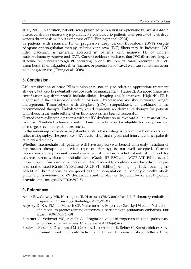
Indirect evidence of RV dysfunction from echocardiography includes raised pulmonary
artery systolic pressure (PASP). This can be estimated from the right ventricular systolic
pressure (RVSP) according to the formula: PASP = RVSP + estimated right atrial pressure
(Figure 5). The RVSP is obtained from the velocity of the tricuspid regurgitant jet (v), such
that RVSP = 4v2 and the right atrial pressure is estimated from the size and respiratory
variation of the inferior vena cava.
www.intechopen.com
Pulmonary Embolism
26
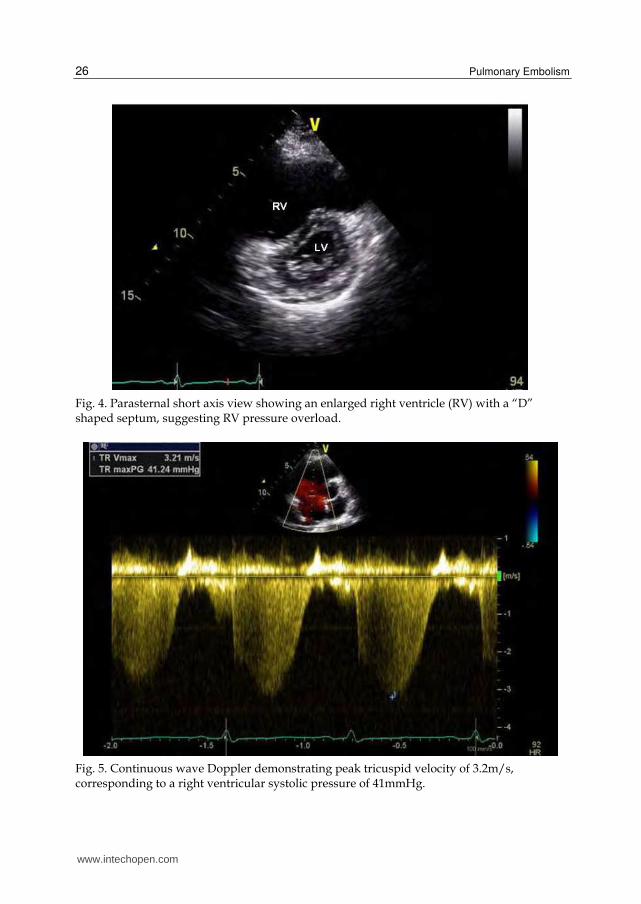
Fig. 4. Parasternal short axis view showing an enlarged right ventricle (RV) with a “D” shaped septum, suggesting RV pressure overload.
Fig. 5. Continuous wave Doppler demonstrating peak tricuspid velocity of 3.2m/s, corresponding to a right ventricular systolic pressure of 41mmHg.
www.intechopen.com

Risk Stratification of Patients with Acute Pulmonary Embolism
27
An elevated pulmonary artery systolic pressure of more than 50mmHg at time of diagnosis is associated with persistent pulmonary hypertension at 1 year (Ribeiro et al., 1999). In patients with acute PE, the absence of any significant tricuspid regurgitation makes the severe pulmonary hypertension less likely. Besides the evidence of RV dysfunction and elevated pulmonary arterial pressures, other echocardiographic features with prognostic implications include: 1. A right-to-left shunt, such as a patent foramen ovale (PFO). In a prospective study of
139 consecutive patients with acute PE, PFO was diagnosed in 48 patients by contrast echocardiography. Evidence of a PFO in patients with acute PE was associated with higher mortality rate (33% vs. 14%) and higher incidence of peripheral thromboembolic events (Konstantinides et al., 1998). These patients are particularly prone to paradoxical embolism due to increased right-to-left shunt from elevated right-sided pressures.
2. A free-floating right heart thrombus (Figure 6). The prevalence of patients with a right heart thrombus visualized during echocardiography was about 4% (Torbicki et al., 2003). Thrombus from the right heart usually arises from the lower limb veins. These thrombi are highly mobile and often described as having the appearance of a worm, or snake. Free-floating thrombus can embolize at any time and have a dismal prognosis regardless of therapeutic option (Chin et al., 2010). The mortality rate of about 20% within 24 hours of diagnosis, and mortality is significantly linked with the occurrence of cardiac arrest (Chartir et al., 1999).
5.2 Computed tomography Contrast enhanced computer tomography (CT) of the pulmonary arteries is increasingly used as a first-line imaging modality for PE diagnosis. The anatomical distribution and burden of embolic occlusion of the pulmonary arterial bed can be assessed easily by CT (Figure 7). However, the anatomical assessment seems less relevant for risk stratification than assessment based on functional (hemodynamic) consequences of PE. Most scanners allow reconstruction of standardized cardiac views and direct measurements of ventricular dimensions can be made. RV enlargement based on RV-to-LV dimension ratio, RVd/LVd, (Figure 8) on the reconstructed CT four-chamber view correlated with RV dysfunction on echocardiogram. Using RVd/LVd > 0.9 as cut-off, the sensitivity and specificity for predicting PE-related adverse events were 83% and 49% on the reconstructed CT, respectively. Comparatively, the sensitivity and specificity of RVd/LVd >0.9 on echocardiography were 71% and 56%, respectively (Quiroz et al., 2004). In addition to having good correlation with RV dysfunction on echocardiography, assessment of RV enlargement on chest CT in acute PE also predicted patients at risk of death from RV failure (Van der Meer et al., 2005; Schoepf et al., 2004). The greatest role appears to be the identification of low-risk patients due to its high negative predictive value (Table 4).
Author CT equipment
(Cutoff) Sensitivity
(%) Specificity
(%) NPV (%)
PPV (%)
Van der Meer et al., 2005
SDCT (RV/LV >1)
100 45 100 10
Schoepf et al., 2004
4 – 16 MDCT (RV/LV > 0.9)
78 38 92 16
Table 4. Trials reporting RV/LV diameter ratio assessed by CT as a risk marker for 30-day all cause mortality in acute pulmonary embolism.
www.intechopen.com

Pulmonary Embolism
28
Fig. 6. Free floating thrombus (red arrow) transiting from the RA causing acute pulmonary embolism (RA, right atrium; LA, left atrium; LV, left ventricle).
Other CT-derived parameters have also been investigated. The presence of interventricular septal bowing is predictive of PE-related deaths but has low sensitivity and high inter-observer variability (Araoz et al., 2007), scores to quantify the extent and location of pulmonary artery obstruction have been developed but not shown to be of prognostic relevance yet (Qanadil et al., 2001; Ghanima et al., 2007).
5.3 Ventilation-perfusion scintigraphy Lung ventilation-perfusion scintigraphy (V/Q scan) is a well-established diagnostic test used in patients suspected of PE. Interpretation of the scans can vary, depending on the algorithms used (PIOPED criteria, modified PIOPED criteria, McMaster Clinical criteria and PisaPED criteria) and the experience of the reader. The diagnostic roles and limitations of V/Q scan are beyond the scope and will not be discussed in this chapter.
www.intechopen.com

Risk Stratification of Patients with Acute Pulmonary Embolism
29
Fig. 7. Computed tomography pulmonary angiogram showing a large embolus within the right main pulmonary artery, extending to the main right upper lobe.
Fig. 8. Measurement of the short axes of the RV (47 mm) and LV (39 mm) on computed tomography pulmonary angiogram of the same patient (RV, right ventricle; LV, left ventricle)
www.intechopen.com

Pulmonary Embolism
30
Perfusion defects due to PE increase with the number and size of emboli, without
corresponding ventilation compromise (“mismatch” defects). However, the prognostic
implications of the number and size of defects on a V/Q scan have not been investigated.
6. Risk assessment based on biomarkers of myocardial injury
Cardiac troponins I and T as well as NT-pro brain natriuretic peptide (NT-proBNP) and
brain natriuretic peptide (BNP) have emerged as promising tools for risk stratification.
6.1 Cardiac troponins Cardiac troponins may be increased in patients with PE, even in the absence of coronary artery disease. The presumed mechanism is acute right heart overload attributed to myocardial ischemia and from oxygen supply-demand mismatch. The elevation usually resolves within 40 hours following PE in contrast to more prolonged elevation after an acute myocardial infarction. The peak level is usually lower than in acute myocardial infarction (Müller-Bardorff et al., 2002). Patients with an elevated troponin I or troponin T levels had an increased risk for short-term mortality (OR 5.24, 95% CI 3.28 – 8.38) or PE-related deaths (OR 9.44, 95% CI 4.14 – 21.49). Elevated troponin levels even among patients who are hemodynamically stable are associated with higher mortality (Becattini et al., 2007; Jimenez et al., 2008). Irrespective of various methods and cut-off values applied, most trials reported a low positive predictive value for PE-related mortality in the range of 12% to 44%, but with a very high negative predictive value between 99% and 100%.
6.2 Brain natriuretic peptide Right ventricular dysfunction is associated with increased myocardial stretch which leads to the release of BNP and its amino terminal portion, NT-proBNP. In acute PE, increasing levels of BNP or NT-proBNP predict the severity of RV dysfunction and mortality (Cavallazzi et al., 2008; Klok et al., 2008; Lega et al., 2009). Although elevated concentrations are related to worse outcome, the positive predictive value is low. On the other hand, low levels of BNP or NT-proBNP can be used reliably to identify patients with a good prognosis (Table 5).
6.3 Novel biomarker Heart-type fatty acid binding protein (H-FABP), a protein released earlier than troponins
during myocardial ischemia, has been evaluated as a prognostic marker in acute PE. The
studies have reported a high sensitivity (78% to 100%) and negative predictive value (96% to
100%), but these studies are small and such measurements are not widely available (Puls et
al., 2007; Kaczynska et al., 2006).
6.4 Summary of evidence on the prognostic value of biomarkers Many studies did not perform an extensive comparison between all the available biomarkers, thus it remains debatable which biomarker will yield the best prognostic value. Another limitation is biomarker thresholds were determined retrospectively, thus no
consistent cut-off values were used in the studies. Despite this, it appears BNP/NT-proBNP
and cardiac troponins could be used as rule-out tests.
www.intechopen.com

Risk Stratification of Patients with Acute Pulmonary Embolism
31
Author Test used
(Threshold) Outcome
Definition Sensitivity
(%) Specificity
(%) NPV (%)
PPV (%)
Ten Wolde et al, 2003
BNP Shionoriaa
(21.7 pmol/L)
PE-related deaths
86 71 99 17
Kucher et al, 2003a
NT-proBNPb
(500 pg/mL)
In-hospital death or adverse eventse
95 57 97 45
Kucher et al, 2003b
BNP Triagec
(50 pg/mL)
In-hospital death or adverse eventse
95 60 97 48
Pruszczyk et al, 2003
NT-proBNPb
(600 pg/mL) In-hospital mortality
100 33 100 23
Binder et al, 2005
NT-proBNPb
(1000 pg/mL)
In-hospital mortality
100 49 100 10
Kostrubiec et al, 2007
NT-proBNPb
(7500 pg/mL)
30-day all cause
mortality 65 93 94 61
(a-cTests used: aShionoria, CIS Bio International; bElecsys, Roche Diagnostics; cTriage, Biosite Technologies. eAdverse events include the need for resuscitation, mechanical ventilation, inotropic support, thrombolytics, or embolectomy)
Table 5. Prognostic value of BNP or NT-proBNP in acute pulmonary embolism
Due to the high negative predictive value for PE-related mortality and adverse events, a potential approach consists of a combination of biomarker testing and echocardiography. In the setting of an acute PE, further risk stratification with echocardiography is warranted in patients with elevated cardiac biomarkers due to limited specificity of the assays for predicting RV dysfunction. Conversely, in patients with levels below cut-off, echocardiography will likely not add prognostic information. This approach was demonstrated in a prospective study of 124 patients diagnosed with
acute PE. The presence of RV dysfunction on echocardiography in patients with elevated
NT-proBNP (cut-off of 1000 pg/mL) or cardiac troponins (cut-off of 0.04 ng/mL) is
associated with a 10-fold increase in complication risk compared with patients biomarker
levels below threshold (Binder et al., 2005).
7. Risk of recurrence
Recurrent PE can occur despite adequate anticoagulation therapy in patients who had survived an acute PE. Patients with unprovoked PE (PE occurring in the absence of established risk factors or predisposing illnesses) are at a higher risk for recurrent PE compared to patients with risk factors for PE. In contrast, patients with risk factors of PE have a higher mortality risk (Klok
www.intechopen.com
Pulmonary Embolism
32
et al., 2010). In addition, patients who presented with a first symptomatic PE are at a 4-fold increased risk of recurrent symptomatic PE compared to patients who presented with deep venous thrombosis without symptoms of PE (Eichinger et al., 2004). In patients with recurrent PE or progressive deep venous thrombosis (DVT) despite adequate anticoagulation therapy, inferior vena cava (IVC) filters may be indicated. IVC filter placement is generally accepted in patients with massive PE or limited cardiopulmonary reserve and DVT. Current evidence indicates that IVC filters are largely effective, with breakthrough PE occurring in only 0% to 6.2% cases. Recurrent PE, IVC thrombosis, filter migration, filter fracture, or penetration of caval wall can sometimes occur with long-term use (Chung et al., 2008).
8. Conclusion
Risk stratification of acute PE is fundamental not only to select an appropriate treatment strategy, but also to potentially reduce costs of management (Figure 2). An appropriate risk stratification algorithm would include clinical, imaging and biomarkers. High risk PE is diagnosed in the presence of shock or persistent hypotension and should warrant urgent management. Thrombolysis with alteplase (rtPA), streptokinase, or urokinase is the recommended therapy. Embolectomy could represent an alternative therapy for patients with shock in the acute setting when thrombolysis has been unsuccessful. Hemodynamically stable patients without RV dysfunction or myocardial injury are at low-risk for PE-related adverse events. These patients may be eligible for early hospital discharge or even outpatient treatment. In the remaining normotensive patients, a plausible strategy is to combine biomarkers with echocardiography. The presence of RV dysfunction and myocardial injury identifies patients at intermediate risk. Whether intermediate risk patients will have any survival benefit with early initiation of reperfusion therapy (and what type of therapy) is not well accepted. Current recommendations proposed thrombolysis be instituted in selected patients at high risk for adverse events without contraindications (Grade IIB ESC and ACCP VIII Edition), and intravenous unfractionated heparin should be reserved to conditions in which thrombolysis is contraindicated (Grade IA ESC and ACCP VIII Edition). An ongoing study assessing the benefit of thrombolysis as compared with anticoagulation in hemodynamically stable patients with evidence of RV dysfunction and an elevated troponin levels will hopefully provide some insights (NCT00639743).
9. References
Araoz PA, Gotway MB, Harrington JR, Harmsen WS, Mandrekar JN. Pulmonary embolism: prognostic CT findings. Radiology 2007;242:889.
Aujesky D, Roy PM, Le Manach CP, Verschuren F, Meyer G, Obrosky DS et al. Validation of a model to predict adverse outcomes in patients with pulmonary embolism. Eur Heart J 2006;27:476–481.
Becattini C, Vedovati MC, Agnelli G. Prognostic value of troponins in acute pulmonary embolism: a meta-analysis. Circulation 2007;116(4):427.
Binder L, Pieske B, Olschewski M, Geibel A, Klostermann B, Reiner C, Konstantinides S. N-terminal pro-brain natriuretic peptide or troponin testing followed by
www.intechopen.com
Risk Stratification of Patients with Acute Pulmonary Embolism
33
echocardiography for risk stratification of acute pulmonary embolism. Circulation. 2005 Sep 13;112(11):1573-9. Epub 2005 Sep 6.
Cavallazzi R, Nair A, Vasu T, Marik PE. Natriuretic peptides in acute pulmonary embolism: a systematic review. Intensive Care Med. 2008;34(12):2147.
Chartier L, Bera J, Delomez M, Asseman P, Beregi JP, Bauchart JJ, Warembourg H, Thery C. Free floating thrombi in the right heart: diagnosis, management, and prognostic indexes in 38 consecutive patients. Circulation 1999;99:2779-83.
Chin C, Lim ST, Ho KW, et al. Free Floating Thrombus in the Right Heart Causing Pulmonary Embolism. Postgraduate Medical Journal 2010;86:307.
Chung J, Owen RJ. Using inferior vena cava filters to prevent pulmonary embolism. Can Fam Physician. 2008 Jan;54(1):49-55.
Eichinger S, Weltermann A, Minar E, Stain M, Schönauer V, Schneider B, Kyrle PA. Symptomatic pulmonary embolism and the risk of recurrent venous thromboembolism. Arch Intern Med. 2004 Jan 12;164(1):92-6.
Ghanima W, Abdelnoor M, Holmen LO, Nielssen BE, Sandset PM. The association between the proximal extension of the clot and the severity of pulmonary embolism (PE): a proposal for a new radiological score for PE. J Intern Med. 2007;261:74.
Goldhaber SZ, Elliott CG. Acute pulmonary embolism: part 1. Epidemiology, pathophysiology, and diagnosis. Circulation 2003;108:2726.
Goldhaber SZ, Haire WD, Feldstein ML, et al. Alteplase versus heparin in acute pulmonary embolism: randomised trial assessing right-ventricular function and pulmonary perfusion. Lancet. 1993;341:507-511.
Goldhaber SZ, Visani L, De Rosa M. Acute pulmonary embolism: clinical outcomes in the International Cooperative Pulmonary Embolism Registry (ICOPER). Lancet 1999;353:1386.
Goldhaber SZ, Visani L, De Rosa M. Acute pulmonary embolism: clinical outcomes in the International Cooperative Pulmonary Embolism Registry (ICOPER). Lancet. 1999;353:1386-1389.
Grifoni S, Olivotto I, Cecchini P, et al. Short-term clinical outcome of patients with acute pulmonary embolism, normal blood pressure, and echocardiographic right ventricular dysfunction. Circulation. 2000;101:2817-2822.
Grifoni S, Olivotto I, Pieralli F, et al. Long-term clinical outcome of patients with pulmonary embolism with or without right ventricular dysfunction [abstract]. Thromb Haemost. 2001;86(suppl). Abstract P2231.
Grifoni S, Vanni S, Magazzini S, Olivotto I, Conti A, Zanobetti M, Polidori G, Pieralli F, Peiman N, Becattini C, Agnelli G. Association of persistent right ventricular dysfunction at hospital discharge after acute pulmonary embolism with recurrent thromboembolic events. Arch Intern Med. 2006;166(19):2151.
Jerjes-Sanchez C, Ramirez-Rivera A, Arriaga-Nava R, et al. High dose and short term streptokinase infusion in patients with pulmonary embolism: prospective with seven-year follow-up trial. J Thromb Thrombolysis. 2001;12:237-247
Jimenez D, Aujesky D, Moores L, Gomez V, Lobo Jose, et al. Simplification of the pulmonary embolism severity index for prognostication in patients with acute symptomatic pulmonary embolism. Arch Intern Med 2010;170:1383-1389.
www.intechopen.com

Pulmonary Embolism
34
Jiménez D, Díaz G, Marín E, Vidal R, Sueiro A, Yusen RD. The risk of recurrent venous thromboembolism in patients with unprovoked symptomatic deep vein thrombosis and asymptomatic pulmonary embolism. Thromb Haemost. 2006 Mar;95(3):562-6.
Jiménez D, Díaz G, Molina J, Martí D, Del Rey J, García-Rull S, Escobar C, Vidal R, Sueiro A, Yusen RD. Troponin I and risk stratification of patients with acute nonmassive pulmonary embolism. Eur Respir J. 2008 Apr;31(4):847-53. Epub 2007 Dec 19.
Jiménez D, Yusen RD, Otero R, Uresandi F, Nauffal D, Laserna E, Conget F, Oribe M, Cabezudo MA, Díaz G. Prognostic models for selecting patients with acute pulmonary embolism for initial outpatient therapy. Chest. 2007;132(1):24-30.
Kaczyńska A, Pacho R, Bochowicz A et al. Does saddle embolism influence short-term prognosis in patients with acute pulmonary embolism? Kardiol Pol, 2005; 62: 119–127.
Kaczynska An, Pelsers MM, Bochowicz A, Kostrubiec M, Glatz JF, Pruszczyk P. Plasma heart-type fatty acid binding protein is superior to troponin and myoglobin for rapid risk stratification in acute pulmonary embolism. Clin Chim Acta. 2006;371:117.
Kasper W, Konstantinides S, Geibel A, Tiede N, Krause T, Just H. Prognostic significance of right ventricular afterload stress detected by echocardiography in patients with clinically suspected pulmonary embolism. Heart. 1997;77:346-349.
Klok FA, Mos IC, Huisman MV. Brain-type natriuretic peptide levels in the prediction of adverse outcome in patients with pulmonary embolism: a systematic review and meta-analysis. Am J Respir Crit Care Med. 2008;178(4):425.
Klok FA, Zondag W, van Kralingen KW, van Dijk AP, Tamsma JT, Heyning FH, Vliegen HW, Huisman MV. Patient outcomes after acute pulmonary embolism. A pooled survival analysis of different adverse events. Am J Respir Crit Care Med. 2010 Mar 1;181(5):501-6. Epub 2009 Dec 3.
Konstantinides S, Geibel A, Kasper W, Olschewski M, Blumel L, Just H. Patent foramen ovale is an important predictor of adverse outcome in patients with major pulmonary embolism. Circulation 1998;97:1946.
Kostrubiec M, Pruszczyk P, Kaczynska A, Kucher N. Persistent NT-proBNP elevation in acute pulmonary embolism predicts early death. Clin Chim Acta 2007;382:124.
Kucher N, Printzen G, Doernhoefer T, Windecker S, Meier B, Hess OM. Low pro-brain natriuretic peptide levels predict benign clinical outcome in acute pulmonary embolism. Circulation 2003;107:1576–1578.
Kucher N, Printzen G, Goldhaber SZ. Prognostic role of brain natriuretic peptide in acute pulmonary embolism. Circulation 2003;107:2545.
Kucher N, Rossi E, De Rosa M, et al. Prognostic role of echocardiography in patients with acute PE and a systemic arterial pressure of 90mmHg or higher. Arch Intern Med 2005;165:1777.
Lega JC, Lacasse Y, Lakhal L, Provencher S. Natriuretic peptides and troponins in pulmonary embolism: a meta-analysis. Thorax. 2009;64(10):869.
McConnell MV, Solomon SD, Rayan ME, Come PC, Goldhaber SZ, Lee RT. Regional right ventricular dysfunction detected by echocardiography in acute pulmonary embolism. Am J Cardiol. 1996 Aug 15;78(4):469-73.
www.intechopen.com

Risk Stratification of Patients with Acute Pulmonary Embolism
35
Müller-Bardorff M, Weidtmann B, Giannitsis E, Kurowski V, Katus HA. Release kinetics of cardiac troponin T in survivors of confirmed severe pulmonary embolism. Clin Chem. 2002;48(4):673-5.
Pieralli F, Olivotto I, Vanni S, Conti A, Camaiti A, Targioni G, Grifoni S, Berni G. Usefulness of bedside testing for brain natriuretic peptide to identify right ventricular dysfunction and outcome in normotensive patients with acute pulmonary embolism. Am J Cardiol 2006;97:1386–1390.
Pruszczyk P, Kostrubic M, Bochowicz A, Styczynski G, Szulc M, Kurzyna M et al. N terminal pro-brain natriuretic pepetide in patients with acute pulmonary embolism. Eur Respir J 2003;22:649.
Pruszczyk P, Pacho R, Ciurzynski M et al. Short term clinical outcome of acute saddle pulmonary embolism. Heart 2003; 89: 335–336.
Puls M, Dellas C, Lankeit M, et al. Heart-type fatty acid-binding protein permits early risk stratification of pulmonary embolism. Eur Heart J 2007;28:224.
Qanadli SD, El Hajjam M, Viellard-Baron A, et al. New CT index to quantify arterial obstruction in pulmonary embolism: comparison with angiographic index and echocardiography. Am J Roentgenol 2001;176:1415.
Quiroz R, Kucher N, Schoepf UJ, Kipfmueller F, Solomon SD, Costello P, et al. Right ventricular enlargement on chest computed tomography: prognostic role in acute pulmonary embolism. Circulation 2004;109:2401.
Ribeiro A, Lindmarker P, Johnsson H, Juhlin-Dannfelt A, Jorfeldt L. Ribeiro A, Lindmarker P, Johnsson H, Juhlin-Dannfelt A, Jorfeldt L. Pulmonary embolism: one-year follow-up with echocardiography doppler and five-year survival analysis. Circulation. 1999 Mar 16;99(10):1325-30.
Ribeiro A, Lindmarker P, Juhlin-Dannfelt A, Johnsson H, Jorfeldt L. Echocardiography Doppler in pulmonary embolism: right ventricular dysfunction as a predictor of mortality rate. Am Heart J. 1997;134:479-487.
Ryu JH, Pelikka PA, Froehling DA, Peters SG, Aughenbaugh GL. Saddle pulmonary embolism diagnosed by CT angiography: frequency, clinical features and outcome. Respir Med 2007;101:1537.
Sanchez O, Trinquart L, Colombet I, Durieux P, Huisman MV, Chatellier G, Meyer G. Prognostic value of right ventricular dysfunction in patients with hemodynamically stable pulmonary embolism: a systemic review. Eur Heart J 2008;29:1569.
Schoepf UJ, Kucher N, Kipfmueller F, Quiroz R, Costello P, Goldhaber SZ. Right ventricular enlargement on chest computed tomography: a predictor of early death in acute pulmonary embolism. Circulation. 2004 Nov 16;110(20):3276-80. Epub 2004 Nov 8.
Ten Wolde M, Söhne M, Quak E, Mac Gillavry MR, Büller HR. Prognostic value of echocardiographically assessed right ventricular dysfunction in patients with pulmonary embolism. Arch Intern 2004;164:1685.
Ten Wolde M, Tulevski II, Mulder JW, Sohne M, Boomsma F, Mulder BJ et al. Brain natriuretic peptide as a predictor of adverse outcome in patients with pulmonary embolism. Circulation 2003;107:2082.
The urokinase pulmonary embolism trial. JAMA 1970; 214:2163-72
www.intechopen.com

Pulmonary Embolism
36
Torbicki A, Galie N, Covezzoli A, Rossi E, De Rosa M, Goldhaber SZ. Right heart thrombi in pulmonary embolism: results from the International Cooperative Pulmonary Embolism Registry. J Am Coll Cardiol 2003;41:2245.
Torbicki A, Perrier A, Konstantinides S, et al., Guidelines on the diagnosis and management of pulmonary embolism. European Heart Journal 2008;29:2276-2315.
van der Meer RW, Pattynama PM, van Strijen MJ, van den Berg-Huijsmans AA, Hartmann IJ, Putter H, de Roos A, Huisman MV Right ventricular dysfunction and pulmonary obstruction index at helical CT: prediction of clinical outcome during 3-month follow-up in patients with acute pulmonary embolism. Radiology. 2005 Jun;235(3):798-803. Epub 2005 Apr 21.
Vieillard-Baron A, Page B, Augarde R, Prin S, Qanadli S, Beauchet A, Dubourg O, Jardin F. Acute cor pulmonale in massive pulmonary embolism: incidence, echocardiographic pattern, clinical implications and recovery rate. Intensive Care Med 2001;27:1481–1486.
Wicki J, Perrier A, Perneger TV, et al. Predicting adverse outcome in patients with acute pulmonary embolism: a risk score. Thromb Haemost 2000;84:548.
Wood KE. Major pulmonary embolism: review of a pathophysiologic approach to the golden hour of hemodynamically significant pulmonary embolism. Chest 2002;121:877–905.
www.intechopen.com

Pulmonary EmbolismEdited by Dr. Ufuk Çobanoğlu
ISBN 978-953-51-0233-5Hard cover, 236 pagesPublisher InTechPublished online 14, March, 2012Published in print edition March, 2012
InTech EuropeUniversity Campus STeP Ri Slavka Krautzeka 83/A 51000 Rijeka, Croatia Phone: +385 (51) 770 447 Fax: +385 (51) 686 166
InTech ChinaUnit 405, Office Block, Hotel Equatorial Shanghai No.65, Yan An Road (West), Shanghai, 200040, China
Phone: +86-21-62489820 Fax: +86-21-62489821
Pulmonary embolism is a serious, potentially life-threatening cardiopulmonary disease that occurs due topartial or total obstruction of the pulmonary arterial bed. Recently, new improvement occurred in the diagnosisand treatment of the disease. The aim of this disease is to re-review pulmonary embolism in the light of newdevelopments. In this book, in addition to risk factors causing pulmonary embolus, a guide for systematicapproaches to lead the risk stratification for decision making is also presented. In order to provide a maximumlength of active life and continuation of functional abilities as the aim of new interventional gerontology, the riskfactors causing pulmonary embolus in elderly individuals are evaluated, and the approach to prevention andtreatment are defined. The risk of the development of deep vein thrombosis and pulmonary embolism,combined with obesity due to immobility, the disease of this era, irregular and excessive eating, and treatmentmanagement are highlighted. Non-thrombotic pulmonary emboli are also covered and an attempt is made toconstitute an awareness of this picture that can change the treatment and prognosis of the disease to aconsiderable extent. In addition to the pathophysiological definition of pulmonary embolus, the priority goal ofquick and definitive diagnosis is emphasized, and diagnostic strategies are discussed in the book. A numericalanalysis of the vena cava filters, which is a current approach to prevent pulmonary emboli recurrences, ispresented in the last chapter.
How to referenceIn order to correctly reference this scholarly work, feel free to copy and paste the following:
Calvin Woon-Loong Chin (2012). Risk Stratification of Patients with Acute Pulmonary Embolism, PulmonaryEmbolism, Dr. Ufuk Çobanoğlu (Ed.), ISBN: 978-953-51-0233-5, InTech, Available from:http://www.intechopen.com/books/pulmonary-embolism/risk-stratification-of-patients-with-acute-pulmonary-embolism
www.intechopen.com

www.intechopen.com

© 2012 The Author(s). Licensee IntechOpen. This is an open access articledistributed under the terms of the Creative Commons Attribution 3.0License, which permits unrestricted use, distribution, and reproduction inany medium, provided the original work is properly cited.