risk evaluation of nitrofurans in animal food...
TRANSCRIPT

Risk evaluation of nitrofurans in animalfood products
Linus Carlsson Forslund
Degree project in biology, Master of science (2 years), 2014Examensarbete i biologi 45 hp till masterexamen, 2014Biology Education Centre, Uppsala University, and the Swedish National Food AgencySupervisors: Bitte Aspenström-Fagerlund and Lilianne Abramsson-ZetterbergExternal opponent: Björn Brunström

1
Table of contents Abstract ............................................................................................................................ 3
1 Introduction .............................................................................................................. 5
2 Nitrofurazone (nitrofural) ......................................................................................... 9
2.1 Physicochemical properties ............................................................................. 10
2.2 Pharmacodynamics .......................................................................................... 10
2.3 Pharmacokinetics ............................................................................................. 10
2.4 Toxicology of nitrofurazone ............................................................................ 16
3 Nitrofurantoin ......................................................................................................... 30
3.1 Physicochemical properties ............................................................................. 30
3.2 Pharmacodynamics .......................................................................................... 31
3.3 Pharmacokinetics ............................................................................................. 31
3.4 Toxicology of nitrofurantoin ........................................................................... 39
4 Furazolidone ........................................................................................................... 51
4.1 Physicochemical properties ............................................................................. 51
4.2 Pharmacodynamics .......................................................................................... 52
4.3 Pharmacokinetics ............................................................................................. 53
4.4 Toxicology of furazolidone.............................................................................. 66
5 Furaltadone ............................................................................................................. 79
5.1 Physicochemical properties ............................................................................. 79
5.2 Pharmacodynamics .......................................................................................... 79
5.3 Pharmacokinetics ............................................................................................. 79
5.4 Toxicology of furaltadone................................................................................ 84
6 Summary of the effects of nitrofurans .................................................................... 85
7 Risk assessment of nitrofurans ............................................................................... 86
7.1 Benchmark dose (BMD) .................................................................................. 87
7.2 Exposure assessment ........................................................................................ 89
7.3 Margin of exposure (MoE) .............................................................................. 91
8 Discussion and conclusions .................................................................................... 92
9 Acknowledgments .................................................................................................. 97
10 References .............................................................................................................. 98
11 Appendix .............................................................................................................. 107

2
11.1 Bacterial reverse mutation test ................................................................... 107
11.2 Mammalian erythrocyte micronucleus test ................................................ 107
11.3 In vitro mammalian chromosome aberration assay.................................... 107

3
Abstract
Residues from non-allowed pharmacologically active substances are sometimes found
in food products of animal origin in EU border controls. Nitrofurans are one such class
of substances, to which nitrofurazone, nitrofurantoin, furazolidone and furaltadone
belong. Nitrofurans have been used in both human and veterinary medicine for their
antibacterial and antiprotozoal activities. In the 1990s, they were completely banned
from use in food producing animals within the EU due to their genotoxicity. The only
nitrofuran still in use is nitrofurantoin, which is utilised in human medicine to treat
urinary tract infections. The aim of this thesis was to review the research on nitrofurans
and to determine if any levels can be allowed in food products of animal origin. All
substances, except for furaltadone, have shown to be genotoxic and mutagenic in vitro.
No clear conclusions regarding the genotoxicity in vivo could be drawn due to
contradicting results. Concerning the reproductive toxicity of these compounds only
nitrofurazone and nitrofurantoin have clearly shown that they are toxic to the
reproductive system of animals. The lowest daily dose which caused adverse effects on
the reproduction was 10 mg nitrofurazone per kg BW. In carcinogenicity tests the most
commonly observed effect was an increase in mammary tumours. Nitrofurazone and
furazolidone was shown to be carcinogenic, while nitrofurantoin may be carcinogenic.
The lowest dose that caused this effect was 0.16 mg nitrofurazone per kg BW.
The margin of exposure (MoE) approach was used in order to determine the risk
for the Swedish population from nitrofurans in food products of animal origin. Several
benchmark dose lower confidence limits (BMDLs) were derived from carcinogenicity
studies and the lowest BMDL was chosen for the MoE. The exposure to nitrofurans for
adults and children were estimated from intake data in Riksmaten, a Swedish national
dietary survey, and the level in food was assumed to be 1 µg/kg. The MoE for adults
only exposed to nitrofurans via medical products was calculated. It is considered to be a
risk when the MoE value is lower than 10000. The MoEs for adults treated with
nitrofurantoin for urinary tract infections were below 10000 indicating that there may be
a risk for those on a course of treatment with nitrofurantoin. Hence, the safety of
nitrofurantoin as a drug used in human medicine should be revaluated. The MoEs
indicated that there is a negligible risk to the health of the Swedish population from
nitrofurans in food at the level of 1 µg/kg. If all food products of animal origin
consumed in one day contain 8 µg/kg there may be a risk to human health. Therefore, it
is advised that the current reference point of action (RPA) of 1 µg/kg should be

4
retained. Food products of animal origin containing nitrofurans over this level should
not be allowed to enter the market, thus protecting the health of the Swedish population.

5
1 Introduction
Residues from pharmacologically active substances can be present in food from animals
treated with veterinary medicinal products (VMPs) prior to slaughter, sampling of milk,
eggs and honey. In Regulation (EEC) No 2377/90, later repealed by Regulation (EC)
470/2009, the European Council stated that in order to protect consumers from the
potentially harmful effects of these residues maximum residue limits (MRLs) in food of
animal origin should be established for all pharmacologically active substances used in
VMPs to treat food producing animals. The substances could fall into four annexes.
Annex I contained a list of the substances for which MRLs had been established. Annex
II contained a list of the substances for which no MRLs were necessary because they
pose no risk to the public health, although with some exceptions. Annex III contained a
list of the substances for which provisional MRLs had been established. Annex IV
contained a list of the substances for which MRLs could not be established because they
pose a risk to the public health at any concentration. The allowed substances and their
MRLs are now presented in the first table in Commission Regulation (EU) No 37/2010.
The prohibited substances from annex IV are found in the second table of the same
regulation.
In Council Regulation (EEC) No 2309/93, later repealed by Regulation (EC) No
726/2004, it is stated that the applicant shall submit an application for a VMP to the
European Medicines Agency (EMA) who will form opinions on MRLs. The risk
assessments and opinions of the EMA on MRLs are formulated by the Committee for
Medicinal Products for Veterinary Use (CVMP), established by Council Directive
(EEC) No 81/851, after reviewing the data provided by the applicants concerning the
toxicological and pharmacological effects, pharmacokinetics and pharmacodynamics,
and also physicochemical properties of the substances as well as validated analytical
methods (Council Directive 81/851/EEC, Council Regulation (EEC) No 2309/93). The
provided opinion consists of a scientific risk assessment and risk management
recommendations (Regulation (EC) No 470/2009).
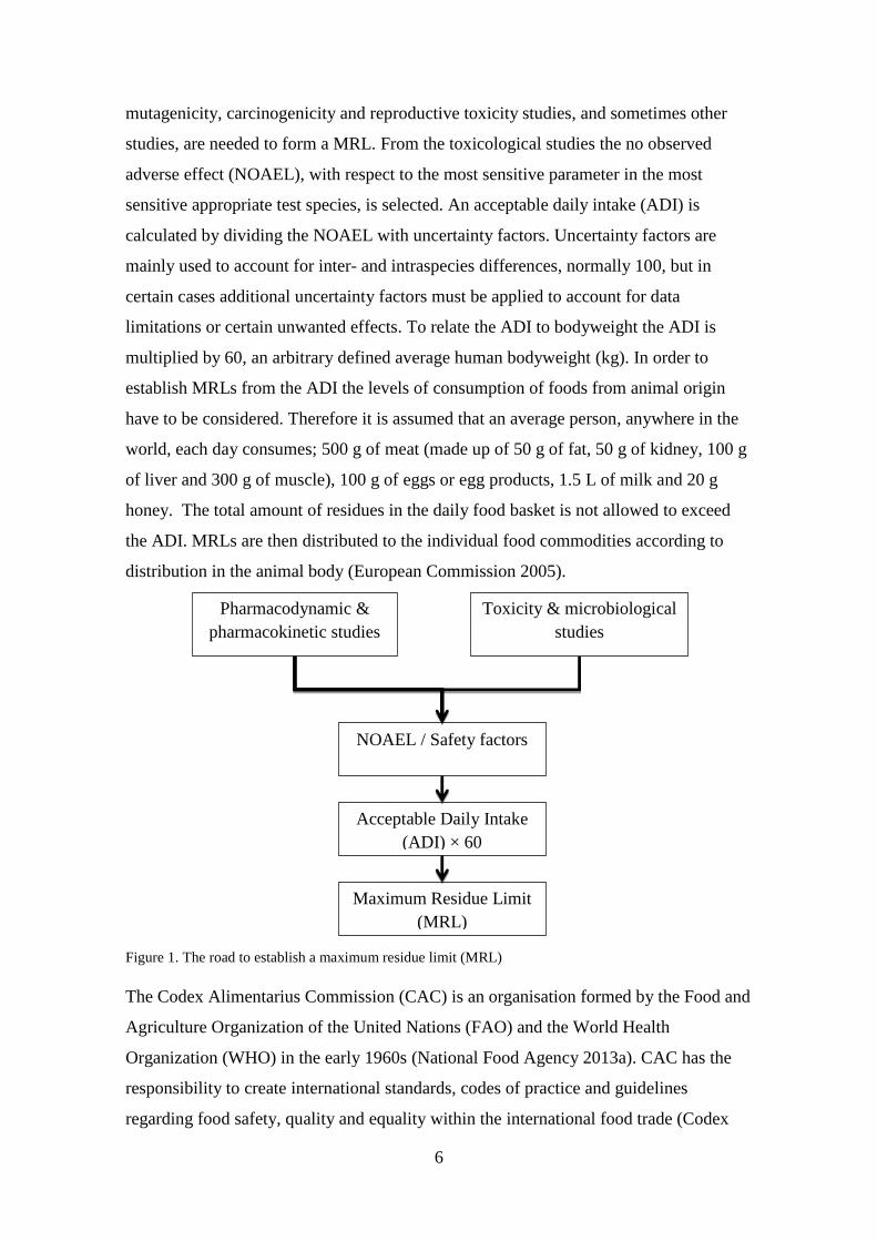
In order to establish MRLs the CVMP first reviews the toxicological, pharmacological,
pharmacokinetic, microbiological and other studies submitted (Figure 1) (European
Commission 2005). The results from acute and repeated dose toxicity studies,
6
mutagenicity, carcinogenicity and reproductive toxicity studies, and sometimes other
studies, are needed to form a MRL. From the toxicological studies the no observed
adverse effect (NOAEL), with respect to the most sensitive parameter in the most
sensitive appropriate test species, is selected. An acceptable daily intake (ADI) is
calculated by dividing the NOAEL with uncertainty factors. Uncertainty factors are
mainly used to account for inter- and intraspecies differences, normally 100, but in
certain cases additional uncertainty factors must be applied to account for data
limitations or certain unwanted effects. To relate the ADI to bodyweight the ADI is
multiplied by 60, an arbitrary defined average human bodyweight (kg). In order to
establish MRLs from the ADI the levels of consumption of foods from animal origin
have to be considered. Therefore it is assumed that an average person, anywhere in the
world, each day consumes; 500 g of meat (made up of 50 g of fat, 50 g of kidney, 100 g
of liver and 300 g of muscle), 100 g of eggs or egg products, 1.5 L of milk and 20 g
honey. The total amount of residues in the daily food basket is not allowed to exceed
the ADI. MRLs are then distributed to the individual food commodities according to
distribution in the animal body (European Commission 2005).
Figure 1. The road to establish a maximum residue limit (MRL)
The Codex Alimentarius Commission (CAC) is an organisation formed by the Food and
Agriculture Organization of the United Nations (FAO) and the World Health
Organization (WHO) in the early 1960s (National Food Agency 2013a). CAC has the
responsibility to create international standards, codes of practice and guidelines
regarding food safety, quality and equality within the international food trade (Codex
Pharmacodynamic &
pharmacokinetic studies
Toxicity & microbiological
studies
NOAEL / Safety factors
Acceptable Daily Intake
(ADI) × 60
Maximum Residue Limit
(MRL)

7
Alimentarius 2013). This includes the establishment of MRLs for VMPs by CAC after
scientific evaluations by the Joint FAO/WHO Expert Committee on Food Additives
(JECFA) and discussions in the codex committee for residues of veterinary drugs in
food (CCRVDF) (Codex Alimentarius 2014, National Food Agency 2013b).
To ensure that the MRLs are not exceeded, withdrawal periods should be determined for
the concerned pharmaceuticals and species (Directive 2001/82/EC). The withdrawal
period is the time after the last administration of a VMP during which the animal is not
allowed to be slaughtered or during which eggs or milk is not allowed to be taken for
human consumption. The withdrawal periods are determined by the CVMP or the
Member States for each species of food-producing animal and their edible products
(National Food Agency 2013c). To determine the withdrawal periods marker residue
depletion studies are performed on individual animal species, e.g. swine, poultry, cattle,
horses and sheep (European Medicines Agency 2011). During the study the highest
treatment dose is used for the maximum intended duration. The studies show how long
it takes for the marker residue to be depleted down to the MRL after the treatment has
ceased (European Medicines Agency 2011).
In the European Council directive 96/23/EC it is stated that Member States should
perform controls to determine residue levels in animal products and to make certain that
regulations are followed. The National Food Agency (NFA) in Sweden analyses around
5000 samples of food products (milk, fish, eggs, honey and meat during slaughter) each
year (Nordlander et al. 2013). Before this directive the analysis of non-allowed residues
of certain substances in food products from animals differed in limits of detection
between, and even in, Member States. The differences lead to differential treatment of
food producers supplying different countries in the European Union (EU). As the
methods of analysis became more sophisticated the limits of detection decreased which
lead to more non-allowed substances being detected. To ensure the same level of
consumer protection throughout the EU and to harmonize the treatment of imported
food products it was decided that minimum required performance limits (MRPLs) of
analytical methods, to be used for substances for which no MRLs have been established,
should be implemented (Commission Decision 2002/657/EC). The MRPLs are not
health based and should not be interpreted as it is safe to consume food products
containing levels below this limit. The MRPLs only establish the lowest levels that
Member States must be able to analyse residues in food stuff. The substances that have

8
specified MRPLs are; chloramphenicol, malachite green, medroxyprogesterone, and
nitrofuran metabolites (Commission Decision 2002/657/EC as amended by Commission
Decision 2003/181/EC and Commission Decision 2004/25/EC).
Nitrofurans are a class of drugs that have been used for both veterinary and human
medicine due to their antibacterial and antiprotozoal properties (Reynolds 1982,
Sweetman 2002). To this class belong the substances nitrofurazone, nitrofurantoin,
furazolidone and furaltadone. Of these compounds only nitrofurantoin is still in use in
Sweden, whereas the others were withdrawn from the market in the 50s, 60s and 70s
(FASS 2014a, 2014b, 2014c, 2014d). European legislation prohibited all nitrofurans,
except furazolidone, from use in food-producing animals in 1993 due to their
genotoxicity (Council Regulation (EEC) No 2901/93). Furazolidone was also prohibited
two years later (Commission Regulation (EC) No 1442/95). During controls of food
products the presence of marker residues for these nitrofurans are investigated. For
nitrofurazone, nitrofurantoin, furazolidone and furaltadone the marker residues are:
semicarbazide (SEM), 1-aminohydantoin (AHD), 3-amino-2-oxazolidone (AOZ) and 5-
morpholinomethyl-3-amino-2-oxazolidone (AMOZ), respectively (National Food
Agency 2012).
Residues of non-allowed and prohibited substances have been found in food of animal
origin in the controls both in Sweden and other European countries (Gustavsson et al.
2012, European Commission 2013, Nordlander et al. 2013). In 2011 three samples of
shrimp from China tested positive for nitrofuran metabolites in Swedish controls
(Gustavsson et al. 2012). Between 2005 and March 2014 there have been 365
notifications on nitrofuran metabolites in food across Europe via the Rapid Alert
System for Food and Feed (RASFF) (RASFF Portal 2014). The number of RASFF
notifications was around 50 per year between 2005 and 2008. In 2009 that number
increased to 94, but then dropped the following years to around 20 notifications per year
(Figure 2). Since genotoxic substances are considered to exert their effects at any
concentration no MRLs for such substances can be determined and consequently no
withdrawal period can be established (Falk-Filipsson et al. 2007). Pharmacologically
active substances not mentioned in Table 1 of Commission Regulation (EU) No
37/2010 are not allowed to be used in food-producing animals (Regulation (EC) No
470/2009). When such substances are found in residue surveillance above their

9
reference point of action (RPA), then that country’s responsible agency is required to
take action (European Commission 2013). However, these actions can differ between
member countries, leading to a continued differential treatment of imported food
products within the EU. When levels lower than the RPA, or in the case of nitrofurans
the MRPL, are found it is up to that country if actions will be taken (European
Commission 2013). For nitrofuran metabolites the RPA is the MRPL (1 µg/kg) (EFSA
Panel on Contaminants in the Food Chain 2013). Sometimes during controls these
metabolites are found in low concentrations, below the MRPL, and it can be difficult to
know exactly what risk they pose to the consumers and what, and even if, actions
should be taken. The objective of this master’s thesis is to determine if any levels of
nitrofurans can be allowed in animal food products. Is there a point where the levels in
food are so low that the risk to the human population is negligible? What actions should
be taken when nitrofurans are detected above and below the RPA?
Figure 2. The number of RASFF notifications from European border controls regarding nitrofurans in
food products between 2005 and 2014
2 Nitrofurazone (nitrofural)
Nitrofurazone is a broad spectrum antibiotic effective against both Gram-positive and
Gram-negative bacteria (Reynolds 1982, Sweetman 2002). In human medicine it was
used in the treatment of burns, ulcers, wounds and skin infections (Reynolds 1982,
Sweetman 2002). In veterinary medicine it was used to treat necrotic enteritis in pigs
and coccidiosis (a parasite) in farm animals and poultry (Reynolds 1982).
0
10
20
30
40
50
60
70
80
90
100

10
2.1 Physicochemical properties
International Non-proprietary Name (INN)
Nitrofurazone (nitrofural)
Chemical Abstract Service (CAS) name
5-Nitro-2-furaldehyde semicarbazone
CAS number: 59-87-0
Structural formula
Molecular formula
C8H6N4O4
Molecular weight
198.14
Table 1. Physicochemical properties (Debnath et al. 1991, FAO 1993a)
Melting point 236-240 °C
Solubility in water (pH 6.0-6.5) Very slightly soluble (1:4200)
Solubility in ethanol Slightly soluble (1:590)
Solubility in benzene Not soluble
Octanol/Water Partition Coefficient Log Kow = 0.23
2.2 Pharmacodynamics
Ali et al. (1988) orally administered nitrofurazone (7.5, 15 or 30 mg/kg BW) to male
turkeys for two weeks and then measured the levels of luteinizing hormone (LH) and
prolactin (PRL). It was shown that nitrofurazone treatment (30 mg/kg BW)
significantly increased the levels of PRL and significantly decreased the levels of LH.
Depending on the concentration, nitrofurazone is either bacteriostatic (low
concentration) or bactericidal (high concentration) (Dodd & Stillman 1944, Cramer &
Dodd 1945).
2.3 Pharmacokinetics
2.3.1 In vitro studies
Nitrofurazone was incubated with 8500g supernatant of liver homogenate prepared from
male rats aged 6-10 weeks (Akao et al. 1971). After 60 minutes of incubation under
Figure 3. Structural formula of nitrofurazone

11
aerobic conditions there was a change in optical absorbance corresponding to a loss of a
nitro group. This was also the case under anaerobic conditions, but there was also an
increase in absorbance at the maximum absorption wave length of 5-amino-2-
furaldehyde semicarbazone. It was concluded that 5-amino-2-furaldehyde
semicarbazone is a metabolite of nitrofurazone.
Human liver microsomes were incubated with 50 µL nitrofurazone or 250 µL 14
C-
nitrofurazone for two hours at 37 °C (Wang et al. 2010). Using liquid chromatography
(LC)-radiometric and liquid chromatography-tandem mass spectrometry (LC-MS/MS) a
metabolite was detected and identified as a cyano metabolite.
2.3.2 In vivo studies
2.3.2.1 In pig
Pigs (n=18) were fed feed containing nitrofurazone (400 mg/kg feed) ad libitum for ten
days, corresponding to around 24 mg/kg BW per day, followed by a withdrawal period
for 6 weeks (Cooper et al. 2005). Three pigs were sacrificed each week of the
withdrawal period and samples of muscle, liver and kidney were taken and analysed for
nitrofurazone and semicarbazide (SEM) using LC-MS/MS and high performance liquid
chromatography-UV (HPLC-UV). Nitrofurazone was detected in all muscle samples (4-
21.9 µg/kg) at week 0 of the withdrawal period. SEM was detected in all samples and
the levels found at week 6 of the withdrawal period were around 50 µg/kg in kidney and
liver and 250 µg/kg in muscle. The depletion half-lives of SEM in muscle, liver and
kidney were 15.5±3.1 days, 7.3±0.6 days and 7.0±0.9 days respectively
2.3.2.2 In bovine
A cow was administered a capsule containing; 0.88 mg/kg BW furazolidone,
nitrofurazone and furaltadone and 4.4 mg/kg BW nitrofurantoin (Chu & Lopez 2007).
Milk samples were then collected for two weeks at intervals of 12 hours. Milk from
non-treated cows was used as control. The levels of nitrofuran side-chain residues in the
milk were determined using LC-MS/MS. The level of the side-chain of nitrofurazone,
SEM, in milk was highest 12 hours after dosing (~32 µg/kg) and decreased rapidly.
Seventy-two hours after dosing the level of SEM was below the detection limit (0.2
µg/kg).

12
Three cows were treated with 14
C-nitrofurazone to investigate its distribution after
intramammary (cow 1), intrauterine (cow 2) or topical ocular (cow 3) administration
(Smith et al. 1998). Cow 1 was injected with nitrofurazone (~0.1 mg/kg BW) into the
udder, cow 2 was injected with nitrofurazone (~0.2 mg/kg BW) into the uterus, and 2.1
mg nitrofurazone per day was applied to the surface of the eye of cow 3 for four days,
corresponding to roughly 3 µg/kg BW per day. Blood samples were collected at
different time points and urine and faeces was collected during the entire experiment.
Milk was sampled at 12 hour intervals. The cows were killed 72 hours after the
administration for cows 1 and 2, and 144 hours after the first treatment for cow 3. The
tissue and fluid samples were analysed for nitrofurazone.
The highest levels of nitrofurazone in blood were reached one hour after
intrauterine (cow 2) and topical ocular (cow 3) administration and three hours after
intramammary (cow 1) administration (Smith et al. 1998). After this the levels in blood
decreased. Overall about 1 % of the administered dose was excreted via the milk by cow
1. Cows 2 and 3 excreted 0.5 % of the administered dose via milk. The major excretion
pathway for nitrofurazone residues was via the urine except for cow 3, treated by ocular
administration. The major excretion pathway was for that cow via the faeces indicating
that ocular administration resulted in less absorption than the other routes. Cows 1, 2
and 3 excreted 62.9, 43.7 and 17.5 % of the administered dose via the urine,
respectively. The excreted nitrofurazone residues in faeces constituted 20.2, 18.5 and
28.5 % of the total dose for cows 1, 2 and 3, respectively. After intramammary
treatment the highest levels of nitrofurazone residues were found in the stomach
complex, blood and skin. The highest levels after intrauterine administration were seen
in liver, stomach complex and skin. After topical ocular administration the highest
levels were found in head, skin and stomach complex (Smith et al. 1998).
Furaltadone and nitrofurazone (14.0 mg/kg BW) suspended in milk were given orally to
five preruminant MRY male calves (Nouws et al. 1987). Furaltadone was given three
days before nitrofurazone. Blood samples were taken at different time points after each
administration and urine was collected from three calves and analysed for the
nitrofurans. The maximum concentration of nitrofurazone in plasma (3.5 µg/mL) was
achieved three hours after administration and the half-life of nitrofurazone was
calculated to be around five hours.

13
2.3.2.3 In fish
The depletion of nitrofurans and their tissue-bound residues in channel catfish
(Ictakurus punctatus) was investigated by Chu et al. (2008). Fish (n=55) were orally
administered furazolidone, nitrofurantoin, nitrofurazone and furaltadone (1 mg/kg BW)
at the same time. After 2, 4, 8 and 12 hours, and 1, 4, 7, 10, 14, 28 and 56 days, five fish
were killed and muscle samples collected for analysis of parent nitrofurans and their
tissue-bound residues. The highest concentration of nitrofurazone in muscle (104 µg/kg)
was reached 12 hours after administration. Nitrofurazone could no longer be detected 96
hours after administration. The level of SEM was highest (31.1 µg/kg) 24 hours after
administration. The elimination of all tissue-bound residues was biphasic and could still
be detected 56 days after administration. The half-life for SEM was calculated to be 63
days.
Chu et al. (2008) also examined the levels of nitrofurans and tissue-bound
metabolites in muscle of fish after waterborne exposure to nitrofurans. Fish (5 per
treatment) were exposed to nitrofurantoin, nitrofurazone, furazolidone or furaltadone
(10 mg/L) for 8 hours. After this time the fish were killed and their muscle tissue was
analysed for parent nitrofuran and tissue-bound metabolites. The concentrations of
nitrofurazone and SEM were around 61 and 18 µg/kg, respectively, at 8 hours.
14C-nitrofurazone (1 mg/kg BW) was given orally to channel catfish for metabolic
profiling and sampled after 18 hours (Wang et al. 2010). The major metabolite found
was a cyano metabolite containing the SEM side-chain and the nitroreduced ring
portion of nitrofurazone.
Channel catfish (n=45) were orally administered nitrofurazone (10 mg/kg BW)
and after 2, 4, 8, 12, 96, 168, 192, 240 and 336 hours five fish at each time point were
killed, muscle samples taken and analysed for nitrofurazone and the cyano metabolite.
The level of nitrofurazone was highest eight hours after administration and could be
detected in muscle up to 48 hours. The half-life was calculated to be 6.3 hours. For the
cyano metabolite the level was highest after 10 hours and could be measured even two
weeks after the administration. The half-life in the terminal phase of elimination was
calculated to be around 81 hours.
2.3.2.4 In poultry
McCracken et al. (2005a) fed six broiler hens and one cockerel 120-140 g of feed
containing nitrofurazone (400 mg/kg feed), corresponding to 24-28 mg/kg BW per day.

14
Eggs were collected and analysed for SEM and when it was clear that nitrofurazone
residues had transferred to eggs, eggs laid after this were collected and allowed to hatch.
After hatching four chicks were sacrificed at determined intervals and muscle and liver
samples were analysed for SEM. SEM could be detected up to slaughter age of 42 days.
The levels of SEM in liver and muscle in one day old chicks were approximately 30
µg/kg. In 42 days old chicks the levels of SEM in liver and muscle were around 0.2
µg/kg.
Twenty-four laying hens were fed feed containing 300 mg furaltadone, nitrofurazone,
nitrofurantoin or furazolidone per kg feed for one week, corresponding to about 15
mg/kg BW per day (McCracken & Kennedy 2007). Eggs were then collected for two
days and immediately analysed for nitrofuran parent compound and their bound
residues in albumen, yolk and shell using LC-MS/MS. The levels of nitrofurazone in
yolk, albumen and shell were 0.828, 0.258 and 0.0476 mg/kg, respectively. The levels
of SEM in yolk, albumen and shell were 1.14, 0.634 and 1.82 mg/kg, respectively.
Broiler hens (n=30) were fed feed containing nitrofurazone (0.03, 0.3, 3, 30 or 300
mg/kg feed), corresponding to around 6 µg/kg BW to 60 mg/kg BW, for 16 days
(Cooper et al. 2008). The experiment was then terminated except for the hens treated
with 300 mg/kg feed who were kept on a control diet for an additional 16 days. Eggs
were collected during the entire experiment and analysed for nitrofurazone and SEM.
The levels of nitrofurazone and SEM quickly increased in eggs during the exposure and
reached a steady-state around day 4 for all doses except for 0.03 mg/kg feed. It was
calculated that during the steady-state 28 % of detected SEM was in the form of
nitrofurazone. The half-life of nitrofurazone and SEM in eggs was calculated to be 1.1
and 2.4 days, respectively. SEM could still be detected in eggs after the 16 days long
withdrawal period, but nitrofurazone could not be detected after eight days. In eggs
from the hens treated with 3, 30 and 300 mg/kg feed around 60 % of the nitrofurazone
was found in the yolk and 40 % in the albumen. Around 75 % of SEM was found in
yolk and 25 % in albumen.
2.3.2.5 In rat
Samsonova et al. (2008) investigated which proteins would bind to nitrofurazone
metabolites containing the SEM side-chain. A Sprague-Dawley rat was fed a total of

15
315 mg nitrofurazone in the feed over a period of seven days, corresponding to 225
mg/kg BW per day. After this time the rat was sacrificed and proteins were extracted
from the liver. Proteins binding to metabolites were identified as albumin, liver
regeneration-related protein LRRG03 and glutathione S-transferase.
Paul et al. (1960) administered nitrofurazone to rats and examined the urine for
metabolites. They tentatively identified a urinary metabolite of nitrofurazone as
hydroxylaminofuraldehyde semicarbazone or aminofuraldehyde semicarbazone.
Yeung et al. (1983) administered nitrofurazone (0.13 mg/kg) to male germfree (n=4)
and conventional (n=3) Sprague-Dawley rats by gavage. The authors investigated if any
metabolites of nitrofurazone could be found in the urine of treated rats. They found one
reduced metabolite, 4-cyano-2-oxobutyraldehyde semicarbazone, in the urine of both rat
types. The amount of 4-cyano-2-oxobutyraldehyde semicarbazone found in the urine of
conventional rats was almost the double of that found in the urine of germfree rats, 68
and 37 nmoles respectively.
2.3.3 Conclusions on pharmacokinetics of nitrofurazone and its metabolites
The pharmacokinetic studies that have been performed showed that nitrofurazone is
rapidly absorbed, distributed throughout the body and excreted. After oral
administration of nitrofurazone to calves the half-life in blood was calculated to be
around five hours. In cows administered nitrofurazone via intramammary and
intrauterine injection the major excretion pathway was via urine (43-63 % of
administered dose). Around 20 % of the administered dose was excreted via the faeces.
It was also shown that nitrofurazone can be transferred to milk of treated cows and to
eggs of treated poultry. In eggs, the half-life of nitrofurazone was approximately 24
hours. The half-life of nitrofurazone in fish muscle after oral administration was around
six hours.
The metabolites of nitrofurazone can be seen in Table 2. The most commonly
found metabolite of nitrofurazone is the side-chain SEM which is the marker residue for
nitrofurazone. Quite recent studies have revealed some pharmacokinetic properties of
SEM. SEM has been shown to be transferred to milk in cows and to eggs in poultry. In
eggs, the half-life of SEM was about two days. The half-lives have been shown to be
longer in other animals. In pigs fed nitrofurazone SEM could be detected in muscle,

16
kidney and liver with the highest levels found in muscle. The calculated half-lives were
15 days in muscle and 7 days in kidney and liver. In fish muscle SEM had a half-life of
63 days.
Table 2. Metabolites of nitrofurazone in different species found in the literature
Metabolite Species/cells Reference
4-cyano-2-oxobutyraldehyde
semicarbazone
Rat Yeung et al. (1983)
Semicarbazide (SEM) Broiler, pig, cow,
fish
McCracken et al. (2005a),
Cooper et al. (2005), Chu &
Lopez (2007 & 2008)
Cyano metabolite Human liver
microsomes, fish
Wang et al. (2010)
Hydroxylaminofuraldehyde
semicarbazone
Rat Paul et al. (1960)
5-amino-2-furaldehyde
semicarbazone
Rat Akao et al. (1971), Paul et al.
(1960)
2.4 Toxicology of nitrofurazone
2.4.1 Acute toxicity
Acute toxicity tests with nitrofurazone have been performed on rats and mice (JECFA
1993a). The lethal dose for 50 % of the test animals (LD50) after oral administration was
determined to be between 590 and 800 mg/kg BW in rats, and between 380 and 590
mg/kg BW in mice. In one study mice were administered nitrofurazone intraperitoneally
and the LD50 was determined to be 300 mg/kg BW (JECFA 1993a).
2.4.2 Conclusions on acute toxicity of nitrofurazone
Based on the results from the different studies on the acute toxicity of nitrofurazone
described in JECFA (1993a) nitrofurazone is slightly toxic to rats and mice. However,
the studies are quite old and it is not clear whether they follow guidelines or good
laboratory practice (GLP).
2.4.3 Chronic toxicity
No chronic toxicity studies were found.
2.4.4 Reproductive toxicity including teratogenicity
2.4.4.1 Mice
In a teratogenicity study by Nomura et al. (1984), summarized in JECFA (1993a), 16
pregnant ICR/Jcl mice were administered nitrofurazone (100 mg/kg BW) via
subcutaneous injection on gestation days 9-11. Another group of six pregnant mice were
administered 300 mg/kg BW via subcutaneous injection, only on gestation day 10. No
17
increase in foetus malformations was seen in the 100 mg/kg BW dose group. The group
dosed with 300 mg/kg BW did have an increased incidence of malformations,
particularly tail anomalies, leg defects and oligodactyly in their offspring. The overall
incidence of malformations was 0.3 % in controls and 21 % in the 300 mg/kg BW dose
group.
The study does not follow today’s study guidelines and probably does not follow
GLP. It was only seen as a summary in JECFA (1993a). There is no mention of
maternal toxicity but a single dose of 300 mg/kg BW at gestation day 10 caused
malformations in the foetus.1
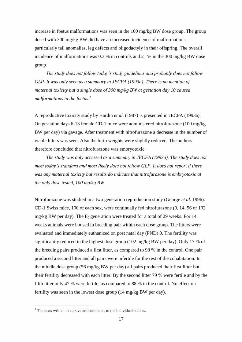
A reproductive toxicity study by Hardin et al. (1987) is presented in JECFA (1993a).
On gestation days 6-13 female CD-1 mice were administered nitrofurazone (100 mg/kg
BW per day) via gavage. After treatment with nitrofurazone a decrease in the number of
viable litters was seen. Also the birth weights were slightly reduced. The authors
therefore concluded that nitrofurazone was embryotoxic.
The study was only accessed as a summary in JECFA (1993a). The study does not
meet today’s standard and most likely does not follow GLP. It does not report if there
was any maternal toxicity but results do indicate that nitrofurazone is embryotoxic at
the only dose tested, 100 mg/kg BW.
Nitrofurazone was studied in a two generation reproduction study (George et al. 1996).
CD-1 Swiss mice, 100 of each sex, were continually fed nitrofurazone (0, 14, 56 or 102
mg/kg BW per day). The F0 generation were treated for a total of 29 weeks. For 14
weeks animals were housed in breeding pair within each dose group. The litters were
evaluated and immediately euthanized on post natal day (PND) 0. The fertility was
significantly reduced in the highest dose group (102 mg/kg BW per day). Only 17 % of
the breeding pairs produced a first litter, as compared to 98 % in the control. One pair
produced a second litter and all pairs were infertile for the rest of the cohabitation. In
the middle dose group (56 mg/kg BW per day) all pairs produced their first litter but
their fertility decreased with each litter. By the second litter 79 % were fertile and by the
fifth litter only 47 % were fertile, as compared to 88 % in the control. No effect on
fertility was seen in the lowest dose group (14 mg/kg BW per day).
1 The texts written in cursive are comments to the individual studies.

18
Due to the observed decrease in fertility a crossover mating trial was carried out
using F0 control and high dose animals (George et al. 1996). At week 23 the animals
were cohabited for maximum one week and fed control feed. They were then separated
and the females were allowed to deliver the litters. The pups from the litters were
evaluated and euthanized on PND 0 and the F0 generation were killed and necropsied at
week 29. Treated males mated with control females produced no live pups and control
males and treated females had a significant decrease in the number of live pups per
litter. The necropsies and histological examinations of the F0 generation revealed that
liver and kidney weights were significantly increased in middle- and high-dose males
and testis weight was significantly decreased in high-dose males. At all dose levels the
incidence of seminiferous tubule degeneration and atrophy was increased. At the low-
and middle-dose groups there was a significant increase in the percentage of aberrant
sperm. In high-dose males and females hepatic hypertrophy was observed. Compared to
the control, females in the highest dose group had an altered oestrus cycle and at all
doses the relative ovary plus oviduct weight was decreased (George et al. 1996).
The last litter produced by the F0 generation was saved and used for F1 fertility
assessment (George et al. 1996). Pups from each dose group (0, 14 or 56 mg/kg BW per
day) were randomly selected on PND 21 and fed nitrofurazone in the same doses as the
parents. At 74 days of age breeding pairs were housed together for a maximum of one
week and then separated. The litters were euthanized on PND 0 and the F1 generation
were killed at 119 days of age. The fertility of the 56 mg/kg BW per day breeding pairs
was significantly decreased as well as the number of live pups per litter. The necropsies
and histology showed that males (56 mg/kg BW per day) had decreased testis weight
and epididymal sperm concentration. They also had an increased percentage of aberrant
sperm. Hepatic hypertrophy was also seen in these males. Females had altered oestrus
cycles at all dose levels and females in the middle-dose group had reduced liver and
ovary weights.
The study does not follow European guidelines but does follow GLP. The results
show that nitrofurazone reduced fertility in mice at 56 and 102 mg/kg BW per day. At
the lowest dose tested, 14 mg/kg BW per day, the oestrus cycle was altered in F1
females and there was an increase in abnormal sperm in F0 males. Therefore, no
NOAEL can be set.
19
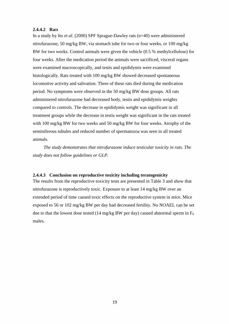
2.4.4.2 Rats
In a study by Ito et al. (2000) SPF Sprague-Dawley rats (n=40) were administered
nitrofurazone, 50 mg/kg BW, via stomach tube for two or four weeks, or 100 mg/kg
BW for two weeks. Control animals were given the vehicle (0.5 % methylcellulose) for
four weeks. After the medication period the animals were sacrificed, visceral organs
were examined macroscopically, and testis and epididymis were examined
histologically. Rats treated with 100 mg/kg BW showed decreased spontaneous
locomotive activity and salivation. Three of these rats died during the medication
period. No symptoms were observed in the 50 mg/kg BW dose groups. All rats
administered nitrofurazone had decreased body, testis and epididymis weights
compared to controls. The decrease in epididymis weight was significant in all
treatment groups while the decrease in testis weight was significant in the rats treated
with 100 mg/kg BW for two weeks and 50 mg/kg BW for four weeks. Atrophy of the
seminiferous tubules and reduced number of spermatozoa was seen in all treated
animals.
The study demonstrates that nitrofurazone induce testicular toxicity in rats. The
study does not follow guidelines or GLP.
2.4.4.3 Conclusion on reproductive toxicity including teratogenicity
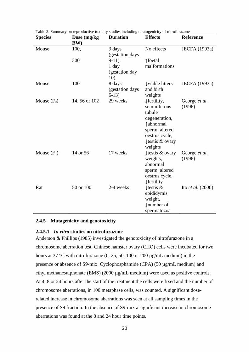
The results from the reproductive toxicity tests are presented in Table 3 and show that
nitrofurazone is reproductively toxic. Exposure to at least 14 mg/kg BW over an
extended period of time caused toxic effects on the reproductive system in mice. Mice
exposed to 56 or 102 mg/kg BW per day had decreased fertility. No NOAEL can be set
due to that the lowest dose tested (14 mg/kg BW per day) caused abnormal sperm in F0
males.
20
Table 3. Summary on reproductive toxicity studies including teratogenicity of nitrofurazone
Species Dose (mg/kg
BW)
Duration Effects Reference
Mouse 100,
300
3 days
(gestation days
9-11),
1 day
(gestation day
10)
No effects
↑foetal
malformations
JECFA (1993a)
Mouse 100 8 days
(gestation days
6-13)
↓viable litters
and birth
weights
JECFA (1993a)
Mouse (F0) 14, 56 or 102 29 weeks ↓fertility,
seminiferous
tubule
degeneration,
↑abnormal
sperm, altered
oestrus cycle,
↓testis & ovary
weights
George et al.
(1996)
Mouse (F1) 14 or 56 17 weeks ↓testis & ovary
weights,
abnormal
sperm, altered
oestrus cycle,
↓fertility
George et al.
(1996)
Rat 50 or 100 2-4 weeks ↓testis &
epididymis
weight,
↓number of
spermatozoa
Ito et al. (2000)
2.4.5 Mutagenicity and genotoxicity
2.4.5.1 In vitro studies on nitrofurazone
Anderson & Phillips (1985) investigated the genotoxicity of nitrofurazone in a
chromosome aberration test. Chinese hamster ovary (CHO) cells were incubated for two
hours at 37 °C with nitrofurazone (0, 25, 50, 100 or 200 µg/mL medium) in the
presence or absence of S9-mix. Cyclophosphamide (CPA) (50 µg/mL medium) and
ethyl methanesulphonate (EMS) (2000 µg/mL medium) were used as positive controls.
At 4, 8 or 24 hours after the start of the treatment the cells were fixed and the number of
chromosome aberrations, in 100 metaphase cells, was counted. A significant dose-
related increase in chromosome aberrations was seen at all sampling times in the
presence of S9 fraction. In the absence of S9-mix a significant increase in chromosome
aberrations was found at the 8 and 24 hour time points.

21
The study mostly follows today’s guidelines and shows that nitrofurazone is
genotoxic to CHO cells in vitro.
In an in vitro mammalian cell gene mutation test, performed two times, CHO-K1-BH4
cells were incubated for two hours at 37 °C with nitrofurazone (0, 25, 50, 100 or 200
µg/mL medium) in the presence or absence of S9-mix (Anderson & Phillips 1985).
Cells were then cultured for seven days and on the eighth day after treatment cells were
transferred to plates with hypoxanthine-free medium either containing or not containing
thioguanine. The cells were incubated for an additional seven days and the number of
mutant colonies was calculated. EMS and benzo[a]pyrene was used as positive controls.
The frequency of mutants did not increase in a dose-related manner after exposure to
nitrofurazone but a few treatments caused an increased mutant frequency (experiment 2
without S9-mix: 25 & 100 µg/mL, experiment 1 with S9-mix: 25, 50 & 100 µg/mL).
Since the increase in mutant frequencies could not be reproduced nitrofurazone
should be considered non-mutagenic in this test. The study, for the most part, follows
guidelines but it is not known whether GLP was followed.
A reverse mutation test was performed on E. coli 343/113/R-9 by Baars et al. (1980).
Cells were exposed to nitrofurazone (0-75 µg/mL), with and without metabolic
activation from Drosophila melanogaster microsomes, for two hours at 37 °C. Exposure
to nitrofurazone, especially at the highest dose, with metabolic activation increased the
number of arg+ and gal
+ mutants. The number of mutants did not increase without the
metabolic activation system.
The study does not follow guidelines or GLP, but does show that metabolic
activation of nitrofurazone caused mutations in E. coli 343/113/R-9 in what appeared to
be a dose-related manner.
In an Ames test, using the plate incorporation method, nitrofurazone (0.25-1.5 µg/plate)
was incubated with S. typhimurium TA 98 and 100 for 48 hours (Goodman et al. 1977).
The revertant his+ colonies were then counted and it was concluded that nitrofurazone
was mutagenic.
The study mostly follows guidelines and shows that nitrofurazone is mutagenic.

22
2.4.5.2 In vivo studies on nitrofurazone
Nitrofurazone was tested in a mammalian bone marrow chromosome aberration test
(Anderson & Phillips 1985). Male Wistar rats were given a single dose of nitrofurazone
(40, 120 or 400 mg/kg BW, n=72), 200 mg EMS/kg BW (positive control, n=24) or
corn oil (negative control, n=36) via gavage. The animals were killed 6, 24 or 48 hours
after administration but were intraperitoneally injected with the metaphase-arresting
colchicine (3 mg/kg BW) two hours before. Bone marrow cells were harvested and 50
cells in metaphase per animal were examined for chromosome aberrations and 500 cells
were examined to provide a mitotic index. There was no increase in chromosome
aberrations after exposure to nitrofurazone.
Another study was performed where rats were given daily doses of nitrofurazone
(15, 45 or 150 mg/kg BW), corn oil or EMS (200 mg/kg BW) for five days. The rats
were killed six hours after the last administration and bone marrow cells were harvested
and examined. No increase in chromosome aberrations was observed. Colchicine (3
mg/kg BW) were intraperitoneally injected two hours before harvesting the cells.
The study does not follow guidelines but provide some indications that
nitrofurazone is not genotoxic in rat in vivo.
The mutagenicity of nitrofurazone was tested in a bone marrow micronucleus test using
Sprague-Dawley and Long-Evans rats (n=24) (Goodman et al. 1977). Rats were
intraperitoneally injected with nitrofurazone (15, 30 or 60 mg/kg BW in Sprague-
Dawley rats and 60 mg/kg BW in Long-Evans rats). Half of the dose was given 30
hours before sacrifice and the other half was given six hours before sacrifice.
Triethylenemelamine served as positive control. The number of reticulocytes with
micronuclei out of 2000 or 3000 cells per rat was then counted. Nitrofurazone did not
increase the number of micronucleated reticulocytes at any dose or rat strain and was
concluded to be non-mutagenic in vivo.
The study does not follow current guidelines. According to the authors the results
show that nitrofurazone is not mutagenic under these conditions. However, the
sampling only occurred six hours after the last dosing and that is not long enough for
micronucleated reticulocytes to be formed. Therefore, the study is not that reliable since
the results may be false negative. There is also no mention of any other signs of toxicity
being observed or not observed during the study.

23
In a chromosome aberration test nitrofurazone (60 mg/kg BW) was injected
intraperitoneally into Sprague-Dawley rats (n=5 per treatment) and samples of bone
marrow were retrieved 6 and 24 hours after administration (Goodman et al. 1977).
Triethylenemelamine served as positive control. Fifty cells in metaphase per animal
were examined for chromosome aberrations. The results showed that nitrofurazone did
not induce chromosome aberrations at these time points.
No mitotic index was determined, no meta-phase arresting agent was used and
only 50 cells per animals were examined instead of 100. Otherwise the study follows
guidelines and does not indicate that nitrofurazone is genotoxic in rat.
2.4.5.3 In vitro studies on the metabolite SEM
Parodi et al. (1981) performed an Ames test with Salmonella typhimurium strain TA
1535. The bacteria were exposed to SEM (67 µmol/plate) with or without S9-mix. The
results showed that SEM was slightly mutagenic in the absence of S9. The mutagenicity
decreased in the presence of S9-mix.
The study does not follow the guidelines or GLP. The results indicate that SEM
may not be mutagenic after metabolic activation and thereby may not be mutagenic in
vivo.
The mutagenicity of SEM was tested in a bacterial reverse mutation test using the plate-
incorporation method with histidine-requiring Salmonella typhimurium mutants TA 98,
TA 100, TA 1535 and TA 1537 and tryptophan-requiring Escherichia coli mutant WP2
uvrA (TNO 2004a). Bacteria were incubated with SEM at five concentrations (62-5000
µg/plate) in the presence and absence of S9-mix, transferred to minimal glucose agar
plates and incubated for 48-72 hours at 37 °C. The trp+ and his
+ revertant colonies were
then counted. Appropriate positive controls were used. SEM was cytotoxic to S.
typhimurium TA 100, TA 1537 and E. coli WP2 uvrA at the highest concentration in the
presence of S9-mix. There was a dose-related increase in the number of revertant
colonies in strain TA 1535 in the absence of S9-mix and an increase at the highest dose
in the presence of S9-mix. In TA 100 the number of revertant colonies also increased in
the absence of S9-mix at 1667 µg/plate and slightly at 5000 µg/plate. A slight increase
in revertant colonies was seen in WP2 uvrA at the highest concentration in the absence
of S9-mix.

24
The study follows GLP and the guidelines. SEM was cytotoxic to TA 100, TA 1537
and WP2 uvrA at the highest dose in the presence of S9-mix but that is not believed to
have impacted the results. The results show that under these test conditions SEM is
mutagenic.
In a cell gene mutation test at the tk locus in mouse lymphoma (L5178Y) cells, cells
were exposed to 13 concentrations of SEM (0.21-10 mM) in the presence and absence
of S9-mix (TNO 2004b). Methyl methanesulphonate (MMS) and 3-methylcholanthrene
(MCA) were used as positive controls. Cells were cultured in wells of two 96-wells
microtiter plates in TFT (4 µg/mL) containing medium. After 10-14 days of incubation
the number of surviving colonies (mutants) was counted. In the presence of S9-mix the
mutant frequency was only increased compared to the control at the highest
concentration (10 mM). In the absence of S9 there was a dose-related increase in the
mutant frequency. SEM was not cytotoxic in the presence of S9 but slightly cytotoxic in
the absence of S9-mix.
The study follows GLP and guidelines. SEM is mutagenic at the TK-locus in
mouse lymphoma cells.
SEM was tested in a chromosome aberration test where CHO cells were treated both
with and without S9-mix (TNO 2004c). In one test, cells were treated with SEM (10-
1115 µg/mL) for 4 or 18 hours and then harvested 18 hours after the start of treatment.
Colcemid (0.1 µg/mL) was added two hours before harvest. Two-hundred cells in
metaphase per dose were examined for chromosome aberrations. In another test cells
were treated with SEM (50-900 µg/mL) for 4, 18 or 32 hours, harvested and examined
18 or 32 hours after the start of treatment. Two hours before harvest colcemid (0.1
µg/mL) was added. The results showed that SEM did not significantly increase the
number of aberrant cells at any dose or time-point. However, it was shown that SEM in
the presence of S9 significantly increased the number of endoreduplicated cells at the 18
hour, but not 32 hour, sampling time. It was concluded that SEM may have the potential
to inhibit mitotic processes.
The results from this study indicate that SEM is not clastogenic in CHO cells but
may have the potential to interfere with mitotic processes. The study followed GLP and
guidelines.

25
In an in vitro sister-chromatid exchange (SCE) assay it was shown that SEM (0.5, 2.5,
5, 10 or 20 µg/mL) only caused a minor significant increase (p=0.05) in SCE frequency
in human lymphocytes at the highest dose tested (Vlastos et al. 2010). SEM and MMC
(positive control, 0.1 µg/mL) were incubated with the cells for 72 hours. Demolcine (0.3
µg/mL) was added two hours before harvesting the cells. Twenty-five cells in
metaphase were examined per culture.
The study shows that SEM may be genotoxic in this test system at 20 µg/mL, which is
stated to be 1000 times higher the worst-case intake of a 6-months old infant. The study
mostly follows test guidelines but does not follow GLP.
The genotoxicity of SEM was tested in a micronucleus test in lymphocytes in vitro
(Vlastos et al. 2010). Human lymphocytes were cultured for 72 hours. Cells were
incubated with SEM (0, 0.5, 2.5, 5, 10 or 20 µg/mL) or mitromycin-C (MMC) (positive
control, 0.5 µg/mL) starting 24 hours after the beginning of the culture period.
Cytochalasin-B (6 µg/mL) was added 44 hours after culture initiation. At the end of the
culture period cells were collected. The frequency of micronuclei was derived by
examining 1000 binucleate cells per culture. The results showed that there was no
statistical difference in the frequency of micronuclei between SEM treated cells and the
control.
The study does not follow GLP but mostly follows guidelines. The results in this
study indicate that SEM may not be genotoxic.
2.4.5.4 In vivo studies on the metabolite SEM
SEM was tested in a flow cytometry-based micronucleus assay in vivo (Abramsson-
Zetterberg & Svensson 2005). Male Balb/C mice were intraperitoneally injected with
SEM (0, 40, 80 or 120 mg/kg BW) and CBA mice were injected with 80 or 120 mg/kg
BW. Three or four Balb/C mice were included in each dose group while five CBA mice
per dose group were used. Colchicine (1 mg/kg BW) was used as positive control and
PBS (1 mg/kg BW) was used as negative control. Blood samples were collected 42
hours after injection. Between 20000 and 100000 erythrocytes per sample were
examined and the frequency of micronucleated polychromatic erythrocytes was
determined. It was shown that SEM did not affect the frequency of micronucleated
erythrocytes in either mouse strain at any dose.

26
The study mostly follows the guidelines, except for a low number of Balb/C mice,
and indicates that SEM is not genotoxic in vivo.
Male Wistar rats (n=25) were orally administered SEM (0, 50, 100 or 150 mg/kg BW)
or cyclophosphamide (positive control, 20 mg/kg BW) and killed 24 hours after
administration (Vlastos et al. 2010). Bone marrow cells were collected and the
frequency of micronuclei in 2000 polychromatic erythrocytes (PCEs) per animal was
determined. It was shown that SEM statistically increased (p≤0.05) the frequency of
micronuclei at all doses.
The study mostly follows guidelines and demonstrates a genotoxic effect of SEM
in vivo but the effect was not dose-dependent. When using rat for the micronucleus
assay the risk for false negatives are increased due to micronucleated erythrocytes
being removed from the circulatory system by the spleen. This increases the reliability
of the results in this study.
2.4.5.5 Conclusions on mutagenicity and genotoxicity studies
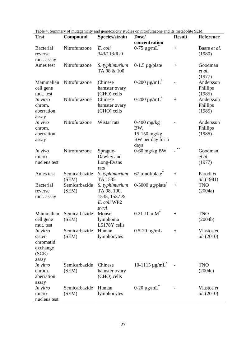
The results of the mutagenicity and genotoxicity studies are presented in Table 4.
Nitrofurazone has been shown to be genotoxic and mutagenic in the bacterial tests and
in in vitro mutation and chromosome aberration tests. Several other in vitro
mutagenicity and genotoxicity studies have been performed with nitrofurazone and the
results of these are presented in JECFA (1993a). Most of the results are positive for
mutagenicity and genotoxicity. In the two in vivo studies mentioned above,
nitrofurazone did not test positive in rat. However, one of these may be a false negative.
From these studies it is not possible to draw any conclusions on the mutagenicity of
nitrofurazone in vivo but nitrofurazone is mutagenic in vitro both with and without
metabolic activation.
In the studies mentioned above the metabolite SEM has tested positive in all
bacterial mutation test, but there has been both negative and positive results in the in
vitro and in vivo studies. Further study is needed before any conclusions on the
mutagenicity and genotoxicity of SEM can be drawn.
27
Table 4. Summary of mutagenicity and genotoxicity studies on nitrofurazone and its metabolite SEM
Test Compound Species/strain Dose/
concentration
Result Reference
Bacterial
reverse
mut. assay
Nitrofurazone E. coli
343/113/R-9
0-75 µg/mL*
+ Baars et al.
(1980)
Ames test Nitrofurazone S. typhimurium
TA 98 & 100
0-1.5 µg/plate + Goodman
et al.
(1977)
Mammalian
cell gene
mut. test
Nitrofurazone Chinese
hamster ovary
(CHO) cells
0-200 µg/mL*
- Andersson
Phillips
(1985)
In vitro
chrom.
aberration
assay
Nitrofurazone Chinese
hamster ovary
(CHO) cells
0-200 µg/mL*
+ Andersson
Phillips
(1985)
In vivo
chrom.
aberration
assay
Nitrofurazone Wistar rats 0-400 mg/kg
BW,
15-150 mg/kg
BW per day for 5
days
- Andersson
Phillips
(1985)
In vivo
micro-
nucleus test
Nitrofurazone Sprague-
Dawley and
Long-Evans
rats
0-60 mg/kg BW - **
Goodman
et al.
(1977)
Ames test Semicarbazide
(SEM)
S. typhimurium
TA 1535
67 µmol/plate* + Parodi et
al. (1981)
Bacterial
reverse
mut. assay
Semicarbazide
(SEM)
S. typhimurium
TA 98, 100,
1535, 1537 &
E. coli WP2
uvrA
0-5000 µg/plate*
+ TNO
(2004a)
Mammalian
cell gene
mut. test
Semicarbazide
(SEM)
Mouse
lymphoma
L5178Y cells
0.21-10 mM*
+ TNO
(2004b)
In vitro
sister-
chromatid
exchange
(SCE)
assay
Semicarbazide
(SEM)
Human
lymphocytes
0.5-20 µg/mL + Vlastos et
al. (2010)
In vitro
chrom.
aberration
assay
Semicarbazide
(SEM)
Chinese
hamster ovary
(CHO) cells
10-1115 µg/mL*
- TNO
(2004c)
In vitro
micro-
nucleus test
Semicarbazide
(SEM)
Human
lymphocytes
0-20 µg/mL*
- Vlastos et
al. (2010)

28
Table 4 continued
In vivo
micro-
nucleus
assay
Semicarbazide
(SEM)
Balb/C & CBA
mice
0-120 mg/kg BW - Abramsson
-Zetterberg
&
Svensson
(2005) + positive, - negative * With and without metabolic activation,
** May be false negative
2.4.6 Carcinogenicity and long-term toxicity
2.4.6.1 Mice
B6C3F1 mice, 50 of each sex and dose group, were fed feed containing nitrofurazone (0,
150 or 310 mg/kg feed) for two years and were then killed and necropsied (Kari et al.
1989). The daily nitrofurazone dose was estimated to be 15 or 31 mg/kg BW for the low
and high dose, respectively. During the first year some stimulus-sensitive seizures were
observed in the high dose group and in females of the low dose group. Also there was a
significant decrease in the survival of males in the high dose group. Females in the high
dose group had significantly increased incidences of granulosa cell tumours. There was
also a significant increase in the incidences of benign mixed ovarian tumours in the low
and high-dose groups. No effects were seen in male mice.
The study mostly follows the guidelines and demonstrates that nitrofurazone can
induce both benign and malignant tumours in mice.
2.4.6.2 Rats
Fisher 344 rats, 50 of each sex and dose group, were fed feed containing nitrofurazone
(0, 310 or 620 mg/kg feed) for two years and were then killed and necropsied (Kari et
al. 1989). The daily nitrofurazone dose was estimated to be 12 or 25 mg/kg BW for the
low and high dose, respectively. Nitrofurazone caused abnormal posture and pelvis limb
gait at all doses. There was also a significant decrease in the survival of males in the
high dose group. Degeneration of joint articular cartilage was seen at all dose levels and
dosed male rats had increased incidences of testicular degeneration. In female rats there
was a significant increase in mammary gland fibroadenomas at both dose levels. Low-
dose male rats had significantly increased incidences of carcinomas and mesothelioma
in tunica vaginalis. High-dose males had significantly increased incidences of
trichoepithelioma or sebaceous adenoma.
The results from the study show that nitrofurazone causes mammary gland
neoplasms. The authors conclude that the results in male rats are equivocal. The study
mostly follows the guidelines for carcinogenicity studies. Exposure to nitrofurazone has
29
previously been shown to be reproductively toxic and cause testicular degeneration in
both mice and rats.
The results from a study by Siedler & Searfoss (1967) are presented in JECFA (1993a).
CFE rats (20 of each sex) were fed feed containing nitrofurazone for 45 weeks. The
daily intake was around 50-55 mg/kg BW per day. After this medication period the rats
were fed control feed for seven weeks and then examined. The number of female rats
with benign mammary tumours was significantly higher in the treated rats than the
control. In male rats there was no increase in tumour incidence.
The study is quite old, does not follow GLP and does not meet today’s standard of
testing. Also it was only accessed as an abstract and a small number of animals were
used. It does however, indicate that female CFE rats are more susceptible to the
carcinogenic effects of nitrofurazone compared to males.
JECFA (1993a) summarised the results from another study by Siedler & Searfoss
(1967). Female Holtzman rats (n=35) were administered nitrofurazone in the feed,
corresponding to 0, 75 or 150 mg/kg BW per day, for 45 weeks and were then given
control feed for eight weeks. At the end of the study there was a significant increase in
the number of benign mammary tumours at all dose levels.
Only an abstract of the study was found. The study does show that nitrofurazone
increases the incidences of benign mammary tumours, but due to it being old and not
following guidelines it may not be very reliable.
Ertürk et al. (1970) administered female weanling Sprague-Dawley rats (n=60) control
feed or feed containing nitrofurazone (1000 mg/kg) for 46 weeks. Over the course of the
experiment this corresponded to about 50-400 mg/kg BW, depending on the weight of
the rat. After the medication period the rats were fed control feed for another 20 weeks.
At the end of the study there was a significant increase in the number of rats with
mammary fibroadenomas in the treated group compared to the control.
The study does not follow guidelines and the survival is not stated. But it shows
that nitrofurazone induced mammary fibroadenomas in female rats.
30
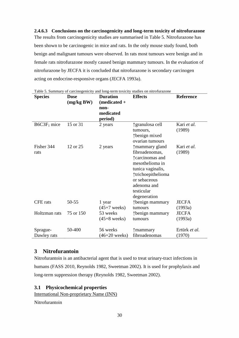
2.4.6.3 Conclusions on the carcinogenicity and long-term toxicity of nitrofurazone
The results from carcinogenicity studies are summarised in Table 5. Nitrofurazone has
been shown to be carcinogenic in mice and rats. In the only mouse study found, both
benign and malignant tumours were observed. In rats most tumours were benign and in
female rats nitrofurazone mostly caused benign mammary tumours. In the evaluation of
nitrofurazone by JECFA it is concluded that nitrofurazone is secondary carcinogen
acting on endocrine-responsive organs (JECFA 1993a).
Table 5. Summary of carcinogenicity and long-term toxicity studies on nitrofurazone
Species Dose
(mg/kg BW)
Duration
(medicated +
non-
medicated
period)
Effects Reference
B6C3F1 mice 15 or 31 2 years ↑granulosa cell
tumours,
↑benign mixed
ovarian tumours
Kari et al.
(1989)
Fisher 344
rats
12 or 25 2 years ↑mammary gland
fibroadenomas,
↑carcinomas and
mesothelioma in
tunica vaginalis,
↑trichoepithelioma
or sebaceous
adenoma and
testicular
degeneration
Kari et al.
(1989)
CFE rats 50-55 1 year
(45+7 weeks)
↑benign mammary
tumours
JECFA
(1993a)
Holtzman rats 75 or 150 53 weeks
(45+8 weeks)
↑benign mammary
tumours
JECFA
(1993a)
Sprague-
Dawley rats
50-400 56 weeks
(46+20 weeks)
↑mammary
fibroadenomas
Ertürk et al.
(1970)
3 Nitrofurantoin
Nitrofurantoin is an antibacterial agent that is used to treat urinary-tract infections in
humans (FASS 2010, Reynolds 1982, Sweetman 2002). It is used for prophylaxis and
long-term suppression therapy (Reynolds 1982, Sweetman 2002).
3.1 Physicochemical properties
International Non-proprietary Name (INN)
Nitrofurantoin
31
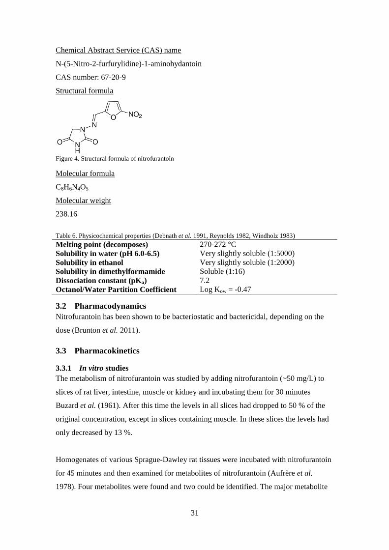
Chemical Abstract Service (CAS) name
N-(5-Nitro-2-furfurylidine)-1-aminohydantoin
CAS number: 67-20-9
Structural formula
Figure 4. Structural formula of nitrofurantoin
Molecular formula
C8H6N4O5
Molecular weight
238.16
Table 6. Physicochemical properties (Debnath et al. 1991, Reynolds 1982, Windholz 1983)
Melting point (decomposes) 270-272 °C
Solubility in water (pH 6.0-6.5) Very slightly soluble (1:5000)
Solubility in ethanol Very slightly soluble (1:2000)
Solubility in dimethylformamide Soluble (1:16)
Dissociation constant (pKa) 7.2
Octanol/Water Partition Coefficient Log Kow = -0.47
3.2 Pharmacodynamics
Nitrofurantoin has been shown to be bacteriostatic and bactericidal, depending on the
dose (Brunton et al. 2011).
3.3 Pharmacokinetics
3.3.1 In vitro studies
The metabolism of nitrofurantoin was studied by adding nitrofurantoin (~50 mg/L) to
slices of rat liver, intestine, muscle or kidney and incubating them for 30 minutes
Buzard et al. (1961). After this time the levels in all slices had dropped to 50 % of the
original concentration, except in slices containing muscle. In these slices the levels had
only decreased by 13 %.
Homogenates of various Sprague-Dawley rat tissues were incubated with nitrofurantoin
for 45 minutes and then examined for metabolites of nitrofurantoin (Aufrère et al.
1978). Four metabolites were found and two could be identified. The major metabolite
32
was 1-{[(3-cyano-1-oxopropyl)methylene]amino}-2,4-imidazolidinedione and a minor
metabolite was identified as aminofurantoin (1{[(5-amino-2-furanyl)methylene]amino}-
2,4-imidazolidinedione).
3.3.2 In vivo studies
3.3.2.1 In swine
Pigs (n=18) were fed feed containing nitrofurantoin (400 mg/kg feed) ad libitum for ten
days, corresponding to around 24 mg/kg BW per day, followed by a withdrawal period
for six weeks (Cooper et al. 2005). Three pigs were sacrificed each week of the
withdrawal period and samples of muscle, liver and kidney were taken and analysed for
nitrofurantoin and its marker residue 1-aminohydantoin (AHD) using LC-MS/MS and
high performance liquid chromatography-UV (HPLC-UV). At week 0 of the withdrawal
period nitrofurantoin was not detected in any sample while AHD was detected in all
samples. The levels of AHD found at week 6 of the withdrawal period were around 4
µg/kg in kidney and liver and 8 µg/kg in muscle. The depletion half-lives of AHD in
muscle, liver and kidney were 8.3±0.9 days, 5.7±0.5 days and 5.8±0.7 days
respectively.
3.3.2.2 In bovine
One cow was administered one capsule containing; 0.88 mg/kg BW furazolidone,
nitrofurazone and furaltadone, and 4.4 mg/kg BW nitrofurantoin (Chu & Lopez 2007).
Milk samples were then collected for two weeks at intervals of 12 hours. Milk from
non-treated cows was used as control. The levels of nitrofuran side-chain residues in the
milk were determined using LC-MS/MS. The level of the side-chain of nitrofurantoin,
AHD, in milk was highest 12 hours after dosing (~47 µg/kg), decreased rapidly and 72
hours after dosing the level of AHD was below the detection limit (0.2 µg/kg).
3.3.2.3 In fish
The depletion of nitrofurans and their tissue-bound residues in channel catfish
(Ictakurus punctatus) was investigated by Chu et al. (2008). Fish (n=55) were
simultaneously orally administered furazolidone, nitrofurantoin, nitrofurazone and
furaltadone (1 mg/kg BW). After 2, 4, 8 and 12 hours, and 1, 4, 7, 10, 14, 28 and 56
days five fish were killed and muscle samples collected for analysis of parent
nitrofurans and their tissue-bound residues. The highest concentration of nitrofurantoin
in muscle (9.8 µg/kg) was reached 12 hours after administration. Nitrofurantoin could
no longer be detected 96 hours after administration. The level of AHD was highest (9.1

33
µg/kg) 24 hours after administration. The elimination of all tissue-bound residues was
biphasic and could still be detected 56 days after administration. The half-life for AHD
was calculated to be 45 days.
Chu et al. (2008) also examined the levels of nitrofurans and tissue-bound
metabolites in muscle of fish after waterborne exposure to nitrofurans. Fish (5 per
treatment) were exposed to nitrofurantoin, nitrofurazone, furazolidone or furaltadone
(10 mg/L) for eight hours. After this time the fish were killed and their muscle tissue
was analysed for parent nitrofuran and tissue-bound metabolites. The concentrations of
nitrofurantoin and AHD were around 3 and 2 µg/kg, respectively.
Channel catfish were given 14
C-nitrofurantoin, 1 or 10 mg/kg BW, via intravascular
administration or 1 mg/kg BW via oral administration (Stehly & Plakas 1993). The fish
(n=9) which received nitrofurantoin through intravascular administration were sampled
for blood over a 72 hour period. For the 16 fish treated orally with nitrofurantoin four
were killed at each sampling time (4, 24, 48 and 72 hours). Bile, urine, plasma, head
and trunk kidneys, muscle, liver and spleen were collected and analysed for
nitrofurantoin residues. An additional five fish were given nitrofurantoin orally and
their blood sampled over a 72 hour period to determine the bioavailability.
After intravascular injection the levels of nitrofurantoin in plasma quickly
decreased and the half-lives were determined to be 23 and 46 minutes for the 1 and 10
mg/kg BW doses, respectively (Stehly & Plakas 1993). A two- and three-compartment
pharmacokinetic model best described the plasma concentrations after intravascular
administration of 1 and 10 mg/kg BW, respectively. The pharmacokinetic values
indicated a limited tissue distribution and rapid clearance.
The bioavailability of nitrofurantoin was 17 % after oral administration and the
highest plasma level of nitrofurantoin (0.06 µg/mL) was seen two hours after
administration (Stehly & Plakas 1993). The highest concentrations of 14
C-nitrofurantoin
and its metabolites in tissues were observed four hours after administration. The levels
were highest in plasma (1.22 mg equivalents of nitrofurantoin per kg) and liver (1.64
mg/kg) and the lowest in muscle (0.05 mg/kg). The levels in bile ranged from 9.3 to
13.8 mg/kg over the 72 hour period. The major excretion pathway for nitrofurantoin
was via the urine with around 21 % of the administered dose being eliminated in the
first 24 hours. The eliminated residues in urine mostly consisted of parent compound
but five unidentified metabolites were found in urine.

34
3.3.2.4 In poultry
McCracken et al. (2005a) fed six broiler hens and one cockerel 120-140 g of feed
containing nitrofurantoin (400 mg/kg feed), corresponding to about 96-112 mg/kg BW.
Eggs were collected and analysed for AHD and when it was clear that nitrofurantoin
residues had transferred to the eggs, the eggs laid after this were collected and allowed
to hatch. After hatching, four chicks were sacrificed at determined intervals and muscle
and liver samples were analysed for AHD. However, AHD could only be detected up to
day 10. The levels of AHD in liver and muscle in one day old chicks were around 2.9
µg/kg. In five days old chicks the levels of AHD in liver and muscle were around 0.3
and 0.9 µg/kg, respectively. In 10-days-old chicks no AHD could be detected in liver
and levels in muscle were approximately 0.1 µg/kg.
Twenty-four laying hens were fed feed containing 300 mg furaltadone, nitrofurazone,
nitrofurantoin or furazolidone per kg feed for one week, corresponding to around 60
mg/kg BW (McCracken & Kennedy 2007). Eggs were then collected for two days and
analysed immediately for nitrofuran parent compound and their bound residues in
albumen, yolk and shell using LC-MS/MS. The levels of nitrofurantoin in yolk,
albumen and shell were 66.7, 54.8 and 35.7 ng/g, respectively. The levels of AHD in
yolk, albumen and shell were 169, 171, 19.6 ng/g, respectively.
3.3.2.5 In rabbit
The bioavailability of nitrofurantoin via different administration routes was investigated
in rabbits (Watari et al. 1983). Two groups of rabbits (n=16) were given nitrofurantoin
(1.25 or 10 mg/kg BW) once a week via different administration routes; intravenous
infusion (i.v. inf.), intraduodenal administration (i.d.), portal vein infusion (p.v. inf.) or
oral administration (p.o.). Plasma samples were taken before administration and over a
90 minute period after administration for the low dose group and over a 2.5 hour period
after administration for the high dose group. Urine was collected before and eight hours
after administration. The plasma concentration-time course could be described by a one-
compartment model. Plasma levels were significantly lower after oral administration
compared to the other administration routes. At the low dose the maximum level of
nitrofurantoin was 0.26 µg/mL after oral administration and around 1.55 µg/mL after
the other routes of administration. At the high dose it was 1.73 µg/mL after oral
administration and approximately 14 µg/mL after the other routes of administration. In
the animals given the low dose of nitrofurantoin around 16 % of the low dose was

35
excreted as parent compound via the urine after oral administration and 50 % after the
other routes of administration. At the high dose, 20 % of the dose was excreted as
parent compound via the urine, independent of the route of administration. The
bioavailability was calculated to be 30 % after oral administration. The authors
concluded that the reduced bioavailability of nitrofurantoin after oral administration was
due to degradation of the compound in the stomach.
3.3.2.6 In rat
Female Sprague-Dawley rats with their first litter were treated via gavage with
nitrofurantoin (50 mg/kg BW) on lactation day 10 (Kari et al. 1997). Pups were
removed three hours before collection to allow milk to accumulate. Milk and plasma
samples were retrieved at different times after administration. The highest levels in milk
and plasma, ~100 and 20 µg/mL respectively, were found 30 and 15 minutes after
administration, respectively. The levels of nitrofurantoin were higher in milk than
plasma at all time-points and the milk/plasma partitioning ratio was calculated to be 23.
The absorption, metabolism and elimination of nitrofurantoin in rat were investigated
by Buzard et al. (1961). To show at what site nitrofurantoin is absorbed after oral
administration, nitrofurantoin (25 or 100 mg/kg BW) was injected into different
segments of the rat gastrointestinal tract; small intestine, colon, cecum and stomach.
Blood samples were then retrieved and analysed for nitrofurantoin. It was shown that
nitrofurantoin was rapidly absorbed in the small intestine, somewhat absorbed in the
colon and not absorbed in cecum or stomach.
Following intravenous injection of nitrofurantoin (25 mg/kg BW) in rats (n=9) the
half-life was calculated to be 25 minutes Buzard et al. (1961). It was also shown that
after bilateral nephrectomy or ligation of the ureters the half-life of nitrofurantoin
increased to around 70 minutes, indicating that urinary excretion of nitrofurantoin is an
important elimination pathway.
3.3.2.7 In man
Six healthy men, weighing between 62 and 80 kg, received 50 mg nitrofurantoin via
intravenous infusion for 45 minutes and via oral administration, with and without food
(Hoener & Patterson 1981). One week passed between each treatment. After the
intravenous infusion blood and urine samples were collected over a 6 and 24 hour
period. After oral administration of nitrofurantoin, blood and urine were collected over

36
an 8 and 24 hour period. The bioavailability of nitrofurantoin after oral administration
was around 87-94 % and 34 % of the administered dose was excreted in the urine as
unchanged parent compound. After intravenous infusion the fraction of unchanged
parent compound was higher, i.e. 47 %. The half-life of nitrofurantoin in blood after
intravenous infusion was calculated to be around 58 minutes. The metabolite
aminofurantoin and possibly its acylated conjugate were found in the urine of the test
subjects. It was also shown in a serum protein binding test that around 60 % of
nitrofurantoin binds to plasma proteins.
Four postpartum women were given a dose of 100 mg nitrofurantoin and the levels in
breast milk and plasma were examined over a period of 12 hours after the
administration (Gerk et al. 2001). The results showed that the levels in milk were higher
than those in plasma at all of the time points. The mean maximum concentration in milk
was 2.71 µg/mL and was reached five hours after administration. In plasma the mean
maximum concentration was 0.50 µg/mL and was reached 4.9 hours after
administration. The observed milk:serum ratio (M:S) was 20 times higher than the
predicted, 6.2 compared to 0.28, which indicates an active transport of nitrofurantoin
into milk.
3.3.2.8 In dog
Buzard et al. (1961) intravenously injected dogs (n=3) with nitrofurantoin (4 mg/kg
BW), collected plasma samples and measured the levels of nitrofurantoin. The levels
decreased quickly and the half-life was calculated to be 25 minutes. After oral (15
mg/kg BW) and intravenous (4 mg/kg BW) administration of nitrofurantoin to dogs the
saliva contained nitrofurantoin. Demonstrating that nitrofurantoin is widely distributed.
Male Beagle dogs, weighing 10-16 kg, were intravenously injected with nitrofurantoin
(1.5, 3. 6, 12 or 24 mg/kg BW) and samples of blood, urine and bile were collected over
a period of six hours (Conklin & Wagner 1971). Thirty minutes after the dogs were
administered 3 mg/kg BW, their bile to blood concentration ratio was around 200,
indicating an active transport of nitrofurantoin into bile. Over a six hour time period
after dosing the percentage of dose found in bile and urine were 16.5-22.6 % and 24.1-
36.2 %, respectively, for the doses 1.5-6.0 mg/kg BW. For the 12 and 24 mg/kg BW
doses the percentages were 15.3-18.3 % and 3.1-10.0 % in bile and urine, respectively.

37
The decrease in the dose recovered was concluded to be due to a saturation of the biliary
and urinary excretion system. Half-lives of nitrofurantoin in blood were determined to
be between 10 and 26 minutes at this dose range after intravenous injection. There was
also an increase in bile flow immediately after the administration of nitrofurantoin
which was proportional to the dose.
It was shown by Conklin et al. (1973) that nitrofurantoin is subjected to enterohepatic
circulation in dogs. Donor and recipient dogs were surgically attached so that the bile
from the donor dog would flow to the duodenum of the recipient dog. The donors were
then administered nitrofurantoin (3 mg/kg BW) intravenously and urine, bile and blood
were collected for three hours after administration. Within three hours, donors excreted
around 13.9 and 21.5 % of the administered dose in bile and urine, respectively. The
recipients excreted approximately 1.5 and 3.4 % of the administered donor dose.
Nitrofurantoin (2-5 µg/mL) could be detected in blood of donors within 30 minutes of
administration, but no nitrofurantoin was found in recipients’ blood. It was estimated
that around 5 % of the donors’ dose is reabsorbed in the intestines.
Female Beagle dogs, weighing 10.5-12.5 kg, were administered nitrofurantoin
intravenously (50 mg) or orally (50 or 100 mg) with or without food (Niazi et al. 1983).
Blood samples were collected for four or eight hours after administration. The half-life
in blood after intravenous administration was calculated to be around 31 minutes. The
maximum plasma levels after oral administration of 50 mg nitrofurantoin in a solution
(3.0-4.6 µg/mL) were observed 30-60 minutes after treatment, and the half-life was
calculated to be around 30 minutes. The absorption of nitrofurantoin was determined to
be around 94 % after oral administration of a solution. When 100 mg nitrofurantoin was
given in tablet form either with or without food the elimination half-lives ranged
between 39 and 63 minutes. The absorption ranged between 46 and 88 %.
3.3.3 Conclusions on pharmacokinetics of nitrofurantoin and its metabolites
Studies on the pharmacokinetics of nitrofurantoin have been performed in swine, cow,
fish, poultry, rat, rabbit, dog and man. Many of these studies are quite old and may not
meet today’s standards in testing. In dogs the oral absorption of nitrofurantoin was 94
%, in a solution, and between 46 and 88 % in tablet form. The availability of
nitrofurantoin after oral administration was 16 % in fish and 30 % in rabbit. In man it is

38
quickly absorbed in the gastro-intestinal tract and the bioavailability is between 87 and
94 %. It was shown that in rat most of the absorption of nitrofurantoin occurs in the
small intestine. Nitrofurantoin has been found in muscle, liver, bile, plasma and milk. In
both rat and man the levels of nitrofurantoin have been higher in milk than in plasma
after exposure to nitrofurantoin indicating that the drug is actively transported into milk.
Women who received 100 mg nitrofurantoin had a maximum concentration of 2.71 µg
nitrofurantoin per mL breast milk. Nitrofurantoin is rapidly excreted with half-lives
being between 30 and 60 minutes in fish, rabbit, rat, dog and man. Man had the longest
reported half-life, 58 minutes. Nitrofurantoin is also subjected to enterohepatic
circulation in dogs. It seems that the major excretion pathway of nitrofurantoin is via the
urine. It has been reported that around 40 % of the administered dose is excreted
unchanged in the urine in man (Brunton et al. 2011).
Metabolites that have been identified are seen in Table 7. From the studies
mentioned above AHD is distributed throughout the body. In swine, after administration
of 24 mg/kg BW per day for 10 days and a withdrawal period of six weeks, AHD was
still found in liver and kidney (~4 µg/kg), and in muscle (~8 µg/kg). The reported half-
lives of AHD are much longer than those for nitrofurantoin. The half-lives of AHD in
the tissues of swine were calculated to be around six and eight days in kidney and liver,
and muscle, respectively. In fish, the half-life of AHD in muscle was calculated to be 45
days. It has also been shown that AHD is transferred to milk in cows and to chicken
eggs. AHD could be detected in chicks, from hens exposed to nitrofurantoin, for up to
10 days after hatching.
Table 7. Metabolites of nitrofurantoin in different species found in the literature
Metabolite Species/cells Reference
1-aminohydantoin (AHD) Poultry, fish,
cow, swine
McCracken & Kennedy
(2007), McCracken et al.
(2005a), Chu et al. (2008),
Chu & Lopez (2007), Cooper
et al. (2005)
1-{[(3-cyano-1-
oxopropyl)methylene]amino}-2,4-
imidazolidinedione
Rat Aufrère et al. (1978)
1{[(5-amino-2-
furanyl)methylene]amino}-2,4-
imidazolidinedione (aminofurantoin)
Rat, man Aufrère et al. (1978), Hoener
& Patterson (1981)

39
3.4 Toxicology of nitrofurantoin
3.4.1 Acute toxicity
Åkerblom & Campbell (1973) reported LD50-values of 150 and 360 mg nitrofurantoin
per kg BW in mice after intraperitoneal injection and by gavage, respectively. The doses
tested were between 3 and 12800 mg/kg BW.
3.4.1.1 Conclusions on acute toxicity of nitrofurantoin
The results from this study indicate that nitrofurantoin is quite toxic to mice after oral
and intraperitoneal administration, but the study does not follow guidelines or GLP.
3.4.2 Chronic toxicity
In a chronic toxicity study by Butler et al. (1990) Sprague-Dawley rats (n=240) were
administered nitrofurantoin (0, 24, 48 or 96 mg/kg BW per day) for 24 months. The
actual intake was 23.5, 42.8 and 81.3 mg/kg BW per day for males and 31.7, 57.3 and
116.1 mg/kg BW per day for females. Haematological and biochemical investigations
were performed on a total of 25 rats per treatment. Five male and female rats were bled
and necropsied at 1, 3, 6, 12 and 24 months. At the end of the study all remaining rats
were killed and necropsied. Histological examinations were performed on liver, kidney,
heart, gonads, thyroid, brain, uterus or prostate and pituitary. There was no effect seen
on haematological and biochemical parameters. At the highest dose there was an
increase in testicular degeneration in males. At all doses females had an increase in
focal biliary proliferation. There was also sciatic nerve fibrosis and degeneration at the
highest dose in both male and female rats.
3.4.2.1 Conclusions on chronic toxicity of nitrofurantoin
Only one study on chronic toxicity was found which mostly follows guidelines. Since
focal biliary proliferation was observed at all doses in females no NOAEL can be set,
but it is not clear to what extent this cell proliferation occurred and if it may be a sign of
hepatotoxicity.
3.4.3 Reproductive toxicity including teratogenicity
For one month, male rats (36 per treatment) were orally administered nitrofurantoin (10
or 85 mg/kg BW per day) (Yunda et al. 1974). The control group consisted of 18
animals. Treatment with both doses of nitrofurantoin detrimentally affected
spermatogenesis. At 10 mg/kg BW per day the concentration of spermatozoa was
reduced. This was also seen in the high-dose group along with other effects. This effect
was somewhat reversed 48 days after cessation of treatment. Cysteine was shown to

40
prevent the gonadotoxic effect of nitrofurantoin when administered at the same time as
nitrofurantoin.
The study does not meet today’s standard of testing, but demonstrates a
gonadotoxic effect of nitrofurantoin in rat.
IARC (1990) summarized the results of a study by Prytherch et al. (1984). They studied
the teratogenicity in rats and effects on fertility, and peri- and postnatal effects in rat and
rabbit after exposure to nitrofurantoin. When doses of 10, 20 and 30 mg nitrofurantoin
per kg BW were orally administered no adverse effects could be observed. No adverse
effects on testicular histology or fertility in males, which were only given 10 mg/kg
BW, were seen.
The results from this study could only be obtained as an abstract in IARC (1990).
It is not known whether or not the study follows guidelines or GLP. It is also not known
how long the exposure was. The results suggest that nitrofurantoin is not reproductively
toxic.
Nomura et al. (1984) performed a study in mice which is reported in IARC (1990).
ICR/Jcl mice were subcutaneously injected with nitrofurantoin (100 or 250 mg/kg BW)
on gestation days 9-11. There was no observed increase in embryo- or fetomortality. In
the high-dose group there was a decrease in birth-weight and a significant increase in
the incidence of cleft palate and syndactyly.
Only a summary of the study was available. The study does not meet today’s
standard in testing, but demonstrates developmental toxicity of nitrofurantoin in mice.
In a study by NTP (1989) male and female F344/N rats were orally administered
nitrofurantoin (~50-850 mg/kg BW per day) via the feed for 13 weeks. B6C3F1 mice
were treated with nitrofurantoin (~96-1600 mg/kg BW) per day for the same length of
time. Male and female F344/N rats (100 and 60 mg/kg BW per day respectively)
showed testicular and ovarian degeneration. Male mice (285 mg/kg BW per day) also
showed testicular degeneration at the end of the experiment.
The study demonstrates that nitrofurantoin may be toxic to the reproductive
system.

41
3.4.3.1 Conclusions on reproductive toxicity including teratogenicity
The results from reproductive toxicity tests are presented in Table 8. The studies
presented above show that nitrofurantoin is reproductively toxic. The lowest dose that
was seen to cause reproductive effects was 10 mg/kg BW per day in male rats after one
month of treatment. But in another study (length of study is not known) male rats
treated with the same dose showed no effect on fertility. Further study is needed to
elucidate reproductively toxic effects of nitrofurantoin at low doses.
Table 8. Summary on reproductive toxicity studies including teratogenicity of nitrofurantoin
Species Dose (mg/kg
BW)
Duration Effects Reference
Rat 10 or 80 1 month ↓spermatogenesis Yunda et al.
(1974)
Rat, rabbit 10, 20 or 30 ? No effects IARC (1990)
Mouse 100 or 250 3 days,
between day 9
& 11 of
gestation
↓birth-weight,
↑malformations
IARC (1990)
Rat
50-850 13 weeks Testicular &
ovarian
degeneration
NTP (1989)
Mouse 96-1600 13 weeks Testicular
degeneration
NTP (1989)
3.4.4 Mutagenicity and genotoxicity
3.4.4.1 In vitro studies
In an Ames test, without metabolic activation, S. typhimurium TA 100 and TA 100FR
was incubated for three days with 0.3 and 3 µg/plate, respectively (Wang & Lee 1976).
The number of his+ revertant colonies was then counted and the results showed that
nitrofurantoin was mutagenic in both strains at these concentrations. In another
experiment, the urine of rats fed feed containing 0.5 % nitrofurantoin was tested in the
same manner. The urine contained nitrofurantoin and tested positive for mutagenic
properties.
The study does not follow the guidelines but shows that nitrofurantoin is
mutagenic in S. typhimurium strains TA 100 and TA100FR.
The Ames test was used by Ni et al. (1987) to investigate the mutagenicity of
nitrofurantoin. Three strains of Salmonella typhimurium; TA 98, TA 98NR and TA
98/1,8-DNP6, were exposed to nitrofurantoin both with and without rat-liver hepatic S9
activation enzymes. The doses of nitrofurantoin tested were 1, 2, 10, 20 and100

42
µg/plate. Nitrofurantoin tested negative for mutagenicity in all strains at the 1 µg/plate
dose, both with and without S9. Nitrofurantoin was only mutagenic at the highest dose
in strain TA 98NR. In the TA 98 strain, nitrofurantoin was mutagenic at the doses 2 and
10 µg/plate, both in the presence and absence of S9, and also mutagenic at 20 µg/plate
with S9. At the highest dose, nitrofurantoin was toxic to bacteria and was also toxic at
20 µg/plate without S9. Nitrofurantoin was mutagenic in the strain TA 98/1,8-DNP6 at
the doses of 2, 10 and 20 µg/plate, both in the presence and absence of S9.
Nitrofurantoin was toxic to the bacteria at the highest dose.
The study follows OECD guidelines to some extent and the results show that
nitrofurantoin is mutagenic in all tested strains both with and without metabolic
activation. In all test strains, except TA 98NR, the mutagenicity of nitrofurantoin
appeared to be dose-dependent.
In another Ames test, nitrofurantoin (0.1-5.0 µg/plate) was incubated with S.
typhimurium TA 98 and 100 for 48 hours without metabolic activation (Goodman et al.
1977). The revertant his+ colonies were then counted and it was concluded that
nitrofurantoin was mutagenic.
The study mostly follows guidelines and shows that nitrofurantoin is mutagenic.
An in vitro mammalian cell gene mutation test was performed on nitrofurantoin in CHO
cells (Gao et al. 1989). Cells were incubated with four different concentrations of
nitrofurantoin (0-200 µg/mL) for five hours with or without S9-mix. Benzo[a]pyrene
was used as positive control when S9 was present. The results showed that
nitrofurantoin was weakly mutagenic.
The study for the most part follows guidelines but does not follow GLP. From the
results in the study the authors conclude that nitrofurantoin is mutagenic.
An in vitro chromosome aberration test was performed on nitrofurantoin using human
peripheral blood (Slapsytè et al. 2002). Lymphocytes were incubated with
nitrofurantoin (0, 5, 20 or 40 µM) for 24 hours. Colchicine was added three hours
before harvest and metaphasic cells (50 or 25 cells per culture) were examined for
chromosome aberrations and SCEs, respectively. Nitrofurantoin increased the number
of chromosome aberrations and SCEs at 20 and 40 µM.

43
The study follow the guidelines to some extent and shows that nitrofurantoin is
genotoxic in vitro.
E. coli WP2 and WP2 uvrA were plated on agar with limited tryptophan and incubated
with nitrofurantoin (10 or 100 µg/plate) for 48 hours at 37 °C to evaluate its capacity to
mutate tryp- cells into tryp
+ cells (McCalla & Voutsinos 1974). The number of colonies
were then counted and compared to controls. The results showed that nitrofurantoin
induced mutations in E. coli WP2 and WP2 uvrA at 10 µg/plate and in WP2 at 100
µg/plate.
The study does not completely follow guidelines and does not follow GLP. The
results indicate that nitrofurantoin is mutagenic without metabolic activation, although
no positive controls were included.
The effect of nitrofurantoin on DNA was also tested in vitro using rat liver nuclei and
human foreskin fibroblasts (HuF22) (Parodi et al. 1983). Cells were incubated with
nitrofurantoin (112 mg/mL) for 30 minutes at 37 °C. The cells were then evaluated for
DNA fragmentation. At 112 mg nitrofurantoin per mL the amount of DNA
fragmentation was significantly increased compared to controls in both HuF22 cells and
liver nuclei.
The experiments presented in this study demonstrate that nitrofurantoin damages
DNA both in vivo and in vitro and may therefore be mutagenic.
3.4.4.2 In vivo studies
The mutagenicity of nitrofurantoin was tested in a bone marrow micronucleus test using
Sprague-Dawley rats (Goodman et al. 1977). Rats were intraperitoneally injected with
nitrofurantoin (50, 100 or 200 mg/kg BW). Half of the dose was given 30 hours before
sacrifice and the other half was given six hours before sacrifice. Triethylenemelamine
served as positive control. Between 2000 and 3000 cells per rat were examined and the
number of reticulocytes with micronuclei was counted. Nitrofurantoin did not increase
the number of reticulocytes with micronuclei at any dose and was concluded to be non-
mutagenic in vivo.
The study does not follow current guidelines and according to the authors the
results show that nitrofurantoin is not mutagenic under these conditions. However, the
sampling only occurred six hours after the last dosing and that is not long enough for

44
micronucleated reticulocytes to be formed. Therefore, the study is not that reliable since
the results may be false negative. There is also no mention of any other signs of toxicity
being observed or not observed during the study.
In a patient study, the number of sister chromatid exchanges (SCEs) in peripheral
lymphocytes was examined in women (n=15) treated with nitrofurantoin for urinary
tract infection (Sardas et al. 1990). Blood samples were collected before and after oral
treatment with nitrofurantoin (10 mg/kg or 400 mg) for 10 days. Division of the
lymphocytes was induced by phytohaemagglutinin and they were allowed to divide for
two cell cycles at 37 °C for 72 hours. Two hours before harvesting, colcemid (10
µg/mL) was added. Around 30 lymphocytes in metaphase were analysed for SCEs.
Treatment with nitrofurantoin did not increase the number of SCEs.
The results from the study indicate that nitrofurantoin is not genotoxic in vivo at
doses used to treat urinary tract infections.
Blood from children (n=69) suffering from urinary tract infections were collected after
treatment with nitrofurantoin (Slapsytè et al. 2002). The treatment period was between
0 and 12 months long with the treatment consisting of 5-8 mg/kg BW per day for the
first seven days and after that 1-2 mg/kg BW per day for the remainder of the treatment
period. Blood samples were collected 1, 3, 6 and 12 months after the start of the
treatment. Blood from control children were retrieved before the start of treatment.
Blood samples before treatment was available from 13 patients. Around 100
lymphocytes were then analysed for chromosome aberrations and between 25 and 50
cells were examined for SCEs. No significant increase in chromosome aberrations or
SCEs compared to the control could be seen after nitrofurantoin treatment. But in the
children who had available blood samples from before the treatment there was a
significant increase in SCEs after nitrofurantoin treatment.
The results from the study are equivocal. On the one hand nitrofurantoin did not
increase SCEs and chromosome aberrations compared to controls. On the other hand
there was an increase in SCEs in children from which blood samples were available
before and after treatment.
Young and adult BALB/C mice (n=64), three and eight weeks old respectively, were
intraperitoneally injected with nitrofurantoin (5, 10 or 50 mg/kg BW) or
cyclophosphamide (75 mg/kg BW) (Fucic et al. 2005). Each treatment group consisted

45
of four male and four female mice. Peripheral blood was collected before and 48, 96,
168 and 336 hours after administration, and the frequencies of micronuclei in 1000
reticulocytes were analysed. The results showed that in young mice nitrofurantoin
caused a significant increase in the frequency of micronuclei at all doses and time
points. In old mice nitrofurantoin caused a significant increase in the frequency of
micronuclei at all doses, but two weeks after the administration the micronucleus
frequency was the same as before administration.
The study mostly follows guidelines but does not follow GLP. Only 1000 instead
of 2000 cells were examined for micronuclei which can cause the results to be false
negative or positive. The results show that nitrofurantoin is genotoxic in vivo at these
doses.
Transgenic Big Blue C57BL/6[LIZ] mice (n=10) were orally administered
nitrofurantoin (0 or 167 mg/kg BW per day) for five consecutive days (Quillardet et al.
2006). The mice were sacrificed 20 days after the last administration and organs;
stomach, colon, kidney, cecum, bladder, lung, intestine and spleen, were collected. The
frequency of mutants in the cII gene in these tissues was examined. The results showed
that there was only a weak mutagenic effect of nitrofurantoin on genes expressed in the
kidney.
The study does not follow GLP or guidelines from 2011 or 2013. The results,
however, do show that nitrofurantoin is slightly mutagenic to genes expressed in kidney
of treated mice. However, the doses were much higher than the doses used to treat
urinary infections in humans.
Nitrofurantoin was also investigated using the Drosophila wing somatic mutation and
recombination test (SMART) (Graf et al. 1989). Three-day-old larvae of Drosophila
melanogaster were exposed to 5 mL of nitrofurantoin (5 or 10 mM) dissolved in 5 mL
medium for 48 hours. The larvae were allowed to pupate and hatch and their wings were
then examined for spots. Compared to controls, nitrofurantoin significantly increased
the frequency of large and twin spots at both and the highest concentration, respectively.
The frequency of small spots was not affected.
The study shows that nitrofurantoin is mutagenic in Drosophila under these test
conditions.

46
Parodi et al. (1983) tested the ability of nitrofurantoin to produce DNA damage in
tissues of male Swiss mice and Sprague-Dawley rats. Rats and mice were administered
nitrofurantoin (14-112 mg/kg BW) intraperitoneally and killed between six and 168
hours after administration. DNA damage in rat liver, spleen, kidney and lung was
examined while only the bone marrow was examined in mice. The results showed that
compared to controls the amount of damaged DNA was significantly increased in the
tissues of treated animals. Effects were seen in the kidney at the lowest dose tested (14
mg/kg BW).
The experiments presented in this study demonstrate that nitrofurantoin damages
DNA in vivo and may therefore be mutagenic.
Parodi et al. (1983) also investigated the potential of nitrofurantoin to induce sister
chromatid exchanges in bone marrow cells of male Swiss mice. Mice were injected
intraperitoneally with nitrofurantoin (32 or 64 mg/kg BW) and bone marrow cells were
extracted 24 or 72 hours after administration. Thirty cells in metaphase were examined
for SCEs per animal and dose. The results showed that there was a dose-dependent
increase in the frequency of SCEs after treatment with nitrofurantoin.
The study shows that nitrofurantoin is capable of inducing SCEs in mice in vivo.
3.4.4.3 Conclusions on mutagenicity and genotoxicity studies
The results from the mutagenicity and genotoxicity studies are summarised in Table 9.
Nitrofurantoin has shown to be mutagenic in bacterial tests both in the presence and
absence of an exogenous metabolic system. It has also been shown to be mutagenic and
genotoxic in other in vitro tests. In IARC (1990) there are some other in vitro studies
mentioned reporting both negative and positive results. The in vivo studies stated above
have reported both negative and positive results and most of the in vivo studies reported
in IARC (1990) have shown negative results. From all of the studies it appears that
nitrofurantoin is mutagenic in bacteria and mammalian cells in vitro but not in vivo.
However, more in vivo studies should be performed to be able to declare this with
certainty.

47
Table 9. Summary of mutagenicity and genotoxicity studies on nitrofurantoin
Test Species/strain Dose/concentration Result Reference
Ames test S. typhimurium
TA 100 &
TA100FR
0.3 or 3 µg/plate + Wang & Lee
(1976)
Bacterial reverse
mutation test
E. coli WP2 and
WP2 uvrA
10 or 100 µg/plate + McCalla &
Voutsinos
(1974)
Ames test S. typhimurium
TA 98, TA 98NR,
TA 98/1,8-DNP
0.1-2.5 µg/plate* + Ni et al.
(1987)
Ames test S. typhimurium
TA 98 & 100
0.1-5.0 µg/plate + Goodman et
al. (1977)
In vitro DNA
damage test
Rat liver nuclei &
HuF22 cells
112 mg/mL + Parodi et al.
(1983)
In vitro
mammalian cell
gene mutation
test
CHO cells 0-200 µg/mL* + Gao et al.
(1989)
In vitro
chromosome
aberration test
Human
lymphocytes
5-40 µM + Slapsytè et
al. (2002)
Drosophila wing
spot test
D. melanogaster 5 or 10 mM + Graf et al.
(1989)
Transgenic
rodent somatic
cell gene
mutation assay
Transgenic Big
Blue
C57BL/6[LIZ]
mice
167 mg/kg BW +
(in
kidney
genes)
Quillardet et
al. (2006)
In vivo SCE
assay
Swiss mice 32 & 64 mg/kg BW + Parodi et al.
(1983)
In vivo SCE
assay
Human
lymphocytes
10 mg/kg or 400 mg - Sardas et al.
(1990)
In vivo DNA
damage test
Sprague-Dawley
rats & Swiss mice
14-112 mg/kg BW + Parodi et al.
(1983)
In vivo
chromosome
aberration test
Human
lymphocytes
5-8 & 1-2 mg/kg
BW
+/- Slapsytè et
al. (2002)
In vivo
micronucleus test
BALB/C mice 5, 10 or 50 mg/kg
BW
+**
Fucic et al.
(2005)
In vivo bone
marrow
micronucleus test
Sprague-Dawley
rats
50, 100 or 200
mg/kg BW
-***
Goodman et
al. (1977)
+ positive, - negative, +/- equivocal * With and without S9-mix,
** May be false negative/positive,
*** May be false negative
3.4.5 Carcinogenicity and long-term toxicity
In a two year carcinogenicity study male and female Sprague-Dawley rats (50 per sex
and treatment) were fed feed containing nitrofurantoin (Butler et al. 1990). The doses
were for males; 12.4, 22.3 and 43.1 mg/kg BW per day, and for females; 16.8, 31.0 and

48
55.8 mg/kg BW per day. High-dose males showed increased mortality compared to
controls. At the end of the study the rats were killed and necropsied. Tissues were
sampled and histologically examined. The only reported effect observed was an increase
in focal biliary proliferation in middle- and high-dose females.
The study follows guidelines but not GLP. The reported results indicate that
nitrofurantoin is not carcinogenic. IARC (1990) showed that there was a significant
increase in the incidences of malignant lymphomas in males. Therefore nitrofurantoin
may be carcinogenic in male rats.
Male and female F344/N rats and B6C3F1 mice (50 per sex and treatment) were fed
feed containing nitrofurantoin for 103 weeks (NTP 1989). The exposure to
nitrofurantoin in the low and high dose groups were around 30 and 60 mg/kg BW per
day for female rats and 60 and 110 mg/kg BW per day for male rats. For male and
female mice the exposure levels were 300 and 570 mg/kg BW per day, and 280 and 580
mg/kg BW per day, respectively. At the end of the study all surviving animals were
killed, necropsied and histological examinations were performed.
Male rats treated with nitrofurantoin had lesions in the kidney and testis (NTP
1989). Testicular degeneration and abnormal cells of the epididymis were seen in males
of the high-dose group. Dosed male rats also had perivascular infiltration of
mononuclear cells and fibrinoid necrosis of arterioles in the testis. In all treated male
rats chronic nephropathy was seen but judged as most severe in high-dose males. There
was also a significant increase in microscopic renal tubular cell adenomas in treated
males compared to controls. Increased incidences of subcutaneous tissue neoplasms in
dosed males were also observed. Rare osteosarcomas were also discovered in three rats,
one from the low dose and two from the high dose. No effects were seen in female rats.
It was concluded that there is some evidence of nitrofurantoin being carcinogenic in
male rats.
In mice there was a reduced survival of control females compared to the dosed
mice (NTP 1989). Males showed testicular degeneration in high dose males as well as
mineralization of the renal medulla and dilation of the renal tubules. Almost all dosed
female mice had ovarian atrophy, spindle cell hyperplasia of the adrenal cortex and
there was also mineralization of the renal medulla in high-dose females. There was an
increase in hepatocellular neoplasms (adenomas or carcinomas, combined) in high-dose
females. There was a significant increase in tumours in the high dose group when

49
benign mixed ovarian tumours and tubular adenomas were combined (p=0.01) (NTP
1989). No effects were seen in male mice. It was concluded that there was clear
evidence of nitrofurantoin being carcinogenic in female mice.
The study follows guidelines but it is not clear if GLP was followed. The results
suggest that nitrofurantoin may be carcinogenic in male rats and female mice.
Morris et al. (1969) fed female albino Holtzman rats, 20 per treatment, nitrofurantoin
for 44.5 weeks followed by a 15-17 weeks long non-medicated period. Rats that were
60 days old were fed 32 or 37 mg per day, corresponding to about 185 and 214 mg/kg
BW per day respectively. Rats that were 22 days old were fed 35 mg per day,
corresponding to 620 mg/kg BW per day. At the end of this period all animals were
killed and examined. Nitrofurantoin did not cause tumours in this study.
The study is quite old and does not follow guidelines. It appears that female rats
were not sensitive to any carcinogenic effects of nitrofurantoin but the low number of
animals makes it difficult to draw any significant conclusions from the results.
The results from a study by Ito et al. (1983) are presented in IARC (1990). Mice
(C57B1/6N×DBA/2N)F1(BDF1) were fed nitrofurantoin (0, 750 or 3000 mg/kg feed),
corresponding to about 37.5-150 and 150-600 mg/kg BW per day depending on the
weight of the animals, for 104 weeks. The treatment groups contained around 50
animals per sex. No increase in tumour formation was found.
Only a summary was available. Therefore it is difficult to draw any substantial
conclusions about the results in this study.
IARC (1990) reported the results of a study by Stitzel et al. (1989). In that study, female
B6C3F1 mice were orally administered nitrofurantoin (0, 350 or 500 mg/kg BW per
day) for 64 weeks. At the end of the study almost all treated mice had ovarian atrophy,
but there was no observed increase in the incidence of neoplasms in the reproductive
system.
The results from this study could only be obtained as an abstract in IARC (1990).
It does not follow GLP or guidelines. IARC reports that a small number of animals were
used in this study.

50
A study by Cohen et al. (1973) is summarized in IARC (1990). Cohen et al.
administered weanling female Sprague-Dawley rats nitrofurantoin for 75 weeks. For the
first 16 weeks the rats were fed feed containing nitrofurantoin (1870 mg/kg). After this
time the dose was lowered to 1000 mg/kg due to premature mortality and diminished
growth. The doses corresponded to about 374 and 200 mg/kg BW per day. Five weeks
after the medication period the rats were sacrificed and examined. There was no
increase in tumour formation observed.
Only a summary of the study was available and the study does not follow
guidelines. The change in dose during the study makes it not that reliable and IARC
(1990) also reported that a small number of animals were used.
IARC (1990) also reported the results from a study by Wang et al. (1984). Wang et al.
fed female weanling, germ-free Sprague-Dawley rats feed containing nitrofurantoin (0
or 1880 mg/kg), corresponding to roughly 94-204 mg/kg BW per day depending on the
weight, for 104 weeks. The incidence of mammary fibroadenomas was significantly
increased in treated rats.
Results from this study were only retrieved as a summary in IARC (1990), who
reported that only a small number of animals were used. The study does not follow
guidelines. The results indicate that nitrofurantoin may have a carcinogenic effect in the
mammary of female rats.
3.4.5.1 Conclusions on the carcinogenicity and long-term toxicity of
nitrofurantoin
The results from the carcinogenicity studies are presented in Table 10. Some long-term
toxic effects have been reported in a few studies mentioned above. Female mice
exposed to 350 and 500 mg/kg BW per day had ovarian atrophy and female rats
exposed to 31 and 56 mg/kg BW per day had increased focal biliary proliferation. Male
rats showed testicular degeneration when exposed to 110 mg/kg BW per day and male
rats treated with 60 and 100 mg/kg BW per day had chronic nephropathy.
The results from the carcinogenicity studies have been equivocal. Four studies
have observed a carcinogenic effect of nitrofurantoin while four others have not
observed such an effect. Three out of the four studies that did not see an effect had a
small number of test animals. This might have caused the authors to miss any
carcinogenic effects of nitrofurantoin. Nitrofurantoin is possibly carcinogenic but more
studies are needed to be certain of this.
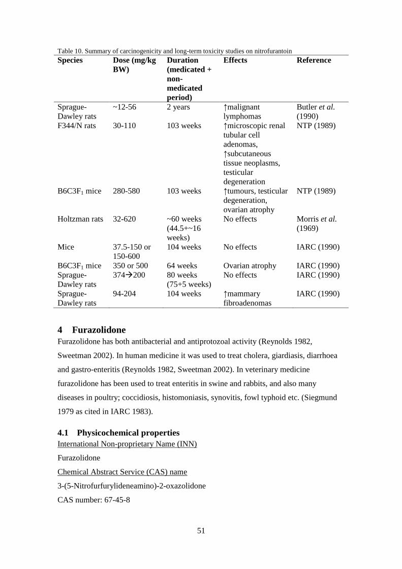
51
Table 10. Summary of carcinogenicity and long-term toxicity studies on nitrofurantoin
Species Dose (mg/kg
BW)
Duration
(medicated +
non-
medicated
period)
Effects Reference
Sprague-
Dawley rats
~12-56 2 years ↑malignant
lymphomas
Butler et al.
(1990)
F344/N rats 30-110 103 weeks ↑microscopic renal
tubular cell
adenomas,
↑subcutaneous
tissue neoplasms,
testicular
degeneration
NTP (1989)
B6C3F1 mice 280-580 103 weeks ↑tumours, testicular
degeneration,
ovarian atrophy
NTP (1989)
Holtzman rats 32-620 ~60 weeks
(44.5+~16
weeks)
No effects Morris et al.
(1969)
Mice 37.5-150 or
150-600
104 weeks No effects IARC (1990)
B6C3F1 mice 350 or 500 64 weeks Ovarian atrophy IARC (1990)
Sprague-
Dawley rats
374200 80 weeks
(75+5 weeks)
No effects IARC (1990)
Sprague-
Dawley rats
94-204 104 weeks ↑mammary
fibroadenomas
IARC (1990)
4 Furazolidone
Furazolidone has both antibacterial and antiprotozoal activity (Reynolds 1982,
Sweetman 2002). In human medicine it was used to treat cholera, giardiasis, diarrhoea
and gastro-enteritis (Reynolds 1982, Sweetman 2002). In veterinary medicine
furazolidone has been used to treat enteritis in swine and rabbits, and also many
diseases in poultry; coccidiosis, histomoniasis, synovitis, fowl typhoid etc. (Siegmund
1979 as cited in IARC 1983).
4.1 Physicochemical properties
International Non-proprietary Name (INN)
Furazolidone
Chemical Abstract Service (CAS) name
3-(5-Nitrofurfurylideneamino)-2-oxazolidone
CAS number: 67-45-8
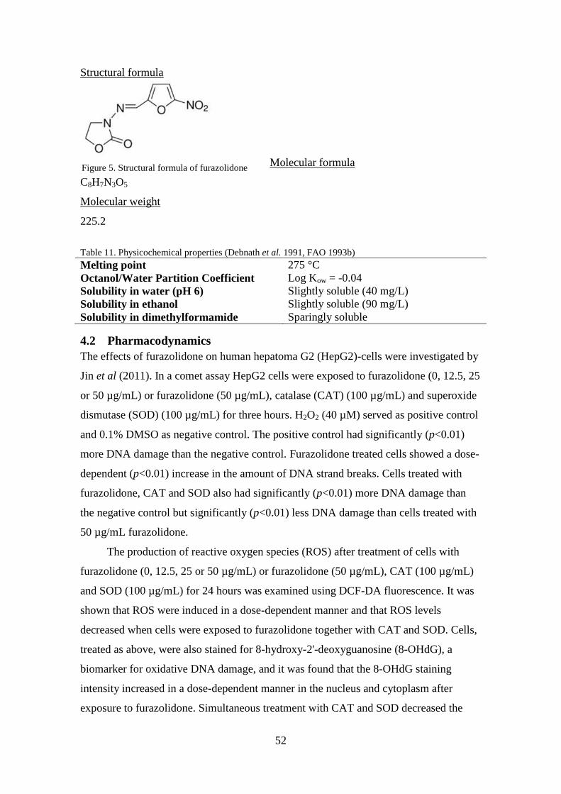
52
Structural formula
Molecular formula
C8H7N3O5
Molecular weight
225.2
Table 11. Physicochemical properties (Debnath et al. 1991, FAO 1993b)
Melting point 275 °C
Octanol/Water Partition Coefficient Log Kow = -0.04
Solubility in water (pH 6) Slightly soluble (40 mg/L)
Solubility in ethanol Slightly soluble (90 mg/L)
Solubility in dimethylformamide Sparingly soluble
4.2 Pharmacodynamics
The effects of furazolidone on human hepatoma G2 (HepG2)-cells were investigated by
Jin et al (2011). In a comet assay HepG2 cells were exposed to furazolidone (0, 12.5, 25
or 50 µg/mL) or furazolidone (50 µg/mL), catalase (CAT) (100 µg/mL) and superoxide
dismutase (SOD) (100 µg/mL) for three hours. H2O2 (40 µM) served as positive control
and 0.1% DMSO as negative control. The positive control had significantly (p<0.01)
more DNA damage than the negative control. Furazolidone treated cells showed a dose-
dependent (p<0.01) increase in the amount of DNA strand breaks. Cells treated with
furazolidone, CAT and SOD also had significantly (p<0.01) more DNA damage than
the negative control but significantly (p<0.01) less DNA damage than cells treated with
50 µg/mL furazolidone.
The production of reactive oxygen species (ROS) after treatment of cells with
furazolidone (0, 12.5, 25 or 50 µg/mL) or furazolidone (50 µg/mL), CAT (100 µg/mL)
and SOD (100 µg/mL) for 24 hours was examined using DCF-DA fluorescence. It was
shown that ROS were induced in a dose-dependent manner and that ROS levels
decreased when cells were exposed to furazolidone together with CAT and SOD. Cells,
treated as above, were also stained for 8-hydroxy-2'-deoxyguanosine (8-OHdG), a
biomarker for oxidative DNA damage, and it was found that the 8-OHdG staining
intensity increased in a dose-dependent manner in the nucleus and cytoplasm after
exposure to furazolidone. Simultaneous treatment with CAT and SOD decreased the
Figure 5. Structural formula of furazolidone

53
staining intensity. The levels of glutathione (GSH) and CAT in cells significantly
decreased after exposure to furazolidone (25 and 50 µg/mL). The levels of SOD
significantly decreased after exposure to 12.5, 25 and 50 µg/mL furazolidone.
Ali et al. (1988) orally administered furazolidone (7.5, 15 or 30 mg/kg BW) to male
turkeys for two weeks and then measured the levels of luteinizing hormone (LH) and
prolactin (PRL). No effects on the levels of PRL or LH were observed at any dose but
at 30 mg/kg BW the hypothalamic levels of dopa, adrenaline and noradrenaline were
significantly increased.
4.3 Pharmacokinetics
4.3.1 In vitro studies
Two metabolites were isolated after in vitro reduction of furazolidone using milk
xanthine oxidase (Tatsumi et al. 1978). Only one metabolite could be identified, 3-(4-
cyano-2-oxobutylideneamino)-2-oxazolidone. This metabolite was also found in the
urine of four male albino rabbits following a single oral administration of furazolidone
at a dose of 100 mg/kg BW.
The in vitro metabolism of furazolidone using milk xanthine oxidase and the S9-mix for
20 minutes was investigated by Tatsumi et al. (1981). Three metabolites were detected
and identified; 3-(4-cyano-2-oxobutylideneamino)-2-oxazolidone, an aminofuran
derivative N-(5-amino-2-furfurylidene)-3-amino-2-oxazolidone and 2,3-dihydro-3-
cyanomethyl-2-hydroxyl-5-nitro-1α,2-di(2-oxo-oxazolidin-3-yl) iminomethylfuro[2,3-
b]furan.
Vroomen et al. (1987a) examined the metabolism of 14
C-furazolidone by rat liver
microsomes under anaerobic and aerobic conditions. Around 2-3% of the total
metabolites were covalently bound to proteins. Two major metabolites, 2,3-dihydro-3-
cyanomethyl-2-hydroxyl-5-nitro-1α,2-di(2-oxo-oxazolidin-3-yl)iminomethylfuro[2,3-
b]furan and 3-(4-cyano-2-oxobutylideneamino)-2-oxazolidone, were formed and
accounted for 16.4 and 16.6 %, respectively, of the total extractable radioactivity. It was
shown that the metabolism of furazolidone was not dependent on cytochrome P-450,
but is reliant on another enzymatic NADPH-dependent process.

54
To study the degradation pathways in different cell lines (Caco-2, HEp-2 and V79) cells
were incubated with 14
C-furazolidone, labelled in the nitrofuran part, for 24 or 48 hours
(de Angelis et al. 1999). The degradation of furazolidone led to the formation of several
hydrophilic compounds which to a large extent differed between the cell lines. In HEp-2
cells small amounts of the open-chain cyano metabolite 3-(4-cyano-2-
oxobutylideneamino)-2-oxazolidone was formed.
de Angelis et al. also evaluated the degree of protein-bound furazolidone
metabolites by incubating cells with 14
C-furazolidone, labelled in either the furan or
oxazolidone ring, for 24 hours. Of the three cell lines, HEp-2 cells showed the most
binding followed by Caco-2 and V79 cells. In Caco-2 and HEp-2 cells there was a
higher degree of binding of molecules labelled in the furan ring. In V79 cells the degree
of binding was the same.
Liver microsomes from a female swine weighing 90 kg were incubated with 14
C-
furazolidone (36.7 µM) for six minutes at 37 °C (Vroomen et al. 1987b). At the end of
the incubation 53.6 % of the furazolidone had been metabolised, of which 11.6 % was
covalently bound to protein. 27.4 % could be extracted in ethyl acetate and 61.1 % in
water. Microsomes that had been inactivated by heating showed no furazolidone
conversion when NADH or NADPH was added. Three major peaks were seen after
HPLC-analysis of the ethyl acetate extract of which two metabolites were identified as
3-(4-cyano-2-oxobutylideneamino)-2-oxazolidone and 2,3-dihydro-3-cyanomethyl-2-
hydroxyl-5-nitro-1α,2-di(2-oxo-oxazolidin-3-yl) iminomethylfuro[2,3-b]furan,
constituting 10.5 and 6.4 % of the total metabolites formed respectively.
The amount of radioactivity in the ethyl acetate increased while the radioactivity
in the water and protein-bound fraction decreased when 5 mM of mercaptoethanol was
added to the incubation mixture. When the ethyl acetate fraction was analysed by HPLC
three new peaks had appeared. One accounted for around 50 % of the total metabolites
formed and the other two accounted for 4 and 8 %. The new major metabolite formed
was tentatively identified as 3-(4-cyano-3(or 4)-β-hydroxyethylmercapto-2-
oxobutylideneamino)-2-oxazolidone, a mercaptoethanol conjugate of furazolidone. This
indicates that at least 50 % of the total metabolites of furazolidone are formed through
reductive processes in swine liver microsomes.

55
Pig hepatocytes were incubated for 4 or 20 hours with furazolidone (2.5 or 25 µM),
AOZ (2.5 or 25 µM) to investigate the formation of protein-bound metabolites
(Hoogenboom et al. 2002). The results showed that the formation of protein-bound
metabolites was dose and time related for AOZ. In a similar experiment pig hepatocytes
were incubated for 4 or 20 hours with furazolidone or AOZ and the formation of
protein-bound metabolites and their release under acidic conditions were examined.
Protein-bound metabolites were formed in all dose and incubation groups. The fraction
of released AOZ from proteins in cells incubated with furazolidone ranged between 31
and 59 % while in the cells incubated with AOZ the fraction ranged between 21 and 35
%.
Several other metabolites of furazolidone have also been discovered, e.g. 5-nitro-
furaldehyde and 5-nitrofuran-2-carboxylic acid (FAO 1993b).
4.3.2 In vivo studies
4.3.2.1 In swine
Six individually housed three-weeks-old male piglets (Dutch Landrace × Great
Yorkshire) were divided into three groups; control, 14
C-furazolidone (labelled in the
methylene group of the oxazolidone moiety), and unlabelled furazolidone (Vroomen et
al. 1986). The piglets were fed twice daily and the furazolidone preparations (12 mg/kg
BW) were administered, via stomach tube, at the same time for 10 days. A 14 days long
withdrawal period then followed. Faeces and urine were collected throughout the entire
experiment. Two hours after each morning feeding blood samples were retrieved and
the concentration of furazolidone was analysed in blood and plasma. Expired air was
analysed for radioactivity during the medication period.
At day 10 one piglet from each group was sacrificed and at the last day of the
withdrawal period the three remaining piglets were sacrificed. Samples of fat, muscle
and liver were taken and analysed for radioactivity and unlabelled furazolidone.
Samples were also taken from several other organs and analysed for radioactivity. The
concentration of furazolidone was analysed by HPLC-UV at 362 nm. Radioactivity (µg
equivalents of parent compound per g tissue) was measured in a Philips liquid
scintillation counter.
Mean levels of furazolidone in blood during the medication period were between
0.31 and 0.53 µg/mL. Similar values were seen in plasma, but no accumulation in blood

56
was seen during the experiment. After the last administration on day 10, the maximum
concentrations of furazolidone in blood and plasma were reached within 30 minutes
(0.84 µg/mL in blood and 0.96 µg/mL in plasma). After this point the levels decreased
and were below the limit of detection (1 ng/mL) 3-4 hours after administration. The
half-life of furazolidone in blood and plasma was around 60 minutes.
Radioactivity in blood and plasma increased during the medication period and
reached maximum levels (9-10 µg equivalents of parent compound per mL) after 5-6
days. During the first day of the withdrawal period there was a fast decrease in
radioactivity followed by a slower decrease during the next 13 days, with a half-life of
approximately six days. At the end of the withdrawal period, residual values of 2.6 and
1.5 µg equivalents of parent compound per mL were still present in blood and plasma
respectively.
The main excretion pathway of radiolabelled furazolidone was via urine. At the
end of the experiment the total excretion of radioactive labelled material via the urine
was 61 %. No radioactivity was measured in the expired air and 18-21 % of the
radioactivity was excreted via the faeces. No parent furazolidone could be detected in
muscle, liver, kidney or fat immediately after the medication period. The tissues with
the highest radioactivity after the medication period were bile, liver and kidney (81.1,
32.9 and 30.1 mg/kg). The radioactivity in muscle was between 5.7 and 7.2 mg/kg. At
the end of the withdrawal period the levels ranged between 1.1 and 3.1 mg/kg. The
recovery of total dose radioactivity was 80%.
The percentage of non-extractable radioactivity increased during the withdrawal
period in liver, kidney and muscle. At this time the non-extractable radioactivity levels
were 0.8, 0.9 and 1.2 mg/kg. The interaction between furazolidone and DNA was also
investigated. Immediately after the medication period there were marked interaction in
the pancreas and kidney, 382 and 372 pmol equivalents of parent compound per mg
DNA respectively. At the end of the withdrawal period the DNA binding was around
100 pmol equivalents of parent compound per mg DNA.
Pigs (n=18) were fed feed containing furazolidone (400 mg/kg feed) ad libitum for ten
days, corresponding to around 24 mg/kg BW per day, followed by a withdrawal period
of six weeks (Cooper et al. 2005). Three pigs were sacrificed each week of the
withdrawal period and samples of muscle, liver and kidney were taken and analysed for
furazolidone and AOZ using LC-MS/MS and high performance liquid chromatography-

57
UV (HPLC-UV). Furazolidone was only detected in one muscle sample (0.33 µg/kg
BW) at week 0 of the withdrawal period. AOZ was detected in all samples and the
levels found at week 6 of the withdrawal period were around 15 µg/kg BW in kidney
and 50 µg/kg BW in muscle and liver. The depletion half-lives of AOZ in muscle, liver
and kidney were 11.5±0.9 days, 7.3±0.3 days and 6.9±0.8 days respectively.
Adult swine (Dutch Landrace × Great Yorkshire) were orally administered feed
containing around 7.8 mg/kg BW of furazolidone per day for 10 days followed by a
withdrawal period of 14 days (Vroomen et al. 1987c). Ten swine were treated with
furazolidone and two served as control. Urine was collected during the medication
period and analysed for furazolidone. On day 10 of the medication period blood
samples were taken after the last administration and analysed for furazolidone and 3-(4-
cyano-2-oxobutylidene amino)-2-oxazolidone. On every other day of the withdrawal
period two animals were sacrificed and samples of liver, kidney and muscle were
retrieved and analysed for furazolidone and 3-(4-cyano-2-oxobutylidene amino)-2-
oxazolidone. On the second day of medication the urinary excretion of furazolidone was
at its highest, 447-1784 µg per animal, and then decreased during the rest of the
medication period. The daily mean amount of excreted furazolidone in the urine was
calculated to be 0.06% ± 0.04% of the daily dose (range: 0.01-0.26%). Five hours after
the last administration of furazolidone over 60% of the total excreted amount of
furazolidone had been excreted via the urine and after an additional 19 hours no
furazolidone could be detected. The maximum plasma levels of furazolidone, 62-121
ng/mL, were reached within 60-75 minutes after the last administration and the
elimination half-life was calculated to be approximately 45 minutes. 3-(4-cyano-2-
oxobutylidene amino)-2-oxazolidone reached its maximum concentration, 24.9-45.3
ng/mL, in plasma within 1.5 and 4 hours. It could no longer be detected 17 hours after
the administration. No furazolidone could be detected in any tissue samples two hours
after the last administration on day 10, but 3-(4-cyano-2-oxobutylidene amino)-2-
oxazolidone was detected in muscle samples at a mean level of 17 µg/kg.
McCracken et al. (1995) administered furazolidone to 15 pigs weighing around 30 kg
via stomach tube, corresponding to 20 mg/kg BW. At 1, 2, 4, 6 and 12 hours after the
exposure, three pigs were sacrificed and samples of the liver, muscle and kidney were
collected. Additional muscle samples were taken from the pigs slaughtered after one

58
hour and stored at -20 °C, -70 °C and in liquid nitrogen to be analysed weekly for up to
four weeks. The levels of furazolidone in the samples were examined using thermospray
LCMS. It was found that the levels in liver and kidney were much lower than those in
the muscle tissue. The half-life of furazolidone in muscle was calculated to be around
one hour. The stability of furazolidone in muscle kept in storage at -20 °C was tested.
Furazolidone proved unstable and after one week only 10 % of the original
concentration remained. The levels of furazolidone in the samples stored at -70 °C had
dropped to 50 % of the original concentration and samples stored in liquid nitrogen also
showed decreased levels.
Ten twelve-weeks-old pigs were given feed two times per day containing 300 mg
furazolidone per kg feed (corresponding to around 17 mg/kg BW) for seven days
(Hoogenboom et al. 1992). Following the medication period was a withdrawal period of
four weeks. At 0, 1, 2, 3 and 4 weeks of the withdrawal period two animals were
slaughtered at each time point. Samples of kidney, liver and rump muscle were taken
and stored at -20 °C before analysis. The samples were analysed for protein-bound
metabolites containing the AOZ side chain. The results showed that the AOZ side chain
was released from protein-bound metabolites in liver, muscle and kidney throughout the
experiment. At the end of the medication period, the levels of AOZ released from liver,
muscle and kidney were 915-1070, 103-144 and 434-765 µg/kg w.w., respectively. At
the end of the four week withdrawal period, these levels had dropped significantly and
were now 35-46, 6-13 and 6-7 µg/kg w.w. in liver, muscle and kidney samples,
respectively.
Hoogenboom et al. (1991) studied the nature of protein-bound metabolites of
furazolidone using pig hepatocytes, microsomes and liver samples. Hepatocytes were
isolated from a liver sample from a sow between five and nine months old and cultured.
These cells were then incubated for different time intervals with 25 or 50 µM 14
C-
furazolidone. There was a time-related increase in the amount of protein-bound
metabolites and 36 hours after the exposure ended, the levels decreased to
approximately 50 % of the maximal levels of bound metabolites. From a test using
hepatocytes incubated with furazolidone for 24 hours it was shown that almost 70 % of
the bound metabolites contained an AOZ side-chain that could be released under acidic
conditions similar to those in the stomach.

59
Microsomes prepared from a sow between five and nine months old were
incubated with 50 µM 14
C-furazolidone for 24 hours. Extraction of AOZ from
microsome proteins showed that around 60 % of the bound metabolites contained an
AOZ side-chain that could be released under acidic conditions.
Liver samples were collected from two male piglets with a bodyweight of 6 kg
administered furazolidone (75 mg/day), corresponding to 12.5 mg/kg BW per day, for
10 days followed by a withdrawal period of 2 hours or 14 days. Levels of released AOZ
from liver samples after 2 hours and 14 days withdrawal were 23 and 14 % of the total
protein-bound metabolites respectively, corresponding to 3.8 and 0.3 µg furazolidone
per g of tissue.
Castrated male pigs (n=24) weighing 15-18 kg were fed ad libitum feed containing
furazolidone (400 mg/kg feed), corresponding to 23.5 mg/kg BW per day, for seven
days (Liu et al. 2010). A control group of six pigs were fed untreated feed for seven
days. At 0.5, 7, 21, 35, 56 and 63 days of the withdrawal period, four treated pigs and
one control pig were euthanized and samples of blood, liver, kidney and muscle were
taken. Following the last dosing urine was collected daily. The levels of AOZ in the
samples were analysed using an indirect competitive enzyme-linked immunosorbent
assay (ic-ELISA). Twelve hours after the last dosing the highest concentration of AOZ
was in liver (2169.7 ± 253.1µg/kg) and the lowest concentration was in muscle (691.0 ±
242.4 µg/kg). This was the case throughout the withdrawal period. At the last day of the
withdrawal period, day 63, AOZ was still present in all tissue samples (0.6-1.1 µg/kg)
and urine and plasma samples (1.0-1.2 µg/L). The elimination half-lives of AOZ was
calculated to be 13.6 days in liver and kidney, 13.7 days in plasma, 14.7 days in urine
and 15.0 days in muscle.
Gottschall & Wang (1995) investigated the depletion and bioavailability of 14
C-
furazolidone residues in swine tissues. Four swine (2 males and 2 females) served as
control and six swine (3 females and 3 males) were administered a daily oral dose of
furazolidone (16.5 mg/kg BW) for 14 days followed by a withdrawal period of 10
hours, 21 or 45 days. After the swine were euthanized samples of liver, fat, kidney and
muscle were collected. The total radioactive residues at the first day of the withdrawal
period were highest in liver (~41 mg/kg) followed by kidney (~34 mg/kg), muscle (~13
mg/kg) and fat (~6 mg/kg). After 21 days these levels had quickly decreased, between

60
50-90 %. The depletion of residues between days 21 and 45 of the withdrawal period
was much slower than that between days 0 and 21. The levels of tissue residues at the
last day of the longest withdrawal period were around 2 mg/kg in all tissues. The
biphasic elimination may be due to rapid elimination of free drug and metabolites
followed by elimination and turnover of residues bound to macromolecules.
The percentage of extractable residues from the tissue samples decreased with
increasing withdrawal time. Of the residues in liver collected at the first day of the
withdrawal period 44 % were extractable. From liver samples taken on day 45, only 8.3
% of the residues could be extracted. In muscle most of the residues were bound and
only 21.8 % could be extracted on day 0 of withdrawal (Gottschall & Wang 1995).
4.3.2.2 In bovine
One cow was administered one capsule containing; 0.88 mg/kg BW furazolidone,
nitrofurazone and furaltadone and 4.4 mg/kg BW nitrofurantoin (Chu & Lopez 2007).
Milk samples were then collected for two weeks every 12 hours. Milk from non-treated
cows was used as control. The levels of nitrofuran side-chain residues in the milk were
determined using LC-MS/MS. The level of the side-chain of furazolidone, AOZ, in milk
was highest 12 hours after dosing (~32 µg/kg) and decreased rapidly to below the
detection limit (0.2 µg/kg) 72 hours after dosing.
Veal calves (n=6) were administered furazolidone and furaltadone (7-8 mg/kg BW)
orally via the morning milk (Nouws & Laurensen 1990). Three hours after the
administration four calves were slaughtered and samples of heart muscle, kidney, liver,
muscularis gracilis, diaphragm, fat, lung, brain, urine and blood were collected and
stored for analysis using HPLC. The remaining two calves were slaughtered four hours
after administration. Only low levels of furazolidone could be detected in fat, brain and
lung. The liver and kidney samples were analysed at 10, 40, 120 minutes and 24 hours
after collection and the muscle and heart samples after 15, 60, 120 minutes and 24
hours. There were large variations between tissues and animals, but immediately after
the slaughter furazolidone could be detected in the tissues. The highest levels were
found in plasma (0.336-3.34 µg/mL) and urine (1.11-6.22 µg/mL). Two hours after the
slaughter, trace amounts of furazolidone could be detected in the muscle and kidney
samples, but not in liver samples. No limit of quantification (LOQ) was reported. After
24 hours, no levels could be detected. The postmortal degradation half-lives of

61
furazolidone were calculated to be 17 minutes in muscularis gracilis and kidney, 30
minutes in diaphragm and heart, and <7 minutes in liver.
4.3.2.3 In fish
The depletion of nitrofurans and their tissue-bound residues in channel catfish
(Ictakurus punctatus) was investigated by Chu et al. (2008). Fish (n=55) were orally
administered furazolidone, nitrofurantoin, nitrofurazone and furaltadone (1 mg/kg BW).
After 2, 4, 8 and 12 hours, and 1, 4, 7, 10, 14, 28 and 56 days five fish were killed and
muscle samples collected for analysis of parent nitrofurans and their tissue-bound
residues. The highest concentration of furazolidone in muscle (30.4 µg/kg) was reached
four hours after administration. Furaltadone could no longer be detected 96 hours after
administration. The level of AOZ was the highest (33.7 µg/kg) 12 hours after
administration. The elimination of all tissue-bound residues was biphasic and could still
be detected 56 days after administration. The half-life for AOZ was calculated to be 32
days.
Chu et al. (2008) also examined the levels of nitrofurans and tissue-bound
metabolites in muscle of fish after waterborne exposure to nitrofurans. Fish (5 per
treatment) were exposed to either nitrofurantoin, nitrofurazone, furazolidone or
furaltadone (10 mg/L) for eight hours. After this time the fish were killed and their
muscle tissue was analysed for parent nitrofuran and tissue-bound metabolites. The
concentrations of furazolidone and AOZ were around 401 and 203 µg/kg, respectively.
4.3.2.4 In rat
The bioavailability of AOZ residues to rats were investigated by McCracken &
Kennedy (1997). Liver, kidney and muscle samples were taken from pigs administered
furazolidone (~20 mg/kg BW) for five days and fed to female Sprague Dawley rats,
four per feeding group, for three days. Another two groups of two rats were fed either
pig liver, which had undergone solvent extraction, or control feed. Liver, kidney and
muscle samples were collected on the third day and analysed for extractable and bound
residues of AOZ. No AOZ was found in the control animals but was detected in all
tissues from the rats fed pig liver or kidney. The rats fed liver had the highest levels of
bound AOZ in liver (55 µg/kg) and kidney (40 µg/kg), and lowest levels in muscle (2
µg/kg). Extractable levels were highest in kidney (172 µg/kg) and liver (66 µg/kg), and
lowest in muscle (5 µg/kg). The rats fed kidney had similar levels of bound AOZ in
liver and kidney (33-34 µg/kg), and the levels in muscle were about 1 µg/kg.

62
Extractable levels were highest in kidney (318 µg/kg) and liver (42 µg/kg), and lowest
in muscle (5 µg/kg). The rats fed muscle had similar levels of bound AOZ in kidney and
liver (7-9 µg/kg). Extractable levels were highest in kidney (38 µg/kg) and liver (17
µg/kg). Trace amounts (<0.5 µg/kg) of bound and extractable AOZ was found in
muscle. The two rats fed solvent-extracted pig liver had detectable levels of bound AOZ
in their tissues; liver (52 and 79 µg/kg), kidney (45 and 48 µg/kg) and muscle (3 and 2
µg/kg). The levels of extractable AOZ were; 3 and 8 µg/kg in muscle, 175 and 260
µg/kg in kidney, and 94 and 116 µg/kg in liver.
Tatsumi et al. (1984) orally administered furazolidone (100 mg/kg BW) daily to six
male Wistar-strain rats for four days. Urine was collected during the experiment and
urinary metabolites were isolated. Simultaneously, five rats were given 14
C-furazolidone
(100 mg/kg BW) orally for four days to investigate the metabolic pathways of
furazolidone. Urine was also collected and analysed for urinary metabolites.
Four metabolites were isolated from the collected urine; an open-chain cyano
derivative 3-(4-cyano-2-oxobutylideneamino)-2-oxazolidone, an acetamidofuran N-(5-
acetamido-2-furfurylidene)-3-amino-2-oxazolidone, a methylated open-chain carboxylic
acid N-(4-meth-oxycarbonyl-2-oxobutylideneamino)-2-oxazolidone, and α-ketoglutaric
acid.
Four rats were injected into the stomach with an amount equivalent to 1 g of dry weight
liver from pigs that were killed immediately after the last administration of furazolidone
containing about 70 µg non-extractable residues per gram dry weight (Hoogenboom et
al. 2002). The rats were killed three or six hours after the injection. The levels of AOZ
in plasma of the rats were estimated to be between 3 and 5 ng/mL.
Three rats in three groups were fed with liver taken either from non-medicated
pigs or pigs administered 14
C-furazolidone followed by a withdrawal period of 0 or 21
days (Hoogenboom et al. 2002). After six hours the rats were killed and the levels of
AOZ in the plasma were analysed. The results showed that rats fed liver from pigs
killed immediately after the last administration had levels of AOZ in their plasma. The
livers of these rats were also analysed and the total residue levels were 26 ng/g and the
levels of non-extractable residues were 18 ng/g. In the rats fed liver from non-medicated
pigs or liver taken from pigs 21 days after the last administration, no AOZ could be
detected in plasma or liver.

63
The bioavailability of furazolidone and its residues in rats was also investigated
(Gottschall & Wang 1995). Male rats were fed control lyophilized swine muscle,
control lyophilized swine liver and control laboratory chow fortified with 14
C-
furazolidone. The bioavailability of furazolidone in the laboratory chow and swine
muscle were around 90 %, while in swine liver the bioavailability was approximately 73
%. For the absorbed furazolidone, the major excretion pathways were via urine and bile.
For the non-absorbed fraction it was via the faeces.
Rats were also fed tissues from the residue depletion study with swine by
Gotschall & Wang (1995). The bioavailability of the furazolidone residues was around
40 % for muscle taken 45 days into the withdrawal period and for liver and muscle
taken 0 days into the withdrawal period. For liver taken at day 45 the bioavailability
was 20 %. Also here the major excretion pathway for absorbed furazolidone was via
urine and for the non-absorbed fraction via the faeces.
The bioavailability of the non-extractable residues was also examined by feeding
rats tissues that had been treated with organic solvents to remove extractable residues. It
was shown that the bioavailability was the highest for muscle collected at day 45 (37 %)
followed by liver collected at day 0 (31 %) and liver taken at day 45 (16 %). Once again
the major pathways were via the urine and faeces.
4.3.2.5 In poultry
To investigate the transfer of furazolidone from hen to egg four White Leghorn hens
were given 500 mg furazolidone per kg feed orally for 14 days, corresponding to about
31 mg/kg BW and day (Furusawa 2001). Five hens were used as control. Eggs were
collected at two day intervals and albumen and yolk were separated immediately. The
concentration of furazolidone was measured by HPLC with 80 and 82 % recovery in
albumen and egg yolk, respectively. During the experiment there was no difference in
body weight, egg production or feed intake between the control group and treated
animals. The levels of furazolidone in eggs increased during the first days of the
experiment and reached a plateau around day 4 and remained at those levels throughout
the experiment. The mean concentration of furazolidone in eggs was 0.27 mg/kg (range:
0.10-0.29 mg/kg). The transfer rate was calculated to be 0.03 % of the drug intake.

64
Furazolidone was administered to 28-day-old broilers (n=20) in the feed (30, 100, 300,
1000 or 3000 µg/kg feed) corresponding to 6, 20, 60, 200 and 600 µg/kg BW, for 12
days (McCracken et al. 2005b). After this time the broilers were killed and samples of
muscle and liver were collected and analysed for AOZ using LC-MS/MS. At the lowest
dose AOZ was still detected in muscle (0.33 µg/kg) and liver (1.1 µg/kg) samples. At
the highest dose the levels in liver and muscle were around 25 and 12 µg/kg,
respectively.
Six untreated broilers were placed in a dirty pen, which had housed four broilers
administered furazolidone (600 µg/kg BW) for 12 days, for 24 hours. Liver and muscle
samples were then collected and analysed for AOZ. AOZ was detected in all samples
and the mean level of AOZ in muscle was 0.13 µg/kg (range: 0.05-0.31 µg/kg) and in
liver the mean level was 0.10 µg/kg (range: 0.04-0.21 µg/kg).
McCracken et al. (2005a) fed six broiler hens and one cockerel 120-140 g of feed
containing furazolidone (400 mg/kg feed), corresponding to about 96-112 mg/kg BW
per day. Eggs were collected and analysed for AOZ and when it was clear that
furazolidone residues had transferred to the eggs, the eggs laid after this were collected
and allowed to hatch. After hatching, four chicks were sacrificed at determined intervals
and muscle and liver samples were analysed for AOZ. However, AOZ could only be
detected up to day 21. The levels of AOZ in liver were around 6.5 µg/kg, 1.6 µg/kg, 0.3
µg/kg and 0.1 µg/kg in 1, 4, 12 and 21 days old chicks, respectively. In muscle the
levels of AOZ were around 4.6 µg/kg, 0.7 µg/kg, 0.2 µg/kg and 0.05 µg/kg in 1, 4, 12
and 21 days old chicks, respectively.
Twenty-four laying hens were fed feed containing 300 mg furaltadone, nitrofurazone,
nitrofurantoin or furazolidone per kg feed for one week, corresponding to approximately
60 mg/kg BW per day (McCracken & Kennedy 2007). Eggs were then collected for two
days and analysed immediately for nitrofuran parent compound and their bound
residues in albumen, yolk and shell using LC-MS/MS. The levels of furazolidone in
yolk, albumen and shell were 410, 291 and 51.8 ng/g, respectively. The levels of AOZ
in yolk, albumen and shell were 478, 491 and 114 ng/g, respectively.

65
4.3.2.6 In eel
The metabolism of furazolidone in eels and eel liver homogenate was examined by
Nakabeppu & Tatsumi (1984). Liver homogenate was incubated with 20 µmol of
furazolidone for one hour in an open vessel in the dark at 37 °C. The mixture was then
extracted with ethyl acetate and dried over anhydrous Na2SO4 and evaporated to
dryness. The residue was chromatographed on a silica gel column and a metabolite, 3-
(4-cyano-2-oxobutylideneamino)-2-oxazolidone was discovered.
Two eels were bathed in 2 L 14
C-furazolidone (0.5 µL/L) for 24 hours and four
eels were bathed in 3 L unlabelled furazolidone (15 µL/L) for four days. Analysis
showed that water-soluble metabolites and two ethyl acetate-extractable metabolites,
one of which was identified as 3-(4-cyano-2-oxobutylideneamino)-2-oxazolidone, were
formed from nitrofurazone metabolism.
4.3.3 Conclusions on pharmacokinetics of furazolidone and its metabolites
Pharmacokinetic studies in swine, rat, calf and poultry have shown that furazolidone is
quickly absorbed and distributes throughout the body, with the highest concentrations
found in liver and kidney. It was shown that furazolidone is readily degraded after
slaughter and is degraded even when frozen, making it difficult to monitor illegal use by
measuring the parent compound in food stuffs. Furazolidone is excreted from the body
with half-lives of about one hour. The excretion of furazolidone appears to be biphasic
in swine with a fast excretion in the beginning of the withdrawal period followed by a
slower excretion. The major excretion pathways are via the urine, bile and faeces. From
the study on the bioavailability of furazolidone for rats fed liver and muscle from pigs
administered furazolidone it seems that furazolidone has a quite high bioavailability
even after 45 days of withdrawal. It was also shown that furazolidone could be
transferred to eggs from poultry treated with furazolidone.
The identified metabolites of furazolidone are presented in Table 12. From the
studies above the most common metabolites observed in swine and rat are 3-(4-cyano-
2-oxobutylideneamino)-2-oxazolidone and AOZ, formed via reductive pathways.
However, it is the metabolite AOZ that is the marker residue for furazolidone due to its
long half-lives in tissues. In swine the half-lives of AOZ were shown to be between one
to two weeks in kidney, muscle, liver and plasma. But it was still present in tissues 63
days after the last administration of furazolidone. AOZ has been shown to accumulate
more in liver than in muscle. Residues of furazolidone (e.g. AOZ) were also shown to
be bioavailable for uptake for rats fed muscle and liver from furazolidone treated pigs.

66
Table 12. Metabolites of furazolidone in different species found in the literature
Metabolite Species/cells Reference
3-(4-cyano-2-
oxobutylideneamino)-2-
oxazolidone
Rabbit, eel, rat and
pig liver
microsomes, Caco-
2, Hep-2, V79 cells
de Angelis et al. (1999),
Nakabeppu & Tatsumi
(1984), Tatsumi et al.
(1981 & 1984), Vroomen et
al. (1987a, 1987b)
3-amino-2-oxazolidone (AOZ) Broiler, pig Cooper et al. 2005,
Hoogenboom et al. (1991
& 1992), Liu et al. (2010),
McCracken et al. (2005b)
N-(5-amino-2-furfurylidene)-3-
amino-2-oxazolidone
Rat, pig FAO 1993b, Tatsumi et al.
(1981)
2,3-dihydro-3-cyanomethyl-2-
hydroxyl-5-nitro-1α,2-di(2-oxo-
oxazolidin-3-yl)
iminomethylfuro[2,3-b]furan
Rat, pig liver
microsomes
Tatsumi et al. (1981),
Vroomen et al. (1987b)
N-(5-acetamido-2-furfurylidene)-
3-amino-2-oxazolidone
Rat Tatsumi et al. (1984)
3-(4-cyano-3(or 4)-β-
hydroxyethylmercapto-2-
oxobutylideneamino)-2-
oxazolidone
Pig liver
microsomes
Vroomen et al. (1987b)
N-(4-meth-oxycarbonyl-2-
oxobutylideneamino)-2-
oxazolidone
Rat Tatsumi et al. (1984)
α-ketoglutaric acid Rat Tatsumi et al. (1984)
5-nitro-furaldehyde Rat, pig FAO 1993b
5-nitrofuran-2-carboxylic acid Pig FAO 1993b
4.4 Toxicology of furazolidone
4.4.1 Acute toxicity
The results from acute toxicity studies by Mitchell et al. (1990a, 1990b) are summarized
in JECFA (1993b). The acute toxicity of furazolidone was tested by administering
furazolidone orally to male and female mice and rats. The LD50 was 1110 mg/kg BW
for mice and 1508 mg/kg BW for rats.
4.4.1.1 Conclusions on acute toxicity of furazolidone
From this study it is concluded that furazolidone is slightly toxic to mice and rats.
However, it is not clear whether or not the study followed OECD guidelines and GLP.
4.4.2 Chronic toxicity
Male Wistar rats (six rats per group) were administered furazolidone (0, 0.5, 5 or 10
mg/kg BW) daily via food for 13 months (Aisio et al. 1962 as seen in JECFA 1993b).
No effects were seen on mortality, body weight or food consumption. Blood samples

67
were taken from two rats per dose group at the end of the experiment. No changes in the
number of leucocytes or erythrocytes were detected. At the highest dose the relative
spleen and liver weights were increased slightly and during histological examinations
some hypertrophy of liver cells was seen at all doses.
4.4.2.1 Conclusions on chronic toxicity of furazolidone
No NOAEL could be set in this study due to hypertrophy of liver cells at all doses. The
study does not meet current standards set in the OECD guideline 452 for chronic
toxicity studies.
4.4.3 Reproductive toxicity including teratogenicity
4.4.3.1 Mouse
JECFA (1993b) presented the results from a study by Jackson & Robson (1957).
Pregnant albino C strain mice were given furazolidone (≤2 g/kg BW) starting at
different times during pregnancy (day 1-11). All mice treated with furazolidone (≤1
mg/kg BW) before day 8 had abortions or foetal deaths. Abortions or foetal deaths only
occurred in two out of nine mice when the treatment started at day 10. No congenital
abnormalities were observed.
Only a summary was available. The abortions seen in mice treated before day 8
may be due to failure of embryo implantation. It is not clear how long the medication
period was. The study is quite old and does not meet the current standard for
reproductive toxicity studies.
4.4.3.2 Rat
The study by Larson et al. (1963a) is summarised in JECFA (1993b). Male rats (n=10)
were fed furazolidone (16 or 33 mg/kg BW) in the diet for 14 weeks. Another six rats
were fed furazolidone (16 or 33 mg/kg BW) in the diet for 12 weeks followed by two
weeks recovery period. Four rats constituted the control group. No dose-related effects
were seen in the rats of the lowest dose group. In the high-dose groups the testes weight
of the rats was decreased compared to the control group but more pronounced in the
group treated for 14 weeks. Degeneration of the sperm-producing tubules, atrophy and
oedema of the interstitium were seen in the testes of the rats from the high-dose groups.
There is no mention of if the study was carried out according to OECD guidelines
or GLP. Due to the study being dated it does not meet the current standards for
reproductive toxicity studies, but it does indicate a deleterious effect of furazolidone on

68
male reproduction at doses of 33 mg/kg BW. A NOAEL can be set to 16 mg/kg BW in
this study.
JECFA (1993b) presented an abstract of studies by Borgman & Prytherch (1964b,
1966). Sprague-Dawley rats (20 animals per sex) were used in a three-generation
reproduction study in which only females were initially given feed containing
furazolidone (500 mg/kg feed). The dose was then lowered to 400 mg/kg feed on day 16
and again to 250 mg/kg feed (corresponding to12.5 mg/kg BW per day ) on day 37 due
to growth depression. Each generation were allowed to mate three times. The first litter
was sacrificed on day 21 after birth; the second litter was used for the second generation
and the third litter was used for teratological examination. No effects on reproduction
could be seen and a NOEL was set to 12.5 mg/kg BW per day.
The study was only available as an abstract in JECFA (1993b). The study does
not meet today’s standard regarding reproductive toxicity studies. The fact that the
authors decreased the dose twice during the study makes the results, or lack thereof,
difficult to interpret.
4.4.3.3 Poultry
The results from a study by Francis & Shaffner (1956) are summarised in JECFA
(1993b). New Hampshire chickens were administered furazolidone in the feed (55, 110
or 220 mg/kg feed), corresponding to 11, 22 and 44 mg/kg BW, for 4 to 16 weeks.
There was no observed effect on hatchability of eggs, shell quality, egg production or
body weight.
Based on the results presented in the study a NOEL-value of 44 mg/kg BW could
be set for the above mentioned end points. It does not follow OECD guidelines or GLP
and does not meet today’s standard in testing.
4.4.3.4 Swine
JECFA (1993b) presented a summary of a study by Hughes & McMinn (1963).
Hampshire and Duroc pigs (12 gilts and 9 sows) were given furazolidone (300 mg/kg)
in feed, corresponding to 6 mg/kg BW, for two weeks at breeding and 150 mg/kg,
corresponding to 3 mg/kg BW, for three weeks after giving birth. No effects on number
of piglets born, weaned or weight gain could be observed.

69
The doses are lower than in the other studies and there is no mention of the study
following OECD guidelines or GLP. Due to the age of the study it probably does not
meet today’s criteria.
4.4.3.5 Goat
Mustafa et al. (1987) performed a study and the results are summarised in JECFA
(1993b). The biochemistry and semen morphology of male Nubian goats administered
furazolidone suspended in distilled water (10 or 40 mg/kg BW) for five days was
studied. Furazolidone significantly decreased the semen fructose concentration, the
number of live and motile spermatozoa per ejaculate and ejaculate volume at both
doses.
It is not clear if the study was conducted according OECD guidelines or GLP,
since only an abstract was available. The results of the study indicate a detrimental
effect of furazolidone on male reproduction system.
4.4.3.6 Rabbit
JECFA (1993b) summarised the results from a study by Borgmann & Prytherch
(1964a). Furazolidone (30 mg/kg BW) was orally administered to New Zeeland white
rabbits on days 7-15 of pregnancy. The dams were sacrificed at day 29 and the foetuses
were examined. No teratogenicity or embryotoxicity was found.
There is no mention of following OECD guidelines or GLP and the study most
likely does not meet today’s standards.
4.4.3.7 Conclusions on reproductive toxicity including teratogenicity
The results from the reproductive toxicity studies are presented in Table 13. Of the
seven studies described above only three presented results showing reproductive
toxicity of furazolidone. Treating pregnant mice with furazolidone (≤1 mg/kg BW)
before day 8 caused abortions and foetal deaths. In rats treated with 33 mg/kg BW for
12 to 14 weeks testicular degeneration was found. Goats treated with 10 mg/kg BW for
five days showed decreased semen quality. Due to all studies being quite old and not
living up to today’s criteria in reproductive toxicity testing, it is difficult to draw any
clear conclusions. From these results it appears that the male reproductive system is
sensitive to toxic effects of furazolidone.

70
Table 13. Summary on reproductive toxicity studies including teratogenicity of furazolidone
Species Dose (mg/kg
BW)
Duration Effects Reference
Mouse ? - <2 ? Abortions and
foetal deaths
JECFA (1993b)
Rat 16 or 33 12 or 14 weeks Testicular
degeneration
JECFA (1993b)
Rat 12.5 ? No effects
observed
JECFA (1993b)
Chicken 11, 22, 44 4-6 weeks No effects
observed
JECFA (1993b)
Pig 6 & 3 2 & 3 weeks No effects
observed
JECFA (1993b)
Goat 10 or 40 5 days Decreased
semen quality
JECFA (1993b)
Rabbit 30 9 days No effects
observed
JECFA (1993b)
4.4.4 Mutagenicity and genotoxicity
4.4.4.1 In vitro studies with furazolidone
In a forward mutation assay on Vibrio cholera by Chatterjee et al. (1983), cells were
treated with up to 12 µg/mL furazolidone to induce streptomycin resistance. To allow
for expression of streptomycin resistant mutants, untreated and treated cells were
incubated in NB medium at 37 °C for 18 to 20 hours. The bacteria were then plated in
selective medium (NA containing 100 µg/mL of streptomycin) or a non-selective
medium to score the number of formed mutant colonies and assess the total viable
count, respectively. After 24 hours of incubation the viable counts were recorded and
after 72 hours the mutant colonies were counted. The ratio of mutant counts on the
selective medium to the total viable counts on the non-selective medium gave the
mutation frequency. The results showed that furazolidone induced forward mutation
leading to streptomycin-resistance with the mutation frequency being positively
correlated with the drug concentration up to ~7.0 µg/mL. Above this concentration the
mutation frequency declined.
The study was performed in 1983 and no positive control was used but the results
show that furazolidone is mutagenic.
Furazolidone was tested in a chromosome aberration assay by Cohen & Sagi (1979).
Furazolidone was dissolved in 1 % dimethylsulfoxide (DMSO) to produce
concentrations of 0.2, 2.0 and 20 µg/mL of culture. Phytohemagglutinin (PHA)-
stimulated human lymphocyte cultures were incubated with the different furazolidone

71
concentrations for 24 hours before harvest. From each culture 100 cells in metaphase
were examined for chromosome breaks. Cohen & Sagi (1979) also studied the sister-
chromatid exchange (SCE). For 72 hours PHA-stimulated lymphocytes were incubated
with 10-5
M BUDR. After 48 hours the different concentrations of furazolidone was
added. The lymphocytes were then harvested and 50 cells from each treatment were
examined and the number of SCEs per cell was recorded.
There was no difference in the number of chromosome breaks in lymphocytes
treated with 0.2 µg/mL furazolidone and the control. But lymphocytes treated with 2.0
µg/mL furazolidone showed an increase in chromosome breaks. The lymphocytes
treated with 20.0 µg/mL furazolidone did not enter mitosis and could not be analysed
for chromosome breakage. The number of SCEs per cell was significantly higher
(p<0.001) in cells treated with furazolidone (0.2 and 2.0 µg/mL) compared to the
control. Cells treated with 20.0 µg/mL furazolidone did not enter metaphase.
The study does not follow current OECD guidelines but shows that furazolidone
causes SCEs (0.2 and 2.0 µg/mL) and other chromosome aberrations (2.0 µg/mL) in
lymphocytes. It also shows that furazolidone interferes with mitosis at 20 µg/mL.
The Ames test was used by Ni et al. (1987) to investigate the mutagenicity of
furazolidone. Three strains of Salmonella typhimurium; TA98, TA98NR and TA98/1,8-
DNP6, were exposed to furazolidone both with and without rat-liver hepatic S9
activation enzymes. The doses of furazolidone tested were 0.1, 0.5, 1.0 and 2.5 µg/plate.
Furazolidone tested positive for mutagenicity in the TA98 strain at all doses, both with
and without S9, except for the highest dose which was toxic to the bacteria.
Furazolidone was also mutagenic to TA98NR at 0.5 µg/plate without S9 and 1.0
µg/plate with and without S9. Also in this strain the highest dose was toxic to the
bacteria. In the TA98/1,8-DNP6 strain furazolidone was mutagenic at the two lowest
doses, with and without S9, and toxic at the two highest doses.
The study mostly follows OECD guidelines and the results show that furazolidone
is mutagenic in all tested strains both with and without metabolic activation. In all test
strains the mutagenicity of furazolidone appeared dose-dependent.
Furazolidone tested positive at all concentrations (0.01, 0.1, 1.0 and 10 µg/plate) in an
Ames test performed with S. typhimurium TA 100, both with and without S9-mix
(Vroomen et al. 1987c). A dose-response relationship could be observed.

72
The study follows guidelines to some extent but only the conclusion not the data is
reported.
Tatsumi et al. (1978) examined the mutagenicity of furazolidone in an Ames test using
S. typhimurium TA 100. The bacteria were incubated with furazolidone (0.03, 0.1, 0.3
or 1 µg/plate) for two days at 37 °C on plates lacking histidine. The revertant colonies
were then counted in test and control plates. The results showed that furazolidone was
strongly mutagenic.
The study follows OECD guidelines to some extent and the results indicate that
furazolidone is mutagenic to S. typhimurium TA 100.
4.4.4.2 In vitro studies with metabolites of furazolidone
The mutagenicity of 3-(4-cyano-2-oxobutylideneamino)-2-oxazolidone was also tested
by Tatsumi et al. (1978). The test followed the same procedure as stated above with the
exception that the dose added to each plate was higher (1, 3, 10 or 100 µg/plate). The
number of revertant colonies was far less than those seen on plates containing
furazolidone and 3-(4-cyano-2-oxobutylideneamino)-2-oxazolidone was considered
nonmutagenic.
The study follows OECD guidelines and the results indicate that 3-(4-cyano-2-
oxobutylideneamino)-2-oxazolidone is not mutagenic.
3-(4-cyano-2-oxobutylideneamino)-2-oxazolidone (10 and 100 µg/plate) tested negative
in an Ames test using S. typhimurium TA98 and TA100, with and without S9-mix
(Vroomen et al. 1987c).
The study mostly follows guidelines and the results show that 3-(4-cyano-2-
oxobutylideneamino)-2-oxazolidone is not mutagenic.
AOZ (0.1, 0.33, 1, 3.33 or 6 mg/plate) was tested in an Ames test using several S.
typhimurium strains (TA 98, TA 100, TA 1535, TA 1537), both with and without S9-
mix (Hoogenboom et al. 2002). It was shown that AOZ was mutagenic to TA 1535 in a
dose-dependent manner with rat liver S9 fraction. A positive response was also seen in
strain TA 100, but not in TA 98 or TA 1537.

73
The experiments were carried out at NOTOX and RIKILT and most likely follow
guidelines. The results show that AOZ is mutagenic in some strains of S. typhimurium.
AOZ was also studied in a chromosome aberration test with human lymphocytes
(Hoogenboom et al. 2002). Cells were exposed to AOZ (1000, 3330 or 5000 µg/mL) for
24 or 48 hours without S9-mix and for three hours with S9. MMC (0.2 µg/mL) was
used as positive control. One hundred cells in metaphase were examined for
chromosome aberrations. The number of cells with chromosome aberrations increased
significantly after exposure to AOZ in the absence of S9-mix. No effects were seen
when S9-mix was present.
The study mostly follows OECD guidelines. The results from this study
demonstrates that AOZ is genotoxic to human lymphocytes in the absence of S9-mix
4.4.4.3 In vivo studies with metabolites of furazolidone
In a bone marrow micronucleus-test male and female Swiss mice (n=120) were
intraperitoneally administered with AOZ at different concentrations (32, 62.5, 125, 250
or 500 mg/kg BW in males and 250, 500, 750, 1000 or 1500 mg/kg BW in females)
(Hoogenboom et al. 2002). Prior to this experiment two limit tests were performed.
Cyclophosphamide (50 mg/kg BW) and vehicle (0.9 % NaCl) served as positive and
negative control, respectively. Each dose group consisted of between three and five
animals per sex. Sampling occurred at 24 and 48 hours. The number of micronucleated
polychromatic erythrocytes was counted in 1000 cells. No dose-response relationship
was found and significant increases in micronucleated cells was only seen in the
positive controls and in male mice administered 500 mg/kg BW when sampling
occurred at 48 hours.
The study mostly follows OECD guidelines and the authors conclude that AOZ is
mutagenic due to increases in the fraction of animals with three or more
micronucleated polychromatic erythrocytes per 1000 polychromatic erythrocytes. The
results could be considered positive but since only one dose produced a significant
increase in micronucleated cells, only 1000 cells were examined instead of 2000 and
that the dose group only consisted of four animals the results should be interpreted as
negative.

74
4.4.4.4 Conclusions on mutagenicity and genotoxicity studies
The results from mutagenicity anf genotoxicity studies are summarised in Table 14. In
the studies mentioned above furazolidone has been shown to be mutagenic and
genotoxic in both bacterial tests and an in vitro chromosome aberration test. The
bacterial tests were performed both with and without metabolic activation and using
different species and strains. Furthermore furazolidone has also tested positive in
several other in vitro assays; mammalian cell gene mutation test, HPRT test, sex-linked
recessive lethal test etc. (JECFA 1993b). Of all the in vitro studies stated in JECFA
(1993b) only four had results that were negative. These were; two chromosome
aberration tests using human lymphocytes, an UDS assay using human lymphocytes and
an Ames test. Only two in vivo tests for chromosomal effects, the micronucleus test, are
mentioned in JECFA (1993b). Both tests were performed on mice. In the first one, mice
were injected intraperitoneally with furazolidone (300 mg/kg BW) in methylcellulose
which proved toxic to the animals. The reported results were negative. In the second test
mice were orally administered furazolidone (100 or 500 mg/kg BW) and the results
were equivocal since there was a slight induction of micronuclei but it was not dose-
related. Furazolidone has shown to be mutagenic and genotoxic in vitro but does not
appear to be genotoxic in vivo. To ascertain whether or not furazolidone truly is not
mutagenic in vivo further study is required.
The metabolite 3-(4-cyano-2-oxobutylideneamino)-2-oxazolidone did not test
positive in an Ames test using S. typhimurium TA 100 and might not be mutagenic,
although more studies are needed to verify this. The metabolite AOZ tested positive in
vitro but not in vivo and further study is needed to determine its mutagenic properties.
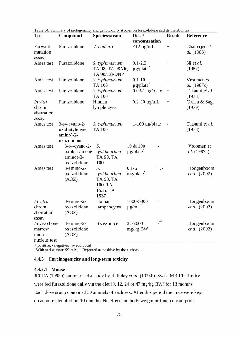
75
Table 14. Summary of mutagenicity and genotoxicity studies on furazolidone and its metabolites
Test Compound Species/strain Dose/
concentration
Result Reference
Forward
mutation
assay
Furazolidone V. cholera ≤12 µg/mL + Chatterjee et
al. (1983)
Ames test Furazolidone S. typhimurium
TA 98, TA 98NR,
TA 98/1,8-DNP
0.1-2.5
µg/plate*
+ Ni et al.
(1987)
Ames test Furazolidone S. typhimurium
TA 100
0.1-10
µg/plate*
+ Vroomen et
al. (1987c)
Ames test Furazolidone S. typhimurium
TA 100
0.03-1 µg/plate + Tatsumi et al.
(1978)
In vitro
chrom.
aberration
assay
Furazolidone Human
lymphocytes
0.2-20 µg/mL + Cohen & Sagi
(1979)
Ames test 3-(4-cyano-2-
oxobutylidene
amino)-2-
oxazolidone
S. typhimurium
TA 100
1-100 µg/plate - Tatsumi et al.
(1978)
Ames test 3-(4-cyano-2-
oxobutylidene
amino)-2-
oxazolidone
S.
typhimurium
TA 98, TA
100
10 & 100
µg/plate*
- Vroomen et
al. (1987c)
Ames test 3-amino-2-
oxazolidone
(AOZ)
S.
typhimurium
TA 98, TA
100, TA
1535, TA
1537
0.1-6
mg/plate*
+/- Hoogenboom
et al. (2002)
In vitro
chrom.
aberration
assay
3-amino-2-
oxazolidone
(AOZ)
Human
lymphocytes
1000-5000
µg/mL*
+ Hoogenboom
et al. (2002)
In vivo bone
marrow
micro-
nucleus test
3-amino-2-
oxazolidone
(AOZ)
Swiss mice 32-2000
mg/kg BW
-**
Hoogenboom
et al. (2002)
+ positive, - negative, +/- equivocal * With and without S9-mix,
** Reported as positive by the authors
4.4.5 Carcinogenicity and long-term toxicity
4.4.5.1 Mouse
JECFA (1993b) summarised a study by Halliday et al. (1974b). Swiss MBR/ICR mice
were fed furazolidone daily via the diet (0, 12, 24 or 47 mg/kg BW) for 13 months.
Each dose group contained 50 animals of each sex. After this period the mice were kept
on an untreated diet for 10 months. No effects on body weight or food consumption

76
could be seen during the experiment. For high-dose males and mid- and high-dose
females the survival was decreased at the end of the experiment. Males in the mid- and
high-dose group had a significantly increased incidence of lymphosarcomas. The
incidence of bronchial adenocarcinomas was significantly increased in both female and
male mid- and high-dose groups.
The study does not meet today’s standard in testing but shows a carcinogenic
effect in mice after furazolidone exposure. No effects were seen at the lowest dose level
and a NOAEL value of 12 mg/kg BW can be set in mice although, further study is
needed.
4.4.5.2 Rat
The results from studies by King et al. (1972a) and Halliday et al. (1973a) are
summarised in JECFA (1993b). In a two year study Sprague-Dawley rats were daily
administered furazolidone (0, 0.8, 4.3 or 14 mg/kg BW for females and 0, 0.7, 4 or 10
mg/kg BW for males) via feed with 35 animals per dose group and sex. Females from
the mid- and high-dose group showed a dose-related decrease in haemoglobin,
erythrocyte and haematocrit counts and an increased neutrophil/lymphocyte ratio.
Females in all dose groups also had increased incidences of mammary tumours. At
histopathology it was seen that; females from the highest dose group had increased
thyroid atrophy, males from the highest dose group had increased parathyroid
hyperplasia and both males and females from the highest dose group had increased
adrenal cortical heperplasia.
The study does not follow guidelines but demonstrates an effect of furazolidone on
haematological values. It also shows that exposure to furazolidone increased the
number of observed mammary tumours in all female dose groups.
JECFA (1993b) also presented an abstract of the studies by King et al. (1972b) and
Halliday et al. (1974a). Fisher 344 rats were treated with furazolidone (0, 12.5, 25 or 50
mg/kg BW) added to the feed for 20 months followed by four months of control diets.
The dose groups contained 50 animals of each sex. Clinical chemistry and
haematological examinations were done after one year and at the end of the treatment
period. Histopathology examinations were performed on all rats. Male rats in the mid-
and high-dose groups and females in the highest dose group had an increased mortality.
The body-weight gain was significantly decreased at the end of the treatment period for

77
animals in the mid- and high-dose groups. Male rats showed significantly decreased
haematocrit (25 and 50 mg/kg BW), haemoglobin (50 mg/kg BW) and erythrocytes
counts (50 mg/kg BW). Females in the highest dose group also showed decreased
erythrocytes counts. Male rats in the highest dose group had increased incidences of
haemorrhage, lipolysis, adrenal cortical hyperplasia and congestion. Mid- and high-dose
males had increased incidences of testicular atrophy. The incidence of thyroid adenomas
and sebaceous gland adenomas was increased in both sexes exposed to the mid- and
high-dose. For males in the highest dose group, basal-cell carcinomas and epithelioma
were observed and for females in the same dose group there was an increase in
mammary gland adenocarcinomas. At all dose levels the incidence of mammary
neoplasms was increased in female rats but did not show a dose-response relationship.
The study mostly follows guidelines and the results show that furazolidone is
carcinogenic and affects haematology values in rats..
Other studies described in JECFA (1993b) were carried out by King et al. (1972b) and
Halliday et al. (1974a). They were carried out as described above but with Sprague-
Dawley rats (as seen in JECFA 1993b). The body-weight gain was significantly
decreased at the end of the treatment period for males in the mid- and high-dose groups
and for females in the high-dose group. Females in the mid- and high-dose group had
decreased number of erythrocytes. A decreased lymphocyte count and an increased
neutrophil/lymphocyte ratio were seen in males from the high-dose group. Hepatic
necrosis was seen in all treated rats and testicular atrophy was observed in males from
the mid- and high-dose groups. Females from all dose groups had a dose-related
increase in adrenal cortical hyperplasia. At all dose levels the incidence of mammary
neoplasms was increased in female rats but did not show a dose-response relationship.
Females from the highest dose group had a significantly increased incidence of
mammary adenocarcinomas while males from the same group had significantly
increased incidence of neural astrocytomas.
The study mostly follows guidelines and the results show that furazolidone is
carcinogenic and affects haematology values in rats. No NOAEL value can be set since
effects were observed at all dose levels.

78
4.4.5.3 Conclusions on the carcinogenicity and long-term toxicity of furazolidone
The results from the carcinogenicity studies are presented in Table 15. Male and female
Swiss MBR/ICR mice administered furazolidone for 13 months had increased
incidences of bronchial adenocarcinoma (24 and 47 mg/kg BW). Male mice (24 and 47
mg/kg BW) also showed increased incidences of lymphosarcomas.
Female Sprague-Dawley rats treated with furazolidone (0.8, 4.3 or 14 mg/kg BW)
for two years developed mammary tumours to a higher extent than controls.
Female Sprague-Dawley and Fisher 344 rats treated with furazolidone (50 mg/kg
BW) for 20 months had increased incidences of mammary gland adenocarcinomas. At
all dose levels (12.5, 25 or 50 mg/kg BW) the incidence of mammary neoplasms was
increased but did not show a dose-response relationship.
The studies above show that furazolidone is carcinogenic and causes
predominantly mammary tumours in female rats. It is also shown that furazolidone can
affect haematology values of the test animals and long-term exposure can cause
testicular atrophy at doses of 25 to 50 mg/kg BW.
Table 15. Summary of carcinogenicity and long-term toxicity studies on furazolidone
Species Dose
(mg/kg BW)
Duration
(medicated +
non-
medicated
period)
Effects Reference
Swiss
MBR/ICR
mice
12, 24 or 47 23 months
(13+10
months)
↑bronchial
adenocarcinomas,
↑lymphosarcomas
JECFA
(1993b)
Sprague-
Dawley rats
0.7-14 24 months ↑mammary
tumours,
↓haemoglobin,
erythrocyte and
haematocrit counts
JECFA
(1993b)
Fisher 344
rats
12.5, 25 or 50 24 months
(20+4 months)
↑mammary gland
adenocarcinomas
↓haemoglobin,
erythrocyte and
haematocrit counts
JECFA
(1993b)
Sprague-
Dawley rats
12.5, 25 or 50 24 months
(20+4 months)
↑mammary gland
adenocarcinomas,
↓lymphocytes,
erythrocytes,
hepatic necrosis
JECFA
(1993b)
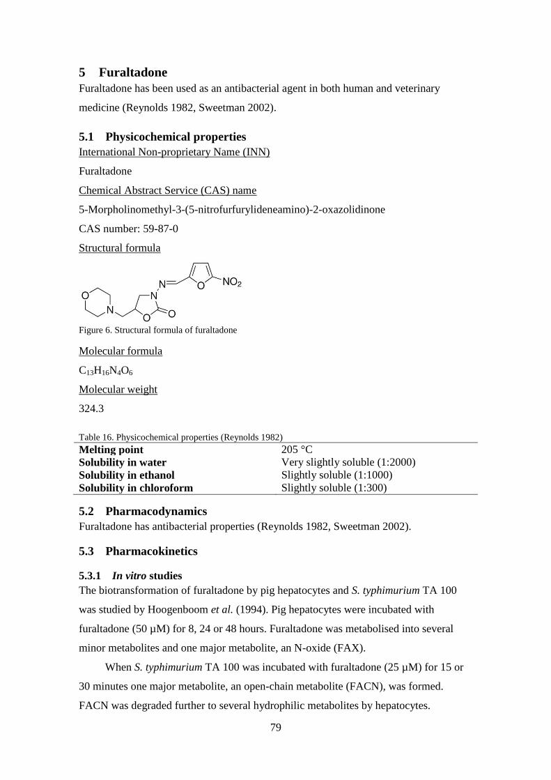
79
5 Furaltadone
Furaltadone has been used as an antibacterial agent in both human and veterinary
medicine (Reynolds 1982, Sweetman 2002).
5.1 Physicochemical properties
International Non-proprietary Name (INN)
Furaltadone
Chemical Abstract Service (CAS) name
5-Morpholinomethyl-3-(5-nitrofurfurylideneamino)-2-oxazolidinone
CAS number: 59-87-0
Structural formula
Figure 6. Structural formula of furaltadone
Molecular formula
C13H16N4O6
Molecular weight
324.3
Table 16. Physicochemical properties (Reynolds 1982)
Melting point 205 °C
Solubility in water Very slightly soluble (1:2000)
Solubility in ethanol Slightly soluble (1:1000)
Solubility in chloroform Slightly soluble (1:300)
5.2 Pharmacodynamics
Furaltadone has antibacterial properties (Reynolds 1982, Sweetman 2002).
5.3 Pharmacokinetics
5.3.1 In vitro studies
The biotransformation of furaltadone by pig hepatocytes and S. typhimurium TA 100
was studied by Hoogenboom et al. (1994). Pig hepatocytes were incubated with
furaltadone (50 µM) for 8, 24 or 48 hours. Furaltadone was metabolised into several
minor metabolites and one major metabolite, an N-oxide (FAX).
When S. typhimurium TA 100 was incubated with furaltadone (25 µM) for 15 or
30 minutes one major metabolite, an open-chain metabolite (FACN), was formed.
FACN was degraded further to several hydrophilic metabolites by hepatocytes.

80
It was also shown that proteins obtained from bacteria and hepatocytes exposed to
furaltadone for 24 hours were bound to residues containing an intact AMOZ side-chain,
of which 60-80 % could be released after mild acid treatment.
Furaltadone was incubated with a mixture of 8500g supernatant of liver homogenate
taken from male rats aged 6-10 weeks (Akao et al. 1971). After 60 minutes of
incubation under aerobic conditions there was a change in optical absorbance
corresponding to a loss of a nitro group.
5.3.2 In vivo studies
5.3.2.1 In bovine
Veal calves (n=6) were administered furazolidone and furaltadone (6-8 mg/kg BW)
orally via the morning milk (Nouws & Laurensen 1990). Three hours after the
administration four calves were slaughtered and samples of heart muscle, kidney, liver,
muscularis gracilis, diaphragm, fat, lung, brain, urine and blood were collected and
stored for analysis using HPLC. The remaining two calves were slaughtered four hours
after administration. The liver and kidney samples were analysed at 10, 40, 120 minutes
and 24 hours after collection and the muscle and heart samples after 15, 60, 120 minutes
and 24 hours. There were large variations between tissues and animals, but immediately
after the slaughter furaltadone could be detected in the tissues, with the highest levels
found in plasma (0.259-3.02 µg/mL) and urine (7.08-24.86 µg/mL). Two hours after the
slaughter trace amounts of furaltadone could be detected in the muscle and kidney
samples, but not in liver samples. After 24 hours no levels could be detected. Only low
levels of furaltadone could be detected in fat, brain and lung. The postmortal
degradation half-lives of furaltadone were calculated to be 22 minutes in muscularis
gracilis, 29 minutes in diaphragm, 63 minutes in kidney, <43 minutes in heart, and <8
minutes in liver.
Furaltadone and nitrofurazone (14.0 mg/kg BW) suspended in milk were given orally to
five preruminant MRY male calves (Nouws et al. 1987). Furaltadone was given three
days before nitrofurazone. Blood samples were taken at different time points after each
administration and urine was collected from three calves and analysed for the
nitrofurans. The maximum concentration of furaltadone in plasma (2.5 µg/mL) was

81
achieved 2.5 hours after administration and the half-life of furaltadone was calculated to
be around 2.5 hours.
A cow was administered a capsule containing; 0.88 mg/kg BW furazolidone,
nitrofurazone and furaltadone and 4.4 mg/kg BW nitrofurantoin (Chu & Lopez 2007).
Milk samples were then collected for two weeks at intervals of 12 hours. Milk from
non-treated cows was used as control. The levels of nitrofuran side-chain residues in the
milk were determined using LC-MS/MS. The level of the side-chain of furaltadone,
AMOZ, in milk was highest 12 hours after dosing (~55 ng/g) and decreased rapidly and
72 hours after dosing the level of AMOZ was below the detection limit (0.2 ng/g).
5.3.2.2 In swine
Pigs (n=18) were fed feed, medicated with furaltadone (400 mg/kg feed), ad libitum for
10 days, corresponding to around 24 mg/kg BW per day, followed by a withdrawal
period for six weeks (Cooper et al. 2005). Three pigs were sacrificed each week of the
withdrawal period and samples of muscle, liver and kidney were taken and analysed for
furaltadone and AMOZ using LC-MS/MS and HPLC-UV. Furaltadone was detected in
one liver sample (0.7 µg/kg) at week 0 of the withdrawal period. AMOZ was detected in
all samples and the levels found at week 6 of the withdrawal period were around 15
µg/kg in kidney and liver and 60 µg/kg in muscle. The depletion half-lives of AMOZ in
muscle, liver and kidney were 8.7±1.2 days, 5.4±0.5 days and 5.7±0.7 days
respectively.
5.3.2.3 In fish
The depletion of nitrofurans and their tissue-bound residues in channel catfish
(Ictakurus punctatus) was investigated by Chu et al. (2008). Fish (n=55) were orally
administered furazolidone, nitrofurantoin, nitrofurazone and furaltadone (1 mg/kg BW).
After 2, 4, 8 and 12 hours, and 1, 4, 7, 10, 14, 28 and 56 days five fish were killed and
muscle samples collected for analysis of parent nitrofurans and their tissue-bound
residues. The highest concentration of furaltadone in muscle (35.2 µg/kg) was reached
12 hours after administration. Furaltadone could no longer be detected 96 hours after
administration. The level of AMOZ was the highest (46.8 µg/kg) 12 hours after
administration. The elimination of all tissue-bound residues was biphasic and could still
be detected 56 days after administration. The half-life for AMOZ was calculated to be
27 days.

82
Chu et al. (2008) also examined the levels of nitrofurans and tissue-bound
metabolites in muscle of fish after waterborne exposure to nitrofurans. Fish (5 per
treatment) were exposed to nitrofurantoin, nitrofurazone, furazolidone or furaltadone
(10 mg/L) for eight hours. After this time the fish were killed and their muscle tissue
was analysed for parent nitrofuran and tissue-bound metabolites. The concentrations of
furaltadone and AMOZ were around 46 and 37 µg/kg, respectively, and indicates
uptake of furaltadone via the branchial route.
5.3.2.4 In poultry
Furaltadone was administered to 28-day-old broilers (n=20) in the feed (30, 100, 300,
1000 or 3000 µg/kg feed) corresponding to 6, 20, 60, 200 and 600 µg/kg BW, for 12
days (McCracken et al. 2005b). After this time the broilers were killed and samples of
muscle and liver were collected and analysed for AMOZ using LC-MS/MS. At the
lowest dose, AMOZ was detected in liver (0.6 µg/kg) samples. At the highest dose the
levels in liver and muscle were around 40 and 18 µg/kg respectively.
Isa Brown chickens (10 per treatment) were treated with furaltadone (150 mg/kg feed)
or furaltadone and nifursol (15 and 10 mg/kg, respectively feed), corresponding to
around 30 mg/kg BW of furaltadone per day, 3 mg/kg BW of furaltadone per day and 2
mg/kg BW of nifursol per day, for five weeks followed by a withdrawal period of three
weeks (Barbosa et al. 2011). At the end of the medication period five chickens were
slaughtered and samples of muscle, liver and gizzard were collected and analysed for
AMOZ using LC-MS/MS. The remaining chickens were slaughtered and sampled at the
end of the withdrawal period, i.e. 21 days. Furaltadone was only measured in the
samples taken at the end of the medication period. The levels in the tissues were around
50 µg/kg in muscle and liver, and 80 µg/kg in gizzard in the birds treated with
furaltadone (30 mg/kg BW). The levels in birds treated with approximately 3 mg/kg
BW of furaltadone and 2 mg/kg BW of nifursol per day were around 17 µg/kg in all
tissues. Directly after the medication period the levels of AMOZ in birds treated with
furaltadone or furaltadone and nifursol were much higher than after three weeks of
withdrawal. The levels of AMOZ in tissues retrieved after the withdrawal period were
270 µg/kg in muscle, 80 µg/kg in liver and 331 µg/kg in gizzard in birds treated with
furaltadone. The levels in tissues from birds administered furaltadone and nifursol were

83
29 µg/kg in muscle, 17 µg/kg in liver and 32 µg/kg in gizzard after the withdrawal
period.
Barbosa et al. (2012) also investigated the transfer of furaltadone and AMOZ from
laying hens to eggs by administering 150 mg furaltadone per kg feed (treatment 1) or 15
mg furaltadone per kg feed and 10 mg nifursol per kg feed (treatment 2), corresponding
to around 30 mg/kg BW of furaltadone per day, 3 mg/kg BW of furaltadone per day and
2 mg/kg BW of nifursol per day, for five weeks followed by a withdrawal period of
three weeks. Five hens per treatment were used and a minimum of five eggs were
collected on the last two days of weeks 3-8. The levels of furaltadone and AMOZ were
measured using LC-MS/MS. The levels of furaltadone in yolk and white peaked at
week 4 and could not be detected at week 6. The maximum levels of furaltadone, for
treatment 1, measured in yolk and white were 445 and 253 µg/kg, respectively. For
treatment 2, the maximum levels were 64 µg/kg yolk and 45 µg/kg white. AMOZ could
be detected in all egg tissues at all weeks except for in white (treatment 2) the last two
weeks of the study. Like furaltadone the maximum levels of AMOZ was reached at
week 4 and were for treatment 1 712 and 613 µg/kg in yolk and white, respectively. For
treatment 2, the levels were 89 and 68 µg/kg in yolk and white, respectively. Three
weeks after the last treatment the levels of AMOZ in egg yolk and white were 3.1 and
2.9 µg/kg for treatment 1. For treatment 2, AMOZ could only be detected in yolk at a
level of 0.2 µg/kg.
McCracken et al. (2005a) fed six broiler hens and one cockerel 120-140 g of feed
containing furaltadone (400 mg/kg feed), corresponding to around 96 and 112 mg/kg
BW per day. Eggs were collected and analysed for AMOZ and when it was clear that
furaltadone residues had transferred to the eggs, the eggs laid after this were collected
and allowed to hatch. After hatching four chicks were sacrificed at determined intervals
and muscle and liver samples were analysed for AMOZ. However, AMOZ could only
be detected up to day 10. The levels of AMOZ were 11.4 µg/kg liver and 1.0 µg/kg
muscle in one day old chicks, ~3 µg/kg liver and ~0.5 µg/kg muscle in five days old
chicks, and ~0.7 µg/kg liver and ~0.2 µg/kg muscle in 10 days old chicks.
Twenty-four laying hens were fed feed containing 300 mg furaltadone, nitrofurazone,
nitrofurantoin or furazolidone per kg feed, corresponding to approximately 60 mg/kg

84
BW per day, for one week (McCracken & Kennedy 2007). Eggs were then collected for
two days and analysed immediately for nitrofuran parent compound and their bound
residues in albumen, yolk and shell using LC-MS/MS. The levels of furaltadone in yolk,
albumen and shell were 593, 486 and 35.7 ng/g, respectively. The levels of AMOZ in
yolk, albumen and shell were 590, 624 and 56.3 ng/g, respectively.
5.3.3 Conclusions on pharmacokinetics of furaltadone and its metabolites
In the studies above it has been shown that furaltadone is absorbed and rapidly
distributed in the body. It is quickly eliminated with a half-life in plasma of 2.5 hours in
calves and can be excreted via the urine. Post mortal degradation of furaltadone in
calves was shown to be very fast. The shortest half-life was less than eight minutes in
liver and the longest was one hour in kidney. After broilers were administered
furaltadone orally the highest levels were found in the gizzard (80 µg/kg). It was also
demonstrated that furaltadone can be transferred from laying hens to eggs. Furaltadone
have been shown to be metabolised into protein-bound residues containing the AMOZ
side-chain in cow, pig, fish and poultry. It was also shown that in pig hepatocytes,
furaltadone was metabolised to an N-oxide, FAX, and in bacteria it was metabolised to
an open-chain metabolite, FACN.
AMOZ was shown to be transferred into milk from cows administered
furaltadone. It was also shown that eggs from furaltadone treated hens contained AMOZ
and that it could still be detected in chicks hatched from those eggs. In poultry
administered furaltadone the highest levels of AMOZ were found in the gizzard (331
µg/kg) followed by muscle (270 µg/kg) and liver (80 µg/kg). In pigs fed furaltadone,
the highest levels of AMOZ were found in muscle, followed by kidney and liver (60
and 15 µg/kg, respectively). The half-lives were determined to be around nine days in
muscle and five days in kidney and liver. In channel catfish muscle the half-life of
AMOZ was calculated to be 27 days but could still be detected 56 days after the last
treatment.
5.4 Toxicology of furaltadone
5.4.1 Acute toxicity
No studies could be found.
5.4.2 Chronic toxicity
No studies could be found.

85
5.4.3 Reproductive toxicity including teratogenicity
No studies could be found.
5.4.4 Mutagenicity and genotoxicity
An Ames test using S. typhimurium TA 100 was carried out on furaltadone and the N-
oxide metabolite (FAX) (Hoogenboom et al. 1994). S. typhimurium TA 100 was
incubated with furaltadone or FAX (10, 100 or 1000 ng/plate) without any metabolic
activation. Furaltadone tested positive at all doses and FAX tested negative at all doses.
The study conducted follows guidelines to some extent. Mutagenicity with
metabolic activation was not tested and no positive control was used. It is possible that
FAX could be mutagenic after metabolic activation.
5.4.4.1 Conclusions on mutagenicity and genotoxicity studies
Furaltadone tested positive in the only mutagenicity test found, while its metabolite
FAX tested negative. It is difficult to draw any conclusions from just one study. To be
able to say whether or not furaltadone and FAX are mutagenic and genotoxic more
studies are needed.
5.4.5 Carcinogenicity and long-term toxicity
No studies could be found.
6 Summary of the effects of nitrofurans
The conclusions that can be drawn about the reproductive toxicity, mutagenicity and
genotoxicity, as well as carcinogenicity of the reviewed nitrofurans are presented in
Table 17. Regarding the reproductive toxicity, nitrofurazone and nitrofurantoin have
been shown to be toxic to the reproductive system. Observed effects after exposure to
these substances were decreased fertility, altered oestrus cycle, malformations, aberrant
sperm and testicular degeneration. A reduction in the concentration of spermatozoa was
seen at the lowest dose of nitrofurazone (10 mg/kg BW) tested in rats, while the most
commonly observed effect was testicular degeneration. For furazolidone, a clear
conclusion cannot be drawn and a lack of studies on furaltadone makes it impossible to
determine if it is toxic to the reproductive system.
All nitrofurans reviewed, except for furaltadone, have been shown to be
mutagenic and genotoxic in vitro. Conclusions cannot be drawn regarding the
genotoxicity in vivo due to there being few studies with conflicting results. Further
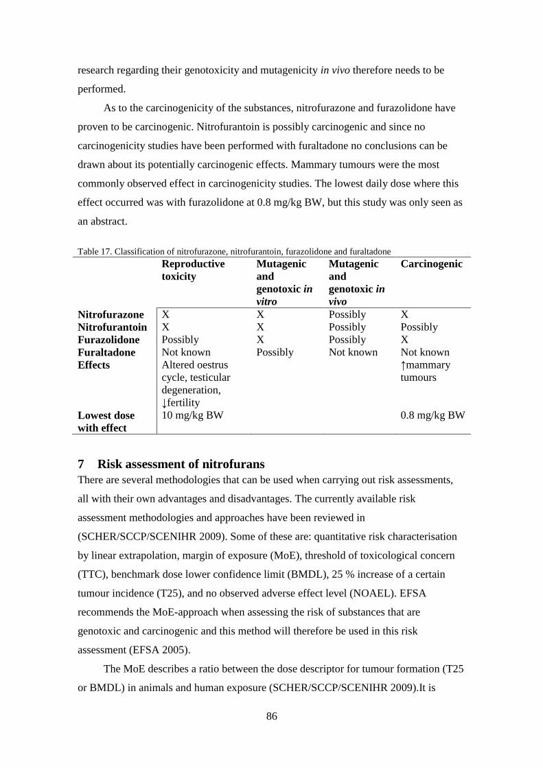
86
research regarding their genotoxicity and mutagenicity in vivo therefore needs to be
performed.
As to the carcinogenicity of the substances, nitrofurazone and furazolidone have
proven to be carcinogenic. Nitrofurantoin is possibly carcinogenic and since no
carcinogenicity studies have been performed with furaltadone no conclusions can be
drawn about its potentially carcinogenic effects. Mammary tumours were the most
commonly observed effect in carcinogenicity studies. The lowest daily dose where this
effect occurred was with furazolidone at 0.8 mg/kg BW, but this study was only seen as
an abstract.
Table 17. Classification of nitrofurazone, nitrofurantoin, furazolidone and furaltadone
Reproductive
toxicity
Mutagenic
and
genotoxic in
vitro
Mutagenic
and
genotoxic in
vivo
Carcinogenic
Nitrofurazone X X Possibly X
Nitrofurantoin X X Possibly Possibly
Furazolidone Possibly X Possibly X
Furaltadone Not known Possibly Not known Not known
Effects Altered oestrus
cycle, testicular
degeneration,
↓fertility
↑mammary
tumours
Lowest dose
with effect
10 mg/kg BW 0.8 mg/kg BW
7 Risk assessment of nitrofurans
There are several methodologies that can be used when carrying out risk assessments,
all with their own advantages and disadvantages. The currently available risk
assessment methodologies and approaches have been reviewed in
(SCHER/SCCP/SCENIHR 2009). Some of these are: quantitative risk characterisation
by linear extrapolation, margin of exposure (MoE), threshold of toxicological concern
(TTC), benchmark dose lower confidence limit (BMDL), 25 % increase of a certain
tumour incidence (T25), and no observed adverse effect level (NOAEL). EFSA
recommends the MoE-approach when assessing the risk of substances that are
genotoxic and carcinogenic and this method will therefore be used in this risk
assessment (EFSA 2005).
The MoE describes a ratio between the dose descriptor for tumour formation (T25
or BMDL) in animals and human exposure (SCHER/SCCP/SCENIHR 2009).It is

87
calculated by first deriving a benchmark dose (BMD), corresponding to a response level
(benchmark response, BMR), and its lower 95%-confidence bound (BMDL) for
different endpoints from carcinogenicity studies. Then, the model that adequately fits
the data and has the lowest BMDL is selected. The BMDL of this model is then divided
by the dietary exposure. EFSA considers that the risk to public health is of low concern
if the MoE is equal to, or higher than, 10000 (EFSA 2005).
7.1 Benchmark dose (BMD)
The BMD and BMDL for different endpoints in carcinogenicity studies using
nitrofurazone, nitrofurantoin and furazolidone was derived using the BMD software
developed by the US EPA (US EPA 2014 http://www.epa.gov/ncea/bmds/). Datasets
which produced a BMD and BMDL are presented in Table 18. Most of the
carcinogenicity studies did not expose the test animals for the normal two year period
and it is assumed that the number of tumours formed would have been higher if the
studies had been performed for two years. To account for this the doses were adjusted to
what they would have been if the study was carried out over a two year period. The
doses were adjusted using the formula (1) presented by Carthew et al. (2010) and the
individual BMD and BMDL were derived.
(1) (
) (
)
w1 = weeks dosed, w2 = weeks observed, d0 = dose used in study
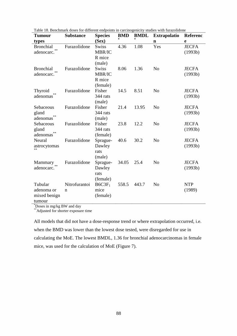
88
Table 18. Benchmark doses for different endpoints in carcinogenicity studies with furazolidone
Tumour
types
Substance Species
(Sex)
BMD*
BMDL*
Extrapolatio
n
Referenc
e
Bronchial
adenocarc.**
Furazolidone Swiss
MBR/IC
R mice
(male)
4.36 1.08 Yes JECFA
(1993b)
Bronchial
adenocarc.**
Furazolidone Swiss
MBR/IC
R mice
(female)
8.06 1.36 No JECFA
(1993b)
Thyroid
adenomas**
Furazolidone Fisher
344 rats
(male)
14.5 8.51 No JECFA
(1993b)
Sebaceous
gland
adenomas**
Furazolidone Fisher
344 rats
(male)
21.4 13.95 No JECFA
(1993b)
Sebaceous
gland
adenomas**
Furazolidone Fisher
344 rats
(female)
23.8 12.2 No JECFA
(1993b)
Neural
astrocytomas**
Furazolidone Sprague-
Dawley
rats
(male)
40.6 30.2 No JECFA
(1993b)
Mammary
adenocarc.**
Furazolidone Sprague-
Dawley
rats
(female)
34.05 25.4 No JECFA
(1993b)
Tubular
adenoma or
mixed benign
tumour
Nitrofurantoi
n
B6C3F1
mice
(female)
558.5 443.7 No NTP
(1989)
*Doses in mg/kg BW and day
**Adjusted for shorter exposure time
All models that did not have a dose-response trend or where extrapolation occurred, i.e.
when the BMD was lower than the lowest dose tested, were disregarded for use in
calculating the MoE. The lowest BMDL, 1.36 for bronchial adenocarcinomas in female
mice, was used for the calculation of MoE (Figure 7).

89
Figure 7. Log-logistic model with BMR of 10 % extra risk for the BMD and 0.95 lower confidence limit
for the BMDL for bronchial adenocarcinomas in female Swiss MBR/ICR mice. Doses are in mg/kg BW
per day.
7.2 Exposure assessment
No exposure assessment has been performed on nitrofurans and it is consequently hard
to know exactly how much people are exposed to. In order to calculate the Swedish
population’s exposure to nitrofurans it is assumed that all consumed foodstuffs of
animal origin in Sweden contain levels of nitrofurans at 1 µg/kg, the current MRPL.
Based on intake data from Riksmaten, a national dietary survey, the average
consumption of foodstuffs of animal origin (meat, bird, sausage, organ meats, blood
products, fish and shellfish, caviar and egg) and dairy products in Sweden is 0.392 and
0.486 kg per day and person for adult women and men, respectively (Amcoff et al.
2012). For the 95th
percentile (high consumer) it is 1.024 and 1.357 kg per day and
person for women and men, respectively (Amcoff et al. 2012). Assuming this, then the
mean exposure to nitrofurans for an average Swedish woman and man per day is 0.392
and 0.486 µg, respectively. For a high consumer the exposure is 1.024 and 1.357 µg for
women and men, respectively. The mean bodyweight for women in Riksmaten (Amcoff
et al. 2012) was 69 kg and for men the mean bodyweight was 84 kg. The exposure for
average women and men is 5.68 and 5.79 ng/kg BW and day, respectively (Table 19).
0.2
0.3
0.4
0.5
0.6
0.7
0.8
0 5 10 15 20 25
Fra
ctio
n A
ffe
cte
d
dose
Log-Logistic Model, with BMR of 10% Extra Risk for the BMD and 0.95 Lower Confidence Limit for the BMDL
17:32 05/28 2014
BMDL BMD
Log-LogisticBMD Lower Bound

90
For a high consumer the exposure is 14.8 and 16.2 ng/kg BW for women and men,
respectively (Table 19).
In Riksmaten from 2003 (Enghardt Barbieri et al. 2006) the food consumption of
children in Sweden was studied. The children looked at were four year olds and children
in 2nd
and 5th
grade. The average consumption of foodstuffs of animal origin (meat, bird,
sausage, organ meats, blood products, fish and shellfish, and egg) and dairy products
was 0.490 and 0.556 kg per day and person for four year old girls and boys, respectively
(Enghardt Barbieri et al. 2006). For boys and girls in 2nd
grade the consumption was
0.711 and 0.608 kg per day and person, respectively. Children in 5th
grade had an
average consumption of 0.525 and 0.651 kg per day and person for girls and boys,
respectively. If again it is assumed that all consumed foodstuffs of animal origin in
Sweden contain levels of nitrofurans at 1 µg/kg the mean exposure to nitrofurans is;
0.490 and 0.556 µg per day for four year old girls and boys, 0.711 and 0.608 µg per day
for boys and girls in 2nd
grade, and 0.525 and 0.651 µg per day for girls and boys in 5th
grade. The mean bodyweight for four year olds was 18 kg, for girls and boys in 2nd
grade 31 and 32 kg respectively, and for 5th
graders 42 kg (Enghardt Barbieri et al.
2006). The exposure for average four year old girls and boys is 27.2 and 30.9 ng/kg BW
and day, respectively (Table 19). For girls and boys in 2nd
grade the exposure is 19.6
and 22.2 ng/kg BW and day, respectively (Table 19). And lastly, for girls and boys in
5th
grade the exposure is 12.5 and 15.5 ng/kg BW and day, respectively (Table 19).
Another source of exposure to nitrofurans can be via the use of pharmaceuticals.
Nitrofurantoin is regularly used to treat urinary tract infections in men and women. The
normal treatment is 50 mg, three times per day for five days, which amounts to a total
dose of 150 mg per day (FASS 2010). Since this dose is so high, the added exposure via
food is negligible and therefore, only the medicinal exposure will be considered in a
separate assessment. Assuming the same mean bodyweights as before the exposure per
day is 2.17 and 1.79 mg/kg BW per day for women and men, respectively. Assuming
that people only undergo one treatment, for five days, in their lifetime (70 years), the
lifetime exposure is 425 and 349 ng/kg BW per day for women and men, respectively
(Table 19).

91
Table 19. The food consumption, bodyweight and exposure for adult female and male (18-80 years)
average and high consumers of nitrofurans via food in Sweden, child average consumers of nitrofurans
via food and men and women treated with nitrofurantoin for five days
Consumption
of animal food
products and
dairy products
(kg/day)
Exposure
via food
(µg/day)
Medicinal
exposure
(mg/day)
Bodyweight
(kg)
Exposure to
nitrofurans
(ng/kg BW
per day)
Women
(average)
0.392 0.392 69 5.68
Women
(high)
1.024 1.024 69 14.8
Men
(average)
0.486 0.486 84 5.79
Men
(high)
1.357 1.357 84 16.2
Four year
old girls
0.490 0.490 18 27.2
Four year
old boys
0.556 0.556 18 30.9
Girls in 2nd
grade
0.608 0.608 31 19.6
Boys in 2nd
grade
0.711 0.711 32 22.2
Girls in 5th
grade
0.525 0.525 42 12.5
Boys in 5th
grade
0.651 0.651 42 15.5
Women
(medicinal)
150 69 425
Men
(medicinal)
150 84 349
7.3 Margin of exposure (MoE)
The MoEs for the average and high consumer of foodstuffs of animal origin in Sweden
are presented in Table 20. None of the MoEs are lower than the margin of 10000 when
only the exposure to nitrofurans via food is considered, indicating a risk of low concern.
However, if only the medicinal exposure is considered the MoEs are below 10000 for
both men and women, indicating that there may be a risk.
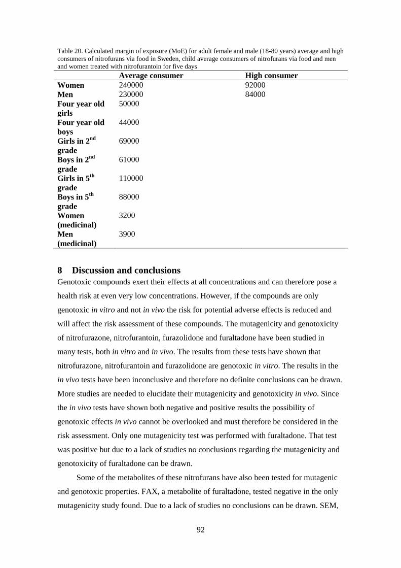
92
Table 20. Calculated margin of exposure (MoE) for adult female and male (18-80 years) average and high
consumers of nitrofurans via food in Sweden, child average consumers of nitrofurans via food and men
and women treated with nitrofurantoin for five days
Average consumer High consumer
Women 240000 92000
Men 230000 84000
Four year old
girls
50000
Four year old
boys
44000
Girls in 2nd
grade
69000
Boys in 2nd
grade
61000
Girls in 5th
grade
110000
Boys in 5th
grade
88000
Women
(medicinal)
3200
Men
(medicinal)
3900
8 Discussion and conclusions
Genotoxic compounds exert their effects at all concentrations and can therefore pose a
health risk at even very low concentrations. However, if the compounds are only
genotoxic in vitro and not in vivo the risk for potential adverse effects is reduced and
will affect the risk assessment of these compounds. The mutagenicity and genotoxicity
of nitrofurazone, nitrofurantoin, furazolidone and furaltadone have been studied in
many tests, both in vitro and in vivo. The results from these tests have shown that
nitrofurazone, nitrofurantoin and furazolidone are genotoxic in vitro. The results in the
in vivo tests have been inconclusive and therefore no definite conclusions can be drawn.
More studies are needed to elucidate their mutagenicity and genotoxicity in vivo. Since
the in vivo tests have shown both negative and positive results the possibility of
genotoxic effects in vivo cannot be overlooked and must therefore be considered in the
risk assessment. Only one mutagenicity test was performed with furaltadone. That test
was positive but due to a lack of studies no conclusions regarding the mutagenicity and
genotoxicity of furaltadone can be drawn.
Some of the metabolites of these nitrofurans have also been tested for mutagenic
and genotoxic properties. FAX, a metabolite of furaltadone, tested negative in the only
mutagenicity study found. Due to a lack of studies no conclusions can be drawn. SEM,

93
a metabolite of nitrofurazone, tested positive in bacterial tests but the results from in
vitro and in vivo tests are inconclusive. To safely determine the mutagenicity and
genotoxicity of SEM more studies are needed. The metabolite of furazolidone, AOZ,
tested positive in in vitro tests but not in the only in vivo study found. To further
elucidate AOZ’s mutagenic and genotoxic properties more studies are needed. Another
metabolite of furazolidone, 3-(4-cyano-2-oxobutylideneamino)-2-oxazolidone, tested
negative in an Ames test and must be examined more before a conclusion can be drawn.
Nitrofurazone and nitrofurantoin have in several studies been shown to be toxic to
the reproductive system of experimental animals and no NOAELs could be set. Such
substances should be extensively reviewed, especially when they are used in human
medicine. Nitrofurantoin, which is currently being used to treat urinary tract infections,
had a somewhat reversible adverse effect on the number of spermatozoa in rats at the
lowest dose tested (10 mg/kg BW). This study does not meet today’s standard of
testing, but the results should still be treated as reliable and an effect at quite a low dose
is troubling since both children as well as adults are taking drugs in which
nitrofurantoin is the active substance.
Nitrofurazone and furazolidone have been shown to be carcinogenic in rats and
mice, mainly causing mammary tumours. No conclusion regarding the carcinogenicity
of furaltadone can be drawn since no studies were found. Contradicting results from
studies with nitrofurantoin makes it difficult to determine whether or not it is
carcinogenic. To ascertain the carcinogenicity of nitrofurantoin additional research is
required. One can argue that compounds which are both carcinogenic and genotoxic in
vivo pose a greater health risk than compounds which are only genotoxic, or
carcinogenic and genotoxic in vitro. This should therefore be taken into account when
performing risk assessments. Nitrofurazone and furazolidone are carcinogenic,
nitrofurantoin and furaltadone might be and all, except for furaltadone, have been
shown to be genotoxic in vitro. Since it cannot be excluded that the nitrofurans are
genotoxic in vivo the precautionary approach must be used and it should be assumed
that the nitrofurans are genotoxic in vivo.
The BMDL is based on a carcinogenicity study on furazolidone in mice.
However, this study was only seen as an abstract and the experimental animals were
only treated for 13 months instead of the usual 24 months. Due to this there are some
uncertainties concerning the results of the study and the number of bronchial
94
adenocarcinomas may be lower than they would be if the exposure was over 24 months.
To account for this uncertainty the results were adjusted as stated previously.
The MoE after a five day long course of treatment with nitrofurantoin, stretched
out over a lifetime, are below 10000 for both men and women. Furthermore,
nitrofurantoin is used as prophylaxis for urinary tract infections with long treatment
periods which would further decrease the MoE. This demonstrates that there may be an
increased risk for bronchial adenocarcinomas for people treated with nitrofurantoin.
Some side effects of nitrofurantoin affect the lungs. Long term treatment can cause
pulmonary fibrosis, while some acute pulmonary reactions include; coughing, shortness
of breath, rapid breathing and chest pains (FASS 2010). In regards to the reproductive
toxicity of nitrofurantoin and that there is an increased risk of developing cancer after
only one course of treatment, it might be wise to revaluate the safety of nitrofurantoin as
a drug used in humans.
The calculated MoEs for children and for adult male and female, average and
high, consumers of foodstuffs of animal origin are above the margin of 10000. This
shows that there is a negligible risk to the Swedish population’s health from nitrofurans
in food and that the MRPL of 1 µg/kg is a safe limit for nitrofurans in food.
Furthermore, in the exposure assessment dairy products constitute a large portion of
exposure but in the Swedish controls in 2011 and 2012, no milk samples tested
contained nitrofurans (Gustavsson et al. 2012, Nordlander et al. 2013). Since it is
assumed that all foodstuffs of animal origin and dairy products contain 1 µg/kg, which
has not been observed during inspections, the risk from nitrofurans is overestimated.
That being said, only a small percentage of total production is tested and there may also
be a risk that imported milk contains nitrofurans. Furthermore, the exposure via food is
based on results from Riksmaten in which young men underreported their food
consumption, thereby underestimating the male food consumption and consequently the
risk for adult males (Amcoff et al. 2012).
When calculating MRLs, as well as the MoE, the consumption of foods from
animal origin is considered. For MRLs the food basket (i.e. exposure via food) is
comprised of 300 g muscle, 50 g kidney, 50 g fat, 100 g liver, 1.5 L milk, 100 g eggs or
egg products and 20 g honey. If this food consumption is used for the MoE exposure
assessment, the subsequent MoEs are 44000 and 54000 for women and men,
respectively. The increased risk is due to a higher estimated consumption of meat, egg
and milk in the MRL assessment than the reported consumption in Riksmaten (Amcoff

95
et al. 2012). It appears that the food consumption used for MRL determinations is not
consistent with that in Sweden and would therefore overestimate the risk. However, it is
a good estimation to use when the precautionary approach is applied.
In order for nitrofurans to have a MoE of 10000 for male high consumers, all of
the dairy products and consumed food of animal origin needs to contain on average
around 8 µg/kg. According to RASFF Portal (2014) the mean nitrofuran level found in
different food products (e.g. crayfish, shrimp, fish and meat) in border controls between
January 1, 2005 and March 7, 2014 was 19.86 µg/kg. The highest reported
concentration was 5000 µg/kg found in shrimp from Bangladesh.
SEM, a metabolite of nitrofurazone, has been shown to occur naturally in crayfish
(Saari & Peltonen 2004, McCracken et al. 2013). In 2003 wild crayfish were caught in a
Swedish lake and analysis showed that they contained between 2 and 5 µg/kg (Christina
Elgerud, personal communication). The Swedish National Food Agency decided in
2006 to stop testing for, and reporting levels of SEM in European crayfish and other
shellfish. This is due to SEM being a rather unspecific marker residue for nitrofurazone
and therefore the found levels could not be connected to illegal use of nitrofurans
(Christina Elgerud, personal communication, National Food Agency, unpublished
report). In light of the MoE it is unlikely that naturally occurring SEM levels in crayfish
would pose a risk to the population’s health, since the consumption of crayfish is not
that great and seasonal. The decision by the NFA to stop testing European crayfish for
SEM is therefore justified.
In relation to the MoEs for nitrofurans, the minimum required performance limit
(MRPL), which has been used as a reference point of action (RPA), provides enough
protection for the Swedish population, both adults and minors, from nitrofurans in food.
However, since no dietary intake and consequent risk assessment for infants were
performed, there may be a risk for infants posed by nitrofurans in foodstuffs. Also, the
current documentation on nitrofurans is incomplete and what is available is quite old.
Most of the found studies do not follow guidelines. It is therefore not possible to
perform a comprehensive risk assessment and be able to fully elucidate what impact
nitrofurans in food have on the human health. Furthermore, the reliability of the
carcinogenicity study, from which the BMDL is based on, can be questioned since it
had to be corrected for not being performed over a two year period. Nonetheless, based
on the results of this risk assessment the current RPA of 1 µg/kg, the MRPL, can be
retained. All foodstuffs containing lower levels can be allowed to enter the market.

96
Food products containing higher levels should be removed from the market to protect
the consumers. However, crayfish containing residues of SEM lower than 5 µg/kg could
be accepted from a consumer health perspective. There is already an inherent safety
margin in the proposed RPA since all animal based foodstuffs and dairy products eaten
in a single day must contain, on average, 8 µg/kg to pose a risk to the population’s
health, which is unlikely. That being said, nitrofurans should not be allowed for use in
food-producing animals due to their reproductive toxicity, genotoxicity and
carcinogenicity.
Under normal circumstances, carcinogenic and genotoxic compounds are not
permitted for use in food-producing animals but exceptions can be made when it is
deemed necessary. Metamizole sodium is used as an anti-inflammatory, antipyretic,
spasmolytic and analgesic compound in veterinary medicine (CVMP unpublished
report). In the assessment report on metamizole sodium the compound is carcinogenic
in mice but produces equivocal results in genotoxicity tests. It was cleared for use since
a drug against colic was needed and therefore a quite arbitrary MRL of 100 µg/kg was
set (Commission Regulation (EU) No 37/2010).
In the future less safe antibiotics, like nitrofurans, may need to be used due to an
increased bacterial resistance to antibiotics. However, there is always a potential risk
when using genotoxic substances. The risks of using nitrofurans have to be weighed
against the potential benefits. At this time there are no great benefits in allowing
nitrofurans to be used in food-producing animals. A risk-benefit analysis should be
performed on nitrofurantoin to evaluate if it is absolutely necessary in human medicine
or if its use can be reduced or eliminated completely. The use of nitrofurans should not
be allowed until the benefits outweigh the potential risks. To safeguard the possible
future use of nitrofurans it must be ensured that bacteria do not gain resistance to
nitrofurans. Thus, nitrofurans should not be used in food-producing animals and the
amounts in food should be limited. It should also be investigated if the metabolites of
the nitrofurans have antibacterial properties like their parent compounds, but without
their toxicity. If so, it is possible that they can be used as alternative antibacterial agents
to today’s antibiotics.
In conclusion, nitrofurans are genotoxic and carcinogenic substances which could
cause adverse health effects in humans. It was shown that one course of treatment with
nitrofurantoin might increase the risk for bronchial adenocarcinomas in humans.
Therefore, the safety of nitrofurantoin ought to be revaluated. In the future it may be
97
necessary to use nitrofurans, due to an increase in bacterial resistance to antibiotics, and
they should therefore not be used today. It would also be prudent to investigate the
antibacterial effects of the metabolites of nitrofurans and possibly use them as
antibiotics in the future. In regards to nitrofurans in food, the Swedish population has
been efficiently protected from the health risks posed by nitrofurans via the MRPL of 1
µg/kg. The RPA of 1 µg/kg sufficiently protects high consumers of foodstuffs of animal
origin and should be retained. When dealing with food products containing nitrofurans
at the RPA, or lower, the Swedish NFA should only notify this in RASFF but take no
further action. However, any products containing nitrofurans over this level should not
be allowed to enter the market, with the exception of SEM in crayfish, in order to
protect the health of the population. At this time, countries within the EU have different
views regarding residues of nitrofurans in foodstuffs and therefore differ in their
treatment of foodstuffs containing nitrofurans. Implementing a RPA for nitrofurans in
food would provide a more uniform treatment of food products containing nitrofurans
above the current MRPL, which is currently lacking within the EU.
9 Acknowledgments
First of all I would like to thank my supervisors, Bitte Aspenström-Fagerlund and
Lilianne Abramsson-Zetterberg, for guidance, feedback and support. A big thanks to
Salomon Sand, Leif Busk and the whole RN department for helping me and making me
feel welcome. I would also like to thank Christina Elgerud, Kristina Granelli and
Pernilla Sjöberg at Kem1for all of their help. Thanks to Björn Brunström for
proofreading and commenting on the written report. Lastly, to my fellow master thesis
colleagues at the National Food Agency, Annachiara Igra and Bianca Maurer, thank you
for the laughs and good company during our time together.

98
10 References
Abramsson-Zetterberg L., Svensson K. 2005. Semicarbazide is not genotoxic in the
flow cytometry-based micronucleus assay in vivo. Toxicology letters 155: 211-
217.
Akao M., Kuroda K., Miyaki K. 1971. Metabolic degradation of nitrofurans by rat liver
homogenate. Biochemical pharmacology 20: 3091-3096.
Ali B., Silsby J., Edens F., El Halawani M. 1988. Effects of furazolidone or
nitrofurazone on the concentrations of hypothalamic amines and plasma
luteinizing hormone (LH) and prolactin (PRL) levels in young turkeys. General
pharmacology: the vascular system 19: 91-95.
Amcoff E., Edberg A., Enghardt Barbieri H., Lindroos A-K., Nälsén C., Pearson M.,
Warensjö Lemming E. 2012. Riksmaten- vuxna 2010-11, livsmedels- och
näringsintag bland vuxna i Sverige. National Food Agency.
Anderson D., Phillips B. 1985. Nitrofurazone-genotoxicity studies in mammalian cells
in vitro and in vivo. Food and chemical toxicology 23: 1091-1098.
Aufrère M., Hoener B-A., Vore M. 1978. Reductive metabolism of nitrofurantoin in the
rat. Drug metabolism and disposition 6: 403-411.
Baars A., Blijleven W., Mohn G., Natarajan A., Breimer D. 1980. Preliminary studies
on the ability of drosophila microsomal preperations to activate mutagens and
carcinogens. Mutation research 72: 257-264.
Barbosa J., Freitas A., Moura S., Mourão J., da Silviera M., Ramos F. 2011. Detection,
accumulation, distribution, and depletion of furaltadone and nifursol residues in
poultry muscle, liver, and gizzard. Journal of agricultural and food chemistry 59:
11927-11934.
Barbosa J., Freitas A., Moura S., Mourão J., da Silviera M., Ramos F. 2012.
Determination of furaltadone and nifursol residues in poultry eggs by liquid
chromatography-electrospray ionization tandem mass spectrometry. Journal of
agricultural and food chemistry 60: 4227-4234.
Brunton L., Chabner B., Knollman B. 2011. Goodman & Gilman’s the pharmacological
basis of therapeutics. 12th
ed. McGraw-Hill, China.
Butler W., Graham T., Sutton M. 1990. Chronic toxicity and oncogenicity studies of
macrodantin in Sprague-Dawley rats. Food and chemical toxicology 28: 269-277.
Buzard J., Conklin J., O’Keefe E., Paul M. 1961. Studies on the absorption, distribution
and elimination of nitrofurantoin in the rat. The Journal of pharmacology and
experimental therapeutics 131: 38-43.
Carthew P., DiNovi M., Woodrow Setzer R. 2010. Application of the margin of
exposure (MOE) approach to substances in food that are genotoxic and
carcinogenic, example: CAS No: 105650-23-5 PhIP (2-amino-1-methyl-6-
phenylimidazo[4,5-b]pyridine). Food and chemical toxicology 48: S98-S105.
Chatterjee S., Banerjee S., Pal A., Basak J. 1983. DNA damage, prophage induction and
mutation by furazolidone. Chemico-biological interactions 45: 315-326.
Chu P-S., Lopez M. 2007. Determination of nitrofuran residues in milk of dairy cows
using liquid chromatography-tandem mass spectrometry. Journal of agriculture
and food chemistry 55: 2129-2135.
Chu P-S., Lopez M., Abraham A., el Said K., PLakas S. 2008. Residue depletion of
nitrofuran drugs and their tissue-bound metabolites in channel catfish (Ictalurus
punctatus) after oral dosing. Journal of agricultural and food chemistry 56: 8030-
8034.
Codex Alimentarius. 2013. About Codex. WWW-document:
http://www.codexalimentarius.org/about-codex/en/. Date visited 2013-11-14.
99
Codex Alimentarius. 2014. Codex Committee on Residues of Veterinary Drugs in
Foods (CCRVDF). WWW-document:
http://www.codexalimentarius.org/committees-and-task-
forces/en/?provide=committeeDetail&idList=6. Date visited 2014-01-26.
Cohen M & Sagi M. 1979. The effect of nitrofurans on mitosis, chromosome breakage
and sister-chromatid exchanges in human peripheral lymphocytes. Mutation
research 59: 139-142.
Commission Decision 2002/657/EC implementing Council Directive 96/23/EC
concerning the performance of analytical methods and the interpretation of results.
OJ L221/8.
Commission Decision 2003/181/EC amending Decision 2002/657/EC as regards the
setting of minimum required performance limits (MRPLs) for certain residues in
food of animal origin. OJ L71/17.
Commission Decision 2004/25/EC amending Decision 2002/657/EC as regards the
setting of minimum required performance limits (MRPLs) for certain residues in
food of animal origin. OJ L6/38.
Commission Regulation (EC) No 1442/95 amending Annexes I, II, III and IV to
Regulation (EEC) No 2377/90 laying down a Community procedure for the
establishment of maximum residue limits of veterinary medicinal products in
foodstuffs of animal origin. OJ L143/26.
Commission Regulation (EU) No 37/2010 on pharmacologically active substances and
their classification regarding maximum residue limits in foodstuff of animal
origin. OJ L15/1.
Conklin J., Sobers R., Wagner D. 1973. Further studies on nitrofurantoin excretion in
dog hepatic bile. British journal of pharmacology 48: 273-277.
Conklin J., Wagner D. 1971. Excretion of nitrofurantoin in dog hepatic bile. British
journal of pharmacology 43: 140-150.
Cooper K., Le J., Kane C., Kennedy D. 2008. Kinetics of semicarbazide and
nitrofurazone in chicken eggs and egg powders. Food additives and contaminants
25: 684-692.
Cooper K., Mulder P., van Rhijn J., Kovacsics L., McCracken R., Young P., Kennedy
D. 2005. Depletion of four nitrofuran antibiotics and their tissue-bound
metabolites in porcine tissues and determination using LC-MS/MS and HPLC-
UV. Food additives and contaminants 22: 406-414.
Council Directive 96/23/EC on measures to monitor certain substances and residues
thereof in live animals and animal products. OJ L125/10.
Council Directive (EEC) No 81/851 on the approximation of the laws of the Member
States relating to veterinary medicinal products. OJ L317/1.
Council Regulation (EEC) No 2377/90 laying down a Community procedure for the
establishment of maximum residue limits of veterinary medicinal products in
foodstuffs of animal origin. OJ L224/1.
Council Regulation (EEC) No 2309/93 laying down Community procedures for the
authorization and supervision of medicinal products for human and veterinary use
and establishing a European agency for the evaluation of medicinal products. OJ
L214/1.
Council Regulation (EEC) No 2901/93 amending Annexes I, II, III and IV to
Regulation (EEC) No 2377/90 laying down a Community procedure for the
establishment of maximum residue limits of veterinary medicinal products in
foodstuffs of animal origin. OJ L264/1.

100
Cramer D., Dodd M. 1945. The mode of action of nitrofuran compounds – I - action
versus staphylococcus aureus. Journal of bacteriology 51: 293-303.
de Angelis E., Rossi L., Pedersen J., Vignoli A., Vincentini O., Hoogenboom L.,
Polman T., Stammati A., Zucco F. 1999. Metabolism of furazolidone: alternative
pathways and modes of toxicity in different cell lines. Xenobiotica 29: 1157-
1169.
Debnath A., Lopez de Compadre R., Debnath G., Shusterman A., Hansch C. 1991.
Structure-activity relationship of mutagenic aromatic and heteroaromatic nitro
compounds – correlation with molecular orbital energies and hydrophobicity.
Journal of medicinal chemistry 34: 786-797.
Directive 2001/82/EC on the Community code relating to veterinary medicinal
products. OJ L311/1.
Dodd M., Stillman W. 1944. The in vitro bacteriostatic action of some simple furan
derivatives. Journal of pharmacology and experimental therapeutics 82: 11-18.
EFSA. 2005. Opinion of the scientific committee on a request from EFSA related to a
harmonized approach for risk assessment of substances which are both genotoxic
and carcinogenic. EFSA journal 282: 1-31.
EFSA Panel on Contaminants in the Food Chain (CONTAM). 2013. Guidance on
methodological principles and scientific methods to be taken into account when
establishing reference points for action (RPAs) for non-allowed
pharmacologically active substances present in food of animal origin. EFSA
journal 2013;11(4):3195.
Enghardt Barbieri H., Pearson M., Becker W. 2006. Riksmaten - barn 2003, livsmedels-
och näringsintag bland barn i Sverige. National Food Agency.
Ertürk E., Emory Morris J., Cohen S., Price J., Bryan G. 1970. Transplantable rat
mammary tumours induced by 5-nitro-2-furaldehyde semicarbazone and formic
acid 2-[4-(5-nitro-2-furyl)-2-thiazolyl]hydrazine. Cancer research 30: 1409-1412.
European Commission. 2005. Notice to applicants and guideline, veterinary medicinal
products, establishment of maximum residue limits (MRLs) for residues of
veterinary medicinal products in foodstuffs of animal origin. The rules governing
medicinal products in the European Union, volume 8. WWW-document:
http://ec.europa.eu/health/files/eudralex/vol-8/pdf/vol8_10-2005_en.pdf.
Retrieved 2013-11-12.
European Commission. 2013. RASFF the Rapid Alert System for Food and Feed 2012
annual report. Report, European Commission.
European Medicines Agency. 2011. VICH GL48: Studies to evaluate the metabolism
and residue kinetics of veterinary drugs in food-producing animals: marker
residue depletion studies to establish product withdrawal periods. WWW-
document:
http://www.ema.europa.eu/docs/en_GB/document_library/Scientific_guideline/20
11/04/WC500105052.pdf. Retrieved 2013-11-13.
Falk-Filipsson A., Hanberg A., Victorin K., Warholm M., Wallén M. 2007. Assessment
factors – applications in health risk assessment of chemicals. Environmental
research 104: 108-127.
FAO (Food and agriculture organization of the United Nations). 1993a. Nitrofurazone.
In: Residues of some veterinary drugs in animals and foods, FAO Food and
nutrition paper 41/5, pp. 105-112.
FAO (Food and agriculture organization of the United Nations). 1993b. Furazolidone.
In: Residues of some veterinary drugs in animals and foods, FAO Food and
nutrition paper 41/5, pp. 87-104.

101
FASS. 2010. Furadantin®
. WWW-document 20 May 2010:
http://www.fass.se/LIF/product?4&userType=2&nplId=19580320000013&docTy
pe=6#dose. Date visited 2014-02-21.
FASS. 2014a. FASS allmänhet. WWW-document:
http://www.fass.se/LIF/result?23&query=nitrofural&userType=2. Date visited
2014-02-24.
FASS. 2014b. FASS allmänhet. WWW-document:
http://www.fass.se/LIF/result?13&query=furaltadon&userType=2. Date visited
2014-02-24.
FASS. 2014c. FASS allmänhet. WWW-document:
http://www.fass.se/LIF/result?16&query=nitrofurantoin&userType=1. Date
visited 2014-02-24.
FASS. 2014d. FASS allmänhet. WWW-document:
http://www.fass.se/LIF/product?21&userType=1&nplId=AP_00009145. Date
visited 2014-02-24.
Fucic A., Markovich D., Ferencic Z., Mildner B., Jazbec A-M., Spoljar J. 2005.
Comparison of genomic damage caused by 5-nitrofurantoin in young and adult
mice using the in vivo micronucleus assay. Environmental and molecular
mutagenesis 46: 59-63.
Furusawa N. 2001. Transference of dietary veterinary drugs into eggs. Veterinary
research communications 25: 651-662.
Gao N., Thornton-Manning J., Fu P., Heflich R. 1989. Mutagenicity of nitrofurantoin
and furazolidone in Chinese hamster ovary cell strains. Mutation research 225:
181-187.
George J., Fail P., Grizzle T., Heindel J. 1996. Nitrofurazone: reproductive assessment
by continuous breeding in Swiss mice. Fundamental and applied toxicology 34:
56-66.
Gerk P., Kuhn R., Desai N., McNamara P. 2001. Active transport of nitrofurantoin into
human milk. Pharmacotherapy 21: 669-675.
Goodman D., Hakkinen P., Nemenzo J., Vore M. 1977. Mutagenic evaluation of
nitrofuran derivatives in Salmonella typhimurium, by the micronucleus test, and
by in vivo cytogenetics. Mutation research 48: 295-306.
Gottschall D., Wang R. 1995. Depletion and bioavailability of [14
C]furazolidone
residues in swine tissues. Journal of agricultural and food chemistry 43: 2520-
2525.
Graf U., Frei H., Katz A., Würgler F. 1989. Thirty compounds tested in Drosophila
wing spot test. Mutation research 222: 359-373.
Gustavsson K., Nordlander I., Aspenström-Fagerlund B., Glynn A., Nilsson I.,
Törnkvist A., Thebo L., Neil Persson K., Persson E., Girma K. Kontroll av
restsubstanser i levande djur och animaliska produkter – resultat 2011. Rapport 9
– 2012. National Food Agency, Uppsala.
Hoener B-A., Patterson S. 1981. Nitrofurantoin disposition. Clinical pharmacology and
therapeutics 29: 808-816.
Hoogenboom L., Berghmans M., Polman T., Parker R., Shaw I. 1992. Depletion of
protein-bound furazolidone metabolites containing the 3-amino-2-oxazolidone
side-chain from liver, kidney and muscle tissues from pigs. Food additives and
contaminants 9: 623-630.
Hoogenboom L., Polman TH., Lommen A., Huveneers M., van Rhijn J. 1994.
Biotransformation of furaltadone by pig hepatocytes and Salmonella typhimurium
102
TA 100 bacteria, and the formation of protein bound metabolites. Xenobiotica 24:
713-727.
Hoogenboom L., van Kammen M., Berghmans M., Koeman J., Kuiper H. 1991. The use
of pig hepatocytes to study the nature of protein-bound metabolites of
furazolidone: a new analytical method for their detection. Food and chemical
toxicology 29: 321-328.
Huveneers-Oorsprong M., Hoogenboom L., Kuiper H. 1997. The use of the MTT test
for determining the cytotoxicity of veterinary drugs in pig hepatocytes.
Toxicology in vitro 11: 385-392.
IARC (International Agency for Research on Cancer). 1983. Furazolidone. In: World
Health Organization. IARC monographs on the evaluation of carcinogenic risk of
chemicals to humans some food additives, feed additives and naturally occurring
substances, pp. 141-151. International Agency for Research on Cancer, France.
IARC (International Agency for Research on Cancer). 1990. Nitrofurantoin. In: World
Health Organization. IARC monographs on the evaluation of carcinogenic risks to
humans pharmaceutical drugs, pp. 211-231. World Health Organization, United
Kingdom.
Ito K., Sasaki S., Yoshida K., Nii A., Okamiya H., Sakai T. 2000. Collaborative work to
evaluate toxicity on male reproductive organs by repeated dose studies in rats 20)
testicular toxicity of nitrofurazone after 2 and 4 weeks. The journal of
toxicological science 25: 195-201.
JECFA (the Joint FAO/WHO Expert Committee on Food Additives). 1993a. Nitrofural
(nitrofurazone). In: World Health Organization (Ed.). Toxicological evaluation of
certain veterinary drug residues in food WHO food additives series: 31, pp. 125-
145. World Health Organization.
JECFA (the Joint FAO/WHO Expert Committee on Food Additives). 1993b.
Furazolidone. In: World Health Organization (Ed.). Toxicological evaluation of
certain veterinary drug residues in food WHO food additives series: 31, pp. 85-
123. World Health Organization.
Jin X., Tang S., Chen Q., Zou J., Zhang T., Liu F., Zhang S., Sun C., Xiao X. 2011.
Furazolidone induced oxidative DNA damage via up-regulating ROS that caused
cell cycle arrest in human hepatoma G2 cells. Toxicology letters 201: 205-212.
Kari F., Huff J., Leininger J., Haseman J., Eustis S. 1989. Toxicity and carcinogenicity
of nitrofurazone in F344/N rats and B6C3F1 mice. Food and chemical toxicology
27: 129-137.
Kari F., Weaver R., Neville M. 1997. Active transport of nitrofurantoin across the
mammary epithelium in vivo. The journal of pharmacology and experimental
therapeutics 280: 664-668.
Liu Y., Huang L., Wang Y., Yang B., Ishan A., Fang K., Peng D., Liu Z., Dai M., Yuan
Z. 2010. Tissue depletion and concentration correlations between edible tissues
and biological fluids of 3-amino-2-oxazolidone in pigs fed with a furazolidone-
medicated feed. Journal of agricultural and food chemistry 58: 6774-6779.
McCalla D., Voutsinos D. 1974. On the mutagenicity of nitrofurans. Mutation research
26: 3-16.
McCracken R., Blanchflower J., Rowan C., McCoy M., Kennedy G. 1995.
Determination of furazolidone in porcine tissue using thermospray liquid
chromatography-mass spectrometry and a study of the pharmacokinetics and
stability of its residues. Analyst 120: 2347-2351.

103
McCracken R., Hanna B., Ennis D., Cantley L., Faulkner D., Kennedy G. 2013. The
occurrence of semicarbazide in the meat and shell of Bangladeshi fresh-water
shrimp. Food chemistry 136: 1562-1567.
McCracken R., Kennedy G. 1997. The bioavailability of residues of the furazolidone
metabolite 3-amin-2.oxazolidone in porcine tissues and the effect of cooking upon
residue concentrations. Food additives and contaminants 14: 507-513.
McCracken R., Kennedy G. 2007. Detection, accumulation and distribution of
nitrofuran residues in egg yolk, albumen and shell. Food additives and
contaminants 24: 26-33.
McCracken R., van Rhijn J., Kennedy G. 2005a. Transfer of nitrofuran residues from
parent broiler breeder chickens to broiler progeny. British poultry science 46:
287-292.
McCracken R., van Rhijn J., Kennedy G. 2005b. The occurrence of nitrofuran
metabolites in the tissues of chickens exposed to very low concentrations of the
nitrofurans. Food additives and contaminants 22: 567-572.
Morris J., Price J., Lalich J., Stein R. 1969. The carcinogenic activity of some 5-
nitrofuran derivatives in the rat. Cancer research 29: 2145-2156.
Nakabeppu H., Tatsumi K. 1984. Metabolism of furazolidone in eels. Chemical and
pharmacological bulletin 32: 4193-4196.
National Food Agency 2012. Analys av nitrofuranmetaboliter i muskel, ägg och honung
med LC-MS/MS. LSDok SLV K1-f”-m332.8, National Food Agency.
National Food Agency. 2013a. Codex Alimentarius. WWW-document 2013-04-19:
http://www.slv.se/sv/Internationellt/codex_alimentarius/. Date visited 2013-11-14.
National Food Agency. 2013b. JECFA. WWW-document 2013-07-02:
http://www.slv.se/sv/Internationellt/codex_alimentarius/Expertkommitteer/JECFA
/. Date visited 2013-11-14.
National Food Agency. 2013c. Läkemedelsrester – fördjupning. WWW-document
2013-08-23: http://www.slv.se/sv/grupp1/Risker-med-mat/Kemiska-
amnen/Lakemedelsrester/Lakemedelsrester---fordjupning/. Date visited 2013-11-
13.
Ni Y-C, Heflich R., Kadlubar F., Fu P. 1987. Mutagenicity of nitrofurans in Salmonella
typhimurium TA98, TA98NR and TA98/1,8-DNP6. Mutation research 192: 15-
22.
Niazi S., Vishnupad K., Veng-Pedersen P. 1983. Absorption and disposition
characteristics of nitrofurantoin in dogs. Biopharmaceutics & drug disposition 4:
213-223.
Nordlander I., Aspenström-Fagerlund B., Glynn A., Törnkvist A., Johansson A.,
Cantillana T., Neil Persson K., Girma K. 2013. Kontroll av restsubstanser i
levande djur och animaliska livsmedel – resultat 2012. Rapport 13 – 2013.
National Food Agency, Uppsala.
Nouws J., Laurensen J. 1990. Postmortal degradation of furazolidone and furaltadone in
edible tissues of calves. The veterinary quarterly 12: 56-59.
Nouws J., Vree T., Aerts M., Degen M., Driessens F. 1987. Some pharmacokinetic data
about furaltadone and nitrofurazone administered orally to preruminat calves.
Veterinary quarterly 9: 208-214.
NTP (National Toxicology Program). 1989. Toxicology and carcinogenesis studies of
nitrofurantoin (CAS No. 67-20-9) in F344/N rats and B6C3F1 mice (feed studies)
(NTP technical report No. 341). Research triangle park, NC.
Parodi S., de Flora S., Cavanna M., Pina A., Robbiano L., Bennicelli C., Brambilla G.
1981. DNA-damaging activity in vivo and bacterial mutagenicity of sixteen

104
hydrazine derivatives as related quantitavely to their carcinogenicity. Cancer
research 41: 1469-1482.
Parodi S., Pala M., Russo P., Balbi C., Abelmoschi M., Taningher M., Zunno A.,
Ottaggio L., de Ferrari M., Carbone A., Santi L. 1983. Alkaline DNA
fragmentation, DNA disentanglement evaluated viscosimetrically and sister
chromatid exchanges, after treatment in vivo with nitrofurantoin. Chemico-
biological interactions 45: 77-94.
Paul H., Ells V., Kopko F., Bender R. 1960. Metabolic degradation of the nitrofurans.
Journal of medicinal and pharmaceutical chemistry 2: 563-594.
Quillardet P., Arrault X., Michel V., Touati E. 2006. Organ-targeted mutagenicity of
nitrofurantoin in Big Blue transgenic mice. Mutagenesis 2: 305-311.
RASFF Portal. 2014. Notifications list. WWW-document:
https://webgate.ec.europa.eu/rasff-
window/portal/index.cfm?event=SearchResultPrint&StartRow=1. Date visited:
2014-03-07.
Regulation (EC) No 726/2004 laying down Community procedures for the authorisation
and supervision of medicinal products for human and veterinary use and
establishing a European Medicines Agency. OJ L136/1.
Regulation (EC) No 470/2009 laying down Community procedures for the
establishment of residue limits of pharmacologically active substances in
foodstuffs of animal origin. OJ L152/11.
Reynolds J. (Ed.). 1982. Martindale: The extra pharmacopoeia. 28th
ed. The
Pharmaceutical Press, USA.
Saari L., Peltonen K. 2004. Novel source of semicarbazide: levels of semicarbazide in
cooked crayfish samples determined by LC/MS/MS. Food additives and
contaminants 21: 825-832.
Samsonova J., Douglas A., Cooper K., Kennedy D., Elliot C. 2008. The identification of
potential alternative biomarkers of nitrofurazone abuse in animal derived food
products. Food and chemical toxicology 46: 1548-1554.
Sardas S., Metin A., Gök S., Karakaya A., Aykol N. 1990. Sister chromatid exchanges
in peripheral lymphocytes of urinary tract infection treated with nitrofurantoin.
International urology and nephrology 22: 513-517.
SCHER/SCCP/SCENIHR. 2009. Scientific opinion on the risk assessment
methodologies and approaches for genotoxic and carcinogenic substances.
WWW-document:
http://ec.europa.eu/health/ph_risk/committees/04_scher/docs/scher_o_113.pdf.
Retrieved 2014-01-15.
Slapsytè G., Jankauskienè A., Mierauskienè J., Lazutka J. 2002. Cytogenetic analysis of
peripheral blood lymphocytes of children treated with nitrofurantoin for recurrent
urinary tract infection. Mutagenesis 17: 31-35.
Smith D., Paulson G., Larsen G. 1998. Distribution of radiocarbon after intramammary,
intrauterine, or ocular treatment of lactating cows with carbon-14 nitrofurazone.
Journal of dairy science 81: 979-988.
Stammati A., Zampaglioni F., Zucco F. 1997. Furaltadone cytotoxicity on three cell
lines in the presence or absence of DMSO: comparison with furazolidone. Cell
biology and toxicology 13: 125-130.
Stehly G., Plakas S. 1993. Pharmacokinetics, tissue distribution, and metabolism of
nitrofurantoin in the channel catfish (Ictalurus punctatus). Aquaculture 113: 1-10.
Sweetman S. (Ed.). 2002. Martindale: The complete drug reference. 33rd
ed. The
Pharmaceutical Press, Great Britain.

105
Tatsumi K., Nakabeppu H., Takahashi Y., Kitamura S. 1984. Metabolism in vivo of
furazolidone: evidence for formation of an open-chain carboxylic acid and α-
ketoglutaric acid from the nitrofuran in rats. Archives of biochemistry and
biophysics 234: 112-116.
Tatsumi K., Ou T., Yamada H., Yoshimura H., Koga H., Horiuchi T. 1978. Isolation
and identification of the metabolite of N-(5-nitro-2-furfurylidene)-3-amino-2-
oxazolidone (furazolidone). Journal of pharmacobio-dynamics 1: 256-261.
Tatsumi K., Yamada H., Yoshimura H., Kawazoe Y. 1981. Metabolism of furazolidone
by milk xanthine oxidase and rat liver 9000g supernatant: formation of a unique
nitrofuran metabolite and an aminofuran derivative. Archives of biochemistry and
biophysics 208: 167-174.
TNO. 2004a. Bacterial reverse mutation test with semicarbazide. TNO report n.
V5005/11.
TNO. 2004b. Gene mutation test at the TK locus of L5178Y cells with semicarbazide.
TNO report n. V5003/09.
TNO. 2004c. Chromosomal aberration test with semicarbazide in Chinese hamster
ovary (CHO) cells. TNO report n. V5002/09.
Vlastos D., Moshou H., Epeoglou K. 2010. Evaluation of genotoxic effects of
semicarbazide on cultured human lymphocytes and rat bone marrow. Food and
chemical toxicology 48: 209-214.
Vroomen L., Berghmans M., Hekman P., Hoogenboom L., Kuiper H. 1987c. The
elimination of furazolidone and its open-chain cyano-derivative from adult swine.
Xenobiotica 17: 1427-1435.
Vroomen L., Berghmans M., van Leeuwen P., van der Struijs T., de Vries P., Kuiper H.
1986. Kineticts of 14
C-furazolidone in piglets upon oral administration during 10
days and its interaction with tissue macro-molecules. Food additives and
contaminants 3: 331-346.
Vroomen L., Groten J., van Muiswinkel K., van Velduizen A., van Bladeren P. 1987b.
Identification of a reactive intermediate of furazolidone formed by swine liver
microsomes. Chemico-biological interactions 64: 167-179.
Vroomen L., van Ommen B., van Bladeren P. 1987a. Quantitative studies of the
metabolism of furazolidone by rat liver microsomes. Toxicology in vitro 1: 97-
104.
Wang Y., Jester E., el Said K., Abraham A., Hooe-Rollman J., Plakas S. 2010. Cyano
metabolite as a biomarker of nitrofurazone in channel catfish. Journal of
agricultural and food chemistry 58: 313-316.
Wang C., Lee L. 1976. Mutagenic activity of carcinogenc and noncarcinogenic
nitrofurans and of urine of rats fed these compounds. Chemico-biological
interactions 15: 69-75.
Watari N., Funaki T., Aizawa K., Kaneniwa N. 1983. Nonlinear assessment of
nitrofurantoin bioavailability in rabbits. Journal of pharmacokinetics and
biopharmaceuticals 11: 529-545.
Windholz M. (Ed.). 1983. The Merck Index. 10th
ed. Merck & Co., Inc., USA.
Yeung T-C., Sudlow G., Koch R., Goldman P. 1983. Reduction of nitroheterocyclic
compounds by mammalian tissues in vivo. Biochemical pharmacology 32: 2249-
2253.
Yunda I., Melnik A., Kushniruk Y. 1974. Experimental study of the gonadotoxic effect
of nitrofurans and its prevention. International urology and nephrology 6: 125-
135.
106
Åkerblom E., Campbell D. 1973. Nitrofuryltriazole derivatives as potential urinary tract
antibacterial agents. Journal of medicinal chemistry 16: 312-317.

107
11 Appendix
11.1 Bacterial reverse mutation test
This test is used to screen for genotoxic compounds able to cause point mutations. The
principle is that strains of Escherichia coli and Salmonella typhimurium, who require an
external supply of amino-acids, are incubated for around two or three days with the
compound at 37 °C. The incubation occurs in the presence and absence of an exogenous
metabolic system. Positive, e.g. Mitomycin C, and negative controls should be used.
Depending on the method the bacteria can either be immediately plated on minimal agar
plates, along with the test substance, and then incubated or first incubated with the test
substance for around 20 minutes and then plated. After the incubation period the
number of revertant colonies, colonies of bacteria who now can produce amino-acids on
their own, are counted per plate. When evaluating and interpreting the results statistical
methods can be used. If there is a dose-related increase in the number of revertant
colonies or a reproducible increase at one or more concentrations the results should be
considered positive.
11.2 Mammalian erythrocyte micronucleus test
The in vivo micronucleus test detects chromosomal damage and impairment of the
mitotic apparatus in erythrocytes. In short, animals (5 per sex and dose) are exposed to a
substance and samples of peripheral blood or bone marrow, are taken at certain time
points after the exposure. Samples should be retrieved at a minimum of two time points
between 24 and 48 hours for bone marrow and between 36 and 72 hours for peripheral
blood. Rats are not recommended when peripheral blood is used due to micronucleated
erythrocytes being effectively filtered in the spleen. Positive, e.g. Mitomycin C, and
negative controls should be used. The erythrocytes are then stained and the proportion
of immature erythrocytes is determined by counting either 200 cells from bone marrow
or 1000 cells from peripheral blood. The frequency of erythrocytes is then determined
by counting 2000 cells. When evaluating and interpreting the results statistical methods
can be used. If there is a dose-related increase in the number micronucleated
erythrocytes or a clear increase in a single dose group at a single time point the results
should be considered positive.
11.3 In vitro mammalian chromosome aberration assay
The in vitro chromosome aberration test is used to test compounds ability to induce
structural aberrations in chromosomes. Mammalian cell cultures are exposed to the

108
substance, with and without metabolic activation, for three to six hours and harvested
after 1.5 cell cycles. A metaphase arresting agent, e.g. colchicine, should be added to the
cultures around one to three hours before harvest. Positive, e.g. benzo(a)pyrene or
methyl methanesulphonate, and negative controls should also be used. After harvesting
the cells they should be fixated and stained. A minimum of 200 cells in metaphase
should be scored for chromosome aberrations per treatment. The type and frequency of
the aberrations should be listed along with the total percentage of cells with aberrations.
The cytotoxicity should also be measured in all cultures. When evaluating and
interpreting the results statistical methods can be used. If there is a dose-related increase
in the number of cells with chromosome aberrations or a reproducible increase the
results should be considered positive.