rickettsia parkeri colonization in amblyomma maculatum: the role of
TRANSCRIPT

RESEARCH Open Access
Rickettsia parkeri colonization inAmblyomma maculatum: the role ofsuperoxide dismutasesGary Crispell, Khemraj Budachetri and Shahid Karim*
Abstract
Background: The Gulf Coast tick (Amblyomma maculatum) is an arthropod vector of Rickettsia parkeri, the causativeagent of American boutonneuse fever and an infectious agent of public health significance. In this study, weevaluated the biological significance of the superoxide dismutases (SODs) of A. maculatum in hematophagy and R.parkeri colonization within the tick host.
Methods: An RNA interference approach was used to measure the functional roles of tick SODs (Cu/Zn-SOD andMn-SOD) in R. parkeri colonization of the tick vector. Total microbial load, R. parkeri infection rate, and compensatorymechanisms by tick genes were examined using quantitative polymerase chain reaction (PCR) and quantitativereverse-transcriptase PCR assays. SOD enzymatic activity assays and malondialdehyde (MDA) lipid peroxidation wereemployed to determine the redox states in the tick tissues.
Results: Knockdown of the Cu/Zn-SOD gene caused the upregulation of Mn-SOD in transcript levels. Single anddual knockdowns of the SOD genes caused an increase in MDA lipid peroxidation while SOD enzymatic activitiesdid not show a significant change. Mn-SOD knockdown resulted in a substantial increase in the microbial load;however, Cu/Zn-SOD transcript depletion prompted an upsurge in the midgut bacterial load, and significantlydecreased the bacterial load in salivary gland tissues. Additionally, Cu/Zn-SOD transcript silencing led to significantlyfewer R. parkeri DNA copy numbers in both tick tissues (midguts and salivary glands).
Conclusions: SOD enzymes play an important function in the regulation of bacterial communities associated withtick vectors and also in the defense mechanisms against the damage caused by reactive oxygen species within thetick. Knockdown experiments increased the levels of total oxidative stress in ticks, revealing the interplay betweenSOD isozymes that results in the transcriptional regulation of tick antioxidants. Moreover, the tick's Cu/Zn-SOD aidsin the colonization of R. parkeri in tick tissues providing evidence of A. maculatum's vectorial success for a spottedfever group rickettsial pathogen.
Keywords: Amblyomma maculatum, Rickettsia parkeri, Tick, American boutonneuse fever, Superoxide dismutase,Reactive oxygen species, Selenoprotein, Lipid peroxidation, Bacterial load
* Correspondence: [email protected] of Biological Sciences, University of Southern Mississippi,Hattiesburg, MS 39406, USA
© 2016 Crispell et al. Open Access This article is distributed under the terms of the Creative Commons Attribution 4.0International License (http://creativecommons.org/licenses/by/4.0/), which permits unrestricted use, distribution, andreproduction in any medium, provided you give appropriate credit to the original author(s) and the source, provide a link tothe Creative Commons license, and indicate if changes were made. The Creative Commons Public Domain Dedication waiver(http://creativecommons.org/publicdomain/zero/1.0/) applies to the data made available in this article, unless otherwise stated.
Crispell et al. Parasites & Vectors (2016) 9:291 DOI 10.1186/s13071-016-1579-1
BackgroundThe Gulf Coast tick, Amblyomma maculatum, is a re-cently emerged arthropod that is posing an increas-ingly significant risk to public health [1]. This tickcovers a geographical range encompassing the coastalareas of the Eastern Atlantic and the Gulf of Mexicowithin the United States. It is an arthropod vector ofthe spotted fever group rickettsial pathogen, Rickettsiaparkeri, which causes American boutonneuse fever, asimilar but milder form of Rocky Mountain spottedfever. Vector competence in ticks (pathogen acquisi-tion, colonization, and transmission) is a multifactor-ial process involving several genes and numerousgene networks within the tick organs (midgut and sal-ivary glands).Tick blood meal digestion elevates the reactive oxygen
species (ROS) level, which in turn can severely harmcellular components, thereby promoting apoptosis. Hy-droxyl radicals, superoxide anions (O2-), and hydrogen per-oxide (H2O2) are generated through ROS. The manganesesuperoxide dismutase (Mn-SOD) enzyme catalyzes a smallpercentage of O2- to H2O2, as molecular oxygen is con-secutively reduced to H2O by the electron transport chaincomplexes. Several other mitochondrial enzymes facilitatethe additional reduction of H2O2 into H2O and molecularoxygen. Under normal circumstances, ROS partake in cellsignaling by the intervention of select thiol residues in pro-teins, which have, among other capacities, a plausible rolein regulating significant changes in transcriptionalexpression [2]. However, certain conditions can disturb thebalance between the antioxidant capacity of the cell and anincrease in the level of ROS. This condition, known asoxidative stress, causes permanent harm to macromole-cules (proteins, lipids and DNA) and can eventually lead tocellular necrosis and cell death. Ticks must maintainhomeostasis to survive, and they take blood meals of spec-tacular size, up to 100 times their unfed mass. Ticks mustsomehow avert the detrimental effects while promoting thebeneficial aspects of ROS, and this suggests that there arespecific factors for maintaining homeostasis within the tickitself and possibly at the tick–host interface. To achieveredox homeostasis in response to the detrimental effects ofROS, the tick antioxidant system acts through enzymes,such as catalase (Cat), superoxide dismutases (SODs),glutathione peroxide, glutathione-S-transferase, glutathionereductase (GSHR), selenoenzymes, and non-enzymaticmolecules [3–6].Localized ROS generation has been described in ar-
thropods as the first line of defense against infectiousagents [7, 8]. Tick-borne pathogens manipulate thegene expression of their host vector to ensure theirsurvival, replication, and transmission to the mamma-lian host. Evidence of a well-organized tick antioxi-dant system lends support to the idea that tick-borne
pathogens manipulate the system so that it is benefi-cial for their survival and colonization within the tickbefore inoculation into the mammalian host. Whilethe role of ROS and antioxidants in the vectorialcompetence of ticks has not yet been explained, theredox balance and the growth and survival of R. par-keri in the tick host is an enigma. Hence, in thisstudy, the biological implications of SODs in R. par-keri colonization within the A. maculatum vector areexamined.
MethodsEthics statementAll animal experiments were conducted in strict accord-ance with the recommendations in the Guide for the Careand Use of Laboratory Animals of the National Institutesof Health, USA. The protocol for tick blood feeding onsheep was approved by the Institutional Animal Care andUse Committee of the University of Southern Mississippi(protocol #15101501 and #15011402). All efforts weremade to minimize animal suffering.
Ticks and other animalsGulf Coast ticks (A. maculatum) were maintained at theUniversity of Southern Mississippi according to estab-lished methods [9]. Unfed adult ticks were obtainedfrom Oklahoma State University’s Tick Rearing Facility(Stillwater, OK, USA). Rickettsia parkeri-infected A.maculatum ticks were maintained in the laboratory aftercollection from the field [10]. Adult ticks were kept atroom temperature with approximately 90 % relative hu-midity under a photoperiod of 14 h light/10 h dark be-fore infestation on sheep. Ticks were blood-fed on sheepand were either allowed to feed to repletion or were re-moved at 3–10 days after attachment, depending on the ex-perimental protocol. Adult ticks were fed on sheep andimmature ticks were fed on hamsters (specifically used forthis study), and the animal studies were performed in ac-cordance with protocols approved by the Institutional Ani-mal Care and Use Committee (IACUC) at the University ofSouthern Mississippi.
Bioinformatics analysesThe coding sequences of SODs for A. maculatum genesused in this study were obtained from an A. maculatumsialotranscriptome study [3]. Nucleotide sequences wereconceptually translated and initially aligned using Clus-talX2 [11, 12] and graphically presented using Jalview[13]. Phylogenetic relationships were inferred by MEGA6 [14] using maximum likelihood method using JTTmatrix-based model [15].
Crispell et al. Parasites & Vectors (2016) 9:291 Page 2 of 12

Tick midgut and salivary gland preparationThe unfed and partially blood-fed female adult ticks weredissected within 4 h of removal from the sheep. The re-markable contribution of midgut and salivary gland tissuesin tick’s hematophagy, and potential role of these tissues inpathogen colonization, propagation, and saliva-assistedtransmission to the vertebrate host led us to focus on thesetissues in this study [16]. The tick midgut and salivary glandtissues were dissected in ice-cold M-199 buffer as describedby Morgan et al. [17, 18]. After removal, the midguts andsalivary glands were washed gently in the same ice-coldbuffer. The tick tissues were either stored immediatelyafter dissection in RNAlater (Invitrogen, Carlsbad,CA, USA) prior to mRNA extraction, or in proteinextraction buffer.
RNA preparation, cDNA synthesis, and quantitativereverse-transcriptase (qRT)-PCRExtraction of total RNA, cDNA synthesis, and qRT-PCRwere conducted as previously described [19]. Briefly, thetick midguts and salivary gland tissues stored in RNAlaterwere used for total RNA extraction using an Illustra™RNAspin Mini Isolation kit (GE Healthcare, Piscataway, NJ,USA) according to the manufacturer’s instructions. TheRNA concentration was determined using a Nanodropspectrophotometer (Thermo Fisher Scientific, Wilmington,DE, USA). Total RNA (1 μg) was reverse transcribed intocDNA using iScript cDNA synthesis kit (Bio-Rad Inc.,Hercules, CA, USA) according to the manufacturer’s in-structions . The gene-specific primer sequences designed toamplify specific cDNA fragments from A. maculatum
tissues are listed in the Table. Transcriptional gene expres-sion of the SODs in uninfected (naïve) ticks was normalizedagainst the β-actin gene, while glyceraldehyde 3-phosphatedehydrogenase (GAPDH) was used to normalize geneexpression in R. parkeri-infected tick tissues. The selectionof β-actin and GAPDH reference genes was based on theirstable gene expression levels in uninfected and rickettsial-infected ticks [19]. All genes used in this study were firstamplified using gene-specific primers (Table 1), and theirsequences were confirmed by sequencing prior to dsRNAsynthesis or gene expression studies. First-strand cDNAwas used to measure mRNA levels using qRT-PCR. SYBRGreen qPCR Master Mix (Bio-Rad Inc., Hercules, CA,USA), 25 ng of cDNA, and 150 nM of gene-specificprimers were used in each reaction mixture [6]. The qRT-PCR mixtures were subjected to 10 min at 95 °C, followedby 35 cycles of 15 s at 95 °C, 30 s at 60 °C, and 30 s at 72 °C using the CFX96 Real Time System (Bio-Rad Inc.).
Double-stranded RNA (dsRNA) synthesis, tick injections,and hematophagySynthesis of dsRNA for SOD analysis and tick manipula-tions was performed according to the methods describedpreviously [20, 21]. Briefly, PCR product, purified usinga QIAquick PCR purification kit (Qiagen, Valencia, CA,USA), was used in a secondary PCR using the sameprimers with SOD-specific sequences (Table 1), but withthe addition of flanking T7 sequences that allow for thebinding of reverse transcriptase and the generation ofdsRNA. After confirming the T7-flanked SOD gene se-quence, the secondary PCR product was reverse transcribed
Table 1 Gene-specific quantitative reverse-transcriptase PCR primers used in the experiments
Gene Accession number Forward primer 5′-3′ Reverse primer 5′-3′ Size (bp)
TrxR JO843723 TGTGACTACACCAACGTGCCTACA AGTAGCCTGCATCCGTTCCTCTTT 175
Catalase JO843741 AAAGGACGTCGACATGTTCTGGGA ACTTGCAGTAGACTGCCTCGTTGT 173
GSHR JO844062 ACCTGACCAAGAGCAACGTTGAGA ATCGCTTGTGATGCCAAACTCTGC 170
MnSOD (SOD3) JO843979 GCATCTACTGGACAAACCTCTC GCAGACATCAGGCCTTTGA 115
Cu/ZnSOD (SOD1) JO844140 GGAACCGAAGACAGCAAGAA GAGAAGAGGCCGATGACAAA 143
Duox N/A ATGACGCACAGCCTGTATATT TGTCCAGAGTGAAGACGATTG 123
16S rRNA N/A AGAGTTTGATCCTGGCTCAG CATGCTGCCTCCCGTAGGAGT N/A
Actin JO842238 TGGCTCCTTCCACCATGAAGATCA TAGAAGCACTTGCGGTGCACAATG 169
GAPDH JO842341 CACCCATCACAAACATGGGTGCAT TTTCAGGAAATGAAGCCTGCCAGC 175
OmpB AF123717 CAAATGTTGCAGTTCCTCTAAATG AAAACAAACCGTTAAAACTACCG 96
Probe: 6-FAM-CGCGAAATTAATACCCTTATGAGCAGCAGTCGCG-BHQ-1
SelM JO842653 ATGATACCTGAATGGCCATCCGCA TGATCGCGGGTCATCTTCTCCAAA 171
eEFSec (SEF) KC989559 TGGCTCCAGAAATGCTGCTCATTG ACGCCTTTGCGACTCTTCTCCTTA 157
SelS JO842687 AGAACAAGTGCACCACAACAGCAG ATTTCTTGCATCCTTCGACGTGCC 107
SelN KC989560 TTAGTTTGGACACTGTGGACGGGT AGGCTTCTCTAACAACGGCACTCA 150
SelK JO843326 AGTTCCAGCAGGTCATCAGTGTCA TCCAGGAATAGGGCAGTCCATTGT 132
Salp25D JO843645 TGCCGCGCTGTCTTTATTATTGGC AGTTGCACGGAGAACCTCATCGAA 102
Crispell et al. Parasites & Vectors (2016) 9:291 Page 3 of 12
into RNA using a T7 Quick High Yield RNA synthesis kit(New England Biolabs, Ipswich, MA, USA) by incubatingthe PCR product with T7 polymerase overnight at 37 °C.The resulting dsRNA was purified by ethanol precipitationand its concentration measured spectrophotometricallyusing a Nanodrop device (Thermo Fisher Scientific). Theproduct was visualized by gel electrophoresis using a 2 %agarose gel. The dsRNAs (dsMn-SOD and dsCu/Zn-SOD)were diluted to working concentrations of 1 μg/μl. Thesame protocol was used to synthesize dsRNA-LacZ to beused as a dsRNA control. Forty-five unfed adult femaleticks (uninfected or R. parkeri-infected) were microinjectedwith 1 μl of dsRNA-SOD or dsLacZ using a 27-gauge nee-dle, after which they were kept overnight at 37 °C to allevi-ate needle trauma and promote survival. The survivingticks were used to infest a sheep and allowed to blood feedin the presence of male ticks. For sample collection, 10 par-tially fed experimental control ticks were removed on days5 and 7 post-tick infestation, and the remaining ticks wereallowed to remain attached and blood feed until repletion.The feeding success of the individual ticks was evaluated byrecording their attachment duration, repletion weight, andthe ability to oviposition [22]. Partially fed ticks removedfrom the dsLacZ or dsSOD groups were dissected to obtaintheir midguts and salivary gland tissues.
Quantification of total bacterial loadThe bacterial load in each tick tissue was estimatedas described previously [6, 23]. Briefly, 25 ng ofcDNA from the tick tissues, 200 μM 16sRNA geneprimers, and 2× iTaq Universal SYBR green mix (Bio-Rad Inc.) in a 25-μl volume reaction was used withthe following thermocycler parameters: 94 °C for5 min followed by 35 cycles at 94 °C for 30 s, 60 °Cfor 30 s and 72 °C for 30 s. Standard curves wereused to estimate the copy numbers of each gene. Thebacterial copy numbers were normalized against A.maculatum actin copy numbers in the uninfectedticks and GAPDH in the R. parkeri-infected ticks. Allsamples were run in triplicate.
Quantification of R. parkeri copy numbers in tick tissuesThe level of infection with R. parkeri within the ticktissues (midguts and salivary glands) was quantifiedusing a probe-based qPCR method described previously[10]. Briefly, 0.4 μM of probe, 0.7 μM of each forwardand reverse primer (Rpa129F, Rpa224R) (Table 1) and8 mM of MgSO4 reaction mixture was subjected to onecycle each of 50 °C for 2 min and 95 °C for 2 min, then45 cycles of 95 °C for 15 s and 60 °C for 30 s. A standardcurve was used to calculate the R. parkeri copy numbersin the tick samples. All samples were run in triplicate.
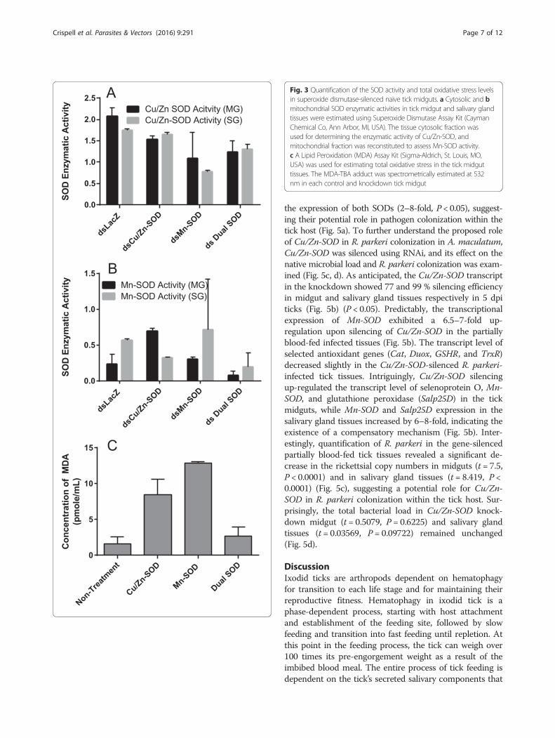
Quantification of total oxidative stress levelsA malondialdehyde (MDA) lipid peroxidation assay wasused to quantify the total oxidative stress levels in the ticktissues. Degradation of lipids as a result of oxidative damagewas estimated by quantification of MDA through the use ofa Lipid Peroxidation MDA Assay Kit (Sigma-Aldrich, St.Louis, MO, USA) following the manufacturer providedprotocol. Twenty milligrams of midgut tissue from individ-ual ticks was filtered through a 0.2 μm filter before the assaywas performed according to the manufacturer’s instructions.
Quantification of SOD activitySuperoxide enzymatic activity was quantified in individ-ual midgut and salivary gland tissues using the Super-oxide Dismutase Assay Kit (Cayman Chemical Co, AnnArbor, MI, USA). The tissues were processed accordingto the manufacturer’s instructions.
Data analysisAll data are expressed as mean ± SEM unless otherwisestated. Statistical significance between the two experimentalgroups or their respective controls was determined by thet-test). Comparative differences amongst the multiple ex-perimental groups were determined by analysis of variancewith statistically significant P-values of < 0.05 (GraphpadPrism 6.05, La Jolla, CA, USA). Transcriptional expressionlevels were determined using Bio-Rad software (Bio-RadCFX MANAGER v.3.1), and the expression values wereconsidered significant if the P-value was 0.05 when com-pared with the control.
ResultsBioinformatic analysesIn this study, the functional role of A. maculatum super-oxide dismustases (SODs) in hematophagy, and bacterialcolonization of endosymbionts and pathogenic microbe R.parkeri was determined. Two gene sequences of ticksuperoxide dismutases cytosolic with signal peptide(Cu/ZnSOD) and intramitrocondrial scavenger (Mn-SOD) were selected to analyze for the presence ofsecretory signal peptide using SignalP [24] server(http://www.cbs.dtu.dk/services/SignalP/). The aminoacid residues deduced from the transcripts of twoSODs identified earlier [3] were translated andvisualized using Jalview and observed six histidineresidues in each sequences showing metal-bindingsites (Additional file 1: Figure S2A, B). The four histi-dine imidazoles are coordinated to Cu(II) while two otherand including one common histidine imadazoles binds toZn(II) metal in Cu/Zn-SOD dismutase [25]. The Mn-SODlocalized in mitochondria in eukaryotes, and found in cyto-plasm in prokaryotes shares common structural propertieswith Fe-SOD [26]. The phylogenetic tree was built inMEGA software version 6 [14] to provide evolutionary
Crispell et al. Parasites & Vectors (2016) 9:291 Page 4 of 12
significance between mitochondrial and cytoplasmic super-oxide dismutases (Additional file 1: Figure S1). Phylogenet-ically Mn-SOD and Cu/Zn-SOD fit in two different clades,sharing common node with corresponding SODs from an-other Amblyomma ticks (Additional file 1: Figure S1).
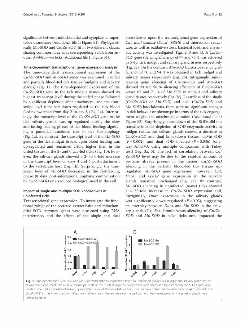
Time-dependent transcriptional gene expression analysisThe time-dependent transcriptional expression of theCu/Zn-SOD and Mn-SOD genes was examined in unfedand partially blood-fed tick tissues (midguts and salivaryglands) (Fig. 1). The time-dependent expression of theCu/Zn-SOD gene in the tick midgut tissues showed itshighest transcript level during the unfed phase followedby significant depletion after attachment, and the tran-script level remained down-regulated as the tick bloodfeeding switched from day 2 to day 8 (Fig. 1a). Interest-ingly, the transcript level of the Cu/Zn-SOD gene in thetick salivary glands was up-regulated during the slowand fasting feeding phases of tick blood feeding, signify-ing a potential functional role in tick hematophagy(Fig. 1a). By contrast, the transcript level of the Mn-SODgene in the tick midgut tissues upon blood feeding wasup-regulated and remained 2-fold higher than in theunfed tissues in the 2- and 6 day-fed ticks (Fig. 1b); how-ever, the salivary glands showed a 2- to 6-fold increasein the transcript level on days 4 and 6 post-attachmentto the vertebrate host (Fig. 1b). Surprisingly, the tran-script level of Mn-SOD decreased in the fast-feedingphase (8 days post-infestation), implying compensationby Cu/Zn-SOD or a reduced biological need in the cell.
Impact of single and multiple SOD knockdowns inuninfected ticksTranscriptional gene expression: To investigate the func-tional role(s) of the secreted extracellular and mitochon-drial SOD enzymes, genes were disrupted using RNAinterference, and the effects of the single and dual
knockdowns upon the transcriptional gene expression ofCat, dual oxidase (Duox), GSHR and thioredoxin reduc-tase, as well as oxidative stress, bacterial load, and enzym-atic activity was investigated (Figs. 2, 3 and 4). A Cu/Zn-SOD gene silencing efficiency of 77 and 76 % was achievedin 5 dpi tick midgut and salivary gland tissues respectively(Fig. 2a). On the contrary, Mn-SOD transcript silencing ef-ficiency of 76 and 94 % was obtained in tick midgut andsalivary tissues respectively (Fig. 2b). Intriguingly, simul-taneous gene silencing of Cu/Zn-SOD and Mn-SODshowed 90 and 98 % silencing efficiency of Cu/Zn-SODverses 63 and 71 % of Mn-SOD in midgut and salivarygland tissues respectively (Fig. 2c). Regardless of the single(Cu/Zn-SOD or Mn-SOD) and dual (Cu/Zn-SOD andMn-SOD) knockdowns, there were no significant changesin tick behavior or phenotype in terms of the tick engorge-ment weight, the attachment duration (Additional file 1:Figure S3). Surprisingly, knockdown of tick SODs did nottranslate into the depletion of SOD enzymatic activity inmidgut tissues but salivary glands showed a decrease inCu/Zn-SOD and dual knockdown tissues, dsMn-SOD(P = 0.002), and dual SOD injected (P = 0.036) (one-way ANOVA using multiple comparison with Tukeytest) (Fig. 3a, b). The lack of correlation between Cu/Zn-SOD level may be due to the residual amount ofproteins already present in the tissues. Cu/Zn-SODsilencing in the partially blood-fed tick tissues up-regulated Mn-SOD gene expression; however, Cat,Duox, and GSHR gene expression in the salivaryglands remained unchanged (Fig. 2a). By contrast,Mn-SOD silencing in uninfected (naïve) ticks showeda 4–35-fold increase in Cu/Zn-SOD expression and,intriguingly, Duox expression in the salivary glandswas significantly down-regulated (P < 0.05), suggestingan interplay between Duox and Mn-SOD in the saliv-ary glands (Fig. 2b). Simultaneous silencing of Cu/Zn-SOD and Mn-SOD in naïve ticks only impacted the
A B
Fig. 1 Time-dependent Cu/Zn-SOD and Mn-SOD transcriptional expression levels in uninfected (naïve) tick midgut and salivary gland tissuesduring the blood meal. The relative transcript levels of the SODs during the blood meal were measured by comparing the SOD expressionlevels in the midgut (Gut) and salivary gland (SG) tissues of the unfed-stage ticks. The changes in transcriptional activity of (a) Cu/Zn-SOD and(b) Mn-SOD in the A. maculatum midgut and salivary gland tissues were normalized to the unfed developmental stage using β-actin as areference gene
Crispell et al. Parasites & Vectors (2016) 9:291 Page 5 of 12
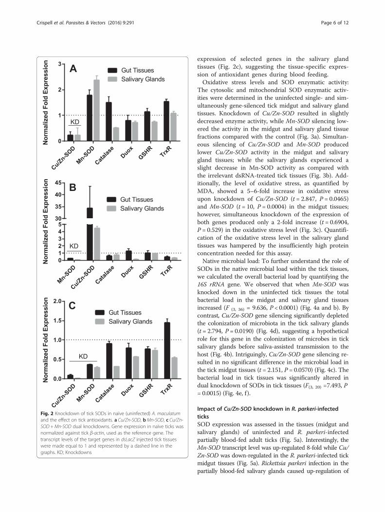
expression of selected genes in the salivary glandtissues (Fig. 2c), suggesting the tissue-specific expres-sion of antioxidant genes during blood feeding.Oxidative stress levels and SOD enzymatic activity:
The cytosolic and mitochondrial SOD enzymatic activ-ities were determined in the uninfected single- and sim-ultaneously gene-silenced tick midgut and salivary glandtissues. Knockdown of Cu/Zn-SOD resulted in slightlydecreased enzyme activity, while Mn-SOD silencing low-ered the activity in the midgut and salivary gland tissuefractions compared with the control (Fig. 3a). Simultan-eous silencing of Cu/Zn-SOD and Mn-SOD producedlower Cu/Zn-SOD activity in the midgut and salivarygland tissues; while the salivary glands experienced aslight decrease in Mn-SOD activity as compared withthe irrelevant dsRNA-treated tick tissues (Fig. 3b). Add-itionally, the level of oxidative stress, as quantified byMDA, showed a 5–6-fold increase in oxidative stressupon knockdown of Cu/Zn-SOD (t = 2.847, P = 0.0465)and Mn-SOD (t = 10, P = 0.0004) in the midgut tissues;however, simultaneous knockdown of the expression ofboth genes produced only a 2-fold increase (t = 0.6904,P = 0.529) in the oxidative stress level (Fig. 3c). Quantifi-cation of the oxidative stress level in the salivary glandtissues was hampered by the insufficiently high proteinconcentration needed for this assay.Native microbial load: To further understand the role of
SODs in the native microbial load within the tick tissues,we calculated the overall bacterial load by quantifying the16S rRNA gene. We observed that when Mn-SOD wasknocked down in the uninfected tick tissues the totalbacterial load in the midgut and salivary gland tissuesincreased (F (3, 36) = 9.636, P < 0.0001) (Fig. 4a and b). Bycontrast, Cu/Zn-SOD gene silencing significantly depletedthe colonization of microbiota in the tick salivary glands(t = 2.794, P = 0.0190) (Fig. 4d), suggesting a hypotheticalrole for this gene in the colonization of microbes in ticksalivary glands before saliva-assisted transmission to thehost (Fig. 4b). Intriguingly, Cu/Zn-SOD gene silencing re-sulted in no significant difference in the microbial load inthe tick midgut tissues (t = 2.151, P = 0.0570) (Fig. 4c). Thebacterial load in tick tissues was significantly altered indual knockdown of SODs in tick tissues (F(3, 20) =7.493, P= 0.0015) (Fig. 4e, f ).
Impact of Cu/Zn-SOD knockdown in R. parkeri-infectedticksSOD expression was assessed in the tissues (midgut andsalivary glands) of uninfected and R. parkeri-infectedpartially blood-fed adult ticks (Fig. 5a). Interestingly, theMn-SOD transcript level was up-regulated 8-fold while Cu/Zn-SOD was down-regulated in the R. parkeri-infected tickmidgut tissues (Fig. 5a). Rickettsia parkeri infection in thepartially blood-fed salivary glands caused up-regulation of
Fig. 2 Knockdown of tick SODs in naïve (uninfected) A. maculatumand the effect on tick antioxidants. a Cu/Zn-SOD, bMn-SOD, c Cu/Zn-SOD +Mn-SOD dual knockdowns. Gene expression in naïve ticks wasnormalized against tick β-actin, used as the reference gene. Thetranscript levels of the target genes in dsLacZ injected tick tissueswere made equal to 1 and represented by a dashed line in thegraphs. KD; Knockdowns
Crispell et al. Parasites & Vectors (2016) 9:291 Page 6 of 12
the expression of both SODs (2–8-fold, P < 0.05), suggest-ing their potential role in pathogen colonization within thetick host (Fig. 5a). To further understand the proposed roleof Cu/Zn-SOD in R. parkeri colonization in A. maculatum,Cu/Zn-SOD was silenced using RNAi, and its effect on thenative microbial load and R. parkeri colonization was exam-ined (Fig. 5c, d). As anticipated, the Cu/Zn-SOD transcriptin the knockdown showed 77 and 99 % silencing efficiencyin midgut and salivary gland tissues respectively in 5 dpiticks (Fig. 5b) (P < 0.05). Predictably, the transcriptionalexpression of Mn-SOD exhibited a 6.5–7-fold up-regulation upon silencing of Cu/Zn-SOD in the partiallyblood-fed infected tissues (Fig. 5b). The transcript level ofselected antioxidant genes (Cat, Duox, GSHR, and TrxR)decreased slightly in the Cu/Zn-SOD-silenced R. parkeri-infected tick tissues. Intriguingly, Cu/Zn-SOD silencingup-regulated the transcript level of selenoprotein O, Mn-SOD, and glutathione peroxidase (Salp25D) in the tickmidguts, while Mn-SOD and Salp25D expression in thesalivary gland tissues increased by 6–8-fold, indicating theexistence of a compensatory mechanism (Fig. 5b). Inter-estingly, quantification of R. parkeri in the gene-silencedpartially blood-fed tick tissues revealed a significant de-crease in the rickettsial copy numbers in midguts (t = 7.5,P < 0.0001) and in salivary gland tissues (t = 8.419, P <0.0001) (Fig. 5c), suggesting a potential role for Cu/Zn-SOD in R. parkeri colonization within the tick host. Sur-prisingly, the total bacterial load in Cu/Zn-SOD knock-down midgut (t = 0.5079, P = 0.6225) and salivary glandtissues (t = 0.03569, P = 0.09722) remained unchanged(Fig. 5d).
DiscussionIxodid ticks are arthropods dependent on hematophagyfor transition to each life stage and for maintaining theirreproductive fitness. Hematophagy in ixodid tick is aphase-dependent process, starting with host attachmentand establishment of the feeding site, followed by slowfeeding and transition into fast feeding until repletion. Atthis point in the feeding process, the tick can weigh over100 times its pre-engorgement weight as a result of theimbibed blood meal. The entire process of tick feeding isdependent on the tick’s secreted salivary components that
Fig. 3 Quantification of the SOD activity and total oxidative stress levelsin superoxide dismutase-silenced naïve tick midguts. a Cytosolic and bmitochondrial SOD enzymatic activities in tick midgut and salivary glandtissues were estimated using Superoxide Dismutase Assay Kit (CaymanChemical Co, Ann Arbor, MI, USA). The tissue cytosolic fraction wasused for determining the enzymatic activity of Cu/Zn-SOD, andmitochondrial fraction was reconstituted to assess Mn-SOD activity.c A Lipid Peroxidation (MDA) Assay Kit (Sigma-Aldrich, St. Louis, MO,USA) was used for estimating total oxidative stress in the tick midguttissues. The MDA-TBA adduct was spectrometrically estimated at 532nm in each control and knockdown tick midgut
Crispell et al. Parasites & Vectors (2016) 9:291 Page 7 of 12

mechanistically facilitate successful transition of eachfeeding phase over the course of multiple days to weeks.Tick saliva, as shown by sialotranscriptome analysis, iscomposed of ~5000 putative secreted salivary proteinscontaining dozens of protein families [3, 27], many ofwhich exhibit pharmacological properties capable of dis-arming the host’s attempts to establish hemostasis or tomount a specific immune response and, ultimately, enhan-cing the tick’s ability to blood-feed. At the same time, ticksface elevated oxidative stress both on and off the mamma-lian host and they must neutralize the harmful effect. Theability of ticks to counterbalance nutritional stress (starva-tion) and blood feeding-related stress (heme digestion) indi-cates the likely development of a proactive detoxificationsystem. Complete studies of the antioxidant systems of
blood-sucking arthropods are presently unavailable; none-theless the scientific literature shows that ticks have an ex-tensive arsenal of antioxidants and selenoproteins thatallow them to survive extreme variations in redox homeo-stasis, with prolonged periods of starvation, the digestion ofhuge blood meals, and exposure to blood-related products[4–6, 20, 27]. Hence, an insight into the tick’s antioxidantrepertoire should offer a glimpse of its defenses against ele-vated levels of oxidative stress. It should also broaden ourunderstanding of tick–pathogen interactions. Although, A.maculatum is a recognized arthropod vector of R. parkeri,this vector-pathogen pair remains the most poorly studiedvector-borne disease. Here, we investigated the functionalrole of two tick SODs (extracellular and mitochondrial) intick hematophagy and pathogen colonization within the
Fig. 4 Impact of SOD knockdown on microbiota load in naïve tick tissues. The bacterial load in the tick tissues (measured in copy numbers) wascalculated by qRT-PCR using the bacterial 16 s rRNA gene and was normalized against the tick β-actin gene (copy number) in tick midguts andsalivary glands each knockdown; Mn-SOD (a, b); Cu/Zn-SOD (c, d); and Dual SODs knockdown (e, f)
Crispell et al. Parasites & Vectors (2016) 9:291 Page 8 of 12

arthropod vector. Bioinformatic analysis of the Cu/Zn-SOD(cytosolic) and Mn-SOD proteins showed amino acid se-quence similarity with arthropod and vertebrate proteins(Additional file 1: Figures S1 and S2).The two A. maculatum SODs we characterized showed
quite different expression patterns. The transcriptional ac-tivity of Cu/Zn-SOD was 2-fold up-regulated throughoutthe blood meal in the salivary tissues, whereas it declinedin the midgut tissues upon blood meal consumption(Fig. 1a). Mn-SOD expression was up-regulated 3–6-foldin the salivary glands on days 4 and 6; however, the mid-gut maintained a steady expression level (Fig. 1b). Further-more, while we obtained both single and dual genetranscriptional knockdowns, significant differences in thetick phenotype (tick engorgement weight, attachment dur-ation, egg mass, egg conversion ratio, and hatchability)(Additional file 1: Figure S3) were lacking, a result that issimilar to previous studies reporting the existence of astrong compensatory mechanism in the tick host for de-toxifying superoxide and H2O2 [20]. The tick salivary
glands undergo tremendous biochemical and physiologicalchanges as soon as a tick attaches itself to the host,followed by engorgement and repletion [28]. In ticks,breakdown of the huge blood meal generates toxic levelsof heme leading to elevated ROS production. The gut bac-terial community is strongly shaped by the ROS levelsupon a blood meal, resulting in a decrease in diversity andan increase in Enterobacteriaceae [6]. The establishmentof vector competence (pathogen survival, colonization,and transmission) is highly specific: R. parkeri can only bevectored by A. maculatum, Ehrlichia chaffeensis byAmblyomma americanum, and Borrelia burgdorferi byIxodes scapularis. It is postulated that these vector-bornedisease agents have developed an antioxidant capacity thataids them to survive and multiply by balancing oxidativetissue-specific homeostasis. Tick-borne pathogens are vul-nerable to a high level of oxidative stress; consequently,disrupting the redox metabolism offers a promising ap-proach for the prevention of tick-borne disease agents andfor disturbing tick microbiota. Our recent work
Fig. 5 Knockdown of Cu/Zn-SOD in R. parkeri-infected A. maculatum. a Tissue-specific SOD transcriptional gene expression in uninfected and R.parkeri-infected partially blood-fed female adults tissues. A. maculatum GAPDH was used as a reference gene to normalize the gene expressiondata. Target gene expression in uninfected tick tissues was adjusted to 1. b Transcriptional gene expression of selected antioxidant genes in Cu/Zn-SOD-silenced partially fed tick tissues. The transcript level of each target gene in the tissues injected with control LacZ-dsRNA was set to 1.0 asa reference point. Expression was normalized against the tick GAPDH gene. c R. parkeri was quantified using a real-time PCR assay designed forspecific detection of the R. parkeri ompB gene in A. maculatum midgut and salivary gland in dsLacZ and dsCu/Zn-SOD injected tick tissues, and(d), the bacterial load estimation in R. parkeri-infected ticks knocked down with Cu/ZnSOD. Tick GAPDH was used as a reference gene for estimat-ing the total bacterial load in R. parkeri-infected ticks. KD; Knockdowns
Crispell et al. Parasites & Vectors (2016) 9:291 Page 9 of 12
highlighted the biological implication of thioredoxin re-ductase (TrxR, a selenoprotein) in preserving the naturalmicrobiota of ticks [6], and a link between selenocysteineelongation factor and R. parkeri survival in the midgut tis-sues [4]. It has been shown that elimination of ROS isrequired to conserve fertility in Anopheles, Drosophila, A.maculatum, and mammals [20, 29–31]. To neutralize thedamaging effects of ROS and attain homeostasis, tickSODs and Cat enzymes act together to catalyze the con-version of superoxide and H2O2. Superoxide and hydro-gen peroxide facilitate the generation of the hydroxylradical, the most reactive oxygen free radical, in the pres-ence of iron metal [32, 33]. Interestingly, lipid peroxida-tion assays showed higher total oxidative stress levels forsingle knockdown tick midguts, while dual knockdownrevealed only a slight change in the oxidative stress level,suggesting the presence of a strong compensatory mech-anism, as has been reported previously (Fig. 3c) [20]. Esti-mation of the total oxidative stress levels in individual ticksalivary glands was not successful because of the low pro-tein concentrations required for this assay. Intriguingly,gene knockdown of Cu/Zn-SOD or Mn-SOD significantlyincreased the total bacterial load in the midgut tissues(Fig. 4a, c), whereas Cu/Zn-SOD knockdown in the saliv-ary glands significantly reduced the total bacterial load(Fig. 4d). The total bacterial load in the dual SOD knock-down remained at a decreased level in both tissues. Theinterplay between ROS levels and survival and thecolonization of intracellular bacteria has not been eluci-dated. Our results indicate that the microbes associatedwith ticks are dependent on maintaining redox homeosta-sis while also producing ROS within the tick cells, andthey use the ROS as a signaling molecule to regulate thebacterial density [34]. Another possible interpretation isthat Cu/Zn-SOD gene knockdown also affected theenzymatic activity of Mn-SOD (Fig. 3a, b), and the in-creased generation of superoxide in the salivary glandssignificantly reduced the total bacterial load. Intriguingly,redox homeostasis is routinely attained by counterbalan-cing ROS and antioxidants by regulating oxidative stress-induced genes [35], including Cat. The transcriptionalexpression of Cat was up-regulated slightly in themidgut tissues upon Cu/Zn-SOD knockdown; how-ever, a compensatory role for Cat in increasing thebacterial load cannot be ruled out (Figs. 2a and 4).SOD facilitates the breakdown of O2− to H2O2 andmolecular oxygen. Mn-SOD (SOD2) from Drosophilamutants show an increased herbicide (Paraquat) sensi-tivity, elevated endogenous oxidative stress, and re-duced longevity [36]. A significant decrease in thetotal bacterial load upon Cu/Zn-SOD knockdown ledus to a follow-up experiment to study the functionalrole of Cu/Zn-SOD gene depletion on R. parkericolonization within the tick host.
ROS provide protection against a variety of pathogens byeliciting immune responses in organisms as diverse asmammals and arthropods, including tick responses againstR. parkeri [4]. Fascinatingly, the interactions between A.maculatum and R. parkeri determine the ability of theseticks to become colonized by and transmit rickettsialagents. In tick-transmitted infections, the infectious agentsmanipulate gene expression of the vector host to ensuretheir colonization and onward transmission to the mamma-lian host. In the current study, Cu/Zn-SOD and Mn-SODtranscript levels in the R. parkeri-infected ticks showed dif-ferential gene expression. Interesting, the transcript level ofMn-SOD significantly upregulated (P < 0.05) in R. parkeri-infected partially blood fed tick tissues and Cu/Zn-SODtranscript level increased only in salivary glands (P < 0.05)and remained down-regulated in gut tissues (Fig. 5a). Itsuggests that tick protects herself from the elevated levelof super-oxides generated and released upon rickettsial in-fection by regulating the expression of superoxide dismut-ase [37] (Fig. 5a). Interestingly, downregulation of Cu/Zn-SOD level in R. parkeri infected midguts co-related withalleviated Cu/Zn-SOD transcript abundance in partiallyblood-fed tissues (Fig. 1a). As expected, the Cu/Zn-SODknockdown up-regulated Mn-SOD (~6.5-fold) expressionin the midgut and salivary glands. Surprisingly, theexpression levels of Salp25D and SelM were 9-fold and 3-fold up-regulated, respectively. It is established that A.maculatum eEFSec, I. scapularis glutathione peroxidase(Salp25D), and Dermacentor variabilis SelM offer asurvival advantage to R. parkeri, B. burgdorferi, andAnaplasma marginale [4, 38, 39].
ConclusionsThis is the first study to show that the silencing of Cu/Zn-SOD decreases the level of R. parkeri colonization intick tissues. The A. maculatum antioxidants, Mn-SODand Cu/Zn-SOD play a role in maintaining redoxhomeostasis within the tick and facilitate thecolonization of human pathogenic bacteria, R. parkeri.Our findings provide a strong foundation for futureresearch to elucidate the molecular determinants of vec-tor competence. The relationship between ROS, vectorcompetence, and R. parkeri-induced antioxidants isundergoing further investigation in our laboratory.
Additional files
Additional file 1: Figure S1. Evolutionary relationships of taxa based onthe SOD amino acid sequence using maximum likelihood method. Theevolutionary history was inferred by using the Maximum Likelihood methodbased on the JTT matrix-based model [15]. The tree is drawn to scale, with thebranch lengths measured by the number of substitutions per site. Evolutionaryanalyses were conducted using MEGA6 software [14]. The sequences wereobtained from Amblyomma maculatum, Amblyomma variegatum, Ixodesscapularis, Anopheles gambiae, Mus musculus, Sus scrofa, and Homo sapiens.
Crispell et al. Parasites & Vectors (2016) 9:291 Page 10 of 12
GenBank accession numbers followed by species names are shown in thetree. Figure S2A. Multiple sequence alignments of Cu/Zn-SOD amino acidsequences from different taxa. Regions outlined by red boxes indicatemetal-binding sites that are conserved between all of the listed species. Thesequences for Cu/Zn-SODs were obtained from Drosophila melanogaster, Apismellifera, Amblyomma maculatum, Thermostable mutant of Human Cu/ZnSOD, Mus musculus, Equus caballus, Danio rerio, Anopheles gambiae and Culexquinquefasciatus. Figure S2B. Multiple sequence alignments of Mn-SODamino acid sequences from different taxa. Regions outlined by red boxesindicate metal-binding sites that are conserved between all the listed species.The sequences were obtained from Haliotis discus, Lottia gigantea, Perinereisnuntia, Stegastes partitus, Takifugu rubripes, Struthio camelus, Tauraco erythrolo-phus, Thamnophis elegans, Sus scrofa, Macaca mulatta, Mus musculus, Xenopuslaevis, Amblyomma variegatum, Amblyomma maculatum and Ixodes scapularis.Figure S3. The engorged tick weight in dsRNA-SODs injected ticks. Theengorged weights of ticks injected with dsLacZ, dsCu/Zn-SOD, dsMn-SOD anddual SODs (dsCu/Zn-SOD and dsMn-SOD) observed at the detachment ofticks from Sheep. There were no significant effects in tick engorged weightswith dsRNA-SODs (ANOVA, F(3,52) = 0.6274, P= 0.6006). (DOCX 473 kb)
Competing interestsThe authors declare that they have no competing interests.
Authors’ contributionsConceived and designed the experiments: SK. Performed the experiments:GC, KBC, SK. Analyzed the data: GC, KBC, SK. Contributed reagents/materials/analysis tools: SK. Wrote the paper: GC, KBC, SK. All authors have read andapproved the manuscript.
AcknowledgmentsThis work was supported by grants from the National Institute of Allergy andInfectious Diseases (award #AI099919) and the National Institutes of GeneralMedical Sciences (award #P20GM103476). Gary Crispell is the recipient of theMississippi-INBRE summer undergraduate research fellowship. These fundingbodies played no role in the study design, data collection, analysis, decisionto publish, or manuscript preparation.
Received: 12 April 2016 Accepted: 9 May 2016
References1. Paddock CD, Goddard J. The evolving medical and veterinary
importance of the gulf coast tick (Acari: Ixodidae). J Med Entomol.2015;52:230–52.
2. Fomenko DE, Koc A, Agisheva N, Jacobsen M, Kaya A, Malinouski M, et al.Thiol peroxidases mediate specific genome-wide regulation of geneexpression in response to hydrogen peroxide. Proc Natl Acad Sci U S A.2011;108:2729–34.
3. Karim S, Singh P, Ribeiro JMC. A deep insight into the sialotranscriptome ofthe gulf coast tick Amblyomma maculatum. PLoS One. 2011;6:e28525.
4. Adamson SW, Browning RE, Budachetri K, Ribeiro JMC, Karim S. Knockdownof selenocysteine-specific elongation factor in Amblyomma maculatumalters the pathogen burden of Rickettsia parkeri with epigenetic control bythe Sin3 Histone Deacetylase Corepressor Complex. PLoS One. 2013;8:e82012.
5. Adamson S, Browning R, Singh P, Nobles S, Villarreal A, Karim S.Transcriptional activation of antioxidants may compensate for selenoprotein deficiencies in Amblyomma maculatum (Acari: Ixodidae) injected withselK- or selM-dsRNA. Insect Mol Biol. 2014;23(4):497–510.
6. Budachetri K, Karim S. An insight into the functional role of thioredoxinreductase, a selenoprotein, in maintaining normal native microbiota in theGulf Coast tick (Amblyomma maculatum). Insect Mol Biol. 2015;24:570–81.
7. Hoffmann JA. The immune response of Drosophila. Nature. 2003;426:33–8.8. Ha E-M, Oh C-T, Ryu J-H, Bae Y-S, Kang S-W, Jang I-H, et al. An antioxidant
system required for host protection against gut infection in Drosophila. DevCell. 2005;8:125–32.
9. Patrick CD, Hair JA. Laboratory rearing procedures and equipment for multi-host ticks (Acarina: Ixodidae). J Med Entomol. 1975;12:389–90.
10. Budachetri K, Browning RE, Adamson SW, Dowd SE, Chao C-C, Ching W-M, etal. An insight into the microbiome of the Amblyomma maculatum (Acari:Ixodidae). J Med Entomol Entomological Society of America. 2014;51:119–29.
11. Thompson JD, Gibson TJ, Higgins DG. Multiple sequence alignment usingClustalW and ClustalX. Curr Protoc Bioinforma. 2002;Chapter 2:Unit 2 3.
12. Larkin MA, Blackshields G, Brown NP, Chenna R, McGettigan PA, McWilliamH, et al. Clustal W and clustal X version 2.0. Bioinformatics. 2007;23:2947–8.
13. Waterhouse AM, Procter JB, Martin DM, Clamp M, Barton GJ. Jalview Version2 - a multiple sequence alignment editor and analysis workbench.Bioinformatics. 2009;25:1189–91.
14. Tamura K, Stecher G, Peterson D, Filipski A, Kumar S. MEGA6: Molecularevolutionary genetics analysis version 6.0. Mol Biol Evol. 2013;30:2725–9.
15. Jones DT, Taylor WR, Thornton JM. The rapid generation of mutation datamatrices from protein sequences. Bioinformatics. 1992;8:275–82.
16. Grimm D, Tilly K, Byram R, Stewart PE, Krum JG, Bueschel DM, et al. Outer-surface protein C of the Lyme disease spirochete: a protein induced in ticksfor infection of mammals. Proc Natl Acad Sci U S A. 2004;101:3142–7.
17. MORGAN JF, MORTON HJ, PARKER RC. Nutrition of animal cells in tissue culture;initial studies on a synthetic medium. Proc Soc Exp Biol Med. 1950;73:1–8.
18. MORGAN JF, CAMPBELL ME, MORTON HJ. The nutrition of animal tissuescultivated in vitro. I. A survey of natural materials as supplements tosynthetic medium 199. J. Natl. Cancer Inst. 1955;16:557–67.
19. Browning R, Adamson SW, Karim S. Choice of a stable set of referencegenes for qRT-PCR analysis in Amblyomma maculatum (Acari: Ixodidae).J Med Entomol. 2012;49:1339–46.
20. Kumar D, Budachetri K, Meyers VC, Karim S. Assessment of tick antioxidantresponses to exogenous oxidative stressors and insight into the role ofcatalase in the reproductive fitness of the Gulf Coast tick, Amblyommamaculatum. Insect Mol Biol. 2016;25(3):283–94.
21. Bullard RL, Williams J, Karim S. Temporal gene expression analysis and RNAsilencing of single and multiple members of gene family in the lone startick Amblyomma americanum. PLoS One. 2016;11:0147966.
22. Karim S, Adamson SW, Simpson SJ, Casas J. RNA interference in ticks: Afunctional genomics tool for the study of physiology. In: Jockusch E, editor.Adv. Insect Physiol. Small RNAs Their Divers. Roles Pract. Uses. I. San Diego:Academic; 2012. p. 119–54.
23. Narasimhan S, Rajeevan N, Liu L, Zhao YO, Heisig J, Pan J, et al. Gutmicrobiota of the tick vector Ixodes scapularis modulate colonization of theLyme disease spirochete. Cell Host Microbe. 2014;15:58–71.
24. Petersen TN, Brunak S, von Heijne G, Nielsen H. SignalP 4.0: discriminatingsignal peptides from transmembrane regions. Nat Methods. 2011;8:785–6.
25. Fee JA, Peisach J, Mims WB. Superoxide dismutase. Examination of the metalbinding sites by electron spin echo spectroscopy. J Biol Chem. 1981;256:1910–4.
26. Britton L, Fridovich I. Intracellular localization of the superoxidedismutases of Escherichia coli: a reevaluation. J Bacteriol.1977;131:815–20.
27. Karim S, Ribeiro JMC. An insight into the sialome of the lone star tick,Amblyomma americanum, with a glimpse on its time dependent geneexpression. PLoS One. 2015;10, e0131292.
28. Binnington KC. Sequential changes in salivary gland structure duringattachment and feeding of the cattle tick, Boophilus microplus. Int JParasitol. 1978;8:97–115.
29. DeJong RJ, Miller LM, Molina-Cruz A, Gupta L, Kumar S, Barillas-Mury C.Reactive oxygen species detoxification by catalase is a major determinant offecundity in the mosquito Anopheles gambiae. Proc Natl Acad Sci U S A.2007;104:2121–6.
30. Orr W, Sohal R. Extension of life-span by overexpression of superoxide dismutaseand catalase in Drosophila melanogaster. Science. 1994;263:1128–30.
31. Schriner SE, Linford NJ, Martin GM, Treuting P, Ogburn CE, Emond M, et al.Extension of murine life span by overexpression of catalase targeted tomitochondria. Science. 2005;308:1909–11.
32. Liochev SI, Fridovich I. Superoxide and nitric oxide: consequences of varyingrates of production and consumption: a theoretical treatment. Free RadicBiol Med. 2002;33:137–41.
33. Kehrer JP. The Haber–Weiss reaction and mechanisms of toxicity.Toxicology. 2000;149:43–50.
34. Brennan LJ, Haukedal JA, Earle JC, Keddie B, Harris HL. Disruption of redoxhomeostasis leads to oxidative DNA damage in spermatocytes ofWolbachia-infected Drosophila simulans. Insect Mol Biol. 2012;21:510–20.
35. Dröge W. Free radicals in the physiological control of cell function. PhysiolRev. 2002;82:47–95.
Crispell et al. Parasites & Vectors (2016) 9:291 Page 11 of 12

36. Kirby K, Hu J, Hilliker AJ, Phillips JP. RNA interference-mediated silencing ofSod2 in Drosophila leads to early adult-onset mortality and elevatedendogenous oxidative stress. Proc Natl Acad Sci U S A. 2002;99:16162–7.
37. Santucci LA, Gutierrez PL, Silverman DJ. Rickettsia rickettsii inducessuperoxide radical and superoxide dismutase in human endothelial cells.Infect Immun. 1992;60:5113–8.
38. Narasimhan S, Sukumaran B, Bozdogan U, Thomas V, Liang X, DePonte K, etal. A tick antioxidant facilitates the Lyme disease agent’s successfulmigration from the mammalian host to the arthropod vector. Cell HostMicrobe. 2007;2:7–18.
39. Kocan KM, Zivkovic Z, Blouin EF, Naranjo V, Almazán C, Mitra R, et al.Silencing of genes involved in Anaplasma marginale-tick interactions affectsthe pathogen developmental cycle in Dermacentor variabilis. BMC Dev Biol.2009;9:42.
• We accept pre-submission inquiries
• Our selector tool helps you to find the most relevant journal
• We provide round the clock customer support
• Convenient online submission
• Thorough peer review
• Inclusion in PubMed and all major indexing services
• Maximum visibility for your research
Submit your manuscript atwww.biomedcentral.com/submit
Submit your next manuscript to BioMed Central and we will help you at every step:
Crispell et al. Parasites & Vectors (2016) 9:291 Page 12 of 12