rhinocerebral mucormycosis: changing patterns of disease
TRANSCRIPT

152 Surg Neurol 1994;41:152-56
Rhinocerebral Mucormycosis: Changing Patterns of Disease
Eric S. Nussbaum, M.D., and Walter A. Hall, M.D.
Department of Neurosurgery, University of Minnesota Hospital and Clinic, Minneapolis, Minnesota
Nussbaum ES, Hall WA. Rhinocerebral mucormycosis: changing patterns of disease. Surg Neurol 1994;41:151-56.
Eleven cases of rhinocerebral mucormycosis (RM) encoun- tered over a 13-year period were reviewed. Predisposing factors included leukemia (36%), diabetes mellitius (27%), aplastic anemia (9%), myelodysplastic syndrome (9%), and treatment with immunosuppressive medications necessary to maintain solid organ or bone marrow graft viability (64%). Two patients had no predisposing factors. Clinical findings included headache (73%), fever (55%), black na- sal eschar (45%), orbitofacial cellulitis (36%), cranial nerve palsy (36%), altered sensorium (36%), and hemipa- resis (27%). Seven patients presented with destruction of the paranasal sinuses and local invasion; three with direct extension to the frontal or temporal lobes. Four patients displayed hematogenous dissemination to the cerebrum, brain stem, and cerebellum from a pr imary pulmonary focus. The seven patients with sinus involvement were treated with aggressive surgical debridement. Two pa- tients with focal intracerebral lesions underwent either open craniotomy or stereotactic biopsy. Amphotericin B was administered intravenously to all patients. Local irri- gation via a percutaneous catheter was performed in the seven patients with sinus disease and in one case of intra- cranial abscess. All seven patients with intracranial infec- tion died, in contrast to four patients that survived with infection localized to the sinuses and orbits. All survivors had been treated with a combination of surgery and am- photericin B therapy. This review demonstrates that RM is increasingly affecting patients with sources of immuno- suppression other than diabetes mellitus. Early aggressive therapy to prevent cerebral involvement by this severe infection provides the best chance for a good outcome.
KEY WORDS: Mucormycosis; Rhinocerebral mucormycosis; Zygomycosis
Rhinocerebral mucormycosis (RM), first described in 1885 by Paltauf, was considered uniformly fatal until 1955 when Harris repor ted the case of a child who
Address reprint requests to: Walter A. Hall, M.D., Department of Nurosurgery, Box 96 UMHC, 420 Delaware Street S.E., Minneapolis, MN 55455.
Received April 26, 1993 accepted July 8, 1993.
survived, despite significant neurologic injury [10,19]. The prognosis for RM remained poor until the introduc- tion of the antifungal agent, amphotericin B, in 1958 [26]. Since that time, aggressive surgical debr idement combined with amphotericin B administration has re- mained the primary t reatment for RM infections and subsequent reports have emphasized the severity o f this infection, especially when cerebral involvement is pres- ent [1,9,15,21].
As the number of patients receiving immunosuppres- sive therapy for solid organ and bone marrow trans- plantation has increased, so has the incidence of opportu- nistic infections such as mucormycosis that require neurosurgical evaluation and intervention. Concur- rently, the observed patterns of infection have become more variable, resulting in new diagnostic and therapeu- tic challenges. This repor t summarizes our experience with the presentation, diagnosis, and management of RM during the past 13 years.
C l i n i c a l M a t e r i a l s a n d M e t h o d s
During the 13-year period f rom January 1979 to January 1992, 11 patients with RM were treated at the University of Minnesota Hospital and Clinic. There were eight men and three women, ranging in age f rom 5 to 56 years (mean age 31 years). The diagnosis was confirmed in every case by pathologic examination of transnasal or cerebral biopsy material revealing non-septate, thick- walled hyphae with right-angle branching and marked vascular invasion. Medical records, laboratory data, ra- diographic images, operative reports, and tissue speci- mens were examined in every patient. All patients had plain sinus roentgenograms and computed tomography (CT) of both the brain and sinuses. Intravenous contrast was not given to those patients with abnormal renal function who had CT. Two patients were studied with contrast enhanced magnetic resonance imaging (MRI).
The seven patients with sinus involvement were treated with aggressive bony debr idement and sinus ex- enteration. Two patients with focal intracerebral lesions had either an open craniotomy or stereotactic biopsy. All patients received intravenous amphotericin B therepy at a dose of 0.3 mg/kg on days 1 and 2 of t reatment , 0.5
© 1994 by Elsevier Science Inc. 0090-3019/94/$7.00
Rhinocerebral Mucormycosis Surg Neurol 153 1994;41:152-56
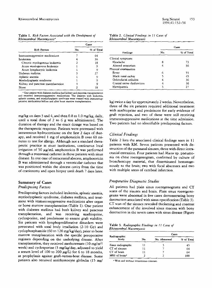
Table 1. Risk Factors Associated with the Development of Rhinocerebral Mucormycosis a
Cases
Risk Factors No. % of Total
Immunosuppress ive medications 7 64 Leukemia 4 36
Chronic myelogenous leukemia 2 18 Acute myelogenous leukemia 1 9 Acute lymphocytic leukemia 1 9
Diabetes mellitus 3 27 Aplastic anemia 1 9 Myelodysplastic syndrome 1 9 Kidney and pancreas transplantation 1 9 N o n e 2 18
One patient with diabetes mellitus had kidney and pancreas transplantation and received immunosuppressive medications. The patients with leukemia, aplastic anemia, and myelodysplastic syndrome were treated with immunosup- pressive medications before and after bone marrow transplantation.
mg/kg on days 3 and 4, and then 0.8 to 1.0 mg/kg, daily, until a total dose of 2 to 6 g was administered. The duration of therapy and the exact dosage was based on the therapeutic response. Patients were pretreated with intravenous hydrocortisone on the first 2 days of ther- apy, and received 1 mg of amphotericin B over 60 mi- nutes to test for allergy. Although not a standard thera- peutic practice at most institutions, continuous local irrigation of 50/zg/mL amphotericin B was performed through a transnasal catheter in those patients with sinus disease. In one case ofintracranial abscess, amphotericin B was administered through a ventricular catheter that was positioned within the abscess cavity from the time of craniotomy and open biopsy until death 7 days later.
S u m m a r y o f Cases Predisposing Factors
Predisposing factors included leukemia, aplastic anemia, myelodysplastic syndrome, diabetes mellitus, and treat- ment with immunosuppressive medications after organ or bone marrow transplantation (Table 1). One patient with diabetes mellitus had both kidney and pancreas transplantation, and was receiving azathioprine, cyclosporine, and prednisone to ensure graft viability. Six patients with lymphoproliferative disorders were pretreated with total body irradiation (2-10 Gy) and cyclophosphamide (60 to 120 mg/kg/day), prior to bone marrow transplantation with the specific preoperative regimen depending on the underlying disease. After transplantation, they received methotrexate (10 mg/m2/ week) and cyclosporine (3 mg/kg/day, adjusted to yield a serum level of 100 to 200/zg/L) for 6 to 18 months, as prophylaxis against graft-versus-host disease. Some patients also received antithymocyte globulin (15 rag/
Table 2. Clinical Findings in I I Cases of Rhinocerebral Mucormycosis
Cases
Findings No. % of Total
Clinical symptoms Headache 8 73 Altered sensor ium 4 36
Physical examination Fever 6 55 Black nasal eschar 5 45 Orbitofacial cellulitis 4 36 Cranial nerve dysfunction 4 36 Hemipares is 3 27
kg) twice a day for approximately 2 weeks. Nevertheless, three of the six patients required additional treatment with azathioprine and prednisone for early evidence of graft rejection, and two of these were still receiving immunosuppressive medications at the time admission. Two patients had no identifiable predisposing factors.
Clinical Findings
Table 2 lists the associated clinical findings seen in 11 patients with RM. Seven patients presented with de- struction of the paranasal sinuses; three with direct intra- cranial extension. Four patients had Mucor sp. pneumo- nia on chest roentgenogram, confirmed by culture of bronchoscopy material, that disseminated hematoge- nously to the brain; two with focal abscesses and two with multiple areas of cerebral infarction.
Preoperative Diagnostic Studies
All patients had plain sinus roentgenograms and CT scans of the sinuses and brain. Plain sinus roentgeno- grams were abnormal in five cases demonstrating bony destruction associated with sinus opacification (Table 3). CT scan of the sinuses revealed thickening and contrast enhancement of the involved sinus mucosa with bony destruction in the seven cases with sinus disease (Figure
Table 3. Radiographic Findings in 11 Cases of Rhinocerebral Mucormycosis
Cases Radiographic
Study No. No. Abnormal % of Total
Sinus radiographs 11 5 45 CT of sinuses 11 7 64 CT of brain 11 7 64 MRI of brain ~ 2 2 100
o With and without intravenous contrast.

154 Surg Neurol Nussbaum and Hall 1994;41:152-56
Figure 1. CT scan of the sinuses demonstrating thickening and inflamma- tion of the mucosa with bony erosion of the posterior wall of the maxillary sinus (arrouJ.
1). In the four cases without paranasal sinus involvement, CT of the head without contrast showed multiple, focal areas of low density within the brain, suggestive of isch- emic infarction or early abscess formation. MRI was performed in the two most recent cases, demonstrating the subtle, soft tissue changes associated with orbitofa- cial cellulitis and maxillary sinusitis (Figure 2). MRI was especially useful in defining the extent of intradural and extradural infection particularly in the case of a temporal lobe abscess that enhanced after intravenous contrast administration in a patient with renal failure (Figure 3).
T r e a t m e n t Resu l t s
All seven patients with intracranial infection died. The four patients with infection localized to the paranasal sinuses and orbit, without intradural extension, were alive 2 years later. One of these patients was readmitted 6 months after discharge with recurrent infection, re- quiring successful retreatment with intravenous ampho- tericin B. All patients who survived had been treated with a combination of surgical debridement and ampho- tericin B therapy.
Postmortem examinations were performed in six of the seven patients with intracranial disease. Diffuse dis- semination of fungal elements throughout the brain with extensive necrosis and hemorrhage was noted in all cases. Neutrophilic infiltration with leptomeningeal and local vascular invasion was seen. Hyphae were found within the lumen and wall of blood vessels and in the
Figure 2. MRI of the head after gadolinium administration showing enhancement of the mucosa of the maxillary sinus consistent with sinusitis.
subarachnoid space. Local disease extension from the paranasal sinuses had a predilection for the subfrontal and temporal regions and dissemination through the bloodstream to the frontal lobe occurred in one case, and to the temporal lobe in two cases. Hematogenous seeding of the brain stem, cerebellum, occipital, and parietal lobes also occurred.
In one dramatic case, diffuse cerebral edema with subarachnoid hemorrhage was seen. There was uncal and tonsillar herniation with bilateral necrosis of the inferior frontal lobes. Unilateral thrombosis of the inter- nal carotid artery with extension into the middle cerebral artery was noted. The patient died 4 days after the onset of headache.
D i s c u s s i o n
The fungal class Zygomycetes contains the order Mucor- ales which is responsible for mucormycosis in man. Rhi - zopus and Rhizomucor are the organisms most commonly isolated from patients with mucormycosis [1,25]. These ubiquitous fungi are typically found in soil, decaying bread and fruit, and as normal human nasal flora [1,12,13,21].

Rhinocerebral Mucormycosis Surg Neurol 155 1994;41:152-56
Figure 3. MRI of the head revealing an enhancing temporal lobe Mucor sp. abscess after intravenous contrast administration.
Mucormycosis usually occurs in patients with diabetes mellitus, in patients with neutropenia due to disease or therapy, or in patients receiving immunosuppressive medications after organ transplantation [ 1,11,15,21,24]. Other identified predisposing factors include renal fail- ure, severe burns, malnutrition, uremia, protracted and severe diarrhea, and treatment with deferoxamine [13,15,17,23,25]. Patients who are not immunocom- promised have occasionally been affected [13,27].
Mucormycosis may present as an isolated pulmonary infection, in a relentlessly invasive rhinocerebral form, or as disseminated disease [13,15,24]. Rhinocerebral involvement is typically associated with poorly con- trolled diabetes mellitus and ketoacidosis, whereas pul- monary infection is associated with leukemia, lymphoma, and other neutropenic states [1,13,14,21, 24]. Intracerebral infection due to hematogenous spread may be associated with intravenous drug abuse [20].
Mucormycosis infection occurs after inhalation of spores that may deposit in the nasal turbinates or pass into the lungs [25]. In rhinocerebral mucormycosis, the initial lesions occur as black, firm areas on the superficial
oral or nasal mucosa that may be mistaken for dried blood in severely ill patients [ 15]. The infection spreads via local extension to the paranasal sinuses and orbits, and eventually can directly invade the meninges and brain as seen in three of our 11 (27%) patients. Clinical findings associated with orbitofacial cellulitis include fa- cial pain, swelling, and tenderness [15,21]. Fever and headache are common as seen in over half our patients and cranial nerve dysfunction that has been characterized by loss of vision, ophthalmoplegia, proptosis, corneal anesthesia, and facial weakness was found in over a third [12,15,21].
We encountered two distinct forms of RM. Seven patients presented with rhinocerebral involvement man- ifest as destruction of the paranasal sinuses, in some cases with intradural extension. However, four patients displayed hematogenous seeding of the brain from a primary pulmonary source. These four patients had bone marrow transplantation for leukemia or aplastic anemia and developed severe pulmonary mucormycosis without evidence of sinus disease or orbitofacial involvement. Instead, these patients presented with an altered senso- rium and focal neurologic findings due to ischemic in- farction or abscess formation. On postmortem examina- tion, vascular thrombosis and occlusion resulted from embolization of fungal elements. For uncertain reasons, the fungus demonstrates a well-described proclivity for aggressive vascular invasion resulting in thrombosis and tissue necrosis [ 1,7,15,21,25]. Thrombosis of major ves- sels resulting in ischemic cerebral infarction has been reported [3,8]. Extensive vascular invasion was noted on biopsy specimens in all cases in this series.
Appropriate confirmatory pathologic examination of transnasal or cerebral biopsy material is necessary to definitively establish the diagnosis of RM [1]. Mucor- mycosis is characterized pathologically by broad, non- septate, thick-walled hyphae that branch at right angles compared to Aspergillus sp. that is segmented [13,15]. Further suggestive evidence may be provided by sam- pling cerebrospinal fluid, in the absence of increased intracranial pressure, which will often reveal a neutro- philic pleocytosis and an elevated protein level [ 15,21]. The peripheral leukocyte count is unreliable because many patients are immunosuppressed or have bone mar- row disorders.
Plain radiographs typically reveal sinusitis with focal bony erosion of the ethmoid sinus, extending into the maxillary sinus and inferior orbit [4]. Computed tomog- raphy is very sensitive for detecting orbital involvement, and with intracranial extension, ring-enhancing lesions in the frontal or temporal lobes can be seen after intrave- nous contrast administration [4,27]. On MRI, soft tissue changes with fungal infiltration of fat planes in the face and orbit and abnormal signal changes along the walls

156 Surg Neurol Nussbaum and Hall 1994;41:152-56
of the bony sinuses may be seen [22,28]. Consistent with prior reports, intracerebral extension was best ap- preciated in our series on T2-weighted images, and gado- linium administration was useful for detecting early vas- cular invasion and intraparenchymal disease [22,28].
Survival in RM remains a rare event despite aggressive treatment, as evidenced in our patients with intracranial infection [10,18]. Successful therapy consists of a joint surgical and medical approach, combining radical surgi- cal debridement of involved areas with systemic ampho- tericin B administration [1,2,9]. Wide exenteration of the paranasal sinuses and orbital contents will occasion- ally result in cure with repeated removal of necrotic tissue often being necessary [23,25]. Sinus irrigation with amphotericin B combined with systemic infusion, may also be useful in the treatment of mucormycosis.
The liposomal forms of amphotericin B may have reduced toxicity and an increased therapeutic effect over the standard form in mucormycosis because of better cerebrospinal fluid penetration [1,7,16,25]. Rifampin has been combined with amphotericin B with one in- stance of a clinical response, and the lack of activity of the azole antifungal agents precludes their use in RM at this time [ 1,16,25 ]. Hyperbaric oxygen therapy has been used in a number of cases of RM with varying success, supporting further investigation of this treatment mod- ality [6]. Patients receiving immunosuppressive medica- tions after organ transplantation that develop RM should have those medications discontinued at the risk of suffer- ing graft rejection until a sufficient neutrophil count returns [1]. New immunostimulating agents such as granulocyte colony stimulating factor, a naturally oc- curring glycoprotein that stimulates the growth ofgranu- locyte precursors and activates mature neutrophils, may prove useful in immunocompromised patients with RM [5].
Rhinocerebral mucormycosis, once thought to occur exclusively in diabetes mellitus, is clearly affecting pa- tients with other sources of immunosuppression. Pa- tients may not present with paranasal sinus involvement but may instead have focal intracerebral lesions that re- quire stereotactic aspiration or biopsy to optimize treat- ment regimens. Despite the poor prognosis in RM with intracranial extension, earlier diagnosis and innovative therapies may result in an improved outcome for patients with this infection.
R e f e r e n c e s 1. Armstrong D. Treatment of opportunistic fungal infections. Clin
Inf Dis 1993;16:1-9.
2. Bell WE. Treatment of fuugal infections of the central nervous system. Ann Neurol 1981;9:417-22.
3. Carpenter DF, Brubaker LH, Powell RD Jr, et al. Phycomycotic thrombosis of the basilar artery. Neurology 1968;14:807-12.
4. Centeno RS, BentsonJR, Mancuso AA. CT scanning in rhinocere- bral mucormycosis and aspergillosis. Radiology 1981 ;140:383-9.
5. Clark SL, Kamen R. The human hematopoietic colony stimulating factors. Science 1987;236:1229-37.
6. Ferguson BT, Mitchell TG, Moon R, et al. Adjunctive hyperbaric oxygen therapy for treatment ofrhinocerebral mucormycosis. Rev Inf Dis 1988;10:551-9.
7. Fisher EW, Toma A, Fisher PH, et al. Rhinocerebral mucor- mycosis: use of liposomal amphotericin B. J Laryngol Otol 1991;105:575-7.
8. Galetta SL, Walc AE, Goldberg HI, et al. Rhinocerebral mucor- mycosis. Management and survival after carotid occlusion. Ann Neurol 1990;28:103-7.
9. Hall WA. Neurosurgical infections in the compromised host. Neurosurg Clin North Am 1992;3:435-42.
10. Harris JS. Mucormycosis. A report of a case. Pediatrics 1955;16:857-67.
11. Hurter RV. Phycomycetes infection in cancer patients: a complica- tion of therapy. Cancer 1959;12:330-50.
12. Ignelzi RJ, Vander Ark GD. Cerebral mucormycosis following open head trauma. J Neurosurg 1975;42:593-6.
13. Lehrer RI, Howard DH, Sypherd PS, et al. Mucormycosis. Ann Intern Med 1980;93:93-108.
14. Medoff G, Kobayashi GS. Pulmonary mucormycosis. N Engl J Med 1972;286:86-7.
15. Meyers BR, Wormser G, Hirschman Z, et al. Rhinocerebral mu- cormycosis. Premortem diagnosis and therapy. Arch Intern Med 1979;139:557-60.
16. Meyer RD. Current role of therapy with amphotericin B. Clin Inf Dis 1992;14:S154-60.
17. Morduchowicz G, Shmueli D, Shapira Z, et al. Rhinocerebral mucormycosis in renal transplant recipients. Report of three cases and review of the literature. Rev Inf Dis 1986;8:441-6.
18. Muresan A. A case of cerebral mucormycosis diagnosed in life with eventual recovery. J Clin Pathol 1960;13:34-6.
19. Paltauf AA. Mycosis mucorina. Virchows' Arch Pathol Anat 1885;102:543-64.
20. Pierce PFJr, Solomon SL, Kaufman L, et al. Zygomycetes brain abscesses in narcotic addicts with serological diagnosis. JAMA 1982;248:2881-2.
21. Pillsbury HC, Fischer ND. Rhinocerebral mucormycosis. Arch Otolaryng 1977;103:600-4.
22. Press GA, Weindling SM, Hesselink SR, et al. Rhinocerebral mucormycosis. MR manifestations. J Comput Assist Tomogr 1988;12:744-9.
23. Rabin ER, Lundberg GD, Mitchell ET. Mucormycosis in severely burned patients. N Engl J Med 1961;264:1286-9.
24. Sandier R, Tallman CB, Kearny DG, et al. Successfully treated rhinocerebral phycomycosis in well controlled diabetics. N Eugl J Med 1971;285:1180-2.
25. Sugar AM. Mucormycosis. Clin Inf Dis 1992;14:$126-9.
26. Smith HW, Kirchner JA. Cerebral mucormycosis. Arch Otolaryng 1958;68:715-8.
27. Young RF, Gade G. Grinnell V. Surgical treatment for fungal infections in the central nervous system. J Neurosurg 1985;63:371-81.
28. Yousem DM, Galetta SL, Gusnard DA, et al. MR findings in rhinocerebral mucormycosis. J Comput Assist Tomogr 1989;13:878-92.