rheumatoid meningitis - core · rheumatoid meningitis maría j. servioli 1, chandril chugh , john...
TRANSCRIPT

CPC ARTICLEpublished: 26 December 2011doi: 10.3389/fneur.2011.00084
Rheumatoid meningitisMaría J. Servioli 1, Chandril Chugh1, John M. Lee2 and José Biller 1*
1 Department of Neurology, Stritch School of Medicine, Loyola University Chicago, Chicago, IL, USA2 Department of Pathology, Stritch School of Medicine, Loyola University Chicago, Chicago, IL, USA
Edited by:
Osvaldo Fustinoni, Universidad deBuenos Aires, Argentina
Reviewed by:
Emilia Gatto, Sanatorio de la TrinidadMitre, ArgentinaIgnacio J. Previgliano, MaimonidesUniversity, Argentina
*Correspondence:
José Biller , Department of Neurology,Stritch School of Medicine, LoyolaUniversity Chicago, 2160 S. FirstAvenue, Chicago, IL 60153, USA.e-mail: [email protected]
An 80-year-old woman with rheumatoid arthritis had gait difficulties and frequent falls. MRIof the brain showed an extra-axial enhancing lesion overlying the right frontal–parietal cor-tex, that progressively extended to the contralateral side.This was accompanied by furtherdecline in her functional status. We discuss the diagnostic and therapeutic approach of apachy–leptomeningeal process in a rheumatoid patient.
Keywords: rheumatoid arthritis, brain biopsy, rheumatoid pachymeningitis, rheumatoid leptomeningitis
An 80-year-old woman presented to the neurology clinic witha history of unsteady gait and frequent falls and was admittedto our hospital for further evaluation. She had history of arte-rial hypertension, chronic obstructive pulmonary disease, andlong-standing rheumatoid arthritis (RA) treated with prednisoneand plaquenil. In her youth, she had pulmonary tuberculosis(TB) treated with streptomycin and para-aminosalicylic acid. Sixmonths prior to her clinic visit, she was admitted to a local hospitalfor surgical treatment of a small bowel obstruction. Postopera-tively, she developed atrial fibrillation. She received intravenousunfractionated heparin and then bridged to warfarin (target INR2.0–3.0). While at the hospital, she fell and had a mild traumaticsubarachnoid hemorrhage (SAH). She made a good recovery andwas discharged home on warfarin. Four months following hospitaldischarge, she fell again and had a left femoral fracture requiringhip replacement.
Neurological examination showed a broad based unsteady gait.A non-contrast head CT scan showed scattered dural and vascularcalcifications. T2 weighted brain MRI showed an extra-axial highsignal intensity lesion overlying the right parietal–occipital regionwith mild gyriform contrast enhancement (Figures 1A,B). Therewere no signal abnormalities on T1 or gradient echo sequences. Adiagnosis of subacute post-traumatic SAH was made. She had suc-cessful cardioversion, and was discharged home on amiodarone.Five months later, she presented with worsening unsteadiness andleft sided weakness. Neurological examination showed impairedleft hand dexterity, mild weakness of the left leg, and a broad basedgait. Non-contrast head CT showed gyriform hyperdensity alongthe lateral aspect of the right frontal cortex associated with sulcaleffacement. Gadolinium enhanced brain MRI showed an enhanc-ing hyperintense signal abnormality overlying the right parietaland posterior frontal lobes compatible with leptomeningeal anddural thickening (Figures 2A,B). Cerebrospinal fluid (CSF) analy-sis showed 62 red blood cells (RBCs), 2 white blood cells (WBCs),
a glucose concentration of 60 mg/dl, and a protein content of75 mg/dl. Cytology was negative for malignancy. HSV and VZVPCRs in CSF were negative. CSF Lyme titer and cryptococcal anti-gen were likewise negative. CSF VDRL was non-reactive. Viral,bacterial, acid fast bacilli (AFB), and fungal cultures were nega-tive in CSF and blood. Serum C-reactive protein was 2.9 mg/dl,and erythrocyte sedimentation rate was 35 mm/h. Antinuclearantibodies, anti-Smith, anti-ribonucleoprotein, anti-SSa, anti-SSb,anti-histone, and SCL-70 were negative. Serum rheumatoid fac-tor (RF) was <20. Serum angiotensin converting enzyme (ACE)level was 10 U/L. Contrast CT scan of the chest, abdomen, andpelvis showed low density lesions throughout the spleen, and 5 mmnodules in lungs.
Symptoms progressed, and a follow-up gadolinium enhancedMRI showed worsening of the right-sided leptomeningeal anddural thickening as well as progression of the leptomeningealenhancement over the left posterior parietal and occipital lobes(Figures 3A,B). A second lumbar puncture was performed. CSFwas clear with an opening pressure of 8 cm of H2O. There were9 RBCs and 12 WBCs (78% lymphocytes). CSF glucose was60 mg/dl, and the protein content was 77 mg/dl. Cytology wasagain negative for malignant cells. Flow phenotyping showedmixed lymphoid population, predominantly T-cells and occa-sional B-cells, with no evidence of clonality. Bacterial, fungal, andAFB cultures were negative in blood and CSF. Quantiferon goldtesting for TB was positive.
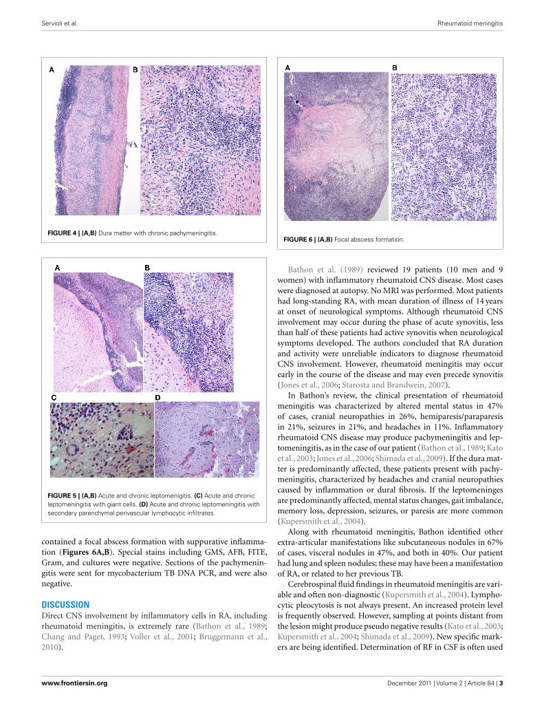
A brain/meningeal biopsy was done which showed a chronicinflammatory process with numerous plasma cells with Rus-sell bodies and granulomatous reaction of the leptomeningesand dura. Findings were consistent with rheumatoid pachyand leptomeningitis (Figures 4A,B). In addition, there was anacute and chronic leptomeningitis with focal giant cell reac-tion with secondary parenchymal perivascular lymphocytic infil-trates with reactive gliosis (Figures 5A–D). The leptomeninges
www.frontiersin.org December 2011 | Volume 2 | Article 84 | 1

Servioli et al. Rheumatoid meningitis
FIGURE 1 | Extra-axial high signal intensity overlying the right parietal–occipital region with mild gyriform contrast enhancement.
FIGURE 2 | Hyperintense signal abnormalities overlying the right parietal and posterior frontal lobes with contrast enhancement, compatible with
dural and leptomeningeal thickening.
FIGURE 3 | Increased right-sided leptomeningeal and dural thickening with worsening of leptomeningeal enhancement over the left posterior parietal
and occipital lobes.
Frontiers in Neurology | Neurology Education December 2011 | Volume 2 | Article 84 | 2
Servioli et al. Rheumatoid meningitis
FIGURE 4 | (A,B) Dura matter with chronic pachymeningitis.
FIGURE 5 | (A,B) Acute and chronic leptomenigitis. (C) Acute and chronicleptomeningitis with giant cells. (D) Acute and chronic leptomeningitis withsecondary parenchymal perivascular lymphocytic infiltrates.
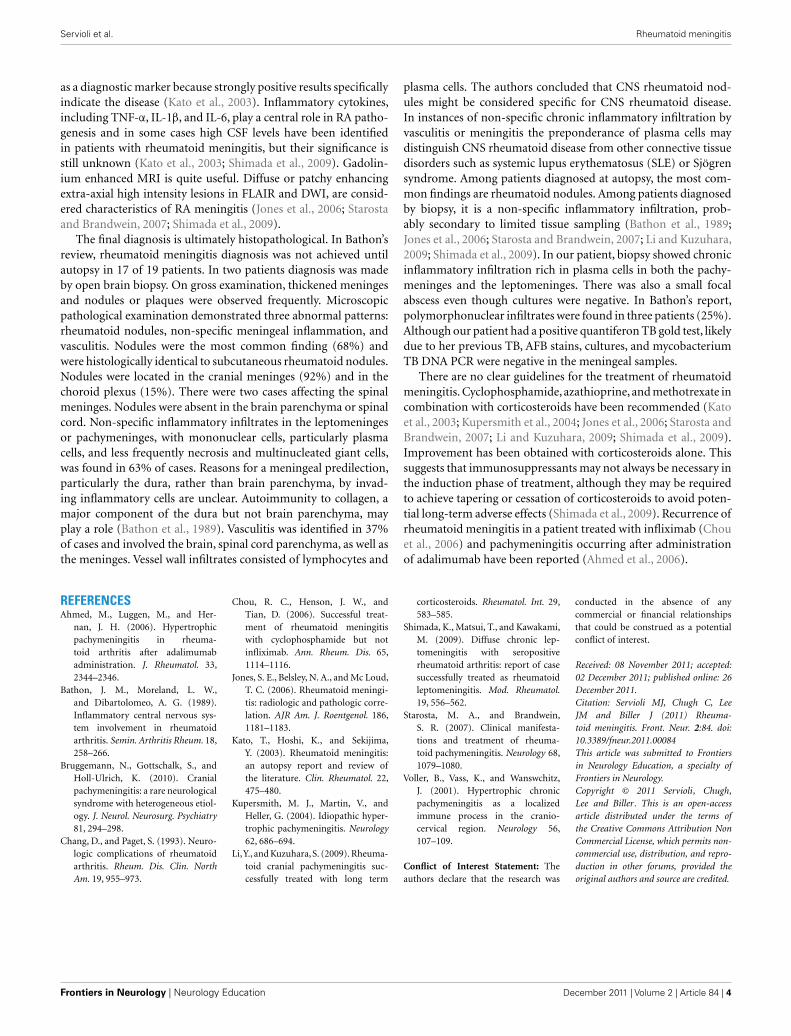
contained a focal abscess formation with suppurative inflamma-tion (Figures 6A,B). Special stains including GMS, AFB, FITE,Gram, and cultures were negative. Sections of the pachymenin-gitis were sent for mycobacterium TB DNA PCR, and were alsonegative.
DISCUSSIONDirect CNS involvement by inflammatory cells in RA, includingrheumatoid meningitis, is extremely rare (Bathon et al., 1989;Chang and Paget, 1993; Voller et al., 2001; Bruggemann et al.,2010).
FIGURE 6 | (A,B) Focal abscess formation.
Bathon et al. (1989) reviewed 19 patients (10 men and 9women) with inflammatory rheumatoid CNS disease. Most caseswere diagnosed at autopsy. No MRI was performed. Most patientshad long-standing RA, with mean duration of illness of 14 yearsat onset of neurological symptoms. Although rheumatoid CNSinvolvement may occur during the phase of acute synovitis, lessthan half of these patients had active synovitis when neurologicalsymptoms developed. The authors concluded that RA durationand activity were unreliable indicators to diagnose rheumatoidCNS involvement. However, rheumatoid meningitis may occurearly in the course of the disease and may even precede synovitis(Jones et al., 2006; Starosta and Brandwein, 2007).
In Bathon’s review, the clinical presentation of rheumatoidmeningitis was characterized by altered mental status in 47%of cases, cranial neuropathies in 26%, hemiparesis/paraparesisin 21%, seizures in 21%, and headaches in 11%. Inflammatoryrheumatoid CNS disease may produce pachymeningitis and lep-tomeningitis, as in the case of our patient (Bathon et al., 1989; Katoet al., 2003; Jones et al., 2006; Shimada et al., 2009). If the dura mat-ter is predominantly affected, these patients present with pachy-meningitis, characterized by headaches and cranial neuropathiescaused by inflammation or dural fibrosis. If the leptomeningesare predominantly affected, mental status changes, gait imbalance,memory loss, depression, seizures, or paresis are more common(Kupersmith et al., 2004).
Along with rheumatoid meningitis, Bathon identified otherextra-articular manifestations like subcutaneous nodules in 67%of cases, visceral nodules in 47%, and both in 40%. Our patienthad lung and spleen nodules; these may have been a manifestationof RA, or related to her previous TB.
Cerebrospinal fluid findings in rheumatoid meningitis are vari-able and often non-diagnostic (Kupersmith et al., 2004). Lympho-cytic pleocytosis is not always present. An increased protein levelis frequently observed. However, sampling at points distant fromthe lesion might produce pseudo negative results (Kato et al., 2003;Kupersmith et al., 2004; Shimada et al., 2009). New specific mark-ers are being identified. Determination of RF in CSF is often used
www.frontiersin.org December 2011 | Volume 2 | Article 84 | 3
Servioli et al. Rheumatoid meningitis
as a diagnostic marker because strongly positive results specificallyindicate the disease (Kato et al., 2003). Inflammatory cytokines,including TNF-α, IL-1β, and IL-6, play a central role in RA patho-genesis and in some cases high CSF levels have been identifiedin patients with rheumatoid meningitis, but their significance isstill unknown (Kato et al., 2003; Shimada et al., 2009). Gadolin-ium enhanced MRI is quite useful. Diffuse or patchy enhancingextra-axial high intensity lesions in FLAIR and DWI, are consid-ered characteristics of RA meningitis (Jones et al., 2006; Starostaand Brandwein, 2007; Shimada et al., 2009).
The final diagnosis is ultimately histopathological. In Bathon’sreview, rheumatoid meningitis diagnosis was not achieved untilautopsy in 17 of 19 patients. In two patients diagnosis was madeby open brain biopsy. On gross examination, thickened meningesand nodules or plaques were observed frequently. Microscopicpathological examination demonstrated three abnormal patterns:rheumatoid nodules, non-specific meningeal inflammation, andvasculitis. Nodules were the most common finding (68%) andwere histologically identical to subcutaneous rheumatoid nodules.Nodules were located in the cranial meninges (92%) and in thechoroid plexus (15%). There were two cases affecting the spinalmeninges. Nodules were absent in the brain parenchyma or spinalcord. Non-specific inflammatory infiltrates in the leptomeningesor pachymeninges, with mononuclear cells, particularly plasmacells, and less frequently necrosis and multinucleated giant cells,was found in 63% of cases. Reasons for a meningeal predilection,particularly the dura, rather than brain parenchyma, by invad-ing inflammatory cells are unclear. Autoimmunity to collagen, amajor component of the dura but not brain parenchyma, mayplay a role (Bathon et al., 1989). Vasculitis was identified in 37%of cases and involved the brain, spinal cord parenchyma, as well asthe meninges. Vessel wall infiltrates consisted of lymphocytes and
plasma cells. The authors concluded that CNS rheumatoid nod-ules might be considered specific for CNS rheumatoid disease.In instances of non-specific chronic inflammatory infiltration byvasculitis or meningitis the preponderance of plasma cells maydistinguish CNS rheumatoid disease from other connective tissuedisorders such as systemic lupus erythematosus (SLE) or Sjögrensyndrome. Among patients diagnosed at autopsy, the most com-mon findings are rheumatoid nodules. Among patients diagnosedby biopsy, it is a non-specific inflammatory infiltration, prob-ably secondary to limited tissue sampling (Bathon et al., 1989;Jones et al., 2006; Starosta and Brandwein, 2007; Li and Kuzuhara,2009; Shimada et al., 2009). In our patient, biopsy showed chronicinflammatory infiltration rich in plasma cells in both the pachy-meninges and the leptomeninges. There was also a small focalabscess even though cultures were negative. In Bathon’s report,polymorphonuclear infiltrates were found in three patients (25%).Although our patient had a positive quantiferon TB gold test, likelydue to her previous TB, AFB stains, cultures, and mycobacteriumTB DNA PCR were negative in the meningeal samples.
There are no clear guidelines for the treatment of rheumatoidmeningitis. Cyclophosphamide, azathioprine, and methotrexate incombination with corticosteroids have been recommended (Katoet al., 2003; Kupersmith et al., 2004; Jones et al., 2006; Starosta andBrandwein, 2007; Li and Kuzuhara, 2009; Shimada et al., 2009).Improvement has been obtained with corticosteroids alone. Thissuggests that immunosuppressants may not always be necessary inthe induction phase of treatment, although they may be requiredto achieve tapering or cessation of corticosteroids to avoid poten-tial long-term adverse effects (Shimada et al., 2009). Recurrence ofrheumatoid meningitis in a patient treated with infliximab (Chouet al., 2006) and pachymeningitis occurring after administrationof adalimumab have been reported (Ahmed et al., 2006).
REFERENCESAhmed, M., Luggen, M., and Her-
nan, J. H. (2006). Hypertrophicpachymeningitis in rheuma-toid arthritis after adalimumabadministration. J. Rheumatol. 33,2344–2346.
Bathon, J. M., Moreland, L. W.,and Dibartolomeo, A. G. (1989).Inflammatory central nervous sys-tem involvement in rheumatoidarthritis. Semin. Arthritis Rheum. 18,258–266.
Bruggemann, N., Gottschalk, S., andHoll-Ulrich, K. (2010). Cranialpachymeningitis: a rare neurologicalsyndrome with heterogeneous etiol-ogy. J. Neurol. Neurosurg. Psychiatry81, 294–298.
Chang, D., and Paget, S. (1993). Neuro-logic complications of rheumatoidarthritis. Rheum. Dis. Clin. NorthAm. 19, 955–973.
Chou, R. C., Henson, J. W., andTian, D. (2006). Successful treat-ment of rheumatoid meningitiswith cyclophosphamide but notinfliximab. Ann. Rheum. Dis. 65,1114–1116.
Jones, S. E., Belsley, N. A., and Mc Loud,T. C. (2006). Rheumatoid meningi-tis: radiologic and pathologic corre-lation. AJR Am. J. Roentgenol. 186,1181–1183.
Kato, T., Hoshi, K., and Sekijima,Y. (2003). Rheumatoid meningitis:an autopsy report and review ofthe literature. Clin. Rheumatol. 22,475–480.
Kupersmith, M. J., Martin, V., andHeller, G. (2004). Idiopathic hyper-trophic pachymeningitis. Neurology62, 686–694.
Li,Y., and Kuzuhara, S. (2009). Rheuma-toid cranial pachymeningitis suc-cessfully treated with long term
corticosteroids. Rheumatol. Int. 29,583–585.
Shimada, K., Matsui, T., and Kawakami,M. (2009). Diffuse chronic lep-tomeningitis with seropositiverheumatoid arthritis: report of casesuccessfully treated as rheumatoidleptomeningitis. Mod. Rheumatol.19, 556–562.
Starosta, M. A., and Brandwein,S. R. (2007). Clinical manifesta-tions and treatment of rheuma-toid pachymeningitis. Neurology 68,1079–1080.
Voller, B., Vass, K., and Wanswchitz,J. (2001). Hypertrophic chronicpachymeningitis as a localizedimmune process in the cranio-cervical region. Neurology 56,107–109.
Conflict of Interest Statement: Theauthors declare that the research was
conducted in the absence of anycommercial or financial relationshipsthat could be construed as a potentialconflict of interest.
Received: 08 November 2011; accepted:02 December 2011; published online: 26December 2011.Citation: Servioli MJ, Chugh C, LeeJM and Biller J (2011) Rheuma-toid meningitis. Front. Neur. 2:84. doi:10.3389/fneur.2011.00084This article was submitted to Frontiersin Neurology Education, a specialty ofFrontiers in Neurology.Copyright © 2011 Servioli, Chugh,Lee and Biller . This is an open-accessarticle distributed under the terms ofthe Creative Commons Attribution NonCommercial License, which permits non-commercial use, distribution, and repro-duction in other forums, provided theoriginal authors and source are credited.
Frontiers in Neurology | Neurology Education December 2011 | Volume 2 | Article 84 | 4