revista mexicana de neurocienciarevista mexicana de neurociencia editorial correspondence:...
TRANSCRIPT

EditorialIndividual health reflected in the collectivity 122Ildefonso Rodriguez-Leyva
External compression headache: A neglected headache 124Luis E. Fernández-Garza and Alejandro Marfil
Original Articles Clinical characteristics and surgical management of spinal cord tumors in non-Caucasian Hispanic children 127Fernando C. Ponce-de-León, José A. Choreño-Parra, Erika L. Cano-Camacho, Vicente González-Carranza, Samuel Torres-García, and Parménides Guadarrama-Ortíz
Factors influencing locomotor capacity of hemiparetic post-stroke patients 135Antonio Vinicius-Soares, Carla T. Juvêncio-de-Oliveira, Fernando L. Fischer-Eichinger, and Fabrício Noveletto
Prevalence of comorbidities in children with attention deficit/hyperactivity disorder: Measured and systematic review of care health studies 143Denise Medici, María M. Suarez-Varela, and Rubén Pérez-Elvira
Review ArticlesNeurological manifestations associated with SARS-CoV-2 – A review 150David A. Aguilar-Andino
The underrated nervous system involvement by COVID-19 158Hector R. Martinez, Jose A. Figueroa-Sanchez, Fernando Castilleja-Leal, Michel F. Martínez-Reséndez, and Ana S. Ferrigno
Update on the management of acute stroke. A practical clinical guide 163Juan J. Méndez-Gallardo, Beatriz Méndez, Vanessa Cano-Nigenda, Esmirna Y. Farington-Terrero, Diana Manrique-Otero, Enrique Castellanos-Pedroza, José G. Merino, and Antonio Arauz
PERMANYERwww.permanyer.com
VOLUME 21 - NUMBER 4 / July-August 2020 – ISSN: 1665-5044
eISSN: 2604-6180
www.revmexneurociencia.com
Revista Mexicana de
NeurocienciaPublicación oficial de la Academia Mexicana de Neurología A.C.
No
par
t o
f th
is p
ub
licat
ion
may
be
rep
rod
uce
d o
r p
ho
toco
pyi
ng
wit
ho
ut
the
pri
or
wri
tten
per
mis
sio
n o
f th
e p
ub
lish
er.
©
Per
man
yer
2020

122
Individual health reflected in the collectivityIldefonso Rodriguez-Leyva*Department of Neurology, Faculty of Medicine, Hospital Central “Dr. Ignacio Morones Prieto,” Universidad Autónoma de San Luis Potosí, San Luis Potosí, Mexico
Revista Mexicana de Neurociencia
EDITORIAL
Correspondence: *Ildefonso Rodriguez-Leyva
Faculty of Medicine
Hospital Central “Dr. Ignacio Morones Prieto”
Universidad Autónoma de San Luis Potosí
San Luis Potosí, Mexico
E-mail: [email protected]
Available online: 05-08-2020
Rev Mex Neuroci. 2020;21(4):122-123
www.revmexneurociencia.com
Date of reception: 15-03-2020
Date of acceptance: 17-03-2020
DOI: 10.24875/RMN.M20000078
It all started in December 2019, the authorities of the Chinese Health Services reported the presence of sev-eral cases of pneumonia in the city of Wuhan, China. The following month, the Shanghai Public Health Clin-ical Center and the School of Public Health, showed the complete genomic sequence of the 2019-nCoV warning of how this infection could affect not a few but thousands of individuals. The first reported case in the United States occurred in a young man who had visited Wuhan1.
The most interest aspect is that person-to-person transmission is demonstrated and that an asymptom-atic and healthy young woman with a positive real-time polymerase chain reaction test for COVID-19 who lives in Wuhan visited her family in the province of Anyang, China, and developed a cluster of familial pneumonia in an incubation period of 1-19 days2.
Most of the cases have occurred in adult people 30-79 years (87%), and only 1% in people younger than 9 years of age. Furthermore, being in a city, it extended in just 30 days to all of China, February 18, 2020, re-ported 72,528 confirmed cases of COVID-19 (99% of the global total) and 1870 deaths (99.8% of the global total), with more than 3000 cases in health personnel. The sanitary measures were isolation, quarantine, so-cial distancing, and community contention. By February 20, 2020, 1073 cases had already been reported out-side of China3.
On February 7, 2020, the WHO Scientific and Tech-nical Advisory Group for Infectious Hazards issued a series of recommendations to try to stop the spread of COVID-19. They were emphasizing that although the transmission was possibly initially from animals (bat) to humans, it is clear that the spread is occurring by hu-man-human transmission. Insisting that although the fatality is challenging to determine, the lung affection mainly like pneumonia, is the most critical (1-2%), although lower than for severe acute respiratory syn-drome (10%). Transmission is possibly oral, so con-glomerations should be avoided. Moreover, it also seems that the most affected are older adults with comorbidities. The WHO insists on the advisability of suspending public activities (especially with high atten-dance) and also closing schools, working remotely, making phone calls, and using telemedicine. Besides, they suggest maintaining and having the ventilatory support equipment, oxygen, and extracorporeal mem-brane oxygenation equipment to be ready for as soon as it is necessary to use4.
In addition to the above, we must consider that just as animal-human and then human-human transmission occurred initially, human-animal (pets) transmission may also be feasible and thus further spread the problem5.
Despite the measures taken by the Chinese govern-ment and the warnings issued by the WHO, cases of
1665-5044/ © 2020 Academia Mexicana de Neurología A.C. Published by Permanyer. This is an open access article under the CC BY-NC-ND license (http://creativecommons.org/licenses/by-nc-nd/4.0/).
No
par
t o
f th
is p
ub
licat
ion
may
be
rep
rod
uce
d o
r p
ho
toco
pyi
ng
wit
ho
ut
the
pri
or
wri
tten
per
mis
sio
n o
f th
e p
ub
lish
er.
©
Per
man
yer
2020

123
I. Rodríguez-Leyva: Pandemic-time individual health
pneumonia began to appear in the Milan metropolitan area, which ended in a health problem that finally iso-lated the country entirely6.
In an “invisible” spread, the problem grew and spread from Milan to Lyon, France, and Alitalia passengers arriving in Mauritius were quarantined. Some countries outside Europe began to reject passengers from Italy. Four people in the UK were confirmed positive for COVID-19 and had been on the Diamond Princess cruise. South Korea reported almost 1000 affected, and the problem continued to grow7.
The problem in Italy began on January 31, 2020, with two Chinese visitors who were staying in a central hotel in Rome and had landed at Milan’s Malpensa Airport on January 23. Just a month later, the Prime Minister of Italy declared the situation to be a national emergency8.
As this is being written, COVID-19 Global Cases by the Center for Systems Science and Engineering at Johns Hopkins University reports 179,029 confirmed cases of COVID-19, 7057 deaths, and only 78,073 fully recovered9. The spread includes an increasing number of cases, from highest to lowest, in China, Italy, Iran, Spain, South Korea, Germany, France, the USA, the UK, and Holland.
Although this is only a partial view of what happened in a pandemic that has grown exponentially, it shows us that measures as old and simple as social isolation, quarantine, public containment, and keeping our hos-pitals ready for who requires them are vital measures.
Measures that are needed to avoid problems that could massively affect a high percentage of the population.
It is evident in this brief story that the health of an individual in an infectious-contagious problem can af-fect an entire community. The preventive health mea-sures taken by countries such as France, Spain, Italy, the United States, and, of course, China itself should be an example to all the nations. Therefore, we must apply what to do and what to avoid; apparent health does not seem to be enough, although older adults with comorbidities are the most susceptible. Let us hope that our health systems manage to be sufficient and adequate for a problem that can potentially affect all the populations of Latin America.
References 1. Paules CI, Marston HD, Fauci AS. Coronavirus infections-more than just
the common cold. JAMA. 2020;323:707-8. 2. Bai Y, Yao L, Wei T, Tian F, Jin DY, Chen L, et al. Presumed asympto-
matic carrier transmission of COVID-19. JAMA. February 21, 2020 [Epub ahead of print].
3. Wu Z, McGoogan JM. Characteristics of and important lessons from the coronavirus disease 2019 (COVID-19) outbreak in China: summary of a report of 72 314 cases from the Chinese center for disease control and prevention. JAMA. February 24, 2020 [Epub ahead of print].
4. Heymann DL, Shindo N, WHO Scientific and Technical Advisory Group for Infectious Hazards. COVID-19: what is next for public health? Lancet. 2020;395:542-5.
5. Bonilla-DK BB, Rabaan A, Gomez WE, Rodriguez-AJ RR. Una nueva zoonosis viral de preocupación global: COVID-19, enfermedad por coro-navirus 2019. Iatreia. 2020;33:107-10.
6. Spina S, Marrazzo F, Migliari M, Stucchi R, Sforza A, Fumagalli R. The response of Milan’s emergency medical system to the COVID-19 out-break in Italy. Lancet. 2020;395:e49-50.
7. Day M. Covid-19: surge in cases in Italy and South Korea makes pan-demic look more likely. BMJ. 2020;368:m751.
8. Carinci F. Covid-19: preparedness, decentralisation, and the hunt for patient zero. BMJ. 2020;368:m799.
9. Available from: https://coronavirus.jhu.edu/map.html.
No
par
t o
f th
is p
ub
licat
ion
may
be
rep
rod
uce
d o
r p
ho
toco
pyi
ng
wit
ho
ut
the
pri
or
wri
tten
per
mis
sio
n o
f th
e p
ub
lish
er.
©
Per
man
yer
2020
124
External compression headache: A neglected headacheLuis E. Fernández-Garza and Alejandro Marfil*Department of Neurology, Hospital Universitario “Dr. José Eleuterio González,” Universidad Autónoma de Nuevo León, Monterrey, Nuevo León, México
Revista Mexicana de Neurociencia
EDITORIAL
Correspondence: *Alejandro Marfil
E-mail: [email protected]
Available online: 05-08-2020
Rev Mex Neuroci. 2020;21(4):124-126
www.revmexneurociencia.com
Date of reception: 05-05-2020
Date of acceptance: 02-06-2020
DOI: 10.24875/RMN.20000033
External compression headache (ECH) is a scarcely studied headache. There are different possible causes, such as mechanical factors, hypoxemia, hypercapnia, or associates stress, or could even be a sum of these factors that lead to the origin of this headache. ECH is provoked by donning objects with tight bands or straps around the head and has been reported with the use of hats, helmets, frontal lux devices, headsets, goggles, and N95 face masks. At present, we are experiencing the pandemic caused by the severe acute respiratory syndrome by a coronavirus 2 (SARS-CoV-2), whose disease name is CoV disease 2019 (COVID-19). Due to this, the protection measures were extended among health care workers and the general population, such as the use of personal protective equipment (PPE), that will, expectedly, rise the incidence of this headache.
Headache due to external compression was incorpo-rated into the international classification as exter-nal-pressure headache since the first edition of the International Classification of Headache Disorders1. According to the ICHD-3 in the heading 4: other primary headaches, to which the ECH belongs, the diagnosis is made by meeting the following criteria: at least two episodes of headache, brought on by and occurring within 60 min during continued pressure from some-thing outside your body, maximal pain at the site of compression, and resolving within 1 h after compres-sion is relieved2.
The pathogenesis of ECH is still uncertain. Depend-ing on the personal accessory used, there are different possible causes or could even be a sum of factors that
lead to the origin of the headache. It could involve mechanical factors, hypoxemia, hypercapnia, or asso-ciated stress. The continuous pressure or tractional force from personal accessories may lead to local tis-sue damage and exert an irritative effect on the under-lying superficial sensory nerves (trigeminal, occipital, and cervical nerves branches) that innerve the face, head, and cervical region3. Furthermore, the alveolar hypoventilation caused by prolonged use of face masks can lead to an increase in carbon dioxide with intra- and extracranial vasodilation (Fig. 1)4,5.
ECH is provoked by donning objects with tight bands or straps around the head and has been reported with the use of hats, helmets, frontal lux devices, headsets, goggles, and more recently N95 face masks6. Besides, it is usually an occupational disease, because in differ-ent professions, the cause of the headache is the equipment they use to work, such as helmets or hats in policemen, firefighters, construction workers, military personnel, athletes, and aviation pilots, and the PPE that use the health care workers7.
Usually, the pain of ECH is described as moderate in intensity, not impeding routine activities, frequently con-stant, consistent with the use of the accessories, of oppressive quality, more severe at the area where the object is pressing and tends to increase with the longer exposure to the compressing object. The pain is not associated with other symptoms and disappears shortly after removing the cause3. In patients with a pre-existing headache, there are two possibilities: one is that the prolonged external compression may lead to a more
1665-5044/ © 2020 Academia Mexicana de Neurología A.C. Published by Permanyer. This is an open access article under the CC BY-NC-ND license (http://creativecommons.org/licenses/by-nc-nd/4.0/).
No
par
t o
f th
is p
ub
licat
ion
may
be
rep
rod
uce
d o
r p
ho
toco
pyi
ng
wit
ho
ut
the
pri
or
wri
tten
per
mis
sio
n o
f th
e p
ub
lish
er.
©
Per
man
yer
2020

125
L.E. Fernández-Garza, A. Marfil: External compression headache
severe episode of the previous headache or that a de novo headache occurs with the previously described characteristics of ECH. For example, patients with pre-vious migraine have reported the triggering of a more severe, pulsatile, unilateral headache with nausea/vom-iting, photophobia, and phonophobia that did not end if the causal item was removed, requiring specific medi-cal treatment8.
Because these attacks disappear when the pres-sure is removed, seeking medical attention is often postponed so this headache is under-recognized. The current treatment is based on education to remove frequently and temporarily the headwear or traction that causes the pressure. Educational materials could be beneficial. The possibility of trying different styles and sizes of headgear may be useful for some of these exposed subjects, to get the most comfortable option. Moreover, the prevention would be skipping headwear (should it be possible) if you are predis-posed to ECH.
In recent years, we have witnessed epidemics involv-ing different strains of influenza, SARS, and Ebola, transmitted by direct or indirect contact and/or the re-spiratory route; therefore, the use of PPE is mandatory to all the personnel involved in the care of patients9. PPE includes N95 face masks or surgical masks, pro-tective eyewear such as goggles, medical gowns, and surgical gloves (sometimes double) for contact, and the use of the powered air-purifying respirators for all high-risk or aerosol-generating procedures. In daily medical
practice, it is required to wear PPE for prolonged periods. At present, we are experiencing the pandemic caused by the SARS-CoV-2, whose disease was bap-tized by the World Health Organization with the name of COVID-1910. Due to its high contagion rate, the wide-spread use of protective measures, such as PPE, will, expectedly, rise the incidence of this headache.
There are only three studies that demonstrate the association of the use of PPE with headache, facial pain, and/or ear lobe discomfort. The prevalence of PPE-associated headache varies from 37 to 81%, with a frequency of > 6 episodes/month in 33%. About 81-88% reported the onset with < 60 min of use and 88% reported the end of the episode with equipment removal. The pain has been bilateral in all the patients; 87% reported as oppressive followed by 11.7% as throbbing quality, and its intensity was mild in 72%. Even when 83% reported a negative impact on their work performance, only 7% took a sick leave because of headache, and 31-59% required the use of abortive analgesics. It is important to take into account the pres-ence of pre-existing headache, which was reported in 29-37%; among these patients, 91% reported an in-crease in the frequency and duration of episodes and poorer work performance. The two individual factors that increase the risk of developing PPE-associated headache are the existence of a previous headache, such as migraine and tension-type headache, and a longer period of usage, usually > 4 h/day8,11,12.
In future, it will be indispensable to prepare specifics protocols for situations like these, since in addition to the protection of hospital staff, it is necessary to think of their comfort as well. There are two approaches that could be exploited: one is directed to the PPE, by invit-ing manufacturers to look for alternatives, either in materials or shapes that reduce the possibility of dis-comfort to their users (one example was a fighter pilot that suffered from ECH from his helmet; the successful treatment was the switching of the non-adjustable one-piece helmet to a two-piece adjustable helmet, this measure disappeared the headache)13 and the second possible contribution would be to modify the infrastruc-ture and policies of hospitals, shortening working peri-ods, rotating periods in different hospital areas, where personnel can rest without the equipment and decrease the period of exposure.
Conflicts of interest
The authors declare no conflicts of interest.
Figure 1. A: pressure or tractional forces from the objects may lead to local tissue damage and exert an irritative effect on sensory nerves (green area: ophthalmic nerve, blue area: maxillary nerve, purple area: mandibular nerve, yellow area: cervical nerves, pink area: superficial cervical plexus). B: prolonged use of face masks may lead to an alveolar hypoventilation that will cause hypoxemia and hypercapnia.
BA
No
par
t o
f th
is p
ub
licat
ion
may
be
rep
rod
uce
d o
r p
ho
toco
pyi
ng
wit
ho
ut
the
pri
or
wri
tten
per
mis
sio
n o
f th
e p
ub
lish
er.
©
Per
man
yer
2020

126
Rev Mex Neuroci. 2020;21(4)
Acknowledgments
The authors received no financial support for the publication of this article.
Ethical disclosures
Protection of human and animal subjects. The authors declare that no experiments were performed on humans or animals for this study.
Confidentiality of data. The authors declare that no patient data appear in this article.
Right to privacy and informed consent. The au-thors declare that no patient data appear in this article.
References 1. Lance JW. Classification and diagnostic criteria for headache disorders,
cranial neuralgias and facial pain. Headache classification committee of the international headache society. Cephalalgia. 1988;8:1-96.
2. Headache classification committee of the international headache society (IHS) the international classification of headache disorders, 3rd edition. Cephalalgia. 2018;38:1-211.
3. Krymchantowski AV. Headaches due to external compression. Curr Pain Headache. 2010;14:321-4.
4. Kao TW, Huang KC, Huang YL. The physiological impact of wearing an N95 mask during hemodialysis as a precaution against SARS in patients with end-stage renal disease. J Formos Med Assoc. 2004;103:624-8.
5. Hoiland RL, Tymko MM, Bain AR, Wildfong KW, Monteleone B, Ainslie PN. Carbon dioxide-mediated vasomotion of extra-cranial cerebral arteries in humans: a role for prostaglandins? J Physiol. 2016;12:3463-81.
6. Pestronk A, Pestronk S. Goggle migraine. N Engl J Med. 1983;308:226-7. 7. Krymchantowski A, Barbosa JS, Cvaigman M, Lorenzatto W, Silva MT.
Helmet-related, external compression headache among police officers in Rio de Janeiro. Medgenmed. 2004;6:45.
8. Ong JJ, Bharatendu C, Goh Y, Tang JZ, Sooi KW, Tan YL, et al. Hea-daches associated with personal protective equipment-a cross-sectional study among frontline healthcare workers during COVID-19. Headache. 2020;60:864-77.
9. Andersen BM. Personal protective equipment. In: Prevention and Control of Infections in Hospitals: practice and Theory. Berlin, Germany: Springer; 2019. p. 1061-4.
10. Fernández-Garza, LE, Marfil A. Neurological aspects that should not be forgotten during the COVID-19 pandemic. Int J Med Health. 2020;3:1-3.
11. Rebmann T, Carrico R, Wang J. Physiologic and other effects and com-pliance with long-term respirator use among medical intensive care unit nurses. Am J Infect Control. 2013;41:1218-23.
12. Lim E, Seet R, Lee KH, Wilder-Smith E, Chuah B, Ong B. Headaches and the N95 face-mask amongst healthcare providers. Acta Neurol Scand. 2006;113:199-202.
13. Kharbanda GC, Ganjoo AR, Chadha WC. A strange case of headache in a fighter aircrew: a case study. Indian J Aerospace Med. 2008;52:20-5.
No
par
t o
f th
is p
ub
licat
ion
may
be
rep
rod
uce
d o
r p
ho
toco
pyi
ng
wit
ho
ut
the
pri
or
wri
tten
per
mis
sio
n o
f th
e p
ub
lish
er.
©
Per
man
yer
2020

127
Clinical characteristics and surgical management of spinal cord tumors in non-Caucasian Hispanic childrenFernando C. Ponce-de-León1, José A. Choreño-Parra2, Erika L. Cano-Camacho1, Vicente González-Carranza1, Samuel Torres-García1, and Parménides Guadarrama-Ortíz3*1Departament of Neurosurgery, Hospital Infantil de México Federico Gómez, Mexico City, Mexico; 2Departament of Clinical Research, Centro Especializado en Neurocirugía y Neurociencias México, Mexico City, Mexico; 3Departament of Neurosurgery, Centro Especializado en Neurocirugía y Neurociencias México, Mexico City, Mexico
Revista Mexicana de Neurociencia
ORIGINAL ARTICLE
Abstract
Objective: Little literature exists about spinal cord tumors in Hispanic children. Methods: We conducted a retrospective review in 45 Mexican children presenting with spinal cord tumors between 1985 and 2015. Results: We observed a higher incidence of spinal cord tumors in males (62.22%). The mean age at diagnosis was 8.75 years. Tumors were more frequent-ly observed among school-age children (42.22%). Motor deficit was the most common clinical manifestation (97.77%). Most tumors were intramedullary. Astrocytoma was the most frequent histological subtype. Laminotomy with laminoplasty was the main operative procedure performed in our study. Total resection of the tumors was achieved in 20% of the cases. Post-sur-gical complications were observed in 44% of the cases. Conclusions: Pediatric patients with spinal cord tumors can receive surgical management with an acceptable low surgical morbidity. The clinical phenotype observed in our population has certain similitudes with respect to what it is described in Caucasians.
Key words: Spinal cord. Spinal cord tumors. Intramedullary tumors. Extradural tumors.
Características clínicas y manejo quirúrgico de tumores raquimedulares en niños hispanos
Resumen
Objetivo: Existe poca literatura acerca de tumores raquimedulares en niños hispanos. Métodos: Realizamos una revisión retrospectiva de 45 niños mexicanos que presentaron tumores raquimedulares entre 1985 y 2015. Resultados: Observamos mayor incidencia de tumores raquimedulares en varones (62.22%). La edad promedio al momento del diagnóstico fue de 8.75 años. Los tumores fueron observados con mayor frecuencia en niños en edad escolar (42.22%). El déficit motor fue la manifestación clínica más frecuente (97.77%). La mayoría de los tumores fueron intramedulares. El astrocitoma fue el subtipo
Correspondence: *Parménides Guadarrama-Ortiz
Departament of Neurosurgery
Centro Especializado en Neurocirugía y
Neurociencias México
Mexico City, Mexico
E-mail: investigació[email protected]
Available online: 05-08-2020
Rev Mex Neuroci. 2020;21(4):127-134
www.revmexneurociencia.com
Date of reception: 19-10-2019
Date of acceptance: 20-01-2020
DOI: 10.24875/RMN.20000140
1665-5044/© 2020. Academia Mexicana de Neurología A.C. Published by Permanyer. This is an open access article under the CC BY-NC-ND license (http://creativecommons.org/licenses/by-nc-nd/4.0/).
No
par
t o
f th
is p
ub
licat
ion
may
be
rep
rod
uce
d o
r p
ho
toco
pyi
ng
wit
ho
ut
the
pri
or
wri
tten
per
mis
sio
n o
f th
e p
ub
lish
er.
©
Per
man
yer
2020

128
Rev Mex Neuroci. 2020;21(4)
Introduction
Spinal cord tumors are rare in adults and occur at a considerably lower frequency in children. The propor-tion between spinal cord and intracranial tumors de-scribed in the pediatric population varies from 1:4.8 to 1:20, depending on the series reviewed1. Incidence of spinal cord tumors in high-income countries such as the United States is 1 case/100,000 children2, mainly affect-ing scholars and adolescents3,4. These tumors can arise from the spinal cord or from the adjacent structures, and according to their location, they can be classified as intra- or extradural. Moreover, intradural tumors are di-vided into intramedullary and extramedullary5.
The current literature describes that two-thirds of the total spinal cord neoplasia occurring in children are extradural. These include bone tumors, tumors of the epidural space, and extraspinal tumors that invade such space. On the other hand, 35% of the spinal cord tumors in pediatric patients are intramedullary, repre-senting 4-6% of the total central nervous system neo-plasia6. Astrocytoma, ganglioma, and ependymoma are the histological subtypes most frequently observed in the intramedullary location2. Such tumors often lo-calize at cervical and thoracic levels of the spinal cord in almost 50% of the cases, being less frequently observed at lumbar segments7,8. Finally, intradural ex-tramedullary neoplasia is rare and most of them are leptomeningeal metastasis from brain tumors9.
Most of what it is currently known about epidemiology and clinical characteristics of spinal cord tumors in chil-dren comes from series in Caucasians. However, few studies have described the clinical behavior of these tumors in other populations. In developing countries like Mexico, there are only few reports of spinal cord tumor in children10-12, some of which were published before the advent and availability of novel brain imaging tools cur-rently used for the diagnostic and surgical approach of these neoplasia10,11. Therefore, it is of major relevance to have updated information about the frequency, clini-cal behavior, diagnostic tools, surgical strategies, and post-operative clinical outcomes of spinal cord tu-mors in our children population. Here, we report our
experience in the management of spinal cord tumors by reviewing the clinical registries of children that attended and were regularly followed at the Department of Neu-rosurgery of the Pediatric Hospital of Mexico, a third-lev-el National Reference Center in Mexico City. We believe that our clinical description contributes to the knowledge of spinal cord tumors in Hispanic children and provides valuable data for future studies aimed to compare the clinical behavior of spinal cord tumors among pediatrics with different ethnic backgrounds.
Methods
We conducted a retrospective review of the clinical and radiological database of the Pediatric Hospital of Mexico “Federico Gomez,” looking for clinical cases of children with spinal cord tumors that attended and were regularly followed at our center during the period from 1985 to 2015. Demographic, clinical, and imaging data from participants were retrieved from the database of our hospital. The collected information included relevant neurological manifestations, anatomical location and histological subtype of the tumors, category of imaging tools used for the diagnosis, surgical approaches em-ployed for tumor resection, and post-operative clinical outcomes. Descriptive statistics were used to clinically characterize the study population. Frequencies and pro-portions were calculated for categorical data. Means, medians, and standard deviations were used for contin-uous data. Calculations for descriptive statistics were performed using GraphPad Prism v5 (La Jolla, CA, USA). The study was approved by the Ethics Committee of our institution and was conducted with strict adher-ence to the Official Mexican Law NOM-012-SSA3-2012 that establishes the criteria for the execution of health research projects in humans.
Results
Participants’ characteristics
A total of 45 children with spinal cord tumors were included in the study. Their mean age at diagnosis was
histológico más común. La laminotomía con laminoplastía fue el procedimiento quirúrgico realizado con mayor frecuencia en nuestro estudio. La resección tumoral total se logro en 20% de los casos. Las complicaciones quirúrgicas ocurrieron en 44% de los pacientes. Conclusiones: los pacientes pediátricos con tumores raquimedulares pueden recibir tratamiento quirúrgico con una baja morbilidad. El fenotipo clínico observado en nuestra población tiene ciertas similitudes respecto a lo descrito en caucásicos.
Palabras clave: Médula espinal. Tumores raquimedulares. Tumores intramedulares. Tumores extradurales.
No
par
t o
f th
is p
ub
licat
ion
may
be
rep
rod
uce
d o
r p
ho
toco
pyi
ng
wit
ho
ut
the
pri
or
wri
tten
per
mis
sio
n o
f th
e p
ub
lish
er.
©
Per
man
yer
2020

129
F.C. Ponce-de-León, et al.: Spinal cord tumors in Hispanic children
8.75 years (range 5 months-17 years). The most ancient case occurred 31 years before the conduction of the current study. From these, 28 patients were male (62.22%) and 17 female (37.77%), with a male/female ratio of 1.64:1. The age group with the highest inci-dence of spinal cord tumors was composed of school-age children with 19 cases (42.22%), followed by adolescents (13 cases, 28.88%), pre-scholars (9 cases, 20%), and infants (4 cases, 8.88%). All the patients were born in Mexico and their parents referred them-selves as belonging to the Mexican Mestizo race. The clinical and demographic characteristics of participants are summarized in Table 1.
Tumors location and histological subtype
Intramedullary tumors were the most frequent lesions affecting our population, representing the 44.44% of the cases (20 patients) followed by extradural and
intradural extramedullary tumors with 18 (40%) and 7 (15.55%) cases, respectively. In addition, six cases of intramedullary lesions were holocordal neoplasia (13.33%). All cases received histopathological diagno-sis. Independently of the tumors’ location, astrocytoma was the most frequent histological subtype, followed by primitive neuroectodermal tumors (PNETs), ependymo-ma, meningioma, and metastases. The frequency and anatomical localization of the different histological sub-types of spinal cord tumors observed in our series are shown in Table 2.
Neurological findings
The most common clinical findings observed in order of frequency were motor deficit (97.77%), pain (86.66%), urinary incontinence (64.44%), abnormal gait (35.55%), and fecal incontinence (22.22%) (Table 1). Further-more, 53.33% of patients had alterations of the statics
Table 1. Clinical characteristics of Hispanic children with spinal cord tumors
Variable n = 45
Age at onset, mean (range) 8.75 (0.5‑17)
GenderMale, n (%)Female, n (%)
28 (62.22)17 (37.77)
Male/female ratio 1.64:1
Age groupInfants, n (%)Pre‑scholars, n (%)School‑age children, n (%)Adolescents, n (%)
4 (8.88)9 (20)
19 (42.22)13 (28.88)
Clinical findings before surgical managementMotor deficit, n (%)Pain, n (%)Pain referred to the corresponding dermatomes, n (%)Radicular pain, n (%)Hypoesthesia, n (%)Dysesthesia, n (%)Paresthesia, n (%)Urinary incontinency, n (%)Abnormal gait, n (%)Fecal incontinence, n (%)Scoliosis, n (%)Kyphoscoliosis, n (%)Falls, n (%)Cervical stiffness, n (%)Muscle contractures, n (%)Hyperreflexia, n (%)Clonus, n (%)Atrophy of muscles of the hands, n (%)Hydrocephalus, n (%)Headache, n (%)Seizures, n (%)
44 (97.77)39 (86.66)
36 (80)
3 (6.66)38 (84.44)25 (55.55)13 (28.88)29 (64.44)16 (35.55)10 (22.22)
18 (40)6 (13.33)6 (13.33)6 (13.33)6 (13.33)6 (13.33)6 (13.33)6 (13.33)4 (8.88)4 (8.88)4 (8.88)
Table 2. Frequency and distribution of spinal cord tumors according to their anatomical location and histological subtype
Localization/histological subtype
Frequency % of tumors at specific
localization
% from total
Intramedullary 20 ‑‑ 44.44
Astrocytoma 14 70 31.11
Ependymomas 5 25 11.11
Ganglioma 1 5 2.22
Extradural 18 ‑‑ 40
PNETs 6 33.33 13.33
Metastases 4 22.22 8.88
Sarcomas 3 16.66 6.66
Neuroblastoma 1 5.55 2.22
Lymphomas 1 5.55 2.22
Other not specified 3 16.66 6.66
Intradural extramedullary
7 ‑‑ 15.55
Meningiomas 4 57.14 8.88
Ependymomas 1 14.28 2.22
Schwannomas 1 14.28 2.22
Dermoid cysts 1 14.28 2.22
PNETs: primary neuroectodermal tumors.
No
par
t o
f th
is p
ub
licat
ion
may
be
rep
rod
uce
d o
r p
ho
toco
pyi
ng
wit
ho
ut
the
pri
or
wri
tten
per
mis
sio
n o
f th
e p
ub
lish
er.
©
Per
man
yer
2020

130
Rev Mex Neuroci. 2020;21(4)
of the spine; 18 cases presented scoliosis (40%) and 6 had kyphoscoliosis (13.33%).
Although motor deficit was the main neurological finding, this sign occurred as the first clinical manifes-tation only in 22 cases (48.88%). The mean time since the presentation of the motor deficit and surgery was 4.5 months. This deficit was observed in all cases of astrocytoma and intramedullary ependymoma. Other neurological findings observed in our series, especially among patients with cervical-thoracic holocordal tu-mors included falls, tabetic gait, cervical stiffness, deltoid, supraspinatus, biceps and triceps muscle con-tractures, as well as hyperreflexia, clonus, and atrophy of intrinsic muscles of the hands.
Regarding pain, this was referred along the der-matomes corresponding to the anatomical location of the tumors in 80% of patients. This symptom was most common in patients with extradural tumors. Three pa-tients had radicular pain (6.66%). Among children affect-ed by cervical tumors, we observed neck pain that was exacerbated with inclination of the head to the contra-lateral side of the lesion. Sensory deficits manifested as hypoesthesia in 38 cases, dysesthesia in 25 patients, and paresthesia in 13 cases. These sensory alterations always occurred in the dermatomes corresponding to the location of the tumors. Urinary and fecal incontinen-cy occurred in patients with neoplasia of the lumbosa-cral region or holocordal tumors. Hydrocephalus was
detected in 4 cases (8.88%) and was accompanied by headache and seizures secondary to intracranial hyper-tension. Neurological features observed in our popula-tion are summarized in Table 1.
Diagnostic approach
From the 45 patients included in the current study, only 13 presented images of brain computerized axial tomog-raphy (computed tomography [CT]) scan and magnetic resonance imaging (MRI) at their first medical appoint-ment. Overall, 36 patients underwent to CT scan (80%), 38 to MRI (84.44%), 27 to electromyography (60%), and 31 to evoked potentials (68.88%). In 29 cases, urody-namic studies were performed (64.44%) including 18 cases with intramedullary tumors. Furthermore, ano-rectal manometry was performed in 19 patients (42.22%), 11 of them with intramedullary lesions.
Surgical management
Overall, 10 patients were subjected to surgery as their only management strategy, whereas the rest re-ceived multidisciplinary oncological management, which included surgery, radiotherapy, and chemother-apy (Fig. 1A). A total of 50 surgical procedures were performed in the 45 cases included in the study (Table 3 and Fig. 1B). Laminotomy was the most
Figure 1. Treatment approaches for pediatric patients with spinal cord tumors. A: Treatment modalities for children with spinal cord tumors. B: Surgical procedures performed for the resection of spinal cord tumors. C: Representative figure of the operative field provided by posterior laminotomy. D: Laminectomy. E: Hemi‑laminectomy.
A B
C D E
No
par
t o
f th
is p
ub
licat
ion
may
be
rep
rod
uce
d o
r p
ho
toco
pyi
ng
wit
ho
ut
the
pri
or
wri
tten
per
mis
sio
n o
f th
e p
ub
lish
er.
©
Per
man
yer
2020
131
F.C. Ponce-de-León, et al.: Spinal cord tumors in Hispanic children
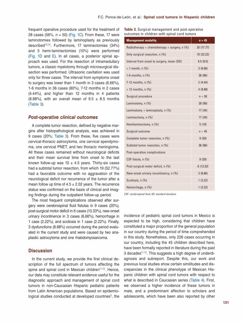
frequent operative procedure used for the treatment of 28 cases (56%, n = 50) (Fig. 1C). From these, 17 were laminotomies followed by laminoplasty as previously described13,14. Furthermore, 17 laminectomies (34%) and 5 hemi-laminectomies (10%) were performed (Fig. 1D and E). In all cases, a posterior spinal ap-proach was used. For the resection of intramedullary tumors, a classic myelotomy through microsurgical dis-section was performed. Ultrasonic cavitation was used only for three cases. The interval from symptoms onset to surgery was lower than 1 month in 3 cases (6.66%), 1-6 months in 36 cases (80%), 7-12 months in 2 cases (4.44%), and higher than 12 months in 4 patients (8.88%), with an overall mean of 9.5 ± 8.5 months (Table 3).
Post-operative clinical outcomes
A complete tumor resection, defined by negative mar-gins after histopathological analysis, was achieved in 9 cases (20%; Table 3). From these, five cases were cervical-thoracic astrocytoma, one cervical ependymo-ma, one cervical PNET, and two thoracic meningioma. All these cases remained without neurological deficits and their mean survival time from onset to the last known follow-up was 10 ± 4.5 years. Thirty-six cases had a subtotal tumor resection, from which 19 (52.77%) had a favorable outcome with no aggravation of the neurological deficit nor recurrence of the tumor after a mean follow up time of 4.5 ± 2.02 years. The recurrence status was confirmed on the basis of clinical and imag-ing findings during the outpatient follow-up period.
The most frequent complications observed after sur-gery were cerebrospinal fluid fistulas in 9 cases (20%), post-surgical motor deficit in 6 cases (13.33%), new-onset urinary incontinence in 3 cases (6.66%), hemorrhage in 1 case (2.22%), and scoliosis in 1 case (2.22%). Finally, 3 dysfunctions (8.88%) occurred during the period evalu-ated in the current study and were caused by two ana-plastic astrocytoma and one rhabdomyosarcoma.
Discussion
In the current study, we provide the first clinical de-scription of the full spectrum of tumors affecting the spine and spinal cord in Mexican children11,12. Hence, our data may constitute relevant evidence useful for the diagnostic approach and management of spinal cord tumors in non-Caucasian Hispanic pediatric patients from Latin American populations. Based on epidemio-logical studies conducted at developed countries2, the
incidence of pediatric spinal cord tumors in Mexico is expected to be high, considering that children have constituted a major proportion of the general population in our country during the period of time comprehended in this study. Nonetheless, only 226 cases occurring in our country, including the 45 children described here, have been formally reported in literature during the past 3 decades11,12. This suggests a high degree of underdi-agnosis and subreport. Despite this, our work and previous local studies show certain similitudes and dis-crepancies in the clinical phenotype of Mexican His-panic children with spinal cord tumors with respect to what is described in Caucasian series (Table 4). First, we observed a higher incidence of these tumors in male, and a predominant affection to scholars and adolescents, which have been also reported by other
Table 3. Surgical management and post‑operative outcomes in children with spinal cord tumors
Management modality n = 45
Radiotherapy + chemotherapy + surgery, n (%) 35 (77.77)
Only surgical resection, n (%) 10 (22.22)
Interval from onset to surgery, mean (SD) 9.5 (8.5)
< 1 month, n (%) 3 (6.66)
1‑6 months, n (%) 36 (80)
7‑12 months, n (%) 2 (4.44)
> 12 months, n (%) 4 (8.88)
Surgical procedure n = 50
Laminotomy, n (%) 28 (56)
Laminotomy + laminoplasty, n (%) 17 (34)
Laminectomy, n (%) 17 (34)
Hemilaminectomy, n (%) 5 (10)
Surgical outcome n = 45
Complete tumor resection, n (%) 9 (20)
Subtotal tumor resection, n (%) 36 (80)
Post‑operative complications
CSF fistula, n (%) 9 (20)
Post‑surgical motor deficit, n (%) 6 (13.33)
New‑onset urinary incontinency, n (%) 3 (6.66)
Scoliosis, n (%) 1 (2.22)
Hemorrhage, n (%) 1 (2.22)
CSF: cerebrospinal fluid; SD: standard deviation.
No
par
t o
f th
is p
ub
licat
ion
may
be
rep
rod
uce
d o
r p
ho
toco
pyi
ng
wit
ho
ut
the
pri
or
wri
tten
per
mis
sio
n o
f th
e p
ub
lish
er.
©
Per
man
yer
2020

132
Rev Mex Neuroci. 2020;21(4)
researchers2,10-12,15,16. Second, considering the experience of our department in the management of brain neoplasms in children (810 cases in 36 years)17, the ratio between spinal cord tumors and intracranial neoplasia derived from the present work (45 cases in 31 years) is 1:15.5 and 1:17.6 if we compare our series with the 511 brain tumors observed during 20 years in the Spanish Hospital of Mexico18. These data coincide with the ratio observed in other studies in Caucasians1; however, this information may not be accurate due to the possible local degree of underdiagnosis mentioned above.
On the other hand, we observed a higher incidence of intramedullary lesions followed by those of extradural location in our population. In this regard, there is no consensus about the anatomical area most predomi-nantly affected by these tumors since some studies show a higher frequency of extradural lesions16,19,20, while others describe a higher incidence of intramed-ullary tumors15,21-26. It is probable that factors specific to each population explain these discrepancies. Our data also confirm the previous descriptions about the most common histological type of spinal cord tumors observed in children2,9, as astrocytoma was the most incident tumor in our series regardless of their location. Furthermore, a remarkable finding derived from this work is the high frequency of extradural PNET type neoplasms occurring in our patients, which has been also reported in other Mexican series12. These tumors are conformed by undifferentiated cells, with varying degrees of pleomorphism and a slight tendency to ac-quire neuroectodermal characteristics27. Their inci-dence is extremely low, and some studies show that they represent less than 1% of all neoplasms that affect the spinal cord28. The average age at which they have
been previously diagnosed is 24 years and their exact epidemiology in children remains unknown29. Thus, our data suggest that the incidence of spinal cord PNET in the pediatric population may be higher than previously thought.
The identification of spinal cord tumors in children is a major diagnostic challenge due to their rarity and unspecific clinical features. In this regard, our data, as well as previous reports30, show that a high proportion of pediatric patients with spinal cord tumors present motor deficits as their initial manifestation. Further-more, our findings suggest that the presence of pain distributed along with a dermatome, radicular pain, hypoesthesia, paresthesia, dysesthesia, urinary and fecal incontinence, as well as gait disorders in a child otherwise healthy, should suggest the presence of a spinal cord tumor. Regarding surgical management of these lesions, we found that laminotomy by posterior approach is a safe and not deforming procedure, as previously reported13,14. Interestingly, we also observed that laminectomy caused kyphosis in only 1 of 17 pa-tients subjected to such procedure at least until the last known follow-up, which represents a lower fre-quency than reported by other researchers who de-scribed that two-thirds of the subjects undergoing lam-inectomy developed deformity of the spine31, especially when laxity of supporting tissues was induced by the adjuvant oncological treatment32. Moreover, the occur-rence of post-surgical complications in our study was low regardless of the operative approach, and fatal cases were not directly associated with the surgical resection of the tumors. Thus, our data show that pe-diatric patients with spinal cord tumors can receive surgical management with an acceptable low surgical morbidity.
Table 4. Clinical characteristics of spinal cord tumors in children from different regions
Race/origin Age at onset (years), mean
Predominant gender
Mortality (%) Tumor localization Histological subtype Reference
Hispanic 8.75 Male 8.88 Intramedullary Astrocytoma Current study
Hispanic ‑‑ Male ‑‑ Intradural extramedullary
Dermoid cyst 11
Hispanic 5.2 Male 21.7 ‑‑ Primary neuroectodermal tumors
12
Caucasian 6.6 Male ‑‑ Intramedullary Dermoid tumors, epidermoid tumors, and teratomas
15
Caucasian 8.5 Male 11.9 Intramedullary Low‑grade glioma 16
No
par
t o
f th
is p
ub
licat
ion
may
be
rep
rod
uce
d o
r p
ho
toco
pyi
ng
wit
ho
ut
the
pri
or
wri
tten
per
mis
sio
n o
f th
e p
ub
lish
er.
©
Per
man
yer
2020

133
F.C. Ponce-de-León, et al.: Spinal cord tumors in Hispanic children
Our study possesses all the limitations of a retrospective design in relation to the access and collection of the clin-ical information. In fact, we could not address the prognostic value of clinical variables and treatment approaches as most of the patients were not further followed after they reach the adulthood. Thus, our survival estimations only represent those patients that remain alive at the last known follow-up. In addition, we did not find objective quantitative evaluations of the post-operative neurological status of the patients within their clinical registries. Such information would have informed us about the effect of the surgical treatment on the neurological functionality. Finally, the fact that the current study was conducted at a single third-level medical center made us unable to perform a popula-tion-based comparison, which limited our ability to esti-mate the local incidence of spinal cord tumors in children and may restrict the representativeness of our data.
Conclusions
Pediatric patients with spinal cord tumors can receive surgical management with an acceptable low surgical morbidity. The clinical phenotype observed in our pop-ulation has certain similitudes concerning what it is de-scribed in caucasians. This study provides a valuable clinical description of spinal cord tumors that can help in future research in non-caucasian Hispanic children.
Conflicts of interest
The authors declare that they do not have any con-flicts of interest to report.
Funding
The current study did not receive any financial support.
Acknowledgments
To the medical and nursing staff of the Pediatric Hos-pital of Mexico “Federico Gomez.”
Ethical responsibilities
Protection of people and animals. The authors declare that the procedures followed in the current study were performed in agreement to the ethical standards of the responsible human experimentation committee, the World Medical Association, and the Declaration of Helsinki.
Confidentiality of the data. The authors declare that they have followed the protocols of their work cen-ter on the publication of patient data.
Right to privacy and informed consent. The au-thors have obtained the informed consent of the pa-tients and/or subjects referred to in the article. This document is held by the correspondence author.
References 1. Di Lorenzo N, Giuffre R, Fortuna A. Primary spinal neoplasms in child-
hood: analysis of 1234 published cases (including 56 personal cases) by pathology, sex, age and site. Differences from the situation in adults. Neurochirurgia (Stuttg). 1982;25:153-64.
2. Constantini S, Epstein FJ. Intraspinal tumors in infants and children. In: Youmans J, editor, Neurological Surgery. 4th ed, Vol. 4. Philadelphia (PA): WB Saundersp; 1996. p. 3123-33.
3. Schellinger KA, Propp JM, Villano JL, McCarthy BJ. Descriptive epide-miology of primary spinal cord tumors. J Neurooncol. 2008;87:173-9.
4. Duong LM, McCarthy BJ, McLendon RE, Dolecek TA, Kruchko C, Dou-glas LL, et al. Descriptive epidemiology of malignant and nonmalignant primary spinal cord, spinal meninges, and cauda equina tumors, United States, 2004-2007. Cancer. 2012;118:4220-7.
5. Van Goethem JW, van den Hauwe L, Ozsarlak O, De Schepper AM, Parizel PM. Spinal tumors. Eur J Radiol. 2004;50:159-76.
6. DeSousa AL, Kalsbeck JE, Mealey J Jr., Campbell RL, Hockey A. Intraspi-nal tumors in children. A review of 81 cases. J Neurosurg. 1979;51:437-45.
7. Epstein F, Ragheb J. Intramedullary tumors of the spinal cord. In: Cheek W, editor. Pediatric Neurosurgery: Surgery of the Developing Nervous System. Philadelphia (PA): WB Saunders; 1994. p. 446-57.
8. Brotchi J, Noterman J, Baleriaux D. Surgery of intramedullary spinal cord tumours. Acta Neurochir (Wien). 1992;116:176-8.
9. Rossi A, Gandolfo C, Morana G, Tortori-Donati P. Tumors of the spine in children. Neuroimaging Clin N Am. 2007;17:17-35.
10. Rueda Franco F, Monson de Souza MB, Takenaga Mesquida R. Intraspi-nal tumors in children. Review of 24 cases. Bol Med Hosp Infant Mex. 1975;32:1073-94.
11. Rueda-Franco F. Tumores intraspinales en niños. Revisión de 130 en-fermos. Cir Cir. 1990;57:143-52.
12. González-Sosa E, Anaya-Jara M, Marhx-Bracho A, Rueda-Franco F. Neoplasias espinales extradurales. Experiencia de 15 años en el Institu-to Nacional de Pediatría. Acta Pediatr Mex. 2009;30:216-9.
13. Raimondi AJ, Gutierrez FA, Di Rocco C. Laminotomy and total recons-truction of the posterior spinal arch for spinal canal surgery in childhood. J Neurosurg. 1976;45:555-60.
14. Rama B, Markakis E, Kolenda H, Jansen J. Reconstruction instead of resection: laminotomy and laminoplasty. Neurochirurgia (Stuttg). 1990;33 Suppl 1:36-9.
15. Wilson PE, Oleszek JL, Clayton GH. Pediatric spinal cord tumors and masses. J Spinal Cord Med. 2007;30 Suppl 1:S15-20.
16. Spacca B, Giordano F, Donati P, Genitori L. Spinal tumors in children: long-term retrospective evaluation of a series of 134 cases treated in a single unit of pediatric neurosurgery. Spine J. 2015;15:1949-55.
17. Chico-Ponce de León F, Castro-Sierra E, Perezpeña-Diazconti M, Gordillo–Domínguez LF, Santana–Montero BL, Rocha–Rivero LE, et al. Tumores intracraneanos del niño. Bol Med Hosp Inf Mex. 2006;63:367-81.
18. Anaya-Delgadillo G, de Juambelz-Cisneros PP, Fernández-Alvarado B, Pazos-Gómez F, Velasco-Torre A, Revuelta-Gutiérrez R. Prevalence of central nervous system tumours and histological identification in the operated patient: 20 years of experience. Cir Cir. 2016;84:447-53.
19. Kanos CC, Muhlbauer MS. Extramedullary, intradural and extradural spinal cord tumors. In: McLone DG, editor. Pediatric Neurosurgery. 4th ed. Philadelphia (PA): WB Saunders Company; 2001. p. 873-84.
20. Wetjen NM, Raffel C. Spinal extradural neoplasms and intradural extrame-dullary neoplasms. In: Albright AL, Pollack IF, Adelson PD, editor. Principles and Practice of Pediatric Neurosurgery. New York: Thieme; 2008. p. 694-705.
21. Nadkarni TD, Rekate HL. Pediatric intramedullary spinal cord tumors. Critical review of the literature. Childs Nerv Syst. 1999;15:17-28.
22. Albright AL. Pediatric intramedullary spinal cord tumors. Childs Nerv Syst. 1999;15:436-8.
23. Jallo GI, Freed D, Epstein F. Intramedullary spinal cord tumors in chil-dren. Childs Nerv Syst. 2003;19:641-9.
24. Townsend N, Handler M, Fleitz J, Foreman N. Intramedullary spinal cord astrocytomas in children. Pediatr Blood Cancer. 2004;43:629-32.
25. Kumar R, Singh V. Intramedullary mass lesion of the spinal cord in children of a developing milieu. Pediatr Neurosurg. 2004;40:16-22.
26. Auguste KI, Gupta N. Pediatric intramedullary spinal cord tumors. Neu-rosurg Clin N Am. 2006;17:51-61.
No
par
t o
f th
is p
ub
licat
ion
may
be
rep
rod
uce
d o
r p
ho
toco
pyi
ng
wit
ho
ut
the
pri
or
wri
tten
per
mis
sio
n o
f th
e p
ub
lish
er.
©
Per
man
yer
2020

134
Rev Mex Neuroci. 2020;21(4)
27. Dehner LP. Primitive neuroectodermal tumor and Ewing’s sarcoma. Am J Surg Pathol. 1993;17:1-3.
28. Ellis JA, Rothrock RJ, Moise G, McCormick PC 2nd, Tanji K, Canoll P, et al. Primitive neuroectodermal tumors of the spine: a comprehensive review with illustrative clinical cases. Neurosurg Focus. 2011;30:E1.
29. Engelhard HH, Villano JL, Porter KR, Stewart AK, Barua M, Barker FG, et al. Clinical presentation, histology, and treatment in 430 patients with primary tumors of the spinal cord, spinal meninges, or cauda equina. J Neurosurg Spine. 2010;13:67-77.
30. Epstein F, Epstein N. Surgical management of holocord intramedullary spinal cord astrocytomas in children. J Neurosurg. 1981;54:829-32.
31. Jallo GI, Kothbauer KF, Epstein FJ. Intrinsic spinal cord tumor resection. Neurosurgery. 2001;49:1124-8.
32. Constantini S, Miller DC, Allen JC, Rorke LB, Freed D, Epstein FJ. Ra-dical excision of intramedullary spinal cord tumors: surgical morbidity and long-term follow-up evaluation in 164 children and young adults. J Neu-rosurg. 2000;93:183-93.
No
par
t o
f th
is p
ub
licat
ion
may
be
rep
rod
uce
d o
r p
ho
toco
pyi
ng
wit
ho
ut
the
pri
or
wri
tten
per
mis
sio
n o
f th
e p
ub
lish
er.
©
Per
man
yer
2020
135
Factors influencing locomotor capacity of hemiparetic post-stroke patientsAntonio Vinicius-Soares1*, Carla T. Juvêncio-de-Oliveira2, Fernando L. Fischer-Eichinger3, and Fabrício Noveletto4
1Post-graduate Program in Health and the Environment, University of the Region of Joinville – UNIVILLE and IELUSC College; 2Department of Physical Therapy, Guilherme Guimbala College; 3Department of Physical Therapy, Guilherme Guimbala College and University of the Joinville Region – UNIVILLE; 4Department of Electrical Engineering, Santa Catarina State University – UDESC and Guilherme Guimbala College, Santa Catarina. Joinville, Brazil
Revista Mexicana de Neurociencia
ORIGINAL ARTICLE
Abstract
Background: The locomotor recovery is the most desired goal by patients and clinicians. Measurement of gait speed (GS) provides a fast and reliable clinical parameter for this function. The aim of this study was to verify the relationship between GS and different variables, such as age, injury time, body composition, functional mobility, spasticity, motor recovery, and muscle strength. Material and methods: The study included 24 patients with average age 57.6 (± 10.5) years post-stroke hemiparetics. Two groups of patients were formed, those who walked with GS higher than 0.80 m/s (n = 8) and another group with GS lower than 0.80 m/s (n = 16). They were evaluated by the GS test, Timed Up and Go Test (TUGT), Fugl-Meyer scale (FMS), Modified Ashworth Scale (MAS), bilateral dynamometry of extensors and flexors knee, and determination of body mass index (BMI). Results: In the correlation analysis between GS and other variables, in the group with GS higher than 0.80 m/s, there was a significant correlation with TUGT (r = −0.77) and strength tests (r ≥ 0.80). In the group with GS <0.80 m/s, there was a moderate to strong correlation with TUGT (r = −0.87), FMS (r = 0.74), MAS (r = −0.62), and quadriceps femoris mus-cle strength in paretic side (r = 0.55). In both groups, no significant correlations were found with age, stroke time, and BMI. Conclusion: The study indicates that the combination of an important motor deficit expressed by greater strength asymme-try between the paretic and non-paretic sides, and a greater degree of spasticity results in worse performance in the GS.
Key words: Gait. Hemiparesis. Stroke.
Factores que influyen en la capacidad locomotora de los pacientes hemiparéticos por accidente cerebrovascular
Resumen
Antecedentes: La recuperación locomotora es el objetivo más deseado por los pacientes y los clínicos. La medición de la velocidad de la marcha (VM) proporciona un parámetro clínico rápido y fiable para esta función. El objetivo de este estudio fue verificar la relación entre VM y diferentes variables, como la edad, el tiempo de lesión, la composición corporal, la mo-
Correspondence: *Antonio Vinicius-Soares
Rua Paulo Malschitzki, 10
Santa Catarina, Joinville, Brazil
E-mail: [email protected]
Available online: 05-08-2020
Rev Mex Neuroci. 2020;21(4):135-142
www.revmexneurociencia.com
Date of reception: 21-08-2019
Date of acceptance:05-02-2020
DOI: 10.24875/RMN.20000118
1665-5044/ © 2020 Academia Mexicana de Neurología A.C. Published by Permanyer. This is an open access article under the CC BY-NC-ND license (http://creativecommons.org/licenses/by-nc-nd/4.0/).
No
par
t o
f th
is p
ub
licat
ion
may
be
rep
rod
uce
d o
r p
ho
toco
pyi
ng
wit
ho
ut
the
pri
or
wri
tten
per
mis
sio
n o
f th
e p
ub
lish
er.
©
Per
man
yer
2020

136
Rev Mex Neuroci. 2020;21(4)
Introduction
Stroke is one of the main causes of hospitalizations and mortality in Brazil and worldwide. In general, it causes some kind of deficiency, either partial or com-plete1. The motor deficits resulting from this disease cause muscle weakness, reduced mobility, and limited ability to perform functional tasks and affect about 40% of people who do not walk independently in the community2.
It is understood that the non-paretic lower limb has a higher proportion of body weight that results in the oscillation of the orthostatic posture, characterizing an asymmetrical profile in weight transfer and greater os-cillations on the paretic side than the non-paretic side, hindering the ability to walk. These changes in gait cause postural instability, limit gait capacity, increasing the risk of falls, and compromising the functional inde-pendence of these patients3.
Recovery from walking is the most desired goal for patients and clinicians. For Fritz and Lusardi4, gait speed (GS) is the “sixth vital sign.” As a basic clinical parame-ter, this function is evaluated by the GS test (GST), which is easy to apply, fast, and reliable. In general, the pa-tients with the best performance in GS (>0.80 m/s) are those who present with the lowest deficits, the best functional performance, and greater independence in the activities of daily living (ADLs), being usually inde-pendent ambulators in the community. On the other hand, those with poorer performance in GS (<0.80 m/s) are the most neurologically compromised patients, who present higher functional dependence, higher risk of falls, and hospitalization, and, in general, are home am-bulators, especially those with GS < 0.40 m/s5,6.
The aim of this research was to verify the relationship between GS and different variables, such as age, injury
time, body composition, muscle strength, spasticity, and functional mobility in hemiparetic stroke patients.
Methodology
The study was descriptive correlational7. It was per-formed at the Center for Neurorehabilitation at Guil-herme Guimbala College, in Joinville, Santa Catarina, Brazil. This study was approved by the Ethics Commit-tee for Research Involving Human Beings (number 1.671.505). The participants were informed about the objectives and procedures of the study and signed the free and informed consent form.
Study participants
Consecutive volunteers of both genders aged 35 years and older, with a clinically stable history of stroke, in the subacute (between 3 and 6 months after the event) or chronic phases (more of 6 months after stroke episode) participated in the study. The patients were informed about the evaluation procedures that would be performed in the application of the study and were then invited to participate in the study.
As requisites for participation in the study, the inclu-sion and exclusion criteria described below were established.
Inclusion criteria
Hemiparetic stroke patients, clinically stable and in the subacute or chronic phases; age range from 35 years; be in agreement and show interest in partic-ipating in the project from start to finish; and then, they signed the free and informed consent form.
vilidad funcional, la espasticidad, la recuperación motora y la fuerza muscular. Material y métodos: El estudio incluyó a 24 pacientes con una edad promedio de 57.6 (± 10.5) años después de un accidente cerebrovascular. Se formaron dos grupos de pacientes, los que caminaron con VM superior a 0.80 m/s (n = 8), y otro grupo con VM inferior a 0.80 m/s (n = 16). Fue-ron evaluados por la prueba de VM, la prueba Timed Up and Go (TUG), la escala de Fugl-Meyer (EFM), la escala de As-hworth modificada (EAM), la dinamometría bilateral de extensores y flexores de rodilla, y la determinación del índice de masa corporal (IMC). Resultados: En el análisis de correlación entre la VM y otras variables, en el grupo con VM superior a 0.80 m/s hubo una correlación significativa con la prueba TUG (r = –0.77) y la pruebas de fuerza (r ≥ 0.80). En el grupo con VM inferior a 0.80 m/s hubo una correlación de moderada a fuerte con la prueba TUG (r = –0.87), la EFM (r: 0.74), la EAM (r = –0.62) y la fuerza muscular del cuádriceps femoral en el lado parético (r = 0.55). En ambos grupos no se encontraron correlaciones significativas con la edad, el tiempo desde el accidente cerebrovascular y el IMC. Conclusión: El estudio indica que la combinación de un importante déficit motor, expresado por una mayor asimetría de fuerza entre los lados parético y no parético, y un mayor grado de espasticidad resulta en un peor desempeño en la VM.
Palabras clave: Marcha. Hemiparesia. Accidente cerebrovascular.
No
par
t o
f th
is p
ub
licat
ion
may
be
rep
rod
uce
d o
r p
ho
toco
pyi
ng
wit
ho
ut
the
pri
or
wri
tten
per
mis
sio
n o
f th
e p
ub
lish
er.
©
Per
man
yer
2020

137
A. Vinicius-Soares, et al.: Locomotion post-stroke
Exclusion criteria
Patients with hemiparesis due to other pathologies than stroke as well as hemiplegic patients; severe visual and/or auditory impairment; non-cooperative pa-tients and/or patients with severe cognitive deficit as-sessed by means of the Mini-Mental State Examination with the cutoff points proposed by Bertolucci et al.8; and patients who could not walk independently, even if us-ing walking aid devices such as crutch, cane, or walker were excluded from the study.
Measuring instruments and procedures
RegistRation foRm
This form included patient identification data and other general information (name, date of birth, address, telephone number, laterality, use of orthoses, and/or walking aids), sociodemographic data (gender, marital status, ethnicity, level of education, professional status, and profession), as well as clinical information (if the patient had more than one stroke, stroke type and time, main complaint, medications in use, adjuvant treat-ments, dysfunctions and/or associated pathologies, smoking, alcohol consumption, and family history of the disease) and anthropometric information (height, body mass, and body mass index [BMI]).
Digital Anthropometric Scale and Stadiometer
To measure body mass, a digital Omron® scale, model HBF-514C, BR, duly calibrated, was used, and the unit of measurement was recorded in kilograms (kg). Height was measured by means of a Sanny® stadiometer, mod-el ES2020, manufactured by American Medical do Brasil Ltda., BR. This instrument has an accuracy of 0.1 mL, and the measurement was recorded in meters (m).
The BMI was obtained by means of the ratio between body mass (kg) and height (m) squared. The classifica-tion was performed according to the following cutoff points proposed by the World Health Organization (WHO): low weight (<18.50 kg/m²); normal weight (18.50-24.99 kg/m²); overweight (25.00-29.99 kg/m²); Grade I obesity (30.00-34.99 kg/m²); Grade II obesity (35.00-39.99 kg/m²); and Grade III obesity (≥40.0 kg/m²)9.
fugl-meyeR scale (fms)
The FMS was used to measure the level of motor impairment. It is noteworthy that in the present study
only, the section destined to the motor evaluation of the lower limb was used, which includes the analysis of reflex activity, synergic muscle action in flexion and extension, and the movements with and without synergy. The patients were classified according to the degree of motor impairment in severe (0-7), strong (>7-14), moderate (>14-21), and light (>21-28)10.
modified ashwoRth scale (mas)
The MAS was used to evaluate the degree of spasticity. It should be noted that only the spasticity of the quadriceps femoris muscle was evaluated.
This scale grades the spasticity in six levels: 0 – there is no increase in muscle tone; 1 – slight increase in muscle tone, manifested by a slight capture and re-lease, or by minimal resistance at the end of the range of motion, when the affected part is moved in flexion or extension; 2 – slight increase in muscle tone, manifest-ed by a slight capture followed by minimal resistance throughout the rest (less than half) of the range of mo-tion; 3 – more accentuated increase in muscle tone during most of the range of motion, but the affected parts are easily moved; 4 – considerable muscle tone increased, difficult passive movement; and 5 – rigid affected parts, in flexion or extension11.
timed up and go test (tugt)
The TUGT was used to evaluate functional mobility12,13. The test requires the individual to stand up from a standardized chair with support, walk 3 m in a straight line on the floor, return to the chair, sitting in the initial position, and the time to perform this task is recorded in seconds. In addition to being a quick and easy to apply test, it has been widely used in individu-als with stroke14-17, as it has proven to be a valid and highly reliable instrument in the evaluation of this population.
At present, the TUGT is considered the best predictor of participation of individuals with stroke in the ADLs18. The instrument demonstrates good intraexaminer (ICC 0.95) and interexaminer reliability (ICC 0.98)19.
gst
GS is considered a fast, practical, and reliable measure. It is related to functional mobility, level of in-dependence, risk of falls, and hospitalization4,6.
The patients were divided into two groups. The first group had the best performance in GST (>0.80 m/s). In
No
par
t o
f th
is p
ub
licat
ion
may
be
rep
rod
uce
d o
r p
ho
toco
pyi
ng
wit
ho
ut
the
pri
or
wri
tten
per
mis
sio
n o
f th
e p
ub
lish
er.
©
Per
man
yer
2020
138
Rev Mex Neuroci. 2020;21(4)
general, these patients are community ambulators and are less neurologically compromised. The second group, with the worst performance in GST (<0.80 m/s), is patients who present greater neurological impair-ment, present worse functional performance, risk of falls, and hospitalization, and, in general, are home walkers4,6.
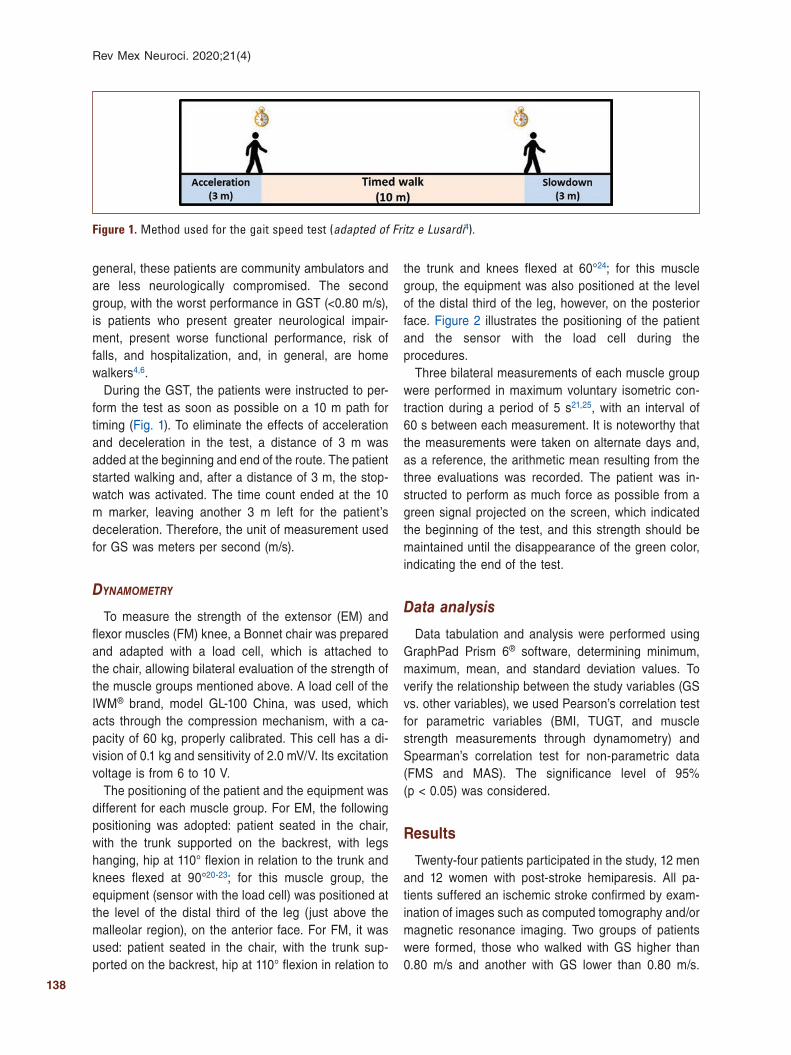
During the GST, the patients were instructed to per-form the test as soon as possible on a 10 m path for timing (Fig. 1). To eliminate the effects of acceleration and deceleration in the test, a distance of 3 m was added at the beginning and end of the route. The patient started walking and, after a distance of 3 m, the stop-watch was activated. The time count ended at the 10 m marker, leaving another 3 m left for the patient’s deceleration. Therefore, the unit of measurement used for GS was meters per second (m/s).
dynamometRy
To measure the strength of the extensor (EM) and flexor muscles (FM) knee, a Bonnet chair was prepared and adapted with a load cell, which is attached to the chair, allowing bilateral evaluation of the strength of the muscle groups mentioned above. A load cell of the IWM® brand, model GL-100 China, was used, which acts through the compression mechanism, with a ca-pacity of 60 kg, properly calibrated. This cell has a di-vision of 0.1 kg and sensitivity of 2.0 mV/V. Its excitation voltage is from 6 to 10 V.
The positioning of the patient and the equipment was different for each muscle group. For EM, the following positioning was adopted: patient seated in the chair, with the trunk supported on the backrest, with legs hanging, hip at 110° flexion in relation to the trunk and knees flexed at 90°20-23; for this muscle group, the equipment (sensor with the load cell) was positioned at the level of the distal third of the leg (just above the malleolar region), on the anterior face. For FM, it was used: patient seated in the chair, with the trunk sup-ported on the backrest, hip at 110° flexion in relation to
the trunk and knees flexed at 60°24; for this muscle group, the equipment was also positioned at the level of the distal third of the leg, however, on the posterior face. Figure 2 illustrates the positioning of the patient and the sensor with the load cell during the procedures.
Three bilateral measurements of each muscle group were performed in maximum voluntary isometric con-traction during a period of 5 s21,25, with an interval of 60 s between each measurement. It is noteworthy that the measurements were taken on alternate days and, as a reference, the arithmetic mean resulting from the three evaluations was recorded. The patient was in-structed to perform as much force as possible from a green signal projected on the screen, which indicated the beginning of the test, and this strength should be maintained until the disappearance of the green color, indicating the end of the test.
Data analysis
Data tabulation and analysis were performed using GraphPad Prism 6® software, determining minimum, maximum, mean, and standard deviation values. To verify the relationship between the study variables (GS vs. other variables), we used Pearson’s correlation test for parametric variables (BMI, TUGT, and muscle strength measurements through dynamometry) and Spearman’s correlation test for non-parametric data (FMS and MAS). The significance level of 95% (p < 0.05) was considered.
Results
Twenty-four patients participated in the study, 12 men and 12 women with post-stroke hemiparesis. All pa-tients suffered an ischemic stroke confirmed by exam-ination of images such as computed tomography and/or magnetic resonance imaging. Two groups of patients were formed, those who walked with GS higher than 0.80 m/s and another with GS lower than 0.80 m/s.
Figure 1. Method used for the gait speed test (adapted of Fritz e Lusardi4).
No
par
t o
f th
is p
ub
licat
ion
may
be
rep
rod
uce
d o
r p
ho
toco
pyi
ng
wit
ho
ut
the
pri
or
wri
tten
per
mis
sio
n o
f th
e p
ub
lish
er.
©
Per
man
yer
2020

139
A. Vinicius-Soares, et al.: Locomotion post-stroke
Patients are seen at a physical therapy service twice a week regularly.
Next, data from the statistical analysis of the group with the best performance in GST (> 0.80 m/s) are pre-sented. Tables 1 and 2 show descriptive statistics and correlation analysis data. Eight patients participated in this group, five men and three women.
Then, data from the statistical analysis of patients who wander with GST < 0.80 m/s (16 patients, 7 men and 9 women) are presented. Tables 3 and 4 present the descriptive statistics and correlation analysis data.
The groups when stratified based on GST were not different in terms of epidemiological and general clini-cal aspects, such as age, post-stroke injury time, and BMI. However, when compared to the other variables between the groups such as GST, TUGT, FMS, and MAS, it is observed that there is a significant difference between them. As for muscle strength tests, no signif-icant difference was observed, although on the paretic side, the group with the worst performance in GS tests had a deficit of 28.4% in extensors muscles and 38.0%
in flexors muscles lower than the group with the best performance. This may have an important clinical repercussion, as indicated in correlation tests involving this variable.
Discussion
Walking in hemiparetic patients after stroke is described as uncoordinated, arrhythmic, and unbal-anced5,26,27. The GS is a clinical and biomechanical parameter that quickly, easily, and reliably translates performance in this primary function. In fact, there is a relationship between the performance in GST and the level of functional independence, risk of falls, and hos-pitalization of these patients5,6.
In individuals affected by stroke, although GS is a determining factor of functional capacity, other vari-ables may be associated with functional deficit, such as anthropometric characteristics28. In our study, the correlation between BMI and GS was weak and not significant for both groups. Similar results were found
Table 1. Descriptive statistics of the group with good performance in gait speed test (> 0.80 m/s)
Age ST BMI TUGT GST FMS MAS ESp ESnp FSp FSnp
M 59.3 23.3 27.4 12.7 1.06 25.6 0.50 17.6 24.0 7.9 12.0
DP 10.3 35.7 5.68 2.71 0.27 3.0 0.76 10.2 7.9 5.9 6.4
Minimum 46.0 4.00 17.6 8.62 0.80 19.0 0.0 7.30 13.3 2.8 5.4
Maximum 70.0 108 37.7 18.4 1.64 28.0 2.00 35.3 36.2 18.3 23.9
ST: stroke time (months); BMI: body mass index (kg/m2), TUGT: timed up and go test (time in s); GST: gait speed test in 10 m (m/s); FMS: Fugl‑Meyer scale – lower limb section (0‑28); MAS: Modified Ashworth Scale (0‑5); ESp: extensors muscle strength paretic side (kgf); ESnp: extensors muscle strength non‑paretic side (kgf); FSp: flexors muscle strength paretic side (kgf); FSnp: flexors muscle strength non‑paretic side (kgf).
Figure 2. Positioning of the patient and the load cell for the evaluation of the muscle groups addressed in the study. A: extensors muscles dynamometry. B: flexors muscles dynamometry.
A B
No
par
t o
f th
is p
ub
licat
ion
may
be
rep
rod
uce
d o
r p
ho
toco
pyi
ng
wit
ho
ut
the
pri
or
wri
tten
per
mis
sio
n o
f th
e p
ub
lish
er.
©
Per
man
yer
2020

140
Rev Mex Neuroci. 2020;21(4)
in a study by Sheffler et al.29, where the influence of body mass on hemiparetic gait was evaluated. Al-though overweight may negatively affect post-stroke gait, the authors find no association between BMI and specific spatial-temporal parameters, including GS, ca-dence, double support time, or step length when mea-sured over short distances.
Muscle strength and endurance seem to play a special role for good performance in this function, and this di-rectly affects the functional capacity6. Although the pa-retic side is the most affected in relation to muscle strength, the non-paretic side may also show reduced strength. In individuals affected by stroke, the strength deficits range from 17% to 40% on the paretic side and from 5% to 40% on the non-paretic side. It is worth not-ing that this asymmetry of strength can also occur in healthy people, but in a smaller proportion (up to 10%)27.
In a study conducted by Hyun et al.30, the strength of the knee extensors and the dynamic balance was eval-uated as predictors of walking in hemiparetic patients due to stroke in the acute phase. Fifty-three patients
participated in the study, who were stratified in domestic ambulators (GST < 0.4 m/s) and community ambulators (GST > 0.4 m/s). The function of balance and functional mobility was evaluated using the Berg Balance Scale and the TUGT, respectively. The strength of the exten-sors and flexors knee was measured with an isokinetic dynamometer. The results found in the study indicate that the balance and strength of the knee extensors are the strongest predictors for the gait function of patients. However, the authors point out that the results may vary according to the severity of the gait. These findings corroborate those obtained in our study, where in the group with the best performance in GST (> 0.8 m/s), there was a strong and significant correlation (r ≥ 0.80) of both muscle groups, flexors and extensors knee on the paretic side. Moreover, for the other group, with poorer performance in GST (< 0.8 m/s), the extensors knee strength also showed significant correlation.
Still regarding the evaluation of muscle strength, it is important to comment on the asymmetry found between the paretic and non-paretic sides. In the group with the
Table 2. Correlation analysis of the group with good performance (gait speed test > 0.80 m/s)
Age ST BMI TUGT FMS MAS ESp ESnp FSp FSnp
r 0.66 −0.37 0.15 −0.77 0.70 −0.54 0.87 0.80 0.85 0.85
p 0.077 0.370 0.718 0.026 0.061 0.060 0.005 0.018 0.007 0.008
ST: Stroke time (months); BMI: body mass index (kg/m2); TUGT: timed up and go test (time in s); FMS: Fugl‑Meyer scale – lower limb section (0‑28); MAS: Modified Ashworth Scale (0‑5); ESp: Extensors muscle strength paretic side (kgf); ESnp: Extensors muscle strength non‑paretic side (kgf); FSp: flexors muscle strength paretic side (kgf); FSnp: flexors muscle strength non‑paretic side (kgf).
Table 3. Descriptive statistics of the group with worst performance (gait speed test < 0.80 m/s)
Age ST BMI TUGT GST FMS MAS ESp ESnp FSp FSnp
M 56.8 20.1 28.9 35.1 0.38 19.5 1.9 12.6 23.3 4.9 11.7
DP 10.8 22.5 8.34 20.7 0.2 4.9 1.7 7.3 8.0 3.0 4.6
Minimum 37.0 3.0 18.4 14.2 0.1 11.0 0.0 3.8 11.4 0.8 6.4
Maximum 79.0 96.0 44.8 88.5 0.7 27.0 5.0 28.1 39.6 12.8 21.1
ST: stroke time (months); BMI: body mass index (kg/m2); TUGT: timed up and go test (time in s); GST: gait speed test in 10 m (m/s); FMS: Fugl‑Meyer scale – lower limb section (0‑28); MAS: Modified Ashworth Scale (0‑5); ESp: extensors muscle strength paretic side (kgf); ESnp: extensors muscle strength non‑paretic side (kgf); FSp: flexors muscle strength paretic side (kgf): FSnp: flexors muscle strength non‑paretic side (kgf).
Table 4. Correlation analysis of the group with worst performance (GST < 0.80 m/s)
Age ST BMI TUGT FMS MAS ESp ESnp FSp FSnp
r 0.27 −0.26 −0.03 −0.87 0.69 −0.61 0.55 0.30 0.39 0.13
p 0.316 0.335 0.928 0.000 0.004 0.010 0.026 0.257 0.135 0.637
ST: stroke time (months); BMI: body mass index (kg/m2); TUGT: timed up and go test (time in s); FMS: Fugl‑Meyer scale – lower limb section (0‑28); MAS: Modified Ashworth Scale (0‑5); ESp: Extensors muscle strength paretic side (kgf); ESnp: Extensors muscle strength non‑paretic side (kgf); FSp: Flexors muscle strength paretic side (kgf); FSnp: flexors muscle strength non‑paretic side (kgf).
No
par
t o
f th
is p
ub
licat
ion
may
be
rep
rod
uce
d o
r p
ho
toco
pyi
ng
wit
ho
ut
the
pri
or
wri
tten
per
mis
sio
n o
f th
e p
ub
lish
er.
©
Per
man
yer
2020
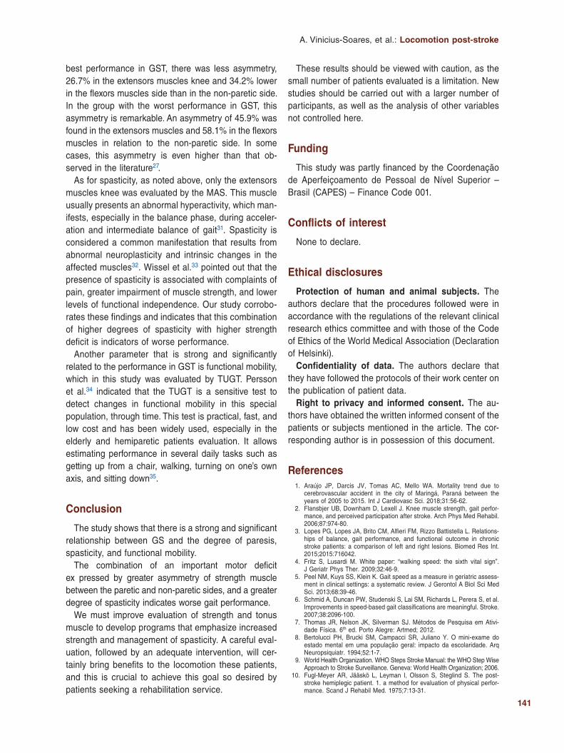
141
A. Vinicius-Soares, et al.: Locomotion post-stroke
best performance in GST, there was less asymmetry, 26.7% in the extensors muscles knee and 34.2% lower in the flexors muscles side than in the non-paretic side. In the group with the worst performance in GST, this asymmetry is remarkable. An asymmetry of 45.9% was found in the extensors muscles and 58.1% in the flexors muscles in relation to the non-paretic side. In some cases, this asymmetry is even higher than that ob-served in the literature27.
As for spasticity, as noted above, only the extensors muscles knee was evaluated by the MAS. This muscle usually presents an abnormal hyperactivity, which man-ifests, especially in the balance phase, during acceler-ation and intermediate balance of gait31. Spasticity is considered a common manifestation that results from abnormal neuroplasticity and intrinsic changes in the affected muscles32. Wissel et al.33 pointed out that the presence of spasticity is associated with complaints of pain, greater impairment of muscle strength, and lower levels of functional independence. Our study corrobo-rates these findings and indicates that this combination of higher degrees of spasticity with higher strength deficit is indicators of worse performance.
Another parameter that is strong and significantly related to the performance in GST is functional mobility, which in this study was evaluated by TUGT. Persson et al.34 indicated that the TUGT is a sensitive test to detect changes in functional mobility in this special population, through time. This test is practical, fast, and low cost and has been widely used, especially in the elderly and hemiparetic patients evaluation. It allows estimating performance in several daily tasks such as getting up from a chair, walking, turning on one’s own axis, and sitting down35.
Conclusion
The study shows that there is a strong and significant relationship between GS and the degree of paresis, spasticity, and functional mobility.
The combination of an important motor deficit ex pressed by greater asymmetry of strength muscle between the paretic and non-paretic sides, and a greater degree of spasticity indicates worse gait performance.
We must improve evaluation of strength and tonus muscle to develop programs that emphasize increased strength and management of spasticity. A careful eval-uation, followed by an adequate intervention, will cer-tainly bring benefits to the locomotion these patients, and this is crucial to achieve this goal so desired by patients seeking a rehabilitation service.
These results should be viewed with caution, as the small number of patients evaluated is a limitation. New studies should be carried out with a larger number of participants, as well as the analysis of other variables not controlled here.
Funding
This study was partly financed by the Coordenação de Aperfeiçoamento de Pessoal de Nível Superior – Brasil (CAPES) – Finance Code 001.
Conflicts of interest
None to declare.
Ethical disclosures
Protection of human and animal subjects. The authors declare that the procedures followed were in accordance with the regulations of the relevant clinical research ethics committee and with those of the Code of Ethics of the World Medical Association (Declaration of Helsinki).
Confidentiality of data. The authors declare that they have followed the protocols of their work center on the publication of patient data.
Right to privacy and informed consent. The au-thors have obtained the written informed consent of the patients or subjects mentioned in the article. The cor-responding author is in possession of this document.
References 1. Araújo JP, Darcis JV, Tomas AC, Mello WA. Mortality trend due to
cerebrovascular accident in the city of Maringá, Paraná between the years of 2005 to 2015. Int J Cardiovasc Sci. 2018;31:56-62.
2. Flansbjer UB, Downham D, Lexell J. Knee muscle strength, gait perfor-mance, and perceived participation after stroke. Arch Phys Med Rehabil. 2006;87:974-80.
3. Lopes PG, Lopes JA, Brito CM, Alfieri FM, Rizzo Battistella L. Relations-hips of balance, gait performance, and functional outcome in chronic stroke patients: a comparison of left and right lesions. Biomed Res Int. 2015;2015:716042.
4. Fritz S, Lusardi M. White paper: “walking speed: the sixth vital sign”. J Geriatr Phys Ther. 2009;32:46-9.
5. Peel NM, Kuys SS, Klein K. Gait speed as a measure in geriatric assess-ment in clinical settings: a systematic review. J Gerontol A Biol Sci Med Sci. 2013;68:39-46.
6. Schmid A, Duncan PW, Studenski S, Lai SM, Richards L, Perera S, et al. Improvements in speed-based gait classifications are meaningful. Stroke. 2007;38:2096-100.
7. Thomas JR, Nelson JK, Silverman SJ. Métodos de Pesquisa em Ativi-dade Física. 6th ed. Porto Alegre: Artmed; 2012.
8. Bertolucci PH, Brucki SM, Campacci SR, Juliano Y. O mini-exame do estado mental em uma população geral: impacto da escolaridade. Arq Neuropsiquiatr. 1994;52:1-7.
9. World Health Organization. WHO Steps Stroke Manual: the WHO Step Wise Approach to Stroke Surveillance. Geneva: World Health Organization; 2006.
10. Fugl-Meyer AR, Jääskö L, Leyman I, Olsson S, Steglind S. The post-stroke hemiplegic patient. 1. a method for evaluation of physical perfor-mance. Scand J Rehabil Med. 1975;7:13-31.
No
par
t o
f th
is p
ub
licat
ion
may
be
rep
rod
uce
d o
r p
ho
toco
pyi
ng
wit
ho
ut
the
pri
or
wri
tten
per
mis
sio
n o
f th
e p
ub
lish
er.
©
Per
man
yer
2020

142
Rev Mex Neuroci. 2020;21(4)
11. Heidari M, Abootalebi C, Hosseini SA. Validity of modified ash worth scale as a measure of wrist spasticity in stroke patients. Iran Rehabil J. 2011;9:26-30.
12. Podsiadlo D, Richardson S. The timed “Up and Go”: a test of basic functional mobility for frail elderly persons. J Am Geriatr Soc. 1991;39:142-8.
13. Soares AV, Matos FM, Laus LH, Suzuki S. Estudo comparativo sobre a propensão de quedas em idosos institucionalizados e não institucionaliza-dos através do nível de mobilidade funcional. Fisioter Bras. 2003;4:12-6.
14. Hafsteinsdóttir TB, Rensink M, Schuurmans M. Clinimetric properties of the timed up and go test for patients with stroke: a systematic review. Top Stroke Rehabil. 2014;21:197-210.
15. Chen CL, Chang KJ, Wu PY, Chi CH, Chang ST, Cheng YY. Comparison of the effects between isokinetic and isotonic strength training in suba-cute stroke patients. J Stroke Cerebrovasc Dis. 2015;24:1317-23.
16. Huh JS, Lee YS, Kim CH, Min YS, Kang MG, Jung TD. Effects of balan-ce control training on functional outcomes in subacute hemiparetic stroke patients. Ann Rehabil Med. 2015;39:995-1001.
17. Isho T, Usuda S. Association of trunk control with mobility performance and accelerometry-based gait characteristics in hemiparetic patients with subacute stroke. Gait Posture. 2016;44:89-93.
18. Faria CD, Teixeira-Salmela LF, Silva EB, Nadeau S. Expanded timed up and go test with subjects with stroke: reliability and comparisons with matched healthy controls. Arch Phys Med Rehabil. 2012;93:1034-8.
19. Piva SR, Fitzgerald GK, Irrgang JJ, Bouzubar F, Starz TW. Get up and go test in patients with knee osteoarthritis. Arch Phys Med Rehabil. 2004;85:284-9.
20. Martins JC, Aguiar LT, Lara EM, Moura JB, Souza LA, Teixeira-Salmela LF, et al. Assessment of the strength of the lower limb muscles in subjects with stroke with portable dynamometry: a literature review. Fisioter mov. 2016;29:193-208.
21. Souza LA, Martins JC, Teixeira-Salmela LF, Lara EM, Moura JB, Aguiar LT, et al. Validity and reliability of the modified sphygmomanome-ter test to assess strength of the lower limbs and trunk muscles after stroke. J Rehabil Med. 2014;46:620-8.
22. Cooper A, Alghamdi GA, Alghamdi MA, Altowaijri A, Richardson S. The relationship of lower limb muscle strength and knee joint hyperextension during the stance phase of gait in hemiparetic stroke patients. Physiother Res Int. 2012;17:150-6.
23. Akbari A, Karimi H. The effect of strengthening exercises on exaggerated muscle tonicity in chronic hemiparesis following stroke. J Med Sci. 2006;6:382-8.
24. Correa CS, da Silva BG, Alberton CL, Wilhelm EN, Moraes DA, Lima CS, et al. Análise da força isométrica máxima e do sinal de EMG em exer-cícios para os membros inferiores. Rev Bras Cineantropom Desempenho Hum. 2011;13:429-5.
25. Kiyama R, Fukudome K, Hiyoshi T, Umemoto A, Yoshimoto Y, Maeda T. The loss of dexterity in the bilateral lower extremities in patients with stroke. J Appl Biomech. 2011;27:122-9.
26. Iwabe C, Diz MA, Barudy DP. Análise cinemática da marcha em indiví-duos com acidente vascular encefálico. Rev Neurociênc. 2008;16:292-6.
27. Signal NE. Strength training after stroke: rationale, evidence and poten-tial implementation barriers for physiotherapists. N Z J Physiother. 2014;42:101-7.
28. Beavers KM, Beavers DP, Houston DK, Harris TB, Hue TF, Koster A, et al. Associations between body composition and gait-speed decline: results from the health, aging, and body composition study. Am J Clin Nutr. 2013;97:552-60.
29. Sheffler LR, Bailey SN, Gunzler D, Chae J. Effect of body mass index on hemiparetic gait. PM R. 2014;6:908-13.
30. Hyun CW, Han EY, Im SH, Choi JC, Kim BR, Yoon HM, et al. Hemiparetic knee extensor strength and balance function are predictors of ambulatory function in subacute stroke patients. Ann Rehabil Med. 2015;39:577-85.
31. Boudarham J, Roche N, Pradon D, Delouf E, Bensmail D, Zory R. Effects of quadriceps muscle fatigue on stiff-knee gait in patients with hemipa-resis. PLoS One. 2014;9:e94138.
32. Li S, Francisco GE. New insights into the pathophysiology of post-stroke spasticity. Front Hum Neurosci. 2015;9:192.
33. Wissel J, Schelosky LD, Scott J, Christe W, Faiss JH, Mueller J. Early development of spasticity following stroke: a prospective, observational trial. J Neurol. 2010;257:1067-72.
34. Persson CU, Danielsson A, Sunnerhagen KS, Grimby-Ekman A, Hansson PO. Timed up and go as a measure for longitudinal change in mobility after stroke - postural stroke study in gothenburg (POSTGOT). J Neuroeng Rehabil. 2014;11:83.
35. Soares AV, Marcelino E, Borges NG Jr., Domenech SC, Loch MS, Sagawa Y Jr. Relação entre dinapenia, sarcopenia e mobilidade funcional em idosos frágeis institucionalizados. Medicina (Ribeirão Preto). 2016;49:195-201.
No
par
t o
f th
is p
ub
licat
ion
may
be
rep
rod
uce
d o
r p
ho
toco
pyi
ng
wit
ho
ut
the
pri
or
wri
tten
per
mis
sio
n o
f th
e p
ub
lish
er.
©
Per
man
yer
2020

143
Prevalence of comorbidities in children with attention deficit/hyperactivity disorder: Measured and systematic review of care health studiesDenise Medici1,2*, María M. Suarez-Varela2,3, and Rubén Pérez-Elvira4
1Department of Neuropsychology, Hospital Vithas Nisa 9 de Octubre, Valencia, Spain; 2Department of Preventive Medicine, University of Valencia, Valencia, Spain; 3Department of Biomedical Research Consortium for Epidemiology and Public Health (CIBERESP), Valencia, Spain; 4NEPSA Neurological Rehabilitation, Salamanca, Spain
Revista Mexicana de Neurociencia
ORIGINAL ARTICLE
Abstract
Background and objectives: Children and adolescents with a diagnosis of attention deficit hyperactivity disorder demons-trate a developmental delay in emotional and social functions, sleep difficulties, enuresis, and/or paroxysms in electroen-cephalographic measurements.The purpose of the study is to know if there is an association between comorbidities and ADHD. The second objective was to compare the results with previous studies in different countries. Material and methods: Re-trospective analysis of a clinical database (CDB) was conducted identifying 1049 Spanish children and adolescents diag-nosed with ADHD according to DSM-IV (Diagnostic and Statistical Manual of Mental Disorders) in the Department of Pedia-trics of the Hospital Universitario Dr. Peset. Results: In the 297 CDB aged 6 - 16 years children included, the comorbidities were statistically significant. This medical conditions in Spanish children with ADHD coexist outside of Spain and are statis-tically significant in all aspects analyzed except in anxiety, Asperger’s syndrome and enuresis. Conclusions: Our results support the association between comorbidities and ADHD. It is important for professionals to make sure that they identify different comorbidities during the diagnostic process as during the clinical follow-up. The article is recommended to teachers, therapists and health professionals, for adopting a proactive and intermodal team approach in the detection and treatment of comorbidities in children and youth.
Key words: Attention deficit hyperactivity disorder. Comorbidities. Detection. Diagnosis.
Prevalencia de comorbilidades en niños con trastorno por déficit de atención/hiperactividad: revisión medida y sistemática de estudios de salud asistencial
Resumen
Antecedentes y objetivos: Los niños y adolescentes con diagnóstico de Trastorno por Déficit de Atención/Hiperactividad (TDAH) presentan un retraso en el desarrollo de las funciones sociales y emocionales, dificultades para dormir, enuresis y/o paroxismos en las mediciones electroencefalográficas (EEG). El propósito del estudio es saber si existe una asociación entre las comorbilidades y el TDAH. El segundo objetivo fue comparar los resultados con estudios previos en diferentes
Correspondence: *Denise Medici
Valle de la Ballestera, 59
C.P. 46015, Valencia, España
E-mail: [email protected]
Available online: 05-08-2020
Rev Mex Neuroci. 2020;21(4):143-149
www.revmexneurociencia.com
Date of reception: 15-11-2019
Date of acceptance: 10-02-2020
DOI: 10.24875/RMN.20000154
1665-5044/ © 2020. Academia Mexicana de Neurología A.C. Published by Permanyer. This is an open access article under the CC BY-NC-ND license (http://creativecommons.org/licenses/by-nc-nd/4.0/).
No
par
t o
f th
is p
ub
licat
ion
may
be
rep
rod
uce
d o
r p
ho
toco
pyi
ng
wit
ho
ut
the
pri
or
wri
tten
per
mis
sio
n o
f th
e p
ub
lish
er.
©
Per
man
yer
2020

144
Rev Mex Neuroci. 2020;21(4)
Introduction
Attention deficit hyperactivity disorder (ADHD) is the most commonly diagnosed neurodevelopmental disorder of childhood, and it is associated with social and aca-demic difficulties and psychiatric comorbidities such as depression, anxiety, low self-esteem, and pervasive across settings can cause problems in the daily activities of a child as well as later in their adult life1. In addition, studies detected difficulties in behavioral regulation and emotional stability in children younger than 2 years of age who were later diagnosed diagnosis with ADHD2. ADHD during childhood is one of the most common causes of school failure and social problems. It is a dis-order that can persist and manifest in adulthood in more than 60% of cases. Several studies have found that the quality of life in adolescents and in adults who were di-agnosed with ADHD during their childhood is clearly related to the severity of comorbidities symptoms3. In studies with adults who have a residual form of this dis-order, the research has found a higher frequency of be-havioral abnormalities in labor activities and in family relationships, substance abuse, accidents, and crime4. Depression during childhood and adolescence also pre-dicts negative outcomes in adulthood such as stressful life events, low social support, and low satisfaction with life5. Maternal depression and concurrent symptoms at 4-6 years of age predict which children with ADHD are at greatest risk for depression and/or suicidality as ado-lescents6. A diagnosis of both ADHD and depression is associated with the most significant deficiencies for either disorder than one alone. For example, young people with ADHD and depression have an increased risk of devel-oping bipolar disorder and oppositional defiant disorder, and require much more intensive interventions compared to young people with ADHD but without depression5. Interventions for both diagnoses also report more psy-chosocial and family problems as well as higher levels of
stress7,8. Despite the impact of ADHD and depressive symptoms in young people, few studies have examined the underlying mechanisms which contribute to the emer-gence of these symptoms9. One promising mechanism is emotion regulation (ER), as the deficit in the ability of ER has been associated with young children with ADHD and with depressive symptoms. This point is primarily relevant for young people with ADHD symptoms as it emphasizes the role of effort in control – the deliberate modulation of emotional states, emotional regulation, and subsequent behaviors, which involve the ability to focus deliberately, divert attention, hyperactivity, and inhibit or activate an appropriate behavior10-12. Several studies suggest that young people with ADHD demonstrate an inability to continue a task when frustrated or they have an inability to seek help from their parents. In extreme levels, when they are solving more limited problems, they focus more on negative aspects of a task compared to healthy controls. Young people with ADHD also have difficulties identifying and processing negative emotions and high-stress exposure between childhood and young adulthood is strongly intertwined with a persistent course of ADHD and with comorbid problems taking the form of severe and persistent emotion dysregulation (irritability, extreme reactivity, and frustration) or elevated and in-creasing irritability, anxiety, and depression13,14. Longitu-dinal research has shown that the ADHD diagnosis per-sists into adulthood, participants manifest higher levels of impulsive emotions (defined by symptoms such as low tolerance to frustration, impatience, and irritability) com-pared to those who do not have ADHD as an adult. This situation contributes to deficits in family, peer, financial, and labor relationships15. When comparing subtypes, children with inattentive ADHD have a higher rate in eat-ing disorders, anxiety, and depression than in those with hyperactive and/or combined ADHD. In this both hyper-active and combined ADHD, children often have social problems, as they take things that do not belong to them,
países. Método: Se realizó un análisis retrospectivo de una base de datos de historias clínicas (CDB) que identificó a 1049 niños y adolescentes españoles diagnosticados con TDAH de acuerdo con el DSM-IV (Manual diagnóstico y estadístico de trastornos mentales) en el Departamento de Pediatría del Hospital Universitario Dr. Peset. Resultados: En las 297 CDB de niños con edades entre los 6 a 16 años, las comorbilidades fueron estadísticamente significativas. Estas condiciones médi-cas en niños españoles con TDAH coexisten fuera de España y han sido estadísticamente significativos en todos los aspec-tos analizados, excepto en ansiedad, síndrome de Asperger y enuresis. Conclusiones: Nuestros resultados apoyan la asociación entre comorbilidades y TDAH. Es importante que los profesionales se aseguren de identificar las diferentes co-morbilidades durante el proceso de diagnóstico y durante el seguimiento clínico. El artículo se recomienda a maestros, te-rapeutas y profesionales de la salud para que adopten un enfoque de equipo proactivo e intermodal en la detección y tratamiento de comorbilidades en niños y jóvenes.
Palabras clave: Trastorno por déficit de atención/hiperactividad. TDAH. Comorbilidades. Detección. Diagnóstico.
No
par
t o
f th
is p
ub
licat
ion
may
be
rep
rod
uce
d o
r p
ho
toco
pyi
ng
wit
ho
ut
the
pri
or
wri
tten
per
mis
sio
n o
f th
e p
ub
lish
er.
©
Per
man
yer
2020

145
D. Medici, et al.: Comorbidities in ADHD
do not wait their turn, and act without considering before the feelings of others. Children with ADHD inattentive type, rather, tend to be socially isolated, self-absorbed, and most likely have an introverted behavior16 (Diamond, 2005). ADHD without hyperactivity and ADHD with hyperactivity are two different disorders with different cognitive and behavioral profiles, different patterns of comorbidities, different responses to medication, and dif-ferent underlying neurobiology16 characteristics that be-come chronic and progressively worse (i.e., findings sup-port the diagnosis of ADHD in younger children by demonstrating that the symptoms and associated impair-ment are likely to persist well into elementary school)17. It is unclear whether the features of anxiety and ADHD are possible to identify very early of age; for example, in one child, his emotional deregulation at 18 months of age was associated with symptoms of anxiety and ADHD at 3 years of age. The study could not confirm if the emo-tional deregulation at 18 months predicts the cooccur-rence of anxiety and ADHD symptoms. This implies that the identification risk at 18 months is a clinical challenge. By identifying early, there is a risk of over-identification and treatment of preschool children who are not going to develop ADHD or anxiety in their immediate future18. However, longitudinal studies in school-age children found continuity of symptoms for anxiety and ADHD19,20. In children with ADHD with an anxiety disorder have more than 50% of them also oppositional defiant disorder or conduct disorder21.
The relationship between ADHD and sleep disorders is unclear. According to a categorical approach, specific sleep disorders are a common comorbidity in children with ADHD. Those with ADHD more often display hypo-pnea and/or apnea, movements in peripheral limbs during sleep, insomnia, and narcolepsy which could result in significant functional impairments that affect mood, attention, behavior, and ultimately school/work performance and quality of life22. Polysomnography shows an increased latency in sleep onset of families ADHD children report less maintenance and duration of sleep, resistance by the child at bedtime, difficulties in sleep onset, difficult in nocturnal breathing, enuresis, nocturnal awakenings, and difficulty in waking up in the morning and daytime sleepiness23. In another multi-center cross-sectional study in 12 Spanish hospitals, neurologists reported that sleep disorder affects less than a quarter of patients (24.5%) and up to 23% stated that the prevalence was < 10%, while between 58% and 92% of parents of patients attending consultations in pediatric neurology report that their children have some aspect disturbed in the sleep. Although most
neurologists appreciate the importance of diagnosis and treatment of sleep disorders in children with neuro-logical disorders, it is often underestimated and there-fore essential to include in ADHD evaluations as proper and early treatment would improve symptoms and the quality of life in patients and families24.
Moreover, many children with ADHD develop epilepsy. In one study of 23 patients with epilepsy, 19 of them had ADHD symptoms preceding the onset of seizures25. An epidemiological study showed that the risk of epilepsy is 2.5 times higher in children who have already devel-oped symptoms of ADHD26. With regard to abnormal electroencephalographic (EEG) recordings outside epi-lepsy, a recent study found abnormal EEG readings in 19 of 50 children diagnosed with ADHD. The most ab-normal results were in focal and generalized paroxysms of sharp waves in phase opposition and slow waves. The most frequent location of EEG abnormalities was in the temporal lobe (45%), although there were focal parox-ysms in other lobes; 28% in the right temporal region, 17% in the left temporal region, and 22% in the left fron-tal lobe, followed by 11% in the left parietal region27.
In children’s ADHD diagnosis, enuresis is a relatively new clinical aspect and is part of our ongoing effort to better understand comorbidities. Children with ADHD are more likely than their peers without ADHD to develop enuresis with a similar trend for encopresis and comor-bid enuresis implying an immaturity in the developing nervous system of children who have this combination of problems28. Children with primary nocturnal enuresis (PNE) show a significant reduction in activity in the left posterior cerebellum compared to controls without PNE29. Similarly, functional magnetic resonance imaging (fMRI) demonstrated low activation in the right prefrontal cortex and increased activity in the left hemisphere during inhibition of motor response compared to controls without PNA, indicating abnormal network brain areas during response inhibition in children with PNE30.
Therefore, this study aims to characterize the diag-nostic complexity of children and adolescents who re-ceive a diagnosis of ADHD. These findings could be helpful to develop guidelines that reflect the needs of children with ADHD. We treat a review of the comor-bidities in itself complex in the diagnosis of ADHD.
Methods
Participants
Retrospective analysis of a clinical database (CDB) was conducted identifying 1049 Spanish children and
No
par
t o
f th
is p
ub
licat
ion
may
be
rep
rod
uce
d o
r p
ho
toco
pyi
ng
wit
ho
ut
the
pri
or
wri
tten
per
mis
sio
n o
f th
e p
ub
lish
er.
©
Per
man
yer
2020

146
Rev Mex Neuroci. 2020;21(4)
adolescents diagnosed with ADHD according to the DSM-IV (Diagnostic and Statistical Manual of Mental Disorders) in the Department of Pediatrics of the Hos-pital Universitario Dr. Peset. The comorbidities were diagnosed in the corresponding medical service (Pedi-atrics, Mental Health, Neurology). Personal and clinical data of patients were reviewed to determine those who met the clinical criteria previously established and de-scribed below. After eliminating patients who did not meet for the above, 297 CDB aged 6-16 years of chil-dren (79 females and 218 males; mean age, 09.10 years) diagnosed with ADHD were used for this study.
Inclusion criteria
– Patients diagnosed with ADHD born in Spain from January 1999 to December 2009.
– Patients diagnosed with ADHD with intelligence quo-tient (IQ) of 80 or higher with Spanish parents.
– Patients diagnosed with ADHD who attended medical appointments.
Exclusion criteria
– Patients diagnosed with ADHD born and raised from January 1999 to December 2009 in Spain with IQ below 80.
– Patients aged in or outside the range 6-15 years and 11 months adopted or born and raised in Spain of foreign parents
– Patients diagnosed with ADHD who not attended medical appointments.
– Patients diagnosed with ADHD who later acquired brain injury, surgery, and/or neurological diseases such as traumatic brain injury, epilepsy, and/or Gilles de la Tourette Syndrome (Table 1).In the following table are described by the participants:Medical data related to the child were recorded in a
confidential database for exclusive use of the project. Data about diagnosis, treatments, personal circum-stances, and about clinical evolution were collected ac-cording to the law 15/99 on protection of personal data.
Statistical analysis
First, descriptive statistics as comorbidity prevalence of the different pathologies studies and their confidence interval 95% were calculated. The relationships be-tween the categorical variables the prevalence of ADHD child’s comorbidity with the ADHD child’s without comorbidity were analyzed using the Chi-square test (p < 0.05) by each comorbidity studied. Finally, a com-parison of each comorbidity prevalence identify was also compared through the review of outside studies from other countries; it was analyzed using the Chi-square test (p < 0.05). Statistical analyses were conducted using IBM SPSS Statistics 25.
Results
Many children diagnosed with ADHD have comorbid-ities. In Spain, children and adolescents diagnosed with ADHD, the current study found that all potential comorbid conditions were statistically significant in the aspects analyzed (p ≤ 0.001) (Table 2). When examin-ing outside studies, results on the presence or absence of conditions that coexist in children diagnosed with ADHD outside of Spain are statistically significant in all aspects analyzed (p ≤ 0.001), except in anxiety, and in Asperger’s disease31 and in female and male Enuresis32 (Table 2a).
Discussion
In this study in ADHD, 25% of patients showed social problems, 28% depression and/or anxiety, and 19% had difficulties in the maintenance of nighttime sleep. The results were associated with those obtained by Hoza12, Mellon28, and Sheerman32 in United States of America, by Chou in China33, by Fonseca in Brazil34,
Table 1. Total excluded and included
226 participants born since 1990 and 1998
379 participants without an interdisciplinary diagnosis
14 participants had ADS
47 participants had a neurological disease diagnosed as epilepsy, brain traumatic
9 participants had a genetic syndrome diagnosed as deletions
26 participants coming from national or international adoptions or born in Spain of foreign parents
39 participants had not neurophysiological studies (EEG, qEEG, and ERPs)
12 participants without approach less medical data
Total included
297 participants met the clinical criteria
ADS: autism spectrum disorder; EEG: electroencephalographic; qEEG: quantitative EEG; ERPs: event‑related potentials.
No
par
t o
f th
is p
ub
licat
ion
may
be
rep
rod
uce
d o
r p
ho
toco
pyi
ng
wit
ho
ut
the
pri
or
wri
tten
per
mis
sio
n o
f th
e p
ub
lish
er.
©
Per
man
yer
2020

147
D. Medici, et al.: Comorbidities in ADHD
Table 2. Prevalence of comorbidities in children with attention deficit hyperactivity disorder for the sample (n = 297)
Variable Frequency (n) Prevalence (%) Prevalence (95% CI) Chi-square test (p)
Peer/dyadic relationship difficulties 74 25.00 20.18‑30.31 ≤ 0.001
Depression/anxiety 83 28.00 23.62‑34.18 ≤ 0.001
Anxiety 41 14.00 10.48‑18.74
Sleep disorders 56 18.90 15.87‑25.31 ≤ 0.001
Insomnia for lack of limits on family 7 2.35 1.25‑5.44 ≤ 0.001
Nocturnal enuresis 15 5.10 2.95‑8.36 ≤ 0.001
Snorer 3 1.01 0.26‑3.17 ≤ 0.001
Nighttime awakenings 11 3.70 1.95‑6.71 ≤ 0.001
Delay sleep onset syndrome 3 1.01 0.26‑3.17 ≤ 0.001
Restless leg syndrome 3 1.01 0.26‑3.17 ≤ 0.001
Bruxism 3 1.01 0.26‑3.17 ≤ 0.001
Catathrenia (night whimper) 4 1.70 0.43‑3.64 ≤ 0.001
Night terrors 2 0.70 0.11‑2.67 ≤ 0.001
Somniloquy 5 1.70 0.62‑4.10 ≤ 0.001
Epilepsy (EEG) 72 24.35 19.69‑29.79 ≤ 0.001
Primary nocturnal enuresis 10 3.37 1.72‑6.29
Female 2 0.67 0.11‑2.67
Male 8 2.70 1.25‑5.44 0.055
ANOVA test: p < 0.01. RF: relative frequency; %: proportion; 95% CI: Confidence Interval 95%; p: Fisher values; EEG: encephalogram.
Table 2a. Comorbidities in children with attention deficit hyperactivity disorder according to previous studies
Variable Frequency (n)
Prevalence (%)
Prevalence (95% CI)
Chi-square test (p)
Previous studies
Peer/dyadic relationship difficulties 92 56.00 47.83‑63.41 ≤ 0.001 Hoza, 2005
Depression 10 6.70 3.59‑12.82 0.029 Bauermeister, 2007
Depression 68 21.30 17.03‑23.30 0.020 Silva, 2015
Anxiety 34 23.77 17.23‑31.75 0.012 Bauermeister, 2007
Anxiety 169 52.60 47.34‑58.54 ≤ 0.001 Silva, 2015
Anxiety 12 11.90 6.55‑20.20 0.665 Ogrim, 2012
Asperger syndrome (symptoms) 5 4.95 1.83‑11.71 0.148 Ogrim, 2012
Sleep disorders 45 31.46 24.10‑39.84 0.003 Bauermeister, 2007
Epilepsy 6‑18 years old (EEG) 2843 15.63 15.10‑16.17 ≤ 0.001 Chou, 2013
Epilepsy 8‑11 years old (EEG) 3 10.00 2.61‑27.67 0.095 Fonseca, 2008
Primary nocturnal enuresis 100 9.00 7.25‑10.63 0.044 Shreeram, 2009
Female 32 2.51 1.95‑4.00 0.198
Male 68 6.21 4.70‑7.56 0.216
Primary nocturnal enuresis 35 12.10 6.99‑13.45 ≤ 0.001 Mellon, 2013
Primary nocturnal enuresis 165 17.60 15.02‑19.94 0.010 Joinson, 2007
ANOVA test: p < 0.01 (11, 20, 8, 33, 34, 28, 35); p ≤ 0.05 (31, 32). RF: relative frequency; %: proportion; 95% CI: Confidence Interval 95%; p: Fisher values; EEG: electroencepham.
No
par
t o
f th
is p
ub
licat
ion
may
be
rep
rod
uce
d o
r p
ho
toco
pyi
ng
wit
ho
ut
the
pri
or
wri
tten
per
mis
sio
n o
f th
e p
ub
lish
er.
©
Per
man
yer
2020

148
Rev Mex Neuroci. 2020;21(4)
by Joinson in United Kingdom35, by Silva in Australia9, and by Bauermeister in Puerto Rico20.
Children with ADHD often have conflicts with adults and peers, and suffer from unpopularity, rejection by peers, and a lack of friendships, in part as a conse-quence of their ADHD symptoms. In summary, social relations are also one of the biggest problems in chil-dren with ADHD. Parents, teachers, and their own col-leagues systematically report that those with ADHD can be aggressive, intrusive, disruptive, manipulative, and less able to communicate and socialize effectively11,13,36. Some studies reported little difference and related problems between genders20.
Parents with ADHD children compared with norma-tive data reported more problems in terms of emotion-al-behavioral role function, behavior, mental health, and self-esteem. Children with multiple comorbidity disor-ders have poorer psychosocial health-related quality of life across a range of domains compared with children with none and one comorbid disorder4. In addition, compared to children who have no comorbidities, psy-chosocial health-related quality of life was significantly lower in children with comorbid oppositional defiant disorder or conduct disorder, but not in children with a comorbid learning disorder4. In summary, social relations are one of the biggest problems in children with ADHD.
Sleep disorders may also induce symptoms of ADHD and are believed to be the result of excessive daytime sleepiness. However, it may be difficult for the clinical professional to recognize differences between the co-morbidity from one sleep disorder to similar symptoms in ADHD37,38. We found abnormal results in 24.35% of patients; therefore, the EEG is a valid study in the valuation process for test underlying or comorbid epi-lepsy in children with ADHD. Epilepsy and ADHD can affect social, educational, and emotional life. The as-sociation between ADHD and epilepsy is of great interest in many studies published recently because children with epilepsy have a significant risk in present-ing ADHD, and often display deficits in the performance of working memory26,33,39. Studies have detected sig-nificant similarities between epilepsy and ADHD in males (citation). ADHD with or without epilepsy may share a common abnormal and underlying neurobio-logical cause40.
In our study, the PNE in children with ADHD was in 5.05%, we consider the increase of psychological problems reported by parents of children with enuresis compared to those children without enuresis, and the possible pathogenic etiological between ADHD and
incontinence according to neurophysiological and neu-roimaging results30. However, enuresis is difficult to treat and show lower compliance. Given results in the treatment in incontinence are appreciated in the relevant clinical data regarding ADHD and enuresis, this appears to be a main comorbid condition that should be evaluated and specifically address in ADHD children40.
Conclusions
The current study carried out a broad and compre-hensive characterization of comorbidities in children and adolescents with ADHD in an effort to promote appropriate clinical decision making and reduced con-fusion. Detection and resolution of depressive symp-toms, anxious symptoms, and/or difficulties in sleep in those diagnosed with ADHD may reduce emotional risk and morning fatigue. It is very important to detect these symptoms to decrease social and cognitive conse-quences. Finally, using EEG in this population provides greater assessment and diagnosis of complex develop-mental disorders such as ADHD.
Study limitations
First, we recruited data about ADHD comorbidities from different medical specialties departments at the hospital. Our findings should be interpreted in light of limitations. Accordingly, we were unable to stratify symptoms according to high- and low-comorbidity. Second, we were not able to test whether comorbidities are unique to children with ADHD or are also observed in other disorders such as depression and learning delay. Future studies are needed about the relationship between comorbidities and ADHD.
Acknowledgments
The authors would like to thank the approbation of the study to the Ethics Committee for Clinical Research of the Hospital Universitario Dr. Peset of Valencia, Spain in 2015. It was conducted with not supported in keeping with the Declaration of Helsinki (World Medical Association, 2013, October) Ethics Committee: 10/061 and 11/083.
Funding
No funding was provided for this project.
No
par
t o
f th
is p
ub
licat
ion
may
be
rep
rod
uce
d o
r p
ho
toco
pyi
ng
wit
ho
ut
the
pri
or
wri
tten
per
mis
sio
n o
f th
e p
ub
lish
er.
©
Per
man
yer
2020

149
D. Medici, et al.: Comorbidities in ADHD
Conflicts of interest
The authors declare that they have no conflicts of interest.
Ethical disclosures
Protection of human and animal subjects. The authors declare that no experiments were performed on humans or animals for this study.
Confidentiality of data. The authors declare that no patient data appear in this article.
Right to privacy and informed consent. The authors declare that no patient data appear in this article.
References 1. Harpin VA. The effect of ADHD on the life of an individual, their family,
and community from preschool to adult life. Arch Dis Child. 2005;90 Su-ppl 1:i2-7.
2. Magallón S, Crespo-Eguílaz N, Ecay M, Poch-Olivé ML, Narbona J. Estilo comportamental al inicio del segundo año de vida: estudio retros-pectivo en escolares afectados de trastorno por déficit de atención e hiperactividad. An Pediatr. 2009;70:562-9.
3. Klassen AF, Miller A, Fine S. Health-related quality of life in children and adolescents who have a diagnosis of attention-deficit/hyperactivity disor-der. Pediatrics. 2004;114:e541-7.
4. McGough JJ, Smalley SL, McCracken JT, Yang M, Del’Homme M, Lynn DE, et al. Psychiatric comorbidity in adult attention deficit hyperac-tivity disorder: findings from multiplex families. Am J Psychiatry. 2005; 162:1621-7.
5. Biederman J, Ball SW, Monuteaux MC, Mick E, Spencer TJ, Mc-CREARY M, et al. New insights into the comorbidity between ADHD and major depression in adolescent and young adult females. J Am Acad Child Adolesc Psychiatry. 2008;47:426-34.
6. Chronis-Tuscano A, Molina BS, Pelham WE, Applegate B, Dahlke A, Overmyer M, et al. Very early predictors of adolescent depression and suicide attempts in children with attention-deficit/hyperactivity disorder. Arch Gen Psychiatry. 2010;67:1044-51.
7. Bussing R, Mason DM, Bell L, Porter P, Garvan C. Adolescent outcomes of childhood attention-deficit/hyperactivity disorder in a diverse commu-nity sample. J Am Acad Child Adolesc Psychiatry. 2010;49:595-605.
8. Silva D, Houghton S, Hagemann E, Bower C. Comorbidities of attention deficit hyperactivity disorder: pregnancy risk factors and parent mental health. Community Ment Health J. 2015;51:738-45.
9. Martel MM, Nigg JT. Child ADHD and personality/temperament traits of reactive an effortful control resiliency and emotionality. J Child Psychol Psychiatry. 2009;46:1437-44.
10. Seymour KE, Chronis-Tuscano A, Iwamoto DK, Kurdziel G, Macpherson L. Emotion regulation mediates the association between ADHD and depres-sive symptoms in a community sample of youth. J Abnorm Child Psychol. 2014;42:611-21.
11. Landis TD, Garcia AM, Hart KC, Graziano PA. Differentiating symptoms of ADHD in preschoolers: the role of emotion regulation and executive function. J Atten Disord. 2020; Jan 6:1087054719896858.
12. Hoza B, Mrug S, Gerdes AC, Hinshaw SP, Bukowski WM, Gold JA, et al. What aspects of peer relationships are impaired in children with atten-tion-deficit/hyperactivity disorder? J Consult Clin Psychol. 2005;73:411-23.
13. Hartman CA, Rommelse N, van der Klugt CL, Wanders RB, Timmerman ME. Stress exposure and the course of ADHD from child-hood to young adulthood: comorbid severe emotion dysregulation or mood and anxiety problems. J Clin Med. 2019;8:E1824.
14. Barkley RA, Fischer M. The unique contribution of emotional impulsive-ness to impairment in major life activities in hyperactive children as adults. J Am Acad Child Adolesc Psychiatry. 2010;49:503-13.
15. Diamond A. Attention-deficit disorder (attention-deficit/hyperactivity disor-der without hyperactivity): a neurobiologically and behaviorally distinct disorder from attention-deficit/hyperactivity disorder (with hyperactivity). Dev Psychopathol. 2005;17:807-25.
16. Lahey BB, Pelham WE, Loney J, Kipp H, Ehrhardt A, Lee SS, et al. Three-year predictive validity of DSM-IV attention deficit hyperactivity disorder in children diagnosed at 4-6 years of age. Am J Psychiatry. 2004;161:2014-20.
17. Overgaard KR, Aase H, Torgersen S, Reichborn-Kjennerud T, Oerbeck B, Myhre A, et al. Continuity in features of anxiety and attention deficit/hyperactivity disorder in young preschool children. Eur Child Adolesc Psychiatry. 2014;23:743-52.
18. Larsson H, Dilshad R, Lichtenstein P, Barker ED. Developmental trajec-tories of DSM-IV symptoms of attention-deficit/hyperactivity disorder: genetic effects, family risk and associated psychopathology. J Child Psy-chol Psychiatry. 2011;52:954-63.
19. Bauermeister JJ, Bird HR, Shrout PE, Chavez L, Ramírez R, Canino G. Short-term persistence of DSM-IV ADHD diagnoses: influence of context, age, and gender. J Am Acad Child Adolesc Psychiatry. 2011;50:554-62.
20. Shea CK, Lee MM, Lai KY, Luk ES, Leung PW. Prevalence of anxiety disorders in Hong Kong Chinese children with ADHD. J Atten Disord. 2018;22:403-13.
21. Wajszilber D, Santiseban JA, Gruber R. Sleep disorders in patients with ADHD: impact and management challenges. Nat Sci Sleep. 2018;10:453-80.
22. Vila MT, Torres AM, Soto BB, Gomar MR, Langa MJ, Sierra AI. Relación entre el trastorno por déficit de atención e hiperactividad y los trastornos del sueño. Resultados de un estudio epidemiológico en la población escolar de la ciudad de Gandía. An Pediatr. 2008;69:251-7.
23. Pérez-Villena A, Soto-Insuga V, Castaño-De la Mota C, Martín-Del Valle F, Pons-Rodríguez M, Lozada-Del Pozo M. Importancia de los problemas de sueño en los niños con cefalea y otros trastornos del neurodesarrollo en las consultas de neuropediatría. Rev Neurol. 2016;62:61-7.
24. Hermann B, Jones J, Dabbs K, Allen CA, Sheth R, Fine J, et al. The frequency, complications and aetiology of ADHD in new onset paediatric epilepsy. Brain. 2007;130:3135-48.
25. Sand T, Breivik N, Herigstad A. Assessment of ADHD with EEG. Tidss-kr Nor Laegeforen. 2013;133:312-6.
26. Cornelio-Nieto JO, Borbolla-Sala ME, Gallegos-Dimas A. Alteraciones electroencefalográficas en niños con trastorno por déficit de atención/hiperactividad. Rev Neuro. 2011;52 Suppl 1:S97-101.
27. Mellon MW, Natchev BE, Katusic SK, Colligan RC, Weaver AL, Voigt RG, et al. Incidence of enuresis and encopresis among children with atten-tion-deficit/hyperactivity disorder in a population-based birth cohort. Acad Pediatr. 2013;13:322-7.
28. Yu B, Guo Q, Fan G, Ma H, Wang L, Liu N. Evaluation of working memory impairment in children with primary nocturnal enuresis: evidence from event-related functional magnetic resonance imaging. J Paediatr Child Health. 2011;47:429-35.
29. Lei D, Ma J, Du X, Shen G, Tian M, Li G. Altered brain activation during response inhibition in children with primary nocturnal enuresis: an fMRI study. Hum Brain Mapp. 2012;33:2913-9.
30. Ogrim G, Kropotov J, Hestad K. The quantitative EEG theta/beta ratio in attention deficit/hyperactivity disorder and normal controls: sensitivity, specificity, and behavioral correlates. Psychiatry Res. 2012;198:482-8.
31. Shreeram S, He JP, Kalaydjian A, Brothers S, Merikangas KR. Preva-lence of enuresis and its association with attention-deficit/hyperactivity disorder among U.S. children: results from a nationally representative study. J Am Acad Child Adolesc Psychiatry. 2009;48:35-41.
32. Chou IC, Chang YT, Chin ZN, Muo CH, Sung FC, Kuo HT, et al. Corre-lation between epilepsy and attention deficit hyperactivity disorder: a population-based cohort study. PLoS One. 2013;8:e57926.
33. Fonseca LC, Tedrus GM, Moraes Cd, Vicente Machado Ad, Almeida MP, Oliveira DO. Epileptiform abnormalities and quantitative EEG in children with attention-deficit/hyperactivity disorder. Arq Neuropsiquiatr. 2008;66:462-7.
34. Joinson C, Heron J, Butler R, Von Gontard A, Butler U, Emond A, et al. A United Kingdom population-based study of intellectual capacities in children with and without soiling, daytime wetting, and bed-wetting. Pe-diatrics. 2007;120:e308-16.
35. Mikami AY, Normand S. The importance of social contextual factors in peer relationships in children with ADHD. Curr Dev Disord Rep. 2015;2:30-7.
36. Walcott CM, Landau S. The relation between disinhibition and emotion regulation in boys with attention deficit hyperactivity disorder. J Clin Child Adolesc Psychol. 2004;33:772-82.
37. Bioulac S, Micoulaud-Franchi JA, Philip P. Excessive daytime sleepiness in patients with ADHD-diagnostic and management strategies. Curr Psy-chiatry Rep. 2015;17:608.
38. Hvolby A. Associations of sleep disturbance with ADHD: implications for treatment. Atten Defic Hyperact Disord. 2015;7:1-8.
39. Bechtel N, Kobel M, Penner IK, Specht K, Klarhöfer M, Scheffler K, et al. Attention-deficit/hyperactivity disorder in childhood epilepsy: a neuropsy-chological and functional imaging study. Epilepsia. 2012;53:325-33.
40. von Gontard A, Equit M. Comorbidity of ADHD and incontinence in chil-dren. Eur Child Adolesc Psychiatry. 2015;24:127-40.
No
par
t o
f th
is p
ub
licat
ion
may
be
rep
rod
uce
d o
r p
ho
toco
pyi
ng
wit
ho
ut
the
pri
or
wri
tten
per
mis
sio
n o
f th
e p
ub
lish
er.
©
Per
man
yer
2020

150
Neurological manifestations associated with SARS-CoV-2 – A reviewDavid A. Aguilar-Andino*Escuela Universitaria de Ciencias Médicas de la UNAH-VS. San Pedro Sula, Cortés, Honduras
Revista Mexicana de Neurociencia
REVIEW ARTICLE
Abstract
A new virus belonging to the Coronavirus (COV) family burst into the Wuhan region of China causing severe acute respira-tory syndrome of high lethality. COV not only is pathogenic that mainly affects the human respiratory system but also has neuroinvasive capabilities. The transmission mechanism is person-to-person; also air propagation and other mechanisms have been described. The main entrance mechanism is through the binding of the virus’ S protein with angiotensin-converting enzyme receptors, these are present in multiple systems including the nervous system. The spread of SARS-CoV-2 in sys-temic circulation or through the cribriform plaque of the ethmoid bone during an early or later phase of infection can lead to brain involvement. Multiple cases with general neurological manifestations have been reported worldwide, such as head-ache and confusion, but also more specific cases of cerebrovascular disease, encephalitis, and anosmia, among others described in this review. Specialists are encouraged, within patients with neurological manifestations, to investigate epidemi-ological links to establish the relationship between a neurological manifestation probably secondary to COVID-19. Diagnostic tests in patients with such manifestations are recommended to establish an association. It is recommended to conduct studies oriented to the different neurological manifestations, and also to clinicians to stay alert with them to improve both the evolution and the prognosis of the patient.
Key words: Neurotropism. COVID-19. SARS-CoV-2. Manifestations. Central nervous system.
Manifestaciones neurologicas asociadas a SARS-CoV-2: una revisión bibliografica
Resumen
Un nuevo virus perteneciente a la familia de los coronavirus irrumpió como agente patógeno en la región de Wuhan, China provocando un síndrome respiratorio agudo severo de alta letalidad. El coronavirus es patógeno que principalmente afecta al sistema respiratorio humano, pero también tiene capacidades neuroinvasivas. El mecanismo de transmisión es de perso-na a persona, también la propagación aérea y otros mecanismos se han descrito. El principal mecanismo de entrada es a través de la unión de la proteína S del virus con los receptores de enzima convertidora de angiotensina, estos presentes en múltiples sistemas incluyendo el sistema nervioso. La diseminación de SARS-CoV-2 en la circulación sistémica o a través de la placa cribiforme del hueso etmoides durante una fase temprana o posterior de la infección puede conducir a la afec-tación cerebral. Múltiples casos con manifestaciones neurológicas generales han sido reportados a nivel mundial, como ser cefalea y confusión, pero también casos más específicos de enfermedad cerebrovascular, encefalitis, anosmia entre otros
Correspondence: *David A. Aguilar-Andino
E-mail: [email protected]
Available online: 05-08-2020
Rev Mex Neuroci. 2020;21(4):150-157
www.revmexneurociencia.com
Date of reception: 06-05-2020
Date of acceptance: 02-06-2020
DOI: 10.24875/RMN.20000034
1665-5044/ © 2020 Academia Mexicana de Neurología A.C. Published by Permanyer. This is an open access article under the CC BY-NC-ND license (http://creativecommons.org/licenses/by-nc-nd/4.0/).
No
par
t o
f th
is p
ub
licat
ion
may
be
rep
rod
uce
d o
r p
ho
toco
pyi
ng
wit
ho
ut
the
pri
or
wri
tten
per
mis
sio
n o
f th
e p
ub
lish
er.
©
Per
man
yer
2020

151
D.A. Aguilar-Andino: SARS-COV-2 and its neurological manifestations
Introduction
In December 2019, a new virus belonging to the Coronavirus (CoV) family broke in as a pathogen in China’s Wuhan region causing high-lethal severe acute respiratory syndrome (SARS). The World Health Orga-nization (WHO) gave it the name SARS-CoV-2 given its similarity virologically and also in its clinical expres-sion with SARS-CoV (229E [HCoV-229E]), responsible for a syndrome of similar characteristics also originating in animal markets in China in 20031. As of 24 February 2020, more than 80,000 confirmed cases have been reported, including more than 2700 deaths worldwide, affecting at least 37 countries. The WHO declared it a global health emergency by the end of January 20202.
CoVs are members of the family Coronaviridae, wrapped viruses that possess extraordinarily large sin-gle-chain RNA genomes ranging from 26 to 32 kb in length3. An unknown that continues to be investigated is the recognition of the zoonotic origin of said virus, but due to its close similarity to bat CoVs, it is likely that these are the primary reservoir of the virus, since with the reappearance of this new class of CoV various studies was performed and SARS-CoV-2 was found to be 96% identical at the genome level to a bat CoV; the same study revealed that the virus belongs to the SARS-CoV species. This is how SARS-CoV is specu-lated to have been transmitted to humans from exotic animals in markets in the outbreak 18 years ago, while Middle East respiratory syndrome coronavirus (MERS-CoV) was transmitted from camels to humans4.
Transmission from one person to another has been demonstrated, and the transmission mechanism is known to be by respiratory drops generated during coughs and sneezes by symptomatic patients, but may also occur by asymptomatic individuals and before symptoms onset5. Anderson et al. suggest transmission through aerosols predominantly < 1 µm, produced when speaking or with normal breathing by asymptomatic patients. It also suggests that aerosols may be a long-distance transmission vehicle due to suspension of aerosols in the air for several hours. All
of the above related to survival time and distances, virus concentrations, effects of temperature and hu-midity, and the implications of aerosol size and viral load in contact with the respiratory tract6. Chan et al. report findings that suggest the transmission of per-son to person and that intercity spread of SARS-CoV-2 by air is possible7.
In a cohort study conducted by Li et al.8, SARS-CoV-2 has been reported in semen of the 38 patients undergoing the study8. Fecal-oral transmission has also been suggested and a vertical transmission mech-anism has recently been proposed9.
SARS-CoV-2 uses the angiotensin-converting enzyme (ACE2) as its main receptor, which is widely expressed in vascular endothelium, respiratory epithe-lium, alveolar monocytes, macrophages, and neurons (Fig. 1). Later in the course of the disease, COVID-19 resembles SARS in terms of viral replication in the lower respiratory tract, and generates secondary vire-mia, followed by an extensive attack on target organs expressing ECA2, such as the heart, kidney, gastroin-testinal tract, and vast distal vasculature10.
The clinical characteristics of COVID-19 are varied, ranging from asymptomatic state to acute respiratory distress syndrome and multiorgan dysfunction11. Symp-toms that have been reported in particular are fever, dry cough, dyspnea, myalgia and fatigue, less frequent confusion, headache, pharyngeal pain, rhinorrhea, ab-dominal pain, diarrhea, nausea, and vomiting9.
CoV is one of the main viruses that mainly affects the human respiratory system, but it also has neuroinvasive capabilities and can spread from the respiratory tract to the central nervous system (CNS)12.
In January 2020, Chen et al. published a retrospec-tive analysis based on 99 patients diagnosed with SARS-CoV-2 pneumonia at a hospital in Wuhan, China. Neurological symptoms presented were confusion (9%) and headache (8%)13. In addition, different studies have been reported that assert neurological manifestations secondary to SARS-CoV-2, so attention should be paid
que son descritos en esta revisión. Se recomienda a los especialistas que, en aquellos pacientes con manifestaciones neu-rológicas, que se investigue acerca de nexos epidemiológicos, para así establecer el vínculo entre una manifestación neu-rológica probablemente secundaria a COVID-19. Se recomienda realizar las pruebas de diagnóstico, en pacientes con dichas manifestaciones, para establecer una asociación. Se recomienda realizar estudios orientados a las diferentes manifestacio-nes neurológicas, y también a los clínicos a mantenerse alerta con las mismas para mejorar tanto la evolución como el pronóstico del paciente.
Palabras clave: Neurotropismo. COVID-19. SARS-CoV-2. Manifestaciones. SNC.
No
par
t o
f th
is p
ub
licat
ion
may
be
rep
rod
uce
d o
r p
ho
toco
pyi
ng
wit
ho
ut
the
pri
or
wri
tten
per
mis
sio
n o
f th
e p
ub
lish
er.
©
Per
man
yer
2020

152
Rev Mex Neuroci. 2020;21(4)
The spread of SARS-CoV-2 in systemic circulation or through the cribriform plaque of the ethmoid bone during an early or later stage of infection can lead to brain involvement as has been reported in the past for SARS-CoV16. The latter can be supported by the fact that in the study of Mao et al., some patients had hyposmia17.
The presence of the COVID-19 virus in the general circulation allows it to, understandably, move to brain circulation where the slow flow of blood within microcir-culation could be one of the factors that can facilitate the interaction of the spike protein (protein S) of the SARS-CoV-2 virus with ACE-2 receptors16. These receptors are known to be expressed in the lungs, heart, kidneys, in-testines, brain, and testicles by converting these different organs into possible SARS-CoV-2 targets16.
CNS manifestations
Inespecific manifestations
SARS-CoV-2 has been shown to exhibit non-specific neurological manifestations, which are well unknown whether they can be caused directly or indirectly by the virus. In a study by Mao et al., 214 patients, 88 (41.1%) and 126 (58.9%) they were not serious. Of these, 78 (36.4%) neurological manifestations involve CNS, pe-ripheral nervous system, and skeletal muscles. Severe patients were more likely to develop neurological symp-toms, especially acute cerebrovascular disease, altered consciousness, and muscle injuries17,18. It should be emphasized that headache is the most common neu-rological symptom in Wuhan, China according to study conducted by Guan et al.19.
Furthermore, in a study conducted in two different intensive care unit (ICU) in France, 58 patients report-ed neurological findings; agitation was present in 40 patients (69%). A total of 26 out of 40 patients were observed to have confusion under the ICU Confusion Assessment Method. Diffuse signs of the corticoespi-nal tract such as muscle stretching hyperreflexia, aq-uileo clonus, and bilateral Babinski sign were present in 39 patients (67%). Of the patients who had been discharged, 15 out of 45 (33%) had a dysexecutive syndrome that consisted of inattention, disorientation, or poorly organized movements in response to the command20.
These neurological manifestations have also been reported in Latin America. In Chile, a study was con-ducted with 922 positive cases (Table 1), of which 597 (64.8%) headache as a cardinal symptom, while only
Figure 1. Angiotensin‑converting enzyme (ACE)‑2 receptors at a medullary junction to the SPIKE protein in severe acute respiratory syndrome coronavirus 2. Emerging data suggest that ACE 2 receptors are expressed in multiple regions of the human brain, including the motor cortex, posterior cingulate cortex, ventricles, Nigri substance, olfactory bulb, medium temporal turn, ventrolateral marrow, solitary tract nucleus, and vagus dorsal motor nucleus (taken from Zubair et al.28).
to these manifestations because they may affect the prognosis and evolution of the patient.
Neurotropism’s background
It is currently known that CoVs are not always limited to the respiratory tract and that they can also invade CNS by inducing neurological diseases. This neuroin-vasive capacity of CoVs has been documented almost for all COVs, including SARS-CoV, MERS-CoV, HCoV-229E, HCoV-OC43, mouse hepatitis virus, and porcine hemagglutinating encephalomyelitis14.
The SARS pandemic that occurred in 2002 detected sequences of the virus genome in the brain of all SARS deceased autopsies with light microscopy, electron mi-croscopy, and real-time reverse transcription-poly-merase chain reaction (rRT-PCR). The signals were limited to the cytoplasm of numerous neurons in the hypothalamus and cortex15. The edema and scattered red degeneration of neurons were present in the brains of six of the eight confirmed cases of SARS. Viral SARS sequences and pathological changes were not present in the brain of unconfirmed cases or control cases15. Likewise, patients with acute SARS-CoV disease have also demonstrated the presence of the virus in cere-brospinal fluid16.
Overall, neuroinvasive ability has been shown to be a common feature of COVs. Considering the high sim-ilarity between SARS-CoV and SARS-CoV-2, SARS-CoV-2 is also likely to have similar potential14.
No
par
t o
f th
is p
ub
licat
ion
may
be
rep
rod
uce
d o
r p
ho
toco
pyi
ng
wit
ho
ut
the
pri
or
wri
tten
per
mis
sio
n o
f th
e p
ub
lish
er.
©
Per
man
yer
2020

153
D.A. Aguilar-Andino: SARS-COV-2 and its neurological manifestations
8.5% and 49.0% had fever and cough, respectively, among other symptoms. The presence of headache in most of these patients may suggest the potential neu-rotropism and neurovirulence of SARS-CoV-2 as seen in other naturally neurotropic human CoVs such as SARS-CoV and HCoV-OC43 and −229E21.
Several observational studies have looked at symp-toms of the disease, but few have addressed neurological symptoms that go beyond headache and confusion22.
Meningitis and encephalitis
The first case of aseptic encephalitis was reported in a 24-year-old patient, brought to the emergency by an ambulance due to a seizure accompanied by uncon-sciousness, presenting multiple generalized tonic-clonic epileptic seizures, and obvious neck stiffness. It was tak-en both nasopharyngeal and cerebral spinal fluid (CSF) samples and was diagnosed using RT-PCR technique by finding SARS-CoV-2 RNA in cerebrospinal fluid23.
Interestingly, imaging findings were also found in this patient, reporting a brain magnetic resonance imaging (MRI) with hyperintensity along the wall of the right lateral ventricle and hyperintense signal changes in the right mesial temporal lobe and hippocampus, suggest-ing the possibility of SARS-CoV-2 meningitis23.
In the current pandemic, Xiang et al.’s team, the Bei-jing Ditan Hospital confirmed the presence of SARS-CoV-2 virus in cerebrospinal fluid in patients pos-itive for COVID-19. The finding was made using genomic sequencing techniques, as well as clinically confirming viral encephalitis in patients. This provides a solid foun-dation for considering CoVs as causing encephalitis18.
Positive COVID-19 patients have been reported, which usually present a clinical picture of meningitis/encephalitis, but with CSF RT-PCR tests for SARS-CoV-2 negative. Some authors argue that this happens because of the low inoculum that can occur in the CSF.
This is the case of a 64-year-old patient, who at the neurological examination indicated that the patient was impaired in mental state, with an alternating conscious-ness between lethargy and irritability. Both lower limbs showed positive ankle clonus, which was more pro-nounced in the left limb. The lower left limb was positive for Babinski’s sign and Chaddock’s sign; the lower right limb was suspected positive for Chaddock’s sign. He also showed meningeous signs24.
There is also a positive patient for COVID-19 who has impaired consciousness accompanied by marked meningeous signs and Babinski’s sign. After a thorough neurological evaluation by experts, the diagnosis of SARS-associated encephalitis CoV-2 was established, even though the CSF sample test tested negative for SARS-CoV-225.
Encephalopathy
Encephalopathy is a transient brain dysfunction syndrome that manifests as an acute or subacute affectation of the level of consciousness. COVID-19-as-sociated encephalopathy may be due to toxic and met-abolic causes, and the effect of hypoxia or drugs. Another associated indirect mechanism is the presence of subclinical crises26. Encephalopathy is a demonstra-tion of how CNS circuits responsible for arousal, perception and focus are distributed and how are sus-ceptible to infectious, toxic, or metabolic systemic dis-orders. The CNS’s response to such disorders is often relatively acute, and a variety of different irritants often produce the same nonspecific behavioral reactions27.
Elderly patients with chronic diseases have an in-creased risk of mental state disturbance in the acute infection environment. One of the possible etiological mechanisms of encephalopathy could be due to the cytokine storm which along with other comorbidities and risk factors contributes significantly to toxic meta-bolic encephalopathy in severe cases28.
Mehta et al.29 describes a cytokine profile associated with the severity of COVID-19, characterized by an in-crease in interleukin (IL)-2, IL-6, IL-7, granulocyte colony stimulating factor, interferon-gamma inducible protein 10, 1-necroattract monocyte protein, 1-alpha inflamma-tory macrophage protein, and necrosis tumor factor. This cytokine storm in combination with different
Table 1. Main clinical findings in the first 922 cases of COVID 19 in Chile
Síntomas n %
Headache 597 64.8
Dyspnea 498 54.0
Cough 452 49.0
Thoracic pain 407 44.1
Fever 78 8.5
Abdominal pain 41 4.4
Myalgias 32 3.5
Adapted from Rodriguez-Morales et al., 202021.
No
par
t o
f th
is p
ub
licat
ion
may
be
rep
rod
uce
d o
r p
ho
toco
pyi
ng
wit
ho
ut
the
pri
or
wri
tten
per
mis
sio
n o
f th
e p
ub
lish
er.
©
Per
man
yer
2020

154
Rev Mex Neuroci. 2020;21(4)
comorbidities and metabolic imbalances could be the cause of encephalopathy.
Because COVID-19 affects more elderly and those with pre-existing conditions, patients with the previous neurological conditions and acute respiratory symp-toms have an increased risk of encephalopathy at baseline17,30. One study reported an electroencephalo-gram that showed diffuse slow waves in the bilateral temporal region in the patient’s studied30.
Acute necrotizing encephalopathy (ANE)
A case of ANE hemorrhagic has also been reported in a patient diagnosed with COVID-19 who had symp-toms of fever, cough, and altered mental state. Diagno-sis was performed by detection of SARS-CoV-2 by PCR-TR in a nasopharyngeal sample. Brain computed tomography (CT) detected a symmetrical, bilateral hy-podense area in the medial thalamic nucleus. The MRI showed hemorrhagic lesions that enhanced after con-trast, multifocal and symmetrical disposition, in annular form in both thalamus, insula, and the medial region of the temporal lobes31.
ANE is a rare complication of influenza and other infections and has been related to intracranial cytokine storms, which result in decay of the blood–brain barrier, but without direct viral invasion or parainfectious demy-elination32. Accumulated evidence suggests that a sub-group of severe patients with COVID-19 may have cy-tokine storm syndrome29.
Cerebrovascular disease
The mechanism by which SARS-CoV-2 virus enters the system by binding the S protein to ACE 2 receptors is known. ACE 2 through a signaling mechanism lowers blood pressure. Since the expression of these recep-tors is decreased in hypertensive patients, the ability of ACE2 to reduce blood pressure is affected in these patients. After SARS-CoV-2 infection, the expression and function of ACE2 proteins are reduced33. As a second line of evidence suggesting that SARS-CoV-2 infection may induce brain hemorrhage, patients with COVID-19 often suffer from coagulopathy and pro-longed prothrombin time, which also contributes to sec-ondary brain haemorrhage33.
In addition, patients in critical condition with severe SARS-CoV-2 infections often show elevated levels of D-dimer34 and severe reduction of platelets, which can make these patients prone to acute cerebrovascular events15.
In a retrospective study in Wuhan, China, it was re-ported that 13 out of 221 COVID-19 patients developed CVD after infection; 11 (5%) developed acute ischemic stroke, 1 (0-5%) cerebral venous sinus thrombosis, and 1 (0-5%) brain hemorrhage. Patients with CVD had elderly and cardiovascular and cerebrovascular risk factors. Importantly, 11 of the 13 CVD were severe pa-tients with SARS-CoV-2 infection, suggesting that se-vere infection may be an indicator of CVD, especially acute ischemic stroke. Older patients with COVID-19 may be more likely to develop CVD and more attention should be paid to older patients with cerebrovascular risk factors. Mortality was 38%35.
The Mao et al. series describes five patients with stroke (80% ischemic), who had severe forms of COVID-19, with increased levels of D-dimer, thrombo-cytopenia, and multiple organ involvement17.
Oxley et al. report the case of five patients under the age of 50, positive for COVID-19 in New York City, who suffered an ischemic cerebrovascular event36. Some have symptoms such as impaired consciousness, leth-argy, and headache. All patients reported involvement of large brain vessels such as the internal carotid ar-tery, the middle cerebral artery, and the posterior cere-bral artery.
Anosmia, hyposmia, and dysgeusia
Although SARS-CoV-2 has many similarities to SARS-CoV and MERS, clinical data reveal differential characteristics, including olfactory alterations and hal-lucinations; these symptoms have not been reported in patients with SARS-CoV or MERS-CoV infection22.
Several countries, including Germany, the United Kingdom, and Italy, have reported an increasing num-ber of neurological manifestations such as anosmia (loss of the ability to detect one or more odors, tempo-rary, or permanent), hyposmia (decreased sensitivity to some or all odors), ageusia (loss of taste functions), dysgeusia, or parageusia (the distortion of taste), and hypogeusia (decreased sensitivity to taste) in patients with confirmed SARS-CoV-2 infection21.
The mechanism of neuroinvasion through the cribri-form plate to the olfactory nerve, besides from being an entry mechanism to the CNS, is believed to affect the sensory capacity of the olfactory nerve. A recent study showed that nasal epithelial cells show a very high expression of ACE 2 receptors37 within the SARS-CoV-2 infection, allowing a wide viral entry38. SARS-CoV has demonstrated, in a mouse model, transneuronal penetration through the olfactory bulb14,39.
No
par
t o
f th
is p
ub
licat
ion
may
be
rep
rod
uce
d o
r p
ho
toco
pyi
ng
wit
ho
ut
the
pri
or
wri
tten
per
mis
sio
n o
f th
e p
ub
lish
er.
©
Per
man
yer
2020

155
D.A. Aguilar-Andino: SARS-COV-2 and its neurological manifestations
In a European multicenter study conducted by Le-chien et al. 357 patients (85.6%) out of a total of 417 patients interviewed, had infection-related olfactory dysfunction. Among them, 284 (79.6%) patient had anosmia and 73 (20.4%) were hypostomatic. Fantos-mia and parosmia account for 12.6% and 32.4% of patients during the course of the disease, respective-ly40. In the above-mentioned study, taste disturbances have also been reported, a total of 342 patients (88.8%) reported taste disorders, which was characterized by the deterioration of the following four flavor modalities: salty, sweet, bitter, and acidic. Taste dysfunction con-sisted of a reduced/discontinued or distorted capacity to identify flavors in 78.9% and 21.1% of patients, respectively40.
The cause of dysgeusia is not yet very well estab-lished, however, in one study, a high expression of ACE 2 receptors was found in the epithelium of the tongue41. Animal studies show the expression of ACE 2 receptors in the solitary tract nucleus42 which could point to the central cause of dysgeusia and a possible neuroinva-sive route by continuous retrograde axonal transport in humans43.
Periferic nervous system manifestations
Guillain-Barre syndrome (GBS)
The first case of GBS was reported, being a 61-year-old woman who had acute leg weakness and fatigue, which progressively advanced from the first day of initial symptoms. She returned from Wuhan on Janu-ary 19, but denied having submitted COVID-19 com-pliant clinic. She was approached through a neuro-logical examination that revealed symmetrical weakness (Medical Research Council Grade 4/5) and arreflexia in both legs and feet. Her symptoms pro-gressed after 3 days of her admission. Muscle strength was 4/5 grade on both arms and hands and 3/5 on both legs and feet. CSF showed (5-10/L, nor-mal: 0-8-10/L) and increased protein level (124 mg/dL, normal: 8-43 mg/dL). Nerve conduction studies (day 5) showed delayed distal latencies and absent F-waves in early course, supporting demyelinating neuropathy. On day 8 of disease evolution (January 30), the patient developed dry cough and a fever of 38.2°C. Chest CT showed ground-glass opacities in both lungs. Oropharyngeal swabs were positive for SARS-CoV-2 on RT-PCR assay. Furthermore, she was diagnosed with GBS and was given immunoglob-ulin intravenously44.
It is also the case of a 76-year-old woman, trans-ferred to the Navarra Hospital Complex for presenting a 10-day picture of the evolution of lumbar pain radiated to the back of both legs and progressive tetraparesis with distal onset paresthesias. Eight days before the beginning of the clinical manifestation, he had started with cough and fever without dyspnea, 72 h of evolu-tion, having been treated with amoxicillin-clavulanic and azithromycin. PCR test was performed for SARS-CoV-2 detection with a positive result45.
Even more characteristic cases of GBS have been reported, such as the report of a case of Miller Fisher syndrome and the case of a patient with multiple cranial neuropathies, both associated with COVID-1946.
Transverse myelitis
Transverse myelitis includes a pathologically hetero-geneous syndrome characterized by acute or subacute spinal dysfunction resulting in paresis, a sensory level, and an autonomic impairment (bladder, bowel, and sex-ual impairment) below the level of the injury47.
A 66-year-old man with COVID-19 was admitted with acute flaccid bilateral lower extremity paralysis and urinary and intestinal incontinence. All serum microbio-logical test results were negative, except SARS-CoV-2 nucleic acid tests. Clinical findings indicated acute post-infectious myelitis. He was treated with ganciclovir, lopinavir/ritonavir, moxifloxacin, dexamethasone, human immunoglobulin, and mecobalamin. With a diagnosis of acute post-infectious myelitis and comprehensive treat-ment, bilateral lower limb paralysis improved48.
Discussion
SARS-CoV-2 has been shown not only to be an injury to the respiratory system, affectation of different sys-tems but has also been reported, and one of the most affected is the nervous system. The mechanisms, di-rect or indirect, by which many of the above manifes-tations are caused by COVID-19, are unknown. There is evidence of a close relationship between the onset of neurological symptoms and a history of viral infec-tion. General symptoms such as headache or a state of confusion in a confirmed COVID-19 patient should alert the clinician, and always keep in mind the likely onset of a manifestation or eventuality that compromis-es the patient’s prognosis.
Detection and diagnosis of COVID-19 should be rapid and accurate to reduce the rate of contagion inside and outside hospital institutions. Specialists in the area of
No
par
t o
f th
is p
ub
licat
ion
may
be
rep
rod
uce
d o
r p
ho
toco
pyi
ng
wit
ho
ut
the
pri
or
wri
tten
per
mis
sio
n o
f th
e p
ub
lish
er.
©
Per
man
yer
2020

156
Rev Mex Neuroci. 2020;21(4)
neurology are advised to be those patients with manifestations that guide a neurological pathology, to supplement the clinic with epidemiology in search of epidemiological links or risk factors in these patients, to establish the link between a neurological manifestation probably secondary to COVID-19. Due to the large percentage of asymptomatic patients today, it is recom-mended to perform current diagnostic tests such as RT-PCR, rRT-PCR, and reverse transcription loop mediated isothermal amplification49. Also take all biosecurity measures recommended by the Centers for Disease Control50.
Once the diagnosis is established, Natoli et al. rec-ommend that a record containing epidemiological data should be taken into account in patients with neurolog-ical manifestation associated with COVID-19 to fully understand whether and to what extent SARS-CoV-2 infections may cause CNS involvement. Also measure the viral load of SARS-CoV-2 in CSF in symptomatic and asymptomatic patients, for comparison. Finally, conduct research through autopsies to develop a char-acterization and find a distribution of the virus in different tissues, in this way, the neuropathological con-sequences can be determined51.
Conclusion
SARS-CoV-2 has demonstrated great neuroinvasive ability. A neurological complication added to a disease such as COVID-19 can turn the patient’s evolution and prognosis into something bleak, not to mention the possible consequences that it may leave. The mecha-nism by which many of the above manifestations are caused by COVID-19 is unknown, but there is evidence of a close relationship between the onset of neurolog-ical symptoms and a history of viral infection. Studies aimed at establishing physical mechanisms of these manifestations are recommended to improve both the patient’s evolution and prognosis.
Funding
This research has not received any specific grants from agencies in the public, commercial, or non-profit sectors.
Conflicts of interest
None.
Ethical disclosures
Protection of human and animal subjects. The authors declare that no experiments were performed on humans or animals for this study.
Confidentiality of data. The authors declare that they have followed the protocols of their work center on the publication of patient data.
Right to privacy and informed consent. The au-thors have obtained the written informed consent of the patients or subjects mentioned in the article. The cor-responding author is in possession of this document.
References 1. Serrano-Castro PJ, Estivill-Torrús G, Cabezudo-García P, Re-
yes-Bueno JA, Petersen NC, Aguilar-Castillo MJ, et al. Influencia de la infección SARS-Cov2 sobre enfermedades neurodegenerativas y neu-ropsiquiátricas: una pandemia demorada? Impact of SARS-CoV-2 infec-tion on neurodegenerative and neuropsychiatric diseases: a delayed pandemic? Neurología. 2020;35:245-51.
2. Yuen KS, Ye ZW, Fung SY, Chan CP, Jin DY. SARS-CoV-2 and CO-VID-19: the most important research questions. Cell Biosci. 2020;10:40.
3. Zheng J. SARS-CoV-2: an emerging coronavirus that causes a global threat. Int J Biol Sci. 2020;16:1678-85.
4. Palacios Cruz M, Santos E, Velázquez Cervantes MA, León Juárez M. COVID-19, a worldwide public health emergency. Rev Clin Esp. 2020;S0014-2565(20)30092-8:1-7.
5. Rothe C, Schunk M, Sothmann P, Bretzel G, Froeschl G, Wallrauch C, et al. Transmission of 2019-nCoV infection from an asymptomatic contact in Germany. N Engl J Med. 2020;382:970-1.
6. Anderson EL, Turnham P, Griffin JR, Clarke CC. Consideration of the aerosol transmission for COVID-19 and public health. Risk Anal. 2020;40:902-7.
7. Chan JF, Yuan S, Kok KH, To KK, Chu H, Yang J, et al. A familial clus-ter of pneumonia associated with the 2019 novel coronavirus indicating person-to-person transmission: a study of a family cluster. Lancet. 2020;395:514-23.
8. Li D, Jin M, Bao P, Zhao W, Zhang S. Clinical characteristics and results of semen tests among men with coronavirus disease 2019. JAMA Netw Open. 2020;3:e208292.
9. Miranda-Novales MG, Vargas-Almanza I, Aragón-Nogales R. COVID-19 por SARS-CoV-2: la nueva emergencia de salud. Rev Mex Pediatr. 2019;86:213-8.
10. Cao W, Li T. COVID-19: towards understanding of pathogenesis. Cell Res. 2020;30:367-9.
11. Singhal T. A review of coronavirus disease-2019 (COVID-19). Indian J Pediatr. 2020;87:281-6.
12. Asadi-Pooya AA, Simani L. Central nervous system manifestations of COVID-19: a systematic review. J Neurol Sci. 2020;413:116832.
13. Cardona GC, Pájaro LD, Marzola ID, Villegas YR, Salazar LR. Neurotro-pism of SARS-CoV 2: mechanisms and manifestations. J Neurol Sci. 2020;412:116824.
14. Li YC, Bai WZ, Hashikawa T. The neuroinvasive potential of SARS-CoV2 may play a role in the respiratory failure of COVID-19 patients. J Med Virol. 2020;92:552–5.
15. Gu J, Gong E, Zhang B, Zheng J, Gao Z, Zhong Y, et al. Multiple organ infection and the pathogenesis of SARS. J Exp Med. 2005;202:415-24.
16. Baig AM, Khaleeq A, Ali U, Syeda H. Evidence of the COVID-19 virus targeting the CNS: tissue distribution, host-virus interaction, and propo-sed neurotropic mechanisms. ACS Chem Neurosci. 2020;11:995-8.
17. Mao L, Wang M, Chen S, He Q, Chang J, Hong C, et al. Neurological manifestations of hospitalized patients with COVID-19 in Wuhan, China: a retrospective case series study. MedRxiv. 2020. Disponible en: https://www.medrxiv.org/content/early/2020/02/25/2020.02.22.20026500.
18. Wu Y, Xu X, Chen Z, Duan J, Hashimoto K, Yang L, et al. Nervous system involvement after infection with COVID-19 and other coronaviru-ses. Brain Behav Immun. 2020 [Epub ahead of print].
19. Guan WJ, Ni ZY, Hu Y, Liang WH, Ou CQ, He JX, et al. Clinical charac-teristics of coronavirus disease 2019 in China. N Engl J Med. 2020;382:1708-20.
20. Helms J, Kremer S, Merdji H, Clere-Jehl R, Schenck M, Kummerlen C, et al. Neurologic features in severe SARS-CoV-2 infection. N Engl J Med. 2020;382:2268-70.
No
par
t o
f th
is p
ub
licat
ion
may
be
rep
rod
uce
d o
r p
ho
toco
pyi
ng
wit
ho
ut
the
pri
or
wri
tten
per
mis
sio
n o
f th
e p
ub
lish
er.
©
Per
man
yer
2020

157
D.A. Aguilar-Andino: SARS-COV-2 and its neurological manifestations
21. Rodriguez-Morales AJ, Rodriguez-Morales AG, Méndez CA, Hernán-dez-Botero S. Tracing new clinical manifestations in patients with CO-VID-19 in Chile and its potential relationship with the SARS-CoV-2 diver-gence. Curr Trop Med Rep. 2020;2020:1-4.
22. Matías-Guiu J, Gomez-Pinedo U, Montero-Escribano P, Gomez-Iglesias P, Porta-Etessam J, Matias-Guiu JA. Should we expect neurological symp-toms in the SARS-CoV-2 epidemic? Neurología. 2020;35:170-5.
23. Moriguchi T, Harii N, Goto J, Harada D, Sugawara H, Takamino J, et al. A first case of meningitis/encephalitis associated with SARS-coronavi-rus-2. Int J Infect Dis. 2020;94:55-8.
24. Yin R, Feng W, Wang T, Chen G, Wu T, Chen D, et al. Concomitant neurological symptoms observed in a patient diagnosed with coronavirus disease 2019. J Med Virol. 2020.
25. Ye M, Ren Y, Lv T. Encephalitis as a clinical manifestation of COVID-19. Brain Behav Immun. 2020;S0889-1591(20)30465-7.
26. Carod-Artal FJ. Complicaciones neurológicas por coronavirus y CO-VID-19. Rev Neurol. 2020;70:311-22.
27. Krishnan V, Leung LY, Caplan LR. A neurologist’s approach to delirium: diagnosis and management of toxic metabolic encephalopathies. Eur J Intern Med. 2014;25:112-6.
28. Zubair AS, McAlpine LS, Gardin T, Farhadian S, Kuruvilla DE, Spudich S. Neuropathogenesis and neurologic manifestations of the coronaviru-ses in the age of coronavirus disease 2019: a review. JAMA Neurol. 2020 [Epub ahead of print].
29. Mehta P, McAuley DF, Brown M, Sanchez E, Tattersall RS, Manson JJ. COVID-19: consider cytokine storm syndromes and immunosuppression. Lancet. 2020;395:1033-4.
30. Filatov A, Sharma P, Hindi F, Espinosa PS. Neurological complications of coronavirus disease (COVID-19): encephalopathy. Cureus. 2020;12:e7352.
31. Poyiadji N, Shahin G, Noujaim D, Stone M, Patel S, Griffith B. CO-VID-19-Associated acute hemorrhagic necrotizing encephalopathy: CT and MRI features. Radiology. 2020;201187.
32. Rossi A. Imaging of acute disseminated encephalomyelitis. Neuroimaging Clin N Am. 2008;18:149-61.
33. Wang HY, Li XL, Yan ZR, Sun XP, Han J, Zhang BW. Potential neuro-logical symptoms of COVID-19. Ther Adv Neurol Disord. 2020;13:1-2.
34. Wang Y, Wang Y, Chen Y, Qin Q. Unique epidemiological and clinical features of the emerging 2019 novel coronavirus pneumonia (COVID-19) implicate special control measures. J Med Virol. 2020 [Epub ahead of print].
35. Li Y, Wang M, Zhou Y, Chang J, Xian Y, Mao L, et al. Acute cerebro-vascular disease following COVID-19: a single center, retrospective, observational study. SSRN Electron J. 2020 [Epub ahead of print].
36. Oxley TJ, Mocco J, Majidi S, Kellner CP, Shoirah H, Singh IP, et al. Large-vessel stroke as a presenting feature of covid-19 in the young. N Engl J Med. 2020;382:e60.
37. Sungnak W, Huang N, Bécavin C, Berg M, Queen R, Litvinukova M, et al. SARS-CoV-2 entry factors are highly expressed in nasal epithelial cells together with innate immune genes. Nat Med. 2020;26:681-7.
38. Marinosci A, Landis BN, Calmy A. Possible link between anosmia and COVID-19: sniffing out the truth. Eur Arch Otorhinolaryngol. 2020;77(7):2149-50.
39. Giacomelli A, Pezzati L, Conti F, Bernacchia D, Siano M, Oreni L, et al. Self-reported olfactory and taste disorders in SARS-CoV-2 patients: a cross-sectional study. Clin Infect Dis. 2020:ciaa330.
40. Lechien JR, Chiesa-Estomba CM, de Siati DR, Horoi M, Le Bon SD, Rodriguez A, et al. Olfactory and gustatory dysfunctions as a clinical presentation of mild-to-moderate forms of the coronavirus disease (CO-VID-19): a multicenter European study. Eur Arch Otorhinolaryngol. 2020:1-11.
41. Xu H, Zhong L, Deng J, Peng J, Dan H, Zeng X, et al. High expression of ACE2 receptor of 2019-nCoV on the epithelial cells of oral mucosa. Int J Oral Sci. 2020;12:8.
42. Yamazato M, Ferreira AJ, Yamazato Y, Diez-Freire C, Yuan L, Gillies R, et al. Gene transfer of angiotensin-converting enzyme 2 in the nucleus tractus solitarius improves baroreceptor heart rate reflex in spontaneously hypertensive rats. J Renin Angiotensin Aldosterone Syst. 2011;12:456-61.
43. Toljan K. Letter to the editor regarding the viewpoint evidence of the COVID-19 virus targeting the CNS: tissue distribution, host-virus interac-tion, and proposed neurotropic mechanism. ACS Chem Neurosci. 2020;11:1192-4.
44. Zhao H, Shen D, Zhou H, Liu J, Chen S. Guillain-barré syndrome asso-ciated with SARS-CoV-2 infection: causality or coincidence? Lancet Neu-rol. 2020;19:383-4.
45. Marta-Enguita J, Rubio-Baines I, Gastón-Zubimendi I. Síndrome de gui-llain-barré fatal tras infección por virus SARS-CoV2. Neurología. 2020;35:3.
46. Gutiérrez-Ortiz C, Méndez A, Rodrigo-Rey S, San Pedro-Murillo E, Ber-mejo-Guerrero L, Gordo-Mañas R, et al. Miller fisher syndrome and polyneuritis cranialis in COVID-19. Neurology. 2020 [Epub ahead of print].
47. Beh SC, Greenberg BM, Frohman T, Frohman EM. Transverse myelitis. Neurol Clin. 2013;31:79-138.
48. Zhao K, Huang J, Dai D, Feng Y, Liu L, Nie S. Acute myelitis after SARS-CoV-2 infection: a case report. MedRxiv. 2020 [Epub ahead of print].
49. Zhai P, Ding Y, Wu X, Long J, Zhong Y, Li Y. The epidemiology, diag-nosis and treatment of COVID-19. Int J Antimicrob Agents. 2020;55:105955.
50. Protecting Healthcare Personnel. HAI/CDC; 2020. Available from: https://www.cdc.gov/hai/prevent/ppe.html. [Last accessed on 2020 Jun 01].
51. Natoli S, Oliveira V, Calabresi P, Maia LF, Pisani A. Does SARS-Cov-2 invade the brain? Translational lessons from animal models. Eur J Neu-rol. 2020 [Epub ahead of print].
No
par
t o
f th
is p
ub
licat
ion
may
be
rep
rod
uce
d o
r p
ho
toco
pyi
ng
wit
ho
ut
the
pri
or
wri
tten
per
mis
sio
n o
f th
e p
ub
lish
er.
©
Per
man
yer
2020

158
The underrated nervous system involvement by COVID-19Hector R. Martinez1,2*, Jose A. Figueroa-Sanchez1,2, Fernando Castilleja-Leal1,3, Michel F. Martínez-Reséndez1,3, and Ana S. Ferrigno1
1Tecnologico de Monterrey, Escuela de Medicina y Ciencias de la Salud, Monterrey, Mexico; 2Instituto de Neurología y Neurocirugía, Nuevo León, Mexico; 3Instituto de Medicina Interna, Hospital Zambrano Hellion, Tecnológico de Monterrey, San Pedro Garza García. Nuevo León, Mexico
Revista Mexicana de Neurociencia
REVIEW ARTICLE
Abstract
The severe acute respiratory syndrome coronavirus 2 (SARS-CoV-2) is a highly pathogenic virus that causes severe pneu-monia and acute respiratory distress syndrome. On March 11, 2020, this novel coronavirus was declared a pandemic by the World Health Organization. To date, millions of patients have been infected. Recent reports indicate that a substantial proportion of cases present with neurological symptoms. However, it remains to be elucidated whether these manifestations are secondary to direct nervous system invasion, indirect damage mediated by a systemic inflammatory response, or a combination of both. In this review, we explore the potential routes for central nervous system involvement, the possible pathogenic mechanisms in the nervous system and the conceivable neurological long-term sequela of infection by SARS-CoV-2 infection. Future efforts should concentrate on clarifying the pathophysiology of the neurological component of CO-VID-19.
Key words: COVID-19. SARS-CoV-2. Coronavirus. Neurological manifestations. Nervous system.
Compromiso del sistema nervioso por COVID-19
Resumen
El SARS-CoV-2 es un virus altamente patogénico que causa neumonía grave y síndrome de dificultad respiratoria aguda. El 11 de marzo de 2020, este nuevo coronavirus fue declarado como pandemia por la Organización Mundial de la Salud. Hasta la fecha, millones de pacientes se han infectado. Informes recientes indican que una proporción sustancial de casos presenta síntomas neurológicos. Sin embargo, aún no se ha dilucidado si estas manifestaciones son secundarias a una invasión directa del virus al sistema nervioso, efecto del daño indirecto mediado por una respuesta inflamatoria sistémica o el resultado de una combinación de ambas situaciones. En esta revisión se describen las posibles vías para la invasión del sistema nervioso central por SARS-CoV-2 y sus mecanismos patogénicos en el sistema nervioso y las secuelas neurológicas de infección a largo plazo. Se requieren investigaciones adicionales para precisar la fisiopatología del componente neuro-lógico del COVID-19.
Palabras clave: COVID-19. SARS-CoV-2. Coronavirus. Sistema nervioso.
Correspondence: *Hector R. Martínez
E-mail: [email protected]
Available online: 05-08-2020
Rev Mex Neuroci. 2020;21(4):158-162
www.revmexneurociencia.com
Date of reception: 10-05-2020
Date of acceptance: 02-06-2020
DOI: 10.24875/RMN.20000037
1665-5044/ © 2020 Academia Mexicana de Neurología A.C. Published by Permanyer. This is an open access article under the CC BY-NC-ND license (http://creativecommons.org/licenses/by-nc-nd/4.0/).
No
par
t o
f th
is p
ub
licat
ion
may
be
rep
rod
uce
d o
r p
ho
toco
pyi
ng
wit
ho
ut
the
pri
or
wri
tten
per
mis
sio
n o
f th
e p
ub
lish
er.
©
Per
man
yer
2020

159
H.R. Martinez, et al.: Nervous system involvement by COVID-19
Introduction
The Coronaviridae family is comprised by enveloped positive-sense single-stranded RNA viruses, some of which are capable of causing human infection. These include several alpha-coronaviruses (human coronavirus [HCoV]-NL63 and HCoV-229E) and be-ta-coronaviruses (HCoV-HKU1, HCoV-OC43, Middle East respiratory syndrome-CoV [MERS-CoV], and se-vere acute respiratory syndrome [SARS-CoV])1. In De-cember 2019, a highly pathogenic coronavirus (CoV) emerged in Wuhan, China and rapidly spread around the world2,3. Initially, this pathogen was named as 2019 novel CoV (2019‐nCoV) by the World Health Organiza-tion. After a similar viral structure and infection pathway was discovered between this novel virus and SARS-CoV, the official name was changed to SARS-CoV-24,5. The diseases caused by this nCoV are mostly related to the respiratory system and are named CoV disease 2019 (COVID-19)5.
Even though the most widely recognized organ af-fected by CoVs is the respiratory system, it is important to note that CoV are not exclusively confined to the respiratory tract6. In fact, the neuroinvasive ability of this pathogen has been documented for almost all be-ta-coronaviruses in animal (mouse hepatitis virus [MHV] and porcine hemagglutinating encephalitis virus) and human (HCoV-229E, HCoV-OC43, SARS-CoV, and MERS-CoV) hosts1,7,8. Because of the high similar-ity between SARS-CoV and the novel SARS-CoV-2, it is likely that this new pathogen has analogous neuro-invasive properties. Moreover, it has been described that 36-84% of hospitalized patients with COVID-19 manifest neurological symptoms, underscoring the po-tential nervous system involvement by this disease9,10. Thus, it is important to examine the possible routes of infection by this pathogen and the neurological mani-festations that patients affected by this disease might express.
Routes for nervous system involvement by SARS-CoV-2
The main mode of human-to-human transmission of the SARS-CoV-2 infection is presumed to be by respiratory droplets, which allows the virus to come into contact with mucosal epithelium (e.g., nasal mucosa) and invade sus-ceptible cells11. Specifically, it has been demonstrated that this virus exhibits a high affinity to the angiotensin-con-verting enzyme 2, a cellular receptor that is ubiquitously expressed by respiratory epithelium, lung parenchyma,
vascular endothelium renal tissue, small intestine cells, neurons and glial cells, among others5,6.
Experimental models using transgenic mice have re-vealed that intranasal CoV inoculation could result in central nervous system (CNS) infection through retro-grade axonal transport from olfactory nerves12,13. Hence, SARS-CoV-2 present in nasopharyngeal struc-tures could access the brain similarly by invading local cranial nerves (i.e., olfactory, trigeminal, glossopharyn-geal, and vagus nerves) (Fig. 1). Furthermore, it has been postulated that other potential pathways of inva-sion in patients with this disease is through the enteric nervous system or the hematogenous route8,14. Specif-ically, viremia could result in subsequent invasion of the CNS by infected leukocytes or through the endothelium of the blood–brain barrier1. In addition, circumventricu-lar organs as well as dorsal root and autonomic ganglia which lack a blood-nerve barrier could act as potential hematogenous routes for pathogen entry into the CNS15,16. Thus, considering that multiple pathways for neurological invasion of the CNS exist, neurological manifestations of COVID-19 could be more frequent than initially presumed.
Potential mechanisms of neurological manifestations of SARS-CoV-2
A variety of neurological manifestations have been attributed to SARS-CoV-2 infection, ranging from head-ache to acute cerebrovascular events17. The mecha-nism of the neurological disease exhibited by COVID-19
Figure 1. Potential routes of CNS invasion by SARS‑CoV‑2 through retrograde axonal transport from the olfactory (I), trigeminal (V), glossopharyngeal (IX), and vagus (X) cranial nerves.
No
par
t o
f th
is p
ub
licat
ion
may
be
rep
rod
uce
d o
r p
ho
toco
pyi
ng
wit
ho
ut
the
pri
or
wri
tten
per
mis
sio
n o
f th
e p
ub
lish
er.
©
Per
man
yer
2020

160
Rev Mex Neuroci. 2020;21(4)
patients might be a consequence of direct viral invasion of the nervous system, indirect CNS injury by abolition of systemic homeostasis, or a combination of both4–6. However, how to differentiate if a manifestation of the disease is a consequence of direct damage to the ner-vous system or evidence of systemic derangements is currently unclear.
Notably, the CoVs have been shown to mediate ner-vous system damage through virus-mediated injury to neurons and glial cells, as well as through damage mediated by the activation of the immune system8. On the other hand, indirect neurological manifestations can occur when there is a widespread dysregulation of ho-meostasis secondary to multiple organ involvement. It is now evident that SARS-CoV-2 can precipitate multi-ple organ failure, shock, heart failure, arrhythmias, and renal injury in addition to pneumonia and acute respi-ratory distress syndrome18. These presentations might be secondary to hypoxia, hypercoagulability, and re-lease of excessive pro-inflammatory cytokines, which have the potential to indirectly cause neurological symptoms without CNS invasion by the virus19,20. Fur-thermore, the release of cytokines caused by viral in-fection could result in the permeabilization of the blood–brain barrier, allowing the invasion of the CNS by T lymphocytes and resulting in neuroinflamma-tion21,22. Such disruption of the blood–brain barrier could facilitate the entry of SARS-CoV-2 through the hematogenous route, thus resulting in concurrent direct and indirect damage of the CNS mediated by this pathogen22.
To elucidate the nervous pathophysiology of SARS-CoV-2 infection, it has been proposed that direct invasion of the CNS can be supported by the presence of viral RNA in cerebrospinal fluid (CSF). Nonetheless, failure to detect virus in the CSF does not exclude in-vasion of the CNS as illustrated in a case report by Paniz-Mondolfi et al.23. Consequently, further research is needed to determine which markers can be useful to distinguish between the direct involvement of the ner-vous system by SARS-CoV-2 and nervous injury sec-ondary to systemic inflammation.
Reports of neurological manifestations in COVID-19 patients
Two large retrospective case series have examined the prevalence of neurological manifestations in patients hospitalized with COVID-19. Mao et al9. ana-lyzed data from three centers located in Wuhan, Chi-na, including a total of 214 hospitalized COVID-19
patients. They found that 36.4% of their cohort exhibited neurological symptoms: 36 (16.8%) mani-fested dizziness, 28 (13.1%) headache, 23 (10.7%) skeletal muscle injury, 16 (7.5%) altered mental status, 12 (5.6%) hypogeusia, 11 (5.1%) hyposmia, 6 (2.8%) acute cerebrovascular disease, 5 (2.3%) neuralgia, 3 (1.4%) vision impairment, 1 (0.5%) ataxia, and 1 (0.5%) seizures. On the other hand, Helms et al10. reported the findings of 58 COVID-19 patients admitted to two intensive care units located in Strasbourg, France. They discovered neurological signs in 49 (84%) of their cohort: 40/58 (69%) presented agitation, 39/58 (67%) corticospinal tract signs, 26/40 (65%) confusion, 14/39 (36%) dysexecutive syndrome, and 8/49 (16%) fever > 38.5°C. Of the 13 patients that had a brain MRI because of unexplained encephalopathic features, 11/11 (100%) presented perfusion abnormalities, 8 (62%) leptomeningeal enhancement, and 3 (23%) ce-rebral ischemic stroke. CSF samples were analyzed for seven cases, all of which had a negative reverse transcription-polymerase chain reaction for SARS-CoV-2, which suggests that the neurologic clinical findings could have been secondary to indirect CNS injury or toxic encephalopathy.
In addition to the aforementioned studies, the study performed by Li et al24. merits mention as they assessed the prevalence of cerebrovascular disease in hospital-ized patients with COVID-19. In 211 consecutive patients were admitted to a center in Wuhan China, 11 (5%) de-veloped acute cerebrovascular ischemic stroke, 1 (0.5%) exhibited cerebral venous sinus thrombosis, and 1 (0.5%) presented with intracerebral hemorrhage.
Several authors have published case-series and case reports of neurological manifestations of COVID-19. These reports comprise patients aged from 24 to 79 years presenting with a variety of symptoms such as headache, altered mental status, seizures, neck rigidity, muscle weakness, incontinence, and cor-ticospinal tract signs. The neurological diagnosis established in such cases include Guillain-Barre syn-drome25,26, encephalopathy27,28, meningoencephalitis2, acute myelitis29, and intracerebral hemorrhage30.
Noteworthy, another neurological manifestation of COVID-19 that has been proposed is neurogenic respi-ratory failure, which together with diffuse alveolar dam-age, results in difficult to treat hypoxia characteristic of severe COVID-19 cases. In particular, several authors have advocated for brain stem dysfunction caused by direct viral invasion as a possible cause for respiratory and cardiovascular derangements observed in SARS-CoV-2 infection4,15,31.
No
par
t o
f th
is p
ub
licat
ion
may
be
rep
rod
uce
d o
r p
ho
toco
pyi
ng
wit
ho
ut
the
pri
or
wri
tten
per
mis
sio
n o
f th
e p
ub
lish
er.
©
Per
man
yer
2020

161
H.R. Martinez, et al.: Nervous system involvement by COVID-19
In summary, the amount of evidence available indicates that COVID-19 is irrefutably associated with neurologic manifestations. However, to what extent these manifestations are a consequence of direct viral mediated damage has not been elucidated.
Potential long-term repercussions of COVID-19 infection
It has been previously described that CoVs have the potential to produce chronic infections in the CNS7,8. This can have significant clinical implications as the persistence of the viral infection could be associated with long-term neurological sequela and predispose to the development or progression of neurodegenerative and demyelinating disorders.
To date, there is no definitive evidence that links hu-man infection by CoV to the development of chronic neurological diseases. Nonetheless, the presence of HCoV-229E and HCoV-OC43 RNA has been demon-strated in the CNS of patients with Parkinson’s disease, Alzheimer’s disease, multiple sclerosis (MS), and acute disseminated encephalomyelitis8. Particularly, the pro-portion of patients with detectable HCoV-OC43 RNA in brain parenchyma of patients with MS has been shown to be statistically significantly increased compared to healthy controls32. While it remains to be elucidated if persistent CoV infection plays a role in the pathogene-sis of MS in humans, murine models have demonstrat-ed that chronic MHV infection can induce immune-me-diated demyelination7. Furthermore, persistence of HCoV-OC43 RNA in CNS of mice was associated with neuronal degeneration with secondary motor deficits appearing several months after acute infection by intra-cerebral inoculation, supporting the pathogenic role of chronic CoV infection33.
Of note, even in the absence of chronic presence of SARS-CoV-2 in the CNS, COVID-19 infection may have long-term neurologic sequela as a consequence of microglial priming and astrogliosis. These cells might become chronically active and release pro-in-flammatory cytokines even after the initial stimulus for the inflammatory response has been eliminated, pos-sibly leading to chronic neuroinflammation even in the absence of active infection22,31.
Consequently, as no information is currently available about the ability (of lack thereof) of SARS-CoV-2 infec-tion to persist in CNS and mediate chronic neuronal damage, its potential to cause long-term sequela should not be dismissed.
Conclusion
Infection by SARS-CoV-2 might be associated with significant nervous involvement. However, the specific pathways for nervous system invasion and the mecha-nisms for neurological disease are currently unknown. As the magnitude of the acute neurological manifesta-tions and the potential long-term repercussions of this disease are unclear, future efforts should concentrate on elucidating the pathophysiology of the neurological component of COVID-19.
Conflicts of interest
The authors declare that the research was conducted in the absence of any commercial or financial relation-ships that could be construed as a potential conflict of interest.
Sources of funding
The authors declare that the research was conducted in the absence of any funding.
Acknowledgments
The authors thank Celene Rocha Reza for her per-mission to use the figure included in this article.
Ethical disclosures
Protection of human and animal subjects. The authors declare that no experiments were performed on humans or animals for this study.
Confidentiality of data. The authors declare that no patient data appear in this article.
Right to privacy and informed consent. The authors declare that no patient data appear in this article.
References 1. Desforges M, Le Coupanec A, Dubeau P, Bourgouin A, Lajoie L, Dubé M,
et al. Human coronaviruses and other respiratory viruses: underestimated opportunistic pathogens of the central nervous system? Viruses. 2019;12:14.
2. Moriguchi T, Harii N, Goto J, Harada D, Sugawara H, Takamino J, et al. A first case of meningitis/encephalitis associated with SARS-coronavi-rus-2. Int J Infect Dis. 2020;94:55-8.
3. Wang Y, Wang Y, Chen Y, Qin Q. Unique epidemiological and clinical features of the emerging 2019 novel coronavirus pneumonia (COVID-19) implicate special control measures. J Med Virol. 2020;92:568-76.
4. Li YC, Bai WZ, Hashikawa T. The neuroinvasive potential of SARS-CoV2 may be at least partially responsible for the respiratory failure of CO-VID-19 patients. J Med Virol. 2020;(6);552-5.
5. Baig AM, Khaleeq A, Ali U, Syeda H. Evidence of the COVID-19 virus targeting the CNS: tissue distribution, host-virus interaction, and proposed neurotropic mechanisms. ACS Chem Neurosci. 2020; 11:995-8.
No
par
t o
f th
is p
ub
licat
ion
may
be
rep
rod
uce
d o
r p
ho
toco
pyi
ng
wit
ho
ut
the
pri
or
wri
tten
per
mis
sio
n o
f th
e p
ub
lish
er.
©
Per
man
yer
2020

162
Rev Mex Neuroci. 2020;21(4)
6. Wu Y, Xu X, Chen Z, Duan J, Hashimoto K, Yang L, et al. Nervous system involvement after infection with COVID-19 and other coronaviru-ses. Brain Behav Immun. 2020 [Epub ahead of print].
7. Bergmann CC, Lane TE, Stohlman SA. Coronavirus infection of the central nervous system: host-virus stand-off. Nat Rev Microbiol. 2006;4:121-32.
8. Desforges M, Le Coupanec A, Stodola JK, Meessen-Pinard M, Talbot PJ. Human coronaviruses: viral and cellular factors involved in neuroinvasi-veness and neuropathogenesis. Virus Res. 2014;194:145-58.
9. Mao L, Jin H, Wang M, Hu Y, Chen S, He Q, et al. Neurologic manifes-tations of hospitalized patients with coronavirus disease 2019 in Wuhan, China. JAMA Neurol. 2020;2020:e201127.
10. Helms J, Kremer S, Merdji H, Clere-Jehl R, Schenck M, Kummerlen C, et al. Neurologic features in severe SARS-CoV-2 infection. N Engl J Med. 2020;382:2268-70.
11. Zhou M, Zhang X, Qu J. Coronavirus disease 2019 (COVID-19): a clini-cal update. Front Med. 2020;14:126-35.
12. Netland J, Meyerholz DK, Moore S, Cassell M, Perlman S. Severe acute respiratory syndrome coronavirus infection causes neuronal death in the absence of encephalitis in mice transgenic for human ACE2. J Virol. 2008;82:7264-75.
13. Dubé M, Le Coupanec A, Wong AH, Rini JM, Desforges M, Talbot PJ. Axonal transport enables neuron-to-neuron propagation of human coro-navirus OC43. Am Soc Microbiol. 2018;92:e00404-18.
14. Pereira A. Long-term neurological threats of COVID-19: a call to update the thinking about the outcomes of the coronavirus pandemic. Front Neurol. 2020;11:308.
15. Román GC, Spencer PS, Reis J, Buguet A, El Alaoui Faris M, Katrak SM, et al. The neurology of COVID-19 revisited: a proposal from the environmen-tal neurology specialty group of the world federation of neurology to imple-ment international neurological registries. J Neurol Sci. 2020;414:116884.
16. Chigr F, Merzouki M, Najimi M. Comment on the neuroinvasive potential of SARS-CoV-2 may play a role in the respiratory failure of COVID-19 patients. J Med Virol. 2020;25960.
17. Asadi-Pooya AA, Simani L. Central nervous system manifestations of COVID-19: a systematic review. J Neurol Sci. 2020;413:116832.
18. Wang T, Du Z, Zhu F, Cao Z, An Y, Gao Y, et al. Comorbidities and multi-organ injuries in the treatment of COVID-19. Lancet. 2020;395:e52.
19. Connors JM, Levy JH. Thromboinflammation and the hypercoagulability of COVID-19. J Thromb Haemost. 2020;14849.
20. Mehta P, McAuley DF, Brown M, Sanchez E, Tattersall RS, Manson JJ, et al. COVID-19: consider cytokine storm syndromes and immunosu-ppression. Lancet. 2020;395:1033-4.
21. Varatharaj A, Galea I. The blood-brain barrier in systemic inflammation. Brain Behav Immun. 2017;60:1-12.
22. Ogier M, Andéol G, Sagui E, Dal Bo G. How to detect and track chronic neurologic sequelae of covid-19? Use of auditory brainstem responses and neuroimaging for long-term patient follow-up. Brain Behav Immun Health. 2020;5:100081.
23. Paniz‐Mondolfi A, Bryce C, Grimes Z, Gordon RE, Reidy J, Lednicky J, et al. Central nervous system involvement by severe acute respiratory syndrome coronavirus‐2 (SARS‐CoV‐2). J Med Virol. 2020;25915.
24. Li Y, Wang M, Zhou Y, Chang J, Xian Y, Mao L, et al. Acute cerebro-vascular disease following COVID-19: a single center, retrospective, observational study. SSRN Electron J. 2020 [Epub ahead of print].
25. Toscano G, Palmerini F, Ravaglia S, Ruiz L, Invernizzi P, Franciotta D, et al. Guillain-barré syndrome associated with SARS-CoV-2. N Engl J Med 2020 [Epub ahead of print].
26. Zhao H, Shen D, Zhou H, Liu J, Chen S. Correspondence guillain-barré syndrome associated with SARS-CoV-2 infection: causality or coinciden-ce? Lancet Neurol. 2020;19:P383-4.
27. Filatov A, Sharma P, Hindi F, Filatov A. Neurological complications of coronavirus disease (COVID-19): encephalopathy. Cureus. 2020;12:e7930.
28. Poyiadji N, Shahin G, Noujaim D, Stone M, Patel S, Griffith B. CO-VID-19-associated acute hemorrhagic necrotizing encephalopathy: CT and MRI features. Radiology. 2020;201187.
29. Zhao K, Huang J, Dai D, Feng Y, Liu L, Nie S. Acute myelitis after SARS-CoV-2 infection: a case report. MedRxiv. 2020 [Epub ahead of print].
30. Sharifi-Razavi A, Karimi N, Rouhani N. COVID 19 and intra cerebral hemorrhage: causative or coincidental. New Microbes New Infect. 2020;35:100669.
31. Steardo L, Steardo L, Zorec R, Verkhratsky A. Neuroinfection may con-tribute to pathophysiology and clinical manifestations of COVID-19. Acta Physiol. 2020;3:e13473.
32. Arbour N, Day R, Newcombe J, Talbot PJ. Neuroinvasion by human respiratory coronaviruses. J Virol. 2000;74:8913-21.
33. Jacomy H, Fragoso G, Almazan G, Mushynski WE, Talbot PJ. Human coronavirus OC43 infection induces chronic encephalitis leading to disa-bilities in BALB/C mice. Virology. 2006;349:335-46.
No
par
t o
f th
is p
ub
licat
ion
may
be
rep
rod
uce
d o
r p
ho
toco
pyi
ng
wit
ho
ut
the
pri
or
wri
tten
per
mis
sio
n o
f th
e p
ub
lish
er.
©
Per
man
yer
2020

163
Update on the management of acute stroke. A practical clinical guideJuan J. Méndez-Gallardo1, Beatriz Méndez1*, Vanessa Cano-Nigenda1, Esmirna Y. Farington-Terrero1, Diana Manrique-Otero1, Enrique Castellanos-Pedroza1, José G. Merino2, and Antonio Arauz1
1Stroke Clinic, Instituto Nacional de Neurología y Neurocirugía “Manuel Velasco Suárez”, Mexico City, Mexico; 2Department of Neurology, Georgetown University School of Medicine, Washington DC, USA
Revista Mexicana de Neurociencia
REVIEW ARTICLE
Abstract
The diagnosis and treatment of acute ischemic stroke (AIS) have undergone great changes in the past few decades. The keys to a correct selection of the best therapeutic modality are an early identification of patients with AIS symptoms, the correct interpretation of the different neuroimaging techniques, and, if possible, to provide a reperfusion therapy as soon as possible. This review will analyze the principles of each of the new neuroimaging techniques for AIS, how these new tech-niques can help in clinical practice, the different treatment options, as well as the inclusion and exclusion criteria for each of them. A systematic research was conducted on MEDLINE (PubMed), using the following Medical Subject Headings terms: (AIS) + (neuroimaging/techniques) + (thrombolytic therapy) + (mechanical thrombectomy). We selected original articles, as well as clinical trials and review articles. Each article was read to completion, to check for other useful references. We also reviewed the different therapeutic options for AIS, including endovascular approaches through a selection of the most recent neuroimaging techniques. In conclusion, intravenous thrombolysis continues to be the cornerstone for the treatment of AIS. The attending physician has a fundamental role in suspecting and confirming large vessel occlusions, and in deciding which is the most appropriate therapeutic option for each case, with important prognostic implications for the patient.
Key words: Stroke. Acute ischemic stroke. Thrombolysis. Mechanical thrombectomy.
Actualización sobre el manejo del infarto cerebral agudo. Guía clínica práctica
Resumen
El diagnóstico y tratamiento del infarto cerebral agudo (ICA) han sufrido grandes modificaciones en las últimas décadas. Las claves para la correcta selección de la mejor modalidad terapéutica son la identificación temprana de pacientes con datos de ICA, la correcta interpretación de las diferentes técnicas de neuroimagen y el proveer una terapia de reperfusión tan pronto como sea posible. Esta revisión analizará los principios de las distintas técnicas de neuroimagen en el ICA, la aplicación de estas nuevas técnicas en la práctica clínica, las diferentes opciones de tratamiento, y los criterios de inclusión/exclusión para cada uno de ellos. Se realizó una búsqueda sistemática de la literatura en MEDLINE (Pubmed), unificando los criterios de búsqueda mediante términos MeSH [infarto cerebral agudo] + [neuroimagen/métodos] + [terapia trombolíti-ca] + [trombectomía mecánica]. Se llevó a cabo una selección de los estudios originales, así como de los ensayos clínicos,
Correspondence: *Beatriz Méndez
E-mail: [email protected]
Available online: 05-08-2020
Rev Mex Neuroci. 2020;21(4):163-177
www.revmexneurociencia.com
Date of reception: 15-05-2020
Date of acceptance: 23-06-2020
DOI: 10.24875/RMN.20000041
1665-5044/ © 2020 Academia Mexicana de Neurología A.C. Published by Permanyer. This is an open access article under the CC BY-NC-ND license (http://creativecommons.org/licenses/by-nc-nd/4.0/).
No
par
t o
f th
is p
ub
licat
ion
may
be
rep
rod
uce
d o
r p
ho
toco
pyi
ng
wit
ho
ut
the
pri
or
wri
tten
per
mis
sio
n o
f th
e p
ub
lish
er.
©
Per
man
yer
2020

164 165
Rev Mex Neuroci. 2020;21(4) J.J. Méndez-Gallardo, et al.: Current management of acute stroke
Introduction
Worldwide, stroke is responsible for approximately 5.5 million deaths and 116 disability-adjusted life years1. The estimated incidence of this disease will keep on increasing, in part due to the ever-expanding popula-tion with metabolic disease. Until 2015, the only acute treatment available to patients with acute stroke was intravenous thrombolysis with recombinant tissue plas-minogen activator (rtPA), and only when it could be administered in the first 4.5 h from symptom onset2. Over the past few years, new therapies for acute isch-emic stroke (AIS) have had a major impact on the func-tional outcome and survival of stroke patients2. AIS caused by large vessel occlusion (LVO), which 10 years ago had a somber prognosis, is today an important therapeutic target of interventional vascular neurology3. LVO ischemic strokes are more likely to have a poor functional outcome when compared to those occurring due to distal occlusion and are responsible for up to 90% of the mortality and 60% of dependence in pa-tients with ischemic stroke4,5.
In 2015, based on the evidence of five clinical trials, the American Heart Association/American Stroke As-sociation (AHA/ASA) included mechanical thrombecto-my (MT) within 6 h of symptom onset as a therapy with level IA evidence, for patients with AIS due to a LVO6. After several trials showed that this therapy could be used for selected patients in even longer time windows, the 2019 update of these guidelines adopted an extend-ed therapeutic window7. MT requires a selection of patients based on clinical and imaging criteria, includ-ing the need for vessel imaging with computed tomog-raphy angiography (CTA) or magnetic resonance angiography (MRA) in the 6-h window and an evalua-tion of mismatch between the stroke core and the area of tissue at risk using magnetic resonance imaging (MRI), computed tomography perfusion (CTP) and/or perfusion-weighted imaging MRI (Magnetic resonance perfusion [MRP])8,9.
Despite their important impact on AIS outcome, rtPA and MT are only available in two thirds of countries
worldwide, but are underused in low- and middle-in-come countries10. To effectively employ these new ther-apies, healthcare systems must implement policies to ensure access to diagnostic and therapeutic interven-tions for patients with AIS. In Mexico, few of these processes have been implemented, and there are sig-nificant delays in accessing such health system and few hospitals have the necessary processes to deploy these new treatment modalities, although the use of rtPA has increased since 200511,12. In a recent study done at four Mexican hospitals with AIS protocols that included data from 500 patients, the time from symp-tom onset to arrival at the hospital was approximately 11 h; for 7.6% of patients who were treated with rtPA, the door-to-needle time was 82 ± 51 min. The propor-tion of patients who lived independently 6 months later was 68.4% among those treated with rtPA and 41.7% in those not treated11.
Here, we review the principles of neuroimaging, how to interpret data and summarize the imaging criteria for AIS treatment. We also discuss how the new tech-niques can help in clinical practice, the different treat-ment options, as well as the inclusion and exclusion criteria for each of them, according to the available evidence.
This article was created through a systematic quali-tative search in DATABASES, PUBMED, MEDLINE, LI-LACS, and EMBASE using the following Medical Subject Headings terms: (AIS) + (neuroinmaging/tech-niques) + (thrombolytic therapy) + (MT). Then, we se-lected the main papers and trials regarding the current treatment and selection criteria for the management of AIS, as well as the most recent and used criteria for the different neuroimaging modalities. All papers were required to have been published in a good quality jour-nal with a good impact factor. The most widely used guidelines were added and reviewed. Finally, our team discussed all the information that was included, with the senior researcher (AA) having the final word on any disagreements among the junior researchers.
What are the important concepts related to AIS imaging?
The aim of the most novel neuroimaging techniques in the context of acute cerebral ischemia is to distin-guish likely infarcted and unsalvageable areas of the brain (ischemic core) from potentially salvageable tis-sue (penumbra). The ischemic core is key to determine the outcome. It is time-sensitive and could be assessed using diffusion-weighted imaging (DWI) or CTP. Pen-umbra is the target of reperfusion therapy and, while it is not a novel concept, it is now measured with neuro-imaging techniques. Perfusion imaging has helped proof-of-concept studies, and reperfusion has been ob-served to correlate with a better clinical outcome in patients with a considerable amount of tissue at risk of infarction. Below, we review the most important con-cepts in the AIS treatment and its correlation with neuroimaging13.
Cerebral blood flow (CBF)
−CBF is defined as the blood volume that flows per unit mass per unit time in brain tissue and is typically expressed in units of mlblood/100 gtissue/min14. When discussing neuroimaging studies, CBF has a very similar definition, with the tissue being represented as a voxel15.
−Normal: 60-100 ml/100 g/min−Hypoperfusion refers to CBF < 60 ml/100 g/min. When
CBF ranges from 22 to 60 ml/100 g/min, the tissue is considered oligemic and may or may not sponta-neously recover without reperfusion therapy, and thus vigilance is warranted. Ischemia develops when CBF < 22 ml/100 g/min. If reperfusion is re-established quickly, the tissue may recover; otherwise, it will die. Hypoperfusion is further classified in the following:• Oligemia: asymptomatic hypoperfusion with spon-
taneous recovery and no need for reperfusion ther-apy. CBF is 22-60 ml/100 g/min.
• Ischemia: symptomatic hypoperfusion that may or may not respond to reperfusion therapies, depend-ing on stroke severity. CBF is < 22 ml/100 g/min.
• Penumbra: the tissue surrounding the ischemic core, and by definition, can recover from ischemia. Penumbra may be sustained by collateral circula-tion, and the time it will remain viable will differ from patient to patient. This condition warrants reperfu-sion therapy. CBF is 22-10 ml/100 g/min.
• Ischemic core: dead, non-recoverable tissue. CBF is < 10 ml/100 g/min14.
The mismatch may have different meanings, depend-ing on the imaging method; it is used differently when talking about perfusion imaging, MRI, and the relation-ship between symptoms and imaging. Mainly, it is used to describe the presence of salvageable tissue. Several studies (EPITHET16, DEFUSE17, and DEFUSE-218) have shown an association between the degree of reperfu-sion and clinical outcomes in patients with mismatch; this confirms that reperfusion relates to better clinical outcomes in patients with mismatch. A mismatch can be defined according to the study with which it is assessed.−CTP mismatch: we talk about a mismatch when there
is a difference in the volume of the area with a severe cerebral blood volume (CBV) decrease and delayed CBF; it is a way to operationalize the concept of isch-emic core and penumbra19.
−DWI-perfusion weighted imaging (DWI-PWI) MRI mismatch: the mismatch between these two imaging modalities is thought to be a hallmark of ischemic penumbra19 and can be used to estimate the extent of the penumbra20. Some of the DWI lesion is poten-tially reversible, and some of the peripheral regions of the perfusion abnormality will not progress to in-farction, and thus may benefit from intravenous thrombolysis20.
−Fluid attenuation inversion recovery-DWI (FLAIR-DWI) MRI mismatch: related to the differences in DWI and FLAIR or T2 sequence; ischemic injuries will appear at different times in each of the previously mentioned sequences: DWI will show hyperintensi-ties in the 1st few min of a stroke, while FLAIR will manifest hyperintensities (in the same regions as DWI) from 6 h onward. A mismatch in this context refers to DWI that shows hyperintensities, but FLAIR and/or T2 has no such changes, which suggests that the lesion is acute, and thus may warrant reperfusion therapy. On the other hand, if the same hyperinten-sity appears both in DWI and FLAIR, there is no mismatch, which means that the ischemic injury is already established, and thus, this tissue is not salvageable21.
−Clinical-radiological mismatch refers to patients that may present with clinical features that do not seem to correlate to the neuroimaging findings (severe defi-cit and mild imaging features, and vice versa). Such is the case with posterior circulation injuries, in which small lesions may cause a very striking clinical pic-ture; this is because structures in the posterior fossa and brainstem are very small and tend to be packed together9.
incluyendo artículos de revisión. Cada artículo fue revisado en su totalidad en busqueda de otras referencias que pudieran ser aportadas por cada artículo. Posteriormente, se desarrolló una revisión de las opciones terapéuticas actuales de reper-fusión en el ICA, incluyendo el manejo endovascular mediante la selección de nuevas técnicas de neuroimagen. En conclu-sión, la piedra angular del tratamiento del ICA continúa siendo la trombólisis intravenosa. El médico tratante tiene un papel fundamental en la sospecha y confirmación de oclusión de gran vaso y en la selección de las diferentes modalidades te-rapéuticas, todo esto con importantes implicaciones pronósticas para el paciente.
Palabras clave: Infarto cerebral. EVC. EVC agudo. Trombólisis. Trombectomía mecánica.
No
par
t o
f th
is p
ub
licat
ion
may
be
rep
rod
uce
d o
r p
ho
toco
pyi
ng
wit
ho
ut
the
pri
or
wri
tten
per
mis
sio
n o
f th
e p
ub
lish
er.
©
Per
man
yer
2020

165
J.J. Méndez-Gallardo, et al.: Current management of acute stroke
What are the important concepts related to AIS imaging?
The aim of the most novel neuroimaging techniques in the context of acute cerebral ischemia is to distin-guish likely infarcted and unsalvageable areas of the brain (ischemic core) from potentially salvageable tis-sue (penumbra). The ischemic core is key to determine the outcome. It is time-sensitive and could be assessed using diffusion-weighted imaging (DWI) or CTP. Pen-umbra is the target of reperfusion therapy and, while it is not a novel concept, it is now measured with neuro-imaging techniques. Perfusion imaging has helped proof-of-concept studies, and reperfusion has been ob-served to correlate with a better clinical outcome in patients with a considerable amount of tissue at risk of infarction. Below, we review the most important con-cepts in the AIS treatment and its correlation with neuroimaging13.
Cerebral blood flow (CBF)
−CBF is defined as the blood volume that flows per unit mass per unit time in brain tissue and is typically expressed in units of mlblood/100 gtissue/min14. When discussing neuroimaging studies, CBF has a very similar definition, with the tissue being represented as a voxel15.
−Normal: 60-100 ml/100 g/min−Hypoperfusion refers to CBF < 60 ml/100 g/min. When
CBF ranges from 22 to 60 ml/100 g/min, the tissue is considered oligemic and may or may not sponta-neously recover without reperfusion therapy, and thus vigilance is warranted. Ischemia develops when CBF < 22 ml/100 g/min. If reperfusion is re-established quickly, the tissue may recover; otherwise, it will die. Hypoperfusion is further classified in the following:• Oligemia:asymptomatichypoperfusionwithspon-
taneous recovery and no need for reperfusion ther-apy. CBF is 22-60 ml/100 g/min.
• Ischemia:symptomatichypoperfusionthatmayormay not respond to reperfusion therapies, depend-ing on stroke severity. CBF is < 22 ml/100 g/min.
• Penumbra: the tissue surrounding the ischemiccore, and by definition, can recover from ischemia. Penumbra may be sustained by collateral circula-tion, and the time it will remain viable will differ from patient to patient. This condition warrants reperfu-sion therapy. CBF is 22-10 ml/100 g/min.
• Ischemiccore:dead,non-recoverabletissue.CBFis < 10 ml/100 g/min14.
The mismatch may have different meanings, depend-ing on the imaging method; it is used differently when talking about perfusion imaging, MRI, and the relation-ship between symptoms and imaging. Mainly, it is used to describe the presence of salvageable tissue. Several studies (EPITHET16, DEFUSE17, and DEFUSE-218) have shown an association between the degree of reperfu-sion and clinical outcomes in patients with mismatch; this confirms that reperfusion relates to better clinical outcomes in patients with mismatch. A mismatch can be defined according to the study with which it is assessed.−CTP mismatch: we talk about a mismatch when there
is a difference in the volume of the area with a severe cerebral blood volume (CBV) decrease and delayed CBF; it is a way to operationalize the concept of isch-emic core and penumbra19.
−DWI-perfusion weighted imaging (DWI-PWI) MRI mismatch: the mismatch between these two imaging modalities is thought to be a hallmark of ischemic penumbra19 and can be used to estimate the extent of the penumbra20. Some of the DWI lesion is poten-tially reversible, and some of the peripheral regions of the perfusion abnormality will not progress to in-farction, and thus may benefit from intravenous thrombolysis20.
−Fluid attenuation inversion recovery-DWI (FLAIR-DWI) MRI mismatch: related to the differences in DWI and FLAIR or T2 sequence; ischemic injuries will appear at different times in each of the previously mentioned sequences: DWI will show hyperintensi-ties in the 1st few min of a stroke, while FLAIR will manifest hyperintensities (in the same regions as DWI) from 6 h onward. A mismatch in this context refers to DWI that shows hyperintensities, but FLAIR and/or T2 has no such changes, which suggests that the lesion is acute, and thus may warrant reperfusion therapy. On the other hand, if the same hyperinten-sity appears both in DWI and FLAIR, there is no mismatch, which means that the ischemic injury is already established, and thus, this tissue is not salvageable21.
−Clinical-radiological mismatch refers to patients that may present with clinical features that do not seem to correlate to the neuroimaging findings (severe defi-cit and mild imaging features, and vice versa). Such is the case with posterior circulation injuries, in which small lesions may cause a very striking clinical pic-ture; this is because structures in the posterior fossa and brainstem are very small and tend to be packed together9.
No
par
t o
f th
is p
ub
licat
ion
may
be
rep
rod
uce
d o
r p
ho
toco
pyi
ng
wit
ho
ut
the
pri
or
wri
tten
per
mis
sio
n o
f th
e p
ub
lish
er.
©
Per
man
yer
2020

166
Rev Mex Neuroci. 2020;21(4)
Vessel occlusion
In imaging studies with an angiographical sequence, an occlusion can be appreciated as an amputation, interruption, or reduction in vessel continuity or width. CTA and MRA identify LVOs involving the internal ca-rotid artery (ICA), middle cerebral artery (MCA), and anterior cerebral artery (ACA) in the anterior circulation, and the basilar and vertebral arteries in the posterior circulation. By location, the LVO can be classified as22: – Proximal occlusion: the occlusion is at the level of the
first portion of ACA, MCA, and the final portion of the ICA.
– Distal occlusion: the occlusion is beyond the estab-lished limits for a proximal occlusion, or in vessels other than those mentioned for proximal occlusion.LVOs can be identified with a neuroimaging study of
the intracranial vessels. Because of their availability and diagnostic precision, CTA is usually the preferred method, and it should include the supra-aortic vessels. In the acute period (< 24 h), patients with a confirmed National Institutes of Health Stroke Scale (NIHSS) > 6 may warrant neuroimaging with angiography to deter-mine the level of the occlusion23, depending on the patient’s characteristics.
Clinical severity
The most widely used tool to clinically quantify AIS severity is the NIHSS, in which a higher score means greater stroke severity24. While the definition of minor stroke has changed over time, the accepted definition by the AHA/ASA guidelines is NIHSS < 57.
According to a trial that analyzed various clinical scales for AIS, a NIHSS ≥ 11 may yield good accuracy (79%) and for LVO diagnosis, with the drawback of 27% false negatives. NIHSS ≥ 6 has a greater sensibility (87%) for LVO diagnosis, at the expense of 40% false positives and an accuracy of 69%25.
Which are the imaging methods to evaluate AIS patients?
The timely detection of an AIS, and correct identifi-cation of candidates for rtPA or MT (or a combination of both), depends on clinical and neuroimaging find-ings. The different neuroimaging methods depend on the availability in each center, and all the techniques are useful to predict which patients with AIS will benefit from acute treatment, and which ones have a higher risk of hemorrhagic complications. The time criterion is
still the most widely used marker for patient selection for reperfusion therapies, especially for rtPA7.
Nowadays, neuroimaging protocols are being devel-oped to optimize patient selection, with a renewed in-terest in perfusion studies. In this line of thought, some trials have begun to focus on collaterals, which may be a valuable biomarker in the future9. Ideally, in neuroim-aging studies, we should evaluate the parenchyma, cerebral vessels, cerebral perfusion, ischemic core, and penumbra. Although there are multiple imaging techniques for patient selection, a non-contrast com-puted tomography (NCCT) may be sufficient for the management with rtPA, which remains the standard treatment7.
NCCT
NCCT alone is used in the initial assessment of pa-tients with the clinical diagnosis of AIS. NCCT has three uses in the acute period: ruling out hemorrhage, detec-tion of non-acute lesions, and detecting hyperacute changes that may suggest acute ischemia. In most stroke centers, the first study in the evaluation of sus-pected AIS is a NCCT, as it answers the first question one should ask themselves when dealing with an acute, sudden neurological deficit: “does this patient have an intracranial hemorrhage?”9 If no bleeding is detected, and, depending on clinical variables, rtPA could be ini-tiated if inclusion criteria were met. The classic early ischemic signs seen in NCCT (Fig. 1) are obscuration of the lentiform nucleus, loss of gray-white matter differ-entiation at the insula (insular ribbon sign), loss of gray-white matter differentiation at the surface cortex, and hyperdense artery sing (may be indicative of a thrombus within the MCA)22. The Alberta Stroke Program Early CT Score (ASPECTS, Fig. 2)26 is the most widely used method to assess the extent of early ischemic chang-es27. ASPECTS is a validated, prospective score, that allows us to have an idea of the infarct’s extension. This method divides the MCA’s territory into ten regions, and for each hypodensity, in each of these regions we will subtract a point (the abnormality must appear on at least two consecutive 5 mm cuts); a score of < 6 is considered to suggest extensive, established injury, and a lower probability of a good outcome27. In general, an ASPECTS of 6-10 was used as an inclusion criterion in endovascular trials, such as ESCAPE28, SWIFT PRIME29, and REVASCAT30, to select patients with a relatively small core infarct. ASPECTS assessment is affected by CT quality. It is important for the grayscale of NCCT to be properly adjusted for brain parenchyma
No
par
t o
f th
is p
ub
licat
ion
may
be
rep
rod
uce
d o
r p
ho
toco
pyi
ng
wit
ho
ut
the
pri
or
wri
tten
per
mis
sio
n o
f th
e p
ub
lish
er.
©
Per
man
yer
2020

167
J.J. Méndez-Gallardo, et al.: Current management of acute stroke
imaging protocol to identify patients with LVO. CTA is also the most frequently used vascular imaging modal-ity in clinical trials and clinical practice. This sequence can be obtained on all modern CT scanners and can be easily incorporated into an AIS imaging protocol. It requires intravenous iodinated contrast and, depending on the equipment that is used, is usually completed within 2 min. There may be concern for the use of con-trast, but the 2019 update to the AHA guidelines states that the risk of contrast-induced nephropathy is rela-tively low, and waiting for the laboratory results may delay treatment; this is especially true in patients with no known history of kidney disease7, and even then, according to a systematic review, neither CTA nor CTP increase the risk of acute kidney injury in patients with known chronic kidney disease33. CTA allows us to pin-point the location, extension of the vessel occlusion, and identifying thrombi in large proximal intracranial arteries. Determining the location of the thrombus helps predict the likelihood that thrombus will respond to rtPA or MT, especially in tandem lesions (occlusions with both intracranial and extracranial components)34. An LVO will appear as an amputation (Fig. 3) or reduction
so that the gray-white matter differentiation is readily distinguished9.
NCCT is considered suboptimal to evaluate the pos-terior fossa due to artifacts caused by bony structures. Since it is the most basic study for AIS, it does not allow us to evaluate other relevant variables, such as the site of occlusion or collaterals22. Still, there may be times when the clinician will have to evaluate the posterior fossa with only a NCCT, and it is for these situations that pc-ASPECTS31 was developed; it is used in the same manner as ASPECTS and accounts for the terri-tories supplied by the vertebrobasilar system. As of now, it has not seen much use in the clinical field.
NCCT is also useful to discard any stroke mimics that could otherwise lead to unnecessary reperfusion ther-apy. Mimics are non-stroke disorders with a presenta-tion that could suggest AIS, such as an intracranial hemorrhage or tumors32.
CTA
The primary modality used to assess blood vessels is CTA, which is increasingly used as part of the initial
Figure 1. Classic early ischemic signs in a non‑contrast computed tomography: A and B: show a hyperdense right middle cerebral artery sign (white arrow head). C: shows the loss of gray‑white matter differentiation at the surface cortex (white arrows). D: hypoattenuation of the lentiform nucleus (white arrow). E: loss of gray‑white matter differentiation at the left insula (insular ribbon sign, white arrow) compared to a normal right insular ribbon (white arrowheads). F: shows a chronic cortical infarct (white arrow) with ventricular deforming due to gliosis.
d
cb
f
a
e
No
par
t o
f th
is p
ub
licat
ion
may
be
rep
rod
uce
d o
r p
ho
toco
pyi
ng
wit
ho
ut
the
pri
or
wri
tten
per
mis
sio
n o
f th
e p
ub
lish
er.
©
Per
man
yer
2020

168
Rev Mex Neuroci. 2020;21(4)
of the affected vessel’s caliber. It is also possible to measure thrombus length, which will influence recanal-ization potential, which is to say that clot length has been reported to be inversely proportional to rtPA ef-fectiveness, especially if said clot is > 8 mm35. This is not normally used in the clinical setting and is currently not included in any guidelines.
CTA also allows us to qualify collaterals using a less standardized scale than the one used for digital sub-traction angiography (DSA), characterizing findings as: robust collaterals (collateral circulation is symmet-ric between the affected and the healthy hemispheres); intermediate collaterals (collateral circulation is appre-ciated in 50-99% of the tissue at risk); poor collaterals (30-50% in the tissue at risk). Patients with better collaterals seem to not only fare better but also appear to have a lower bleeding risk by reperfusion injury36. As of now, this information is still under research, there are no standardized CTA scales for collaterals, and collateral grading is not used in guidelines, so it is not a parameter to exclude patients from reperfu-sion therapy.
CTA is also useful to determine the etiology of an ischemic stroke and to identify underlying pathologies, such as carotid atherosclerosis (artery-to-artery embo-lism), intracranial atherosclerosis, or arterial dissec-tion22. Figure 3 shows a CTA with occlusion of the left MCA.
Computed tomography (CT) perfusion
Perfusion imaging, using either CT or MRI, has been used to select patients for treatment outside the rec-ommended time window. Perfusion studies use con-trast to measure the amount and time it takes for intravenous contrast to pass through certain areas of the brain and can help identify the ischemic core and penumbra. Recent studies (DAWN37 and DEFUSE 338) showed improved outcomes after MT for patients se-lected by specific parameters or perfusion. Its primary role in AIS is determining whether brain tissue is hy-poperfused and, therefore, at risk of infarction. Penum-bra (viable tissue) is the target for reperfusion therapy. CTP can be performed in a few minutes and gives us a number of measures, especially CBF and CBV. During acquisition for a CTP, the brain is repeatedly scanned during the passing of intravenous contrast throughout the brain parenchyma. As the contrast flows, the relative increase, peak, and the decrease in radiodensity create an attenuation-time curve. These curves are calculated for an arterial and venous input function and outflow, allowing to measure perfusion for each voxel. Unlike multiphase CTA, CTP involves ac-quiring many such images, which results in a higher radiation dose than CTA. This repeated scanning of the brain allows the generation of estimates for CBF, blood volume, median transit time, time to peak (TTP), time
Figure 2. Middle cerebral artery territories according to ASPECTS in a NCCT. Caudate nucleus (C), insular ribbon (I), posterior arm of the internal capsule (IC), lenticular nucleus (L), anterior MCA cortex (M1), MCA cortex lateral to the insular ribbon (M2), posterior MCA cortex (M3), superior‑anterior MCA cortex (ACA‑MCA watershed territory; M4), frontal posterior MCA cortex (M5), parietal cortex (M6). It is necessary to have at least one cut at the level of the basal ganglia and thalamus and another one just above the basal ganglia. ACA: anterior cerebral artery; ASPECTS: Alberta stroke program early CT score; MCA: middle cerebral artery; NCCT: non‑contrast computed tomography.
Figure 3. Computed tomography angiography with occlusion of the left middle cerebral artery (white arrow).
No
par
t o
f th
is p
ub
licat
ion
may
be
rep
rod
uce
d o
r p
ho
toco
pyi
ng
wit
ho
ut
the
pri
or
wri
tten
per
mis
sio
n o
f th
e p
ub
lish
er.
©
Per
man
yer
2020

169
J.J. Méndez-Gallardo, et al.: Current management of acute stroke
to drain, and tissue permeability. These measurements are shown in color-coded parametric maps, represent-ing each variable. The ischemic core is identified by markedly reduced CBF and reduced CBV, with a marked delay in TTP and mean transit time. By con-trast, the ischemic penumbra, which usually surrounds the ischemic core, has prolonged mean transit time but has only moderately reduced CBF and near-normal or even increased CBV (Fig. 4)17. It is now established that its use does not delay rtPA application, nor intervention through MT when compared to NCCT39, and it is asso-ciated with more reperfusion therapy use40. A system-atic review of AIS diagnosis with CTP showed high sensitivity (80-82%) and very high specificity (95%). Its limitation in AIS includes that it can miss lacunar in-farcts and has a relatively low sensitivity for posterior circulation infarcts41. CTP may also cause significant delays in workflow due to the longer acquisition and
processing times and does not invariably provide ac-curate information. Several studies have shown that automated processing of CTP can provide a quantita-tive mismatch classification. Recently, DAWN37 and DEFUSE 338 used automated software (RAPID) to de-termine the ischemic core and showed excellent clinical outcomes in patients treated up to 24 h from symptom onset.
MRI
MRI can provide estimates of penumbral tissue through the combination of diffusion and perfusion im-aging, but in practice, this is less available and more difficult to interpret. MRI sequences considered to be essential for AIS are apparent diffusion coefficient (ADC), DWI (ischemic findings appear from the 1st few min after symptom onset), susceptibility-weighted im-aging (SWI, so as to detect any hemorrhage that may not be evident in other sequences), and FLAIR (isch-emic changes will appear after 6 h from symptom on-set). ADC is used in conjunction with DWI to distinguish truly ischemic lesions. DWI is highly sensible (88-100%), specific (95-100%), and accurate (95%) to de-tect and delimitate parenchyma at risk of infarction9. This sequence can demonstrate hyperintensities in oli-gemic tissue and may overestimate infarct size13, so information obtained with his method must be interpret-ed with caution. Lesion volume determined by DWI and its pattern seems to be related to collateral flow grade in AIS42. Supratentorial infarcts with a great volume (> 100 ml) are related to higher bleeding risk and lower possibilities of benefit from MT; those of intermediate size (70 - 100 ml) have an uncertain prognosis regard-ing reperfusion therapy43. MRI is more useful than NCCT to evaluate the posterior fossa and brainstem, as MRI presents no bone artifact. Both NCCT and MRI have distinct and clearly defined roles in decision-mak-ing. Figure 5A and B shows a DWI-FLAIR mismatch, while figure 5C-D shows no mismatch.
MRA
MRA is useful when a patient in whom LVO is sus-pected and cannot receive iodinated contrast for CTA. The main limitation lies in that MRA may overestimate the degree of stenosis and may be inaccurate to detect a distal occlusion, as MRI is dependent on flow and does not adequately represent intraluminal anatomy, unlike CTA9.
Figure 4. CTP with mismatch due to a large region of ischemic penumbra (salvageable hypoperfused tissue), compared to the ischemic core (unsalvageable tissue). A: CBF map shows a region of decreased perfusion within the right MCA territory (white arrowheads). B: median transit time map shows an increased blood contrast time that matches the same region as (white arrowheads). C: CBV map demonstrates no abnormality. D: large ischemic penumbra (green) with a small ischemic core (purple), representing a CTP mismatch. CBF: cerebral blood flow; CBV: cerebral blood volume; CTP: computed tomography perfusion; MCA: middle cerebral artery; MTT: median transit time.
dc
ba
No
par
t o
f th
is p
ub
licat
ion
may
be
rep
rod
uce
d o
r p
ho
toco
pyi
ng
wit
ho
ut
the
pri
or
wri
tten
per
mis
sio
n o
f th
e p
ub
lish
er.
©
Per
man
yer
2020
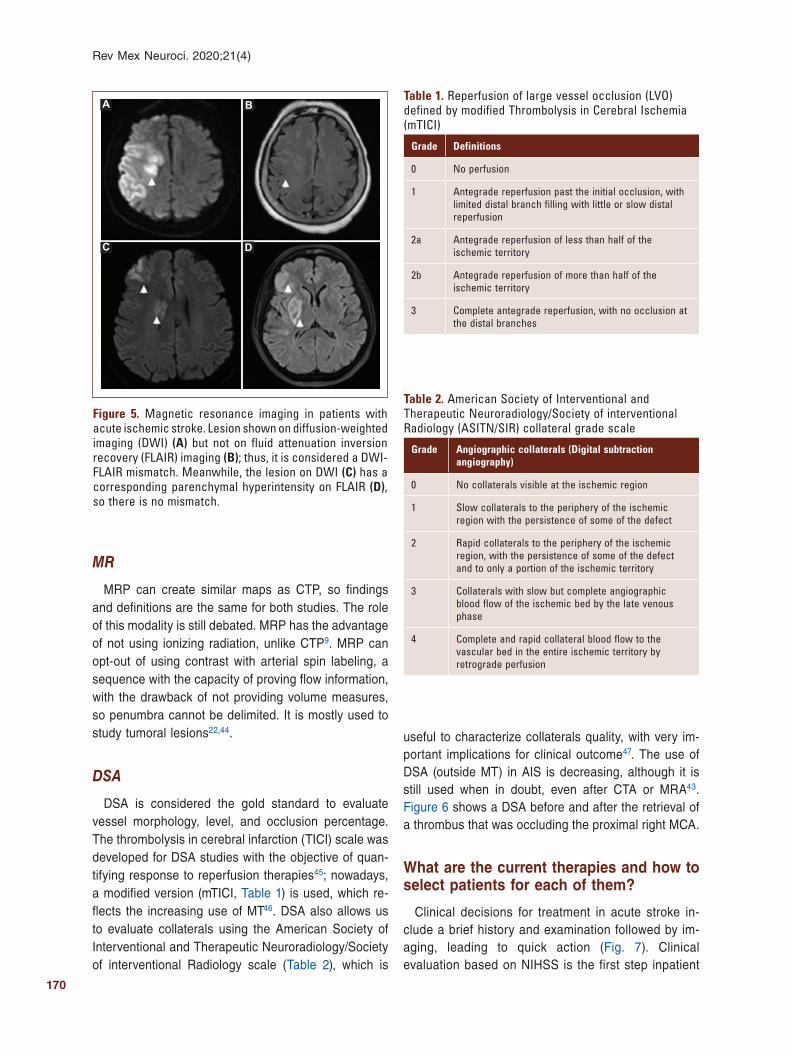
170
Rev Mex Neuroci. 2020;21(4)
MR
MRP can create similar maps as CTP, so findings and definitions are the same for both studies. The role of this modality is still debated. MRP has the advantage of not using ionizing radiation, unlike CTP9. MRP can opt-out of using contrast with arterial spin labeling, a sequence with the capacity of proving flow information, with the drawback of not providing volume measures, so penumbra cannot be delimited. It is mostly used to study tumoral lesions22,44.
DSA
DSA is considered the gold standard to evaluate vessel morphology, level, and occlusion percentage. The thrombolysis in cerebral infarction (TICI) scale was developed for DSA studies with the objective of quan-tifying response to reperfusion therapies45; nowadays, a modified version (mTICI, Table 1) is used, which re-flects the increasing use of MT46. DSA also allows us to evaluate collaterals using the American Society of Interventional and Therapeutic Neuroradiology/Society of interventional Radiology scale (Table 2), which is
useful to characterize collaterals quality, with very im-portant implications for clinical outcome47. The use of DSA (outside MT) in AIS is decreasing, although it is still used when in doubt, even after CTA or MRA43. Figure 6 shows a DSA before and after the retrieval of a thrombus that was occluding the proximal right MCA.
What are the current therapies and how to select patients for each of them?
Clinical decisions for treatment in acute stroke in-clude a brief history and examination followed by im-aging, leading to quick action (Fig. 7). Clinical evaluation based on NIHSS is the first step inpatient
Table 1. Reperfusion of large vessel occlusion (LVO) defined by modified Thrombolysis in Cerebral Ischemia (mTICI)
Grade Definitions
0 No perfusion
1 Antegrade reperfusion past the initial occlusion, with limited distal branch filling with little or slow distal reperfusion
2a Antegrade reperfusion of less than half of the ischemic territory
2b Antegrade reperfusion of more than half of the ischemic territory
3 Complete antegrade reperfusion, with no occlusion at the distal branches
Table 2. American Society of Interventional and Therapeutic Neuroradiology/Society of interventional Radiology (ASITN/SIR) collateral grade scale
Grade Angiographic collaterals (Digital subtraction angiography)
0 No collaterals visible at the ischemic region
1 Slow collaterals to the periphery of the ischemic region with the persistence of some of the defect
2 Rapid collaterals to the periphery of the ischemic region, with the persistence of some of the defect and to only a portion of the ischemic territory
3 Collaterals with slow but complete angiographic blood flow of the ischemic bed by the late venous phase
4 Complete and rapid collateral blood flow to the vascular bed in the entire ischemic territory by retrograde perfusion
Figure 5. Magnetic resonance imaging in patients with acute ischemic stroke. Lesion shown on diffusion‑weighted imaging (DWI) (A) but not on fluid attenuation inversion recovery (FLAIR) imaging (B); thus, it is considered a DWI‑FLAIR mismatch. Meanwhile, the lesion on DWI (C) has a corresponding parenchymal hyperintensity on FLAIR (D), so there is no mismatch.
dc
ba
No
par
t o
f th
is p
ub
licat
ion
may
be
rep
rod
uce
d o
r p
ho
toco
pyi
ng
wit
ho
ut
the
pri
or
wri
tten
per
mis
sio
n o
f th
e p
ub
lish
er.
©
Per
man
yer
2020

171
J.J. Méndez-Gallardo, et al.: Current management of acute stroke
evaluation. It is clear, according to different studies, that there is a correlation between the NIHSS score and outcome48. Seminal MT trials DAWN35 and DE-FUSE 336 included patients with a median NIHSS of 16-17 points, and DEFUSE 338 included patients with a mean ASPECTS of 8, which implies that in extended therapeutic windows, patients with severe stroke could be excluded.
Current reperfusion therapies are rtPA and MT, or a combination of both of them. rtPA is the first-line treatment for AIS, as long as it can be administered in < 4.5 h from symptom onset, and no exclusion cri-teria are met (Table 3); it is within this timeframe that mismatch criteria are not needed, and the only ad-vanced neurimaging technique that could be used is CTA, if the patient were suspected to be a candidate for MT. These first 4.5 h are divided into a classical window (0-3 h after symptom onset) and an extended window (3-4.5 h); the latter has some specific criteria that must be met before initiating rtPA7. It must be stressed that some of the criteria that were previously considered to be contraindications for rtPA (age > 80 years-old, NIHSS > 25, previous stroke in a diabetic patient, etc.) are now considered merely warnings for rtPA during the extended window and are in no way a reason for the physician to refrain from administer-ing rtPA7.
The recanalization rate for patients with a distal oc-clusion who are treated with rtPA goes from 38% to 50%49,50. rtPA is a drug with a number of benefits that
Table 3. Inclusion and exclusion criteria for the treatment of acute ischemic stroke with alteplase, with additional warnings for the extended therapeutic window
Inclusion criteria− Clinical diagnosis of IS causing the measurable neurologic deficit− Onset of symptoms < 4.5 h before beginning treatment; if the exact time of
stroke onset is not known, it is defined as the last time the patient was known to be normal or at neurologic baseline
− Age ≥ 18 yearsExclusion criteria− IS or severe head trauma in the previous 3 months− Previous ICH− Intra‑axial intracranial neoplasm− Gastrointestinal malignancy or hemorrhage in the previous 21 days− Intracranial or intraspinal surgery within the prior 3 months− Symptoms suggestive of SAH− Persistent BP elevation (systolic ≥ 185 mmHg or diastolic ≥ 110 mmHg)− Active internal bleeding− Presentation consistent with infective endocarditis− Stroke known or suspected to be associated with aortic arch dissection− Acute bleeding diathesis− Platelet count < 100,000/mm3
− Current anticoagulant use with an INR > 1.7 or PT > 15 s or aPTT > 40 s or PT > 15 s
− Therapeutic doses of LMWH within 24 h (e.g., to treat VTE and ACS); this exclusion does not apply to prophylactic doses
− Current use of a direct thrombin inhibitor or direct factor Xa inhibitor with evidence of anticoagulant effect by laboratory tests such as aPTT, INR, ECT, TT, or appropriate factor Xa activity assays
− Head CT: evidence of hemorrhage, or extensive regions of obvious hypodensity suggestive or irreversible injury
Warnings− Only minor and isolated neurologic signs or
rapidly improving symptoms − Serum glucose < 50 mg/dL (< 2.8 mmol/L)− Serious trauma in the previous 14 days− Major surgery in the previous 14 days− History of gastrointestinal bleeding (remote) or
genitourinary bleeding− Seizure at the onset of stroke with postictal
neurologic impairments− Pregnancy− Arterial puncture at a non‑compressible site in
the previous 7 days− Large (≥ 10 mm), untreated, unruptured
intracranial aneurysm− Untreated intracranial vascular malformation
Additional warnings for treatment from 3 to 4.5 h from symptom onset− Age > 80 years− Oral anticoagulant use regardless of INR− Severe stroke (NIHSS score > 25)− Combination of both previous ischemic stroke
and diabetes mellitus
ACS: acute coronary syndrome; aPTT: activated partial thromboplastin time; ECT: ecarin clotting time; INR: international normalized ratio; PT: prothrombin time; NIHSS: National Institutes of Health Stroke Scale; tPA: intravenous alteplase; TT: thrombin time; VTE: venous thromboembolism; ISL: ischemic stroke; ICH: intracranial hemorrhage; SBP: systolic blood pressure; SAH: subaracnoid hemorrhage; LMWH: low molecular weight heparin; BP: blood pressure.
Figure 6. Posteroanterior digital subtraction angiography before (A) and after (B) thrombectomy for a right middle cerebral artery occlusion.
ba
No
par
t o
f th
is p
ub
licat
ion
may
be
rep
rod
uce
d o
r p
ho
toco
pyi
ng
wit
ho
ut
the
pri
or
wri
tten
per
mis
sio
n o
f th
e p
ub
lish
er.
©
Per
man
yer
2020

172
Rev Mex Neuroci. 2020;21(4)
depend on the speediness with which it is applied. It is estimated that for every 30 min of treatment delay, chances of a good clinical outcome decrease by 10-15%51. The therapeutic windows are determined by the time from symptom onset. Inclusion and exclusion cri-teria, as well as additional warnings for rtPA have not been modified since 2008. rtPA is the most used drug for intravenous thrombolysis, although there is a grow-ing body of evidence that supports tenecteplase (TNK) as either and safe alternative or even as a potential replacement for rtPA. rtPA binds to the fibrin in a thrombus and causes the plasminogen within the clot to convert to plasmin, which will break down the thrombus46.
Candidates for MT must receive rtPA as soon as possible. The treating physician must keep in mind that MT is not a contraindication for rtPA and that under no circumstances, one should wait for rtPA response in a patient with LVO to decide if they should be taken to MT23. During MT, every time a device is directed at the thrombus with the objective or remov-ing it, it is called a “pass.” With second-generation
devices, MT may achieve a complete or near-com-plete (TICI 2B/3) recanalization rate of up to 84%52. In a trial that included 330 patients, the investigators tried to determine which was the minimal necessary number of attempt for MT; there was a median of 1 pass, with an achieved TICI 2B or 3 in 46.8% of pa-tients; after three passes, the goal was met in 67.9%. Better clinical outcomes were observed in those with 1-3 passes53.
After 4.5 h of symptom onset, the only treatment op-tion is MT. Although the therapeutic window has ex-tended up to 24 h, the more time passes, the selection of patients becomes more rigorous and requires more elements to demonstrate viable brain tissue7,35. The criteria on which treatments are based beyond 6 h of evolution, wake-up stroke, minor stroke, and in some specific situations are described below.
Thrombolysis in minor stroke
Minor stroke is one of the most commonly cited rea-sons for nonuse of rtPA within the classical and
Figure 7. Treatment algorithm in acute ischemic stroke. CTA: computed tomography angiography; CTP: computed tomography perfusion; DWI: diffusion‑weighted imaging; FLAIR: fluid attenuation inversion recovery; IVT: intravenous thrombolysis; LVO: large vessel occlusion; MRI: magnetic resonance imaging; NIHSS: National Institutes of Health Stroke Scale; RAPID is an automated software for CTP; rtPA: recombinant tissue plasminogen activator; TNK: tenecteplase; w/o: without. No brain perfusion imaging needed before 6 h for thrombectomy. *Maximum dose: 90 mg. **Depending if the patient will receive MT. Maximum dose: 25 mg.
No
par
t o
f th
is p
ub
licat
ion
may
be
rep
rod
uce
d o
r p
ho
toco
pyi
ng
wit
ho
ut
the
pri
or
wri
tten
per
mis
sio
n o
f th
e p
ub
lish
er.
©
Per
man
yer
2020
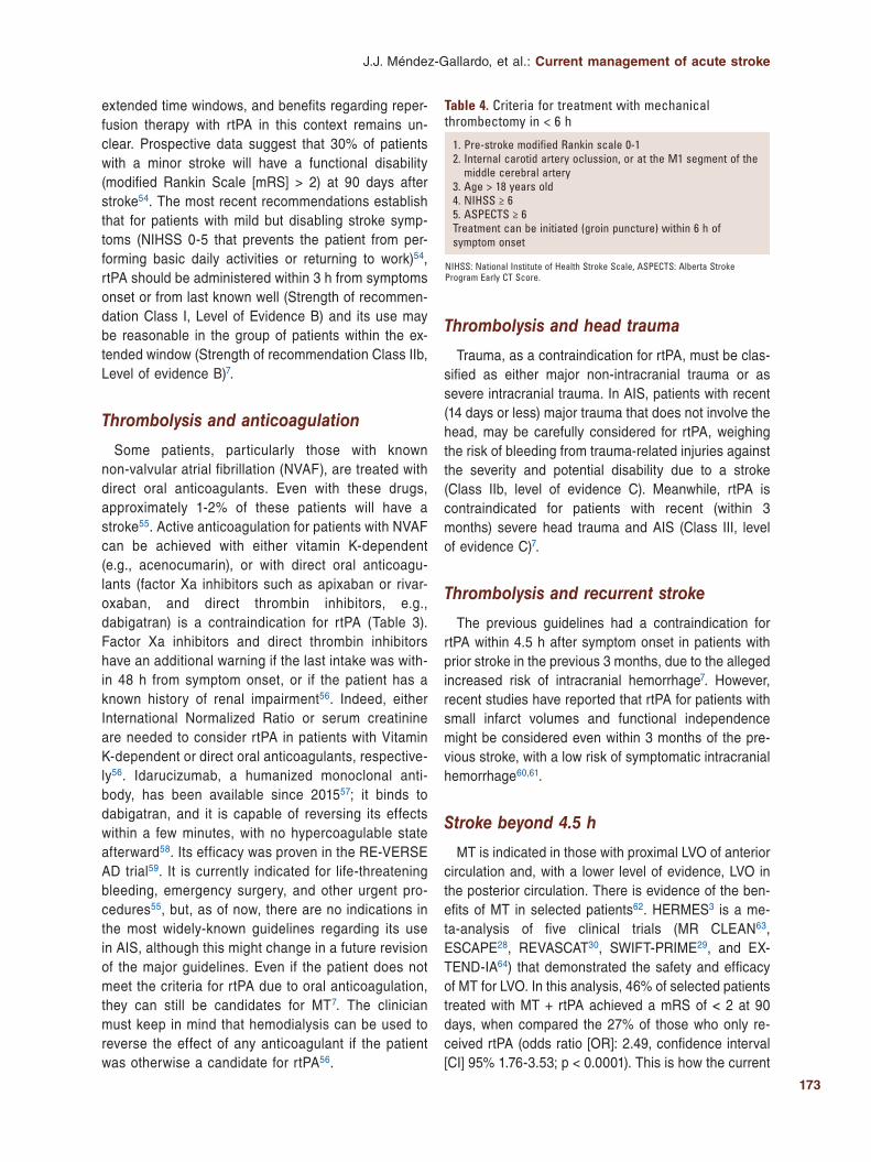
173
J.J. Méndez-Gallardo, et al.: Current management of acute stroke
extended time windows, and benefits regarding reper-fusion therapy with rtPA in this context remains un-clear. Prospective data suggest that 30% of patients with a minor stroke will have a functional disability (modified Rankin Scale [mRS] > 2) at 90 days after stroke54. The most recent recommendations establish that for patients with mild but disabling stroke symp-toms (NIHSS 0-5 that prevents the patient from per-forming basic daily activities or returning to work)54, rtPA should be administered within 3 h from symptoms onset or from last known well (Strength of recommen-dation Class I, Level of Evidence B) and its use may be reasonable in the group of patients within the ex-tended window (Strength of recommendation Class IIb, Level of evidence B)7.
Thrombolysis and anticoagulation
Some patients, particularly those with known non-valvular atrial fibrillation (NVAF), are treated with direct oral anticoagulants. Even with these drugs, approximately 1-2% of these patients will have a stroke55. Active anticoagulation for patients with NVAF can be achieved with either vitamin K-dependent (e.g., acenocumarin), or with direct oral anticoagu-lants (factor Xa inhibitors such as apixaban or rivar-oxaban, and direct thrombin inhibitors, e.g., dabigatran) is a contraindication for rtPA (Table 3). Factor Xa inhibitors and direct thrombin inhibitors have an additional warning if the last intake was with-in 48 h from symptom onset, or if the patient has a known history of renal impairment56. Indeed, either International Normalized Ratio or serum creatinine are needed to consider rtPA in patients with Vitamin K-dependent or direct oral anticoagulants, respective-ly56. Idarucizumab, a humanized monoclonal anti-body, has been available since 201557; it binds to dabigatran, and it is capable of reversing its effects within a few minutes, with no hypercoagulable state afterward58. Its efficacy was proven in the RE-VERSE AD trial59. It is currently indicated for life-threatening bleeding, emergency surgery, and other urgent pro-cedures55, but, as of now, there are no indications in the most widely-known guidelines regarding its use in AIS, although this might change in a future revision of the major guidelines. Even if the patient does not meet the criteria for rtPA due to oral anticoagulation, they can still be candidates for MT7. The clinician must keep in mind that hemodialysis can be used to reverse the effect of any anticoagulant if the patient was otherwise a candidate for rtPA56.
Thrombolysis and head trauma
Trauma, as a contraindication for rtPA, must be clas-sified as either major non-intracranial trauma or as severe intracranial trauma. In AIS, patients with recent (14 days or less) major trauma that does not involve the head, may be carefully considered for rtPA, weighing the risk of bleeding from trauma-related injuries against the severity and potential disability due to a stroke (Class IIb, level of evidence C). Meanwhile, rtPA is contraindicated for patients with recent (within 3 months) severe head trauma and AIS (Class III, level of evidence C)7.
Thrombolysis and recurrent stroke
The previous guidelines had a contraindication for rtPA within 4.5 h after symptom onset in patients with prior stroke in the previous 3 months, due to the alleged increased risk of intracranial hemorrhage7. However, recent studies have reported that rtPA for patients with small infarct volumes and functional independence might be considered even within 3 months of the pre-vious stroke, with a low risk of symptomatic intracranial hemorrhage60,61.
Stroke beyond 4.5 h
MT is indicated in those with proximal LVO of anterior circulation and, with a lower level of evidence, LVO in the posterior circulation. There is evidence of the ben-efits of MT in selected patients62. HERMES3 is a me-ta-analysis of five clinical trials (MR CLEAN63, ESCAPE28, REVASCAT30, SWIFT-PRIME29, and EX-TEND-IA64) that demonstrated the safety and efficacy of MT for LVO. In this analysis, 46% of selected patients treated with MT + rtPA achieved a mRS of < 2 at 90 days, when compared the 27% of those who only re-ceived rtPA (odds ratio [OR]: 2.49, confidence interval [CI] 95% 1.76-3.53; p < 0.0001). This is how the current
Table 4. Criteria for treatment with mechanical thrombectomy in < 6 h
1. Pre‑stroke modified Rankin scale 0‑12. Internal carotid artery oclussion, or at the M1 segment of the
middle cerebral artery 3. Age > 18 years old4. NIHSS ≥ 65. ASPECTS ≥ 6Treatment can be initiated (groin puncture) within 6 h of symptom onset
NIHSS: National Institute of Health Stroke Scale, ASPECTS: Alberta Stroke Program Early CT Score.
No
par
t o
f th
is p
ub
licat
ion
may
be
rep
rod
uce
d o
r p
ho
toco
pyi
ng
wit
ho
ut
the
pri
or
wri
tten
per
mis
sio
n o
f th
e p
ub
lish
er.
©
Per
man
yer
2020

174
Rev Mex Neuroci. 2020;21(4)
criteria for the “classic” window of 6 h for MT came to be. Table 4 summarizes the criteria for MT in these patients, in accordance with the 2019 update of the North American guidelines7.
Reperfusion therapy from 6 to 24 h
Two clinical trials published in 2018 changed the par-adigm of the treatment of AIS. Both of these studies are based on the concept of mismatch and had to be stopped early due to benefit from thrombectomy. DE-FUSE 338 (Endovascular Therapy Following Imaging Evaluation for Ischemic Stroke) demonstrated that if a patient with LVO has NIHSS > 6, and a small ischemic core (< 70 ml) with an extensive penumbra (penumbra: core ratio > 1.8), there is a great possibility of benefit with MT from 6 to 16 h from symptom onset. DAWN37 (Thrombectomy 6-24 h after stroke with a mismatch between deficit and infarct) required LVO with NIHSS > 10, small core (stratified in different age groups, from < 21 ml to < 51 ml), which would cause a clinical-radio-logical mismatch. Both studies required the patient to be previously independent (mRS < 2 for DEFUSE 3, and mRS < 1 for DAWN) and used PWI. In DEFUSE 3, patients treated with MT at a median of 11 h, had a 28% increase in functional independence and an addi-tional 20% absolute reduction in death or severe dis-ability. These two trials used a proprietary automated software (RAPID) to determine the ischemic core and penumbra. The median time from symptoms to enroll-ment was 12.5 h in DAWN and 11 h in DEFUSE 3, and core volumes were < 10 mL; it is worth noting that the patients in both studies were very slow progressors. Effectiveness for late window thrombectomy was main-tained across all subgroups of patients, including those defined by time, age, mode of presentation, and AS-PECTS score. Undoubtedly, the results of DAWN and DEFUSE 3 have shown great benefit from MT in se-lected patients with AIS within a therapeutic window of 6-24 h. However, as suggested by the large treatment effect size observed in both trials, the clinical and im-aging criteria were probably too strict35,36. In addition, not all hospitals that treat AIS have this specific soft-ware required in the inclusion criteria of both studies. However, physicians treating AIS patients should be familiar with this technique and its inclusion criteria, so as to offer the treatment to as many patients as possi-ble, when appropriate.
Wake-up strokes and patients with an unknown time of symptom onset
Wake-up stroke includes patients that wake up with a focal deficit attributed to an AIS, while those that are simply found with an established deficit, with an un-known time from symptom onset, are deemed to have an AIS with unknown time of symptom onset. There are fundamental differences with therapeutic implications in these apparently all-too-similar stroke definitions. WAKE UP (MRI-Guided thrombolysis for stroke with unknown time of onset, 2018)21 trial demonstrated the benefit of rtPA in both of these situations, with a median mRS of 1 after 90 days in the alteplase group, and 2 in the placebo group (adjusted common OR, 1.62; 95% CI, 1.17-2.23; p = 0.003); the trial used a DWI-FLAIR mismatch in the ischemic region to determine candi-dates for rtPA. The trial was stopped early due to fund-ing issues, but the benefit was clear among those who received rtPA, as opposed to placebo.
EXTEND65 (Thrombolysis Guided by Perfusion Imag-ing up to 9 h After Onset of Stroke) was published in May 2019, which assessed rtPA in a period defined by the researchers as the late window (4.5-9 h from symp-tom onset) in 225 patients. The primary outcome (mRS 0-1 at 90 days) occurred in 35.4% of the patients in the alteplase group and in 29.5% of the placebo group (ad-justed risk ratio, 1.44; 95% CI, 1.01-2.06; p = 0.04). This trial evaluated cerebral perfusion using CTP. They con-cluded that these imaging modalities increased rtPA use, with the risk of more symptomatic hemorrhages (6.2% in the rtPA group and 0.9% in the placebo arm).
In patients with AIS on awakening or with a known time of onset and who have MRI DWI/FLAIR mismatch, the European guidelines suggest rtPA over no rtPA, even when the quality of evidence is moderate, and the strength of recommendation is weak66.
Stroke units
Stroke units are equipped with continuous non-invasive monitorization, with trained personnel, and coordinated by neurologists who lead a multidisciplinary team. The AHA/ASA has demonstrated, with a level of evidence I, a re-duction in mortality or dependency of 18%, compared to the hospitalization in general wards23,67. A British study reported that physicians working at stroke units are more likely to treat patients based on up-to-date evidence, with better functional outcomes68. There is also a Cochrane69 review with similar results, with independent outcomes for age, gender, and stroke severity. To date, there are few
No
par
t o
f th
is p
ub
licat
ion
may
be
rep
rod
uce
d o
r p
ho
toco
pyi
ng
wit
ho
ut
the
pri
or
wri
tten
per
mis
sio
n o
f th
e p
ub
lish
er.
©
Per
man
yer
2020

175
J.J. Méndez-Gallardo, et al.: Current management of acute stroke
stroke units in our country, and, even in developed coun-tries70, the characteristics of stroke units are still not stan-dardized, leading to varying results.
Future prospects
TNK is a new-generation fibrinolytic agent. This drug represents a promising alternative to rtPA as a throm-bolytic treatment for patients with AIS. An important advantage of TNK over rtPA is the manner of adminis-tration, as TNK is applied in a single bolus, compared to rtPA that requires a bolus and a 60-min infusion71. EXTEND-IA TNK72 (TNK vs. rtPA before Thrombecto-my for Ischemic Stroke) randomized 202 patients with AIS who candidates for intravenous thrombolysis (ei-ther rtPA or TNK) were followed by MT. The primary endpoint was the demonstration of reperfusion greater than 50% of the ischemic territory, or absence of any retrievable thrombus. This outcome was reached in 22% of the TNK arm, and in 10% of the rtPA group, with no differences in symptomatic intracranial hemor-rhage. The accepted dose for TNK to treat AIS depends on whether or not the patient will receive MT: 0.25 mg/kg if the patient will receive MT, and 0.40 mg/kg if the pa-tient will not receive MT, with a maximum dose of 25 mg in a single intravenous bolus7.
A number of trials mean to establish whether patients with less favorable imaging findings (i.e., with no DAWN or DEFUSE-3 criteria) could receive some degree of benefit from MT. Ongoing studies TESLA, SELECT 2, TENSION, and IN EXTREMIS, all use ASPECTS to define if MT is reasonable and safe for moderate to large infarcts, defined as ASPECTS 2-573. Depending on the results, the number of candidates for reperfusion therapy could be greatly increased.
Conclusions
Intravenous thrombolysis continues to be the corner-stone of reperfusion therapy for AIS. LVOs are to be rapidly diagnosed and may have an even greater ben-efit with a combined therapy (rtPA + MT). Neurologists have a fundamental role in suspecting of LVO and confirming this suspicion using advanced neuroimag-ing; if proximal LVO is confirmed and the patient is within the therapeutic window from rtPA, the infusion must begin as soon as possible, while simultaneously alerting the team responsible for MT. The main function of the neurologist in the context of AIS is to not delay rtPA and to recognize LVO in a timely manner, so as to offer MT to all possible candidates, to achieve the
maximum possible benefit. The implementation of al-gorithms to act according to the reality of each one of our hospitals is central for the proper diagnosis and treatment of patients with AIS caused or not by LVO.
Lessons to take home1. Only 10‑20% of AIS is due to LVO, but these are responsible
for the greatest morbidity and mortality burden.2. For patients with symptom onset between 3 and 4.5 h, one
must mind the additional warning criteria for rtPA3. In a patient with more than 4.5 h and LVO, must consider MT4. In patients with symptom onset of < 4.5 h, no perfusion
imaging is required5. In a patient with 6‑24 h of symptom onset, perfusion imaging
should be performed to assess core and penumbra, so as to determine if the patient is a candidate to MT by DAWN or DEFUSE 3 criteria.
6. Wake‑up stroke patients can receive rtPA only if they have a DWI‑FLAIR mismatch, according to the WAKE‑UP criteria
7. TNK is a promising alternative to rtPA and seems to have a better safety profile and to be easier to administrate. Its use in the clinical field is gradually increasing
Lessons to take home for physicians in Latin America8. For patients with either wake‑up stroke or stroke of
undetermined time, should the receiving hospital only have NCCT, patients with ASPECTS 7‑10 would benefit from a referral to a third‑level hospital, where advanced neuroimaging may be implemented
9. Within the first 4.5 h from symptom onset, only NCCT with ASPECTS 7‑10 is required to start rtPA
10. Given the possibility, the physician should obtain at least NCCT and CTA before referring a patient to a third‑level hospital. CTP is required for AIS > 4.5 h, and it should be obtained if possible
11. If the physician detects a high NIHSS (≥ 6), they should consider referring the patient to a center with MT. rtPA must not be withheld if the patient meets criteria for thrombolysis
Acknowledgments
We thank the team at our Stroke Clinic for their sup-port and hard work during the creation of this paper.
Funding
This research received no specific grant from any funding agency in public, commercial, or not-for-profit sectors.
Conflicts of interest
None.
Ethical disclosures
Protection of human and animal subjects. The authors declare that no experiments were performed on humans or animals for this study.
No
par
t o
f th
is p
ub
licat
ion
may
be
rep
rod
uce
d o
r p
ho
toco
pyi
ng
wit
ho
ut
the
pri
or
wri
tten
per
mis
sio
n o
f th
e p
ub
lish
er.
©
Per
man
yer
2020

176
Rev Mex Neuroci. 2020;21(4)
Confidentiality of data. The authors declare that they have followed the protocols of their work center on the publication of patient data.
Right to privacy and informed consent. The authors declare that no patient data appear in this article.
References 1. Romano JG. Thrombolysis in Mexico-current status and opportunities.
Rev Mex Neurocienc. 2019;20:208-9. 2. Lyden PD. Thrombolytic therapy for acute ischemic stroke. Stroke.
2019;50:2597-603. 3. Goyal M, Menon BK, van Zwam WH, Dippel DW, Mitchell PJ, Dem-
chuk AM, et al. Endovascular thrombectomy after large-vessel ischaemic stroke: a meta-analysis of individual patient data from five randomised trials. Lancet. 2016;387:1723-31.
4. Malhotra K. Ischemic strokes due to large-vessel occlusions contribute disproportionately to stroke-related dependence and death : a review. Front Neurol. 2017;8:1-5.
5. Campbell BC, Berkhemer OA, Majoie CB, Dippel DW. Endovascular therapy for ischemic stroke. N Engl J Med. 2015;372:2363-6.
6. Powers WJ, Derdeyn CP, Biller J, Coffey CS, Hoh BL, Jauch EC, et al. 2015 American heart association/American stroke association focused update of the 2013 guidelines for the early management of patients with acute ischemic stroke regarding endovascular treatment. Stroke. 2015;46:3020-35.
7. Powers WJ, Rabinstein AA, Ackerson T, Adeoye OM, Bambakidis NC, Becker K, et al. Guidelines for the early management of patients with acute ischemic stroke: 2019 update to the 2018 guidelines for the early management of acute ischemic stroke: a guideline for healthcare profes-sionals from the American heart association/American stroke. Stroke. 2019;50:3331-2.
8. Yuh WT, Alexander MD, Ueda T, Maeda M, Taoka T, Yamada K, et al. Revisiting current golden rules in managing acute ischemic stroke: eva-luation of new strategies to further improve treatment selection and outcome. Am J Roentgenol. 2017;208:32-41.
9. Kamalian S, Lev MH. Stroke imaging. Radiol Clin North Am. 2019;57:717-32. 10. Martins SC, Sacks C, Hacke W, Brainin M, de Assis Figueiredo F, Pon-
tes-Neto OM, et al. Priorities to reduce the burden of stroke in Latin American countries. Lancet Neurol. 2019;18:674-83.
11. Arauz A, Mendez B, Soriano-Navarro E, Ruiz-Franco A, Quinzaños J, Rodríguez-Barragán M, et al. Thrombolytic therapy for acute stroke in Mexico-experience of four Mexican hospitals. Rev Mex Neurocienc. 2019;20:210-3.
12. León-Jiménez C, Ruiz-Sandoval JL, Chiquete E, Vega-Arroyo M, Arauz A, Murillo-Bonilla LM, et al. Tiempo de llegada hospitalaria y pronóstico funcional después de un infarto cerebral: resultados del estu-dio PREMIER. Neurologia. 2014;29:200-9.
13. Wu L, Wu W, Tali ET, Yuh WT. Oligemia, Penumbra, infarction: unders-tanding hypoperfusion with neuroimaging. Neuroimaging Clin N Am. 2018;28:599-609.
14. Fantini S, Sassaroli A, Tgavalekos KT, Kornbluth J. Cerebral blood flow and autoregulation: current measurement techniques and prospects for noninvasive optical methods. Neurophotonics. 2016;3:031411.
15. Sotoudeh H, Bag AK, Brooks MD. Code-stroke CT perfusion; challenges and pitfalls. Acad Radiol. 2019;26:1565-79.
16. Davis SM, Donnan GA, Parsons MW, Levi C, Butcher KS, Peeters A, et al. Effects of alteplase beyond 3 h after stroke in the echoplanar imaging thrombolytic evaluation trial (EPITHET): a placebo-controlled randomised trial. Lancet Neurol. 2008;7:299-309.
17. Albers GW, Thijs VN, Wechsler L, Kemp S, Schlaug G, Skalabrin E, et al. Magnetic resonance imaging profiles predict clinical response to early reperfusion: the diffusion and perfusion imaging evaluation for unders-tanding stroke evolution (DEFUSE) study. Ann Neurol. 2006;60:508-17.
18. Lansberg MG, Straka M, Kemp S, Mlynash M, Wechsler LR, Jovin TG, et al. MRI profile and response to endovascular reperfusion after stroke (DEFUSE 2): a prospective cohort study. Lancet Neurol. 2012;11:860-7.
19. Lui YW, Tang ER, Allmendinger AM, Spektor V. Evaluation of CT perfu-sion in the setting of cerebral ischemia: patterns and pitfalls. Am J Neuroradiol. 2010;31:1552-63.
20. Merino JG, Warach S. Imaging of acute stroke. Nat Rev Neurol. 2010;6:560-71.
21. Thomalla G, Simonsen CZ, Boutitie F, Andersen G, Berthezene Y, Cheng B, et al. MRI-guided thrombolysis for stroke with unknown time of onset. N Engl J Med. 2018;379:611-22.
22. Smith WS, Lev MH, English JD, Camargo EC, Chou M, Johnston SC, et al. Significance of large vessel intracranial occlusion causing acute ischemic stroke and tia. Stroke; a J Cereb Circ. 2009;40:3834-40.
23. Powers WJ, Rabinstein AA, Ackerson T, Adeoye OM, Bambakidis NC, Becker K, et al. 2018 Guidelines for the early management of patients with acute ischemic stroke: a guideline for healthcare professionals from the American heart association/American stroke association. Stroke. 2018;49:e46-110.
24. Tseng MC, Chang KC. Stroke severity and early recovery after first-ever ischemic stroke: results of a hospital-based study in Taiwan. Health Policy. 2006;79:73-8.
25. Turc G, Maïer B, Naggara O, Seners P, Isabel C, Tisserand M, et al. Clinical scales do not reliably identify acute ischemic stroke patients with large-artery occlusion. Stroke. 2016;47:1466-72.
26. Barber PA, Demchuk AM, Zhang J, Buchan AM. Validity and reliability of a quantitative computed tomography score in predicting outcome of hype-racute stroke before thrombolytic therapy. Lancet. 2000;355:1670-4.
27. Prakkamakul S, Yoo AJ. ASPECTS CT in acute ischemia: review of current data. Top Magn Reson Imaging. 2017;26:103-12.
28. Goyal M, Demchuk AM, Menon BK, Eesa M, Rempel JL, Thornton J, et al. Randomized assessment of rapid endovascular treatment of ische-mic stroke. N Engl J Med. 2015;372:1019-30.
29. Saver JL, Goyal M, Bonafe A, Diener HC, Levy EI, Pereira VM, et al. Stent-retriever thrombectomy after intravenous t-PA vs. t-PA alone in stroke. N Engl J Med. 2015;372:2285-95.
30. Jovin TG, Chamorro A, Cobo E, de Miquel MA, Molina CA, Rovira A, et al. Thrombectomy within 8 hours after symptom onset in ischemic stroke. N Engl J Med. 2015;372:2296-306.
31. Tei H, Uchiyama S, Usui T, Ohara K. Posterior circulation ASPECTS on diffusion-weighted MRI can be a powerful marker for predicting functional outcom. J Neurol. 2010;257:767-73.
32. Hextrum S, Biller J. Clinical distinction of cerebral ischemia and triaging of patients in the emergency department: mimics, wake-ups, late strokes, and chameleons. Neuroimaging Clin N Am. 2018;28:537-49.
33. Brinjikji W, Demchuk AM, Murad MH, Rabinstein AA, McDonald RJ, McDonald JS, et al. Neurons over nephrons. Stroke. 2017;48:1862-8.
34. Tsivgoulis G, Katsanos AH, Schellinger PD, Köhrmann M, Varelas P, Magoufis G, et al. Successful reperfusion with intravenous thrombolysis preceding mechanical thrombectomy in large-vessel occlusions. Stroke. 2018;49:232-5.
35. Riedel CH, Zimmermann P, Jensen-Kondering U, Stingele R, Deuschl G, Jansen O. The importance of size: successful recanalization by intrave-nous thrombolysis in acute anterior stroke depends on thrombus length. Stroke. 2011;42:1775-7.
36. Bang OY, Saver JL, Kim SJ, Kim GM, Chung CS, Ovbiagele B, et al. Collateral flow averts hemorrhagic transformation after endovascular the-rapy for acute ischemic stroke. Stroke. 2011;42:2235-9.
37. Nogueira RG, Jadhav AP, Haussen DC, Bonafe A, Budzik RF, Bhuva P, et al. Thrombectomy 6 to 24 hours after stroke with a mismatch between deficit and infarct. N Engl J Med. 2017;378:11-21.
38. Albers GW, Marks MP, Kemp S, Christensen S, Tsai JP, Ortega-Gutie-rrez S, et al. Thrombectomy for stroke at 6 to 16 hours with selection by perfusion imaging. N Engl J Med. 2018;378:708-18.
39. Vagal A, Foster LD, Menon BK, Livorine A, Shi J, Qazi E, et al. Multimo-dal CT imaging: time to treatment and outcomes in the IMS III trial. Am J Neuroradiol. 2016;37:1393-8.
40. Vagal A, Meganathan K, Kleindorfer DO, Adeoye O, Hornung R, Khatri P. Increasing use of computed tomographic perfusion and computed tomo-graphic angiograms in acute ischemic stroke from 2006 to 2010. Stroke. 2014;45:1029-34.
41. Shen J, Li X, Li Y, Wu B. Comparative accuracy of CT perfusion in diagnosing acute ischemic stroke: a systematic review of 27 trials. PLoS One. 2017;12:1-17.
42. Souza LC, Chaudhry SA, Payabvash S, Yoo AJ, Kemmling A, Schaefer PW, et al. Malignant CTA collateral profile is highly specific for large admission DWI infarct core and poor outcome in acute stroke. AJNR Am J Neuroradiol. 2012;33:1331-6.
43. Leslie-Mazwi TM, Hirsch JA, Falcone GJ, Schaefer PW, Lev MH, Rabinov JD, et al. Endovascular stroke treatment outcomes after patient selection based on magnetic resonance imaging and clinical criteria. JAMA Neurol. 2016;73:43-9.
44. Bang OY, Goyal M, Liebeskind DS. Collateral circulation in ischemic stroke: assessment tools and therapeutic strategies. Stroke. 2015;46:3302-9.
45. Higashida RT, Furlan AJ, Roberts H, Tomsick T, Connors B, Barr J, et al. Trial design and reporting standards for intra-arterial cerebral thromboly-sis for acute ischemic stroke. Stroke. 2003;34:2774.
46. Zaidat OO, Yoo AJ, Khatri P, Tomsick TA, Von Kummer R, Saver JL, et al. Recommendations on angiographic revascularization grading standards for acute ischemic stroke: a consensus statement and for the cerebral angio-graphic revascularization, grading (CARG) collaborators, STIR revascula-rization working group, and STIR thrombol. Stroke. 2013;44:2630-63.
47. Sheth SA, Liebeskind DS. Collaterals in endovascular therapy for stroke. Curr Opin Neurol. 2015;28:10-5.
48. Rost NS, Bottle A, Lee JM, Randall M, Middleton S, Shaw L, et al. Stroke severity is a crucial predictor of outcome: an international prospective validation study. J Am Heart Assoc. 2016;5:1-7.
No
par
t o
f th
is p
ub
licat
ion
may
be
rep
rod
uce
d o
r p
ho
toco
pyi
ng
wit
ho
ut
the
pri
or
wri
tten
per
mis
sio
n o
f th
e p
ub
lish
er.
©
Per
man
yer
2020

177
J.J. Méndez-Gallardo, et al.: Current management of acute stroke
49. del Zoppo GJ, Poeck K, Pessin MS, Wolpert SM, Furlan AJ, Ferbert A, et al. Recombinant tissue plasminogen activator in acute thrombotic and embolic stroke. Ann Neurol. 1992;32:78-86.
50. Molina CA, Montaner J, Arenillas JF, Ribo M, Rubiera M, Alvarez-Sabín J. Differential pattern of tissue plasminogen activator-induced proximal middle cerebral artery recanalization among stroke subtypes. Stroke. 2004;35:486-90.
51. Mazighi M, Chaudhry SA, Ribo M, Khatri P, Skoloudik D, Mokin M, et al. Impact of onset-to-reperfusion time on stroke mortality: a collaborative pooled analysis. Circulation. 2013;127:1980-5.
52. Demchuk AM, Goyal M, Yeatts SD, Carrozzella J, Foster LD, Qazi E, et al. Recanalization and clinical outcome of occlusion sites at baseline CT angiography in the interventional management of stroke III trial. Ra-diology. 2014;273:202-10.
53. Flottmann F, Leischner H, Broocks G, Nawabi J, Bernhardt M, Faizy TD, et al. Recanalization rate per retrieval attempt in mechanical thrombec-tomy for acute ischemic stroke. Stroke. 2018;49:2523-5.
54. Khatri P, Kleindorfer DO, Devlin T, Sawyer RN, Starr M, Mejilla J, et al. Effect of alteplase vs aspirin on functional outcome for patients with acute ischemic stroke and minor nondisabling neurologic deficits the PRISMS randomized clinical trial. J Am Med Assoc. 2018;320:156-66.
55. Fang CW, Te TY, Chou PC, Chen HM, Lu CM, Tsao CR, et al. Intrave-nous Thrombolysis in acute ischemic stroke after idarucizumab reversal of dabigatran effect: analysis of the cases from Taiwan. J Stroke Cere-brovasc Dis. 2019;28:815-20.
56. Seiffge DJ, Polymeris AA, Fladt J, Lyrer PA, Engelter ST, De Mar-chis GM. Management of patients with stroke treated with direct oral anticoagulants. J Neurol. 2018;265:3022-33.
57. Baule A, Cabigiosu F, Zanda B, Sanna A, Mongili C, Manca A. Throm-bolysis in Acute ischemic stroke after idarucizumab for dabigatran etexi-late reversal in elderly: a case report. J Vasc Interv Neurol. 2018;10:15-7.
58. Meyer D, Chu F, Derry K, Hailey L. Acute reversal of dabigatran with idarucizumab for intravenous thrombolysis as acute stroke treatment. J Clin Neurosci. 2019;59:355-7.
59. Pollack CV, Reilly PA, Van Ryn J, Eikelboom JW, Glund S, Bernstein RA, et al. Idarucizumab for dabigatran reversal-full cohort analysis. N Engl J Med. 2017;377:431-41.
60. Kahles T, Mono ML, Heldner MR, Baumgartner RW, Sarikaya H, Luft A, et al. Repeated intravenous thrombolysis for early recurrent stroke: cha-llenging the exclusion criterion. Stroke. 2016;47:2133-5.
61. Wu C, Wu D, Chen J, Li C, Ji X. Why not intravenous thrombolysis in patients with recurrent stroke within 3 months? Aging Dis. 2018;9:309-16.
62. Fransen PS, Beumer D, Berkhemer OA, Van Den Berg LA, Lingsma H. MR CLEAN, a multicenter randomized clinical trial of endovascular treat-ment for acute ischemic stroke in the Netherlands : study protocol for a randomized controlled trial. Trials. 2014;15:343.
63. Berkhemer OA, Fransen PS, Beumer D, Van Den Berg LA, Lingsma HF, Yoo AJ, et al. A randomized trial of intraarterial treatment for acute is-chemic stroke. N Engl J Med. 2015;372:11-20.
64. Yan B, Dowling RJ, Parsons MW, Oxley TJ, Wu TY, Brooks M, et al. Endovascular Therapy for ischemic stroke with perfusion-imaging selec-tion. N Engl J Med. 2015;372(11):1009-18.
65. Ma H, Campbell BC, Parsons MW, Churilov L, Levi CR, Hsu C, et al. Thrombolysis guided by perfusion imaging up to 9 hours after onset of stroke. N Engl J Med. 2019;380:1795-803.
66. Turc G, Bhogal P, Fischer U, Khatri P, Lobotesis K, Mazighi M, et al. European stroke organisation (ESO)-European society for minimally invasive neurological therapy (ESMINT) guidelines on mechanical throm-bectomy in acute ischaemic stroke endorsed by stroke alliance for Euro-pe (SAFE). Eur Stroke J. 2019;4:6-12.
67. de Leciñana MA, Egido JA, Casado I, Ribó M, Dávalos A, Masjuan J, et al. Guía para el tratamiento del infarto cerebral agudo. Neurologia. 2014;29:102-22.
68. Ramsay AI, Morris S, Hoffman A, Hunter RM, Boaden R, McKevitt C, et al. Effects of centralizing acute stroke services on stroke care provision in two large metropolitan areas in England. Stroke. 2015;46:2244-51.
69. Langhorne P, Ramachandra S, Stroke Unit Trialists’ Collaboration. Organised inpatient (stroke unit) care for stroke (Review). Cochrane Rev. 2013;11:2-3.
70. Rac VE, Sahakyan Y, Fan I, Ieraci L, Hall R, Kelloway L, et al. The characteristics of stroke units in Ontario: a pan-provincial survey. BMC Health Serv Res. 2017;17:1-10.
71. Rønning OM, Logallo N, Thommessen B, Tobro H, Novotny V, Kvis-tad CE, et al. Tenecteplase versus alteplase between 3 and 4.5 hours in low national institutes of health stroke scale. Stroke. 2019;50:498-500.
72. Campbell BC, Mitchell PJ, Churilov L, Yassi N, Kleinig TJ, Dowling RJ, et al. Tenecteplase versus alteplase before thrombectomy for ischemic stroke. N Engl J Med. 2018;378:1573-82.
73. Phipps MS, Cronin CA. Management of acute ischemic stroke. BMJ. 2020;368:1-15.
No
par
t o
f th
is p
ub
licat
ion
may
be
rep
rod
uce
d o
r p
ho
toco
pyi
ng
wit
ho
ut
the
pri
or
wri
tten
per
mis
sio
n o
f th
e p
ub
lish
er.
©
Per
man
yer
2020