revision of 13 genera of haploporidae (trematoda)
TRANSCRIPT

The University of Southern MississippiThe Aquila Digital Community
Dissertations
Spring 5-2014
Revision of 13 Genera of Haploporidae(Trematoda)Eric Edward PulisUniversity of Southern Mississippi
Follow this and additional works at: https://aquila.usm.edu/dissertations
Part of the Zoology Commons
This Dissertation is brought to you for free and open access by The Aquila Digital Community. It has been accepted for inclusion in Dissertations by anauthorized administrator of The Aquila Digital Community. For more information, please contact [email protected].
Recommended CitationPulis, Eric Edward, "Revision of 13 Genera of Haploporidae (Trematoda)" (2014). Dissertations. 23.https://aquila.usm.edu/dissertations/23

May 2014
The University of Southern Mississippi
REVISION OF 13 GENERA OF HAPLOPORIDAE (TREMATODA)
by
Eric Edward Pulis
Abstract of a Dissertation Submitted to the Graduate School
of The University of Southern Mississippi in Partial Fulfillment of the Requirements
for the Degree of Doctor of Philosophy

ii
ABSTRACT
REVISION OF 13 GENERA OF HAPLOPORIDAE (TREMATODA)
by Eric Edward Pulis
May 2014
The Haploporidae is a family of digeneans united by the combination of the
possession of a hermaphroditic sac and a single testis or, rarely, two tandem testes. The
major divisions in the Haploporidae have been based on the organization, development,
and nature of the male and female reproductive systems. Overstreet and Curran (2005)
has been the only attempt to organize the genera of the Haploporidae in a subfamilial
framework. In the present work the validity of the subfamily Waretrematinae by
Overstreet and Curran (2005) is assessed by morphological and molecular methods,
based on original descriptions, type and vouchered, specimens and newly collected
material for morphology and rDNA sequence data analyses. These analyses conflicted
with hypotheses and framework by Overstreet and Curran (2005) in that: (1)
Megasoleninae is the basal subfamily within the Haploporidae; (2) Waretrematinae and
Haploporinae are not sister groups; (3) species of Spiritestis and Capitimitta, both
previously considered members of Waretrema, are not closely related; (4)
Waretrematinae was not monophyletic when Unisaccus and New Genus 1, new species
are included; (5) species of Unisaccoides and Unisaccus species are closely related; and
(6) species of Intromugil are allocated to Chalcinotrematinae rather than Waretrematinae.
Reports of Waretrema were determined to comprise members of three different genera.
The morphology of Intromugil was accessed by the redescription of I. mugilicolus from
newly collected material and a new species is described. Species from the Indo-Pacific

iii
region possessing spirally arranged pads in the hermaphroditic duct and a caecum were
accessed and required changes to the organization and membership of several genera plus
changes to the intergeneric relations within the family based on phylogenetic analysis.
Members of the genera Platydidymus and Carassotrema were assessed and a new species
was described. Two new genera of haploporid were diagnosed, a new species was
described for each, and phylogenetic relations are estimated. A new genus and new
species of Megaperidae are described, and molecular data were provided for three other
species. Previously, megaperid species were members of the Apocreadiidae rather than
the Haploporidae. Phylogenetic hypotheses based on Bayesian Inference analysis of an
alignment of partial 28S gene sequences of haploporids provide a framework for the
evaluation of the interrelationships within the Haploporidae.

COPYRIGHT BY
ERIC EDWARD PULIS
2014

May 2014
The University of Southern Mississippi
REVISION OF 13 GENERA OF HAPLOPORIDAE (TREMATODA)
by
Eric Edward Pulis
A Dissertation Submitted to the Graduate School
of The University of Southern Mississippi in Partial Fulfillment of the Requirements
for the Degree of Doctor of Philosophy Approved: _Robin M. Overstreet__________________ Director _Reginald B. Blaylock_________________ _Richard W. Heard____________________ _Jeffrey M. Lotz______________________
_Maureen A. Ryan____________________ Dean of the Graduate School

vi
ACKNOWLEDGMENTS
Working at the Gulf Coast Research Laboratory (GCRL, Ocean Springs,
Mississippi) has been a privilege that I will always look back on fondly. I have been truly
lucky to have had the opportunity to work there and learn from all with whom I have
come in contact.
I would like to extend special thanks to Robin Overstreet for sharing his
tremendous knowledge of symbionts with me, especially concerning taxonomy and
systematics. I would like to thank my other committee members Reg Blaylock, Jeffery
Lotz, and Richard Heard for their direction. I would like to thank laboratory members
Michael Andres, Stephen Curran, and Ronnie Palmer for their time spent conversing
about and collecting helminths. I am grateful for help with molecular techniques provided
by Jean A. Jovonovich and Janet Wright
I am grateful to the following people for their contributions to the procurement of
Australian hosts, worms, literature, and identification of hosts: Robert Adlard, Michael
Andres, Mal Bryant, David Gibson, Gavin Dally, Penny Desouza, Mark Grubert, Jason
Lally, Jeff Johnson, Richard Wilan, Rex Williams, and the late Ilan Paperna for
specimens of Spiritestis arabii. I thank MengYang Yang, Shanmei Cao, Xiong Xiang
Ying, and Xue Juan Ding for assistance in all aspects of traveling and collecting in China;
National Marine Fisheries Service personel, especially Alonzo Hamilton, Nick Hopkins,
Adam Pollack, Mark Grace, Kevin Rademacher, and the crew of the NOAA research
vessels Pisces and Gordon Gunter for assistance in collecting fish; Vasyl Tkach for
producing scanning electron microscopy photomicrographs of specimens; Eric P.
Hoberg, Patricia Pilitt, Roman Kutchta, Irena Podvyaznaya, Toshiaki Kuramochi, and

vii
Masaaki Machida for lending specimens; Gui Tang Wang, (Institute of Hydrobiology,
China) for attempting to find specimens of Carassotrema; Ervin Otvos, Huang Hailong,
Lina Fu, Shuo Shen, Wei Wu, and Hongwei Ma for help with translations.
I am grateful to my wife, Kelly Pulis, my daughter, Evelyn Pulis, and my family
and friends for their unwavering patience, support, and encouragement.
This material is based on work supported by the National Science Foundation
under grant no. 0529684; the United States Department of Commerce, National
Oceanographic and Atmospheric Administration award no. NA08NOS4730322; and by
the US Fish and Wildlife Service/Mississippi Department of Marine Resources MSCIAP
MS.R.798 Award M10AF20151.
This dissertation is not intended as a scientific record (see article 8.2, ICZN,
International Code of Zoological Nomenclature) for the taxonomic names and
nomenclatural acts contained within the dissertation under article 8.3 of the ICZN. This
dissertation is not a contribution to the primary scientific literature, nor should it be cited
as such.

viii
TABLE OF CONTENTS
ABSTRACT ...................................................................................................................... ii
ACKNOWLEDGMENTS ................................................................................................ vi
LIST OF TABLES ............................................................................................................ xi
LIST OF ILLUSTRATIONS .......................................................................................... xii
CHAPTER
I. INTRODUCTION ..................................................................................... 1 II. REVIEW OF PISCINE (TREMATODA) GENERA WITH ORNATE MUSCULARISATION IN THE REGION OF THE ORAL SUCKER, INCLUDING FOUR NEW SPECIES AND A NEW GENUS ................. 4 Abstract Introduction Materials and Methods Results Discussion III. A NEW SPECIES OF INTROMUGIL (DIGENEA: HAPLOPORIDAE) AND REDESCRIPTION OF INTROMUGIL MUGILICOLUS .............. 53 Abstract Introduction Materials and Methods Results Discussion IV. SOME NEW AND OTHER SPECIES OF HAPLOPORIDAE (TREMATODA) POSSESSING A SINGLE CAECUM INFECTING MUGILID FISHES, WITH PHYLOGENETIC AFFINITIES..................75 Abstract Introduction Materials and Methods Results Discussion V. A NEW SPECIES OF CARASSOTREMA (TREMATODA: HAPLOPORIDAE) AND NOTES ON SOME OTHER MEMBERS OF THE GENUS ................................................................................... 125

ix
Abstract Introduction Materials and Methods Results Discussion VI. NEW GENUS 1, NEW SPECIES (TREMATODA: HAPLOPORIDAE) ..................................................................................................................175 Abstract Introduction Materials and Methods Results Discussion VII. NEW GENUS 2, NEW SPECIES (TREMATODA: HAPLOPORIDAE) ..................................................................................................................187 Abstract Introduction Materials and Methods Results Discussion VIII. CHANGE IN RANK OF MEGAPERIDAE (TREMATODA) TO THE MEGAPERINAE WITHIN THE APOCREADIIDAE AND DESCRIPTION OF HAINTESTINUM AMPLUM N. G. N. SP. ........... 197 Abstract Introduction Material and Methods Results Discussion APPENDIX..................................................................................................................... 216
REFERENCES............................................................................................................... 218

xi
LIST OF TABLES
CHAPTER II
1. List of species, hosts, origins and GenBank accession numbers of specimens used in this study .................................................................................................. 11 2. Dimensions and ratios of Capitimitta darwinensis n. sp., C. costata n. sp.,
Spiritestis herveyensis n. sp. and S. arabii from Red Sea collection.................... 39 3. Comparison of the ITS1region and 5.8S gene between four species .................. 43 4. Comparison of the ITS2 region and partial 28S gene between four species ....... 43
CHAPTER III 1. Metric data for Intromugil species ....................................................................... 67 2. Comparison of fragment of nuclear rDNA between Intromugil mugilicolus and
Intromugil alachuaensis n. sp .............................................................................. 69 CHAPTER IV 1. Sequences used for phylogenetic analysis in this chapter ................................. 115
2. Comparison of the ITS1 region and the 5.8S gene among six species .............. 118 3. Comparison of the ITS2 region and 28S gene among six species ..................... 119
4. COI sequence data for some mullet hosts .......................................................... 121 CHAPTER V 1. Supplementary data for species of Carassotrema collected for this study ........ 163 2. Supplementary data for additional species of Carassotrema collected for this
study ................................................................................................................... 165 3. Comparison of the 5.8S gene and partial ITS1 region among nine species ...... 170 4. Comparison of the ITS2 region and partial 28S gene among nine species ....... 170
CHAPTER VIII 1. Accession and deposition information for species of Megaperinae collected from the Gulf of Mexico .................................................................................... 210

xii
LIST OF ILLUSTRATIONS CHAPTER II 1-9. Oral suckers of Waretrema, Spiritestis, and Capitimitta ..................................... 13 10-13. Spiritestis arabii and Spiritestis machidai ........................................................... 20 14-18. Spiritestis herveyensis n. sp. ................................................................................ 25 19-23. Capitimitta darwinensis n sp. .............................................................................. 30 24-28. Capitimitta darwinensis n. sp. ............................................................................. 32 29-31. Capitimitta costata n. sp. ..................................................................................... 36 32. Estimated position of Spiritestis and Capitimitta with in the Haploporidae ....... 47 CHAPTER III 1-4. Intromugil mugilicolus (Shireman, 1964) from Mugil cephalus ......................... 58 5-8. Intromugil alachuaensis n. sp. from Mugil cephalus .......................................... 66 9-10. Nomarski photomicrographs of living material of species of Intromug .............. 71 CHAPTER IV 1-3. Malabarotrema lobolecithum and Malabarotrema megaorchis .......................... 84 4-7. Malabarotrema n. sp. 1 ........................................................................................ 90 8-9. Unisaccoides vitellosus ........................................................................................ 92 10-12. Unisaccoides n. sp. 1............................................................................................ 96 13. Unisaccus brisbanensis ...................................................................................... 100 14. Unisaccus lizae .................................................................................................. 104 15-17. Unisaccus n. sp. 1 .............................................................................................. 108 18-19. Unisaccus n, sp. 2 .............................................................................................. 111 20. Estimated position of species of Malabarotrema, Unisaccus, and
Unisaccoides within the Haploporidae .............................................................. 114

xiii
CHAPTER V 1. Carassotrema estuarinum .................................................................................. 137 2-4. Carassotrema ginezinskajae ex. Zacco platypus ............................................... 139 5-7. Carassotrema heterorchis ex. Spinibarbus hollandi ......................................... 142 8-9. Carassotrema kui ............................................................................................... 146 10-12. Carassotrema pterorchis, Carassotrema sp. 1, and Carassotrema sp. 2 .......... 156 13-16. Carassotrema n. sp. 1 ....................................................................................... 160 17. Estimated position of Carassotrema within the Haploporidae .......................... 168 18. Estimated relations of species of Carassotrema ................................................ 169 CHAPTER VI 1-4. New genus 1 new species .................................................................................. 180 5-7. New genus 1 new species scanning electron micrographs ............................... 182 8-10. New genus 1 new species micropictographs ..................................................... 183 11. Estimated position of New genus 1 new species within the Haploporidae ....... 184 CHAPTER VII 1. New genus 2 ....................................................................................................... 190 2. New genus 2 new species .................................................................................. 193 3. Estimated position of New Genus 2 new species .............................................. 195 CHAPTER VIII 1-4. Haintestinum amplum n. g., n. sp. ...................................................................... 209 5-9. SEM of Haintestinum amplum n. g., n. sp. ........................................................ 210
10. Phylogenetic relationships among purported relatives of the Megaperinae ....... 213

1
CHAPTER I
INTRODUCTION
Members of the Haploporidae Nicoll, 1914 inhabit the gastrointestinal tract of
marine, brackish, and freshwater fishes. The defining characters of the family are a
hermaphroditic sac and a single testis or, rarely, two tandem testes. Members of the
closely related family Atractotrematidae Yamaguti, 1939 also possess a hermaphroditic
sac, but they always possess two symmetrical or slightly oblique testes. The most recent
treatments of the family included the Megasolenidae Manter, 1935, Waretrematidae
Srivastava, 1937, and Hyporhamphitrematidae Machida and Kuramochi, 2000 as
synonyms of Haploporidae (Overstreet & Curran, 2005; Blasco-Costa, Balbuena et al.,
2009; Pulis et al., 2013; Pulis & Overstreet, 2013). The key by Overstreet and Curran
(2005) serves as the basis for the most recent treatment of the genera and subfamilies
within the Haploporidae, even though they recognize that there will still be problems in
the framework they proposed (Overstreet & Curran, 2005). Subfamilies they considered
are Megasoleninae Manter, 1935 (syn. Scorpidicolinae Yamaguti, 1971), Haploporinae
Nicoll, 1914 (syns. Dicrogasterinae Yamaguti, 1958 and Unisaccinae Martin, 1973), and
Waretrematinae Srivastava, 1937 (syns. Carassotrematinae Skrjabin, 1942, Spiritestinae
Yamaguti, 1958, and Phanurinae Liu & Yang, 2002). Overstreet and Curran (2005) also
proposed the Chalcinotrematinae Overstreet and Curran, 2005 for those genera
possessing an extensive uterus and vitellarium irregularly surrounding the testis or in the
hindbody and were confined geographically, largely within the neotropics in freshwater
fishes. Based on phylogenetic analysis of some members of the Haploporinae,
Saccocoelioides sp., and a single species of Forticulcita Overstreet, 1982, Blasco-Costa,

2
Balbuena et al., (2009) proposed the Forticulcitinae because of the non-monophyly of the
Haploporinae when Forticulcita was included.
Further, molecular sequence data analysis has established that the morphological
framework of Overstreet and Curran (2005) has other problems. Pulis and Overstreet
(2013) established that Waretrema as diagnosed by Overstreet and Curran (2005) was not
monophyletic, and members were better allocated to at least three genera. Pulis and
Overstreet (2013) found that Spiritestis Nagaty, 1948 and Capitimitta Pulis and
Overstreet, 2013 are probably not the closest relatives to each other, showing that
characters used to establish morphological generic affiliations require careful assessment
to establish relations. While describing a new species of Intromugil Overstreet and
Curran, 2005, Pulis et al. (2013) did not provide analysis of the sequence data used to
differentiate Intromugil mugilicolus (Shireman, 1964) Overstreet and Curran, 2005 from
Intromugil alachuaensis Pulis, Fayton, Curran, and Overstreet, 2013, presumably because
of the poor representation of genera with molecular data available within the sub-familial
framework currently accepted, and they hinted at a closer relationship of Intromugil to
the Chalcinotrematinae than to the Waretrematinae.
Analysis of partial 28S sequence data of members of the family Haploporidae
(Pulis & Overstreet, 2013; Blasco-Costa, Balbuena et al., 2009; Blasco-Costa et. al.,
2010) has consistently shown that diversity within the family has been underestimated,
and characters used for morphological segregation are most likely more plastic than
previously considered.
The objectives of this study are to (1) assess the availability of names within
selected genera, (2) assess the monophyly of the Waretrematinae, (3) determine the

3
relations among the subfamilies of the Haploporidae, (4) describe new species, and (5)
construct a phylogenetic hypothesis of the relationships among genera.

4
CHAPTER II
REVIEW OF PISCINE HAPLOPORID (TREMATODA) GENERA WITH ORNATE
MUSCULARISATION IN THE REGION OF THE ORAL SUCKER, INCLUDING
FOUR NEW SPECIES AND A NEW GENUS
Abstract
Species of the Haploporidae Nicoll, 1914 with elaborate muscularisation of the
oral sucker belong in three trematode genera, including three new species and a new
genus from the intestine of fishes in Australian waters. Spiritestis Nagaty, 1948 is
resurrected and S. herveyensis n. sp. is described from the mullet Moolgarda seheli
(Forsskål) collected in Hervey Bay, Queensland, Australia. Spiritestis herveyensis differs
from S. arabii Nagaty, 1948 in that the position of the genital pore is pharyngeal rather
than post-pharyngeal and the geographical range is off Australia rather than in the Red
Sea. A new genus is proposed for two new species with uniquely ornamented oral
suckers, which infect Australian scatophagids. Members of Capitimitta n. g. are
distinguished from Waretrema Srivastava, 1937, species of which have simple oral
suckers with six radially arranged anterior muscular lobes, in that their oral sucker is V-
shaped with six embedded muscular finger-like structures in the anteroventral portion.
The relatively small C. darwinensis n. sp., collected from Selenotoca multifasciata
(Richardson) at Darwin, Northern Territory, Australia, is distinguished from C. costata n.
sp., collected from Scatophagus argus (Linnaeus) in the same locality and S.
multifasciata off Brisbane, Australia, by having smaller eggs, a vitellarium commencing
at a level close to the ventral sucker rather than at greater than one ovarian length
posterior to the ventral sucker, and shorter tegumental body spines. Sequence data of an
5
approximately 2,500 bp region of the 3' end of 18S gene, the entire ITS region and the 5'
end of the 28S gene sequence revealed that Spiritestis and Capitimitta are not as closely
related as some morphological features would suggest and probably are not the closest
relative of each other. What has been reported as Waretrema piscicolum Srivastava, 1937
probably consists of several species, some in different genera, and one, based on material
collected by Dr. Masaaki Machida, is proposed as Spiritestis machidai n. sp. from
Crenimugil crenilabis (Forsskål) off Japan. Phylogenetic hypotheses, based on analysis
of an alignment of partial 28S sequences with other haploporids, provide a framework for
the evaluation of interrelationships within the Haploporidae. These analyses show that (1)
Spiritestis and Capitimitta are supported within the Haploporidae; (2) branches to
Forticulcita Overstreet, 1982, Saccocoelioides Szidat, 1954, Spiritestis and Capitimitta
create a clade that is sister to haploporines from the Mediterranean Sea; (3) the branch to
Saccocoelioides, Spiritestis and Capitimitta create a polytomy; and (4) the two new
species of Capitimitta, plus an immature specimen of an unnamed species, form a
monophyletic clade. All new taxa were previously published in Pulis and Overstreet
(2013).
Introduction
Haploporid trematodes are cosmopolitan parasites of the alimentary tract of fishes
characterised primarily by the presence of a hermaphroditic sac and a single testis
(Overstreet & Curran, 2005). The organisation of the subfamilies and genera has been
called into question by many authors (reviewed by Overstreet & Curran, 2005) and more
recently by the proposal of Forticulcitinae Blasco-Costa, Balbuena, Kostadinova, and
Olson, 2009. Haploporids have the greatest diversity in the Mugilidae in both the number

6
of species and genera described, but some also infect members of other fish families.
Prior to this study, only two species of haploporids, both placed in Waretrema Srivastava,
1937 by Overstreet and Curran (2005), were reported to have an ornamented oral sucker.
The type-species for Waretrema, the type-genus of Waretrematinae Srivastava, 1937, is
W. piscicolum Srivastava, 1937. Although this species has been reported six times
(Srivastava, 1939; Velasquez, 1961; Gupta & Miglani, 1976; Bilqees, 1980; Machida,
1996; Liu & Yang, 2003), I doubt that any of the subsequent reports represent a species
in Waretrema as diagnosed by Srivastava. Waretrema piscicolum was described in detail
by Srivastava (1939) from specimens obtained from Liza vaigiensis (Quoy & Gaimard)
(reported as Mugil waigiensis [Quoy & Gaimard]) in the Arabian Sea, off Karachi,
Pakistan. Specimens identified as W. piscicolum, or W. piscicola, have been reported
from Crenimugil crenilabis (Forsskål) off Okinawa, Japan (Machida 1996) and a marine
fish off the Andaman and Nicobar Islands, India (Gupta & Miglani, 1976). It also has
been reported from Scatophagus argus (Linnaeus) off Karachi, Pakistan (Bilqees, 1980),
off the Philippines (Velasquez, 1961), and in the South China Sea (Liu & Yang, 2003).
Nagaty (1948) described W. arabii (Nagaty, 1948), as Spiritestis arabii Nagaty, 1948,
from Mugil sp. in the Red Sea. Spiritestis Nagaty, 1948 was proposed as a junior
synonym of Waretrema by Overstreet and Curran (2005) because of the superficial
similarity of the oral sucker in S. arabii and W. piscicolum.
In this study, on the basis of old and new specimens, I concluded that haploporids
with ornamentation in the region of the oral sucker are best considered as representing
three genera. One is Waretrema (stricto sensu), one is resurrected, and the last is new. I
discuss records of W. piscicolum and examine several specimens with an ornate anterior
7
end and show that Waretrema, as defined by Overstreet and Curran (2005), is
polyphyletic, resurrect Spiritestis, describe two new species in that genus, propose a new
genus for three species infecting Scatophagus argus and Selenotoca multifasciata
(Richardson) in Australian waters, and describe two of these species as new. Molecular
data obtained from the ITS1, ITS2 and 28S gene fragments are used to support decisions
to separate Spiritestis from the new genus and describe two species in the new genus.
Also, I place these two new species, and an unnamed one, within a phylogenetic
framework of a larger group of haploporids using the 28S gene fragment.
Materials and Methods
Specimens of three undescribed species belonging to Spiritestis and a new genus,
along with other trematodes, were collected from Moolgarda seheli (Forsskål),
Selenotoca multifasciata, and Scatophagus argus at several locations in Australian waters
during March 2010 using cast-nets. Fish length was measured as total length (TL),
extending from the tip of the snout to the end of the tail. Fish names follow those given
by FishBase (Froese & Pauly, 2013).
Trematodes were collected following Cribb and Bray (2010) for gastrointestinal
species, often skipping the initial examination under a dissecting scope for mullets due to
the volume of the intestinal contents. Live worms were rinsed and cleaned in a container
with saline, and examined briefly; then most of the saline was removed from the
container, and the worms were killed by pouring hot, steaming water (not boiling) over
them. This procedure was followed by fixing the worms in 70% ethanol. When the
number of specimens of a species allowed, a few additional ones were placed at room
temperature directly into 95% ethanol for molecular analysis, and a couple were heat-

8
killed while under coverslip pressure for the critical examination of ducts. Worms were
stained in aqueous alum carmine, Mayer's haematoxylin or Van Cleave's haematoxylin,
dehydrated in a graded ethanol series, cleared in clove oil (carmine and Van Cleave's) or
methyl salicylate (Mayer's), and mounted permanently in Damar gum. Measurements
were taken using a differential interference contrast (DIC) equipped Leica compound
microscope using a ProgRes® CapturePro camera (Version 2.8 Jenoptic, Jena, Germany)
and software. All measurements are in micrometres unless otherwise noted. Museum
abbreviations are as follows: GCRLM, Gulf Coast Research Laboratory Museum;
MAGNT, Museum and Art Gallery of the Northern Territory, Darwin, Australia; NSMT,
National Science Museum Tokyo; QM, Queensland Museum, Brisbane, Australia; and
USNPC, US National Parasite Collection, Beltsville, Maryland.
With regard to the terminology of structures, I want to clarify one matter
involving the terminal genitalia reported differently by various authors. The uterus enters
the posterior portion of the hermaphroditic sac, also termed "hermaphroditic pouch" by
some, (see Overstreet & Curran, 2005) and it continues its path to join the male duct as a
hermaphroditic duct or intromittent organ. I do not consider the distal portion of the
uterus before entering the hermaphroditic sac a "metraterm" unless there occurs a distinct
sphincter, which is usually considerably more muscular than the uterus-proper. This
"metraterm" controls or inhibits the passage of eggs into the hermaphroditic sac. I did not
encounter such a structure in specimens reported herein. Once the uterus enters the sac, I
consider the tube, which is usually quite muscular, as the "female duct", and this duct
can, in rare cases (not observed here), be subdivided into two portions by a sphincter
forming an internal metraterm.

9
For comparisons with previously labelled and deposited specimens, I examined
the following from the USNPC: Spiritestis arabii (USNPC 038164.00, voucher [Nagaty,
1948]), Spiritestis arabii (USNPC 059541.00, paratype [Nagaty, 1948]) and Waretrema
piscicola (USNPC 039476.00, labelled as topotype [Velasquez, 1961]); and from the
NSMT: W. piscicola (NSMT Pl- 3841, 4700 1/2, 4700 2/2, 4705, and 4731 [Machida,
1996]). No type of W. piscicolum Srivastava, 1937 was designated at the time of
publication.
Genomic DNA was isolated using Qiagen DNAeasy Tissue Kit (Qiagen, Inc.,
Valencia, California, USA) following the instructions provided. DNA fragments c.2,500
base pairs (bp) long, comprising the 3' end of the 18S nuclear rDNA gene, internal
transcribed spacer region (including ITS1 + 5.8S + ITS2), and the 5' end of the 28S gene
(including variable domains D1-D3) were amplified from the extracted DNA by
polymerase chain reaction (PCR) on a PTC-200 Peltier Thermal Cycler using forward
primers ITSF (5' - CGCCCGTCGCTACTACCGATTG-3') or LSU5 (5'-
TAGGTCGACCCGCTGAAYTTAAGCA-3') and reverse primer 1500R (5’-
GCTATCCTGAGGGAAACTTCG-3’). These PCR primers and multiple internal
primers were used in sequencing reactions. The internal forward primers were DIGL2
(5’-AAGCATATCACTAAGCGG-3’), 300F (5’-CAAGTACCGTGAGGGAAAGTTG-
3’), and 900F (5’-CCGTCTTGAAACACGGACCAAG-3’), and the internal reverse
primers were 300R (5’-CAACTTTCCCTCACGGTACTTG-3’), DIGL2R (5’-
CCGCTTAGTGATATGCTT-3’), and ECD2 (5’-CTTGGTCCGTGTTTCAAGACGGG-
3’).

10
The resulting PCR products were excised from PCR gel using QIAquick Gel
Extraction Kit (Qiagen, Inc., Valencia, California, USA) following the kit instructions,
cycle-sequenced using ABI BigDye TM chemistry (Applied Biosystems, Inc., Carlsbad,
California, USA), ethanol-precipitated and run on an ABI 3130 Genetic AnalyzerTM.
Contiguous sequences were assembled using SequencherTM (GeneCodes Corp., Ann
Arbor, Michigan, USA, Version 4.10.1) and submitted to GenBank. Previously published
28S ribosomal RNA gene sequences of Atractotrema sigani Durio and Manter, 1969,
Dicrogaster contracta Looss, 1902, D. perpusilla Looss, 1902, Forticulcita gibsoni
Blasco-Costa, Montero, Balbuena, Raga, and Kostadinova, 2009, Hapladena nasonis
Yamaguti, 1970, Haploporus benedeni Looss, 1902, Lecithobotrys putrescens Looss,
1902, Pseudomegasolena ishigakiense Machida & Kamiya, 1976, Saccocoelioides sp. of
Overstreet and Curran (2005), Saccocoelium brayi Blasco-Costa, Montero, Balbuena,
Raga, Kostadinova, and Olson 2009, S. cephali Blasco-Costa, Montero, Gibson,
Balbuena, Raga, and Kostadinova, 2009, S. obesum Looss, 1902, S. tensum Looss, 1902,
and Paragonimus westermani (Kerbert, 1878) Braun, 1899 were used for comparison
(see Table 1 for all accession numbers and host information). Sequences were aligned
using the ClustalW application in the BioEdit program, Version 7.0.9 (Hall, 1999). The
alignment was further refined by eye and trimmed to the shortest sequence on both 5' and
3' ends. The resulting alignment utilised 14 haploporids and the two atractotrematids
Pseudomegasolena ishigakiense and Atractotrema sigani, with P. westermani as the
outgroup, and it was 1,204 characters long, including gaps, with 790 sites conserved, 411
sites variable, and 276 sites informative. Phylogenetic analysis of the data was performed
using Bayesian inference (BI) with MrBayes 3.1.2 software (Huelsenbeck & Ronquist,

11
Table II.1 List of species, hosts, origins, and GenBank accession numbers of sequences used in this study Species Host species Country Locality 28S Paragonimus westermani
Experimental host India Meghlaya DQ836244
Atractotrema sigani Siganus lineatus Australia Lizard Island AY222267 Hapladena nasonis Naso unicornis Australia Lizard Island AY222265 Pseudomegasolena ishigakiense
Scarus rivulatus Australia Heron Island AY222266
Dicrogaster contracta Liza aurata Spain Santa Pola FJ211261 Dicrogaster perpusilla Liza ramado Spain Santa Pola FJ211238 Forticulcita gibsoni Mugil cephalus Spain Santa Pola FJ211239 Haploporus benedeni Liza ramado Spain Santa Pola FJ211237 Lecithobotrys putrescens
Liza saliens Spain Ebro Delta FJ211236
Saccocoelium brayi Liza saliens Spain Ebro Delta FJ211234 Saccocoelium cephali Mugil cephalus Spain Ebro Delta FJ211233 Saccocoelium obesum Liza ramado Spain Ebro Delta FJ211259 Saccocoelium tensum Liza aurata Spain Santa Pola FJ211258 Saccocoelioides sp. Poecilidae Nicaragua EF032696 Capitimitta arwinensis n. sp.
Selenotoca multifasciata
Australia Northern Territory, Darwin KC206498
Capitimitta costata n. sp.
Selenotoca. multifasciata, Scatophagus argus
Australia Brisbane, Queensland (S. multifasciata); Darwin, Northern Territory (S. argus)
KC206497
Capitimitta sp. Selenotoca multifasciata
Australia Causeway Lake, Queensland KC206499
Spiritestis herveyensis n. sp.
Moolgarda seheli Australia Hervey Bay, Queensland KC206500
2001; Ronquist & Huelsenbeck, 2003). The best nucleotide substitution model was
estimated with jModeltest Version 0.1.1 (Guindon & Gascuel, 2003; Posada, 2008) as
general time reversible with estimates of invariant sites and gamma-distributed among
site-rate variation (GTR + I + Γ). The following model parameters were used in
MrBayes: nst=6, rates=invgamma, ngen=1,000,000, and samplefreq=100. Burn-in value
was 1,780 estimated by plotting the log-probabilities against generation and visualising
plateau in parameter values (sump burnin=1780), and nodal support was estimated by

12
posterior probabilities (sumt) (Huelsenbeck et al., 2001), with all other settings left as
default.
Waretrema Srivastava, 1937
Diagnosis: (Figure 1)
Body fusiform. Eye-spot pigment unknown but assumed present. Tegument
spinous, with spines on forebody. Oral sucker subspherical with 6 separate anterodorsal
conical lobes (arranged radially in relation to anterior half), ventral, with spines like those
of tegument. Ventral sucker larger than oral sucker, in anterior third of body. Prepharynx
present. Pharynx well developed. Oesophagus relatively long. Intestinal bifurcation
posterior to ventral sucker. Caeca elongate, saccular, ending blindly anterior to ovary.
Testis singular, ovoid, in posterior third of body. External seminal vesicle present.
Hermaphroditic sac contains internal seminal vesicle, pars prostatica with surrounding
prostatic gland cells, female duct and hermaphroditic duct; female duct and
hermaphroditic duct about equal in length. Ovary slightly dextral, contiguous with testis
to pretesticular. Laurer's canal present. Canicular seminal receptacle present. Vitellarium
composed of 10 elongate spindle-shaped follicles, extends anteriorly to mid-body and
posteriorly close to posterior extremity of body. Uterus pre-ovarian; eggs few, medium-
sized, thin-shelled. Excretory vesicle Y-shaped, bifurcates at posterior level of testis; pore
subterminal. Parasites of Mugilidae. Type and only recognised species: Waretrema
piscicolum Srivastava, 1937.
Etymology: The genus was named by Har Dayal Srivastava for Mr. F. Ware,
Director, Imperial Veterinary Institute, Mukteshwar-Kumaon, Uttarakhand, India.
Because of the Greek neuter "trema" for hole, the genus is treated as neuter in gender.

13
Figures II.1-9. Oral suckers of Waretrema, Spiritestis, and Capitimitta. 1. Waretrema piscicolum, illustration of the anterior end, redrawn from Srivastava (1939); 2. Spiritestis arabii, ventral view of specimen heat-killed while under pressure; 3. Spiritestis herveyensis n. sp., ventral view of specimen heat-killed without pressure; 4. S. herveyensis, lateral view of specimen heat-killed without pressure; 5. S. machidai n. sp., extended oral sucker of specimen killed while under pressure; 6. S. machidai, contracted oral sucker of specimen killed while under pressure; 7. Capitimitta darwinensis n. sp., oral sucker of specimen heat-killed without pressure; 8. C. darwinensis, oral sucker of specimen heat-killed while under pressure; 9. Capitimitta sp., oral sucker of worm labelled as "topotype" of Waretrema piscicolum collected from Scatophagus argus by Velasquez (1961), specimen killed while under pressure. Scale-bars 1, 250 µm; 2-9, 100 µm.

14
Waretrema piscicolum Srivastava, 1937
Syns Waretrema piscicola Srivastava, 1937; W. piscicola of Srivastava (1939)
Type- and only known host: Ellochelon vaigiensis (Quoy & Gaimard, 1825),
squaretail mullet (Mugilidae).
Etymology: The Latin adjectival name piscicolum refers to "dwelling in" a fish
and corresponds with the neuter generic name.
Description: (Figure 1) With characters of genus.
Remarks
Waretrema piscicolum was first named W. piscicola in an abstract presented by
Srivastava (1937) that included enough information to separate both species and genus
from related haploporid taxa at the time. Soon after, Srivastava (1939) provided the
description in more detail. In the abstract, Srivastava (1937) recorded the host as
Trichiurus mutieus Gray, which I assume to be a misspelling of Trichiurus muticus Gray,
currently considered to be Eupleurogrammus glossodon (Gray). With the full description,
Srivastava (1939) corrected the identification of the fish from the Arabian Sea to Liza
vaigiensis (Quoy & Gaimard) (reported as Mugil waigiensis Quoy & Gaimard).
Srivastava's (1939) description and illustration are clear and coherent, but subsequent
reports (Velasquez, 1961; Gupta & Miglani, 1976; Bilqees, 1980; Machida, 1996; Liu &
Yang, 2003) of W. piscicolum are suspect and will be discussed under other sections. The
two most definitive features of Waretrema that separate it from other genera of
haploporids are (1) the possession of six radially arranged lobes located anterior to the
oral sucker that are covered with spines resembling those of the body tegument and (2)
the few elongate vitelline follicles.

15
The most recent diagnosis of the Waretrema by Overstreet and Curran (2005)
included members of both Spiritestis and a new genus reported herein, which necessitate
the narrowing of their concept to that diagnosed above. The oral area of W. piscicolum is
very unusual for a haploporid, and this unusual appearance is what I believe led to a
history of attributing specimens and species to this genus based on superficial characters.
Additionally, when specimens are fixed under coverslip pressure, the oral sucker area
becomes compressed, making the anterior end of different species appear similar. The
illustration of, and description by, Srivastava (1939) is clear, concise and thorough, and
there is no evidence to support the possibility that the oral sucker does not have six
projections anterior to it. I have provided photomicrographs (Figures 2-9) of species of
Spiritestis and the new genus and have redrawn the oral area of W. piscicolum (Figure 1).
Based on these comparisons, I believe that variations in the oral sucker in specimens
caused authors to treat all as W. piscicolum. For example, when I initially examined live
specimens of Spiritestis, I thought that I was dealing with Waretrema, although
recognising others as a new genus of atypical haploporids. The differentiation of
Waretrema from other genera will be discussed below under Spiritestis and the new
genus.
Spiritestis Nagaty, 1948
Amended diagnosis: Body elongate. Anteriormost portion of worm deeply cleft,
with origin of cleft near level of prepharynx origin. Eye-spot pigment dispersed from
levels of oral sucker to ventral sucker, densest between pharynx and oral sucker.
Tegument spinous. Oral sucker terminal, with 6 muscular lobes arranged in 3 distinct
pairs; first pair forms ventral or anterior rim of oral sucker with slight cleft medially;

16
second pair dorsal to first pair and extends laterally; third pair dorsal and extends
anteriorly. Mouth subterminal, opens ventrally. Prepharynx relatively long. Pharynx
pyriform. Oesophagus longer than pharynx. Ventral sucker slightly elevated, without any
specialisation, located quarter to third of body length from anterior end. Intestinal
bifurcation near posterior margin of ventral sucker. Caeca sac-like to relatively long,
narrow, end blindly. Testis longer than wide, in hindbody close to posterior end of caeca,
with post-testicular field not more than 10% of body length (BL). External seminal
vesicle elongate, sinuous, longer than internal seminal vesicle. Hermaphroditic sac
elongate, arcuate. Ovary pretesticular. Vitellarium with numerous (>50) small follicles,
located between level slightly anterior to ovary and post-testicular region. Uterus pre-
ovarian, posterior to genital pore. Eggs thin-shelled, non-operculate; miracidium lacks
pigmented eye-spots. Lymphatic system present in forebody. Excretory vesicle weakly
Y-shaped, extends to ovarian region; pore terminal. In Mugilidae; in Indo-Pacific. Type-
species Spiritestis arabii Nagaty, 1948.
Etymology: Nagaty (1948) described Spiritestis based on S. arabii as having a
single, elongate, more-or-less superficially spiralled testis, but he did not provide an
etymological origin of the name. I consider the name a combination of the Latin feminine
spira, meaning coil or twist, and the Latin testis and consider the name masculine, since
"testis" is clearly masculine.
Remarks
The combination of morphological features including a spinose tegument, the
possession of a hermaphroditic sac, a single testis and a Y-shaped excretory vesicle
makes members of this genus fully consistent with the diagnosis of the Haploporidae (see

17
Overstreet & Curran, 2005), even though the genus was originally placed in the
Lepocreadiidae Odhner, 1905 by Nagaty (1948). I resurrect the available name Spiritestis
because it differs significantly from Waretrema. Members of Waretrema have the oral
sucker composed of six conical, muscular, independent, retractable lobes directed
anteriorly (Figure 1). However, in specimens of Spiritestis, the oral sucker bears three
pairs of lobes (Figures 2-6, 12, 14) of variable mobility in live material, which, in
contracted, fixed specimens, remains distinct and are not retracted into the oral sucker
even when the latter is withdrawn into the body. Nagaty (1948) stated that there were
four lobes in S. arabii; presumably, he was referring to the second and third pairs. Based
on my examination of two of his specimens, I found that the first pair was inconspicuous
because of the contracted nature of the specimens. In these, there was a noticeable
thinning of what I consider to be the first pair, as in my Red Sea specimens. Other
features that distinguish specimens of Spiritestis from Waretrema include (1) a pyriform
rather than ovoid pharynx, (2) long tubular rather than saccate caeca, (3) numerous small
vitelline follicles rather than a few long, relatively large, tubular follicles, and (4) a
delicate Y-shaped excretory vesicle extending to the ovary rather than bifurcating at
ovarian level and extending at least to the level of the ventral sucker. Spiritestis
specimens have several features which are odd for a waretrematine, i.e., a long, sinuous
external seminal vesicle, a very delicate Y-shaped excretory vesicle and numerous small
vitelline follicles. The testis is not spiralled as the name suggests, but it is usually
elongate and may give the appearance of being twisted in specimens under pressure or
not heat-killed. Other members of the family Haploporidae that exhibit these characters

18
are in the subfamily Megasoleninae Manter, 1935, suggesting Spiritestis may occupy a
basal position within the Waretrematinae.
Spiritestis arabii Nagaty, 1948
Syn Waretrema arabii (Nagaty, 1948) Overstreet & Curran, 2005
Type-host: Unidentified Mugil sp. known locally as "Boory or Arabi"
(Mugilidae).
Other hosts: Crenimugil crenilabis (Forsskål), fringlip mullet (Mugilidae);
Moolgarda seheli (Forsskål), bluespot mullet (Mugilidae).
Type-locality: Red Sea.
Other locality: Off Eilat, Israel, Red Sea.
Material examined: Spiritestis arabii Nagaty, 1948 (USNPC 059541.00, paratype;
USNPC 038164.00, voucher); 5 voucher specimens from Red Sea collection, 2 from
Crenimugil crenilabis and 3 from Moolgarda seheli, collected from the Gulf of Aqaba,
Red Sea, Eilat, Israel, by Ilan Paperna USNPC 106213.00-106215.00.
Description: (Figures 2,10-11, Table 2)
Based on 5 gravid specimens collected by Ilan Paperna killed under varying
degrees of coverslip pressure with heat. Body elongate, ellipsoidal, 2,371-3,249 long,
346-606 wide, with width 14-26% of BL. Forebody 710-926 or 26-31% of BL. Hindbody
1,389-2,155 or 58-66% of BL. Tegument spinous, with spines 8-10 long in forebody.
Eye-spot pigment dispersed. Oral sucker (Figure 2) subterminal, with mouth opening
ventrally, 229-368 long, 229-397 wide, with 6 muscular lobes; first pair of lobes ventral
and anterior to mouth, with weakly M-shaped extension of anterior oral sucker rim
projecting ventrally; second pair dorsal to first pair, extending laterally, forming widest

19
part of oral sucker apparatus; third pair dorsal to second and extending anteriorly as
anteriormost extension of entire worm, flattened dorsoventrally, with total width about
same as first pair, overlapping slightly along median junction, uniting posterior to level of
anterior margin of first pair. Ventral sucker slightly elevated, 212-282 long, 210-296
wide. Prepharynx 107-297 long. Pharynx pyriform, 137-209 long, 147-181 wide, widest
in posterior half, with length 88-139% of width and 55-195% of prepharynx length.
Oesophagus 322-482 long, extending to near level of posterior margin of ventral sucker.
Intestinal bifurcation 851-1165 from anterior end or 33-41% of BL. Caeca long, tubular,
terminating blindly 572-887 from posterior end; postcaecal field 22-28% of BL.
Testis elongate, medial, slightly pointed at posterior end, 713-803 long, 215-251
wide, 608-1,211 from posterior margin of ventral sucker, 42-266 from posterior end of
body or post-testicular field 2-9% of BL. External seminal vesicle 444-888 long, 85-137
wide, sinuous, extending posteriorly to near ovary, often obscured by eggs.
Hermaphroditic sac thick-walled, arcuate to straight, passing dorsal to ventral sucker,
415-557 long, 163-222 wide, containing internal seminal vesicle measuring 297-434
long, 82-106 wide in posterior region, male duct arising from anterior region of internal
seminal vesicle, and pars prostatica which unites with female duct at roughly middle of
sac and forms hermaphroditic duct; hermaphroditic duct strongly muscularised, S-shaped,
about half length of hermaphroditic sac. Genital pore medial, 100-151 anterior to ventral
sucker, 571-785 from anterior extremity or 22-26% of BL.
Ovary medial, 158-205 long, 110-162 wide, 413-609 posterior to ventral sucker,
16-412 anterior to testis. Uterus confined between levels of ovary and slightly posterior to
ventral sucker, proximal portion filled with sperm. Laurer's canal not observed.

20
Figures II. 10-13. Spiritestis arabii and Spiritestis machidai 10. S. arabii, dorsal wholemount, not all eggs or vitelline follicles illustrated, specimens killed while under pressure; 11. S. arabii, hermaphroditic sac of same specimen in Figure 10; 12. S. machidai n. sp. ventral wholemount, not all eggs or vitelline follicles illustrated, specimen killed while pressure; 13. S. machidai n. sp. hermaphroditic sac, specimen killed while pressure. Scale-bars 10,12, 600 µm; 11,13, 300 µm.
Vitellarium follicular; follicles numerous, more than 100, ovoid, distinct, appear as
extensive dendritic masses when under pressure, commencing near level of ovary, 243-
578 dorsal to ventral sucker, densest when surrounding caeca, absent in area between

21
testis and ovary, confined to near tegumental surface, terminating 57-110 from posterior
end. Eggs thin-shelled, 61-70 long, 33-42 wide, with those in distal uterus not containing
miracidium with pigmented eye-spots.
Lymphatic system not observed. Excretory vesicle slightly Y-shaped, bifurcating
dorsal to ovary, arms extending slightly anterior to anterior margin of ovary; excretory
pore terminal.
Remarks
Examination of two specimens of S. arabii (USNPC 038164.00 voucher and
USNPC 059541.00 paratype) collected by Nagaty revealed that, even when the oral
sucker apparatus was contracted, its state was similar to those I described from my
material collected from Crenimugil crenilabis and Moolgarda seheli in the Red Sea.
These specimens were fixed a few decades ago, with extreme coverslip pressure being
applied to the specimens from M. seheli and moderate pressure to those from C.
crenilabis, making them unsuitable for comparison with unflattened specimens from
recent collections. This species will be discussed under the new species of Spiritestis. I
encourage the recollection of this species and a redescription.
Spiritestis herveyensis n. sp.
Type- and only host: Moolgarda seheli (Forsskål), bluespot mullet (Mugilidae).
Type-locality: Mouth of Beelbi Creek, Hervey Bay, Queensland, Australia
(25°14'48"S, 152°40'02"E).
Other locality: Eli Creek, Queensland, Australia (25°15'45"S, 152°48' 28"E).
Site of infection: Intestine.
22
Type-material: Holotype QM G234006; paratypes QM G234007, G234008,
USNPC 106216.00-106218.00 (including one flattened specimen USNPC 106217.00 and
one lateral mount USNPC 106218.00); representative DNA sequences partial 18S, entire
ITS region, partial (D1-D3) 28S: GenBank accession no. KC206500, from two identical
sequences from Beelbi Creek, QLD.
Etymology: The Latinised, adjectival, masculine name refers to Hervey Bay, from
which the type material was collected.
Description (Figures 3-4, 14-18; Tables 1-4)
Measurements based on 6 gravid, unflattened, wholemount specimens;
measurements of holotype below and of entire series in Table 2. Body elongate,
ellipsoidal, 2,629 long, 447 wide, with width 17% of BL, widest in posterior half of body.
Tegument spinose; spines 7-9 long (on forebody of flattened specimen), becoming
progressively shorter and more sparse posteriorly. Eye-spot pigment dispersed in anterior
third of body. Oral sucker (Figures 3-4, 14) terminal, with 6 muscular lobes (3 pairs), 252
long, 279 wide at widest lateral point of outermost lobe pair (second pair); first pair of
lobes less distinctive than other pairs, in form of rounded 'm' anterior to mouth, forms
dorsoventral rim of oral sucker; second pair extends laterally and slightly anterior to first
pair, forming widest part of oral sucker apparatus; third pair dorsal, about as narrow as
first but with sharper 'm' , with deep cleft extending to near mouth; second and third pairs
of lobes move independently of other pairs in life. Mouth subterminal, opens ventrally.
Ventral sucker slightly elevated, circular in outline, 236 long, 231 wide, with anterior
margin 824 from anterior most extremity or 31% of BL. Hindbody 1,563 or 59% of BL.
Prepharynx 349 long, with small atrium proximally. Pharynx pyriform, 198 long, 159

23
wide, widest in posterior half. Oesophagus 365 long, extending posteriorly to near level
of posterior
Figures II.14-18. Spiritestis herveyensis n. sp. 14. Ventral wholemount, not all eggs or vitelline follicles illustrated; 15. ovarian complex of specimen killed while under pressure; 16. ventral wholemount showing extent of excretory vesicle; 17. hermaphroditic sac of lateral mount; 18. hermaphroditic sac of specimen killed with coverslip pressure. Scale-bars 14,16, 400 µm; 15, 100 µm; 17,18, 200 µm.

24
Testis elongate, medial, slightly pointed at posterior end, 524 long, 254 wide, 913
from posterior margin of ventral sucker, with post-testicular field 127 or 5% of BL.
External seminal vesicle 217 long, 62 wide, sinuous, often obscured by eggs.
Hermaphroditic sac (Figures 14, 17-18) thick-walled, arcuate to straight, usually
somewhat dextral, passing dorsal to ventral sucker, 425 long, 153 wide, containing
internal seminal vesicle in dextroposterior region measuring 209 long, 42 wide, male duct
arising from anterior region of internal seminal vesicle, pars prostatica looping ventral to
hermaphroditic duct and uniting with female duct to form hermaphroditic duct roughly in
middle of sac; hermaphroditic duct strongly muscularised, U- to S-shaped, with total
length about length of hermaphroditic sac. Genital pore medial, anterior to ventral sucker,
in pharyngeal region, 732 from anterior extremity or 28% of BL.
Ovary (Figure 11) medial, circular to triangular in outline, 136 long, 159 wide,
416 from posterior margin of ventral sucker, 359 from testis. Laurer's canal opens
dorsally between levels of ovary and testis. Mehlis' gland slightly anterior to ovary.
Vitellarium follicular; follicles numerous, more than 100, commencing near level of
ovary about 400 posterior to ventral sucker, densest where surrounding caeca, absent in
area between testis and ovary, confined to near tegumental surface, terminating 63 from
posterior end; vitelline reservoir ventral to ovary. Uterus confined between levels of
ovary and ventral sucker, with proximal portion filled with sperm. Eggs thin-shelled, 61-
65 long, 32-34 wide, with those in distal uterus not containing miracidium with
pigmented eye-spots.
Lymphatic system consists of 2 large tubes; canals lateral and parallel to
prepharynx, terminate near level of ventral sucker, associated with numerous gland-cells

25
near pharynx. Excretory vesicle slightly Y-shaped, bifurcates dorsal to ovary, terminates
slightly anterior to ovary; excretory pore terminal.
Remarks
Based on the combination of features, such as a spinose tegument, hermaphroditic
sac and single testis, the new species is consistent with the Haploporidae. The presence of
small and numerous vitelline follicles, a pyriform pharynx, tubular caeca and a sinuous
external seminal vesicle, in addition to the nature of the oral sucker, places the new
species in Spiritestis.
Spiritestis herveyensis n. sp. can be differentiated from S. arabii by its
geographical location, with S. herveyensis being from Australian waters and S. arabii
from the Red Sea. In terms of morphology, the position of the genital pore in S.
herveyensis is anterior to the posterior margin of the pharynx rather than being at the
posterior margin of the pharynx or postpharyngeal; and the third pair of oral lobes (the
most anterior and dorsal) in S. herveyensis (Figure 3) is almost conical rather than being
dorsoventrally flattened and leaf-like (Figure 2).
Spiritestis machidai n. sp.
Syn Waretrema piscicolum of Machida (1996); also redrawn in Figure 12.23 from
same collection by Overstreet and Curran (2005).
Type- and only known host: Crenimugil crenilabis (Forsskål), fringlip mullet
(Mugilidae).
Type-locality: Off Nago, Okinawa Prefecture, Japan.

26
Material examined (originally identified as Waretrema piscicolum): Holotype
herein designated from four specimens as the one circled with a diamond pen on slide
NSMT Pl-4731; paratypes NSMT Pl- 3841, 4700 1/2, 4700 2/2, 4705, 4705 2/5, 4731.
Description: (Figures 5-6, 12-13).
Refer to that by Machida (1996; Figure 1).
Remarks
I examined 21 specimens collected by Machida, including the 10 specimens used
for the description of Waretrema piscicolum by Machida (1996). The specimens
examined possess features such as the three pairs of oral lobes, pyriform pharynx and
numerous small vitelline follicles that are consistent with species of Spiritestis. I consider
these specimens to represent a distinct species. S. machidai n. sp. (Figures 5-6) can be
differentiated from both S. arabii and S. herveyensis n. sp. (Figures 2-4) by its larger and
more elaborate oral sucker; furthermore, in S. machidai, the first pair of muscular lobes is
directed posteriorly from the oral opening forming a 'W' at the ventroposterior margin of
the oral sucker, whereas in S. arabii and S. herveyensis the first pair of muscular lobes are
directed anteriorly and M-shaped. Even though the specimens of S. machidai examined
were fixed with considerable coverslip pressure, they appear to show that the female duct
unites with the male duct to form the hermaphroditic duct more posteriorly in the
hermaphroditic sac than in S. arabii or S. herveyensis. Machida (1996) stated that the
tegument of the Japanese specimens was smooth; upon review of the specimens, I
determined that many had a few spines in the area around the oral and ventral suckers and
assumed that some spines had either been shed or dissolved during fixation or slide
preparation. Also, his Figure 1 (Machida, 1996) shows the vitellarium to be larger with

27
fewer follicles than I observed; this difference probably resulted from the considerable
pressure applied to these specimens, distorting some features. S. machidai is in need of an
amended description based on fresh material that has been killed with hot water without
pressure and on accompanying material providing molecular data, preferably including
the ITS region and 3' end of the 28s gene.
Capitimitta n. g.
Diagnosis: Body fusiform, elongate, with distinct constriction immediately
posterior to oral sucker, widest posterior to ventral sucker, tapers posteriorly. Eye-spot
pigment dispersed in forebody, densest between pharynx and oral sucker. Tegument
spinose, but spines sparse in area between oral and ventral suckers on ventral surface.
Oral sucker specialised, V-shaped, ventroterminal, lies in transverse diagonal plane to
body, with anterodorsal margin flat and mouth near posterior margin; anterior region of
oral sucker possesses 6 muscular structures in 3 symmetrical pairs; outer pair forms left
and right margins of sucker, fanning out to much greater size when extended (like fingers
in a mitten), with ventral surface of oral sucker muscular without spines. Ventral sucker
smaller than oral sucker. Prepharynx distinct but appearance dependent on orientation of
oral sucker on ‘neck’-region. Oesophagus ranges from indistinct to longer than pharynx.
Caeca moderately long, sac-like, terminate near mid-body. Testis slightly longer than
wide, located near mid-hindbody. External seminal vesicle sac-like, subspherical.
Hermaphroditic sac long, J-shaped, dorsal to ventral sucker, terminates slightly posterior
to ventral sucker. Genital pore median, anterior to ventral sucker. Ovary contiguous with
anterior margin of testis. Vitellarium follicular; follicles few (< 15), relatively large,
elongate, tube-like. Uterus anterior to ovary, posterior to hermaphroditic sac. Eggs

28
relatively few, with miracidium lacking pigmented eye-spots. Excretory vesicle Y-
shaped; pore terminal. In Scatophagidae; in Indo-West Pacific Region. Type-species
Capitimitta darwinensis n. sp.
Etymology: The name Capitimitta is constructed from the Latin "capitalis,"
referring to the anterior end of the worm and the Medieval Latin feminine "mitta,"
referring to the mitten covering the muscular structures in the oral sucker.
Remarks
Capitimitta n. g. fits within the Haploporidae based on the morphological features
listed by Overstreet and Curran (2005), with an emphasis on the possession of a
hermaphroditic sac, a single testis, a spinose tegument and a Y-shaped excretory vesicle.
The specialised nature of the oral sucker separates it from other haploporid genera, except
for Spiritestis and Waretrema. Capitimitta is most similar to Waretrema, but can be
distinguished from it by the nature of the oral sucker. In specimens of Waretrema, the
oral sucker (Figure 1) consists of a subspherical oral sucker with six anterodorsal, radially
arranged, conical and retractable lobes with a spinose tegument, and the host is a mugilid
(Srivastava, 1939). In members of Capitimitta, the oral sucker (Figures 7-9, 19, 20, 24,
27, 29) is basically V-shaped and perched on the 'neck' facing ventrally, with the surface
of the sucker being smooth. When fixed without pressure, the leading edge of the oral
sucker has six humps which represent the anterior region of the muscular structures. The
oral sucker in both extended and contracted live worms resembles the fingers in a mitten,
giving a webbed appearance. Even in flattened specimens, there is no indication of a
subspherical oral sucker (Figures 8-9, 24); in such flattened specimens, the oral sucker
has the appearance of a large cup with the bottom rim and muscular structures embedded

29
within the anterior region. Capitimitta superficially resembles Spiritestis in that its
species possess a complex oral sucker. The oral sucker of Spiritestis spp. also has six
structures, but they are independent of one another. When fixed under pressure,
differences in the oral suckers of members of all three genera are not fully apparent
because superficially they appear similar.
Capitimitta darwinensis n. sp.
Type- and only known host: Selenotoca multifasciata (Richardson), spotbanded
scat (Scatophagidae).
Type-locality: Boat ramp in parking lot of MAGNT, Darwin, Northern Territory
(NT), Australia, (12°26'09"S, 130°45'57"E).
Other localities: Buffalo Creek (NT), (12°20'16"S, 130°50'31"E); Sandy Creek
(NT), (12°20'37"S, 130°54'05"E).
Site of infection: Intestine.
Type-material: Holotype NTM D001480; paratypes NMT D001481-D001484,
USNPC 106219.00-106220.00 and QM G234009, specimens fixed under pressure NMT
001484 and USNPC 106220.00; 2 specimens prepared for SEM; representative DNA
sequence of partial 18S, entire ITS region, partial (D1-D3) 28S: GenBank accession no.
KC206498, from 6 identical sequences (2 adults from DMANH boat ramp, one adult and
one immature specimen from Buffalo Creek, and 2 immature specimens from Sandy
Creek).
Etymology: The Latinised feminine adjectival name darwinensis refers to the
Darwin Metropolitan Area, where the holotype and all other specimens were collected.

30
Figures II.19-23. Capitimitta darwinensis.n. sp. 19. Holotype, ventral aspect of wholemount, not all eggs illustrated; 20. paratype, lateral aspect mount, not all eggs illustrated, 21. holotype, ventral aspect, wholemount showing extent of excretory vesicle; 22. hermaphroditic sac of specimen killed while under pressure; 23. ovarian complex of specimen killed while under pressure. Scale-bars 19-21, 200 µm; 22, 100 µm; 23, 50 µm.

31
Description (Figures 7-8, 19-23, 24-28; Tables 1-4).
Measurements based on 8 gravid, unflattened, wholemount specimens, with those
of holotype given in description and of entire series in Table 2. Body long, fusiform,
widest near mid-body, 817 long, 148 wide, with width 18% of length. Tegument bears
minute spines (Figures 26-28) c.2 long; spines densest in region between oral sucker and
ventral sucker laterally and dorsally, becoming progressively less dense posteriorly; area
of tegument between ventral and oral suckers with few irregularly spaced spines (Figure
26), appearing almost smooth. Eye-spot pigment dispersed in anterior quarter of body.
Oral sucker (Figures 7, 19, 20, 27-28) large, V-shaped, terminal, 112 long, 132 wide; 6
papilla-like, muscular structures embedded within sucker, giving scalloped appearance to
anterior margin; width of sucker measured at widest point of outer pair of muscular
structures considerably wider than body immediately posterior to oral sucker. Mouth
opens ventrally. Ventral sucker slightly elevated, 104 long, 98 wide, with anterior margin
283 from anteriormost extremity or 35% of BL; length 88% of oral sucker length; width
79% of oral sucker width. Hindbody 448 or 55% of BL. Prepharynx 96 long, with
considerable widening at junction with pharynx; linear length of prepharynx in ventral or
dorsal wholemounts shorter than total length because of oblique angle of neck. Pharynx
thick-walled, 81 long, 80 wide. Oesophagus150 long, 156% of prepharyngeal length.
Glands surrounding prepharynx and pharynx probably associated with digestion.
Intestinal bifurcation at level of posterior margin of ventral sucker to slightly further
posteriorly, 526 from anterior end of body or 48% of BL. Caeca sac-like, terminate in
testicular region, 376 from posterior end of body or 34% of BL.

32
Figures II. 24-28. Capitimitta darwinensis n. sp. 24. Wholemount of flattened specimen, note displacement of organs to posterior end and appearance of oral sucker; 25-28. scanning electron micrographs: 25. posterior end of body; 26. ventral sucker with spine free patch anteriorly; 27. oral sucker; 28. transition of muscular oral sucker to spinous tegument. Arrows designate spines. Scale-bars 24, 200 µm; 25, 100 µm; 26,27, 50 µm; 28, 10 µm.

33
Testis ovoid, 281 long, 144 wide, 148 from ventral sucker, with post-testicular
field 157 long or 19% of body length. External seminal vesicle sac-like, 48 long, 29 wide,
with shape variable, often distorted by eggs. Hermaphroditic sac 138 long, 53 wide, with
length appearing shorter because of curvature dorsal to ventral sucker, with majority of
posterior region dorsal to ventral sucker, contains internal seminal vesicle measuring 65
long by 27 wide, pars prostatica and hermaphroditic duct, with male and female ducts
uniting in proximal half. Genital pore medial, anterior to ventral sucker, 262 from
anterior end of body or 32% of BL.
Ovary (Figures 19-20) medial, contiguous with testis, 65 long, 50 wide, 88 from
ventral sucker, with oviduct arising from anterior portion. Mehlis' gland anterolateral to
ovary. Laurer's canal opens dorsally, anterior to or at level of ovary. Vitellarium tubular,
commencing 11 from ventral sucker (extending to ventral sucker in most specimens),
terminating 58 from posterior extremity of body. Uterus occupies space between
hermaphroditic sac and testis, ventral to caeca, with proximal region containing sperm in
most specimens, with no sphincter demarcating uterine seminal receptacle. Mature eggs
thin-shelled, operculate, 61-64 long, 29 wide (3 measured from holotype), with 11 eggs
from 4 specimens fixed under pressure in permanent mounts measuring 61-64 long by
31-39 wide, with terminal eggs not containing miracidium with pigmented eye-spots.
Excretory vesicle Y-shaped, bifurcates at anterior region of testis, posterior to
ovary, with arms extending to ventral sucker before reflexing and forming small
expansion; pore terminal. Immature specimens generally have same shape as adults. Oral
sucker well developed. Caeca relatively wider than in adult, filling predetermined space

34
of uterus and hermaphroditic sac. Testis well developed. Ovary poorly developed or
indistinct; vitellarium not developed.
Capitimitta costata n. sp.
Type-host: Selenotoca multifasciata (Richardson), spotbanded scat
(Scatophagidae).
Other host: Scatophagus argus (Linnaeus), spotted scat (Scatophagidae).
Type-locality: Cabbage Tree Creek, Queensland, Australia, (27°19'47"S,
150°03'11"E) (Selenotoca multifasciata).
Other locality: Buffalo Creek, Northern Territory, Australia, (12°20'16"S
130°50'31"E) (Scatophagus argus).
Site: Intestine.
Type-material: Holotype QM G234010; paratype, USNPC 106221.00,
representative DNA sequence partial 18S, entire ITS region, partial (D1-D3) 28S:
GenBank accession no. KC206497, from 3 identical sequences (one adult and one
immature specimen from Cabbage Tree Creek, QLD, Selenotoca multifasciata, and one
adult from Buffalo Creek, NT, Scatophagus argus).
Etymology: The Latin feminine adjective costata, meaning "ribbed," refers to the
ribbed appearance of the oral sucker resulting from the protrusion of muscular structures.
Description; (Figures 29-31; Table 1-4).
Measurements based on 3 gravid, unflattened, wholemount specimens, with those
of holotype given in description and of entire series in Table 2. Body elongate, fusiform,
1,405 long, 268 wide in middle third, with width 19% of BL. Tegument armed with
spines; spines 2-4 long, dense except for region between oral and ventral suckers where

35
only few irregularly spaced spines occur. Eye-spot pigment dispersed in anterior quarter
of body. Oral sucker (Figure 29) large, V- to U-shaped depending on amount of
expansion, terminal, 207 long, 243, wide, greatly extensible; aperture almost circular;
anterior region with 6 muscular, papilla-like structures embedded within sucker, which
appear webbed in between giving scalloped appearance to anterior margin of oral sucker;
outer pair curved slightly ventrally in holotype, forming slight cup with mouth at base;
width, measured at widest point of outer pair of muscular structures, considerably wider
than body immediately posterior to oral sucker. Mouth opens ventrally. Ventral sucker
slightly elevated, 140 long, 68% of oral sucker length, 127 wide, with width 52% of oral
sucker width. Forebody 372 long or 26% of BL; hindbody 911 long or 65% of BL.
Prepharynx 35 long, 28% of pharyngeal length, linear length shorter because of position
of oral sucker on peduncle. Pharynx 123 long, 92 wide, length 134% of width;
prepharynx and pharynx surrounded by dense dispersed eye-spot pigment and gland-cells
probably associated with digestion. Oesophagus 287 long. Intestinal bifurcation posterior
to ventral sucker or 43% of BL. Caeca sac-like, terminate 492 from posterior end of body
or 35% of BL.
Testis ovoid, 183 long, 151 wide, 454 from ventral sucker or 32% of BL; post-
testicular space 285 or 20% of BL. External seminal vesicle sac-like, 87 long, 64 wide,
variable in shape. Hermaphroditic sac (Figures 29-30) 347 long, 94 wide, thick-walled,
terminates well posterior to ventral sucker, contains internal seminal vesicle 207 long by
53 wide, pars prostatica surrounded by prostatic cells, female duct, and hermaphroditic
duct; male and female ducts unite roughly at mid-point of hermaphroditic sac; female

36
Figures II.29-31. Capitimitta costata n. sp. 29. Holotype, ventral aspect mount; 30. holotype, hermaphroditic sac; 31. holotype, ventral aspect, wholemount showing extent of excretory vesicle. Scale-bars 29, 250 µm; 30, 200 µm; 31, 500 µm.
duct thin-walled, about half length of sac; hermaphroditic duct highly muscular. Genital
pore medial, anterior to ventral sucker, 325 from anterior end or 23% of BL.
Ovary nearly spherical, smooth, near mid-axis of body, slightly dorsal to testis, 56
long, 45 wide, 412 from the ventral sucker. Laurer's canal not observed. Vitellarium

37
tubular, commences 235 posterior to ventral sucker, terminates 61 from posterior
extremity of body. Uterus arises from anterior region of ovary, pretesticular, encroaches
into region of ventral sucker, with proximal portion filled with sperm; distal region enters
posterior end of hermaphroditic sac, with no evidence of metraterm. Mature eggs thin-
shelled, 67-69 long, 27-31 wide (4 measured from holotype), with those in distal uterus
not containing miracidium with pigmented eye-spots.
Excretory vesicle Y-shaped, bifurcates near ovary, with arms extending to
between levels of ventral sucker and pharynx; excretory pore terminal.
Remarks
Capitimitta costata n. sp. can be distinguished by a number of characters from the
slightly smaller C. darwinensis. The pharynx of C. darwinensis is almost equal in length
and width, with its length ranging from 89-108% of its width, whereas in P. costatathe
pharynx is noticeably longer than wide, with the length being 132-134% of its width. The
vitelline follicles of C. darwinensis commence at between half or less of an ovarian
length from the ventral sucker, compared with more than one ovarian length in P.
costata. The hermaphroditic sac and duct are more muscular and prominent in C. costata
than in C. darwinensis. Finally, the eggs of P. darwinensis are shorter and rounder than in
C. costata, i.e. 58-64 by 29-36 µm rather than 67-70 by 27-31 µm. Both specimens of C.
costata described from Selenotoca multifasciata in Cabbage Tree Creek, Queensland,
were fixed with a near maximum expansion of their oral sucker (Figure 29). In the
mounted specimen of this species from Scatophagus argus at Darwin, NT, the oral sucker
appeared the same as that of C. darwinensis (Figures 7, 19, 27), with its lobes not
extended. rDNA sequences from specimens of C. costa from the different hosts in

38
different localities did not differ, even though the worms were separated by a linear
distance of more than 2,800 km, or 3,400 km when calculated around the Cape York
Peninsula. The mounted specimen from S. argus was slightly contracted but not
morphologically different than those from Selenotoca multifasciata.
Capitimitta sp.
Host: Selenotoca multifasciata (Richardson), spotbanded scat (Scatophagidae).
Locality: Causeway Lake, Queensland, Australia, (23°12'00" S 150°47'21" E).
Site: Intestine.
Material: No mounted specimen; representative DNA sequence, partial 18S,
entire ITS region, partial (D1-D3) 28S: GenBank accession no. KC206499, from single
immature specimen from Causeway Lake.
Remarks
A single immature specimen was sequenced from Selenotoca multifasciata
collected at the outlet of Causeway Lake in Queensland, Australia. The sequence
obtained from this specimen matched neither the sequence of C. darwinensis n. sp. nor C.
costata n. sp.; therefore, I think it represents an undescribed species. I include it for
molecular comparisons and analysis, and encourage others to find and describe it.
Species inquirendae
Waretrema piscicolum of Velasquez (1961) (Figure 9).
This species, reported as W. piscicolum, from the scatophagid Scatophagus argus
in fish ponds at Bulacan and Luzon Island in the Philippines (Velasquez, 1961) appears
to belong to Capitimitta. I examined three deposited specimens on one slide (USNPC
039476.00), and they conformed with the diagnosis of Capitimitta; however, because of

39
Table II.2 Dimensions and ratios of Capitimitta darwinensis n. sp., C. costata n. sp., Spiritestis herveyensis n. sp. and S. arabii from Red Sea collection (the latter had been killed under varying degrees of coverslip pressure) Species Capitimitta
darwinensis Capitimitta
costata Spiritestis
herveyensis Spiritestis
herveyensis flat
Spiritestis arabii GCRL
collection Host Selenotoca
multifasciata Selenotoca
multifasciata, Scatophagus
argus
Moolgarda seheli
Moolgarda seheli
Moolgarda seheli,
Crenimugil crenilabis
N 8 2 + 1 6 1 3 + 2 Length 785-1,101
(889) 1,142-1,577
(1,375) 2,491-3,140
(2,936) 3879 2,371-3,249
(2,753) Width 148-214
(170) 200-287(252) 447-642(551) 840 346-606
(517) Pre-genital pore distance
226-361(274) 267-339 (310)
643-776 (709)
932 571-785 (660)
Genital pore to ventral sucker
0-75 (31) 8-17 (11) 45-119 185 100-151 (130)
Forebody length 242-388 (307)
372-411(387) 687-898 (803)
1172 710-926 (795)
Hindbody length 411-607 (488)
664-1,036 (870)
1563-1,990 (1,778)
2377 1,389-2,155 (1,710)
Pre-intestinal bifurcation distance
366-526 (425)
573-725 (634)
953-1,220 (1,075)
1470 851-1,165 (1,001)
Postcaecal distance 248-414 (297)
379-528 (466)
518-694 (589)
948 572-887 (706)
Oral sucker (OS) length
110-157 (124)
155-228 (197)
197-277 (253)
303 229-368 (296)
OS width 126-176 (147)
193-255 (230)
279-342 (311)
386 229-397 (328)
Ventral sucker (VS) length
88-126 (103) 108-140 (128)
219-290 (261)
352 212-282 (246)
VS width 98-122(106) 127-155(138) 224-291(258) 371 210-296 (242)
Prepharyngeal length 30-100(72) 35-88(59) 248-396 (342)
443 107-297 (195)
Pharynx length 72-109 (88) 99-123 (113) 157-222 (197)
225 137-209 (172)
Pharynx width 71-102 (85) 75-92 (85) 156-186 (170)
211 147-181 (156)
Oesophagus length 116-240 (181)
287-351(316) 235-365(309) 518 322-482 (381)
Testis length 144-281 (181)
172-217 (191)
457-669(594) 782 713-803 (761)
Testis width 101-150 (119)
135-169 (152)
206-320 (268)
250 215-251 (233)
Testis to ventral sucker
64-202 (124) 258-493 (402)
910-1,184 (1053)
1392 608-1,211 (857)

40
Table II.2 (continued). Species Capitimitta
darwinensis Capitimitta
costata Spiritestis
herveyensis Spiritestis
herveyensis flat
Spiritestis arabii GCRL
collection Post-testicular field 156-280
(194) 235-334
(285) 86-196 (143) 171 42-266 (135)
Ovary width 44-85 (57) 45-69 (55) 140-179 (168)
183 110-162 (134)
Ovary length 101-150 (119)
56-73 (67) 124-199 (153)
199 158-205 (177)
Ovary to ventral sucker
27-167 (79) 195-425 (344)
416-608 (451)
747 413-609 (519)
Testis to ovary 0 0 153-527 (370)
453 16-412 (152)
Vitellarium to ventral sucker
0-36 (6) 98-235 (185) 400-608 (513)
600 243-578 (404)
Vitellarium to posterior end
43-81 (63) 45-61 (52) 36-128 (78) 96 57-110 (72)
Hermaphroditic sac length
99-177 (137) 161-360 (289)
369-510 (429)
611 415-557 (468)
Hermaphroditic sac width
53-81 (67) 75-99 (89) 116-214 (17) 192 163-222 (193)
Internal seminal vesicle length
65-109 (86) 107-227 (180)
181-286 (229)
353 297-434 (357)
Internal seminal vesicle width
27-40 (33) 47-56 (52) 42-88 (71) 58 82-106 (93)
External seminal vesicle length
44-88(63) 69-87 (80) 125-272 (203)
636 444-888 (601)
Externals seminal vesicle width
29-51(38) 48-64 (58) 41-73 (59) 78 85-137 (117)
Clear egg length 58-64 (61) 67-70 (68) 61-67 (64) 54-58 (55) 61-70 (66) Clear egg width 29-36 (32) 27-31(29) 29-36 (32) 35-40 (38) 33-42 (38) Width%* 18-21%
(19%) 18-19% (18%)
17-24% (19%)
22% 14-26% (19%)
Forebody* 31-38% (34%)
26-33% (29%)
25-31% (29%)
30% 26-31% (29%)
Hindbody* 52-58% (55%)
58-66% (63%)
59-66% (63%)
61% 58-66% (62%)
Pre-intestinal bifurcation distance*
43-53% (48%)
43-50% (46%)
34-41% (38%)
38% 33-41% (36%)
Postcaecal distance* 29-38% (33%)
33-35% (34%)
17-23% (21%)
24% 22-28% (26%)
VS length % OS length
80-88% (84%)
60-70% (66%)
94-111% (104%)
86% 70-104% (84%)
VS width % OS width
67-79% (73%)
52-80% (62%)
76-90% (82%)
82% 59-92% (76%)
Pharynx length % prepharynx length
72-377% (150%)
29-75% (53%)
43-90% (59%)
51% 55-195% (101%)
Pharyngeal length % pharynx width
89-108% (100%)
132-134% (133%)
101-124% (115%)
107% 88-139% (110%)

41
Table II.2 (continued). Species Capitimitta
darwinensis Capitimitta
costata Spiritestis
herveyensis Spiritestis
herveyensis flat
Spiritestis arabii GCRL
collection Testis to ventral sucker*
8-19% (14) 23-32 (29) 34-39% (37%)
36% 26-37% (30%)
Post-testicular space*
15-28% (22 ) 20-21(21) 3-7% (5%) 4% 2-9% (5%)
Ovary length/ovary to VS distance
0.5-2.6 (1.4) 2.7-7.4 (5.0) 2.8-4.9 (3.5) 3.8 2.4-3.1 (2.9)
* as percentage of body length
the extreme pressure applied to the specimens when fixed, I cannot with confidence
assign its specific status. Eggs measured 60-68 by 23-30, or similar in size to eggs of
C. costata, but other features prevented us from designating them as conspecific. I think
these specimens represent an undescribed species because the arms of the excretory
vesicle extend anteriorly to the pharynx, compared with not reaching into the
prepharyngeal region as in C. darwinensis n. sp. and C. costata n. sp. In these Philippine
specimens, the oral sucker (Figure 9) does not appear as though it would be as large,
proportional to body size, as in C. darwinensis (Figures 8, 19) if not flattened, but the
lobe-like structures appear embedded, as in the two species I describe. The caeca of the
Philippine specimens appear to be less robust than in C. darwinensis and C. costata,
although the flattening of specimens causes a significant shifting of the position of the
organs (Figure 24).
Waretrema piscicolum of Liu & Yang (2003)
This species, reported as W. piscicolum from the scatophagid Scatophagus argus
off Zhanjiang, China (South China Sea) (Liu & Yang, 2003), also appears to belong to
Capitimitta. The thorough description of this worm and the accompanying illustration

42
(Liu & Yang, 2003) appear to provide an accurate record. The specimens are similar to
those of Velasquez (1961) and, although unlikely, may represent the same species.
Specimens used by Velasquez are generally larger (1.25-3.15 vs 0.96-2.24mm), the oral
sucker reported by Liu and Yang appears to be smaller proportionally in relation to the
ventral sucker and body size, and the maximum measurement of the ventral sucker
diameter is 296 µm in specimens reported by Liu and Yang (2003) and 240 µm in those
measured by Velasquez (1961), despite Velasquez's largest specimen being 1.4 times
larger than any measured by Lui and Yang.
Waretrema piscicolum of Bilqees (1980)
This species from Scatophagus argus off Karachi, Pakistan, was identified as W.
piscicolum despite clear differences in its oral sucker (Bilqees, 1980; Bilqees, 1981, same
figure with additional measurements). Also, the figure showed the intestinal bifurcation
located immediately posterior to the pharynx and an indistinct oesophagus. My attempts
to borrow the specimens were unsuccessful. I consider this species to represent an
undescribed member of Capitimitta.
Species incertae sedis
Waretrema piscicolum of Gupta & Miglani (1976)
Although the record of W. piscicolum from an unidentified marine fish off Port
Blair (Andaman and Nicobar islands), India, by Gupta and Miglani (1976) provides a
scant description, their Figure 13 shows a subspherical oral sucker with some type of
lobe-like apparatus anteriorly and a pharynx broader than long. These features suggest
that this record may represent what I accept as W. piscicolum. Although the authors stated
that the terminal part of hermaphroditic duct was protrusible, the duct does not protrude

43
in a properly fixed specimens of members of Waretrema, Spiritestis or Capitimitta; I
have seen this condition in other haploporid species when they have been outside the host
too long in a non-isotonic solution. Because of the lack of a host identity and
questionable state of the described specimens, I consider the material reported by Gupta
and Miglani (1976) as incertae sedis.
Additional material of species of Capitimitta from off China, the Philippines, and
Pakistan infecting Scatophagus argus and the specimens reported by Gupta and Miglani
(1976) from a marine fish in the Andaman and Nicobar islands all require recollection.
Table II.3 Length and number of variable sites based on pairwise comparison of the ITS1 region (above diagonal) and 5.8S gene (below diagonal) between Spiritestis herveyensis n. sp., Capitimitta darwinensis n. sp., C. costata n. sp. and an undescribed species of Capitimitta
S. herveyensis C. darwinensis C. costata Capitimitta sp. Length 626 614 449 583
S. herveyensis 157 - 206 109 194 C. darwinensis 157 4 - 39 68 C. costata 157 6 2 - 33 Capitimitta sp. 157 3 1 3 -
Table II.4 Length and number of variable sites based on pairwise comparison of the ITS2 region (above diagonal) and partial 28S gene (below diagonal) between Spiritestis herveyensis n. sp., Capitimitta darwinensis n. sp., C. costata n. sp. and an undescribed species of Capitimitta
S. herveyensis C. darwinensis C. costata Capitimitta sp. Length 310 299 300 301
S. herveyensis 1,383 - 52 58 58 C. darwinensis 1,370 147 - 22 33 C. costata 1,369 145 38 - 24 Capitimitta sp. 1,369 137 37 38 -

44
Molecular Data
I compared DNA sequence data from three species of Capitimitta and Spiritestis
herveyensis. The fragment sequenced encompassed the 3' end of the 18S gene, the ITS
region (ITS1 + 5.8S + ITS2) and c.1,300 bp of the 5' end of the 28S gene because this
region has been shown to be suitable for species differentiation and phylogenetic analysis
(Nolan & Cribb, 2005; Olson & Tkach, 2005; Parker et al., 2010; Tkach et al., 2010). The
total length of the region sequenced and used for species discrimination was 2,342 bp in
C. costata, 2,477 bp in the undescribed Capitimitta sp., 2,507 bp in C. darwinensis and
2,543 bp in S. herveyensis. No intraspecific variation was found in cases where sequences
were obtained from multiple individuals of each species. The length differences for
different species resulted primarily from indels of various lengths in the ITS1 region
(Table 3). For species of Capitimitta, the percent variation of the values (Tables 3, 4)
were 5.7–11.1% in ITS1, 0.6–1.9% in 5.8S, 7.3–11.0% in ITS2, and 2.7–2.8% in the
partial 28S, all of which were consistent with the intrageneric variation found by Blasco-
Costa, Balbuena et al. (2009) for species of Dicrogaster Looss, 1902 and Saccocoelium
Looss, 1902. Capitimitta spp. differed from S. herveyensis (Tables 3, 4) by 17.4– 30% in
ITS1, 1.3–3.8% in 5.8S, 14.8 to 17.7% in ITS2 and 10.0–10.6% in the 28S. Levels of
intrageneric variation reported by Blasco-Costa, Balbuena et al. (2009) are comparable
for the ITS2 and higher for the partial 28S regions for species of haploporines, but lower
than those in species that they considered to be in separate subfamilies. My values are
consistent with the proposal for the separate generic status for Capitimitta and Spiritestis.
The BI analysis of partial 28S rDNA gene sequences (Figure 32) included the
outgroup Paragonimus westermani and two species of the Atractotrematidae in addition

45
to the 15 species of the Haploporidae. The ingroup of haploporids formed a monophyletic
clade. Hapladena nasonis appeared to be well-supported as basal to the other
haploporids. The other 14 haploporid species formed two clades, one composed of the
genera Dicrogaster, Lecithobotrys Looss, 1902, Haploporus Looss, 1902 and
Saccocoelium, and the other formed by Forticulcita Overstreet, 1982, Spiritestis,
Saccocoelioides and Capitimitta. Forticulcita was basal to a polytomy of the three genera
Spiritestis, Saccocoeliodes and Capitimitta. The three species of Capitimitta formed a
highly supported clade. Because of the high degree of variation among species for
available ITS2 sequences, the resulting alignment was highly ambiguous due to indels
and left too few informative sites. After the exclusion of ambiguous regions of the
alignment, reliable conclusions still could not be made, although the three species of
Capitimitta formed a clade (data not presented here)
Key to the species of the Haploporidae with an ornamented oral sucker
1a. Vitellarium composed of numerous (>40) small follicles (Figures 10, 12, 14); in
Mugilidae ……. ........................................................................ Spiritestis Nagaty, 1948 (2)
b. Vitellarium composed of few (c.12) large tubular rod-like structures (Figures 19, 20,
24, 29)...... ............................................................................................................................4
2a. Oral sucker with first pair of oral lobes (ventral pair) directed posteriorly towards oral
opening, forming 'W' on the posterior margin of oral sucker (Figures 5, 12); body
elongate oval; caeca sac-like ......................... Spiritestis machidai Pulis & Overstreet, 2013
b. Oral sucker with ventral pair of lobes directed anteriorly, with lobes forming 'M'-
shape along the anterior rim of the sucker (Figures 2); body elongate; caeca long,
relatively narrow .................................................................................................................3

46
3a. Genital pore located at posterior margin of pharynx (Figure 10); from Red Sea ...........
.......................................................................................................... S. arabii Nagaty, 1948.
b. Genital pore at level of pharynx (Figure 14); from Australian waters ............................
............................................................................... S. herveyensis Pulis & Overstreet, 2013.
4a. Oral sucker with six retractable lobes (Figure 1); lobes with spines; infecting
Mugilidae .............................................................Waretrema piscicolum Srivastava, 1937.
b. Oral sucker with six muscular, non-retractable structures embedded within anterior
portion of oral sucker (Figures 7-9, 19-20, 24, 27, 29), lacking spines on ventral surface
of sucker (Figures 27-28); infecting Scatophagidae .............................................................
............................................................................... Capitimitta Pulis & Overstreet, 2013 (5)
5a. Vitelline follicles commencing less than half one ovarian length posterior to ventral
sucker (Figures 19-20); pharyngeal length <105% of width; eggs 58-64 × 29-36 µm ........
............................................................................... C. darwinensis Pulis & Overstreet, 2013
b. Vitelline follicles commencing more than one ovarian length posterior to ventral
sucker (Figure 29); pharyngeal length >130% of width; eggs 67-70 × 27- 31 µm ..............
.......................................................................................C. costata Pulis & Overstreet, 2013
Discussion
The status of the type-species of Waretrema is uncertain, as discussed above, and
the only possible valid report, other than the original description, is by Gupta and Miglani
(1976). They reported the host as a "marine fish," and illustrated the broad pharynx and
nature of the oral sucker similar to that originally illustrated for W. piscicolum by
Srivastava (1939). The specimens of Machida (1996) attributed to W. piscicolum clearly
represented a species of Spiritestis based on the oral sucker, pyriform pharynx and

47
vitellarium with numerous follicles. Despite the contracted state of the specimens
described by Nagaty (1948) as S. arabii, and with the addition of the Red Sea specimens
also from mullet, the general nature of the oral sucker, pharynx, and vitellarium shows
this species to be considerably different from those which I have placed in Capitimitta n.
g. The other three records of W. piscicolum were reported as hosted by scatophagids.
Molecular data from S. herveyensis n. sp. support the decision to resurrect Spiritestis. The
reports from scatophagids by Velasquez (1961), Bilqees (1980) and Liu and Yang (2003)
all involve material that has the general characteristics of Capitimitta, and most likely
include undescribed species. The absence of subsequent records of species of Waretrema-
like haploporids from other locations and from mullets is perplexing. The illustration and
Figure II.32. Estimated position of Spiritestis and Capitimitta with in the haploporidae. Phylogenetic relationships of the Haploporidae using partial 28S rDNA sequences with Paragonimus westermani as the outgroup. Posterior probability score given at the nodes (see Table 1 for accession numbers).

48
description by Srivastava (1939) show an almost Capitimitta-like oral sucker, and the of
the vitellarium and pharynx are similar. In subsequent reports, no species referable to
Waretrema or Capitimitta has been reported from a mullet.
Species of Spiritestis have been reported from the mullets Moolgarda seheli and
Crenimugil crenilabis. Specimens morphologically identified as M. seheli, C. crenilabis,
unidentified specimens of Moolgarda spp. and Valamugil spp. from many areas have
been found to form a monophyletic clade, probably comprising five species (Durand,
Shen et al., 2012). Specimens identifiable as M. seheli from Australian waters (host to S.
herveyensis n. sp.) belonged in the same clade, but were distinct from the other members
identifiable as M. seheli from the other areas and identified as C. crenilabis. This
distinctness in hosts further supports a difference in the species of Spiritestis. Based on
their hosts, species of Spiritestis appear more closely related to species of Waretrema
than to species of Capitimitta. Moreover, I also collected specimens of the more distantly
related Liza vaigiensis from the waters of Western Australia, Northern Territory, and
Queensland and found no species of haploporid with ornate muscularisation in the region
of the oral sucker.
All specimens of Capitimitta I collected occurred in the scatophagids Selenotoca
multifasciata and Scatophagus argus measuring under 12 cm, and each infected fish
harboured only a few individuals. Only nine specimens of Selenotoca multifasciata and
no specimen of Scatophagus argus larger than 12 cm were examined, but many
specimens of Selenotoca multifasciata between 12 and 24 cm, in some cases even caught
in the same throw of the castnet as the small infected individuals, harboured numerous
trematodes other than members of Capitimitta. Known life-cycles of waretrematines

49
involve the ingestion of cercariae (Sheena & Janardanan, 2007) or metacercariae (Tang &
Lin, 1979; Shameem & Madhavi, 1991;Diaz et al., 2009). As the life-cycle for a species
of Capitimitta is unknown, I can only speculate that the pattern involves diet change by
the host related to growth or competition among these and other trematode species;
regardless, species of Capitimitta seem to be replaced with other trematodes in large fish.
The gut contents of small S. multifasciata consisted mostly of filamentous algae, and
those of large individuals consisted of leaf-like matter (EEP, pers. obs.). No report of
material that I consider belonging to Capitimitta in scatophagids (Velasquez, 1961;
Bilqees, 1980; Liu & Yang, 2003) indicated host sizes, but perhaps exhaustive sampling
of large fish would produce infections of Capitimitta. Future life-cycle work involving
the first intermediate host of a species of Capitimitta should begin with snails of the
Rissooidae and Potamididae, families found to host other haploporids, that inhabit
filamentous algae and mangrove habitats in areas where species of Capitimitta have been
found in scatophagids.
I used a partial 28S alignment of the three species of Capitimitta, including the
unnamed species represented by juveniles, and Spiritestis herveyensis, all waretrematines,
along with some available haploporid sequences from GenBank (Table 1). Hapladena
nasonis appears basal in the haploporid clade, in agreement with Blasco-Costa, Balbuena
et al. (2009). Blasco-Costa, Balbuena et al. (2009) proposed the new subfamily
Forticulcitinae for Forticulcita, because of the paraphyletic classification of the
haploporines if Forticulcita is included. I agree that Forticulcita does not belong to the
subfamily Haploporinae, but, because of the paucity of genera available for molecular

50
analysis, there remains considerable uncertainty whether the subfamily framework of
Overstreet and Curran (2005) is valid or an artificial arrangement.
The result of incorporating sequence data for S. herveyensis and Capitimitta spp.
with previously published sequences and analyses by BI (Figure 32), shows that
Capitimitta and Spiritestis are no more related to each other than they are to
Saccocoelioides; consequently, I could consider both Forticulcita and Saccocoelioides as
members of Waretrematinae or I could propose a distinct subfamily for either Spiritestis
or Capitimitta, although none of these proposals seem warranted until more genera of the
Haploporidae are analysed. I view the morphologically-based subfamilial placements
proposed by Overstreet and Curran (2005) with scepticism because molecular data are
now available for eight of about 29 genera but for only two type-genera, and these do not
emphasise geography and convergent evolution. From a geographical and morphological
standpoint, the close grouping of Dicrogaster, Lecithobotrys, Haploporus, and
Saccocoelium seems logical, and all fit well into the Haploporinae. The monophyly of
this portion of the clade may result from diversification within the Mediterranean Sea, but
other currently accepted haploporines may not fit well with this group. The absence of
available species from areas outside the Mediterranean Sea, and of other genera placed
within the Haploporinae, may represent a monophyly of portions of the clade resulting
from diversification within the Mediterranean Sea and not accounting for the diversity of
species currently regarded as haploporines according to Overstreet and Curran (2005).
Until more type-species and representative haploporid and haploporid-like species are
sequenced and analysed, any major revision or change should proceed with caution. At
the current time, five haploporid subfamilies are recognised, but sequence data for the

51
type-genera exist for only Forticulcita and Haploporus. I emphasise this uncertainty by
separating the waretrematine species of Capitimitta, Spiritestis, and Waretrema. Based on
published records, descriptions, and systematic treatments, I would have incorrectly
expected species attributed to Spiritestis and Capitimitta to be closely related. The
appearance of the superficial similarity of the oral sucker is not the result of shared
evolutionary history; rather, the ornate muscularisation seems to be acquired at least
twice. Also, I do not think W. piscicolum has been convincingly reported since the
original description by Srivastava (1937, 1939), and W. piscicolum (sensu stricto) may
not be closely related to either Spiritestis or Capitimitta. Using either Spiritestis or
Capitimitta as a surrogate in phylogenetic treatment of the subfamily is not advisable, as
one or both genera may not be members of the Waretrematinae. Waretrema shares more
characteristics in common with Capitimitta than with Spiritestis, but the affiliation with
the definitive hosts would suggest a closer relationship with Spiritestis.
The present study demonstrates the poorly-understood nature of the species
diversity of haploporids and the importance of proper methods for fixing and preserving
trematodes. For reliable morphological differentiation of haploporid genera and species,
specimens for comparison should be killed with hot steaming, but not boiling, water
without pressure. Other hot fixatives also produce good results but are more noxious than
hot tap water. With species that possess an ornamented oral sucker, killing with pressure
precludes full appreciation of the nature of the oral apparatus (Figures 8-9, 10, 12, 24)
and leads to erroneous interpretations of those features, as well as causing shifts in the
position of some internal organs (Figure 24). However, pressure applied to a few
additional specimens not used for measurements or the precise location of their organs

52
allows the most critical interpretation of the terminal genitalia and the female complex.
For example, problems arising from not using hot fixatives and the improper handling of
specimens has led to several misidentifications of species as W. piscicolum. Molecular
data should be collected whenever possible to accompany fixed and mounted specimens
and to produce phylogenies.

53
CHAPTER III
A NEW SPECIES OF INTROMUGIL (DIGENEA: HAPLOPORIDAE) AND
REDESCRIPTION OF INTROMUGIL MUGILICOLUS
Abstract
Intromugil alachuaensis n. sp. is described based on specimens collected from the
flathead grey mullet (Mugil cephalus) from the Santa Fe River in Florida. The new
species is the fourth recognized species in the genus and the second from North America,
with the other two being confined to South America. Intromugil mugilicolus from
Louisiana and Mississippi is redescribed based on the holotype and newly collected
material that was not flattened prior to fixation. Two generic features not previously
reported are apparent in the new material from I. mugilicolus and I. alachuaensis n. sp.:
an armed oral sucker and a series of sacs containing glandular material arranged in
symmetrical rows in the hermaphroditic duct. Intromugil alachuaensis differs from I.
mugilicolus by having an oral sucker longer than wide, body spines smaller and
lanceolate rather than longer and hastate, and smaller vitelline follicles. Intromugil
alachuaensis n. sp. differs from Intromugil simonei by having a large elongated pharynx
rather than a small subspherical one, a proportionally larger and longer oral sucker, and a
longer prepharynx (greater than one pharyngeal length). Intromugil alachuaensis n. sp.
differs from Intromugil annakohnae by having a longer than wide pharynx, a relatively
large oral sucker, less extensive vitellarium, and smaller body spines. Comparison of
more than 2,400 base-pair long sequences of nuclear rDNA (partial 18S, complete ITS1,
complete 5.8S, complete ITS2, and partial 28S) from I. mugilicolus and I. alachuaensis n.
54
sp. reveals 110 pairwise differences, including gaps, thus supporting my proposal of a
new species. These represent the first published sequences from species in this genus.
Introduction
During examinations of flathead grey mullet (Mugil cephalus Linnaeus, 1758)
collected from Davis Bayou, Mississippi, and the Santa Fe River, Florida, for helminths, I
encountered two species of Intromugil Overstreet and Curran, 2005, one of which
represented an undescribed species. Intromugil was erected by Overstreet and Curran
(2005) to accommodate Carassotrema mugilicolum Shireman, 1964, as the type-species
for the genus. Overstreet (1971) had previously transferred Carassotrema mugilicolum to
Chalcinotrema Freitas, 1947. Chalcinotrema simonei Travassos, Freitas, and Bührnheim,
1965 also was assigned to Intromugil (see Overstreet & Curran, 2005). The subsequent
description of I. annakohnae Fernandes and Cohen, 2006 brought the total number of
species reported for the genus to three. All three original descriptions featured material
that had been killed under coverslip pressure, a method that appears to have shifted the
position and shape of some organs, thus altering some species-level diagnostic features.
Intromugil mugilicolus has been reported from M. cephalus in Louisiana, USA
(Shireman, 1964). Also, Conroy and Conroy (1986) reported I. mugilicolus from the
white mullet (Mugil curema Valenciennes, 1836) in Falcon, Venezuela; however, I
suspect that the identification should have been I. annakohnae. Intromugil simonei was
described from the Lebranche mullet (Mugil liza Valenciennes, 1936 reported as Mugil
platanus Günther, 1880) in Espirito Santo, Brazil (Travassos et al., 1965), and
supplemental material was described from M. curema in Sao Paulo, Brazil (Conroy &
Conroy, 1984). Intromugil annakohnae was described from M. liza in Rio de Janeiro

55
State, Brazil by Fernandes and Cohen (2006). In the present study, I redescribe I.
mugilicolus using the holotype plus supplemental specimens and describe the new
species of Intromugil from M. cephalus in Florida. In addition, I present ribosomal DNA
(rDNA) sequence data for I. mugilicolus and the new species and compare divergence
between the two.
Materials and Methods
Mugil cephalus was collected from 2007 through 2012 from the Gulf Coast
Research Laboratory pier, Davis Bayou, Mississippi, (30°23'30"N, 88°47'57"W) with a
cast net and in 2011 from the Santa Fe River, Florida (between points 29°51'54"N,
82°44'24"W 29°50'27"N, 82°42'20"W) with a Hawaiian sling. Fish length was measured
as total length (TL), extending from the tip of the snout to the end of the tail. Fish names
follow those in FishBase (Froese & Pauly, 2013). Live worms from the fish were rinsed
in saline, examined briefly, killed with hot steaming water (not boiling), and fixed in 70%
ethanol. Fixed worms were stained in aqueous alum carmine, Mayer's hematoxylin, or
Van Cleave's hematoxylin, dehydrated in a graded ethanol series, cleared in clove oil
(carmine and Van Cleave's) or methyl salicylate (Mayer's), and mounted permanently in
Damar Gum. For comparisons, some specimens also were killed under coverslip pressure
and fixed in 70% ethanol. Some living worms were examined in detail with a compound
microscope. Measurements of fixed material were taken with a differential interference
contrast (DIC) equipped compound microscope using a ProgRes® CapturePro camera
(ver. 2.8 Jenoptic, Jena, Germany) and software. All measurements are in micrometres
(µm) unless otherwise noted.

56
The holotype, of I. mugilicolus from the United States National Parasite
Collection (USNPC No. 6006-5), labeled as Carassotrema mugilicolum, with an
additional label from 2003 indicating Intromugil n. g. by Overstreet and Curran, was
examined, for comparison with new material. Specimens were deposited in the USNPC
Beltsville, Maryland and in the Gulf Coast Research Laboratory Museum, Ocean Springs,
Mississippi (GCRLM).
Genomic DNA was extracted from individual specimens of I. mugilicolus (n = 4)
and the new species (n = 2) using Qiagen DNAeasy tissue kit (Qiagen, Inc., Valencia,
California) following the instructions provided. DNA fragments of approximately 2,500
base pairs (bp) comprising the 3' of the 18S nuclear rDNA gene, internal transcribed
spacer region (ITS) (= ITS1 + 5.8S + ITS2), and the 5' end of the 28S gene (including
variable domains D1-D3) were amplified from the extracted DNA by polymerase chain
reaction (PCR). Forward primer ITSF (5' - CGCCCGTCGCTACTACCGATTG-3') and
reverse primer 1500R (5’-GCTATCCTGAGGGAAACTTCG-3’) were used for PCR
reactions. The same PCR primers and multiple internal primers were used in sequencing
reactions, the internal forward primers were digl2 (5’-AAGCATATCACTAAGCGG-3’),
300F (5’-CAAGTACCGTGAGGGAAAGTTG-3’), 900F (5’-
CCGTCTTGAAACACGGACCAAG-3’); internal reverse primers were Digl2R (5’-
CCGCTTAGTGATATGCTT-3’), 300R (5’-CAACTTTCCCTCACGGTACTTG-3’),
and ECD2 (5’-CTTGGTCCGTGTTTCAAGACGGG-3’).
The resulting PCR products were purified with Qiagen QiaquickTM columns,
cycle-sequenced using ABI BigDye TM chemistry (Applied Biosystems, Inc., Carlsbad,
California), alcohol-precipitated, and run on an ABI 3130 Genetic AnalyzerTM.

57
Contiguous sequences were assembled using SequencherTM (GeneCodes Corp., Ann
Arbor, Michigan, 5.10) and submitted to GenBank. Sequences were aligned using
ClustalW application in BioEdit 7.0.9 (Tom Hall, Ibis Biosciences, Carlsbad, California
[2007]). The limits among the 5.8S, ITS2, and 28S gene fragments were located using the
Internal Transcribed Spacer 2 Ribosomal Database (Keller et al., 2009). Presence of
variation across replicates and species was looked for in the regions sequenced.
Results
Intromugil mugilicolus (Shireman, 1964) Overstreet and Curran, 2005
Syns Carassotrema mugilicola Shireman, 1964; Chalcinotrema mugilicola of
Overstreet (1971); Intromugil mugilicolus of Overstreet and Curran (2005)
Taxonomic summary
Type- and only host: Mugil cephalus Linnaeus, flathead grey mullet (Mugilidae).
Site: Intestine.
Type-Locality: Brackish pond, Norco, St. Charles Parish, Louisiana.
Locality of additional material: Davis Bayou, Ocean Springs, Jackson County,
Mississippi (30°23'30"N, 88°47'57"W).
Specimens deposited: Holotype, USNPC 6006-5; voucher material USNPC
106385.00 - 106386.00, GCRLM 06356-06358.
Representative sequence deposited: partial 18S, entire ITS region, partial (D1-D3)
28S: GenBank accession no. KC430096 (identical sequences from 4 individual worms).
Redescription: (Figures 1-4, 9; Tables 1,2).

58
Figures III.1-4. Intromugil mugilicolus (Shireman, 1964) from Mugil cephalus. 1. Ventral view of nonovigerous wholemount, scale bar = 600 µm (USNPC 6006-5). 2. Lateral view of wholemount, only dextral vitelline follicles illustrated, eg = egg, scale bar = 600 µm. 3. Hermaphroditic sac of holotype (USNPC 6006-5); specimen had been killed under coverslip pressure, and not all glands in hermaphroditic sac clearly visible, esv = external seminal vesicle, fd = female duct, gp = genital pore, hd = hermaphroditic duct, isv = internal seminal vesicle, md = male duct, pb = prostatic bulb, pc = prostatic cells, ut = uterus, scale bar = 100 µm. 4. Oral sucker showing 3 rows of diminutive spines along anterior margin of mouth and tegumental spines, scale bar = 50µm.
Based on holotype, two newly collected flattened specimens, and 10 unflattened
wholemounts (6 mounted ventrally and 4 laterally), with measurements in description
based on illustrated wholemount (Figure 1) unless otherwise noted; non- flattened

59
specimens represented in Table I. Body elongate, tapered at both ends, 2,106 long, 530
wide, with width measured in hindbody where widest, with width 25% of body length
BL, whitish in life. Tegument spinous over entire surface; spines becoming smaller and
more sparse posteriorly; spines measured from flattened specimens in area anterior to
ventral sucker on ventral surface ensuring proper orientation, with very slight bend
towards tegument when viewed laterally, 15-20 long, hastate, with ovoid root, 5-7wide
by 3-5 long, with blade 10-13 long and pointed tip. Eye-spot pigment dispersed
throughout body, most prominent in anterior half of body. Oral sucker subterminal,
opening ventrally, 159 long, 188 wide, armed with 3 rows of spines along anteriormost
margin of mouth; spines diminutive and peg-like in middle oral sucker, with spines in
lateral corners much larger than those in central oral sucker; approaching the size of
tegument spines, with about 35-38 spines per row (Figure 4); spines apparently missing
or worn off in some specimens, with lateral corners slightly elevated forming widest
portion and appearing lobe-like. Ventral sucker slightly elevated, 175 long, 199 wide,
covered by small blunt concentric rows of spines; similar in size and shape to other
spines on body. Oral sucker length 91% of ventral sucker length, with width 94% of
ventral sucker width. Forebody 563 or 27% of BL; hindbody 1,369 or 65% of BL.
Prepharynx well-defined, 232 long, surrounded by numerous gland cells, with posterior
portion of oral sucker surrounded by densest concentration of eye-spot pigment. Pharynx
pyriform, thick-walled, 219 long, 165 wide, with length 133% of width and length 94%
of prepharyngeal length oesophagus less distinct than prepharynx, 240 long. Intestinal
bifurcation near posterior margin of ventral sucker, 869 from anterior end or 41% of BL,
caeca terminating 464 from posterior end of body or 22% of BL.

60
Testis medial, intercaecal, 550 long, 371 wide, 498 from ventral sucker or 24% of
BL; post-testicular space 341 or 16% of BL. Hermaphroditic sac arcuate, passing dorsal
to ventral sucker, often slightly displacing esophagus, 456 long, 186 wide; sac containing
following structures (internal seminal vesicle 283 long by 124 wide, male duct thin-
walled; prostatic bulb situated laterally to anterior half of internal seminal vesicle; female
duct joining pars prostatica in posterior third of sac as hermaphroditic duct);
hermaphroditic duct strongly muscularized for about 3/4 length of hermaphroditic sac,
containing a series of 42-46 sacs bearing glandular material; sacs in 2 symmetrical rows.
Genital pore medial, anterior to ventral sucker, 506 from anterior end of body or 24% of
BL. External seminal vesicle sac-like, 227 long, 176 wide, often folded over on itself and
displaced by uterus.
Ovary subspherical, medial, slightly anterior or contiguous with anterior dorsal
edge of testis, 135 long, 150 wide, 354 from ventral sucker, with oviduct exiting from
anterior portion. Mehlis' gland anterior to ovary. Laurer's canal present; opening not
observed. True seminal vesicle absent. Vitellarium with elongate rod to plate-like
follicles, with largest follicles roughly 550 long; follicles numbering about 15 or fewer,
surrounding testis laterally and dorsally, confluent in post-testicular space, extending
anteriorly to 203 from ventral sucker, terminating 53 from posterior end. Uterus looping
posterior to anterior level of testis in ventral hindbody before ascending to
hermaphroditic sac, with proximal portion filled with sperm. Eggs thin-shelled,
operculate, numbering 1 to 63 (mean 14.2 from 12 ovigerous specimens), slightly golden,
67-73 long, 38-47 wide when measured from the distal portion of uterus. Distal eggs
containing underdeveloped miracidia lacking eye-spots; miracidium in eggs cultured for

61
7 days in saline developing eye-spots before degenerating; proximal eggs recently
formed, slightly larger. Lymphatic system prominent in living material. Excretory vesicle
I-shaped, widest anteriorly, ascending to level of ovary, distinct in live but not fixed
specimens, usually collapsing within few minutes in saline outside of host; pore terminal.
Remarks
My flattened specimens were morphologically consistent with the holotype.
Shireman (1964) did not report the number of specimens measured in the original
description, but I presume it was small because his measurements had a much smaller
range of variability compared with that of my material. My observations revealed 2
previously undetected taxonomically important features: first, 3 rows of diminutive
spines on the anterior margin of the mouth were apparent in specimens killed without
coverslip pressure. The oral spines were relatively smaller than the tegumental spines
(See Figure 4), peg-like centrally, becoming larger, and approaching the shape and size of
the tegumental spines laterally; these were discernable only in worms that had been killed
without coverslip pressure. Even in the flattened specimens, I could not definitively
discern if the oral spines were originating from the oral sucker or in the edge of the
tegument where it joins the oral sucker. The posterior half of the oral sucker was smooth.
Second, in the hermaphroditic duct, which did not contain scleritization, there was a
series of sacs containing a glandular substance, whose function requires further
investigation. In both living specimens and those heat-killed and stored in ethanol, the
sacs were readily discernable when examined using dissecting scope or a DIC-equipped
compound scope. After processing the worms for permanent mounts, I could no longer
observe the structures easily. Also, fixation under coverslip pressure displaces organs,

62
potentially leading to erroneous conclusions. For example, pressure on I. mugilicolus
displaces the testis posteriorly, but without pressure the caeca terminate at the level of the
posterior half of the testis rather than at the anterior margin, and the vitellarium
converges in the post-testicular space. In flattened specimens, the Mehlis' gland often
occurs posterior to the ovary, whereas in unflattened ones it occurs anterior or lateral to
the ovary. In addition, flattening made the oral sucker appear terminal, whereas the oral
sucker was subterminal with the mouth opening ventrally in material both living material
and that fixed without pressure.
Intromugil mugilicolus exhibits the following features that serve to distinguish it
from congeners: (1) the prepharynx is considerably longer than in I. simonei and I.
annakohnae, (2) the pharynx is relatively large and elongate compared with the small one
in I. annakohnae and the spherical one in I. simonei, (3) the esophageal length is about 1
pharynx length compared with about 3 pharyngeal lengths in I. simonei, (4) although, not
reported for I. simonei, the tegumental spines of I. annakohnae are small relative to those
in I. mugilicolus, (5) vitelline follicles of I. mugilicolus are much larger than those in I.
simonei, and (6) eggs are shorter than those in I. annakohnae or in I. simonei.
Intromugil alachuaensis n. sp.
Type- and only host: Mugil cephalus Linnaeus, flathead grey mullet (Mugilidae).
Site: Intestine.
Prevalence: 6 of 8 infected.
Type- Locality: Sante Fe River, where forms border between Columbia and
Gilchrist counties, Florida, USA, between points 29°51'54"N, 82°44'24"W and
29°50'27"N, 82°42'20"W.

63
Specimens deposited: Holotype, USNPC 106387.00; paratypes
USNPC106388.00, GCRL 06359-06360.
Representative sequence deposited: partial 18S, entire ITS region, partial (D1-D3)
28S: GenBank accession no. KC430095 (Identical sequences from 2 individual worms).
Etymology: The Latinized masculine name alachuaensis refers to 'Alachua' or
'chua', which are Timucuan for 'sinkhole', alluding to the geological nature of the springs
that were the source of the river and the infected fish.
Description: (Figures 5-8, 10; Tables I-II)
Based on 6 heat-killed specimens without pressure, 2 flattened specimens, and 2
specimens used for molecular sequencing. Measurements of holotype given in text, with
those of unflattened specimens given in Table I. Body elongate, fusiform, 2,178 long, 553
wide, widest in hindbody, with width 24% of BL, dark yellowish brown in life. Tegument
spinous over entire surface; spines becoming smaller and more sparse posteriorly,
measured from flattened specimens, 9-13.5 total length, lanceolate, triangular when
viewed laterally, with root 4-6 wide by 1.5-3 long, with blade 6-11 long when measured
in area anterior to ventral sucker on ventral surface. Eye-spot pigment most prominent in
anterior half of body. Oral sucker infundibuliform, subterminal, opening anteroventrally,
312 long, 276 wide, armed with 3 rows of spines along anteriormost margin of mouth,
spines diminutive and peg-like in middle oral sucker, with spines in lateral corners much
larger than those in central oral sucker approaching the size of tegument spines; spines
numbering 45-55 in each row a few not attributable to particular row near lateral ends of
rows. Ventral sucker slightly elevated, nearly spherical, 164 long, 208 wide, appearing
ovoid when not orientated parallel to rest of body, covered by concentric rows of spines;

64
sucker spines similar in shape to body spines, smaller than body spines; oral sucker
length 190% of ventral sucker length, width 133% of ventral sucker width. Forebody 754
or 35% of BL; hindbody 1,257 or 58% of BL. Prepharynx well defined, 245 long.
Pharynx thick-walled, muscular, with longitudinal edges nearly parallel, 242 long, 167
wide, with length 145% of width; pharynx and prepharynx surrounded by numerous
gland cells, surrounded by densest concentration of eye-spot pigment. Oesophagus less
distinct than prepharynx, 210 long. caeca terminating 282 from posterior end of BL;
postcaecal space 22% of BL; bifurcation in region of ventral sucker, 1,044 from anterior
end or 48% of BL.
Testis medial, intercaecal, 440 long, 260 wide, 624 from ventral sucker or 29% of
BL; post-testicular space 222, representing 10% of BL. Hermaphroditic sac situated
dorsal to ventral sucker, displaced from medial, 314 long, 155 wide, terminating posterior
to ventral sucker; sac containing the following structures (internal seminal vesicle, 202
long by 97 wide; male duct thin-walled; prostatic bulb prominent with size dependent on
amount of sperm and eggs present; male duct joining female duct in posterior half of
sac); hermaphroditic duct strongly muscularized, about half length of sac, containing
series of 18 sacs bearing glandular material; sacs arranged in 2 symmetrical rows, dark
grey to blackish in color in life. External seminal vesicle sac-like, 150 long by 129 wide,
often obscured by eggs. Genital atrium small, hermaphroditic sac appearing as several
diverticula, without evidence of scleritization; genital pore immediately anterior to
ventral sucker, medial, 714 from anterior end or 33% of BL.
Ovary medial, pretesticular, intercaecal, 123 long, 114 wide, 394 from ventral
sucker, 134 from testis, with oviduct emerging from anterior portion. Mehlis' gland

65
anterior to ovary. Laurer's canal not observed. True seminal receptacle absent.
Vitellarium consisting of about 20 or fewer ovoid follicles, usually not contiguous with
one another, about 150-275 long, surrounding testis, confluent in post-testicular space,
extending anteriorly to 440 from ventral sucker, terminating 70 from posterior end or 3%
of BL. Uterus descending to testis then ascending to hermaphroditic sac, with proximal
portion filled with sperm; metraterm lacking. Eggs thin-shelled, operculate, 2-47 in
number (mean 23.5, from 12 ovigerous specimens), 71-86 long, 38-43 wide when
measured from distal portion of uterus, with underdeveloped miracidium lacking eye-
spots; proximal eggs slightly larger. Lymphatic system prominent in living material.
Excretory vesicle I-shaped, elongate, dorsal to testis, extending to near ovary, increasing
in width anteriorly; pore terminal.
Remarks
Based on a combination of morphological features, including spined tegument,
single testis, and hermaphroditic sac, the new species belongs in the family Haploporidae
Nicoll, 1914. The elongate body, long cylindrical caeca terminating in the testicular
region, absence of pigmented eye-spots in miracidium, location of uterus confined
between the testis and ventral sucker, long arcuate hermaphroditic sac, and American
mugilid host place the species in Intromugil.
Intromugil alachuaensis n. sp. can be easily differentiated from the previously
described congeners based on a number of morphological features: (1) the
infundibuliform, elongated oral sucker that differs from the wider oral sucker in the other
three species of

66
Figures III.5-8. Intromugil alachuaensis n. sp. from Mugil cephalus. 5. Ventral view of holotype, scale bar = 600 µm. 6. Lateral view of paratype, scale bar = 600 µm. 7. Hermaphroditic sac of specimen killed under coverslip pressure; esv = external seminal vesicle, fd = female duct, gp = genital pore, hd = hermaphroditic duct, isv = internal seminal vesicle, md = male duct, pb = prostatic bulb, pc = prostatic cells, ut = uterus, scale bar = 100 µm. 8. Oral sucker showing 3 rows of diminutive spines along anterior margin of mouth and tegumental spines, scale bar = 100 µm.
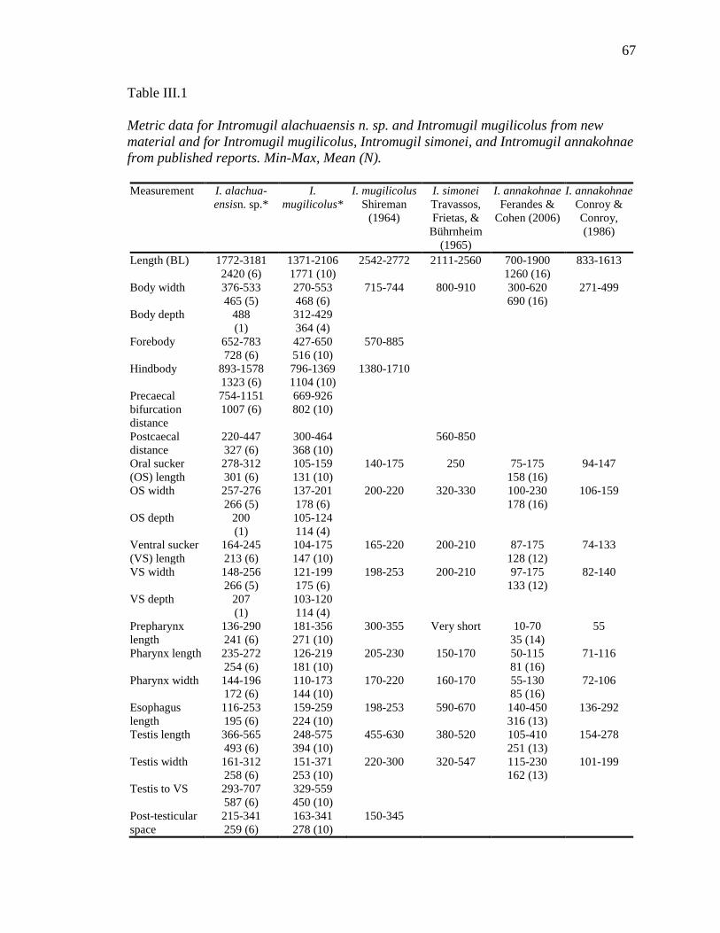
67
Table III.1 Metric data for Intromugil alachuaensis n. sp. and Intromugil mugilicolus from new material and for Intromugil mugilicolus, Intromugil simonei, and Intromugil annakohnae from published reports. Min-Max, Mean (N). Measurement I. alachua-
ensisn. sp.* I.
mugilicolus* I. mugilicolus
Shireman (1964)
I. simonei Travassos, Frietas, &
Bührnheim (1965)
I. annakohnae Ferandes &
Cohen (2006)
I. annakohnae Conroy & Conroy, (1986)
Length (BL) 1772-3181 2420 (6)
1371-2106 1771 (10)
2542-2772 2111-2560 700-1900 1260 (16)
833-1613
Body width 376-533 465 (5)
270-553 468 (6)
715-744 800-910 300-620 690 (16)
271-499
Body depth 488 (1)
312-429 364 (4)
Forebody 652-783 728 (6)
427-650 516 (10)
570-885
Hindbody 893-1578 1323 (6)
796-1369 1104 (10)
1380-1710
Precaecal bifurcation distance
754-1151 1007 (6)
669-926 802 (10)
Postcaecal distance
220-447 327 (6)
300-464 368 (10)
560-850
Oral sucker (OS) length
278-312 301 (6)
105-159 131 (10)
140-175 250 75-175 158 (16)
94-147
OS width 257-276 266 (5)
137-201 178 (6)
200-220 320-330 100-230 178 (16)
106-159
OS depth 200 (1)
105-124 114 (4)
Ventral sucker (VS) length
164-245 213 (6)
104-175 147 (10)
165-220 200-210 87-175 128 (12)
74-133
VS width 148-256 266 (5)
121-199 175 (6)
198-253 200-210 97-175 133 (12)
82-140
VS depth 207 (1)
103-120 114 (4)
Prepharynx length
136-290 241 (6)
181-356 271 (10)
300-355 Very short 10-70 35 (14)
55
Pharynx length 235-272 254 (6)
126-219 181 (10)
205-230 150-170 50-115 81 (16)
71-116
Pharynx width 144-196 172 (6)
110-173 144 (10)
170-220 160-170 55-130 85 (16)
72-106
Esophagus length
116-253 195 (6)
159-259 224 (10)
198-253 590-670 140-450 316 (13)
136-292
Testis length 366-565 493 (6)
248-575 394 (10)
455-630 380-520 105-410 251 (13)
154-278
Testis width 161-312 258 (6)
151-371 253 (10)
220-300 320-547 115-230 162 (13)
101-199
Testis to VS 293-707 587 (6)
329-559 450 (10)
Post-testicular space
215-341 259 (6)
163-341 278 (10)
150-345

68
Table III.1 (continued). Measurement I.
alachuaensis n. sp.*
I. mugilicolus* I. mugilicolus I. simonei I. annakohnae I. annakohnae
Ovary length 123-158 142 (5)
62-155 108 (10)
155-160 130-170 42-215 111 (10)
Ovary width 88-116 105 (5)
67-178 114 (10)
95-120 150-160 30-110 70 (10)
41-83
Ovary to VS 350-450 399 (5)
221-438 347 (10)
Vitellarium to VS
72-440 316 (6)
117-337 190 (10)
Vitellarium to posterior end
39-80 64 (6)
25-103 61 (10)
Hermaphroditic sac length
280-477 389 (6)
233-473 388 (10)
270-335 330 140-450 238 (14)
114-176
Hermaphroditic sac width
125-157 149 (6)
109-192 162 (10)
60-80 150-180 60-145 107 (14)
Internal seminal vesicle length
170-301 234 (6)
122-283 213 (10)
60-340 184 (9)
Internal seminal vesicle width
73-135 109 (6)
42-172 113 (10)
30-150 69 (9)
External seminal vesicle length
146-248 180 (5)
124-228 186 (10)
170 60-215 110 (13)
External seminal vesicle width
82-152 114 (5)
59-176 113 (10)
30-115 61 (13)
Egg length 71-86 76 (19)
67-73 70 (10)
60-80 90-100 82-105,110 93 (53)
91-116
Egg width 38-48 43 (19)
38-47 42 (10)
40 53-57 45-72, 67 54 (53)
57
Width † 17-21% 19 % (5)
18-28% 25% (6)
Forebody† 24-37% 31% (6)
25-34% 29% (10)
Hindbody† 40-65% 55% (6)
58-65% 62% (10)
Precaecal bifurcation†
33-49% 42% (6)
40-50% 46% (10)
Postcaecal distance†
9-20% 14% (6)
17-25% 21% (10)
OS length % VS length
113-190% 144% (6)
74-101% 90% (10)
OS width % VS width
107-175% 130% (5)
93-113% 102% (6)
60-80% 70% (12)
Pharynx length/ prepharynx length
86-193% 113% (6)
46-104% 69% (10)
Pharynx length / Pharynx width
124-183% 150% (6)
111-139% 126% (10)
Prepharynx/ esophagus
91-189% 127% (6)
97-156% 122% (10)

69
Table III.1 (continued). Measurement I.
alachuaensis n. sp.*
I. mugilicolus* I. mugilicolus I. simonei I. annakohnae I. annakohnae
Testis to VS distance†
17-29% 24% (6)
22-32% 26% (10)
Post-testicular space†
7-14% 11% (6)
10-19% 16% (10)
10-14%
* Non-flattened specimens from this study
†as percentage of body-length
Table III.2. Total number of variable sites based on pairwise comparision of the studied fragment of nuclear rDNA (partial 18S, complete ITS1, complete 5.8S, complete ITS2 and partial 28S) between Intromugil mugilicolus and Intromugil alachuaensis n. sp. Intromugil
alachuaensis length
Intromugil mugilicolus length
Aligned length
Aligned base pair differences
Gaps Percent difference
18S partial 133 133 133 1 0 0.8% ITS1 504 506 508 42 6 9.4% 5.8S 157 157 157 0 0 0 ITS2 287 287 287 17 0 5.9% 28S partial 1381 1382 1384 39 5 3.2% Total 2462 2465 2469 99 11 4.5%
the genus, (2) the considerably longer prepharynx is than in I.annakohnae and I. simonei,
(3) the robust and elongate compared with pyriform pharynx in I. mugilicolus, or smaller
pharynx with a nearly equal length to width ratio as in I. annakohnae and I. simonei, (4)
the less extensive vitellarium than in I. mugilicolus, and (5) the caeca that terminate at the
level of the posterior half of the testis rather than at or near the anterior testicular margin
like in I. mugilicolus, or anterior to the testis in I. annakohnae and I. simonei.
Intromugil alachuaensis, possibly sympatric with I. mugilicolus, is much less
transparent than that species both in life and in mounts. The eye-spot pigment in I.
alachuaensis is much denser throughout the body, and it has many glands in the forebody

70
that significantly darken the appearance. Both I. alachuaensis and I. mugilicolus have
two large collecting lymphatic ducts extending roughly parallel to the prepharynx,
pharynx, and oesophagus, but the tubules in I. alachuaensis are larger and more
prominent in both live and fixed material than in I. mugilicolus. Intromugil alachuaensis
appears more robust than the other three members of the genus.
Molecular Data
According to the available information on the sequenced contiguous fragment,
these regions and especially the highly variable ITS1 are generally suitable for the
discrimination of congeneric digenean species (e.g. Nolan & Cribb, 2005; Olson &
Tkach, 2005; Parker et al., 2010; Tkach & Kinsella; 2011; Tkach & Mills, 2011). No
intraspecific variability was detected among replicates of either species. The region
sequenced and used for differentiation (Table 2) when aligned and trimmed was 2,477 bp
for I. mugilicolus and 2,474 bp for I. alachuaensis, and it differed at 110 sites across all
sequenced regions, breaking down as follows: 18S 133 bases, 1 difference; ITS1, 48
differences, including 6 gaps; 5.8S 0 difference; ITS2, 17 differences; and 28S 44
differences, including 5 gaps.
Discussion
Prior to this study, Intromugil contained 3 nominal species from fishes in the
genus Mugil, all found in water bodies associated with the Western Atlantic Ocean in
both North and South America. Overstreet and Curran (2005) considered
Skrjabinolecithum Belous, 1954 to be the eastern hemisphere counterpart of Intromugil,
and differentiated them by geography, the extent of the caeca, and position of the ovary

71
relative to testis. With the addition of I. alachuaensis and new material of I. mugilicolus,
geography and caecal extent still serve to differentiate the two genera. In material of I.
Figures III.9-10. Nomarski photomicrographs of living Intromugil material. 9. Intromugil mugilicolus showing glands in hermaphroditic duct, scale bar = 200 µm. 10. Intromugil alachuaensis n. sp. showing glands in hermaphroditic duct, arrows indicate sacs containing glandular material, scale bar = 200 µm.
mugilicolus that has not been flattened, the ovary is not distinctly pretesticular, but rather
contiguous with the testis or only slightly pretesticular. In I. alachuaensis, the ovary is up

72
to about one ovarian length pretesticular. In the current study, I observed the excretory
vesicle of I. mugilicolus and I. alachuaensis as I-shaped, which was previously
unreported. Species in Skrjabinolecithum have a Y-shaped excretory vesicle. Of
sympatric species in haploporid genera in mullet, those of Culuwiya Overstreet & Curran,
2005 are more commonly found, but they are smaller, possess shorter sac-like caeca, and
have a Y- shaped excretory vesicle. Overstreet and Curran (2005) placed Intromugil into
the subfamily Waretrematinae, whose members are largely confined to the western
Pacific and Indian Ocean Basin, with the exception of species in the genera Intromugil,
Culuwiya and Conohelmins from the Americas. I suspect that once molecular data
become available for waretematines from the eastern hemisphere, Culuwiya and
Intromugil will be more closely allied with the chalcinotrematines than with other
waretrematines. As species of haploporids are found predominantly in mullets throughout
their range, I expect to detect considerable convergence of morphological features in
different geographical areas from different lineages.
Unfortunately, I do not have fresh, properly-killed material from I. simonei and I.
annakohnae. Specimens of I. mugilcolus killed with pressure are inconclusive in
demonstrating three rows of spines on a terminal oral sucker, whereas the flattened large
muscular oral sucker of I. alachuaensis retains its shape and position. Additional material
from I. simonei and I. annakohnae is needed to determine if three rows of spines occur on
the oral sucker and if the sacs of glandular material occurring in the hermaphroditic duct
are specific or generic characteristics. I suspect both features to be present in I. simonei
and I. annakohnae. Conroy and Conroy (1986) reported I. mugilicolus from M. curema in
Venezuela. Based on their description of a short prepharynx and small pharynx in worms

73
not available for examination, I think their worms were I. annakohnae. They reported
pronounced muscularization visible in the hermaphroditic duct and that alludes to the
sacs I observed in I. mugilicolus and I. alachuaensis. As I already mentioned, these are
best observed in living worms. Molecular data also are needed to determine the pattern of
diversification of Intromugil spp. Molecular data from the two South American species
would help delineate how diversification in Intromugil species evolved and whether the
group has a Neotropical or Nearctic origin.
Some morphological features of the new species and redescription of I.
mugilcolus with unflattened specimens warrants emendation of the generic diagnosis of
the genus provided by Overstreet and Curran (2005) to include oral sucker as terminal to
subterminal (vs. terminal in current diagnosis), oral sucker with or without three rows of
small spines along anterior margin of mouth within oral sucker, caeca terminating at a
level from anterior to testis to posterior half of testis (vs. anterior margin of testis), and
ovary as contiguous with to anterior to testis (vs. anterior to testis).
Intromugil mugilcolus collected in Mississippi appears to be rare because many
fish have been examined in recent years. Mugil cephalus seems to be infected only in
winter and spring, from roughly January to April. The parasites appear to occur only in
fish larger than about 250 mm in total length (TL) with relatively low intensity. For many
years before Hurricane Katrina (August 2005) and until March 2007 it has not been found
in any of numerous mullet examined (Overstreet, 2007). I do not present prevalence data
because of the temporal absence of this parasite and its seeming restriction to large
mullets. During the time of year when I find I. mugilicolus, the salinity tends to be lower
than during the rest of the year, but the period also immediately follows that of the

74
offshore spawning migration of M. cephalus. In my sample of M. cephalus that yielded
specimens of I. alachuaensis from the Santa Fe River, four fish measured 405-550 mm
TL and were infected with one to six specimens. Mullet smaller than about 300 mm were
not observed during the time of collection. The four mullet that harbored I. alachuaensis
were captured more than 80 km inland. Although infections of I. alachuaensis could have
been acquired in marine waters, I suspect that they were acquired in freshwater.
Intromugil alachuaensis is a large and distinct worm, and previous investigations of
Florida marine waters (Hutton & Sogandares-Bernal, 1964; Hutton, 1964; Skinner, 1975)
failed to uncover this species or I. mugilicolus. No larval form of any species of
Intromugil has been reported. Further investigation is needed into the life-cycle and
whether I. alachuaensis occurs in other rivers with similar geology in peninsular Florida.

75
CHAPTER IV
SOME NEW AND OTHER SPECIES OF HAPLOPORIDAE (DIGENEA)
POSSESSING A SINGLE CAECUM INFECTING MUGILID FISHES, WITH
PHYLOGENETIC AFFINITIES
Abstract
Nine species of Haploporidae, possessing an undivided caecum from hosts in
Mugilidae are reported, four of which are new. In this study, it is concluded that the Indo-
Pacific haploporids with a caecum are closely related, requiring several changes to the
prior organization of the Haploporidae. Malabarotrema Zhukov, 1972 is resurrected for
Malabarotrema indica Zhukov, 1972; Skrjabinolecithum lobolecithum Martin, 1973 is
transferred to Malabarotrema as M. lobolecithum (Martin, 1973) n. comb.;
Pseudohapladena megaorchis Liu and Yang, 2002 is transferred to Malabarotrema as M.
megaorchis (Martin, 1973) n. comb.; and a new species is described. Species of
Unisaccus Martin, 1973 are transferred out of the Haploporinae to the Waretrematinae on
the basis of morphological and molecular data. Unisaccoides Martin, 1973 is resurrected
with Unisaccoides vitellosus Martin, 1973 as the type-species, and a new species of
Unisaccoides is described. Saccocoelioides lizae Liu, 2002 is transferred to Unisaccus as
Unisaccus lizae (Liu, 2002) n. comb., and three new species are described. Phylogenetic
hypotheses based on analysis of partial 28S sequences with other available haploporid
sequences indicate that (1) members of Malabarotrema, Unisaccoides, and Unisaccus are
more closely related to each other than to other species of haploporids; (2) Haploporinae
is not a monophyletic group when species of Unisaccus are included; (3) Saccocoelioides
lizae is better allocated to Unisaccus than to Saccocoelioides; and (4) Skrjabinolecithum

76
vittellosus and five species of Unisaccus form a monophyletic group. Reduced
vitellarium and the presence of eye-spotted miracidium in-utero are no longer considered
characters that exclude species from Waretrematinae.
Introduction
The subfamily Unisaccinae Martin, 1973 was originally erected for species that
possess an undivided caecum, accommodating the genera Unisaccus Martin, 1973 and
Unisaccoides Martin, 1973 (Martin, 1973b). Martin also described Paraunisaccoides
Martin, 1973, which he also placed in the subfamily (Martin, 1973c). Martin (1973b, c)
apparently was unaware of the description of Malabarotrema indica Zhukov, 1972 by
Zhukov (1972). Malabarotrema shares many features with Paraunisaccoides. The genus
Pseudouncoelium Ahmad, 1987 was erected for two species described from mugilids off
India that also possessed a caecum and were also included in the subfamily (Ahmad,
1987). Overstreet and Curran (2005) considered Pseudounicoelium Ahmad, 1987 a junior
synonym of Unisaccus and did not accept the subfamily; they considered it a junior
synonym of Haploporinae Nicoll, 1914. Overstreet and Curran (2005) considered
Malabarotrema, Unisaccoides, and Paraunisaccoides as junior synonyms of
Skrjabinolecithum Belous, 1954, a member of Waretrematinae Srivastava, 1937.
Although they noted the similarity of Pseudohapladena megaorchis Liu & Yang, 2002 to
the concept of Paraunisaccoides, they retained Ps. megaorchis in Pseudohapladena
Yamaguti, 1952. Blasco-Costa, Gibson et al. (2009) and Blasco-Costa, Montero, Gibson,
Balbuena, and Kostadinova (2009) retained Unisaccus within the Haploporinae and
transferred two additional species into the genus as Unisaccus sprenti (Martin, 1973)
Blasco-Costa, Mantero, Gibson, Balbuena, Raga, and Kostadinova, 2009 and Unisaccus

77
mugilis (Rekharani & Madhavi, 1985) Blasco-Costa, Mantero, Gibson, Balbuena, Raga,
and Kostadinova, 2009. Unicoelium Thatcher and Dossman, 1975 was placed into the
subfamily Chalcinotrematinae Overstreet and Curran, 2005 by Overstreet and Curran
(2005), even though Nasir and Gómez (1976) had previously considered Unicoelium as a
synonym of Unisaccus. Overstreet and Curran (2005) considered the single caecum to
have evolved independently in at least three of the four haploporid subfamilies they
recognized.
In this paper, I report on nine species of haploporids from the Indo-West Pacific
region and attempt to place them within a larger framework of the Haploporidae Nicoll,
1914, as well as provide keys to species with an undivided caecum in mullets.
Materials and Methods
Specimens of Haploporidae with a caecum were collected from mugilid
(Actinopterygii: Mugiliformes) hosts collected at several locations off Australia and
purchased from fish markets in China (assumed to be wild caught). Fish names follow
those of FishBase (Froese & Pauly, 2013).
Recently collected haploporids (since 2009) were isolated similarly to the process
advocated by Cribb and Bray (2010) for gastrointestinal species, often skipping an initial
examination because of the volume of the intestinal contents. Live worms were rinsed
and cleaned in saline and observed briefly. Worms were killed by addition of steaming
hot, but not boiling, tap water. Haploporids were then stored in 70% molecular-grade
ethanol. Parasite specimens for morphological and molecular analysis were processed
and studied according to the protocols used by Pulis et al. (2013) and Pulis and Overstreet
(2013). Prepared permanent slides also were obtained from the personal collections of

78
Thomas Cribb and Robin Overstreet. All measurements are in micrometres unless noted
otherwise. Museum abbreviations are as follows: MNT, Museum and Art Gallery of the
Northern Territory, Darwin, Australia; QM, Queensland Museum, Brisbane, Queensland,
Australia; and USNPC, US National Parasite Collection, Beltsville, Maryland.
Genomic DNA was isolated using Qiagen DNAeasy Tissue Kit (Qiagen, Inc.,
Valencia, California, U.S.A.) following the instructions provided. DNA fragments
approximately 2,500 basepairs (bp) long comprising the 3' end of the 18S nuclear rDNA
gene, internal transcribed spacer region (including ITS1 + 5.8S + ITS2), and the 5' end of
the 28S gene (including variable domains D1-D3) were amplified from the extracted
DNA by polymerase chain reaction (PCR) on a PTC-200 Peltier Thermal Cycler using
forward primers ITSF (5' - CGCCCGTCGCTACTACCGATTG-3') or LSU5 (5'-
TAGGTCGACCCGCTGAAYTTAAGCA-3') and reverse primer 1500R (5’-
GCTATCCTGAGGGAAACTTCG-3’). These PCR primers and multiple internal
primers were used in sequencing reactions. The internal forward primers were DIGL2
(5’-AAGCATATCACTAAGCGG-3’), 300F (5’-CAAGTACCGTGAGGGAAAGTTG-
3’), 900F (5’-CCGTCTTGAAACACGGACCAAG-3’), and internal reverse primers
were 300R (5’-CAACTTTCCCTCACGGTACTTG-3’), Digl2r (5’-
CCGCTTAGTGATATGCTT-3’), and ECD2 (5’-CTTGGTCCGTGTTTCAAGACGGG-
3’).
Previously published 28S ribosomal RNA gene sequences of species of
Haploporidae were used for comparison (see Table 1 for accession numbers and host
information) with newly submitted sequences. Sequences were aligned using the
ClustalW application in the BioEdit program, Version 7.0.9 (Hall, 1999). The alignment

79
was further refined by eye and trimmed to the shortest sequence on both 5' and 3' ends.
The resulting alignment utilised 29 haploporids with Hapladena nasonis as the outgroup.
The alignment was 1,183 characters long, including gaps, with 743 sites conserved, 435
sites variable, and 325 parsimony-informative sites. Phylogenetic analysis of the data was
performed using Bayesian inference (BI) with MrBayes 3.1.2 software (Huelsenbeck &
Ronquist, 2001; Ronquist & Huelsenbeck, 2003). The best nucleotide substitution model
was estimated with jModeltest Version 0.1.1 (Guindon & Gascuel, 2003; Posada, 2008)
as general time reversible, with invariant sites and gamma-distributed among site-rate
variation (GTR + I + Γ). The following model parameters were used in MrBayes: nst = 6,
rates = invgamma, ngen = 1,000,000, and samplefreq = 100. The first one quarter of
samples were discarded as burn-in (sump burnin = 2500), and nodal support was
estimated by posterior probabilities (sumt) (Huelsenbeck et al., 2001), with all other
settings left as default.
Results
Malabarotrema Zhukov, 1972
Syn Paraunisaccoides Martin, 1973
Diagnosis: Body elongate. Eye-spot pigment dispersed in forebody. Oral sucker
subterminal. Prepharynx longer than pharynx and oesophagus. Oesophagus short.
Caecum single, longer than wide, extending to near or into testicular region, with
posterior indentation. Testis in hindbody. Hermaphroditic sac dorsal to ventral sucker,
often widest in anterior half. Hermaphroditic duct with distal portion composed of
smooth muscle, proximally lined with muscular pads. Female duct short. Internal seminal
vesicle about half length of hermaphroditic sac. External seminal vesicle usually longer

80
than internal seminal vesicle. Ovary contiguous with or short distance anterior to testis.
Uterus confined between ovary and hermaphroditic sac. Vitellarium composed of large
areas of elongate follicles, confined mainly to hindbody. In-utero eggs with incompletely
developed miracidium lacking eye-spots. Excretory vesicle Y-shaped, bifurcating in
testicular region; arms reaching to middle of hindbody; pore terminal. In intestine of
Mugilidae from Indo-West Pacific region.
Type-species: Malabarotrema indica Zhukov, 1972
Remarks
The type-species of Malabarotrema had previously been allocated to
Skrjabinolecithum by Overstreet and Curran (2005).
Malabarotrema indica Zhukov, 1971
Syns Skrjabinolecithum indicum (Zhukov, 1971) Overstreet and Curran, 2005.
Type-host: Etroplus suratensis (Bloch), pearlspot (Cichlidae).
Type-locality: Malabar, India.
Site of infection: Intestine.
Specimens examined: Russian Academy of Sciences, No. 46-4, 2 specimens on
one slide. ex. Etroplus suratensis (Cichlidae) Malabar, India.
Examination of slide 46-4 RAS reveals that Malabarotrema indica has a caecum.
Zhukov's (1972) Figure 3 is accurate, whereas Overstreet and Curran (2005)
misinterpreted the wide oesophagus as the bifurcation of the oesophagus prior to the sac-
like caecum. Examination of the specimens also revealed the presence of pads lining the
hermaphroditic duct as in the other species of Malabarotrema. I consider those two

81
features along with the extensive vitellarium to necessitate the resurrection of
Malabarotrema from Skrjabinolecithum.
Because the type specimens of M. indica were killed under considerable coverslip
pressure, I infer the position and nature of the organs from the specimens I collected from
the other three species I place in the genus.
Malabarotrema lobolecithum (Martin, 1973) n. comb.
Syns Paraunisaccoides lobolecithum Martin, 1973; Skrjabinolecithum
lobolecithum (Martin, 1973) Overstreet & Curran, 2005
Type-host: Mugil cephalus Linnaeus, flathead grey mullet (Mugilidae).
Type-locality: Brisbane River, Brisbane, Queensland, Australia. (Martin, 1973c)
Site of infection: intestine.
Other hosts: Paramugil georgii (Smith), silver mullet (Mugilidae); Moolgarda
seheli (Forsskål), bluespot mullet (Mugilidae); Moolgarda perussii (Valenciennes),
longfinned mullet (Mugilidae).
Material examined: ex. Paramugil georgii; Western Australia, Australia; Barred
Creek (17° 40'S 122° 12'E) February 2010, Coll. Pulis, Andres, Six Mile Creek 20° 19'S
118° 40'E Feburary2010 Coll. Pulis, Andres, Northern Territory, Australia; Ludmilla
Creek (12° 25'S 130° 50'E) March 2010, Coll. Pulis, Andres. Queensland, Australia;
Woody Point (27° 16'S 153° 06'E) Coll. Overstreet February 1995, Sandgate (27° 19'S
153° 05'E) Coll. Clarke, Cribb, Maguire January 1995, Boat Passage (27° 24'S 153° 10'E)
Coll. Cribb, Anderson, Bray January 1993, Wynnum (27° 26'S 153° 11'E) Coll. Pulis,
Andres March 2010, Wynnum (27° 27'S 153° 10'E) Coll. Cribb et al. February 1995,
Wynnum Purchased fresh Overstreet Nov. 1997, Logan River (27° 42'S 153° 19'E) Coll.

82
Overstreet February 1995. ex. Moolgarda seheli: Western Australia, Australia; Dampier
Creek (17° 57'S 122° 15'E) February 2010 Coll. Pulis, Andres, Queensland, Australia;
Beelbi Creek (25° 15'S 152° 40'E) Coll. Pulis, Andres March 2010. ex. Moolgarda
perussii: Northern Territory, Australia, Doyle's Boat Ramp (12° 26'S 130° 50'E) Coll.
Pulis, Andres March 2010.
Specimens deposited: USNPC NTM BM-TBD, representative sequence, EP-568.
Holotype: No. 7114 Allen Hancock Parasitology Collection, University of
Southern California, but current whereabouts unknown; unsuccessful attempts were made
to borrow specimens from University of Southern California Museum, Santa Barbara
Museum of Natural History, and Los Angeles County Museum of Natural History (Pers.
comm. Daniel Geiger SBMNH, Jody Martin NHMLAC).
Supplementary Data (Figures 1-2, Tables 1-3).
Measurements based on 11 wholemount specimens. Body elongate, fusiform,
1,270-2,005 long, 219-366 wide, widest in hindbody, width 14-21% of body length (BL),
whitish in life. Tegument spined, with ventral surface between ventral sucker and oral
sucker containing only few irregularly spaced spines; spines bent-hastate, about 9-13
long, 6-8 wide at widest point of root, directed posteriorly. Dorsal to oral sucker 3 rows
of enlarged spines, blunt, conical, with bases about 7-11 wide. Eye-spot pigment
dispersed in forebody, most prominent in anterior fifth of body. Oral sucker subterminal,
99-140 long, 92-163 wide, with mouth opening anteroventrally. Ventral sucker slightly
elevated, 115-181 long, 136-204 wide, 113-147% longer than oral sucker, 106-157%
wider than oral sucker, covered by spines; spines covering ventral sucker spines smaller
than body spines, similar in shape. Forebody 351-549 long or 25-31% of BL; hindbody

83
774-1284 long or 60-67% of BL. Prepharynx well defined, 327-685 long. Pharynx thick-
walled, muscular, usually pyriform, located dorsal to completely posterior to ventral
sucker, 91-144 long, 90-150 wide; prepharynx 3.3-5.2 times longer than pharynx, with
posterior portion and pharynx surrounded by few gland cells probably of digestive nature.
Oesophagus not as well defined as prepharynx, often folded slightly among other organs,
43-161 long. Caecum in hindbody, lined with large cells, with anterior end expanded,
with posterior end slightly indented, 268-498 long, 132-210 wide, commencing 567-
1,001 from anterior end of body or 41-53% of BL; post-caecal space 235-619 or 18-36%
of BL.
Testis medial, with anterior margin straight or beveled, with posterior end
rounded to pointed, overlapping posterior end of caecum to 139 posterior, 257-644 from
ventral sucker, 230-397 long, 136-207 wide; post-testicular space 98-395 or 6-20% of
BL. Hermaphroditic sac dorsal to ventral sucker, ventral to pharynx, 190-315 long, 87-
145 wide, terminating posterior to ventral sucker; sac containing following structures
(internal seminal vesicle 42-148 long, 30-60 wide; male duct thin-walled; prostatic bulb
prominent, with size dependent on amount of sperm and eggs present; male duct joining
female duct in posterior half of sac); hermaphroditic duct strongly muscularised, with
posterior portion containing muscular pads. External seminal vesicle sac-like, variously
orientated, 86-152 long, 38-83 wide. Genital pore elongate laterally, without evidence of
scleritization, located at about level of anterior margin of ventral sucker in crease
between elevated ventral sucker and of plane forebody.
Ovary medial, contiguous with testis, 94-148 long, 61-104 wide, 257-644 from
ventral sucker. Mehlis' gland slightly anterior to ovary. Laurer's canal dorsal to testis,
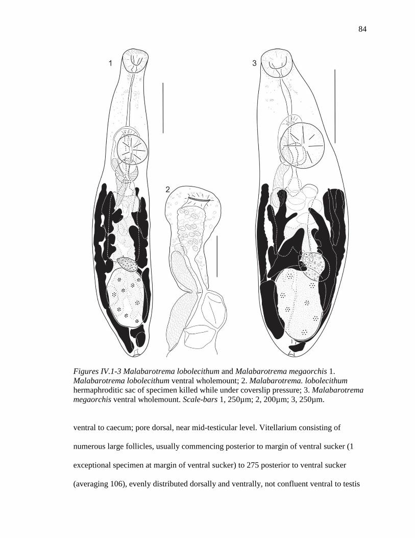
84
Figures IV.1-3 Malabarotrema lobolecithum and Malabarotrema megaorchis 1. Malabarotrema lobolecithum ventral wholemount; 2. Malabarotrema. lobolecithum hermaphroditic sac of specimen killed while under coverslip pressure; 3. Malabarotrema megaorchis ventral wholemount. Scale-bars 1, 250µm; 2, 200µm; 3, 250µm.
ventral to caecum; pore dorsal, near mid-testicular level. Vitellarium consisting of
numerous large follicles, usually commencing posterior to margin of ventral sucker (1
exceptional specimen at margin of ventral sucker) to 275 posterior to ventral sucker
(averaging 106), evenly distributed dorsally and ventrally, not confluent ventral to testis

85
or uterus, confluent posterior to testis. Uterus extending directly from ovary to
hermaphroditic sac, without loops; proximal portion often filled with sperm; metraterm
lacking. Eggs thin-shelled, 1-17 in number, 75-106 long, 40-67 wide (averaging 93 X 55)
when measured from distal end of uterus, with incompletely developed miracidium
lacking eye-spots.
Lymphatic tubes in forebody. Excretory vesicle Y-shaped, dorsal to testis,
bifurcating near mid-level of testis, terminating near anterior margin of ovary; pore
terminal.
Remarks
Martin (1973c) described the species from two specimens. I included additional
specimens in my series, accounting for greater variability in measurements and metrics.
There was no major difference from the original description other that I could not
confidently observe the sclerotized denticles on/in the pads lining the posterior portion of
the hermaphroditic duct. I believe them to be glands or gland products of an uncertain
function and not apparent in specimens fixed by both methods. Although the species was
originally described from Mugil cephalus, I did not find this species in M. cephalus but
have encountered it in other mullet species. I believe the host was either originally
misidentified or from an irregular host; regardless, I have no reason to think my worms
are not conspecific with those described by Martin (1973c).
Malabarotrema megaorchis (Liu & Yang, 2002) n. comb.
Syn Pseudohapladena megaorchis Liu and Yang, 2002.
Type-host: Moolgarda engeli (Bleeker), Kanda (Mugilidae); reported as Mugil
engeli by Liu and Yang (2002).

86
Type-locality: Xiamem, Fuijian Province, People's Republic of China.
Site of infection: Intestine.
Material Studied: Paratype, USNPC No. 91532.
New Material: ex. Mugil cephalus purchased at fish market, Daya Bay (22° 43'N
114°32'E), Guangdong Province, People's Republic of China.
Specimens deposited: TBD, represenative sequence, EP-644.
Supplementary Data (Figures, 3,20; Tables, 1-3)
Measurements based on 5 wholemount specimens. Body elongate, fusiform 951-
1075 long, 257-336 wide, widest in hindbody, with width 27-32% of BL. Tegument
spined over entire surface; spines becoming smaller and more sparse posteriorly, hastate,
about 4-8 long (measured from forebody), directed posteriorly. Eye-spot pigment
dispersed in forebody, most prominent in anterior half of forebody. Oral sucker
subterminal, 91-100 long, 98-115 wide, with mouth opening anteroventrally. Ventral
sucker slightly elevated, 130-155 long, 130-159 wide, 135-170% longer than oral sucker;
125-159% wider than oral sucker, covered by spines; spines covering ventral sucker
smaller than body spines, similar in shape. Forebody 251-343 long or 25-32% of BL,
hindbody 519-623 long or 53-62% of BL. Prepharynx well defined, 211-294 long.
Pharynx thick-walled, muscular, usually pyriform, located dorsal to ventral sucker to
partially dorsal posterior to posterior margin of ventral sucker, 79-98 long, 92-109 wide;
prepharynx 2.5-3.2 times longer than pharynx, with posterior portion of prepharynx and
pharynx surrounded by few gland cells of probable digestive nature. Oesophagus not as
well defined as prepharynx, often folded slightly among other organs, 32-98 long.
Caecum in hindbody, lined with large cells, (specimen in Figure 3 anterior end expanded

87
and protruding anteriorly due to displacement by eggs in uterus), with posterior end
indented, 223-272 long, 111-145 wide, commencing 461-521 from anterior end of body
or 46-50% of BL; post-caecal space 273-308 or 26-30% of BL.
Testis medial, rounded, overlapping posterior end of caecum to 25 distant, 228-
336 from ventral sucker, 205-240 long, 132-170 wide; post-testicular space 54-138 or 5-
13% of BL. Hermaphroditic sac dorsal to ventral sucker, ventral to pharynx, 161-186
long, 72-89 wide, terminating near posterior margin ventral sucker; sac containing
following structures (internal seminal vesicle 60-89 long, 28-36 wide; male duct thin-
walled; prostatic bulb prominent, with size dependent on amount of sperm and eggs
present; male duct joining female duct in posterior half of sac); hermaphroditic duct
strongly muscularized, with posterior portion containing muscular pads. External seminal
vesicle sac-like, 98-162 long, 28-43 wide. Genital pore immediately anterior to ventral
sucker in crease between elevated ventral sucker and plane of forebody.
Ovary medial, ventral to caecum, situated laterally along ventral anterior margin
of testis, 94-121 long, 60-84 wide, 166-288 from ventral sucker. Mehlis' gland anterior or
lateral to ovary. Laurer's canal not observed. Vitellarium plate-like or tubular, usually
commencing at posterior margin of ventral sucker (1 specimen at 31 posterior to posterior
margin of ventral sucker), confluent posterior to testis. Uterus extending directly from
ovary to hermaphroditic sac, without loops; proximal portion often filled with sperm;
metraterm lacking. Eggs thin-shelled, 2-16 in number, 58-71 long, 42-51 wide (average
64 X 46) when measured from distal end of uterus, with incompletely developed
miracidium lacking eye-spots.

88
Lymphatic tubes in forebody. Excretory vesicle Y-shaped, dorsal to testis,
bifurcating near mid-level of testis, terminating near anterior margin of ovary; pore
terminal.
Remarks
The smaller, stouter, Malabarotrema megaorchis can be differentiated from M.
lobolecithum by geography, smaller body spines, a lack of enlarged spines dorsal to oral
sucker, having smaller eggs, and molecular differences.
Malabarotrema n. sp. 1
Type-host: Paramugil georgii (Ogilby), silver mullet (Mugilidae).
Type-Locality: Queensland, Australia: Beelbi Creek (25° 15'S 152° 40'E).
Site: Intestine.
Other hosts: Moolgarda seheli (Forsskål), bluespot mullet (Mugilidae);
Paramugil georgii (Ogilby), silver mullet (Mugilidae).
Other localities: ex. Paramugil georgii; Northern Territory, Australia: Ludmilla
Creek (12° 25'S 130° 50'E) Coll. Pulis, Andres March 2010, Queensland, Australia:
Beelbi Creek (25° 15'S 152° 40'E) Coll. Pulis, Andres March 2010, Eli Creek (25° 16'S
152° 49'E) Coll. Pulis, Andres March 2010, Woody Point (27° 16'S 153° 06'E) Coll.
Overstreet February 1995, Sandgate (27° 19'S 153° 05'E) Coll. Clarke, Cribb, Maguire
January 1995, Morton Bay (27° 21'S 153° 07'E) Coll. Cribb, Barker, Anderson, Wright
July 1999, Boat Passage (27° 24'S 153° 10'E) Coll. Cribb, Anderson, Bray January 1993,
Wynnum (27° 26'S 153° 11'E) Coll. Pulis, Andres March 2010, Wynnum (27° 27'S 153°
10'E) Coll. Cribb et al. February 1995, Logan River (27° 42'S 153° 19'E) Coll. Overstreet
February 1995. ex. Moolgarda seheli: Northern Territory, Australia: Buffalo Creek (12°

89
12'S 130° 55'E) Coll. Pulis, Andres March 2010, Queensland, Australia: Eli Creek (25°
16'S 152° 49'E) Coll. Pulis, Andres March 2010, Wynnum (27° 26'S 153° 11'E) Coll.
Pulis, Andres March 2010.
Specimens Deposited: Holotype: TBD Paratypes USNPC NTM BM TBD,
representative sequence, EP-148.
Description Figures 4-7, Tables 1-3.
Measurements based on 15 wholemount specimens. Body elongate, fusiform 897-
1,644 long (BL), 177-370 wide, widest in hindbody, width 18-27% of BL. Tegument
spined over entire surface, except for area surrounding genital pore; spines hastate, about
6-9 long, 2-4 wide at widest point of root, directed posteriorly. Eye-spot pigment
dispersed in forebody, most prominent in anterior fifth of body. Oral sucker subterminal,
70-120 long, 91-149 wide, with mouth opening anteroventrally. Ventral sucker slightly
elevated, 98-157 long, 107-161 wide, 115-154% longer than oral sucker, 93-148% wider
than oral sucker, covered by concentric rows of spines; spines covering ventral sucker
similar to body spines, smaller than body spines. Forebody 280-546 long or 26-37% of
BL; hindbody 520-976 long or 50-64% of BL. Prepharynx well defined, 206-574 long.
Pharynx thick-walled, muscular, usually pyriform, located with anterior margin extends
beyond anterior margin of ventral sucker to completely posterior to ventral sucker, 69-
112 long, 66-126 wide; prepharynx 2.8-8.3 times longer than pharynx, with posterior
prepharynx and pharynx surrounded by few gland cells of probable of digestive nature.
Oesophagus not as well defined as prepharynx, often folded slightly among other organs,
39-188 long. Caecum in hindbody, lined with large cells, with posterior end having

90
Figures IV.4-7. Malabarotrema n.sp.1 4. ventral wholemount; 5. ventral wholemount; Scale-bars 4, 250 µm; 5, 250µm; 6,7 100 µm. 6. ovarian complex of specimen killed while under coverslip pressure; 7. hermaphroditic sac of specimen killed while under coverslip pressure;
slightly indented, 212-354 long, 108-240 wide, commencing 340-820 from anterior end
of body or 35-53% of BL; post-caecal space 125-495 or 14-32% of BL.

91
Testis medial, with anterior margin straight or beveled, with posterior end
rounded, overlapping posterior end of caecum to 74 distant, 195-520 from ventral sucker,
182-377 long, 95-214 wide; post-testicular space 49-240 or 4-15% of BL.
Hermaphroditic sac dorsal to ventral sucker, ventral to pharynx, 171-245 long, 82-121
wide, terminating posterior to ventral sucker; sac containing following structures (internal
seminal vesicle 70-107 long, 41-65 wide; male duct thin-walled; prostatic bulb
prominent, with size dependent on amount of sperm and eggs present; male duct joining
female duct in posterior half of sac); hermaphroditic duct strongly muscularized,
posterior portion containing spirally arranged muscular pads. External seminal vesicle
sac-like, 68-269 long, 32-60 wide. Genital atrium elongate laterally, without evidence of
scleritization, genital pore about at level of anterior margin of ventral sucker in crease
between elevated ventral sucker and plane of forebody.
Ovary medial, ventral to caecum, situated laterally along anterior margin ventral
to testis, 79-137 long, 44-83 wide, 145-461 from ventral sucker. Mehlis' gland anterior to
ovary. Laurer's canal dorsal to testis, ventral to caecum; pore dorsal, near mid-testicular
level. Vitellarium consisting of numerous areas follicles, commencing near level of
posterior margin of ventral sucker to 43 posterior, evenly distributed dorsally, ventrally
not confluent; ventral to testis or uterus, confluent posterior to testis. Uterus direct from
ovary to hermaphroditic sac, without loops; proximal portion often filled with sperm;
metraterm lacking. Eggs thin-shelled, 1-15 in number (from 20 ovigerous specimens), 68-
88 long, 41-67 (averaging 78 X 54) wide when measured from distal end of uterus, with
incompletely developed miracidium lacking eye-spots.

92
Figures IV.8-9. Unisaccoides vitellosus. 8. ventral wholemount. 9. ventral wholemount showing extent of excretory vesicle. Scale-bars 200µm.
Lymphatic tubes in forebody. Excretory vesicle Y-shaped, dorsal to testis,
bifurcating near mid level of testis, terminating near anterior margin of ovary; pore
terminal.
Remarks
Malabarotrema n. sp. 1 differs from M. lobolecithum by the presence of spines
between the oral and ventral suckers, the lack of enlarged spines dorsal to the oral sucker,

93
slightly smaller eggs, and molecular differences. Malabarotrema n. sp. 1 differs from M.
megaorchis by the more elongate body, having slightly larger eggs, and molecular
differences.
Unisaccodies Martin, 1973
Diagnosis: Body pyriform to fusiform. Eye-spot pigment diffuse. Oral sucker
terminal to sub-terminal. Prepharynx distinct. Pharynx relatively long. Oesophagus
longer or shorter than prepharynx. Caecum sac-like, longer than wide, with or without
indentation posteriorly. Hermaphroditic sac sac-like to arcuate about twice as long as
wide. Hermaphroditic duct long, unarmed, with spirally arranged pads posteriorly, with
distal end smooth and muscular. Female duct short. Metraterm absent. External and
internal seminal vesicle present. Prostatic bulb distinct. Genital pore circular. Testis
spherical to ellipsoidal in posterior half of body. Uterus restricted to between testis and
hermaphroditic sac. Vitellarium numerous small groups of follicles occupying most of
hindbody. Miracidium in utero underdeveloped, lacking eye-spots. Excretory vesicle Y-
shaped, reaching anterior to testis; pore terminal, with muscular sphincter. In intestine of
Mugilidae from Indo-West Pacific region. Type-species Unisaccoides vitellosus Martin,
1973.
The type-species of Unisaccoides had previously been allocated to
Skrjabinolecithum by Overstreet and Curran (2005) but is resurrected herein.
Unisaccoides vitellosus Martin, 1973
Syns Skrjabinolecithum vitellosum (Martin, 1973) Overstreet and Curran, 2005.
Type-host: Mugil cephalus Linnaeus, flathead grey mullet (Mugilidae).

94
Other host: Liza argentea (Quoy & Gaimard), flat-tail mullet (Mugilidae) by
Martin, 1973b); Chelon subvirdis (Valenciennes), greenback mullet (Mugilidae) (this
chapter)
Type-locality: Brisbane River, Brisbane, Queensland, Australia.
Site of infection: intestine.
Material examined: ex. Chelon subvirdis; Queensland, Australia: Beelbi Creek
(25° 15'S 152° 40'E) Coll. Pulis, Andres March 2010, Fishing Creek (25° 57'S 150° 47'E)
Coll. Pulis, Andres March 2010.
Specimens Deposited: USNPC NTM BM TBD, representative sequence, EP-379.
Holotype: No. 716 Allen Hancock Parasitology Collection, University of Southern
California, but current whereabouts unknown; unsuccessful attempts were made to
borrow specimens from University of Southern California Museum, Santa Barbara
Museum of Natural History, and Los Angeles County Museum of Natural History (Pers.
comm. Daniel Geiger SBMNH, Jody Martin NHMLAC).
Description: Measurements based on 8 wholemounted specimens. Body fusiform,
547-805 long, 171-287 wide, widest in hindbody, width 31-41% of BL. Tegment spined
over entire body; spines becoming smaller and more sparse posteriorly. Eye-spot pigment
dispersed in forebody. Oral sucker subterminal, 64-84 long, 68-96 wide, with mouth
opening anteroventrally. Ventral sucker slightly elevated, 65-87 long, 73-96 wide,
covered by spines, ventral sucker 97-115% longer than oral sucker, 97-107% wider than
oral sucker; spines covering ventral sucker smaller than body spines, similar in shape.
Forebody 177-244 long or 28-33% of BL; hindbody 304-479 long or 50-60% of BL.
Prepharynx 81-145 long. Pharynx thick-walled, located dorsal to ventral sucker, 66-86

95
long, 60-93 wide, with posterior portion surrounded by few gland cells probably of
digestive nature; prepharynx 1.2-1.7 pharyngeal lengths. Oesophagus 61-95 long, 54-
80% of prepharynx length. Caecum in hindbody, lined with large cells, with posterior end
slightly indented, 96-169 long, 74-123 wide, commencing 255-410 from anterior end of
body or 28-33% of BL; post-caecal space 146-243 or 25-32% of BL.
Testis commencing near posterior level of caecum, 112-275 from ventral sucker,
shape rounded to slightly elongate, transverse to longitudinal, 99-215 long, 89-148 wide;
post-testicular space 32-87 or 5-16% of BL. Hermaphroditic sac dorsal to ventral sucker,
105-149 long, 68-88 wide, terminating posterior to ventral sucker; sac containing
following structures (internal seminal vesicle long, narrow, 31-57 long, 17-30 wide; male
thin-walled; prostatic bulb prominent, with size dependent on amount of sperm and eggs
present; male duct joining female duct near mid-length of sac); hermaphroditic duct
muscularised, posterior portion containing series of spirally arranged pads without
evidence of scleritization. External seminal vesicle sac-like, terminating near ovarian
level, 91-102 long, 30-37 long. Genital pore medial, round, immediately anterior to
ventral sucker.
Ovary medial, ventral to caecum, contiguous with testis, 44-70 long, 38-58 wide,
95-218 from ventral sucker. Mehlis' gland anterior to lateral to ovary. Laurer's canal
present; pore to external not observed. Vitellarium tubes or round, in hindbody, ventrally
not confluent; follicles present dorsal to testis, terminating posterior to testis. Uterus
confined to area anterior to testis and posterior to hermaphroditic sac; proximal portion
often filled with sperm; metraterm lacking. Eggs, 3-32 in number, 58-76 long, 27-42

96
Figures IV.10-12. Unisaccoides n. sp. 1 10. Ventral wholemount; 11. Lateral wholemount;12. Ventral wholemount showing extent of excretory vesicle. Scale-bars 10-12, 200µm.
wide (average 71 X 33) when measured from distal end of uterus, with incompletely
developed miracidium lacking eye-spots.
Excretory vesicle Y-shaped, dorsal to testis, bifurcating near posterior testis,
terminating near ventral sucker region; pore terminal.

97
Remarks
The four specimens described by Martin (1973b) were slightly smaller than my
specimens, and only one was ovigerous. My series of eight specimens provided a wider
variation in measurements. I did not observe spines on the pads in the hermaphroditic
duct reported by Martin (1973b).
Unisaccoides n. sp. 1
Type-host: Chelon macrolepis (Smith), largescale mullet (Mugilidae).
Other hosts: Liza subvirdis (Valenciennes), greenback mullet (Mugilidae).
Type-locality: Western Australia, Australia: Withnell Bay (20° 35'S 116° 47'E)
Feburary 2010 Coll. Pulis, Andres.
Other localities: ex. Chelon macrolepis Western Australia, Australia: creek south
of Learmouth (22° 14'S 114° 07'E) February 2010 Coll. Pulis, Andres. ex. Liza subvirdis
Western Australia, Australia, Barred Creek (17° 40'S 122° 12'E) February 2010 Coll.
Pulis, Andres.
Site of infection: intestine.
Type-material: USNPC NTM BM TBD, representative sequence, EP-077
Description: Figures 10-11, 20. Tables 1-3.
Measurements based on 13 wholemounted specimens. Body fusiform, 498-649
long, 189-269 wide, widest in hindbody, width 37-47% of body length. Tegument spined
over entire body; spines becoming smaller and more sparse posteriorly. Eye-spot pigment
dispersed in forebody. Oral sucker subterminal, with mouth opening anteroventrally, 71-
95 long, 75-98 wide. Ventral sucker slightly elevated, 67-100 long, 75-104 wide, length
91-116% of oral sucker length; width 92-109% of oral sucker width, covered by spines;

98
spines on ventral sucker smaller than body spines, similar in shape. Forebody 151-208
long, or 26-35% of BL; hindbody 257-362 long, or 47-57% of BL. Prepharynx 45-90
long. Pharynx thick-walled, dorsal to ventral sucker or further posterior, 75-102 long, 85-
109 wide, surrounded posteriorly by a few gland cells probably of digestive nature;
prepharynx; 0.4-1.0 times longer than pharynx. Esophagus 44-87 long, 60-114% of
prepharynx length. Caecum lined with large cells, posterior end slightly indented, 120-
179 long, 119-184 wide, commencing 256-334 from anterior end, or 45-58% of BL; post-
caecal space 84-188, or 15-29% of BL.
Testis variously positioned from parallel to perpendicular to plane of body,
usually oblique, commencing at level of posterior of caecum; anteriormost portion
usually sinestral, spherical to bean shaped, 87-171 long, 70-179 wide, 66-176 from
ventral sucker; post-testicular space 43-77 or 8-12% of BL. Hermaphroditic sac dorsal to
ventral sucker, 129-155 long, 77-99 wide, terminating posterior to ventral sucker; sac
containing the following (internal seminal vesicle long, narrow, 34-56 long, 23-43 wide;
male duct thin-walled; prostatic bulb prominent with size dependent on amount of sperm
and eggs present; male duct joining female duct near middle of sac); hermaphroditic duct
muscularized, with posterior portion containing series of pads; pads without evidence of
scleritization. External seminal vesicle long, usually directed posteriorly, terminating near
ovarian level, 45-121 long, 24-54 wide. Genital pore medial, immediately anterior to
ventral sucker.
Ovary ventral to caecum, contiguous with testis, usually dextral, 37-67 long, 30-
50 wide, 91-159 from ventral sucker. Mehlis' gland anterior to lateral to ovary. Laurer's
canal present; pore dorsal to testis. Vitellarium arranged lateral and dorsal to testis,

99
ventrally not confluent, terminating posterior to testis, small round follicles dorsally,
tubes lateral to testis. Vitelline duct wide, ventral to testis and ovary. Uterus confined to
area anterior to testis and posterior to hermaphroditic sac; proximal portion often filled
with sperm; metraterm lacking. Eggs, 1-15 in number, 60-75 long, 23-46 wide (averaging
66 X 34) when measured from distal portion of uterus, with incompletely developed
miracidium, not exhibiting eye-spots.
Excretory vesicle Y-shaped, dorsal to testis, bifurcating near posterior testis,
terminating near ventral sucker region; pore terminal.
Remarks
Unisaccoides n. sp 1 differs from the slightly larger, trimmer Unisaccoides
vitellous by having a larger pharynx, and being located in Western Australia rather than
Queensland.
Unisaccus Martin, 1973
Syn Pseudounicoelium Ahmad, 1987
Body pyriform to fusiform. Eye-spot pigment diffuse. Oral sucker terminal to
sub-terminal. Prepharynx relatively long. Pharynx present. Oesophagus longer or shorter
than prepharynx. Caecum sac-like, longer than wide, with or without indentation
posteriorly. Hermaphroditic sac sac-like about twice as long as wide. Hermaphroditic
duct long, with spirally arranged pads posteriorly, with distal end smooth and highly
muscular. Female duct short. Metraterm absent. External and internal seminal vesicle
present. Prostatic bulb distinct. Testis spherical to ellipsoidal in posterior half of body.
Uterus in hindbody, occasionally extending into post-testicular space. Vitellarium
restricted to lobes of follicles lateral to area between ventral sucker and testicular space.

100
Eggs thin-shelled; mature distal eggs containing well-developed miracidium with eye-
spots. Excretory vesicle Y-shaped, reaching anterior to testis; pore terminal, with
muscular sphincter. In intestine of Mugilidae in Indo-West Pacific Region. Type-species:
Unisaccus brisbanensis Martin, 1973.
Figure IV.13. Unisaccus brisbanensis, ventral view of wholemount; scale-bar 200µm. Unisaccus brisbanensis Martin, 1973
Type-host: Mugil cephalus Linnaeus, 1758, flathead grey mullet (Mugilidae).
Other hosts: Chelon subvirdis (Valenciennes), greenback mullet (Mugilidae).

101
Type-locality: Brisbane River, Brisbane, Queensland, Australia
Site of infection: Intestine
Material examined: ex. Chelon subvirdis: Queensland, Australia: Fishing Creek
(22° 57'S 150° 47'E) Coll. Pulis, Andres March 2010, Causeway Lake Outlet (23° 12'S
150° 47'E) Coll. Pulis, Andres March 2010, boat ramp near Keppel Sands (23° 19'S 150°
47'E) Coll. Pulis, Andres March 2010, Beelbi Creek (25° 15'S 152° 40'E) Coll. Pulis,
Andres March 2010, Eli Creek (25° 16'S 152° 49'E) Coll. Pulis, Andres March 2010.
Specimens Deposited: Holotype: No. 715 Allen Hancock Parasitology Collection,
University of Southern California, but current whereabouts unknown; unsuccessful
attempts were made to borrow specimens from University of Southern California
Museum, Santa Barbara Museum of Natural History, and Los Angeles County Museum
of Natural History (Pers. comm. Daniel Geiger SBMNH, Jody Martin NHMLAC).
USNPC NTM BM-TBD, representative sequence, EP-376.
Description: Figures 13-20, Tables 1-3.
Measurements based on 13 wholemount specimens. Body fusiform, 519-919 long,
186-382 wide, widest in hindbody, width 31-42% of BL. Tegument spined over entire
surface, spines becoming smaller and more sparse posteriorly; spines hastate, about 4-6
long (measured from forebody), directed posteriorly. Eye-spot pigment dispersed in
forebody. Oral sucker subterminal, 63-92 long, 68-130 wide, with mouth opening
anteroventrally. Ventral sucker slightly elevated, 56-108 long, 63-120 wide, ventral
sucker 89-126% longer than oral sucker; 76-117% wider than oral sucker, covered by
spines; spines on ventral sucker smaller than body spines, similar in shape. Forebody
181-320 long or 33-41% of BL; hindbody 251-503 long or 45-60% of BL. Prepharynx

102
well defined, 61-160 long. Pharynx anterior to level of ventral sucker, 33-51 long, 51-67
wide; prepharynx 1.8-3.8 times longer than pharynx. Oesophagus 63-137 long,
terminating near level of posterior ventral sucker. Caecum mostly in hindbody, lined with
large cells, posterior end having indentation, 109-234 long, 55-179 wide, commencing
201-394 from anterior end of body or 33-50% of BL; post-caecal space 149-332 or 28-
47% of BL.
Testis medial, round, overlapping posterior end of caecum, 14-100 from ventral
sucker, 97-184 long, 88-173 wide; post-testicular space 108-231 or 18-29% of BL.
Hermaphroditic sac dorsal to ventral sucker, ventral to pharynx, 95-161 long, 79-113
wide, terminating near posterior margin of ventral sucker; sac containing following
structures (internal seminal vesicle 51-93 long, 14-27 wide; male duct thin-walled;
prostatic bulb prominent, with size dependent on amount of sperm and eggs present; male
duct joining female duct in posterior half of sac); hermaphroditic duct strongly
muscularized, posterior portion containing spirally arranged muscular pads. External
seminal vesicle sac-like, 60-116 long, 28-41 wide. Genital pore noticeably anterior to
ventral sucker.
Ovary medial, ventral to caecum and testis, contiguous with anterior portion of
testis, 36-64 long, 32-48 wide, 20-80 from ventral sucker. Mehlis' gland anterior to ovary.
Laurer's canal dorsal to testis, ventral to caecum; pore dorsal, near mid-testicular level.
Vitellarium consisting of two areas of follicles lateral to ovary, about 6-10 follicles in
each group. Uterus extensive, with numerous eggs, with loops descending posterior to
testis; proximal portion often filled with sperm; metraterm lacking. Eggs thin-shelled, 4-

103
>70 in number, 50-66 long, 23-33 wide (average 58 X 29) when measured from distal
end of uterus; maturing distal eggs containing miracidium with eye-spots.
Lymphatic tubes in forebody.Excretory vesicle Y-shaped, dorsal to testis,
bifurcating near posterior testis, terminating near ventral sucker region; pore terminal.
Remarks
Other than the published values for prepharynx length and ovary, which appear to
have a typographical error, my specimens agree with those described by Martin (1973b).
Based on scale-bar associated with Figure 1 (Martin, 1973b), the ovarian diameter is
about 40 rather than 18-22, and the oesophagus is about 130 rather than 170-280; other
values presented by Martin agreed with his Figure 1.
Unisaccus lizae (Liu, 2002) n. comb.
Syn Saccocoelioides lizae Liu, 2002.
Type-host: Liza carinata (Valenciennes), kelled mullet (Mugilidae), reported as
Liza carinatus (Cuvier et Valenciennes) by Liu (2002).
Other host: Mugil cephalus, Linnaeus, 1758, flathead grey mullet (Mugilidae).
Type-locality: Jimei, Xiamen (24° 25' 24" ~ 33' 14" N, 118° 03' 58" ~ 11' 48" E),
Fujian, China.
Other-locality: ex. Mugil cephalus purchased at fish market, Daya Bay (22° 43'N
114° 32'E) March 2009, Guangdong Province, China.
Site of infection: Intestine.
New-material: ex. Mugil cephalus USNPC-TBD, represenative sequence, EP-640
Description. (Figures 14, 20 Tables 1-3).

104
Figure IV.14. Unisaccus lizae, dorsal vie wholemount. Scale-bar 200µm.
Measurements based on 6 wholemounted specimens. Body fusiform, 697-964
long, 287-382 wide, widest in hindbody, width 35-49% of BL. Tegument spined over
entire surface, spines becoming smaller and more sparse posteriorly; spines hastate, about
4-6 long (measured from forebody), directed posteriorly. Eye-spot pigment dispersed in
the forebody. Oral sucker subterminal, 87-125 long, 102-129 wide, with mouth opening
anteroventrally. Ventral sucker slightly elevated, 103-120 long, 107-139 wide, 82-126%
longer than oral sucker length; 85-124% wider than oral sucker, covered by spines; spines

105
on ventral sucker smaller than body spines, similar in shape. Forebody 217-277 long or
23-36% of BL; hindbody 345-625 long or 49-65% of BL. Prepharynx 47-134 long.
Pharynx near anterior margin of ventral sucker, 51-76 long, 69-102 wide; prepharynx
0.6-2.5 times longer than pharynx. Posterior of prepharynx and pharynx surrounded by
few gland cells probably of digestive nature. Oesophagus 67-110 long, terminating near
posterior margin of ventral sucker. Caecum in hindbody, lined with large cells, posterior
end having indentation, 130-189 long, 106-130 wide, commencing 308-386 from anterior
end of body or 38-45% of BL; post-caecal space 207-483 or 30-50% of BL.
Testis medial, rounded, overlapping posterior end of caecum, 52-130 from ventral
sucker, 116-117 long, 103-181 wide; post-testicular space 180-373 or 26-39% of BL.
Hermaphroditic sac dorsal to ventral sucker, 123-177 long, 86-128 wide, terminating near
posterior margin ventral sucker; sac containing following structures (internal seminal
vesicle 51-81 long, 26-39 wide; male duct thin-walled; prostatic bulb prominent, with
size dependent on amount of sperm and eggs present; male duct joining female duct in
posterior half of sac); hermaphroditic duct strongly muscularized, with posterior portion
containing spirally arranged pads of uncertain function. External seminal vesicle sac-like,
66-88 long, 36-55 wide. Genital pore immediately anterior to ventral sucker.
Ovary ventral to caecum and testis, contiguous with testis, 51-79 long, 49-55
wide, 39-103 from ventral sucker. Mehlis' gland anterior to ovary. Laurer's canal dorsal
to testis, ventral to caecum; pore dorsal, testicular level. Vitellarium consisting of two
areas of follicles lateral to ovary, about 6-8 follicles each side, with anterior and posterior
group, configuration essentially H-shaped in most specimens including vitelline duct and
reservoir. Uterus extensive, with numerous eggs loops descending posterior to testis;

106
proximal portion often filled with sperm; metraterm lacking. Eggs thin-shelled, 63-78
long, 30-50 wide (average 70 X 40) when measured from distal end of uterus, maturing
distal eggs containing well developed miracidium with eye-spots.
Lymphatic tubes in forebody.Excretory vesicle Y-shaped, dorsal to testis,
bifurcating near posterior testis, terminating near ventral sucker region; pore terminal.
Remarks
The specimens of Liu (2002) appear to have been killed while under coverslip
pressure; those measured here were killed by addition of hot water. Unisaccus lizae can
be differentiated from other species in the genus by oral and ventral suckers nearly equal
in size, ovary smaller than testis, and vitellarium in four goups of follicles.
Unisaccus n. sp. 1
Type-host: Chelon planiceps (Valenciennes), tade grey mullet (Mugilidae) keyed
to Liza planiceps in FAO guide (Harrison & Senou, 1997) and tentatively identified as
Moolgarda ordensis, which is currently a synonym of Liza alata (Steindachner) diamond
mullet (Mugilidae) (did not posses falcate fins), (pers. com. Rex Williams of the Northern
Territory Museum). I collected tissue samples from three fish that keyed to L. planiceps,
and produced partial COI sequences for these specimens. Unfortunately, these sequences
most likely represent 2 species of mullet, neither of which is L. planiceps (sensu stricto).
One fish from Ludmilla Creek (666) and one from Buffalo Creek (671) are 99% similar
to GenBank sequence JQ060429 of a vouchered fish (NTMS.15537-001) identified as
Chelon planiceps from Northern Territory Australia (Durand, Shen et al., 2012) and
suggested as Planiliza sp. F in Durand Chen et al. (2012). One fish from Buffalo Creek
(670) was 97% similar to JQ060488 identified as Liza sp. collected at Pangasinan,

107
Philippines suggested as Planilizae sp. C by Durand, Chen, et al., (2012). The problem is
that the fish sequenced were not the specimens I obtained Unisaccus n. sp. 1 from, but
without the benefit of the recent sequencing of mugilids by Durand Shen et al. (2012) I
would have simply listed the host as L. planiceps.
Type-locality: Northern Territory, Australia; Ludmilla Creek (12° 25'S 130° 50'E)
Coll. Pulis, Andres March 2010, Buffalo Creek (12° 12'S 130° 55'E) Coll. Pulis, Andres
March 2010.
Site of infection: Intestine.
Type-material: USNPC NTM BM-TBD, representative sequence, EP-277
Description: Figures 15-17, 20 Tables 1-3.
Measurements based on 6 wholemount specimens. Body fusiform, 514-647 long,
194-256 wide, widest in hindbody, width 34-42% of BL. Tegument spined over entire
surface, spines becoming smaller and more sparse posteriorly; spines hastate, about 6-7
long (measured from forebody), directed posteriorly. Eye-spot pigment dispersed in the
forebody. Oral sucker subterminal, 66-87 long, 71-94 wide, with mouth opening
anteroventrally. Ventral sucker slightly elevated, 73-87 long, 72-90 wide; 96-112%
longer than oral sucker, 93-103% wider than oral sucker, covered by spines, ventral
sucker spines smaller than body spines, similar in shape. Forebody 140-191 long or 27-
33% of BL; hindbody 291-359 long or 53-57% of BL. Prepharynx 36-86 long. Pharynx
dorsal to ventral sucker, 65-92 long, 84-112 wide; prepharynx 0.6-0.9 times longer than
pharynx. Posterior prepharynx and pharynx surrounded by few gland cells probably of
digestive nature. Oesophagus 60-88 long, terminating near posterior margin of ventral
sucker. Caecum located in hindbody, lined with large cells, posterior end having

108
Figures IV.15-17. Unisaccus n. sp.1 15. Ventral view of wholemount; 16. Lateral view of wholemopunt; 17. Lateral view of wholemount without eggs showing excretory vesicle. Scale-bars 15-17, 200µm.
indentation, 116-168 long, 60-127 wide, commencing 238-306 from anterior end of body
or 42-52% of BL; post-caecal space 116-176 or 20-28% of BL. Testis medial, rounded,
overlapping posterior end of caecum, 84-116 long, 91-117 wide, 73-152 from ventral
sucker; post-testicular space 82-123 or 15-21% of BL. Hermaphroditic sac dorsal to
ventral sucker, ventral to pharynx, 104-132 long, 63-90 wide, terminating near posterior
to ventral sucker; sac containing following structures (internal seminal vesicle sac-like,

109
50-68 long, 21-31 wide; male duct thin-walled; prostatic bulb prominent, with size
dependent on amount of sperm and eggs present; male duct joining female duct in
posterior half of sac); hermaphroditic duct strongly muscularized, with posterior portion
containing spirally arranged muscular pads; pads without evidence of scleritization.
External seminal vesicle sac-like, 52-107 long, 25-41 wide. Genital pore immediately
anterior to ventral sucker.
Ovary medial, ventral to caecum and testis, usually contiguous with testis, 43-61
long, 27-50 wide, 70-103 from ventral sucker. Mehlis' gland anterior to ovary. Laurer's
canal passes between testis and caecum; pore dorsal. Vitellarium consisting of two areas
of follicles lateral space between ventral sucker and testis, about 8-10 follicles in each
group. Uterus extensive specimens with numerous eggs, uterine loops posterior to testis;
proximal portion often filled with sperm; metraterm lacking. Eggs thin-shelled, 6-26 in
number, 68-83 long, 26-36 wide (average 76 X 31) when measured from distal end of
uterus, maturing distal eggs containing miracidium with eye-spots.
Lymphatic tubes in forebody. Excretory vesicle Y-shaped, dorsal to testis,
bifurcating near posterior portion of testis, terminating near ventral sucker region; pore
terminal.
Remarks
Unisaccus n. sp 1 can be differentiated from the other members of Unisaccus by
having the testis larger than ovary, oral sucker about the same size as the ventral sucker,
and the prepharynx shorter than the pharynx.

110
Unisaccus n. sp. 2
Type-host: Chelon macrolepis (Smith), largescale mullet (Mugilidae).
Type-locality: Western Australia, Australia; Withnell Bay (20° 35'S 116° 47'E)
February 2010 Coll. Pulis, Andres, creek mouth south of Learmouth airport (22° 14'S
114° 07'E) February 2010 Coll. Pulis, Andres.
Site of infection: Intestine.
Type-material: USNPC NTM BM GenBank.
Description: Figures 18-20, Tables 1-3.
Measurements based on 9 wholemount specimens. Body fusiform, 451-591 long,
158-202 wide, widest in hindbody, width 32-38% of BL. Tegument spined over entire
surface, spines becoming smaller and more sparse posteriorly; spines hastate, about 4-6
long (measured in forebody), directed posteriorly. Eye-spot pigment dispersed in
forebody. Oral sucker subterminal, 62-72 long, 58-85 wide, mouth opening
anteroventrally. Ventral sucker slightly elevated, 62-90 long, 62-88 wide, 89-134%
longer than oral sucker, 89-108% wider than oral sucker, covered by spines; spines
covering ventral sucker smaller than body spines; similar in shape. Forebody 168-199
long or 31-39% of BL; hindbody 212-329 long or 47-56% of BL. Prepharynx 43-96 long.
Pharynx anterior to ventral sucker, 31-46 long, 46-96 wide; prepharynx 1.2-2.8 times
longer than pharynx. Oesophagus 51-98 long, terminating near posterior margin of
ventral sucker. Caecum in hindbody, lined with large cells, posterior end having slight
indentation, 87-148 long, 63-100 wide, commencing 178-272 from anterior end of body
or 31-39% of BL; post-caecal space 146-211 or 28-38% of BL.

111
Figures IV.18-19. Unisaccus n. sp. 2. 18. ventral view of wholemount; 19. terminal genitailia Scale-bars 18, 200µm; 19, 50µm.
Testis medial, rounded, overlapping posterior end of caecum, 21-107 from ventral
sucker, 86-128 long, 74-110 wide; post-testicular space 70-141 or 14-26% of BL.
Hermaphroditic sac dorsal to ventral sucker, ventral to pharynx, 90-128 long, 70-88 wide,
terminating posterior to posterior margin of ventral sucker; sac containing following
structures (internal seminal vesicle 45-61 long, 16-40 wide; male duct thin-walled;
prostatic bulb prominent, with size dependent on amount of sperm and eggs present; male
112
duct joining female duct in posterior half of sac); hermaphroditic duct strongly
muscularized, posterior portion containing spirally arranged muscular pads; pads without
evidence of scleritization. External seminal vesicle sac-like, 32-74 long, 14-33 wide.
Genital pore medial, immediately anterior to ventral sucker.
Ovary medial, ventral to caecum, contiguous with testis to 35 anterior, 27-44
long, 22-46 wide, 11-80 from ventral sucker. Mehlis' gland anterior to ovary. Laurer's
canal dorsal to testis, ventral to caecum; pore dorsal, near mid-testicular level.
Vitellarium consisting of two areas of follicles lateral to space between testis and ventral
sucker, about 6-10 follicles in each group. Uterus extensive with numerous eggs, with
loops descending posterior to testis; proximal portion often filled with sperm; metraterm
lacking. Eggs thin-shelled, 5-18 in number, 51-65 long, 19-29 wide (average 56 X 24)
when measured from distal end of uterus, maturing distal eggs containing miracidium
with eye-spots.
Excretory vesicle Y-shaped, dorsal to testis, bifurcating near posterior testis,
terminating near ventral sucker region; pore terminal.
Species incertae sedis
Unisaccus sprenti (Martin, 1973) Blasco-Costa, Montero, Gibson, Balbuena,
Raga, and Kostadinova, 2009
Syns Lecithobotrys sprenti Martin, 1973; Saccocoelium sprenti (Martin, 1973)
Overstreet and Curran, 2005.
I agree with Overstreet and Curran (2005) that Lecithobotrys is not the proper
placement for Unisaccus sprenti and with Blasco-Costa, Montero, Gibson, Balbuena,
Raga and Kostadinova (2009) that Saccocoelium is not a better repository. I cannot

113
confidently accept Unisaccus sprenti in Unisaccus. Figure 24 by Martin (1973a)
illustrated what appears to be a caecum and does not show an oesophagus or indentation
posteriorly (other Australian species are indented at the posterior portion of the caecum).
In the description of Unisaccus sprenti, Martin used the term "ceca" (Page 93) rather than
cecum, and, in the following papers dealing with species possessing an undivided
caecum, he did not transfer or otherwise address the generic affiliation of Unisaccus
sprenti (Martin, 1973b; 1973c). Martin (1973a) also found a second type of cercaria that
he believed to be that of Unisaccus sprenti. Both of his figures of that cercaria (Martin,
1973a; Figure 6) and metacercaria (Martin, 1973a; Figure 18), clearly showed the two
caeca. Had there been only one caecum and if Unisaccus sprenti truly has an undivided
caecum, this feature should have eliminated the larval forms from consideration as
conspecific with Unisaccus sprenti. Additionally, the pads of the hermaphroditic duct in
Unisaccus sprenti are figured (Martin, 1973a; Figure 25) as two lateral rows rather than
spirally arranged rows as occurs in other Australian species of Unisaccus. Therefore, I
consider Unisaccus sprenti as incertae sedis. Unisaccus sprenti most likely has a close
relation to one of those species from Valamugil spp. in the Indo-West Pacific region (see
Blasco-Costa, Gibson et al., 2009).
Nomen nudum
Unisaccus corsulai Datta, Manna, & Kundu, 1997
The name is found in an abstract (Datta et al., 1997) for a species from
Rhinomugil corsula Hamilton, corsula (Mugilidae), that does not differentiate it from
other members of the genus.

114
Figure IV.20. Estimated position of species of Malabarotrema, Unisaccus, and Unisaccoides within the Haploporidae Phylogenetic relationships of the Haploporidae using partial 28S rDNA sequences. Posterior probability score is given at the nodes (See Table 1.
Skrjabinolecithum Belous, 1954.
I consider Skrjabinolecithum monotypic for S. spasskii Belous, 1954. The
defining characteristics of Skrjabinolecithum are short prepharynx, tapiform vitellarium,
hermaphroditic duct without pads, and bifurcating caeca, with the bifurcation anterior to
the majority of the hermaphroditic sac; all of these characteristics differentiate the genus
from other genera of the Haploporidae.
Molecular data
I compared DNA sequence data from three species of Malabarotrema, two
species of Unisaccoides, and four species of Unisaccus. The fragment sequenced
encompasses the 3' end of the 18S gene, the ITS region (ITS1 + 5.8S + ITS2), and about

115
Table IV.1 Sequences used for phylogenetic analysis in this study. Species Country Host GenBank† Hapladena nasonis Australia Naso unicornis AY222265.1 Megasolenae hysterospina* USA Lagodon rhomboides EP-625 Dicrogaster contracta Spain Liza aurata FJ211261.1 Dicrogaster perpusilla Spain Liza ramado FJ211238.1 Forticulcita gibsoni Spain Mugil cephalus FJ211239.1 Haploporus benedeni Spain Liza ramado FJ211237.1 Lecithobotrys putrescens Spain Liza saliens FJ211236.1 Saccocoelium brayi Spain Liza saliens FJ211234.1 Saccocoelium cephali Spain Mugil cephalus FJ211233.1 Saccocoelium obesum Spain Liza ramado FJ211259.1 Saccocoelium tensum Spain Liza aurata FJ211258.1 Saccocoelioides sp. Nicaragua Poecilidae EF032696.1 Capitimitta costata Australia Selenotoca mulifasciata KC206497.1 Capitimitta darwinensis Australia Selenotoca multifasciata KC206498.1 Capitimitta sp. Australia Selenotoca multifasciata KC206499.1 Spiritestis herveyensis Australia Moolgarda seheli KC206500.1 Carassotrema estuarium* China Mugil cephalus EP-198 Pseudohapladena lizae* China Mugil cephalus EP-620 Intromugil alachuaensis USA Mugil cephalus KC430095.1 Intromugil mugilicolus USA Mugil cephalus KC430096.1 Malabarotrema lobolecthium*
Australia Paramugil georgii, Moolgarda seheli Moolgarda perusii
EP-568
Malabarotrema megaorchis*
Australia Mugil cephalus EP-644
Malabarotrema n. sp. 1* Australia Paramugil georgii, Moolgarda seheli
EP-148
Unisaccoides vitellosus* Australia Chelon subvirdis EP-379 Unisaccoides n. sp. 1* Australia Chelon macrolepis EP-077 Unisaccus brisbanensis* Australia Chelon subvirdis EP-376 Unisaccus lizae* China Mugil cephalus EP-640 Unisaccus n. sp. 1* Australia Chelon planiceps EP-277 Unisaccus n. sp. 2* Australia Chelon macrolepis EP-591
* new sequence data produced for this study
† representative sequence to be deposited in GenBank

116
1,350 bp of the 5' end of the 28S gene because this region has been shown to be suitable
for species differentiation in the haploporids (Blasco-Costa, Balbuena et al., 2009;
Blasco-Costa et al., 2010; Pulis & Overstreet, 2013; Pulis et al., 2013). No intraspecific
variation was found in cases when sequences were obtained from multiple individuals of
each species. The BI analysis of partial 28S rDNA gene sequences (Figure 20) included
Hapladena nasonis as the outgroup, Megasolena hysterospina and an ingroup of 27 non-
Megasolenae haploporids. The ingroup of haploporids form a monophyletic clade. The
haploporine genera Dicrogaster, Haploporus, Lecithobotrys, and Saccocoelium formed a
monophyletic clade sister to a polytomy formed by three groups: (1) Forticulcita gibsoni,
(2) Saccocolioides sp. + Intromugil, and (3) Spiritestis, Carassotrema, Capitimitta,
Paraunisacoides, and Unisaccus. The three species of Malabarotrema form a clade sister
to the six species of Unisaccoides and Unisaccus.
Discussion
The most recent treatments of the Haploporinae (Overstreet & Curran 2005;
Blasco-Costa, Gibson, Balbuena, Raga, & Kostadinova 2009; Blasco-Costa, Montero,
Gibson, Balbuena, & Kostadinova 2009) all included Unisaccus within the Haploporinae.
According to my analysis, Unisaccus is most closely related to Unisaccoides, which were
formerly in the genera Skrjabinolecithum of the waretrematines. Thus, to make
Haploporinae monophyletic, I must (1) reduce the number of subfamilies of
Haploporidae to only Megasolenae and Haploporinae, (2) resurrect the Unisaccinae and
propose several new subfamilies, or (3) transfer Unisaccus to Waretrematinae.
Synonymizing Waretrematinae, Chalcinotrematinae, and Forticulcitinae would result in
the oversimplification of the relations within the group based on a small sampling of the

117
recognized species in the haploporidae. Resurrecting the Unisaccinae would require the
establishment of several new subfamilies according the phylogeny produced here, with
Spiritestis Nagaty, 1948, Carassotrema Park, 1938, Pseudohapladena Belous, 1954, and
Capitimitta Pulis and Overstreet, 2013 each being members of a separate subfamily. That
option would reduce the value of subfamilies because presently accepted subfamilies
would basically become genera. Transferring species of Uninsaccus to the
Waretrematinae is the most parsimonious option in my opinion, even though this option
reduces the phyletic utility of morphological characters for separating the subfamilies as
currently defined because members of Unisaccus exhibit two small clusters of vitelline
follicles lateral to the ovary and in-utero eggs containing miracidium with developed eye-
spots. Regardless, like the species of Unisaccus treated here, there are likely other genera
and species that are misplaced and will require substantial reclassification of the
Haploporidae subfamilies.
Pseudolecithobotrys Blasco-Costa, Gibson, Balbuena, Raga, and Kostadinova,
2009 was proposed to accommodate Lecithobotrys stomachicola Machida, 1996, another
species that can exhibit a single caecum (Machida, 1996, Blasco-Costa, Gibson et al.,
2009). In contrast to the species of Malabarotrema and Unisaccus treated here and by
others (Martin, 1973b, 1973c, Liu, 2002), I have not observed such dramatic variation in
caecal configuration in the species I consider Malabarotrema, Unisaccoides, or
Unisaccus. I agree with Blasco-Costa, Gibson, et al. (2009) that Pseudolecithobotrys is a
valid genus and is differenciated from Unisaccus by (1) eye-spot pigment absent, (2)
hermaphroditic sac about about three time as long as wide, arcuate, (3) female duct about,
(4) hermaphroditic duct lined intensely staining cells, and (5) eggs thick-shelled. The

118
position of Pseudolecithobotrys within the haploporids is not certain. The variable caeca/
caecum perhaps could point to a position basal to the Malabarotrema, Unisaccoides, and
Unisaccus clade with caecum shape having been fixed in the Malabarotrema and
Unisaccus species. Alternatively, several morphological features provide stronger
evidence for a closer relation of Pseudolecithobotrys of the Haploporinae; short
prepharynx, long esophagus, long narrow hermaphroditic sac, and absence of eye-spot
pigment in adults, but this is probably an oversimplification of morphological characters.
Table IV.2 Number of variable sites based on pairwise sequence comparison of the ITS1 (c. 380 pb) region (above diagonal) and the 5.8S (c. 157 bp) gene (below diagonal) of nuclear ribosomal DNA among 3 species of Malabarotrema, 2 species of Unisaccoides, and 4species of Unisaccus. Malabarotrema lobolecthium not included in ITS1 comparison due to long indels.
Mal
abar
otre
ma
lobo
lect
hium
Mal
abar
otre
ma.
m
egao
rchi
s
Mal
abar
otre
ma
n. sp
. 1.
Uni
sacc
oide
s vi
ttelo
sus
Uni
sacc
oide
s n.
sp. 1
Uni
sacc
us
brisb
anen
sis
Uni
sacc
us
lizae
Uni
sacc
us
n. sp
. 1
Uni
sacc
us
n. sp
. 2
M. megaorchis 2 - 108 137 137 132 151 130 132
M. n. sp. 1 0 2 - 119 119 142 160 152 142
Unisaccoides vittelosus 0 2 0 - 2 60 108 58 60
Unisaccoides n. sp. 1 1 3 0 0 - 60 108 58 60
Unisaccus brisbanensis 1 3 1 1 1 - 81 22 0
Unisaccus lizae 0 2 1 0 1 1 - 89 81
Unisaccus n. sp. 1 0 2 0 1 1 0 0 - 22
Unisaccus n. sp. 2 0 2 0 1 1 0 0 0 -

119
Table IV.3 Number of variable sites based on pairwise sequence comparison of the ITS2 (c. 280) region (above diagonal) and about 1,360 bases at the 5' end of the 28S gene (below diagonal) of nuclear ribosomal DNA among 3 species of Malabarotrema , 2 species of Unisaccoides, and 4 species of Unisaccus.
Mal
abar
otre
ma
lobo
leci
thum
Mal
abar
otre
ma
meg
aorc
his
Mal
abar
otre
ma
n. sp
. 1.
Uni
sacc
oide
s vi
ttelo
sus
Uni
sacc
oide
s n.
sp. 1
Uni
sacc
us
bris
bane
nsis
Uni
sacc
us
lizae
Uni
sacc
us
n. sp
. 1
Uni
sacc
us
n. sp
. 2
M. lobolecthium - 21 25 36 34 43 37 45 44
M. megaorchis 34 - 25 36 34 42 36 42 42
M. n. sp. 1 37 53 - 40 38 51 47 50 50
Unisaccoides vittelosus 61 70 80 - 2 21 21 22 20
Unisaccoides. n. sp. 1 61 70 80 0 - 19 19 20 18
Unisaccus brisbanensis 62 71 78 36 36 - 20 19 19
Unisaccus lizae 66 75 82 40 40 34 - 19 19
Unisaccus n. sp. 1 62 68 74 34 34 10 62 - 8
Unisaccus n. sp. 2 63 72 79 36 34 2 34 10 -
Unicoelium prochilodorum Thatcher and Dossman, 1975 bears a striking
resemblance to species of Unisaccus, but my proposing a synonymization at this time
would be based on characters that do not appear to be of systematic importance. The
caecum, Y-shaped excretory vesicle, in-utero eggs containing miracidium with eye-spots,
and two clumps of vitelline follicles all show great similarity with species of Unisaccus, a
quarter or less the length of the hermaphroditic sac (referred to as metraterm by Blasco-
Costa, Gibson, et al. 2009), but other than species with a caecum, other members of the
Haploporinae (Saccocoelium, Haploporus) and Chalcinotrematinae (Saccocoelioides,

120
Paralecithobotries) share these features as well. I do not consider the non-mugilid host to
be of much significance as other genera of species primarily infecting mugilids have
representatives hosted by non-mugilids, but, based on restriction of other species to the
Indo Pacific Region, I do consider a freshwater South American host to be highly
unlikely for a species of Unisaccus. Formerly, many species and genera within the
haploporids have been misallocated based on features that are not of phyletic
significance, such as Intromugil within the Waretrematinae (Figure 20), Forticulcita
Overstreet, 1981 within the Haploporinae (Blasco-Costa, Balbuena, et al., 2009),
Spiritestis Nagaty, 1948 and Capitimitta spp. Pulis & Overstreet, 2013 as synonyms of
Waretrema Srivastava, 1937. I consider the similarity of Unicoelium to Unisaccus to be a
product of convergent evolution and await molecular data to test this hypothesis. Extent
of the uterus and position of the hermaphroditic sac and vitellarium of Unicoelium
support its relation with Chalcinotrematinae and radiation of the freshwater forms in
South America. The presence of a developed miracidium with pigmented eye-spots in in-
utero eggs have long been one of the characters used to make differentiations among
groups within the haploporids (see Overstreet & Curran, 2005). My proposal to remove
Unisaccus species from the Haploporinae and incorporate them into Waretrematinae
changes the characters that can be used to seperate the subfamilies and casts doubt on the
current organization of the family. Baysian Inference analysis suggests that Intromugil
Overstreet and Curran, 2005 is more closely related to Saccocoelioides sp. of the
chalcinotrematines than to waretrematines as proposed by Shireman (1964) and
Overstreet and Curran (2005). I advocate the transfer of Intromugil to Chacinotrematinae.
Apparently, there has been considerable convergence in characters of the haploporids; at

121
Table IV.4
COI sequence data for some mullet host of species of Malabarotrema, Unisaccoides, and Unisaccus (Fishbase accessed 12/2013) FAO identification FishBase current name Host # GenBank
similarity Durand et al. proposed classification
Liza macrolepis Chelon macrolepis (Smith, 1846) 460 99% JQ060418
Planiliza macrolepis (Smith, 1846)
Valamugil georgii Paramugil georgii (Ogilby, 1897) 462 85% JQ060445
†
Liza planiceps Chelon planiceps (Valenciennes, 1836)
497 99% JQ060429
Planiliza sp. F
Valamugil perusii Moolgarda perusii (Valenciennes, 1836)
520 100% JQ060496
Osteomugil sp. A
Valamugil perusii Moolgarda perusii (Valenciennes, 1836)
521 99% JQ060496
Osteomugil sp. A
Liza planiceps Chelon planiceps (Valenciennes, 1836)
525 97% JQ060488
Planiliza*
Liza planiceps Chelon planiceps (Valenciennes, 1836)
525 99% JQ060429
Planiliza sp. F
Valamugil seheli Moolgarda seheli (Forsskål, 1775) 569 100% JQ060516
Crenimugil sp. C
Valamugil seheli Moolgarda seheli (Forsskål, 1775) 569 100% JQ060516
Crenimugil sp. C
Liza subviridis Chelon subviridis (Valenciennes, 1836)
601 98% JQ060432
Planiliza subviridis (Valenciennes, 1836)
Liza subviridis Chelon subviridis (Valenciennes, 1836)
601 98% JQ060432
Planiliza subviridis (Valenciennes, 1836)
† GenBank nucleotide blast search most similar sequence 85%. I believe this to be the first published COI sequence obtained from
Paramugil georgii (Ogilby, 1897).
* In the Planiliza clade, 97% similar to Planizia sp. C
this time more molecular data is needed to better understand diversification of the family
and to determine which characters are of systematic value.
I have removed from Skrjabinolecithum members of two genera with a caecum
rather than two caeca. The monotypic Skrjabinolecithum spasski Belous, 1954 possesses
two sac-like or tubular caeca, a tapeiform vitellarium, caecal bifurcation in the forebody,
and a hermaphroditic sac almost entirely anterior to the ventral sucker. At least some of
these characters are of generic significance and preclude a natural grouping of S. spasski
with Unisaccoides or Malabarotrema species.

122
To accurately describe and understand the diversity of the haploporids, their
habitats and definitive hosts should be accurately known. Unfortunately, there exists
uncertainty in the taxonomy of mullets. When I collected some of the fish used in this
study, I saved a tissue sample to confirm the identity of the fish host. With the recent
sequencing work of Durand, Shen et al. (2012), I now have access to comparable
sequences with those fish I collected. For the most part, FAO keys (Harrison & Senou,
1997) allow accurate identification of the mullet species of Australia, based on
identification assistance and confirmation from R. Williams NTM and Jeff Johnson QM.
Until mullet taxonomy is better understood, I treat all host identifications with caution,
especially when comparing what has been thought to be the same host species across
large geographic distances. With the somewhat unexpected diversity of the mullets, more
emphasis in the future should be put towards understanding the patterns of diversification
of haploporid lineages with their associated mullet lineages.
Key to genera and species of Waretrematinae possessing undivided caecum.
1a. Body elongate; pharynx at anterior margin of ventral sucker or further posterior;
excretory vesicle terminating at about mid hindbody; vitellarium consisting of
large areas of occupying most of hindbody ............. Malabarotrema Zhukov, 1972
b. Body fusiform; prepharynx terminating prior to ventral sucker; vitellelarium reduced
to clumps lateral to ovary or small areas of follicles and sparse in hindbody ........2.
2a. Mature in-utero eggs containing underdeveloped miracidium without eye-spots ...........
........................................................................................ Unisaccoides Martin, 1973
b. Mature in-utero eggs containing miracidium with eye-spots ... Unisaccus Martin, 1973

123
Malabarotrema
1a. Area between genital pore and oral sucker with only a few irregularly spaced spines.
................................................................................................................................. 2
b. Area between genital pore and oral sucker on ventral surface covered in spines of the
same size and pattern or other tegument spines ...... ................................................3
2a. 3-4 rows of enlarged spines dorsal to the oral sucker.. ....................................................
............................................. Malabarotrema lobolecthium (Martin, 1973) n. comb.
b. Spines dorsal to oral sucker not enlarged compared to body spines ................................
....................................................................... Malabarotrema indica Zhukov, 1972
3a. Body width ≥ 27% of BL; egg length 58-71 ....................................................................
....................................... Malabarotrema megaorchis (Lui & Yang, 2002) n. comb.
b. Body width ≤ 27%; egg length 68-88 ......................................... Malabarotrema n. sp. 1
Unisaccoides
1a. Pharynx shorter than 0.9 length of prepharynx .... Unisaccoides vitellosus Martin, 1973
b. Pharynx longer than 1.0 length of prepharynx .............................. Unisaccoides n. sp. 1
Unisaccus
1a. Ventral sucker noticeably smaller than oral sucker; sucker ratio always 1:<0.7 .........2.
b. Ventral sucker smaller or larger than oral sucker; ratio 1:>0.7 ....................................3.
2a. Ovary and testis separated; mature eggs about same size as ventral sucker ....................
................................. Unisaccus gupttai (Ahmad, 1987) Overstreet & Curran, 2005
b. Ovary and testis nearly contiguous; mature eggs larger than ventral sucker ....................
............................................................................... Unisaccus martini Ahmad, 1986
3a. Ovary larger than testis ....................................................................................................

124
........................... Unisaccus overstreeti (Ahmad, 1987) Overstreet & Curran, 2005
b. Testis larger than Ovary .................................................................................................4.
4a. Eggs significantly larger than ventral and oral suckers ...................................................
............................................................... Unisaccus mugilis (Rekharani & Madhavi,
1985) Blasco-Costa, Montero, Gibson, Balbuena, Raga, & Kostadinova, 2009
b. Eggs not significantly larger (usually smaller) than ventral and oral suckers ...............5.
5a. Vitellarium composed of four groups of lobes in H-pattern (including vitelline duct),
lateral anterior part near into ventral sucker region, posterior part into testicular
region ............................................................................... Unisaccus lizae Liu, 2002
b. Vitellarium composed of 2 groups of lobes, largely confined to region between ventral
sucker posterior and testicular anterior margin .......................................................6.
6a. Mature eggs shorter than pharynx .................................................................................7.
b. Mature eggs longer than pharynx ..................................................................................8.
7a. Prepharynx shorter than pharynx ....................................................... Unisaccus n. sp 1.
b. Prepharynx longer than pharynx ................................ .Unisaccus spinosus Martin, 1973
8a. Hermaphroditic sac terminating at or anterior to posterior margin of ventral sucker;
hosted by M. cephalus and L. subvirdis, in eastern Australia ....................................
........................................................................ Unisaccus brisbanensis Martin, 1973
b. Hermaphroditic sac terminating at or posterior to posterior ventral sucker margin;
hosted by L. macrolepis, in western Australia ......................................... Unisaccus n. sp 2

125
CHAPTER V
A NEW SPECIES OF CARASSOTREMA (TREMATODA; HAPLOPORIDAE) AND
NOTES ON SOME OTHER MEMBERS OF THE GENUS
Abstract
Carassotrema Park, 1938 is revised. A new species is described from the
Australian clupeid Nematalosa come. Carassotrema estuarinum Tang and Lin, 1979, C.
ginezinskajae Kulakova and Ha Ky, 1976, C. heterorchis Wang and Pan, 1984, C. kui
Tang and Lin, 1979, C. pterorchis, Wang, 1973, and three unidentified species are
characterized morphologically and molecularly based on new material. Carassotrema
lizae Al-Daraji, 1999 is considered a synonym of C. bengalense Rakharani and Madhavi,
1985. Phylogenetic hypotheses based on partial ITS region and partial 28S gene along
with those reported genes from other haploporids show (1) species of Carassotrema used
herein form a monophyletic clade, (2) species of Carassotrema are well supported within
the Waretrematinae Srivastava, 1937, (3) species that were formerly allocated to
Platydidymus Overstreet and Curran, 2005 do not form a monophyletic clade separate
from species Overstreet and Curran (2005) considered within Carassotrema, and (4)
species from cyprinid hosts do not form a monophyletic clade when species from
mugilids and clupeids are included. Platydidimus is accepted as monophyletic for
Platydidimus flecterotestis (Zhukov, 1971). Discrepancies in authority dates of species
are disscussed when necessary. The following species are accepted in the genus
Carassotrema: C. koreanum Park, 1938, C. bengalense Rakharani and Madhavi, 1985, C.
clupanodonae Liu, 2003, C. estuariunum, C. ginezinskajae, C. heterorchis, C. kui, C.

126
lemellorchis Wang, 1973, C. megapharyngus Wang, 1973, C. philippinensis Machida,
1996, C. pterorchis, C. schistorchis Wang and Pan, 1984, and the new species.
Introduction
Carassotrema Park, 1938 was established with C. koreanum Park, 1938 as the
type-species for specimens obtained from Carassius auratus (Linnaeus) (Cyprinidae)
(Park, 1938). Without formally placing it in a family, Park made comparisons of C.
koreanum with Allocreadidae Looss, 1902 and Monorchidae Odhner, 1911 and
differentiated from Deradena ovalis Linton, 1910, now Hapladena ovalis (Linton, 1910)
Manter, 1947, a haploporid. The number of species and geographic distribution of
Carassotrema has been reviewed several times (Nasir & Gomez, 1976; Tang & Lin,
1979; Rekharani & Madhavi, 1985; Moravec & Sey, 1989; Kohn et al., 1999) The most
recent treatments of the genus have been conflicting. Overstreet and Curran (2005)
erected Platydidymus Overstreet and Curran, 2005 for the preoccupied Haplotrema
Zhukov, 1971 and added four other species originally described in Carassotrema that
possess wider than long testis located near the posterior extremity of the body, including
C. philippense Machida, 1996, C. megaphyryngus Wang, 1973 (including tenative syns.
C. ginezinskajae Kulakova and Hy Ka, 1976 and C. heterosacca Pan, 1965), C.
heterorchis Wang and Pan 1984, and C. wui Tang and Lin, 1963. Conversely, Yu et al.
(2005) synonomized the preoccupied Haplotrema with Carassotrema and included 14
species in the genus, including Intromugil mugilicolus (Shireman, 1964) Overstreet and
Curran, 2005 and Culuwiya tilapiae (Nasir & Gomez, 1976) Overstreet and Curran, 2005.
I agree with Overstreet and Curran (2005) in their placement of Culuwiya tilapiae and

127
Intromugil mugilicolus outside of Carassotrema, but consider the species they placed in
Platydidymus, other than P. flecterotestis, as Carassotrema spp.
Only one species of Carassotrema has been described from a clupeid, C.
clupanodonae Liu, 2003 (Liu, 2003); usual hosts are Mugilidae (3 spp.) and Cyprindae (7
spp.). No species of Carassotrema has been reported from the Southern Hemisphere. I
collected an undescribed species of Carassotrema from Nematalosa come (Richardson)
(Clupeidae) off Australia. Herein, I describe the new species and provide morphological
and molecular data from eight species of Carassotrema collected from China. Due to the
paucity of type specimens, differential diagnoses rely heavily on original descriptions and
subsequent publications by the original authors.
Materials and Methods
Chinese specimens were obtained from hosts purchased live at fish markets, and
those hosts were assumed to be from the nearby water bodies. Australian specimens were
recovered from fish collected by cast net. No prevalence data is provided due to lack of
knowledge of exact capture location of the fishes. Further, most parasite collections were
from pooled gut wash of several individuals of the same species. Specific fish names
follow those given by FishBase (Froese & Pauly, 2013), with the exception of subspecies
designations. Live worms were rinsed and cleaned in a container with saline and
examined briefly. Following gross examination, most of the saline was removed from the
container, and the worms were killed by pouring hot steaming water (not boiling) over
them. This procedure was followed by fixation in 70% ethanol. When the number of
specimens of a species allowed, a few other specimens were placed at room temperature
directly into 95% ethanol for molecular analysis, and a few others were heat-killed while

128
under coverslip pressure for the critical examination of ducts and comparision with other
descriptions. Worms were stained in aqueous alum carmine, Mayer's haematoxylin or
Van Cleave's haematoxylin, dehydrated in a graded ethanol series, cleared in clove oil
(carmine and Van Cleave's) or methyl salicylate (Mayer's), and mounted permanently in
Damar gum. Measurements were taken using a Leica compound microscope equipped
with differential interference contrast (DIC) and ProgRes® CapturePro camera (Version
2.8 Jenoptic, Jena, Germany) and software. All measurements are in micrometres unless
otherwise noted. Museum abbreviations are as follows: ISCAS, Helminthological
Collection of the Institute of Parasitology, Branišovská, Czech Republic; NSMT,
National Science Museum, Tokyo; QM, Queensland Museum, Brisbane, Australia; RAS,
Russian Academy of Sciences, Parasitic Worms Division, St. Petersburg, Russia; and
USNPC, US National Parasite Collection, Beltsville, Maryland, USA.
Museum material examined consisted of specimens from the ISCAS labelled as
Carassotrema koreanum D 214/1 (2 slides), D 214/2 (2 slides), D 214/3; from NSMT
Carassotrema philipinensis NSMT-Pl 3939 1/2, 3939 2/2, 3925 2/3, 3925 3/3 (multiple
specimens on each slide); from RAS Parasitic Worms Division Carassotrema
ginezinskajae, No. 6-168 (5), pictures taken by Dr. Irina Podvyaznaya of type series and
Platydidymus flecterotestis, No. 1.7.58 several specimens on one slide. Other attempts to
borrow vouchers and types were unsuccessful. The types of C. schistorchis, C.
heterorchis, and vouchers associated with Wang and Pan (1984) could not be located
(pers. comm. G.T. Wang, Institute of Hydrobiology).
Genomic DNA was isolated using Qiagen DNAeasy Tissue Kit (Qiagen, Inc.,
Valencia, California, USA) following the instructions provided. DNA fragments c. 2,500

129
base pairs (bp) long, comprising the 3' end of the 18S nuclear rDNA gene, internal
transcribed spacer region (including ITS1 + 5.8S + ITS2), and the 5' end of the 28S gene
(including variable domains D1-D3) were amplified from the extracted DNA by
polymerase chain reaction (PCR) on a PTC-200 Peltier Thermal Cycler using forward
primers ITSF (5' - CGCCCGTCGCTACTACCGATTG-3') or LSU5 (5'-
TAGGTCGACCCGCTGAAYTTAAGCA-3') and reverse primer 1500R (5’-
GCTATCCTGAGGGAAACTTCG-3’). These PCR primers and multiple internal
primers were used in sequencing reactions. The internal forward primers were DIGL2
(5’-AAGCATATCACTAAGCGG-3’), 300F (5’-CAAGTACCGTGAGGGAAAGTTG-
3’), and 900F (5’-CCGTCTTGAAACACGGACCAAG-3’). Internal reverse primers
were 300R (5’-CAACTTTCCCTCACGGTACTTG-3’), DIGL2R (5’-
CCGCTTAGTGATATGCTT-3’), and ECD2 (5’-CTTGGTCCGTGTTTCAAGACGGG-
3’). The resulting PCR products were excised from PCR gel using QIAquick Gel
Extraction Kit (Qiagen, Inc., Valencia, California, USA) following the kit instructions,
cycle-sequenced using ABI BigDye TM chemistry (Applied Biosystems, Inc., Foster City,
California, USA), ethanol-precipitated and run on an ABI 3130 Genetic AnalyzerTM.
Contiguous sequences were assembled using SequencherTM 5.10 (GeneCodes Corp., Ann
Arbor, Michigan, USA, Version) and submitted to GenBank.
Newly obtained sequences and sequences of other haploporids were used in
phylogenetic analyses. Sequences obtained from GenBank and used in the present
analysis are as follows: Capitimitta costata Pulis & Overstreet, 2013 KC206497;
Capitimitta darwinensis Pulis & Overstreet, 2013 KC206498; Capitimitta sp. of Pulis and
Overstreet, 2013 KC206497; Dicrogaster contracta Looss, 1902 FJ211261; Dicrogaster

130
perpusilla Looss, 1902 FJ211238; Forticulcita gibsoni Blasco-Costa, Montero, Gibson,
Balbuena, and Kostadinova, 2009 FJ211239; Hapladena nasonis Yamaguti, 1970
AY222265; Haploporus benedeni Looss, 1902 FJ211237; Intromugil mugilicolus
(Shireman, 1964) KC430096; Intromugil alachuaensis Pulis et al., 2013 KC430095;
Lecithobotrys putrescens Looss, 1902 FJ211236; Megasolena hysterospina (Manter,
1931) EP-625; Malabarotrema n. sp. 1 Pulis, Chapter IV. EP-148; Malabarotrema
lobolecithium Martin, 1973 EP-568; Megbarotrema megaorchis (Lui & Yang, 2002) EP-
644; Pseudohapladena lizae, Liu and Yang, 2002 EP-620; Saccocoelioides sp.
EF032696; Saccocoelium brayi Blasco-Costa, Montero, Gibson, Balbuena, Raga, and
Kostadinova, 2009 FJ211234; Saccocoelium obesum Looss, 1902 FJ211259;
Saccocoelium tensum Looss, 1902 FJ211258; Saccocoelium cephali Blasco-Costa et al.,
2009 FJ211233; Spiritestis herveyensis Pulis and Overstreet, 2013 KC2006500;
Unisaccoides vittelosum (Martin, 1973) EP-379; Unisaccus brisbanensis Martin, 1973
EP-376; and Unisaccus lizae (Liu, 2002) EP-640.
Previously published 28S rDNA gene sequences and new sequences were used
for comparison (see Table 1). The 28S alignment of 34 haploporid species was generated
in ClustalX 2.1, masked with GBlocks using default settings with gaps set to "some"
(Talavera & Castresana, 2007), verified in Bioedit, and trimmed to the shortest sequence
on each end. The Baysian Inference (BI) analysis of partial 28S gene was run using a
dataset consisting of 33 Haploporidae taxa and Hapladena nasonis (Megasolenae) as the
out group. The alignment included a total of 1,185 sites, including gaps. Positions
excluded were generated using GBlocks software (Castresana, 2000; Talavera &
Castresana, 2007), set to gaps = some with all other options left as default. BI analysis of

131
the ITS region (including partial ITS1, 5.8S, and ITS 2) the 5' end of the ITS 1 trimmed
and ~380 of the 3' were analyzed. The resulting ITS alignment included 920 sites. The
best nucleotide substitution model was estimated with jModeltest Version 0.1.1 (Guindon
& Gascuel, 2003; Posada, 2008) as general time reversible with estimates of invariant
sites and gamma-distributed among site-rate variation (GTR + I + Γ) (partial 28S) and
general time reversible gamma-distributed among site-rate variation (GTR + Γ) (Partial
ITS region). Phylogenetic analysis of the data was performed using Bayesian Inference
(BI) with MrBayes 3.1.2 software (Huelsenbeck & Ronquist, 2001; Ronquist &
Huelsenbeck, 2003) and H. nasonis or S. herveyensis as the outgroup (28S and ITS data
sets, respectively). For 28S tree the model parameters used were 28S tree nst = 6,
rates=invgamma, ngen = 1,000,000, and samplefreq = 100. For the ITS tree the model
parameters used were nst = 6, rates = gamma, ngen = 1,000,000, and samplefreq = 100.
By 250,000 generations the split deviations had stabilized for both data sets and the first
25% of samples were discarded (sump burnin) and nodal support was estimated by
posterior probabilities (sumt burnin) (Huelsenbeck et al., 2001), with all other settings
left as default.
Carassotrema Park, 1938
Diagnosis: Body fusiform to pyriform. Eye-spot pigment diffuse. Oral sucker
globular, terminal to subterminal, mouth opening anteriorly to anteroventrally.
Prepharynx indistinct to longer than pharynx. Pharynx well-developed, larger or smaller
than suckers. Ventral sucker usually larger than oral sucker. Oesophagus distinct. Caeca
long, cylindrical terminating in testicular space or further posterior. Testis shape variable,
U, Y, V, irregular, or elongate, in hindbody. Hermaphroditic sac ellipsoidal to elongate,
132
hermaphroditic duct often longer than half the length of sac. Internal seminal vesicle
usually half or more length of hermaphroditic sac. External seminal vesicle sac-like,
usually as large or larger than internal seminal vesicle. Ovary often bean-shaped, directed
ventrally, usually contiguous with testis, often between lobes of testis. Seminal receptacle
canicular with or without swelling proximally. Mehlis' gland near distal end of ovary.
Uterus confined to area between testis and hermaphroditic sac. Eggs in utero few,
relatively large, shell yellow, lacking developed miracidium with eye-spots. Miracidium
assumed to develop eye-spots after deposition in all species. Excretory vesicle Y-shaped;
pore terminal. In marine and freshwater fishes (Mugilidae, Clupeididae, Cyprinidae,
Bagridae, Channidae ), Indo West Pacific Region.
Carassotrema koreanum Park, 1938
Syns Asymphylodora pavlovskajae Ha Ky, 1969 nomen nudum
Material studied: Saurogobio dabryi Bleeker, 1871 D214_2a, D214, D214 _2b,
D214_3; Hemiculter leucisculus (Basilewsky).
Records
References: 1. Park (1938) 2. Tang and Lin (1963) 3. Wang (1973) 4. Yamaguti
(1971) 5. Kulakova and Ha Ky (1976), 6. Tang and Lin (1979) 7. Wang et al., (1983) 8.
Wang and Pan, (1984), 9. Bykhovskaya-Pavlovskaya and Kulakova (1987), 10. Moravec
and Sey (1989) 11. Wang (1991) 12. Wang and Zhou (1993).
Descriptions: 1; 3; 5; 6; 8; 9; 10; 11; 12.
Distribution: China: Fujian (2, 6, 8), Guangdong (8), Hubei (3, 8), Hunan (8),
Jiangsu (7, 8), Jiangxi (12), Zhejiang (8, 11). Japan: not specified (4). Korea: near Seoul
(1). Russia: Amur Basin (9). Vietnam: Bac-kan (5), Lao-cai (5), near Hanoi (10).

133
Definitive Hosts: Acheilognathus taenianalis (Günther) (3, 11, 12), no common
name (Cyprinidae); Carassius auratus (Linnaeus) (type host) (1, 2, 3, 4, 6 [experimental
and wild], 7, 8, 11, 12), goldfish (Cyprinidae); Channa argus (Cantor) (7), snakehead
(Channidae); Chanodichthys dabryi (Bleeker) (8, 11), humpback (Cyprinidae);
Chanodichthys erythropterus (Basilewsky), predatory carp (Cyprinidae); / Culter
alburnus Basilewsky* (8, 11, 12), no common name (Cyprinidae); Chanodichthys
mongolicus (Basilewsky) (8, 11, 12), Mongolian redfin (Cyprinidae); Cirrhinus
molitorella (Valenciennes) (5), mud carp (Cyprinidae); Ctenopharyngodon idella
(Valenciennes) (3, 8, 11, 12), grass carp (Cyprinidae); Cyprinis carpio Linnaeus (3, 4, 6,
7, 8, 11, 12), common carp (Cyprinidae); Hemiculter leucisculus (Basilewsky) (8, 10, 11,
12), sharpbelly (Cyprinidae); Hypophthalmichthys molitrix (Valnciennes) (3, 9, 12),
silver carp (Cyprinidae); Hypophthalmichthys nobilis (Richarson) (3, 12), bighead carp
(Cyprinidae); Megalobrama amblycephala Yih (8, 11), Wuchang bream (Cyprinidae);
Megalobrama terminalis (Richardson) (11), Black Amur bream (Cyprinidae);
Mylopharyngodon piceus Richardson (8, 11, 12), black carp (Cyprinidae); Opsariichthys
bidens (Günther) (6, 8, 11, 12), no common name (Cyprinidae); Parabramis pekinensis
(Basilewsky) (11), White Amur bream (Cyprinidae); Squaliobarbus curriculus
(Richardson) (3, 8, 11, 12), barbel chub (Cyprinidae); and Saurogobio dabryi Bleeker
(10) Chinese lizard gudgeon (Cyprinidae).
* Kottelat (2006) noted that from approximately 1964 until 2001, the synonymies
of Culter alburnus and Chanodichthys erythropterus have been inverted in the Chinese
and Southeast Asian literature. Several articles listed two synonyms for C. erythropterus
(Froese & Pauly, 2013) as definitive host. In light of this, I use Chanodichthys

134
erythropterus (Basilewsky, 1855)/Culter alburns Basilewsky, 1855 for the host when
either has been listed throughout this paper.
Intermediate host: Stenothyra glabra (Adams) (6)
Remarks
Characters useful for the differentiation of C. koreanum from congeners include
distinct to long prepharynx, oesophagus terminating near posterior margin of ventral
sucker, caeca terminating near posterior end of testis, triangular to weakly Y-shaped
testis, hermaphroditic sac terminating near posterior margin of ventral sucker, and
vitellarium commencing near the level of mid-ventral sucker.
Since its original description, this species has been widely reported, both in
definitive hosts and geographic distribution. Due to the unavailability of type and
corresponding voucher specimens I have elected to accept most reports as listed by their
authors when I cannot definitively exclude the reported specimens as C. koreanum. For
example, after examining the specimens of Moravec and Sey (1989), I conclude that the
specimens from Hemiculter leucisculus are C. ginezinskajae. Those from Squaliobarbus
curriculus and Saurogobio dabryi are in poor condition; therefore, I cannot exclude them
from C. koreanum as identified by the authors that collected these specimens.
Carassotrema bengalense Rekharani and Madhavi, 1985
Syns Carassotrema lizae Al-Daraji, 1999.
Records
References: 1. Rekharani and Madhavi (1985) 2. Shameem and Madhavi (1991),
3. Al-Daraji (1999). 4. Hassanine and Ahmed (2002)
Descriptions: 1; 2; 3; 4.

135
Definitive Hosts: Chelon macrolepis (Smith) (3) largescale mullet (Mugilidae),
Moolgarda cunnesius (Valenciennes) (2) longarm mullet (Mugilidae), and Mugil
cephalus Linnaeus, Type host (1, 2 (natural and experimental) 4). flathead grey mullet
(Mugilidae).
Intermediate host: Stenothyra blanfordiana Nevill (2), no common name
(Gastropoda).
Distribution: India: Chilka Lake (2), Visakhapatnam (1); Iraq: Khor Abdullah (3)
Remarks
Carassotrema bengalense can be differentiated from congeners by a bottle-shaped
body, pharynx smaller than suckers, noticeable prepharynx, caeca terminating in the
posterior half of testicular space, elongate testis occupying about 70% of the hindbody,
long tube leading to internal seminal vesicle proper, genital pore noticeably anterior to
ventral sucker, ovary lateral to testis, and vitellarium commencing at anterior margin of
ventral sucker.
I consider C. lizae Al-Daraji, 1999 to be a synonym of C. bengalense. The
description of C. lizae has considerable problems (i.e., testis reported as 220-356 but
shown in Figure 2a by Al-Daraji (1999) as >500, oesophagus reported as 117-132 long
but shown in Figure 2a by Al-Daraji (1999) as >150 (Al-Daraji, 1999)). There appears to
be a typographical error in the differential diagnosis that confuses the differentiation of C.
lizae from C. bengalense and I. mugilicous, but the main justification for proposing a new
species was the difference in the sucker ratio between C. lizae and C. bengalense.
Specimens of both Rekharani and Madhavi (1985) and Al-Daraji (1999) were fixed in
AFA under slight coverslip pressure; therefore, the amount of pressure may have changed

136
the sucker ratios. While C. lizae may prove to be a valid species, redescription and
collection of new material is needed. Hassanine and Ahmed (2002) were unaware of the
description by Al-Daraji (1999) or did not consider C. lizae a valid species, and they
recorded C. bengalense from the Northern Red Sea. From the figured specimen, it
appears to have been killed under less coverslip pressure than those used for other
descriptions and, therefore, conforms to those descriptions (Rekharani & Madhavi, 1985;
Shameem & Madhavi, 1991; Al-Daraji, 1999).
Carassotrema bengalense possesses an external seminal vesicle that has a long
tubular portion that almost crosses dorsally the length of the ventral sucker before
widening to become the sac-like structure more typical of other members of the genus.
The vitellarium is composed of numerous small follicles (compared to other members of
the genus) beginning at the anterior margin of the ventral sucker. Carassotrema
bengalense may not be congeneric with the other species of Carassotrema.
Carassotrema estuarinum Tang and Lin, 1979
Records
References: 1. Tang and Lin (1979) 2. Present study.
Descriptions: 1; 2.
Definitive Hosts: Mugil cephalus Linnaeus, flathead grey mullet (Mugilidae) (1,
2).
Distribution: China: Fujian (1), Guangdong (2).
Material examined: Two mounted specimens USNPC TBA and one specimen
DNA extracted, from Mugil cephalus, purchased at a live fish market Da Ya Bay, (22°
42' N, 114° 32'E).

137
Supplementary Data: Table 1, Figure 1
Remarks
C. estuarinum can be differentiated from congeners by body width about 1/3 of
BL, suckers small relative to body size, caeca occupying about 50% of BL terminating
posterior to testis, testis without deep recess centrally or deep lateral indents, but irregular
in outline, post-testicular space greater than about 25% of body length, genital pore
Figure V.1. Carassotrema estuarinum 1. ventral wholemount ex. Mugil cephalus; Scale-bar 100µm.

138
noticeably anterior to ventral sucker, and posterior to pharyngeal region, and vitellarium
commencing posterior to ventral sucker.
The description of C. estuarinum (Tang & Lin, 1979) states the testis is without
lobes or indents, but their Figure 1 shows a shallow recess on the anterior margin of the
testis making the testis weakly Y-shaped (Tang & Lin, 1979). My specimens also are
weakly Y-shaped to irregular in shape.
Carassotrema clupanodonae Liu, 2003
Records
References: 1. Liu (2003)
Descriptions: 1.
Definitive Hosts: Konosirus punctatus (Temminck & Schlegel), dotted gizzard
shad (Clupeidae) type host. The species summary designates Mugil engeli (Bleeker) as
the type host, but all other information in the paper indicates the type host is K. punctatus
(reported as Clupanodon punctatus). I regard the listing of M. engeli as unintended.
Distribution: China; Fujian.
Remarks
Carassotrema clupanodonae can be differentiated from congeners by ventral
sucker larger than oral sucker, distinct prepharynx, vitellarium commencing near mid-
level of ventral sucker, oesophagus terminating near mid-level of ventral sucker, testis
elongate and entire, ovary lateral to testis, genital pore between pharyngeal and ventral
sucker levels, and hermaphroditic sac reaching only just beyond anterior margin of
ventral sucker.

139
Carassotrema ginezinskajae Kulakova & Ha Ky, 1976
Syns Asymphylodora ginezinskajae Ha Ky, 1969 (a dissertation referenced in
Kulakova & Ha Ky, 1976) nomen nudum, C. koreanum of Moravec and Sey, 1989 in
part.
Figure V.2-4. Carassotrema ginezinskajae ex Zacco platypus 2. Ventral wholemount; 3. Lateral wholemount; 4. Dorsal wholemount, heat killed under coverslip pressure. Scale-bars 100µm.

140
Records
References: 1. Ha Ky (1969) (Cited in Kulakova & Ha Ky, 1976, apparently a
thesis) 2. Kulakova and Ha Ky (1976) (used same specimens as Ha Ky, 1969) 3. Moravec
and Sey (1989) 4. Present study.
Descriptions: 1; 2; 3; 4.
Distribution: China: Guangdong (4); Vietnam: Bac-kan (1, 2), Lao-cai (1, 2), Red
River near Hanoi (3).
Definitive Hosts: Distoechodon tumirostris (4), no common name (Cyprinidae),
Hemiculterella sauvagei Warpachowski (4), no common name (Cyprinidae); Spinibarbus
denticulatus (Oshim) type host (1, 2), no common name (Cyprinidae); Squaliobarbus
curriculus (Richardson) (3), barbel chub (Cyprinidae); and Zacco platypus (4), freshwater
minnow (Cyprinidae).
Material Studied: ex Zacco platypus (Temminck & Schlegel, 1846) ex 011,012
Host 189; ex Hemiculterella sauvagei Warpachowski, 1888 ex033,034 immature DNA
host 206; ex Distoechodon tumirostris Peters, 1881 ex184 immature DNA host 221.
Purchased at fish market Shaoguan, Guangdong, China (24° 03' N, 113° 23' E); ex
Squaliobarbus curriculus from Moravec and Sey, 1989 D-214-1a, D-214-1b; ex
Spinibarbus denticulatus Micropictographs of type series RAS, No. 6-168 (5).
Supplementary Data: Table 1-3, Figures 2-4.
Remarks
Carassotrema ginezinskajae can be differentiated from congeners by pharynx,
oral and ventral sucker being approximately equal in size and large relative to body size,
distinct short prepharynx, pharynx occupying almost entire length between the ventral

141
and oral suckers, oesophagus terminating near mid-point of ventral sucker, testis
occupying more than 1/3 of hindbody, caeca terminating near posterior margin of testis,
genital pore near anterior margin of ventral sucker, and vitellarium composed of more
numerous and smaller areas of follicles.
I do not accept the synonymy of C. ginezinskajae with either C. koreanum
(Moravec & Sey, 1989) or C. megapharyngus (Overstreet & Curran, 2005). Moravec and
Sey (1989) collected six specimens of Carassotrema from three species of fishes and
concluded C. koreanum was highly variable with respect to the size of the suckers and
pharynx, extent of vitellarium, and tegument spination. However Moravec and Sey
(1989) did not state that any corresponded to C. ginezinskajae. The specimens Moravec
and Sey (1989) collected were from preserved fish which may have confounded the
identification of Carassotrema species. Overstreet and Curran (2005) did not accept the
synonomy of C. ginezinskajae with C. koreanum, but considered it instead a synonym of
C. megapharyngus on the basis of the literature. Examination of micropictographs of the
type series of C. ginezinskajae leads me to consider my material as conspecific with those
specimens and that C. ginezinskajae is not a synonym of C. megapharyngus.
Carassotrema heterorchis Wang and Pan, 1984
Syns Carassotrema koreanum of Long and Lee, 1958.
Records
References: 1. Long and Lee (1958) 2. Wang and Pan (1984), 3. Wang (1991) 4.
Wang and Zhou (1993) 5. Present study
Descriptions: 1; 2; 3; 4; 5.
Distribution: China: Guangdon (5), Jiangsu (1), Jiangxi (4), Zhejiang (2, 3).

142
Figures V.5-7. Carassotrema heterorchis ex. Spinibarbus hollandi 5. Ventral wholemount; 6. Ventral wholemount, heat killed while under coverslip pressure; 7. Hemaphroditic sac of specimen heat killed while under coverslip pressure. Scale-bars 100µm.
Definitive Hosts: Carassius auratus (Linnaeus) (1) goldfish (Cyprinidae);
Chanodichthys erythropterus (Basilewsky), predatory carp (Cyprinidae) / Culter
alburnus Basilewsky (1), no common name (Cyprinidae); Chanodichthys mongolicus
Basilewsky (1),Mongollian redfin (Cyprinidae); Ctenopharyngodon idella
(Valenciennes) (type host) (1, 2, 3, 4), grass carp (Cyprinidae); Cyprinus carpio Linnaeus

143
(1), common carp (Cyprinidae); Hemiculter leucisculus (1) (Basilewsky), sharpbelly
(Cyprinidae); Hypophthalmichthys nobilis Richardson (1), bighead carp (Cyprinidae);
Hypophthalmichthys molitrix (Valenciennes) (1), silver carp (Cyprinidae); Megalobrama
terminalis (Richardson) (1), Black Amur carp (Cyprinidae); Mylopharyngodon piceus
Richardson (1), black carp (Cyprinidae); Spinibarbus hollandi (Nichols) (5), no common
name (Cyprinidae); Squaliobarbus curriculus (Richardson (1), barbel chub (Cyprinidae);
and Xenocypris davidi (3), no common name (Cyprinidae).
Material Studied: ex Spinibarbus hollandi host 201 ex-002, ex-196, purchased at
fish market Shaoguan, Guangdong, China (24° 03' N, 113° 23' E).
Supplementary Data: Figures 5-7, Table 1-3.
Remarks
Carassotrema heterorchis is differentiated from congeners by a Y-shaped testis,
distinct prepharynx, pharynx very close or overlapping ventral sucker, caeca extending
beyond testis, and genital pore in posterior pharyngeal region.
My nonflattened specimens differ from those of Wang and Pan (1984) and Wang
and Zhou (1993) in that the testis does not show the extreme bifurcation, the vitellarium
commences at the posterior margin of the ventral sucker rather than posterior to ventral
sucker, and the oesophagus extends to the midventral sucker rather than to the posterior
margin of ventral sucker. I think this is due to my specimens not being compressed at the
time of heat killing.
Long and Lee (1958) documented their finding of Carassotrema from 11 species
of hosts, but their measurements of five specimens and their Figure 6 do not specify the
hosts from which the data were derived. Although there appears to be a typographical

144
error associated with the measurement reported for the prepharynx and oesophagus, other
measurements and Figure 6 by Long and Lee (1958) both correspond well to other
published reports for C. heterorchis. The hermaphroditic duct of the illustrated specimen
is extruded, and the description reports some spines on the hermaphroditic duct (Long &
Lee, 1958). Although I have not examined these specimens, I postulate that the "spines"
are actually glands associated with the hermaphroditic duct as seen in Intromugil (Pulis et
al., 2013), Malabarotrema, and Unisaccus (Pulis, Chapter IV). In other species of
haploporids
I have observed the extrusion of the hermaphroditic duct that appears have spine-like
structures. Although the eggs measured by Long and Lee (1958) are unusually small, I
still consider their specimens to be C. heterorchis. Across all the reports of Carassotrema
species from China (Wang, 1973; Tang & Lin, 1979; Wang et al., 1983; Wang & Pan,
1984; Wang, 1991; Wang & Zhou, 1993), there is considerable variability among egg
size of the specimens of the various authors; I suspect there are considerable differences
in the individual Carassotrema species from different hosts.
Carasssotrema kui Tang and Lin, 1979
Syns Carasssotrema kui Tang and Lin, 1963; Carassotrema koreanum of Wang
and Jiang, 1985.
Records
References: 1. Tang and Lin (1963), 2. Tang and Lin (1979), 3. Wang and Pan
(1984) 4. Wang and Jiang (1985) 5. Wang (1991) 6. Wang and Zhou, (1993) 7. Present
study.
Descriptions: 2; 3; 4; 5; 6; 7.

145
Distribution: China; Fujian (1, 2, 3), Guangdong (7) Hubei (3), Jaingxi (6),
Sichuan (4), Zhejiang (5)
Definitive Hosts: Acanthorhodeus chankaensis (Dybowski) (6), Khanka spiny
bitterling (Cyprinidae); Bangana rendahli (Kimura) (4), no common name (Cyprinidae);
Carassius auratus (Linnaeus) (5), goldfish (Cyprinidae); Ctenopharyngodon idella
(Valenciennes) (5), grass carp (Cyprinidae); Distoechodon tumirostris (7),
Hypophthalmichthys molitrix (Richardson) (5), silver carp (Cyprinidae);
Hypophthalmichthys nobilis (Valenciennes) (5), bighead carp (Cyprinidae);
Megalobrama terminalis† (Richardson) (1, 2, 3, 6), Black Amur bream (Cyprinidae);
Rhodeus spinalis Oshima (7), no common name (Cyprinidae); Xenocypris davidi†
Bleeker (2, 3, 5, 6), no common name (Cyprinidae); Xenocypris macrolepis Bleeker,
1871 (3, 5, 6), yellowfin (Cyprinidae).
†Multiple species listed as definitive hosts in original description, but no host is
designated as type host.
Material Studied: ex. Rhodeus spinalis USNPC: TBD GENBANK: TBD ex-007,
ep-008, ep-193, ep-194, ep-195; Distoechodon tumirostris Peters, 1881 USNPC: TBD
GENBANK: TBD ex-185 Purchased at fish market Shaoguan, Guangdong, China (24°
03' N, 113° 23' E).
Supplementary Data: Figures 8-9, Table 1-3.
Remarks
Carassotrema kui can be differentiated from congeners by a dorsoventrally
flattened body, short prepharynx, long oesophagus terminating in posterior half of ventral
sucker, wide U-shaped testis often irregular in outline, caeca terminating posterior to

146
testis, hermaphroditic sac terminating prior to posterior margin of ventral sucker, and
vitellarium commencing in region of anterior half of ventral sucker.
I conclude that the specimens of C. koreanum of Wang and Jiang (1985) from
Bangana rendahli in Sichuan, China, are C. kui. Both the description and figure
Figures V.8-9. Carassotrema kui 8. Ventral wholemount ex. Rhodeus spinalis; 9. Ventral wholemount ex. Distoechodon tumirostris. Scale-bars 100µm.

147
correspond with those in other reports (Tang & Lin, 1979; Wang & Pan, 1984; Wang,
1991; Wang & Zhou, 1993, present study) of C. kui, including the U-shaped testis,
flattened body, dendtritic vitellarium (compared to other species of Carassotrema),
hermaphroditic sac terminating prior to posterior margin of ventral sucker, and pharynx
smaller than suckers in large specimens.
Carasssotrema lemellorchis Wang 1973
Synonyms: Carassotrema lamellorchis (sic.) Wang, 1973 Figure 27;
Carassotrema wui Tang and Lin, 1963 nomen nudum; Carassotrema wui Tang and Lin,
1979.
Records
References: 1. Tang and Lin, (1963) 2. Wang (1973) 3. Tang and Lin (1979) 4.
Wang et al. (1983) 5. Wang and Pan (1984) 6. Wang (1991) 7. Wang and Zhou, (1993).
Descriptions: 2; 3; 5; 6; 7.
Distribution: China: Fujian (1, 3, 5), Guangdong (5), Hubei (2, 5), Hunan (5),
Jiangsu (4), Jiangxi (7), Zhejiang (6).
Definitive Hosts: Acheilognathus taenianalis (Günther) (7), no common name
(Cyprinidae) Carassius auratus (Linnaeus) (3, experimental), goldfish (Cyprinidae);
Chanodichthys dabryi† (Bleeker) (2), humpback (Cyprinidae); Chanodichthys
erythropterus (Basilewsky), predatory carp (Cyprinidae)/ Culter alburnus Basilewsky†*
(2, 4, 5, 6, 7), no common name (Cyprinidae); Chanodichthys mongolicus (Basilewsky)†
(2), Mongolian redfin (Cyprinidae); Ctenopharyngodon idella (Valenciennes) (4, 5, 6, 7),
grass carp (Cyprinidae); Cyprinus carpio Linnaeus (6), common carp (Cyprinidae);
Hemiculter leucisculus (Basilewsky) (4, 6, 7), sharpbelly (Cyprinidae);

148
Hypophthalmichthys moltrix (Valenciennes) (6), silver carp (Cyprinidae);
Hypophthalmichthys nobilis (Richardson) (6), bighead carp (Cyprinidae); Parabramis
pekinensis (Basilewsky) (6), White Amur bream (Cyprinidae); Plagiognathops
microlepis (Bleeker) (7), smallscale yellowfin (Cyprinidae); Squaliobarbus curriculus
(Richardson) (1, 3, 4, 5, 6, 7, ), barbel chub (Cyprinidae); and Xenocypris davidi
(Bleeker) (6), no common name (Cyprinidae).
†Multiple species listed as definitive hosts in original description, but no host is
designated as type host.
Intermediate host: Stenothyra glabra (Adams) (3), no common name
(Gastropoda).
Remarks
Carassotrema lemellorchis can be differentiated from congeners by nearly equal
suckers, short prepharynx, pharynx smaller than suckers, pharynx far anterior to ventral
sucker, oesophagus terminating in anterior half of ventral sucker, tripartite testis Y-
shaped, with three parts nearly equal in size, testis at about mid length of hindbody,
hermaphroditic sac terminating near anterior margin of ventral sucker, genital pore
noticeably anterior to ventral sucker, and vitellarium commencing in region of ventral
sucker (based on description and illustration by Wang, 1973).
Carassotrema lemellorchis was first described by Wang (1973). Tang and Lin
(1979) considered C. lemellorchis a synonym of C. wui Tang and Lin, 1963.
Carassotrema wui Tang and Lin, 1963 was used in an abstract that listed only the name
and name of the host, with Tang and Lin (1979) presenting the first description using that
name in Tang and Lin, 1979. In 1973 Wang (1973) provided the first use of C.

149
lemellorchis with an accompanying description and illustration, but as C. lemellorchis
Wang, 1964, apparently resulting from a delay in publication (Wang & Pan, 1984).
Carassotrema wui Tang and Lin, 1963 is a nomen nudum and C. lemellorchis predates
the 1979 description of C. wui. I consider C. wui Tang & Lin, 1963, C. wui Tang & Lin,
1979, and C. lemellorchis Wang, 1964 as synonyms of C. lemellorchis Wang, 1973.
Additionally, in Wang (1973) lemellorchis is used for the description that occurs on page
184, and subsequently occurs at plate CVI Figure 27 as C. lamellorchis, a spelling that
has been used by some subsequent authors. I consider C. lemellorchis Wang, 1973 as the
accepted authority and spelling for this species.
Carassotrema lemellorchis and its synonyms have been reported numerous times
in the literature, and these reports may represent more than one species (see remarks for
Carassotrema sp. 1 and sp. 2).
Carassotrema megapharyngus Wang, 1973
Synonyms: Carassotrema megapharyngus Wang, 1964 nomen nudum; C.
heterosacca Pan, 1965 (apparently a thesis) listed in Wang and Pan, 1984.
Records
References: 1. Wang (1973) 2. Tang and Lin (1979) 3. Wang et al. (1983) 4.
Wang and Pan (1984) 5. Wang (1991) 6. Wang and Zhou, (1993).
Descriptions: 1; 2; 4; 5; 6.
Distribution: China: Guangdong (4), Hubei (1, 4), Fujian (2, 4), Jiangsu (3),
Jiangxi (6), Zhejiang (5).
Definitive Hosts: Acheilognathus taenianalis† (Günther) (1), no common name
(Cyprinidae); Carassius auratus (Linnaeus) (5), goldfish (Cyprinidae); Crenimugil

150
crenilabis (Forsskål, 1775), fringelip mullet (Mugilidae) (4), Ctenopharyngodon idella
(Valenciennes) (4,5), grass carp (Cyprinidae); Hemibarbus maculatus Bleeker (3),
spotted steed (Cyprinidae); Hemiculter leucisculus (Basilewsky) (5), sharpbelly
(Cyprinidae); Hypophthalmichthys moltrix (Valenciennes) (5), silver carp (Cyprinidae);
Parabramis pekinensis (Basilewsky)† (1, 3, 4, 5, 6), White Amur bream (Cyprinidae);
Megalobrama amblycephala Yih (4,5), Wuchang bream (Cyprinidae); and Megalobrama
terminalis (Richardson)† (1, 2, 4, 5,6), barbel chub (Cyprinidae).
†Multiple species listed as definitive hosts in original description, but no host is
designated as type host.
Remarks
Carassotrema megapharyngus can be differentiated from congeners by indistinct
prepharynx, pharynx larger than oral and ventral suckers, oesophagus terminating near
posterior margin of ventral sucker, testis small, triangular, with shallow depression
anteriorly, hermaphroditic sac terminating near mid-ventral sucker, genital pore
noticeably anterior to ventral sucker, and vitellarium sparse, commencing in region of
ventral sucker.
The original description of C. megapharyngus listed the authority as Wang, 1964,
apparently originating from a delay in publication. The article was published in 1973
making the authority Wang, 1973 (Wang & Pan, 1984).
Carassotrema philippinense Machida, 1996
Records
References: 1. Machida (1996)
Distribution: Philippines: Mactan (1), Palawan (1).

151
Descriptions: 1.
Definitive Hosts: Mugil cephalus Linnaeus, flathead grey mullet (Mugilidae).
Type host (1).
Material Studied: NSMT-P1-3939 1/2, 3939 2/2, 3525 2/3, 3525 3/3
Remarks
Carassotrema philippinense can be differentiated from congeners by the ventral
sucker larger than pharynx, pharynx larger than oral sucker, distinct prepharynx, sac-like
caeca and genital pore anterior to ventral sucker in the pharyngeal region.
The specimens of Machida (1996) were killed under coverslip pressure, which
may make the caeca appear more saccular than they would be in life. Additionally, the
testis shape may be ambiguous (in some specimens it is crushed and almost follicular),
but in all likelihood is ovate in life; I tentatively retain this species within Carassotrema.
Overstreet and Curran (2005) suggested this species may be conspecific with
Platydidymus flecterotestis, but I do not support that assertion. Platydidymus
flecterotestis possesses a crenulated oesophagus, H-shaped caecal arrangement,
proportionally smaller ventral sucker, and U- to V-shaped testis, none of which appear in
Machida's specimens. Pending molecular evidence and new non-flattened specimens, I
retain C. philippinensis within Carassotrema.
On slide 3939 2/2 there is one specimen that does not appear to be conspecific
with C. philippinense. The species possesses a very small ventral sucker and pharynx
relative to body size. That specimen is marked on the slide with a illegible black mark,
whereas the specimens that appear to have been used in the original description are

152
marked in blue. I, therefore, do not believe it was used for the original description by
Machida (1996).
Carassotrema pterorchis Wang, 1973
Records
References: 1. Wang (1973) 2. Wang and Pan (1984) 3. Wang (1991) 4. Wang
and Zhou (1993) 5. Present study.
Descriptions: 1; 2; 3; 4; 5.
Distribution: China: Guangdong (5), Hubei (1, 2), Jiangsu (2), Jiangxi (2, 4),
Zhejiang (2, 3).
Definitive Hosts: Carassius auratus (Linnaeus) (4), goldfish (Cyprinidae);
Chanodichthys erythropterus (Basilewsky), predatory carp (Cyprinidae) / Culter
alburnus Basilewsky* (3), no common name (Cyprinidae); Ctenopharyngodon idella
(Valenciennes)† (1, 2, 3, 4), grass carp (Cyprinidae); Cyprinus carpio Linnaeus (5),
common carp (Cyprinidae); Megalobrama terminalis (Richardson) (3), Black Amur
bream (Cyprinidae); Mylopharyngodon piceus Richardson (3), black carp (Cyprinidae);
Parabramis pekinensis (Basilewsky)† (1, 2, 3, 4), White Amur bream (Cyprinidae);
Squaliobarbus curriculus (Richardson)† (1, 3, 4), barbel chub (Cyprinidae); Xenocypris
davidi Bleeker (3), no common name (Cyprinidae); and Tachysurus fulvidraco
(Richardson) (3), yellow catfish (Bagridae).
†Multiple species listed as definitive hosts in original description, but no host is
designated as type host.
Material Studied: ex. Cyprinus carpio Intestine. Purchased at fish market
Shaoguan, Guangdong, China (24° 03' N, 113° 23' E) ex-187, ex-189.

153
Supplementary Data: Figure 10, Tables 1-3.
Remarks
Carassotrema pterorchis can be differentiated from congeners by
suckers nearly equal in size, oesophagus terminating near level of mid-ventral sucker,
Y-shaped testis, hermaphroditic sac terminating prior to posterior margin of ventral
sucker, and genital pore noticeably anterior to ventral sucker, near pharyngeal region.
Unfortunately, I did not kill any of my specimens under coverslip pressure, and
the identification of my specimens is inferred in a large part from how I believe the
organs would shift under pressure and in comparison to other species.
The original description of C. pterorchis listed the authority as Wang, 1964,
apparently originating from a delay in publication (Wang & Pan, 1984).
Carassotrema schistorchis Wang and Pan, 1984
Records
References: 1. Wang and Pan (1984) 2. Wang S. (1991) 3. Wang and Zhou,
(1993).
Distribution: China: Guangdong (1), Hubei (1), Hunan (1), Jiangxi (3), Zhejiang
(1, 2).
Descriptions: 1; 2; 3.
Definitive Hosts: Carassius auratus (Linnaeus) (2), goldfish (Cyprinidae);
Ctenopharyngodon idella (Valenciennes)† (1, 2), grass carp (Cyprinidae); Hemiculter
leucisculus (Basilewsky) (2), sharpbelly (Cyprinidae); Hypophthalmichthys molitrix
(Valenciennes)† (1, 2), silver carp (Cyprinidae); Hypophthalmichthys nobilis

154
(Richardson)† (1, 2, 3), bighead carp (Cyprinidae); and Parabramis pekinensis
(Basilewsky) (2), White Amur bream (Cyprinidae).
†Multiple species listed as definitive hosts in original description, but no host is
designated as type host.
Remarks
Carassotrema schistorchis can be differentiated from congeners by distinct, short
prepharynx, oesophagus extending to ventral sucker level, testis with numerous clefts
laterally, and vitellarium commencing near level of mid-ventral sucker.
The most notable feature of C. schistorchis, the elongate testis with numerous
lateral clefts, is noticeable even in immature specimens (Wang & Pan, 1984, Plate II
Figure 1.) making reliable diagnosis possible even in immature specimens (Wang & Pan,
1984).
Carassotrema sp. 1 present study.
Definitive Host: Carassius auratus (Linnaeus), goldfish (Cyprinidae); present
work
Material Studied: ex Carassius auratus ex-623, purchased at fish market
Shaoguan, Guangdong, China (24° 03' N, 113° 23' E).
Supplementary Data: Figs 11, Tables 1-3. ep-623.
Remarks
Carassotrema sp. 1 specimens although immature, can be differentiated from
other specimens by the elongate body shape, relatively large suckers, pharynx compared
to body width, Y-shape testis, and genital pore at the anterior margin of the ventral
sucker. The specimens procured do not contain eggs, although the internal and external

155
seminal vesicles and uterus were filled with sperm. Molecular data allowed us to
differentiate this species from other specimens I obtained for the present study. Even
though they are immature they appear to be morphologically closest to C. lemellorchis or
C. koreanum. Additional specimens of this species are needed to determine the specific
identity of these specimens and if they are representative of fully mature specimens.
Carassotrema sp. 2 present study.
Definitive Host: Carassius auratus (Linnaeus), goldfish (Cyprinidae); present
work.
Material Studied: ex Carassius auratus ex-026, ep-190, ep-191,ep-192, ep-188-
Cyprinus carpio immature specimen. Purchased at fish market Shaoguan, Guangdong,
China (24° 03' N, 113° 23' E).
Supplementary Data: Figures 12, Tables 1-3.
Remarks
The specimens obtained had a few, perhaps underdeveloped, eggs so I consider
the specimens as immature. Therefore, I decline to identify them beyond genus. The
hindbody of Carassotrema sp. 2 is proportionately short compared to body length, from
my specimens reported here (other species) and Wang and Pan (1984) (C. schistorchis
and C. megapharyngus); as species of Carassotrema mature they increase more in the
hindbody proportion, making direct comparisons with fully mature specimens unreliable.
Morphologically these specimens appear to be closest to C. lemellorchis or C. koreanum.
Additional specimens of this species are needed to determine the specific identity of these
specimens and if they are representative of fully mature specimens. Sequence data
supports their inclusion within Carassotrema.

156
Figures V.10-12. Carassotrema pterorchis, Carassotrema sp. 1, and Carassotrema sp. 2 10. Carassotrema pterorchis ex Cyprinus carpio Ventral wholemount; 11. Carassotrema sp. 1, ex. Carassius auratus Ventral wholemount, external seminal vesicle not illustrated due to uterus being filled with sperm and indistinguishable; 12. Carassotrema sp. 2, ex. Carassius auratus Ventral wholemount. Scale-bars 100µm.

157
Carassotrema sp. 3 present study.
Definitive Host: Chelon subvirdis (Valenciennes) greenback mullet (Mugilidae);
present work.
Material Studied: specimens from gall bladder of mullet very poor quality for
morphological study. DNA. ex- Chelon subvirdis (Valenciennes) ep-618, ep-619.
Remarks
A number a degraded haploporids were procured from the gall bladder of a single
Chelon subvirdis These specimens were dead prior to procurement and are not
identifiable beyond the family level of Haploporidae Nicoll, 1914 in that they possess a
single testis and hermaphroditic sac. Based on molecular data, I include these specimens
as Carassotrema sp. 3. Because no species of Carassotrema of which I am aware are
reported from the gall bladder of mullets, I encourage collection and description of this
almost certainly unnamed species.
Platydidymus flecterotestis (Zhukov, 1971) Overstreet and Curran, 2005
Syns Haplotrema flecterotestis Zhukov, 1971, preoccupied; Carassotrema
flecterotestis (Zhukov, 1971) Yu, Peng, and Liu, 2005
Records
References: 1. Zhukov (1971) 2. Li (1984) 3. Shen and Qiu (1995) 4. Overstreet
and Curran (2005) 5. Yu, Peng, and Liu (2005).
Distribution: China: Liaoning (1); Bo Hai Sea (2,3); off west Tiawan (5).
Descriptions: 1; 3; 5.
Definitive Hosts: Mugil cephalus Linnaeus, (2) flathead grey mullet (Mugilidae);
Trachysurus brashnikowi (Berg)† (1), Brazhnikov's catfish (Bagridae); Liza

158
haematocheila (Temminck & Schlegel)† (1, 3), so-iuy mullet (Mugilidae); and Liza
carinata (Valenciennes) (4), keeled mullet (Mugilidae).
†Multiple species listed as definitive hosts in original description, but no host is
designated as type host.
Material studied: RAS 11-199, 6 specimens on one slide ex. Liza haematocheila.
Remarks
Platydidymus flecterotestis possesses a crenulated oesophagus, H-shaped
intestine, internal seminal vesicle occupying most of the hermaphroditic sac, and weakly
organized vitellarium. These characters, in combination, prevent placement in
Carassotrema. Caecal shape and nature is an important generic character and for this
species has not been considered as a generic character (Chapter IV).
Amendments to Platydidymus Overstreet & Curran, 2005 diagnosis.
Caeca H-shaped. Oesophagus crenulated longer than prepharynx.
Carassotrema sp. n. 1 (Fiugures 13-18 Tables 2-4)
Type-host: Nematalosa come (Richardson) Western Pacific gizzard shad
(Clupedae).
Site: Intestine.
Type-locality: Boat ramp south of Zilzie, Queensland, Australia, (23° 17' 55” S
150° 46' 00" E).
Other-locality: outlet from Causeway Lake, Queensland, Australia, (23° 12' 00” S
150° 47' 21" E).

159
Prevalence: Boat ramp south of Zilzie >50 from 12 N. come ~15 cm total length;
Cause way lake outlet - 0 from 3 N. come ~22 cm total length; 5 from 2 N. come ~10 cm
total length.
Material studied: QM TBD, USNPC TBD; Representative DNA sequence: partial
18S, entire ITS region, partial (D1-D3) 28S: representative sequence, GenBank accession
no. TBD, from 4 identical sequences from Zilzie boat ramp, QLD.
Description: 12 specimens measured, range followed by mean in parentheses.
Body fusiform, widest near midbody, 608-835 (741) long, 153-232 (184) wide, width 22-
28% of body length (BL). Eye-spot pigment diffused, most prominent in forebody.
Tegument spined, spines ~3-4 long, spines becoming smaller and more sparse toward
posterior end of body. Oral sucker terminal, 56-80 (70) long, 63-98 (86) wide, width 38-
53 (47%) of body width (BW). Ventral sucker slightly elevated, 69-91 (84) long, 71-106
(89) wide, width 45-51 (48%) of BW. Forebody 179-238 (213) or 27-30 (29%) of BL,
hindbody 362-506 (448) or 59-62 (60%) of BL. Ventral sucker length 104-130 (120%) of
oral sucker length, width 95-135 (104%) of oral sucker width. Prepharynx 63-76 (68)
long. Pharynx muscular, globular, 48-64 (58) long, 49-63 (58) wide, length 70-102 (86%)
of prepharynx length. Oesophagus 80-124 (102) long, bifurcation near posterior margin
of ventral sucker, 255-331 (298) from anterior end of body, 37-43 (40%) of BL. Caeca
long, tubular, ending blindly, 56-119 (84) from posterior end of body, postcaecal space 9-
16 (12%) of BL.
Testis medial, Y-shaped, widest in anterior third, 197-280 (241) long, 96-139
(118) wide, width 43-54 (49% )of length, 69-104 (89) from ventral sucker, 83-174 (122)
from posterior end of body, post-testicular space 13-21 (16%) of BL. Hermaphroditic sac,

160
Figures V.13-16. Carassotrema n. sp. 1 ex. Nematalosa come. 13. Holotype ventral wholemount; 14. Lateral wholemount; 15. Lateral wholemount without vitellarium; 16. Ventral wholemount of specimens killed while under coverslip pressure, without vitellarium which commences at posterior margin of ventral sucker. Scale-bars 100µm.
medial, dorsal to ventral sucker, at reflex angle to body plane, 88-116 (104) long, 37-50
(45) wide, containing internal seminal vesicle, 41-74 (56) long, 15-34 (25) wide, male
duct arising from anterior internal seminal vesicle, prostatic bulb not prominent, only a
few cells of a different type than that of male duct, joins with female duct to form
hermaphroditic duct, lining of female and hermaphroditic duct smooth. Genital pore

161
medial, anterior to ventral sucker. External seminal vesicle sac-like, 58-98 (81) long, 43-
68 (54) wide, duct from internal seminal vesicle often turns anteriorly overlapping
posterior end of hermaphroditic sac, or with small loop, before turning posteriorly.
Ovary medial, often elongate, nestled in indent between lobes of testis, main
portion with smallest follicles dorsal, with uterus emerging from anterior ventral portion,
contiguous with testis, usually oriented near vertical, 76-98 (85) long by 41-64 (52) wide,
47-85 (61) posterior to ventral sucker. Ootype surrounded by Mehlis' gland situated
ventral and anterior to ovary. True seminal vesicle absent. Laurer's canal present; pore
dorsal in ovarian region. Vitellarium extensive, follicles plate-like sections, commencing
near ventral sucker margin, although in minority of specimens vitellarium commences at
posterior margin of ventral sucker, confluent posterior to testis. Uterus occupying space
between testis and ventral sucker, direct without extraneous loops, proximal end filled
with sperm, distal end without evidence of metraterm. Eggs few (1-14 from 11 ovigerious
specimens, average 5, median 3), 54-62 × 29-32 (total of 10 eggs measured), terminal
eggs not containing miracidium with pigmented eye-spots.
Lymphatic tubes in forebody. Excretory pore terminal. Excretory bladder Y-
shaped, bifurcating in ovarian region with arms reaching to ventral sucker.
Remarks
Based on the combination of morphological features including diffused eye-spot
pigment, spined tegument, distinct oesophagus, caeca tube-like terminating posterior to
the testis, Y-shaped testis, possession of a hermaphroditic sac, sac-like external seminal
vesicle, uterus confined to area between testis and hermaphroditic sac, extensive
vitellarium commencing near the ventral sucker extending into the postesticular space,

162
and Y-shaped excretory vesicle, C. n. sp. 1 is consistent with the diagnosis of
Carassotrema.
Carassotrema koreanum from freshwater cyprinid host differs from C. n. sp. 1 by
is larger size, proportionately longer prepharynx, and vitellarium commencing near the
mid ventral sucker. The figures of Tang and Lin (1979) show the vitellarium extending
anteriorly beyond the ventral sucker and a proportionally smaller testis. Figure 4 (of a
naturally dead specimen) by Park (1938) shows the vitellarium confined to the area
dorsal to the testis, and the testis located noticeably ventral rather than middle body
depth.
C. bengalense differs from C. n. sp. 1 by the shape of the body being bottle
shaped, vitellarium commencing anterior to the midline of the ventral sucker, longer
prepharynx, caecal bifurcation in region of ventral sucker, and elongate testis with ovary
situated anterolateral to the testis (Rekharani & Madhavi, 1985; Shameem & Madhavi,
1991)
C. cluanodonae described from the clupeid Konosirius punctatus differs from C.
n. sp. 1 by the shorter prepharynx, proportionately longer forebody, hermaphroditic sac
not extending posterior to the ventral sucker, elongate entire testis, and ovary antero-
sinistral (Liu, 2003).
C. estuarinum, hosted by the gray mullet Mugil cephalus, differs from C. n. sp. 1
by being a larger, wider worm, with a weakly Y-shaped to irregularly shaped testis, testis
width 3/4 or greater of length, post-testicular space greater than 1/4 of BL, distinctly
longer male duct, and vitellarium more densely developed with smoother margins (Tang
& Lin, 1979).

163
Table V.1 Supplementary data for species of Carassotrema collected for this study. Measurements of wholemount non-flattened dorsal or ventral mounts. Carassotrema sp. estuariunum ginezinskajae heterorchis kui N measured- Host 2-Mugil cephalus 6-Zacco platypus 5-Spinibarbus
hollandi 4-Rhodeus spinalis,
4-Distoechodon tumirostris
Body L. (BL) 1459-1462 803-1202 906-1123 533-1130 Body W. 462-524 345-503 304-431 221-482 % of BL 32-36% 39-45% 32-41% 37-51% Forebody 404-409 246-367 304-358 190-379 % of BL 28% 28-31% 30-37% 30-40% Hindbody 901-906 418-665 452-607 230-616 % of BL 62% 52-56% 48-55% 43-55% Body Spine L Oral Sucker (OS) L. 142-150 132-178 130-159 76-126 OS W. 166-168 161-209 147-173 83-132 Ventral sucker (VS) L. 147-156 143-190 128-164 88-148 VS W. 156-173 157-233 148-191 97-161 VSL / OSL % 98-110% 102-118% 89-116% 107-123% VSW / OSW % 93-104% 98-115% 101-114% 98-144% Prepharynx 69-70 4-26 49-73 0-44 Pharynx (PH) L 128-132 123-181 110-142 53-84 PH W 127-136 134-182 105-138 54-89 PH L / PH W 94-104% 88-112% 101-112% 92-108% PH L / OS L 88-90% 91-104% 76-97% 62-73% PH W / OS W 76-82% 73-96% 68-80% 61-70% PH L / VS L 82-90% 79-102% 73-87% 56-63% PH W / PH L 79-81% 68-89% 60-73% 48-63% PH L / Prepharynx % 53-54% 3-17% 40-62% 0-54% Oseophagus 215-218 92-163 93-126 92-213 Termination of Oesophagus
Posterior to VS Near posterior margin of VS
Mid -VS Posterior 1/2 of VS
Pre-caecal / BL 38% 30-46% 40-45% 42-49% Post-caecal / BL 17% 10-19% 9-13% 11-20% Caecal L / BL 49-50% 41-45% 41-47% 33-45% Testis L 258-277 280-427 195-310 91-285 Testis W 211-221 255-350 138-307 110-363 Testis shape irregular Y Y U Testis W / Testis L 76-86 80-92 79-106 93-166 Testis to VS distance 241-245 62-140 68-94 43-130 % BL 17% 8-13% 6-10% 5-12% Post-testicular space 413-417 86-131 148-224 89-243 % BL 28-29% 9-13% 16-20% 17-24% Ovary L 102-111 99-157 81-125 35-112 Ovary W 61-126 54-117 76-86 32-75 Ovary to VS distance 186-195 27-99 42-80 27-90 Testis to Ovary dist. 0 0 0 0 Hermaphroditic sac L 274-308 181-233 136-196 93-183 Hermaphroditic sac W 149-150 97-162 96-150 55-110 ISV L 205-233 107-157 75-141 51-118 ISV W 82-90 55-116 40-83 21-63

164
Table V.1 (continued). Carassotrema sp. estuariunum ginezinskajae heterorchis kui ESV L 126-189 119-221 61-143 54-179 ESV W 111-112 67-166 51-114 40-98 Vitellarium to VS 42-54 posterior Mid VS Posterior 1/3 to 34
posterior Anterior 1/2
Egg L 57-63 58-67 57-65 51-63 Egg W 29-34 27-34 28-33 26-41
C. ginezinskajae differs from C. n. sp. 1 by the larger pharynx, shorter
prepharynx, and vitellarium commencing further anteriorly (Kulakova & Hy Ka, 1976).
C. heterorchis differs from C. n. sp. 1 by the wider body, the shape of testis being
Y-shaped approaching V-shaped with deep recess in the anterior forks, the anterior end of
the testis reaching the region of the ventral sucker, and genital pore in the pharyngeal
level (Wang, 1973).
C. kui differs from C. n. sp. 1 by having a pyriform body shape, a vitellarium that
commences in the region of the anterior margin of the oral sucker, hermaphroditic sac
terminating anterior to the posterior margin of the ventral sucker, and a wider U-shaped
testis (Tang & Lin, 1979; Wang & Pan, 1984; Wang, 1991).
C. lemellorchis differs from C. n. sp. 1 by several features, including the
hermaphroditic sac terminating prior to the ventral margin of the ventral sucker, shorter
prepharynx, testis being more strongly Y-shaped with deeper anterior cleft, and greater
width to length ratio (Wang, 1973; Tang & Lin, 1979; Wang & Pan, 1984; Wang, 1991).
C. megapharynx differs from C. n. sp. 1 by the much larger pharynx, shorter
prepharynx, and proportionately smaller testis (Kulakova & Hy Ka, 1976; Tang & Lin,
1979; Wang & Pan, 1984; Wang, 1991).

165
Table V.2 Supplementary data for species of Carassotrema collected for this study. Measurements of wholemount non-flattened dorsal or ventral mounts. Carassotrema sp. pterorchis Sp. 1 Sp. 2 n. sp. 1 n. sp. N measured- Host 4-Cyprinus carpio 2-Carassius
auratus 6-Carassius
auratus 12-Nematalosa
come Body L. (BL) 1002-1031 619-680 535-651 608-835 Body W. 272-313 145-165 182-217 153-232 % of BL 27-31% 23-24% 31-35% 22-28% Forebody 355-400 182-201 204-249 179-238 % of BL 35-40% 29-30% 33-41% 27-30% Hindbody 457-517 325-372 233-326 362-506 % of BL 46-51% 53-55% 41-50% 59-62% Body Spine L Oral Sucker (OS) L. 129-153 92 107-121 56-80 OS W. 144-155 95-100 109-118 63-98 Ventral sucker (VS) L. 124-160 105-114 104-117 69-91 VS W. 136-169 115-135 114-128 71-106 VSL / OSL % 88-113% 114-124% 93-106% 104-130% VSW / OSW % 94-111% 121-135% 101-113% 95-135% Prepharynx 76-90 29-32 20-33 63-76 Pharynx (PH) L 97-109 67-76 75-84 48-64 PH W 88-97 62-67 66-74 49-63 PH L / PH W 100-124% 108-113% 104-120% 95-107% PH L / OS L 70-75% 73-83% 69-74% 71-91% PH W / OS W 59-64% 65-67% 59-66% 58-89% PH L / VS L 65-80% 59-72% 68-76% 56-76% PH W / PH L 57-65% 50-54% 54-62% 58-69% PH L / Prepharynx % 70-93% 42-43% 26-42% 70-102% Oseophagus 61-82 60-69 34-52 80-124 Termination of Oesophagus
Anterior 1/2 of VS Posterior 1/2 of VS Mid-VS Near posterior margin of VS
Pre-caecal / BL 39-43% 41% 40-43% 37-43% Post-caecal / BL 11-19% 13-17% 14-20% 9-16% Caecal L / BL 41-47% 52-53% 39-55% 43-53% Testis L 200-243 133-144 124-155 197-280 Testis W 141-190 69-74 99-127 96-139 Testis shape Y Y Y Y Testis W / Testis L 71-79 48-56 79-89 43-54 Testis to VS distance 48-66 58-75 0-20 69-104 % BL 5-7% 9-11% 0-3% 9-14% Post-testicular space 200-222 131-160 103-177 83-174 % BL 19-22% 21-24% 17-29% 13-21% Ovary L 91-122 69-90 58-71 76-98 Ovary W 59-98 39-44 38-69 41-64 Ovary to VS distance 0-48 5-15 0 47-85 Testis to Ovary dist. 0 0 0 0 Hermaphroditic sac L 145-170 110-113 89-110 88-116 Hermaphroditic sac W 68-92 65 51-78 37-50 Carassotrema sp. pterorchis Sp. 1 Sp. 2 n. sp. 1 n. sp. ISV L 61-75 61-73 39-63 41-74 ISV W 29-33 22-27 29-35 15-34

166
Table V.2 (continued). Carassotrema sp. pterorchis Sp. 1 Sp. 2 n. sp. 1 n. sp. ESV L 69* 58* 59* 58-98 ESV W 50* 56* 35* 43-68 Vitellarium to VS Posterior 1/2 Mid-VS Mid-VS Posterior 1/3 to 14
posterior Egg L 60-70 NA 54-59† 54-60 Egg W 26-31 26-31† 29-32
C. philippinensis differs from C. n. sp. 1 in the following characters pharynx
pyriform, ventral sucker width at least 160% of oral sucker width, testis round to
elongate, and prostatic bulb large and distinct large in comparison to C. n. sp. 1
(Machida, 1996).
C. pterorchis, differs from C. n. sp. 1 by having a distinctly Y-shaped testis, with
the three parts being nearly equal in size and oesophagus terminating near mid-ventral
sucker level (Tang & Lin, 1979; Wang, 1991).
The larger C. schistorchis differs from C. n. sp. 1 by having an elongate testis
with many lateral indents (Wang & Pan, 1984).
Platydidymus flecterotestis differs from C. n. sp. 1 by possession of an H-shaped
intestine rather than the long tubular caeca C. n. sp. 1, crenulated rather than smooth
oesophagus, possession of a comparatively large distinct prostatic bulb (type slide
examined not clear in all specimens), and deeply cleft V-shaped testis (Zhukov, 1971;
Overstreet & Curran, 2005; Yu et al., 2005).

167
Molecular Results.
I compared DNA sequence data from 9 species of Carassotrema. The fragment
sequenced encompassed the 3' end of the 18S gene, the ITS region (ITS1 + 5.8S + ITS2)
and c. 1,370 bp of the 5' end of the 28S gene. According to the available information on
the sequenced contiguous fragment, these regions are generally suitable for
discrimination of congeneric digenean species (e.g., Nolan & Cribb, 2005; Olson &
Tkach, 2005; Tkach & Mills, 2011; Pulis et al., 2013; Pulis & Overstreet, 2013).
Intraspecific variation was detected at one position in Carassotrema sp. 3 from the
gallbladder of C. subvirdis. Lengths of the sequenced region used for species
discrimination (when trimmed to the utilized portions) ranged from 2,359 for C. kui to
2,628 for C. n. sp. 1. The main discrepancies in length are largely attributed to indels in
the ITS1 region. As such, the 5' end was not usefully alignable for the entire set of
species. The ITS1 sequence lengths were 810 for C. n. sp. 1, 710 for C. sp. 3, 706 for C.
estuarinum, 632 for C. heterorchis, 603 for C. pterorchis, 576 for C. ginezinskajae, 558
for C. spp. 1 and 2, and 544 for C. kui. Due to the variability in lengths only about 380 bp
(aligned length) of the ITS1 were used for analysis. The intrageneric pairwise percent
variation ranges are 0.5-10.1% in ITS1(c. 380 of 3' end), 0-1.9% for 5.8S, 0.4-7.4% for
ITS2, and 0.7-6.0% for c. 1380 bp of 5' of 28S. The high end of these ranges found for
Carassotrema are slightly higher than previous authors (Blasco-Costa, Balbueno et al.,
2009; Pulis et al., 2013; Pulis & Overstreet, 2013) have found for other comparisons of
congeneric haploporids. This is however the first time such a comparison was conducted
with more than four species of a particular genus. Carassotrema n. sp. 1 consistently has
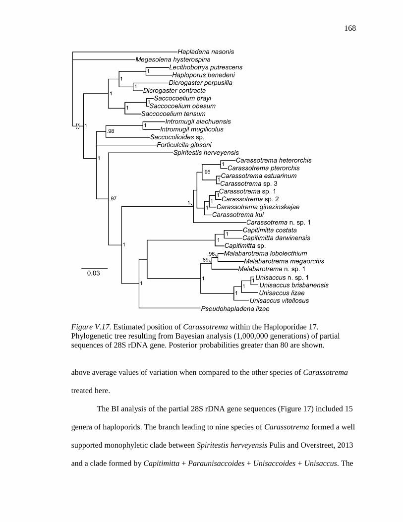
168
Figure V.17. Estimated position of Carassotrema within the Haploporidae 17. Phylogenetic tree resulting from Bayesian analysis (1,000,000 generations) of partial sequences of 28S rDNA gene. Posterior probabilities greater than 80 are shown.
above average values of variation when compared to the other species of Carassotrema
treated here.
The BI analysis of the partial 28S rDNA gene sequences (Figure 17) included 15
genera of haploporids. The branch leading to nine species of Carassotrema formed a well
supported monophyletic clade between Spiritestis herveyensis Pulis and Overstreet, 2013
and a clade formed by Capitimitta + Paraunisaccoides + Unisaccoides + Unisaccus. The

169
monophyletic Carassotrema species form three subgroups formed by C. n. sp. 1, C. kui +
C. ginezinskajae + C. sp. 1 + C. sp. 2, and C. heterorchis + C. pterorchis + C. estuarinum
+ C. sp. 3. The BI tree of partial ITS region (partial ITS1, 5.8S, and ITS2) showed lower
support overall and differed in topology (Figure 18); C. n. sp. 1 grouped internally with
C. estuarinum + C. Sp.3 with C. heterorchis being basal to the remaining species of
Carassotrema.
Discussion
The composition of Carassotrema and its geographic distribution has been
controversial. For differing opinions see (Nasir & Gomez, 1976; Kohn et al., 1999;
Overstreet & Curran, 2005; Yu et al., 2005). In this study I have provided sequence data
for nine species that form a monophyletic clade that I recognize as species of
Carassotrema. Intromugil mugilicolus originally described within Carassotrema is
clearly not a close relative of Carassotrema species (Figure 17). Additionally, those
species Overstreet and Curran (2005) removed from Carassotrema (C. heterorchis, C.kui,
and C. ginezinskajae [as a synonym of C. megapharyngus]) and placed into Platydidymus
are not supported as a natural group (Figures 17-18, Tables 2-3).
Figure V.18. Estimated relations of Carassotrema species 18. Phylogenetic tree resulting from Bayesian analysis (1,000,000 generations) of partial sequences of ITS region. Posterior probabilities greater than 80 are shown.

170
Table V.3 Percent variable sites based on pairwise sequence comparison of the 5.8S gene (above diagonal) and about 380 bases at the 3' end of the ITS1 region (below diagonal) of nuclear ribosomal DNA among 9 species of Carassotrema.
Table V.4 Number of variable sites based on pairwise sequence comparison of the ITS2 region (below diagonal) and partial (c. 1300) 28S gene (above diagonal).
Car
asso
trem
a es
tuar
inum
Car
asso
trem
a gi
nezi
nska
jae
Car
asso
trem
a he
tero
rchi
s
Car
asso
trem
a ku
i
Car
asso
trem
a pt
eroc
his
Car
asso
trem
a sp
. 1
Car
asso
trem
a sp
. 2
Car
asso
trem
a sp
. 3
Car
asso
trem
a n.
sp. 1
C. estuarinum - 3.1 4.1 2.8 3.3 2.9 3.1 0.6 5.1 C. ginezinskajae 7.0 - 4.3 4.1 3.3 1.1 1.2 3.1 4.7 C. heterorchis 4.2 4.2 - 1.5 1.9 1.7 1.2 4.3 6.0 C. kui 7.4 4.6 4.6 - 3.0 0.7 2.9 2.8 4.4 C.pterorchis 5.7 4.6 1.8 4.9 - 3.3 3.5 3.3 5.1 C. sp. 1 6.7 2.1 3.2 4.2 3.5 - 3.1 2.9 4.9 C. sp. 2 6.4 4.6 3.5 4.6 3.9 0.4 - 3.1 4.9 C. sp. 3 3.2-3.5 5.3 3.2 5.3 3.9 4.9 4.6-4.9 - 4.9 C. n. sp. 1. 6.6 5.2 3.8 5.2 4.2 4.2 4.2 4.2 -
* Of the two sequences generated for C. sp. 3 of the ITS2 there was an ambiguous base at the 78th position in one sequence and a T in
the other sequence which is at an indel compared to most species; pairwise difference calculated for both alternatives.
Car
asso
trem
a es
tuar
inum
Car
asso
trem
a gi
nezi
nska
jae
Car
asso
trem
a he
tero
rchi
s
Car
asso
trem
a ku
i
Car
asso
trem
a pt
eror
chis
Car
asso
trem
a sp
. 1
Car
asso
trem
a sp
. 2
Car
asso
trem
a sp
. 3
Car
asso
trem
a n.
sp. 1
C. estuarinum - 1.9 1.3 1.3 1.3 1.9 1.9 0.6 1.3 C. ginezinskajae 6.5 - 0.6 0.6 0.6 0 0 1.3 1.3 C. heterorchis 6.5 5.7 - 0 0 0.6 0.6 0.6 1.3 C. kui 7.3 2.3 6.0 - 0 0.6 0.6 0.6 1.3 C. pterorchis 8.6 6.2 3.9 6.8 - 0.6 0.6 0.6 1.3 C. sp. 1 6.0 1.6 5.5 3.1 6.0 - 0 1.3 1.3 C. sp. 2 6.0 1.6 5.5 3.1 6.0 0.5 - 1.3 1.3 C. sp. 3 2.6 5.7 6.0 6.0 7.3 5.2 5.2 - 1.9 C. n. sp. 1 9.7 8.4 8.6 7.6 10.6 8.6 8.6 8.4 -

171
Both levels of analysis identified the species I consider here as Carassotrema as a
monophyletic group, albeit with differences in topology of the group (Figures 17-18). In
both trees a clade composed of C. kui, C. ginezinskajae, C. sp. 1, and C. sp. 2 was
recovered. The most notable difference between the two trees is the position of the new
species C. n. sp. 1. In the 28S tree (Figure 17), C. n. sp. 1 is represented as sister to the
remaining species of Carassotrema in the analysis. The less robust ITS tree (Figure 18)
shows an internal clade with C. n. sp. 1 as sister to the a clade of C. estuarinum and C. sp.
3. Based on morphological features and definitive hosts infected I would have expected
for Carassotrema species treated here to be divided into two divisions represented by a
marine clade and a freshwater clade. Neither analysis supports that assumption. I must
also note that material of C. sp. 3 was poor, so I do not have good specimens for
morphological characterization. Also, based solely on morphological characterization
of the species I collected, I would have expected C. heterorchis, C. pterorchis, C. sp. 1,
and C. sp. 2 to group together rather than C. kui and C. ginezinskajae with C. sp.1 and C.
sp. 2. In both trees (Figures 17-18), the second node in the generic clade has low support,
probably indicative of rapid radiation at an early point in the history of the genus. More
species and genes are needed to resolve these problems. While Carassotrema is a
biologically cohesive group, there is still a disconnection between morphology and
sequence data. The C. n. sp. 1 morphologically is closest to C. estuarinum, although they
are certainly not each other's closest relative based on pairwise sequence divergence
(Tables 2-3) and the trees built (Figure 17) of the species of Carassotrema with sequence
data available. The pairwise difference between C. n. sp. 1 from the 28S sequence data
shows higher divergence from other members of the genus, 4.4-6%.

172
Results of the BI analysis for the larger group of haploporids (Figure 17)
confidently places the genus within what is currently considered the Waretrematinae
(Pulis & Overstreet, 2013; Chapter IV). As molecular data for Waretrema is unavailable
and Unisaccus has been transferred into the Waretrematinae, there is currently no
morphologically based diagnosis for the subfamily and its limits are uncertain. With the
addition of Carassotrema species to the public domain, I conclude that the generic
synonymies of Nasir and Gomez (1976) are untenable as Intromugil mugilicolus and
Pseudohapladena lizae are recovered as distinct from Carassotrema species. Several
species that Overstreet and Curran (2005) had considered to be in Skrjabinolecithum have
been transferred to Malabarotrema Zhukov, 1972 and Unisaccoides Martin, 1973
(Chapter IV). I consider that Skrjabinolecithum spasskii Belous, 1954 is likely a closer
relative of Carassotrema than to Malabarotrema or Pseudohapladena.
Carassotrema n. sp. 1 appears to be furthest from the core of the genus as would
be expected for a species the furthest removed geographically. Other geographically
distant species are not available for molecular analysis, namely C. philippinensis and C.
bengalense. The addition of these species to this data set would improve inferences as to
the radiation of the genus. While I have made some inferences on the relations among the
species of Carassotrema based on 28S rDNA data my purpose for this gene was
primarily to place the genus within the Haploporidae. I also generated a BI tree based on
the partial ITS region of the Carassotrema species (Figure 17), which differed in
topology from the 28S tree. ITS showed a polytomy of C. heterorchis, C. koreanum, and
a clade formed by the other seven species. Carassotrema n. sp. 1, rather than forming a
clade by itself, showed a stronger affiliation to the C. estuarinum / C. sp. 3. The lengths

173
of the ITS 1 region of these three species are all significantly longer than the other six
species, although trimmed of indels, which appeared to significantly affect the resulting
alignment and, therefore, the BI generated tree. Using either the generated 28S or ITS
trees, I revealed that Overstreet and Curran's (2005) transfer of several Carassotrema
species C. ginezinskajae (which they considered a synonym of C. megapharyngus), C.
kui, C. megapharyngus, and C. heterorchis)to the genus Platydidymus is unwarranted.
My morphological study also shows that the testis shape of some species (most notably
C. heterorchis) is highly dependent on the amount of pressure applied to the specimen
when heat killed.
I do not know where the mullet from which C. estuarinum and C. sp 3 collected
acquired the infection; while I expect that the ancestral Carassotrema was of a marine
origin, C. estuarinum / C. sp. 3 may be a case of a secondary acquisition of a brackish
host or alternatively multiple acquisitions of freshwater host from a basal marine
Carassotrema species. The conflicting topology of the 28S and ITS trees (Figures 17, 18)
shows that more study is required to determine relations of species of Carassotrema.
Taxonomy and systematics of Carassotrema would benefit from recollection of
all Southeast Asian species from other host and major river drainages to determine how
widespread these species are and if the individual species do show the wide host use and
geographic range as indicated by myself and others. All my freshwater cyprinid
specimens were from the same river drainage and constitute mostly new host records. I
have provided molecular data, but I include the caveat that the worms collected may
represent morphotypes rather than the species as originally described. Alternatively,
China has a long history of aquaculture, and it is possible that species of Carassotrema

174
have been widely transported outside their native ranges. Collection of specimens for
molecular analysis of population level genes may help to understand if these species have
been transported by human activity or are widespread species with distinct lineages.
If all the records of Carassotrema synonyms, locations, and hosts are correct, the
distribution and breadth of the freshwater cyprinid hosted species of Carassotrema are
some of the most broadly distributed trematodes known. I suspect that there are numerous
unnamed species, but the lack of type specimens severely limits making robust decisions
about the patterns of diversification or host use in these species. While any one of these
species could be imagined to have such a wide host use, the majority of them possessing
such breadth of host use most likely indicates that the diversification of this genus in
freshwater Asia is being driven primarily by first intermediate host use rather than
definitive host use. Most other species of haploporids have been reported only at the time
of their original description; thus, only one host is reported. In China there have been
many reports of the genus from many localities and, thus, perhaps the host use is more a
product of intensive sampling and other species in the family would benefit from the
same broad examination of host. The most commonly reported hosts also are those fish
which attain larger size and are of economic importance such as Carassius auratus,
Cyprinis carpio, Hypophthalmichthys molitrix, Hypophthalmichthys nobilis,
Ctenopharyngodon idella, and Mylopharyngodon piceus, thus giving them higher priority
for examination for parasites than other, less economically important fishes.

175
CHAPTER VI
NEW GENUS 1 NEW SPECIES (TREMATODA: HAPLOPORIDAE) FROM THE
SQUARETAIL MULLET ELLOCHELON VAIGIENSIS (ACTINOPTERYGII:
MUGILIFORMES) FROM AUSTRALIA
Abstract
New genus 1 is proposed as monotypic for New genus 1 new species, a new
species of trematode in the family Haploporidae. The new species is described from the
squaretail mullet (Ellochelon vaigiensis) in Australia. The new genus differs from other
known members of the family by having a cleft oral sucker, row of spines on the
posterior opening to the mouth, wider than long caecum, thick-walled hermaphroditic
sac, and cells lining the external seminal vesicle having large nucleated cells. The new
genus is most similar to members of the Haploporinae in possessing in-utero eggs
containing miracidium with eye-spots and reduced vitellarium. Based on analysis of the
partial 28S rDNA sequences, the species is most closely related to those in the
Waretrematinae with a single caecum.
Introduction
The collection of squaretail mullet from Australia yielded specimens of an
unknown genus and species of Haploporidae Nicoll, 1914. The new genus and species
are described below, and their position in the Haploporidae is discussed.
Materials and Methods
Fish hosts were collected by castnet. Identification of the host conforms to
currently valid names and common name given in FishBase (Pauly & Froese, 2013).
Haploporids were isolated similarly to the process advocated by Cribb and Bray (2010)

176
for gastrointestinal species, skipping an initial examination because of the volume of the
intestinal contents. Live worms were rinsed and cleaned in saline and observed briefly.
The saline was removed from the container, and the worms were killed by addition of
hot steaming, but not boiling, tap water. Haploporids were then stored in 70% ethanol.
Parasite specimens for morphological and molecular analysis were processed and studied
according to the protocols used by Pulis et al. (2013) and Pulis and Overstreet (2013). All
measurements are in micrometres unless noted otherwise. Museum abbreviations are as
follows: MNT, Museum and Art Gallery of the Northern Territory, Darwin, Australia;
and USNPC, US National Parasite Collection, Beltsville, Maryland.
Genomic DNA was isolated using Qiagen DNAeasy Tissue Kit (Qiagen, Inc.,
Valencia, California, USA) following the instructions provided. DNA fragments
approximately 2,500 basepairs (bp) long comprising the 3' end of the 18S nuclear rDNA
gene, internal transcribed spacer region (including ITS1 + 5.8S + ITS2), and the 5' end of
the 28S gene (including variable domains D1-D3) were amplified from the extracted
DNA by polymerase chain reaction (PCR) on a PTC-200 Peltier Thermal Cycler using
forward primers ITSF (5' - CGCCCGTCGCTACTACCGATTG-3') or LSU5 (5'-
TAGGTCGACCCGCTGAAYTTAAGCA-3') and reverse primer 1500R (5’-
GCTATCCTGAGGGAAACTTCG-3’). These PCR primers and multiple internal
primers were used in sequencing reactions. The internal forward primers were DIGL2
(5’-AAGCATATCACTAAGCGG-3’), 300F (5’-CAAGTACCGTGAGGGAAAGTTG-
3’), 900F (5’-CCGTCTTGAAACACGGACCAAG-3’), and internal reverse primers
were 300R (5’-CAACTTTCCCTCACGGTACTTG-3’), Digl2r (5’-
CCGCTTAGTGATATGCTT-3’), and ECD2 (5’-CTTGGTCCGTGTTTCAAGACGGG-
177
3’). Previously published 28S ribosomal RNA gene sequences of species of Haploporidae
were used for comparison (see Figure 11 for accession numbers) with newly submitted
sequences. Sequences were aligned using the ClustalW application in the BioEdit
program, Version 7.0.9 (Hall, 1999). The alignment was further refined by eye and
trimmed to the shortest sequence on both 5' and 3' ends. The resulting alignment utilised
32 haploporids with Hapladena nasonis as the outgroup, and the alignment was 1,098
characters long, including gaps, with 727 sites conserved, 371 sites variable, and 279
parsimony-informative sites. Phylogenetic analysis of the data was performed using
Bayesian Inference (BI) with MrBayes 3.1.2 software (Huelsenbeck & Ronquist, 2001;
Ronquist & Huelsenbeck, 2003). The best nucleotide substitution model was estimated
with jModeltest Version 0.1.1 (Guindon & Gascuel, 2003; Posada, 2008) as general time
reversible with gamma-distributed among site-rate variation and estimates of invariant
sites (GTR + I + Γ). The following model parameters were used in MrBayes: nst = 6,
rates = invgamma, ngen = 1,000,000, and samplefreq = 100. The first one quarter of
samples were discarded as burn-in (sump burnin = 2500), and nodal support was
estimated by posterior probabilities (sumt) (Huelsenbeck et al., 2001). All other settings
were left as default.
Results
New genus 1
Diagnosis: Body of adult elongate, approximately 5 times longer than wide, with
posterior end tapered. Eye-spot pigment dispersed. Tegument spined. Oral sucker
subterminal, opening to mouth with anterior cleft, single row of small spines along
posterior ventral margin ventral to mouth. Ventral sucker prior to equatorial. Prepharynx

178
long. Pharynx longer than wide. Caecum near mid-hindbody, bilobed, wider than long.
Testis in hindbody near posterior margin of ventral sucker. Hermaphroditic sac dorsal to
ventral sucker, elongate, thick-walled, and originating noticeably posterior to genital
pore. Hermaphroditic duct about 1/3 the length of hermaphroditic sac, muscular, with
smooth appearance. Male duct short. Female duct long, muscular, rugose in appearance.
Internal seminal vesicle well-defined, straight, not sac-like. External seminal vesicle
round, made up of large nucleated cells. Ovary contiguous with anterior portion of testis.
Uterus long, convoluted, extending posterior to testis, metraterm absent. Vitellarium
dorsal to ovary and testis, composed of about 15 lobes. Eggs thin-shelled, distal eggs
containing developed miracidium with eye-spots. Excretory vesicle I-shaped with two
collecting tubes originating from anterior portion. Excretory pore subterminal dorsal.
Lymphatic system present. In intestine of Mugilidae from Indo-West Pacific region.
Type-species: New genus 1 new species.
Remarks
In its possession of a spined tegument, hermaphroditic sac, and single testis, N. g.
1 is a member of the family Haploporidae Nicoll, 1914.
New genus 1 new species can be separated from other genera in the family
Haploporidae by the combination of single caecum, reduced vitellarium, uterus extending
into post-testicular area, in-utero eggs containing developed miracidium with an eye-spot,
I-shaped excretory vesicle, and sub-terminal dorsal excretory pore. Additional characters
unique to N. g. 1 n. sp. include oral sucker with anteroventral cleft; oral sucker armed
posteriorly with single row of small spines; caecum wider than long; hermaphroditic sac
muscular, thick walled; non-sac-like internal seminal vesicle; and external seminal

179
vesicle lined with large nucleated cells. The gross anatomy of N. g. 1 n. sp. is most
similar to the genus Unisaccus Martin, 1973 due to the single caecum, reduced
vitellarium, uterus extending post-testicularly, and in-utero eggs containing miracidium
with eye spots. Characters possessed by Unisaccus not found in N. g. 1 n. sp. include
spirally arranged pads in the posterior end of the hermaphroditic duct, sac-like internal
seminal vesicle, sac-like external seminal vesicle, and Y-shaped excretory vesicle. The
presence of eye-spots in the miracidium of in-utero eggs distinguishes N. g. 1 n. sp. from
Unisaccoides Martin, 1973 and Paraunisaccoides Martin, 1973. Unicoelium Thatcher &
Dossman also has a single caecum as found in N. g. 1 n. sp., but possesses vitellarium in
two distinct clumps lateral to ovary, uterus extending anterior to hermaphroditic sac, and
Y-shaped excretory vesicle. Pseudolecithobotrys Blasco-Costa, Gibson, Balbuena, Raga,
& Kostadinova, 2009 has a variable caecal shape ranging from singular to bifurcating and
differs from N. g. 1 n. sp. by short prepharynx, the sac-like external seminal vesicle,
muscular genital atrium, and vitellarium in two distinct clumps.
New genus 1 new species (Figures 1-11)
Description: Measurements based on 15 wholemount specimens. Body elongate,
1552-2016 long (BL), 254-396 wide, widest in hindbody, width 16-22% of BL.
Tegument spined; spines hasate, about 10-12 long in forebody, majority of spine beneath
surface of tegument (Figure 7) becoming smaller and more sparse posteriorly. Eye-spot
pigment dispersed, most prominent in forebody. Oral sucker subterminal, anterior half
cleft (Figures 1, 5), 174-232 long, 168-221 wide, with mouth opening anteroventrally,
posterior ventral margin or oral sucker with single row of minute spines (Figure 5).

180
Figures VI.1-4. New genus 1 new species 1. Holotype ventral wholemount; 2. Paratype lateral whole mount; 3. Paratype hermaphroditic sac, ventral mount ventral sucker outline shown; 4.Paratype ovarian complex. Scale-bars 1-2. 300µm, 3. 200µm, 4. 100 µm.

181
Ventral sucker slightly elevated, 134-180 long, 138-188 wide, with about four rows of
small spines on most ventral rim of ventral sucker, inner circle of 12 papilla, inside of
papilla a few rows of spines smaller then body spines, larger than three rows of the rim,
deepest portion unarmed (Figure 6). Ventral sucker 68-87% as long as oral sucker, 73-
88% as wide as oral sucker. Forebody 500-762 long or 34-40% of BL; hindbody 848-
1222 long or 52-62% of BL. Prepharynx well defined, 274-420 long. Pharynx 140-184
long, 123-167 wide, length 102-125% of width, widest in posterior half. Oesophagus
313-577 long. Caecum bilobed, 150-254 long, 218-326 wide, lined with large cells
(which was used to determine the anterior extent), commencing 931-1322 from anterior
end of body or 58-69% of BL; post-caecal space 344-637 or 18-32% of BL.
Testis medial, round, overlapping posterior end of caeca to 92 posterior, 362-670
from ventral sucker, 132-171 long, 140-203 wide; post-testicular space 220-449 or 14-
24% of BL. Hermaphroditic sac dorsal to ventral sucker, 301-469 long, 122-160 wide,
outer muscular wall about 20 thick, terminating posterior to ventral sucker; sac
containing following structures (internal seminal vesicle straight, well-defined, 109-180
long, 24-50 wide; male duct thin-walled; prostatic bulb immediately anterior to internal
seminal vesicle; female duct muscular, appearance rugose; male duct joining female in
anterior third of sac); hermaphroditic duct smooth. External seminal vesicle 107-164
long, 90-162 wide, with definite layer of thick nucleated cells, about 10-15 thick. Genital
pore laterally elongate, immediately anterior to ventral sucker, hermaphroditic duct

182
Figures VI.5-7. New genus 1 new species scanning electron micrographs. 5. Oral sucker showing anterior cleft and row of small spines on posterior ventral rim; 6. Ventral sucker with papillae and laterally elongate genital pore; 7 Tegumental spines of forebody. Scale-bars: 5. 50µm; 6. 50µm; 7. 10µm. noticeably long from hermaphroditic sac to genital pore, area immediately around genital
pore devoid of spines.
Ovary medial, contiguous with testis, ventral to testis, 63-101 long, 67-110 wide.
Mehlis' gland antero-dorsal to ovary. Laurer's canal dorsal to testis; pore dorsal to testis.
Vitellarium consisting of mass of about 9-20 lobes (number subjective), nestled dorsal to
ovary, between testis and caecum. Uterus convoluted, occupying most of hindbody
posterior to hermaphroditic sac; proximal portion often filled with sperm; metraterm
absent. Eggs thin-shelled, yellow, numerous, 50-67 long, 22-38 wide (measured from
fully developed distal portion of uterus), considerable variation in size between
specimens (weak correlation of longer worm = longer egg), with developed miracidium
possessing eye-spot.

183
Figures VI.8-10. New genus 1 new species micropictographs. 8. hermaphroditic sac showing thick muscular wall; 9. external seminal vesicle, ventral surface showing large nucleated cells; 10. external seminal vesicle mid-depth showing thickness of nucleated cells. Scale-bars: 8. 100µm; 9. 20µm; 10. 20µm.
Lymphatic tubes in forebody. Excretory vesicle I-shaped, posterior to testis, with
two narrow cura emerging from anterior portion; pore dorsal subterminal.
Type-host: Ellochelon vaigiensis (Quoy & Gaimard), squaretail mullet
(Mugilidae)
Type-locality: Ludmilla Creek, Darwin, Northern Territory, Australia (caught on
mud flat that appeared to be flooded only during highest spring tides). (12° 24' 52" S,
130° 50' 10" E).
Additional locality: Whithnell Bay, Burrup Peninsula, near Dampier, Western
Australia, Australia, (20° 35' 01" S, 116° 47' 21" E).
Site: Intestine.
Type-material: MNT TBD holotype TBD paratype; BM TBD paratypes; USNPC
TBD Paratypes. Gen bank TBD

184
Remarks
New genus 1 new species is the only species within the genus.
Phylogenetic Analysis
In the BI analysis of partial 28S sequences presented here N. g. 1 n. sp. was found
to be within the clade considered Waretrematinae by Pulis and Overstreet (2013), sister to
the clade of other single caecum waretrematins (Malabarotrema Zhukov, 1972;
Unisaccoides Martin, 1973; Unisaccus Martin, 1973) (Figure 11)
Figure VI.11. Estimated position of New genus 1 new species within the Haploporidae. Bayesian Inference tree of phylogenetic relationships of the Haploporidae using partial 28S rDNA sequences. Position of New genus 1 new species shown in bold.
Discussion
Molecular analysis of partial 28S rDNA fragments of haploporid genera with N.
g. 1 n. sp. reveal that is not particularly related to any genus from which sequence data is
available. Based on the most recent comprehensive evaluation of the family by Overstreet
and Curran (2005), I would have expected N. g. 1 n. sp. to be closer to the Haploporinae

185
than to the Waretrematinae. In the assessment of some species possessing a single
caecum in the previous chapter, Unisaccus was found to be a member of the
Waretrematinae, which was not suspected by previous authors who speculated on the
subfamilial relations of the family. The defining characters for the Haploporinae were
proposed as the in-utero eggs with developed miracidium possessing eye-spots and
reduced vitellarium. Now that another genus with eye-spotted miracidium, reduced
vitellarium, and a single caecum has been proposed in this paper it beguiles attempts to
form a morphological basis for the subfamilies as recognized by Overstreet and Curran
(2005), although as shown by other phylogenetic work on the family (Blasco-Costa et al,
2010; Pulis & Overstreet, 2013) there are clear clades that I believe merit subfamily
designations. After the work of Blasco-Costa, Balbuena et al. (2009) phylogenetic
analysis of the family has been limited to the addition of species that happen to fall within
the Waretrematinae clade, while no species have been added from the Haploporinae or
Forticulcitanae Blasco-Costa et al. (2010) clades. I do not expect the addition of more
species to the tree produced here to change the conclusions of this paper, but additional
species will provide more robust inferences to the stability and usefulness of characters to
determine relationships.
New genus 1 new species has several characters not reported for any other
haploporid genera, including armed posterior oral sucker, papilla encircling the ventral
sucker, and the robust external seminal vesicle composed of large nucleated cells. Also,
the internal seminal vesicle is more robust than has been reported for other members of
the family, but many original and subsequent descriptions have been based on material
killed under coverslip pressure, which lessens the utility of this character. Because N. g. 1

186
n. sp. possesses many unusual characters compared with other described species, I
consider these features to be derived.
The present study demonstrates that morphological characters used by Overstreet
and Curran (2005) to differentiate the Waretrematinae from Haploporinae are not of
systematic importance. Based on 28S partial sequence analyses (Figure 11) Haploporinae
and Waretrematinae form well-supported, distinct clades. The extensive uterus, reduced
vitellarium, and well-developed miracidium in-utero are all characters that are found in
other subfamilies of haploporid other than Megasoleninae, species which occupy a basal
position in the Haploporidae. This suggests that reduced vitellarium and well-developed
miracidium in-utero have been acquired at least a few times in the family. Ellochelon
vaigiensis that were collected for this work were actively feeding in waters at the highest
spring tides (personal observation). This along with the advanced development of the
miracidium in-utero could suggest that the miracidium leaves the fish and seeks out a
snail host, whereas an egg that requires and incubation period outside the host may be
subject to desiccation. I suspect that the first intermediate host of N. g. 1 n. sp. is a
intertidal snail.

187
CHAPTER VII
NEW GENUS 2 NEW SPECIES FROM THE CLUPEID NEMATALOSA COME FROM
AUSTRALIA
Abstract
New genus 2 is proposed for New genus 2 new species collected from the clupeid
Nematalosa come. The new genus is most similar to the genera Carassotrema and
Platydidymus. New genus 2 can be distinguished from Platydidymus by the lack of a
crenulated oesophagus, Y-shaped rather than H-shaped gut, and vitellarium not extending
anterior to testis. While two species of Carassotrema have been reported from Clupeids,
an unusual host for haploporids, N. g. 2 can be differentiated from Carassotrema species
by the vitellarium being a mass of follicles not organized into distinct tubes or plates
restricted dorsally and posterior to testis but not anterior, and gut being sac-like rather
than tubular. Based on a 28S rDNA gene fragment Bayesian Inference analysis suggest
that N. g. 2 n. sp. is most closely related to species of the genus Carassotrema.
Introduction
Collections of Nematalosa come (Richardson, 1846) from Australia were infected
with specimens of a member of the family Haploporidae Nicoll, 1914 that are not
attributable to any known genus. Therefore, I propose a new genus for the new species.
Materials and Methods
Fish hosts were collected by castnet. Names of fish host conforms to currently
valid names and common name given in FishBase (Froese & Pauly, 2013). Haploporids
were isolated as advocated by Cribb and Bray (2010) for gastrointestinal species. Live
worms were rinsed and cleaned in saline and observed briefly. The saline was removed

188
from the container, and the worms were killed by addition of hot, but not boiling, tap
water. Haploporids were then stored in 70% ethanol. Parasite specimens for
morphological and molecular analysis were processed and studied according to the
protocols used by Pulis et al. (2013) and Pulis and Overstreet (2013). All measurements
are in micrometres unless otherwise noted. Museum abbreviations are as follows: MNT,
Museum and Art Gallery of the Northern Territory, Darwin, Australia and USNPC, US
National Parasite Collection, Beltsville, Maryland.
Genomic DNA was isolated using Qiagen DNAeasy Tissue Kit (Qiagen, Inc.,
Valencia, California, USA) following the instructions provided. DNA fragments
approximately 2,500 basepairs (bp) long comprising the 3' end of the 18S nuclear rDNA
gene, internal transcribed spacer region (including ITS1 + 5.8S + ITS2), and the 5' end of
the 28S gene (including variable domains D1-D3) were amplified from the extracted
DNA by polymerase chain reaction (PCR) on a PTC-200 Peltier Thermal Cycler using
forward primers ITSF (5' - CGCCCGTCGCTACTACCGATTG-3') or LSU5 (5'-
TAGGTCGACCCGCTGAAYTTAAGCA-3') and reverse primer 1500R (5’-
GCTATCCTGAGGGAAACTTCG-3’). These PCR primers and multiple internal
primers were used in sequencing reactions. The internal forward primers were DIGL2
(5’-AAGCATATCACTAAGCGG-3’), 300F (5’-CAAGTACCGTGAGGGAAAGTTG-
3’), 900F (5’-CCGTCTTGAAACACGGACCAAG-3’), and internal reverse primers
were 300R (5’-CAACTTTCCCTCACGGTACTTG-3’), Digl2r (5’-
CCGCTTAGTGATATGCTT-3’), and ECD2 (5’-CTTGGTCCGTGTTTCAAGACGGG-
3’). Previously published 28S ribosomal RNA gene sequences of species of Haploporidae
were used for comparison (see Figure 11 for accession numbers) with newly submitted

189
sequences. Sequences were aligned using the ClustalW application in the BioEdit
program, Version 7.0.9 (Hall, 1999). The alignment was further refined by eye and
trimmed to the shortest sequence on both 5' and 3' ends. The resulting alignment utilised
32 haploporids with Hapladena nasonis as the outgroup, and the alignment was 1,098
characters long, including gaps, with 727 sites conserved, 371 sites variable, and 279
parsimony-informative sites. Phylogenetic analysis of the data was performed using
Bayesian Inference (BI) with MrBayes 3.1.2 software (Huelsenbeck & Ronquist, 2001;
Ronquist & Huelsenbeck, 2003). The best nucleotide substitution model was estimated
with jModeltest Version 0.1.1 (Guindon & Gascuel, 2003; Posada, 2008) as general time
reversible with gamma-distributed among site-rate variation and estimates of invariant
sites (GTR + I + Γ). The following model parameters were used in MrBayes: nst = 6,
rates = invgamma, ngen = 1,000,000, and samplefreq = 100. The first one quarter of
samples were discarded as burn-in (sump burnin = 2500), and nodal support was
estimated by posterior probabilities (sumt) (Huelsenbeck et al., 2001) with all other
settings left as default.
Results
New genus 2 Figures. 1-3.
Diagnosis: Body minute, pyriform. Tegument spined. Diffuse eye-spot pigment
prominent in forebody. Oral sucker terminal. Ventral sucker near equatorial. Prepharynx
long. Pharynx smaller than suckers. Oeseophagus smooth, long, near length of
prepharynx, terminating near posterior margin of ventral sucker. Caeca sac-like, in
hindbody, reaching posterior to testis. Testis large, V-shaped, wider than long.

190
Figure VII.1. New genus 2. 1.Ventral wholemount scale-bar 100 µm.

191
Hermaphroditic sac ellipsoidal. Internal seminal vesicle occupying less than half of
hermaphroditic sac. External seminal vesicle sac-like, roughly same size as internal
seminal vesicle. Ovary round, dorsal to testis. Uterus direct from ovary to hermaphroditic
sac, proximal portion filled with sperm. Vitellarium of homogenous mass of rounded
follicles surrounding posterior and dorsal portion of testis to posterior end of body. Eggs
not observed. Excretory vesicle Y-shaped; pore terminal. In Clupeidae Indo-West Pacific
region. Type-species: N. g. 2. n. sp.
Remarks
New genus 2 possess a spined tegument, a single testis, and a hermaphroditic sac
consistent with other members of the family Haploporidae Nicoll, 1914. New genus 2 can
be differentiated from other genera in the family by the combination of characters,
including pyriform body, V-shaped, wide testis, ovary dorsal to testis, and vitellarium
confined to area posterior to posterior margin of testis.
New genus 2 is most similar to the genera Carassotrema Park, 1938 and
Platydidymus Overstreet and Curran, 2005 (syn. Haplotrema Zhukov, 1971 preoccupied).
New genus 2 can be differentiated from Carassotrema by the vitellarium not extending
anterior to testis and from Platydidymus by the sac-like non H-shaped gut configuration,
smooth oesophagus, and vitellarium not extending anterior to testis.
Specimens for members of this genus were collected from Nematlosa come from
Western Australia and Queensland, Australia. Due to the lack of molecular material from
the Queensland specimen, I only include specimens from Western Australia in the
description of N. g. 2 n. sp. The specimen from Queensland, Australia is certainly
congeneric with N. g. 2 n. sp. from Western Australia, but it may prove to be separate

192
species once the eastern form has more specimens collected. Due to the lack of molecular
data from the eastern specimen I take a very conservative approach an decline to include
it in the description of the new species, although it was used in the generic diagnosis.
New genus 2 new species (Figures 2-3)
Type-host: Nemataslosa come (Richardson), western Pacific gizzard shad
(Clupeidae).
Site: Intestine.
Type-locality: Barred creek, Western Australia, Australia (17° 39' 48" S 122° 12'
06" E).
Description: Measurments based on 2 wholemount worms. Body pyriform, 285-
333 long, 134-162 wide, measured in hindbody where it is greatest. Body width 47-49%
of length. Tegument armed with spines about 4 long, becoming smaller and more sparse
toward the posterior end of the body, small size of the body gives anterior end thorny
appearance. Oral sucker sub-terminal, 52-63 long, 64-76 wide. Ventral sucker slightly
elevated, pad-like, without deep depression in center, 64-75 long, 74-83 wide, 119-123%
longer than oral sucker, 109-116% wider than oral sucker. Forebody 104-114 long or 34-
36% of BL; hindbody 117-151 long or 41% of BL. Prepharynx 20-25 long. Pharynx
muscular 31-37 long, 29-36 wide, length 124-185% of prepharynx length. Oesophagus
47-58 long, bifurcating near posterior margin of ventral sucker, 154-180 from anterior
end of body, 54% of body length. Caeca long and tubular, ending blindly 62-64 from
posterior end of body; postcaecal space 21-24% of BL.
Testis medial, strongly V-shaped, orientated nearly vertically with point of V
dorsal, with tips of V near ventral surface, 68-71 long, 100-122 wide, width 147-172% of

193
Figure VII.2. New genus 2 new species 2. ventral wholemount Holotype. Scale-bar, 100µm.

194
length, anterior tips at level of posterior quarter of ventral sucker, 52-92 from posterior
end, post-testicular space 18-28% of BL. Hermaphroditic sac, medial, dorsal to ventral
sucker, 45-61 long, 26-37 wide, containing internal seminal vesicle, 22-25 long, 16-17
wide, male duct not observed, female duct smooth, and hermaphroditic duct proximal end
muscular and smooth distal end smooth. Genital pore medial, immediately at anterior
margin of ventral sucker. External seminal vesicle sac-like, 23-51 long, 21-23 wide.
Ovary medial, dorsal to midpoint of testis, 30-42 long, 31-38 wide, 12-14
posterior to ventral sucker. Ootype surrounded by Mehlis' gland, situated anterior to
ovary. Laurer's canal not observed. Vitellarium extensive, follicles in plate-like sections,
commencing near level of posterior half of testis, filling most of post-testicular space.
Uterus occupying space between ovary and hermaphroditic sac, proximal end filled with
sperm, distal end without evidence of metraterm. Eggs not observed. Excretory bladder
Y-shaped, bifurcation dorsal to posterior end of testis; pore terminal.
Molecular Results:
N. g. 2. n. sp. shows the greatest affinity to Species of Carassotrema according to
the BI analysis (Figure 3).
Discussion
Clupeids are an overlooked host for haploporids. To date only three species of
haploporids have been recorded from the family, two in Carassotrema (Liu,
2003; Chapter V) and the new genus reported here. Further sampling of Indo-Pacific
clupeids should yield more undescribed species. The subfamily Waretrematinae
Srivastava, 1939, of which N. g. 2 is a member, is hosted predominantly by mullets. The

195
Figure VII.3. Estimated position of New Genus 2 New species 3. Bayesian Inference tree of phylogenetic relationships of the Haploporidae using partial 28S rDNA sequences with Hapladena nasonsis as the outgroup. N. g.2 n. sp. shown in bold. Posterior probability scores greater than 0.80 given.
finding of representatives of two genera in these fishes is intriguing. The known life
cycles of haploporids involve two hosts with the cercaria or metacercaria being ingested
by the definitive host (Martin, 1973a; Tang & Lin, 1979; Shameen & Madhavi, 1991; Yu
et al., 2005). As such, those clupeids which are detritivores, like mullets, should provide
ample opportunity for host switching, indicating the host associations in the haploporids
are the result of host switching rather than co-evolution.
Although eggs were not observed, I suspect that they would not contain a
developed miracidium with eye-spots while in the uterus. Those members of the
waretrematins, Unisaccus and N. g. 1, have in-utero eggs with developed miracidium

196
with eye-spots, have a long uterus and reduced vitellarium, whereas N. g. 2 has a short
uterus and extensive vitellarium. I predict that, when observed, the eggs of N. g. 2 when
deposited will lack developed miracidium and take some time to mature before being
infective to the first intermediate host as in the two species of Carassotrema with known
life-cycles (Tang & Lin, 1979).

197
CHAPTER VIII
CHANGE IN RANK OF MEGAPERIDAE (TREMATODA) TO THE MEGAPERINAE
WITHIN THE APOCREADIIDAE AND DESCRIPTION OF HAINTESTINUM
AMPLUM N. G. N. SP.
Abstract
Haintestinum amplum n. g. n. sp. is described from the scrawled cowfish,
Acanthostracion quadricornis, collected in the Eastern Gulf of Mexico off Florida, USA.
The new species is relatively large and shares characters of the Apocreadiidae and
Megaperidae but conforms to the diagnosis of neither. It belongs in a new genus
possessing a pharynx with lobed anterior margin and intestine terminating in paired ani,
like in megaperids, and, when compared with apocreadiids, it shares important
anatomical features, including an I-shaped excretory vesicle, canicular seminal vesicle,
eye-spot remnants, and pretesticular uterus and lacks a cirrus and cirrus sac. The H-
shaped intestine and large funnel-shaped oral sucker without a U-shaped sphincter
encircling half the anterior aperture are the most notable diagnostic characters of the new
monotypic genus. Additionally, the phylogenetic position of the Megaperidae is
investigated for the first time, using analysis of partial 28S rDNA gene sequences from
H. amplum, two species in the Megapera, Thysanopharynx elongatus, and previously
published 28S sequences of species from members of the Apocreadiata, Haploporoidae,
Lepocreadiata, and Opisthorchiata. The resulting analysis demonstrated a close
relationship among the new genus and the three species of megaperids, and the
megaperids were most closely allied with Schistorchis zancli of the apocreadiids.

198
Moreover, I now consider Megaperidae as the subfamily Megaperinae within the
Apocreadiidae.
Introduction
Prior to the present study, five species in the genus Megapera Manter, 1934 and
Thysanopharynx elongatus Manter, 1933 comprised the Megaperindae Manter, 1934, all
of which are parasites of tetradontiform fishes from the warm waters of the western North
Atlantic Ocean. Manter (1933) initially noted that adult megaperids shared similar traits
with worms that were considered to belong in the Allocreadiidae Looss, 1902 and the
Lepocreadiidae Odhner, 1905 at that time, but Manter deemed that the presence of paired
ani, an undivided seminal vesicle, pre-ovarian symmetrical testes situated in the
forebody, and peculiar shaped pharynx plus the lack of a cirrus and cirrus sac served to
unite the group as a family. Cable (1954) investigated the life history of Megapera gyrina
(Linton, 1907) Manter, 1934, and observed that the cercaria emerged from a slipper
limpet (Crepidula convexa Say, 1822) at Salinas Bay, Puerto Rico, USA, and encysted
almost immediately on vegetation or substratum, suggesting lack of a second
intermediate host, and developed in a herbivorous definitive host that fed on infested
vegetation.
Manter (1963) reassessed the relationship between the Megaperidae and the
Lepocreadiidae Odhner, 1905, noting similarities between specialization of the oral
sucker in the megaperids and certain genera that were initially considered to belong in the
Lepocreadiidae, but presently considered in the Apocreadiidae Skrjabin, 1942 (i.e.,
Schistorchis Lühe, 1906, Sphincterostoma Yamaguti, 1937, Megacreadium Nagaty, 1956
and Lobatotrema Manter, 1963 [syn. Sphincteristomum Oshmarin, Mamaev, and

199
Parukhin, 1961]). Manter (1963) reduced the family Megaperidae to the subfamily rank
Megaperinae Manter, 1963. Megapera and Thysanopharynx Manter, 1933 had previously
considered to be in the Lepocreadiidae; Sphincterostoma and Sphincteristomum were
considered in the Sphincterostomatinae Yamaguti, 1958; and Megacreadium was
considered a synonym of Schistorchis in Lepocreadiidae without placement in a
subfamily (Manter, 1963; Sogandares-Bernal & Hutton, 1959). Yamaguti (1971)
transferred Schistorchis and Megacreadium to the family Schistorchiidae Yamaguti, 1942
and treated the two megaperid genera in their own family, Megaperidae, based on
morphological features of the cercaria of M. gyrina. Mehra (1961) and Brooks et al.
(1985) also questioned an affinity between the Megaperinae and the Lepocreadiidae
based on the presence of a Y-shaped excretory bladder in the cercaria of M. gyrina,
suggesting a relationship with the Haploporidae Nicoll, 1914. Cribb and Bray (1999)
proposed that Schistorchis, Sphincterostoma, Megacreadium, and Sphincteristomum all
belonged in the subfamily Schistorchiinae Yamaguti, 1942 in the Apocreadiidae. In the
most recent classification of the Trematoda, Bray (2005a; 2005b) tentatively treated the
Megaperidae as a family within the artificial Superfamily Lepocreadioidea Odhner, 1905.
In the most recent molecular phylogeny (Bray et al., 2009) and treatment (Bray & Cribb,
2012) of the superfamily Lepocreadioidea, three families, Megaperidae, Liliatrematidae
Gubanov, 1953, and Deropristidae Cable and Hunninen, 1942, have not had molecular
data available to support or challenge their inclusion into that or another superfamily.
In the present study, I present a description of a new species and genus that
possesses morphological features that are diagnostic for both the Apocreadiidae and the
Megaperidae, provide sequence data for the new genus and three species of megaperids,

200
and conduct an analysis of sequence data estimating the phylogenetic relationships of the
new species and the megaperids.
Materials and Methods
The scrawled cowfish, Acanthostracion quadricornis (Linnaeus), and honeycomb
cowfish, Acanthostracion polygonius Poey were collected during the National Marine
Fisheries Service (NMFS) Fall Pelagic Trawls in the eastern Gulf of Mexico (GOM)
from 2007 through 2010 and by cast net off Grassy Key, Florida, in 2012. Fish were
examined for helminths while fresh. The trematodes for this study were cleaned in saline,
examined briefly under a dissecting scope, killed by submersion in hot water, and fixed in
70% ethanol for specimens used in molecular analyses, morphological identifications,
and scanning electron microscopy (SEM). Specimens used for wholemounts were stained
in aqueous Mayer's hematoxylin or Van Cleave's hematoxylin, dehydrated in a graded
ethanol series, cleared in clove oil (Van Cleave's) or methyl salicylate (Mayer's), and
mounted permanently in Canada balsam. Measurements were taken with a differential
interference contrast (DIC) equipped Leica compound microscope using a ProgRes®
CapturePro camera (Version 2.8 Jenoptic, Jena, Germany) and software. All
measurements are in micrometres (µm) unless noted otherwise. Sequences used in the
present study were either derived from trematode specimens I collected from the
intestines of fishes between 2007 and 2012 (Table I) or obtained from GenBank. The
type specimens of the new species and representative voucher specimens were deposited
at the United States National Parasite Collection (USNPC), Beltsville, Maryland.
Genomic DNA was extracted from two hologenophores (terminology of Pleijel et
al., 2008) of the new species, and one hologenophore of T. elongatus, and entire single

201
specimens of M. gyrina and Megapera orbicularis (Manter, 1933), using Qiagen
DNAeasy Tissue Kit (Qiagen, Inc., Valencia, California, USA) following the instructions
provided. DNA fragments measuring approximately 2,500 basepairs (bp) long
comprising the 3' end of the 18S nuclear rDNA gene, internal transcribed spacer region
(including ITS1 + 5.8S + ITS2), and 5' end of the 28S gene (including variable domains
D1-D3) were amplified from the extracted DNA by polymerase chain reaction (PCR)
using a PTC-200 Peltier Thermal Cycler and forward primers ITSF (5' -
CGCCCGTCGCTACTACCGATTG-3') or LSU5 (5'-
TAGGTCGACCCGCTGAAYTTAAGCA-3') and reverse primer 1500R (5’-
GCTATCCTGAGGGAAACTTCG-3’). These PCR primers and additional internal
primers were used in sequencing reactions. Those additional internal forward primers
were Digl2 (5’-AAGCATATCACTAAGCGG-3’), 300F (5’-
AAGTACCGTGAGGGAAAGTTG-3’), and 900F (5’-
CCGTCTTGAAACACGGACCAAG-3’), and internal reverse primers were 300R (5’-
CAACTTTCCCTCACGGTACTTG-3’), Digl2r (5’-CCGCTTAGTGATATGCTT-3’),
and ECD2 (5’-CTTGGTCCGTGTTTCAAGACGGG-3’).
The resulting PCR products were excised from PCR gel using QIAquick Gel
Extraction Kit (Qiagen, Inc., Valencia, California, USA) following kit instructions, cycle-
sequenced using ABI BigDyeTM chemistry (Applied Biosystems, Inc., Carlsbad,
California, USA), ethanol-precipitated, and run on an ABI 3130 Genetic AnalyzerTM.
Contiguous sequences were assembled using SequencherTM (GeneCodes Corp., Ann
Arbor, Michigan, USA, Version 5.0), and representative sequences were submitted to
GenBank.

202
Newly obtained sequences (Table 1) and sequences of purported relatives of the
Megaperidae were used in the phylogenetic analysis. Sequences obtained from GenBank
and used in the present analysis are listed as follows: Caecincola parvulus Marshall and
Gilbert, 1905 AY222231, Adlardia novaecaledoniae Miller, Bray, Goiran, Justine, and
Cribb, 2009 FJ788496, and Siphodera vinaledwardsii (Linton, 1901) Linton, 1910
AY222230 (all species in the Cryptogonimidae Ward, 1917); 1942; Schistorchis zancli
Hanson, 1953 AY222240, Homalometron elongatum Manter, 1947 HM038039,
Homalometron pallidum Stafford, 1904 HM038043, Homalometron armatum
(MacCallum, 1895) Manter, 1947 HM038045, Homalometron synagris (Yamaguti, 1953)
Cribb and Bray, 1999 AY222243, Neoapocreadium splendens Cribb and Bray, 1999
AY222242, and Callohelmins pichelinae Cribb and Bray, 1999 FJ88495 (all species in
the Apocreadiidae); Atractotrema sigani Durio and Manter, 1969 AY222267
(Atracotrematidae Yamaguti, 1939); Hapladena nasonis Yamaguti, 1970 AY222265,
Haploporus benedeni (Stossich, 1887) Looss, 1902 FJ211237, and Intromugil
mugilicolus (Shireman, 1964) Overstreet and Curran, 2005 KC430096 (all species in the
Haploporidae Nicoll, 1914); Robphildollfusium fractum Paggi and Orecchia, 1963
FJ788505 and Petalocotyle adenometra Hall and Cribb, 2000 FJ788504 (both species in
the Gyliauchenidae Fukui, 1929); Enenterum aureum Linton, 1910 AY222232 and
Koseiria xishaense Gu and Shen, 1983 AY222233 (both species in the Enenteridae
Yamaguti, 1958); Gorgocephalus kyphosi Manter, 1966 222234 (Gorgocephalidae
Manter, 1966); Preptetos caballeroi Pritchard, 1960 AY222236, Bianium spongiosum
Bray and Cribb, 1998 FJ788469, Opechona kahawai Bray and Cribb, 2003 FJ788491,
and Clavogalea trachinoti (Fischthal & Thomas, 1968) Bray and Gibson, 1990 FJ788471

203
(all species in the Lepocreadiidae); Bulbocirrus aulostomi Yamaguti, 1933 FJ88470,
Intusatrium robustum Durio and Manter, 1968 FJ788481, and Myzoxenus insolens
(Crowcroft, 1945) Manter, 1947 FJ788486 (all species in the Lepidapedidae Yamaguti,
1958); Aephnidiogenes major Yamaguti, 1933 FJ788468 and Neolepocreadium
cabelleroi Thomas, 1960 FJ788488 (both species in the Aephnidiogenidae Yamaguti,
1934).
For sequences used in building phylogenetic trees, the new sequences and those
from GenBank were initially aligned using ClustalX 2.1 (Larkin et al., 2007) with default
settings and penalties as gap opening = 10, gap extension = 0.2, delay divergent
sequences = 30%, and DNA transition weight = 0.5. The alignment was adjusted by eye
using Bioedit, ver. 7.1.3.0 (Hall, 1999). The alignment was further refined by eye in
Bioedit and trimmed to the shortest sequence on both ends. The alignment included a
total of 1,279 sites of which 1,219 could be aligned unambiguously. Regions that could
not be aligned unambiguously were excluded from the analysis. Pairwise sequence
comparisons were calculated from alignments of only the two sequences compared for
the region in question (i.e., I did not use the manipulated alignment utilized in tree
building to compute pairwise differences for species sequence comparisons).
Phylogenetic analysis of the data was performed using Bayesian Inference (BI)
with MrBayes 3.1.2 software (Huelsenbeck & Ronquist, 2001; Ronquist & Huelsenbeck,
,2003). Only a single outgroup (A. sigani) was used in the analysis due to the requirement
of the MrBayes software. The best nucleotide substitution model was estimated with
jModeltest Version 0.1.1 (Guindon & Gascuel, 2003; Posada, 2008) as general time
reversible with estimates of invariant sites and gamma-distributed among site-rate

204
variation (GTR + I + Γ) from a pool of candidate models using AIC (Akaike, 1973). The
following model parameters were used in MrBayes: nst = 6, rates = invgamma, ngen =
1,000,000, and samplefreq = 100. Posterior probabilities were approximated over
1,000,000 generations, log-likelihood scores plotted, and only the final 75% of trees was
used to produce the consensus trees by setting burnin parameter to 250,000 generations
with all other settings left as default.
Results
Haintestinum n. g.
Diagnosis: Body of adult elongate, approximately 5 times longer than wide, with
posterior end tapered. Tegument spined, with numerous small papillae covering surface.
Oral sucker relatively large, infundibuliform-shaped. Pharynx lamellar flaps comprising
anterior margin fitting against posterior end of oral sucker; prepharynx indistinct.
Oesophagus present. Intestine comprising paired caeca directed anteriorly and
posteriorly, H-shaped; posterior arms each with anus. Testes 2, intercaecal. Pars
prostatica without membrane surrounding it or terminal genitalia. Cirrus-sac absent.
Ovary pretesticular. Seminal receptacle canicular. Vitellarium follicular, commencing in
region of oral sucker, extending to posterior end of body. Mehlis' gland relatively large,
anterioventral to anterior testis. Laurer's canal with pore dorsal. Excretory vesicle I-
shaped in adult. In intestine of ostraciid fish.
Etymology: Haintestinum refers to H-shaped intestine, formed from the Latin
letter H, "ha," and the Latin nominative neuter intestinum.
Type species: Haintestinum amplum n. sp.

205
Remarks
The new genus resembles members of the Megaperinae and members of the
Apocreadiidae (subfamily Schistorchinae) in lacking a cirrus sac and having a spined
tegument, dispersed eye-spot pigment, oral sucker larger than ventral sucker, pars
prostatica with distinct prostatic cells free in parenchyma, extensive follicular vitellarium,
intestine opening by separate ani, and an I-shaped excretory vesicle. Features possessed
by Haintestinum n. g. that separate it from the schistorchines are a pharynx with a lobed
anterior margin and an unspecialized oral sucker without a U-shaped sphincter encircling
half of the oral aperture. Unusual features for Megaperidae possessed by Haintestinum n.
g. include an oesophagus and the testis being post-ovarian and tandem-to-oblique rather
than being opposite. Although Haintestinum n. g. has an oesophagus, it appears to be
composed of the same type of cell as that for the intestine of Thysanopharynx as
described by Manter (1933) rather than having distinctly different cell types lining the
oesophagus and intestine of most other trematode groups.
Because the new genus does not fit into the most recent diagnoses for the
Apocreadidae (Cribb, 2005) and the Megaperidae (Bray, 2005b), amendments are
necessary.
Apocreadiidae amendment to that of Cribb (2005): Pharynx with or without
lamellar flaps on anterior margin. Intestine with or without extensions anterior to
bifurcation. Testis preovarian or postovarian in forebody or hindbody.
Megaperinae amendment to that of Bray (2005b): Oral sucker globular to funnel-
shaped. Oesophagus present or absent. Caeca broad to narrow, with or without anterior
extensions to oral sucker. Testes 2, large, opposite or tandem, in forebody or hindbody,

206
preovarian or postovarian. Eggs few to many. Excretory vesicle Y-shaped in cercaria, I-
shaped in adults.
Haintestinum amplum n. sp.
Type host: Acanthostracion quadricornis (Linnaeus, 1766),
Site of infection: Intestine.
Type locality: Gulf of Mexico off Florida, (26° 57' N 83° 46'W) depth 57 m.
Other locality: Gulf of Mexico off Florida, (27° 58' N 84° 16' W) depth 60 m.
Specimens deposited: Holotype: USNPC 107290.00, Paratypes: USNPC
107291.00, 107293.00, Hologenophore: USNPC 107292.00, DNA sequence: partial 18S,
entire ITS region, partial 28S: GenBank accession no. KF33447.
Etymology: The Latin, adjectival, neuter name amplum refers to the ample or
relatively large size of this species.
Description: (Figures 1-9)
Based on 4 wholemounts, 2 hologenophores, 1 of which was used for SEM with
posterior end mounted to examine ani, measurements are for 4 wholemounts and 1 partial
hologenophore specimen. Body 11.9-13.4 mm long, 2.1-2.8 mm wide, with body width
(BW) 18-22% of body length (BL). Tegument with small blunt spines, with posterior half
of body mostly devoid of spines (Figures 6-7, 9); papillae larger than spines, located
irregularly, numerous (Figures 5, 7, 9). Eye-spot pigment remnants in forebody. Oral
sucker subterminal, 1,228-1,650 long, 1,101-1,350 wide, widest in anterior half, tapering
posteriorly, with width 79-90% of length, with tegument extending 24-65 anterior to oral
sucker, with sucker rim sharp (Figure 7). Ventral sucker slightly protruding (Figures 5-6,
8), 448-526 long, 459-515 wide, with elevated surface surrounding, cavity lacking spines

207
and having numerous larger papillae (Figure 8), with intrasucker distance 1,504-1,855
representing 11-13% of BL; ventral sucker length 32-36% of oral sucker length; ventral
sucker width 36-43% of oral sucker width. Forebody 2,858-3,540 long representing 23-
25% of BL; hindbody 8,453-9,285 representing 65-71% of BL. Pharynx 488-569 long,
547-590 wide, with approximately 8 lamellar flaps fitting inside posterior-most portion of
oral sucker (Figure 2); junction between oral sucker and pharynx with numerous gland
cells (Figure 1). Oesophagus 374-685 long. Caeca with H-shaped junction in forebody,
bifurcating 415-541 anterior to ventral sucker, with anterior extension of caeca
terminating 596-767 from anterior end of body at roughly midlevel of oral sucker or 5-
6% of BL, with ani at posterior end of body.
Testes tandem to slightly oblique, contiguous to separation of up to 185, with
entire to irregular margins; anterior testis 652-1250 long, 535-980 wide; posterior testis
723-1190 long, 533-908 wide; testicular space 10-18% of BL; post-testicular space
5,560-8,668, representing 46-54% of BL. Seminal vesicle dextral, elongate, preovarian.
Pars prostatica with prostatic glands dorsal to ventral sucker and slightly anterior,
occupying area nearly as large as ventral sucker, merging with female duct as
hermaphroditic duct, prostatic duct wider that hermaphroditic duct; duct nearly vertical in
most specimens (making any measurement inaccurate), opening into genital atrium;
genital atrium shallow, appearing laterally compressed in some specimens, immediately
anterior to elevated area of ventral sucker region. Tegumental pit crescent shaped,
shallow on ventral forebody, approximately half way between pharynx and ventral
sucker, anterior to genital pore, surrounded by numerous gland cells.

208
Ovary subspherical, dextral, 465-520 long, 340-380 wide, posterior to ventral
sucker, with posterior margin at level of anterior margin of anterior testis to slightly
pretesticular. Seminal receptacle extending lateral to ovary, communicating with Laurer's
canal; Laurer's canal muscular, opening sinistraly. Mehlis' gland medial, voluminous,
mostly sinistral. Vitellarium consisting of left and right fields of follicles, larger follicles
about 150 long, confluent at posterior end of body; follicles densest in testicular and post-
testicular regions. Uterus originating from left side of ovary, with few loops prior to
joining with prostatic duct, thin-walled, intercaecal, anterior to testis, containing hundreds
of eggs, with width several eggs wide; eggs darkly tanned; distal eggs 73-83 (mean 79 n
= 30) long, 28-42 (mean 34) wide, not containing developed miracidia.
Lymphatic tubes in forebody. Excretory vesicle I-shaped, encroaching into
testicular space, 5,594-6,835 long, 46-55% of BL; excretory pore terminal.
Remarks
The new species occurred in 2 of 35 scrawled cowfish, but none of the 10
examined sympatric honeycombed cowfish. Many of the morphological features
exhibited are consistent with the recent diagnoses for members of the Apocreadiidae
(Cribb, 2005). Haintestinum amplum is readily differentiated from other members of the
Megaperinae by having an H-shaped intestine, testes in the hindbody, and a pretesticular
ovary.

209
Figures VIII.1-4. Haintestinum amplum n. g., n. sp. 1. Wholemount, ventral view, holotype; 2. Pharynx, ventral view, paratype; 3. Terminal genitalia, ventral view, paratype; 4. Female complex, ventral view, amalgamation of specimens. Scale bars: 1. 2mm, 2. 200 µm 3. 500 µm, 4. 500 µm. Abbreviations: eg = egg, gp = genital pore, hd = hermaphroditic duct, Lc = Laurer's canal, Mg = Mehlis' gland, od = oviduct, oo = ootype, ov = ovary, pd = prostatic duct, sr = seminal receptacle, sv = seminal vesicle, u = uterus, vd = vitelline duct, vr = vitelline reservoir, vs = ventral sucker.

210
Figures VIII.5-9. SEM of Haintestinum amplum n. g., n. sp. 5. Anterior portion; 6. Ventral sucker, genital pore, and pit; 7. Oral sucker; 8. Area of protrusion surrounding ventral sucker; 9. Tegument, showing spines and larger apparently nonsensory papillae. Scale bars: 5. 500 µm, 6. 500 µm, 7. 500 µm, 8. 50 µm, 9. 100 µm.
Table VIII.1 Accession and deposition information for Megaperinae species collected from the Gulf of Mexico. Species Host Year of
collection Depth in meters
Coordinates GenBank USNPC
Megapera orbicularis
Acanthostracion quadricornis 2010 62 24° 45' N 83° 41' W KF33450 107287
Megapera gyrina A. quadricornis 2012 1 24° 47' N 80° 56' W KF33448 107288 Thysanopharynx elongatus A. quadricornis 2008 60 27° 43' N 84° 07' W KF33449 107289
Haintestinum amplum n.sp. type loc.
A. quadricornis 2010 57 26° 57' N 83° 46' W KF33447 107290-107292
Haintestinum amplum n.sp. other loc.
A. quadricornis 2008 60 27° 58' N 84° 16' W 107293

211
Molecular sequence data
The two sequences obtained from two specimens of H. amplum revealed no
intraspecific variation. When the new molecular sequence data for H. amplum, M. gyrina,
M. psuedura, and T. elongatus were compared with available sequences of ITS2 and
partial 28S from the schistorchine apocreadiid S. zancli, they revealed a much closer
relation of H. amplum with the three members of the Megaperinae than with S. zancli,
suggesting that the lobed pharynx is an important phylogenetic character uniting the
species infecting tetradontiform fishes in the western Atlantic Ocean. When sequence
data are considered in association with the relative position of the gonads, which was
previously considered to be taxonimically important (Manter, 1933; Cable, 1954; Manter,
1963; Bray, 2005b), the position is now considered less important. Haintestinum amplum
differed from M. gyrina and M. orbicularis by 82-92 base pairs (bp) (15.5-17.2% variable
sites) in the ITS1, 40bp (13.0% variable sites) in the ITS2, and 58 bp (4.2% sequence
divergence) in the 28S Haintestinum amplum differed from T. elongatus by 91bp (17.2%
variable sites) in the ITS1, 35 bp (11.4% variable sites) in the ITS2, and 62 bp (4.8%
sequence divergence) in the 28S; it differed from S. zancli by 42 bp (13.8% variable
sites) in the ITS2 and 114 bp (8.8% variable sites) in the 28S. Schistorchis zancli differed
from the megaperine species by 13.7-15.7% variable sites in the ITS2 and 8.4-8.8% for
the 28S while intrageneric variation within the Megaperinae was 4.6-13.0% for the ITS2
and 2.7 to 4.5% for the partial 28S. The tree resulting from the BI analysis (1,000,000
generations) showed very high support for the Megaperinae (Figure 10). Haintestinum
amplum formed a well-supported clade with the three recognized species of megaperines.
The megaperines were most closely related to S. zancli, a species considered to belong to

212
the Apocreadiidae (Cribb & Bray, 1999; Cribb, 2005) rather than to members of the
Haploporidae or Lepocreadioidea.
Discussion
Since no member of the Megaperidae was included in the recent phylogenetic
analyses by Olson et al. (2003), Bray et al. (2009), or Bray and Cribb (2012) (using the
phylogeny of Bray et al., 2009), the family had been retained in the Lepocreadioidea for
convenience of identification, pending phylogenetic analyses (Bray, 2005b). The
Megaperidae was included in the Lepocreadioidea based on morphologic similarity to the
Apocreadiidae (Bray, 2005a), although Olson et al. (2003) suggested that Apocreadiidae
may require its own superfamily. My sequence data and BI analysis support the
monophyly of the Apocreadiidae when the Megaperinae is included. The close
association of S. zancli with the megaperines indicates that megaperines are more closely
allied with apocreadiids than with the Haploporoidae, Lepocreadioidea, or
Opisthorchioidea Braun, 1901 based on taxa included here. Also, a nuceolotide BLAST
search of GenBank of the 28S fragment used here for H. amplum and the other
megaperine species resulted in the highest maximum identity score being that of S.
zancli, followed by various other species of apocreadiids. I advocate the transfer of
members of the Megaparidae as well as H. amplum into the family Apocreadiidae within
the subfamily Megaperinae. No molecular evidence exists for members of
Liliatrematidae and Deropristidae, which have a questionable status but are still
considered Lepocreadioid; members of both families possess a well-developed cirrus-sac,
which most likely precludes a close association of either with Apocreadiidae. The
presented phylogeny and morphological observations suggest that the Megaperinae and

213
Figure VIII.10. Phylogenetic relationships among purported relatives of the Megaperinae 10. Bayesian analysis (1,000,000 generations) tree of partial sequences of 28S rDNA gene. Posterior probabilities greater than 80% are shown above internodes. Shaded rectangle indicates the Apocreadiidae that includes Haintestinum amplum n. g., n. sp.
Schistorchinae are very closely related. In particular, members of both groups share
features such as the possession of an anus or ani, while other members do not. The
megaperines can be differentiated from the schistorchiines by having a pharynx with a
lobed anterior rim and lack of a U-shaped sphincter half encircling the oral aperture.
According to the Apocreadiidae key presented by Cribb (2005), H. amplum
would key to Apocreadinae rather than Schistorchinae, to which Haintestinum shows the
closest molecular phylogenetic affinity; however, molecular data are available for only
one schistorchine, S. zancli. None of the known members of the Apocreadiinae or

214
Postporinae exhibits an H-shape intestine that terminates posteriorly in paired ani, a
pharynx with anterior lamellar flaps, or the lack of a partial muscular U-shaped sphincter
encircling the anterior aperture of the oral sucker. Consequently those features precluded
placement of Haintestinum in the Schistorchinae according to Cribb (2005). Also,
members of the Schistorchinae as currently recognized are restricted to the Indian and
Pacific oceans rather than the Atlantic Ocean as for megaparines. I consider the presence
of an anus (Sphincterostoma) or ani (Megapera, Thysanopharynx, Haintestinum,
Sphincteristomum, Megacreadium, Schistorchis, and Neomegacreadium Machida and
Kuramochi, 1999) to be a critical feature separating the Schistorchinae and Megaperinae
from the Postporinae and Apocreadinae. The major ambiguity of the new species and the
other megaperines is the position of the gonads. From a morphological point of view, the
position of the gonads in H. amplum would strongly support placement of this species
within the Schistorchinae rather than Megaperinae. However, unusual morphological
features of H. ampla include the possession of a raised fleshy area surrounding the
ventral sucker and a large crescent-shaped pit of unknown function located anterior to the
raised area. A raised fleshy area of the tegument also occurs in M. pseudura, with a
shallow pit anteriorly and ventral to the caecal bifurcation. Numerous gland cells of
uncertain function lying between the ventral surface and the intestine of H. amplum more
likely relate to the pit than to the intestine. Of the specimens of Megapera and
Thysanopharynx I examined, only M. pseudura was conclusive for this pit; it is almost
certainly present in M. gyrina and inferred for other species of Megapera and requires
further investigation into its function and occurrence in these and other megaperines.

215
Cable (1954) reported a cercaria that he presumed was M. gyrina from C. convexa
based on morphologic features. Characters that support the cercaria belonging to
Megapera are convincing despite an absence of molecular data or demonstration of a
corresponding adult; it includes eye-spots, large oral sucker, large pharynx, post-
testicular ovary, and prepharynx (short, wide, and ribbed). The main discrepancy between
the reported cercaria and adult megaperines is the possession of a Y-shaped excretory
vesicle in the cercaria, which apparently degenerates into I-shaped in the adult. The
putative cercaria of M. gyrina possesses a pair of notched lateral fins and a low ventral
fin along most of the length of the tail but neither spines nor setae on its body. The body
and tail of known apocreadines possess setae without lateral or ventral fins (Hopkins,
1937; Stunkard, 1963; Stunkard, 1964; Scholz et al., 1995;). In contrast, known life-
cycles of apocreadiines involve three hosts (Stunkard, 1963; Stunkard, 1964; Scholz et
al., 1995). This contrast between the cercarial stage of the Megaperinae and
Apocreadinae is substantial, and, if the schistorchine cercariae are found to be similar to
that for M. gyrina, this diagnosis might indicate that Schistorchinae and Megaperinae
represent a family.
Whereas my molecular analysis shows good support for the inclusion of the
megaperines within the Apocreadiidae, the placement of H. synagris nested between N.
splendens and C. pichelinae (Figure 10) suggests that the inclusion of H. synagris in
Homalometron Stafford, 1904 may need critical evaluation. Clearly, the addition to my
analysis of more species of Schistorchinae is needed to clarify the interrelationships and
more complete evolution of members of the Apocreadiidae.

216
APPENDIX A
INSTITUTIONAL ANIMAL CARE AND USE COMMITTEE NOTICES OF
COMMITTEE ACTION
217

218
REFERENCES
Ahmad, J. (1987). On seven new digenetic trematodes of marine fishes from the arabian
sea, off the Panjim coast, Goa. Pakistan Journal of Zoology, 19, 321-340.
Akaike, H. (1973). Information theory as an extension of the maximum likelihood
principle. In Second international symposium on information theory. eds. Petrov,
B. N. & Csaki, F. Budapest, Hungary: Akademiai Kiado, pp. 267-81.
Al-Daraji, S. A. M. (1999). Carassotrema lizae sp. nov. (Trematoda: Haploporidae) from
the gut of the mugilid fish, Liza macrolepis (Smith, 1849). Marina
Mesopotamica, 14, 83-89.
Belous, E. V. (1954). [Contribution to the systematics of the trematode family
Haploporidae Nicoll, 1914] Trudy Gel'mintologicheskoi Laboratorii, Akademiya
Nauk SSR, 7, 277-279. (In Russian).
Bilqees, F. M. (1980). Three trematodes including two new species from fishes of the
Karachi Coast. Zoologica Scripta, 9, 89-91.
Bilqees, F. M. (1981). Digenetic trematodes fishes of Karachi Coast. Karachi, Pakistan:
Kifayat Academy, 207 pp.
Blasco-Costa, I., Balbuena, J. A., Kostadinova, A., & Olson, P. D. (2009).
Interrelationships of the Haploporinae (Digenea: Haploporidae): a molecular test
of the taxonomic framework based on morphology. Parasitology International,
58, 265-269.
Blasco-Costa I., Balbuena J. A., Kostadinova A., & Olson, P. D. (2010). Molecules and
Morphology reveal cryptic variation among digeneans infecting sympatric mullets
in the Mediterranean. Parasitology, 137, 287-302.

219
Blasco-Costa, I., Gibson, D. I., Balbuena, J. A., Raga, J. A., & Kostadinova, A. (2009). A
revision of the Haploporinae Nicoll, 1914 (Digenea: Haploporidae) from mullets
(Mugilidae): Haploporus Looss, 1902 and Lecithobotrys Loos, 1902. Systematic
Parasitology, 72, 107-133.
Blasco-Costa, I., Montero, F. E., Gibson, D. I., Balbuena, J. A., & Kostadinova, A.
(2009). A revision of the Haploporinae Nicoll, 1914 (Digenea: Haploporidae)
from mullets (Mugilidae): two new haploporine genera and a key to the genera of
the subfamily. Systematic Parasitology, 72, 207-215.
Blasco-Costa, I., Montero, F. E., Gibson, D. I., Balbuena, J. A., Raga, J. A., &
Kostadinova, A. (2009). A revision of the Haploporinae Nicoll, 1914 (Digenea:
Haploporidae) from mullets (Mugilidae): Saccocoelium Looss, 1902. Systematic
Parasitology, 72, 159-186.
Bray, R. A. (2005a). Superfamily Lepocreadioidea Odhner, 1905. In: Jones A, Bray RA,
Gibson DI, (Eds) Keys to the Trematoda, Vol. 2, Wallingford, UK: CABI
International, Museum, pp. 541-543.
Bray, R. A. (2005b). Family Megaperidae Manter, 1934. In: Jones A, Bray RA, Gibson
DI, (Eds) Keys to the Trematoda, Vol. 2, Wallingford, UK: CABI International,
Museum, pp. 683-685.
Bray, R. A. (2009). Waeschenbach A, Cribb TH, Weedall GD, Dyal P, Littlewood DTL.
The phylogeny of the Lepocreadioidea (Platyhelminthes, Digenea) inferred from
nuclear and mitochondrial genes: Implication for their systematics and evolution.
Acta Parasitologica, 54, 310-329.

220
Bray, R. A., & Cribb T. H. (2012). Reorganisation of the superfamily Lepocreadioidea
Odhner, 1905 based on an inferred molecular phylogeny. Systematic
Parasitology, 83, 169-177.
Brooks, D. R., O'Grady T. O., & Glen D. R. (1985). Phylogenetic analysis of the Digenea
(Platyhelminthes: Ceromeria) with comments on their adaptive radiation.
Canadian Journal of Zoology, 63, 411-443.
Bykhovskaya-Pavlovskaya, I. E., & Kulakova A. P. (1987). Classes Aspidogastrea Faust
and Tang, 1936 and Trematoda Rudolphi, 1808. In: Bauer O.N. (Ed) Key to the
parasites of freshwater fishes of the USSR fauna, Parasitic metazoans, Pt. 2.Vol.
3, St. Petersburg, RU. Publishing House Nauka, Leningrad, pp. 76-198 (in
Russian).
Cable, R. M. (1954). Studies on marine digenetic trematodes of Puerto Rico. The life
cycle in the family Megaperidae. Journal of Parasitology, 40, 202-208.
Castresana, J. (2000). Selection of conserved blocks from multiple alignments for their
use in phylogenetic analysis. Molecular Biology and Evolution, 17, 540-552.
Conroy, G., & Conroy, D. A. (1984). Diseases and parasites detected in the grey mullets
(Mugilidae) from the coastal waters of Sao Paulo State, Brasil. I. Adult silver
mullet (Mugil curema Val., 1836). Rivista Italiana di Piscicultura e Ittiopatologia
19, 14˗28.
Conroy, G., & Conroy, D. A. (1986). First records of some digenetic trematodes
occurring as intestinal parasites of the silver mullet (Mugil curema Val., 1836).
Rivista Italiana di Piscicultura e Ittiopatologia, 21, 100˗106.

221
Cribb, T. H. (2005). Family Apocreadiidae Skrjabin, 1942. In: Jones A, Bray RA, Gibson
DI, (Eds) Keys to the Trematoda, Vol. 2, Wallingford, UK: CABI International,
Museum, pp. 621-639.
Cribb, T. H., & Bray R. A. (1999). A review of the Apocreadiidae Skrjabin, 1942
(Trematoda: Digenea) and description of Australian species. Systematic
Parasitology, 44, 1-36.
Cribb, T. H., & Bray, R. A. (2010). Gut wash, body soak, blender and heat-fixation:
approaches to effective collection, fixation and preservation of trematodes of
fishes. Systematic Parasitology, 76, 1-7.
Datta, I.B., Manna, B., & Kundu, D.K. (1997). On a new species of the genus Unisaccus
Martin, 1973 (Trematoda: Haplopodidae [sic]) from Digha Coast, Bay of Bengal.
Proceedings of the Indian Science Congress Association, 84, 92-93.
Diaz, M. T., Bashirullah, A. K., Hernandez, L. E., & Gomez, E. (2009). Life cycle of
Culuwiya tilapiae (Nasir and Gomez, 1976) (Trematoda: Haploporidae) in
Venezuela. Revista Cientifica, 5, 439-446.
Durand, J. D., Chen, W. J., Shen, K. N., Fu, C., & Borsa, P. (2012). Genus-level
taxonomic changes implied by the mitochondrial phylogeny of grey mullets
(Teleostei: Mugilidae). Comptes Rendus Biologies, 335, 687-697.
Durand, J. D., Shen, K. N., Chen, W. J., Jamandre B. W., Blel, H., Diop, K., Nirchio, M.,
Garcia de Leon, F. J., Whitfield, A. K., Chang, C. W., & Borsa, P. (2012).
Systematics of the grey mullet (Teleostei: Mugiliformes: Mugilidae): molecular
phylogenetic evidence challenges two centuries of morphology-based taxonomy.
Molecular Phylogenetics and Evolution, 64, 73-92.

222
Ferandes, B. M. M., & Cohen, S. C. (2006). A new species of Intromugil Overstreet and
Curran, 2005 (Digenea, Haploporidae) from the Brazilian marine fish Mugil liza
Valenciennes (Perciformes, Mugilidae). Zootaxa, 1328, 63˗68.
Froese, R., & Pauly, D. (Eds) (2013). FishBase. World Wide Web electronic publication.
http://www.fishbase.org, version (12/2013).
Guindon, S., & Gascuel, O. (2003). A simple, fast, and accurate algorithm to estimate
large phylogenies by maximum likelihood. Systematic Biology, 52, 696-704.
Gupta, N. K., & Miglani, A. (1976). Digenetic trematodes from marine fish food fishes
and wild ducks of Port Blair (Andaman and Nicobar islands) India. Revista
Iberica de Parasitologia, 36, 219-248.
Hall, T. A. (1999). BioEdit: a user-friendly biological sequence alignment editor and
analysis program for Windows 95/98/NT. Nucleic Acids Research, 41, 95-98.
Harrison, I. J., & Senou, H. (1997). Order Mugiliformes. Mugilidae. Mullets. In:
Carpenter, K. E., & Niem, V. H. (Eds) FAO species identification guide for
fishery purposes. The living marine resources of the Western Central Pacific.
Volume 4. Bony fishes part 2 (Mugilidae to Carangidae). Rome, IT: Food and
Agricultural Organization of the United Nations, pp. 2069-2108.
Hassanine, R.M.E-S., & Ahmed, A. (2002). Endoparasitic helminthes of the striped
mullet, Mugil cephalus, from the northern Red Sea: Effect of the host's mode of
life on the incidence and intensity of infection. Egyptian Journal of Aquatic
Biology and Fish, 6, 41-58.
Hopkins S. H. (1937). A new type of allocreadiid cercaria: the cercaria of
Anallocreadium and Microcreadium. Journal of Parasitology, 23, 94-97.

223
Huelsenbeck, J. P., & Ronquist, F. (2001). MRBAYES: Bayesian inference of
phylogeny. Bioinformatics, 17, 754-755.
Huelsenbeck, J. P., Ronquist, F., Nielsen, R., & Bollback, J. P. (2001). Bayesian
inference of phylogeny and its impact on evolutionary biology. Science, 294,
2310-2314.
Hutton, R. F. (1964). A second list of parasites from marine and coastal animals of
Florida. Transactions of the American Microscopical Society 84, 439˗447.
Hutton, R. F., & Sogandares-Bernal, F. (1964). A list of parasites from marine and
coastal animals of Florida. Transactions of the American Microscopical Society,
79, 287˗292.
Keller, A., Schleicher, T., Schultz, J., Müller, T., Dandekar, T., & Wolf, M. (2009).
5.8S-28S rRNA interaction and HMM-based ITS2 annotation. Gene, 430, 50-57.
Kohn, A., Fernandea, B. M. M., & Gibson, D.I. (1999). Chalcinotrema thatcheri n. sp.
(Digenea: Haploporidae) from Brazilian freshwater fishes, a redescription of C.
ruedasueltensis Thatcher, 1978 and comments on the validity of the genus.
Systematic Parasitology, 44, 211-215.
Kottelat, M. (2006). Fishes of Mongolia. A check-list of the fishes known to occur in
Mongolia with comments on systematics and nomenclature. Washington DC,
USA: The World Bank, pp. 103.
Kulakova, A. P., & Ha Ky P. (1976). Carassotrema koreanum and a new species of
Carassotrema (Trematoda, Waretrematidae) from freshwater fish in North
Vietnam. Parazitologiya 10, 460-462 (in Russian).

224
Larkin M. A., Blackshields, G., Brown, N. P., Chenna, R., McGettigan, P. A., McWilliam
H., Valentin, F., Wallace I. M., Wilm, A., Lopez, R., Thompson, J. D., Gibson, T.
J., & Higgins, D. G. (2007). Clustal W and Clustal X version 2.0. Bioinformatics,
23, 2947-2948.
Li, M. M. (1984). Parasites of the mullets Mugil cephalus (Linnaeus) and Liza
haematocheila (Temminck et Schlegel) in the areas of Bohai Gulf. I. Hangu area.
Acta Zoologica Sinica, 30, 153–158. (In Chinese).
Liu, S. (2002). One new species of Saccocoelioides (Digenea: Haploporidae) from Liza
carinatus in Taiwan Strait. Journal of Oceanography in Taiwan Strait, 21, 37-44
(in Chinese with English translation).
Liu, S.F. (2003). A new species of Carassotrema Park, 1938 (Digenea: Waretrematidae).
Acta Hydobiologica Sinca, 27, 283-285.
Liu, S., & Yang T. (2002). Two new species of Pseudohapladena Yamaguti, 1952
(Digenea: Waretrematidae) from mugilid fish from the Taiwan Straits, China.
Journal of Parasitology. 88, 358-361.
Liu, S., & Yang, T. (2003). A redescription and a new geographical record of Waretrema
piscicola Srivastava, 1939 (Trematoda: Waretrematidae) in South China Sea.
Journal of Tropical Oceanography, 22, 82-86.
Long, S., & Lee W.C. (1958). Parasitic worms from Tai Hu fishes: Digenetic trematodes.
II. Opisthorchiidae and other families, with a description of a new species of
Opisthorchis. Acta Zoologica Sinica, 10, 369-376.
Machida, M. (1996). Digenean trematodes from mullets in Japanese waters. Japanese
Journal of Parasitology, 45, 123-133.

225
Manter, H. W. (1933). A new family of trematodes from marine fishes. Transactions of
the American Microscopical Society, 52, 233-242.
Manter, H. W. (1963). Studies on digenetic trematodes of fishes of Fiji. II. Families
Lepocreadiidae, Opistholebetidae, and Opecoelidae. Journal of Parasitology, 49,
99-113.
Martin, W. E. (1973a). Life history of Saccocoelioides pearsoni n. sp. and the description
of Lecithobotrys sprenti n. sp. (Trematoda: Haploporidae). Transactions of the
American Microscopical Society, 92, 80-95.
Martin, W. E. (1973b). A new subfamily, two new genera, and three new species of
haploporid trematodes. Proceeding of the Helminthological Society of
Washington. 40, 237-240.
Martin, W. E. (1973c). A new genus and species of haploporid trematode (Haploporidae:
Trematoda) from Australian mullet. Bulletin of the Southern California Academy
of Sciences 72, 166-168.
Mehra, H. R. (1961). New superfamily Haploporoidea (suborder Echinostomata Szidat,
1939; order Echinostomida La Rue, 1957 syn. Fasciolatoidea Szidat, 1936).
Proceeding of the National Academy of Sciences, India Section, B 31, 383-384.
Moravec, F., & Sey, O. (1989). Some trematodes of freshwater fishes from north
Vietnam with a list of recorded endohelminthes by fish host. Folia
Parasitologica. 36, 243-262.
Nagaty, H. F. (1948). Trematodes of the Red Sea. Part 4. On some new forms with a
single testis. Journal of Parasitology, 34, 355-363.

226
Nasir, P., & Gómez, Y. (1976). Carassotrema tilapiae n. sp. (Haploporidae Nicoll, 1914)
from the freshwater fish, Tilapia mossambica (Peters), in Venesula. Rivista di
Parassitologia 37, 207-228.
Nolan, M. J., & Cribb, T. H. (2005). The use and implications of ribosomal DNA
sequencing for the discrimination of digenean species. Advances in Parasitology,
60, 101-163.
Olson P. D., Cribb, T. H., Tkach, V. V., Bray, R. A., & Littlewood, D. T. J. (2003).
Phylogeny and classification of the Digenea (Platyhelminthes: Trematoda).
International Journal for Parasitology, 33, 733-55.
Olson, P. D., & Tkach, V. V. (2005). Advances and trends in the molecular systematics
of the parasitic Platyhelminthes. Advances in Parasitology, 60, 165-243.
Overstreet, R. M. (1971). Some adult digenetic trematodes in striped mullet from the
Northern Gulf of Mexico. Journal of Parasitology, 57, 967˗974.
Overstreet, R. M. (2007). Effects of a hurricane on fish parasites. Parassitologia, 49,
161˗168.
Overstreet, R. M., & Curran, S. S. (2005). Family Haploporidae Nicoll, 1914. In: Jones,
A., Bray, R. A., & Gibson, D. I. (Eds) Keys to the Trematoda. Vol.2. Wallingford,
UK: CAB International, pp. 129-167.
Park, J. T. (1938). A new fish trematode with single testis from Korea. Keijo Journal of
Medicine 9, 290–298.
Parker, J. H., Curran, S. S., Overstreet, R. M., & Tkach, V. V. (2010). Examination of
Homalometron elongatum Manter, 1947 and description of a new congener from

227
Eucinostomus currani Zahuranec, 1980 in the Pacific Ocean off Costa Rica.
Comparative Parasitology, 77, 154-163.
Pleijel, F., Jondelius, U., Norlinder, E., Nygren, A., Oxelman, B., Schander, C.,
Sundberg, P., & Thollesson, M. (2008). Phylogenies without roots? A plea for the
use of vouchers in molecular phylogenetic studies. Molecular Phylogenetics and
Evolution, 48, 369-371.
Posada, D. (2008). jModelTest: phylogenetic model averaging. Molecular Biology and
Evolution, 25, 1253-1256.
Pulis, E. P., & Overstreet, R. M. (2013). Review of haploporid (Trematode) genera with
ornate muscularisation in the region of the oral sucker, including four new species
and a new genus. Systematic Parasitology. 84, 167-191.
Pulis, E. P., Fayton, T. J., Curran, S. S., & Overstreet, R. M. (2013). A new species of
Intromugil (Digenea: Haploporidae) and redescription of Intromugil mugilicolus.
Journal of Parasitology, 99, 501-508.
Rekharani, Z., & Madhavi, R. (1985). Digenetic trematodes from mullets of
Visakhapatnam (India). Journal of Natural History, 19, 929-951.
Ronquist, F., & Huelsenbeck, J. P. (2003). MRBAYES 3: Bayesian phylogenetic
inference under mixed models. Bioinformatics, 19, 1572-1574.
Scholz, T., Pech-Ek, M. C. F., & Rodriguez-Canul, R. (1995). Biology of Crassicutis
cichlasomae, a parasite of cichlid fishes in Mexico and Central America. Journal
of Helminthology, 69, 69-75.

228
Shameem, U., & Madhavi, R. (1991). Observations on the life-cycles of two haploporid
trematodes, Carassotrema bengalense Rekharani & Madhavi, 1985 and
Saccocoelioides martini Madhavi, 1979. Systematic Parasitology, 20, 97-107.
Sheena P., & Janardanan, K. P. (2007). The life cycle of Hapladena gymnocephali sp.
nov. (Digenea: Haploporidae) from the bald glassy perchlet Ambassis
gymnocephalus in Kerala, India. Journal of Helminthology, 81, 301-306.
Shen, J. W., & Qiu, Z. Z. (1995). Studies on the trematodes of fishes from the Yellow
Sea and the Bo Hai Sea. Beijing: Science Press, 207 pp. (In Chinese).
Shireman, J. V. (1964). Carassotrema mugilicola, a new haploporid trematode from the
striped mullet, Mugil cephalus, in Louisiana. Journal of Parasitology, 50,
555˗556.
Skinner, R. (1975). Parasites of the striped mullet, Mugil cephalus, from Biscayne Bay,
Florida, with descriptions of a new genus and three new species of trematodes.
Bulletin of Marine Science, 25, 318˗345.
Sogandares-Bernal, F., & Hutton, R. F. (1959). Studies on helminth parasites from the
coast of Florida. I. Digenetic trematodes of marine fishes from Tampa and Boca
Ciega bays with descriptions of two new species. Bulletin of Marine Science of
the Gulf and Caribbean, 9, 53-68.
Srivastava, H. D. (1937). The morphology and systematic relationships of a new parasite-
Waretrema piscicola - gen., et sp., nov., referable to a new family -
Waretrematidae - n. fam., of digenetic trematodes. Proceedings of the 24th Indian
Science Congress (Hyderabad) 1937, pp, 399-400.

229
Srivastava, H. D. (1939). The morphology and systematic relationships of a new parasite
- Waretrema piscicola - gen. et. sp. nov., referable to a new family -
Waretrematidae - of digenetic trematodes. Indian Journal of Veterinary Science
and Animal Husbandry, 9, 169-174.
Stunkard, H. W. (1963). Succesive stages in the life-cycle of Homalometron pallidum, a
digenetic trematode from the intestine of Fundulus heteroclitus. Biological
Bulletin, 125, 393.
Stunkard, H. W. (1964). The morphology, life-history, and systematics of the digenetic
trematode, Homalometron pallidum Stafford, 1904. Biological Bulletin, 126, 163-
173.
Talavera, G., & Castresana, J. (2007). Improvement of phylogenies after removing
divergent and ambiguously aligned blocks from protein sequence alignments.
Systematic Biology, 56, 564-577.
Tang, Z. Z., & Lin, H. M. (1963). in Chinese Zoological Congress, Peking, 1963 (in
Chinese).
Tang, Z., & Lin, H. (1979). Studies on Carassotrema Park, 1938 life-histories and
distribution. Journal of Xiamen University, 1, 81-98 (In Chinese with English
Summary).
Tkach, V. V., & Kinsella, J. M. (2011). New Macroderoides (Digenea:
Macroderoididdae) from Florida gar, with molecular phylogeny of the genus.
Journal of Parasitology, 97, 920˗923.

230
Tkach, V. V., Littlewood, D. T. J., Olson, P. D., Kinsella, J. M., and Swiderski, Z.
(2003). Molecular phylogenetic analysis of the Microphalloidea Ward, 1901
(Trematoda: Digenea). Systematic Parasitology, 56, 1–15.
Tkach, V. V., & Mills, A. M. (2011). Alloglossidium fonti sp. nov. (Digenea,
Macroderoididae) from black bullheads in Minnesota with molecular
differentiation from congeners and resurrection of Alloglossidium kenti. Acta
Parasitologica, 56, 154˗162.
Tkach, V. V., Pulis, E. E., & Overstreet, R. M. (2010). A new Paramacroderoides
(Digenea: Macroderoididae) from two species of gar in the southeastern United
States. Journal of Parasitology, 96, 1002-1006.
Travassos L., Freitas, J. F. T., & Bührnheim, P. F. (1965). Trematodeos de peixes do
litoral capixaba: Chalcinotrema simonei sp. n., parasita de tainha. Atlas da
Sociedade de Biologia do Rio de Janeiro, 9, 38˗40 (in Portuguese).
Velasquez, C. C. (1961). Some digenetic trematodes of Philippine food fishes. Journal of
Parasitology, 47, 521-526.
Wang, S. (1991). Haploporidae. In: Wu, B., Sun, X., & Song, C. (Eds) Fauna of Zhejiang
Trematoda. Haungzhou, CN: Zhejiang Science and Technology Publishing
House, pp. 295-301. (in Chinese).
Cribb, T. H. (2005). Family Apocreadiidae Skrjabin, 1942. In: Jones A, Bray RA, Gibson
DI, (Eds) Keys to the Trematoda, Vol. 2, Wallingford, UK: CABI International,
Museum, pp. 621-639.

231
Wang, W.-j. (1973). Megasolenidae. In: An illustrated guide to fish diseases and
causative pathogenis fauna and flora in the Hubei province. (Ed) Institute of
Hydrobiology) Academia Sinica Press, Bejing, p. 183-185. (In Chinese).
Wang, W,-j., & Pan, J.-p. (1984). Studies on the genus Carassotrema Park, 1938
(Digenea: Haploporidae Nicoll, 1914) in China. In: Institute of Hydrobiology
Academia Sinica (Ed.) Parasitic organisms of freshwater fish of China. Beijing:
Agricultural Publishing House, pp. 139-146. (In Chinese.).
Wang, P., & Jiang, X. (1985). Studies on the helminth fauna of Sichuan Part 1.
Trematodes of fishes. Sichuan Journal of Zoology. 4, 1-7 (In Chinese with
English summary).
Wang, P-Q., Zhao, Y-R., Chen, Q-G., & Tao, J-Y. (1983). Notes on some species of
parasitic helminths from freshwater fishes and five new species in Hongze
Lake. Journal of Fujian Teacher's University Natural Science Edition. 1983, 125–
134. (In Chinese).
Wang, X. Y., & Zhou, J. Y. 1993. Fauna of Jiangxi: Trematodes of animals and man.
Jiagxi Science and Technology Press, China p. 530-539 (In Chinese).
Wu B. H., Sun, X., & Song C. C. (1991) Fauna of Zhejiang (Trematoda). Hang Zhou:
Zhejiang Science and Technology Publishing House, 416 pp. (In Chinese).
Yamaguti, S. (1971). Synopsis of digenetic trematodes of vertebrates. Parts 1& 2.
Keigaku Publishing Company. Tokyo, Japan. 1074 pp.
Yu, S. Z., Peng, W. F., & Liu, S. F. (2005). Carassotrema flecterotestis (Zhukov, 1917)
comb. nov. and observation of its miracidia development in vitro. Journal of
Oceanography in Taiwan Strait. 24, 97-103. (In Chinese).

232
Zhukov, E. V. (1971). New trematodes of marine and freshwater fishes from the basins of
the Japan and Yellow Seas. Parazitologiya. 5, 155-161. (In Russian).
Zhukov, E. V. (1972). New genera of trematodes from marine fishes of India.
Parazitologiya 6, 346-350. (In Russian.).