revision graphite paradox in baikal geyserite paleovalley
TRANSCRIPT

Revision 2 1
Graphite paradox in Baikal geyserite paleovalley, Russia 2
Tatyana G. Shumilova1*, Yulia V. Danilova2, Joachim Mayer3, Sergey I. Isaenko1 and Boris S. 3
Danilov2, Vasily V. Ulyashev1 4
1 Institute of Geology, FRC Komi SC UB RAS Pervomayskaya st., 54, Syktyvkar, 167982, 5
Russia; [email protected], ORCID: 0000-0002-1772-3606; 6
2 Institute of Earth's Crust, SB RAS Lermontov st., 128, Irkutsk, 664033, Russia; e-mail: 7
[email protected], ORCID: 0000-0002-6454-3552; 8
[email protected]; ORCID: 0000-0002-3262-6636; 9
3 Central Facility for Electron Microscopy, RWTH Aachen University Ahornstrasse 55, 10
D52074 Aachen, Germany; e-mail: [email protected] 11
* Correspondence: [email protected]; Tel.: +79212240970 12
ABSTRACT 13
Natural graphite, a polygenic mineral, is a product of regional, contact, impact 14
metamorphism, and magmatic or fluid deposition. In fluid-deposited graphite, aqueous C–O–H 15
systems play a special role in determining the characteristics of hydrothermal products by 16
shifting the chemical equilibrium. From this viewpoint, the recently discovered carboniferous 17
mineralization in the Baikal hydrothermalites has attracted increasing interest with regard to 18
graphite crystallization under the influence of low-pressure low-temperature (LPLT) 19
carboniferous H2O-rich fluids. Herein, we studied graphite mineralization in the geyserites and 20
travertines of the Baikal geyserite paleovalley (Eastern Siberia, Russia) by applying a multitude 21
This is the peer-reviewed, final accepted version for American Mineralogist, published by the Mineralogical Society of America. The published version is subject to change. Cite as Authors (Year) Title. American Mineralogist, in press.
DOI: https://doi.org/10.2138/am-2021-7711. http://www.minsocam.org/
Always consult and cite the final, published document. See http:/www.minsocam.org or GeoscienceWorld

2
of mineralogical studies. Optical, scanning, transmission electron, and atomic force microscopy, 22
energy dispersive spectroscopy, Raman spectroscopy, and carbon isotopic composition analyses 23
of graphite, carbonate carbon, and oxygen in both the hydrothermalites and host rocks were 24
conducted. The obtained results revealed a number of peculiar features regarding the graphite in 25
geyserites and travertines. We found that Baikal graphite, earlier predicted to be a product of 26
hydrothermalites, generally occurs as a relict graphite of the host metamorphic rocks with partial 27
in situ redeposition. The newly formed LPLT fluid-deposited graphite is characterized by 28
micrometer- and submicrometer-sized idiomorphic crystallites overgrown on the relict 29
metamorphic graphite seeds and between calcite sinter zones during the last stage of travertine 30
formation. The results present additional valuable data for understanding the mechanism, range 31
of the formation conditions, and typomorphism of fluid-deposited graphite with probable 32
crystallization from carbon solution in the C–O–H system at LPLT conditions. 33
Keywords: graphite; geyserite; travertine; hydrothermal conditions; C–O–H fluid; fluid 34
deposition; paleovalley; Baikal rift zone 35
INTRODUCTION 36
Natural graphite is formed either from organic matter during metamorphic processes 37
(regional, contact, and impact metamorphism) or from fluid mineralization resulting from 38
carbon containing fluids (Beyssac et al., 2002, 2003; Luque et a., 2011; Jaszczak et al., 2003, 39
2007; Pasteris and Wopenka, 1991; Shumilova et al., 2003; Shumilova et al., 2018; Wopenka 40
This is the peer-reviewed, final accepted version for American Mineralogist, published by the Mineralogical Society of America. The published version is subject to change. Cite as Authors (Year) Title. American Mineralogist, in press.
DOI: https://doi.org/10.2138/am-2021-7711. http://www.minsocam.org/
Always consult and cite the final, published document. See http:/www.minsocam.org or GeoscienceWorld
3
and Pasteris, 1993 and others). Metamorphic graphite is characterized by a wide range of 41
crystallinity levels (from poorly crystalline to high ordered crystalline structures) resulting from 42
a multitude of metamorphic grades, and can be used for thermodynamic calculations (Beyssac et 43
al., 2002, 2003; Wopenka and Pasteris, 1993). Fluid-deposited graphite, regardless of its 44
formation conditions, differs from the metamorphic graphite by a universal high crystalline 45
structure and has different well-shaped crystalline habits (Luque et al., 1998; Luque et al., 2009a; 46
Luque and Rodas, 1999; Jaszczak et al., 2003, 2007; Pasteris, 1999), in contrast to the preferable 47
colloform, cryptocrystalline, and flaky graphite crystals found in metamorphic rocks. Some 48
studies have stated that metamorphic and fluid-deposited varieties can occur simultaneously in 49
the same geological object, and sometimes, fluid-deposited graphite can overgrow the earlier 50
formed metamorphic graphite (Luque et al, 2011). The nature of a certain graphite variety can be 51
identified via isotopic studies, which utilize a series of geological data to recognize the different 52
carbon sources of magmatic and sedimentary carbonates, organic matter assimilation, and 53
devolatilization origin and mixture of different graphite carbon sources (Luque et al., 2011). 54
Fluid-deposited graphite is a result of carbon bearing CO2, CO2 + CH4, CO2–CH4–H2O, and CH4 55
fluids, where H2O plays an important role in both graphite initial deposition and precipitation 56
(Duke and Rumble, 1986). Open C–O–H systems with H2O-rich fluid are of increasing interest 57
for understanding fluid-deposited graphite formation. Thus, the graphite occurrences in the Late 58
Quaternary hydrothermalites formed from the aqueous fluid (travertines and geyserites) of the 59
This is the peer-reviewed, final accepted version for American Mineralogist, published by the Mineralogical Society of America. The published version is subject to change. Cite as Authors (Year) Title. American Mineralogist, in press.
DOI: https://doi.org/10.2138/am-2021-7711. http://www.minsocam.org/
Always consult and cite the final, published document. See http:/www.minsocam.org or GeoscienceWorld
4
Baikal rift zone (Danilova et al. 2016; Shumilova et al. 2009, 2011; Sklyarov et al, 2014) are of 60
special interest. 61
Hydrothermal sources are also the subjects of intensive research and use (Altunel and 62
Hancock 1993; Pentecost and Viles 1994; Pentecost 1995; Minissale et al. 2002; Jones and 63
Renaut 2003; Lund et al. 2005; Omelon et al. 2006; Renaut et al. 2008; Crossey et al. 2009; 64
Gibert et al. 2009; Shumilova et al. 2018 and others). As these sources are widespread, several 65
of these are a part of active tourism and health resorts; examples include: Pamukkale in Turkey 66
(Dilsiz et al. 2004; Pentecost 2005), Italy hot springs (Cascate del Mulino, Saturina, terme di San 67
Filippo et al.), the Kamchatka valley (Naboko et al. 1999), the Mammoth Hot Springs at 68
Yellowstone in the USA (Fouke 2000), and the Iceland hot springs (Fraedrich and Heidari 69
2019). Furthermore, hot springs are useful as hydrothermal energy sources. In some cases, hot 70
springs form large travertine deposits that are used as construction and ornamental materials 71
(Dilsiz et al. 2004; Lund et al. 2005). The other type of the hot springs deposits are geyserites 72
which have been studied intensively with respect to active volcanism and post volcanic 73
hydrothermal activity as well as in connection with the vital activities of organisms under 74
extreme conditions and microfossils; evidence of the first manifestations of life on land has been 75
discovered in hot spring deposits (Tatarinov et al. 2006; Van Kranendonk 2006; Harris et al. 76
2009; Campbell et al. 2015; Djokic et al. 2017; Zhegallo et al. 2018). 77
The formation of the majority of the current surface hydrotherms is associated with 78
relatively low temperatures, approximately 20–70 °C. Under these conditions thermal waters 79
among carbonate sediments precipitate travertine deposits. However, there are instances of 80
This is the peer-reviewed, final accepted version for American Mineralogist, published by the Mineralogical Society of America. The published version is subject to change. Cite as Authors (Year) Title. American Mineralogist, in press.
DOI: https://doi.org/10.2138/am-2021-7711. http://www.minsocam.org/
Always consult and cite the final, published document. See http:/www.minsocam.org or GeoscienceWorld
5
travertine formation at slightly higher temperatures, reaching 97.5 С (Renaut et al. 2013). 81
Siliceous geyserites form at moderately high temperatures, generally up to 100 °C. 82
From this viewpoint, the Late Quaternary graphite geyserites and travertines of the Ol`khon 83
area and northern tip of Ol`khon Island on Lake Baikal (Eastern Siberia, Russia) are unique 84
geological and petrological objects, as detailed by Sklyarov et al. (2014), as their formation 85
proceeded at a minimum temperature of 400 °C. Conclusions regarding the high-temperature 86
formation of these geyserites are mainly based on carbon phases, such as newly formed graphite 87
and carbyne (Shumilova et al. 2009, 2011), which are shown to form with the participation of 88
carbonaceous fluid (Danilova et al. 2016). 89
However, a detailed study on the typomorphic features of graphite in geyserites and 90
travertines of the Baikal Paleovalley indicated a more complex developmental history, which is 91
based on metamorphic graphite dissolution and in situ redeposition. Note that despite the known 92
graphite occurrences deposited from C–O–H fluids, objects that coexist with free H2O and 93
graphite in low-pressure low-temperature (LPLT) hydrothermalites have not been found 94
elsewhere. Therefore, these findings involve unique objects of mineralogical and geological 95
significance. 96
MATERIALS AND METHODS 97
Samples for this study were provided by E.V. Sklyarov. The travertine and geyserite 98
samples were from the natural outcrops described in Sklyarov et al. (2004; 2007; 2014). Herein, 99
we used large square polished sections (up to 5–10 cm in size) and petrographic polished 100
sections. Note that some of the analytical analyses were conducted directly “in situ,” and the 101
graphite concentrates were chemically separated from rocks for use in the bulk structural, 102
isotopic, and micromorphological studies. For detailed isotopic studies, individual particles of 103
This is the peer-reviewed, final accepted version for American Mineralogist, published by the Mineralogical Society of America. The published version is subject to change. Cite as Authors (Year) Title. American Mineralogist, in press.
DOI: https://doi.org/10.2138/am-2021-7711. http://www.minsocam.org/
Always consult and cite the final, published document. See http:/www.minsocam.org or GeoscienceWorld

6
graphite were separated “grain-by-grain” with a needle and processed with dilute hydrochloric 104
acid to remove the host carbonate matrix. Most of the mineralogical analyses were conducted at 105
the CCU Geoscience (IG Komi SC UB RAS, Syktyvkar). 106
The carbon isotope composition of the graphite was measured using an analytical complex 107
Flash EA instrument connected with a mass spectrometer Delta V Advantage (Thermo Fisher 108
Scientific, Germany). The carbonate decomposition and carbon and oxygen isotopic 109
measurements were conducted using an analytical complex Gas Bensh II with a 110
mass-spectrometer Delta V Advantage. The measurement errors for carbon, δ13C (PDB), and 111
oxygen, δ18O (SMOW), for the carbonates were ± 0.01 ‰ and 0.02 ‰ respectively (1σ), for the 112
carbon of graphites ± 0.15 ‰ (1σ) (PDB). These studies were performed at the IG Komi SC UB 113
RAS (Syktyvkar) and UIGGM SB RAS (Novosibirsk). Lab preparations and measurements 114
were used to avoid contamination from any kind of environmental carbon (any carbon source 115
present in the laboratory, e.g., grease from skin), only chemically pure acids and gases were used 116
for preparations and analytical methods. 117
Scanning electron microscopy (SEM; VEGA 3 TESCAN, Tescan, Czech Republic) and 118
energy dispersive spectroscopy (using an energy dispersive detector; VEGA 3LMN, INCA 119
ENERGY 450) analyses were conducted on polished thin sections to determine the chemical 120
composition. Morphological details at the IG Komi SC UB RAS (Syktyvkar, Russia) and the 121
fine morphology of nanocrystallites were studied using MIRA3 TESCAN (Tescan, Czech 122
Republic) at the Syktyvkar State University (Syktyvkar, Russia). 123
The Raman spectra of graphite and related mineral phases were obtained with a 124
high-resolution Raman spectrometer (HR800; Horiba Jobin Yvon, France) at 20C with a 125
spectral and spatial resolution of approximately 1 cm–1 and 1 μm, respectively. For this study, 126
graphite particles from concentrates were used. In situ measurements were conducted on thin 127
This is the peer-reviewed, final accepted version for American Mineralogist, published by the Mineralogical Society of America. The published version is subject to change. Cite as Authors (Year) Title. American Mineralogist, in press.
DOI: https://doi.org/10.2138/am-2021-7711. http://www.minsocam.org/
Always consult and cite the final, published document. See http:/www.minsocam.org or GeoscienceWorld

7
polished sections without glass coverings and in cut plates on fresh crashed surfaces of rough 128
samples. 129
The micro- and nanomorphology analyses were performed using an atomic force 130
microscope (AFM; Burleigh ARIS-3300AFM, Germany). Fresh surfaces, obtained by removing 131
the upper layers of graphite with adhesive tape, were studied. The analysis was conducted in 132
standard air atmosphere at 20 C immediately after the graphite layers were removed. 133
Preliminary studies using transmission electron microscopy (TEM; Tesla BS-500, Czech 134
Republic) were conducted at the Institute of Geology, Komi SC UB RAS (Syktyvkar, Russia) at 135
accelerating voltages of 60 kV and 90 kV. Herein, we used powder specimens prepared by 136
applying ultrasound-treated powder suspensions of chemically separated and ground particles of 137
the carbon substance on perforated carbon films. Further studies were performed at the Central 138
Facility for Electron Microscopy of RWTH Aachen University (Aachen, Germany) with the 139
JEM 2000 FX II (JEOL, Japan), operating at a voltage of 200 kV. 140
GEOLOGICAL POSITION OF GEYSERITES AND TRAVERTINES 141
The studied objects belong to the Ol`khon terrane (Gladkochub et al. 2010), which 142
occupies a part of the western coast of Lake Baikal (the Ol`khon area) and Ol`khon Island. The 143
basement of the Ol`khon terrane is characterized by a metamorphic complex comprising 144
gneisses with silicate and silicate-carbonate compositions, crystalline schists of basic 145
composition, amphibolites with relics of primary magmatic structures, quartzites, marbles, 146
gabbroids, ultramafites, subalkaline and volcanogenic-intrusive series (from monzogabbro to 147
rhyodacite), and rare migmatites and granitoides (Rozen and Fedorovsky 2001). These rocks are 148
This is the peer-reviewed, final accepted version for American Mineralogist, published by the Mineralogical Society of America. The published version is subject to change. Cite as Authors (Year) Title. American Mineralogist, in press.
DOI: https://doi.org/10.2138/am-2021-7711. http://www.minsocam.org/
Always consult and cite the final, published document. See http:/www.minsocam.org or GeoscienceWorld
8
composed of diopside, two-pyroxene-hornblende, hornblende-diopside-plagioclase schists, are 149
often garnet-containing, and have plagiogneisses in alternation with quartzites and marbles. 150
High-temperature and post-magmatic metasomatites are widely spread, while metaultramafites 151
and granitoids are less common. 152
The rocks of the complex are characterized by widely varying levels of metamorphism, 153
from green schist to granulite facies. The maximum degree of metamorphism corresponds to the 154
granulite facies, which exist under an estimated pressure range of 7–8 kbar and temperatures of 155
700–850 °C (Petrova and Levitsky 1984). Granulite metamorphism at the Ol`khon terrane 156
occurred in the Early Paleozoic as U-Pb data obtained from zircons from the granulites 157
correspond to 485 ± 5 Ma (Bibikova et al. 1990; Letnikov et al. 1995). The final endogenous 158
events of the Ol`khon terrane include the formation of migmatites and the introduction of 159
syntectonic granite intrusions. Rb-Sr dating of granites showed the age of 449 ± 22 Ma 160
(Serebryansky et al. 1998), which can be considered close to the age of metamorphism and 161
folding (490 ± 10 Ma), although they do not overlap within error. 162
Currently, within the Baikal rift zone (Fig. 1), more than 100 outcrops of Late Quaternary 163
geyserites are known (Sklyarov et al. 2004; 2007; 2014). Further, within the Ol`khon area and 164
Ol`khon Island, travertines are more rare as compared to geyserites. Only two occurrences of 165
travertines have been found (Sklyarov et al. 2007; 2014), appearing as irregularly shaped small 166
bodies among the geyserites of the Krasnaya Gorka area; a travertine vein is exposed in the 167
rocky cliff of the northern tip of Ol`khon Island. 168
This is the peer-reviewed, final accepted version for American Mineralogist, published by the Mineralogical Society of America. The published version is subject to change. Cite as Authors (Year) Title. American Mineralogist, in press.
DOI: https://doi.org/10.2138/am-2021-7711. http://www.minsocam.org/
Always consult and cite the final, published document. See http:/www.minsocam.org or GeoscienceWorld

9
The relationships between hydrothermalites and their host rocks were studied and described 169
earlier by Sklyarov et al. (2004, 2007, 2014). The geyserites are represented by eluvial 170
disintegrated blocks (approximately 2 m3) and less often by bedrock subhorizontal exposures. 171
The bedrock is composed of gneiss, marble, quartzite, amphibolite, granite, ultramafic rocks, 172
and loose Quaternary rocks. The hosting silicate rocks have been found to be completely 173
replaced by the siliceous material of geyserites (Sklyarov et al. 2014). 174
Travertines were not observed to be in original contact with the geyserites, however, the 175
occurrence of their blocks in close proximity to each other has been described (Sklyarov et al. 176
2014). 177
Bedrock outcrops and eluvial disintegrated blocks generally tend to occur in 178
north-northeast-striking faults (which is consistent with the basic structural plan of the region) 179
and are expressed by thick zones of high-temperature milonites and blastomilonites. The 180
formation of these faults is associated with the final stages of Early Paleozoic geological 181
development of the Ol`khon area and Ol`khon Island (Dobrzhinetskaya et al. 1992; Fedorovsky 182
et al. 2005; Gladkochub et al. 2008). During the onset and further development of the Baikal rift 183
zone, the inherited strike-slip faults were periodically repeated, and cases of hydrothermal 184
activity were seemingly associated with them (Sklyarov et al. 2007, 2014). 185
According to radiocarbon measurements (Sklyarov et al. 2007), the age of graphite 186
mineralization within the Ol`khon geyserite vein in the host marble is 23720 ± 425 years 187
(Sklyarov et al. 2007, 2014), and the formation of travertines by calcite radiocarbon exists within 188
the range from 23420 ± 425 to 19550 ± 300 years (radiocarbon calcite dating; Sklyarov et al. 189
2007). Meanwhile, it is necessary to note that graphite mineralization has been previously 190
This is the peer-reviewed, final accepted version for American Mineralogist, published by the Mineralogical Society of America. The published version is subject to change. Cite as Authors (Year) Title. American Mineralogist, in press.
DOI: https://doi.org/10.2138/am-2021-7711. http://www.minsocam.org/
Always consult and cite the final, published document. See http:/www.minsocam.org or GeoscienceWorld

10
described in the Early Paleozoic igneous and metamorphic formations of the Ol`khon area, as 191
well as in the later products of tectonic and metasomatic transformation. The latter circumstance 192
requires the consideration of the presence of graphite in the rocks that contain geyserites and 193
travertines when assessing the formation of graphite in the Ol`khon hydrothermalites and its use 194
in genetic reconstruction. 195
MICROSCOPIC STUDIES 196
Graphite from geyserite 197
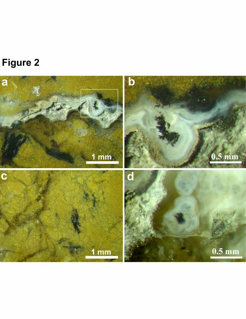
Geyserites are fine-grained siliceous (opal-like) rocks. Their porous structure is 198
complicated by spherulitic, globular, microlayer, fibrous, fluidic, looped, and breccia textures 199
(Fig. 2). The carbon content in graphite-containing geyserites varies from 0.01 wt.% to 1 wt.%. 200
Graphite is a prevailing native carbon phase of geyserites. Geyserites contain inclusions of 201
tremolite and fuchsite in equilibrium with the chalcedony matrix (Sklyarov et al. 2014), and are 202
enriched by iron oxides and hydroxides, such as magnetite, hematite, and goethite, as supported 203
by Raman spectroscopy (Figs. 2–4). 204
The graphite in geyserites appears as isolated flakes or is concentrated in aggregates that are 205
several millimeters in size. The carbonaceous matter in the rocks is unevenly distributed, 206
suggesting that the outer zones of globules, spherolites, and surface crusts are the most enriched 207
in particles and clusters of graphite, and spatial conjugation with chalcedonic sites is observed. 208
Graphite particles fill the leaching porous space between inclusions of chalcedony and radially 209
fibrous quartz. An unusual feature of geyserite graphite is the strong splitting at the terminal 210
edges of the plates in cross-section; such formations look like shiefs (Figs. 3–4). Lateral 211
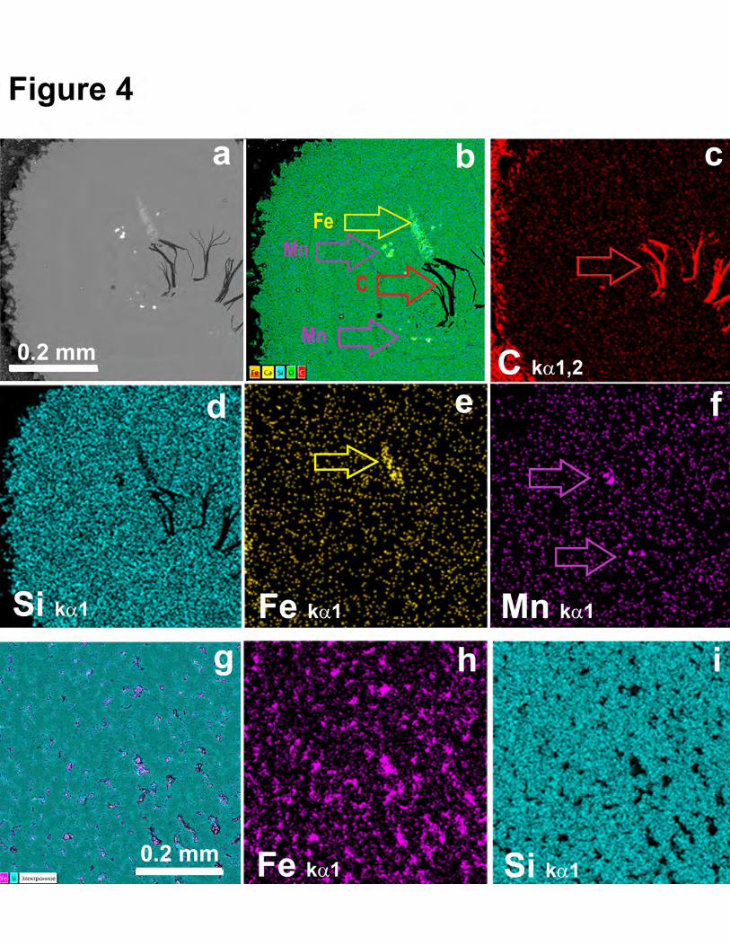
fragmentation has also been observed. Particles of Fe and Mn oxides occur within the nodules 212
This is the peer-reviewed, final accepted version for American Mineralogist, published by the Mineralogical Society of America. The published version is subject to change. Cite as Authors (Year) Title. American Mineralogist, in press.
DOI: https://doi.org/10.2138/am-2021-7711. http://www.minsocam.org/
Always consult and cite the final, published document. See http:/www.minsocam.org or GeoscienceWorld
11
(Fig. 4) where the oxides and graphite co-exist, which usually form at different fO2 conditions. 213
With reference to the elemental map (Fig. 4b), it is clear that the graphite and the metal oxides 214
belong to different nodule zones indicating different oxygen fugacity during hydrothermalite 215
formation, wherein the highly reductive conditions marked by graphite belong to the central 216
parts of the nodules. Simultaneously, it can be observed that the matrix of silica nodules is clear 217
of impurities in contrast to the general matrix of geyserite, which is strongly enriched in Fe (Figs. 218
4 g-i); this supports the optical observations shown in Fig. 2c. 219
Previously, we used an X-ray phase analysis to characterize the graphite of geyserites; it was 220
found to have a high degree of crystallinity (Danilova et al. 2016). The Raman data produced 221
completely analogous results to the spectral characteristics of the reference Ceylon graphite. 222
According to thermal analysis, the temperatures at the onset of the exothermic effect for 223
graphites from geyserites were within 570–710 °С. 224
Our study of the monomineral fractions of graphite showed that the graphite particles from 225
geyserite were characterized by a pinacoidal habit, which often occurs in rounded and hexagonal 226
forms, and have an irregular flattened shape with sizes from few tenths to a few millimeters. 227
On the surface of a number of flat-faced graphite particles (approximately 10–15% of the 228
particles studied), we observed very unusual submicrometer-sized crystallites with 229
hexagonal-pyramidal, hexagonal-prismatic habitus, as well as subtrigonal nanocrystallites via 230
SEM (Fig. 5). The size of the crystallites ranged from 50 nm to 1 μm. The layers of 231
submicrometer crystals were parallel or slightly disoriented as they were set close to dissolution 232
zones (Fig. 5c). 233
Using TEM to achieve a higher resolution, we found that the graphite from geyserites has 234
numerous nanoscale formations with a rounded shape (Fig. 6c), as well as previously described 235
pseudosuperstructures and nanofibers (Danilova et al., 2016). Most often, the graphite particles 236
This is the peer-reviewed, final accepted version for American Mineralogist, published by the Mineralogical Society of America. The published version is subject to change. Cite as Authors (Year) Title. American Mineralogist, in press.
DOI: https://doi.org/10.2138/am-2021-7711. http://www.minsocam.org/
Always consult and cite the final, published document. See http:/www.minsocam.org or GeoscienceWorld
12
in geyserites have rather large regions with a monocrystalline structure (Figs. 6 a–b). Note that 237
the graphite layers did not just shift relative to each other, but collapsed into folds resulting in 238
peculiar wrinkles as well as Moire patterns (Fig. 6 a). This is most likely caused by an 239
exceptionally weak bond between the graphite layers. 240
241
Graphite from travertine 242
Having studied graphite from the natural travertine outcrop at the northern tip of Ol`khon 243
Island, we further investigated the travertines in the outcrop, which exist as a subvertical vein 244
with a thickness of approximately 30 cm in Early Paleozoic gneisses. 245
According to the description by Sklyarov et al. (2007), travertines are characterized by three 246
consecutive formation phases. The first phase is represented by highly iron-rich 247
cryptocrystalline carbonates of brown color. They are confined to the boundary regions of the 248
vein and are observed as inclusions of irregular shape in the travertines of the second phase, 249
composed of light brownish-yellow fine-crystalline carbonates. The travertines of the first phase 250
contain fragments of silicate rocks, ranging in size from a few millimeters to a few centimeters. 251
They consist mainly of quartz and plagioclase with relatively minor quantities of pyroxene, 252
biotite, apatite, and titanite. The second phase also contains fragments of graphite-containing 253
gneisses and often very highly enriched in graphite with individual grains of quartz and 254
plagioclase; however, the grain sizes do not exceed 1 mm. The third phase, composed of 255
translucent calcite and white and light gray dolomite, is characterized by flow "agate" textures. 256
Herein, travertines contain xenoliths of enclosing silicate rocks with graphite, as well as 257
fragments of the travertines of the first and second phases. This phase fills the cracks in the rocks 258
crossing the travertines of the first and second phases. 259
This is the peer-reviewed, final accepted version for American Mineralogist, published by the Mineralogical Society of America. The published version is subject to change. Cite as Authors (Year) Title. American Mineralogist, in press.
DOI: https://doi.org/10.2138/am-2021-7711. http://www.minsocam.org/
Always consult and cite the final, published document. See http:/www.minsocam.org or GeoscienceWorld

13
Through optical microscopy (Fig. 7) and SEM, we determined that the Ol`khon travertines 260
had not three, but at least five generations (Fig. 8a, Table 1). However, the number of macro- and 261
micro-observations do not correspond well (Table 1). We think that the travertines observed by 262
Sklyarov et al. (2007) are attributed to the aforementioned phases; however, herein, a more 263
detailed portion of travertine formation was observed, allowing us to propose different 264
generations. In general, travertine’s variety demonstrates the complicated multiple generations 265
of travertines, attributed to the periodical tectonic activation of the Baikal geyserite paleovalley 266
during the Late Quaternary period. Therefore, it is possible to propose that the earliest travertines 267
generations are iron-free rocks with preferably cryptocrystalline structures without silicate rock 268
fragments and graphite (similar to those in this work). This is followed by the multiple deposited 269
middle stage of iron-rich cryptocrystalline or fine-crystalline travertine abundant in silicate rock 270
fragments and minerals, including graphite, in which multiple fragments of different travertines 271
phases occur (Skyarov et al., 2007 and this work). The latest carbonate generation is represented 272
by Fe-free calcite with 2 wt.% of MgO (this work), which is characterized by a sinter texture. In 273
this study, we did not recognize dolomite as the last portion of carbonate as was reported by 274
Skyarov et al. (2007). 275
The elemental carbon content in the travertines was approximately 0.1 wt.%. The carbon 276
phases found in these rocks were mainly represented by graphite and, to a lesser extent, by finely 277
dispersed amorphous carbon matter (Danilova et al., 2016). The graphite in travertines is mainly 278
represented by flattened particles of irregular and often elongated shape with sizes ranging from 279
a few micrometers to a few millimeters (Fig. 8 b). These relatively large flattened particles can 280
be bent across the surfaces and are characterized by a large number of overgrowths, which form 281
serrate surfaces in the cross-section, which is similar to the aforementioned geyserite graphite. 282
This is the peer-reviewed, final accepted version for American Mineralogist, published by the Mineralogical Society of America. The published version is subject to change. Cite as Authors (Year) Title. American Mineralogist, in press.
DOI: https://doi.org/10.2138/am-2021-7711. http://www.minsocam.org/
Always consult and cite the final, published document. See http:/www.minsocam.org or GeoscienceWorld

14
The graphite particles are distributed in the travertines in an extremely irregular fashion 283
(Figs. 7, 8). Graphite is observed as separate, rather large (> 1 mm), individual particles, as well 284
as in the form of fine-crystalline aggregates filling voids in the travertines of the first and second 285
phases (Fig. 7). The individual graphite particles are confined to xenoliths of enclosing silicate 286
rocks (Fig. 7d) wherein, the travertine matrix contains many graphite particles, most of which 287
are moderately or strongly fragmented. 288
The smallest isolates of amorphous carbon matter are confined to the travertine of the last 289
stage formation, and are represented by flowing zonal crusts, which are shapeless particles that 290
correspond to the spread of the crystallization front of calcite. The abrupt change of carbon-free 291
zones with periodic occurrences of areas substantially enriched in carbon indicates a rapid 292
change in the composition of feeding solutions, which in some cases is manifested by alternating 293
calcite-dolomite zones. 294
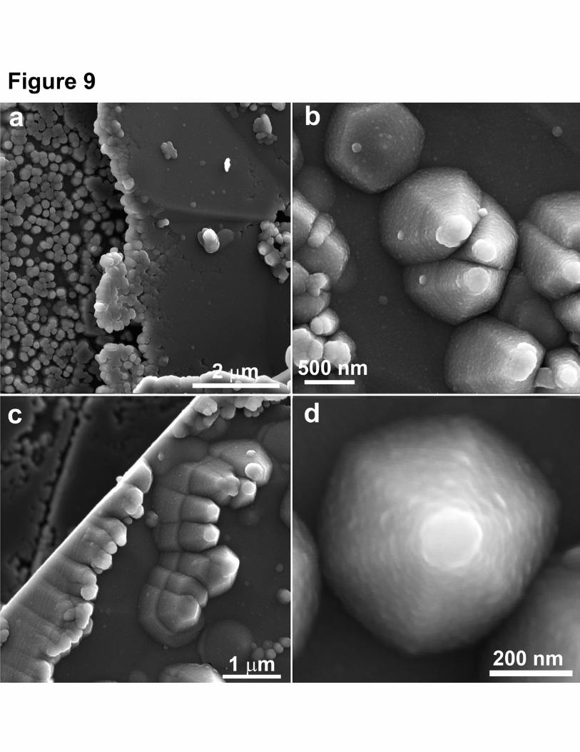
In some scenarios, submicrometer crystals are observed on the surface of the isolated 295
graphite particles and have a flat-faced or curved-faced hexagonal-pyramidal and 296
hexagonal-pyramidal-pinacoidal habit (Fig. 9). The size of the crystallites can vary from 50 nm 297
to 1 μm. However, crystallites are more common with sizes between 200 nm and 500 nm along 298
the {002} plane. Upon closer examination, the identified submicrometer crystals, as well as the 299
surface of the substrate on which they are located, exhibit a nanoglobular/blocky character, 300
which can be clearly seen in Figs. 9 b and d where the size scale is approximately 20–30 nm. In 301
addition to the visible hexagonal crystallites, neo formations with non-crystalline forms, which 302
are formed by the fusion of globular-like particles, are observed on the surface of the substrate 303
graphite (Fig. 9a). 304
Although graphite submicrometer crystals have a preferential orientation, some 305
misorientation with respect to each other is still noticeable, especially in the upper parts of the 306
This is the peer-reviewed, final accepted version for American Mineralogist, published by the Mineralogical Society of America. The published version is subject to change. Cite as Authors (Year) Title. American Mineralogist, in press.
DOI: https://doi.org/10.2138/am-2021-7711. http://www.minsocam.org/
Always consult and cite the final, published document. See http:/www.minsocam.org or GeoscienceWorld
15
crystallites. Further, many hexagonal subpyramidal submicrometer crystals have peculiar “caps” 307
that also consist of carbon nanoblocks. 308
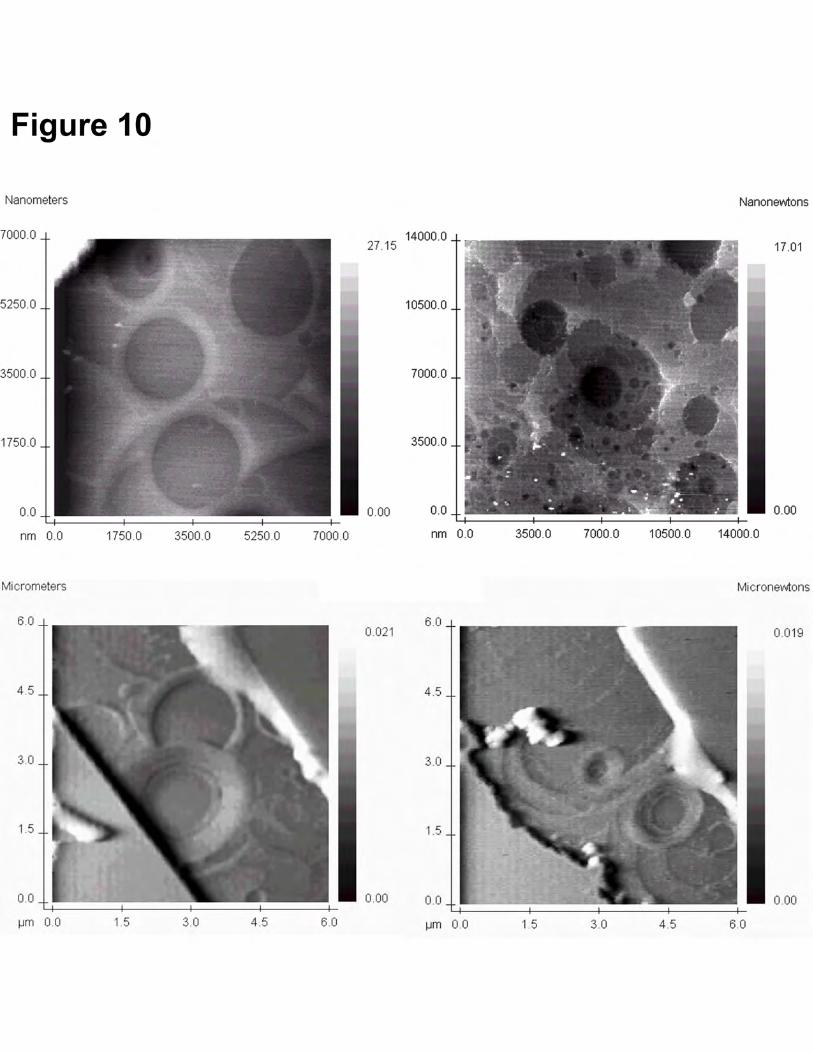
In addition to the morphology studies from the surface, we also needed to understand the 309
internal structure of the graphite particles. In this regard we have studied the fresh chips of 310
graphite along (002) plane, on which peculiar microscale rounded morphostructures were found 311
(Fig. 10). On the chips of graphite from the geyserites and travertines, we observed a 312
combination of almost perfectly round negative relief forms, with the diameter of the cavities 313
ranging from 100 nm to a few micrometers. Note that sometimes they were co-located so 314
frequently that they formed a peculiar spongy morphostructure (Fig. 10 b) In such areas, the 315
edges of the cavities had irregular outlines. Moreover, the diameters of these relief forms were 316
proportional to the bases of the submicrometer crystals on the surface of the graphite particles of 317
geyserites and travertines. 318
We have never before observed such microstructures in any other type of graphite, including 319
metamorphic, metasomatic, pneumatolytic, and magmatic (Shumilova 2003). These 320
morphostructures occur alongside ideally smooth chips and determine the features of the internal 321
structure of graphite under redeposition conditions during the formation of travertine. 322
ISOTOPIC STUDIES 323
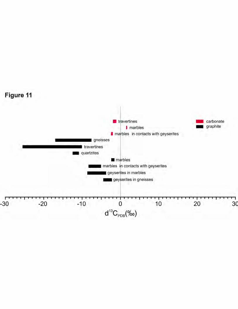
Analyses of stable carbon and oxygen isotopes were conducted in samples of 324
graphite-containing geyserites, travertines, and the hydrothermalite-enclosing rocks of the 325
Ol`khon area and the northern edge of Ol`khon Island (Figs. 11, 12, and Table 2). Herein, we 326
evaluated both the gross composition of graphite via chemically isolated concentrates, and the 327
individual particles of graphite extracted directly from some areas of hydrothermalites. 328
This is the peer-reviewed, final accepted version for American Mineralogist, published by the Mineralogical Society of America. The published version is subject to change. Cite as Authors (Year) Title. American Mineralogist, in press.
DOI: https://doi.org/10.2138/am-2021-7711. http://www.minsocam.org/
Always consult and cite the final, published document. See http:/www.minsocam.org or GeoscienceWorld

16
Based on our studies on the concentrate, in general, the geyserite graphite had heavy isotope 329
values δ13Cgraph. The graphite from the remnant of geyserites in gneisses was isotopically the 330
heaviest of all the analyzed samples δ13Cgraph (-2.1 to -2.5‰) (Fig. 11, Table 2). Further, the 331
graphite from the geyserite body in marbles was slightly less enriched in heavy isotope δ13Cgraph 332
(-3.8 to -8.7‰). The values of δ13Cgraph in graphite marbles in contact with geyserites were 333
similar (-5.1 to -5.7‰). The measured isotope composition of δ13Ccarb carbonate carbon and 334
δ18Ocarb oxygen was -2.1 to -2.2‰ and -11.0 to -11.6‰, respectively. Meanwhile, 335
graphite-containing marbles, exposed at some distance from the geyserite outcrop (5 km), were 336
characterized by typical values of the Ol`khon region rocks for δ13Cgraph (-1.6 to -2.3‰), 337
carbonate carbon composition - δ13Ccarb (1.5–1.6‰), and carbonate oxygen composition - 338
δ18Ocarb (-7.1 to -8.8‰). For graphite gneisses and quartzite graphite, the values of δ13Cgraph 339
ranged from -14.9 to -18.9 ‰ and -10.9 to -12.6 ‰, respectively. The isotopic composition of 340
the individual graphite particles from geyserite lies within the range of -3.8–8.7‰ (Table 2); 341
thus, it can be inferred that the graphite isotopy in geyserite is determined by locally differing 342
values. 343
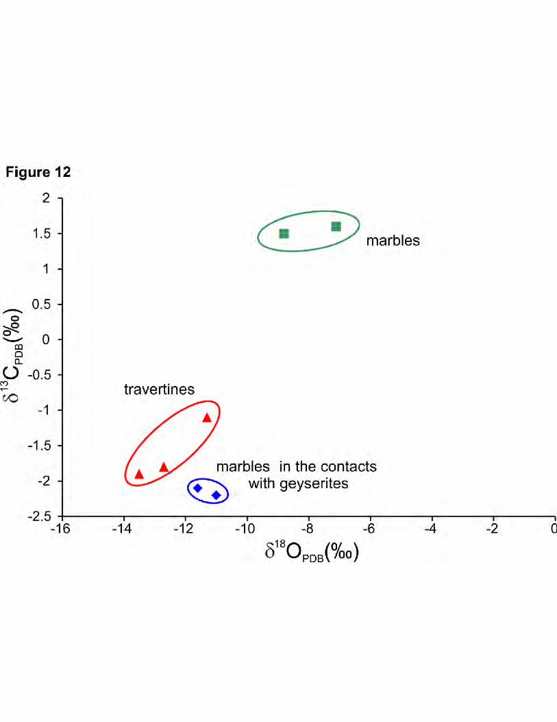
The study of carbon isotopic composition in the graphite of carbon travertine and calcite 344
oxygen showed that the graphite from travertine had a gross isotope-light composition δ13Cgraph 345
(-24.3 to -25.7‰), while the carbonate component of the travertines was characterized by a 346
carbon isotopic composition δ13Ccarb within -1.1 to -1.9‰ (Fig. 12, Table 2). The δ18Ocarb values 347
fall between -11.3 to -13.5‰. Meanwhile, the grain analysis showed that graphite from 348
travertines was very similar to the graphite from gneisses, with a definite tendency toward 349
isotopic composition, which could be associated with the participation of isotopically light 350
hydrocarbons in the process of graphite formation. 351
This is the peer-reviewed, final accepted version for American Mineralogist, published by the Mineralogical Society of America. The published version is subject to change. Cite as Authors (Year) Title. American Mineralogist, in press.
DOI: https://doi.org/10.2138/am-2021-7711. http://www.minsocam.org/
Always consult and cite the final, published document. See http:/www.minsocam.org or GeoscienceWorld
17
RAMAN SPECTROSCOPY 352
Raman spectroscopy was conducted on the extracted particles of graphite as well as 353
directly “in situ” on the hydrothermalites. The latter turned out to be more informative and 354
allowed us to exclude the possibility of distortion of the structural state of carbonaceous matter 355
that could occur during the chemical extraction of graphite concentrates. 356
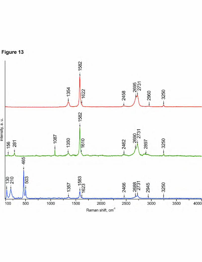
Based on the spectroscopic observations of graphite cross-sections, we revealed not only 357
the characteristics of graphite but also its important paragenetic relationship with the mineral 358
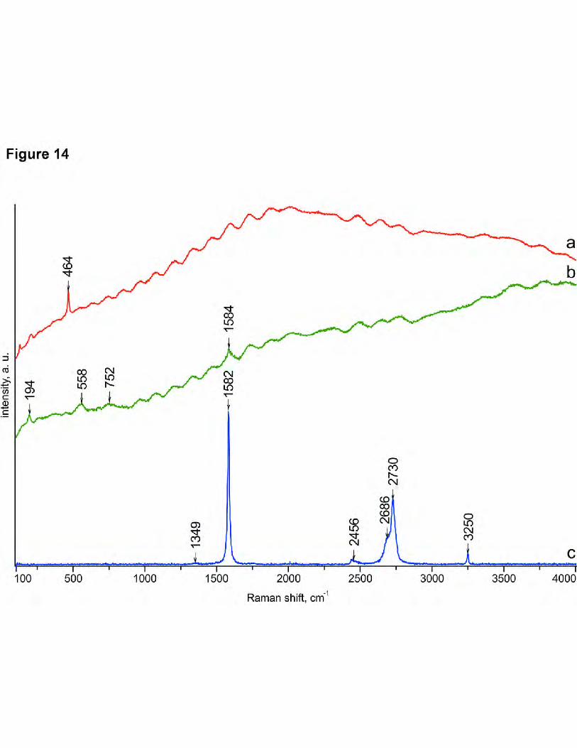
phases. We found micro-inclusions of calcite and chalcedony in geyserite graphite (Fig. 13). 359
Micro-inclusions of quartz and biotite were detected in the travertine graphite, and the presence 360
of hydrocarbons can be assumed owing to a specifically high level of luminescence observed 361
(Fig. 14). The presence of bitumen was confirmed through chemical reactions conducted by 362
chloroform extraction techniques and UV luminescence as described in Danilova et al. (2016). 363
The Raman spectra indicate that the graphite in both geyserites and travertines is 364
characterized rather by a high degree of crystallinity (French 1964; Tuinstra and Koenig 1970; 365
Wopenka and Pasteris 1993; Tan et al. 2004; Ferrari 2007). In this study, the Raman active 366
2E2g graphite mode, also called the G band, has a position at 1582 cm-1, which corresponds to 367
the standard position of well-ordered graphite. Note that the full-width half-maximum of the 368
G-band has a value of 16–19 cm-1. We did not analyze the characteristics of the D band; this 369
analysis was not useful here because of the significant dependence of a Raman spectrum 370
relative to the crystal orientation in highly ordered graphites. Although, at a qualitative level, 371
note, that while our analyzes have been conducted on the cross sections of graphite plates, i.e., 372
perpendicular to the (002) plane of graphite, the relative intensity of the D band is very 373
insignificant, indirectly indicating a high degree of graphite order in geyserites and travertines. 374
This is the peer-reviewed, final accepted version for American Mineralogist, published by the Mineralogical Society of America. The published version is subject to change. Cite as Authors (Year) Title. American Mineralogist, in press.
DOI: https://doi.org/10.2138/am-2021-7711. http://www.minsocam.org/
Always consult and cite the final, published document. See http:/www.minsocam.org or GeoscienceWorld

18
However, in addition to the highly ordered graphite variety in travertines, amorphous 375
carbonaceous matter as fine inclusions was also observed in the travertine of the last generation 376
(Figs. 7 e, f; 14 b). 377
378
DISCUSSION 379
Our studies of the graphite in geyserites and travertines of the Ol`khon Paleovalley and 380
Ol`khon Island on Lake Baikal (Eastern Siberia) have provided new mineralogical information, 381
increasing our understanding of the genetic thermal paleoreconstruction of hydrothermalites in 382
the Baikal rift zone as well as other hydrothermal objects through graphite mineralization. 383
Herein, the comparison of hydrothermalite graphite to the carbon mineralization of host 384
rocks was specifically considered; the rocks included gneisses, quartzites, and marbles, that 385
showed graphite mineralization in various degrees. The most useful information was obtained 386
from the isotopic analyses, which showed a correlation between the isotopic composition of 387
geyserite graphite and the graphite of the enclosing marbles, as well as the relationship between 388
travertine graphite and the graphite of the enclosing gneisses. Grain-by-grain analysis revealed a 389
local differentiation of the carbon isotopic composition in graphite that resulted from its initial 390
metamorphic origin, dissolution, and redeposition, with possible isotopic influence from the host 391
carbonates (which displayed different composition in individual particles). 392
Further, we found substantial fragmentation and alteration of the graphite in both geyserites 393
and travertines, which was accompanied by splitting, dissolution, and the appearance of growths 394
on the basal planes and plate edges. Zones of dissolution and growth were clearly observed in 395
optical microscopy observations of thin sections using electron microscopy (Figs. 5, 8, 9). To a 396
certain extent, similar microstructures have been described in Glad et al. (2014), wherein the 397
microstructures were observed on the surface of synthesized graphite produced from gas phase 398
This is the peer-reviewed, final accepted version for American Mineralogist, published by the Mineralogical Society of America. The published version is subject to change. Cite as Authors (Year) Title. American Mineralogist, in press.
DOI: https://doi.org/10.2138/am-2021-7711. http://www.minsocam.org/
Always consult and cite the final, published document. See http:/www.minsocam.org or GeoscienceWorld
19
by plasma at low pressure and at the temperature approximately 650 °C, and were observed in 399
natural graphite from Tanzania with crystallite sizes from 50 nm to 800 nm across and up to 1 400
µm along the c-axis. However, the submicrometer crystals found in our study consisted of 401
smaller blocks, approximately 20–30 nm in size, which have never been described before. 402
Although the bulk of graphite from the travertines and geyserites is characterized by a high 403
degree of ordering and belongs to the full-crystal variety after French classification (French 404
1964), we also found amorphous carbon and bitumen among the microscopic carboniferous 405
particles, indicating the heterogeneity of formation conditions of the carbonaceous matter. This 406
could be due to the non-equilibrium conditions of carbon condensation and/or the imposition of 407
carbonaceous mineralization of different origins in hydrothermalites. 408
In addition, bitumen (a hydrocarbon) was detected using Raman spectroscopy, and was 409
found to exist directly in aggregate with graphite (Fig. 14 a), indicating the possible participation 410
of hydrocarbons in hydrothermalite formation. The presence of hydrocarbons could also be the 411
cause of a sharp change in the redox situation, which was observed in the periodical formation of 412
carbon mineralization in the last stage travertines (Figs. 7e, f). This mineralization could be a 413
result of changes in fluid temperature and the corresponding chemical activity in the host 414
metamorphic and sedimentary rocks. Further, possible methane emissions that were described 415
for the different stages of the travertine formation (Lein et al. 2006; Luque et al. 2012) cannot be 416
excluded. 417
In previous works, we have described the presence of single crystalline α-carbyne among 418
native carbons within geyserites (Shumilova et al. 2011; Danilova et al. 2016). This finding was 419
used by Sklyarov et al. for paleothermal reconstruction of the Baikal hydrothermalites (Sklyarov 420
et al. 2014). Following the previously published carbyne data, we suppose that this phase cannot 421
This is the peer-reviewed, final accepted version for American Mineralogist, published by the Mineralogical Society of America. The published version is subject to change. Cite as Authors (Year) Title. American Mineralogist, in press.
DOI: https://doi.org/10.2138/am-2021-7711. http://www.minsocam.org/
Always consult and cite the final, published document. See http:/www.minsocam.org or GeoscienceWorld

20
be used as a proven indicator for high temperatures as phases have not been studied in detail and 422
the carbyne position on carbon diagrams (Bundy et al. 1996) is not entirely clear. 423
The graphite in geyserites and travertines is comparable in its degree of structural ordering 424
to graphite from the rocks of the amphibolite facies of metamorphism. The onset temperatures of 425
the exothermic effect for the graphites from geyserites and travertines range between 570–710 426
°C (Danilova et al. 2016), which is a characteristic of graphites from the green shale and 427
epidote-amphibolite facies of regional metamorphism (Ivanova et al. 1974). According to 428
Yardley (1991), this upper limit temperature corresponds to graphite from the amphibolite 429
facies. Such temperature conditions are especially unusual for travertines, for which the 430
temperature regime of formation, as a rule, lies within the range of 20–70 °C (Bargar 1978; 431
Pisarsky 1987; Fouke et al. 2000; Pentecost et al., 2011; Campbell et al., 2015 and others). 432
Sklyarov et al. (2014) used the presence of graphite and carbyne as a justification for the 433
high-temperate nature of the Ol`khon hydrothermalites, estimating a temperature of at least 400 434
C. However, the features of the studied graphite from travertine and geyserites herein clearly 435
indicate graphite’s original genetic relationship with the host rocks, e.g., graphite-containing 436
gneisses and marbles. Therefore, the graphite cannot be used as an unambiguous 437
geothermometer to determine the specifics of the hydrothermalites formation. 438
The mineralogical characteristics of graphite indicate that the travertines and geyserites 439
from the Ol`khon area and Ol`khon Island contain carbonaceous minerals of different origins. 440
The primary graphite in the host rocks during the chemically aggressive hydrothermal 441
processing of gneisses and marbles underwent significant changes associated with 442
defragmentation, partial dissolution, and in situ redeposition that occurred directly on the relict 443
substrates of the primary metamorphic graphite. At the same time, the graphite from travertines 444
and geyserites has very specific characteristics as observed via optical microscopy (Fig. 7b), 445
This is the peer-reviewed, final accepted version for American Mineralogist, published by the Mineralogical Society of America. The published version is subject to change. Cite as Authors (Year) Title. American Mineralogist, in press.
DOI: https://doi.org/10.2138/am-2021-7711. http://www.minsocam.org/
Always consult and cite the final, published document. See http:/www.minsocam.org or GeoscienceWorld

21
SEM, TEM, and AFM images (Figs. 5, 6, 8-10); these observations support the presence of 446
newly formed graphite by micro- and submicrometer crystals and unusual micro- and 447
nanostructural features on the surface (Figs. 5, 8, 9), and within the host metamorphic graphite 448
(Figs. 6c, 10). 449
The above presented data explain well the difference between the predominant isotopic 450
composition of graphitic carbon in geyserites and travertines evidenced by the enrichment of a 451
heavy carbon isotope in the geyserites graphite due to the presence of isotopically heavy graphite 452
of marbles and lighter carbon from gneisses associated with travertines (Table 2, Fig. 12). Thus, 453
the travertines and geyserites inherit not only the geochemical features of their host rocks, as 454
indicated in Sklyarov et al. (2007), but also the relict graphite, which generally preserves its 455
initial isotopic composition. 456
In geyserites, a stable silicate framework enables a relatively good preservation of isotopic 457
ratios of graphite (Wada and Suzuki 1983; Dunn and Valley 1992). During the formation of the 458
travertine, isotopic exchange occurred between the graphite of the clastic material in the host 459
rocks and carbon dioxide. Most likely, hydrothermalite graphite has been repeatedly subjected to 460
intense chemical treatment by aggressive hot hydrotherms, possibly with some participation of 461
hydrocarbons, which cause a drastic change in the oxidation-reduction conditions. For example, 462
in the Angel terrace hot springs, Fouke et al (2000) identified five travertine deposition facies 463
that varied in deposition temperature, and δ13C and δ18O isotope compositions (Mammoth Hot 464
Springs, Yellowstone National Park, USA). Further, methane emissions could contribute to the 465
prominent depletion of 13C isotope at different stages of travertine formation (Lein et al. 2006; 466
Luque et al. 2012). 467
Moreover, the redox conditions of graphite alteration in the geyserites and travertines could 468
be affected by seasonal temperature fluctuations and spring melt waters, which have a significant 469
This is the peer-reviewed, final accepted version for American Mineralogist, published by the Mineralogical Society of America. The published version is subject to change. Cite as Authors (Year) Title. American Mineralogist, in press.
DOI: https://doi.org/10.2138/am-2021-7711. http://www.minsocam.org/
Always consult and cite the final, published document. See http:/www.minsocam.org or GeoscienceWorld
22
influence on the growth and dissolution of calcite during the formation of travertines (Turi 1986; 470
Naboko et al. 1999; Dilsiz et al. 2004). 471
The established features of the nanoblock nature of new formations and the fusion of 472
graphite microcrystallites on the surface of graphite particles in the geyserites and travertines 473
indicate a probable mechanism for the coalescence of islands according to Kukushkin and 474
Osipov (1998) under the conditions of free growth from supersaturated solutions and/or from the 475
gas phase. However, the discussion regarding the high-temperature formation of travertines 476
entertained in literature (Sklyarov et al. 2014) cannot be proven with the presence of high 477
crystalline graphite in the samples herein, as they appear, generally, as relict fragments from the 478
host graphite-bearing rocks. Additionally, we must consider the wide range of graphite 479
crystallization temperatures, which reach as low as 98 C (Dunin-Borkovsky et al. 2000). Thus, 480
these data allow us to conclude that the newly formed redeposited graphite cannot be used as an 481
indicator of high temperature. 482
Therefore, according to the detailed research on the graphite from the Baikal 483
hydrothermalites paleovalley, we observe a paradox in the graphite nature. After intensive 484
hydrothermal changes of the host metamorphic rocks, only graphite is preserved, thus, the 485
graphite looks to have a hydrothermal nature. Only the provided detailed graphite studies 486
recognize this graphite and understand its deceptive nature. At the same time the real 487
fluid-deposited graphite is represented just by redeposited in situ submicrometer crystallites that 488
overgrow on the surface of the relict metamorphic graphite and between the calcite zones of the 489
last travertine generation. Currently, the fluid deposition of graphite in C–O–H systems occurs at 490
a known high temperature, which is higher than 400 C with a pressure 2 kbar or greater (Luque 491
et al., 1998; 2009; 2012; Luque and Rodas, 1999). The process of the graphite deposition from 492
C–O–H fluids is completed by the chemical reactions from CO2-CH4-H2O mixtures, such as CO2 493
This is the peer-reviewed, final accepted version for American Mineralogist, published by the Mineralogical Society of America. The published version is subject to change. Cite as Authors (Year) Title. American Mineralogist, in press.
DOI: https://doi.org/10.2138/am-2021-7711. http://www.minsocam.org/
Always consult and cite the final, published document. See http:/www.minsocam.org or GeoscienceWorld
23
C + O2 and CH4 + O2 C + 2H2O (Luque et al., 2012; Ortega et al., 2010). Note that the data 494
complex on the Baikal fluid-deposited graphite formation look different. 495
Considering the surface and subsurface travertine and geyserite formation at atmospheric 496
pressure, it is hard to explain the reason for the thermal conditions for their hot springs being 497
higher than 100 C, which is the boiling point of water at the atmospheric pressure. Among hot 498
springs, warm (20–40 °C), mesothermal (40–75 °C), and hyperthermal (> 75 °C) varieties are 499
recognized (Pentecost et al., 2003; Renaut and Jone, 2000). Further, travertines are said to 500
deposit at lower temperature conditions, generally below 75 °C, while geyserites more 501
commonly occur at higher temperature conditions (Sklyarov et al, 2014). Thus, we can 502
approximate that the Baikal hydrothermalites (as well as the corresponding graphite 503
redeposition) were formed at atmospheric pressure with a thermal range below 100 C. For such 504
LPLT-conditions with a large variation from typical C–O–H graphite deposition, the evidence of 505
dissolution and redeposition of the free carbon (relict metamorphic graphite/redeposited 506
graphite) within the C–O–H system indicate that there was a different mechanism involved in the 507
Baikal fluid-deposited graphite formation that did not involve the classic crystallization from a 508
carbon water solution as it has been previously proposed (Dunin-Borkovsky et al. 2000). This 509
novel mechanism put forth herein should be analyzed in detail in a future work. By this work we 510
demonstrate that the PT-conditions for graphite formation within the C–O–H system continue to 511
expand and reveal new information. 512
513
IMPLICATIONS 514
In this study, we evaluated the paradox graphite mineralization from the Late Quaternary 515
geyserites and travertines of the Ol`khon area and Ol`khon Island to clarify the genetic value of 516
graphite for hydrothermalite paleoreconstruction. Herein, we demonstrated that the primary 517
This is the peer-reviewed, final accepted version for American Mineralogist, published by the Mineralogical Society of America. The published version is subject to change. Cite as Authors (Year) Title. American Mineralogist, in press.
DOI: https://doi.org/10.2138/am-2021-7711. http://www.minsocam.org/
Always consult and cite the final, published document. See http:/www.minsocam.org or GeoscienceWorld

24
source of graphite in geyserites was enclosing marble, whereas for travertines, it was enclosing 518
gneiss. Metamorphic graphite under the influence of aggressive hydrotherms was forced to 519
partially dissolve and in situ redeposition occurred on the surface of relict graphite particles. The 520
obtained isotope-characteristics and presence of bitumen in our samples indicate 521
non-equilibrium redox conditions of graphite alteration with possible influence from 522
temperature fluctuation and variation in the geochemical nature of the environment with the 523
participation of hydrocarbons. Several mineralogical characteristics, including the micro- and 524
nanostructured features of graphite, can be considered typomorphic for hydrothermalites. Hence, 525
the revealed genetic specifics of graphite crystallized within H2O-rich LPLT fluid have 526
broadened the established ideas (Luque et al. 1998, 2012; Jaszczak et al. 2007) on typomorphism 527
and diversity of carbon mineralization in connection with fluidogenic objects. The redeposition 528
of the graphite within the Baikal Quaternary geyserites and travertines supported by earlier 529
experimental LPLT synthesis in aqueous solutions allow for the consideration of a new natural 530
mechanism of LPLT graphite formation in a C–O–H system, that is, crystallization from carbon 531
water solution, which should be further detailed in future studies. 532
533
ACKNOWLEDGMENTS 534
The authors thank E.V. Sklyarov for providing the samples for research and for the scientific 535
advice. E.M. Tropnikov, I.V. Smoleva, A. Sologubenko, V.N. Filippov, V.A. Radaev, G.N. 536
Kablis, G.N. Modyanova, N.A. Priezzheva, V.A. Ponomarchuk, B.A. Makeev, and N.V. 537
Nartova are acknowledged for their assistance in the analyses. 538
FUNDING 539
The work was supported by the projects: NIR GR # AAAA-A17-117121270036-7; Basic 540
This is the peer-reviewed, final accepted version for American Mineralogist, published by the Mineralogical Society of America. The published version is subject to change. Cite as Authors (Year) Title. American Mineralogist, in press.
DOI: https://doi.org/10.2138/am-2021-7711. http://www.minsocam.org/
Always consult and cite the final, published document. See http:/www.minsocam.org or GeoscienceWorld
25
Research Program IX.130, project # 0346-2019-0003. 541
542
Author Contributions: Tatyana Shumilova provided the idea for the study, conducted all 543
mineralogical investigations, collected and interpreted the instrumental data, and wrote the 544
manuscript. Yulia Danilova conducted the petrographic and geological analyses, participated in 545
data interpretation, and wrote the manuscript. Joachim Mayer performed the TEM studies and 546
edited the manuscript. Sergey Isaenko measured the Raman spectra, and Boris Danilov 547
performed the geological analysis. Vasily Ulyashev assisted in the high resolution SEM studies. 548
Conflicts of interests: The authors declare no conflict of interest. 549
550
REFERENCES CITED 551
552
Altunel, E., and Hancock, P.L. (1993) Morphology and structural setting of Quaternary 553
travertines at Pamukkale, Turkey. Geological Journal, 28, ¾, 335–346. 554
Bargar, K. (1978) Geology and thermal history of Mammoth Hot Springs, Yellowstone National 555
Park, Wyoming: U.S. Geological Survey Bulletin, 1444, 1–54. 556
Bibikova, E.V., Karpenko, S.F., Sumin, and L.M. (1990) U-Rb, Sm–Nd, and K–Ar Age of 557
Metamorphic and Igneous Rocks of the Ol’khon Region (Western Baikal Region), In 558
Precambrian Geology and Geochronology of the Siberian Craton and Its Fold Framework, 559
p. 170–183. Nauka, Leningrad (in Russian). 560
Bundy, F.P., Basset, W.A., Weathers, M.S., Hemley, R.J., Mao, H.K., and Goncharov, A.F. 561
(1996) The pressure-temperature phase and transformation diagram for carbon: updated 562
This is the peer-reviewed, final accepted version for American Mineralogist, published by the Mineralogical Society of America. The published version is subject to change. Cite as Authors (Year) Title. American Mineralogist, in press.
DOI: https://doi.org/10.2138/am-2021-7711. http://www.minsocam.org/
Always consult and cite the final, published document. See http:/www.minsocam.org or GeoscienceWorld
26
through 1994. Carbon, 34, 2, 141–153. 563
Campbell, K.A., Guido, D., Gautret, P., Foucher, F., Ramboz, C. et. al. (2015) Geyserite in 564
Hot-Spring Siliceous Sinter: Window on Earth’s Hottest Terrestrial (Paleo) environment 565
and its Extreme Life. Earth-Science Reviews, Elsevier, 148, 44–64. 566
Crossey, L.J., Karlstrom, K.E., Springer, A.E., Newell, D., Hilton, D.R., and Fischer, T. (2009) 567
Degassing of mantle-derived CO2 and He from springs in the southern Colorado Plateau 568
region — Neotectonic connections and implications for groundwater systems. GSA 569
Bulletin, 121, 7/8, 1034–1053. 570
Danilova,, Yu.V, Shumilova, T.G., Mayer, J., and Danilov, B.S. (2016) Conditions and 571
Formation Mechanism of Carbon Phases in Late Quarternary Geyzerites and Travertines 572
of Ol’khon Area and Ol’khon Island (Baikal Rift Zone). Petrology, 24, 1, 35–48. 573
Dilsiz, C., Marques, J.M.M., and Carreira, P.M.M. (2004) The impact of hydrological changes 574
on travertine deposits related to thermal springs in the Pamukkale area (SW Turkey). 575
Environmental Geology, 45, 808–817 576
Djokic, T., Van, Kranendonk, M.J., Kathleen, A.C., Malcolm, R.W., and Colin, R.W. (2017) 577
Earliest signs of life on land preserved in ca. 3.5 Ga hot spring deposits. Nature 578
Communications, 8, 15263. https://doi.org/10.1038/ncomms15263. 579
Dobrzhinetskaya, L.F., Molchanova, T.V., Sonyushkin, V.E., Likhachev, A.B., and 580
Fedorovskii, V.S. (1992) Napped and strikeslip ductile deformations of the Ol’khon 581
Region metamorphic complex (western Baikal region). Geotektonika, 2, 58–71. 582
Duke, E. F. and Rumble, D. (1986). Textural and isotopic variations in graphite from plutonic 583
rocks, South-Central New Hampshire. Contributions to Mineralogy and Petrology, 93(4), 584
409–419.doi:10.1007/bf00371711 585
This is the peer-reviewed, final accepted version for American Mineralogist, published by the Mineralogical Society of America. The published version is subject to change. Cite as Authors (Year) Title. American Mineralogist, in press.
DOI: https://doi.org/10.2138/am-2021-7711. http://www.minsocam.org/
Always consult and cite the final, published document. See http:/www.minsocam.org or GeoscienceWorld

27
Dunin-Barkovsky, R.L., Dunin-Barkovsky, A.R., Drozdova, O.V., Kidyarov, B.I., Kolyago, 586
S.S., Kozhbakhteyev, E.M., Lisitsina, E.E., Mar`in, A.A., and Slovtsov, I.B. (2000) 587
Low-Temperature Growth of Diamond Seedings in Acid Solutions. Chemistry for 588
Sustainable Development, 8, 147–153. 589
Dunn, S., and Valley, J. (1992) Calcite-graphite isotope thermometry: a test for 590
polymetamorphism in marble, Tudor gabbro aureole, Ontario, Canada. Journal of 591
Metamorphic Geology, 10, 4, 487–501. 592
Fedorovsky, V.S., Donskaya, T.V., Gladkochub, D.P., Khromykh, S.V., Mazukabzov, A.M., 593
Mekhonoshin, A.S., Sklyarov, E.V., Sukhorukov, V.P., Vladimirov, A.G., Volkova, N.I., 594
and Yudin, D.S. (2005) The Ol’khon collision system (Baikal region) in Structural and 595
tectonic correlation across the Central Asia orogenic collage: northeastern segment. In 596
Guidebook and Abstract Volume of the Siberian Workshop IGCP-480, Sklyarov EV, 597
Ed., Print. IEC SB RAS: Irkutsk. 598
Ferrari, A. (2007) Raman spectroscopy of graphene and graphite: Disorder, electron–phonon 599
coupling, doping and nonadiabatic effects. Solid State Communications, 143, 47–57. 600
Fouke, B., Farmer, J., Marais, D., Pratt, L., Sturchio, N., Burns, P., and Discipulo, M. (2000) 601
Depositional facies and aqueous-solid geochemistry of travertine-depositing hot springs 602
(Angel Terrace, Mammoth Hot Springs, Yellowstone National Park, U.S.A.). Journal of 603
Sedimentary Research, 70, 3, 565–585. 604
Fraedrich W., Heidari N. (2019) Iceland from the West to the South. GeoGuide. Springer, 605
Nature, Switzerland AG. 606
French, B.M. (1964) Graphitization of organic material in a progressively metamorphosed 607
Precambrian iron formation. Science, 146, 3646, 147–153. 608
Gibert, R.O., Taberner, C., Sáez, A., Giralt, S., Alonso, R.N., Edwards, R.L., and Pueyo, J.J. 609
This is the peer-reviewed, final accepted version for American Mineralogist, published by the Mineralogical Society of America. The published version is subject to change. Cite as Authors (Year) Title. American Mineralogist, in press.
DOI: https://doi.org/10.2138/am-2021-7711. http://www.minsocam.org/
Always consult and cite the final, published document. See http:/www.minsocam.org or GeoscienceWorld

28
(2009) Igneous Origin of CO2 in Ancient and Recent Hot-Spring Waters and Travertines 610
from the Northern Argentinean Andes. Journal of Sedimentary Research, 79, 8, 554–567. 611
Glad, X., Poucques, L., Jaszczak, J.A., Belmahi, M., Ghanbaja, J., and Bougdira, J. (2014) 612
Plasma synthesis of hexagonal-pyramidal graphite hillocks. Carbon, 76, 330–340. 613
Gladkochub, D.P., Donskaya, T.V., Fedorovsky, V.S., Mazukabzov, A.M., Larionov, A.N., and 614
Sergeev, S.A. (2010) The Olkhon metamorphic terrane in the Baikal region: An Early 615
Paleozoic collage of Neoproterozoic active margin fragments. Russian Geology and 616
Geophysics, 51, 5, 447–460. 617
Gladkochub, D.P., Donskaya, T.V., Wingate, M.T.D., Poller, U., Kröner, A., Fedorovsky, 618
V.S., Mazukabzov, A.M., Todt, W., and Pisarevsky, S.A. (2008) Petrology, 619
geochronology, and tectonic implications of c. 500 Ma metamorphic and igneous rocks 620
along the northern margin of the Central-Asian orogen (Olkhon Terrane, Lake Baikal, 621
Siberia) Journal of the Geological Society of London, 165, 1, 235–246. 622
Harris, A.C., White, N.C., McPhie, J., Bull, S.W., Line, M.A., Skrzeczynski, R., Mernagh, T.P., 623
and Tosdal, R.M. (2009) Early Archean hot springs above epithermal veins, North Pole, 624
Western Australia: new insights from fluid inclusion microanalysis. Economic Geology, 625
104, 6, 793-814. 626
Ivanova, V.P., Kasatov, B.K., Krasavina, and T.N. (1974) Thermal analysis of minerals and 627
rocks. Nedra, Leningrad (in Russian). 628
Jaszczak, J.A., Dimovski, S., Hackney, S.A., Robinson, G.W., Bosio, P., and Gogotsi, Yu. 629
(2007) Micro- and nanoscale graphite cones and tubes from Hackman Valley, Kola 630
Peninsula, Russia. The Canadian Mineralogist, 45, 2, 379–389. 631
This is the peer-reviewed, final accepted version for American Mineralogist, published by the Mineralogical Society of America. The published version is subject to change. Cite as Authors (Year) Title. American Mineralogist, in press.
DOI: https://doi.org/10.2138/am-2021-7711. http://www.minsocam.org/
Always consult and cite the final, published document. See http:/www.minsocam.org or GeoscienceWorld
29
Jones, B., and Renaut, R.W. (2003) Hot spring and geyser sinters: the integrated product of 632
precipitation, replacement, and deposition. Canadian Journal of Earth Sciences, 40, 11, 633
1549–1569. 634
Kukushkin, S.A., and Osipov, A.V. (1998) Condensation processes of thin films. Success of 635
physical sciences, 168, 10, 1083–1116 (in Russian), 636
Lein, A.Yu,, Bogdanov, Yu.A, Grichuk, D.V., Rusanov, I.I. and Sagalevich, A.M. (2006) 637
Geochemistry of hydrothermal solutions from 9°50′ N at the East Pacific Rise (EPR) 638
within twelve years after the eruption of a submarine volcano. Geochemistry International, 639
44, 7, 690–703. 640
Letnikov, F.A., Khalilov, V.A., Savel’eva, and V.B. (1995) Isotopic Dating of Endogenic 641
Processes in the Ol’khon Region. Doklady Earth Sciences, 344, 1, 96–100. 642
Luque, F.J., Crespo-Feo, E., Barrenechea, J.F., Ortega, L. (2012) Carbon isotopes of graphite: 643
Implications on fluid history. Geoscience Frontiers, 3, 2, 197–207. 644
Luque, F.J., Pasteris, J.D., Wopenka, B., Rodas, M., and Barrenechea, J.F. (1998) Natural 645
fluid-deposited graphite: mineralogical characteristics and mechanisms of formation. 646
American Journal of Science, 298, 471–498. 647
Lund, J., Freeston, D.H., and Boyd, T.L. (2005) World-wide direct uses of geothermal energy. 648
Proceedings of World Geothermal Congress, Antalya, Turkey. 649
http://citeseerx.ist.psu.edu/viewdoc/download?doi=10.1.1.536.1578&rep=rep1&type=pdf 650
Minissale, A,, Kerrick, D.M., Magro, G., Murrell, M.T., Paladini, M., Rihs, S., Sturchio, N.C., 651
Tassi, F., and Vaselli, O. (2002) Geochemistry of Quaternary travertines in the region 652
north of Rome (Italy): structural, hydrologic and paleoclimatic implications. Earth and 653
Plane Science Letters, 203, 2, 709–728. 654
Naboko, S.I., Lugovaya, I.P., and Zagnitko, V.N. (1999) The isotope composition of oxygen and 655
This is the peer-reviewed, final accepted version for American Mineralogist, published by the Mineralogical Society of America. The published version is subject to change. Cite as Authors (Year) Title. American Mineralogist, in press.
DOI: https://doi.org/10.2138/am-2021-7711. http://www.minsocam.org/
Always consult and cite the final, published document. See http:/www.minsocam.org or GeoscienceWorld
30
carbon in modern travertines and Kamchatka geyserites. Mineralogical journal, 21, 5/6, 656
33–39 (in Russian). 657
Omelon, C.R., Pollard, W.H., and Andersen, D.T. (2006) A geochemical evaluation of 658
perennial spring activity and associated mineral precipitates at Expedition Fjord, Axel 659
Heiberg Island, Canadian High Arctic. Applied Geochemistry, 21, 1–15. 660
Pisarsky, B.I. (1987) Patterns of formation of underground flow of Lake Baikal. Nauka, 661
Novosibirsk (in Russian) 662
Pentecost, A. (1995) Geochemistry of carbon dioxide in six travertine-depositing waters of 663
Italy. Journal of Hydrolody, 167, 263–278. 664
Pentecost, A. (2005) Travertine. Springer-Verlag, London. https://www.academia.edu/8180604 665
Pentecost, A., and Viles, H.A. (1994) Review and Reassessment of Travertine Classification. 666
Géographie physique et Quaternaire, 48, 3, 305–314. 667
Petrova, Z.I., and Levitsky, V.I. (1984) Petrology and Geochemistry of Granulite Complexes of 668
the Baikal Region. Nauka, Novosibirsk (in Russian). 669
Renaut, R.W., Owen, R.B., and Ego, J.K. (2008) Recent changes in geyser activity at Loburu, 670
Lake Bogoria, Kenya rift valley. GOSA Transact, 10, 4–14. 671
Renaut, R.W., Owen, R.B., Jones, B., Tiercelin, J-J., Ego, J.K., and Konhauser, K.O. (2013) 672
Impact of lake-level changes on the formation of thermogene travertine in continental 673
rifts: evidence from Lake Bogoria, Kenya rift valley. Sedimentology, 60, 428–468. 674
Rozen, O.M., and Fedorovsky, V.S. (2001) Collisional granitoids and stratification of the Earth's 675
crust (examples of Cenozoic, Paleozoic and Proterozoic collisional systems). In: 676
Proceedings GIN RAS. Mir, Moscow (in Russian). 677
This is the peer-reviewed, final accepted version for American Mineralogist, published by the Mineralogical Society of America. The published version is subject to change. Cite as Authors (Year) Title. American Mineralogist, in press.
DOI: https://doi.org/10.2138/am-2021-7711. http://www.minsocam.org/
Always consult and cite the final, published document. See http:/www.minsocam.org or GeoscienceWorld
31
Serebryansky, E.P., Kostitsyn, Yu..A., Fedorovsky, V.S., and Vladimirov, A.G. (1998) 678
Comparative isotopic studies of granites and metamorphic rocks of the Olkhon region. In: 679
XU Symposium on Geochemistry of Isotopes. GEOKHI RAS, Moscow. 680
Shumilova, T.G. (2003) Mineralogy of native carbon. Ural Branch of RAS press: Ekaterinburg, 681
Russia, 316 p. (in Russian). 682
Shumilova, T.G., and Danilova, Yu.V. (2009) New genetic types of graphite in connection 683
with travertines. Doklady Earth Sciences, 428, 2, 1171–1173. 684
Shumilova, T.G., Danilova, Yu.V., Gorbunov, M.V., and Isaenko, S.I. (2011) Natural 685
Monocrystalline α-Carbyne. Doklady Earth Sciences, 436, 1, 152–154. 686
Shumilova, T.G., Isaenko, S.I., and Yashin, N.V. (2018) Structure and mineral characteristics 687
of travertine object Vas`kin Klyuch (Sukhona River, Vologda District). Mineralogy, 4, 4, 688
119–129 (in Russian). 689
Sklyarov, E.V., Fedorovskii, V.S., Kulagina, N.V., Sklyarova, O.A., and Skovitina, T.M. 690
(2004) The Late Quaternary “Geyser Valley” in the western flank of the Baikal Rift 691
(Ol’khon Region). Doklady Earth Sciences, 395A, 3, 324–327. 692
Sklyarov, E.V., Fedorovskii, V.S., Sklyarova, O.A., Skovitina, T.M., Danilova, Yu.V., Orlova, 693
L.A., and Ukhova, N.N. (2007) Hydrothermal Activity in the Baikal Rift Zone: Recent Hot 694
Springs and Deposits of Paleothermal Waters. Doklady Earth Sciences, 412, 2, 101-105. 695
Sklyarov, E.V., Skovitina, T.M., Sklyarova, O.A., Kotov, A.B., Tolmacheva, E.V., and 696
Velikoslavinskii, S.D. (2014) Late Quaternary high-temperature geyserites in the Ol’khon 697
area, Baikal rift zone: Petrography, mineralogy, chemical composition, and genesis. 698
Petrology, 22, 6, 536-546. 699
This is the peer-reviewed, final accepted version for American Mineralogist, published by the Mineralogical Society of America. The published version is subject to change. Cite as Authors (Year) Title. American Mineralogist, in press.
DOI: https://doi.org/10.2138/am-2021-7711. http://www.minsocam.org/
Always consult and cite the final, published document. See http:/www.minsocam.org or GeoscienceWorld
32
Tan, P.H., Dimovski, S., and Gogotsi, Y. (2004) Raman scattering of non-planar graphite: arched 700
edges, polyhedral crystals, arched edges, polyhedral crystals. Phil. Trans. R Soc. Lond. A, 701
362, 2289–2310. 702
Tatarinov, A.V., Yalovik, L.I., Danilova, E.V., and Namsaraev, Z.B. (2006) Participation of 703
Microorganisms in the Formation of Travertines and Sapropelite Kerogen in Sediments 704
of Thermal Carbonic Waters in the Baikal Rift Zone. Doklady Earth Science,s 411A, 9, 705
1435–1438. 706
Tuinstra, F. and Koenig, J.L. (1970) Raman Spectrum of Graphite. Journal of Chemical Physics, 707
53, 1126–1130. 708
Turi, B. (1986) Stable isotope geochemistry of travertines. Handbook of environmental isotope 709
geochemistry, Elsevier: Amsterdam. 710
Van Kranendonk, M.J. (2006) Volcanic degassing, hydrothermal circulation and the flourishing 711
of early life on Earth: a review of the evidence from c. 3490-3240 Ma rocks of the Pilbara 712
Supergroup, Pilbara Craton, Western Australia. Earth Science Reviews, 74, 3-4, 197–240. 713
Wada, H., and Suzuki, K. (1983) Carbon isotopic thermometry calibrated by dolomite-calcite 714
solvus temperatures. Geochimica et Cosmochimica Acta, 47, 4, 697–706. 715
Wopenka, B., and Pasteris, J.D. (1993) Structural characterization of kerogens to granulite-facies 716
graphite: applicability of Raman microprobe spectroscopy. The American Mineralogist, 717
78, 5/6, 533–557. 718
Yardley, B.W.D. (1991) An introduction to metamorphic petrology. Longman Scientific and 719
Technical, New York. 720
721
722
This is the peer-reviewed, final accepted version for American Mineralogist, published by the Mineralogical Society of America. The published version is subject to change. Cite as Authors (Year) Title. American Mineralogist, in press.
DOI: https://doi.org/10.2138/am-2021-7711. http://www.minsocam.org/
Always consult and cite the final, published document. See http:/www.minsocam.org or GeoscienceWorld
33
FIGURE CAPTIONS 723 724
FIGURE 1. Geographic position of Lake Baikal and the Ol’khon Island (Russia) (b) on 725
the Google map (a); fragment of the Baikal rift zone on the geological scheme (c). Legend: 1 – 726
Siberian craton; 2 – the Ol’khon Area and the Island Ol’khon system of shear plates formed 727
igneous and metamorphic complexes; 3 – faults (3a) the boundary of the Siberian craton and the 728
Ol’khon Area, (3b) the main shift zone of the region; 4 – outcrops with (4a) geyserite and (4b) 729
travertine; 5 –corresponding geyserites (5a) and travertine (5b) objects with graphite (sampling 730
sites). 731
FIGURE 2. Graphite particles in different areas of geyserites of a polished section: (a, b) 732
Graphite aggregates in chalcedony nodules. (a) overview of a nodule with graphite inclusion. (b) 733
Magnified view of the section demarcated by the rectangle in (a). (c) Graphite in general 734
geyserite matrix. (d) A graphite aggregate inclusion inside a closed chalcedony nodule. 735
FIGURE 3. Morphological features of graphite aggregates and iron hydroxide particles in 736
geyserite. (a) Aggregate of graphite plates in cross-section (center), where hydroxides in the 737
main microglobular matrix of the geyserite fill the gaps between microglobules (brown). (b) 738
Particles of hematite (indicated by arrows) surrounded by a halo of iron hydroxides. (c, d) 739
Sheaf-like graphite particles. (e, f) fragmented graphite particles in geyserite. Note: (a – c, e) are 740
the optical images in transmitted light, with parallel polarizers and (d, f) are the optical images in 741
transmitted light, with crossed polarizers. 742
FIGURE 4. Electron microscopy images of graphite-containing geyserite. (a–f) 743
Chalcedony nodule with graphite-containing core. (g–i) general geyserite matrix. Note that (a) is 744
a BSE image; (b–i) are the elemental maps wherein (b, g) are the multi-element maps and (c–f, h, 745
i) are the individual element maps. 746
This is the peer-reviewed, final accepted version for American Mineralogist, published by the Mineralogical Society of America. The published version is subject to change. Cite as Authors (Year) Title. American Mineralogist, in press.
DOI: https://doi.org/10.2138/am-2021-7711. http://www.minsocam.org/
Always consult and cite the final, published document. See http:/www.minsocam.org or GeoscienceWorld
34
FIGURE 5. Scanning electron microscopy (SEM) images (in the secondary electron mode 747
(SE)) of graphite from geyserite showing dissolution and new-growth regions. (a) Graphite 748
particle overview; the area demarcated by the square is the region considered for detailed study 749
(b–d are the magnified images of the area demarcated in (a)). (b) The region with newly formed 750
hexagonal subprismatic and tiny pyramidal graphite crystallites. (c) The region with graphite 751
dissolution. (d) Subtrigonal and smooth pyramidal graphite crystals, micro- and submicrometer 752
in size. 753
FIGURE 6. Transmission electron microscopy (TEM) data of graphite from (a–b) 754
geyserite and (d–f) and travertine. (a) Bright field image (BF) of single crystalline graphite 755
particle with layer wrinkles (white arrows) and Moire fringes (black arrows). (b) Electron 756
diffraction pattern (ED) of the graphite in (a). (c) Rounded graphitic nanosized nuclear particles 757
of newly-forming graphite. (d) Typical shapes of crushed graphite particles with rounded 758
fragments in chips. (e) A fragment of oriented growth of graphite nanocrystals in (002) direction 759
and the corresponding ED pattern. (f) The BF images of (a, c, d, and e). 760
FIGURE 7. Optical images of graphite-containing travertine in (a, c) reflected (skew 761
angle) and (b, d–f) transparent light. (a) Travertine overview with different travertine 762
generations. (b) Cross-section of a graphite particle within travertine matrix (tooth-like surface, 763
with overgrowths). (c) Boundary between the latest Fe-free travertine generation and an earlier 764
travertine. (d) The fragment of graphite-rich gneiss within travertine. (e) The carbonate of the 765
last generation with microcrystalline graphite. (f) Magnified image of (e). 766
FIGURE 8. SEM images of travertine and graphite particles chemically extracted from 767
the host travertine. Travertine of several generations (I-V), graphite inclusions are indicated with 768
arrows (a). (a) BS mode and (b–g) secondary electron (SE) mode where (b) shows shape of 769
graphite particles, (c) shows an individual particle with the studied region demarcated with a 770
This is the peer-reviewed, final accepted version for American Mineralogist, published by the Mineralogical Society of America. The published version is subject to change. Cite as Authors (Year) Title. American Mineralogist, in press.
DOI: https://doi.org/10.2138/am-2021-7711. http://www.minsocam.org/
Always consult and cite the final, published document. See http:/www.minsocam.org or GeoscienceWorld
35
square, (d) magnified view of the region demarcated (c). (e) Regions with dissolution (indicated 771
with arrows) and newly formed graphite crystallites on the {002} planes. (f) Newly formed 772
graphite crystallites on the {002} plane edges and regions with dissolution (indicated with 773
arrows). (g) Newly formed graphite crystallites on the {002} plane, regions with dissolution, and 774
{002} plane relics (indicated with arrows). 775
FIGURE 9. SEM images in the SE mode. (a) Micro- and submicrometer features of newly 776
formed graphite from travertine at the edge of the seed graphite plate. (b) Growth of nanoblock 777
sub-hexagonal micro- and submicrometer crystals on the surface of the graphite seed plate. (c) 778
Tight growth of nanoblock micro- and submicrometer crystals along the seed graphite plate 779
edge. (d) Nanoblock sub-hexagonal microcrystallite. 780
FIGURE 10. Atomic force microscope (AFM) data of fresh crushed surfaces of graphite 781
from (a, b) geyserite and (c, d) travertine. (a, c, d) have rounded morphology while (b) has a 782
sponge-like morphology. 783
FIGURE 11. Isotope composition of graphite and carbonates in geyserites, travertines, 784
and their host rocks. 785
FIGURE 12. Carbon and oxygen isotopic composition of carbonates in hydrothermalites 786
and their host rocks. 787
FIGURE 13. Raman spectra of graphite “in situ” in geyserite in perpendicular direction to 788
{002} of (a) pure graphite, (b) graphite with calcite inclusion (156 cm-1, 281 cm-1, and 1087 789
cm-1), and (c) graphite with chalcedony inclusion (130 cm-1, 210 cm-1, 465 cm-1, and 503 cm-1). 790
FIGURE 14. Raman spectra of graphite “in situ” in travertine in crosscut direction to 791
{002} of the graphite particle where (a) is the spectrum from hydrocarbon (luminescence) and 792
quartz (464 cm-1) impurities within graphite, (b) is graphite with biotite inclusion (194, 558, 962 793
cm-1), probably with hydrocarbon (luminescence), and (c) is pure graphite. 794
This is the peer-reviewed, final accepted version for American Mineralogist, published by the Mineralogical Society of America. The published version is subject to change. Cite as Authors (Year) Title. American Mineralogist, in press.
DOI: https://doi.org/10.2138/am-2021-7711. http://www.minsocam.org/
Always consult and cite the final, published document. See http:/www.minsocam.org or GeoscienceWorld
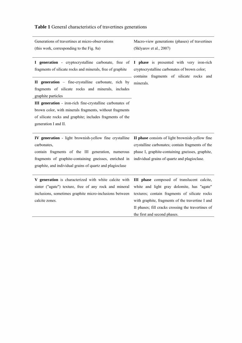
Table 1 General characteristics of travertines generations Generations of travertines at micro-observations
(this work, corresponding to the Fig. 8a)
Macro-view generations (phases) of travertines
(Sklyarov et al., 2007)
I generation - cryptocrystalline carbonate, free of
fragments of silicate rocks and minerals, free of graphite
I phase is presented with very iron-rich
cryptocrystalline carbonates of brown color;
contains fragments of silicate rocks and
minerals.
II generation – fine-crystalline carbonate, rich by
fragments of silicate rocks and minerals, includes
graphite particles
III generation - iron-rich fine-crystalline carbonates of
brown color, with minerals fragments, without fragments
of silicate rocks and graphite; includes fragments of the
generation I and II.
IV generation - light brownish-yellow fine crystalline
carbonates,
contain fragments of the III generation, numerous
fragments of graphite-containing gneisses, enriched in
graphite, and individual grains of quartz and plagioclase
II phase consists of light brownish-yellow fine
crystalline carbonates; contain fragments of the
phase I, graphite-containing gneisses, graphite,
individual grains of quartz and plagioclase.
V generation is characterized with white calcite with
sinter ("agate") texture, free of any rock and mineral
inclusions, sometimes graphite micro-inclusions between
calcite zones.
III phase composed of translucent calcite,
white and light gray dolomite, has "agate"
textures; contain fragments of silicate rocks
with graphite, fragments of the travertine I and
II phases; fill cracks crossing the travertines of
the first and second phases.

Table 2 Isotope composition of carbon and oxygen in host geyserites and travertines and graphite carbon
No Specimen Specimen type
Specimen characteristic
δ13Cgraph,‰ δ13Ccarb,‰ δ18Ocarb,‰ δ18Ocarb,‰
(PDB) (PDB) (PDB) (SMOW)
Geyserites and host rocks
1 57/05-01 Chemical concentrate
Geyserite with graphite in gneisses
-2.1
2 57/05-02 –”– -2.5
3 57/05 –”– -4.5
4 Skl05-gn –”– Geyserite with graphite in marbles
-8.7
5 Skl05-g1 –”– -7.3
6 Skl05-g2 –”– -3.8
7 Skl05 –”– -6.0
8
65/06-m1
–”–
Marble with graphite, on the contact with geyserites
-5.7
-2.2
-11.0
19.5
9 65/06-m2 –”– -5.1 -2.1 -11.6 18.5
10
65/06-1
Grain
Graphite from marble enclosing geyserite
-7.91
11 65/06-2 –”– –”– -8.39
12 65/06-3 –”– –”– -7.97
13 65/06-4 –”– –”– -7.28
14 65/06-5 –”– –”– -6.82
15 1124/94 Chemical concentrate
Marble with graphite
-2.3 1.5 -8.8 21.8
16 1126/94 –”– -1.6 1.6 -7.1 23.5
17 66/06 –”– Quartzite with graphite -12.6
Travertines and host rocks
18 B-1 Chemical concentrate
Quartzite with graphite -10.9
19 G2-1 –”– Travertine with graphite -24.3 -1.1 -11.3 19.2
20 G2-1k –”– Travertine - -1.9 -13.5 17.3
21 G2-2 –”– Travertine with graphite -25.7 -1.8 -12.7 17.4
22
G2-1-6
Grain
Graphite from gneiss fragment in travertine
-7.69
23 G2-1-7 –”– –”– -9.12
24 G2-1-8 –”– –”– -12.08
25 G2-1-9 –”– –”– -10.67
26 G2-1-10 –”– –”– -17.03

27
G2-1-11
Grain
Graphite from travertine matrix
-10.08
28 G2-1-12 –”– –”– -11.04
29 G2-1-13 –”– –”– -15.04
30 G2-1-14 –”– –”– -16.77
31 G2-1-15 –”– –”– -18.66 * Conducted at IG FRC Komi SC UB RAS, Syktyvkar, analyst I.V. Smoleva. ** Conducted at UIGGM SB RAS, Novosibirsk, V.A. Ponomarchuk.