revision 1 protoenstatite: a new mineral in oregon ...1 revision 1 2 3 protoenstatite: a new mineral...
TRANSCRIPT

Revision 1 1
2
Protoenstatite: A new mineral in Oregon sunstones with “watermelon” colors 3
4
Huifang Xu * 1, Tina R. Hill1, Hiromi Konishi1#, and Gabriela Farfan2## 5
6
1 Department of Geoscience, University of Wisconsin-Madison, Madison, Wisconsin 53706, 7
U.S.A. 8
2 Madison West High School, 30 Ash Street, Madison, Wisconsin, 53726, U.S.A. 9
10
(Submitted to “American Mineralogist” as letter) 11
12
# Present address: Department of Geology, Niigata University, Niigata 850-2181, Japan 13
## Present address: MIT-WHOI Joint Program in Oceanography and Applied Ocean Science & 14
Engineering, Woods Hole, MA 02543 15
16
17
18
*Corresponding author19
21

22 23 24
25
26
27
28
29
30
31
32
33
34
35
36
Abstract
Al-Fe-bearing protoentatite was discovered in Oregon sunstones with unusual pleochroic
/ dichroic red to green coloration using high-resolution transmission electron microscopy
(HRTEM) and X-ray energy dispersive spectroscopy (EDS). The empirical formula calculated
on the basis of 6 O apfu is (Mg1.17Fe0.43Al0.26Ca0.03Na0.10Ti0.01)∑2.00(Si1.83Al0.17)∑2.00O6. The
protoenstatite has a space group of Pbcn; its unit-cell parameters refined from selected-area
electron diffraction patterns are a = 9.25(1) Å, b = 8.78(1) Å, and c = 5.32(1) Å. The esds on the
cell parameters were determined based on electron diffraction patterns from the coexisting native copper
inclusion and the host labradorite with known cell parameters. Protoenstatite nanocrystals are
quenchable to low temperature. The crystallographically-oriented nanocrystals of protoenstatite
and clinoenstatite in association with copper nanocrystals are responsible for the unusual green
and “watermelon” coloration of the labradorite gemstone.
Keywords: Oregon sunstone, labradorite, new pyroxene, clinoenstatite, protoenstatite, HRTEM,
native copper, dichroic 37
38

39
40
41
42
43
44
45
46
47
48
49
50
51
52
53
54
55
56
57
58
59
60
Introduction
Enstatite, Mg2Si2O6, with a space group of Pbca, has several polymorphic counterparts
including clinoenstatite (P21/c), high-temperature clinoenstatite (C2/c), high-pressure
clinoenstatite (C2/c), protoenstatite (Pbcn), and high-pressure protoenstatite (P21cn) (Cameron
and Papike 1981; Tribaudino et al., 2002; Angel et al., 1992; Yang et al., 1999). Protenstatite is
reported to be a high temperature form that cannot be quenched to room temperature (Cameron
and Papike 1981; Tribaudino et al., 2002). Protoenstatite would transform to enstatite or
clinoenstatite at low temperature based on the results of synthetic protoenstatite (Cameron and
Papike, 1981; Chen and Prensnall, 1975; Smith, 1969; Smyth, 1974). However, a synthetic Li-
Sc-bearing protoenstatite with smaller cations of Li and Sc in octahedral sites is quenchable at
low temperature (Smyth and Ito, 1975). It is reported that protoenstatite was a precursor of
clinoenstatite in some Mg-rich basalts (Dallwitz et al., 1966; Shiraki et al., 1980) and even in
star dusts (Schmitz and Brenker, 2008). In this letter, results from electron diffraction and high-
resolution TEM (HRTEM) imaging are presented. The mineral and name have been approved
by Commission on New Minerals, Nomenclature and Classification (CNMNC) of the
International Mineralogical Association (IMA 2016-117). Two characterized specimens
(catalogue numbers UWGM 3538 and UWGM 3539) are deposited in the collections of the
Geology Museum, Department of Geoscience, University of Wisconsin-Madison (1215 West
Dayton Street, Madison, WI 53706, USA).
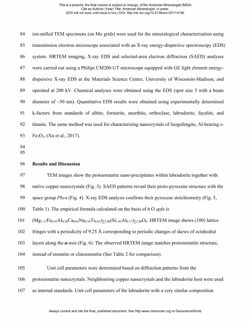
Samples and Experimental Methods
Protoenstatite occurs as precipitates associated with copper nanocrystals in gem-quality
labradorite phenocrysts (Oregon sunstones) from Dust Devil Mine, Lake County, Oregon 61

(Figures 1 and 2). The site is located in the Rabbit Basin within the Oregon high desert. The host 62
rock is a mid-Miocene basalt (Johnson et al., 1991; Peterson, 1972). The phenocrysts are 63
generally tabular plates and large laths ranging from ~1 cm in the greatest dimension to lathes 64
8.3 cm long, 2.6 cm wide and 1 cm thick (Hofmeister and Rossman, 1985; Johnson et al., 1991; 65
Peterson, 1972; Stewart et al. 1966). Protoenstatite was discovered in the green part of the 66
“watermelon” variety that possesses a clear rim and a core of transparent red surrounded by a 67
clear vibrant green border that can only be seen in certain orientations (Figure 1). Some carved 68
or faceted red, green and watermelon Oregon sunstones are illustrated in supplementary material 69
(Fig. S1). 70
71
The “watermelon” sunstones exhibit pleochroism and dichroism. Color and schiller are 72
always localized in the cores of the phenocrysts where the native copper micro- or nano-platelets 73
that populate the interior are exsolved clusters of crystals. They are most commonly exsolved 74
parallel to the feldspar crystal plane (010), but also (001) (Hofmeister and Rossman, 1985). This 75
dichroism is exhibited in hand samples of “watermelon” sunstones. The crystals exhibit 76
dichroism with a clear red when oriented approximately parallel to the feldspar (001) plane, and 77
both red and green are seen when oriented in [100] and [010] directions (Figures 1). The parts 78
with green color also exhibit a brownish red / green pleochroism under a plane polarized light 79
(Figure 2). The same phenomenon was observed by Johnson et al. (1991). Pleochroism appears 80
to become stronger in deeply colored specimens. By contrast, the clear rim does not show 81
pleochroism. 82
Because protoenstatite occurs as nano-inclusions in gem-quality “watermelon” sunstones, 83
ion-milled TEM specimens (on Mo grids) were used for the mineralogical characterization using 84
transmission electron microscope associated with an X-ray energy-dispersive spectroscopy (EDS) 85
system. HRTEM imaging, X-ray EDS and selected-area electron diffraction (SAED) analyses 86
were carried out using a Philips CM200-UT microscope equipped with GE light element energy-87
dispersive X-ray EDS at the Materials Science Center, University of Wisconsin-Madison, and 88
operated at 200 kV. Chemical analyses were obtained using the EDS (spot size 5 with a beam 89
diameter of ~50 nm). Quantitative EDS results were obtained using experimentally determined 90
k-factors from standards of albite, forsterite, anorthite, orthoclase, labradorite, fayalite, and91
titanite. The same method was used for characterizing nanocrystals of luogufengite, Al-bearing ε-92
Fe2O3, (Xu et al., 2017). 93
94 95
Results and Discussion 96
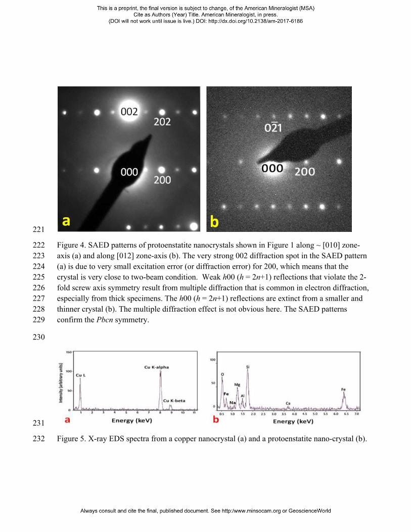
TEM images show the protoenstatite nano-precipitates within labradorite together with 97
native copper nanocrystals (Fig. 3). SAED patterns reveal their proto-pyroxene structure with the 98
space group Pbcn (Fig. 4). X-ray EDS analysis confirms their pyroxene stoichiometry (Fig. 5, 99
Table 1). The empirical formula calculated on the basis of 6 O apfu is 100
(Mg1.17Fe0.43Al0.26Ca0.03Na0.10Ti0.01)∑2.00(Si1.83Al0.17)∑2.00O6. HRTEM image shows (100) lattice 101
fringes with a periodicity of 9.25 Å corresponding to periodic changes of skews of octahedral 102
layers along the a-axis (Fig. 6). The observed HRTEM image matches protoenstatite structure, 103
instead of enstatite or clinoenstatite (See Table 2 for comparison). 104
Unit cell parameters were determined based on diffraction patterns from the 105
protoenstatite nanocrystals. Neighbouring copper nanocrystals and the labradorite host were used 106
as internal standards. Unit cell parameters of the labradorite with a very similar composition 107

(from Lake County, Oregon) are from Wenk et al. (1980). The measured unit-cell parameters are 108
a = 9.25(1) Å, b = 8.78(1) Å, and c = 5.32(1) Å. Calculated density of protoenstatite is 3.30 109
g·cm-3. Calculated powder X-ray diffraction peaks are listed in Supplementary Material (Table 110
S1). Comparison among the enstatite polymorphs with pyroxene structures is listed in table 2. 111
The crystals larger than ~ 200 nm transformed into clinoenstatite with a high density of 112
stacking faults (Fig. S5), which is very similar to the observed microstructures in a fast-cooled 113
protoenstatite (Ijima and Buseck, 1975). Cooling of the lava resulted in transformations from 114
protoenstatite to clinoenstatite with a high density of stacking faults in large protoenstatite 115
crystals (> 200 nm), whereas small protoenstatite crystals (< 200nm) are preserved in the host 116
labradorite phenocrysts. The Al-bearing protoenstatite nanocrystals with large surface areas may 117
lower the phase transformation temperature and stabilize the structure at low temperature. 118
Similar phenomenon occurs in hematite-luogufengite system (Lee and Xu, 2016). It is also 119
reported that protoenstatite nanocrystals with a large surface area of 615 m2/g, synthesized using120
sol-gel and freeze-dry methods, can be quenched to room temperature (Jones et al, 1999). 121
Protoenstatite nanocrystals were synthesized by sol-gel method at 800 °C (Jones et al, 1999), 122
which is lower than the reported phase transition temperature (~1000 °C). 123
124
Implications 125
The labradorite (An65) phenocrysts are very homogeneous in composition (Stewart et al., 126
1966; Wenk et al., 1980). We infer that the cores of “watermelon” crystals formed at early stages 127
of magma chamber formation at high P-T conditions. The clear phenocryst rims without any 128
precipitates suggest that they formed at a late stage under different conditions. Crystallization of 129
protoenstatite and associated native copper might happen also at a late stage but before magma 130

eruption. The collective effect of the oriented crystals of protoenstatite and clinoenstatite results 131
in the vibrant green colour of “watermelon” sunstones. These results may help understand and 132
determine size-dependent stability of these minerals. We agree that the phenocrysts experienced 133
a thermal shock due to the rapid rising and quenching of the crystals (Hofmeister and Rossman 134
1985). This thermal shock origin can explain why the labradorite crystals without schiller and 135
colors are all cracked. The colored sunstones are thus thermal shock resistant. Like a “single 136
crystal concrete,” their nano-inclusions of protoenstatite and Cu probably serve as cushion to 137
absorb the thermal shock due to metallic / plastic behavior of the Cu nanocrystals. This observed 138
texture may inspire the design of new crystalline materials that have strengths to resist thermal 139
shock while being optically clear and colorful. 140
141
ACKNOWLEDGMENTS 142
Authors thank Dr. Hongwu Xu and two anonymous reviewers for many constructive 143
suggestions and comments. This study was supported by NSF (EAR-0810150, EAR-1530614) 144
and the NASA Astrobiology Institute (N07-5489). Samples were collected by G. Farfan, C. 145
Peralta and the Dust Devil Mining Company. 146
147
148
149

REFERENCES 150
Angel, R.J., Chopelas, A., and Ross, N.L. (1992) Stability of high-pressure clinoenstatite at 151
upper-mantle pressures. Nature, 358, 322-324. 152
Cameron, M., and Papike, J.J. (1981) Structural and chemical variations in pyroxenes. American 153
Mineralogist, 66, 1–50. 154
Chen, C.-H. and Presnall, D.C. (1975) The system Mg2SiO4-SiO2 at pressure up to 25 kilobars. 155
American Mineralogist, 60, 398-406. 156
Dallwitz, W.B., Green, D. H., and Thompson, J. E. (1966) Clinoenstatite in a volcanic rock from 157
the Cape Vogel area, Papua. Journal of Petrology, 7(3), 375-403. 158
Hofmeister, A. M., and Rossman, G. R. (1985) Exsolution of metallic copper from Lake County 159
labradorite. Geology, 13(9), 644-647. 160
Ijima, S., and Buseck, P. R. (1975) High resolution electron microscopy of enatatite I: twinning, 161
polymorphism, and polytypism. American Mineralogist, 60, 758-770. 162
Johnston, C. L., Gunter, M. E., and Knowles, C. R. (1991). Sunstone Labradorite from the 163
Ponderosa Mine, Oregon. Gems and Gemology, XXVII, 220-233. 164
Jones, S. A., Burlitch, J. M, Duchamp, J. C., and Duncan, T. M. (1999) Sol-gel synthesis of 165
protoenstatite and a study of the factors that affect crystallization. Journal of Sol-Gel 166
Science and Technology, 15, 201-209. 167
Lee, S., and Xu, H. (2016) Size-dependent phase map and phase transformation kinetics for 168
nanometeric iron(III) oxides (g→e→a pathway). The Journal of Physical Chemistry C, 169
120, 13316–13322. 170

Murakami, T., Takeuchi, Y., and Yamanaka, T. (1982) The transition of orthoenstatite to 171
protoenstatite and the structure at 1080°C. Zeitschrift für Kristallographie, 160, 299-312. 172
Peterson, N. (1972) Oregon "Sunstones". The Ore Bin, 34(12), 197-215. 173
Schmitz, S. and Brenker, F.B. (2008) Microstructural Indications for Protoenstatite Precursor of 174
Cometary MgSiO3 Pyroxene: A Further High-Temperature Component of Comet Wild 2. 175
The Astrophysical Journal, 681 (2008) L105. doi:10.1086/590411. 176
Shiraki, K., Kuroda, N., Urano, H., and Maruyama, S. (1980) Clinoenstatite in boninites from the 177
Bonin Islands, Japan. Nature, 285, 31-32. 178
Smith, J. V. (1969) Crystal structure and stability of MgSiO3 polymorphs: physical properties 179
and phase relations of Mg-Fe pyroxenes. Mineralogical Society of America Special 180
Paper, 2, 3–29. 181
Smyth, J. R. (1974) Experimental study on the polymorphism of enstatite. American 182
Mineralogist, 59, 345-352. 183
Smyth, J.R. and Ito, J. (1975) The synthesis and crystal structure of a magnesium-lithium- 184
scandium protopyroxene. American Mineralogist, 62, 1252–1257. 185
Stewart, D., Walker, G., Wright, T., and Fahey, J. (1966) Physical Properties of Calcic 186
Labradorite from Lake County, Oregon. American Mineralogist, 51, 177-197. 187
Tribaudino, M., Nestola, F., Camara, F., and Domeneghetti, M. C. (2002) The high-temperature 188
P21/c-C2/c phase transition in Fe-free pyroxene (Ca0.15Mg1.85Si2O6): Structural and 189
thermodynamic behavior. American Mineralogist, Volume 87, pages 648–657. 190

Wenk, H. R., Joswig, W., Tagai, T., Korekawa, M., and Smith, B. K. (1980) The average 191
structure of An 62-66 labradorite. American Mineralogist, 65, 81-95. 192
Xu, H.F., Lee, S., and Xu, H.W. (2017) Luogufengite: a new nano-mineral of Fe2O3 polymorph 193
with giant coercive field. American Mineralogist, 102, 711-719. 194
Yang, H. Finger, L.W., Conrad, P.G., Prewitt, C.T., and Hazen, R.M. (1999) A new pyroxene 195
structure at high pressure: Single-crystal X-ray and Raman study of the Pbcn-P21cn phase 196
transition in protopyroxene. American Mineralogist, 84, 245–256. 197
198
199

Figures and Captions 200
201
202 Figure 1. A gem-quality “watermelon" Oregon sunstone looked down along the normal of (001) 203
(a), and along b-axis (b). Note the exceptionally clear rim, along with the zoning of colors. The 204
green border becomes brownish red when looked down along the normal of (001) (a). Top up-205
right inset shows the detail of the red to green transition from a polished sample (~ 4 mm thick) 206
(b). Dark areas are due to uneven surfaces that bend the transmitted light away. Another 207
“watermelon” sunstone (c) with a clear rim looked down along ~b-axis under a transmitted light 208
from bottom (left). Linear features with dark color in red core and green board are microplates of 209
native Cu. 210

211
Figure 2: Transmitted light photomicrographs of a polished sunstone crystal (~ 5 mm thickness) 212
from the green border part show pleochroism (light brown to pale green). The cleavage plane 213
(001) is perpendicular to the light. Some inclusions appear as linear features distributed along the214
(010) plane of labradorite. The appearance of the inclusions is much larger than their actual sizes215
due to the strain existing between thenanocrystals and the host labradorite. 216
217
218
Figure 3. Dark-field TEM image (a) and bright-field TEM image (b) showing two protoenstatite 219 (PEN) nanocrystals together with native copper nanocrystals (Cu) within labradorite. 220
221
Figure 4. SAED patterns of protoenstatite nanocrystals shown in Figure 1 along ~ [010] zone-222 axis (a) and along [012] zone-axis (b). The very strong 002 diffraction spot in the SAED pattern 223 (a) is due to very small excitation error (or diffraction error) for 200, which means that the224 crystal is very close to two-beam condition. Weak h00 (h = 2n+1) reflections that violate the 2-225 fold screw axis symmetry result from multiple diffraction that is common in electron diffraction,226 especially from thick specimens. The h00 (h = 2n+1) reflections are extinct from a smaller and227 thinner crystal (b). The multiple diffraction effect is not obvious here. The SAED patterns228 confirm the Pbcn symmetry.229
230
231
Figure 5. X-ray EDS spectra from a copper nanocrystal (a) and a protoenstatite nano-crystal (b). 232

233
Figure 6. HRTEM image (a) and noise-filtered HRTEM image (b) of a protoenstatite nanocrystal 234
showing (100) lattice fringes with a periodicity of 9.25 Å. A [012] zone-axis fast Fourier 235
transform (FFT) pattern is inserted at the lower-right corner of the HRTEM image. (c) A simple 236
protoenstatite model based on unit-cell twining of clinoenstatite. (d) A polyhedral model of 237
protoenstatite projected onto (010) showing periodic changes of skews of octahedral layers along 238
the a-axis. The model is based on a Li-Sc-bearing protoenstatite at room temperature (Yang et 239
al., 1999) with unit cell parameters and composition measured from the protoenstatite 240
nanocrystals. 241
242
243
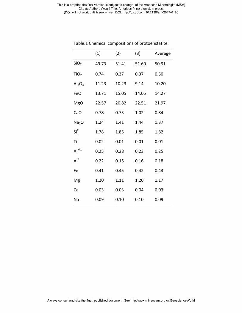
Table.1 Chemical compositions of protoenstatite.
(1) (2) (3) Average
SiO2 49.73 51.41 51.60 50.91
TiO2 0.74 0.37 0.37 0.50
Al2O3 11.23 10.23 9.14 10.20
FeO 13.71 15.05 14.05 14.27
MgO 22.57 20.82 22.51 21.97
CaO 0.78 0.73 1.02 0.84
Na2O 1.24 1.41 1.44 1.37
SiT 1.78 1.85 1.85 1.82
Ti 0.02 0.01 0.01 0.01
AlM1 0.25 0.28 0.23 0.25
AlT 0.22 0.15 0.16 0.18
Fe 0.41 0.45 0.42 0.43
Mg 1.20 1.11 1.20 1.17
Ca 0.03 0.03 0.04 0.03
Na 0.09 0.10 0.10 0.09

Table 2: Comparison among all enstatite polymorphs
Phase Space group Chain rotation Skew of octahedra
β angle (°)
Enstatite (En)* Pbca OA, OB + + - - + + - - 90
Clinoenstatite (CEN)* P21/c SA, OB + + + + ~ 108
High-CEN** C2/c O + + + + ~ 109
High-P CEN*** C2/c O + + + + ~ 101
Protoenstatite (PEN)* Pbcn O + - + - 90
High-P PEN**** P21cn SA, OB + - + - 90
Notes: * Cameron and Papike 1981; ** Tribaudino et al. (2002); *** Angel et al. (1992); **** Yang et al. (1999).