revised- june 29, 2000 regulation of ap1 (jun/fos) factor ... · the ap1 transcription factor...
TRANSCRIPT

1
REVISED- JUNE 29, 2000
Regulation of AP1 (Jun/Fos) Factor Expression and Activation inOvarian Granulosa Cells: Relation of JunD and Fra2 to Terminal
Differentiation
S Chidananda Sharma and JoAnne S Richards
Department of Molecular and Cellular Biology
Baylor College of Medicine
One Baylor Plaza
Houston, Texas 77030
To whom correspondence should be addressed:
JoAnne S. RichardsDepartment of Molecular and Cellular BiologyBaylor College of MedicineOne Baylor PlazaHouston, Texas 77030email: [email protected]:713-798-6238Fax:713-790-1275
Key words: AP1, menin, cAMP, FSH, LH, granulosa cells, ovary
JBC Papers in Press. Published on August 8, 2000 as Manuscript M003555200 by guest on M
arch 4, 2020http://w
ww
.jbc.org/D
ownloaded from

2
Running Title: AP1 factors in the ovary: regulation by FSH and LH
SummaryAP1 transcription factors control rapid responses of mammalian cells to stimuli
that impact proliferation, differentiation, and transformation. To determine which AP1
factors are present in and regulated by hormones in ovarian cells during specific stages of
proliferation and differentiation we used both in vitro and in vivo models, Western
blotting, immunohistochemistry, DNA binding assays and transfections of AP1
promoter-reporter constructs. The expression patterns of Jun and Fos family members in
response to hormones (FSH, LH and cAMP) were distinct. JunB, cJun, cFos and Fra2
were rapidly but transiently induced by FSH in immature granulosa cells. JunD and Fra2
were induced by LH and maintained as granulosa cells terminally differentiated into
luteal cells. Forskolin and PMA acted synergistically to enhance transcription of an
AP1(-73COL)-luciferase construct. JunD appears to be one mediator of this affect, since
JunD was a major component of the AP1/DNA binding complex in granulosa cells and
menin, a selective inhibitor of JunD, blocked transcription of –73COL-luciferase. Thus,
FSH and LH via cAMP induce specific AP1 factors, the AP1 expression patterns are
distinct and that of JunD and Fra2 correlates with the transition of proliferating granulosa
cells to terminally differentiated, non-dividing luteal cells. by guest on March 4, 2020
http://ww
w.jbc.org/
Dow
nloaded from

3
Introduction
Growth of ovarian follicles involves regulated proliferation and differentiation of
granulosa cells (1,2). Exponential growth of granulosa cells in preovulatory follicles is
followed by their terminal differentiation and formation of the corpus luteum (CL), a
transition initiated by the ovulatory surge of luteinizing hormone, LH. This transition of
proliferating granulosa cells to non-dividing luteal cells is associated with specific
changes in the expression of cell cycle regulatory molecules that control specific kinase
cascades. The cell cycle activators, cyclin D2 and cyclin E, are increased by FSH and
steroids in rapidly growing preovulatory follicles but are rapidly turned off by the surge
of LH which terminates follicular growth (1,2). Conversely, the cell cycle inhibitors,
p27KIP1 and p21CIP1 are induced by the LH surge (1,2). Thus, the gonadotropins impact
cell cycle progression at multiple steps depending on the stage of granulosa cell
differentiation.
The effects of the gonadotropins on ovarian cell proliferation and differentiation
are mediated by changes in intracellular cAMP which activates cAMP-dependent protein
kinases (A-kinases) (3), as well as several other kinases (4,5). Known targets of A-kinase
are the cAMP regulatory element (CRE) binding protein CREB and the CREB binding
protein CBP (6). CREs are essential for transcriptional activation of aromatase (7) and
inhibin α (8), as well as the AP1 factors, cFos (9) and Fra2 (10). AP1 factors, in turn,
mediate FSH-regulated expression of inhibin βA by binding to a variant CRE in the
promoter of the βA gene (11). Members of the AP1 transcription factor superfamily are
proto-oncogenes which regulate cell proliferation and transformation (12). They have
also been associated with differentiation (13,14). These observations suggest that
specific AP1 factors or the combination of specific AP1 factors may regulate different
sets of functions at specific stages of granulosa cell differentiation. Despite the intense
research efforts that have analyzed the function of the Jun/Fos family in many different
cell types, relatively few studies have analyzed how hormones regulate the expression of
AP1 factors in granulosa cells or how these factors might impact FSH or LH activation of
specific genes. Therefore, it becomes imperative to know which AP1 factors are present
in granulosa cells and which might be regulated or activated by FSH and LH.
The AP1 transcription factor family is comprised of Jun family members (c-Jun,
JunB and JunD) which can form homo- or hetero-dimers among themselves and bind to
AP1 consensus DNA (TGACTCA) (10,12,15). Jun proteins also dimerize with Fos
family members (cFos, FosB, Fra1 and Fra2)(10,12,16). Thus, the functional activity of
AP1 in any given cell is dependent in part on the relative amount of specific
by guest on March 4, 2020
http://ww
w.jbc.org/
Dow
nloaded from

4
Jun/Fos factors present in that cell. Most AP1 transcription factors are present at low
levels in cells but are rapidly induced and activated in response to specific stimuli. For
example, the c-fos gene has a complex promoter that contains a CRE, a serum response
element (SRE) and a Sis inducible enhancer (SIE) allowing it to respond to multiple
hormones, growth factors and cytokines (10,15,17). Not surprisingly then, cfos has been
shown to be induced by FSH in granulosa cells (9). The promoter of the JunB gene is
also complex with Stat3, CRE, CAAT enhancer (CAAT) binding domains as well as an
IL-6 regulatory region (10,18). Different promoter regions are utilized in different cell
types (18). Although JunB is expressed in ovarian cells (19), the factors regulating its
expression have not been analyzed. In contrast, the promoter of cJun is simpler (10,15)
and JunD is constitutively expressed in many cell lines (19).
The functional activation of AP1 factors in mammalian cells requires
phosphorylation (15). The activated AP1 factors have been shown to regulate gene
expression by direct as well as indirect mechanisms (12,16). Direct activation requires
that AP1 factors bind an AP1 regulatory domain present in the promoters of target genes
and that the AP1 factors be phosphorylated in their activation domain. The AP1 DNA
binding domain was originally known as the TPA response element (TRE), since in
response to phorbol ester tumor promoters such as TPA, the AP1 factors are
phosphorylated, activated and induce transcription (15). However, AP1 factors can be
phosphorylated and activated in response to numerous other agonists, suggesting that
their activation profile is more complex than originally thought. In fact, PMA which
activates cJun N-terminal kinase (JNK) does not activate Fos regulating kinase, Frk (15).
Activated AP1 factors can also enhance gene transcription by indirect mechanisms that
involve their binding to other transcription factors via protein-protein interactions but do
not require their binding to DNA. For example, members of the Jun/Fos family of
transcription factors have recently been shown to interact with both Sp1/Sp3 and Smad
proteins to enhance transcriptional activity of specific promoters with GC-rich and
SMAD binding regulatory elements (SBE), respectively (14,20). This may be one
attractive way in which FSH or LH regulate the expression of cyclins, p21CIP or p27KIP as
well as Sgk. cJun has also been shown recently to recruit CBP for enhanced activation of
CREB (21). Such a mechanism may be contribute to FSH activation of aromatase. The
importance of cJun is underscored by the embryonic lethalilty of mice null for cJun (22).
In contrast, mice null for cFos are viable but exhibit pleotropic effects (23).
Based on the roles of AP1 factors in cell proliferation, differentiation and
transformation, we sought to determine which AP1 factors are present in and regulated by
hormones in granulosa cells during follicular growth and terminal differentiation to luteal
by guest on March 4, 2020
http://ww
w.jbc.org/
Dow
nloaded from

5
cells. For these studies we have used both in vitro and in vivo models, Western blotting
and DNA binding-electrophoretic mobility shift assays to identify and quantify Jun (cJun,
JunD and JunB) and Fos (cFos, FosB, Fra1 and Fra2) family members present in
granulosa cells. The hormonal activation of AP1 factors in granulosa cells was analyzed
by transiently transfecting cells with AP1 promoter-luciferase reporter constructs.
Specific cellular signaling cascades were activated by adding forskolin, an agonist that
stimulates cAMP, as well as the phorbol ester PMA that is a known activator of AP1
factors in other cell types. Results show that each AP1 factor exhibited a specific
expression pattern and response to hormones. Rapid induction of JunB, cFos and Fra2 by
FSH corresponds to the pattern of other immediate-early genes in granulosa cells whereas
rapid induction of JunD and Fra2 by LH in granulosa cells of preovulatory follicles
resulted in their prolonged, stable expression during the transition to luteal cells. JunD, in
addition to cJun and cFos, was present as a major Jun factor comprising the AP1/DNA
binding complex and controlled transactivation of the AP1-promoter reporter construct.
Thus, JunD and Fra2, as well as cJun and cFos appear to be selectively expressed in
terminally differentiated luteal cells indicating the composition of AP1 factors changes
during granulosa cell proliferation and differentiation.
Experimental Procedures
Reagents
Media and cell culture reagents and materials were purchased from GIBCO
(Grand Island, NY), Sigma (St. Louis, MO), Research Organics (Cleveland, OH), Fisher
Scientific (Fairlawn, NJ) Corning (Corning, NY and Hyclone (Logan UT).Trypsin,
soybean trypsin inhibitor, DNAse, phorbol myristate (PMA), dithiothreitol (DTT), 17-β
estradiol (E) and propylene glycol were all purchased from Sigma (St. Louis, MO).
Ovine FSH (oFSH-16) was a gift of the National Hormone and Pituitary Program
(Rockville, MD). Human chorionic gonadotropin (hCG) was from Organon Special
Chemicals (West Orange, NJ). Forskolin was from Calbiochem (San Diego, CA).
Antibodies for cJun (#PC06) and cFos (#PC05) were obtained from Calbiochem (San
Diego, CA). Antibodies for JunB (#SC-73 for Westerns and immunohistochemistry;
#SC46 for EMSA), JunD (#SC74x), FosB (#SC-48x), Fra1 (#SC605x), and Fra2 (#SC-
604x) were from Santa Cruz (Santa Cruz, CA). TRIzol reagent (#15596) was obtained
from Life Technologies (Grand Island, NY). Electrophoresis and molecular biology grade
reagents were purchased form Sigma (St. Louis, MO), BioRad (Richmond, CA) and
Boehringer Mannheim Biochemicals (Indianapolis, IN). Oligonucleotides were
purchased from Genosys (The Woodlands, TX). All reverse transcriptase-polymerase
by guest on March 4, 2020
http://ww
w.jbc.org/
Dow
nloaded from

6
chain reactions (RT-PCR) reagents were from Promega (Madison, WI) except for
deoxyribonucleotides (dNTPs; Boehringer Mannheim Biochemicals, Indianapolis, IN). α-
32P[dCTP] was from ICN Radiochemicals (Costa Mesa, Ca). Hyperfilm was purchased
from Amersham (Arlington Heights, IL). The menin expression plasmid and antibodies
were generously provided by Dr. Sunita Agarwal, NIDDK, NIH, Bethesda, MD. The
Gal4-ELK and 4XGAl-luciferase vectors were generously provided by Dr. Philip Stork
(Oregon Health Science Center, Portland OR), and the -73COL-luciferase vector by Dr.
Michael Karin (University of California, San Diego, La Jolla, CA).
Animals
Intact and hypohysectomized (H) immature (day 23 of age) Holtzman Sprague-
Dawley female rats were obtained from Harlan (Indianapolis, IN) and housed under a
16:8 light:dark schedule in the Center for Comparative Medicine at Baylor College of
Medicine and provided food and water ad libitum. Animals were treated in accordance
with the NIH Guide for the Care and Use of Laboratory Animals, as approved by the
Animal Care and Use Committee at Baylor College of Medicine (Houston, TX). To
obtain granulosa cells for primary cultures, immature rats were injected with estradiol (E;
1.5mg/day for 3 days)(7). To analyze the expression of AP1 factors during follicular
development in vivo, H rats were treated with E (HE: as above) to stimulate the growth of
large preantral follicles. To analyze the response of granulosa cells to acute exposure to
FSH, some HE rats were injected intravenously (iv) with 5µg oFSH. Ovaries (for
immunohistochemistry) and granulosa cells (for whole cell extracts) were isolated from H
and HE rats prior to FSH (0h) or from HE rats exposed to FSH for 2 and 8h (HE, FSH 2,
8h). Other HE rats were injected subcutaneously (sc) with 1.0µg oFSH twice daily for 2
days (HEF, 48h) to stimulate the growth of preovulatory follicles which express
aromatase, LH receptor and inhibin α (24). HEF rats were subsequently injected iv with
10 IU human chorionic gonadotropin (hCG) an LH like hormone to stimulate ovulation
and luteinization. Ovaries and granulosa cells were prepared prior to (0h) and at 2,4,8,12
and 24h after hCG for immunohistochemistry and WCE, respectively (Shown
schematically in Figure 1).
Granulosa Cell Cultures
Granulosa cells were harvested from estradiol (E)-primed intact immature (day
25) rats as previously described (7). Briefly, cells were cultured at a density of 1 x 10⁶
cells per 3 ml serum free medium (DMEM:F12 containing Penicillin and Streptomycin)
in multi-well (35mm) dishes that were serum-coated. Cells were cultured in defined
by guest on March 4, 2020
http://ww
w.jbc.org/
Dow
nloaded from

7
medium over-night (0h) followed by the addition of FSH (50 ng/ml) and testosterone (T;
10 ng/ml), forskolin (10µM) and other agonists as indicated in the figures legends.
FSH/T were used to stimulate the differentiation of granulosa cells to a preovulatory
phenotype in which aromatase (7), LH receptor (25), and inhibin (8) are expressed.
Forskolin was added to these cells to mimic the LH surge and luteinization. Forskolin
alone was used to determine the relative effects of cAMP on specific cell functions (4,7).
PMA was used as an agonist for C-kinase (15,26) (Shown schematically in Figure 1).
Hormones, agonists, and antagonists were added as indicated in the Figure legends.
Transfections
After culture overnight, granulosa cells were transfected by the calcium phosphate
precipitation method (27) using 5µg of vector DNA: –73COL-luciferase (15,28), ERE-
E1b-luciferase (29), 4X-Gal-luciferase and Gal-4-Elk-1(30), or menin (31) as indicated in
the Figure legends. Four hours later the cells were washed thoroughly in fresh medium.
At that time (0h) the cells were stimulated with agonists. After 6h of agonist stimulation,
cell lysates were prepared and used to measure luciferase activity according to standard
protocols. Relative light units were normalized to protein (mini-Bradford assay; Bio-
Rad Laboratories, Inc.) in each sample. All transfections were run in triplicate and each
set of experiments was repeated at least three times. Unless otherwise indicated, values
represent the mean +/-SEM of three experiments.
Cell extracts and Western blot analyses
Total cell extracts were prepared from cultured granulosa cells according to a
method of Ginty (32) by adding to each well hot (100C) Tris-buffer containing 10%
sodium dodecyl sulfate (SDS) and beta-mercaptoethanol (4). The cells were rapidly
scraped with a rubber policeman. The cell extract was immediately transferred to an
eppendorf tube for 5 min at 100C and then stored at 4C. Equal volumes (50µl) of samples
were analyzed by SDS-polyacrylamide gel electrophoresis (SDS-PAGE). Following
SDS-PAGE, proteins were electrophoretically transferred to nylon filter, washed briefly in
phosphate buffered saline (PBS) and blotted with either 3% bovine serum albumin (BSA)
or 5% Carnation milk at room temperature for 1 h. Antibodies were added in the same
blocking solutions at the dilutions indicated in the figure legends. Immunoreactive
proteins were visualized with enhanced chemiluminescent (ECL) reagents according to the
specification of the supplier (Pierce Chemical Co., Rockford, IL). Immunoreactive bands
were quantified by image analysis of autoradiograms (ECL) using AlphaImager 2000
(3.3), Alpha Innotech Corporation, San Leandro, CA.
by guest on March 4, 2020
http://ww
w.jbc.org/
Dow
nloaded from

8
Electrophoretic mobility shift assays (EMSA)
Whole cell extracts (WCE) were prepared from hormonally stimulated granulosa
cells in culture or from hormonally-primed H rats as described previously (29) and in the
Figure legends. WCE were incubated 20 min on ice with a P32-labeled consensus AP1
oligonucleotide probe and then subjected to non-denaturing electorphoresis (0.5X TBE;
Tris-borate-EDTA) at 150V. Where indicated, either unlabeled probe or antibodies to
specific AP1 transcription factors were added to the reactions 30min on ice before the
addition of labeled DNA.
Immunohistochemical analyses
Immunohistochemical analyses of AP1 factors in whole ovaries isolated from
hormonally-primed rats were done as described previously (24). Ovaries were fixed in 4%
paraformaldehyde and paraffin-embedded. Sections (6 microns) were processed according to
routine procedures then blocked with 10% non-immune goat serum followed by incubation with
specific antibodies diluted 1:50 in 10% goat serum overnight at room temperature. After
washing in PBS, biotinylated anti-rabbit antiserum (Vector, Berlingame, CA) was added for 30
min, slides were washed and streptavidin conjugated horseradish peroxidase was applied for 30
min. Sections were incubated with DAB substrate for 2 min, dehydrated without counter
staining and mounted.
Statistical Analyses
Transfection data were analyzed by one-way ANOVA or Students T test. Values were
considered significantly different if P<0.05.
Results
Models for analyzing hormone induced changes in granulosa cell expression of AP1 factors
The AP1 transcription factor superfamily represents an important signal transduction
system in mammalian cells. However, relatively little is known about which AP1 factors are
expressed or activated in ovarian granulosa cells. Therefore, our initial goals were to determine
which members of the AP1 family of transcription factors were present in undifferentiated
granulosa cells and which factors might be regulated by hormones in association with
proliferation versus differentiation.
Two models were used to analyze hormone induced changes in AP1 expression in
granulosa cells (Figure 1). In vitro cultures: In the first model, undifferentiated granulosa cells
by guest on March 4, 2020
http://ww
w.jbc.org/
Dow
nloaded from

9
were harvested and cultured in defined medium overnight . At that time the undifferentiated
cells were either stimulated acutely (1.5h) with forskolin to increase cAMP, the phorbol ester
PMA (a known activator of AP1 factors) or the combination of forskolin and PMA. To stimulate
the differentiation of granulosa cells to a preovulatory phenotype, FSH/T were added to the cells
for 48h. These differentiated cells (FSH/T), which express aromatase and LH receptor, were also
stimulated acutely with forskolin, PMA or the combination to mimic the acute effects of the LH
surge. Lastly, the differentiated cells were cultured with forskolin for longer periods of time to
mimic the LH-induced process of luteinization in which granulosa cells cease to divide and are
terminally differentiated. Hormonal treatment of rats in vivo: In the second model,
hypophysectomized immature rats (H) were injected with estradiol (E; HE rats) to stimulate
granulosa cell proliferation and the growth of large preantral follicles. Subsequently, FSH (F)
was administered to the HE rats (HEF) to examine rapid effects as well as long-term effects of
the gonadotropins, leading to the development of preovulatory follicles (see Experimental
Procedures for details). HEF rats were injected with an ovulatory dose of the LH-like hormone,
hCG, to stimulate terminal differentiation of granulosa cells. This process of luteinization leads
to the formation of corpora lutea (CL).
Differential induction of Jun/Fos transcription factors in undifferentiated granulosa cells.
To analyze the response of undifferentiated cells, granulosa cells were harvested from
immature rats and cultured with forskolin to stimulate cAMP production for different time
intervals [0, 0.33 (20min), 0.66 (40min), 1.5, 3, 6, 12, 24 and 48h]. Cell extracts were prepared
and analyzed by Western blotting using antibodies specific for individual members of the AP1
transcription factor family: cJun, JunB, JunD, cFos, FosB, Fra1 and Fra2 (Figure 2).
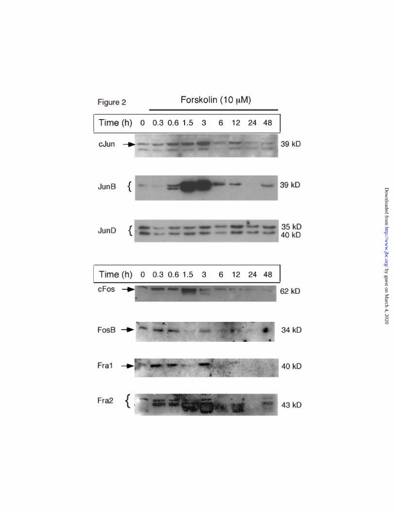
Jun family members. When undifferentiated granulosa cells were acutely stimulated with
forskolin, different patterns of AP1 protein expression were observed (Figure 2). Among the Jun
family members, immunoreactive cJun was present at low levels in granulosa cells cultured
overnight in medium alone (0h). The addition of forskolin stimulated 1-, 5-, 6- and 8-fold
increases in cJun at 20,40, 90min and 3h, respectively. Levels of cJun decreased at 6h and
remained at this level at subsequent time intervals. JunB exhibited a similar pattern of response
as that of c-Jun but the magnitude of induction was greater. JunB increased 1-, 3-, 14-, 16-fold at
20, 40, 90min and 3h, respectively. Levels of JunB protein were decreased at 6h and remained
low. In contrast, JunD was present in granulosa cells at 0h and its expression was relatively
unaffected by the addition of forskolin.
Fos family members. Among the Fos family members, immunoreactive cFos increased
rapidly in response to forskolin; 2- and 6-fold increases occurred at 20 and 90 min, respectively
(Figure 2). However, this increase was transient; immunoreactive cFos returned to the baseline
by guest on March 4, 2020
http://ww
w.jbc.org/
Dow
nloaded from

10
(0h) level between 3-6h and remained low thereafter. Similarly, FosB and Fra1 exhibited
increases in response to forskolin at 20 and 40min but declined to basal or near undetectable
levels, respectively, between 6-24h of culture. Fra2 showed the most dramatic changes in
response to forskolin (Figure 2; bottom panel). Fra2 increased 3-, 3- and 11-fold at 20, 40 and 90
min, respectively, remained elevated at 3h and then declined gradually at 6-12h and was low
again at 24-48.
Collectively these data indicate that JunD is constitutively expressed in cultured
granulosa cells whereas other AP1 factors, especially cJun, JunB, Fra2 and cFos are rapidly but
transiently induced/increased by forskolin/cAMP. Immunocytochemical analyses documented
that AP1 factors were localized to the nuclei of granulosa cells (data not shown).
Induction of AP1 transcription factors in undifferentiated and differentiated granulosa
cells: comparison of the effects of forskolin and PMA.
The transcriptional activation of AP1 factors has been shown in other cells types to be
stimulated by the phorbol ester, PMA. Therefore, we next determined if the effects of forskolin
(cAMP) on AP1 factor expression were similar to, different from or synergistic with those of
PMA and if the effects were dependent on the stage of granulosa cells differentiation. For these
experiments, granulosa cells were cultured over-night in defined medium. At that time (0h), the
undifferentiated cells were treated acutely (for 1.5h) with either forskolin (10µM), PMA (20nM)
or the combination of forskolin and PMA. Some cells were exposed to forskolin for 24h. To
examine the responses of differentiated granulosa, additional cells were cultured in the presence
of FSH/T for 48h. The differentiated cells were stimulated acutely (for 1.5h) with agonists to
mimic the LH surge or were exposed to forskolin for 24 and 48h to induce luteinization. Cell
extracts were prepared and the inducibility of two factors, JunB and cFos, was analyzed by
Western blotting (Figure 3).
In untreated granulosa cells, immunoreactive JunB increased 44-,19- or 52-fold,
respectively, by exposure to forskolin, PMA or the combination for 1.5h (Figure 3; upper panel) .
The induction by forskolin was transient and JunB protein returned to basal levels by 24h. JunB
was also low in the FSH/T differentiated cells but could be induced by forskolin, PMA or the
combination, 28-, 21- and 34-fold, respectively. These results indicate that JunB is rapidly but
transiently induced by forskolin in control and differentiated granulosa cells. PMA alone or in
combination with forskolin also increased JunB. However, since the effects were not additive or
synergistic, these two agonists appear to induce JunB by similar or overlapping mechanisms.
Likewise, cFos was induced equally well by forskolin or PMA in undifferentiated granulosa cells
and the combined treatment was additive (Figure 3; lower panel). In the FSH/T differentiated
cells, the response to forskolin appeared less in this experiment than in subsequent experiments
by guest on March 4, 2020
http://ww
w.jbc.org/
Dow
nloaded from
11
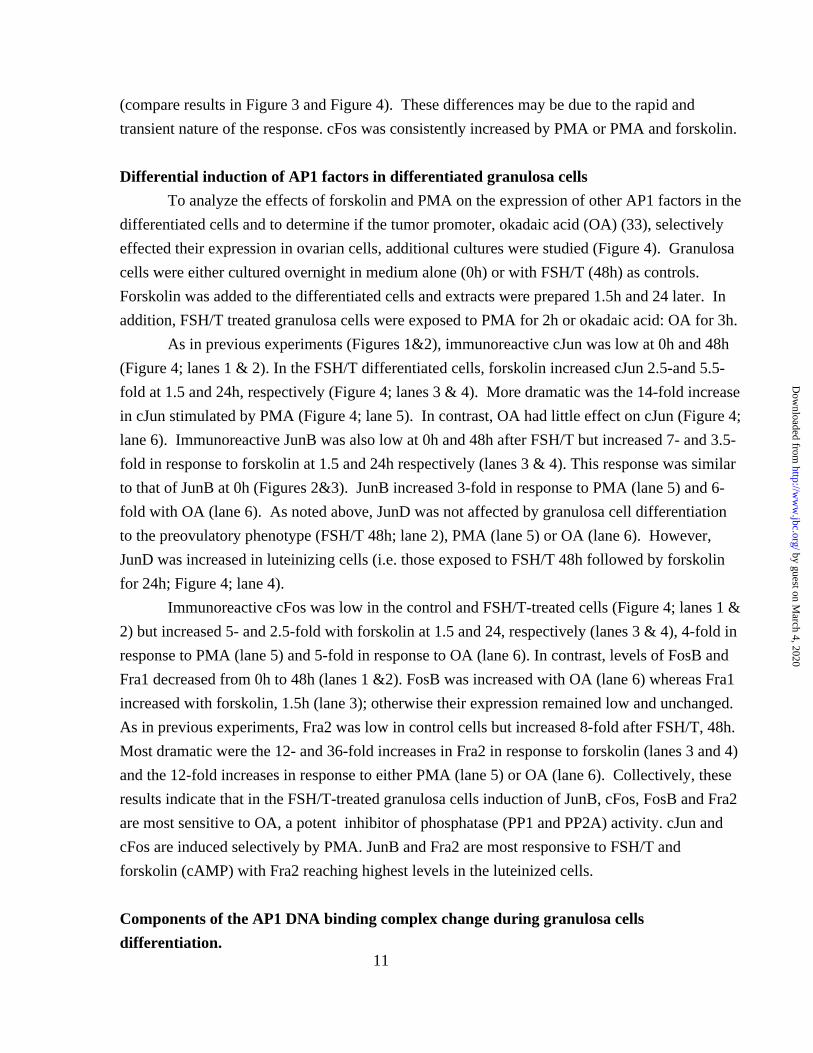
(compare results in Figure 3 and Figure 4). These differences may be due to the rapid and
transient nature of the response. cFos was consistently increased by PMA or PMA and forskolin.
Differential induction of AP1 factors in differentiated granulosa cells
To analyze the effects of forskolin and PMA on the expression of other AP1 factors in the
differentiated cells and to determine if the tumor promoter, okadaic acid (OA) (33), selectively
effected their expression in ovarian cells, additional cultures were studied (Figure 4). Granulosa
cells were either cultured overnight in medium alone (0h) or with FSH/T (48h) as controls.
Forskolin was added to the differentiated cells and extracts were prepared 1.5h and 24 later. In
addition, FSH/T treated granulosa cells were exposed to PMA for 2h or okadaic acid: OA for 3h.
As in previous experiments (Figures 1&2), immunoreactive cJun was low at 0h and 48h
(Figure 4; lanes 1 & 2). In the FSH/T differentiated cells, forskolin increased cJun 2.5-and 5.5-
fold at 1.5 and 24h, respectively (Figure 4; lanes 3 & 4). More dramatic was the 14-fold increase
in cJun stimulated by PMA (Figure 4; lane 5). In contrast, OA had little effect on cJun (Figure 4;
lane 6). Immunoreactive JunB was also low at 0h and 48h after FSH/T but increased 7- and 3.5-
fold in response to forskolin at 1.5 and 24h respectively (lanes 3 & 4). This response was similar
to that of JunB at 0h (Figures 2&3). JunB increased 3-fold in response to PMA (lane 5) and 6-
fold with OA (lane 6). As noted above, JunD was not affected by granulosa cell differentiation
to the preovulatory phenotype (FSH/T 48h; lane 2), PMA (lane 5) or OA (lane 6). However,
JunD was increased in luteinizing cells (i.e. those exposed to FSH/T 48h followed by forskolin
for 24h; Figure 4; lane 4).
Immunoreactive cFos was low in the control and FSH/T-treated cells (Figure 4; lanes 1 &
2) but increased 5- and 2.5-fold with forskolin at 1.5 and 24, respectively (lanes 3 & 4), 4-fold in
response to PMA (lane 5) and 5-fold in response to OA (lane 6). In contrast, levels of FosB and
Fra1 decreased from 0h to 48h (lanes 1 &2). FosB was increased with OA (lane 6) whereas Fra1
increased with forskolin, 1.5h (lane 3); otherwise their expression remained low and unchanged.
As in previous experiments, Fra2 was low in control cells but increased 8-fold after FSH/T, 48h.
Most dramatic were the 12- and 36-fold increases in Fra2 in response to forskolin (lanes 3 and 4)
and the 12-fold increases in response to either PMA (lane 5) or OA (lane 6). Collectively, these
results indicate that in the FSH/T-treated granulosa cells induction of JunB, cFos, FosB and Fra2
are most sensitive to OA, a potent inhibitor of phosphatase (PP1 and PP2A) activity. cJun and
cFos are induced selectively by PMA. JunB and Fra2 are most responsive to FSH/T and
forskolin (cAMP) with Fra2 reaching highest levels in the luteinized cells.
Components of the AP1 DNA binding complex change during granulosa cells
differentiation.
by guest on March 4, 2020
http://ww
w.jbc.org/
Dow
nloaded from

12
To determine if the AP1 factors present in granulosa cells were capable of binding to AP1
consensus DNA, WCE were prepared from granulosa cells cultured overnight in medium alone
(control, undifferentiated cells; 0h) or in the presence of forskolin, PMA or the combination for
1.5h. Additional extracts were prepared from FSH/T differentiated cells (48h) followed by
forskolin for 24h, a regimen shown (Figure 2) to selectively increase levels of immunoreactive
JunD and Fra2. WCE were incubated with a labeled AP1 consensus oligonucleotide probe alone,
a 100-fold excess of unlabeled oligonucleotide or in the presence of antibodies specific for cJun,
JunD, cFos, FosB, Fra1 and Fra2. As shown in Figure 5, two protein/DNA complexes, one
major and one minor, were formed when WCE from control (Figure 5; 0h; lane 1) granulosa
cells was incubated with probe alone. Both complexes were reduced in the presence of a 100-
fold excess of unlabeled DNA (lane 2). Antibodies to cJun (lane 3), JunD (lane 4), cFos (lane 5),
fobs (lane 6) and Fra2 (lane 8) all caused a supershift of the major protein/DNA complex but did
not alter the migration of the minor complex. Antibodies to Fra1 (lane 7) did not stimulate a
supershift of the complex present in control granulosa cells.
When WCE of differentiated granulosa cells (FSH/T+forskolin) were analyzed by
EMSA, the major protein/DNA complex was markedly increased (Figure 5, lane 9). Supershift
analyses indicated that cJun (lane 10), JunD (lane 11), cFos (lane 12), and Fra2 (lane 15) were
the AP1 components of the complex since each antibody caused a marked shift in the major
protein/DNA complex. In contrast, antibodies to FosB (lane 13) and Fra1 (lane 14) did not cause
supershifts, consistent with their lower abundance in extracts of differentiated cells (Figure 3).
Although no antibody alone caused a complete supershift of the complex, the addition of two or
more antibodies to the reaction mixture caused a more complete supershift of the complex (data
not shown). These data indicate that cJun, JunD, cFos and Fra2 are more abundant in the
differentiated granulosa cells than in the undifferentiated cells.
To analyze the binding activity of JunB, extracts of undifferentiated granulosa cells were
prepared at 0h or after treatment with forskolin, PMA or forskolin and PMA for 1.5h . The
protein/DNA complexes formed with WCE from control cells were not supershifted with JunB
antibody whereas extracts from the forskolin treated cells (containing high levels of
immunoreactive Jun B; Figure 1&2) were supershifted with the JunB antibody (data not shown)
Thus, JunB is an inducible component of the AP1 complex in cells exposed to acute stimulation
by forskolin.
Forskolin and PMA act synergistically to induce expression of the -73COL-luciferase
transgene.
To examine the ability of granulosa cells to transactivate promoters containing AP1
regulatory domains, three promoter-luciferase reporter constructs were initially tested. The
by guest on March 4, 2020
http://ww
w.jbc.org/
Dow
nloaded from

13
promoters contained either -73bp of the human collagenase gene (-73COL), 4 concatermers of a
consensus AP1 binding site (4XAP1) or a single AP1 domain (1XAP1). Since in initial tests,
each responded in a similar manner to forskolin, PMA or the combination, we have used the -
73COL-luciferase construct for the studies described herein. When the -73COL-luciferase
construct was transfected into control granulosa cells (0h), forskolin stimulated a 9-fold increase
whereas PMA stimulated only a 3-fold increase. The combination of forskolin and PMA
induced a 30-fold increase in luciferase activity, indicating they exert a synergistic response
(Figure 6).
To determine if the effects of forskolin and PMA on the AP1 promoter elements were
specific, we tested the response of two other promoter-reporter constructs to forskolin and PMA
(Figure 6). We chose an estrogen receptor (ER) response element (ERE) E1b-luciferase
construct since we have previously shown that it responds to forskolin (29) and others have
reported activation of ER by MAPK (34). When this vector was transfected into granulosa cells,
forskolin alone induced a 10-fold increase, PMA a 7-fold increase and the combination a 12-fold
increase. Thus the effects of forskolin and PMA were additive but not synergistic.
Since cJun and cFos were preferentially increased by PMA alone, we sought to determine
if a PMA specific pathway was operative in granulosa cells. For this, we tested the functional
activation of Elk-1, a ternary complex factor that binds serum response factor and enhances
transactivation from SREs. Granulosa cells were co-transfected with a 4X-Gal-luciferase reporter
construct and an expression vector containing a chimeric gene in which the Gal-4 DNA binding
domain was ligated to the activation domain of Elk-1 (Figure 6; 15). Luciferase activity was low
in the absence of agonists, increased 2-fold with forskolin, 31-fold with PMA and 39-fold with
forskolin and PMA (Figure 6).
Lastly, since JunD was present in the highest concentrations in unstimulated granulosa
cells, we sought to determine if the activity of the AP1 complex could be altered by menin, a
specific inhibitor of JunD. Additional cells were co-transfected with the –73COL-luciferase
reporter construct and a menin expression vector or empty vector (Figure 6). As shown, menin
markedly decreased (75%) the forskolin +PMA-mediated luciferase activity but did not affect the
control or forskolin-stimulated effects. Thus, JunD, mostly likely in complex with FosB or Fra2
(factors rapidly induced by forskolin and PMA) comprises the functional AP1 complex in
forskolin stimulated (undifferentiated) granulosa cells (Figure 6).
FSH and LH regulate transient versus stable AP1 expression in granulosa cells in vivo.
To determine if the expression of AP1 factors was regulated by hormones in vivo,
hypophysectomized (H) rats were administered estradiol (E), FSH and LH/hCG to stimulate the
growth of antral follicles in which granulosa cells are proliferative, preovulatory follicles in
by guest on March 4, 2020
http://ww
w.jbc.org/
Dow
nloaded from

14
which granulosa cells have acquired a transitional state of differentiation and luteinized follicles
in which the granulosa cells have terminally differentiated to non-dividing cells, respectively
(Figure 1 and Methods).
Western blots: Each AP1 factor exhibited its own specific pattern of expression in
granulosa cells of growing, ovulating and luteinizing follicles (Figure 7). Immunoreactive JunB
was negligible in granulosa cells of H and HE rats. However, JunB increased dramatically (13-
fold) within 2h of exposure to FSH. Three distinct immunoreactive bands of JunB indicate that
they are either phosphorylated forms or degraded products of JunB. The latter may be more
likely since JunB was no longer detected at 8h. JunB was also induced rapidly by hCG in
granulosa cells of preovulatory follicles; JunB was highest at 8h (24-fold increase) but was non-
detectable at 24h. Multiple bands were also observed in the 8h sample, indicating that JunB is
rapidly synthesized and processed (phosphorylated or degraded).
Fra2, like JunB, was negligible in granulosa cells of H and HE rats but was increased
rapidly and transiently by acute exposure to FSH (Figure 7). However, the expression of Fra2 in
granulosa cells of preovulatory follicles was distinct from that of JunB. Fra2 was rapidly
induced by hCG but the elevated levels were then sustained in luteinizing granulosa cells (HEF-
hCG, 4-12h) and in corpora lutea (HEF-hCG, 24h; CL). In this manner, Fra2 was similar to
JunD.
Immunoreactive JunD was absent in immature granulosa cells of H and HE rats but was
increased rapidly (2-fold) by 2h following acute injection of FSH. The increase was transient
since levels of JunD returned to control levels at 8h. JunD also increased rapidly in granulosa
cells of HEF rats following administration of hCG. In contrast to the transient response of
immature HE granulosa cells to FSH, the response of differentiated HEF granulosa cells to hCG
was sustained from 4-24h. The 11-fold increase in JunD at 8h was maintained in the ovaries of
HEF-hCG, 24h rats that are comprised mostly of corpora lutea.
The JunD inhibitor, menin, exhibited a different pattern of expression (Figure 7). Levels
of menin were low in granulosa cells of H, HE and HEF rats but were increased in luteinized
ovaries of HEF-hCG,24h rats, and in corpora lutea isolated from pregnant rats in early pregnancy
(day 7 of gestation) but low in later pregnancy (day 22). Thus, menin is also regulated and may
modify the functional activity of JunD at specific stages of granulosa cells differentiation.
EMSAs. Changes in the relative binding of AP1 factors to a consensus AP1
oligonucleotide exhibited patterns similar to those observed by Western blotting. AP1 factor
binding activity was low in WCE of granulosa cells from H and HE rats (Figure 8A). AP1
binding increased rapidly (11-fold) but transiently in response to acute exposure to FSH at 2h.
Low levels of AP1 in granulosa cells of preovulatory follicles (HEF; lane 5) were increased by
acute stimulation with hCG and were then maintained at levels between 11- and 30-fold above
by guest on March 4, 2020
http://ww
w.jbc.org/
Dow
nloaded from

15
that observed in the H control (lane 1) (Figure 8A).
Supershift analyses identified the AP1 factors that were present in granulosa cells of
preovulatory HEF follicles as JunD and c-Jun (data not shown). Neither JunB nor Fra2 was
detected (data not shown). In contrast, WCE of granulosa cells from HEF, hCG 8h rats
contained c-Jun, JunD, c-Fos and Fra2 (Figure 8B; lanes 4,5,7 and 9). JunB appeared to be a
minor component of the AP1/DNA complex (Figure 8B, lane 3) consistent with data obtained by
Western blots (Figure 7). FosB and Fra1 were not detected (lanes 6 and 8). Similar but higher
levels of AP1 factors were present in the HEF, hCG 24h extracts (Figure 8B, lanes 10-17)
Collectively, these results show that the hormonal regulation of AP1 factor expression in
granulosa cells of growing follicles in vivo is similar to that in cultured granulosa cells (Figure
10). Western blots and EMSAs confirm that JunB is rapidly but transiently induced by FSH (and
forskolin) in undifferentiated as well as by LH in differentiated cells. FosB and Fra1 are present
in undifferentiated cells but not in luteal cells. In contrast, Fra2, as well as cFos and cJun, are
rapidly but transiently induced in undifferentiated cells but are expressed at elevated levels in
luteinizing granulosa cells and luteal cells. JunD is expressed in granulosa cells of growing
follicles and is elevated in luteinized cells. In vivo, JunD is also regulated by FSH and LH.
Thus, JunD and Fra2, as well as cJun and cFos likely different sets of AP1 regulated genes
during the transition of proliferating granulosa cells to terminally differentiated luteal cells.
Immunohistochemical localization of AP1 factors in ovarian cells.
Immunohistochemical data confirm the hormonal regulation and nuclear localization of
JunD and Fra2 in granulosa cells and further demonstrate regulation of these factors in theca cells
(Figure 9). JunD was present but low in ovaries of HEF rats. JunD increased in theca cells of
preovulatory follicles in response to hCG at 2h. By 4h immunoreactive JunD was detected in
granulosa cells and remained elevated and in nuclei of granulosa cells during luteinization.
Immunoreactive Fra2 (and JunB, data not shown) was negligible in granulosa cells and theca
cells of preovulatory follicles. Immunoreactive Fra2 and JunB (not shown) was increased first in
theca cells at 2h and 4h after hCG and then appeared in granulosa cells at 4h after which it
reached maximal levels between 8-12h. Immunoreactive Fra2, unlike JunB (not shown) was
present in nuclei of corpora lutea of immature rats 24h after hCG as well as of pregnant rats on
day 7 of gestation. Thus, JunB, JunD and Fra2 are low in preovulatory follicles but are increased
rapidly in response to hCG, appearing first in the theca cells and then in the granulosa cells.
JunD and Fra2, but not JunB, remain persistent in luteal cell nuclear suggesting that they are
selectively associated with terminal differentiation of the granulosa cells.
Discussion
by guest on March 4, 2020
http://ww
w.jbc.org/
Dow
nloaded from

16
The FSH, LH and forskolin-induced changes in the expression and activation of
AP1 factors in granulosa cells in vivo and in vitro indicate that the AP1 signaling
pathways are important downstream targets of cAMP in granulosa cells. Since the FSH
induced AP1 complex in proliferating granulosa cells is distinct (in composition and
temporal pattern) from that of the LH induced complex in terminally differentiated, non-
dividing luteal cells, these changes likely impact specific AP1 target genes during this
transitional period (Figure 10).
In proliferating (undifferentiated) granulosa cells of small follicles, induction by
FSH (forskolin) of cJun, JunB, cFos, FosB, Fra1 and Fra2 is rapid but transient; the most
dramatic increases occurred for JunB (16-fold) and Fra2 (11-fold). The increases in these
AP1 factors relate temporally to the expression of other immediate-early genes, such as
Sgk (24) and Egr-1 (12,35). JunB, cJun, cFos and FosB were also induced in granulosa
cells by the tumors promoters, PMA and okadaic acid. That the hormone (as well as
cAMP-, PMA-, and okadaic acid-) induced increases in AP1 factors are transient in
granulosa cells during the normal progression of follicular growth may be important for
preventing granulosa cell transformation. In numerous transformed cell lines, several AP1
factors (cJun, JunB, Fra1 and Fra2) are expressed at high levels, hence their designation
as proto-oncogenes. Of potential relevance to these studies, JunB is expressed at high
levels in ovarian cancer cell lines (36). Moreover, the increased levels of JunB in the
transformed cells have been associated with sustained activation of MAPK by the
Ras/Raf pathway (37). The Ras/Raf MAPK pathways may also mediate the effects of
FSH on AP1 factor expression and activation in granulosa cells. Recent evidence shows
that FSH/cAMP can impact the ERK, p38MAPK and PI3K pathways (5). Importantly,
the effects of FSH/cAMP on these pathways are independent of A-kinase. Likely, cAMP
activates a newly identified group of cAMP regulated proteins, cAMP-guanine nucleotide
exchange factors (cAMP-GEFs), that activate Ras-like molecules ((38,39)). Thus, FSH
may modify AP1 factor expression and activity by diverse signaling pathways. Although
JunB exhibits high sequence homology to cJun, and like cJun has no introns, the
promoter of JunB is more complex than that of cJun (10). As shown herein, the hormonal
induction of JunB in granulosa cells is far more dramatic and transient than that of cJun.
What factors contribute to JunB induction in the ovary, or its expression in Sertoli cells in
the testis (45), remain to be determined. Based on regulatory regions in the JunB
promoter, these factors could be CREB or related family members (18), Smads (40,41),
C/EBPβ (18), Stat factors (18) or a combination of these all of which are expressed and
regulated by hormones in ovarian granulosa cells.
In contrast to JunB and cJun, JunD was not regulated markedly by either forskolin
by guest on March 4, 2020
http://ww
w.jbc.org/
Dow
nloaded from
17
or PMA in undifferentiated granulosa cells in culture. Based on the lack of regulation in
cultured cells, it was surprising to observe that hormones regulated JunD expression in
vivo. Notably, FSH increased JunD transiently in granulosa cells of small follicles
whereas LH increased JunD in granulosa cells of preovulatory follicles, a response that
persisted as the cells luteinized. The JunD promoter contains several potential regulatory
elements including a CRE capable of binding CREB, a GC-rich region with potential
binding sites for Sp1/Egr-1, an AP1 site and a CAAT site to which NF-Y binds (10). The
complexity of the JunD promoter, as well as the presence of multiple hormones and
growth factors present in vivo, may explain the more complex pattern of JunD expression
in vivo compared to in vitro.
The molecular mechanisms by which hormones, forskolin (cAMP) and PMA
regulate expression of Fos family members in proliferating (undifferentiated) granulosa
cells are equally diverse (10,12,17,37). For example, although the gene structure and
promoter regulatory regions of the cFos and FosB genes are highly conserved, their
regulation in granulosa cells by cAMP and the tumor promoters differs markedly. In the
undifferentiated granulosa cells, cFos increased more in response to forskolin (and PMA)
than did FosB. In contrast, both were increased by okadaic acid. The cAMP response
elements (CREs) and AP1 elements within the cFos promoter are likely targets of cAMP
induction in granulosa cells (15,17). In contrast, the serum response element (SRE) of
the cFos promoter is likely to be the target of the PMA response, especially since PMA,
but not forskolin/cAMP, was a potent stimulator of transactivation of the Elk-Gal and
Gal-luciferase reporter system in granulosa cells. These two different promoter sites and
the preferential activation of A-kinase by FSH and ERK by PMA appear to contribute to
the additive effects of forskolin and PMA on induction of cFos in the undifferentiated
cells. The mechanisms by which Fra2 is induced clearly involves cAMP and A-kinase
since H89 completely blocked induction of Fra2 by FSH or forskolin (data not shown)
confirming the strong induction of Fra2 by cAMP in other cell types (10).
The LH/hCG (forskolin)-induced expression patterns of the AP1 factors in
differentiated granulosa cells of preovulatory follicles differed markedly from that in
undifferentiated cells. Despite the similarities of the cFos and FosB promoters, expression
of FosB but not cFos was turned off in response to hCG. In marked contrast, induction of
cJun and cFos, as well as JunD and Fra2, was sustained and stable as granulosa cells
terminally differentiated to luteal cells. This change from transient to stable expression of
JunD and Fra2 suggests that they exert specific functions during granulosa cell
proliferation and differentiation. That JunD is a functional component of the AP1/DNA
binding complex in differentiated granulosa cells suggests that it is an important regulator
by guest on March 4, 2020
http://ww
w.jbc.org/
Dow
nloaded from

18
of specific AP1 responsive genes. In other systems, JunD can act as an inhibitor or an
activator of transcription, dependent on the composition of the heterodimeric complex,
the promoter element and the cell type. For example, cFos/cJun or cFos/JunB, but not
cFos/JunD, activate the -73COL-luciferase construct in vascular endothelial cells (42). In
contrast, a Fra2/JunD complex was more effective than cFos/cJun in activating the
promoter of the oncostatin gene in ROS 17/2.8 osteosarcoma cells (43). Thus, based on
the composition of AP1 factors in differentiating granulosa cells, JunD/Fra2 heterodimers
may activate one set of AP1 responsive genes whereas cFos/cJun complexes regulate
other AP1 responsive genes. Furthermore, other factors such as menin, a specific JunD
inhibitor, can alter the activity of JunD. In support of this, co-expression of menin
blocked forskolin induced transcription of –73COL-luciferase, suggesting that JunD is a
functional component of the AP1 complex in granulosa cells. In terminally
differentiating granulosa cells, JunD may play a critical role in terminating cell
proliferation. Specifically, JunD has been shown to be low in certain ovarian cancer cell
lines and over-expression of JunD in these cells can suppress cell growth in a cell line
specific manner. Jun D and Fra2 have recently been shown to increase during osteoblast
differentiation (43). Thus, the increase in JunD and Fra2 in granulosa cells of
preovulatory follicles exposed to an LH surge may control the transcription of specific
genes that regulate the exit of granulosa cells from the cell cycle, thereby terminating
granulosa cell proliferation (1,2). Of note, Jun D is low in the testis (19), perhaps because
the number of non-proliferative, JunD-positive Sertoli cells (data not shown) is low
compared to the number of proliferating germ cells.
There are likely to be many genes regulated in granulosa cells by members of the
Jun/Fos family of transcription factors. First, AP1 factors appear to control their own
expression (10,15). Other genes in which AP1 factor regulation has been determined
include inhibin βA (11), the GnRH receptor (26), TIMP-1 (44,45) and as already
mentioned p21CIP (1,2,14). Each of these genes is hormonally regulated in granulosa
cells but only the promoter of the inhibin βA gene has been specifically examined in
granulosa cells. The inhibin βA promoter contains a variant CRE that binds AP1-like
factors and is inducible by forskolin and PMA as well as by over-expression of JunB and
FosB proteins in granulosa cells (11). These synergistic effects of forskolin and PMA on
the inhibin bA promoter mimic that observed herein for activation of the AP1 site within
the human collagenase gene. Synergism between cAMP and PMA has also been observed
recently to control induction of progesterone receptor (PR) mRNA in granulosa cells
(46). Based on the results described herein, the synergy in granulosa cells could involve
the selective induction by PMA of cJun and cFos and by cAMP of Fra2, JunB and JunD.
by guest on March 4, 2020
http://ww
w.jbc.org/
Dow
nloaded from

19
In addition to the induction of AP1 factors, forskolin and PMA activate several kinase
cascades (A-kinase, p38MAPK, C-kinase and ERKs) that would lead to the
phosphorylation and activation of specific AP1 factors and their coactivators. For
example, cJun but not JunB is a target of N-terminal c-Jun kinase (JNK); cFos is
activated by FRK (15). Lastly, AP1 factors may regulate the expression of ovarian genes
by indirect mechanisms that involve protein-protein interactions with other transcription
factors. Of note, cJun has been shown to interact with the cell cycle regulator Rb (47).
cJun also interacts with CBP to activate CREB (21), with Sp1/Sp3 to activate p21CIP1
gene (14), and with Smads (20) to regulate specific chimeric reporter genes. Thus,
hormonal regulation of AP1 factors in the ovary has far reaching effects on many cellular
signaling cascades that regulate proliferation and differentiation. The studies herein
provide the specific observation that JunD and Fra2, as well as cJun and cFos, may be
critical for regulating specific sets of AP1 responsive genes that control the terminal
differentiation of granulosa cells, their exit from the cell cycle and the prevention of
granulosa cell transformation associated with elevated levels of AP1.
AcknowledgmentsThe authors wish to thank Dr. Philip Stork (Oregon Health Sciences Center) for
the Gal-4-luciferase reporter construct and Elk-Gal expression vector, Dr. Michael Karin
(UCSD) for the -73COL-luciferase vector and Dr. Sunita Agarwal (NIDDK, NIH) for the
menin vectors.
REFERENCES
1. Robker RL, and Richards JS. (1998) Biol Reprod 59, 476-482
2. Robker RL, and Richards JS. (1998) Mol Endocrinol 12, 924-940.
3. Richards J. (1994) Endocr Rev 15, 725-751
4. Gonzalez-Robayna IJ, Alliston TN, Buse P, Firestone GL, and Richards JS.
(1999) Mol Endocrinol 13, 1318-1337
5. Gonzalez-Robayna IJ, Falender AE, Ochsner S, Firestone GL, and Richards JS.
(2000) Mol Endocrinol 14, in press
6. Habener JF, Miller CP, and Vallejo M. (1995) Vit Horm 51, 1-57
7. Carlone DL, and Richards JS. (1997) Mol Endocrinol 11, 292-304
8. Pei L, Dodson R, Schoderbek WE, Maurer RA, and Mayo KE. (1991) Mol
Endocrinol 5, 521-534
9. Mukherjee A, Park-Sarge O-K, and Mayo KE. (1996) Endocrinology 137, 3234-
by guest on March 4, 2020
http://ww
w.jbc.org/
Dow
nloaded from

20
3245
10. Herdegen T, and Leah JD. (1998) Brain Research Rev 28, 170-190
11. Ardekani AM, Romanelli JCD, and Mayo KE. (1998) Endocrinology 139, 3271-
3279
12. Morgan JI, and Curran T. (1991) Ann Rev Neurosci 14, 421-451
13. Vojtek AB, and Der CJ. (1998) J Biol Chem 273, 19925-19928
14. Kardassis D, Papakosta P, Pardali K, and Moustakas A. (1999) J Biol Chem 274,
29572-29581
15. Karin M. (1995) J Biol Chem 270, 16483-16486
16. Morgan JI, and Curran T. (1995) TINS 18, 66-67
17. Robertson LM, Kerppola TK, Vendrell M, Luk D, Smeyne RJ, Bocchiaro C,
Morgan JI, and Curran T. (1995) Neuron 14, 241-252
18. Tjin Tham Sjin RM, Lord KA, Abdollahi A, Hoffman B, and Liebermann DA.
(1999) J Biol Chem 274, 28697-28707
19. Hirai S-I, Ryseck R-P, Mechta F, Bravo R, and Yaniv M. (1989) EMBO J 8,
1433-1439
20. Zhang Y, Feng X-H, and Derynck R. (1998) Nature 394, 909-913
21. Hu PP-c, Harvat BL, Hook SS, Shen X, Wang X-F, and Means AR. (1999) Mol
Endocrinol 13, 2039-2048
22. Johnson RS, Lingen V, Papaioannou V, and Spiegelman BM. (1993) Genes Dev
7, 1309-1317
23. Johnson RS, Spiegelman BM, and Papaioannou V. (1992) Cell 71, 577-586
24. Alliston TN, Gonzalez-Robayna IJ, Buse P, Fireston GL, and Richards JS. (2000)
Endocrinology 141, 385-395
25. Segaloff DL, Wang H, and Richards JS. (1990) Mol Endocrinol 4, 1856-1865
26. White BR, Duval DL, Mulvaney JM, Roberson MS, and Clay CM. (1999) Mol
Endocrinol 13, 566-577
27. Fitzpatrick SL, and Richards JS. (1994) Mol Endocrinol 8, 1309-1319
28. Angel P, Baumann I, Stein B, Delius H, Rahmsdorf HJ, and Herrlich P. (1987)
Mol Cell Biol 7, 2256-2266
29. Sharma SC, Clemens JW, Pisarska MD, and JS R. (1999) Endocrinology 140,
4320-4334
30. Vossler MR, Yao H, York RD, Pan M-G, Rim CS, and Stork PJS. (1997) Cell 89,
73-82
31. Agarwal SK, Guru SC, Heppner C, Erdos MR, Collins RM, Park SY, Saggar S,
Chandrasekharappa SC, Collins FS, Spiegel AM, Marx SJ, and Burns AL. (1999) Cell
by guest on March 4, 2020
http://ww
w.jbc.org/
Dow
nloaded from

21
96, 143-152
32. Ginty DD, Kornhauser JM, Thompson MA, Bading H, Mayo KE, Takahashi JS,
and Greenberg ME. (1993) Science 260, 238-241.
33. Rosenberger SF, Finch JS, Gupta A, and Bowden GT. (1999) 1999 274, 1124-
1130
34. Smith CL. (1998) Biol Reprod 58, 627-632
35. Espey LL, Ujoka T, Russell DL, Skelsey M, Vladu B, Robker RL, Okamura H,
and Richards JS. (2000) Endocrinology in press
36. Neyns B, Teugels E, Bourgain C, Birrer M, and DeGreve J. (1999) Int J Cancer
82, 687-693
37. Cook SJ, Aziz N, and McMahon M. (1999) Mol Cell Biol 19, 330-341
38. Kawasaki H, Springett GM, Mochizuki N, Toki S, Nakaya M, Matsuda M,
Housman DE, and Graybiel AM. (1998) Science 282, 2275-2279
39. de Rooij J, Zwartkruis FJT, Verheijen MHG, Cool RH, Nijman SMB,
Wittinghofer A, and Bos JL. (1998) Nature 396, 474-477
40. Hashimoto M, Gaddy-Kurten D, and Vale W. (1993) Endocrinology 133, 1934-
1940
41. Jonk LJC, Itoh S, Heldin C-H, ten Dijke P, and Kruijer W. (1998) J Biol Chem
273, 21145-21152
42. Rao GN, Katki KA, Madamanchi NR, Wu Y, and Birrer MJ. (1999) J Biol Chem
274, 6003-6010
43. McCabe LR, Banerjee C, Kundu R, Harrison RJ, Dobner PR, Stein JL, Lian JB,
and GS S. (1996) Endocrinology 137, 4398-4408
44. Botelho FM, Edwards DR, and Richards CD. (1998) J Biol Chem 273, 5211-5218
45. Hagglund AC, Ny A, Leonardsson G, and Ny T. (1999) Endocrinology 140,
4351-4358
46. Richards JS, Robker RL, Russell D., Sharma CS, Espey LE, Lydon J, and
O'Malley BW. (2000) Steroids in press
47. Nishitani J, Nishinaka T, Cheng C-H, Rong W, Yokoyama KK, and Chiu R.
(1999) J Biol Chem 274, 5454-5461
Figure Legends
Figure 1: Schematic of models for analyzing hormone-induced expression of AP1 factors
in ovarian granulosa cells. (See text for details).
by guest on March 4, 2020
http://ww
w.jbc.org/
Dow
nloaded from

22
Figure 2: The expression of AP1 factors in granulosa cells is regulated by
forskolin/cAMP.
Granulosa cells were isolated from E-primed immature rats and cultured in
defined medium overnight (0h). Forskolin was added and cell extracts were prepared at
the time intervals indicated. Equal volumes of extract were resolved by SDS-PAGE and
electorphoretically transferred to nylon filters. AP1 factors were identified by specific
antibodies and ECL detection. Antibodies to cJun, JunB and Jun D were diluted 1:250,
1:500 and 1:3000, respectively. Antibodies to cFos were diluted 1:500; antibodies to
FosB, Fra1 and Fra2 were diluted 1:3000.
Figure 3: AP1 factors are increased by forskolin and PMA in undifferentiated and
differentiated granulosa cells.
Granulosa cells were isolated and cultured overnight (0h) as in Figure 1. At 0h,
the undifferentiated cells were stimulated either with forskolin (10µM), PMA (20nM) or
the combination for 1.5h or with forskolin for 24h. Other cells were cultured with FSH
(50ng/ml) and testosterone (T; 10ng/ml) for 48h to stimulate differentiation. At that time,
agonists were added to the differentiated cells as indicated. Immunoreactive JunB and c-
Fos were detected by Western blotting as in Figure 1.
Figure 4: Expression of AP1 factors in differentiated cells is agonist and time dependent.
Granulosa cells were cultured as in Figure 2. Following 48h of culture with
FSH/T, granulosa cells were exposed to forskolin (Fo) for 1.5 or 24h, PMA (P) for 2h or
okadaic acid (OA; 10nM) for 3h. Western blotting was performed as in Figure 1.
Immunoreactive bands were detected using an AlphaImager 2000 and plotted as % of
control, 0h values. The data are representative of two separate experiments.
Figure 5: Granulosa cell AP1 factors bind an AP1 consensus DNA.
Whole cell extracts (WCE) were prepared from undifferentiated granulosa cells
cultured overnight in defined medium (0h) or from differentiated granulosa cells that had
been cultured in the presence of FSH/T for 48h followed by forskolin (Fo) for 24h to
stimulate luteinization. Extracts (5µg protein) were incubated with labeled AP1
oligonucleotide with or without unlabeled competitor DNA or were preincubated with
antibody prior to the addition of labeled probe. Protein/DNA complexes were resolved
by PAGE in 0.5X TBE. Complex I contained AP1 factors whereas the minor complex II
appeared to be non-specific. Supershifted complexes are depicted by the brackets. Note
that in the differentiated cell extracts, the increased binding activity appeared to be
by guest on March 4, 2020
http://ww
w.jbc.org/
Dow
nloaded from

23
comprised of JunD and Fra2, as well as cJun and cFos.
Figure 6: Forskolin and PMA act synergistically to transactivate the -73COL-luciferase
reporter construct but not other promoters
Granulosa cells were cultured overnight in defined medium (0h) at which time the
cells were transfected with specific promoter-reporter constructs: -73COL-luciferase
vector, ERE-E1b-luciferase vector, Gal4(4X)-luciferase in combination with expression
vectors for Gal4-ELK and the -73COL-luciferase vector and either a menin expression
plasmid or empty vector. Following 6h of transfection, the cells were washed and
stimulated with forskolin, PMA or the combination. Data represent the mean +/-SEM of
three separate experiments; LSU=light specific units.
Figure 7: FSH and LH regulate the expression of AP1 factors in granulosa cells of
growing follicles and during luteinization
Immature hypophysectomized (H) rats were treated with estradiol (E; HE), FSH
(F; HEF) and hCG HEF-hCG to stimulate follicular growth and luteinization (See Figure
1 and Experimental Procedures). WCE were prepared from granulosa cells isolated from
the ovaries of H and HE rats and from HE rats 2 and 8h after an iv injection of FSH or
48h after twice daily injections sc of FSH. WCE were also prepared from granulosa cells
2, 4, 8, 12, 24h after injection of hCG (sc). Western blots document the presence and
hormonal regulation of JunB, Fra2 and JunD in the extracts of granulosa cells from H
rats. Menin was also regulated. For Fra2 and JunD, 20µg of WCE protein were loaded in
each lane; for JunB 35µg of protein was needed and for menin 75µg were used.
Figure 8: AP1 binding activity of is regulated by hormones in granulosa cells in vivo
Immature hypophysectomized (H) rats were treated with hormones to stimulate
follicular growth and luteinization as above (Figures 1 & 7). EMSAs were run using
2.5µg of WCE protein and a labeled AP1 oligonucleotide as the probe (Panel A). The
AP1 factors that were present in WCE of HEF-hCG, 8h and 24h granulosa cells (panel B)
were identified by supershift assays. Antibodies to the AP1 factors were added 1h prior to
the addition to the labeled probe.
Figure 9: Immunohistochemical localization of JunD and Fra2 in hormonally stimulated
rat ovaries.
Immature hypophysectomized (H) rats were treated with hormones to stimulate
follicular growth and luteinization as above (Figures 1 & 7). At selected time interval
by guest on March 4, 2020
http://ww
w.jbc.org/
Dow
nloaded from
24
ovaries were isolated, fixed in 4% paraformaldehye and embedded in paraffin. Sections
(6 micron) were processed for immunohistochemistry by standard procedures. Sections
were incubated with JunD and Fra2 antibodies diluted 1:50 as well as JunB and c-Fos
(data not shown). Granulosa cells (GC), theca cells (TC) and oocytes are identified. The
asterisk (*) in each panel demarcates immunopositive cells. The magnification of the
JunD photomicrographs is 20X; that of Fra2 is 40X.
Figure 10: Schematic of AP1 expression patterns at specific stages of granulosa cell
differentiation. (See discussion).
by guest on March 4, 2020
http://ww
w.jbc.org/
Dow
nloaded from


S. Chidananda Sharma and JoAnne S. Richardsgranulosacells: Relation of JunD and Fra2 to terminal differentiation
Regulation of AP1 (Jun/Fos) Factor expression and activation in ovarian
published online August 8, 2000J. Biol. Chem.
10.1074/jbc.M003555200Access the most updated version of this article at doi:
Alerts:
When a correction for this article is posted•
When this article is cited•
to choose from all of JBC's e-mail alertsClick here
by guest on March 4, 2020
http://ww
w.jbc.org/
Dow
nloaded from