reviewarticle - hindawi publishing...
TRANSCRIPT

Review ArticleThe Biological Activities of Vitamin D and Its Receptor inRelation to Calcium and Bone Homeostasis, Cancer, Immuneand Cardiovascular Systems, Skin Biology, and Oral Health
R. A. G. Khammissa ,1 J. Fourie,1 M. H. Motswaledi,2 R. Ballyram,1
J. Lemmer,1 and L. Feller 1
1Department of Periodontology and Oral medicine, Sefako Makgatho Health Sciences University, Medunsa,Pretoria, 0204, South Africa2Department of Dermatology, Faculty of Health Sciences, Sefako Makgatho Health Sciences University, Medunsa,0204 Pretoria, South Africa
Correspondence should be addressed to R. A. G. Khammissa; [email protected]
Received 19 December 2017; Revised 13 March 2018; Accepted 16 April 2018; Published 22 May 2018
Academic Editor: German Vicente-Rodriguez
Copyright © 2018 R. A. G. Khammissa et al. This is an open access article distributed under the Creative Commons AttributionLicense, which permits unrestricted use, distribution, and reproduction in any medium, provided the original work is properlycited.
VitaminD plays an important role in calcium homeostasis and bonemetabolism, with the capacity tomodulate innate and adaptiveimmune function, cardiovascular function, and proliferation and differentiation of both normal and malignant keratinocytes.1,25(OH)
2D, the biologically active formof vitaminD, exertsmost of its functions through the almost universally distributed nuclear
vitaminD receptor (VDR).Upon stimulation by 1,25(OH)2D,VDR forms a heterodimerwith the retinoidX receptor (RXR). In turn,
VDR/RXR binds to DNA sequences termed vitamin D response elements in target genes, regulating gene transcription. In order toexert its biological effects, VDR signalling interacts with other intracellular signalling pathways. In some cases 1,25(OH)
2Dexerts its
biological effects without regulating either gene expression or protein synthesis. Although the regulatory role of vitamin D in manybiological processes is well documented, there is not enough evidence to support the therapeutic use of vitamin D supplementationin the prevention or treatment of infectious, immunoinflammatory, or hyperproliferative disorders. In this review we highlightthe effects of 1,25(OH)
2D on bone and calcium homeostasis, on cancer, and refer to its effects on the cardiovascular and immune
systems.
1. Introduction
Vitamin D and its metabolites are steroid hormones and hor-mone precursors. About 80%derive fromultraviolet B (UVB)induced photoconversion in the skin of 7-dehydrocholesterolto previtamin D
3, and the remainder from the diet and
from food supplements. Whether derived from skin, food, orsupplements, previtamins D
2and D
3are biologically inactive
and, in the liver and in the kidney, undergo two stages ofhydroxylation to the biologically active form of vitamin D,1,25(OH)
2D (Figure 1). Vitamin D and its metabolites are
transported in the circulation by vitamin D binding protein(VDBP), and having reached their target cells, they dissociatefrom the VDBP and enter the cells [1–8] (Figure 1).
The major function of 1,25(OH)2D is to regulate calcium
homeostasis [4, 9, 10]; other biological activities includeregulation of proliferation and differentiation of several celllines including keratinocytes, endothelial cells, osteoblasts,and lymphocytes. Most of these biological functions aremediated via the vitamin D nuclear receptor (VDR) whichacts as a transcription factor regulating transcription of targetgenes [1, 4, 10, 11].
Serum levels of 25(OH)D, the major circulating form ofvitamin D, are used for screening for vitamin D deficiencyin healthy persons, and its normal range is considered tobe 75–125 nmol/L [12]. The serum level between 75 and80 nmol/L of 25(OH)D is generally considered to be optimalfor control of circulating parathyroid hormone (PTH) and
HindawiBioMed Research InternationalVolume 2018, Article ID 9276380, 9 pageshttps://doi.org/10.1155/2018/9276380

2 BioMed Research International
SUNLIGHT
SkinPhotoconversion of 7-dehydrocholesterol
Exogenous sources of vitamin D(i) oily fish (tuna, salmon, sardines, herring)(ii) cod liver oil(iii) fortification of food (cereal, milk, juice)(iv) supplementation
Intestine
Vitamin Denters the circulation
Liver
Kidney
circulation totarget cells
The enzymeCYP2R1(vitamin D-25-hydroxylase)
The enzyme
hydroxylation to 25(OH)D, the major circulating form of vitamin D
25(OH)D
The enzymeCYP24A1 (25(OH)D-24 hydroxylase)
CYP27B1 (25(OH)D-1 hydroxylase)
into previtamin $3
1,25(OH)2D
Degradation of 25(OH)D and 1,25(OH)2Dinto biologically inactive, water soluble
calcitroic acid
hydroxylation to 1,25(OH)2D, the biologicallyactive form of vitamin D
Figure 1: Synthesis of vitamin D precursors and metabolites. Photosynthesis in the skin, dietary intake, and supplements are the sourcesof vitamin D. Melanin and sunblock preparations, which protect the skin from sunlight damage, reduce the UVB penetration resultingin decreased cutaneous photoconversion of 7-dehydrocholesterol to vitamin D [6]. Older persons have a decreased capacity to producecutaneous previtamin D
3[2]. CYP2R1 is the major enzyme responsible for hydroxylation of vitamin D in the liver into 25(OH)D. 25(OH)D
is then hydroxylated by the enzyme CYP27B1 in the kidney to become hormonal 1,25(OH)2D. CYP27B1 is also expressed by nonrenal tissues
[1, 4–6]. 1,25(OH)2D, the biologically active form of vitamin D, acts on target cells including cells of the parathyroid glands, osteoblasts,
dendritic cells, T cells, and keratinocytes. Small amounts of 1,25(OH)2D can also be produced locally in the skin by cutaneous keratinocytes,
but only insignificantly contributing to the blood levels of 1,25(OH)2D. Usually 25(OH)D and 1,25(OH)
2D are metabolized by CYP24A1 into
water soluble inactive forms which are secreted in bile [7, 8].
formaximising calcium absorption and bonemineral density[13].
A serum level of less than <50 nmol/L of 25(OH)Dis generally regarded as vitamin D deficiency [14]. Thephysiological levels of vitamin D may vary: supplementation
may be required by the debilitated, the elderly, and those withinsufficient exposure to sunlight [5, 6, 13]. The serum level ofbiologically active 1,25(OH)
2D depends on multiple factors
including the efficacy of synthesis of vitamin D precursorin the skin, on its absorption from dietary sources, on the
BioMed Research International 3
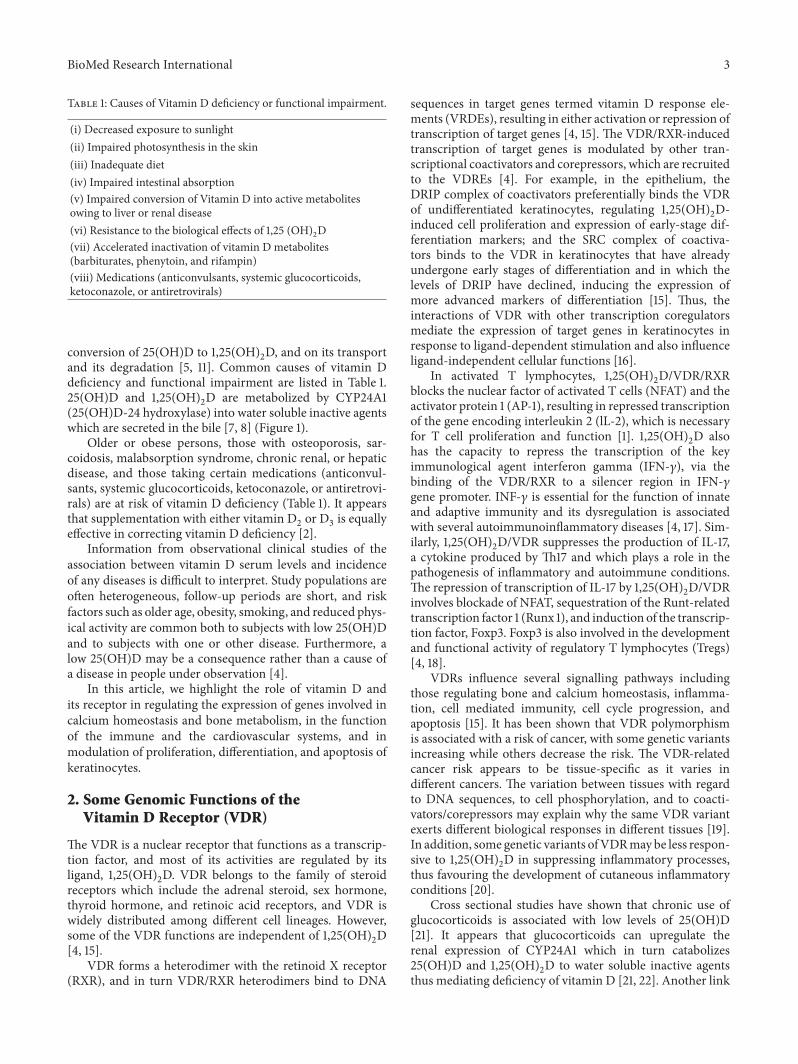
Table 1: Causes of Vitamin D deficiency or functional impairment.
(i) Decreased exposure to sunlight(ii) Impaired photosynthesis in the skin(iii) Inadequate diet(iv) Impaired intestinal absorption(v) Impaired conversion of Vitamin D into active metabolitesowing to liver or renal disease(vi) Resistance to the biological effects of 1,25 (OH)
2D
(vii) Accelerated inactivation of vitamin D metabolites(barbiturates, phenytoin, and rifampin)(viii) Medications (anticonvulsants, systemic glucocorticoids,ketoconazole, or antiretrovirals)
conversion of 25(OH)D to 1,25(OH)2D, and on its transport
and its degradation [5, 11]. Common causes of vitamin Ddeficiency and functional impairment are listed in Table 1.25(OH)D and 1,25(OH)
2D are metabolized by CYP24A1
(25(OH)D-24 hydroxylase) into water soluble inactive agentswhich are secreted in the bile [7, 8] (Figure 1).
Older or obese persons, those with osteoporosis, sar-coidosis, malabsorption syndrome, chronic renal, or hepaticdisease, and those taking certain medications (anticonvul-sants, systemic glucocorticoids, ketoconazole, or antiretrovi-rals) are at risk of vitamin D deficiency (Table 1). It appearsthat supplementation with either vitamin D
2or D
3is equally
effective in correcting vitamin D deficiency [2].Information from observational clinical studies of the
association between vitamin D serum levels and incidenceof any diseases is difficult to interpret. Study populations areoften heterogeneous, follow-up periods are short, and riskfactors such as older age, obesity, smoking, and reduced phys-ical activity are common both to subjects with low 25(OH)Dand to subjects with one or other disease. Furthermore, alow 25(OH)D may be a consequence rather than a cause ofa disease in people under observation [4].
In this article, we highlight the role of vitamin D andits receptor in regulating the expression of genes involved incalcium homeostasis and bone metabolism, in the functionof the immune and the cardiovascular systems, and inmodulation of proliferation, differentiation, and apoptosis ofkeratinocytes.
2. Some Genomic Functions of theVitamin D Receptor (VDR)
The VDR is a nuclear receptor that functions as a transcrip-tion factor, and most of its activities are regulated by itsligand, 1,25(OH)
2D. VDR belongs to the family of steroid
receptors which include the adrenal steroid, sex hormone,thyroid hormone, and retinoic acid receptors, and VDR iswidely distributed among different cell lineages. However,some of the VDR functions are independent of 1,25(OH)
2D
[4, 15].VDR forms a heterodimer with the retinoid X receptor
(RXR), and in turn VDR/RXR heterodimers bind to DNA
sequences in target genes termed vitamin D response ele-ments (VRDEs), resulting in either activation or repression oftranscription of target genes [4, 15]. The VDR/RXR-inducedtranscription of target genes is modulated by other tran-scriptional coactivators and corepressors, which are recruitedto the VDREs [4]. For example, in the epithelium, theDRIP complex of coactivators preferentially binds the VDRof undifferentiated keratinocytes, regulating 1,25(OH)
2D-
induced cell proliferation and expression of early-stage dif-ferentiation markers; and the SRC complex of coactiva-tors binds to the VDR in keratinocytes that have alreadyundergone early stages of differentiation and in which thelevels of DRIP have declined, inducing the expression ofmore advanced markers of differentiation [15]. Thus, theinteractions of VDR with other transcription coregulatorsmediate the expression of target genes in keratinocytes inresponse to ligand-dependent stimulation and also influenceligand-independent cellular functions [16].
In activated T lymphocytes, 1,25(OH)2D/VDR/RXR
blocks the nuclear factor of activated T cells (NFAT) and theactivator protein 1 (AP-1), resulting in repressed transcriptionof the gene encoding interleukin 2 (lL-2), which is necessaryfor T cell proliferation and function [1]. 1,25(OH)
2D also
has the capacity to repress the transcription of the keyimmunological agent interferon gamma (IFN-𝛾), via thebinding of the VDR/RXR to a silencer region in IFN-𝛾gene promoter. INF-𝛾 is essential for the function of innateand adaptive immunity and its dysregulation is associatedwith several autoimmunoinflammatory diseases [4, 17]. Sim-ilarly, 1,25(OH)
2D/VDR suppresses the production of IL-17,
a cytokine produced by Th17 and which plays a role in thepathogenesis of inflammatory and autoimmune conditions.The repression of transcription of IL-17 by 1,25(OH)
2D/VDR
involves blockade of NFAT, sequestration of the Runt-relatedtranscription factor 1 (Runx 1), and induction of the transcrip-tion factor, Foxp3. Foxp3 is also involved in the developmentand functional activity of regulatory T lymphocytes (Tregs)[4, 18].
VDRs influence several signalling pathways includingthose regulating bone and calcium homeostasis, inflamma-tion, cell mediated immunity, cell cycle progression, andapoptosis [15]. It has been shown that VDR polymorphismis associated with a risk of cancer, with some genetic variantsincreasing while others decrease the risk. The VDR-relatedcancer risk appears to be tissue-specific as it varies indifferent cancers. The variation between tissues with regardto DNA sequences, to cell phosphorylation, and to coacti-vators/corepressors may explain why the same VDR variantexerts different biological responses in different tissues [19].In addition, some genetic variants ofVDRmaybe less respon-sive to 1,25(OH)
2D in suppressing inflammatory processes,
thus favouring the development of cutaneous inflammatoryconditions [20].
Cross sectional studies have shown that chronic use ofglucocorticoids is associated with low levels of 25(OH)D[21]. It appears that glucocorticoids can upregulate therenal expression of CYP24A1 which in turn catabolizes25(OH)D and 1,25(OH)
2D to water soluble inactive agents
thus mediating deficiency of vitamin D [21, 22]. Another link

4 BioMed Research International
Circulation: Vitamin D
Liver: 25(OH)D
CYP2R1
CYP27B1
Parathyroid GlandsBone Intestine
Blood Calcium
Low serum calcium
increaserenal calciumreabsorption
PTH promotessynthesis of
PTH
Mobilization of
calcium from
bone
Absorption of calcium from
the intestine
Calcification
Mobilization ofcalcium from
boneCYP24A1
Activation Degradation
(b)
CYP27B1
(a)
PTHsynthesis
(c)
Lowcalcium levels
LowPTH levels
CYP24A1
+
+
FGF23FGF23
1,25(OH)2D
1,25 (OH)2D
1,25 (OH)2D
1,25 (OH)2D
Kidney: 1,25 (OH)2D
#;2+ (
0/42−#
; 2+(0/
4 2−
Figure 2: Schematic representation of the functions of vitamin D in bone and calcium homeostasis. Low serum calcium triggers the productionand secretion of parathyroid hormone (PTH) by the parathyroid glands, which in turn induces the synthesis of 1,25(OH)
2D in the kidney
by CYP27B1. FGF 23 can inhibit renal tubular reabsorption of phosphate and CYP27B1 activity and can stimulate CYP24A1 [7, 8, 26, 27].Vitamin D regulates calcium homeostasis by promotion of calcium absorption in the intestine, by reabsorption of calcium by the kidney, andby mobilization of calcium from the bone. 1,25(OH)
2D through a negative feedback loop regulates the synthesis of PTH and of CYP27B1 (a)
[4–6, 10]. Lower 25(OH)D is associated with increased PTH levels and lower bone density.This is owing to PTH/Vitamin D-induced increasein bone turnover, which results in increased bone resorption. Persistent vitamin D deficiency may result in osteopenia and osteomalacia [5].1,25(OH)
2D promotes the production of CYP24A1, the enzyme that degrades 1,25(OH)
2D (b) [1, 5], and low calcium and low PTH levels
inhibit the production of CYP24A1 (c) [4]. Thus levels of circulating 1,25(OH)2D are regulated, in part, by CYP27B1-mediated production
and by CYP24A1-mediated degradation [4].
between the use of glucocorticoids and vitamin D is that1,25(OH)
2D/VDR and glucocorticoids/glucocorticoid recep-
tor (GR) intracellular signalling pathways cross talk so thatincreased levels of vitamin D may enhance responsivenessof certain target cells to glucocorticoids [23–25]. Glucocor-ticoids with their cognate receptors translocate from the cellcytoplasm to the nucleus where they bind to glucocorticoidresponse element (GRE) to regulate gene transcription. AsVDR and GR share some coactivators, VDR may promoteindividual gene transcription induced by GR; and it hasbeen reported that vitamin D may upregulate the binding ofGR to GRE [23]. These may be the biological mechanismsby which vitamin D enhances cellular responsiveness toglucocorticoids.
The 1,25(OH)2D/VDR pathway, via transcriptional
repression, downregulates the expression of the CYP27B1(25(OH)D 1𝛼 hydroxylase) gene in the kidney, and the
expression of the gene encoding parathyroid hormone(PTH), resulting in decreased production of 1,25(OH)
2D
[1, 4]. Thus, through a negative feedback loop, 1,25(OH)2D
regulates both the synthesis of PTH and its own production(Figures 1 and 2).
3. 1,25(OH)2D/VDR in Calcium andBone Homeostasis
In the intestine, the 1,25(OH)2D induced-calcium absorp-
tion is mediated by the epithelial calcium channel TRPV6(transient potential vanilloid type 6) and in the kidney,1,25(OH)
2D mediates the active reabsorption of calcium
via TRP5, in a mechanism similar to intestinal calciumabsorption [4]. In the context of calcium homeostasis, thetranscription of the gene encoding TRPV6 is regulated byvitamin D/VDR/RXR pathway [4].

BioMed Research International 5
Available information regarding the direct effect ofvitamin D on bone is contentious. 1,25(OH)
2D has the
capacity directly or indirectly to regulate the proliferation,differentiation, andmaturation of osteoblasts and osteoclasts,bone resorption, and mineralization [9]. It appears that1,25(OH)
2D via VDR upregulates the expression of genes
encoding type 1 collagen, osteocalcin, and osteopontin thatdrive bone formation. In addition, 1,25(OH)
2D/VDR induces
the expression of RANK ligand by osteoblasts, which in turnmediates differentiation and increased activity of osteoclasts,thus ensuring bone turnover. This sequence of metabolicprocesses suggests that 1,25(OH)
2D has the capacity to
exert both anabolic and catabolic effects on bone [4, 5].On a background of a low serum calcium level, regardlessof the cause, 1,25(OH)
2D/VDR-induced bone resorption is
important inmaintaining calciumhomeostasis [4] (Figure 2).However, not all studies have shown any significant cor-
relation between levels of vitamin D and the bone turnovermarkers, bone-specific alkaline phosphatase, osteocalcin,procollagen type 1, N-terminal propeptide, and C-terminaltelopeptide; and vitamin D supplementation has not beenshown to have any effect on these markers in persons withlow 25(OH)D, but with normal levels of calcium [9].
Thus, it appears that if the calcium balance is negative,1,25(OH)
2D/VDR directly mediates bone resorption with
the mobilization of calcium from bone into the circulation,thus correcting the negative balance, but the effect of the1,25(OH)
2D/VDR on osteogenic cells in the face of a nor-
mal calcium balance is not fully understood. Under suchcircumstances in the context of normal calcium serum levels,1,25(OH)
2D regulates bone homeostasis indirectly through
its effects on intestinal calcium absorption and renal calciumreabsorption, which maintain normal serum calcium levels[4].
Fibroblast growth factor 23 (FGF 23) is a protein pri-marily produced by osteocytes and osteoblasts that regu-lates the metabolism of vitamin D and the homeostasis ofsystemic phosphate. Once it is in the circulation, it caninhibit renal tubular reabsorption of phosphate, resultingin hypophosphatemia. Furthermore, by inhibiting CYP27B1which converts 25(OH)D to 1,25(OH)
2D in the kidney, and
by stimulating CYP24A1 which catabolizes 25(OH)D and1,25(OH)
2D, FGF 23 suppresses the circulating 25(OH)D
[7, 8, 26, 27]. Conversely, 1,25(OH)2D directly increases
bone levels of FGF 23, probably via the upstream vitamin Dresponse element in the FGF 23 gene promoter, in osteoblastsand osteocytes [26]. Thus, this bone-kidney feedback loopregulates production of FGF 23 in bone and of 1,25(OH)
2D
in the kidney, playing a role in the modulation of boneremodelling [27] (Figure 2).
1,25(OH)2D/VDR signalling in osteoblasts regulates
expression of genes mediating differentiation of osteoblastprecursor and bone mineralization, including the roles ofalkaline phosphatase, osteocalcin, and osteopontin. Fur-thermore, 1,25(OH)
2D/VDR signalling has the capacity to
influence other signalling pathways involved in physiologicalactivities of osteoblasts including transforming growth factor𝛽 (TGF-𝛽), insulin growth factor 1 (IGF-1), bone mor-phogenic protein, interferon, PTH, hepatocyte growth factor
(HGF), epidermal growth factor (EGF), and the Wnt/𝛽-catenin signalling pathways [5, 28].
Osteoblasts express the CYP27B1 gene with the capacityto synthesize 1,25(OH)
2D, but the biological significance
of this is not clear. Once locally produced by osteoblasts,via a negative feedback loop, 1,25(OH)
2D itself inhibits
CYP27B1 expression, thus regulating its own production inbone (Figure 2); and it appears that interferon-𝛽 (INF-𝛽)and interleukin-1 (IL-1) also play roles in the regulation ofosteoblastic CYP27B1 [28].
The expression of specific osteogenic genes by osteoblastsin response to 1,25(OH)
2D/VDR activation is dependent
on the stage of differentiation of the cell and is influencedby immune cell-derived cytokines in the microenvironment[28].
However, as long as calcium levels are normal, bonemetabolism is not affected by inhibition of 1,25(OH)
2D/VDR
signalling in osteoblasts. This is probably owing to thecomplex adaptive system which determines both osteoblastfunction and bone metabolism, in which deficiency of1,25(OH)
2D/VDR signalling is compensated by other factors
in the system.
4. The Role of 1,25(OH)2D/VDR in Cancer
There is a strong biological rationale for the role of vitamin Ddeficiency in increasing cancer risk and for the use of vitaminD or its bioactive analogues for cancer prevention and treat-ment. VDR is expressed inmost cancerous tissues; and resultsof in vivo animal studies and in vitro cell culture studies showthat 1,25(OH)
2D inhibits cell proliferation, angiogenesis,
invasion and promotes differentiation and apoptosis. In can-cer cells, 1,25(OH)
2D/VDRactivates cyclin-dependent kinase
inhibitors (e.g., p21, p27), inhibits mitogenic growth factorssuch IGF-1 andEGF, and promotes the activity of TGF-𝛽, thusinhibiting cell proliferation and cancer growth [4, 15].
1,25(OH)2D/VDR signalling has the capacity to downreg-
ulate cyclooxygenase-2, prostaglandin, and NF-kB pathways,thus inhibiting tumour-associated inflammation, to suppressantiapoptotic proteins (e.g., Bcl2) and to activate proapop-totic proteins (e.g., Bax, RAK). Acting together, all these cansuppress cancer growth [4, 15].
The transcription factor NF-kB regulates the expressionof genes controlling inflammation, cell proliferation, apop-tosis, invasion, and metastasis [29]. It has been reportedthat 1,25(OH)
2D/VDR can suppress NF-kB activities, result-
ing in decreased expression of proinflammatory cytokinesand inhibition of cell proliferation and of cancer-associatedinflammation, with slowing down cancer growth [1, 4].
Another potential anticancer mechanism of 1,25(OH)2D
is through the interaction with the transforming growthfactor-𝛽 (TGF-𝛽) family of secreted proteins. Some of theseproteins promote differentiation and apoptosis of epithe-lial cells but inhibit their proliferation. As outlined above,1,25(OH)
2D has the capacity to activate TGF-𝛽 signalling
pathways, thus it may promote the tumour-suppressor activ-ities [1].
The canonical Wnt signalling pathway is associated withtranslocation of 𝛽-catenin from adherence junctions to

6 BioMed Research International
the nucleus with subsequent activation of the 𝛽 catenin/Tcell factor (TCF) transcriptional complex, and with thefunctional loss of E-cadherin. In cancer, activated Wnt/𝛽-catenin signalling activates genes which promote increasedproliferation, invasion, and metastasis of cancer cells [30].The dysregulated Wnt/𝛽-catenin signalling is observed earlyin carcinogenesis and is maintained throughout the courseof cancer progression, but Wnt-associated activation of theepithelial-mesenchymal transition (EMT) cellular geneticprogram that results in cancer cell detachment, motility,and migration with subsequent invasion and metastasis is arelatively late event in tumourigenesis [30].
1,25(OH)2D/VDR enhances E-cadherin expression and
nuclear export of 𝛽-catenin and induces the dickkopf 1,an extracellular inhibitor of Wnt signalling, all of whichresult in the inhibition of cancer growth [1, 4]. Furthermore,in cancerous keratinocytes, 1,25(OH)
2D/VDR may alter the
expression of genes encoding cytoskeletal proteins, affectingcytoskeletal organization and function resulting in increasedcell adhesion and decreased cell motility, even further sup-pressing cancer growth [4].
Most cancers express not only the VDR but also CYP27B1and CYP24A1 thus allowing cancer cells to regulate locally1,25(OH)
2D levels, and it has been reported that cancers
expressing CYP27B1 tend to be well differentiated whilecancers which do not express CYP27B1 tend to be poorlydifferentiated. Cancers which express CYP24A1 tend to bemore aggressive than those which do not [4].
Evidence from in vitro studies support the concept of anantiproliferative effect of 1,25(OH)
2D; but results of in vivo
studies on laboratory animals show that the anticarcinogenicactivity of 1,25(OH)
2D mainly affects cancer progression
rather than cancer initiation. In humans, the results ofobservational clinical studies of associations between vita-min D supplementation and cancer incidence do not showdiminution in cancer incidence in response to vitamin Dsupplementation. However, one needs to be aware that mostof these studies had objectives unrelated to cancer incidenceand that while carcinogenesis is a slow biopathologicalprocess the duration of vitamin D supplementation and thefollow-up period have been relatively short [4]. Differencesin ethnicity, environmental living conditions, and life-stylebetween populations in various geographic locations mostlikely influence the results of studies investigating the asso-ciation between the level of circulating vitamin D and cancerrisk [4].
5. Vitamin D in Relation to theImmune System
Many cells of both the innate and the adaptive immunesystems express the VDR, and some of these cells also havethe capacity to express the CYP27B1 and to produce thebiologically active 1,25(OH)
2D.
The molecular patterns of certain microorganisms acti-vate the molecular pattern recognition receptor Toll-likereceptor (TLR) 1/2 of innate immunocytes (e.g., monocytes,macrophages, and keratinocytes), resulting in upregulation of
the expression of CYP27B1 and VDR, with the productionof 1,25(OH)
2D. In some of these cells 1,25(OH)
2D/VDR
signalling induces the expression of the genes encoding theantibacterial agents cathelicidin and𝛽-defenisin. 1,25(OH)
2D
may also mediate antimicrobial effector responses inde-pendently of TLR receptors. Thus, it appears that vita-min 1,25(OH)
2D/VDR signalling, in combination with, or
independently of TLR signalling, fortifies the antibacterialresponses of innate immune cells [17, 18, 31].
1,25(OH)2D can suppress the maturation of dedicated
antigen presenting dendritic cells, resulting in reduction oftheir capacity to present antigens to naıve T lymphocytes inregional lymph nodes. This may lead to decreased antigen-specific T cell activation and proliferation, and possibly to Tcell anergy [4, 18, 31].
It has been found that in laboratory animals1,25(OH)
2D/VDR signalling downregulates chronic T
cell mediated hyperactive immunoinflammatory reactionsthrough inhibition of T cell proliferation, and decreasingproduction of IL-2 and INF-𝛾 by Th1 cells, and of IL17by Th17 cells. 1,25(OH)
2D/VDR-induced increase in the
production of IL-4 by Th2 cells and in the activity of Tregulatory cells (Treg) further moderates autoimmune T cellresponses [4, 31, 32].
Despite these moieties of evidence derived from labo-ratory studies, in humans, the physiological significance of1,25(OH)
2D activity in the context of the immune system is
not fully understood, and its role in the pathogenesis and inthe clinical course of immunoinflammatory diseases is notclear.However, in general, as it is evident that in humans thereis no causal association between vitamin D deficiency andthe incidence or severity of immunoinflammatory diseases,augmenting treatment with vitamin D or its biologicallyactive analogues does not improve the outcome of treatment[4, 17]. Asmedical science is not infallible, treatment practicesthat according to evidence-based experiments have not beenfound to be completely effective for the general populationmay still be successful for a subset of patients [33].
6. 1,25(OH)2D/VDR Signalling and theCardiovascular System
VDR andCYP27B1 are observed in cardiacmyocytes, cardiacfibroblasts, vascular smooth muscle, and endothelial cells,and it appears that activated 1,25(OH)
2D/VDRpathwaysmay
well play some role in cardiovascular function [18].Experimental studies in laboratory animals have shown
that limitation of 1,25(OH)2D/VDR signalling can cause
increased renin/angiotensin activity with hypertension andcardiac hypertrophy, reduction in the bioavailability of thevasodilator nitric oxide with consequent impaired bloodvessel relaxation, endothelial cell dysfunction, upregulationof proinflammatory cytokines, and increased proliferationand migration of vascular smooth muscle cells [4, 34, 35].
In humans, results of observational studies on the asso-ciation between low 25(OH)D and increased cardiovascularrisk and of interventional clinical randomized controlledstudies into the effects of supplementation of vitamin D orits bioactive analogues on cardiovascular risk have yielded

BioMed Research International 7
inconsistent results. Therefore, while it is evident that1,25(OH)
2D/VDR signalling has the capacity to modulate
several pathways involved in maintaining the homeostasis ofendothelial cells and cardiac function, its biological signif-icance under physiological conditions is not clear, and anyrole vitamin D deficiency may have in the pathogenesis ofcardiovascular disease is not scientifically proven [4, 35].
7. Effects of Vitamin D on Skin Biology
The role of vitamin D as a regulator of skin physiology iscomplex. Vitamin D and its receptor are expressed by ker-atinocytes of the basal and spinous layers of the epithelium,and 1,25(OH)
2D/VDR signalling influences proliferation,
differentiation, and apoptosis of keratinocytes, and cutaneousimmune responses [36].
In the basal cell layer of the epithelium, 1,25(OH)2D
regulates proliferation of basal cell keratinocytes, while inthe spinous cell layer it upregulates expression of agents ofdifferentiation mediating the synthesis of keratins (K1, K10),involucrin, transglutaminase, loricrin, and filaggrin [36]. Invitro, a high level of 1,25(OH)
2D, via VDR-mediated genomic
mechanisms, inhibits proliferation of keratinocytes [14] andvia nongenomic mechanisms promotes differentiation ofkeratinocytes by increasing levels of intracellular calcium[36].
More mature keratinocytes in the upper layers of theepithelium produce proteins, lipids, and glucosylceramideswhich provide a physical epidermal barrier protecting theunderlying tissue from penetration by infectious and toxicagents and also provide a permeability barrier [15, 20, 37].
Calcium signalling in keratinocytes is induced by thecalcium sensing receptor (Casr), a plasma membrane-boundmember of the G protein coupled receptor family, andcross-talk between the 1,25(OH)
2D/VDR signalling and
the Ca/Casr signalling pathways regulates the formationof plasma membrane E-cadherin/catenin complexes whichform adherent junctions [37]. Synergism between Ca/Casrand 1,25(OH)
2D/VDR signalling inhibits the translocation
of 𝛽-catenin from the plasma membrane E-cadherin/catenincomplex, thus diminishing the nuclear transcriptional activ-ity of 𝛽-catenin [37].
Physiologically, the hedgehog intracellular signallingpathway regulates the differentiation and proliferation ofkeratinocyte stem cells. Functional upregulation of this path-way is an early genetic event in the pathogenesis of cuta-neous basal cell carcinoma conferring upon affected basalkeratinocytes enhanced proliferative capacity thus increasingrisk of basal cell carcinoma. Furthermore, reduced functionalactivity of genes regulating repair of DNA damage causedby UVB radiation is also implicated in the pathogenesisof cutaneous basal cell carcinoma [38]. In this context as1,25(OH)
2D/VDR signalling has been shown to inhibit the
hedgehog signalling pathway and to upregulate the activityof DNA nucleotide excision repair enzymes, 1,25(OH)
2D has
the potential to reduce the risk of basal cell carcinoma [14].In the context of cutaneous squamous cell carcinoma,
results of in vitro studies show that 1,25(OH)2D/VDR sig-
nalling inhibits cell proliferation by inducing cell cycle arrest,
by triggering apoptosis, by downregulating DNA synthesis,and by promoting repair of UVB radiation-induced DNAdamage [14, 20].
8. Vitamin D and Oral Health
It appears that 1,25(OH)2D/VDR plays a role in maintaining
the homeostasis of oral epithelium and of oral immunity.VDR is expressed by oral keratinocytes in which it has aligand-independent function in restraining proliferation oforal keratinocytes; and 1,25(OH)
2D/VDR signalling has an
even stronger inhibitory effect on proliferation of oral ker-atinocytes. Both in vitro and in vivo studies show that vitaminD deficiency brings about an increase in proliferation of oralkeratinocytes, but without any morphological or histologicalalterations. Although it is clear that vitamin D deficiency initself is not sufficient to cause precancerous transformation,synergistically with other genetic or environmental factors itmay increase the risk of oral squamous carcinoma [39].
Since melanin diminishes the cutaneous production ofvitamin D, black persons experienced vitamin D deficiencymore frequently than white persons and this account in partfor the higher prevalence of oral squamous cell carcinoma inblack people than in white people [40].
The anti-inflammatory, antimicrobial, and immunomod-ulating effects of the 1,25(OH)
2D/VDR pathway most proba-
bly play roles inmaintaining the homeostasis of oral tissues ingeneral, thus providing some protection against the develop-ment of bacterial plaque-induced periodontal diseases.Thereis evidence that vitamin D deficiency or VDR polymorphismare associated with increased risk of chronic periodontitis[41]. Thus supplementing the conventional treatment ofchronic periodontitis with administration of biologicallyactive vitamin D may have a beneficial effect [31, 42, 43].
9. Conclusion
1,25(OH)2D is regulated by a complex system of feedback
mechanismsmediated by enzymes, hormones, and receptors.1,25(OH)
2D/VDR signalling has an essential role in calcium
and bone homeostasis and has the capacity to regulate variouscellular responses of the immune and cardiovascular systems,and to regulate differentiation, proliferation, and apoptosis ofnormal and malignant keratinocytes.
Conflicts of Interest
The authors declare that there are no conflicts of interestregarding the publication of this paper.
References
[1] V. Dimitrov, R. Salehi-Tabar, B.-S. An, and J. H. White, “Non-classical mechanisms of transcriptional regulation by the vita-min D receptor: Insights into calcium homeostasis, immunesystem regulation and cancer chemoprevention,”The Journal ofSteroid Biochemistry and Molecular Biology, vol. 144, pp. 74–80,2014.
[2] J. Y. Tang, T. Fu, C. Lau, D. H. Oh, D. D. Bikle, and M. M.Asgari, “Vitamin D in cutaneous carcinogenesis,” Journal of the

8 BioMed Research International
American Academy of Dermatology, vol. 67, no. 5, pp. 803.e1–803.e12, 2012.
[3] D. A. Searing and D. Y. M. Leung, “Vitamin D in atopicdermatitis, asthma and allergic diseases,” Immunology andAllergy Clinics of North America, vol. 30, no. 3, pp. 397–409,2010.
[4] S. Christakos, P. Dhawan, A. Verstuyf, L. Verlinden, and G.Carmeliet, “Vitamin D: metabolism, molecular mechanism ofaction, and pleiotropic effects,” Physiological Reviews, vol. 96,no. 1, pp. 365–408, 2016.
[5] F. R. Bringhurst, M. B. Demay, S. M. Krane, and H. M.Kronenberg, “Bone and mineral metabolism in health anddisease,” in Harrison’s principles of internal medicine, D. L.Kasper, A. S. Fauci, S. L. Hauser, D. L. Longo, L. J. Jameson, andJ. Loscalzo, Eds., vol. 2, pp. 2454–2465, McGraw Hill, 2015.
[6] I. E. Kochwvar, C. R. Taylor, and J. Krutmann, “Fundamen-tals of cutaneous photobiology and photoimmunology,” in inFitzpatrick’s Dermatology in General Medicine, K. Wolff, L. A.Goldsmith, S. I. Katz et al., Eds., vol. 2, pp. 1031–1048, McGraw-Hill, New York, NY, USA, 2012.
[7] M. F. Holick, “Vitamin D deficiency,”The New England Journalof Medicine, vol. 357, no. 3, pp. 266–281, 2007.
[8] A. Hossein-Nezhad and M. F. Holick, “Vitamin D for health: aglobal perspective,” Mayo Clinic Proceedings, vol. 88, no. 7, pp.720–755, 2013.
[9] V. Schwetz, C. Trummer, M. Pandis et al., “Effects of vitaminD supplementation on bone turnover markers: A randomizedcontrolled trial,” Nutrients, vol. 9, no. 5, article no. 432, 2017.
[10] M. Gallieni, M. Cozzolino, G. Fallabrino, S. Pasho, L. Olivi, andD. Brancaccio, “Vitamin D: Physiology and pathophysiology,”The International Journal of Artificial Organs, vol. 32, no. 2, pp.87–94, 2009.
[11] A. Corcoran, S. Nadkarni, K. Yasuda et al., “Biological evalua-tion of double point modified analogues of 1,25-dihydroxyv-itamin D2 as potential anti-leukemic agents,” InternationalJournal of Molecular Sciences, vol. 17, no. 2, article no. 91, 2016.
[12] M. F. Holick, “Sunlight and vitamin D for bone health and pre-vention of autoimmune diseases, cancers, and cardiovasculardisease,” American Journal of Clinical Nutrition, vol. 80, no. 6,pp. 1678S–1688S, 2004.
[13] B. Dawson-Hughes, R. P. Heaney, M. F. Holick, P. Lips, P. J.Meunier, and R. Vieth, “Estimates of optimal vitamin D status,”Osteoporosis International, vol. 16, no. 7, pp. 713–716, 2005.
[14] J. Y. Tang, T. Fu, C. Lau, D. H. Oh, D. D. Bikle, andM.M. Asgari,“Vitamin D in cutaneous carcinogenesis: Part II,” Journal of theAmericanAcademy ofDermatology, vol. 67, no. 5, pp. 817–817.e11,2012.
[15] C. J. Rosen, J. S. Adams, D. D. Bikle et al., “The nonskeletaleffects of vitamin D: an endocrine society scientific statement,”Endocrine Reviews, vol. 33, no. 3, pp. 456–492, 2012.
[16] S. Rieger, H. Zhao, P. Martin, K. Abe, and T. S. Lisse, “The roleof nuclear hormone receptors in cutaneous wound repair,” CellBiochemistry & Function, vol. 33, no. 1, pp. 1–13, 2015.
[17] R. Wei and S. Christakos, “Mechanisms underlying the regula-tion of innate and adaptive immunity by vitamin D,” Nutrients,vol. 7, no. 10, pp. 8251–8260, 2015.
[18] S. Christakos, M. Hewison, D. G. Gardner et al., “Vitamin D:beyond bone,” Annals of the New York Academy of Sciences, vol.1287, no. 1, pp. 45–58, 2013.
[19] Y. Xu, B. He, Y. Pan et al., “Systematic review and meta-analysison vitamin D receptor polymorphisms and cancer risk,” TumorBiology, vol. 35, no. 5, pp. 4153–4169, 2014.
[20] A. Piotrowska, J. Wierzbicka, andM. A. Zmijewski, “Vitamin Din the skin physiology and pathology,”Acta Biochimica Polonica,vol. 63, no. 1, pp. 17–29, 2016.
[21] A. L. Skversky, J. Kumar, M. K. Abramowitz, F. J. Kaskel, andM. L. Melamed, “Association of glucocorticoid use and low 25-hydroxyvitamin D levels: results from the National Health andNutrition Examination Survey (NHANES): 2001–2006,” TheJournal of Clinical Endocrinology & Metabolism, vol. 96, no. 12,pp. 3838–3845, 2011.
[22] A. A. Hidalgo, D. L. Trump, and C. S. Johnson, “Glucocorticoidregulation of the vitamin D receptor,” The Journal of SteroidBiochemistry and Molecular Biology, vol. 121, no. 1-2, pp. 372–375, 2010.
[23] Y. Zhang, D. Y. M. Leung, and E. Goleva, “Anti-inflammatoryand corticosteroid-enhancing actions of vitamin D in mono-cytes of patients with steroid-resistant and those with steroid-sensitive asthma,”The Journal of Allergy and Clinical Immunol-ogy, vol. 133, no. 6, pp. 1744–e1, 2014.
[24] N. Kalra and F. T. Ishmael, “Cross-talk between vitamin D,estrogen and corticosteroids in glucocorticoid resistant asthma,”OA Inflammation, vol. 2, no. 1, pp. 2–12, 2014.
[25] Y. Zhang, D. Y. M. Leung, and E. Goleva, “Vitamin D enhancesglucocorticoid action in human monocytes: Involvement ofgranulocyte-macrophage colony-stimulating factor and medi-ator complex subunit 14,” The Journal of Biological Chemistry,vol. 288, no. 20, pp. 14544–14553, 2013.
[26] K. Wesseling-Perry, “FGF-23 in bone biology,” PediatricNephrology, vol. 25, no. 4, pp. 603–608, 2010.
[27] N. C. W. Mackenzie, D. Zhu, E. M. Milne et al., “Altered bonedevelopment and an increase in FGF-23 expression in Enpp1 -/-mice,” PLoS ONE, vol. 7, no. 2, Article ID e32177, 2012.
[28] J. van de Peppel and J. P. T. M. van Leeuwen, “Vitamin D andgene networks in human osteoblasts,” Frontiers in Physiology,vol. 5, article 137, Article ID Article 137, 2014.
[29] K. Dekate, “Inflammation in the context of oral canc,” OralOncology, vol. 49, no. 9, pp. 887–892, 2014.
[30] L. Feller, B. Kramer, and J. Lemmer, “Pathobiology of cancermetastasis: A short account,” Cancer Cell International, vol. 12,article no. 24, 2012.
[31] Y. Amano, K. Komiyama, and M. Makishima, “Vitamin D andperiodontal disease.,” Journal of oral science, vol. 51, no. 1, pp.11–20, 2009.
[32] M. T. Cantorna, L. Snyder, Y.-D. Lin, and L. Yang, “Vitamin Dand 1,25(OH)
2D regulation of T cells,” Nutrients, vol. 7, no. 4,
pp. 3011–3021, 2015.[33] G. Klein, “Evidence-based medicine,” in This Idea Must Die, J.
Brockman, Ed., pp. 220–222, HarperCollins, 2015.[34] A. R.Menezes, M. C. Lamb, C. J. Lavie, and J. J. DiNicolantonio,
“VitaminDand atherosclerosis,”CurrentOpinion inCardiology,vol. 29, no. 6, pp. 571–577, 2014.
[35] K. Kienreich, M. Grubler, and A. Tomaschitz, “arterial hyper-tension and cerebrovascular disease,” Indian J Med Res, vol. 137,no. 4, pp. 669–679, 2013.
[36] L. Barrea, M. C. Savanelli, C. Di Somma et al., “Vitamin Dand its role in psoriasis: An overview of the dermatologist andnutritionist,” Reviews in Endocrine andMetabolic Disorders, vol.18, no. 2, pp. 195–205, 2017.
[37] D. D. Bikle, Y. Jiang, T. Nguyen, Y. Oda, and C.-L. Tu, “Dis-ruption of vitamin D and calcium signaling in keratinocytespredisposes to skin cancer,” Frontiers in Physiology, vol. 7, articleno. 296, 2016.

BioMed Research International 9
[38] L. Feller, R. A. G. Khammissa, B. Kramer, M. Altini, and J.Lemmer, “Basal cell carcinoma, squamous cell carcinoma andmelanoma of the head and face,”Head & Face Medicine, vol. 12,no. 1, p. 11, 2016.
[39] A. Yuan and S.-B. Woo, “Adverse drug events in the oral cavity,”Oral Surgery, OralMedicine, Oral Pathology, Oral Radiology, andEndodontology, vol. 119, no. 1, pp. 35–47, 2014.
[40] L. Lipworth, M. Rossi, J. K. McLaughlin et al., “Dietary vitaminD and cancers of the oral cavity and esophagus,” Annals ofOncology, vol. 20, no. 9, pp. 1576–1581, 2009.
[41] T. Dietrich and R. I. Garcia, “Associations between periodontaldisease and systemic disease: Evaluating the strength of theevidence,” Journal of Periodontology, vol. 76, no. 11, pp. 2175–2184, 2005.
[42] D. Miley, M. N. Garcia, C. F. Hildebolt et al., “Cross-Sectionalstudy of vitamin d and calcium supplementation effects onchronic periodontitis,” Journal of Periodontology, vol. 80, no. 9,pp. 1433–1439, 2009.
[43] J. D. Bashutski, R. M. Eber, J. S. Kinney et al., “The impact ofvitamin D status on periodontal surgery outcomes,” Journal ofDental Research, vol. 90, no. 8, pp. 1007–1012, 2011.

Hindawiwww.hindawi.com
International Journal of
Volume 2018
Zoology
Hindawiwww.hindawi.com Volume 2018
Anatomy Research International
PeptidesInternational Journal of
Hindawiwww.hindawi.com Volume 2018
Hindawiwww.hindawi.com Volume 2018
Journal of Parasitology Research
GenomicsInternational Journal of
Hindawiwww.hindawi.com Volume 2018
Hindawi Publishing Corporation http://www.hindawi.com Volume 2013Hindawiwww.hindawi.com
The Scientific World Journal
Volume 2018
Hindawiwww.hindawi.com Volume 2018
BioinformaticsAdvances in
Marine BiologyJournal of
Hindawiwww.hindawi.com Volume 2018
Hindawiwww.hindawi.com Volume 2018
Neuroscience Journal
Hindawiwww.hindawi.com Volume 2018
BioMed Research International
Cell BiologyInternational Journal of
Hindawiwww.hindawi.com Volume 2018
Hindawiwww.hindawi.com Volume 2018
Biochemistry Research International
ArchaeaHindawiwww.hindawi.com Volume 2018
Hindawiwww.hindawi.com Volume 2018
Genetics Research International
Hindawiwww.hindawi.com Volume 2018
Advances in
Virolog y Stem Cells International
Hindawiwww.hindawi.com Volume 2018
Hindawiwww.hindawi.com Volume 2018
Enzyme Research
Hindawiwww.hindawi.com Volume 2018
International Journal of
MicrobiologyHindawiwww.hindawi.com
Nucleic AcidsJournal of
Volume 2018
Submit your manuscripts atwww.hindawi.com