review - university of malaya · pdf fileinto the bio macromolecules, ... botany,...
TRANSCRIPT
Asian Pacific Journal of Cancer Prevention, Vol 14, 2013 5553
DOI:http://dx.doi.org/10.7314/APJCP.2013.14.10.5553Herbal Remedies for Combating Irradiation: A Green Anti-irradiation Approach
Asian Pac J Cancer Prev, 14 (10), 5553-5565
Introduction
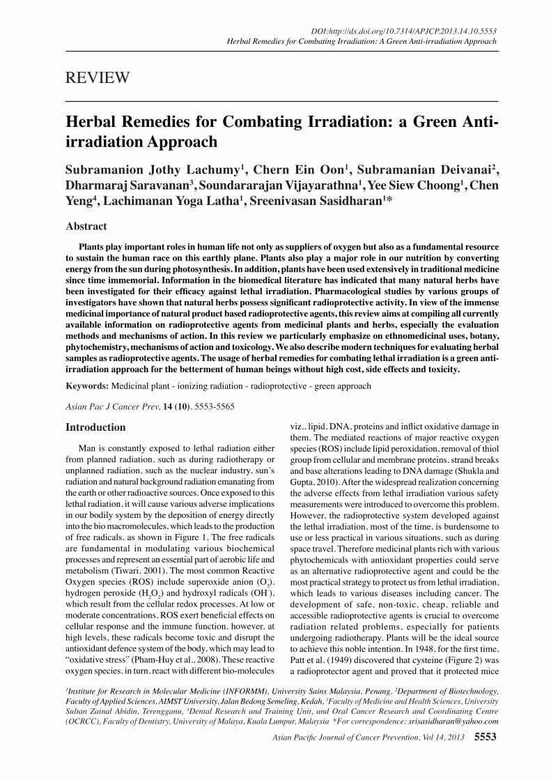
Man is constantly exposed to lethal radiation either from planned radiation, such as during radiotherapy or unplanned radiation, such as the nuclear industry, sun’s radiation and natural background radiation emanating from the earth or other radioactive sources. Once exposed to this lethal radiation, it will cause various adverse implications in our bodily system by the deposition of energy directly into the bio macromolecules, which leads to the production of free radicals, as shown in Figure 1. The free radicals are fundamental in modulating various biochemical processes and represent an essential part of aerobic life and metabolism (Tiwari, 2001). The most common Reactive Oxygen species (ROS) include superoxide anion (O2
-), hydrogen peroxide (H2O2) and hydroxyl radicals (OH-), which result from the cellular redox processes. At low or moderate concentrations, ROS exert beneficial effects on cellular response and the immune function, however, at high levels, these radicals become toxic and disrupt the antioxidant defence system of the body, which may lead to “oxidative stress” (Pham-Huy et al., 2008). These reactive oxygen species, in turn, react with different bio-molecules
1Institute for Research in Molecular Medicine (INFORMM), University Sains Malaysia, Penang, 2Department of Biotechnology, Faculty of Applied Sciences, AIMST University, Jalan Bedong Semeling, Kedah, 3Faculty of Medicine and Health Sciences, University Sultan Zainal Abidin, Terengganu, 4Dental Research and Training Unit, and Oral Cancer Research and Coordinating Centre (OCRCC), Faculty of Dentistry, University of Malaya, Kuala Lumpur, Malaysia *For correspondence: [email protected]
Abstract
Plants play important roles in human life not only as suppliers of oxygen but also as a fundamental resource to sustain the human race on this earthly plane. Plants also play a major role in our nutrition by converting energy from the sun during photosynthesis. In addition, plants have been used extensively in traditional medicine since time immemorial. Information in the biomedical literature has indicated that many natural herbs have been investigated for their efficacy against lethal irradiation. Pharmacological studies by various groups of investigators have shown that natural herbs possess significant radioprotective activity. In view of the immense medicinal importance of natural product based radioprotective agents, this review aims at compiling all currently available information on radioprotective agents from medicinal plants and herbs, especially the evaluation methods and mechanisms of action. In this review we particularly emphasize on ethnomedicinal uses, botany, phytochemistry, mechanisms of action and toxicology. We also describe modern techniques for evaluating herbal samples as radioprotective agents. The usage of herbal remedies for combating lethal irradiation is a green anti-irradiation approach for the betterment of human beings without high cost, side effects and toxicity. Keywords: Medicinal plant - ionizing radiation - radioprotective - green approach
REVIEW
Herbal Remedies for Combating Irradiation: a Green Anti-irradiation ApproachSubramanion Jothy Lachumy1, Chern Ein Oon1, Subramanian Deivanai2, Dharmaraj Saravanan3, Soundararajan Vijayarathna1, Yee Siew Choong1, Chen Yeng4, Lachimanan Yoga Latha1, Sreenivasan Sasidharan1*
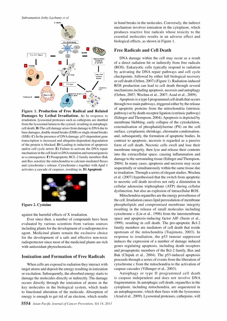
viz., lipid, DNA, proteins and inflict oxidative damage in them. The mediated reactions of major reactive oxygen species (ROS) include lipid peroxidation, removal of thiol group from cellular and membrane proteins, strand breaks and base alterations leading to DNA damage (Shukla and Gupta, 2010). After the widespread realization concerning the adverse effects from lethal irradiation various safety measurements were introduced to overcome this problem. However, the radioprotective system developed against the lethal irradiation, most of the time, is burdensome to use or less practical in various situations, such as during space travel. Therefore medicinal plants rich with various phytochemicals with antioxidant properties could serve as an alternative radioprotective agent and could be the most practical strategy to protect us from lethal irradiation, which leads to various diseases including cancer. The development of safe, non-toxic, cheap, reliable and accessible radioprotective agents is crucial to overcome radiation related problems, especially for patients undergoing radiotherapy. Plants will be the ideal source to achieve this noble intention. In 1948, for the first time, Patt et al. (1949) discovered that cysteine (Figure 2) was a radioprotector agent and proved that it protected mice
Subramanion Jothy Lachumy et al
Asian Pacific Journal of Cancer Prevention, Vol 14, 20135554
against the harmful effects of X irradiation. Ever since then, a number of compounds have been evaluated by various scientists from various sources including plants for the development of a radioprotective agent. Medicinal plants remain the exclusive choice for the development of a safe and effective non-toxic radioprotector since most of the medicinal plants are rich with antioxidant phytochemicals.
Ionization and Formation of Free Radicals When cells are exposed to radiation they interact with target atoms and deposit the energy resulting in ionization or excitation. Subsequently, the absorbed energy starts to damage the molecules directly or indirectly. The damage occurs directly through the ionization of atoms in the key molecules in the biological system, which leads to functional alteration of the molecule. Absorption of energy is enough to get rid of an electron, which results
in bond breaks in the molecules. Conversely, the indirect mechanism involves ionization in the cytoplasm, which produces reactive free radicals whose toxicity to the essential molecules results in an adverse effect and biological effects, as shown in Figure 1.
Free Radicals and Cell Death DNA damage within the cell may occur as a result of a direct radiation hit or indirectly from free radicals (ROS). Eukaryotic cells typically respond to radiation by activating the DNA repair pathways and cell cycle checkpoints, followed by either full biological recovery or cell death (Ozben, 2007) (Figure 1). Radiation-induced ROS production can lead to cell death through several mechanisms including apoptosis, necrosis and autophagy (Ozben, 2007; Wochna et al., 2007; Azad et al., 2009). Apoptosis is a type I programmed cell death that occurs through two main pathways, triggered either by the release of apoptotic proteins from the mitochondria (intrinsic pathway) or by death-receptor ligation (extrinsic pathway) (Edinger and Thompson, 2004). Apoptosis is depicted by membrane blebbing, early collapse of the cytoskeleton, externalisation of phosphatidylserine (PS) on the cell surface, cytoplasmic shrinkage, chromatin condensation, and, subsequently, the formation of apoptotic bodies. In contrast to apoptosis, necrosis is regarded as a passive form of cell death. Necrotic cells swell and lose their membrane integrity, then lyse and release their contents into the extracellular space, causing inflammation and damage to the surrounding tissue (Edinger and Thompson, 2004). In many cases, apoptosis and necrosis may occur sequentially or simultaneously within the same tissue due to irradiation. Through a series of elegant studies, Wochna et al. (2007) hypothesised that the switch from apoptotic to necrotic cell death involves not only a diminution in cellular adenosine triphosphate (ATP) during cellular dysfunction, but also an explosion of intracellular ROS. Mitochondria organelles are the energy powerhouse of the cell. Irradiation causes lipid peroxidation of membrane phospholipids and compromised membrane integrity resulting in the release of small molecules including cytochrome c (Liu et al., 1996) from the intermembrane space and apoptosis-inducing factor AIF (Susin et al., 1999), resulting in cell death. The pro-apoptotic Bcl-2 family members are mediators of cell death that reside upstream of the mitochondria (Tsujimoto, 2003). In response to irradiation, the p53 tumour suppressor induces the expression of a number of damage induced genes regulating apoptosis, including death receptors and proapoptotic members of the Bcl-2 family, Bax and Bak (Chipuk et al., 2004). The p53-induced apoptosis proceeds through a series of events from the liberation of cytochrome c from the mitochondria to the activation of caspase cascades (Villunger et al., 2003). Autophagy or type II programmed cell death is caspase independent and does not involve DNA fragmentation. In autophagic cell death, organelles in the cytoplasm, including mitochondria, are sequestered in an autophagosome, which then fuses with the lysosomes (Azad et al., 2009). Lysosomal proteases, cathepsins, will
Figure 2. Cysteine
Figure 1. Production of Free Radical and Related Damages by Lethal Irradiation. A) In response to irradiation, lysosomal proteases such as cathepsins are shuttled from the lysosomal lumen to the cytosol, resulting in autophagic cell death; B) The cell damage arises from damage to DNA due to base damages, double strand breaks (DSB) or single strand breaks (SSB); C) In the presence of DNA damage, p53-dependent gene transcription is increased and ubiquitin-dependent degradation of the protein is blocked; D) Leading to induction of apoptosis and/or cell cycle arrest; E) Failure to activate the DNA repair mechanism in the cell leads to DNA mutation and tumourigenesis as a consequence; F) Proapoptotic BCL-2 family members Bak and Bax sensitize the mitochondria to calcium-mediated fluxes and cytochrome c release. Cytochrome c together with Apaf-1 activates a cascade of caspases, resulting in; D) Apoptosis
Asian Pacific Journal of Cancer Prevention, Vol 14, 2013 5555
DOI:http://dx.doi.org/10.7314/APJCP.2013.14.10.5553Herbal Remedies for Combating Irradiation: A Green Anti-irradiation Approach
be shuttled from the lysosomal lumen to the cytoplasm in response to ROS. The hydroxide produced, as in mitochondria by ROS, diffuses into lipofuscin-loaded lysosomes, and the hydroxide causes damage to the lysosomal membranes, which causes the leak of lysosomal enzymes. The lysosomal enzymes permeabilise the mitochondrial membranes, resulting in the release of cytochrome c, the apoptosis-inducing factor (AIF), and the second mitochondria-derived activator of caspase (Smac)/direct inhibitor of apoptosis protein binding protein with low pI (DIABLO), hence triggering cell death (Ghavami et al., 2010; Szumiel, 2011).
Free Radicals and Cancer However, the irradiated cells that escape cell death may undergo mutation, which creates an error in the DNA blueprint leading to altered gene expression and protein modification; peptide bond cleavage and cross linking, for example, may affect protein localization, interactions and enzyme activity. Although ROS-mediated DNA damage may enable cells to function partially and proliferate, they eventually develop into cancer, especially if the regulation of the tumour suppressor genes is impaired (Wu, 2006). The high levels of ROS in cancer cells can further contribute to oxidative stress, which may further stimulate tumour growth, invasion, angiogenesis and metastasis (Wu, 2006; Girdhani et al., 2013). The level of ROS production and antioxidant signalling appear to be altered in malignant cells, contributing to cancer progression. However, the results from different studies have been paradoxical, for instance, superoxide dismutase (SOD) expression has been shown to decrease cancer cell proliferation and tumorigenicity in vitro (Oberley, 2005), albeit its expression was found to be associated with bad prognosis in patients with gastric cancer (Kim et al., 2002).
Radioprotection Mechanisms by Plant Extract or Compounds Numerous investigations on radioprotection mechanisms have been carried out in several biological systems and the following radioprotection mechanisms have been proposed from these studies: free radical scavenger, repair by hydrogen donation to target molecules, formation of mixed sulphides, delaying of cellular division and induction of hypoxia in the tissue (Varanda and Tavares, 1998). The mechanism of free radical scavenger suggests that medicinal plants will donate electrons to the free radicals and form a stable compound incapable of reacting with other cellular components. This mechanism prevents the free radicals from reacting with the vital cell components. Another mechanism that has been proposed is the repair by hydrogen donation. If a R-H molecule is converted into a radical R by exposure to radiation, the antioxidant plant extract or compound can donate a hydrogen atom to this radical, restoring it to its original state (Biaglow, 1987), which is not vulnerable to the vital components of our bodily system. In addition, the mechanism of the formation
of mixed sulphides suggests aminothiols, which involves radioprotector binding to cellular components. According to this proposed mechanism, the sulphydryl compound of medicinal plants form mixed disulphides with sulphydryl compounds of cellular proteins. Once the free radicals generated by irradiation attack the disulphides, the sulphur atoms will be reduced and the other sulphur atom will be oxidized (Varanda and Tavares, 1998). This mechanism prevents the free radicals from reacting with the vital cell components because if the sulphur atom of the protein is reduced by the free radicals and the sulphur atom of the protective agent is oxidized, the protein is not damaged. Delaying of cellular division and granting additional time for repairing DNA damage caused by irradiation has been considered a potentially important mechanism in radioprotection activity. For this type of mechanism Brown (Brown, 1967) proposed that the sulphydryl compounds of the radioprotective agents will bind to the cellular DNA and inhibit its replication and provide additional time for repair of the damaged DNA. Protection by the induction of hypoxia in the tissue has also been considered a potentially important mechanism in radioprotection activity. Oxidation of the radioprotective agents uses enough oxygen to reduce its tension, and it has already been revealed that hypoxia is radioprotective. Moreover, the induction of hypoxia in tissue in certain conditions may contribute to radioprotection. Nevertheless, other mechanisms might be involved, since some compounds exhibit radioprotective activity without altering the oxygen tension on the tissue (Varanda and Tavares, 1998). There is evidence of the existence of more than one radioprotective mechanism of a certain compound, and that one of the compounds might be more or less important, depending on the irradiated system and on the specific radiation conditions (Prasad, 1982).
Plant as Anti-radiation Sources Traditional usage of medicinal plant as radioprotective agent For eons, plants and plant products have been infused in human life, as palatable and remedial sources. Traditional healers exploited plants to treat various maladies long before the discovery of drugs (Cragg et al., 1997). What’s more, the conventional plant preparations are also demonstrated to be non-toxic or less-toxic, considering their derivation from natural resources. Gingko biloba is one of the world’s ancient trees and is believed to have survived an atomic bomb explosion dropped on Hiroshima on 6 August 1945 by the Americans (Anonymous, 2013a). The surviving trees were found near the blast centre and appeared to sprout without major deformations. The observation substantiates the plant’s amazing resistance to mutagen agents like radiation (Pickstone, 2010). On a different occasion, the Buddhist monks took delight in tending to these trees by preserving them near the pagodas in China’s Imperial Gardens and on sacred grounds to ward off fire. G biloba is also denoted as a symbol of longevity. Although folklore does not directly imply that plants impart a radioprotective effect, much evidence
Subramanion Jothy Lachumy et al
Asian Pacific Journal of Cancer Prevention, Vol 14, 20135556
has been found of their incorporation in ceremonies and rituals in which specific plants are utilized. The Tulsi or Ocimum sanctum, for example, is worshipped along with milk, yogurt, honey and Ganga (river) water, which are consumed by the devotees at the end of the ritual (McGuire, 2012). The ancient Indian legend states that this Queen of Herbs came as an incarnation of the Hindu goddess Tulsi and is favoured by the Lord Vishnu, Krishna and Ram (Miller and Miller, 2003). A plant with radioprotective effect can also be identified with the presence of other properties, such as anti-inflammatory, antioxidant, antimicrobial and immune modulatory (Jagetia, 2007). Likewise, Tulsi, within the confinement of Ayurveda, was used to regulate fever, relieve coughs and flu, and mobilize mucus in bronchitis and asthma. The leaves especially were used to treat tuberculosis and ringworm of the skin. The tulsi oil is rich in vitamin C, carotene, calcium and phosphorus and is also believed to possess other properties including that of antibacterial, antifungal and antiviral (Anonymous, 2013b). Radiation interacts and distresses the atoms that compose the cells. The affected atoms will subsequently form free radicals that disrupt molecules, cells, tissues and organs that eventuate to the detriment of the organism (USNRC Technical Training Center, 2013). Since free radicals are responsible for inducing radiation-damage, the radioprotective property of Panax ginseng is associated either directly or indirectly with its free radical scavenging capability (Lee et al., 2005). Ginseng refers to the root and
the rhizome of Panax ginseng C.A. Meyer (Araliaceae), which have been conventionally utilised by the Chinese for more than 200 years. The Chinese believe that ginseng is a reservoir with a range of pharmacological roles, such as restorative, tonic, nootropic, anti-aging and more (Lee et al., 2005).
Medicinal plant with radioprotective effects Naturally occurring herbs constitute a wide variety of antioxidants, such as alpha carotene, ascorbic acids, flavones, flavanones, flavanols, stilbenoids, anthocyanins, phenolic acids, etc., which are reported to have a broad spectrum of radiation absorption properties (Bajpai et al., 2005; Ashawat et al., 2006; Nichols and Katiyar, 2010; Vaid and Katiyar, 2010). In addition, it has been shown that these phytoconstituents have a synergistic photo-protective effect and can be used as sunscreen to protect cellular damage of the skin from radiation light exposure (Afaq et al., 2003; Campos et al., 2006). Recently, the radioprotective effect of phytochemicals has been gaining popularity in skin care and attention has been focused in developing topical formulations, which can be used as complementary as well as alternate medicine to heal and rejuvenate skin from various disorders (Svobova et al., 2003; Griffiths et al., 2005; Kapoor et al., 2009; Saraf and Kaur, 2010). Some of the medicinal plants with radioprotective properties – antioxidant, anti-inflammatory and immunomodulatory – are listed in Table 1.
Table 1. Plant with Radioprotective Activity or Antioxidant Activity
Plant spe-cies
Scientific names Component Activity Reference
Tomato Solanum Lycopersicum
Carotenoids –lycopenes antioxidant Griffiths et al., 2005; Saraf and Kaur, 2010; Ravichandran et al., 2005
Carrot Daucus carota β-carotene antioxidant Griffiths et al., 2005; Svobova et al., 2003
Papaya Carica papaya L-ascorbic acid Antioxidant and photoprotective Vile, 1997
Orange Citrus sinensis L-ascorbic acid antioxidant Cimino et al., 2007
Lemon Citrus limon L-ascorbic acid antioxidant Apak et al., 2007
Mango Mangifera indica L-ascorbic acid antioxidant with anti-inflammatory and immunomodulatory activities.
Song et al., 2013
Pomegranate Punica granatum ascorbic acid antioxidant Kumar et al., 2009
Celery Apium graveolens Flavones – 5,7,4’-trihydroxystilibine
antioxidant and ROS scavenger Griffiths et al., 2005; Svobova et al., 2003
Red clover Trifolium pratense Isoflavone – Genistein Inhibit UV induced peroxidase production
Widyarini et al., 2001
Soybean Glycine max Anthocyanin Photo protective of UV radiation Tsoyi et al., 2008
Green tea Camellia sinensis Flavanol – Epigallocatechin gallate
antioxidant and ROS scavenger Katiyar et al., 2000; 2001; Katiyar and Elmets, 2001; Higdon, 2007; Li et al., 2009; Sharangi, 2009; Kaur and Saraf, 2011b
Milk thistle Silybum marianum Stilbenoid- Silybin,silibinin,silidianin, Silychristin
anti-inflammatory and immunomodulatory
Katiyar, 2002; Fguyer et al., 2003; Vaid and Katiyar, 2010
Grape Vitis vinifera Stilbenoid- Resveratrol, Flavanol -proanthocyanidin
antioxidant and ROS scavenger Afaq et al., 2003; Saraf and Kaur, 2010; Aziz et al., 2005; Mantena and Katiyar, 2006
Apple Malus domestica Flavanoid-Quercetin antioxidant Erden Inal et al., 2001; Korac and Khambholja, 2001
Boldo Peumus boldus Quercetin, Flavanol-catechin; aporphine
antioxidant and anti-inflammatory Peter et al., 2006; Russo et al., 2011
Turmeric Curcuma longa Phenolic -curcumin anti-inflammatory, antiproliferative, Photoprotective effect
Saraf and Kaur, 2010; Garcia Bores and Avila, 2008
Aloe vera Aloe barbadensis antraquinones Cellular repair West and Zhu, 2003
Rhubarb Rheum rhaponticum stilbene antioxidant and ROS scavenger Silveira et al., 2013
Asian Pacific Journal of Cancer Prevention, Vol 14, 2013 5557
DOI:http://dx.doi.org/10.7314/APJCP.2013.14.10.5553Herbal Remedies for Combating Irradiation: A Green Anti-irradiation Approach
0
25.0
50.0
75.0
100.0
New
ly d
iagn
osed
with
out
trea
tmen
t
New
ly d
iagn
osed
with
tre
atm
ent
Pers
iste
nce
or r
ecur
renc
e
Rem
issi
on
Non
e
Chem
othe
rapy
Radi
othe
rapy
Conc
urre
nt c
hem
orad
iatio
n
10.3
0
12.8
30.025.0
20.310.16.3
51.7
75.051.1
30.031.354.2
46.856.3
27.625.033.130.031.3
23.738.0
31.3
0
25.0
50.0
75.0
100.0
New
ly d
iagn
osed
with
out
trea
tmen
t
New
ly d
iagn
osed
with
tre
atm
ent
Pers
iste
nce
or r
ecur
renc
e
Rem
issi
on
Non
e
Chem
othe
rapy
Radi
othe
rapy
Conc
urre
nt c
hem
orad
iatio
n
10.3
0
12.8
30.025.0
20.310.16.3
51.7
75.051.1
30.031.354.2
46.856.3
27.625.033.130.031.3
23.738.0
31.3
Antiradiation compounds Antiradiation compounds are studied by in vitro and in vivo tests that assess some of these aspects. Assay of free radicals and antioxidant assay of pharmacological agents are suggested as a good means for evaluating the radioprotective potential (Jagetia, 2007). The polyphenolic compounds, especially flavonoids, ubiquitously present in plants, have been reported to possess various beneficial biological properties, most of which are attributed to antioxidant activity. It is not surprising that radioprotective potential has been reported for extracts of herbs containing flavonoids, as well as for individually isolated flavonoids. The radioprotective effect of two extracts of Caesalpinia digyna and the isolated compound bergenin were compared using in vitro methods by Singh et al. (Singh et al., 2009). The in vitro approach compared the protective action against the damaging effect of protein carbonylation in bovine serum albumin, lipid peroxidation in liposomes, and DNA breakage in pBR322 plasmid. The study showed that the flavonoid, bergenin, from the plant is equally potent in inhibiting DNA damage as the extracts, albeit the extracts were more potent in protecting the proteins and lipids. The pBR322 model was also used in assessing the protective effect of pure compounds isolated from Phyllanthus amarus (Londhe et al., 2009). The flavonoids, quercetin 3-O-glucoside followed by rutin, offered the greatest protection on DNA as seen by the decrease in the nicked circular form of plasmid. However, the ellagitannins namely amariin, 1-galloyl-2,3-dehydrohexahydroxydiphenyl (DHHDP)-glucose, repandusinic acid, geraniin, corilagin, phyllanthusiin D were also effective. The protective effects of these compounds on protein and lipids damage by radiation were assessed by using rat liver mitochondria. The compounds, rutin and repandusinic acid offered maximum protection against lipid damage whereas protection against carbonyl formation in proteins was highest in rutin, phyllanthusiin D, geraniin and quercetin 3-O-glucoside. The effects of flavonoids have also been studied by using in vivo techniques. For example, various doses of preparation containing twelve flavonoids (FAC) from seeds of Astragalus complanatus protected mice from radiation damage (Qi et al., 2011). Basically, FAC increased the survival rate of irradiated mice and had a protective effect on haematopoietic tissue and the immune system. The alkaline comet assay, which involves single cell electrophoresis was able to show the protective effect against DNA damage in mouse liver cells by the FAC. Studies on radioprotection have also taken advantage of the availability of synthetic drugs that have been used clinically in humans. The protective effect of troxerution, a flavonoid derivative used for treating venous disorders, was also ascertained by using the comet assay. In this study, the method assessed the protection against DNA damage in mice blood, bone marrow and tumour cells (Maurya et al., 2004).
Modern technique for evaluation of radioprotective activity of medicinal plants In this section we analyse and compare the various reliable methods available for the study of radioprotective
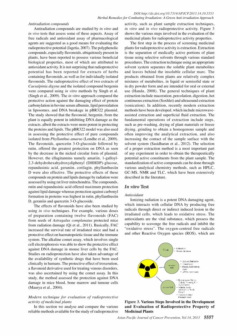
activity, such as plant sample extraction techniques, in vitro and in vivo radioprotective activity. Figure 3 shows the various steps involved in the evaluation of the medicinal plants for radioprotective activity properties. The first step in the process of screening medicinal plants for radioprotective activity is extraction. Extraction is the separation of medically active portions of plant tissue using selective solvents through various standard procedures. The extraction technique using an appropriate solvent system separates the soluble plant metabolites and leaves behind the insoluble cellular marc. The products obtained from plants are relatively complex mixtures of metabolites, in liquid or semisolid state or in dry powder form and are intended for oral or external use (Handa, 2008). The general techniques of plant extraction include maceration, percolation, digestion, hot continuous extraction (Soxhlet) and ultrasound extraction (sonication). In addition, recently modern extraction methods have been developed, which include microwave-assisted extraction and superficial fluid extraction. The fundamental operations of extraction include steps, such as pre-washing, drying of plant materials or freeze drying, grinding to obtain a homogenous sample and often improving the analytical extraction, and also increasing the contact of the sample surface with the solvent system (Sasidharan et al., 2012). The selection of a proper extraction method is a most important part of any experiment in order to obtain the therapeutically potential active constituents from the plant sample. The standardization of active compounds can be done through various analytical chemistry methods, such as HPLC, GC-MS, NMR and TLC, which have been extensively described in the literature.
In vitro TestAntioxidant
Ionizing radiation is a potent DNA damaging agent, which interacts with cellular DNA by producing free radicals through direct or indirect induced lesion in the irradiated cells, which leads to oxidative stress. The antioxidants are the vital substance, which possess the capability to scavenge the free radicals and inhibit the “oxidative stress”. The oxygen-centred free radicals and other Reactive Oxygen species (ROS), which are
Figure 3. Various Steps Involved in the Development and Evaluation of Radioprotective Property of Medicinal Plants
Subramanion Jothy Lachumy et al
Asian Pacific Journal of Cancer Prevention, Vol 14, 20135558
continuously produced, become toxic, and directly disrupt the antioxidant defence system of the body causing oxidative stress. Basically, oxidative stress is the cascade initiated by the free radicals to obtain stability through electron pairing with biological macromolecules, such as proteins, DNA and lipids, in healthy cells and causes damage to the cell structure. The most common ROS include superoxide anion (O2
-), hydrogen peroxide (H2O2), and hydroxyl radicals (OH-), which result from the cellular redox process. The antioxidants were found to be an important substance in protecting DNA from various ROS mediated damage and useful in the treatment of diseases where oxygen free-radical production is particularly implicated (Rajkumar et al., 2010). Although a number of synthetic antioxidants are available, their safety issues are highly debated, thus generating the search for substitute materials from natural resources. Therefore, recently, there has been an upsurge of interest in the therapeutic potential of medicinal plants with antioxidant properties in reducing such free radical induced damage rather than looking for synthetic ones. The medicinal plant extracts are reported to provide DNA protection and their protective nature is attributed to the presence of antioxidant compounds (Attaguile et al., 2000). Generally, antioxidant compounds like phenolic acids, polyphenols and flavonoids scavenge free radicals, such as peroxide and hydroperoxide of lipid hydroxyl, and thus inhibit the oxidative mechanisms that lead to degenerative diseases. Epidemiologists have observed that a diet rich in polyphenolic compounds may result in a positive health effect attributed to its antioxidants properties (Hertog et al., 1993; Frankel et al., 1996).
However, it is very appealing to researchers to have a convenient method for the quick quantification of the antioxidant effectiveness of the medical plants. The multifaceted aspects of antioxidant capacity assays can roughly be classified into two types: i) hydrogen atom transfer (HAT) reactions and; ii) electron transfer (ET). The HAT-based assays apply a competitive reaction scheme, in which the antioxidant and substrate compete for peroxyl radicals that are generated through the decomposition of azo compounds. These assays include oxygen radical absorbance capacity (ORAC), total radical trapping antioxidant parameter (TRAP) and anti-lipid peroxidation. The ET-based assays are able to measure the capacity of an antioxidant in the reduction of an oxidant, which changes colour when reduced. The degree of colour change correlates with the concentration of antioxidant in the reaction mixture (Huang et al., 2005). ET-based assays include the total phenolic content by Folin-Ciocalteu reagent, Trolox equivalence antioxidant assay (TEAC), ferric ion reducing power (FRAP), total antioxidant potential capacity using copper (II) complex and DPPH assay. However, there are other antioxidant assays for measuring the scavenging capacity of biologically relevant antioxidants such as singlet oxygen, superoxide anion, peroxynitrite, and hydroxyl radicals. Table 1 shows the list of antioxidant assays. Generally, the antioxidant potential of plant extracts was quantified based on the inhibition concentration at 50% (IC50) obtained from the assay.
Percentage of inhibition (IC50)=[(A0-A1)/A0]×100
Where A0 is the absorbance of the control and A1 was the absorbance in the presence of the extract or positive control.
Comet assay The primary target of ionizing radiation damage is
the DNA, which modulates the possible mechanisms and alters the cellular response in irradiated cells. In parallel, there is always a need for the development of new rapid and more sensitive method for DNA damage evaluation. Recently, the comet assay (single cell gel electrophoresis) was developed, which is a new method of choice because of its simplicity, quickness, and sensitivity for the detection of various DNA lesions including DNA double strand breaks, base damage and apoptotic nuclei induced by a variety of genotoxic agents (Collins, 2004). The concept of comet assay has been introduced with minor modification of the microgel electrophoresis method to measure DNA single-strand breaks, which is relatively determined based on the amount of DNA strands that migrate out the individual agarose-embedded cells (Ostling and Johanson, 1984; Singh et al., 1988).
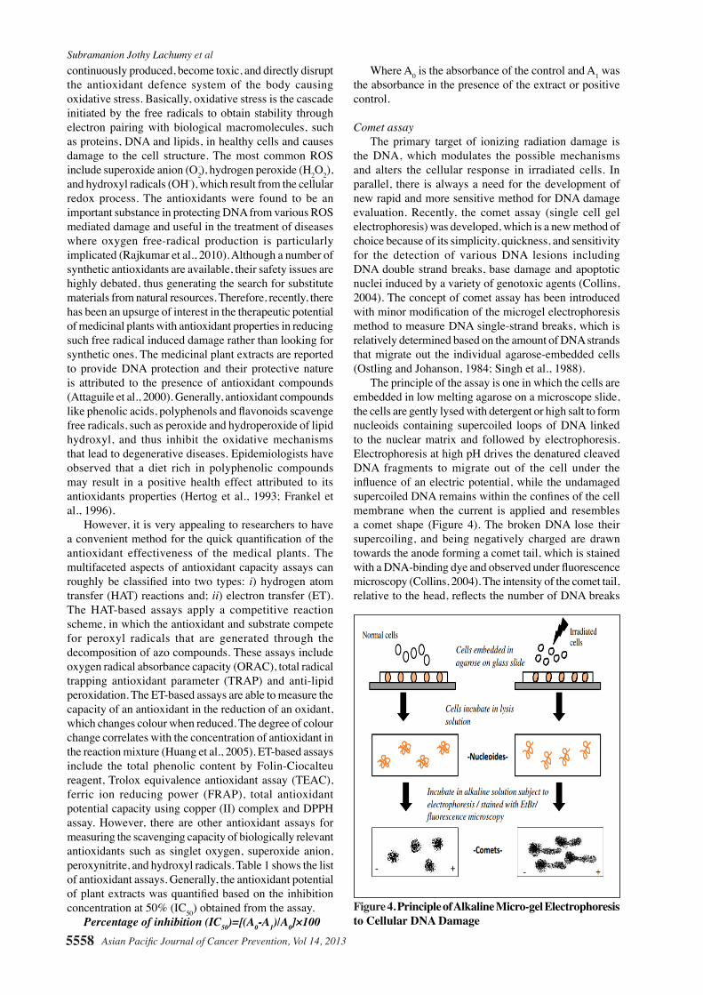
The principle of the assay is one in which the cells are embedded in low melting agarose on a microscope slide, the cells are gently lysed with detergent or high salt to form nucleoids containing supercoiled loops of DNA linked to the nuclear matrix and followed by electrophoresis. Electrophoresis at high pH drives the denatured cleaved DNA fragments to migrate out of the cell under the influence of an electric potential, while the undamaged supercoiled DNA remains within the confines of the cell membrane when the current is applied and resembles a comet shape (Figure 4). The broken DNA lose their supercoiling, and being negatively charged are drawn towards the anode forming a comet tail, which is stained with a DNA-binding dye and observed under fluorescence microscopy (Collins, 2004). The intensity of the comet tail, relative to the head, reflects the number of DNA breaks
Figure 4. Principle of Alkaline Micro-gel Electrophoresis to Cellular DNA Damage
Asian Pacific Journal of Cancer Prevention, Vol 14, 2013 5559
DOI:http://dx.doi.org/10.7314/APJCP.2013.14.10.5553Herbal Remedies for Combating Irradiation: A Green Anti-irradiation Approach
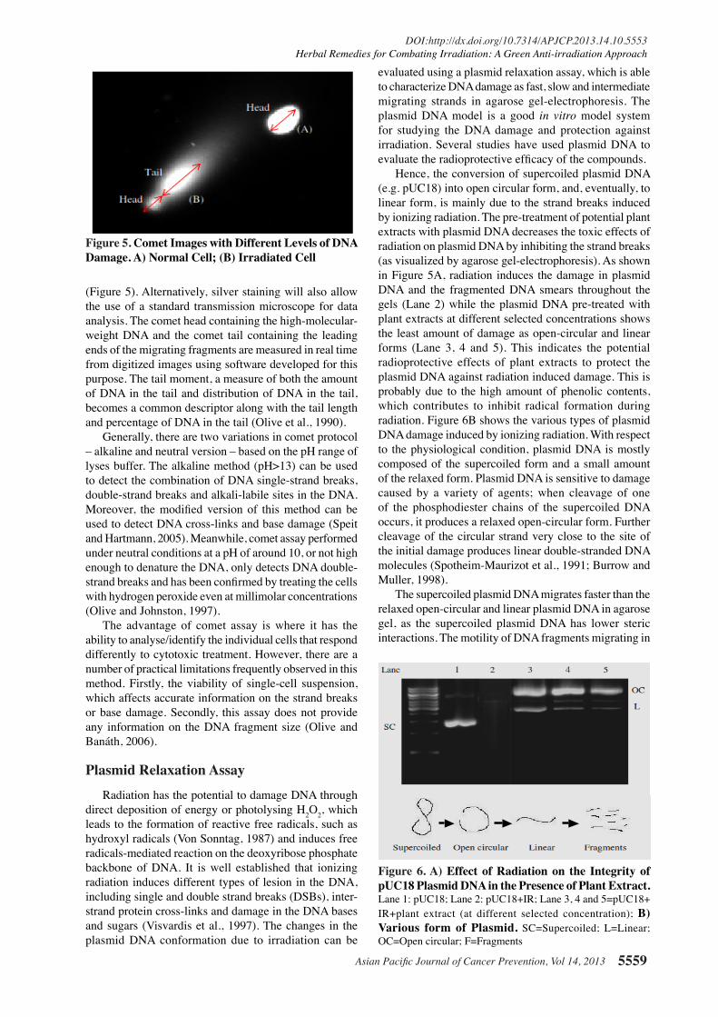
(Figure 5). Alternatively, silver staining will also allow the use of a standard transmission microscope for data analysis. The comet head containing the high-molecular-weight DNA and the comet tail containing the leading ends of the migrating fragments are measured in real time from digitized images using software developed for this purpose. The tail moment, a measure of both the amount of DNA in the tail and distribution of DNA in the tail, becomes a common descriptor along with the tail length and percentage of DNA in the tail (Olive et al., 1990).
Generally, there are two variations in comet protocol – alkaline and neutral version – based on the pH range of lyses buffer. The alkaline method (pH>13) can be used to detect the combination of DNA single-strand breaks, double-strand breaks and alkali-labile sites in the DNA. Moreover, the modified version of this method can be used to detect DNA cross-links and base damage (Speit and Hartmann, 2005). Meanwhile, comet assay performed under neutral conditions at a pH of around 10, or not high enough to denature the DNA, only detects DNA double-strand breaks and has been confirmed by treating the cells with hydrogen peroxide even at millimolar concentrations (Olive and Johnston, 1997).
The advantage of comet assay is where it has the ability to analyse/identify the individual cells that respond differently to cytotoxic treatment. However, there are a number of practical limitations frequently observed in this method. Firstly, the viability of single-cell suspension, which affects accurate information on the strand breaks or base damage. Secondly, this assay does not provide any information on the DNA fragment size (Olive and Banáth, 2006).
Plasmid Relaxation AssayRadiation has the potential to damage DNA through
direct deposition of energy or photolysing H2O2, which leads to the formation of reactive free radicals, such as hydroxyl radicals (Von Sonntag, 1987) and induces free radicals-mediated reaction on the deoxyribose phosphate backbone of DNA. It is well established that ionizing radiation induces different types of lesion in the DNA, including single and double strand breaks (DSBs), inter-strand protein cross-links and damage in the DNA bases and sugars (Visvardis et al., 1997). The changes in the plasmid DNA conformation due to irradiation can be
evaluated using a plasmid relaxation assay, which is able to characterize DNA damage as fast, slow and intermediate migrating strands in agarose gel-electrophoresis. The plasmid DNA model is a good in vitro model system for studying the DNA damage and protection against irradiation. Several studies have used plasmid DNA to evaluate the radioprotective efficacy of the compounds.
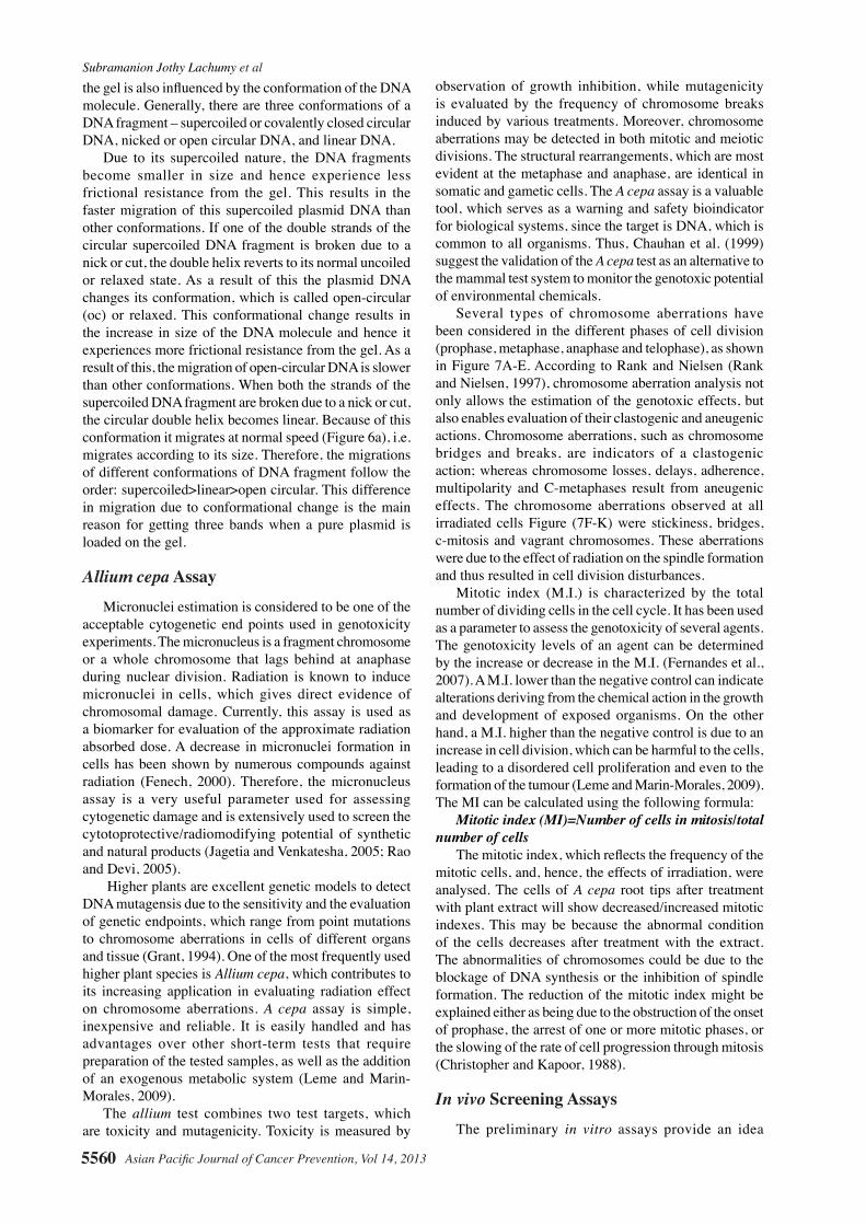
Hence, the conversion of supercoiled plasmid DNA (e.g. pUC18) into open circular form, and, eventually, to linear form, is mainly due to the strand breaks induced by ionizing radiation. The pre-treatment of potential plant extracts with plasmid DNA decreases the toxic effects of radiation on plasmid DNA by inhibiting the strand breaks (as visualized by agarose gel-electrophoresis). As shown in Figure 5A, radiation induces the damage in plasmid DNA and the fragmented DNA smears throughout the gels (Lane 2) while the plasmid DNA pre-treated with plant extracts at different selected concentrations shows the least amount of damage as open-circular and linear forms (Lane 3, 4 and 5). This indicates the potential radioprotective effects of plant extracts to protect the plasmid DNA against radiation induced damage. This is probably due to the high amount of phenolic contents, which contributes to inhibit radical formation during radiation. Figure 6B shows the various types of plasmid DNA damage induced by ionizing radiation. With respect to the physiological condition, plasmid DNA is mostly composed of the supercoiled form and a small amount of the relaxed form. Plasmid DNA is sensitive to damage caused by a variety of agents; when cleavage of one of the phosphodiester chains of the supercoiled DNA occurs, it produces a relaxed open-circular form. Further cleavage of the circular strand very close to the site of the initial damage produces linear double-stranded DNA molecules (Spotheim-Maurizot et al., 1991; Burrow and Muller, 1998).
The supercoiled plasmid DNA migrates faster than the relaxed open-circular and linear plasmid DNA in agarose gel, as the supercoiled plasmid DNA has lower steric interactions. The motility of DNA fragments migrating in
Figure 5. Comet Images with Different Levels of DNA Damage. A) Normal Cell; (B) Irradiated Cell
Figure 6. A) Effect of Radiation on the Integrity of pUC18 Plasmid DNA in the Presence of Plant Extract. Lane 1: pUC18; Lane 2: pUC18+IR; Lane 3, 4 and 5=pUC18+ IR+plant extract (at different selected concentration); B) Various form of Plasmid. SC=Supercoiled; L=Linear; OC=Open circular; F=Fragments
Subramanion Jothy Lachumy et al
Asian Pacific Journal of Cancer Prevention, Vol 14, 20135560
the gel is also influenced by the conformation of the DNA molecule. Generally, there are three conformations of a DNA fragment – supercoiled or covalently closed circular DNA, nicked or open circular DNA, and linear DNA.
Due to its supercoiled nature, the DNA fragments become smaller in size and hence experience less frictional resistance from the gel. This results in the faster migration of this supercoiled plasmid DNA than other conformations. If one of the double strands of the circular supercoiled DNA fragment is broken due to a nick or cut, the double helix reverts to its normal uncoiled or relaxed state. As a result of this the plasmid DNA changes its conformation, which is called open-circular (oc) or relaxed. This conformational change results in the increase in size of the DNA molecule and hence it experiences more frictional resistance from the gel. As a result of this, the migration of open-circular DNA is slower than other conformations. When both the strands of the supercoiled DNA fragment are broken due to a nick or cut, the circular double helix becomes linear. Because of this conformation it migrates at normal speed (Figure 6a), i.e. migrates according to its size. Therefore, the migrations of different conformations of DNA fragment follow the order: supercoiled>linear>open circular. This difference in migration due to conformational change is the main reason for getting three bands when a pure plasmid is loaded on the gel.
Allium cepa AssayMicronuclei estimation is considered to be one of the
acceptable cytogenetic end points used in genotoxicity experiments. The micronucleus is a fragment chromosome or a whole chromosome that lags behind at anaphase during nuclear division. Radiation is known to induce micronuclei in cells, which gives direct evidence of chromosomal damage. Currently, this assay is used as a biomarker for evaluation of the approximate radiation absorbed dose. A decrease in micronuclei formation in cells has been shown by numerous compounds against radiation (Fenech, 2000). Therefore, the micronucleus assay is a very useful parameter used for assessing cytogenetic damage and is extensively used to screen the cytotoprotective/radiomodifying potential of synthetic and natural products (Jagetia and Venkatesha, 2005; Rao and Devi, 2005).
Higher plants are excellent genetic models to detect DNA mutagensis due to the sensitivity and the evaluation of genetic endpoints, which range from point mutations to chromosome aberrations in cells of different organs and tissue (Grant, 1994). One of the most frequently used higher plant species is Allium cepa, which contributes to its increasing application in evaluating radiation effect on chromosome aberrations. A cepa assay is simple, inexpensive and reliable. It is easily handled and has advantages over other short-term tests that require preparation of the tested samples, as well as the addition of an exogenous metabolic system (Leme and Marin-Morales, 2009).
The allium test combines two test targets, which are toxicity and mutagenicity. Toxicity is measured by
observation of growth inhibition, while mutagenicity is evaluated by the frequency of chromosome breaks induced by various treatments. Moreover, chromosome aberrations may be detected in both mitotic and meiotic divisions. The structural rearrangements, which are most evident at the metaphase and anaphase, are identical in somatic and gametic cells. The A cepa assay is a valuable tool, which serves as a warning and safety bioindicator for biological systems, since the target is DNA, which is common to all organisms. Thus, Chauhan et al. (1999) suggest the validation of the A cepa test as an alternative to the mammal test system to monitor the genotoxic potential of environmental chemicals.
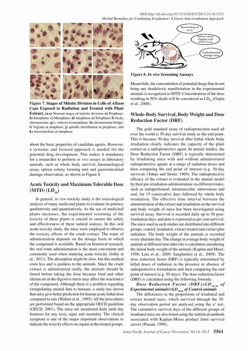
Several types of chromosome aberrations have been considered in the different phases of cell division (prophase, metaphase, anaphase and telophase), as shown in Figure 7A-E. According to Rank and Nielsen (Rank and Nielsen, 1997), chromosome aberration analysis not only allows the estimation of the genotoxic effects, but also enables evaluation of their clastogenic and aneugenic actions. Chromosome aberrations, such as chromosome bridges and breaks, are indicators of a clastogenic action; whereas chromosome losses, delays, adherence, multipolarity and C-metaphases result from aneugenic effects. The chromosome aberrations observed at all irradiated cells Figure (7F-K) were stickiness, bridges, c-mitosis and vagrant chromosomes. These aberrations were due to the effect of radiation on the spindle formation and thus resulted in cell division disturbances.
Mitotic index (M.I.) is characterized by the total number of dividing cells in the cell cycle. It has been used as a parameter to assess the genotoxicity of several agents. The genotoxicity levels of an agent can be determined by the increase or decrease in the M.I. (Fernandes et al., 2007). A M.I. lower than the negative control can indicate alterations deriving from the chemical action in the growth and development of exposed organisms. On the other hand, a M.I. higher than the negative control is due to an increase in cell division, which can be harmful to the cells, leading to a disordered cell proliferation and even to the formation of the tumour (Leme and Marin-Morales, 2009). The MI can be calculated using the following formula:
Mitotic index (MI)=Number of cells in mitosis/total number of cells
The mitotic index, which reflects the frequency of the mitotic cells, and, hence, the effects of irradiation, were analysed. The cells of A cepa root tips after treatment with plant extract will show decreased/increased mitotic indexes. This may be because the abnormal condition of the cells decreases after treatment with the extract. The abnormalities of chromosomes could be due to the blockage of DNA synthesis or the inhibition of spindle formation. The reduction of the mitotic index might be explained either as being due to the obstruction of the onset of prophase, the arrest of one or more mitotic phases, or the slowing of the rate of cell progression through mitosis (Christopher and Kapoor, 1988).
In vivo Screening AssaysThe preliminary in vitro assays provide an idea
Asian Pacific Journal of Cancer Prevention, Vol 14, 2013 5561
DOI:http://dx.doi.org/10.7314/APJCP.2013.14.10.5553Herbal Remedies for Combating Irradiation: A Green Anti-irradiation Approach
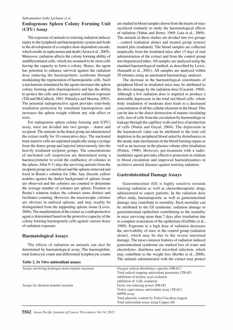
about the basic properties of candidate agents. However, a systemic and focused approach is needed for the potential drug development. This makes it mandatory for a researcher to perform in vivo assays in laboratory animals, such as whole body survival, haematological assay, spleen colony forming unit and gastrointestinal damage observation, as shown in Figure 8.
Acute Toxicity and Maximum Tolerable Dose (MTD) (LD50)
In general, in vivo toxicity study is the toxicological analysis of many medicinal plants to evaluate its potency qualitatively and quantitatively. As the use of medicinal plants increases, the experimental screening of the toxicity of these plants is crucial to ensure the safety and effectiveness of these natural sources. In this oral acute toxicity study, the mice were employed to observe the toxicity effects of the crude extract. The route of administration depends on the dosage form in which the compound is available. Based on historical research, the oral route administration is the most convenient and commonly used when studying acute toxicity (Jothy et al., 2011). The absorption might be slow, but this method costs less and is painless to the animals. Since the crude extract is administered orally, the animals should be fasted before taking the dose because food and other chemicals in the digestive tracts may affect the reaction(s) of the compound. Although there is a problem regarding extrapolating animal data to humans, a study has shown that mice give better prediction for human acute lethal dose compared to rats (Walum et al., 1995). All the procedures are performed based on the appropriate OECD guideline (OECD, 2001). The mice are monitored daily until day fourteen for any toxic signs and mortality. The clinical symptom is one of the major important observations to indicate the toxicity effects on organs in the treated groups.
Meanwhile, the concentration of potential drugs that do not bring any death/toxic manifestation in the experimental animals is recognized as MTD. Concentration of the dose resulting in 50% death will be considered as LD50 (Gupta et al., 2008).
Whole-Body Survival, Body Weight and Dose Reduction Factor (DRF)
The gold standard assay of radioprotection used all over the world is 30-day survival study as the end point. This is because 30-day survival after lethal whole body irradiation clearly indicates the capacity of the plant extract as a radioprotective agent. In animal studies, the Dose Reduction Factor (DRF) is typically determined by irradiating mice with and without administrated radioprotective agents at a range of radiation doses and then comparing the end point of interest (e.g. 30-day survival) (Yuhas and Storer, 1969). The radioprotective efficacy of the extract is evaluated in the animal model by their pre-irradiation administration via different routes, such as intraperitoneal, intramuscular, intravenous and oral, for 15 consecutive days followed by whole body irradiation. The effective time interval between the administration of the extract and irradiation on the survival and body weight of mice has been investigated using survival assay. Survival is recorded daily up to 30 post-irradiation days, and data is expressed as per cent survival. The mice used in such studies are distributed into different groups, control, irradiated, extract treated and extract plus radiation. The body weight of the animals is recorded every alternate day. The change in average body weight of animals at different time intervals is calculated considering the initial body weight of the animals (Kaplan and Meier, 1958; Lata et al., 2009; Sanghmitra et al., 2009). The dose reduction factor (DRF) is typically determined by lethal doses of radiation in the presence or absence of radioprotective formulation and then comparing the end point of interest (e.g. 30-days). The dose reduction factor (DRF) is calculated using the following formula:
Dose Reduct ion Factor (DRF)=LD 50/30 of Experimental animals/=LD50/30 of Control animals
The differences in the proportions of irradiated and extract treated mice, which survived through the 30-day observation period are analysed using the x2 test. The cumulative survival days of the different groups of irradiated mice are also tested using the statistical methods associated with Kaplan-Meier product limit survival curves (Prasad, 1999).
Figure 8. In vivo Screening Aassays
Figure 7. Stages of Mitotic Division in Cells of Allium Cepa Exposed to Radiation and Treated with Plant Extract. (a-e) Normal stages of mitotic division; a) Prophase; b) Interphase; c) Metaphase; d) Anaphase; e) Telophase; f) sticky chromosome; g) c- mitosis in metaphase; h) chromosome bridge; i) Vagrant at anaphase; j) spindle distribution at prophase; and k) micronucleus at intephase
ab
c d
e fg
h
i
jk
Subramanion Jothy Lachumy et al
Asian Pacific Journal of Cancer Prevention, Vol 14, 20135562
Endogenous Spleen Colony Forming Unit (CFU) Assay
The exposure of animals to ionizing radiation induces injury to the lymphoid and haemopoietic system and leads to the development of a complex dose-dependent cascade, which results in septicaemia and death (Arora et al., 2005). Moreover, radiation affects the colony forming ability of undifferentiated cells, which are assumed to be stem cells having the capacity to form a colony. Hence, the agent has potential to enhance survival against the radiation dose inducing the haemopoitetic syndrome through modulating the regeneration of haemopoietic cells. Such a mechanism stimulated by the agents increases the spleen colony forming units (haemopoiesis) and has the ability to protect the cells and tissue against radiation exposure (Till and McCulloch, 1961; Pahadiya and Sharma, 2003). The potential radioprotective agent provides total-body irradiation protection by stimulatin haemopoiesis and increases the spleen weight without any side effect or toxic.
For endogenous spleen colony forming unit (CFU) assay, mice are divided into two groups – donor and recipient. The animals in the donor group are administered the extract orally for 10 consecutive days. The nucleated bone marrow cells are aspirated aseptically using a syringe from the donor group and injected intravenously into the heavily irradiated recipient groups. The concentrations of nucleated cell suspension are determined using a haemocytometer to avoid the confluency of colonies in the spleen. After 9-11 days the surviving animals from the recipient group are sacrificed and the spleens removed and fixed in Bouin’s solution for 24hr. Any discrete yellow nodules against the darker background of splenic tissue are observed and the colonies are counted to determine the average number of colonies per spleen. Fixation in Bouin’s solution makes the colonies more distinct and facilitates counting. However, the macroscopic colonies are obvious in unfixed spleens, and may readily be distinguished from the supporting splenic tissue (Lewis, 2006). The manifestation of the extract as a radioprotective agent is determined based on the protective capacity of the colony forming haemopoietic cells against various doses of radiation exposure.
Haematological Assays
The effects of radiation on animals can also be determined by haematological assay. The haemoglobin, total leukocyte count and differential lymphocyte counts
are studied in blood samples drawn from the hearts of mice sacrificed routinely to study the haematological effects of radiation (Yuhas and Storer, 1969; Lata et al., 2009). The animals in these studies are divided into two groups – control (radiation alone) and treated group (extract treated plus irradiated). The blood samples are collected aseptically from the irradiated mice after 15 days of oral administration of the extract and from the control group into heparinized tubes. All samples are analysed using the standard haematological method, as described by Lewis, (Samarth et al., 2001). All samples are analysed within 30 minutes using an automated haematology analyser.
The decrease in the haematological constituents of peripheral blood in irradiated mice may be attributed to the direct damage by the radiation dose (Casarett, 1968). Although a low radiation dose is required to produce a detectable depression in the total red blood cells, whole body irradiation of moderate dose leads to a decreased concentration of all the cellular elements in the blood. This can be due to the direct destruction of mature circulating cells, loss of cells from the circulation by haemorrhage or leakage through the capillary walls and loss of production of cells (Nunia and Goyal, 2004). The depression in the haematocrit value can be attributed to the total cell depletion in the peripheral blood aided by disturbances in the steady state mechanisms in the blood forming organs as well as an increase in the plasma volume after irradiation (Potten, 1990). However, pre-treatment with a radio-modulator agent provides effective protection in relation to blood circulation and improved haemodynamics in occlusive arterial diseases against ionizing radiation.
Gastrointestinal Damage Assays
Gastrointestinal (GI) is highly sensitive towards ionizing radiation as well as chemotherapeutic drugs administered to cancer patients. In the radiation dose effect study, haematopoietic as well as gastrointestinal damage may contribute to mortality. Such mortality can be attributed to the GI syndrome; radiation damage to gastrointestinal epithelium contributing to the mortality in mice surviving more than 7 days after irradiation due to complete restoration of the epithelium (Griffiths et al., 1999). Exposure to a high dose of radiation decreases the survivability of mice in the control group (radiation alone), which may be due to the severe intestinal damage. The most common features of radiation induced gastrointestinal syndrome are marked loss of water and electrolytes, diarrhoea and microbial infection, which may contribute to the weight loss (Bertho et al., 2008). The animals administrated with the extract may protect
0
25.0
50.0
75.0
100.0
New
ly d
iagn
osed
with
out
trea
tmen
t
New
ly d
iagn
osed
with
tre
atm
ent
Pers
iste
nce
or r
ecur
renc
e
Rem
issi
on
Non
e
Chem
othe
rapy
Radi
othe
rapy
Conc
urre
nt c
hem
orad
iatio
n
10.3
0
12.8
30.025.0
20.310.16.3
51.7
75.051.1
30.031.354.2
46.856.3
27.625.033.130.031.3
23.738.0
31.3
Table 2. In Vitro antioxidant assaysAssays involving hydrogen atom transfer reactions Oxygen radical absorbance capacity (ORAC) Total radical trapping antioxidant parameter (TRAP) inhibition of linoleic acid oxidation inhibition of LDL oxidation Assays by electron-transfer reaction Ferric ion reducing power (FRAP) Trolox equivalence antioxidant assay (TEAC) DPPH assay Total phenolic content by Folin-Ciocalteu reagent Total antioxidant assay using Copper (II)
Asian Pacific Journal of Cancer Prevention, Vol 14, 2013 5563
DOI:http://dx.doi.org/10.7314/APJCP.2013.14.10.5553Herbal Remedies for Combating Irradiation: A Green Anti-irradiation Approach
the radiation induced intestinal damage. The histological observation method is commonly used to compare radiation induced intestinal damage between the controls and extract treated animals, and also to determine the efficacy to the plant extract as a radioprotective agent (Vigneulle et al., 2002).
Acknowledgements
This project was funded by the Research University Post Graduate Research Grant Scheme (RU-PRGS) (1001/CIPPM/846041) from Universiti Sains Malaysia, Penang, Malaysia and UM-MoHE HIR Grant (grant no.: UM-MoHE HIR H-18001-C0020) From University of Malaya, Malaysia. Subramanion L Jothy was supported by MyPhD fellowship from Ministry of Higher Education, Government of Malaysia, Malaysia.
References
Afaq F, Adhami VM, Ahmad N (2003). Prevention of short-term ultraviolet B radiation-mediated damages by resveratrol in SKH-1 hairless mice. Toxicol Appl Pharmacol, 186, 28-37.
Anonymous (2013a). A-bombed Ginkgo trees in Hiroshima, Japan+documentary film.
Anonymous (2013b). Laughter Yoga. Medicinal value of Tulsi. Apak R, Guclu K, Demirata B, et al (2007). Comparative
evaluation of various total antioxidant capacity assays applied to phenolic compounds with the CUPRAC assay. Molecules, 12, 1496-547.
Arora R, Gupta D, Chawla R, et al (2005). Radioprotection by plant products: present status and future prospects. Phytother Res, 19, 1.
Ashawat MS, Saraf S, Saraf S (2006). Cosmetic potentiality of plant extracts and natural oils. Biosci Biotechnol Res Asia, 3, 181-8.
Attaguile G, Russo A, Campisi A, et al (2000). Antioxidant activity and protective effect on DNA cleavage of extracts from cistus incanus L and cistus monspeliensis L. Cell Biol Toxicol, 16, 83-90.
Azad MB, Chen Y, Gibson SB (2009). Regulation of autophagy by reactive oxygen species (ROS): implications for cancer progression and treatment. Antioxid Redox Signal, 11, 777-90.
Aziz MH, Shaw SR, Wu J, Longley BJ, Ahmad N (2005). Chemoprevention of skin cancer by grape constituent resveratol: relevance to human disease? FASEB, 19, 1.
Bajpai M, Parade A, Tiwari SK, Prashad D (2005). Phenolic content and antioxidant activity of some food and medicinal plants. Int J Food Sci Nut, 4, 287-91.
Bertho JM, Roy L, Souidi M, et al., (2008). New biological indicators to evaluate and monitor radiation-induced damage: an accident case report. Radiat Res, 169, 543-0.
Biaglow JE (1987). The effects of ionizing radiation on mammalian cells. In: Farhataziz Rodgers Maj. Ed. Radiation chemistry: principles and applications. New York: VCH, 527-63.
Brown PE (1967). Mechanism of action of aminothiol radioprotectors. Nature, 213, 363-4.
Burrow CJ, Muller JG (1998). Oxidative nucleobase modifications leading to strand scission. Cancer Res, 98, 1109-51.
Campos PMBGM, Gianeti MD, Kanashiro A, Lucisano VYM, Gaspar LR (2006). In vitro antioxidant and in vivo photoprotective effects of an association of bioflavonoids with liposoluble vitamins. Photochem Photobiol, 82, 683-8.
Casarett AP (1968). Radiation Biology. Prentice-Hall, Englewood Cliffs, NJ, pp. 158-89.
Chauhan LKS, Saxena PN, Gupta SK (1999). Cytogenetic effects of cypermethrin and fenvalerate on the root meristem cells of Allium cepa. Environ Exp Bot, 42, 181-9.
Chipuk JE, Kuwana T, Bouchier-Hayes L, et al (2004). Direct activation of Bax by p53 mediates mitochondrial membrane permeabilization and apoptosis. Science, 303, 1010-4.
Christopher HB, Kapoor MB (1988). The cytogenetic effects of sodium salicylate on the root meristem cells of Allium sativa L. Cytologia, 54, 203-9.
Cimino F, Cristani M, Saija A, Bonina FP, Virgili F (2007). Protective effects of a red orange extract on UVB-induced damage in human keratinocytes. Biofactors, 30, 129-38.
Collins AR (2004). The comet assay for DNA damage and repair: principles, applications, and limitations. Mol Biotechnol, 26, 249-61.
Cragg GM, Newman DJ, Snader KM (1997). Natural products in drug discovery and development. J Nat Prod, 60, 52-60.
Edinger AL, Thompson CB (2004). Death by design: apoptosis, necrosis and autophagy. Curr Opin Cell Biol, 16, 663-9.
Erden Inal M, Kahraman A, Koken T (2001). Beneficial effects of quercetin on oxidative stress induced by ultraviolet A. Clin Exp Dermatol, 26, 536-9.
Fenech M (2000). The in vitro micronucleus test technique. Mut Res, 455, 81-95.
Fernandes TCC, Mazzeo DEC, Marin-Morales MA (2007). Mechanism of micronuclei formation in polyploidizated cells of Allium cepa exposed to trifluralin herbicide. Pestic Biochem Phys, 88, 252-9.
Fguyer S, Afaq F, Mukhtar H (2003). Photochemoprevention of skin cancer by botanical agents. Photodermatol Photoimmunol Photomed, 19, 56-72.
Frankel EN, Huang SW, Aeschbach R, Prior E (1996). Antioxidant activity of a rosemary extract and its constituents, carnosic acid, carnosol and rosmarinic acid, in bulk oil and oil-in-water emulsion. J Agri Food Chem, 44, 131-5.
Garcia Bores AM, Avila JG (2008). Natural products: Molecular mechanisms in the photo chemoprevention of skin cancer. Rev Latinoamer Quim, 36, 83-102.
Ghavami S, Eshragi M, Ande SR, et al (2010). S100A8/A9 induces autophagy and apoptosis via ROS-mediated cross-talk between mitochondria and lysosomes that involves BNIP3. Cell Res, 20, 314-31.
Grant WF (1994). The present status of higher plant bioassays for the detection of environmental mutagens. Mutat Res, 310, 175-85.
Griffiths CEM, Maddin S, Weidow O, et al (2005). Treatment of photoaged skin with a cream containing 0.05% isotretinoin and sunscreens. J Dermatol Treat, 16, 79-86.
Griffiths NM, Dublineau I, Francois A, Ksas B (1999). Radiation-induced colonic injury: decreased fluid absorption and effects of granisetron a 5-HT3 receptor inhibitor. Adv Radiat Biol Peace, 2, 1-10.
Gupta ML, Agarwala PK, Prem KI, et al (2008). Modulation of gamma radiation-inflicted damage in swiss albino mice by an alcoholic fraction of Podophyllum hexandrum rhizome. J Medicinal Food, 11, 486-92.
Handa SS (2008). An overview of Extraction Techniques for Medicinal and Aromatic Plants. In: Extraction Technologies for Medicinal and Aromatic Plants, Handa, S.S.; Khanuja, S.P.S.; Longo, G.; Rakesh, D.D; (Eds.), pp 21-54.
Hertog MGL, Feskens EJM, Hollman PCH, Katan MB, Kromhout D (1993). Dietary antioxidant flavonoids and risk of coronary heart disease: the Zutphen elderly study. Lancet, 342, 1007-11.
Higdon J (2007). An evidence-based approach to dietary
Subramanion Jothy Lachumy et al
Asian Pacific Journal of Cancer Prevention, Vol 14, 20135564
phytochemicals. New York: Thieme Medical. Huang D, Ou B, Prior RL (2005). Review: the chemistry
behind antioxidant capacity assays. J Agric Food Chem, 53, 1841-56.
Jagetia GC (2007). Radioprotective potential of plants and herbs against the effects of ionizing radiation. J Clin Biochem Nutr, 40, 74-81.
Jagetia GC, Venkatesha VA (2005). Effect of mangiferin on radiation-induced micronucleus formation in cultured human peripheral blood lymphocytes. Environ Mol Mutagen, 46, 12-21.
Jothy SL, Zakaria Z, Chen Y, et al (2011). Acute oral toxicity of methanolic seed extract of cassia fistula in mice. Molecules, 16, 5268-82.
Kaplan EL, Meier P (1958). Nonparametric estimation from in-complete observations. J Amer Stat Assoc, 53, 457-81.
Kapoor VK, Dureja J, Chadha R (2009). Herbals in the control of ageing. Drug Discov Today, 14, 992-8.
Katiyar SK, Afaq F, Perez A, Mukhtar H (2001). Green tea polyphenols (-)-epigallocatechin-3-gallate treatment of human skin inhibits ultraviolet radiationinduced oxidative stress. Carcinogenesis, 22, 287-94.
Katiyar SK, Ahmad N, Mukhtar H (2000). Green tea and skin. Arch Dermatol, 136, 989-94.
Katiyar SK, Elmets CA (2001). Green tea polyphenolic antioxidant and skin photoprotection- review. Int J Oncol, 18, 1307-13.
Katiyar SK (2009). Treatmetn of silymarin, a plant flavonoid prevent ultra violet light induced suppression and oxidative stress in mouse skin. Int J Oncol, 21, 1213-22.
Kaur CD, Saraf S (2011b). Photoprotective herbal extract loaded nanovesicular creams inhibiting ultra violet radiations induced photoaging. Int J Drug Delivery, 3, 699-711.
Korac RR, Khambholja KM (2011). Potential herbs in skin protection from ultra violet radiation. Pharmacogn Rev, 5, 164-73.
Kumar S, Maheshwari KK, Singh V (2009). Protective effects of Punica granatum seeds extract against aging and scopolamine induced cognitive impairments in mice. Afr J Trad CAM, 6, 49-56.
Lata M, Prasad J, Singh S, et al (2009). Whole body protection against lethal ionizing radiation in mice by REC-2001: a semi-purified fraction of Podophyllum hexandrum. Phytomed, 16, 47-55.
Lee TK, Johnke RM, Allison RR, O’Brien KF, Dobbs LJ Jr (2005). Radioprotective potential of ginseng. Mutagenesis, 20, 237-43.
Leme DM, Marin-Morales MA (2009). Allium cepa test in environmental monitoring: a review on its application. Muta Res, 682, 71-81.
Lewis SM (2006). Reference ranges and normal values, in Dacie and Lewis-Practical Haematology, Lewis, S.M., Bain, B.J., Bates, I., Eds., Churchill Levingstone: New York, NY, USA, 3th edition, pp.11-24.
Li YH, Wu Y, Wei HC, et al (2009). Protective effects of green tea extracts on photo aging and photommunosuppression. Skin Res Technol, 15, 338-45.
Liu X, Kim CN, Yang J, Jemmerson R, Wang X (1996). Induction of apoptotic program in cell-free extracts: requirement for dATP and cytochrome c. Cell, 86, 147-57.
Londhe JS, Devasagayam TP, Foo LY, Ghaskadbi SS (2009). Radioprotective properties of polyphenols from Phyllanthus amarus Linn. J Radiat Res, 50, 303-9.
Mantena SK, Katiyar SK (2006). Grape seed proanthocyanidins inhibit UV-radiation induced oxidative stress and activation of MAPK and NF-kappa B signaling human epidermal kerationcytes. Free Radic Biol Med, 40, 1603-14.
Maurya DK, Salvi VP, Krishnan Nair CK (2004). Radioprotection of normal tissues in tumor-bearing mice by troxerutin. J radiat res, 45, 221-8.
McGuire K (2012). Earth Fairy’s Dream. Holy Basil, Ocimum sanctum.
Miller R, Miller S (2003). Tulsi Queen of Herbs. India’s Holy Basil.
Nichols JA, Katiyar SK (2010). Skin protection by natural phenols: anti-inflammatory, antioxidant and DNA repair mechanism. Arch Dermatol Res, 302, 71.
Nunia V, Goyal PK (2004). Prevention of gamma radiation induced anaemia in mice by diltiazem. J Radiat Res, 45, 11-7.
OECD (2001). OECD Guidelines for Acute Toxicity of Chemicals; Organisation for Economic Co-operation and Development: Paris, France, No. 420.
Olive PL, Banáth JP (2006). The comet assay: a method to measure DNA damage in individual cells. Nat Protoc, 1, 23-9.
Olive PL, Banáth JP, Durand RE (1990). Heterogeneity in radiation-induced DNA damage and repair in tumor and normal cells measured using the “comet” assay. Radiat Res, 122, 86-94.
Olive PL, Johnston PJ (1997). DNA damage from oxidants: influence of lesion complexity and chromatin organization. Oncol Res, 9, 287-94.
Ostling O, Johanson KJ (1984). Microelectrophoretic study of radiation-induced DNA damages in individual mammalian cells. Biochem Biophys Res Commun, 123, 291-8.
Ozben T (2007). Oxidative stress and apoptosis: impact on cancer therapy. J Pharm Sci, 96, 2181-96.
Pahadiya S, Sharma J (2003). Alteration of lethal effects of gamma rays in Swiss albino mice by Tinospora cordifolia. Phytother Res, 17, 522.
Patt HM, Tyree EB, Straube RL, Smith DE (1949). Cysteine protection against X-irradiation. Science, 110, 213-4.
Peter OB, Catalina CP, Hernan S (2006). Boldine and its antioxidant or health promoting properties. Chemico-Biological Interactions, 159, 1-17.
Pham-Huy LA, He H, Pham-Huyc C (2008). Free radicals, antioxidanta in disease and health. Int J Biomed Sci, 4, 89-96.
Pickstone W (2010). Mother Nature’s Natural Healing Gifts! A-Z Of Herbs! Wayne Pickstone.
Potten CS (1990). A comprehensive study of the radiobiological response of the murine (BDF1) small intestine. Int J Radiat Biol, 58, 925-73.
Prasad KN (1982). Acute radiation syndromes. In Pizzarelo, D.L. Ed. Radiation biology, Boca Raton: CRC, 205-35.
Prasad KN (1999). Handbook of Radiobiology. CRC Press: Boca Raton, FL, 344.
Qi L, Liu CY, Wu WQ, Gu ZL, Guo CY (2011). Protective effect of flavonoids from Astragalus complanatus on radiation induced damages in mice. Fitoterapia, 82, 383-92.
Rajkumar V, Guha G, Ashok Kumar R, Mathew L (2010). Evaluation of antioxidant activities of bergenia ciliata rhizome. Rec Nat Prod, 4, 38-48.
Rank J, Nielsen MH (1997). Allium cepa anaphase-telophase root tip chromosome aberration assay on N-methyl-N-nitrosourea, maleic hydrazide, sodium azide, and ethyl methanesulfonate. Muta Res, 390, 121-7.
Rao BSS, Devi PU (2005). Response of S 180 murine tumor to bleomycin in combination with radiation and hyperthermia using micronucleus assay: a multimodality approach for therapeutic augmentation. Indian J Exp Biol, 43, 596-600.
Ravichandran G, Bharadwaj VS, Kolhapure SA (2005). Evaluation of the efficacy and safety of “Anti-Wrinkle cream” in the treatment of facial skin wrinkles: a prospective, open, phase III clinical trial. The Antiseptic, 102, 65-70.
Asian Pacific Journal of Cancer Prevention, Vol 14, 2013 5565
DOI:http://dx.doi.org/10.7314/APJCP.2013.14.10.5553Herbal Remedies for Combating Irradiation: A Green Anti-irradiation Approach
Russo A, Cardile V, Caggia S, et al (2011). Boldo prevents UV light and nitric oxide-mediated plasmid DNA damage and reduces the expression of Hsp70 protein in melanoma cancer cells. J Pharm Pharmacol, 63, 1219-29.
Samarth RM, Goyal PK, Kumar A (2001). Radioprotective effects of Mentha piperita. J Med Aromat Plant Sci, 22, 91-7.
Sanghmitra S, Gupta M, Gupta V, et al (2009). Podophyllum hexandrum- mediated survival protection and restoration of other cellular injuries in lethally irradiated mice. eCAM, 16, 47-55.
Saraf S, Kaur CD (2010). Phytoconstituents as photoprotective novel cosmetic formulations. Pharmacognosy Review, 4, 1-11.
Sasidharan S, Yoga Latha L, Ping KY, Jothy Lachumy S (2012). Screening Methods in the Study of Fungicidal Property of Medicinal Plants. In: Fungicides for Plant and Animal Diseases, Dharumadurai Dhanasekaran, (Ed.), 107-118.
Sharangi AB (2009). Medicinal and therapeutic potentialities of tea (Camellia sinensis L.)-A review. Food Res Inl, 42, 529-35.
Shukla SK, Gupta ML (2010). Approach towards development of a radioprotector using herbal source against lethal irradiation. Int Res J Plant Sci, 1, 118-25.
Silveira JPS, Seito LN, Eberlin S, et al (2013). Photoprotective and antioxidant effects of Rhubarb: inhibitory action on tyrosinase and tyrosine kinase activities and TNF-α, IL-1α and α-MSH production in human melanocytes. BMC Complement Altern Med, 13, 49.
Singh NP, McCoy MT, Tice RR, Schneider EL (1988). A simple technique for quantitation of low levels of DNA damage in individual cells. Exp Cell Res, 175, 184-91.
Singh U, Kunwar A, Srinivasan R, Nanjan MJ, Priyadarsini KI (2009). Differential free radical scavenging activity and radioprotection of Caesalpinia digyna extracts and its active constituent. J Radiat Res, 50, 425-33.
Song JH, Bae EY, Choi G, et al (2013). Protective effect of mango (Mangifera indica L.) against UVB-induced skin aging in hairless mice. Photodermatol Photoimmunol Photomed, 29, 84-9.
Speit G, Hartmann A (2005). The comet assay: a sensitive genotoxicity test for the detection of DNA damage. Methods Mol Biol, 291, 85-95.
Spotheim-Maurizot M, Franchet J, Sabattier R, Charlier M (1991). DNA radiolysis by fast neutrons: II. Oxygen, thiols and ionic strength effects. Int J Radiat Biol, 59, 1313-24.
Susin SA, Lorenzo HK, Zamzami N, et al (1999). Molecular characterization of mitochondrial apoptosis-inducing factor. Nature, 397, 441-6.
Svobova A, Psotova J, Walterova D (2003). Natural phenolics in the prevention of UV induced skin damage. A review. Biomed Papers, 147, 137-45.
Szumiel I (2011). Autophagy, reactive oxygen species and the fate of mammalian cells. Free Radic Res, 45, 253-65.
Till JE, McCulloch EA (1961). A direct measurement of the radiation sensitivity of normal mouse bone marrow cells. Radiat Res, 14, 213-22.
Tiwari A (2001). Imbalance in antioxidant defense and human diseases: multiple approach of natural antioxidant therapy. Current Science, 81, 1179-87.
Tsoyi K, Hyung BP, Young MK, et al (2008). Protective effect of anthocyanins from black soybean seed coats on UVB-induced apoptotic cell death in vitro and in vivo. J Agric Food Chem, 56, 10600-5.
Tsujimoto Y (2003). Cell death regulation by the Bcl-2 protein family in the mitochondria. J Cell Physiol, 195, 158-67.
USNRC Technical Training Center (2013). Biological Effects of Radiation. Reactor concepts manual.
Vaid M, Katiyar SK (2010). Molecular mechanisms of inhibition of phtocarcinogenesis by silymarin, a phytochemical from milk thistle (Silybum marianum L. Gaerth). Int J Oncol, 36, 1053-60.
Varanda EA, Tavares DC (1998). Radioprotection: mechanisms and radioprotective agents including honeybee venom. J Venom Anim Toxins, 4, 5-21.
Vigneulle RM, Rao S, Fasano A, MacVittie T (2002). Structural and functional alterations of the gastrointestinal tract following radiation induced injury in the rhesus monkey. Digestive Dis Sci, 47, 1480-91.
Vile GF (1997). Active oxygen species mediate the solar ultraviolet radiation dependent increase in the tumour suppressor protein p53 in human skin fibroblasts. FEBS Lett, 412, 70-4.
Villunger A, Michalak EM, Coultas L, et al (2003). p53- and drug-induced apoptotic responses mediated by BH3-only proteins puma and noxa. Science, 302, 1036-8.
Visvardis EE, Tassiou AM, Piperakis SM (1997). Study of DNA damage induction and repair capacity of fresh and cryopreserved lymphocytes exposed to H2O2 and gamma irradiation with the alkaline comet assay. Mutat Res, 383, 71-80.
Von Sonntag C (1987). The Chemical Basis of Radiation Biology, Taylor & Francis, London, pp. 65-84.
Walum E, Nilsson M, Clemedson C, Ekwall B (1995). The MEIC program and its implications for the prediction of acute human systemic toxicity. In Alternative Methods in Toxicology and theLife Sciences; Goldberg, A.M., van Zutphen, L.F.M., Eds.; Mary Ann Liebert: New York, NY, USA, Vol.11, pp. 275-82.
West DP, Zhu YF (2003). Evaluation of Aloe vera gel gloves in the treatment of dry skin associated with occupational exposure. Am J Infect Control, 31, 40-2.
Widyarini S, Spinks N, Husband AJ, Reeve VE (2001). Isoflavonoid compounds from red clover (Trifolium pratense) protect from inflammation and immune suppression induced by UV radiation. Photochem Photobiol, 74, 465-70.
Wochna A, Niemczyk E, Kurono C, et al (2007). A possible role of oxidative stress in the switch mechanism of the cell death mode from apoptosis to necrosis-studies on rho0 cells. Mitochondrion, 7, 119-24.
Wu WS (2006). The signaling mechanism of ROS in tumor progression. Cancer Metastasis Rev, 25, 695-705.
Yuhas JM, Storer JB (1969). Chemoprotection against three modes of radiation death in the mouse. In. J Radiat Biol Relat Stud Phys Chem Med, 15, 233-7.