review soft tissue management in endodontic surgery term “periodontal plastic surgery,” first...
TRANSCRIPT

REVIEW
Soft Tissue Management in Endodontic Surgery
Peter Velvart, DMD, and Christine I. Peters, DMD
Modern endodontic surgery involves both root-endpreparation and proper sealing of all apical portalsof exit. Both components are requirements for me-chanical and biological success, but the manage-ment of soft tissues becomes increasingly impor-tant for an esthetically successful treatment. Ahealthy appearance of soft tissues plays an impor-tant role in the esthetic outcome of periradicularsurgery. This is true considering maintenance ofattachment levels and regarding the amount ofpossible recession after surgical procedures.Complete, recession-free and predictable healingof gingival tissue is one important goal of endodon-tic surgical treatment. A critical review of currentlyused techniques based on clinical and scientificdata reveals great potential for improvements.Possible reasons for scar formation and recessionspecifically in healthy periodontal conditions re-quiring surgical endodontic intervention are high-lighted. Based on anatomical considerationsvarious incision types are evaluated and recom-mendations made. Clear understanding of woundclosure and tissue-healing patterns call for the useof atraumatic procedures, nonirritating suture ma-terials and adequate suturing techniques. This ar-ticle gives an overview and guidance for integrat-ing current and new successful flap designs andwound closure methods. The methods describedhave the intention of maintaining the attachmentlevel and avoiding postoperative recession aftersurgical endodontic therapy.
Endodontic failures that do not respond to conventional root canalretreatment require surgical treatment (1, 2). The prime objectiveof surgical treatment is the same as in conventional root canaltherapy, which is to provide conditions such that healing and repairof the periradicular tissue can take place. These conditions includeremoval of necrotic tissue and tissue breakdown products from theapical part of the root canal system, elimination of bacterial or-
ganisms persisting in the root canal system and removing the roottip. A fluid-tight seal of the apical part of the root canal with amaterial that is biologically compatible is created (3). Severalstudies have identified factors that influence the prognosis ofendodontic surgery (4–11). According to Friedman (12) they canbe divided into preoperative-, operative-, and postoperative factors.Success rates vary between 25% and 98% depending on the study(13, 14). The assessment of success and failure after endodonticsurgery is mostly based on clinical and radiographic criteria ofhealing of the periradicular tissues (15, 16). In the literature, thecontributing factor of present periodontal conditions in relation tothe postoperative success of apical surgery has not been consideredextensively. In a study by Jansson and coworkers (17), a persistingendodontic infection has been shown to be a contributing riskfactor for progressing marginal attachment loss after periradicularsurgery. In the long-term perspective, periodontal healing in teethwith root canal infection, evident as periradicular radiolucency,may result in retarded or impaired healing after periodontal therapy(18). On the other hand, before endodontic surgery is undertaken,the periodontium should be treated in cases where periodontitis ispresent (4, 12, 17, 19–21).
The ultimate goal in modern dentistry, after pathologic pro-cesses have been eradicated, is to achieve “white” and “pink”esthetics in obviously visible areas (22). “White esthetics” refers tothe natural dentition or tooth-colored restoration of dental hardtissues with suitable materials. This notion has reached a very highlevel of importance (23). “Pink esthetics” refers to the surroundingsoft tissues and ultimately the underlying bone, which are equallyimportant for an esthetic result. Treatment of these tissues withadequate surgical and reconstructive techniques and maintenanceof the results are a challenge in modern esthetic dentistry and theprimary objective of preservation of the dentition is no longeracceptable without consideration of esthetic consequences (24).For many years, periodontal surgery has been associated withcreating esthetic problems, but it should be understood that peri-odontal disease itself is disfiguring and that great care must beexercised during surgery to minimize the unaesthetic impact oftherapy. The term “periodontal plastic surgery,” first suggested byMiller (25), was defined in a consensus report as surgical proce-dures performed to prevent or correct anatomical, development,traumatic or plaque disease-induced defects of gingiva, alveolarmucosa, or bone (26, 27). Furthermore, periodontal microsurgerywas introduced, which is defined as refinements in existing basicsurgical techniques that are made possible by the use of the
JOURNAL OF ENDODONTICS Printed in U.S.A.Copyright © 2004 by The American Association of Endodontists VOL. 31, NO. 1, JANUARY 2005
4

surgical microscope and microsurgical instruments (28). Improve-ments in flap design and soft tissue manipulation are consideredkey elements in improving the outcomes of regenerative periodon-tal surgery. Improved visual perception and better soft tissue han-dling hold great promise to further improve predictability of peri-odontal treatment (29). The same basic microsurgical principlesapply to endodontic surgical interventions (30, 31).
ANATOMY
Gingival tissue comprises epithelial and connective tissue, thelatter consisting to about 55 to 60% of supragingival fibers, whichattach gingiva to teeth and provide the basis for its firmness andbiomechanical resistance during mastication (32). The gingivaltissue reaches from papilla to mucogingival junction where it joinsthe alveolar mucosa and attaches to teeth and to the alveolarprocess (22, 32). The height of the gingiva from the mucogingivaljunction to the gingival margin is highest on the labial aspect ofmaxillary incisors (33). The gingival tissue between two adjacentteeth, the papilla, was considered roughly pyramidal and triangularin shape (34). The embrasure contour and the anatomy of itsadjacent teeth determine the shape of a papilla (32, 35–37). Thepapilla has a lingual and a buccal peak, joined by a concave col (22,35, 36, 38). It contains both nonkeratinized sulcular and col epi-thelium as well as keratinized oral epithelium (32, 35, 36). The colarea consists of squamous stratified nonkeratinized epithelium. Inmesio-distal direction, the midsection of the col slopes towardseach tooth. On these slopes, the epithelium gradually changes itsappearance toward the characteristics of the epithelial cuff (epi-thelial attachment). The width of the col between the buccal andlingual papilla and the depth of concavity in the col area increasesgradually from the anterior to the posterior teeth (36).
The presence or absence of the interdental papilla depends uponthe distance between the contact point and the crest of bone (37).When the distance from the contact point to the bone was 5 mm orless, the papilla was present almost 100% of the time. With adistance of 6 mm, the papilla was present 56% of the time, andwhen the distance measured 7 mm or more, the papilla was present27% of the time or less. Holmes (38) excised interdental papillaein sixteen dental students, one from the anterior and one from theposterior area of each student. From 32 specimens, 22 papillae didnot regenerate to their original shape and height. The regeneratedpapillae appeared flatter, did not fill the embrasure as completelyas before excision and the cols were less concave.
An important anatomical consideration during endodontic sur-gery is the course of blood vessels supplying alveolar mucosa andgingiva. Four interconnected pathways of blood supply exist: Thesubepithelial capillaries of the gingiva and alveolar mucosa, thevascular network within the periosteum, the intraseptal arteries inthe bone marrow and the plexus of the periodontium. The perios-teal and the periodontal plexus communicate directly via Volk-mann’s canals without participation of the vessels of the bonemarrow, thus unified histological responses to surgical woundingare observed (39) (Fig. 1). The gingiva and periosteum are blood-supplied mainly through supraperiosteal vessels, which runroughly parallel to the teeth’s long axis, branch and subdivide inthe lamina propria of the gingiva and form the vascular network onthe periosteum (39–41). To a lesser degree, rami perforantes of theintraseptal arteries penetrating the interdental bone and the peri-odontal ligament vessels supply the gingiva with blood, the vesselsfinally ending in loops in the tips of the connective tissue papillae,
termed the gingival plexus (22, 31, 32, 42–44). The multipleinterconnections between different plexus through numerous anas-tomoses and collateral pathways of circulation establish adequateblood supply, if single vessels are severed surgically (39–41, 43,44). Mormann and Ciancio (45) studied the effect of various typesof surgical procedures on the gingival capillary blood circulationby means of fluorescein angiography. The circulation changesobserved suggested that flaps receive their major blood supplyfrom their apical aspect, but interestingly not exclusively. Shortand long full thickness flaps in clinically healthy gingiva revealedthat, functionally, orientation of blood supply was mainly from thevestibule to the gingival margin (Fig. 2). On the other hand,internal beveled incisions severed the anastomosis between gingi-val and periodontal vasculature and between gingival and inter-dental vasculature. Angiographically, separations from either vas-
FIG 1. Collateral circulation through blood vessels communicatingbetween the periodontal plexus and the periosteal plexus. Reprintedwith permission from (39).
FIG 2. Difference in circulation disturbance in a short full thicknessflap (SF) versus a long full thickness flap (LF) as shown by fluores-cein angiography. (A–D) Indicated the distances from the cemento-enamel junction. Reprinted with permission from (45).
Vol. 31, No. 1, January 2005 Soft Tissue Management 5

culature showed no effect on blood circulation within free andalveolar gingiva. When flaps were raised the greater the ratio offlap length to base, the greater was the amount of circulatorydisturbance. Flap perfusion was maintained up to the point, wherethe ratio of length to width of the parallel pedicle flap equaled 2:1.
ACCESSING THE APICAL PATHOLOGY
The outcome of any surgical procedure depends among otherfactors upon the extent to which an adequate access is possible.Endodontic surgery first requires exposure of the bone overlayingthe tip of the root(s) and then revealing the root end(s) per se (Fig.3). To access the bone, a full thickness flap must be raised. Thiscomprises a soft tissue flap, which consists of gingival and muco-sal tissue as well as periosteum. To mobilize the flap variousmodes of incisions can be selected including horizontal incisions,sulcular and submarginal, and vertical releasing incisions. The flapcan be in its entirety a full thickness flap or a combination of a fulland a split thickness flap.
Certain basic principles must be considered before deciding on thetype of incision and flap design: Firstly, regional anatomical structuressuch as the location and the path of the blood vessels and nervesshould be evaluated, protected and preserved during the surgicalprocedure. Recognition of the position of the root within the mandibleor the maxilla, its inclination and thickness of the bone between thesurface and root structure is important. Furthermore, periodontal con-ditions play an essential role in the decision making process. Probingdepth, attachment loss, recessions and signs of periodontal inflamma-tion in terms of bleeding on probing should be evaluated presurgically.The width of the attached gingiva and the location of the mucogin-gival junction are measured. In addition, presence, type and quality ofrestorations in special reference to the position of the restorationmargin to the gingiva need to be determined and are critical to theesthetic outcome. Moreover, an evaluation of the size and position ofthe expected periradicular pathology in relation to the root, neurovas-cular structures and the sinus has to take place, including assessmentof the local blood supply to the soft tissues in and around the desig-nated flap area. Clear understanding of the reaction pattern of thetissues involved and the healing principles after the wound closure areimportant.
Quality assurance of endodontic therapy is an important issue(46) and microsurgical techniques have been applied in endodonticsurgery for several years. They have even become a standard inpostgraduate education in endodontics (47). Many endodontic sur-gical failures have been directed to poor visibility and ability todiagnose and treat the minute causes of apical pathology (30). Themicrosurgical approach has been purported to improve the prog-nosis of surgical treatment outcome (48).
In a series of studies our group has investigated soft tissuehealing results after microsurgical endodontic treatment. We as-sumed that microsurgical treatment would be beneficiary to softtissue healing. Based on and in analogy to the periodontal litera-ture, microsurgical treatment should allow manipulation of thetissues in a more delicate and less traumatic way when comparedto macrosurgery as it is used up to this point in time (49). Thequestion was whether improved healing was achievable, whenmicrosurgical instruments and techniques were applied. Three ar-eas of interest have been identified as esthetically relevant: Theinterdental papilla, the cervical marginal area and the attachedgingival and mucosa. Specifically the shape, dimensions, surface
texture, recession, scar formation, and attachment level were stud-ied.
FLAP DESIGN
Various flap designs have been discussed in the literature (3, 13,50, 51). Examples include marginal mucoperiosteal flaps with one(triangular flap) or two (trapezoidal or rectangular flap) releasingvertical incisions, submarginal mucoperiosteal flaps with the hor-izontal incision within the attached gingiva and its modificationsand semilunar flaps.
The wide variety of flap designs reflects the number of variablesto be considered before choosing an appropriate flap. As condi-tions vary with each individual patient and specific situation, therewill always be a need to select the best flap design for every singlecase. The literature is replete with basic rules and recommenda-tions (52). A brief discussion will review the pros and cons of theclassic flaps.
Triangular Flap
This type of surgical flap comprises a horizontal incision ex-tending at least one tooth mesially and distally to the involved areacombined with one releasing vertical incision forms a triangularflap (Fig. 4). Usually, the releasing incision is performed on themesial part of the flap. This flap-technique exposes mostly mar-ginal areas of the tooth in question and does not create enoughaccess to the apical region. The triangular flap is mainly indicated
FIG 3. (A) Root end resection on an upper lateral incisor. (B) Re-sected root end with exposed obturation material and a lateral canal(arrow).
FIG 4. A triangular flap is formed by a mesial vertical and sulcularincision extending to at least two teeth distal from the releasingincision. Reprinted with permission from (3).
6 Velvart and Peters Journal of Endodontics

for correction of problems in the cervical and mid-root portions,such as in cases with cervical root resorptions, perforations and inresections of very short roots. The main advantages of this flapdesign are minimal disruption of the blood supply to the mobilizedtissues and easy repositioning of the wound edges. As in allintrasulcular horizontal incisions, recession may result after thehealing process.
Rectangular and Trapezoidal Flap
Rectangular and trapezoidal flaps are an extension of the trian-gular flap with a second vertical releasing incision (Fig. 5). Rect-angular flaps are the most frequently used flaps in endodonticsurgery (3). Both flaps provide excellent access to the apical area.If required, releasing incision may be extended in apical directionfor tension-free retraction. The difference between the rectangularand the trapezoidal flap is the degree of divergence of the releasingincisions. As the blood vessels run mostly parallel to the long axisof the teeth from the apical to the coronal direction, one consid-eration is to disrupt the least number of vascular structures. Avertical incision parallel to the path of the vessels would bestcomply with the above statement, thus favoring the rectangularflap (43, 53). Survival and blood supply of mobilized tissue ap-peared to be best, when the basis was broader than the proximalend of the flap. On the other hand, blood supply to the unreflected
tissues was compromised by this approach (43). The longer theflap length the more important is the ratio between the length andwidth of the flap (54). Several authors advocate a length-widthratio of 2:1 (45, 55). The length-width ratio requirement usuallyfavors a slightly trapezoidal shape of the flap, or extending the flapone tooth further mesially or distally. In all instances, convergingreleasing incisions must be avoided.
Repositioning and wound closure is easy in trapezoidal andrectangular flaps. In esthetically critical areas with prosthetic res-torations involving subgingivally placed crown margins, a postop-erative sequel can result in recession, leading to esthetically com-promising exposure of the crown margins.
Submarginal Flap
The most popular submarginal flap is the flap design by Ochsen-bein and Luebke (56). Two releasing vertical incisions are con-nected by a scalloped horizontal incision (Fig. 6). The submarginalflap is only to be used, when there is a broad zone of attachedgingiva with a minimum of 2 mm (57). In addition, the underlyingapical lesion or surgical bony access must not extend to the flapmargins. This flap design has the advantage of leaving the mar-ginal gingiva untouched and it does not expose any restorationmargins. As crestal bone is not denuded, the risk of attachment lossis minimized. In rare situations, because of insufficient treatmentplanning and poor surgical technique, necrosis of the unreflectedtissue might occur because of deprivation of blood supply to thisarea. The recession resulting from such tissue breakdown can havedevastating effects on the esthetic outcome of surgical treatment.Possible scar tissue formation is another disadvantage of the sub-marginal flap.
Vreeland and Tidwell modified the submarginal incision (50) byplacing a scalloped horizontal incision 1 to 2 mm below thegingival margin. An inverse-bevel incision splits the tissue until 1to 2 mm below the crest of bone, from which point on a fullthickness flap is reflected. After closure, the flap should cover thearea of unreflected split thickness tissue. The authors claim thatless exact replacement of the flap is required when using thisapproach. The type of incision described bears a high risk ofpostoperative necrosis, as the only blood supply to the unreflectedmarginal tissue is derived from the periodontal ligament (39, 45).In addition, when 4/0 sutures are used, this critical area is furthertraumatized. Although the authors claim that scarring is “not muchof a problem” the clinical pictures in the publication clearly dem-onstrate marked scar tissue formation, typical for submarginalincisions (50).
Semilunar Flap
Placing either a straight or a curved horizontal incision in thealveolar mucosa that extends all the way to the bone creates asemilunar flap. The only advantage of this flap design is the factthat the marginal tissue remains untouched and thus no recessionwill occur. On the other hand, many disadvantages accompany itsuse. A semilunar flap will provide only limited access to thesurgical area. Additionally, placement of the incision over the bonydefect means that the wound cannot be closed over sound bonestructure. Furthermore, the content of elastic fibers and muscleattachments of alveolar mucosa is high, both of which exert pullingforces on the reapproximated surgical wound margins. Not only is
FIG 5. A rectangular flap comprising two releasing incisions con-nected by a sulcular horizontal incision. Reprinted with permissionfrom (3).
FIG 6. A submarginal incision formed by a scalloped horizontalincision within the attached gingiva and two releasing vertical inci-sions. Reprinted with permission from (3).
Vol. 31, No. 1, January 2005 Soft Tissue Management 7

suturing difficult, but constant tension will be present on the flap,resulting in poor alignment of wound edges, gap formation, andinsufficient healing. Dehiscence in the incision line will result insecondary healing and scar formation. Moreover, placement of acurved, horizontal incision will sever a maximum of blood vessels.Because of the many drawbacks mentioned a semilunar flap designis no longer recommended (52).
SURGICAL SITE CLOSURE
After irrigation with saline solution to remove debris, the woundedges are reapproximated carefully to allow primary intentionhealing (52).
Compression of the repositioned flap with a saline-moistenedpiece of gauze is necessary to create a thin fibrin layer betweenflapped tissue and cortical bone (53, 58, 59). Replacement of a thinblood clot with parallel fibrin fibers by new fibrous tissue resultsin collagen adhesion (58).
SUTURES: TECHNIQUES AND MATERIALS
Sutures are needed to hold the reapproximated flap in place andto allow healing of the wound by primary intention (49). Acciden-tal premature loss of sutures leads to delayed healing by secondaryintention (60). Bacterial colonization is an important factor leadingto tissue reactions to intraoral sutures (61–65).
Materials
When comparing histological tissue response of different suturematerials, monofilament sutures (e.g. nylon, gut, steel, and chromicgut) produced smaller inflammatory reaction than multifilamentmaterials (e.g. silk, siliconized silk, polyester, teflonized polyester,cotton, or linen) (61–64). Systemic antibiotics did not alter thesereactions (62). While some materials are no longer recommendedtoday, fact remains that bacteria invade suture tracks; this phe-nomenon is most predominant with multifilament materials withwicking action (61, 66). Accordingly, nonabsorbable silk suturesare easy to tie and handle but are no longer recommended as theyaccumulate plaque, allow rapid bacterial colonization and are un-comfortable to remove because of ingrowth of tissue (66, 67).However, some multifilament suture materials seem to inhibitbacterial transmission (65).
Polyglactin (e.g. Vicryl) sutures are colonized more slowly thansilk and offer the advantage of being absorbable (31). Surgical gut,polyglactin 910, poliglecaprone, and polidioxanone are absorbablematerials recommended for periodontal surgery (49). Polyglactin910 (coated Vicryl) is absorbed in 7 to 10 days, has shown verylittle inflammatory reaction, that subsided after 3 days (in com-parison to reactions to polypropylene, silk, or catgut) and goodresults with respect to wound infection in skin wounds (68, 69). Ingeneral, monofilament synthetic sutures are least traumatic, allowless bacterial migration and are the materials of choice (66). Nylonsutures, e.g. Gore-Tex monofilament sutures or other syntheticmonofilament suture elicited a mild inflammatory tissue response(66). A polytetrafluorethylene (PTFE) impregnated monofilamentsuture has good handling characteristics and less bacterial coloni-zation (31, 66, 70). If a multilayered flap is used, absorbable suturematerials (e.g. polyglecaprone, polyglactin) are used for innerlayers only and nonabsorbable materials (e.g. polypropylene) for
outer layers and whenever else possible, to minimize inflammationduring the healing process (28, 69).
Microsuturing makes use of fine needles and suture materials.Round-bodied needles (5/8-circular, 1/2-circular, 3/8-circular) areused for soft tissues, such as mucosa, but each situation mayrequire different needle sizes (Fig. 13) (31). Most needles used indentistry are 3/8 curvature needles but ophthalmic spatula needleswith 140 degrees curvatures are also recommended (49).
To avoid necrosis of papillae by inserting too much suturematerial, # 6–0 to # 8–0 suture sizes are recommended in micro-surgical techniques, and are mandatory for layered suturing (22,28, 49). Other authors recommend nonabsorbable monofilament5-0 sutures (21). Sutures should not act as ligatures and tensioncreated by sutures should be minimal (45). Tension can be reducedby using a greater number of sutures and by choosing fine micro-surgical materials (28).
Techniques
Postoperative gingival recession is a difficult therapeutic di-lemma that can be a sequel of conventional flap and suturingtechniques (71). Esthetically disappointing results are a majorconcern both for the clinician and the patient involved. Suturematerials can cause inflammation and foreign body reactions (66,72). Some authors recommend using only the minimal amount ofsutures necessary to secure the flap as both the suture and the knotitself cause inflammation and delay wound healing (52).
The techniques employed include interrupted sutures, anchorsutures, continuous sling sutures, and vertical mattress sutures(Fig. 7) (31, 73). While full flaps are best secured using verticalmattress sutures and anchor sutures, limited flaps are best retainedusing interrupted, fine-diameter sutures (74).
Avoiding any thinning of the papillae, even in cases of peri-odontitis and using internal (vertical) mattress sutures, supportsthe interdental papilla in a coronal direction, and results in lessloss of papillary height (67). Partial thickness flaps in very thintissue may not provide sufficient blood supply for flap survival(45). Minimal tension during reapproximation and after suturingis important to avoid impairment of circulation in a flap (45,
FIG 7. Examples of commonly employed suture techniques. (A)interrupted suture, (B) anchor suture, (C) sling suture, and (D) verticalmattress suture Reprinted with permission from (102).
8 Velvart and Peters Journal of Endodontics
71). An example of a technique that takes advantage of suturepulling forces would be the ramp mattress suture, a horizontalmattress suture that pulls the buccal flap in coronal directionand the palatal flap in a more apical direction, resulting in agingival slope or ramp to the palate (75).
SUTURE REMOVAL
A perisutural epithelial sleeve develops at 3 days and can enrobethe entire suture track after 7 days (66). An intense inflammatoryresponse to suture materials and the trauma of suture placement isvisible after 3 days (66). As resealing of epithelium at the woundmargin is evident within 2 days, suture removal can take place after48 h but should take place no longer than 96 h after placement (53,59, 74, 76).
CRITICAL REVIEW OF SOFT TISSUE HEALING
The healing capacity of oral tissues is excellent. Only seldomare there serious postsurgical complications, such as tissue necro-sis, nerve damage, profound bleeding, or serious infections. Whengeneral basic rules are followed, fair healing of the soft tissues canbe expected. Recession is a frequent sequel to healing after peri-odontal surgery. Its extent and differences in terms of recessionlocation have not been studied extensively. The goal of periodontalsurgery is to alter, treat and heal diseased gingival tissues andcrestal bone through a host of invasive measures, thereby removingselected areas that are not retainable (52, 77, 78). The healingbehavior of tissues, where attachment has been lost, but that arehealthy, has only been studied in recent years, with the develop-ment of periodontal plastic surgery. This treatment of healthyperiodontal tissue mainly involved restoration of lost attachmentand augmentation procedures (28, 79). The goal has increasinglyshifted, towards restoration of the natural shape, position, color,and appearance of soft tissues as present before trauma, disease, ortreatment induced changes of the tissues (27, 79).
When a lesion resulting of endodontic pathology develops on aspecific tooth and needs surgical intervention, frequently marginalsoft tissues may be healthy (53). With the aid of contemporarytechniques such as magnification under a microscope, suitablematerials and the use of microinstruments, endodontic surgery hasevolved to microsurgery and will result in a predictably successfuloutcome in teeth treated (14, 21, 30, 48).
Healing Process
Healing takes place in several phases that overlap and coexist:wounding, clotting and inflammation, epithelial healing, connec-tive tissue healing, proliferation, maturation, and remodeling (53,59, 78, 80). While a multitude of immune defense mechanismsexists, and the details of intercellular communication pathways andinterdependent signaling processes during hemostasis and woundhealing are beyond the scope of this article, the main events aredescribed below. Within 24 h, polymorphonuclear leukocytes andmacrophages start migrating into a blood clot. Stimulated macro-phages play a central role in angiogenesis and new collagen syn-thesis (58, 76, 78, 80, 81). Inflammatory and reparative cellsmigrate along fibrin strands, followed by capillary buds. Themicrovascularization in the flap itself remains patent, providingnutrition for the mucogingival flap, in concert with contributions
from remaining periosteal, periodontal, and bone microvascularnetworks (60). Parallel fibrin strands after wound compression anda thin hiatus between wound edges accelerate this process (20, 59,76). Epithelial streaming as a sheet or as fingers is observed after2 days, eventually resulting in a multilayered seal (74, 82). After4 days an epithelial barrier has formed (76). Other authors de-scribed complete epithelial healing in the sulcus at 14 days andafter 28 days the wound healing process was accomplished (59, 60,83). Healing and reattachment of an elevated flap to cortical boneis a slower procedure, the periosteum does not survive reflection(58). Granulation tissue replaces the thin fibrin clot between theflap and cortical bone after 4 days, and fibrous connective tissuereplaces granulation tissue by 14 days (58). Because of earlyepithelial bridging, suture removal is therefore advocated after 2 to3 days (76). Initial resistance to rupture forces is attributed toregeneration of epithelial attachment to tooth surfaces (20, 74, 82).Other authors do not recommend suture removal before 4 days, asstainable collagen content in granulation tissue, which determinestensile wound strength, is only present after 3 days (59). Whilehealing wounds that are subject to small amounts of mechanicalstress, demonstrate an increase in collagen strength and formation,excessive forces disrupt the neovasculature and collagen fibers anddelay healing (80).
More and more variables of wound healing, including patientnutritional status, bacterial infection, wound care and availabletissue oxygen, are being researched. Consequently, novel therapiesare evolving, such as growth factor therapy (84). Growth factorsmay lead to new strategies in improvement of soft tissue healing,including skin, mucosa, and nerve tissues (85).
Strategies and Procedures
The choice of flap designs should allow maintenance of optimalblood perfusion during surgery. This implies using a design where
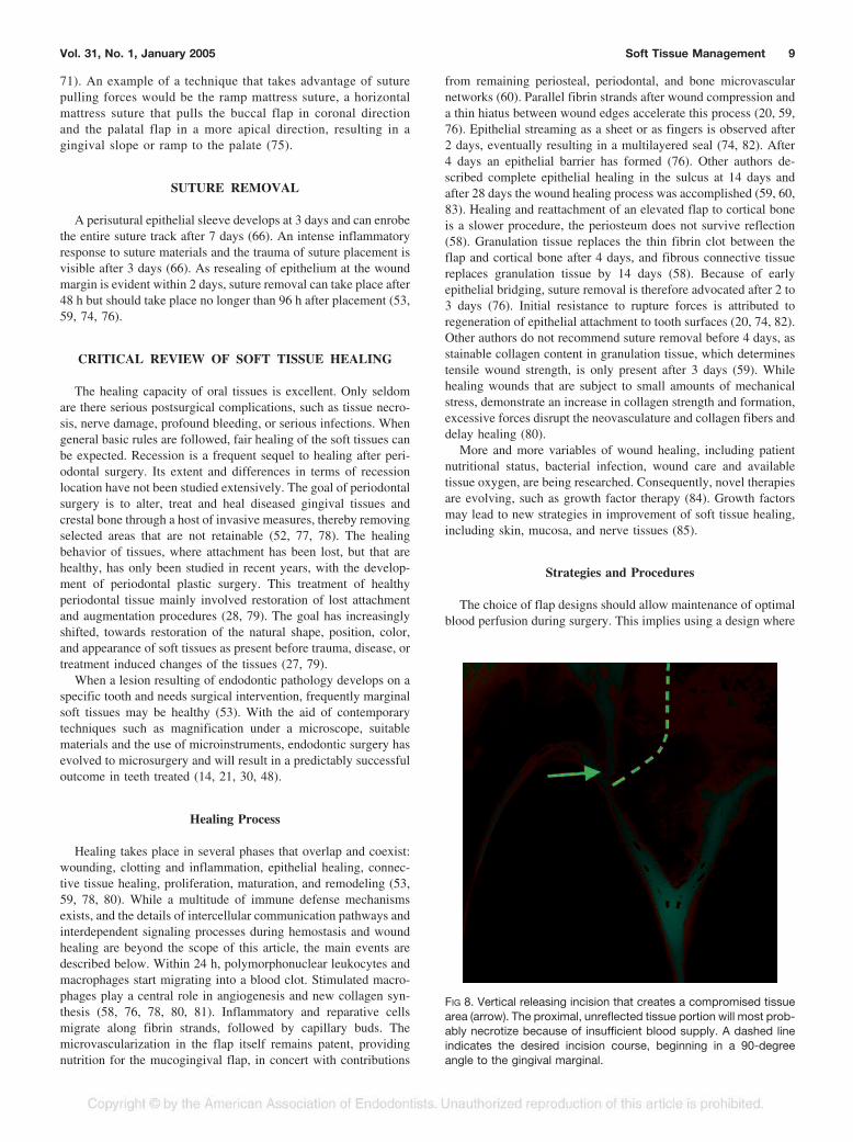
FIG 8. Vertical releasing incision that creates a compromised tissuearea (arrow). The proximal, unreflected tissue portion will most prob-ably necrotize because of insufficient blood supply. A dashed lineindicates the desired incision course, beginning in a 90-degreeangle to the gingival marginal.
Vol. 31, No. 1, January 2005 Soft Tissue Management 9

vertical releasing incisions run vertical, parallel to the tooth axisand to supraperiosteal blood vessels in the mucosa and gingiva,resulting in minimal vascular disruption (30, 31, 43, 45, 53).Paramedian rather than mid-axial releasing incisions are recom-mended to minimize recession risks (Fig. 8) (43). Healing isinfluenced by flap shrinkage and the resulting difficult reapproxi-mation with more sutures than usually necessary. This is true inparticular for submarginal flap types. A vertically oriented releas-ing incision and secondary blood supply from the crestal bone topapillae and attached gingiva can avoid sloughing of unreflectedtissue (43, 86). Unlike horizontal incisions further apically, anincision severing the anastomosis between gingival and periodon-tal vasculature as used in every full mucoperiosteal flap or sulcularflap showed no effect on the circulation in free and alveolargingiva (45). Tissue trauma through stretching, distorting or tear-ing a flap can be avoided through appropriate magnification andcareful manipulation with microsurgical instruments (72, 79).
After reflecting a mucogingival flap, scaling of root-attachedtissues and tissue tags on cortical bone should be avoided to allowrapid reattachment and protection against bone resorption (31, 53,58). Preservation of the papilla in the col area during elevation andreflection is another key point for healing by primary intention(76). Careful handling, undermining elevation and retraction usingthe groove technique are helpful to avoid unnecessary injuries tothe reflected tissue (21, 30, 52). Keeping the flap moist at all timeshelps avoids shrinkage and dehydration (76).
Patient Related Factors
Both the type of tissue involved and the type of surgical wounddetermine the healing process (78). Patients with a “thick” tissuebiotype tend to display coronal soft tissue regrowth to the formerlevel in crown lengthening procedures, whereas patients with a thintissue biotype do not (71, 87). Next to thickness and width of thegingival tissue, the integrity and thickness of underlying boneplays a role in mucogingival stability (79).
MACROSURGICAL VERSUS MICROSURGICALOUTCOME
As stated before, healing results using a macrosurgical approachare generally regarded as fair. Figure 9 shows a representativeresult of flap closure using traditional techniques; in this case, atrapezoidal mucogingival flap was raised. Wound closure wasaccomplished using polyamide 4/0 single knot sutures. The verticalreleasing incisions were closed with three sutures each and thepapilla with a single knot suture. Wound margin adaptation wasconsidered as sufficient at that time. The healing result after 1 wkpostsurgically displayed acceptable healing without any complica-tions and was described as good. Figure 10 shows the surgical sitebefore and directly after suture removal. When the surgical area isexamined more carefully and critically, following observations canbe made: At 7 days, the vertical incision displays complete closureof the wound edges in the apical part. A discrete tissue indentationin the entire extent of the vertical incision is clearly distinguish-able. This indentation becomes more pronounced at the marginallevel and ends in a wound dehiscence, which is esthetically themost critical area. This comes as no surprise as at the time ofconclusion of the surgery the wound edges in the marginal portionwere not properly reapproximated. The healing process requiresclosure of the hiatus between the reflected and the unreflectedtissue with connective tissue and epithelium (76). When adaptationof the tissue edges is ideal and the tissues are positioned in veryclose proximity in the vertical and horizontal dimension to eachother, only few cells need to be generated to bridge the gap. Closeadaptation will expedite wound closure, epithelial cells being thefastest (see section “Wound Healing”). Healing of the verticalincision (Fig. 10B) must be judged as by secondary intention,specifically in the marginal area, resulting in scar tissue formationand tissue defects. The area of the papilla seems to be wellpreserved during the surgical procedure and sutured at its properposition. However, the most coronal portion of the papilla hasshrunk, resulting in a rounded papilla shape and loss of height.
In another case, a submarginal incision was performed usingmicrosurgical techniques and instruments. Flap closure was per-
FIG 9. A trapezoidal flap secured with 4/0 sutures. Postoperativeresult using traditional macrosurgical techniques and instruments.
FIG 10. Same surgical site as in Fig. 9, 7 days postoperatively. Theclinical situation is shown (A) before and (B) directly after sutureremoval. Wound dehiscence visible after suture removal.
FIG 11. Situation of 6/0 sutures after 4 days (A) before and (B) afterremoval. The incision is faintly visible, perfectly healed as well aspoorly healed areas and areas with fair healing are evident.
10 Velvart and Peters Journal of Endodontics

formed with polyamide 6/0 sutures. At suture removal after 4 days(Fig. 11) areas with better healing are visible and the incision isbarely recognizable, just adjacent to a poorly healed portion witha tissue dehiscence and some areas with fair healing. In this clinicalexample, there are incision sections with perfect and poor healingresults directly adjoining to each other. As the surgical techniquewas the same, and the same types of tissues were manipulated itmust be assumed that the different healing patterns were a conse-quence of varying degrees of tissue adaptation after the suturingprocess. It is obvious in this example that there is potential forimproved healing even after very short periods of time. Woundclosure seems to be quite critical in terms of healing outcome.Considerable understanding has been generated from research inmucogingival surgery, specifically in recession coverage and softtissue grafting procedures, as well as in general plastic surgery (22,35, 36, 88).
It has been also pointed out, that if periodontal plastic surgeryis performed, esthetic outcome is often the only important factorand function becomes secondary for example in recession coverageor papilla reconstruction (27). Incision design and suturing tech-nique critically influence the postoperative wound healing processin terms of blood supply and flap survival (45). If both factors arenot guaranteed, esthetic as well as functional success becomeunpredictable (28).
Among other principles (incision and flap design, atraumaticand gentle tissue management), a passive and tension-free woundclosure is fundamental for proper wound healing and for a suc-cessful functional and esthetic outcome. In earlier days, the sutur-ing process was solely regarded as bringing the wound edgestogether and keeping them in this position until body has healed thedefect. It was customary to leave the sutures in place for 7 to 10days and the clinical findings seemed to confirm this protocol. Inan animal experiment the papilla suture was simulated with a
needle corresponding to a 4/0 suture (Fig. 12A). The same area wasphotographed with a magnification of 4! (Fig. 12B). It is obviousthat already the needle tract penetrating through the tissue almostdissects the papilla in half. This generates a large defect, whichmight compromise tissue survival. Changing the size of the suturematerial and accordingly the size of the needle as in microsuturing,it is evident in Fig. 13 that this is much less traumatic. In the sameanimal experiment excessive suture pulling forces on the two tissueparts resulted in crushing and further trauma to the wound. Thescanning electron microphotograph (SEM, Fig. 14A) illustrates theamount of tissue damage inflicted. Note the highly wrinkled andsqueezed tissue beneath the suture. If a suture is applied in such atraumatic way, the healing process will have to repair additionaland considerable damage induced by both the size and excessivetightness of the suture. It is of no surprise that healing will takelonger and will result in esthetically unpleasant healing patterns.Figure 14B (SEM) displays how even minute tissue misalignmentof the wound edges may delay wound healing. In the center of thepicture, there is a small vertical discrepancy in alignment of thetissues. This translates to a larger distance for the cells to cover, ascompared to the area on the left, where both wound edges toucheach other closely. In addition, note small wrinkles in the uppertissue portion, which led to tearing of the tissues at the point of theneedle penetration. Perfect adaptation and atraumatic tissue han-dling will allow improved healing results (Fig. 15).
Papilla Preservation/Protection
The interdental papilla, the portion of the gingiva between twoadjacent teeth, is critical for functional, phonetic and estheticreasons. Complete and predictable restoration of lost interdentalpapillae is one of the biggest challenges in periodontal reconstruc-tive surgery (22). Therefore, it is imperative to maintain the integ-rity of the papilla during surgical procedures. Most frequently, a
FIG 12. Animal model using 4/0 sutures (A) and the same area in 4!magnification (B). Overly traumatic needle tract as needle pene-trates through the papilla.
FIG 13. Same model as in Fig. 12, showing a 7/0 and 8/0 microsu-ture.
FIG 14. Scanning electron microphotographs in 30! magnification.(A) Reveals tissue tension because of excessive suture pullingforces. (B) Displays a small vertical discrepancy in alignment of thetissues adjacent an area on the left, where both wound edges arepositioned more closely.
FIG 15. A microsutured surgical tissue area viewed directly postop-eratively (A) and after 4 days just before suture removal (B). Closeadaptation of the wound margins and atraumatic tissue handlingallow superior healing patterns.
Vol. 31, No. 1, January 2005 Soft Tissue Management 11

sulcular full thickness flap is used in periradicular surgery. In thisflap technique the buccal papilla is mobilized and becomes part ofthe flap (89). Ideally, the sulcular incision should dissect the buccalfrom the lingual papilla. In narrow interproximal spaces, completemobilization of the papilla is often difficult and may cause tissueloss. Shrinkage of the papilla during the healing phase can occur,and may initiate the ultimate loss of papilla height. Zimmermannand coworkers (90), in a preliminary study, investigated the shrink-age of papillae after sulcular flaps in patients with healthy peri-odontal tissues. The reduction of papillary height increased grad-ually during healing. Immediately postoperatively papilla heightloss because of surgical manipulation resulted in a recession rang-ing from 1 quarter (n " 14) to one-quarter to one-half (n " 3) ofthe original height. At suture removal, six sites had a loss of heightof up to one-half the original position. None of the 17 sitesremained at preoperative levels at any time.
A quantitative study analyzed recession of the interdental pa-pilla in periodontally healthy situations after apical surgery usingsulcular flap incisions (91). All experimental sites exhibited asignificant loss of papilla height at 1 month (p # 0.003) and 3months (p # 0.004). Main loss of papilla height occurred betweenbaseline and the 1-month recall situation ($1.1 % 0.8 mm), whilea small but significant further loss occurred between the one and3-month recall appointment (p # 0.05, 0.2 % 0.3 mm). At 3months retractions increased in 10 sites, while in three sites the losshad diminished compared to the value after 1 month. These resultssuggest that the conventional sulcular flap results in considerableretraction of papilla height after 1 month and 3 months postsurgi-cally.
The issue of papilla preservation has been largely addressed inperiodontal therapy. In anterior periodontal surgery, a papillaryretention procedure is advocated to maintain papillary height tomaximize postoperative esthetics (67, 92). Cortellini and cowork-ers (93, 94) suggested a modification of the papilla preservationtechnique, which allows primary closure of the interdental spaceover a bioabsorbable membrane. A horizontal incision is per-formed at the base of the papilla. The papilla is subsequentlyelevated to the buccal side. After coronal repositioning of thebuccal flap over the membrane, the interproximal area is coveredwith the papilla, which is attached to the buccal flap. Primaryclosure over the membrane was obtained in all treated sites usingthe modified preservation technique. Probing attachment levelgains and pocket depth reduction were observed after one yearwhen using this technique.
In endodontic surgical access, Lubow et al. (51) suggested analternative to classical full thickness flaps with the mobilization ofthe papilla. In the technique described, the flap involved fullthickness dissection with easily recognizable landmarks andstraight-line incisions. A beveled horizontal incision was designedto incorporate the maximum amount of facial keratinized tissueinto the body of the flap, while leaving the interproximal tissueuntouched. Healing was described as rapid and with excellentesthetic results.
Preservation of the papilla in periodontal therapy is an acceptedprocedure as described by several authors in the literature (29, 92,95). Straight-line incisions during endodontic surgery, withoutmobilization or inclusion of the papilla into the buccal flap, lead toa clear indentation line where the incision was placed. Figure 16represents a clinical example of such an incision. The same type ofresult is also visible in the publication by Lubow et al. (51). Thisresult was described as esthetically excellent, while in today’scritical and microscope-enhanced judgment it is no longer consid-
ered as such. A further, nevertheless important factor is the loca-tion and microconfiguration of the flapped and unreflected tissue invertical incisions. In the cross-sectional diagram (Fig. 17), the redline represents a paramarginal, single straight incision directed tothe crestal bone. The pointed tissue ending will necrotize at its veryend, creating a small, but visible defect and a recession. This typeof incision is simple to perform, but will result in a poor healingresult. It is evident in the drawing that the mobilized tissue has asharp edge at the coronal end of the flap. The tissue margincomprises unsupported epithelial cells without the epithelial basecells, which are responsible for formation of a multilayered seal ofepithelial cells. The connective tissue at the proximal end of theflap forms a sharp and thin edge, which is not sufficiently vascu-
FIG 16. A single straight horizontal incision as seen in Fig. 17 (redline). A marked indentation line indicates an insufficient incisionplacement.
FIG 17. Schematic drawing with a red line representing a singlestraight incision directed to the crestal bone. The green line repre-sents the papilla base incision with a shallow incision placed at thelevel of the lower third of the papilla in a slight curved line connectingone side of the papilla to the other. In a second step, the scalpel isplaced to the base of the previously created incision inclined api-cally, almost parallel to the long axis of the tooth and directedtowards the crestal bone margin.
12 Velvart and Peters Journal of Endodontics
larized for survival. The healing process will result in localizednecrosis with a small tissue defect, visible as a clearly detectableindentation (Fig. 16).
Consequently, the incision line should begin in a 90-degreeangle to the outer contour of the marginal gingiva as shown andmarked with a green line in Fig. 17. This rule applies to any typeof incision, to avoid thinning out of tissues and allowing sufficientblood supply to reach the area, promoting better healing.
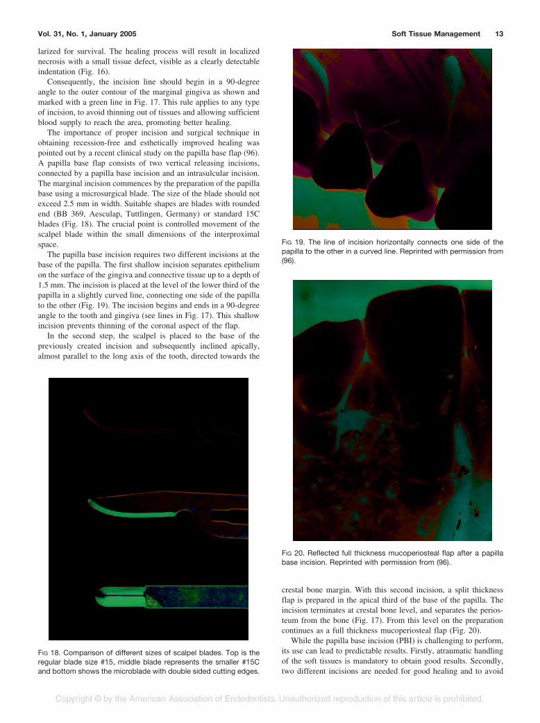
The importance of proper incision and surgical technique inobtaining recession-free and esthetically improved healing waspointed out by a recent clinical study on the papilla base flap (96).A papilla base flap consists of two vertical releasing incisions,connected by a papilla base incision and an intrasulcular incision.The marginal incision commences by the preparation of the papillabase using a microsurgical blade. The size of the blade should notexceed 2.5 mm in width. Suitable shapes are blades with roundedend (BB 369, Aesculap, Tuttlingen, Germany) or standard 15Cblades (Fig. 18). The crucial point is controlled movement of thescalpel blade within the small dimensions of the interproximalspace.
The papilla base incision requires two different incisions at thebase of the papilla. The first shallow incision separates epitheliumon the surface of the gingiva and connective tissue up to a depth of1.5 mm. The incision is placed at the level of the lower third of thepapilla in a slightly curved line, connecting one side of the papillato the other (Fig. 19). The incision begins and ends in a 90-degreeangle to the tooth and gingiva (see lines in Fig. 17). This shallowincision prevents thinning of the coronal aspect of the flap.
In the second step, the scalpel is placed to the base of thepreviously created incision and subsequently inclined apically,almost parallel to the long axis of the tooth, directed towards the
crestal bone margin. With this second incision, a split thicknessflap is prepared in the apical third of the base of the papilla. Theincision terminates at crestal bone level, and separates the perios-teum from the bone (Fig. 17). From this level on the preparationcontinues as a full thickness mucoperiosteal flap (Fig. 20).
While the papilla base incision (PBI) is challenging to perform,its use can lead to predictable results. Firstly, atraumatic handlingof the soft tissues is mandatory to obtain good results. Secondly,two different incisions are needed for good healing and to avoid
FIG 18. Comparison of different sizes of scalpel blades. Top is theregular blade size #15, middle blade represents the smaller #15Cand bottom shows the microblade with double sided cutting edges.
FIG 19. The line of incision horizontally connects one side of thepapilla to the other in a curved line. Reprinted with permission from(96).
FIG 20. Reflected full thickness mucoperiosteal flap after a papillabase incision. Reprinted with permission from (96).
Vol. 31, No. 1, January 2005 Soft Tissue Management 13

excessive scar formation or an indentation at the site of the inci-sion. The key point of the PBI is to avoid thinning of the split flap.The epithelium of the partial thickness portion of the flap needssupport by underlying connective tissue, which has to be thickenough to maintain vitality through sufficient blood supply. If thisgoal cannot be obtained, the tissue will necrotize, resulting in atissue defect, followed by scar formation. On the other hand,excessive thickness of the connective tissue layer of the split flapportion could compromise the survival of the unreflected buccalpapilla. The ideal thickness of a partial thickness flap is unknown.Epithelium thickness varies between 111 to 619 !m with a meanof 364 !m (97). The recommended thickness of free gingival graftswas reported to be 1 to 2 mm (98, 99). Based on gingival graftstudies a thickness of 1 to 1.5 mm was chosen for the split flap inPBI. The selected thickness resulted in excellent healing patterns.
As mentioned previously, flap closure using atraumatic andtension free sutures is a key factor to improved healing. The correctsize of the needle and appropriate suture thickness are equallycritical. Depending on the incision site, the following materialshave been used: Vertical incisions should be sutured with 6/0sutures, because of close proximity to inserting muscles in themucosa, which might exert some tension to the wound duringmastication and speech. As nylon monofilament materials aresomewhat stiff in size 6/0, softer material, such as polyamide(Supramid®) was preferred. This material is a multifilament su-ture, with a coating providing monofilamentous appearance with asmooth surface.
For closure of the horizontal incision and the delicate area of thepapillae, we recommend polypropylene interrupted sutures with asize of 7/0 or smaller (Fig. 21). As the wound edges should beperfectly adapted to each other, depending on the dimensions ofthe papilla two or three interrupted sutures are needed.
Suture removal performed after 3 to 5 days promoted rapidhealing. The critical area for suture removal after 2 to 3 days is theapical portion of the vertical releasing incision—the mucosalwound, in particular in the region with muscle tension, whichmight require 24 to 48 h longer to initiate healing (59, 76, 78).
The evaluation of healing patterns of the papilla base incisionsafter 3 months revealed mainly completely undetectable or onlypartially detectable incision lines and generally demonstrated ex-cellent healing. None of the operated sites displayed any measur-able loss of papilla height, or other complications (96).
Another study analyzed degrees of papilla shrinkage, whenpapilla base flaps and sulcular full-thickness flaps were raised(100). The comparison revealed significant loss of papilla height,when the papilla was mobilized during the surgical procedure. Incontrary, the papilla base incision resulted in rapid and predictablerecession-free healing. To avoid opening of the interproximalspace in esthetically relevant areas, the use of the PBI was recom-mended for periradicular surgical procedures. A further study(101) investigated surgical outcomes concerning main vertical lossof height during 12 months. After three months, only minor ver-tical changes took place; in nine out of 12 sites, increases inrecession depth were observed, while in three sites a small gain ofpapilla height occurred, compared to the 1-month situation. At 12months two sites exhibited further recession, the remaining sitesshowed a certain degree of tissue “creeping.” However, the ob-served gain of the papilla height was not significant, when com-pared to short-term observation periods and the preoperative situ-ation. In cases of full thickness flap elevation, mean papilla loss ofheight after 1 yr was 0.98 mm (Fig. 22).
In contrast to full mobilization of the papilla, the PBI resulted insignificantly lower (p # 0.001) recession depths of only 0.07 %0.09 mm at 1 month, 0.10 % 0.15 mm at three months and$0.062 % 0.21 mm after one year.
CONCLUSIONS
The introduction of microsurgery to surgical endodontics at-tempted to minimize trauma and enhance surgical results. In com-bination with magnification and illumination, resected roots revealintricate anatomical details. In conjunction with ultrasonic root-endpreparation and tight sealing of the root end cavity, the require-ments for mechanical and biological success are more adequatelyfulfilled. Although application of basic rules leads to fair soft tissuehealing after endodontic surgery, there is a great potential forimprovements in postsurgical esthetic outcome. As in other dentalfields, “pink esthetics” of oral soft tissues become increasinglyimportant and efforts are made to minimize scar formation andrecessions after surgical procedures. This is even more the casewhen larger restorations are present and healthy periodontal tissuesare reflected as access flaps for periradicular surgeries. Microsur-gery alone will not accelerate epithelial healing rates, but throughperfect tissue adaptation of wound edges, it can create smallerdistances for epithelial migration during the healing process. More
FIG 21. Size 7/0 polypropylene sutures. The clinical situation (A) atthe conclusion of the surgery and (B) directly after suture removal at4 days. Reprinted with permission from (96).
FIG 22. Clinical example of endodontic microsurgery. The preoper-ative situation, the status presenting immediately postoperativelyand the result after 1, 3, and 12 months. Reprinted with permissionfrom (101).
14 Velvart and Peters Journal of Endodontics

rapid soft tissue healing is a result of reduced tissue trauma andenhanced wound closure during microsurgical procedures.
To achieve these goals several measures are necessary, includ-ing accurate preoperative treatment planning in reference to thecondition and quality of the tissue to be manipulated. Minimaltrauma should be inflicted during incision and raising of the flap.Both the flap and unreflected tissue remaining on the tooth surfaceshould be kept moist during the entire procedure, especially insituations where excellent hemostasis can be achieved. Finally,sensitive handling of the soft tissues during suturing is mandatory,with wound edges being reapproximated without tension and heldin place with nonabsorbable atraumatic sutures. The flap designplays an important role as to how much recession will occurpostoperatively. Papilla base flaps have allowed virtually recessionfree healing after endodontic surgery.
The authors would like to thank Dr. Ove Peters for helpful criticism.
Peter Velvart, DMD, has a private practice in Zurich. Christine I. Peters,DMD, is affiliated with the Department of Preventive Dentistry, Periodontologyand Cariology, University of Zurich, Switzerland and University of the Pacific,San Francisco, CA.
Address requests for reprints to Dr. Peter Velvart, Rennweg 58, CH-8001Zurich, Switzerland; E-mail address: [email protected].
References
1. Friedman S, Stabholz A. Endodontic retreatment—case selection andtechnique. Part 1: criteria for case selection. J Endod 1986;12:28–33.
2. Danin J, Stromberg T, Forsgren H, Linder LE, Ramskold LO. Clinicalmanagement of nonhealing periradicular pathosis. Surgery versus endodonticretreatment. Oral Surg Oral Med Oral Pathol Oral Radiol Endod 1996;82:213–7.
3. Velvart P. Surgical retreatment. In: Bergenholtz G, Hørsted-Bindslev P,Reit C, eds. Textbook of endodontology. Oxford: Blackwell Munksgaard,2003:312–26.
4. Rud J, Andreasen JO, Jensen JF. A multivariate analysis of the influenceof various factors upon healing after endodontic surgery. Int J Oral Surg1972;1:258–71.
5. Rud J, Andreasen JO, Jensen JE. Radiographic criteria for the assess-ment of healing after endodontic surgery. Int J Oral Surg 1972;1:195–214.
6. Rud J, Andreasen JO. A study of failures after endodontic surgery byradiographic, histologic and stereomicroscopic methods. Int J Oral Surg1972;1:311–28.
7. Altonen M, Mattila K. Follow-up study of apicoectomized molars. IntJ Oral Surg 1976;5:33–40.
8. Mikkonen M, Kullaa-Mikkonen A, Kotilainen R. Clinical and radiologicre-examination of apicoectomized teeth. Oral Surg Oral Med Oral Pathol1983;55:302–6.
9. Forssell H, Tammisalo T, Forssell K. A follow-up study of apicectomizedteeth. Proc Finn Dent Soc 1988;84:85–93.
10. Friedman S, Lustmann J, Shaharabany V. Treatment results of apicalsurgery in premolar and molar teeth. J Endod 1991;17:30–3.
11. Maddalone M, Gagliani M. Periapical endodontic surgery: a 3-yearfollow-up study. Int Endod J 2003;36:193–8.
12. Friedman S. Treatment outcome and prognosis of endodontic ther-apy. In: Ørstavik D, Pitt Ford T, eds. Essential endodontology: prevention andtreatment of apical periodontitis. Oxford: Blackwell Science, 1998:388–91.
13. Gutmann J, Harrison J. Success, failure, and prognosis in periradicularsurgery. In: Gutmann J, Harrison J, eds. Surgical endodontics. Oxford: Black-well Scientific Publications, 1991:338–84.
14. Rubinstein RA, Kim S. Short-term observation of the results of end-odontic surgery with the use of a surgical operation microscope and Super-EBA as root-end filling material. J Endod 1999;25:43–8.
15. Ørstavik D. Reliability of the periapical index scoring system. Scand JDent Res 1988;96:108–11.
16. Huumonen S, Lenander-Lumikari M, Sigurdsson A, Ørstavik D. Heal-ing of apical periodontitis after endodontic treatment: a comparison betweena silicone-based and a zinc oxide-eugenol-based sealer. Int Endod J 2003;36:296–301.
17. Jansson L, Sandstedt P, Laftman AC, Skoglund A. Relationship be-tween apical and marginal healing in periradicular surgery. Oral Surg Oral MedOral Pathol Oral Radiol Endod 1997;83:596–601.
18. Ehnevid H, Jansson L, Lindskog S, Blomlof L. Periodontal healing in
teeth with periapical lesions. A clinical retrospective study. J Clin Periodontol1993;20:254–8.
19. Chen S, Wang H, Glickman G. The influence of endodontic treatmentupon periodontal wound healing. J Clin Periodontol 1997;24:449–56.
20. Wirthlin MR. The current status of new attachment therapy. J Peri-odontol 1981;52:529–44.
21. Zuolo ML, Ferreira MO, Gutmann JL. Prognosis in periradicular sur-gery: a clinical prospective study. Int Endod J 2000;33:91–8.
22. Blatz MB, Hurzeler MB, Strub JR. Reconstruction of the lost interprox-imal papilla—presentation of surgical and nonsurgical approaches. Int J Pe-riodontics Restorative Dent 1999;19:395–406.
23. McLean JW. Long-term esthetic dentistry. Quintessence Int 1989;20:701–8.
24. Allen EP. Use of mucogingival surgical procedures to enhance esthet-ics. Dent Clin North Am 1988;32:307–30.
25. Miller PD, Jr. Regenerative and reconstructive periodontal plastic sur-gery. Mucogingival surgery. Dent Clin North Am 1988;32:287–306.
26. Consensus report. Mucogingival therapy. Ann Periodontol 1996;1:702–6.
27. Roccuzzo M, Bunino M, Needleman I, Sanz M. Periodontal plasticsurgery for treatment of localized gingival recessions: a systematic review.J Clin Periodontol 2002;29(Suppl 3):178–94; discussion 95–6.
28. Hurzeler MB, Weng D. Functional and esthetic outcome enhancementof periodontal surgery by application of plastic surgery principles. Int J Peri-odontics Restorative Dent 1999;19:36–43.
29. Cortellini P, Tonetti MS. Microsurgical approach to periodontal regen-eration. Initial evaluation in a case cohort. J Periodontol 2001;72:559–69.
30. Kim S. Principles of endodontic microsurgery. Dent Clin North Am1997;41:481–97.
31. Carr G, Bentkover S. Surgical Endodontics. In: Cohen S, Burns R, eds.Pathways of the pulp. St Louis, MO; Mosby Inc., 1998;608–56.
32. Schroeder HE, Listgarten MA. The gingival tissues: the architecture ofperiodontal protection. Periodontol 2000 1997;13:91–120.
33. Bimstein E, Machtei E, Eidelman E. Dimensional differences in theattached and keratinized gingiva and gingival sulcus in the early permanentdentition: a longitudinal study. J Pedod 1986;10:247–53.
34. Orban B, Wentz F, Everett F, Grant D. Periodontics. St. Louis: Mosby,1958:22.
35. Kohl JT, Zander HA. Morphology of interdental gingival tissues. OralSurg Oral Med Oral Pathol 1961;14:287–95.
36. Pilot T. [Macro-morphology of the interdental papilla]. Dtsch ZahnarztlZ 1973;28:1220–1.
37. Tarnow D, Magner APF. The effect of the distance from the contactpoint to the crest of bone on the presence or absence of the interproximaldental papilla. J Periodontol 1992;63:995–6.
38. Holmes CH. Morphology of the interdental papillae. J Periodontol1965;36:455–60.
39. Nobuto T, Yanagihara K, Teranishi Y, Minamibayashi S, Imai H,Yamaoka A. Periosteal microvasculature in the dog alveolar process. J Peri-odontol 1989;60:709–15.
40. Keller GJ, Cohen DW. India ink perfusions of the vascular plexus oforal tissues. J Oral Surg (Chic) 1955;8:539–42.
41. Folke LE, Stallard RE. Periodontal microcirculation as revealed byplastic microspheres. J Periodontal Res 1967;2:53–63.
42. Castelli WA, Dempster WT. The periodontal vasculature and Its re-sponses to experimental pressures. J Am Dent Assoc 1965;70:890–905.
43. Mormann W, Meier C, Firestone A. Gingival blood circulation afterexperimental wounds in man. J Clin Periodontol 1979;6:417–24.
44. Holmstrup P. Anatomy of the Periodontium. In: Wilson T, Kornmann K,eds. Fundamentals of periodontics. Carol Stream, IL: Quintessence Publish-ing Co., Inc., 2003:21–38.
45. Mormann W, Ciancio SG. Blood supply of human gingiva followingperiodontal surgery. A fluorescein angiographic study. J Periodontol 1977;48:681–92.
46. Maggio JD, Bray KE, Hartwell GR, Lindemann MB, Pisano JV, E.M. R.Appropriateness of care and quality assurance guidelines, 3 ed. Chicago:American Association of Endodontists, 1998:24–32.
47. Commission on Dental Accreditation of the American Dental Associ-ation. Standards for Advanced Specialty Education Programs in Endodontics1998 (revised 2004). http://www.ada.org/prof/ed/accred/standard/endo.pdf.Accessed Nov. 16, 2004.
48. Rubinstein RA, Kim S. Long-term follow-up of cases consideredhealed one year after apical microsurgery. J Endod 2002;28:378–83.
49. Belcher JM. A perspective on periodontal microsurgery. Int J Peri-odontics Restorative Dent 2001;21:191–6.
50. Vreeland DL, Tidwell E. Flap design for surgical endodontics. Oral SurgOral Med Oral Pathol 1982;54:461–5.
51. Lubow RM, Wayman BE, Cooley RL. Endodontic flap design: analysisand recommendations for current usage. Oral Surg Oral Med Oral Pathol1984;58:207–12.
52. Peters LB, Wesselink PR. Soft tissue management in endodonticsurgery. Dent Clin North Am 1997;41:513–28.
53. Gutmann JL, Harrison WH. Flap designs and incisions. In: GutmannJL, Harrison WH, eds. Surgical endodontics. St. Louis, Missouri: IshijakuEuroAmerica, Inc., 1994:162–75.
Vol. 31, No. 1, January 2005 Soft Tissue Management 15

54. Patterson TJ. The survival of skin flaps in the pig. Br J Plast Surg1968;21:113–7.
55. Ohmori S, Kurata K. Experimental studies on the blood supply tovarious types of skin grafts in rabbits using isotope P32. Plast Reconstr Surg1960;25:547–55.
56. Luebke RG. Surgical endodontics. Dent Clin North Am 1974;18:379–91.
57. Lang NP, Loe H. The relationship between the width of keratinizedgingiva and gingival health. J Periodontol 1972;43:623–7.
58. Harrison J, Jurosky K. Wound healing in the tissues of the periodon-tium following periradicular surgery. 2. The dissectional wound. J Endod1991;17:544–52.
59. Selvig K, Torabinejad M. Wound healing after mucoperiosteal surgeryin the cat. J Endod 1996;22:507–15.
60. Kon S, Caffesse RG, Castelli WA, Nasjleti CE. Revascularization fol-lowing a combined gingival flap-split thickness flap procedure in monkeys.J Periodontol 1984;55:345–51.
61. Lilly GE. Reaction of oral tissues to suture materials. Oral Surg OralMed Oral Pathol 1968;26:128–33.
62. Lilly GE, Armstrong JH, Salem JE, Cutcher JL. Reaction of oral tissuesto suture materials. 2. Oral Surg Oral Med Oral Pathol 1968;26:592–9.
63. Lilly GE, Salem JE, Armstrong JH, Cutcher JL. Reaction of oral tissuesto suture materials. 3. Oral Surg Oral Med Oral Pathol 1969;28:432–8.
64. Lilly GE, Cutcher JL, Jones JC, Armstrong JH. Reaction of oral tissuesto suture materials. 4. Oral Surg Oral Med Oral Pathol 1972;33:152–7.
65. Lilly GE, Osbon DB, Hutchinson RA, Heflich RH. Clinical and bacteri-ologic aspects of polyglycolic acid sutures. J Oral Surg 1973;31:103–5.
66. Selvig KA, Biagiotti GR, Leknes KN, Wikesjo UM. Oral tissue reactionsto suture materials. Int J Periodontics Restorative Dent 1998;18:474–87.
67. Michaelides PL, Wilson SG. A comparison of papillary retention versusfull-thickness flaps with internal mattress sutures in anterior periodontal sur-gery. Int J Periodontics Restorative Dent 1996;16:388–97.
68. Yaltirik M, Dedeoglu K, Bilgic B, et al. Comparison of four differentsuture materials in soft tissues of rats. Oral Dis 2003;9:284–6.
69. Parell GJ, Becker GD. Comparison of absorbable with nonabsorbablesutures in closure of facial skin wounds. Arch Facial Plast Surg 2003;5:488–90.
70. van der Horst C, Martinez Portillo FJ, Melchior D, Bross S, Alken P,Juenemann KP. Polytetrafluoroethylene versus polypropylene sutures for Es-sed-Schroeder tunical plication. J Urol 2003;170:472–5.
71. Caudill RF, Oringer RJ, Langer L, Langer B, Bahat O, Handelsman M.Esthetic periodontics (periodontal plastic surgery). In: Wilson T, Kornmann K,eds. Fundamentals of periodontics. Carol Stream, IL: Quintessence Publish-ing Co., Inc., 2003:540–61.
72. Bahat O, Handelsman M. Periodontal reconstructive flaps—classificationand surgical considerations. Int J Periodontics Restorative Dent 1991;11:480–7.
73. Nelson EH, Funakoshi E, O’Leary TJ. A comparison of the continuousand interrupted suturing techniques. J Periodontol 1977;48:273–81.
74. Wirthlin MR, Hancock EB, Gaugler RW. The healing of atraumatic andtraumatic incisions in the gingivae of monkeys. J Periodontol 1984;55:103–13.
75. Tinti C, Benfentai S. The ramp mattress suture: a new suturing tech-nique combined with a surgical procedure to obtain papillae between implantsin the buccal area. Int J Periodontics Restorative Dent 2002;22:63–9.
76. Harrison J, Jurosky K. Wound healing in the tissues of the periodon-tium following periradicular surgery. I. The incisional wound. J Endod 1991;17:425–35.
77. Pihlstrom BL, McHugh RB, Oliphant TH, Ortiz-Campos C. Comparisonof surgical and nonsurgical treatment of periodontal disease. A review ofcurrent studies and additional results after 61/2 years. J Clin Periodontol1983;10:524–41.
78. Harrison JW. Healing of surgical wounds in oral mucoperiosteal tis-sues. J Endod 1991;17:401–8.
79. Tibbetts LS, Shanelec D. Current status of periodontal microsurgery.Curr Opin Periodontol 1996;3:118–25.
80. Wong ME, Hollinger JO, Pinero GJ. Integrated processes responsiblefor soft tissue healing. Oral Surg Oral Med Oral Pathol Oral Radiol Endod1996;82:475–92.
81. Hunt TK, Knighton DR, Thakral KK, Goodson WH, 3rd, Andrews WS.Studies on inflammation and wound healing: angiogenesis and collagen syn-thesis stimulated in vivo by resident and activated wound macrophages.Surgery 1984;96:48–54.
82. Wirthlin MR, Yeager JE, Hancock EB, Gaugler RW. The healing ofgingival wounds in miniature swine. J Periodontol 1980;51:318–27.
83. Afshar-Mohajer K, Stahl SS. The remodeling of human gingival tissuesfollowing gingivectomy. J Periodontol 1977;48:136–9.
84. Hom DB. A new era of discovery in facial plastic surgery. Arch FacialPlast Surg 2000;2:166–72.
85. Hom DB, Thatcher G, Tibesar R. Growth factor therapy to improve softtissue healing. Facial Plast Surg 2002;18:41–52.
86. Kindlova M. The blood supply of the marginal periodontium in Maca-cus rhesus. Arch Oral Biol 1965;10:869–74.
87. Pontoriero R, Carnevale G. Surgical crown lengthening: a 12-monthclinical wound healing study. J Periodontol 2001;72:841–8.
88. Nemcovsky C. Interproximal papilla augmentation procedure: a novelsurgical approach and clinical evaluation of 10 consecutive procedures. Int JPeriodontics Restorative Dent 2001;21:553–9.
89. Beer R, Baumann M, Kim S. Endodontology. Stuttgart: Thieme Verlag,2000:238–9.
90. Zimmermann U, Ebner J, Velvart P. Papilla healing following sulcularfull thickness flap in endodontic surgery. J Endod 2001;27:219.
91. Velvart P, Ebner-Zimmermann U, Ebner JP. Papilla healing followingfull thickness flap in endodontic surgery. (in press)
92. Takei H, Han T, Carranza FJ, Kenney E, Lekovic V. Flap technique forperiodontal bone implants. Papilla preservation technique. J Periodontol1985;56:204–10.
93. Cortellini P, Prato GP, Tonetti MS. The modified papilla preservationtechnique. A new surgical approach for interproximal regenerative proce-dures. J Periodontol 1995;66:261–6.
94. Cortellini P, Pini Prato G, Tonetti MS. The modified papilla preservationtechnique with bioresorbable barrier membranes in the treatment of intrabonydefects. Case reports. Int J Periodontics Restorative Dent 1996;16:546–59.
95. Takei H, Yamada H, Hau T. Maxillary anterior esthetics. Preservationof the interdental papilla. Dent Clin North Am 1989;33:263–73.
96. Velvart P. Papilla base incision: a new approach to recession-freehealing of the interdental papilla after endodontic surgery. Int Endod J 2002;35:453–60.
97. Soehren SE, Allen AL, Cutright DE, Seibert JS. Clinical and histologicstudies of donor tissues utilized for free grafts of masticatory mucosa.J Periodontol 1973;44:727–41.
98. Mormann W, Schaer F, Firestone A. The relationship between successof free gingival grafts and transplant thickness. Revascularization andshrinkage—one-year clinical study. J Periodontol 1981;52:74–80.
99. Wennstrom J, Pini Prato G. Mucogingival therapy. In: Clinical peri-odontology and implant dentistry, 3rd ed. Copenhagen: Munksgaard, 1998:550–96.
100. Velvart P, Ebner-Zimmermann U, Ebner JP. Comparison of papillahealing following sulcular full-thickness flap and papilla base flap in endodon-tic surgery. Int Endod J 2003;36:653–9.
101. Velvart P, Ebner-Zimmermann U, Ebner JP. Comparison of long termpapilla healing following sulcular full thickness flap and papilla base flap inendodontic surgery. Int Endod J (in press).
102. Gutmann JL, Harrison JW. Closure of the surgical site and postsur-gical management. In: Gutmann JL, Harrison JW, eds. Surgical endodontics.St. Louis: Ishiyaku EuroAmerica, 1994:278–99.
16 Velvart and Peters Journal of Endodontics