review series challenges in pulmonary fibrosis 1:...
TRANSCRIPT

REVIEW SERIES
Challenges in pulmonary fibrosis ? 1: Use of high resolutionCT scanning of the lung for the evaluation of patients withidiopathic interstitial pneumoniasMichael B Gotway, Michelle M Freemer, Talmadge E King Jr. . . . . . . . . . . . . . . . . . . . . . . . . . . . . . . . . . . . . . . . . . . . . . . . . . . . . . . . . . . . . . . . . . . . . . . . . . . . . . . . . . . . . . . . . . . . . . . . . . . . . . . . . . . . . . . . . . . . . . . . . . . . . . . . . . .
Thorax 2007;62:546–553. doi: 10.1136/thx.2004.040022
High resolution CT (HRCT) scanning has contributed significantlyto the evaluation of patients with interstitial lung disease and isparticularly useful in the diagnosis of idiopathic pulmonaryfibrosis (IPF). The characteristic radiographic features of theidiopathic interstitial pneumonias on HRCT scans have beenincreasingly analysed and are now fairly well described. Basedon current data, HRCT scanning can provide a confident, highlyspecific diagnosis of IPF in many patients with diffuse lungdisease. This article reviews an organised approach to HRCTscanning and identifies the features that allow an accuratediagnosis of the idiopathic interstitial pneumonias to be made.The role of surgical lung biopsy is discussed in the diagnosis ofcases when a definite HRCT diagnosis cannot be made.. . . . . . . . . . . . . . . . . . . . . . . . . . . . . . . . . . . . . . . . . . . . . . . . . . . . . . . . . . . . . . . . . . . . . . . . . . . . .
See end of article forauthors’ affiliations. . . . . . . . . . . . . . . . . . . . . . . .
Correspondence to:Dr Michael Gotway,Scottsdale MedicalImaging, and affiliate ofSouthwest DiagnosticImaging, 3501 NorthScottsdale Road, Suite 130,Box 1573, Scottsdale, AZ85251, USA
Received31December2004Accepted 21 April 2005. . . . . . . . . . . . . . . . . . . . . . . .
The diagnostic evaluation of a patient withdyspnoea presents a challenge to the clinician.Histopathological evaluation has shown that
the clinical and radiological diagnosis of theidiopathic interstitial pneumonias (IIPs) is moreheterogeneous than previously thought. The sub-classification of the IIPs based on clinical andpathological criteria has important therapeutic andprognostic implications.
A structured, clinical, radiographic and patholo-gical approach to the diagnosis of the IIPs isimportant in ensuring proper diagnosis and man-agement of these patients. The most importantdistinction is the presence of usual interstitialpneumonia (UIP), the histopathological patternseen in idiopathic pulmonary fibrosis (IPF). Assurgical lung biopsy is a morbid procedure, therehas been significant interest in exploring the clinicaldiagnosis of IPF. Historically the clinical diagnosishas been quite non-specific, but recent advances inthe clinical and radiographic description of IPF havedramatically improved the clinicoradiographic diag-nostic accuracy. The focus of this review is to clarifythe role of high resolution computed tomography(HRCT) and surgical lung biopsy in the assessmentof patients with IIP.
ROLE OF HIGH RESOLUTION CT (HRCT)SCANNING IN THE DIAGNOSIS OFIDIOPATHIC INTERSTITIAL PNEUMONIAS(IIPS)The IIPs are comprised of UIP, non-specific inter-stitial pneumonia (NSIP), acute interstitial pneu-monia (AIP), respiratory bronchiolitis-interstitial
lung disease (RB-ILD), desquamative interstitialpneumonia (DIP), cryptogenic organising pneumo-nia (COP) and lymphocytic interstitial pneumonia(LIP). When evaluating a patient with dyspnoea forthe possibility of an IIP, the history remains criticalto eliminate any systemic diseases or potential drugor environmental exposures that may cause pul-monary disease. The physical examination can alsoserve to evaluate the possibility of a systemicillness.1
The conventional chest radiograph is of limitedvalue in the diagnosis of IPF. Given its highsensitivity as a diagnostic test, HRCT scanning ofthe lung has become an indispensable tool toidentify the presence of interstitial lung disease(ILD). Furthermore, in a patient who has anidiopathic ILD, as determined by the history andphysical examination, HRCT evaluation also hasthe potential to narrow the differential diagnosisor to make a specific diagnosis. An organisedapproach to HRCT scanning techniques and inter-pretation in these patients is required for properdiagnosis and management.
HRCT TECHNIQUECollimationNarrow collimation (section thickness) is required,usually of the order of 1 mm, to achieve maximalspatial resolution. HRCT imaging is performedusing axial, as opposed to helical, technique. Atypical HRCT protocol uses 1–2 mm collimationevery 10–20 mm throughout the thorax, whicheffectively images only approximately 10% of thelung parenchyma. Because HRCT scanning istypically used for the assessment of diffuse lungdisease, such a sampling technique providesadequate representation of the disease processwhile minimising the radiation dose delivered tothe patient. Some investigators have recentlyapplied narrow collimation multi-slice helical CTscanning to patients with diffuse lung disease, butthe benefit of volumetric imaging must be weighedagainst the substantial increased radiation doseinherent to this technique.
Abbreviations: AIP, acute interstitial pneumonia; COP,cryptogenic organising pneumonia; DIP, desquamativeinterstitial pneumonia; HRCT, high resolution CT; IIP,idiopathic interstitial pneumonia; ILD, interstitial lungdisease; IPF, idiopathic pulmonary fibrosis; LIP, lymphocyticinterstitial pneumonia; NSIP, non-specific interstitialpneumonia; RB-ILD, respiratory bronchiolitis-interstitial lungdisease; UIP, usual interstitial pneumonia
546
www.thoraxjnl.com
on 11 July 2018 by guest. Protected by copyright.
http://thorax.bmj.com
/T
horax: first published as 10.1136/thx.2004.040022 on 29 May 2007. D
ownloaded from

Expiratory scanningIntegral to all HRCT scanning protocols is the use of expiratoryscanning. Expiratory HRCT may be performed using staticmethods (imaging at functional residual capacity), lateraldecubitus CT,2 or dynamic expiratory HRCT (imaging during aforced vital capacity manoeuvre).3 While expiratory HRCT oftenadds little to the evaluation of patients with IIPs (with theexception of RB-ILD),4 expiratory HRCT is very important forthe evaluation of a number of other diffuse lung diseases thatmay clinically resemble IIPs, such as hypersensitivity pneumo-nitis.
Prone imagingFor many patients with ILD, and in particular for the IIPs,prone imaging is essential. Supine HRCT images commonlyshow opacities in the dependent portions of the lung (fig 1)which are often caused by atelectasis. Such opacities may,however, resemble infiltrative lung disease (fig 2A). Whenpatients are scanned in the prone position, atelectasis willresolve (fig 1B), unlike true lung pathology which will notresolve (fig 2B).
APPROACH TO HRCT EVALUATION IN PATIENTSWITH IIPSHRCT findings of IIPs may be broadly categorised into foci ofincreased opacity and foci of decreased opacity (table 1). Themajor imaging findings manifesting as increased opacity onHRCT imaging in patients with IIPs include ground glassopacity, consolidation, linear and reticular opacities, architec-tural distortion, airway thickening and, occasionally, centri-lobular nodules. While each of these findings individually maybe seen in the various IIPs, certain findings tend to predomi-nate more than others in each disease, as will be discussedsubsequently. The imaging findings in patients with IIP thatmanifest as decreased opacity on HRCT imaging includetraction bronchiectasis, honeycomb lung and air trapping (also,
Figure 1 Value of prone high resolution (HR) CT imaging: resolution ofdependent lung opacity. (A) Axial supine HRCT image showing bilateraldependent ground glass opacity (arrows; compare with fig 2A). (B) Axialprone HRCT image shows resolution of the ground glass opacity in (A),consistent with atelectasis. Note the opacity within the lungs anteriorly,representing dependent atelectasis occurring in the prone position.
Figure 2 Value of prone high resolution (HR) CT imaging: persistence ofinfiltrative lung disease. (A) Axial supine HRCT image shows bilateraldependent ground glass opacity and reticulation (arrows). Note similarityto fig 1A. (B) Axial prone HRCT image showing persistence of the groundglass opacity and reticulation (arrows) seen in (A), consistent withinfiltrative lung disease. Surgical lung biopsy proved non-specific interstitialpneumonitis.
Table 1 HRCT findings commonly encountered in patientswith idiopathic interstitial pneumonias (IIPs)*
IIP Distribution Predominant HRCT findings
UIP Basilar, subpleural Reticulation (usually coarse)Architectural distortionTraction bronchiectasisHoneycombing
NSIP Basilar, subpleural Ground glass opacityReticulation (usually fine)Architectural distortionTraction bronchiectasisConsolidation
AIP Diffuse Ground glass opacityConsolidation
RB-ILD Multifocal Ground glass opacityAirway thickeningAir trappingGround glass centrilobular nodules
DIP Multifocal, often peripheral Ground glass opacityReticulation
COP Subpleural, peribronchiolar ConsolidationGround glass opacity
LIP No predominance Ground glass opacityPeribronchiolar nodulesCentrilobular nodulesLinear opacitiesCysts
HRCT, high resolution CT; IIP, idiopathic interstitial pneumonia; UIP,usual interstitial pneumonia; NSIP, non-specific interstitial pneumonia;AIP, acute interstitial pneumonia; RB-ILD, respiratory bronchiolitis-interstitiallung disease; DIP, desquamative interstitial pneumonia; COP,cryptogenic organising pneumonia; LIP, lymphocytic interstitialpneumonia*The predominant findings may vary somewhat depending on the phase ofthe disease.
HRCT scanning of the lung for idiopathic interstitial pneumonias 547
www.thoraxjnl.com
on 11 July 2018 by guest. Protected by copyright.
http://thorax.bmj.com
/T
horax: first published as 10.1136/thx.2004.040022 on 29 May 2007. D
ownloaded from

because many of these patients are smokers, emphysema is acommon finding).
The first step in the interpretation of HRCT imaging inpatients with IIP is the assessment for the presence or absenceof a pattern suggestive of UIP.1 If HRCT findings typical of UIPare found, one may confidently suggest the diagnosis of UIPand surgical lung biopsy may be avoided. In the absence of clearfindings suggesting UIP, the HRCT scan should be examined forfeatures that specifically indicate an alternative diagnosis,either another IIP or another interstitial lung process. Theimaging differential diagnosis is then ordered accordingly, andthe HRCT scan findings are correlated with the clinical history1
to suggest the most likely diagnosis. Occasionally a confidentalternative diagnosis may be made in this situation (fig 3), butsurgical lung biopsy is often required to firmly establish adiagnosis.
USUAL INTERSTITIAL PNEUMONIA (UIP)Typical HRCT featuresHRCT findings characteristic of UIP consist of bilateral basilarsubpleural reticulation often accompanied by traction bronch-iectasis and architectural distortion and honeycomb cysts(fig 4).1 5–7 In general, these findings gradually decrease inextent from base to apex. The linear and reticular opacities areusually coarser in patients with UIP than those with other IIPs.
Honeycombing in UIPOne of the key findings (see below) that suggests the diagnosisof UIP on HRCT imaging is the presence of honeycomb cysts ina basilar subpleural distribution. Honeycomb changes appearon the HRCT scan as variably-sized cystic spaces that sharewalls and frequently stack upon one another in several layers(fig 4). The presence of centrilobular emphysema can some-times make the diagnosis of honeycombing more difficult.
Atypical HRCT featuresIn addition to these typical findings, UIP is also characterisedby the absence of a number of features. More specifically,ground glass opacity (particularly if extensive), consolidation,nodules, pleural effusion and lymphadenopathy are notconspicuous findings on the HRCT scan in patients with UIP.5
Mildly enlarged lymph nodes are often encountered on HRCTexaminations performed on patients with UIP-IPF, but thepresence of such lymph nodes does not contribute to makingthe diagnosis of UIP on imaging.
Importantly, the presence of ground glass opacity on theHRCT scan does not rule out histopathologically proven UIP butits presence—particularly if extensive—should suggest anotherdiagnosis. The histological correlate of ground glass opacity on
HRCT imaging is often an inflammatory process within theinterstitium or alveolar spaces. However, ground glass opacity isnot synonymous with inflammation; rather, ground glassopacity on HRCT scans obtained in patients with UIP mayreflect the presence of interstitial fibrosis on histologicalexamination.8 9 In this setting, ground glass opacity will beaccompanied by other findings suggestive of fibrosis such asarchitectural distortion, coarse reticulation, traction bronchiec-tasis and honeycombing.
Accuracy of HRCT for the diagnosis of UIPATS/ERS criteriaSeveral studies have been performed to investigate whetherHRCT scanning can obviate the need for surgical lung biopsy tomake the diagnosis of UIP. The current ATS/ERS consensusstatement regarding the diagnosis of IPF includes bibasilarreticular abnormalities with minimal ground glass opacities asone of the major criteria that can be used in the appropriateclinical context. The other criteria include no known causes ofILD, restrictive physiology and impaired gas exchange, and atransbronchial biopsy excluding alternative diagnoses in anindividual over 50 years of age with the insidious onset ofdyspnoea lasting longer than 3 months and crackles onexamination.
‘‘Definite’’ UIP on HRCTIn a group of patients from multiple centres selected on thebasis of the suspicion of IPF, expert radiologists were ableconfidently to diagnose UIP in nearly 60% of patients with a
Figure 3 Patient with suspected idiopathic interstitial pneumonia:alternative diagnosis established with high resolution (HR) CT imaging.Axial HRCT image through the pulmonary apices shows bilateral bizarre-shaped cysts (arrows) and nodules characteristic of Langerhans’ cellhistiocytosis.
Figure 4 Usual interstitial pneumonia (UIP) pattern on high resolution (HR)CT imaging: characteristic findings. Axial prone HRCT image through thelung bases shows bilateral subpleural cysts (arrows) representinghoneycombing, as well as reticulation and traction bronchiectasis (smallwhite arrows).
Figure 5 Pattern of usual interstitial pneumonia (UIP) on high resolution(HR) CT imaging: insufficiently characteristic findings. Axial HRCT imageshows basilar subpleural reticulation but no evidence of honeycomb lung.A confident diagnosis of UIP could not be offered on the basis of theimaging findings. Surgical lung biopsy showed findings characteristic ofUIP.
548 Gotway, Freemer, King
www.thoraxjnl.com
on 11 July 2018 by guest. Protected by copyright.
http://thorax.bmj.com
/T
horax: first published as 10.1136/thx.2004.040022 on 29 May 2007. D
ownloaded from

positive predictive value of 96%.6 In a subsequent analysis, inthe presence of the other typical findings of UIP describedabove, either of two particular findings—upper lobe reticula-tions and lower lobe honeycombing—was found to increase theprobability of a histopathological diagnosis of UIP by 5–6-fold.5
Similarly, in another study of patients suspected of having anIIP, the presence of honeycombing on the HRCT scan in at leastone lobe had a positive predictive value of 92%.10 However, thediagnosis of UIP cannot be confidently offered on HRCTimaging when honeycomb cysts are not seen (fig 5). In fact,some patients without radiographic honeycombing will haveUIP diagnosed on surgical lung biopsy.6
Prognostic value of HRCT scanning in UIPIn addition to its diagnostic value in UIP, HRCT scanning alsohas prognostic value. The presence of a definite (confident)HRCT diagnosis of UIP on the basis of basilar honeycombingportends a worse survival than for individuals withouthoneycombing on the HRCT scan and for those with ahistopathological diagnosis of UIP.10 11
NON-SPECIFIC INTERSTITIAL PNEUMONIA (NSIP)Typical featuresThe most difficult IIP to distinguish from a UIP is NSIP.However, in contrast to UIP, the dominant feature of NSIP onHRCT imaging is basilar, subpleural, symmetrical, bilateralground glass opacity (fig 6).12–14 In fact, ground glass opacitymay be the sole feature in nearly one-third of patients withNSIP.1 A peculiar pattern in which the peripheral ground glassopacity and reticulation spares the immediate subpleural regionof lung has been recognised (fig 7) and is considered suggestiveof NSIP. Irregular linear and reticular opacities are oftenpresent in patients with NSIP (fig 8), and these opacitiesbecome increasingly coarse as the fibrotic elements becomemore pronounced on histopathological specimens.14 Tractionbronchiectasis and bronchiolectasis is often visible and alsobecomes increasingly prominent as the fibrosis progresses.14
Honeycombing in NSIPInitial experience suggested that honeycomb changes are veryuncommon in patients with NSIP,15 but larger studies haveshown that honeycomb lung may be seen in as many as 27% ofpatients with NSIP.14 Patients with NSIP who have honeycombcysts visible on the HRCT scan, however, usually have a morefibrotic (rather than a cellular) form of NSIP, and tractionbronchiectasis, traction bronchiolectasis, coarse reticulationand architectural distortion are usually also evident. It shouldbe noted that, given the frequent difficulty in distinguishingfibrotic NSIP from UIP pathologically, many of these cases with
honeycomb lung are believed to represent cases of UIP-IPF(NSIP Workshop Committee. American Thoracic SocietyWorkshop on Idiopathic Nonspecific Interstitial Pneumonia(NSIP), American Thoracic Society International Conference.Seattle, WA, 2003).
Accuracy of HRCT scanning for the diagnosis of NSIPThe reliability of a ‘‘confident’’ HRCT diagnosis of NSIP is moretenuous than for UIP. Even when cases are known to be eitherUIP or NSIP histopathologically, the diagnostic accuracy ofradiologists when they are confident of the diagnosis of NSIP isonly 72%.16 In practice, such limited accuracy in a group ofpatients known to have a fibrotic IIP (chance alone wouldpredict 50% accuracy) indicates the need for surgical lungbiopsy to diagnose NSIP.
Serial HRCT images may show improvement in ground glassopacity and reticulation in patients with NSIP undergoingtreatment (fig 9), although residual abnormalities oftenremain.
ACUTE INTERSTITIAL PNEUMONIA (AIP)HRCT features can distinguish AIP from UIP. The typical HRCTfeatures of AIP are bilateral, multifocal or diffuse areas ofground glass opacity and consolidation, usually withoutpleural effusion (fig 10).17 18 No clear zonal distribution isidentifiable, although the consolidation is often dependent inlocation. HRCT findings often reflect the stage of the diseaseand underlying histopathological process. During the organis-ing phase of the disease, HRCT findings consistent withevolving fibrosis are often present including traction bronch-iectasis, linear and reticular abnormalities, and architectural
Figure 6 Pattern of non-specific interstitial pneumonia (NSIP) on highresolution (HR) CT imaging shows bilateral subpleural symmetric groundglass opacity (arrows).
Figure 7 Pattern of non-specific interstitial pneumonia (NSIP) on highresolution (HR) CT imaging shows peripheral ground glass opacity sparingthe immediate subpleural lung (arrows).
Figure 8 Pattern of non-specific interstitial pneumonia (NSIP) on highresolution (HR) CT imaging shows bilateral subpleural linear and reticular(arrows) opacities. Note the presence of traction bronchiolectasis(arrowheads).
HRCT scanning of the lung for idiopathic interstitial pneumonias 549
www.thoraxjnl.com
on 11 July 2018 by guest. Protected by copyright.
http://thorax.bmj.com
/T
horax: first published as 10.1136/thx.2004.040022 on 29 May 2007. D
ownloaded from

distortion.17 Among survivors, HRCT scans show clearing ofmost abnormalities, but foci of reticulation, parenchymaldistortion, cystic change or honeycombing may remain.17 18
SMOKING-RELATED INTERSTITIAL LUNG DISEASES:RESPIRATORY BRONCHIOLITIS-INTERSTITIAL LUNGDISEASE (RB-ILD) AND DESQUAMATIVE INTERSTITIALPNEUMONIA (DIP)While a history of smoking is almost always present in RB-ILDand DIP, patients with UIP are also frequently smokers.However, the HRCT features of these smoking related ILDsshow a combination of findings distinct from UIP, such as,prominent ground glass opacity, subpleural reticulation,emphysema and cystic changes that are not classic forhoneycomb lung.
Respiratory bronchiolit is-interstitial lung disease (RB-ILD)The primary HRCT findings in patients with RB-ILD includecentrilobular ground glass attenuation nodules (fig 11), patchyareas of ground glass opacity (fig 12) and airway thicken-ing.1 4 19 20 Patchy areas of hypoattenuation (fig 12) representingmosaic perfusion are often encountered, and may be shown torepresent air trapping on expiratory imaging.4 These findingsmay resolve with treatment. Centrilobular emphysema is oftenpresent in patients with RB-ILD.
Desquamative interstit ial pneumonia (DIP)Desquamative interstitial pneumonia (DIP) primarily manifestson HRCT imaging as multifocal or diffuse ground glass opacity,often with a basilar and peripheral predominance (fig 13).1 19 21
Irregular linear and reticular opacities are commonly seen butare not usually the dominant features of the disease.Honeycombing may be seen in the minority of patients and isusually limited in extent.
CRYPTOGENIC ORGANISING PNEUMONIA (COP)Typical featuresCOP is also usually radiographically distinct from UIP, with themost common HRCT finding being consolidation which ispresent in 90% of patients.1 22 In up to 50% of patients theconsolidation is peripheral or peribronchiolar in distribution(fig 14)1 and is more commonly encountered in the lower lobes.Mild bronchial dilation may be present in the areas ofconsolidation. Ground glass opacity is also commonly present
Figure 9 Non-specific interstitial pneumonia (NSIP) on high resolution(HR) CT imaging: improvement with treatment. (A) Axial HRCT imageshows basilar subpleural ground glass opacity, reticulation andconsolidation (arrows), proved to be NSIP on surgical biopsy. (B) AxialHRCT image obtained 3 months after immunosuppressive therapy showingregression of the ground glass opacity and consolidation; some linear andreticular elements (arrows) remain.
Figure 10 Acute interstitial pneumonia (AIP) on high resolution (HR) CTimaging. Axial HRCT image shows multifocal bilateral areas of groundglass opacity and bronchovascular thickening in a patient with AIP shownon surgical lung biopsy.
Figure 11 Respiratory bronchiolitis-interstitial lung disease (RB-ILD) onhigh resolution (HR) CT imaging: ground glass attenuation centrilobularnodules. Axial HRCT image shows multiple ground glass attenuationnodules (arrows) and patchy areas of ground glass opacity.
Figure 12 Respiratory bronchiolitis-interstitial lung disease (RB-ILD) onhigh resolution (HR) CT imaging: patchy ground glass opacity. Axial HRCTimage shows multifocal areas of ground glass opacity (arrows).Hypoattenuating areas (arrowheads) have a noticeably lobularconfiguration and represent mosaic perfusion due to small airwayobstruction.
550 Gotway, Freemer, King
www.thoraxjnl.com
on 11 July 2018 by guest. Protected by copyright.
http://thorax.bmj.com
/T
horax: first published as 10.1136/thx.2004.040022 on 29 May 2007. D
ownloaded from
and often coexists with consolidation. Small nodules arecommonly seen in patients with COP, but are not usually adominant feature.
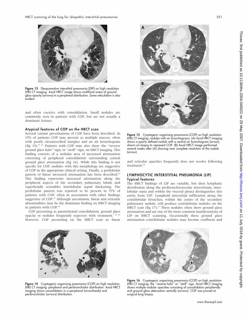
Atypical features of COP on the HRCT scanSeveral variant presentations of COP have been described. In15% of patients COP may present as multiple masses, oftenwith poorly circumscribed margins and an air bronchogram(fig 15).23 24 Patients with COP may also show the ‘‘reverseground glass halo’’ sign, or ‘‘atoll’’ sign, on HRCT imaging. Thisfinding consists of a nodular area of increased attenuationconsisting of peripheral consolidation surrounding centralground glass attenuation (fig 16). While this finding is notspecific for COP, nodules with this morphology are suggestiveof COP in the appropriate clinical setting. Finally, a perilobularpattern of linear increased attenuation has been described.25
This finding represents increased attenuation along theperipheral aspects of the secondary pulmonary lobule andsuperficially resembles interlobular septal thickening. Theperilobular pattern was reported to be present in 57% ofpatients with COP, often in association with other findingssuggestive of COP.25 Although uncommon, linear and reticularabnormalities may be the dominant finding on HRCT imagingin patients with COP.26
COP presenting as parenchymal consolidation, ground glassopacity or nodules frequently regresses with treatment.1 22 26
However, COP presenting on the HRCT scan as linear
and reticular opacities frequently does not resolve followingtreatment.26
LYMPHOCYTIC INTERSTITIAL PNEUMONIA (LIP)Typical featuresThe HRCT findings of LIP are variable, but their lymphaticdistribution along the peribronchovascular interstitium, inter-lobular septa and within the visceral pleura distinguishes thisentity from UIP. Lymphoid interstitial infiltration along thecentrilobular bronchus, within the centre of the secondarypulmonary nodule, will produce centrilobular nodules on theHRCT scan (fig 17).27 These nodules often show ground glassattenuation and are one of the more common manifestations ofLIP on HRCT scanning. Occasionally these ground glassattenuation centrilobular nodules may become confluent and
Figure 13 Desquamative interstitial pneumonia (DIP) on high resolution(HR) CT imaging. Axial HRCT image shows multifocal areas of groundglass opacity (arrows) in a peripheral distribution. Some reticulation is alsoevident.
Figure 14 Cryptogenic organising pneumonia (COP) on high resolution(HR) CT imaging: peripheral and peribronchiolar distribution. Axial HRCTimaging shows consolidation in a peripheral (arrowheads) andperibronchiolar (arrows) distribution.
Figure 15 Cyrptogenic organising pneumonia (COP) on high resolution(HR) CT imaging: nodules with air bronchograms. (A) Axial HRCT imagingshows a poorly defined nodule with a central air bronchogram (arrow),shown on biopsy to represent COP. (B) Axial HRCT image performedseveral weeks after (A) showing near complete resolution of the nodule(arrow).
Figure 16 Cryptogenic organising pneumonia (COP) on high resolution(HR) CT imaging: the ‘‘reverse halo’’ or ‘‘atoll’’ sign. Axial HRCT imagingshows multiple nodular opacities consisting of consolidation peripherallyand ground glass attenuation centrally (arrows). COP was proved atsurgical lung biopsy.
HRCT scanning of the lung for idiopathic interstitial pneumonias 551
www.thoraxjnl.com
on 11 July 2018 by guest. Protected by copyright.
http://thorax.bmj.com
/T
horax: first published as 10.1136/thx.2004.040022 on 29 May 2007. D
ownloaded from

HRCT scans will then show multifocal areas of ground glassopacity in patients with LIP (fig 18).27 When lymphatic tissuealong more central bronchovascular structures is primarilyaffected, HRCT scans will show more solid-appearing nodulesstudding vessels and bronchi (fig 19). Finally, involvement ofthe lymphatics within the visceral pleura will produce nodulesalong fissural and costal pleural surfaces (fig 20).27 Becauseinterlobular septal infiltration is commonly found histopatho-logically in patients with LIP, interlobular septal thickening isoften encountered on HRCT scans in patients with LIP.27
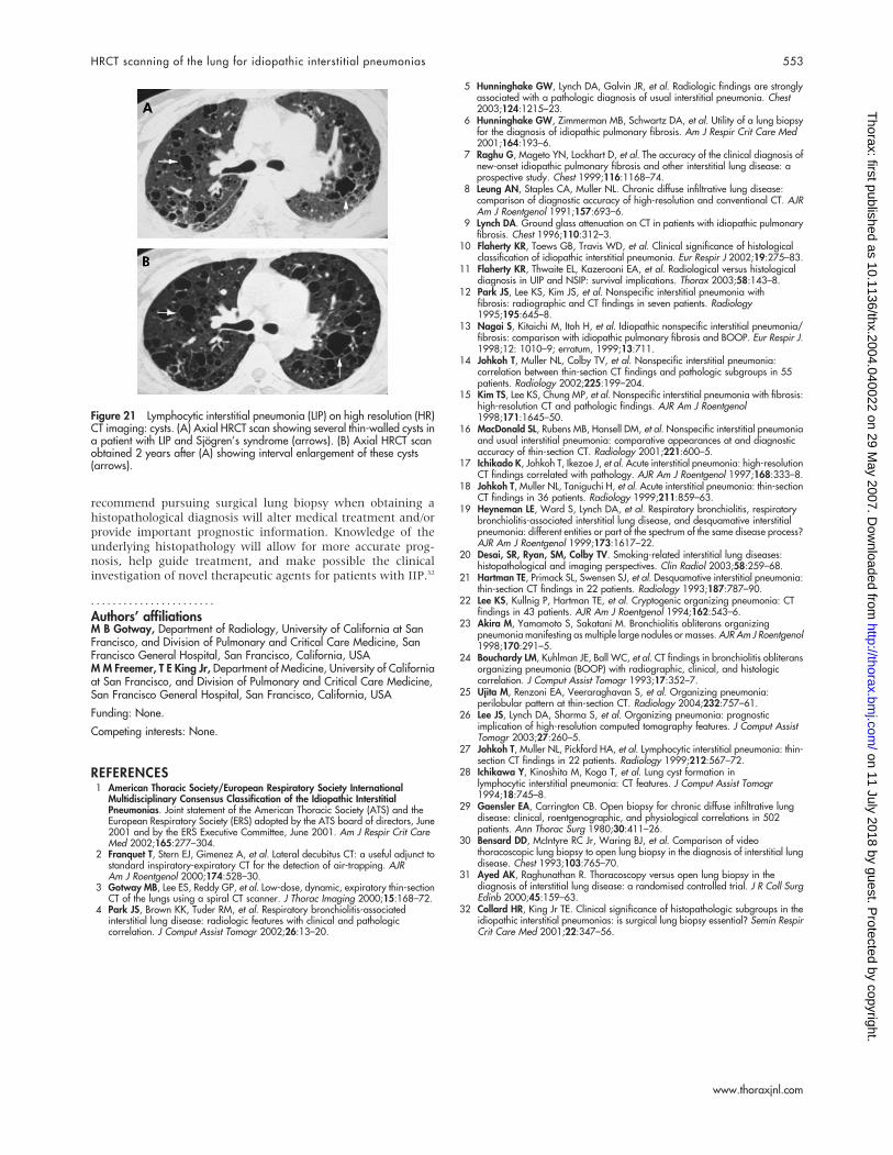
Cysts in LIPCystic spaces ranging in size from 1 to 30 mm have beenreported in patients with LIP (fig 21).27 28 These cysts arethought to be caused by bronchiolar obstruction and subse-quent air trapping.
SURGICAL LUNG BIOPSYCareful histopathological analysis allows the further classifica-tion of infiltrative lung diseases into distinct subgroups. Inmost settings, surgical lung biopsy has a diagnostic yield of.90% and an overall morbidity and mortality of (2.5% and(0.3%, respectively.29–31
Video-assisted thoracoscopic surgery lung biopsy hasreplaced thoracotomy in the diagnosis of ILD. There are threeimportant situations in which thoracoscopic lung biopsy iscontraindicated: inability to tolerate single lung ventilation (eg,severe hypoxaemia, high airway pressures), coagulopathy andpleural adhesions or scarring from previous thoracic surgery orpleurodesis. If a histological diagnosis is essential in thesepatients, a limited thoracotomy is usually the surgical approachof choice.
Surgical lung biopsy should only be pursued when ahistopathological diagnosis will alter medical treatment and/or provide important prognostic information. Patients who aretoo ill to tolerate or to benefit from potential treatments orpatients who do not wish to receive potential treatments shouldgenerally not have surgical lung biopsies. Absolute cutoffsbased on age or comorbidity should be avoided; the decision toforego surgical lung biopsy should be made on a case-by-casebasis.32
CONCLUSIONSIIP remains a source of confusion and consternation forclinicians. However, HRCT scanning has become a valuabletool allowing identification of the presence, extent and severityof ILD. Clinically, the distinction between UIP and all other(non-UIP) IIPs is the key determinant of a patient’s course.Fortunately, when properly performed and evaluated byexperienced thoracic radiologists, HRCT features alone aresufficient to make the diagnosis of UIP/IPF in 50–60% of casesselected based on the clinical suspicion of IPF. In the absence ofperipheral bibasilar honeycombing with or without upper lobereticulation—the features that allow radiologists to make aconfident diagnosis of UIP—surgical lung biopsy is warranted.In this diagnostic process, the importance of both the clinicalevaluation and radiological interpretation are critical. In theabsence of a clear clinical and radiological diagnosis, we
Figure 17 Lymphocytic interstitial pneumonia (LIP) on high resolution (HR)CT imaging: centrilobular ground glass attenuation nodules. Axial HRCTimage shows poorly defined centrilobular ground glass attenuation nodules(arrows).
Figure 18 Lymphocytic interstitial pneumonia (LIP) on high resolution (HR)CT imaging: ground glass opacity. Axial HRCT scan shows multifocal areasof ground glass opacity shown to represent LIP on surgical lung biopsy.
Figure 19 Lymphocytic interstitial pneumonia (LIP) on high resolution (HR)CT imaging: peribronchovascular nodules. Axial HRCT scan shows severalnodules positioned along bronchi (arrow), consistent with a peribronchiolarlocation. Mild interlobular septal thickening and patchy areas of groundglass opacity are also seen in the right lower lobe. Peribronchiolarconsolidation (arrowhead) is also present in the left lower lobe.
Figure 20 Lymphocytic interstitial pneumonia (LIP) on high resolution (HR)CT imaging: costal and fissural pleural nodules. Axial HRCT scan showsnumerous well-defined nodules related to both costal and fissural pleuralsurfaces (arrows).
552 Gotway, Freemer, King
www.thoraxjnl.com
on 11 July 2018 by guest. Protected by copyright.
http://thorax.bmj.com
/T
horax: first published as 10.1136/thx.2004.040022 on 29 May 2007. D
ownloaded from
recommend pursuing surgical lung biopsy when obtaining ahistopathological diagnosis will alter medical treatment and/orprovide important prognostic information. Knowledge of theunderlying histopathology will allow for more accurate prog-nosis, help guide treatment, and make possible the clinicalinvestigation of novel therapeutic agents for patients with IIP.32
Authors’ affiliations. . . . . . . . . . . . . . . . . . . . . . .
M B Gotway, Department of Radiology, University of California at SanFrancisco, and Division of Pulmonary and Critical Care Medicine, SanFrancisco General Hospital, San Francisco, California, USAM M Freemer, T E King Jr, Department of Medicine, University of Californiaat San Francisco, and Division of Pulmonary and Critical Care Medicine,San Francisco General Hospital, San Francisco, California, USA
Funding: None.
Competing interests: None.
REFERENCES1 American Thoracic Society/European Respiratory Society International
Multidisciplinary Consensus Classification of the Idiopathic InterstitialPneumonias. Joint statement of the American Thoracic Society (ATS) and theEuropean Respiratory Society (ERS) adopted by the ATS board of directors, June2001 and by the ERS Executive Committee, June 2001. Am J Respir Crit CareMed 2002;165:277–304.
2 Franquet T, Stern EJ, Gimenez A, et al. Lateral decubitus CT: a useful adjunct tostandard inspiratory-expiratory CT for the detection of air-trapping. AJRAm J Roentgenol 2000;174:528–30.
3 Gotway MB, Lee ES, Reddy GP, et al. Low-dose, dynamic, expiratory thin-sectionCT of the lungs using a spiral CT scanner. J Thorac Imaging 2000;15:168–72.
4 Park JS, Brown KK, Tuder RM, et al. Respiratory bronchiolitis-associatedinterstitial lung disease: radiologic features with clinical and pathologiccorrelation. J Comput Assist Tomogr 2002;26:13–20.
5 Hunninghake GW, Lynch DA, Galvin JR, et al. Radiologic findings are stronglyassociated with a pathologic diagnosis of usual interstitial pneumonia. Chest2003;124:1215–23.
6 Hunninghake GW, Zimmerman MB, Schwartz DA, et al. Utility of a lung biopsyfor the diagnosis of idiopathic pulmonary fibrosis. Am J Respir Crit Care Med2001;164:193–6.
7 Raghu G, Mageto YN, Lockhart D, et al. The accuracy of the clinical diagnosis ofnew-onset idiopathic pulmonary fibrosis and other interstitial lung disease: aprospective study. Chest 1999;116:1168–74.
8 Leung AN, Staples CA, Muller NL. Chronic diffuse infiltrative lung disease:comparison of diagnostic accuracy of high-resolution and conventional CT. AJRAm J Roentgenol 1991;157:693–6.
9 Lynch DA. Ground glass attenuation on CT in patients with idiopathic pulmonaryfibrosis. Chest 1996;110:312–3.
10 Flaherty KR, Toews GB, Travis WD, et al. Clinical significance of histologicalclassification of idiopathic interstitial pneumonia. Eur Respir J 2002;19:275–83.
11 Flaherty KR, Thwaite EL, Kazerooni EA, et al. Radiological versus histologicaldiagnosis in UIP and NSIP: survival implications. Thorax 2003;58:143–8.
12 Park JS, Lee KS, Kim JS, et al. Nonspecific interstitial pneumonia withfibrosis: radiographic and CT findings in seven patients. Radiology1995;195:645–8.
13 Nagai S, Kitaichi M, Itoh H, et al. Idiopathic nonspecific interstitial pneumonia/fibrosis: comparison with idiopathic pulmonary fibrosis and BOOP. Eur Respir J.1998;12: 1010–9; erratum, 1999;13:711.
14 Johkoh T, Muller NL, Colby TV, et al. Nonspecific interstitial pneumonia:correlation between thin-section CT findings and pathologic subgroups in 55patients. Radiology 2002;225:199–204.
15 Kim TS, Lee KS, Chung MP, et al. Nonspecific interstitial pneumonia with fibrosis:high-resolution CT and pathologic findings. AJR Am J Roentgenol1998;171:1645–50.
16 MacDonald SL, Rubens MB, Hansell DM, et al. Nonspecific interstitial pneumoniaand usual interstitial pneumonia: comparative appearances at and diagnosticaccuracy of thin-section CT. Radiology 2001;221:600–5.
17 Ichikado K, Johkoh T, Ikezoe J, et al. Acute interstitial pneumonia: high-resolutionCT findings correlated with pathology. AJR Am J Roentgenol 1997;168:333–8.
18 Johkoh T, Muller NL, Taniguchi H, et al. Acute interstitial pneumonia: thin-sectionCT findings in 36 patients. Radiology 1999;211:859–63.
19 Heyneman LE, Ward S, Lynch DA, et al. Respiratory bronchiolitis, respiratorybronchiolitis-associated interstitial lung disease, and desquamative interstitialpneumonia: different entities or part of the spectrum of the same disease process?AJR Am J Roentgenol 1999;173:1617–22.
20 Desai, SR, Ryan, SM, Colby TV. Smoking-related interstitial lung diseases:histopathological and imaging perspectives. Clin Radiol 2003;58:259–68.
21 Hartman TE, Primack SL, Swensen SJ, et al. Desquamative interstitial pneumonia:thin-section CT findings in 22 patients. Radiology 1993;187:787–90.
22 Lee KS, Kullnig P, Hartman TE, et al. Cryptogenic organizing pneumonia: CTfindings in 43 patients. AJR Am J Roentgenol 1994;162:543–6.
23 Akira M, Yamamoto S, Sakatani M. Bronchiolitis obliterans organizingpneumonia manifesting as multiple large nodules or masses. AJR Am J Roentgenol1998;170:291–5.
24 Bouchardy LM, Kuhlman JE, Ball WC, et al. CT findings in bronchiolitis obliteransorganizing pneumonia (BOOP) with radiographic, clinical, and histologiccorrelation. J Comput Assist Tomogr 1993;17:352–7.
25 Ujita M, Renzoni EA, Veeraraghavan S, et al. Organizing pneumonia:perilobular pattern at thin-section CT. Radiology 2004;232:757–61.
26 Lee JS, Lynch DA, Sharma S, et al. Organizing pneumonia: prognosticimplication of high-resolution computed tomography features. J Comput AssistTomogr 2003;27:260–5.
27 Johkoh T, Muller NL, Pickford HA, et al. Lymphocytic interstitial pneumonia: thin-section CT findings in 22 patients. Radiology 1999;212:567–72.
28 Ichikawa Y, Kinoshita M, Koga T, et al. Lung cyst formation inlymphocytic interstitial pneumonia: CT features. J Comput Assist Tomogr1994;18:745–8.
29 Gaensler EA, Carrington CB. Open biopsy for chronic diffuse infiltrative lungdisease: clinical, roentgenographic, and physiological correlations in 502patients. Ann Thorac Surg 1980;30:411–26.
30 Bensard DD, McIntyre RC Jr, Waring BJ, et al. Comparison of videothoracoscopic lung biopsy to open lung biopsy in the diagnosis of interstitial lungdisease. Chest 1993;103:765–70.
31 Ayed AK, Raghunathan R. Thoracoscopy versus open lung biopsy in thediagnosis of interstitial lung disease: a randomised controlled trial. J R Coll SurgEdinb 2000;45:159–63.
32 Collard HR, King Jr TE. Clinical significance of histopathologic subgroups in theidiopathic interstitial pneumonias: is surgical lung biopsy essential? Semin RespirCrit Care Med 2001;22:347–56.
Figure 21 Lymphocytic interstitial pneumonia (LIP) on high resolution (HR)CT imaging: cysts. (A) Axial HRCT scan showing several thin-walled cysts ina patient with LIP and Sjogren’s syndrome (arrows). (B) Axial HRCT scanobtained 2 years after (A) showing interval enlargement of these cysts(arrows).
HRCT scanning of the lung for idiopathic interstitial pneumonias 553
www.thoraxjnl.com
on 11 July 2018 by guest. Protected by copyright.
http://thorax.bmj.com
/T
horax: first published as 10.1136/thx.2004.040022 on 29 May 2007. D
ownloaded from