review retinoids for treatment of retinal diseases
TRANSCRIPT
Retinoids for treatment of retinaldiseasesKrzysztof Palczewski
Department of Pharmacology, School of Medicine, Case Western Reserve University, Cleveland, OH 44106-4965, USA
Review
Knowledge about retinal photoreceptor signal transduc-tion and the visual cycle required for normal eyesight hasincreased exponentially over the past decade. Substan-tial progress in human genetics has facilitated the identi-fication of candidate genes and complex networksunderlying inherited retinal diseases. Natural mutationsin animal models that mimic human diseases have beencharacterized and advanced genetic manipulation cannow be used to generate small mammalian models ofhuman retinal diseases. Pharmacological repair of defec-tive visual processes in animal models not only validatestheir involvement in vision, but also provides greatpromise for the development of improved therapiesfor millions who are progressing towards blindness orare almost completely robbed of their eyesight.
Visual perceptionThecapacityof thebraintoanalyzeand interpret informationis limited ultimately by the sensory input it receives. For thehumanbrain, visual perception is amajor input pathwayandan essential sense for understanding of the environment andsocial communication. The performance of this sense reflectsan individual’s genetic background, withstands manyenvironmental insults and deteriorates with age [1–4]. Mol-ecular investigations of the fundamental processes in visioninitiation revealed that these can be divided into two stages:phototransduction, which propagates the light signal [5,6],and the visual (retinoid) cycle [7], which consists ofmetabolicpathways that regenerate the visual chromophore and thussustain vision [8]. In reality, both pathways are fully inte-grated and complementary (Figure 1) [2,7,9]. Technical inno-vations and improved methodologies in proteomics andstructural biology have led to substantial advances in ourunderstanding of the molecular basis of vision [10,11]. Suchadvancedknowledge of themolecular basis of phototransduc-tion and the visual cycle means that it is possible to manip-ulate these processes using highly specific retinoid-basedtherapies for disease states and visual deterioration due toaging and environmental insults [2,4,12]. The remarkableanatomyandphysiology of the eye facilitate theuse of uniqueand highly specific pharmacological approaches that arediscussed in this review. Conceptually, these approachescan be extended to other biological systems that requiredelivery of therapeutics in situ.
Phototransduction and the retinoid cycleSensitivity and adaptation to variable environmental lightconditions are hallmarks of vision. Effective vision requires
Corresponding author: Palczewski, K. ([email protected]).
284 0165-6147/$ – see front matter � 2010 Elsevier Ltd. All rights reserv
detection of light ranging from a single photon to a trillionphotons per second and requires rapid restoration of thepre-illumination physiological state. This cyclic process isbased on 11-cis-retinal, a light-sensitive chromophorederived from vitamin A that absorbs light in the visiblerange. When 11-cis-retinal binds to its cognate visualprotein receptors (opsins), the resulting highly concen-trated visual pigment is exquisitely sensitive to differentwavelengths of light ranging from �360 to 620 nm [13].Production of 11-cis-retinal involves several enzymaticsteps, collectively called the visual (retinoid) cycle, thatis split between processes in photoreceptor cells and theadjacent retinal pigment epithelium (RPE).
Rhodopsin in rod photoreceptors and cone pigments incone photoreceptors are the two classes of visual pigmentsthat respond to light [13]. Their common chromophore, 11-cis-retinal, is covalently linked via a protonated Schiff baseto a lysine side-chain amino group embedded within theopsin transmembrane domain, forming 11-cis-retinyli-dene. On photon absorption, the chromophore undergoesphotoisomerization to all-trans-retinylidene, changing thevisual pigments from an inactive to an active conformation[14]. In rods, this active form, known as Meta II, thenrecruits and binds intracellular G proteins, continuing thesignaling cascade that culminates in visual perception(Figure 1) [15]. Various aspects of these phototransductionprocesses have been reviewed in depth elsewhere [5,6,16].
The retinoid (visual) cycle is a complex enzymaticpathway essential for regeneration of 11-cis-retinal. Themaintenance of continuous vision and preservation ofphotoreceptor health require a continuous adequatesupply of this aldehyde, so vertebrates have evolved theretinoid cycle to achieve this objective [2]. The pathwayoperates in sequential reactions in photoreceptor cells, inthe RPE and back in photoreceptors, converting all-trans-retinal back to 11-cis-retinal via several chemical trans-formations. The classical vertebrate retinoid cycle contrib-utes primarily to regeneration of rhodopsin in rod cells(Figure 1). The key enzyme in this pathway is retinoidisomerase or RPE65, which resides in the RPE. RPE65-dependent chromophore production might also be import-ant for cone function [17]. However, the cone retinoid cycleseems to be supplemented by another metabolic pathwaythat is as yet genetically undefined [18–20].
The series of chemical reactions comprising the classicalretinoid cycle is now well established (Figure 1). In rodcells, absorption of a photon of light by rhodopsin causesphotoisomerization of 11-cis-retinylidene to all-trans-retinylidene, resulting in release of all-trans-retinal from
ed. doi:10.1016/j.tips.2010.03.001 Trends in Pharmacological Sciences 31 (2010) 284–295

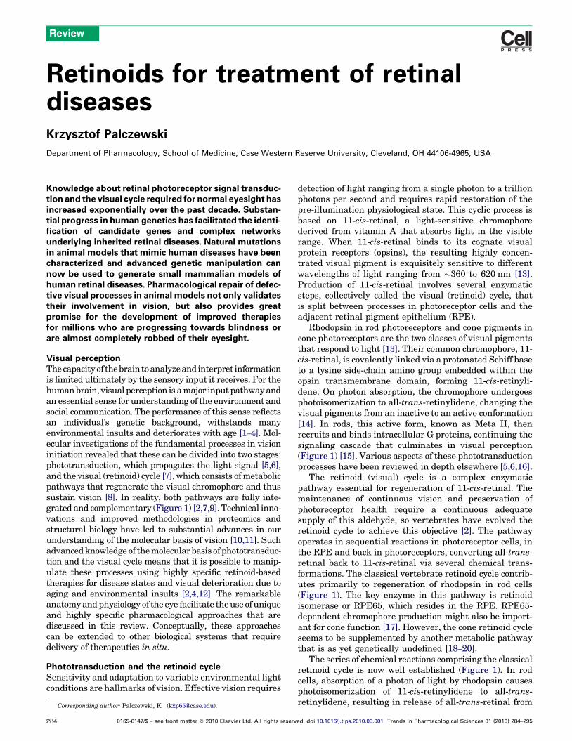
Figure 1. Phototransduction and the visual (retinoid) cycle in vertebrates. Vision is triggered by light-dependent activation of rhodopsin or other visual pigments. In rod
cells, this chromophore couples to the protein opsin, forming rhodopsin. Absorption of a photon of light by rhodopsin causes photoisomerization of 11-cis-retinal to all-
trans-retinal. In turn, photoactivated rhodopsin generates activation of hundreds of heterotrimeric G proteins, called transducin or Gt, in photoreceptors. This G-protein-
coupled receptor cascade is a classic cyclic nucleotide pathway that results in a decrease in cGMP levels (not depicted in the figure) and consequently hyperpolarization of
the plasma membranes and ultimately reduction of glutamate secretion to secondary neurons. The visual cycle regenerates 11-cis-retinal from all-trans-retinal released
from the chromophore-binding pocket of opsin. All-trans-retinal is reduced to all-trans-retinol in a reversible reaction catalyzed by RDH12 and RDH8, which are NADPH-
dependent all-trans-retinol dehydrogenases. All-trans-retinol diffuses into the RPE, where it is esterified in a reaction catalyzed by lecithin:retinol acyl transferase to long-
chain fatty acids. As a consequence of their propensity to aggregate, retinyl esters are stored in lipid-droplet-like structures called retinosomes. The all-trans-retinyl esters
seem to be the substrate for RPE65, which converts them to 11-cis-retinol, which then is further oxidized back to 11-cis-retinal by RDH5, RDH11 and other NAD-dependent
retinol dehydrogenases. 11-cis-Retinal formed in the RPE diffuses back into the ROS and COS, where it completes the cycle by recombining with opsins to form rhodopsin
and cone pigments. Mutations in genes encoding proteins of phototransduction and the retinoid cycle are associated with various retinal diseases, some of which are
indicated by the green boxes. Pharmacological intervention has been successful in animal models in a few instances, as indicated by the compounds in the blue boxes. IPM,
interphotoreceptor matrix; IRBP, interphotoreceptor retinoid-binding protein; CSNB, congenital stationary night blindness; Ral, retinal; Rol, retinol; ABCA4, ATP-binding
transporter 4.
Review Trends in Pharmacological Sciences Vol.31 No.6
the chromophore-binding pocket of opsin. Most of the all-trans-retinal that dissociates from opsin diffuses into thecytoplasm. A fraction that dissociates into the disc lumenalso reaches the cytoplasm via transfer by ATP-bindingcassette, transporter 4 (ABCA4) [21]. All-trans-retinal inthe cytoplasm is reduced to all-trans-retinol in a reversiblereaction catalyzed by an NADPH-dependent all-trans-reti-nol dehydrogenase (RDH). All-trans-retinol then diffusesinto the RPE, where it is esterified in a reaction catalyzedby lecithin:retinol acyl transferase (LRAT). The acyl groupis transferred from the sn1 position of ER phospholipids[22,23]. The most common lipids in the sn1 position aresaturated long-chain fatty acids, with a palmitoyl grouprepresenting the predominant species. Thus, retinyl estersare dominated by retinyl palmitate, but also contain stea-rate (C18) and other minor species. These esters have apropensity to aggregate and in the RPE they form lipophi-lic droplets called retinosomes, which are discussed below.These all-trans-retinyl esters are presumably converted bythe retinoid isomerase RPE65 to 11-cis-retinol, which isfurther oxidized back to 11-cis-retinal by RDH5, RDH11and other RDHs. After diffusion from the RPE, 11-cis-retinal combines with opsin to form a light-sensitive pig-ment that is ready for another cycle of photoisomerization,signal transduction and regeneration.
Retinoid metabolism and retinopathiesRetinoids are required for normal growth, vision, repro-duction, and maturation and maintenance of the immunesystem [24]. Retinoids are also important regulators ofmetabolism in general [25]. All-trans-retinol is an essentialmicro-nutrient because it cannot be synthesized byanimals and therefore must be absorbed as either retinolor retinyl esters from food of animal origin or generatedfrom their precursor, b,b-carotene from plants. Dietaryretinyl esters are hydrolyzed in the intestinal lumen,absorbed into intestinal enterocytes, re-esterified andincorporated into chylomicrons. They are then taken upby hepatocytes and either hydrolyzed and secreted afterbinding to the retinol-binding protein 4 (RBP4) complex orstored as lipid-droplet-like structures in cells called Ito orstellate cells. Thus, the liver is the largest storage depot forretinyl esters in the body [26,27]. b,b-Carotene is symme-trically cleaved into two molecules of all-trans-retinal bymembers of the carotenoid oxygenase enzyme family [28–
30]. Retinal is reduced to retinol, esterified and furtherprocessed in pathways that involve exogenous retinylesters throughout the body [31]. Retinoid-metabolizingenzymes in the liver and peripheral tissues, includingLRAT, transferases, hydrolases, diacylglycerol acyltrans-ferase 1 (DGAT1) and acyl-coenzyme A transferase
285

Review Trends in Pharmacological Sciences Vol.31 No.6
(ACAT), are not highly specific, so they can process retinoidanalogs as well.
Considering the fundamental role of retinoids in vision,it is not surprising that many forms of retinopathy arecaused by defects in genes encoding proteins of the visualcycle (Figure 1) [2]. Storage of absorbed retinoids in theliver, their transport in the plasma and delivery to the RPEcan all be impaired as a result of inactivating mutations inenzymes (such as LRAT), transport carrier proteins (suchas RBP4) or receptors (such as STRA6). Progress in ourunderstanding of these processes was possible because alarge number of animals models of these defects wereeither generated or occur naturally [32]. In Lrat–/– andRpe65–/– mice, lack of 11-cis-retinal leads to rapid degener-ation of cone photoreceptors and progressive death of rods[33]. This phenomenon might involve the mechanism lead-ing to the pathology seen in Leber congenital amaurosis(LCA) patients [17]. LCA has been attributed to continuousactivation of visual phototransduction [34] due to basalactivity of chromophore-free opsin [35–37], disordered vec-torial transport of cone visual pigments lacking boundchromophore [38], instability of nonbound cone visualpigments or a combination of all these mechanisms.
More complete discussions of retinal diseases related togenetic alterations of phototransduction and the visualcycle can be found elsewhere [1,2,39,40]. Because com-ponents of the visual cycle and phototransduction aremostly non- or only partially redundant, genetic defectsresulting in dysfunction of these proteins are manifest asretinopathies (for examples, see Figure 1). The severity ofthese genetic retinopathies is determined by the toxicity ofthe accumulated intermediates (e.g. condensation pro-ducts of all-trans-retinal in Stargardt’s disease) [41], theneed of the product for cellular homeostasis (e.g. cGMPproduction in LCA caused by a mutation in guanylatecyclase) [42], the instability of the mutated protein struc-ture [e.g. rhodopsin mutants in autosomal recessive reti-nitis pigmentosa (RP)] [43] and whether regulation ofprotein function is altered (e.g. Ca2+ coordination in auto-somal dominant cone–rod dystrophy mutants of guanylatecyclase-activating proteins) [44].
All-trans-retinal, condensation products anddegenerative retinal diseasesIn many individuals the visual system degenerates withage. Prevention of vision loss requires a better understand-ing of the fundamental causes of age-dependent changes.Fortunately, our understanding of both retinoid metab-olism outside the eye and production of 11-cis-retinalunique to the eye is accelerating [1,2,7]. Genetic mousemodels are also now available to study these processes andtheir aberrations in vivo [45]. These advances allow thecentral question of what compromises photoreceptor cellsand the underlying RPE to be addressed. Retinoids,despite being essential for vision, can also cause certainretinal pathologies when not tightly controlled.
Typically, retinoids are complexed with soluble proteinsthat protect them. These reactive compounds are bound bya number of retinoid-binding proteins and are rarely freelysolubilized from membranes. Protection of retinoidsalso stems from their ability to cluster when esterified
286
by long-chain fatty acids and to be stored in lipid-likedroplets in the liver or as retinosomes in the eye [46–48].
To absorb light efficiently, visual pigments need to bevery sensitive to light (11-cis-retinal requires a quantumefficiency of 0.65 [14]) and to be highly concentrated.Indeed, a large fraction of rhodopsin forms a paracrystal-line structure in rod outer segments (ROS) [49–52] andcone pigments can form diffractable crystalline structuresin cones [53,54]. ROS contain�5 mMrhodopsin [55] that, ifcompletely bleached, would yield an equal level of free all-trans-retinal. How can cells cope with such an aldehydeflux? Even less than 0.5% bleaching will produce toxiclevels of all-trans-retinal if this retinoid is not properlycleared.
The efficiency of the mammalian visual system and thehealth of photoreceptors and RPE decrease significantlywith age, suggesting that before cell death there are bio-chemical changes that slowly promote retinal damage. Forexample, an abnormally high flux of retinoids through theretinoid cycle can induce retinopathies in some mousemodels [56–58]. This process in turn triggers host immuneand other defense responses that culminate in retinal celldeath. Even in the presence of an efficient and fully func-tional retinoid cycle, all-trans-retinal can condense, produ-cing, among a myriad of other byproducts, di-retinoidpyridinium ethanolamine (A2E) and all-trans-retinal di-mer (RALdi) [59–61] (Figure 2). Initial condensation pro-ducts are formed in theROS and 10% of theROS undergoesphagocytosis and accumulates in the RPE daily. Conden-sation products also accumulate with age [62] and thesecompounds can cause RPE cell toxicity under experimentalconditions [63–65]. Patients affected by age-related macu-lar degeneration (AMD), Stargardt’s disease or otherretinal diseases associated with accumulation of surrogatemarkers such as A2E all develop retinal degeneration [66].
Mutations in ABCA4 cause Stargardt’s maculardegeneration [67], cone–rod dystrophy [68] and recessiveRP [69,70]. Heterozygous mutations in ABCA4 increasethe risk of developing AMD as well [66]. A2E [71,72] andRALdi [61] are the major fluorophores of lipofuscins pro-duced from all-trans-retinal [73] (Figure 2). As a con-sequence of aging, both A2E and RALdi can accumulateover a lifetime of light exposure [62], with toxic effects onRPE cells [74,75]. Patients affected by Stargardt’s diseaseor AMD because of a disabled ABCA4 gene and thoseaffected by other retinal diseases associated with lipofus-cin accumulation eventually develop retinal degeneration.ABCA4 mutations are also linked to an increased risk ofAMD [66]. However, no such degeneration was observed inAbca4–/– mice even though RPE atrophy was detected[21,41,76]. Thus, mice and humans do not always exhibitidentical responses to fluorophore accumulation.
Recently, we showed that mice carrying double knock-out of Abca4 [21] and retinol dehydrogenase 8 (Rdh8), oneof the main enzymes that reduce all-trans-retinal in ROSand cone outer segments (COS) [77], rapidly accumulateall-trans-retinal condensation products and exhibit accen-tuated RPE/photoreceptor dystrophy at an early age [58].Retinas from thesemice exhibited lipofuscin, drusen, basallaminar deposits, Bruch’s membrane thickening and chor-oidal neovascularization. Importantly, the severity of their

Figure 2. Retinoid flow in the visual cycle and condensation of all-trans-retinal. After 11-cis-retinal binds to opsin, forming rhodopsin, the resulting visual chromophore, 11-
cis-retinylidene, is photoisomerized to all-trans-retinylidene, the precursor of all-trans-retinal that is subsequently released. Most of the all-trans-retinal dissociates from
opsin into the cytoplasm, where it is reduced to all-trans-retinol by RDHs, including RDH8. The fraction of all-trans-retinal that dissociates into the disc lumens is transported
by ABCA4 back into the cytoplasm [21] before it is reduced. Thus, condensation products can be generated within both the disc lumens and the cytoplasm. Loss of ABCA4
and RDH8 exacerbates this condensation, reminiscent of an accelerated aging process. In humans, as a result of daily phagocytosis of part of the rod outer segments,
lipofuscin fluorophores accumulate with age in the RPE, especially in RPE cells underlying the cone-rich macula [109,127]. Such accumulation has been considered to
constitute one of the major risk factors for AMD, the predominant cause of legal blindness in developed countries [128]. Lipofuscin fluorophores are especially abundant in
Stargardt disease, the most common juvenile form of macular degeneration [72]. A2E and RALdi, the major fluorophores of lipofuscin, are formed by condensation of
phosphatidyl ethanolamine with two molecules of all-trans-retinal followed by oxidation and hydrolysis of the phosphate ester [129]. Various mechanisms have been
proposed to explain the toxicity of A2E. These include its cationic detergent properties [130], physiological interference with RPE function [131,132] and radical reaction
products induced by light-dependent oxidation [133].
Review Trends in Pharmacological Sciences Vol.31 No.6
visual dysfunction and retinopathy was exacerbated bylight but attenuated by treatment with retinylamine, avisual cycle inhibitor that slows the flow of retinoidsthrough the visual cycle, thus giving other oxidoreductaseenzymes more time to detoxify retinaldehyde molecules.These findings provide direct evidence that aberrant pro-duction of toxic condensation byproducts of the visual cyclecan lead to rapid and progressive retinal degeneration inmice and suggest a pharmacological method for ameliora-tion of these conditions. The similarity of this retinopathyto human AMD means that these mice are invaluable forresearch aimed at ameliorating this devastating blindingdisease.
Although the above studies strongly suggest retinoidtoxicity, it still is unclear if high levels of retinal and/or itscondensation products such as A2E actually cause theseretinopathies or merely constitute non-specific manifes-tations of impaired retinoid metabolism. Recently, wereported that all-trans-retinal is probably responsible forphotoreceptor degeneration in Rdh8–/–Abca4–/– mice [73].Toxic effects of all-trans-retinal induce apoptosis throughcaspase activation andmitochondrial-associated cell death[73]. Therefore, excessive levels of this aldehyde must be
decreased to preserve retinal health. Although A2E for-mation could be a surrogate marker for aberrations in all-trans-retinal clearance, it might also represent a detoxifi-cation product and not the primary toxin previously sup-posed. Regardless of whether all-trans-retinal or A2E isresponsible for retinal degeneration, prevention of all-trans-retinal accumulation will also stop production ofA2E and other similar derivatives formed from this alde-hyde.
Retinoids are a diverse group of compounds withdifferent biological activityThe fact that retinoids are often grouped together in themedical literature without distinction can be misleading.For example, retinol and retinoic acid have dramaticdifferences in biological activity and, even though retinolcan be converted to retinoic acid, this transformation ishighly regulated. Retinol has only limited biologicalactivity per se [78] but it can be dehydrated to anhydror-etinol, which might exert regulatory effects on theimmune system [79] or it can be saturated in the 13–
14 position to produce dihydroretinol precursors involvedin metabolic processes [80,81]. In the retinoid cycle,
287
Review Trends in Pharmacological Sciences Vol.31 No.6
retinol is an excellent substrate for LRAT and is quicklyconverted into fatty acid esters. Their propensity to formoil droplets excludes fatty acid esters of retinol from thecirculation. Retinol can be oxidized to retinal in a reactioncatalyzed by a subset of short-chain alcohol dehydrogen-ases and medium-chain alcohol dehydrogenases. Becausethe redox potential of cells favors reducing conditions andthe oxidation reaction is thermodynamically neutral, onlya tiny fraction of retinol can be oxidized to fulfill thethermodynamic requirements of equilibrium. The all-trans-retinal formed probably does not exert direct bio-logical activity, although there is speculation to the con-trary [82]. Instead, this aldehyde is subsequentlyoxidized by aldehyde dehydrogenase to form retinoic acidin a highly regulated process. Retinoic acid is a potentmitogen involved, via nuclear transcription factors, incontrolling the expression of a large number of genes.One of the most highly upregulated genes is CYP26,which encodes a P450 enzyme that oxidizes retinoic acidto inactive products. Thus, retinoic acid levels are keptrelatively low and tightly controlled by its rates of bio-synthesis and destruction.
Another issue is differences in biological processingexhibited by geometric isomers of retinoids. For example,9-cis-retinal and 11-cis-retinal recombine with opsin togenerate visual pigments, but all-trans-retinal and 13-cis-retinal, two isomers that are in thermodynamic equi-librium, do not. Indeed, all-trans-retinal can be convertedto 11-cis-retinal through the retinoid cycle, but typicallyall-trans-forms are preferably stored as fatty acid esters inthe RPE.
Strategies for treating blinding retinal diseases causedby mutations in retinoid cycle genesConceptually, the simplest way to restore function is toreplace defective genes by viral, nanoparticle or other genetherapymethods. RPE and photoreceptor cells take up andexpress recombinant constructs with great efficiency inmany experimental settings. This strategy was used suc-cessfully in LCA animal models such as mice and dogs, andthe positive effect of gene replacement therapy was sus-tained for several years after a single treatment of RPE65-null dogs [83,84]. Recently, several LCA patients wereenrolled for gene therapy. Initial results revealed that thistreatment at least partially restored vision in retinal areasof gene transfer in those individuals with blindness attrib-uted to RPE65 mutations [85–87].
Retinoids undergo multiple transformations in the eye(Figures 1 and 2), and disabling mutations in genes encod-ing unique enzymes responsible for these transformationscause a deficiency in 11-cis-retinal production. Below wedescribe how such compromised transformations can bebypassed by supplying active chromophore(s).
Theoretically, decreasing production of the 11-cis-retinal chromophore using specific inhibitors of the visualcycle would be beneficial. Slowing the rapid turnover ofretinoids should prevent excessive production of toxic all-trans-retinal and its condensation products. Excessive orgreatly prolonged inhibitionwould be detrimental because,conceptually, this would decrease vision under extremeconditions, as observed in LCA. However, slowing the
288
visual cycle under strong illumination conditions wouldbe beneficial.
Retinosomes as a depot for chromophores or inhibitorsof the visual cycleRetinosomes are storage particles that were discovered bya post-doctoral fellow in my laboratory, Dr YoshikazuImanishi. They bud off the ER, but their mechanism offormation has yet to be clarified. Retinosomes are com-posed of fatty acid retinyl esters, lipids and at least oneother identified component, adipocyte differentiation-related protein [46,47,88,89]. As a result of the high UVsensitivity of retinoids, these structures can be imaged onlyunder long-wavelength infrared light using two-photonmicroscopy (Figure 3A). It was then demonstrated thatthese particles expand under light and contract when lightis removed, providing evidence that retinosomes partici-pate in the regular visual cycle (Figure 3B). These storageparticles become light-insensitive when the visual cycle isdisabled by elimination of retinoid isomerase or LRATactivity. Retinosomes can also be used to store retinyla-mine because this compound can be amidated by LRAT[48]. Importantly, these structures can also be used to storeartificial precursors of the chromophore in the form of 9-cis-retinyl esters.
Pharmacological replacement of missing chromophoreand its precursorIn the most severe cases, insufficient 11-cis-retinal pro-duction leads to congenital or progressive blindness inhumans. LCA is an autosomal recessive, early-onset,severe retinal dystrophy that accounts for 5% of all suchinherited disorders [90]. Pharmacological replacement ofmissing chromophore is applicable to diseases resultingfrom deficient chromophore biosynthesis. Examples in-clude LCA arising from mutations in the LRAT andRPE65 genes (Figure 1). Initial experiments aimed atbypassing the biochemical defect caused by absence ofRpe65 were performed by oral gavage of Rpe65�/� micewith 9-cis-retinal [91]. 9-cis-Retinal, which combineswith opsin to form light-sensitive iso-rhodopsin [91],was initially selected because it is easier to synthesizeand is more stable than 11-cis-retinal. Moreover, iso-rhodopsin has an absorbance maximum at 494 nm com-pared to 502 nm for rhodopsin, facilitating experimentalidentification of reconstituted iso-rhodopsin [92].Further refinement and extensive testing identified 9-cis-retinyl acetate as a useful experimental compound[93] (Figure 4).
The use of cis-retinoids in the treatment of symptoms inLCA mouse models [91,94] seemed mechanistically soundand encouraging results were obtained in the treatment ofaging mice [95]. Not only was it proved that cis-retinoidschaperone the mutant opsin to allow proper in vivo foldingof P23H-opsin, but in experimental cell lines the rescuedprotein also formed pigment, acquired mature glycosyla-tion and was transported to the cell surface [96]. Onlyphotoactive cis-chromophores were beneficial; all-trans-retinal was ineffective.
Dietary supplementation of Rpe65�/� mice with 9-cis-retinoids restored light sensitivity to levels found in wild-

Figure 3. Transformations of visual cycle retinoids in the RPE. All-trans-retinol diffuses from photoreceptor cells into the RPE, where it is esterified by LRAT to all-trans-
retinyl esters. Hydrophobic retinyl esters then form retinosomes (RESTs). All-trans-retinyl esters are isomerized to 11-cis-retinol (reaction a) in a reaction that involves an
RPE-abundant protein, termed RPE65. 11-cis-Retinol is then oxidized by 11-cis-RDH to 11-cis-retinal (reaction b). 11-cis-Retinal diffuses back into the rod and cone outer
segments, where it completes the retinoid cycle by recombining with opsins to reform rhodopsin and cone pigments. (a) Retinosomes in RPE cells captured using two-
photon microscopy (courtesy of Grazyna Palczewska, Polgenix Inc., Cleveland, OH). Fluorescence emission from the isolated intact mouse eye at 560–700 nm in green
pseudocolor was observed after excitation by a 730-nm mode-locked Ti:Sapphire laser. Scale bar, 5 mm. (b) Flash-dependent changes in fluorescence and all-trans-retinol/
all-trans-retinyl esters in the RPE cell layer of isolated mouse eyes. The top shows a row of images of optical sections of the retina perpendicular to the ocular tissue. RPE
fluorescence (a.u., arbitrary unit) was quantified as a function of time. Numbers refer to minutes after the flash. The middle and bottom graphs show fluorescence
quantified for retinoids and retinoid analyses by HPLC (all-trans-retinol and all-trans-retinyl esters; mean�SD, n = 3), respectively. Dashed lines indicate the half-life for
REST formation and the increase in all-trans-retinol and all-trans-retinyl esters. Light-dependent changes in the fluorescent signal in different subcellular compartments are
shown on the right (copied from Ref. [46] with permission from Rockefeller University Press).
Review Trends in Pharmacological Sciences Vol.31 No.6
type animals, as assessed by both single-cell and ERGrecordings. Similar recovery of visual function wasreported following intraperitoneal injection of 11-cis-retinal into Rpe65�/� mice [97]. There are several advan-tages of 9-cis-retinoid over 11-cis-retinoid treatment. First,the 9-cis-compound is effective when taken orally. Second,because it is converted to prodrug forms that are stored inthe liver, transported in the blood, selectively taken up by
the eye and stored in retinosomes (Figure 4a,b), storageparticles that participate in the regular visual cycle [45–
47,88,89] but can also be used to store artificial precursorsof the chromophore from which the active compound isslowly released, a single high dose can produce a long-termtherapeutic effect. The toxicity profile of 9-cis-retinoids hasyet to be investigated. If these retinoids have a narrowtherapeutic window, any toxic effects are likely to be
289

Figure 4. Delivery and action of 9-cis-retinoids. Retinyl esters are effective when taken orally. In the small intestine, 9-cis-retinyl esters are either hydrolyzed and esterified
with fatty acyl coenzyme A, or trans-esterified with phospholipids before being transported in chylomicrons to the liver, where these intermediate products are found in
lipid droplets. This cycle of hydrolysis and esterification can occur several times before storage in hepatic stellate cells. Fatty acid (mostly palmitate) esters and free 9-cis-
retinol are then secreted into the systemic circulation, either bound to RBP or albumin or incorporated into chylomicrons. In the eye, these retinoids are probably hydrolyzed
again as they pass from the choroid capillaries into the RPE by STRA6-dependent and -independent mechanisms. Fatty acid esters of 9-cis-retinol in the RPE are stored in
specific lipid droplets called retinosomes. When required, these esters are hydrolyzed and oxidized to the drug 9-cis-retinal, which is then delivered to opsins in
photoreceptors to form light-sensitive visual pigments.
Review Trends in Pharmacological Sciences Vol.31 No.6
long-lasting. A particular concern is the potential for long-term toxicity during pregnancy. Fortunately, such toxicityis unlikely to emanate in mammals from conversion toretinoic acid, a potent mitogen, because biosynthesis of thelatter from retinol is tightly controlled. As in aging, rho-dopsin regeneration after light exposure is more delayed inhumans and mice with vitamin A deficiency because ofeither inadequate dietary intake or intestinal absorption[9]. Studies in mice have shown that age-related decreasedretinal rod cell function cannot be explained by rod cell loss,abnormal retinal plasticity or any signs of retinal disease[98–100]. However, a dramatic age-associated slowing ofrod-mediated dark adaptation after light exposure inhumans was related to delayed regeneration of rhodopsin[98]. Deteriorating photoreceptor function documented in
290
mice at 10 and 14 versus 4 months of age was improvedsignificantly by long-term monthly administration of theartificial chromophore 9-cis-retinyl acetate. These findingssuggest one potential therapeutic approach for preventionof age-related retinal dysfunction [3].
In a highly discussed study, all-trans-retinol combinedwith vitamin E was tested as a remedy for RP patientslacking genotype characterization. The results of thisstudy support a beneficial effect of 15,000 IU/day of vitaminA on the course of RP [101]. There was no indication thatthese patients were vitamin-deficient, so it seems incon-ceivable that the role of this treatment differed mechan-istically from the known antioxidant role of vitamin A onthe retina of this genetically heterogeneous diseased popu-lation. Surprisingly, vitamin A supplementation slows the

Review Trends in Pharmacological Sciences Vol.31 No.6
rate of photoreceptor degeneration caused by a threonine-17!methionine mutation in the opsin gene. The authorsspeculated that vitamin A supplementation could confertherapeutic benefit by stabilizing mutant opsins throughincreased availability of the chromophore [94], but chro-mophore production does not depend on a further increasein this precursor because it exists in significant excess inthe mature retina.
An observation in Abca4–/– mice is also puzzling. Uponsupplementation with vitamin A, these mice exhibiteddramatically higher levels of retinyl esters in their liverand RPE and, more importantly, lipofuscin pigments suchas A2E were significantly increased as well. Photoreceptordegeneration was also observed in 11-month-old albinomice. The author recommended that ‘vitamin A supple-mentation should be avoided in patients with ABCA4mutations or other retinal or macular dystrophies associ-ated with lipofuscin accumulation in the retinal pigmentepithelium’ [95]. However, A2E formation is related only tohigh levels of flux through the visual cycle, and high levelsof the chromophore ester precursor have nothing in com-mon with A2E formation, as evidenced by Rpe65–/– micethat completely lack A2E but still accumulate hugeamounts of all-trans-retinyl esters [102].
The situation differs for Sorsby’s fundus dystrophy,which is an autosomal dominant retinal degenerationcaused by mutations in the tissue inhibitor of metallopro-teinases-3 gene. During the course of this disease, athickenedmembrane barrier between photoreceptor layerscauses local vitamin A deprivation. Administration ofvitamin A dramatically restored photoreceptor function[103]. Insufficient dietary vitamin A can also cause pro-gressive deterioration of vision and ultimately blindnesswithout genetic abnormalities [104], a major problem inunderdeveloped countries. Dietary supplementation withvitamin A typically reverses this problem.
Pharmacological inhibition of the retinoid cycleAnother treatment strategy is to slow the biosynthesis ofchromophore by either inhibiting steps in the visual cycleor limiting availability of the all-trans-retinol precursor.This approach is applicable to diseases associated withaccumulation of retinoid cycle intermediates. As notedabove, impaired clearance of all-trans-retinal causes acutelight-induced retinal toxicity [73] and induces the for-mation of fluorescent lipofuscin pigments such as A2E inRPE cells. Acute all-trans-retinal toxicity observed in light-induced photoreceptor damage might involve increasedplasmamembrane permeability andmitochondrial poison-ing that leads to caspase activation and mitochondrial-associated cell death [73]. Accumulation of lipofuscin pig-ments is an important pathological feature of Stargardt’sdisease [105,106], but it is not limited to this inheritedcondition. Monitoring fundus autofluorescence is a non-invasive technique developed in the past decade that usesthe fluorescent properties of lipofuscin to study the healthand viability of the retina. Increased fundus autofluores-cence by scanning laser ophthalmoscopy is commonlyobserved in patients with AMD [107,108]. Fluorescentmaterial acquired in aged RPE has spectral propertiessimilar to A2P identified inAbca4�/�mice [59,109]. Strong
fundus autofluorescence is also observed in patients withBest vitelliform macular dystrophy and in a subset ofpatients with cone–rod dystrophy [110]. Patients withdominant Stargardt’s disease, caused by mutations inthe ELOVL4 gene, exhibit a dark choroid on fluoresceinangiography, also owing to lipofuscin in RPE cells[111,112]. The major disadvantage of inhibiting the reti-noid cycle is night blindness, with a resulting inability todrive. Also of concern is that prolonged inadequate pro-duction of chromophore will adversely affect the health ofrod and, more severely, cone cells. The most potent andefficacious retinoid cycle inhibitor identified to date isretinylamine, a transition-state inhibitor of RPE65[19,48,58,113–118]. The unusual fate of this retinoid, in-cluding its route of administration, hepatic storage, releaseinto the circulation, uptake by the eye, storage and event-ual release in retinosomes of the retina, is illustrated inFigure 5.
We systematically studied the effects of retinylamineand other potential inhibitors of visual function in mice.Prolonged and sustainable, but reversible, suppression ofvisual function was observed with retinylamine (Figure 5)as a result of its storage in a prodrug form,N-retinylamide[117]. The drug was directly compared with other inhibi-tors to assess their prevention of light-induced retinaldamage. Retinylamine displayed higher efficacy, speci-ficity and potency and lower transcriptional activationcompared to N-(4-hydroxyphenyl)retinamide [119] and13-cis-retinoic acid [120,121], whereas the other com-pounds tested were ineffective [114]. It should be notedthat other investigators have proposed that N-(4-hydro-xyphenyl)retinamidewould cause immediate dose-depend-ent decreases in serum retinol and RBP in Abca4–/– mice[119]. This prediction is puzzling because excess retinoidsare stored in the eye, which is highly resistant to vitamin Adeprivation. In mice, it takes two generations, even afterliver stores are depleted, for retinoids to be depleted fromthe eye. Methods to reduce circulating retinol would beinadequate to deplete retinoids in the eye because of thepowerful protective mechanisms involved in retinoidtransport (Figures 4 and 5) and retinoid retention withinthe RPE (Figure 5). Thus, it is possible that a small effecton retinoidmetabolism in the eye could be accomplished byweak inhibition of RPE65 by N-(4-hydroxyphenyl)retina-mide and of 11-cis-retinol dehydrogenase by 13-cis-retinoicacid [122]. However, a dramatic decrease in retinol deliv-ery to peripheral tissues for a prolonged period could bedetrimental to the health of the individual.
Is there a future for the use of inhibitors of the visualcycle for treatment of degenerative retinal diseases? Webelieve that these inhibitors could be effective therapiesbased on what we have learned about their effects inanimal models of these diseases. For example, it wasrecognized that cone photoreceptor cells in Rpe65–/– micedegenerate more rapidly than rod photoreceptors [123],with similar observations for Lrat–/– mice [38,124]. InRpe65–/–rhodopsin–/– mice, chromophore additionenhanced proper transport of cone opsins to outer seg-ments while partially preserving cone structure and func-tion in a compromised retina lacking rods because ofrhodopsin elimination [38]. These findings are critical
291

Figure 5. Delivery and action of retinylamide. This amide ester can be taken orally. Once in the intestine, it is hydrolyzed and re-amidated with fatty acid coenzyme A and
transported in chylomicrons to the liver, where it is stored as lipid droplets in hepatic stellate cells. This cycle of hydrolysis and re-amidation can occur several times. Fatty
acid (mostly palmitate) amide and free retinylamine are then secreted into the systemic circulation, either bound to RBP or albumin, or incorporated into chylomicrons. The
amides are probably hydrolyzed again as they pass from the choroid capillaries into the RPE in a STRA6-independent manner. There, retinylamine is stored in specific lipid
droplets called retinosomes. When required, retinylamine is released and acts as a very potent transition-state inhibitor of RPE65, which catalyses the hydrolytic
isomerization of retinyl esters. Suppression of this isomerization can last for weeks because of long-term storage. Retinylamine also conjugates with free retinal, preventing
accumulation of other toxic retinal condensation products. Eventually, retinylamine is metabolized to retinol.
Review Trends in Pharmacological Sciences Vol.31 No.6
because of the importance of cones for human high-resol-ution spatial vision and color perception [125] and the needto evaluate cone status for any potential LCA therapy[126]. Children with RPE65-LCA exhibit cone photo-receptor loss in the first decade of life [118]. The centralretinal RPE layer of the normal primate retina also exhi-bits higher retinoid isomerase activity than the moreperipheral RPE, so we speculated that early cone photo-receptor loss in RPE65-LCA indicates that robust RPE65-based visual chromophore production is vital for cones [17].Residual cone structure and function could be supported bya retinal-based alternative pathway for chromophore pro-duction [18]. Mice chronically treated with retinylamineshowed a decrease in the number of cones that was ame-liorated by administration of 9-cis-retinoids. Togetherthese results suggest that a chronic lack of chromophore
292
leads to progressive loss of cones inmice and humans [118].Thus, prolonged inhibition of the visual cycle, a currentlytested approach for treatment of Stargardt’s and AMDpatients, poses a major unresolved problem. Moreover,night blindness, which accompanies this treatment, mightseverely limit patients, who can no longer drive at night,and suffer other serious inconveniences.
ConclusionsClassical approaches can be combined with emergingtechnologies to address previously challenging thera-peutic questions. By obtaining a proper molecular frame-work with which to view biological systems, newstrategies can be evolved to develop better pharmacologi-cal agents to combat blinding diseases. Further progressin vision research and medicine will require a combi-

Review Trends in Pharmacological Sciences Vol.31 No.6
nation of multiple approaches and techniques to solve thecomplexities of retinal diseases. Only selected retinoids,when properly used, have the potential to combat retinaldiseases. Compounds that can either inhibit the trans–cisisomerization step of the retinoid cycle or that recombinewith opsin to form light-sensitive pigments could bestored in the liver and eye as prodrugs. This is highlyunusual, but it would facilitate the use of novel and verypowerful pharmacology. These properties rely on theability of vertebrates to store inactive fatty acid acylatedchromophore or retinoid inhibitors in cellular structuressuch as stellate cells in the liver and retinosomes in theeye.
Conflict of interest statementUniversity of Washington, Acucela Inc., Retinagenix Inc.and QLT Inc. might commercialize some of the technologydescribed in this work. KP is a consultant for QLT Inc. andAcucela Inc. and a co-founder of Retinagenix Inc.
AcknowledgementsWe thank John C. Saari and members of the Palczewski laboratory forcritical comments on the manuscript. This research was supported in partby grant EY009339 and a core grant P30 EY11373 from the NationalInstitutes of Health and Foundation Fighting Blindness.
References1 Thompson, D.A. and Gal, A. (2003) Vitamin A metabolism in the
retinal pigment epithelium: genes, mutations, and diseases. Prog.Retinal Eye Res. 22, 683–693
2 Travis, G.H. et al. (2007) Diseases caused by defects in the visual cycle:retinoids as potential therapeutic agents. Annu. Rev. Pharmacol.Toxicol. 47, 469–512
3 Maeda, T. et al. (2009) Effects of long-term administration of 9-cis-retinyl acetate on visual function inmice. Invest. Ophthalmol. Vis. Sci.50, 322–333
4 Jackson, G.R. et al. (2006) Impact of aging and age-relatedmaculopathy on inactivation of the a-wave of the rod-mediatedelectroretinogram. Vision Res. 46, 1422–1431
5 Yau, K.W. and Hardie, R.C. (2009) Phototransduction motifs andvariations. Cell 139, 246–264
6 Arshavsky, V.Y. et al. (2002) G proteins and phototransduction. Annu.Rev. Physiol. 64, 153–187
7 McBee, J.K. et al. (2001) Confronting complexity: the interlink ofphototransduction and retinoid metabolism in the vertebrateretina. Prog. Retinal Eye Res. 20, 469–529
8 Rando, R.R. (1996) Polyenes and vision. Chem. Biol. 3, 255–2629 Lamb, T.D. and Pugh, E.N., Jr (2004) Dark adaptation and the
retinoid cycle of vision. Prog. Retinal Eye Res. 23, 307–38010 Ridge, K.D. et al. (2003) Phototransduction: crystal clear. Trends
Biochem. Sci. 28, 479–48711 Ridge, K.D. and Palczewski, K. (2007) Visual rhodopsin sees the light:
structure and mechanism of G protein signaling. J. Biol. Chem. 282,9297–9301
12 Moise, A.R. et al. (2007) Delivery of retinoid-based therapies to targettissues. Biochemistry 46, 4449–4458
13 Filipek, S. et al. (2003) G protein-coupled receptor rhodopsin: aprospectus. Annu. Rev. Physiol. 65, 851–879
14 Palczewski, K. (2006) G protein-coupled receptor rhodopsin. Ann. Rev.Biochem. 75, 743–767
15 von Lintig, J. et al. (2010) The biochemical and structural basis fortrans-to-cis isomerization of retinoids in the chemistry of vision.Trends Biochem. Sci. DOI: 10.1016/j.tibs.2010.01.005
16 Polans, A. et al. (1996) Turned on by Ca2+! The physiology andpathology of Ca2+-binding proteins in the retina. Trends Neurosci.19, 547–554
17 Jacobson, S.G. et al. (2007) Human cone photoreceptor dependenceon RPE65 isomerase. Proc. Natl. Acad. Sci. U. S. A. 104, 15123–
15128
18 Mata, N.L. et al. (2002) Isomerization and oxidation of vitamin a incone-dominant retinas: a novel pathway for visual-pigmentregeneration in daylight. Neuron 36, 69–80
19 Schonthaler, H.B. et al. (2007) Evidence for RPE65-independentvision in the cone-dominated zebrafish retina. Eur. J. Neurosci. 26,1940–1949
20 Wang, J.S. et al. (2009) Intra-retinal visual cycle required for rapidand complete cone dark adaptation. Nat. Neurosci. 12, 295–302
21 Molday, R.S. (2007) ATP-binding cassette transporter ABCA4:molecular properties and role in vision and macular degeneration.J. Bioenerg. Biomembr. 39, 507–517
22 Saari, J.C. and Bredberg, D.L. (1989) Lecithin:retinol acyltransferasein retinal pigment epithelial microsomes. J. Biol. Chem. 264, 8636–
864023 Saari, J.C. et al. (1993) Retinol esterification in bovine retinal pigment
epithelium: reversibility of lecithin:retinol acyltransferase. Biochem.J. 291, 697–700
24 Chambon, P. (1996) A decade of molecular biology of retinoic acidreceptors. FASEB J. 10, 940–954
25 Ross, A.C. (2003) Retinoid production and catabolism: role of diet inregulating retinol esterification and retinoic acid oxidation. J. Nutr.133, 291S–296S
26 Paik, J. et al. (2004) Vitamin A: overlapping delivery pathways totissues from the circulation. J. Nutr. 134, 276S–280S
27 Blaner, W.S. et al. (2009) Hepatic stellate cell lipid droplets: aspecialized lipid droplet for retinoid storage. Biochim. Biophys.Acta 1791, 467–473
28 Voolstra, O. et al. (2009) NinaB is essential for Drosophila vision butinduces retinal degeneration in opsin-deficient photoreceptors. J.Biol. Chem. 285, 2130–2139
29 Fierce, Y. et al. (2008) In vitro and in vivo characterization of retinoidsynthesis from beta-carotene. Arch. Biochem. Biophys. 472, 126–138
30 Moise, A.R. et al. (2005) Related enzymes solve evolutionarilyrecurrent problems in the metabolism of carotenoids. Trends PlantSci. 10, 178–186
31 Yeum, K.J. and Russell, R.M. (2002) Carotenoid bioavailability andbioconversion. Annu. Rev. Nutr. 22, 483–504
32 Baehr, W. and Frederick, J.M. (2009) Naturally occurring animalmodels with outer retina phenotypes. Vision Res. 49, 2636–2652
33 Rohrer, B. et al. (2005) Cone opsinmislocalization inRpe65�/�mice: adefect that can be corrected by 11-cis retinal. Invest. Ophthalmol. Vis.Sci. 46, 3876–3882
34 Fan, J. et al. (2005) Opsin activation of transduction in the rods ofdark-reared Rpe65 knockout mice. J. Physiol. 568, 83–95
35 Hofmann, K.P. et al. (1992) The role of arrestin and retinoids in theregeneration pathway of rhodopsin. J. Biol. Chem. 267, 15701–
1570636 Palczewski, K. et al. (1994) Rod outer segment retinol dehydrogenase:
substrate specificity and role in phototransduction. Biochemistry 33,13741–13750
37 Jager, S. et al. (1996) Opsin/all-trans-retinal complex activatestransducin by different mechanisms than photolyzed rhodopsin.Biochemistry 35, 2901–2908
38 Zhang, H. et al. (2008) Trafficking of membrane-associated proteins tocone photoreceptor outer segments requires the chromophore 11-cis-retinal. J. Neurosci. 28, 4008–4014
39 Dryja, T.P. (1992) Doyne Lecture. Rhodopsin and autosomaldominant retinitis pigmentosa. Eye 6, 1–10
40 Rattner, A. et al. (1999) Molecular genetics of human retinal disease.Annu. Rev. Genet. 33, 89–131
41 Weng, J. et al. (1999) Insights into the function of Rim protein inphotoreceptors and etiology of Stargardt’s disease from the phenotypein abcr knockout mice. Cell 98, 13–23
42 Perrault, I. et al. (1996) Retinal-specific guanylate cyclase genemutations in Leber’s congenital amaurosis. Nat. Genet. 14, 461–
46443 Dryja, T.P. et al. (1990) A point mutation of the rhodopsin gene in one
form of retinitis pigmentosa. Nature 343, 364–36644 Sokal, I. et al. (1998) GCAP1 (Y99C) mutant is constitutively active in
autosomal dominant cone dystrophy. Mol. Cell 2, 129–13345 Baehr, W. et al. (2003) The retinoid cycle and retina disease. Vision
Res. 43, 2957–295846 Imanishi, Y. et al. (2004) Noninvasive two-photon imaging reveals
retinyl ester storage structures in the eye. J. Cell Biol. 164, 373–383
293

Review Trends in Pharmacological Sciences Vol.31 No.6
47 Imanishi, Y. et al. (2004) Retinosomes: new insights into intracellularmanaging of hydrophobic substances in lipid bodies. J. Cell Biol. 166,447–453
48 Golczak, M. et al. (2005) Lecithin:retinol acyltransferase isresponsible for amidation of retinylamine, a potent inhibitor of theretinoid cycle. J. Biol. Chem. 280, 42263–42273
49 Fotiadis, D. et al. (2004) The G protein-coupled receptor rhodopsin inthe native membrane. FEBS Lett. 564, 281–288
50 Fotiadis, D. et al. (2003) Atomic-force microscopy: rhodopsin dimers innative disc membranes. Nature 421, 127–128
51 Liang, Y. et al. (2003) Organization of the G protein-coupled receptorsrhodopsin and opsin in native membranes. J. Biol. Chem. 278, 21655–
2166252 Govardovskii, V.I. et al. (2009) Lateral diffusion of rhodopsin in
photoreceptor membrane: a reappraisal. Mol. Vis. 15, 1717–172953 Corless, J.M. et al. (1982) Two-dimensional rhodopsin crystals from
disk membranes of frog retinal rod outer segments. Proc. Natl. Acad.Sci. U. S. A. 79, 1116–1120
54 Corless, J.M. et al. (1994) Three-dimensional membrane crystals inamphibian cone outer segments. 1. Light-dependent crystal formationin frog retinas. J. Struct. Biol. 113, 64–86
55 Nickell, S. et al. (2007) Three-dimensional architecture of murine rodouter segments determined by cryoelectron tomography. J. Cell Biol.177, 917–925
56 Maeda, A. et al. (2006) Retinol dehydrogenase (RDH12) protectsphotoreceptors from light-induced degeneration in mice. J. Biol.Chem. 281, 37697–37704
57 Wenzel, A. et al. (2001) The Rpe65 Leu450Met variation increasesretinal resistance against light-induced degeneration by slowingrhodopsin regeneration. J. Neurosci. 21, 53–58
58 Maeda, A. et al. (2008) Retinopathy in mice induced by disrupted all-trans-retinal clearance. J. Biol. Chem. 283, 26684–26693
59 Mata, N.L. et al. (2000) Biosynthesis of a major lipofuscin fluorophorein mice and humans with ABCR-mediated retinal and maculardegeneration. Proc. Natl. Acad. Sci. U. S. A. 97, 7154–7159
60 Kim, S.R. et al. (2007) Characterization of dihydro-A2PE: anintermediate in the A2E biosynthetic pathway. Biochemistry 46,10122–10129
61 Fishkin, N.E. et al. (2005) Isolation and characterization of a retinalpigment epithelial cell fluorophore: an all-trans-retinal dimerconjugate. Proc. Natl. Acad. Sci. U. S. A. 102, 7091–7096
62 Yannuzzi, L.A. et al. (2004) Ophthalmic fundus imaging: today andbeyond. Am. J. Ophthalmol. 137, 511–524
63 Finnemann, S.C. et al. (2002) The lipofuscin component A2Eselectively inhibits phagolysosomal degradation of photoreceptorphospholipid by the retinal pigment epithelium. Proc. Natl. Acad.Sci. U. S. A. 99, 3842–3847
64 Kim, S.R. et al. (2006) Photooxidation of A2-PE, a photoreceptor outersegment fluorophore, and protection by lutein and zeaxanthin. Exp.Eye Res. 82, 828–839
65 Kim, S.R. et al. (2008) Mechanisms involved in A2E oxidation. Exp.Eye Res. 86, 975–982
66 Allikmets, R. (2000) Further evidence for an association ofABCR alleles with age-related macular degeneration. TheInternational ABCR Screening Consortium. Am. J. Hum. Genet.67, 487–491
67 Allikmets, R. et al. (1997) Mutation of the Stargardt disease gene(ABCR) in age-related macular degeneration. Science 277, 1805–1807
68 Cremers, F.P. et al. (1998) Autosomal recessive retinitis pigmentosaand cone–rod dystrophy caused by splice site mutations in theStargardt’s disease gene ABCR. Hum. Mol. Genet. 7, 355–362
69 Martinez-Mir, A. et al. (1998) Retinitis pigmentosa caused by ahomozygous mutation in the Stargardt disease gene ABCR. Nat.Genet. 18, 11–12
70 Zhang, L. et al. (2005) Expression of functional G protein-coupledreceptors in photoreceptors of transgenic Xenopus laevis.Biochemistry 44, 14509–14518
71 Parish, C.A. et al. (1998) Isolation and one-step preparation of A2Eand iso-A2E, fluorophores from human retinal pigment epithelium.Proc. Natl. Acad. Sci. U. S. A. 95, 14609–14613
72 Delori, F.C. et al. (1995) In vivo measurement of lipofuscin inStargardt’s disease – fundus flavimaculatus. Invest. Ophthalmol.Vis. Sci. 36, 2327–2331
294
73 Maeda, A. et al. (2009) Involvement of all-trans-retinal in acute light-induced retinopathy of mice. J. Biol. Chem. 284, 15173–15183
74 Radu, R.A. et al. (2004) Light exposure stimulates formation of A2Eoxiranes in a mouse model of Stargardt’s macular degeneration. Proc.Natl. Acad. Sci. U. S. A. 101, 5928–5933
75 De, S. and Sakmar, T.P. (2002) Interaction of A2E with modelmembranes. Implications to the pathogenesis of age-relatedmacular degeneration. J. Gen. Physiol. 120, 147–157
76 Allikmets, R. et al. (1997) A photoreceptor cell-specific ATP-bindingtransporter gene (ABCR) is mutated in recessive Stargardt maculardystrophy. Nat. Genet. 15, 236–246
77 Rattner, A. et al. (2000) Identification and characterization of all-trans-retinol dehydrogenase from photoreceptor outer segments, thevisual cycle enzyme that reduces all-trans-retinal to all-trans-retinol.J. Biol. Chem. 275, 11034–11043
78 Chiu, H.J. et al. (2008) Vitamin A depletion causes oxidative stress,mitochondrial dysfunction, and PARP-1-dependent energydeprivation. FASEB J. 22, 3878–3887
79 O’Connell, M.J. et al. (1996) Retro-retinoids in regulated cell growthand death. J. Exp. Med. 184, 549–555
80 Moise, A.R. et al. (2005) Metabolism and transactivation activity of13,14-dihydroretinoic acid. J. Biol. Chem. 280, 27815–27825
81 Moise, A.R. et al. (2008) Stereospecificity of retinol saturase: absoluteconfiguration, synthesis, and biological evaluation ofdihydroretinoids. J. Am. Chem. Soc. 130, 1154–1155
82 Ziouzenkova, O. et al. (2007) Retinaldehyde represses adipogenesisand diet-induced obesity. Nat. Med. 13, 695–702
83 Acland, G.M. et al. (2005) Long-term restoration of rod and cone visionby single dose rAAV-mediated gene transfer to the retina in a caninemodel of childhood blindness. Mol. Ther. 12, 1072–1082
84 Acland, G.M. et al. (2001) Gene therapy restores vision in a caninemodel of childhood blindness. Nat. Genet. 28, 92–95
85 Bainbridge, J.W. et al. (2008) Effect of gene therapy on visual functionin Leber’s congenital amaurosis. N. Engl. J. Med. 358, 2231–2239
86 Maguire, A.M. et al. (2008) Safety and efficacy of gene transfer forLeber’s congenital amaurosis. N. Engl. J. Med. 358, 2240–2248
87 Cideciyan, A.V. et al. (2008) Human gene therapy for RPE65isomerase deficiency activates the retinoid cycle of vision butwith slow rod kinetics. Proc. Natl. Acad. Sci. U. S. A. 105,15112–15117
88 Imanishi, Y. and Palczewski, K. Visualization of retinoid storage andtrafficking by two-photon microscopy. Methods Mol. Biol. (in press)
89 Imanishi, Y. et al. (2008) Retinyl ester homeostasis in the adiposedifferentiation-related protein-deficient retina. J. Biol. Chem. 283,25091–25102
90 Hanein, S. et al. (2004) Leber congenital amaurosis: comprehensivesurvey of the genetic heterogeneity, refinement of the clinicaldefinition, and genotype–phenotype correlations as a strategy formolecular diagnosis. Hum. Mutat. 23, 306–317
91 Van Hooser, J.P. et al. (2002) Recovery of visual functions in a mousemodel of Leber congenital amaurosis. J. Biol. Chem. 277, 19173–
1918292 Fan, J. et al. (2003) Isorhodopsin rather than rhodopsin mediates rod
function in RPE65 knock-out mice. Proc. Natl. Acad. Sci. U. S. A. 100,13662–13667
93 Batten, M.L. et al. (2005) Pharmacological and rAAV gene therapyrescue of visual functions in a blind mouse model of Leber congenitalamaurosis. PLoS Med. 2, e333
94 Li, T. et al. (1998) Effect of vitamin A supplementation on rhodopsinmutants threonine-17!methionine and proline-347!serine intransgenic mice and in cell cultures. Proc. Natl. Acad. Sci. U. S. A.95, 11933–11938
95 Radu, R.A. et al. (2008) Accelerated accumulation of lipofuscinpigments in the RPE of a mouse model for ABCA4-mediatedretinal dystrophies following vitamin A supplementation. Invest.Ophthalmol. Vis. Sci. 49, 3821–3829
96 Noorwez, S.M. et al. (2003) Pharmacological chaperone-mediated invivo folding and stabilization of the P23H-opsin mutant associatedwith autosomal dominant retinitis pigmentosa. J. Biol. Chem. 278,14442–14450
97 Ablonczy, Z. et al. (2002) 11-cis-Retinal reduces constitutive opsinphosphorylation and improves quantum catch in retinoid-deficientmouse rod photoreceptors. J. Biol. Chem. 277, 40491–40498

Review Trends in Pharmacological Sciences Vol.31 No.6
98 Jackson, G.R. et al. (1999) Aging and dark adaptation. Vision Res. 39,3975–3982
99 Gao, H. and Hollyfield, J.G. (1992) Aging of the human retina.Differential loss of neurons and retinal pigment epithelial cells.Invest. Ophthalmol. Vis. Sci 33, 1–17
100 Jackson, G.R. et al. (1998) Aging and scotopic sensitivity. Vision Res.38, 3655–3662
101 Berson, E.L. et al. (1993) A randomized trial of vitamin A and vitaminE supplementation for retinitis pigmentosa. Arch. Ophthalmol. 111,761–772
102 Katz, M.L. and Redmond, T.M. (2001) Effect of Rpe65 knockout onaccumulation of lipofuscin fluorophores in the retinal pigmentepithelium. Invest. Ophthalmol. Vis. Sci. 42, 3023–3030
103 Jacobson, S.G. et al. (1995) Night blindness in Sorsby’s fundusdystrophy reversed by vitamin A. Nat. Genet. 11, 27–32
104 Schoeff, L. (1983) Vitamin A. Am. J. Med. Technol. 49, 447–452105 Birnbach, C.D. et al. (1994) Histopathology and
immunocytochemistry of the neurosensory retina in fundusflavimaculatus. Ophthalmology 101, 1211–1219
106 De Laey, J.J. and Verougstraete, C. (1995) Hyperlipofuscinosis andsubretinal fibrosis in Stargardt’s disease. Retina 15, 399–406
107 Lois, N. et al. (2002) Fundus autofluorescence in patients with age-related macular degeneration and high risk of visual loss. Am. J.Ophthalmol. 133, 341–349
108 Bindewald, A. et al. (2005) Classification of fundus autofluorescencepatterns in early age-related macular disease. Invest. Ophthalmol.Vis. Sci. 46, 3309–3314
109 Delori, F.C. et al. (2001) Age-related accumulation and spatialdistribution of lipofuscin in RPE of normal subjects. Invest.Ophthalmol. Vis. Sci. 42, 1855–1866
110 Wabbels, B. et al. (2006) Fundus autofluorescence in children andteenagers with hereditary retinal diseases.Albrecht vonGraefes Arch.Klin. Exp. Ophthalmol. 244, 36–45
111 Zhang, K. et al. (2001) A 5-bp deletion in ELOVL4 is associated withtwo related forms of autosomal dominant macular dystrophy. Nat.Genet. 27, 89–93
112 Karan, G. et al. (2005) Lipofuscin accumulation, abnormalelectrophysiology, and photoreceptor degeneration in mutantELOVL4 transgenic mice: a model for macular degeneration. Proc.Natl. Acad. Sci. U. S. A. 102, 4164–4169
113 Golczak, M. et al. (2005) Positively charged retinoids are potent andselective inhibitors of the trans–cis isomerization in the retinoid(visual) cycle. Proc. Natl. Acad. Sci. U. S. A. 102, 8162–8167
114 Maeda, A. et al. (2006) Effects of potent inhibitors of the retinoid cycleon visual function and photoreceptor protection from light damage inmice. Mol. Pharmacol. 70, 1220–1229
115 Maeda, A. et al. (2006) Aberrant metabolites in mouse models ofcongenital blinding diseases: formation and storage of retinyl esters.Biochemistry 45, 4210–4219
116 Tu, D.C. et al. (2006) Inner retinal photoreception independent of thevisual retinoid cycle. Proc. Natl. Acad. Sci. U. S. A. 103, 10426–10431
117 Golczak, M. et al. (2008) Metabolic basis of visual cycle inhibition byretinoid and nonretinoid compounds in the vertebrate retina. J. Biol.Chem. 283, 9543–9554
118 Maeda, T. et al. (2009) Loss of cone photoreceptors caused bychromophore depletion is partially prevented by the artificialchromophore pro-drug, 9-cis-retinyl acetate. Hum. Mol. Genet. 18,2277–2287
119 Radu, R.A. et al. (2005) Reductions in serum vitamin A arrestaccumulation of toxic retinal fluorophores: a potential therapy fortreatment of lipofuscin-based retinal diseases. Invest. Ophthalmol.Vis. Sci. 46, 4393–4401
120 Radu, R.A. et al. (2004) Isotretinoin treatment inhibits lipofuscinaccumulation in a mouse model of recessive Stargardt’s maculardegeneration. Novartis Found. Symp. 255, 51–63 (discussion 63–57,177–178)
121 Radu, R.A. et al. (2003) Treatment with isotretinoin inhibitslipofuscin accumulation in a mouse model of recessive Stargardt’smacular degeneration. Proc. Natl. Acad. Sci. U. S. A. 100, 4742–4747
122 Law,W.C. and Rando, R.R. (1989) The molecular basis of retinoic acidinduced night blindness. Biochem. Biophys. Res. Commun. 161, 825–
829123 Znoiko, S.L. et al. (2005) Downregulation of cone-specific gene
expression and degeneration of cone photoreceptors in the rpe65�/� mouse at early ages. Invest. Ophthalmol. Vis. Sci. 46, 1473–1479
124 Fan, J. et al. (2008)Rpe65�/� andLrat�/�mice: comparablemodels ofLeber congenital amaurosis. Invest. Ophthalmol. Vis. Sci. 49, 2384–
2389125 Rodieck, R.W. (1998) The First Steps in Seeing, Sinauer Associates126 Jacobson, S.G. et al. (2005) Identifying photoreceptors in blind eyes
caused by RPE65 mutations: prerequisite for human gene therapysuccess. Proc. Natl. Acad. Sci. U. S. A. 102, 6177–6182
127 Wing, G.L. et al. (1978) The topography and age relationship oflipofuscin concentration in the retinal pigment epithelium. Invest.Ophthalmol. Vis. Sci. 17, 601–607
128 Rattner, A. and Nathans, J. (2006) Macular degeneration: recentadvances and therapeutic opportunities. Nat. Rev. Neurosci. 7,860–872
129 Sparrow, J.R. et al. (1999) A2E, a lipofuscin fluorophore, in humanretinal pigmented epithelial cells in culture. Invest. Ophthalmol. Vis.Sci. 40, 2988–2995
130 Eldred, G.E. (1993) Age pigment structure. Nature 364, 396131 Yasukawa, T. et al. (2007) Glycoxidized particles mimic lipofuscin
accumulation in aging eyes: a new age-related macular degenerationmodel in rabbits. Albrecht von Graefes Arch. Klin. Exp. Ophthalmol.245, 1475–1485
132 Vives-Bauza, C. et al. (2008) The age lipid A2E and mitochondrialdysfunction synergistically impair phagocytosis by retinal pigmentepithelial cells. J. Biol. Chem. 283, 24770–24780
133 Sparrow, J.R. et al. (2000) The lipofuscin fluorophore A2E mediatesblue light-induced damage to retinal pigmented epithelial cells.Invest. Ophthalmol. Vis. Sci. 41, 1981–1989
295