review open access medical therapy of stricturing crohns
TRANSCRIPT
Bettenworth and Rieder Fibrogenesis & Tissue Repair 2014, 7:5http://www.fibrogenesis.com/content/7/1/5
REVIEW Open Access
Medical therapy of stricturing Crohn’s disease:what the gut can learn from other organs -a systematic reviewDominik Bettenworth1† and Florian Rieder2,3*†
Abstract
Crohn’s disease (CD) is a chronic remitting and relapsing disease. Fibrostenosing complications such as intestinalstrictures, stenosis and ultimately obstruction are some of its most common long-term complications. Despite recentadvances in the pathophysiological understanding of CD and a significant improvement of anti-inflammatorytherapeutics, medical therapy for stricturing CD is still inadequate. No specific anti-fibrotic therapy exists and theincidence rate of strictures has essentially remained unchanged. Therefore, the current therapy of establishedfibrotic strictures comprises mainly endoscopic dilation as well as surgical approaches. However, these treatmentoptions are associated with major complications as well as high recurrence rates. Thus, a specific anti-fibrotictherapy for CD is urgently needed. Importantly, there is now a growing body of evidence for prevention as wellas effective medical treatment of fibrotic diseases of other organs such as the skin, lung, kidney and liver. In faceof the similarity of molecular mechanisms of fibrogenesis across these organs, translation of therapeutic approaches fromother fibrotic diseases to the intestine appears to be a promising treatment strategy. In particular transforming growthfactor beta (TGF-β) neutralization, selective tyrosine kinase inhibitors, blockade of components of the renin-angiotensinsystem, IL-13 inhibitors and mammalian target of rapamycin (mTOR) inhibitors have emerged as potential drug candidatesfor anti-fibrotic therapy and may retard progression or even reverse established intestinal fibrosis. However,major challenges have to be overcome in the translation of novel anti-fibrotics into intestinal fibrosis therapy,such as the development of appropriate biomarkers that predict the development and accurately monitor therapeuticresponses. Future clinical studies are a prerequisite to evaluate the optimal timing for anti-fibrotic treatment approaches,to elucidate the best routes of application, and to evaluate the potential of drug candidates to reach the ultimate goal:the prevention or reversal of established fibrosis and strictures in CD patients.
Keywords: Crohn’s disease, Intestinal fibrosis, Organ fibrosis, Anti-fibrotic agents
MethodsLiterature search and data selectionA comprehensive literature search was performed to assessall relevant citations found in Embase, Medline (service ofthe US National Library of Medicine (NLM) and the Na-tional Institutes of Health (NIH)) and the Cochrane Libraryfor the following key words: (‘Crohn’s disease (CD’) OR‘Crohn’s’ AND (‘stricture’ OR ‘fibrosis’), (‘kidney’ OR ‘liver’
* Correspondence: [email protected]†Equal contributors2Department of Gastroenterology and Hepatology, Digestive DiseaseInstitute, Cleveland Clinic Foundation, Cleveland, OH, USA3Department of Pathobiology, Lerner Research Institute, NC22, ClevelandClinic Foundation, 9500 Euclid Avenue, Cleveland, OH 44195, USAFull list of author information is available at the end of the article
© 2014 Bettenworth and Rieder; licensee BioMCreative Commons Attribution License (http:/distribution, and reproduction in any mediumDomain Dedication waiver (http://creativecomarticle, unless otherwise stated.
OR ‘skin’ OR ‘lung’ OR ‘systemic nephrogenic’AND ‘fibro-sis’ OR ‘anti-fibrotic therapy’). Additionally, references ofcited original articles and reviews were further assessed forrelevant work. The search included studies between 1960and 2013. These data together with the authors’ personalexperience in the field represent the basis of this review.
IntroductionCrohn’s disease (CD) is a chronic remitting and relaps-ing disease [1]. During acute flares, CD patients maypresent with mainly inflammation driven symptomssuch as diarrhea, abdominal pain and weight loss [2].However, over the long-term, the naturally progressivedisease course often culminates in stricture formation.
ed Central Ltd. This is an Open Access article distributed under the terms of the/creativecommons.org/licenses/by/2.0), which permits unrestricted use,, provided the original work is properly credited. The Creative Commons Publicmons.org/publicdomain/zero/1.0/) applies to the data made available in this

Bettenworth and Rieder Fibrogenesis & Tissue Repair 2014, 7:5 Page 2 of 16http://www.fibrogenesis.com/content/7/1/5
For example, around 40% of CD patients with ilealdisease develop clinically apparent strictures [3]. Stric-tures may be subdivided into fibrotic and inflammatoryas well as mixed forms [4]. Accordingly, strictures in-cluding inflammatory alterations might benefit fromanti-inflammatory therapy through a reduction of theinflammation-mediated edema [5]. During the last twodecades, the therapeutic armamentarium for CD hasexpanded significantly, especially with the use of anti-tumor necrosis factor alpha (TNF-α)-based strategiesthat can lead to sustained clinical response rates in asubstantial proportion of CD patients [6-8]. The suc-cess of anti-TNF antibodies fueled the hope for alteringthe natural course of CD. Most recent epidemiologicaldata, however, revealed that despite the establishmentof early immunosuppressive therapy in CD patientswith an increased risk of disabling disease, the fre-quency of fibrostenosing complications did not signifi-cantly change [9]. Thus, a specific anti-fibrotic therapyfor stricturing complications in CD patients is needed.Despite recent advances in the pathophysiological un-derstanding of intestinal fibrosis in CD [10,11] and incontrast to fibrotic complications in other organs, nospecific anti-fibrotic drugs for intestinal strictures arecurrently available, and all existing therapies used inclinically apparent CD-associated stenosis are the samethat are prescribed for active luminal disease. The sameholds true for the treatment of penetrating CD, anotherinflammatory bowel disease (IBD)-associated complica-tion that is linked to impaired intestinal remodeling and
Figure 1 Therapeutic strategies to modify wound healing in Crohn’sdepicted. Data derived from Embase, Medline and ClinicalTrials.gov. 6-MP, 6cellular matrix; IL, interleukin; mAb, monoclonal antibody; TNF, tumor necro
healing. Available drugs for the treatment of fibroste-nosing or penetrating IBD are depicted in Figure 1.Consequently, the therapy of choice for fibrostenosingCD, in conjunction with purely anti-inflammatory ther-apy, comprises endoscopic dilation (ED) procedures aswell as surgical approaches, with all their associatedlimitations and morbidity [12-14]. A significant numberof patients have to undergo multiple surgeries, with thesubsequent risk of developing intestinal failure. In gen-eral, isolated strictures with a length of 4 cm or less [12]which are devoid of ulcers [15] and are accessible bycolonoscopy [16] or double-balloon enteroscopy [17]qualify for ED. Although ED procedures for stricturingCD are usually technically successful, more than onethird of patients will still undergo surgery within thenext years due to insufficient response to ED [12-14]. Inaddition, major complications such as bowel perfor-ation, bleeding or infection are reported in a range of 2to 5% [12,18]. In those CD patients where endoscopicstricture therapy is technically not feasible or not indicated,surgical approaches including resection and strictureplastyare recommended. While repeated surgical bowel resec-tions bear the risk for induction of deficiencies in gastro-intestinal functions and ultimately may manifest shortbowel syndrome and intestinal failure, strictureplasty cantreat intestinal obstruction without reducing intestinallength [10]. Here, the incidence of major complications in-cluding anastomotic leakage, abscess, fistula or sepsis ispresent in about 6% [19]. The recurrence rates differ be-tween various forms of strictureplasty from 23 to 41%
disease. Currently available therapies for stricturing or fistulizing CD are-mercaptopurine; AZA, azathioprine; CD, Crohn’s disease; ECM, extra-sis factor.
Bettenworth and Rieder Fibrogenesis & Tissue Repair 2014, 7:5 Page 3 of 16http://www.fibrogenesis.com/content/7/1/5
[20-22]. To date, no head to head comparison between EDand strictureplasty has been performed yet. Taken together,the insufficient therapeutic impact of currently availableanti-inflammatory drugs on stricture prevention andtreatment, the complications associated with ED or sur-gical treatment approaches associated with high socio-economic burden [23] as well as the high recurrencerates of stricturing CD after procedures demand the de-velopment and evaluation of specific anti-fibrotic agentsfor stricturing CD.
ReviewA basic overview of mechanisms of intestinal fibrosisFibrosis is defined as the accumulation of collagen-richextracellular matrix (ECM) in response to tissue damageand is a common complication of multiple chronic dis-eases [24]. Repetitive or persistent injury of the intestinalepithelium and subsequently deeper layers of the intes-tinal wall may initiate, perpetuate or maintain progres-sive fibrosis [24]. With regard to the gastrointestinaltract, acute, short-lived epithelial damage may occur as aconsequence of peptic ulcers, infectious enteritis or mild di-verticulitis, leading to a full restitution of tissue structure.In contrast, in CD, the gastrointestinal mucosa is exposedto chronic remitting or continuous pro-inflammatory andenvironmental stimuli. Pleiotropic mechanisms are acti-vated like cellular stress, increased production of inflamma-tory cytokines and chemokines such as IL-13 or IL-17[25,26] and growth factors, such as transforming growthfactor beta 1 (TGF-β1) [27], insulin-like growth factor(IGF) [28], platelet-derived growth factor (PDGF) [29] andbasic fibroblast growth factor (bFGF) [30]. These mediatorscrucially contribute to morphological and functional alter-ations within the bowel wall that may finally culminate instricture formation and loss of physiological gut functions[31,32]. In the murine model of 2,4,6-trinitrobenzene sul-fonic acid (TNBS)-induced colitis, inhibition of IL-13 sig-naling by administration of small interfering RNA targetingthe IL-13-α2 receptor attenuated inflammation-associatedintestinal fibrosis [25]. This observation was corroboratedby work from the same group, indicating that TGF-β1secretion by macrophages was increased upon IL-13stimulation in vitro and in vivo [33]. Contractility ofisolated intestinal smooth muscle from CD patients was en-hanced after pre-stimulation with IL-13 [34]. Additionally,increased IL-13 transcripts were detected in muscle ex-tracts from intestinal samples of fibrotic CD patientscompared to samples from non-inflamed areas, whichresults in inhibition of fibroblast matrix metallopro-teinase (MMP) synthesis [35]. In contrary to thesefindings, there was no difference in IL-13 productionin mucosal explants and lamina propria mononuclearcells between patients with stricturing CD and controlsubjects in a different study [36].
IL-17A was found to possess pro-fibrotic activity invarious cell types including cardiac fibroblasts [37], hepaticstellate cells [38], skin fibroblasts [39] and lung epithelialcells [40]. In addition, IL-17E was shown to increase colla-gen production in lung fibroblasts [41]. Consistent with arole of IL-17 in fibrosis IL-17 tissue levels were increased ina murine model of intestinal fibrosis [26]. In human fibroticCD, IL-17A, but not IL-17E, was overexpressed withintissue samples from CD strictures as compared to non-strictured CD areas and healthy gut. IL-17 secretion fromcultured intestinal explants from strictured CD patientswas significantly increased as compared to non-stricturedCD samples. Moreover, myofibroblasts from CD strictures,expressing the IL-17A receptor, generated more collagenand tissue inhibitor of metalloproteinase 1 (TIMP-1) andrevealed inhibitory effects on myofibroblast migration [42].In a clinical trial of patients with inflammatory CD,however, blockade of anti-IL-17A by administration ofthe anti-IL-17A antibody secukinumab failed to im-prove disease activity, was associated with a high rateof serious adverse events and had to be stopped prema-turely since predefined criteria for futility were met[43], indicating that further studies are necessary be-fore using anti-IL-17-based strategies in the therapy ofintestinal fibrosis.The core mediator in various organs for both, the initi-
ation as well as the maintenance of fibrosis is TGF-β[44]. This growth factor is produced by a vast majorityof cells and organs in mammals and is stored in largeamounts extracellularly through chemical cross-links tothe ECM [45]. The TGF-β/Smad signaling pathway ap-pears pivotal for the development of fibrosis [44]. Ca-nonical intracellular signal transduction is mediated bySmad2/3 phosphorylation by TGF-β receptor I kinaseleading to binding of Smad4. This complex translocatesinto the nucleus and induces TGF-β-specific pro-fibroticgene expression [46]. Inhibitory members of the Smadfamily such as Smad6/7 block the phosphorylation ofSmad2/3 via competition with the TGF-β receptor Ikinase [47,48].In addition to the above mentioned mediators, the im-
balance of MMPs and TIMPs, which are physiologicallyinvolved in maintaining a state of ‘healthy’ remodelingand restitution, can aggravate structural changes of thebowel wall [48,49].Restoring the integrity of the intestinal barrier, culmin-
ating in epithelial wound closure may help to resoluteand regress fibrosis, since continuous barrier defects ap-pear to be one potential trigger for chronic inflammationpromoting pro-fibrotic alterations. To this aim ECM-producing mesenchymal cells are recruited. These cellsmay migrate from neighboring tissue [50], originatefrom circulating mesenchymal cell precursors or bonemarrow stem cell-derived mesenchymal cells [51], arise

Bettenworth and Rieder Fibrogenesis & Tissue Repair 2014, 7:5 Page 4 of 16http://www.fibrogenesis.com/content/7/1/5
by proliferation from existing mesenchymal cells [30] orresult from epithelial- or endothelial-mesenchymal tran-sition (EMT and EndoMT, respectively) [52]. Recently,the intestinal microbiota has been identified as a key pro-fibrotic factor, as suggested by several lines of evidence:1) Ligands to Toll-like receptor 4 (TLR4) (predominantlyfrom gram-negative bacteria) or TLR2 (predominantly fromgram-positive bacteria) activate NF-κB, resulting in cyto-kine and chemokine secretion by intestinal mesenchymalcells [53]. 2) In several experimental colitis models, mi-crobes initiate or perpetuate gut inflammation and fibrosis,such as in SAMP1/YitFc mice, the IL-10 knock out mice,TNBS and peptidoglycan-polysaccharide (PG-PS)-inducedcolitis [54]. 3) In humans, gene variants that affect innateimmunity, located in or near genes involved in bacterialrecognition and processing, are genetically associated toIBD or CD as well as complicated CD courses [55].4) Finally, circulating antibodies against microbial compo-nents are commonly found in IBD patients and are believedto arise from an immune response towards the luminalmicrobiota. These antibodies are qualitatively and quantita-tively associated with and predictive of a more complicateddisease phenotype including fibrostenosis [50,56-58].Beside chronic inflammation as a major driver of in-
testinal fibrosis, inflammation-independent mechanismsdeserve closer attention. In particular, activated mesenchy-mal cells, also referred to as disease-activated myofibro-blasts, produce and secrete high levels of several collagentypes, such as type I, III and V [59-61] and ECM com-pounds, such as fibronectin or tenascin C [62,63] which getdeposited, linked and subsequently form matrix networksthat can lead to increased tissue stiffness [64]. Stiffness in it-self in the absence of inflammation activates further mesen-chymal cells in the form of a positive feedback loop [64].The ability of myofibroblasts to contract might further in-crease the luminal narrowing of the intestine and contrac-tion can be induced by factors other than inflammatorymediators [65]. Interestingly, latent matrix-bound TGF-β1can be activated by mesenchymal cells’ traction forces,pulling against a mechanically resistant ECM. This leadsto a conformation change of the latency-associated pep-tide liberating the active TGF-β1 [66].In summary, despite different physiological functions and
unique features of the human gut, such as the high load ofmicrobial components, intestinal fibrosis shares patho-logical core features with fibrosis of other organs, such asthe lung, kidney, skin or liver [24]. Consequently, anti-fibrotic agents with proven efficacy in fibrotic disease ofthese organs may represent promising candidates for stric-turing CD and will be discussed in the following section.
Anti-fibrotic therapeutic approaches in other organsCommonly used drugs for the anti-inflammatory therapyof CD have been observed to possess at least minor anti-
fibrotic properties in other organs. For example, cortico-steroids were found to decrease pro-collagen expressionin vivo and in vitro [67] as well as to inhibit collagenaseactivity [68]. Corticosteroids show some effect in retro-peritoneal fibrosis [69], systemic sclerosis [70] and idio-pathic pulmonary fibrosis (IPF) [71]. In contrast, humanintestinal myofibroblasts respond to corticosteroids withenhanced pro-collagen expression upon dexamethasonestimulation [72]. In stricturing CD, small case series reportvariable therapeutic success rates of intralesional steroid in-jection [73]. The long-term systemic administration ofcorticosteroids in CD, however, is obsolete due to severeand pleiotropic side effects. Azathioprine, one of mostcommonly prescribed immunosuppressive drugs for main-tenance of remission in CD patients, is beneficial in thetreatment of retroperitoneal fibrosis [74] and fibrotic pul-monary disease [75,76]. In CD patients, azathioprine maydelay postoperative, fibrotic complications [77], however,the early use of immunosuppressive treatment regimensdoes not reduce the occurrence of intestinal strictures andfrequency of surgical interventions in the long-term [9,78].TNF-α is a critical cytokine in the pathogenesis of IBD andto date four anti-TNF antibodies have shown clinical effi-cacy as anti-inflammatory agents [79-82] and are availablefor clinical use. Several reports from liver fibrosis [83], pul-monary fibrosis [84] and systemic sclerosis [85] suggest ananti-fibrotic effect of anti-TNF treatment. This could be ex-plained by TNF-α-mediated myofibroblast activation, in-creased collagen production and TIMP-1 expression aswell as inhibition of MMP-2 activity and collagen degrad-ation [86]. In contrast, human intestinal myofibroblastsfrom CD patients show increased expression of TIMP-1and decreased collagen production upon exposure toinfliximab [87]. After initially conflicting data in patientswith CD with a possible pro-fibrotic effect of anti-TNFtherapy in vivo, recent data revealed no link between anti-TNF administration with intestinal stricture formation[88]. Information derived from a small retrospective studypoints towards at least a partial amelioration of strictureformation with anti-TNF therapy [89] and anti-TNF treat-ment may delay the time to surgery in CD [90].In daily clinical practice, most CD patients present with
established strictures, representing the end-stage of the fi-brotic process, and most clinicians see this scenario as aninevitable progression to a likely surgical intervention.This does not have to be so, as the theoretical and prac-tical feasibility to stop or even reverse intestinal fibrosis issupported by the observation that tissue alterations in ex-perimental models of intestinal fibrosis disappear afterelimination of the pro-fibrotic stimulus [54]. No thera-peutic medical strategy for pre-existing intestinal stric-tures exists at the moment, but this clinical scenario couldgreatly improve by looking at existing knowledge derivedfrom other organ systems.
Bettenworth and Rieder Fibrogenesis & Tissue Repair 2014, 7:5 Page 5 of 16http://www.fibrogenesis.com/content/7/1/5
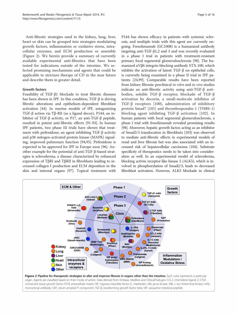
Anti-fibrotic strategies used in the kidney, lung, liver,heart or skin can be grouped into strategies modulatinggrowth factors, inflammation or oxidative stress, intra-cellular enzymes, and ECM production or assembly(Figure 2). We herein provide a summary of currentlyavailable experimental anti-fibrotics that have beentested for indications outside of the intestine. We se-lected promising mechanisms and agents that could beapplicable to stricture therapy of CD in the near futureand describe them in greater detail.
Growth factorsFeasibility of TGF-β1 blockade to treat fibrotic diseaseshas been shown in IPF. In this condition, TGF-β is drivingfibrotic alterations and epithelium-dependent fibroblastactivation [44]. In murine models of IPF, antagonizingTGF-β action via Tβ-RII (as a ligand decoy), P144, an in-hibitor of TGF-β activity, or P17, an anti-TGF-β peptide,resulted in potent anti-fibrotic effects [91-93]. In humanIPF patients, two phase III trials have shown that treat-ment with pirfenidone, an agent inhibiting TGF-β activityand p38 mitogen-activated protein kinase (MAPK) signal-ing, improved pulmonary function [94,95]. Pirfenidone isexpected to be approved for IPF in Europe soon [96]. An-other example for the potential of anti-TGF-β-based strat-egies is scleroderma, a disease characterized by enhancedexpression of TβRI and TβRII in fibroblasts leading to in-creased collagen I production and ECM deposition in theskin and internal organs [97]. Topical treatment with
Figure 2 Pipeline for therapeutic strategies to alter and improve fibrosisorgan. Agents are classified based on their mode of action. Data derived from Econnective tissue growth factor; ECM, extracellular matrix; HIF, hypoxia induciblemonoclonal antibody; SAP, serum amyloid P component; TGF-β, transforming g
P144 has shown efficacy in patients with systemic scler-osis, and multiple trials with this agent are currently on-going. Fresolimumab (GC1008) is a humanized antibodytargeting anti-TGF-β1,2 and 3 and was recently evaluatedin a phase I trial in patients with treatment-resistantprimary focal segmental glomerulosclerosis [98]. The hu-manized αVβ6 integrin-blocking antibody STX-100, whichinhibits the activation of latent TGF-β on epithelial cells,is currently being examined in a phase II trial in IPF pa-tients [24,99]. Comparable results have been reportedfrom kidney fibrosis: preclinical in vitro and in vivo studiesindicate an anti-fibrotic activity using anti-TGF-β anti-bodies, soluble TGF-β receptor, blockade of TGF-βactivation by decorin, a small-molecule inhibitor ofTGF-β receptors [100], administration of inhibitoryprotein Smad7 [101] and thrombospondin-1 (THBS-1)blocking agent inhibiting TGF-β activation [102]. Inhuman patients with focal segmental glomerulosclerosis, aphase I trial with fresolimumab revealed promising results[98]. Moreover, hepatic growth factor, acting as an inhibitorof Smad2/3 translocation in fibroblasts [103] was observedto mediate anti-fibrotic effects in experimental models ofrenal and liver fibrosis but was also associated with an in-creased risk of hepatocellular carcinoma [104]. Substratespecificity of therapeutics needs to be taken into consider-ation as well. In an experimental model of scleroderma,blocking activin receptor-like kinase 5 (ALK5), which is in-volved in phosphorylation of Smad2/3, leads to decreasedfibroblast activation. However, ALK5 blockade in clinical
in organs other than the intestine. Each color represents a particularmbase, Medline and ClinicalTrials.gov. CCL2, chemokine ligand 2; CTGF,factor; IL, interleukin; JAK, janus kinase; JNK, c-Jun N-terminal kinase; mAb,rowth factor beta; VIP, vasoactive intestinal peptide.
Bettenworth and Rieder Fibrogenesis & Tissue Repair 2014, 7:5 Page 6 of 16http://www.fibrogenesis.com/content/7/1/5
trials was associated with adverse events due to cross-reactivity with other kinase inhibitors [105].In addition to the Smad-signaling cascade, non-Smad
pathways comprising TGF-β1 activated MAPKs and sev-eral tyrosine kinases have been targeted for anti-fibroticactions. For example, c-Abelson (c-Abl), a component ofthe Bcr-Abl oncogene, can be effectively blocked by se-lective tyrosine kinase inhibitors such as imatinib. Thisagent inhibits PDGF as well and thus potentially regu-lates fibroblast proliferation and transformation [106].Despite promising results from in vitro and in vivo stud-ies, to date limited experience with tyrosine kinases inhuman fibrotic diseases is available [107,108]. Of note,novel tyrosine kinase inhibitors such as nilotinib anddasatinib mediate dose-dependent decreases in ECMproduction and reveal even greater efficacy as comparedto that of imatinib [109], while being well tolerated bythe patients [110]. In contrast to TGF-β1, other mem-bers of the TGF family such as TGF-β3 possess anti-fibrotic properties. Avotermin is a recombinant bioactivehuman TGF-β3 that has been tested for treatment ofdermal scars and significantly reduces the scar size byintradermal injection therapy [111]. In addition, furthergrowth factors such as serum amyloid P (SAP) havebeen proven effective in experimental models [112,113]of fibrosis and have already entered phase I clinical trialsin human patients [114].The scientific rationale to target TGF-β signaling in
stricturing CD comes from in vitro as well as in vivo ob-servations. For example, intestinal TGF-β overexpressionin mice leads to colonic fibrosis and obstruction [27],while disruption of the TGF-β/Smad signaling cascadeprotects animals from intestinal fibrosis [115]. In humantissue samples from colonic CD strictures, TGF-β and itsreceptors as well as pSmad2/3 expression are increased,while Smad7 expression was significantly reduced [116]. Al-though targeting TGF-β signaling for fibrotic diseases has astrong scientific rationale, it has to be taken into accountthat this growth factor is not only crucially involved infibrogenesis but additionally functions as a key regulator ofcellular processes including differentiation, proliferation,transformation, tumor suppression as well as immunoregu-lation and its actions may be context-dependent [96,117].For example, TGF-β1-deficient mice develop severe multi-organ inflammation and expire by 5 weeks of age [118,119].This outcome occurs even under germ-free conditions[120] and is mediated by CD4+ T cells [121]. Similarly, tar-geted deletion of Smad2 and Smad4 is associated with earlydeath in mice [122,123]. Furthermore, administration ofmetelimumab, a monoclonal antibody against TFG-β1, inhuman systemic sclerosis patients was associated with sig-nificantly more serious adverse events than placebo treat-ment including musculoskeletal pain, progression of skininvolvement and death [124]. Possible side effects during
anti-TGF-β therapy would have to be carefully monitored,in particular in case of pre-existing inflammation. There-fore, neutralizing TGF-β 1 in vivo, as an anti-fibrotic ap-proach in CD may be highly problematic, as this mayactually lead to disease exacerbation given the potentanti-inflammatory and immunoregulatory properties ofthis cytokine.
HMG-CoA reductase inhibitorsHMG-CoA reductase inhibitors were developed with theintention to decrease cholesterol levels. More recently,they were found to mediate anti-inflammatory as well asanti-fibrotic effects in vitro [125], including decreasedproliferation of mesangial cells, lower fibronectin as wellas type IV collagen expression and decreased secretionof TGF-β1 and connective tissue growth factor (CTGF)[126-128]. Corroborating these in vitro findings, HMG-CoA reductase inhibitors revealed various anti-fibroticeffects in murine models of nephropathy and fibrosis[126,127]. In CD patients, HMG-CoA reductase inhibitoratorvastatin was shown to mediate anti-inflammatory ef-fects such as inhibition of T cell recruitment via reducedCXCL10 levels [129] and reduce surrogate inflammatorymarkers such as calprotectin, C-reactive protein and TNF-α expression [130]. Furthermore, it was demonstrated thatsimvastatin reduced TGF-β1 expression in human fibro-blasts by inhibition of Smad3 phosphorylation [131] lead-ing, together with induced apoptosis in fibroblast andmyofibroblasts, to a significant amelioration of experi-mental fibrosis [132]. In addition, mesenchymal cellsisolated from patients with radiation-induced intestinalfibrosis respond to pravastatin treatment with signifi-cantly decreased production of fibronectin and type-1collagen through Rho-/ROCK-dependent reduction ofCTGF expression [133]. Nevertheless, the exact anti-fibrotic potential of statin treatment in stricturing CDstill needs to be defined by the use of hard clinical end-points, but this drug class has an already establishedsafety profile for routine clinical use and could serve asa potential anti-fibrotic treatment approach.
Renin-angiotensin system (RAS) modulatorsAngiotensin II (AT II) is the major mediator of therenin-angiotensin system (RAS). AT II may increase ECMaccumulation through plasminogen activator inhibitor-1-mediated decrease of MMPs and enhance TGF-β1 produc-tion in cardiac and renal fibrosis [134,135]. The impact ofAT II can also be observed in liver fibrosis. In hepatic stel-late cells, AT II induces contraction and proliferation ac-companied by increased collagen and TGF-β expression[136]. Accordingly, progression of liver fibrosis in hepatitisC virus positive patients is significantly decreased aftertreatment with angiotensin-converting enzyme inhibitors[137]. AT II is crucially involved in the manifestations of
Bettenworth and Rieder Fibrogenesis & Tissue Repair 2014, 7:5 Page 7 of 16http://www.fibrogenesis.com/content/7/1/5
renal fibrosis by induction of pro-fibrotic effector moleculesand EMT resulting in enhanced ECM production [138],and inhibition of AT II using angiotensin-converting en-zyme (ACE) inhibitors or blocking agents towards the AT Ireceptor has emerged as a therapeutic approach to slowdown renal disease progression [139] and revealed anti-fibrotic actions in the lung, heart and liver [140-143]. Inter-estingly, all components of the RAS have been detected inthe human colonic mucosa [144] and AT II is increased inthe mucosa of CD patients [145]. In vivo administration ofthe ACE inhibitor enalaprilate has been proven to reduceweight loss and histological damage in murine dextran sul-fate sodium (DSS)-induced colitis [146]. ACE inhibitortreatment was also effective in spontaneous colitis of IL-10-deficient mice [147] and this finding has been confirmed byother studies [148,149].Of note, through its AT1 receptor, AT II enhances the
expression of CTGF, and administration of AT II inhibi-tors and AT1 receptor antagonists significantly amelio-rates or reverses fibrotic alterations in experimentalcolitis reflected by reduced collagen amounts and TGF-β1 mRNA levels [150,151]. Existing preclinical data inIBD combined with clinical trials from the liver and kid-ney make RAS modulation a promising future approachfor CD-associated fibrosis.
Inflammation modulatorsPro-inflammatory cytokines contributing to the patho-genesis of IBD could also be involved in the develop-ment of intestinal fibrosis in CD. For example, IL-1modulates myofibroblast activation, chemokine produc-tion, MMP secretion [152] and is involved in EMT in-duction [153]. IL-6 is known to regulate TGF-β andTGF-βR2 expression as well as fibroblast proliferation[154,155] and is strongly upregulated in serum and tis-sue samples from CD patients [156]. IL-4 and IL-13 rep-resent pivotal mediators of immune activation and Thelper cell 2 responses are crucially involved in the de-velopment of intestinal fibrosis in vivo. IL-13 mediates,through binding to its IL-13Rα, an increased productionof TGF-β and is a key player in the initiation of fibroticalterations in the intestine [25]. Confirmatively, antagon-ism of IL-13 is effective to prevent fibrosis developmentin experimental colitis [33,157]. In addition, several IL-13 antibodies such as lebrikizumab, tralokinumab andQAX576 as well as the anti-TNF antibody etanerceptand the immunomodulatory drug thalidomide are cur-rently being evaluated for their anti-fibrotic potency inliver fibrosis and pulmonary fibrosis [24,158-160].The process of inflammation and fibrosis are likely to
be intertwined through angiogenesis and lymphangio-genesis. Increased levels of factors implicated in angio-genesis have been documented in IBD patients, such asvascular endothelial growth factor A (VEGF-A) [161]. At
a cellular level, PDGF increases proliferation and migra-tion of fibroblasts and myofibroblasts [46]. In the humanintestine, PDGF facilitates ECM deposition and is upreg-ulated in inflamed colonic tissue specimen of CD pa-tients [29]. Experience with blocking these agents inother fibrotic diseases exist. In a phase II trial, combinedblockade of VEGF, PDGF and bFGF by the indolinonederivative BIBF 1120 tends to decrease the developmentof human IPF [162]. Critical for the future use of inflam-mation modulators in the therapy of CD-associated fi-brosis will be the quality, quantity and the timing of theapproach because all of the above mediators act at dif-ferent times throughout the disease course, in differingcombinations and quantities [163]. Additionally, all ofthe above molecules interact with each other and block-ing a single cytokine at a specific time might not be suf-ficient for effective anti-fibrotic therapy.
Extracellular matrix modulatorsThe imbalance between deposition and degradation ofECM in fibrotic disease is a logical target for anti-fibrotictreatment approaches. Stimulation of MMPs as central reg-ulators of ECM disassembly were expected to reverse fi-brotic alterations, however, clinical studies in patients withnephrosclerosis failed to show efficacy [164]. Likewise, de-pletion of TIMP should decrease fibrotic changes, but noamelioration of renal fibrosis was observed following TIMPinhibition in mice [165]. With regard to the intestine, thereis growing evidence for MMPs as a regulator of intestinalbarrier function and mucosal defense [166], indicatingpleiotropic functions of this molecular group in addition topurely matrix regulation. For example, serum MMP-9levels correlate with disease activity in pediatric CD patients[167] and may be used as a biomarker to follow the courseof disease in adult CD patients as well [168,169]. Colonictissue expression of MMP-1, MMP-2, MMP-3 and MMP-9was significantly increased in samples from inflamed mu-cosa as compared to non-inflamed mucosal samples [170].Increased MMP-9 expression in inflamed tissue colonicspecimen from CD patients seems to be associated with de-creased likelihood of disease recurrence [171]. Finally, inmucosa specimen overlaying colonic strictures in CD pa-tients, MMP-3 and MMP-12 expression was significantlyreduced [116]. Thus, MMPs need to be further and care-fully investigated as possible targets for anti-fibrotic treat-ment in as well as outside of the intestine and are not yetready for prime time.
Intracellular enzymes and receptorsThe mammalian target of rapamycin (mTOR) protein is aserine/threonine protein kinase that consists of severalcomplexes among which mTOR complex 1 regulates pro-tein synthesis, proliferation as well as fibrotic actions [46].mTOR inhibitors possess direct anti-fibrotic properties by

Bettenworth and Rieder Fibrogenesis & Tissue Repair 2014, 7:5 Page 8 of 16http://www.fibrogenesis.com/content/7/1/5
decreasing fibroblast and myofibroblast numbers and by re-ducing pro-fibrotic cytokine expression, including IL-4, IL-6, IL-13, IL-17, TGF-β1 as well as type I and III collagen[172,173]. Efficacy of mTOR inhibitors have been demon-strated in numerous fibrotic disorders of the skin, lung,kidney and liver [46]. With regard to CD, a randomized,double-blind clinical trial found that everolimus was as ef-fective as azathioprine to achieve steroid-free remission in138 patients with active CD [174]. Additionally, there aretwo case reports indicating that mTOR inhibitors sirolimusand everolimus are able to induce remission in refractoryCD [175,176]. Given the fact that mTOR inhibitors possessanti-fibrotic as well as immunosuppressive effects, this classof drug appears to be promising for intestinal fibrosis ther-apy, however, the definitive therapeutic potential for intes-tinal fibrosis remains to be defined yet.Peroxisome proliferator-activated receptor gamma
(PPAR-γ) is a nuclear receptor that modulates gene expres-sion and is involved in various physiological and patho-logical processes including inflammation and fibrosis [177].After stimulation with specific ligands, PPAR-γ directlyantagonizes Smad3 or reduces CTGF expression [178].PPAR-γ agonists are able to improve experimental fibrosis,while PPAR-γ selective antagonists abolish anti-fibroticactions [179,180]. Given the fact that CTGF is a key down-stream effector of TGF-β on connective tissue cells, FG-3019, a humanized antibody targeting CTGF, has been
Figure 3 Compounds used as anti-fibrotic therapies in other organs anpathway. Compounds are depicted in red, indicating their mechanism of actioconnective tissue growth factor; ECM, extracellular matrix; HGF, hepatocyte grInhibitor; IL, interleukin; MAPK, mitogen-activated protein kinase; mTOR, mamreceptor; SMAD, small mothers against decapentaplegic; TGF, transforming gr
developed and successfully passed phase I trials in severalfibrotic disorders and recently entered phase II studies [24].In the human intestine, PPAR-γ has been detected in thecolonic mucosa and has been identified as a mediatorof established anti-inflammatory drugs such as 5-ASA[177,181]. The future role of PPAR-γ agonists as a pos-sible target for anti-fibrotic treatment in stricturingCD is promising, given the combined action againstinflammation and fibrosis and its well-defined mechanismof action. Furthermore, other drug candidates such asendothelin A receptor antagonist bosentan have shownpromising results in patients with IPF and renal interstitialfibrosis and deserves further investigation [182,183].A summary of compounds used as anti-fibrotic therapies
in other organs and their mode of action is depicted inFigure 3. Anti-fibrotic clinical trials of major interest areshown in Table 1.
Challenges and future outlookThe most efficient anti-fibrotic treatment approach stillremains the elimination of the primary cause of intes-tinal injury, which would mean nothing less than curingCD. However, since a magic bullet for CD is not and willmost likely not be available in the near future, clinicalevaluation and optimization of anti-fibrotic drug candi-dates for stricturing CD is a justifiable and promisingtreatment strategy.
d their mode of action. The blue boxes represent a major profibroticn. Abs, antibodies; ALK, activin receptor-like kinase; AT, angiotensin; CTGF,owth factor; HMG-CoA, 3-hydroxy-3-methylglutaryl-coenzyme A; Inh,malian target of rapamycin; PPAR, peroxisome proliferator-activatedowth factor.

Table 1 Completed clinical trials evaluating anti-fibrotic drugs in organs other than the gut
Parameter Author Studyorgan
Number ofpatents
Type of trial Study drug Mode of action Outcome measures
Growth factors Noble [95] Lung 779 RCT, phase III Pirfenidone Inhibition of TGF-β Increased FVC in IPF
Trachtman [98] Kidney 16 Open-label, phase I Fresolimumab Antibody targeting allisoforms of TGF-β
Safety, pharmacokinetics
Ferguson [111] Skin 223 RCT, phase I/II Avotermin Antibody targetingTGF-β3
Acceleration and permanent improvementin dermal scaring
Dillingh [114] Lung 29 RCT, phase I rhSAP Substitution of SAP Reduction in SAP levels and circulatingfibrocytes in healthy control andIPF patients
Oxidative stress Raghu [158] Lung 88 RCT, phase II Etanercept Blockade of TNF Physiological and functional decrease indisease progression in IPF
Corren [159] Lung 219 RCT, phase II Lebrikizumab Antibody targeting IL-13 Improved lung function in asthmaticpatients
Horton [160] Lung 23 RCT, phase III Thalidomide Anti-angiogenic andanti-inflammatory
Improvement of cough and respiratoryquality of life in IPF
Intracellular enzymesand receptors
Richeldi [162] Lung 432 RCT, phase II BIBF 1120 Tyrosine kinaseinhibitor
Tendency towards reduced decline of lungfunction in IPF
Daniels [108] Lung 119 RCT, phase II/III Imatinib Tyrosine kinaseinhibitor
No effect on survival and lung function
ECM and other Couluris [140] Lung 20 Uncontrolled, interventionalstudy, phase II
Losartan AT1 antagonist Stabilization of lung function in IPF
el-Agroudy [142] Kidney 162 RCT, phase II Losartan AT1 antagonist Decreased TGF-β1 plasma levels andproteinuria in renal interstitial fibrosis
Kuhn [182] Skin 10 Prospective, open-label,phase II
Bosentan Endothelin receptorantagonist
Reduced skin thickening in systemic sclerosis
Diez [141] Heart 34 Uncontrolled, phase II Losartan AT1 antagonist Decreased myocardial collagen content andleft ventricular chamber stiffness inhypertensive patients
De [143] Liver 39 RCT, phase II Losartan AT1 antagonist Reduction of portal pressure in patientswith liver cirrhosis
King [183] Lung 616 RCT Bosentan Endothelin receptorantagonist
Improvement of FVC and diffusing capacityin IPF
AT1, angiotensin II receptor antagonist subtype 1; CXCL, CXC ligand; ECM, extracellular matrix; FVC, forced vital capacity; IL, interleukin; IPF, idiopathic pulmonary fibrosis; RCT, randomized controlled trial; SAP, serumamyloid P component; TGF-β, transforming growth factor beta; TNF, tumor necrosis factor.
Bettenworth
andRieder
Fibrogenesis&Tissue
Repair2014,7:5
Page9of
16http://w
ww.fibrogenesis.com
/content/7/1/5
Bettenworth and Rieder Fibrogenesis & Tissue Repair 2014, 7:5 Page 10 of 16http://www.fibrogenesis.com/content/7/1/5
However, the simple transfer of these agents into CDtreatment is premature and multiple obstacles have tobe overcome to make use of the above described media-tors and mechanisms.First, the ideal anti-fibrotic drug should target some-
thing uniquely expressed in a specific fibrotic compli-cation in a particular organ and should not display anysystemic side effects. This is particularly true in case ofconcomitant injury elsewhere in the body. To date,however, no specific target for intestinal fibrosis orother fibrotic disease has been identified, which sup-ports the hypothesis that mechanisms of fibrosis areshared between different organs.Second, the optimal timing to commence anti-fibrotic
treatment is of utmost importance to CD patients, how-ever, is not defined yet. It is obvious that an early use ofanti-fibrotics is expected to be associated with a better out-come in patients prone to this complication. Pre-existing fi-brosis and concomitantly increased tissue stiffness canperpetuate fibrosis even in the absence of inflammation.Data from experimental fibrosis seems to confirm thatthere might be a critical point in evolution of fibrosis whenthe progress becomes irreversible [184]. Therefore, an earlytreatment with anti-fibrotics at the same time of anti-inflammatory agents might be mandatory in human CD pa-tients [185]. Unique to CD is a fistulizing disease processand this complication needs to be kept in mind when usingdrugs inhibiting tissue remodeling because this could theor-etically promote fistulizing disease. However, timing maynot be simply based on the disease duration, since up to50% of CD patients present with stricturing or penetratingdisease at the time of first diagnosis [186].
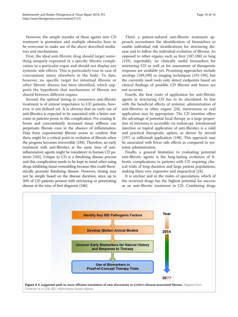
Figure 4 A suggested path to more efficient translation of new discoFriedman et al. [24]. IBD, inflammatory bowel disease.
Third, a patient-tailored anti-fibrotic treatment ap-proach necessitates the identification of biomarkers toenable individual risk stratifications for stricturing dis-ease and to follow the individual evolution of fibrosis. Asopposed to other organs, such as liver [187,188] or lung[159], regrettably, no clinically useful biomarkers forstricturing CD as well as for assessment of therapeuticresponse are available yet. Promising approaches includeserology [189,190] or imaging techniques [191-196], butthe currently used tools only detect endpoints based onclinical findings of possible CD fibrosis and hence arenot accurate.Fourth, the best route of application for anti-fibrotic
agents in stricturing CD has to be elucidated. In linewith the beneficial effects of systemic administration ofanti-fibrotics in other organs [24], intravenous or oralapplication may be appropriate. The CD intestine offersthe advantage of potential local therapy as a large propor-tion of strictures is accessible via endoscopy. Intralesionalinjection or topical application of anti-fibrotics is a validand practical therapeutic option, as shown by steroid[197] or infliximab application [198]. This approach maybe associated with fewer side effects as compared to sys-temic administration.Finally, a general limitation to evaluating potential
anti-fibrotic agents is the long-lasting evolution of fi-brotic complications in patients with CD requiring clin-ical trials of long duration and large patient populations,making them very expensive and impractical [24].It is unclear and in the realm of speculation, which of
the reviewed drugs has the highest potential for successas an anti-fibrotic treatment in CD. Combining drugs
veries to Crohn’s disease-associated fibrosis. Adapted from

Bettenworth and Rieder Fibrogenesis & Tissue Repair 2014, 7:5 Page 11 of 16http://www.fibrogenesis.com/content/7/1/5
with a known safety profile and anti-fibrotic efficacy,such as HMG-CoA reductase inhibitors, mTOR inhibi-tors or inhibitors of the angiotensin system, could serveas a starting point. Given the effect of most substanceson a reduction of TGF-β1 proper precaution needs to betaken for carefully monitoring intestinal inflammation.Novel drug delivery systems, such as the Multi MatrixSystem (MMX®), allowing oral administration of a com-pound with the substance being released in a definedarea of the gastrointestinal tract (such as the terminalileum) can circumvent systemic side effects, such ashypotension for angiotensin blockade.To move forward with specific anti-fibrotic therapies
in the future we need to focus on identifying mecha-nisms of fibrogenesis and to develop new and better ani-mal models for this disease (Figure 4). What, however, isbadly missing is the availability of biomarkers for naturalhistory and response to therapy that are readily availableand easy to measure. Only through utilization of thesemarkers will the field be able to design clinical studies ofreasonable length and sufficient patient number, reflectedin affordable budgets that put our ability for testing anti-fibrotic therapies within reach.
ConclusionsStricturing CD is still an unresolved problem withstrong implications for the patients and a high socioeco-nomic burden due to frequent hospitalizations andsurgery [23]. Despite recent advances in the patho-physiological understanding of fibrosis and significantexpansion of the anti-inflammatory armamentariumover the last few decades, the occurrence of intestinalstrictures in CD patients did not significantly change.To date, effective therapeutic approaches for stricturingCD are limited to ED or surgical interventions. The datapresented in this review highlight the pleiotropic anti-fibrotic actions that have been observed with the use ofnumerous agents in fibrotic complications of the skin,the lung and the kidney. Thus, it is justified to proposefurther evaluation of these drug candidates in clinicaltrials for the management of intestinal fibrosis. How-ever, previous establishment of non-invasive biomarkersto assess the degree of fibrosis, to monitor fibrotic evo-lution and to predict therapeutic response, combinedwith the development of imaging techniques to quantifyintestinal fibrosis, appear to be essential pre-requisitesfor individual risk stratification and proper design ofclinical trials.
Abbreviations6-MP: 6-Mercaptopurine; ACE: Angiotensin-converting enzyme; ALK: Activinreceptor-like kinase; AT II: Angiotensin II; AZA: Azathioprine; bFGF: Basic fibroblastgrowth factor; c-Abl: c-Abelson; CD: Crohn’s disease; CTGF: Connective tissuegrowth factor; DSS: Dextran sulfate sodium; ECM: Extracellularmatrix; ED: Endoscopicdilation; EMT: Epithelial-mesenchymal transition; EndoMT: Endothelial-mesenchymaltransition; HMG-CoA: 3-Hydroxy-3-methyl-glutaryl-coenzymeA; IBD: Inflammatory
bowel disease; IGF: Insulin-like growth factor; IL: Interleukin; IPF: Idiopathic pulmonaryfibrosis; MAPK: Mitogen-activated protein kinase;MMP:Matrix metalloproteinase;mTOR: Mammalian target of rapamycin; NF-κB: Nuclear factor kappa-light-chain-enhancer of activated B cells; NIH: National Institutes of Health; NLM: NationalLibrary of Medicine; PDGF: Platelet-derived growth factor; PG-PS: Peptidoglycan-polysaccharide; PPAR: Peroxisome proliferator-activated receptor; RAS: Renin-angiotensin system; ROCK: Rho-associated protein kinase; SAP: Serum amyloid Pcomponent; TGF: Transforming growth factor; THBS: Thrombospondin; TIMP: Tissueinhibitor of metalloproteinase; TLR: Toll-like receptor; TNBS: 2,4,6-Trinitrobenzenesulfonic acid; TNF: Tumor necrosis factor; VEGF: Vascular endothelial growth factor.
Competing interestsThe authors declare that they have no competing interests.
Authors’ contributionsDB and FR developed the review concept, performed the literature reviewand wrote the manuscript. Both authors read and approved the final versionof the manuscript.
AcknowledgementsDB was supported by a research fellowship from the Faculty of Medicine,University of Münster, Münster, Germany. FR is funded by the NIH (grant:1T32DK083251-01A1). The authors would like to thank Dr Claudio Fiocchiand Dr Bo Shen, Department of Gastroenterology, Hepatology and Nutrition,Cleveland Clinic Foundation, Cleveland, OH, USA, for helpful comments.
Author details1Department of Medicine B, University Hospital of Münster, Münster,Germany. 2Department of Gastroenterology and Hepatology, DigestiveDisease Institute, Cleveland Clinic Foundation, Cleveland, OH, USA.3Department of Pathobiology, Lerner Research Institute, NC22, ClevelandClinic Foundation, 9500 Euclid Avenue, Cleveland, OH 44195, USA.
Received: 3 January 2014 Accepted: 6 March 2014Published: 29 March 2014
References1. Baumgart DC, Sandborn WJ: Crohn’s disease. Lancet 2012, 380:1590–1605.2. Cheifetz AS: Management of active Crohn disease. JAMA 2013,
309:2150–2158.3. Cosnes J, Gower-Rousseau C, Seksik P, Cortot A: Epidemiology and
natural history of inflammatory bowel diseases. Gastroenterology 2011,140:1785–1794.
4. Lenze F, Wessling J, Bremer J, Ullerich H, Spieker T, Weckesser M,Gonschorrek S, Kannengiesser K, Rijcken E, Heidemann J, Luegering A,Schober O, Domschke W, Kucharzik T, Maaser C: Detection anddifferentiation of inflammatory versus fibromatous Crohn’s diseasestrictures: prospective comparison of 18 F-FDG-PET/CT, MR-enteroclysis,and transabdominal ultrasound versus endoscopic/histologic evaluation.Inflamm Bowel Dis 2012, 18:2252–2260.
5. Schoepfer AM, Safroneeva E, Vavricka SR, Peyrin-Biroulet L, Mottet C:Treatment of fibrostenotic and fistulizing Crohn’s disease. Digestion 2012,86(Suppl 1):23–27.
6. Hanauer SB, Feagan BG, Lichtenstein GR, Mayer LF, Schreiber S, Colombel JF,Rachmilewitz D, Wolf DC, Olson A, Bao W, Rutgeerts P, ACCENT I Study Group:Maintenance infliximab for Crohn’s disease: the ACCENT I randomised trial.Lancet 2002, 359:1541–1549.
7. Sandborn WJ, Hanauer SB, Rutgeerts P, Fedorak RN, Lukas M, MacIntosh DG,Panaccione R, Wolf D, Kent JD, Bittle B, Li J, Pollack PF: Adalimumab formaintenance treatment of Crohn’s disease: results of the CLASSIC II trial.Gut 2007, 56:1232–1239.
8. Colombel JF, Sandborn WJ, Rutgeerts P, Enns R, Hanauer SB, Panaccione R,Schreiber S, Byczkowski D, Li J, Kent JD, Pollack PF: Adalimumab formaintenance of clinical response and remission in patients with Crohn’sdisease: the CHARM trial. Gastroenterology 2007, 132:52–65.
9. Cosnes J, Nion-Larmurier I, Beaugerie L, Afchain P, Tiret E, Gendre JP: Impactof the increasing use of immunosuppressants in Crohn’s disease on theneed for intestinal surgery. Gut 2005, 54:237–241.
10. Rieder F, Zimmermann EM, Remzi FH, Sandborn WJ: Crohn’s diseasecomplicated by strictures: a systematic review. Gut 2013, 62:1072–1084.
Bettenworth and Rieder Fibrogenesis & Tissue Repair 2014, 7:5 Page 12 of 16http://www.fibrogenesis.com/content/7/1/5
11. Rieder F, Fiocchi C: Intestinal fibrosis in inflammatory bowel disease: progressin basic and clinical science. Curr Opin Gastroenterol 2008, 24:462–468.
12. Hassan C, Zullo A, De Francesco V, Ierardi E, Giustini M, Pitidis A, Taggi F,Winn S, Morini S: Systematic review: endoscopic dilatation in Crohn’sdisease. Aliment Pharmacol Ther 2007, 26:1457–1464.
13. Thienpont C, D’Hoore A, Vermeire S, Demedts I, Bisschops R, Coremans G,Rutgeerts P, Van Assche G: Long-term outcome of endoscopic dilatationin patients with Crohn’s disease is not affected by disease activity ormedical therapy. Gut 2010, 59:320–324.
14. Singh VV, Draganov P, Valentine J: Efficacy and safety of endoscopicballoon dilation of symptomatic upper and lower gastrointestinalCrohn’s disease strictures. J Clin Gastroenterol 2005, 39:284–290.
15. Hoffmann JC, Heller F, Faiss S, von Lampe B, Kroesen AJ, Wahnschaffe U,Schulzke JD, Zeitz M, Bojarski C: Through the endoscope balloon dilationof ileocolonic strictures: prognostic factors, complications, andeffectiveness. Int J Colorectal Dis 2008, 23:689–696.
16. Froehlich F, Juillerat P, Pittet V, Felley C, Mottet C, Vader JP, Michetti P,Gonvers JJ: Maintenance of surgically induced remission of Crohn’sdisease. Digestion 2007, 76:130–135.
17. Despott EJ, Gupta A, Burling D, Tripoli E, Konieczko K, Hart A, Fraser C:Effective dilation of small-bowel strictures by double-balloon enteroscopy inpatients with symptomatic Crohn’s disease (with video). Gastrointest Endosc2009, 70:1030–1036.
18. Gustavsson A, Magnuson A, Blomberg B, Andersson M, Halfvarson J, Tysk C:Endoscopic dilation is an efficacious and safe treatment of intestinalstrictures in Crohn’s disease. Aliment Pharmacol Ther 2012, 36:151–158.
19. Wibmer AG, Kroesen AJ, Grone J, Buhr HJ, Ritz JP: Comparison ofstrictureplasty and endoscopic balloon dilatation for stricturing Crohn’sdisease–review of the literature. Int J Colorectal Dis 2010, 25:1149–1157.
20. Baba S, Nakai K: Strictureplasty for Crohn’s disease in Japan.J Gastroenterol 1995, 30(Suppl 8):135–138.
21. Ambe R, Campbell L, Cagir B: A comprehensive review of strictureplastytechniques in Crohn’s disease: types, indications, comparisons, and safety.J Gastrointest Surg 2012, 16:209–217.
22. Rutgeerts P, Geboes K, Vantrappen G, Beyls J, Kerremans R, Hiele M:Predictability of the postoperative course of Crohn’s disease.Gastroenterology 1990, 99:956–963.
23. Bodger K, Kikuchi T, Hughes D: Cost-effectiveness of biological therapy forCrohn’s disease: Markov cohort analyses incorporating United Kingdompatient-level cost data. Aliment Pharmacol Ther 2009, 30:265–274.
24. Friedman SL, Sheppard D, Duffield JS, Violette S: Therapy for fibroticdiseases: nearing the starting line. Sci Transl Med 2013, 5:167sr1.
25. Fichtner-Feigl S, Young CA, Kitani A, Geissler EK, Schlitt HJ, Strober W: IL-13signaling via IL-13R alpha2 induces major downstream fibrogenicfactors mediating fibrosis in chronic TNBS colitis. Gastroenterology 2008,135:2003–2013.
26. Zhu MY, Lu YM, Ou YX, Zhang HZ, Chen WX: Dynamic progress of2,4,6-trinitrobenzene sulfonic acid induced chronic colitis andfibrosis in rat model. J Dig Dis 2012, 13:421–429.
27. Vallance BA, Gunawan MI, Hewlett B, Bercik P, Van Kampen C, Galeazzi F,Sime PJ, Gauldie J, Collins SM: TGF-beta1 gene transfer to the mousecolon leads to intestinal fibrosis. Am J Physiol Gastrointest Liver Physiol2005, 289:G116–G128.
28. Mahavadi S, Flynn RS, Grider JR, Qiao LY, Murthy KS, Hazelgrove KB,Kuemmerle JF: Amelioration of excess collagen IalphaI, fibrosis, andsmooth muscle growth in TNBS-induced colitis in IGF-I(+/−) mice.Inflamm Bowel Dis 2011, 17:711–719.
29. Kumagai S, Ohtani H, Nagai T, Funa K, Hiwatashi NO, Shimosegawa, Nagura H:Platelet-derived growth factor and its receptors are expressed in areas ofboth active inflammation and active fibrosis in inflammatory boweldisease. Tohoku J Exp Med 2001, 195:21–33.
30. Lawrance IC, Maxwell L, Doe W: Altered response of intestinal mucosalfibroblasts to profibrogenic cytokines in inflammatory bowel disease.Inflamm Bowel Dis 2001, 7:226–236.
31. Wynn TA, Ramalingam TR: Mechanisms of fibrosis: therapeutic translationfor fibrotic disease. Nat Med 2012, 18:1028–1040.
32. Rieder F, Fiocchi C: Intestinal fibrosis in IBD - a dynamic, multifactorialprocess. Nat Rev Gastroenterol Hepatol 2009, 6:228–235.
33. Fichtner-Feigl S, Strober W, Kawakami K, Puri RK, Kitani A: IL-13 signalingthrough the IL-13alpha2 receptor is involved in induction of TGF-beta1production and fibrosis. Nat Med 2006, 12:99–106.
34. Akiho H, Lovato P, Deng Y, Ceponis PJ, Blennerhassett P, Collins SM:Interleukin-4- and -13-induced hypercontractility of human intestinalmuscle cells-implication for motility changes in Crohn’s disease. Am JPhysiol Gastrointest Liver Physiol 2005, 288:G609–G615.
35. Bailey JR, Bland PW, Tarlton JF, Peters I, Moorghen M, Sylvester PA, ProbertCS, Whiting CV: IL-13 promotes collagen accumulation in Crohn’s diseasefibrosis by down-regulation of fibroblast MMP synthesis: a role for innatelymphoid cells? PLoS One 2012, 7:e52332.
36. Biancheri P, Di Sabatino A, Ammoscato F, Facciotti F, Caprioli F, CurciarelloR, Hoque SS, Ghanbari A, Joe-Njoku I, Giuffrida P, Rovedatti L, Geginat J,Corazza GR, Macdonald TT: Absence of a role for interleukin-13 ininflammatory bowel disease. Eur J Immunol 2014, 44:370–385.
37. Valente AJ, Yoshida T, Gardner JD, Somanna N, Delafontaine P,Chandrasekar B: Interleukin-17A stimulates cardiac fibroblast proliferationand migration via negative regulation of the dual-specificity phosphataseMKP-1/DUSP-1. Cell Signal 2012, 24:560–568.
38. Meng F, Wang K, Aoyama T, Grivennikov SI, Paik Y, Scholten D, Cong M,Iwaisako K, Liu X, Zhang M, Osterreicher CH, Stickel F, Ley K, Brenner DA,Kisseleva T: Interleukin-17 signaling in inflammatory, Kupffer cells, andhepatic stellate cells exacerbates liver fibrosis in mice. Gastroenterology2012, 143:765–776.
39. Okamoto Y, Hasegawa M, Matsushita T, Hamaguchi Y, Huu DL, Iwakura Y,Fujimoto M, Takehara K: Potential roles of interleukin-17A in the developmentof skin fibrosis in mice. Arthritis Rheum 2012, 64:3726–3735.
40. Mi S, Li Z, Yang HZ, Liu H, Wang JP, Ma YG, Wang XX, Liu HZ, Sun W, Hu ZW:Blocking IL-17A promotes the resolution of pulmonary inflammation andfibrosis via TGF-beta1-dependent and -independent mechanisms.J Immunol 2011, 187:3003–3014.
41. Gregory LG, Jones CP, Walker SA, Sawant D, Gowers KH, Campbell GA,McKenzie AN, Lloyd CM: IL-25 drives remodelling in allergic airwaysdisease induced by house dust mite. Thorax 2013, 68:82–90.
42. Biancheri P, Pender SL, Ammoscato F, Giuffrida P, Sampietro G, Ardizzone S,Ghanbari A, Curciarello R, Pasini A, Monteleone G, Corazza GR, MacdonaldTT, Di Sabatino A: The role of interleukin 17 in Crohn’s disease-associatedintestinal fibrosis. Fibrogenesis Tissue Repair 2013, 6:13.
43. Hueber W, Sands BE, Lewitzky S, Vandemeulebroecke M, Reinisch W,Higgins PD, Wehkamp J, Feagan BG, Yao MD, Karczewski M, Karczewski J,Pezous N, Bek S, Bruin G, Mellgard B, Berger C, Londei M, Bertolino AP,Tougas G, Travis SP, Secukinumab in Crohn’s Disease Study Group:Secukinumab, a human anti-IL-17A monoclonal antibody, for moderateto severe Crohn’s disease: unexpected results of a randomised, double-blindplacebo-controlled trial. Gut 2012, 61:1693–1700.
44. Hawinkels LJ, Ten Dijke P: Exploring anti-TGF-beta therapies in cancer andfibrosis. Growth Factors 2011, 29:140–152.
45. Munger JS, Harpel JG, Gleizes PE, Mazzieri R, Nunes I, Rifkin DB: Latenttransforming growth factor-beta: structural features and mechanismsof activation. Kidney Int 1997, 51:1376–1382.
46. Latella G, Sferra R, Speca S, Vetuschi A, Gaudio E: Can we prevent, reduceor reverse intestinal fibrosis in IBD? Eur Rev Med Pharmacol Sci 2013,17:1283–1304.
47. Monteleone G, Pallone F, MacDonald TT: Smad7 in TGF-beta-mediatednegative regulation of gut inflammation. Trends Immunol 2004,25:513–517.
48. McKaig BC, McWilliams D, Watson SA, Mahida YR: Expression and regulationof tissue inhibitor of metalloproteinase-1 and matrix metalloproteinases byintestinal myofibroblasts in inflammatory bowel disease. Am J Pathol 2003,162:1355–1360.
49. Medina C, Santos-Martinez MJ, Santana A, Paz-Cabrera MC, Johnston MJ,Mourelle M, Salas A, Guarner F: Transforming growth factor-beta type 1receptor (ALK5) and Smad proteins mediate TIMP-1 and collagensynthesis in experimental intestinal fibrosis. J Pathol 2011, 224:461–472.
50. Rieder F, Georgieva M, Schirbel A, Artinger M, Zugner A, Blank M,Brenmoehl J, Scholmerich J, Rogler G: Prostaglandin E2 inhibitsmigration of colonic lamina propria fibroblasts. Inflamm Bowel Dis2010, 16:1505–1513.
51. Brittan M, Chance V, Elia G, Poulsom R, Alison MR, MacDonald TT, WrightNA: A regenerative role for bone marrow following experimental colitis:contribution to neovasculogenesis and myofibroblasts. Gastroenterology1984–1995, 2005:128.
52. Kalluri R: EMT: when epithelial cells decide to become mesenchymal-likecells. J Clin Invest 2009, 119:1417–1419.
Bettenworth and Rieder Fibrogenesis & Tissue Repair 2014, 7:5 Page 13 of 16http://www.fibrogenesis.com/content/7/1/5
53. Otte JM, Rosenberg IM, Podolsky DK: Intestinal myofibroblasts in innateimmune responses of the intestine. Gastroenterology 1866–1878,2003:124.
54. Rieder F, Kessler S, Sans M, Fiocchi C: Animal models of intestinal fibrosis:new tools for the understanding of pathogenesis and therapy of humandisease. Am J Physiol Gastrointest Liver Physiol 2012, 303:G786–G801.
55. Jostins L, Ripke S, Weersma RK, Duerr RH, McGovern DP, Hui KY, Lee JC,Schumm LP, Sharma Y, Anderson CA, Essers J, Mitrovic M, Ning K, Cleynen I,Theatre E, Spain SL, Raychaudhuri S, Goyette P, Wei Z, Abraham C, AchkarJP, Ahmad T, Amininejad L, Ananthakrishnan AN, Andersen V, Andrews JM,Baidoo L, Balschun T, Bampton PA, Bitton A, et al: Host-microbeinteractions have shaped the genetic architecture of inflammatorybowel disease. Nature 2012, 491:119–124.
56. Dubinsky MC, Lin YC, Dutridge D, Picornell Y, Landers CJ, Farrior S, Wrobel I,Quiros A, Vasiliauskas EA, Grill B, Israel D, Bahar R, Christie D, Wahbeh G,Silber G, Dallazadeh S, Shah P, Thomas D, Kelts D, Hershberg RM, Elson CO,Targan SR, Taylor KD, Rotter JI, Yang H, Western Regional Pediatric IBDResearch Alliance: Serum immune responses predict rapid diseaseprogression among children with Crohn’s disease: immune responsespredict disease progression. Am J Gastroenterol 2006, 101:360–367.
57. Rieder F, Schleder S, Wolf A, Dirmeier A, Strauch U, Obermeier F, Lopez R,Spector L, Fire E, Yarden J, Rogler G, Dotan N, Klebl F: Serum anti-glycanantibodies predict complicated Crohn’s disease behavior: a cohort study.Inflamm Bowel Dis 2010, 16:1367–1375.
58. Rieder F, Schleder S, Wolf A, Dirmeier A, Strauch U, Obermeier F, Lopez R,Spector L, Fire E, Yarden J, Rogler G, Dotan N, Klebl F: Association of thenovel serologic anti-glycan antibodies anti-laminarin and anti-chitinwith complicated Crohn’s disease behavior. Inflamm Bowel Dis 2010,16:263–274.
59. Geboes KP, Cabooter L, Geboes K: Contribution of morphology for thecomprehension of mechanisms of fibrosis in inflammatory enterocolitis.Acta Gastroenterol Belg 2000, 63:371–376.
60. Graham MF, Diegelmann RF, Elson CO, Lindblad WJ, Gotschalk N, Gay S,Gay R: Collagen content and types in the intestinal strictures of Crohn’sdisease. Gastroenterology 1988, 94:257–265.
61. Borley NR, Mortensen NJ, Kettlewell MG, George BD, Jewell DP, Warren BF:Connective tissue changes in ileal Crohn’s disease: relationship todisease phenotype and ulcer-associated cell lineage. Dis Colon Rectum2001, 44:388–396.
62. Geboes K, El-Zine MY, Dalle I, El-Haddad S, Rutgeerts P, Van Eyken P: Tenascinand strictures in inflammatory bowel disease: an immunohistochemical study.Int J Surg Pathol 2001, 9:281–286.
63. Gelbmann CM, Mestermann S, Gross V, Kollinger M, Scholmerich J, Falk W:Strictures in Crohn’s disease are characterised by an accumulation ofmast cells colocalised with laminin but not with fibronectin orvitronectin. Gut 1999, 45:210–217.
64. Wells RG: The role of matrix stiffness in regulating cell behavior.Hepatology 2008, 47:1394–1400.
65. Ura H, Obara T, Yokota K, Shibata Y, Okamura K, Namiki M: Effects oftransforming growth factor-beta released from gastric carcinoma cellson the contraction of collagen-matrix gels containing fibroblasts.Cancer Res 1991, 51:3550–3554.
66. Hinz B: Tissue stiffness, latent TGF-beta1 activation, and mechanicalsignal transduction: implications for the pathogenesis and treatment offibrosis. Curr Rheumatol Rep 2009, 11:120–126.
67. Oikarinen AI, Vuorio EI, Zaragoza EJ, Palotie A, Chu ML, Uitto J: Modulationof collagen metabolism by glucocorticoids. Receptor-mediated effects ofdexamethasone on collagen biosynthesis in chick embryo fibroblastsand chondrocytes. Biochem Pharmacol 1988, 37:1451–1462.
68. Ketchum LD, Smith J, Robinson DW, Masters FW: The treatment ofhypertrophic scar, keloid and scar contracture by triamcinoloneacetonide. Plast Reconstr Surg 1966, 38:209–218.
69. Vaglio A, Palmisano A, Alberici F, Maggiore U, Ferretti S, Cobelli R, Ferrozzi F,Corradi D, Salvarani C, Buzio C: Prednisone versus tamoxifen in patientswith idiopathic retroperitoneal fibrosis: an open-label randomisedcontrolled trial. Lancet 2011, 378:338–346.
70. Badea I, Taylor M, Rosenberg A, Foldvari M: Pathogenesis and therapeuticapproaches for improved topical treatment in localized scleroderma andsystemic sclerosis. Rheumatology (Oxford) 2009, 48:213–221.
71. Peikert T, Daniels CE, Beebe TJ, Meyer KC, Ryu JH, Interstitial Lung DiseasesNetwork of the American College of Chest Physicians: Assessment of
current practice in the diagnosis and therapy of idiopathic pulmonaryfibrosis. Respir Med 2008, 102:1342–1348.
72. Graham MF, Willey A, Adams J, Diegelmann RF: Corticosteroids increaseprocollagen gene expression, synthesis, and secretion by humanintestinal smooth muscle cells. Gastroenterology 1995, 109:1454–1461.
73. Kochhar R, Poornachandra KS: Intralesional steroid injection therapy in themanagement of resistant gastrointestinal strictures. World J GastrointestEndosc 2010, 2:61–68.
74. Warnatz K, Keskin AG, Uhl M, Scholz C, Katzenwadel A, Vaith P, Peter HH,Walker UA: Immunosuppressive treatment of chronic periaortitis:a retrospective study of 20 patients with chronic periaortitis and areview of the literature. Ann Rheum Dis 2005, 64:828–833.
75. Rogliani P, Mura M, Assunta Porretta M, Saltini C: New perspectives in thetreatment of idiopathic pulmonary fibrosis. Ther Adv Respir Dis 2008, 2:75–93.
76. Dheda K, Lalloo UG, Cassim B, Mody GM: Experience with azathioprine insystemic sclerosis associated with interstitial lung disease. Clin Rheumatol2004, 23:306–309.
77. Peyrin-Biroulet L, Deltenre P, Ardizzone S, D’Haens G, Hanauer SB, HerfarthH, Lemann M, Colombel JF: Azathioprine and 6-mercaptopurine for theprevention of postoperative recurrence in Crohn’s disease: a meta-analysis. Am J Gastroenterol 2089–2096, 2009:104.
78. Spinelli A, Correale C, Szabo H, Montorsi M: Intestinal fibrosis in Crohn’s disease:medical treatment or surgery? Curr Drug Targets 2010, 11:242–248.
79. Hanauer SB, Sandborn WJ, Rutgeerts P, Fedorak RN, Lukas M, MacIntosh D,Panaccione R, Wolf D, Pollack P: Human anti-tumor necrosis factormonoclonal antibody (adalimumab) in Crohn’s disease: the CLASSIC-Itrial. Gastroenterology 2006, 130:323–333. quiz 591.
80. Colombel JF, Sandborn WJ, Reinisch W, Mantzaris GJ, Kornbluth A, RachmilewitzD, Lichtiger S, D’Haens G, Diamond RH, Broussard DL, Tang KL, van der WoudeCJ, Rutgeerts P, SONIC Study Group: Infliximab, azathioprine, or combinationtherapy for Crohn’s disease. N Engl J Med 2010, 362:1383–1395.
81. Sandborn WJ, Feagan BG, Marano C, Zhang H, Strauss R, Johanns J,Adedokun OJ, Guzzo C, Colombel JF, Reinisch W, Gibson PR, Collins J,Jarnerot G, Hibi T, Rutgeerts P, PURSUIT-SC Study Group: Subcutaneousgolimumab induces clinical response and remission in patients withmoderate-to-severe ulcerative colitis. Gastroenterology 2014, 146:85–95.
82. Sandborn WJ, Feagan BG, Stoinov S, Honiball PJ, Rutgeerts P, Mason D,Bloomfield R, Schreiber S, PRECISE 1 Study Investigators: Certolizumab pegolfor the treatment of Crohn’s disease. N Engl J Med 2007, 357:228–238.
83. Bahcecioglu IH, Koca SS, Poyrazoglu OK, Yalniz M, Ozercan IH, Ustundag B,Sahin K, Dagli AF, Isik A: Hepatoprotective effect of infliximab, ananti-TNF-alpha agent, on carbon tetrachloride-induced hepaticfibrosis. Inflammation 2008, 31:215–221.
84. Antoniou KM, Mamoulaki M, Malagari K, Kritikos HD, Bouros D, Siafakas NM,Boumpas DT: Infliximab therapy in pulmonary fibrosis associated withcollagen vascular disease. Clin Exp Rheumatol 2007, 25:23–28.
85. Bargagli E, Galeazzi M, Bellisai F, Volterrani L, Rottoli P: Infliximab treatmentin a patient with systemic sclerosis associated with lung fibrosis andpulmonary hypertension. Respiration 2008, 75:346–349.
86. Theiss AL, Simmons JG, Jobin C, Lund PK: Tumor necrosis factor (TNF)alpha increases collagen accumulation and proliferation in intestinalmyofibroblasts via TNF receptor 2. J Biol Chem 2005, 280:36099–36109.
87. Di Sabatino A, Pender SL, Jackson CL, Prothero JD, Gordon JN, Picariello L,Rovedatti L, Docena G, Monteleone G, Rampton DS, Tonelli F, Corazza GR,MacDonald TT: Functional modulation of Crohn’s disease myofibroblastsby anti-tumor necrosis factor antibodies. Gastroenterology 2007, 133:137–149.
88. Lichtenstein GR, Olson A, Travers S, Diamond RH, Chen DM, Pritchard ML,Feagan BG, Cohen RD, Salzberg BA, Hanauer SB, Sandborn WJ: Factorsassociated with the development of intestinal strictures or obstructionsin patients with Crohn’s disease. Am J Gastroenterol 2006, 101:1030–1038.
89. Pelletier AL, Kalisazan B, Wienckiewicz J, Bouarioua N, Soule JC: Infliximabtreatment for symptomatic Crohn’s disease strictures. Aliment PharmacolTher 2009, 29:279–285.
90. Schnitzler F, Fidder H, Ferrante M, Noman M, Arijs I, Van Assche G, Hoffman I,Van Steen K, Vermeire S, Rutgeerts P: Long-term outcome of treatment withinfliximab in 614 patients with Crohn’s disease: results from a single-centrecohort. Gut 2009, 58:492–500.
91. Yamada M, Kuwano K, Maeyama T, Yoshimi M, Hamada N, Fukumoto J,Egashira K, Hiasa K, Takayama K, Nakanishi Y: Gene transfer of solubletransforming growth factor type II receptor by in vivo electroporationattenuates lung injury and fibrosis. J Clin Pathol 2007, 60:916–920.
Bettenworth and Rieder Fibrogenesis & Tissue Repair 2014, 7:5 Page 14 of 16http://www.fibrogenesis.com/content/7/1/5
92. Arribillaga L, Dotor J, Basagoiti M, Riezu-Boj JI, Borras-Cuesta F, Lasarte JJ,Sarobe P, Cornet ME, Feijoo E: Therapeutic effect of a peptide inhibitor ofTGF-beta on pulmonary fibrosis. Cytokine 2011, 53:327–333.
93. Ezquerro IJ, Lasarte JJ, Dotor J, Castilla-Cortazar I, Bustos M, Penuelas I,Blanco G, Rodriguez C, Lechuga Mdel C, Greenwel P, Rojkind M, Prieto J,Borras-Cuesta F: A synthetic peptide from transforming growth factorbeta type III receptor inhibits liver fibrogenesis in rats with carbontetrachloride liver injury. Cytokine 2003, 22:12–20.
94. Azuma A: Pirfenidone treatment of idiopathic pulmonary fibrosis. Ther AdvRespir Dis 2012, 6:107–114.
95. Noble PW, Albera C, Bradford WZ, Costabel U, Glassberg MK, Kardatzke D,King TE Jr, Lancaster L, Sahn SA, Szwarcberg J, Valeyre D, du Bois RM,CAPACITY Study Group: Pirfenidone in patients with idiopathicpulmonary fibrosis (CAPACITY): two randomised trials. Lancet 2011,377:1760–1769.
96. Akhurst RJ, Hata A: Targeting the TGFbeta signalling pathway in disease.Nat Rev Drug Discov 2012, 11:790–811.
97. Beyer C, Distler O, Distler JH: Innovative antifibrotic therapies in systemicsclerosis. Curr Opin Rheumatol 2012, 24:274–280.
98. Trachtman H, Fervenza FC, Gipson DS, Heering P, Jayne DR, Peters H, Rota S,Remuzzi G, Rump LC, Sellin LK, Heaton JP, Streisand JB, Hard ML, Ledbetter SR,Vincenti F: A phase 1, single-dose study of fresolimumab, an anti-TGF-betaantibody, in treatment-resistant primary focal segmental glomerulosclerosis.Kidney Int 2011, 79:1236–1243.
99. Crunkhorn S: Deal watch: Biogen acquires Stromedix to pursue novelfibrosis therapy. Nat Rev Drug Discov 2012, 11:260.
100. Alan C, Kocoglu H, Altintas R, Alici B, Resit Ersay A: Protective effect ofdecorin on acute ischaemia-reperfusion injury in the rat kidney.Arch Med Sci 2011, 7:211–216.
101. Terada Y, Hanada S, Nakao A, Kuwahara M, Sasaki S, Marumo F: Genetransfer of Smad7 using electroporation of adenovirus prevents renalfibrosis in post-obstructed kidney. Kidney Int 2002, 61:S94–S98.
102. Lu A, Miao M, Schoeb TR, Agarwal A, Murphy-Ullrich JE: Blockade ofTSP1-dependent TGF-beta activity reduces renal injury and proteinuria in amurine model of diabetic nephropathy. Am J Pathol 2011, 178:2573–2586.
103. Liu Y: Hepatocyte growth factor in kidney fibrosis: therapeutic potentialand mechanisms of action. Am J Physiol Renal Physiol 2004, 287:F7–F16.
104. Liu Y: Renal fibrosis: new insights into the pathogenesis and therapeutics.Kidney Int 2006, 69:213–217.
105. Szabo H, Fiorino G, Spinelli A, Rovida S, Repici A, Malesci AC, Danese S: Reviewarticle: anti-fibrotic agents for the treatment of Crohn’s disease - lessons learntfrom other diseases. Aliment Pharmacol Ther 2010, 31:189–201.
106. Daniels CE, Wilkes MC, Edens M, Kottom TJ, Murphy SJ, Limper AH, Leof EB:Imatinib mesylate inhibits the profibrogenic activity of TGF-beta and preventsbleomycin-mediated lung fibrosis. J Clin Invest 2004, 114:1308–1316.
107. Gordon J, Spiera R: Tyrosine kinase inhibitors in the treatment ofsystemic sclerosis: the difficulty in interpreting proof-of-concept studies.Int J Rheumatol 2011, 2011:842181.
108. Daniels CE, Lasky JA, Limper AH, Mieras K, Gabor E, Schroeder DR,Imatinib-IPF Study Investigators: Imatinib treatment for idiopathicpulmonary fibrosis: Randomized placebo-controlled trial results.Am J Respir Crit Care Med 2010, 181:604–610.
109. Kantarjian H, Jabbour E, Grimley J, Kirkpatrick P: Dasatinib. Nat Rev DrugDiscov 2006, 5:717–718.
110. Gordon J, Spiera R: Imatinib and the treatment of fibrosis: recent trialsand tribulations. Curr Rheumatol Rep 2011, 13:51–58.
111. Ferguson MW, Duncan J, Bond J, Bush J, Durani P, So K, Taylor L, Chantrey J,Mason T, James G, Laverty H, Occleston NL, Sattar A, Ludlow A, O’Kane S:Prophylactic administration of avotermin for improvement of skinscarring: three double-blind, placebo-controlled, phase I/II studies.Lancet 2009, 373:1264–1274.
112. Haudek SB, Xia Y, Huebener P, Lee JM, Carlson S, Crawford JR, Pilling D,Gomer RH, Trial J, Frangogiannis NG, Entman ML: Bone marrow-derivedfibroblast precursors mediate ischemic cardiomyopathy in mice.Proc Natl Acad Sci U S A 2006, 103:18284–18289.
113. Pilling D, Roife D, Wang M, Ronkainen SD, Crawford JR, Travis EL, Gomer RH:Reduction of bleomycin-induced pulmonary fibrosis by serum amyloid P.J Immunol 2007, 179:4035–4044.
114. Dillingh MR, van den Blink B, Moerland M, van Dongen MG, Levi M, KleinjanA, Wijsenbeek MS, Lupher ML Jr, Harper DM, Getsy JA, Hoogsteden HC,Burggraaf J: Recombinant human serum amyloid P in healthy volunteers
and patients with pulmonary fibrosis. Pulm Pharmacol Ther 2013,26:672–676.
115. Latella G, Sferra R, Vetuschi A, Zanninelli G, D’Angelo A, Catitti V, Caprilli R,Gaudio E: Prevention of colonic fibrosis by Boswellia and Scutellariaextracts in rats with colitis induced by 2,4,5-trinitrobenzene sulphonicacid. Eur J Clin Invest 2008, 38:410–420.
116. Di Sabatino A, Jackson CL, Pickard KM, Buckley M, Rovedatti L, Leakey NA,Picariello L, Cazzola P, Monteleone G, Tonelli F, Corazza GR, MacDonald TT,Pender SL: Transforming growth factor beta signalling and matrixmetalloproteinases in the mucosa overlying Crohn’s disease strictures.Gut 2009, 58:777–789.
117. Bierie B, Moses HL: Tumour microenvironment: TGFbeta: the molecularJekyll and Hyde of cancer. Nat Rev Cancer 2006, 6:506–520.
118. Diebold RJ, Eis MJ, Yin M, Ormsby I, Boivin GP, Darrow BJ, Saffitz JE,Doetschman T: Early-onset multifocal inflammation in the transforminggrowth factor beta 1-null mouse is lymphocyte mediated. Proc Natl AcadSci U S A 1995, 92:12215–12219.
119. Kulkarni AB, Ward JM, Yaswen L, Mackall CL, Bauer SR, Huh CG, Gress RE,Karlsson S: Transforming growth factor-beta 1 null mice. An animalmodel for inflammatory disorders. Am J Pathol 1995, 146:264–275.
120. Boivin GP, Ormsby I, Jones-Carson J, O’Toole BA, Doetschman T: Germ-freeand barrier-raised TGF beta 1-deficient mice have similar inflammatorylesions. Transgenic Res 1997, 6:197–202.
121. Gorelik L, Flavell RA: Abrogation of TGFbeta signaling in T cells leads tospontaneous T cell differentiation and autoimmune disease. Immunity2000, 12:171–181.
122. Nomura M, Li E: Smad2 role in mesoderm formation, left-right patterningand craniofacial development. Nature 1998, 393:786–790.
123. Yang X, Li C, Xu X, Deng C: The tumor suppressor SMAD4/DPC4 is essential forepiblast proliferation and mesoderm induction in mice. Proc Natl Acad SciU S A 1998, 95:3667–3672.
124. Denton CP, Merkel PA, Furst DE, Khanna D, Emery P, Hsu VM, Silliman N,Streisand J, Powell J, Akesson A, Coppock J, Hoogen F, Herrick A, MayesMD, Veale D, Haas J, Ledbetter S, Korn JH, Black CM, Seibold JR, Cat-192Study Group; Scleroderma Clinical Trials Consortium: Recombinant humananti-transforming growth factor beta1 antibody therapy in systemicsclerosis: a multicenter, randomized, placebo-controlled phase I/II trial ofCAT-192. Arthritis Rheum 2007, 56:323–333.
125. Li C, Yang CW, Park JH, Lim SW, Sun BK, Jung JY, Kim SB, Kim YS, Kim J,Bang BK: Pravastatin treatment attenuates interstitial inflammation andfibrosis in a rat model of chronic cyclosporine-induced nephropathy.Am J Physiol Renal Physiol 2004, 286:F46–F57.
126. Chen HC, Guh JY, Shin SJ, Lai YH: Pravastatin suppress superoxide andfibronectin production of glomerular mesangial cells induced byoxidized-LDL and high glucose. Atherosclerosis 2002, 160:141–146.
127. Hartner A, Klanke B, Cordasic N, Amann K, Schmieder RE, Veelken R, Hilgers KF:Statin treatment reduces glomerular inflammation and podocyte damagein rat deoxycorticosterone-acetate-salt hypertension. J Hypertens 2009,27:376–385.
128. Watts KL, Spiteri MA: Connective tissue growth factor expression andinduction by transforming growth factor-beta is abrogated by simvastatinvia a Rho signaling mechanism. Am J Physiol Lung Cell Mol Physiol 2004,287:L1323–L1332.
129. Grip O, Janciauskiene S: Atorvastatin reduces plasma levels of chemokine(CXCL10) in patients with Crohn’s disease. PLoS One 2009, 4:e5263.
130. Grip O, Janciauskiene S, Bredberg A: Use of atorvastatin as ananti-inflammatory treatment in Crohn’s disease. Br J Pharmacol2008, 155:1085–1092.
131. Burke JP, Watson RW, Murphy M, Docherty NG, Coffey JC, O’Connell PR:Simvastatin impairs smad-3 phosphorylation and modulates transforminggrowth factor beta1-mediated activation of intestinal fibroblasts. Br J Surg2009, 96:541–551.
132. Abe Y, Murano M, Murano N, Morita E, Inoue T, Kawakami K, Ishida K,Kuramoto T, Kakimoto K, Okada T, Narabayashi K, Umegaki E, Higuchi K:Simvastatin attenuates intestinal fibrosis independent of the anti-inflammatoryeffect by promoting fibroblast/myofibroblast apoptosis in the regeneration/healing process from TNBS-induced colitis. Dig Dis Sci 2012, 57:335–344.
133. Haydont V, Bourgier C, Pocard M, Lusinchi A, Aigueperse J, Mathe D, Bourhis J,Vozenin-Brotons MC: Pravastatin Inhibits the Rho/CCN2/extracellular matrixcascade in human fibrosis explants and improves radiation-inducedintestinal fibrosis in rats. Clin Cancer Res 2007, 13:5331–5340.

Bettenworth and Rieder Fibrogenesis & Tissue Repair 2014, 7:5 Page 15 of 16http://www.fibrogenesis.com/content/7/1/5
134. Ruster C, Wolf G: Angiotensin II as a morphogenic cytokine stimulatingrenal fibrogenesis. J Am Soc Nephrol 2011, 22:1189–1199.
135. Siddesha JM, Valente AJ, Sakamuri SS, Yoshida T, Gardner JD, Somanna N,Takahashi C, Noda M, Chandrasekar B: Angiotensin II stimulates cardiacfibroblast migration via the differential regulation of matrixins and RECK.J Mol Cell Cardiol 2013, 65:9–18.
136. Bataller R, Gines P, Nicolas JM, Gorbig MN, Garcia-Ramallo E, Gasull X, BoschJ, Arroyo V, Rodes J: Angiotensin II induces contraction and proliferationof human hepatic stellate cells. Gastroenterology 2000, 118:1149–1156.
137. Corey KE, Shah N, Misdraji J, Abu Dayyeh BK, Zheng H, Bhan AK, Chung RT:The effect of angiotensin-blocking agents on liver fibrosis in patientswith hepatitis C. Liver Int 2009, 29:748–753.
138. Brown NJ: Contribution of aldosterone to cardiovascular and renalinflammation and fibrosis. Nat Rev Nephrol 2013, 9:459–469.
139. Daniel C: Blocking of angiotensin II is more than blocking oftransforming growth factor-beta. Kidney Int 2008, 74:551–553.
140. Couluris M, Kinder BW, Xu P, Gross-King M, Krischer J, Panos RJ: Treatmentof idiopathic pulmonary fibrosis with losartan: a pilot project. Lung 2012,190:523–527.
141. Diez J, Querejeta R, Lopez B, Gonzalez A, Larman M, Martinez Ubago JL:Losartan-dependent regression of myocardial fibrosis is associated withreduction of left ventricular chamber stiffness in hypertensive patients.Circulation 2002, 105:2512–2517.
142. el-Agroudy AE, Hassan NA, Foda MA, Ismail AM, el-Sawy EA, Mousa O,Ghoneim MA: Effect of angiotensin II receptor blocker on plasma levelsof TGF-beta 1 and interstitial fibrosis in hypertensive kidney transplantpatients. Am J Nephrol 2003, 23:300–306.
143. De BK, Bandyopadhyay K, Das TK, Das D, Biswas PK, Majumdar D, MandalSK, Ray S, Dasgupta S: Portal pressure response to losartan comparedwith propranolol in patients with cirrhosis. Am J Gastroenterol 2003,98:1371–1376.
144. Hirasawa K, Sato Y, Hosoda Y, Yamamoto T, Hanai H: Immunohistochemicallocalization of angiotensin II receptor and local renin-angiotensin systemin human colonic mucosa. J Histochem Cytochem 2002, 50:275–282.
145. Jaszewski R, Tolia V, Ehrinpreis MN, Bodzin JH, Peleman RR, Korlipara R,Weinstock JV: Increased colonic mucosal angiotensin I and IIconcentrations in Crohn’s colitis. Gastroenterology 1990, 98:1543–1548.
146. Spencer AU, Yang H, Haxhija EQ, Wildhaber BE, Greenson JK, TeitelbaumDH: Reduced severity of a mouse colitis model with angiotensinconverting enzyme inhibition. Dig Dis Sci 2007, 52:1060–1070.
147. Sueyoshi R, Ignatoski KM, Daignault S, Okawada M, Teitelbaum DH:Angiotensin converting enzyme-inhibitor reduces colitis severity in anIL-10 knockout model. Dig Dis Sci 2013, 58:3165–3177.
148. Lee C, Chun J, Hwang SW, Kang SJ, Im JP, Kim JS: Enalapril inhibits nuclearfactor-kappaB signaling in intestinal epithelial cells and peritonealmacrophages and attenuates experimental colitis in mice. Life Sci 2014,95:29–39.
149. Byrnes JJ, Gross S, Ellard C, Connolly K, Donahue S, Picarella D: Effects ofthe ACE2 inhibitor GL1001 on acute dextran sodium sulfate-inducedcolitis in mice. Inflamm Res 2009, 58:819–827.
150. Wengrower D, Zanninelli G, Pappo O, Latella G, Sestieri M, Villanova A,Faitelson Y, Pines M, Goldin E: Prevention of fibrosis in experimentalcolitis by captopril: the role of tgf-beta1. Inflamm Bowel Dis 2004,10:536–545.
151. Wengrower D, Zanninelli G, Latella G, Necozione S, Metanes I, Israeli E, LysyJ, Pines M, Papo O, Goldin E: Losartan reduces trinitrobenzene sulphonicacid-induced colorectal fibrosis in rats. Can J Gastroenterol 2012, 26:33–39.
152. Gieling RG, Wallace K, Han YP: Interleukin-1 participates in theprogression from liver injury to fibrosis. Am J Physiol Gastrointest LiverPhysiol 2009, 296:G1324–G1331.
153. Liu X: Inflammatory cytokines augments TGF-beta1-inducedepithelial-mesenchymal transition in A549 cells by up-regulatingTbetaR-I. Cell Motil Cytoskeleton 2008, 65:935–944.
154. Diaz JA, Booth AJ, Lu G, Wood SC, Pinsky DJ, Bishop DK: Critical role forIL-6 in hypertrophy and fibrosis in chronic cardiac allograft rejection.Am J Transplant 2009, 9:1773–1783.
155. Luckett-Chastain LR, Gallucci RM: Interleukin (IL)-6 modulates transforminggrowth factor-beta expression in skin and dermal fibroblasts fromIL-6-deficient mice. Br J Dermatol 2009, 161:237–248.
156. Ito H: IL-6 and Crohn’s disease. Curr Drug Targets Inflamm Allergy 2003,2:125–130.
157. Fichtner-Feigl S, Strober W, Geissler EK, Schlitt HJ: Cytokines mediating theinduction of chronic colitis and colitis-associated fibrosis. MucosalImmunol 2008, 1(Suppl 1):S24–S27.
158. Raghu G, Brown KK, Costabel U, Cottin V, du Bois RM, Lasky JA, Thomeer M,Utz JP, Khandker RK, McDermott L, Fatenejad S: Treatment of idiopathicpulmonary fibrosis with etanercept: an exploratory, placebo-controlledtrial. Am J Respir Crit Care Med 2008, 178:948–955.
159. Corren J, Lemanske RF, Hanania NA, Korenblat PE, Parsey MV, Arron JR,Harris JM, Scheerens H, Wu LC, Su Z, Mosesova S, Eisner MD, Bohen SP,Matthews JG: Lebrikizumab treatment in adults with asthma. N Engl JMed 2011, 365:1088–1098.
160. Horton MR, Santopietro V, Mathew L, Horton KM, Polito AJ, Liu MC, DanoffSK, Lechtzin N: Thalidomide for the treatment of cough in idiopathicpulmonary fibrosis: a randomized trial. Ann Intern Med 2012, 157:398–406.
161. Scaldaferri F, Vetrano S, Sans M, Arena V, Straface G, Stigliano E, Repici A,Sturm A, Malesci A, Panes J, Yla-Herttuala S, Fiocchi C, Danese S: VEGF-Alinks angiogenesis and inflammation in inflammatory bowel diseasepathogenesis. Gastroenterology 2009, 136:585–595.
162. Richeldi L, Costabel U, Selman M, Kim DS, Hansell DM, Nicholson AG, BrownKK, Flaherty KR, Noble PW, Raghu G, Brun M, Gupta A, Juhel N, Kluglich M,du Bois RM: Efficacy of a tyrosine kinase inhibitor in idiopathicpulmonary fibrosis. N Engl J Med 2011, 365:1079–1087.
163. Kugathasan S, Saubermann LJ, Smith L, Kou D, Itoh J, Binion DG, Levine AD,Blumberg RS, Fiocchi C: Mucosal T-cell immunoregulation varies in earlyand late inflammatory bowel disease. Gut 2007, 56:1696–1705.
164. Catania JM, Chen G, Parrish AR: Role of matrix metalloproteinases in renalpathophysiologies. Am J Physiol Renal Physiol 2007, 292:F905–F911.
165. Kim H, Oda T, Lopez-Guisa J, Wing D, Edwards DR, Soloway PD, Eddy AA:TIMP-1 deficiency does not attenuate interstitial fibrosis in obstructivenephropathy. J Am Soc Nephrol 2001, 12:736–748.
166. Clutterbuck AL, Asplin KE, Harris P, Allaway D, Mobasheri A: Targetingmatrix metalloproteinases in inflammatory conditions. Curr Drug Targets2009, 10:1245–1254.
167. Kofla-Dlubacz A, Matusiewicz M, Krzystek-Korpacka M, Iwanczak B: Correlation ofMMP-3 and MMP-9 with Crohn’s disease activity in children. Dig Dis Sci 2012,57:706–712.
168. Faubion WA Jr, Fletcher JG, O’Byrne S, Feagan BG, de Villiers WJ, Salzberg B,Plevy S, Proctor DD, Valentine JF, Higgins PD, Harris JM, Diehl L, Wright L,Tew GW, Luca D, Basu K, Keir ME: EMerging BiomARKers in InflammatoryBowel Disease (EMBARK) study identifies fecal calprotectin, serumMMP9, and serum IL-22 as a novel combination of biomarkers forCrohn’s disease activity: role of cross-sectional imaging. Am JGastroenterol 1891–1900, 2013:108.
169. Lakatos G, Hritz I, Varga MZ, Juhasz M, Miheller P, Cierny G, Tulassay Z,Herszenyi L: The impact of matrix metalloproteinases and their tissueinhibitors in inflammatory bowel diseases. Dig Dis 2012, 30:289–295.
170. Meijer MJ, Mieremet-Ooms MA, van der Zon AM, van Duijn W, van HogezandRA, Sier CF, Hommes DW, Lamers CB, Verspaget HW: Increased mucosal matrixmetalloproteinase-1, -2, -3 and -9 activity in patients with inflammatory boweldisease and the relation with Crohn's disease phenotype. Dig Liver Dis 2007,39:733–739.
171. Meijer MJ, Mieremet-Ooms MA, Sier CF, van Hogezand RA, Lamers CB,Hommes DW, Verspaget HW: Matrix metalloproteinases and their tissueinhibitors as prognostic indicators for diagnostic and surgical recurrencein Crohn’s disease. Inflamm Bowel Dis 2009, 15:84–92.
172. Osman B, Akool el S, Doller A, Muller R, Pfeilschifter J, Eberhardt W: Differentialmodulation of the cytokine-induced MMP-9/TIMP-1 protease-antiproteasesystem by the mTOR inhibitor rapamycin. Biochem Pharmacol 2011, 81:134–143.
173. Wang S, Wilkes MC, Leof EB, Hirschberg R: Noncanonical TGF-beta pathways,mTORC1 and Abl, in renal interstitial fibrogenesis. Am J Physiol Renal Physiol2010, 298:F142–F149.
174. Reinisch W, Panes J, Lemann M, Schreiber S, Feagan B, Schmidt S, Sturniolo GC,Mikhailova T, Alexeeva O, Sanna L, Haas T, Korom S, Mayer H: A multicenter,randomized, double-blind trial of everolimus versus azathioprine and placeboto maintain steroid-induced remission in patients with moderate-to-severeactive Crohn’s disease. Am J Gastroenterol 2008, 103:2284–2292.
175. Massey DC, Bredin F, Parkes M: Use of sirolimus (rapamycin) to treatrefractory Crohn’s disease. Gut 2008, 57:1294–1296.
176. Dumortier J, Lapalus MG, Guillaud O, Poncet G, Gagnieu MC, Partensky C,Scoazec JY: Everolimus for refractory Crohn’s disease: a case report.Inflamm Bowel Dis 2008, 14:874–877.
Bettenworth and Rieder Fibrogenesis & Tissue Repair 2014, 7:5 Page 16 of 16http://www.fibrogenesis.com/content/7/1/5
177. Ahmadian M, Suh JM, Hah N, Liddle C, Atkins AR, Downes M, Evans RM:PPARgamma signaling and metabolism: the good, the bad and thefuture. Nat Med 2013, 19:557–566.
178. Rousseaux C, Desreumaux P: [The peroxisome-proliferator-activatedgamma receptor and chronic inflammatory bowel disease (PPARgammaand IBD)]. J Soc Biol 2006, 200:121–131.
179. Aoki Y, Maeno T, Aoyagi K, Ueno M, Aoki F, Aoki N, Nakagawa J, Sando Y,Shimizu Y, Suga T, Arai M, Kurabayashi M: Pioglitazone, a peroxisomeproliferator-activated receptor gamma ligand, suppresses bleomycin-induced acute lung injury and fibrosis. Respiration 2009, 77:311–319.
180. Wu M, Melichian DS, Chang E, Warner-Blankenship M, Ghosh AK, Varga J:Rosiglitazone abrogates bleomycin-induced scleroderma and blocksprofibrotic responses through peroxisome proliferator-activatedreceptor-gamma. Am J Pathol 2009, 174:519–533.
181. Bertin B, Dubuquoy L, Colombel JF, Desreumaux P: PPAR-gamma inulcerative colitis: a novel target for intervention. Curr Drug Targets 2013,14:1501–1507.
182. Kuhn A, Haust M, Ruland V, Weber R, Verde P, Felder G, Ohmann C, GenschK, Ruzicka T: Effect of bosentan on skin fibrosis in patients with systemicsclerosis: a prospective, open-label, non-comparative trial. Rheumatology(Oxford) 2010, 49:1336–1345.
183. King TE Jr, Brown KK, Raghu G, du Bois RM, Lynch DA, Martinez F, Valeyre D,Leconte I, Morganti A, Roux S, Behr J: BUILD-3: a randomized, controlledtrial of bosentan in idiopathic pulmonary fibrosis. Am J Respir Crit CareMed 2011, 184:92–99.
184. Johnson LA, Luke A, Sauder K, Moons DS, Horowitz JC, Higgins PD:Intestinal fibrosis is reduced by early elimination of inflammation in amouse model of IBD: impact of a “Top-Down” approach to intestinalfibrosis in mice. Inflamm Bowel Dis 2012, 18:460–471.
185. Govani SM, Stidham RW, Higgins PD: How early to take arms against a seaof troubles? The case for aggressive early therapy in Crohn’s disease toprevent fibrotic intestinal strictures. J Crohns Colitis 2013, 7:923–927.
186. Cosnes J, Cattan S, Blain A, Beaugerie L, Carbonnel F, Parc R, Gendre JP:Long-term evolution of disease behavior of Crohn’s disease. InflammBowel Dis 2002, 8:244–250.
187. Goodman ZD, Becker RL Jr, Pockros PJ, Afdhal NH: Progression of fibrosisin advanced chronic hepatitis C: evaluation by morphometric imageanalysis. Hepatology 2007, 45:886–894.
188. Duarte-Rojo A, Altamirano JT, Feld JJ: Noninvasive markers of fibrosis:key concepts for improving accuracy in daily clinical practice. Ann Hepatol2012, 11:426–439.
189. Ryan JD, Silverberg MS, Xu W, Graff LA, Targownik LE, Walker JR, Carr R,Clara I, Miller N, Rogala L, Bernstein CN: Predicting complicated Crohn’sdisease and surgery: phenotypes, genetics, serology and psychologicalcharacteristics of a population-based cohort. Aliment Pharmacol Ther 2013,38:274–283.
190. Jurickova I, Collins MH, Chalk C, Seese A, Bezold R, Lake K, von Allmen D,Frischer JS, Falcone RA, Trapnell BC, Denson LA: Paediatric Crohn diseasepatients with stricturing behaviour exhibit ileal granulocyte-macrophagecolony-stimulating factor (GM-CSF) autoantibody production and reducedneutrophil bacterial killing and GM-CSF bioactivity. Clin Exp Immunol 2013,172:455–465.
191. Maglinte DD, Bender GN, Heitkamp DE, Lappas JC, Kelvin FM:Multidetector-rowhelical CT enteroclysis. Radiol Clin North Am 2003, 41:249–262.
192. Al-Hawary M, Zimmermann EM: A new look at Crohn’s disease: novelimaging techniques. Curr Opin Gastroenterol 2012, 28:334–340.
193. Adler J, Swanson SD, Schmiedlin-Ren P, Higgins PD, Golembeski CP, PolydoridesAD, McKenna BJ, Hussain HK, Verrot TM, Zimmermann EM: Magnetizationtransfer helps detect intestinal fibrosis in an animal model of Crohn disease.Radiology 2011, 259:127–135.
194. Griffin N, Goh V: Functional imaging of the bowel. Abdom Imaging 2013,38:1203–1213.
195. Schirin-Sokhan R, Winograd R, Tischendorf S, Wasmuth HE, Streetz K,Tacke F, Trautwein C, Tischendorf JJ: Assessment of inflammatory andfibrotic stenoses in patients with Crohn’s disease using contrast-enhancedultrasound and computerized algorithm: a pilot study. Digestion 2011,83:263–268.
196. Stidham RW, Xu J, Johnson LA, Kim K, Moons DS, McKenna BJ, Rubin JM,Higgins PD: Ultrasound elasticity imaging for detecting intestinal fibrosisand inflammation in rats and humans with Crohn’s disease.Gastroenterology 2011, 141:819–826.
197. Brooker JC, Beckett CG, Saunders BP, Benson MJ: Long-acting steroidinjection after endoscopic dilation of anastomotic Crohn’s stricturesmay improve the outcome: a retrospective case series. Endoscopy 2003,35:333–337.
198. Swaminath A, Lichtiger S: Dilation of colonic strictures by intralesionalinjection of infliximab in patients with Crohn’s colitis. Inflamm Bowel Dis2008, 14:213–216.
doi:10.1186/1755-1536-7-5Cite this article as: Bettenworth and Rieder: Medical therapy ofstricturing Crohn’s disease: what the gut can learn from other organs -a systematic review. Fibrogenesis & Tissue Repair 2014 7:5.
Submit your next manuscript to BioMed Centraland take full advantage of:
• Convenient online submission
• Thorough peer review
• No space constraints or color figure charges
• Immediate publication on acceptance
• Inclusion in PubMed, CAS, Scopus and Google Scholar
• Research which is freely available for redistribution
Submit your manuscript at www.biomedcentral.com/submit