review article the effects of exercise on cognitive
TRANSCRIPT
Review ArticleThe Effects of Exercise on Cognitive Recovery after AcquiredBrain Injury in Animal Models: A Systematic Review
Elise Wogensen, Hana Malá, and Jesper Mogensen
The Unit for Cognitive Neuroscience, Department of Psychology, University of Copenhagen, Oester Farimagsgade 2A,1354 Copenhagen K, Denmark
Correspondence should be addressed to Jesper Mogensen; [email protected]
Received 9 February 2015; Accepted 9 June 2015
Academic Editor: Midori A. Yenari
Copyright © 2015 Elise Wogensen et al.This is an open access article distributed under the Creative Commons Attribution License,which permits unrestricted use, distribution, and reproduction in any medium, provided the original work is properly cited.
The objective of the present paper is to review the current status of exercise as a tool to promote cognitive rehabilitation afteracquired brain injury (ABI) in animal model-based research. Searches were conducted on the PubMed, Scopus, and psycINFOdatabases in February 2014. Search strings used were: exercise (and) animal model (or) rodent (or) rat (and) traumatic brain injury(or) cerebral ischemia (or) brain irradiation. Studies were selected if they were (1) in English, (2) used adult animals subjectedto acquired brain injury, (3) used exercise as an intervention tool after inflicted injury, (4) used exercise paradigms demandingmovement of all extremities, (5) had exercise intervention effects that could be distinguished from other potential interventioneffects, and (6) contained at least onemeasure of cognitive and/or emotional function. Out of 2308 hits, 22 publications fulfilled thecriteria. The studies were examined relative to cognitive effects associated with three themes: exercise type (forced or voluntary),timing of exercise (early or late), and dose-related factors (intensity, duration, etc.). The studies indicate that exercise in many casescan promote cognitive recovery after brain injury. However, the optimal parameters to ensure cognitive rehabilitation efficacy stillelude us, due to considerable methodological variations between studies.
1. Introduction
Physical exercise has long been known to be effective in thetreatment and prevention of many physical conditions suchas type 2 diabetes, hypertension, obesity, dyslipidemia, andcardiovascular disease [1–3]. Furthermore, exercise has beenfound to reduce symptoms of depression and anxiety [4–7].Exercise has also garnered considerable interest as a tool topromote cognitive health. Studies of healthy older adults haveshown positive effects of exercise on measures of cognitivefunction [8, 9]. Research into the effects of physical activityon enhancing cognitive/academic abilities in children showssome promise. However, the findings are still fairly limitedand more randomized, controlled trials are needed [10–12].Similarly, there is some evidence that physical activity canimprove cognition or prevent mental decline in people withneurological and neurodegenerative disorders. The overallresults, however, remain inconclusive due to differences inmethodologies and quality of studies [13–16].
Physical exercise after acquired brain injury (ABI) hasreceived attention as a cost-effective, noninvasive, and practi-cable rehabilitation tool. Preclinical research has shown thatpost-ABI exercise can increase cerebral growth factor levels[17–21], reduce apoptosis-related processes [22–24], promoteneurogenesis, neuronal survival, and regeneration [25–28],reduce lesion size [29, 30], modulate inflammatory responses[31], reduce astrocytosis [32, 33], and improve cerebral bloodflow [34, 35]. However, less is known about the potentialeffects of exercise on cognitive recovery after ABI. Cognitivedysfunctions after brain injury, such as memory, attentional,and executive function impairments, are common and cannegatively affect work performance, social competencies, andexperienced quality of life [36].
In this paper, the preclinical research investigating theeffects of post-ABI exercise on cognitive recovery will besystematically reviewed. Within brain injury rehabilitation,several factors (e.g., timing, repetition, intensity) have beenshown to be of importance for promoting brain plasticity
Hindawi Publishing CorporationNeural PlasticityVolume 2015, Article ID 830871, 22 pageshttp://dx.doi.org/10.1155/2015/830871
2 Neural Plasticity
mechanisms and enhancing recovery outcome [37]. Suchfactors are also believed to be essential when using exerciseas a cognitive rehabilitation tool. In the following, parametersthat are believed to play a role in the efficacy of exercise,including type of exercise, starting point, and dose-relatedissues, will be examined.
2. Inclusion Criteria
Relevant research studies were found using the search terms“exercise (and) animal model (or) rodent (or) rat (and)traumatic brain injury (or) cerebral ischemia (or) brainirradiation,” all in all 9 search strings. The searches wereperformed in February of 2014 on the PubMed, Scopus, andPsycINFO databases, providing a total of 2308 hits. Articleswere then selected using the following inclusion criteria:
(i) In English.(ii) Animal model based.(iii) Employing adult animals (rat models: min. 7 weeks
old or min. 200 g; mouse models: min. 6 weeks oldor min. 20 g; gerbil models: min. 11 weeks old or min.55 g).
(iv) Animals were subjected to acquired brain injury(ABI) in their adult life, either through mechanicalinjury, neurotoxic injection, irradiation, or inductionof cerebral ischemia.
(v) Exercise was used as an intervention/treatment toolafter cerebral injury (habituation to the exercise appa-ratuses prior to injury was accepted).
(vi) The exercise regimens consisted of a general motoractivation of all of the animals’ extremities (i.e.,running, swimming). Sole training of a single musclegroup or extremity (i.e., forced limbuse, grip training)was not included.
(vii) The effects of the exercise intervention could beclearly distinguished from effects of nonexerciseinterventions if such were also investigated.
(viii) Studies contained at least one measure of cognitiveand/or emotional function after (or during) exercisetreatment. Studies solely investigating motor abilities(i.e., balance tests, physical strength tests) or neu-ral/molecular mechanisms were excluded.
Twenty-two research articles fulfilled the above inclusioncriteria. Examination of the references in these articles didnot uncover further publications that fulfilled the inclusioncriteria.
Of the 22 papers, 14 used rats, five used mice, and threeused gerbils as their experimental subjects. All usedmale ani-mals except two (see Table 1). Regarding type of brain injury,eight were ischemia models (common carotid artery occlu-sion, middle cerebral artery occlusion, and photothrombo-sis), five used cortical impact injury, four used fluid percus-sion injury, one used closed head injury equipment, anotherused neurotoxic injection, and three used gamma irradiation.
Experimental groups fell into four types of exercise:nonmotorized running wheel exercise (nine studies), motor-ized treadmill exercise (11 studies), motorized running wheel(one study), swimming in a circular pool (one study), andswimming or running wheel exercise (one study).
Cognitive measures applied in these studies were spa-tial learning/retention paradigms administered in a watermaze (12 studies) or in a Barnes maze (one study), visualdiscrimination and retention in a water maze (one study),object recognition tests (three studies), an object location test(one study), conditioning based learning paradigms (sevenstudies) (i.e., contextual fear learning, step-down avoidancetask, passive avoidance task, stop-signal reaction time task,conditioned learning in a Y-maze), open field tests (threestudies), and tail suspension tests (two studies). Some studiesused more than one test.
3. Voluntary or Forced?
Within animal model based research, exercise is often dif-ferentiated into voluntary or forced paradigms. In voluntaryparadigms, the animals are given a choice betweenmovementand inactivity while having access to the exercise apparatus.In forced exercise, activity levels are controlled by externalfactors. Exercise in a nonmotorized running wheel allowsanimals to exercise at their own accord, while motorizedtreadmill running/running wheel exercise and swimmingexercise do not offer such movement autonomy. The follow-ing section examines whether the type of exercise (voluntaryor forced) exerts differential effects on cognitive recoveryafter ABI.
3.1. Voluntary Exercise. Nine studies included experimentalgroups subjected to voluntary (running wheel) exercise.
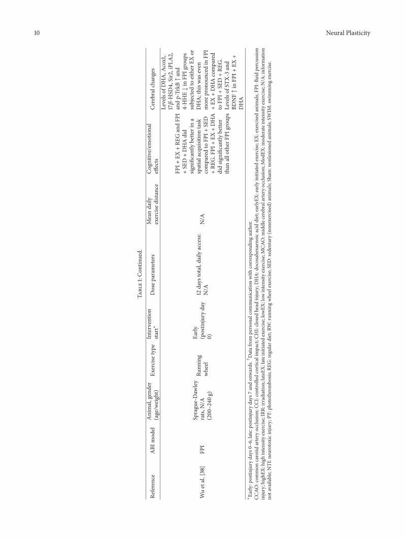
Wu et al. [38] subjected rats to lateral fluid percussioninjury (lFPI) immediately followed by 12 days of runningwheel exercise (7 of those days prior to cognitive testing),a diet high in docosahexaenoic acid (DHA), or both. Theyfound that lFPI exercised animals did significantly better in aspatial learning task in a water maze (as shown by reducedlatency to find a platform) in comparison to nonexercisedlFPI animals kept on a normal diet. The DHA diet wasalso associated with improved spatial learning. Furthermore,injured rats on the combined exercise and DHA diet signifi-cantly outperformed all other lFPI groups.Molecular analysisshowed increased levels of DHA, Acox1, and 17𝛽-HSD4(enzymes involved in DHA metabolism), Sir2 (involvedin mitochondrial function), iPLA2 (molecules involved inmembrane homeostasis), p-TrkB (BDNF receptor), and lowerlevels of 4-HHE (marker for lipid peroxidation) in the groupssubjected to either exercise or the DHA diet (compared tocontrols) and a further increase/decrease in the combinedgroup. The combined group also showed increased STX-3(also involved in membrane homeostasis) and brain derivedneurotrophic factor (BDNF) levels. The study indicates thatearly initiated voluntary exercise and/or the DHA dietcan positively affect cognitive recovery after TBI, possibly
Neural Plasticity 3
Table1:Summaryof
exercise
protocolsa
ndpo
stexercise
cogn
itive/emotionaland
cerebralchangesinABI
anim
almod
els.
Reference
ABI
mod
elAnimal,gender
(age/w
eight)
Exercise
type
Interventio
nsta
rt∗
Dosep
aram
eters
Meandaily
exercise
distance
Cognitiv
e/em
otional
effects
Cerebralchanges
deAraujoetal.
[52]
CCAO
Mon
golian
gerbils,m
ale
(60–
80g)
Treadm
illEa
rly(12,24,48,
or72
hours
postinjury)
Max.3
days
(4sessions),
min.1
day(1session),
15min/sess.Speed:
10m/m
in
150m
CCAO
+EX
after
12ho
urssho
wed
adecreasednu
mbero
ffield
crossin
gsandan
increase
ingroo
mingin
anop
enfield
test
comparedto
sham
+SE
D.N
ootherg
roup
differences
CCAO
+EX
after
12ho
urs:nu
mbero
fcells↓
inCA
1+stria
tum
comparedto
CCAO
+EX
after
24ho
urs
Cechetti
etal.
[47]
CCAO
Wistar
rats,
male(3mon
ths)
Treadm
illEa
rly(24ho
urs
postinjury†)
12weeks,3
times/w
eek,
20min/sessio
n.Weeks
1-2:
12m/m
infor3
min,
24m/m
infor4
min,
36m/m
infor6
min,
24m/m
infor4
min,and
12m/m
infor3
min;w
eeks
3–6,at24
m/m
infor4
min,
36m/m
infor12m
in,and
24m/m
infor4
min;w
eeks
7–10:24m
/min
for2
min,
36m/m
infor16m
in,and
24m/m
infor2
min:w
eeks
11-12:36m
/min
for2
min,
48m/m
infor16m
in,and
36m/m
infor2
min.
480m
/sessio
nup
to912m
/sessio
n(gradedprotocol)
AllEX
grou
psdid
significantly
bette
rina
spatialacquisitionand
retentiontask
aswellasa
working
mem
orytask
than
CCAO
+SE
D
Nodifferences
between
grou
psin
levelsof
free
radicalsor
SOD.C
CAO
+SE
D:hippo
campal
lipop
eroxidationand
thiol-levels↑compared
totheo
ther
grou
ps
Chen
etal.[55]
NTI
Sprague-Daw
ley
rats(12–14
weeks)
Motorized
runn
ing
wheel
Early
(2nd
postinjuryday)
7consecutived
ays,30
min
twiced
aily.
LowEX
:3m
/min
for10m
in.,
4.2m
/min
for10m
in,and
5.4m
/min
for10m
in.
Mod
EX:4.8m/m
infor
10min,6
m/m
infor10m
in,
and7.2
m/m
infor10m
in.
HighE
X:9.6
m/m
infor
10min,10.8m
/min
for
10min,and
12m/m
infor
10min
NTI
+Lo
wtEX:
252m
;NTI
+Mod
EX:360
m;
NTI
+HighE
X:64
8m
NTI
+Mod
EXshow
edsig
nificantly
bette
racqu
isitio
nof
cond
ition
ingtask
inY-mazethanNTI
+SE
D.
Noacqu
isitio
ndifferences
betweenNTI
+Lo
wEx
andNTI
+SE
Dor
NTI
+HighE
xand
NTI
+SE
D
BrdU
-positive
cells
indentateg
yrus↑in
NTI
+Mod
EXcomparedto
NTI
+SE
D.N
oBrdU
-staining
differences
between
otherN
TI+exercise
intensity
grou
psand
NTI
+SE
D.Positive
correlationbetween
acqu
isitio
nand
BrdU
-positive
cells
4 Neural Plasticity
Table1:Con
tinued.
Reference
ABI
mod
elAnimal,gender
(age/w
eight)
Exercise
type
Interventio
nsta
rt∗
Dosep
aram
eters
Meandaily
exercise
distance
Cognitiv
e/em
otional
effects
Cerebralchanges
Chen
etal.[57]
CHI
ICRmice,male
(7weeks)
Treadm
illEa
rly+late(2
or9days
postinjury)
7or14days
(early)o
r7days
(late).1h
our/d
aily.
Speed:
9m/m
inprogressively
increasedto
13.5m/m
in
Between540m
and810m
(graded
protocol)
CHI+
early
EXspent
significantly
moretim
eexploringnewob
jectin
anob
jectrecogn
ition
task
than
CHI+
SED.
CHI+
lateEXandCH
I+SE
Dhadlesstim
eexplorationtim
ewith
then
ewob
jectthan
sham
anim
als
Early
EXhind
ered
progressivec
elllossin
thec
ortexand
hipp
ocam
pusm
orethan
lateEX.
Early
EX↑
neurite
regeneratio
nin
thee
arlypo
stinjury
stages,lateEXhind
ered
later
stage
cellloss.
Early
EXfor14days
resto
redlesio
n-indu
ced
redu
ctionin
BDNFand
MKP
-1
Clarketal.[45]
IRR
C57B
L/6J
mice,
male+
female
(min.50days)
Runn
ing
wheel
Late(114/14
2days
postinjury)
54days
total,24-hou
rdaily
access
IRR+EX
:5.8km
;sham
+EX
:5.7km
Nodifferences
inspatial
learning
+retention
betweenIRR+EX
and
IRR+SE
D.Spatia
llearning
andretention
improved
insham
+EX
.Ex
ercise
improved
retentionin
acon
textual
fear
cond
ition
ingtest
Exercise↑hipp
ocam
pal
neurogenesis.
Exercise
coun
teracted
radiation-indu
ced
redu
ctions
inneurogenesis,
neuron
aldifferentiatio
n,andglia
celllevels
Cranee
tal.[40]
CCI
Long
Evansrats,
male(ca.50
days)
Runn
ing
wheel
Early
(immediately
postinjury)
7days
7-8k
m
CCI+
EXperfo
rmed
significantly
worse
incomplex
stop-sig
nal
reactio
ntim
etaskforthe
firstfivetestd
ays
comparedCC
I+SE
Dandsham
grou
ps.A
ftera
weekof
testing
,CCI
+EX
returned
tobaselin
elevels
CCI+
EX:G
FABand
IBA1p
ositive
cells↑in
thec
ortexand
hipp
ocam
pus,
respectiv
ely;all
CCI-anim
als:DAP1
positivec
ells↓in
the
cortex,hippo
campu
s,mediodo
rsalnu
cleus
and
corpus
callo
sum
comparedto
sham
+SE
D
Neural Plasticity 5
Table1:Con
tinued.
Reference
ABI
mod
elAnimal,gender
(age/w
eight)
Exercise
type
Interventio
nsta
rt∗
Dosep
aram
eters
Meandaily
exercise
distance
Cognitiv
e/em
otional
effects
Cerebralchanges
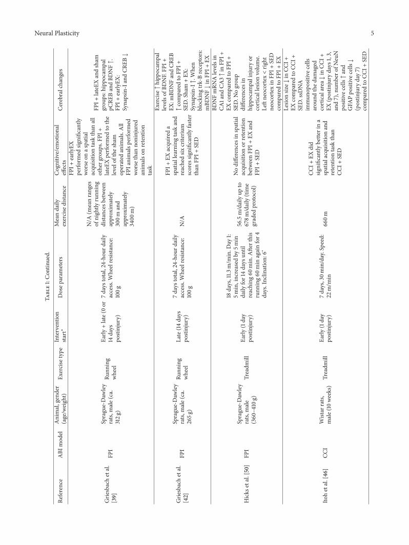
Grie
sbachetal.
[39]
FPI
Sprague-Daw
ley
rats,
male(ca.
312g
)
Runn
ing
wheel
Early
+late(0
or14
days
postinjury)
7days
total,24-hou
rdaily
access.W
heelresistance:
100g
N/A
(meanranges
ofnightly
runn
ing
distancesb
etween
approxim
ately
300m
and
approxim
ately
3400
m)
FPI+
early
EXperfo
rmed
significantly
worse
onas
patia
lacqu
isitio
ntask
than
all
otherg
roup
s.FP
I+lateEX
perfo
rmed
tothe
levelofthe
sham
operated
anim
als.All
FPIanimalsp
erform
edworse
than
noninjured
anim
also
nretention
task
FPI+
lateEXandsham
grou
ps:hippo
campal
pCRE
BandBD
NF↑.
FPI+
early
EX:
Synapsin-IandCR
EB↓
Grie
sbachetal.
[42]
FPI
Sprague-Daw
ley
rats,
male(ca.
265g
)
Runn
ing
wheel
Late(14
days
postinjury)
7days
total,24-hou
rdaily
access.W
heelresistance:
100g
N/A
FPI+
EXacqu
ireda
spatiallearningtask
and
reachedsix
criteriu
mscores
significantly
faste
rthan
FPI+
SED
Exercise↑hipp
ocam
pal
levelsof
BDNF.FP
I+EX
:mBD
NFandCR
EB↑comparedto
FPI+
SED.Sham
+EX
:Synapsin-I↑.W
hen
blocking
trk-Breceptors:
mBD
NF↓in
FPI+
EX
Hicks
etal.[50]
FPI
Sprague-Daw
ley
rats,
male
(360–4
10g)
Treadm
illEa
rly(1day
postinjury)
18days,11.3
m/m
in.D
ay1:
5min,increased
by5m
indaily
for14days
until
reaching
60min.A
fterthis
runn
ing60
min
againfor4
days.Inclin
ation:
6∘
56.5m/dailyup
to678m
/daily(time
graded
protocol)
Nodifferences
inspatial
acqu
isitio
nor
retention
betweenFP
I+EX
and
FPI+
SED
BDNFmRN
Alevelsin
CA1and
CA3↑in
FPI+
EXcomparedto
FPI+
SED.N
ogrou
pdifferences
inhipp
ocam
palinjuryor
corticallesio
nvolume.
Leftneocortex<rig
htneocortexin
FPI+
SED
comparedto
FPI+
EX
Itohetal.[46
]CC
IWistar
rats,
male(10
weeks)
Treadm
illEa
rly(1day
postinjury)
7days,30m
in/day.Speed:
22m/m
in66
0m
CCI+
EXdid
significantly
bette
rina
spatialacquisitionand
retentiontask
than
CCI+
SED
Lesio
nsiz
e↓in
CCI+
EXcomparedto
CCI+
SED.ssD
NA
immun
opositive
cells
arou
ndthed
amaged
corticalarea↓in
CCI+
EX(postin
jury
days
1,3,
and7),num
bero
fNeuN
positivec
ells↑and
GFA
Ppo
sitivec
ells↓
(postin
jury
day7)
comparedto
CCI+
SED
6 Neural Plasticity
Table1:Con
tinued.
Reference
ABI
mod
elAnimal,gender
(age/w
eight)
Exercise
type
Interventio
nsta
rt∗
Dosep
aram
eters
Meandaily
exercise
distance
Cognitiv
e/em
otional
effects
Cerebralchanges
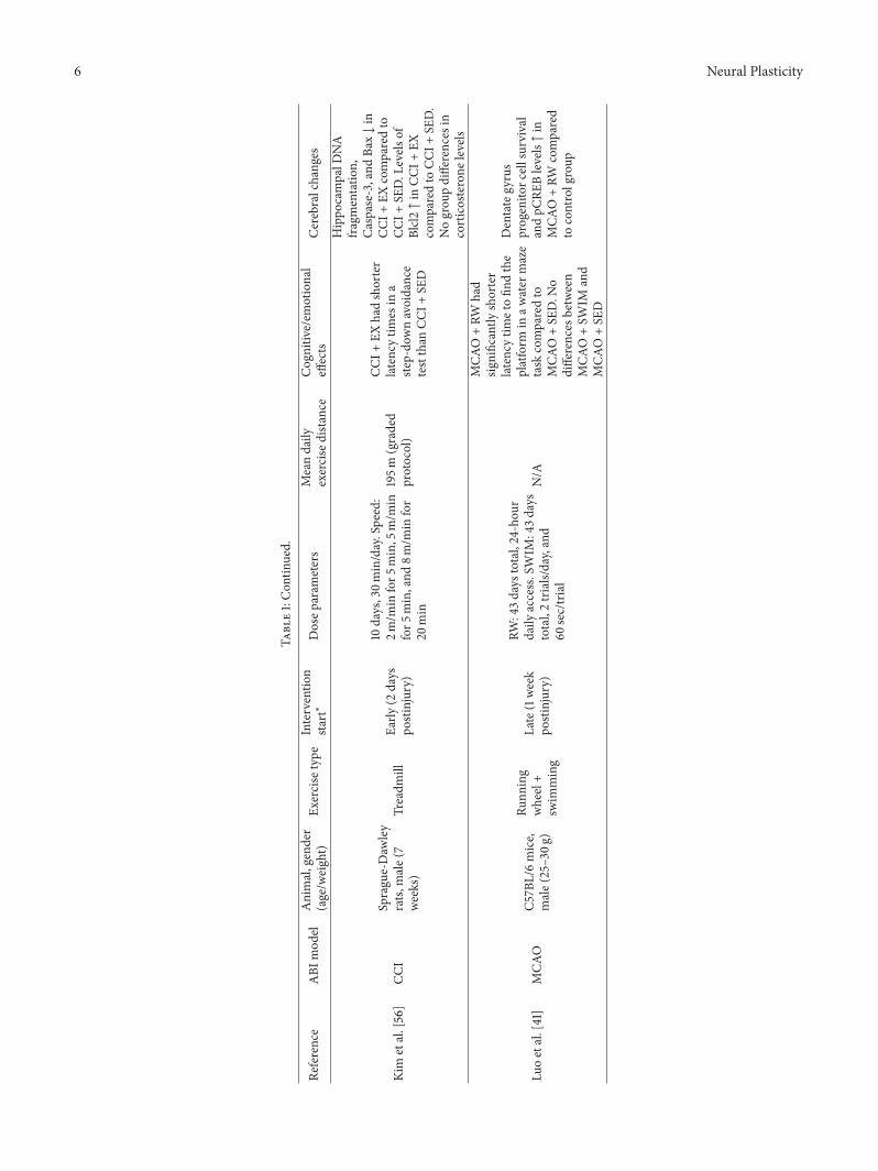
Kim
etal.[56]
CCI
Sprague-Daw
ley
rats,
male(7
weeks)
Treadm
illEa
rly(2
days
postinjury)
10days,30m
in/day.Speed:
2m/m
infor5
min,5
m/m
infor5
min,and
8m/m
infor
20min
195m
(graded
protocol)
CCI+
EXhadshorter
latency
times
ina
step-do
wnavoidance
testthan
CCI+
SED
Hippo
campalD
NA
fragmentatio
n,Caspase-3,and
Bax↓in
CCI+
EXcomparedto
CCI+
SED.L
evels
ofBlcl2↑in
CCI+
EXcomparedto
CCI+
SED.
Nogrou
pdifferences
incorticosterone
levels
Luoetal.[41]
MCA
OC5
7BL/6mice,
male(25–30g
)
Runn
ing
wheel+
swim
ming
Late(1week
postinjury)
RW:43days
total,24-hou
rdaily
access.SWIM
:43d
ays
total,2trials/day,and
60sec/trial
N/A
MCA
O+RW
had
significantly
shorter
latencytim
etofin
dthe
platform
inaw
ater
maze
task
comparedto
MCA
O+SE
D.N
odifferences
between
MCA
O+SW
IMand
MCA
O+SE
D
Dentategyrus
progenito
rcellsurvival
andpC
REBlevels↑in
MCA
O+RW
compared
tocontrolgroup

Neural Plasticity 7
Table1:Con
tinued.
Reference
ABI
mod
elAnimal,gender
(age/w
eight)
Exercise
type
Interventio
nsta
rt∗
Dosep
aram
eters
Meandaily
exercise
distance
Cognitiv
e/em
otional
effects
Cerebralchanges
Piao
etal.[31]
CCI
C57B
L/6mice,
male(10
weeks)
Runn
ing
wheel
Late(1weekor
5weeks
postinjury)
4weeks
total,24-hou
rdaily
acces.
103.2m
(1week),
97.2m
(5weeks).
CCI+
lateEX
perfo
rmed
significantly
bette
rina
spatialacquisitionand
retentiontask
than
CCI
+SE
D.C
CI+lateEX
show
edsig
nificant
improvem
entina
cogn
itive
flexibilitytask
comparedto
CCI+
early
EXandCC
I+SE
D.
Retentionof
the
flexibilitytask
was
significantly
bette
rin
CCI+
lateEX
compared
toCC
I+SE
D.Inan
ovel
objectrecogn
ition
task,
CCI+
lateEX
spent
significantly
longer
time
exploringan
ewob
ject
than
CCI+
early
EXand
CCI+
SED.N
olocomotor
differences
inan
open
field
test.
Ina
tailsuspensio
ntest,
all
CCIg
roup
shad
increasedim
mob
ility
times
CCI+
lateEX
:↓lesio
nsiz
ecom
paredto
CCI+
early
EXandCC
I+SE
D.
IL-1𝛽levels↑in
CCI+
early
EX(w
eek5)
and↓
inCC
I+lateEX(w
eek9)
comparedto
CCI+
SED.
IL-6
andIL-10↑CC
I+lateEX(w
eek9).C
ortic
alGalectin
-3andC1
qBlevels↑increasedin
CCI
+early
EX,but↓in
CCI
+lateEX
.Gp9
1pho
xand
p22pho
x↓in
CCI+
lateEX
.Hippo
campal
CREB
gene
expressio
n,BD
NF,IG
F-1,
neurogenesis,
andcell
survival↑in
CCI+
lateEX
Shen
etal.[49]
CCI
Sprague-Daw
ley
rats,
male
(250–270
g)Treadm
illEa
rly(24ho
urs
postinjury)
14days,30m
in/day.
LowEX
:week1:3m
/min;
week2:3m
/min
for5
min,
5m/m
infor5
min,and
8m/m
infor2
0min.
HighE
X:Day
1:3m
/min;
day2:3m
/min
for10m
in,
6m/m
infor10m
in,and
9m/m
infor10m
in;day
3:6m
/min
for10m
in,
9m/m
infor10m
in,and
12m/m
infor10m
in;day
4–14:12m
/min
Week1:90
m,w
eek
2:200m
(lowEW
).Day
1:90
m,day
2:180m
,day
3:270m
,day
4–14:
360m
(highE
X)(gradedprotocols)
CCI+
lowEX
perfo
rmed
bette
ronas
patia
lacqu
isitio
ntask
comparedto
CCI+
high
EXandCC
I+SE
D.
CCI+
lowEX
show
edbette
rtaskretention
than
CCI+
SED
Con
tralateral
hipp
ocam
palB
DNFand
pCRE
B↑in
CCI+
lowEX
comparedto
CCI
+SE
D.N
ogrou
pdifferences
inlevelsof
Synapsin-IandCR
EB
8 Neural Plasticity
Table1:Con
tinued.
Reference
ABI
mod
elAnimal,gender
(age/w
eight)
Exercise
type
Interventio
nsta
rt∗
Dosep
aram
eters
Meandaily
exercise
distance
Cognitiv
e/em
otional
effects
Cerebralchanges
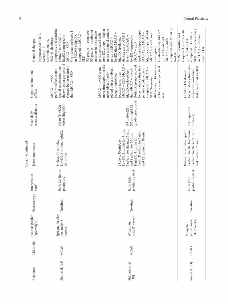
Shih
etal.[48]
MCA
OSprague-Daw
ley
rats,
male(8
weeks)
Treadm
illEa
rly(24ho
urs
postinjury)
14days,30m
in/day.
LowEX
:8m/m
in,highE
X:20
m/m
in
240m
(lowEX
);60
0m(highE
X)
MCA
O+lowEX
perfo
rmed
bette
rina
spatialacquisitionthan
thetwootherg
roup
sand
show
edbette
rretentio
nthan
MCA
O+SE
D
Hippo
campalB
DNF,
Synapsin-I
(con
tralaterally),
PSD-95,dend
ritic
complexity
anddend
rite
spines↑in
MCA
O+
lowEX
comparedto
MCA
O+SE
D.
Corticosterone
levels↑
inMCA
O+high
EXcomparedto
MCA
O+
SED
Shim
adae
tal.
[58]
MCA
OWistar
rats,
male(7weeks)
Treadm
illEa
rly(4th
postinjuryday)
28days,30m
in/day.
LowEX
:2m/m
infor5
min,
5m/m
inforthe
next
5min,
and8m
/min
for2
0min.
HighE
X:8m
/min
for
5min,11m
/min
for5
min,
and22
m/m
infor2
0min
195m
(lowEX
);535m
(highE
X)(gradedprotocols)
MCA
O+lowEX
spent
moretim
eexplorin
gthe
novelobject/n
ewly
placed
objectin
object
recogn
ition
/object
locatio
ntasksthan
MCA
O+SE
D.M
CAO+
high
EXexplored
less
than
MCA
O+lowEX
.Bo
thEX
grou
psshow
edlonger
latency
timeina
passivea
voidance
test
comparedto
MCA
O+
SED.N
ogrou
pdifferences
inlocomotor
activ
ityin
anop
enfield
test
EXgrou
ps↓lesio
nsiz
e.EX
grou
ps↑nu
mbero
fneuron
sinthed
entate
gyrusc
omparedwith
non-EX
grou
ps–high
erin
theipsilaterald
entate
gyrusinMCA
O+
lowEX
than
MCA
O+
high
EX.Ipsilateral
dentateg
yrus
MAP-2
levels↑in
MCA
O+
lowEX
comparedto
MCA
O+SE
D.
Ipsilateralhipp
ocam
pal
MAP-2↓in
MCA
O+
high
EXcomparedwith
MCA
O+lowEX
and
sham
grou
p.Con
tralaterally,
MAP-2
↓in
CA1and
CA3in
MCA
O+high
EXcomparedwith
MCA
O+
lowEX
Sim
etal.[53]
CCAO
Mon
golian
gerbils,m
ale
(11–13
weeks)
Treadm
illEa
rly(2nd
postinjuryday)
10days.30m
in/day.Speed:
2m/m
inforthe
first5m
in,
5m/m
inforthe
next
5min,
and8m
/min
20min
195m
(graded
protocol)
CCAO
+EX
show
edlonger
latency
times
ina
step-do
wnavoidance
task
than
CCAO
+SE
D
TUNEL
-positive
and
Caspase-3
positivec
ells
↓in
CCAO
+EX
comparedto
CCAO
+SE
D.C
ellproliferation↑
inCC
AO+SE
Dand
sham
+EX
Neural Plasticity 9
Table1:Con
tinued.
Reference
ABI
mod
elAnimal,gender
(age/w
eight)
Exercise
type
Interventio
nsta
rt∗
Dosep
aram
eters
Meandaily
exercise
distance
Cognitiv
e/em
otional
effects
Cerebralchanges
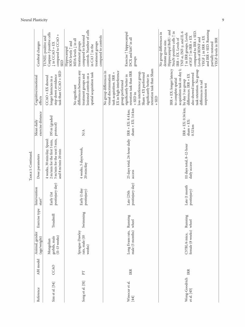
Sim
etal.[54]
CCAO
Mon
golian
gerbils,m
ale
(11–13
weeks)
Treadm
illEa
rly(1st
postinjuryday)
4weeks,30m
in/day.Speed:
2m/m
inforthe
first5m
in,
5m/m
inforthe
next
5min,
and8m
/min
20min
195m
(graded
protocol)
CCAO
+EX
show
edlonger
latencies
ina
step-do
wnavoidance
task
than
CCAO
+SE
D
TUNEL
-positive
and
Caspase-3
positivec
ells
↓in
CCAO
+EX
comparedto
CCAO
+SE
D
Song
etal.[51]
PTSprague-Daw
ley
rats,
male(10
weeks)
Swim
ming
Early
(1day
postinjury)
4weeks,5
days/w
eek,
20min/day
N/A
Nosig
nificant
differences
betweenany
treatmentg
roup
sand
lesio
nedcontrolson
aspatialacquisitiontask
Hippo
campal
SOD-le
vels↑and
MDA-
levels↓in
all
treatmentg
roup
scomparedto
lesio
ned
controls.
Num
bero
fcells
inCA
3↑in
the
treatmentg
roup
scomparedto
controls
Winocur
etal.
[44]
IRR
Long
Evansrats,
male(5mon
ths)
Runn
ing
wheel
Late(25th
postinjuryday)
25days
total,24-hou
rdaily
access
IRR+EX
:8.4km
;sham
+EX
:3.6km
Nogrou
pdifferences
invisualdiscrim
ination
task
acqu
isitio
n.IRR+
EXin
high
-interfe
rence
grou
pperfo
rmed
significantly
bette
rin
retentiontask
than
IRR
+SE
Din
low-in
terfe
renceg
roup
.Sham
+EX
perfo
rmed
significantly
bette
rin
retentiontask
than
Sham
+SE
D
Exercise↑hipp
ocam
pal
DCX
andki67
inall
grou
ps
Won
g-Goo
drich
etal.[43]
IRR
C57B
L/6mice,
female(8weeks)Ru
nning
wheel
Late(1mon
thpo
stinjury)
111d
aystotal,8–12-ho
urdaily
access
IRR+EX
:0.56k
m;
sham
+EX
:0.52
km
IRR+EX
:lon
gerlatency
tocompletes
patia
lacqu
isitio
ntask
onday1;
byday3no
grou
pdifferences.IRR
+EX
also
show
edim
proved
task
retention.
Nogrou
pdifferences
intail
suspensio
ntest
Nogrou
pdifferences
indentateg
yrus
size.
Hippo
campalB
rdU-a
ndNeuN-positive
cells↑in
IRR+EX
.Levels
ofTN
F-𝛼,INF-𝛾,and
IL-6
↑in
IRRgrou
ps.L
evels
ofIG
F↑in
IRR+EX
comparedto
IRR+SE
D.
Levelsof
BDNFand
VEG
F↓in
IRR+EX
andIRR+SE
D.R
unning
partially
resto
red
VEG
F-levelsin
IRR
10 Neural Plasticity
Table1:Con
tinued.
Reference
ABI
mod
elAnimal,gender
(age/w
eight)
Exercise
type
Interventio
nsta
rt∗
Dosep
aram
eters
Meandaily
exercise
distance
Cognitiv
e/em
otional
effects
Cerebralchanges
Wuetal.[38]
FPI
Sprague-Daw
ley
rats,
N/A
(200–240
g)
Runn
ing
wheel
Early
(postin
jury
day
0)
12days
total,daily
access:
N/A
N/A
FPI+
EX+RE
GandFP
I+SE
D+DHAdid
significantly
bette
rina
spatialacquisitiontask
comparedto
FPI+
SED
+RE
G.FPI
+EX
+DHA
didsig
nificantly
bette
rthan
allother
FPIg
roup
s
Levelsof
DHA,A
cox1,
17𝛽-H
SD4,Sir2,iPL
A2,
andp-TrkB↑and
4-HHE↓in
FPIg
roup
ssubjectedto
either
EXor
DHA;thisw
aseven
morep
rono
uncedin
FPI
+EX
+DHAcompared
toFP
I+SE
D+RE
G.
Levelsof
STX-
3and
BDNF↑in
FPI+
EX+
DHA
∗
Early
:postin
jury
days
0–6;late:postin
jury
days
7andon
wards.†Datafrom
person
alcommun
icationwith
correspo
ndingauthor.
CCAO
:com
mon
carotid
artery
occlu
sion;
CCI:controlledcorticalim
pact;C
HI:clo
sedhead
injury;D
HA:docosahexaeno
icacid
diet;earlyEX
:earlyinitiated
exercise;E
X:exercisedanim
als;FP
I:flu
idpercussio
ninjury;highE
X:high
intensity
exercise;IRR
:irradiatio
n;lateEX:
lateinitiated
exercise;lo
wEX
:lowintensity
exercise;M
CAO:m
iddlec
erebralarteryo
cclusio
n;Mod
EX:m
oderateintensitye
xercise
;N/A
:information
notavailable;NTI:neurotoxicinjury;PT
:pho
tothrombo
sis;R
EG:regular
diet;R
W:run
ning
wheelexercise;SED
:sedentary
(non
exercised)
anim
als;Sh
am:n
onlesio
nedanim
als;SW
IM:swim
mingexercise.
Neural Plasticity 11
through counteracting membrane damage and coordinatingDHA metabolism.
Contrary to these results, Griesbach et al. [39] foundthat animals exposed to lFPI and early initiated exercise(from post-injury day 0) performed significantly worse ona spatial acquisition task in a water maze than all othergroups, including an lFPI group starting exercise at a laterpoint in time (at postinjury day 14) and lFPI nonexercisedcontrols. Animals exercised later performed at the level of thesham operated animals. During a retention test (probe trial),all lFPI animals performed worse than noninjured animals,regardless of exercise treatment. The late exercise and shamgroups showed increased hippocampal levels of the tran-scriptional regulator phosphorylated cyclic AMP responseelement-binding protein (pCREB) and BDNF. Moreover,there was a positive correlation between BDNF-levels and theamount of exercise. No BDNF-increase was seen in the earlyexercised group, which also showed lower levels of Synapsin-I (involved in synaptic vesicle clustering and release) andCREB.
Also finding detrimental effects, Crane et al. [40] sub-jected animals to a cortical contusion injury immediatelyfollowed by a 7-day running wheel exercise regimen. In acomplex stop-signal reaction time task (a conditioning-basedlearning task requiring either inhibition or execution of alearned behavior depending on stimuli given), the exercisedanimals performed significantly worse for the first five testdays compared with both the nonexercised injured animalsand the sham animal groups. However, after a week oftesting, the exercised animals returned to their baseline levels.The contused, exercised animals showed larger inflammatoryresponses (more GFAB and IBA1 positive cells) in thecortex and hippocampus, respectively. All contused groupshad fewer surviving cells (less DAP1 positive cells) in thecortex, hippocampus, mediodorsal nucleus of the thalamus,and corpus callosum compared to the nonexercised, shamanimals.
The conflicting results of the two first studies are some-what surprising, as they use similar models and setups.Discrete differences, for example, in the duration of the exer-cise protocol, could potentially account for these divergentfindings.While the studies by Griesbach et al. [39] and Craneet al. [40] both found detrimental effects of early exerciseon cognitive performance, it appears that these effects aretransient, as the animals seem to catch up during the rathershort-time course of task acquisition.
Initiating exercise a little later after injury, Luo et al. [41]subjected C57/BL6 mice to middle cerebral artery occlusion(MCAO) followed by a one week postinjury break. Subse-quently, the animals were exercised for 39 days in eitherrunning wheels or by swimming in a circular pool beforestarting a spatial acquisition task in a watermaze.TheMCAOrunning group found the platform significantly faster thanthe nonexercised MCAO group.This was not the case for theswimming group, whose performance did not differentiatefrom the nonexercisedMCAOgroup. Progenitor cell survivalin the dentate gyrus and pCREB levels were increased in theMCAO running wheel group compared to the control group.This suggests that different exercise types can affect cognitive
recovery differently and that voluntary exercise initiated afterthe first postinjury week can induce functional recovery andhelp promote cell survival.
However, in another study by Piao et al. [31] starting exer-cise 1 week after injury did not produce similar results. Exam-ining the timing effects of a 4-week running wheel regimenafter controlled cortical impact injury (CCI), animals wereexercised beginning either 1 week (“early”) or 5 weeks (“late”)postinjury.The study found that the late exercised CCI-grouphad a significantly reduced latency to find the platform in aspatial water maze learning task and better retention of thetask compared to a nonexercised CCI-group. In a reversedplatform test, a test of cognitive flexibility, they found thatthe late exercised animals showed a significant improvementcompared to both the early exercised CCI-group and thenonexercised CCI-group. Retention of the reversed platformtask was significantly better in the late exercised CCI-animals compared to the nonexercised CCI-group. Therewere no differences between the early initiated group and thenonexercised CCI-group on any of the above parameters. In anovel object recognition task, the late exercised CCI-animalsspent significantly longer time exploring a new object thanboth the early exercised CCI-group and a nonexercised CCI-group, indicating improved short-term memory abilities; infact their exploration time was at the level of uninjured,naıve animals. There were no group differences in locomotoractivity in an open field test. In a tail suspension test, all CCI-groups showed increased immobility times regardless of exer-cise status, suggesting more pronounced behavioral despairdue to injury. Furthermore, the late exercised CCI-group hada reduced lesion size compared to the early exercised CCI-group and the nonexercised CCI-group. There were time-dependent increases and decreases in different microgliaactivation markers: IL-1𝛽 levels (a proinflammatory marker)increased in the early exercise group in week 5 after CCIand levels reduced in the late exercise group in postinjuryweek 9 (both compared to the nonexercised CCI-group).There were also increased levels of IL-6 (a proinflammatorymarker) and IL-10 (an anti-inflammatory marker) in the lateexercise group in week 9. Cortical (ipsilateral) Galectin-3 andC1qB levels (microglial activation markers) were increasedin the early exercised animals, while they were reduced inthe late exercised group together with levels of gp91phoxand p22phox (membrane components of NADPH oxidaseenzyme). Late exercise increased hippocampal CREB geneexpression, BDNF, and IGF-1 (insulin-like growth factor 1)levels and increased neurogenesis and cell survival in the lateexercise group (but not in the early exercise group).The studyconcludes that the improved cognitive performance in thegroup subjected to late exercise is possibly due to a moreoptimal coordination/balance of microglia expression andincreased growth factor levels.
Similar to studies initiating exercise immediately afterABI, starting a voluntary exercise paradigm one week afterinjury produces conflicting results, suggesting that the exer-cise type (voluntary versus forced) is not the only factordetermining the efficacy of exercise.
As already mentioned, Griesbach et al. [39] foundimproved cognitive performance in animals exercised 14 days
12 Neural Plasticity
after injury. In a later study, Griesbach et al. [42] reproducedthis finding. Animals exercised 14 days after lFPI acquired aspatial learning task in a water maze significantly faster thannonexercised lFPI animals. In addition, they reached six outof seven criterion scores (e.g., reaching a platform from 10 secdown to 4 sec) significantly faster than the nonexercised lFPIgroup. Exercise increased hippocampal levels of BDNF in allanimals regardless of lesion status. However, mBDNF andCREB levels were higher in the lFPI exercised animals than inthe lFPI control animals. This also held true for the exercisedsham animals, who showed increased levels of Synapsin-I.When blocking trkB receptors in lFPI animals, the exercise-induced increase in mBDNF was reduced. The two studiesby Griesbach et al. [39, 42] suggest that voluntary exercisestarted at a later stage (14 days) is beneficial for cognitiverecovery, possibly through upregulation of BDNF and down-stream effectors of synaptic transmission.
Wong-Goodrich et al. [43] subjected female C57BL/6mice to whole brain irradiation (5Gy, single dose). Theanimals were then given access to running wheels outsideof their home cages for 8–12 hours a day, starting 1 monthafter irradiation. Prior to initiation of exercise, all animalswere tested in a spatial learning and retention test in a Barnesmaze as well as a tail suspension test. After 6 weeks of wheelrunning, the animals were once again tested in the Barnesmaze (both 3 and 4 months after irradiation) and in thetail suspension test (2.5 months after irradiation). Resultsfrom the first testing period in the Barnes maze (preexercise)showed no differences between the groups: the sham animalslearned the task by day 2, the irradiated animals by day3. There were no differences in retention as assessed byprobe trials. The tests performed after exercise showed thatirradiated, exercised animals had longer latency to completethe task on the first test-day compared to all other groups;however, by day 3 there were no differences. The irradiated,sedentary animals did not exhibit target quadrant preferencesin retention trials (which they did during the first test period).However, the irradiated, exercised animals spent more timein the target quadrant, indicating improved memory. Thispicture was also seen on the test performed 4 months afterirradiation. There were no differences between the groups inimmobility times in the tail suspension test at any test point.Histology showed no differences in dentate gyrus size in anyof the groups. Running elevated the number of hippocampalBrdU and NeuN positive cells (markers of newborn cellsand mature neurons, resp.) in the irradiated animals. Lev-els of proinflammatory cytokines (tumor necrosis factor-𝛼 (TNF-𝛼), interferon-𝛾 (IFN-𝛾), and interleukin-6 (IL-6))were elevated in the irradiated groups (compared to shams).Levels of IGF were increased in the irradiated, exercisedanimals compared to irradiated, sedentary animals. Levelsof BDNF and VEGF (vascular endothelial growth factor)were decreased in all irradiated animals (both exercised andnonexercised). However, running partially restored VEGF-levels in the irradiated, exercised animals. The study showsthat later-initiated voluntary running can prevent memorydecline at a later stage in irradiation-exposed animals.
Showing similar positive recovery effects of late-initiatedvoluntary exercise after brain irradiation, Winocur et al.
[44] irradiated adult rats at a single dose of 8Gy. Twenty-five days after irradiation approximately half the animalswere allowed to exercise in running wheels in their homecages. After two weeks of running, all animals were testedin a visual discrimination task in a water maze, followedby either a high-interference task (an unsolvable task) or alow-interference task (demanding no visual discriminationfor task solution). Lastly, a retention test for the originaldiscrimination taskwas performed.Therewere nodifferencesbetween the groups in acquiring the visual discriminationtask. In the retention task, the irradiated animals had themost errors; this was especially pronounced in the animalsthat had previously performed the high-interference task.Further analysis showed that irradiated, exercised animalsin the high-interference group performed significantly betterin the retention test than the irradiated, sedentary animalsin the low-interference group. The exercised, sham animalsperformed better in the retention test than the sedentarysham animals regardless of interference group affiliation.Analysis of hippocampal DCX and ki67 positive neurons(neurogenesis markers) showed that irradiation decreasedtheir levels, but running increased the levels in all exercisedgroups. The authors conclude that neurogenesis is a partof the mechanism that controls memory interference, assuppressing neurogenesis disrupts retention in the high-interference groups. However, this effect can be diminishedby promoting neurogenesis through exercise.
While three studies found cognitive improvement inanimals starting exercise 25 days after injury or later [31, 43,44], Clark et al. [45] administered running wheel exercisebetween 114 and 142 days after gamma irradiation of thehippocampal area of both male and female C57BL/6J mice.They found that 54 days of wheel exercise did not have aneffect on spatial learning and retention in a water maze inthe gamma radiated group compared with a nonexercisedradiated group. Running did, however, have a positive effecton sham operated animals. In a contextual fear condi-tioning test, running increased freezing time (indicatingincreased memory of a formerly presented painful stim-ulus); however, this was regardless of radiation status. Inother words, in the spatial tasks no effects of exercise werefound in the irradiated animals, yet running did improveperformance in the conditioning task in all exercise groups.Running increased hippocampal neurogenesis regardless oflesion status. Exercise counteracted radiation-induced reduc-tions in neurogenesis, neuronal differentiation, and glia celllevels.
Five studies [31, 39, 42–44] found positive effects of laterinitiated voluntary exercise on measures of spatial learningand retention. However, this was not the case in the studyby Clark et al. [45], who waited 3-4 months with exerciseadministration. This opens the question whether there isa window of rehabilitation opportunity that closes aftercertain amount of time has passed. Other factors could alsoaccount for the conflicting results such as different injurytypes and duration of exercise. Interestingly, running affectedperformance positively in the fear conditioning task in theClark et al. study, indicating that exercise effects can be taskspecific.
Neural Plasticity 13
All in all, the above research shows a somewhat mixedpicture of using voluntary exercise in cognitive rehabilitationafter ABI. Later starting points (from 14-days post-injury)appear to have the most consistent effects on cognitiverecovery. However, further research is needed to determineif exercise interventions can be administered too late toproduce cognitive improvements. Moreover, caution shouldbe taken in making general recommendations based onsuch a limited and methodologically diverse set of studies.The results indicate that the voluntary aspect of exerciseis not the sole determinant of effect; other variables suchas starting point and duration may also play a significantrole.
3.2. Forced Exercise. Fourteen studies have looked into theeffects of forced exercise on cognitive recovery after ABI.
Itoh et al. [46] subjected rats toCCI injury followed by a 7-day treadmill exercise regimenbeginning one day after injury.In acquisition and retention of a spatial task in a water maze,the lesioned, exercised animals did significantly better thanthe lesioned, nonexercised animals; the former performingto the functional level of the sham animals. There was asignificant reduction in lesion size in the exercised groupcompared to the controls. Additionally, therewas a significantreduction in ssDNA immunopositive cells (a marker ofapoptosis) around the damaged cortical area in the exercisedgroup on postinjury days 1, 3, and 7, an increase in the numberof NeuN positive cells, and a reduction in GFAP positivecells (marker for astrocytes) 7 days after TBI compared to thelesioned, nonexercised control group.This suggests that earlyinitiated forced exercise can improve cognitive functionwhilereducing apoptosis and impacting the glial scarring.
Cechetti et al. [47] looked at effects of both pre- andpostinjury treadmill exercise in a bilateral common carotidartery occlusion (CCAO) rat model. The postinjury trainedgroup started exercising 24 hours following surgery andcontinued for 12 weeks, 3 days a week. They found that allexercised groups, including the postinjury exercised group,did significantly better on three of the five testing days inacquisition of a spatial task in a water maze compared to alesioned, nonexercised group. This pattern was also seen ina retention (probe trial) test and in a working memory testin a water maze. There were no differences between groupsin levels of free radicals or SOD (superoxide dismutase, anantioxidant enzyme) levels. However, there were heightenedhippocampal lipoperoxidation (evaluated by TBARS test)and thiol-levels (antioxidants) in the lesioned, nonexercisedgroup compared to the other groups. Like the study by Itohet al. [46], this study shows that early initiated forced exercisecan positively affect cognitive recovery, potentially throughreducing oxidative damage by regulation of antioxidantlevels.
Shih et al. [48] subjected rats to right hemisphereMCAO.After 24 hours, the animals began exercising at either a low ora high-intensity (speed) on a treadmill for 14 days.They foundthat the lesioned, low-intensity group had shorter latencieson three out of the four testing days in a spatial learning taskin a water maze (compared to the lesioned, high-intensity
group and a lesioned, nonexercised control group). The low-intensity group also showed better retention than the controlgroup. Furthermore, the low-intensity paradigm increasedlevels of hippocampal BDNF, Synapsin-I (contralaterally),and PSD-95 (membrane scaffolding protein) as well as thedendritic complexity (measured by Sholl analysis) and thenumber of dendritic spines compared to the control group.There were higher levels of corticosterone (stress-hormone)in the high-intensity group compared to the controls. Thestudy is interesting as it investigates the effects of exerciseintensity on cognitive measures. While both groups initiatedexercise 24 hours after injury, only the low-intensity groupshowed positive cognitive effects concomitant with increasesin plasticity-related proteins and dendrite development. Fur-thermore, the high-intensity group displayed higher levelsof stress-hormone, which may have inhibited the efficacy ofexercise.
In another study investigating the effects of differentexercise intensities, Shen et al. [49] subjected rats to CCIimmediately followed by two different intensities of treadmillexercise for 14 days. They found that the lesioned, low-intensity group performed better on two out of the fourdays of the acquisition part of a spatial task in a watermaze compared to the high-intensity group and a lesioned,nonexercised control group. The low-intensity group alsoshowed better retention than the control group. On a neuro-logical deficit score all CCI animals did worse than the shamanimals, but they all improved by day 6 post-TBI. BDNF andphosphorylated CREB measurements showed higher levelsin the contralateral hippocampus in the low-intensity groupcompared to the control group. There were no differences inmeasurements of Synapsin-I and CREB in any of the groups.
While the above studies suggest that early forced exer-cise can promote cognitive recovery, these results are notunchallenged. Hicks et al. [50] found that animals exposedto lFPI and 18 days of treadmill exercise initiated the dayfollowing injury differed in neither spatial acquisition norretention tasks in a water maze compared to a lesioned,nonexercised group.They saw no differences between groupsin neuromotor scores. They found increased BDNF mRNAlevels in CA1 and CA3 in the exercised, lesioned animalscompared to lesioned, sedentary animals. There were nodifferences in hippocampal injury or cortical lesion volumebetween groups. However, the left neocortex (ipsilaterally tothe injury) was significantly smaller than the right neocortexin the nonexercised, lesioned animals compared to theexercised, lesioned animals. These results are in contrast tomany of the above studies, as they fail to find cognitive effectsof early initiated forced exercise, but do find BDNF and somehistological effects of exercise.
The findings of Hicks et al. [50] were echoed by Song et al.[51], who used photothrombosis to induce cerebral stroke inrats. One day after injury the animals were swim-exercisedin a circular pool for 4 weeks (a total of 20 days), or givenAcetyl-L-carnitine (ALC) injections, or both. They foundno significant differences in any of their treatment groupscompared to lesioned controls on acquisition of a spatial taskin a water maze tested the first, second, and fourth weekafter injury. Hippocampal SOD-levels were increased in all
14 Neural Plasticity
treatment groups compared to the lesioned controls; thesewere significantly higher in the combined (exercise + ALC-injection) group in comparison to the other groups. MDA-levels (related to lipid peroxidation) were reduced in thetreatment groups compared to the controls. Histologically,there were an increased number of cells in CA3 in thetreatment groups compared to the controls.
Investigating emotional parameters, de Araujo et al. [52]exercised gerbils on treadmills either 12, 24, 48, or 72 hoursafter CCAO for 1 up to 3 days. In an open field, animalsexercised 12 hours after injury showed a decreased numberof field crossings and an increase in grooming (indicatingincreased anxiety and stereotyped behavior) compared to anonlesioned, nonexercised group. There were no differencesin any other groups. All CCAO animals showed a reducedtime spent on a rotarod compared with the nonlesioned,nonexercised group. There were a decreased number of cellsin CA1 and striatum in the group exercised after 12 hourscompared to the group exercised after 24 hours. This studyindicates that very early initiated short-duration exercise (12hours after injury) can lead to increased anxiety-like behaviorand cell death, while exercise starting 24 hours (or later)does not induce these emotional responses. Unfortunately,the experimental groups did not exercise the same amount.Exercise doses were decreased with later initiation points(down to a single 15min session), making it difficult todecipher starting point effects from dose-related effects in theexercised groups.
Some studies have opted for exercise initiation two daysafter injury. After inflicting bilateral CCAO in gerbils, Simet al. [53] found that treadmill exercise for 10 days resultedin longer latencies (i.e., better short-term memory for anoxious stimulus) in a step-down avoidance task than in anonexercised, lesioned group.They also found reduced levelsof TUNEL positive and Caspase-3 positive cells (markersfor apoptosis) in the lesioned, exercise group comparedwith the lesioned, nonexercised group. Cell proliferationwas increased in the nonexercised, lesioned group and theexercised, sham group, but not in the lesioned, exercisegroup. The authors hypothesized that this finding might bedue to reduced cell death in the exercised group, reflectedin less cell proliferation. In a later experiment, using thesame injury model but a longer exercise regimen (4 weeks,starting on the first postinjury day), Sim et al. [54] foundthat the lesioned, exercised animals did better than thenonexercised lesioned group in the step-down avoidancetask. The exercised, lesioned group presented with fewerTUNEL and Caspase-3 positive cells than the nonexercised,lesioned group. The studies indicate that exercise mightprotect the brain fromneuronal cell death, which could play apart in the functional recovery. Interestingly, this finding goesfor both a shorter and longer duration exercise paradigm atthe same running speed.
Chen et al. [55] exposed rats to hippocampal injury viaunilateral kainic acid injection to the CA1 area. Starting onthe second postinjury day, the animals were exercised ina motorized running wheel for seven consecutive days atone of three different intensities: light, moderate, and heavy.Exercise took place twice a day (morning and afternoon) for
30 minutes. The animals were then tested in a conditioning(pain-avoidance) learning task in a Y-maze for one session of20 trials.The study found that lesioned animals that had beenexercised atmoderate intensity performed significantly betterin the learning task than nonexercised, lesioned animals,as well as showing significantly higher numbers of BrdUpositive cells. There were no learning or BrdU stainingdifferences between the other lesioned, exercised groups andthe nonexercised, lesioned animals. Furthermore, a positivecorrelation between learning and BrdU positive labelled cellsin the dentate gyrus was found, indicating that neurogenesismay have supported the functional recovery.
Positive recovery effects of second day postinjury ini-tiation were also found by Kim et al. [56]. Using electro-magnetic contusion in rats followed by 10 days of treadmillexercise, they found that lesioned, exercised animals hadshorter latency times in a step-down avoidance test thana lesioned, nonexercised group, indicating better (short-term) memory for a noxious stimulus in the exercisedgroup. Measurements of hippocampal DNA fragmentation(a marker for apoptosis), Caspase-3, and Bax (pro-apoptosismolecules) showed reduced levels in the lesioned, exercisedgroup compared to the lesioned, nonexercised group. Levelsof Blcl2 (antiapoptosis molecules) were increased in thelesioned, exercised group compared to the control group.There were no differences in corticosterone levels betweenthe groups. Besides improvement in short-term memory,the study, like those by Sim et al. [53, 54], shows effectson markers of apoptosis further supporting the assumptionthat enhanced functional recovery after exercise could bemediated by regulation of neuronal cell death mechanisms.
Chen et al. [57] compared the timing of treadmill exerciseinitiated two days (early, for either 7 or 14 days) or nine daysafter injury (late, for 7 days) in a closed head injury mousemodel. In an object recognition task, they found that theearly initiated groups spent significantly more time exploringa new object compared to a lesioned, nonexercised groupindicating better memory for the previously encounteredobject.The late initiated group and the nonexercised, lesionedgroup spent less time exploring the new object than the shamanimals. Furthermore, early exercise hindered progressivecell loss in the cortex and the hippocampus to a largerextent than in the late exercised group. Early exercise boostedneurite regeneration in the early postinjury stages, but lateexercise only hindered later stage cell loss. Early exercise for14 days restored the lesion-induced reduction in BDNF andMKP-1 (an anti-inflammation marker). When animals weregiven triptolide (a MKP-1 synthesis inhibitor) neither thisnor cognitive recovery was seen; however, there were positiveeffects on neuronal loss and neuroinflammation. These find-ings show that different starting points can generate differentoutcomes in short-termmemory, cell survival, and plasticity-related protein levels.
Starting exercise on the fourth day after injury, Shimadaet al. [58] subjected rats to left MCAO followed by one oftwo different treadmill intensities (low or high) for 28 days.In both an object recognition and an object location task,the low-intensity group spent more time exploring the novelobject/newly placed object than the lesioned, nonexercised
Neural Plasticity 15
control group. The high-intensity group explored less thanthe low-intensity group. In a passive avoidance test, bothexercise groups showed longer latencies than the controls,indicating that exercise resulted in bettermemory for noxiousstimuli. An open field analysis did not reveal any locomotordifferences between the groups. Exercise reduced lesion size,but there were no differences between the intensity groups.Both intensity groups had increased number of neurons inthe dentate gyrus compared with the controls and the shams;this was higher in the ipsilateral dentate gyrus in the low-intensity group versus the high-intensity group. In the ipsi-lateral dentate gyrus, MAP-2 levels (microtubule-associatedprotein 2) were increased in the low-intensity group com-pared to the controls. MAP-2 was lower in the high-intensitygroup compared with the low-intensity group and the shamsipsilaterally in all examined hippocampal areas. Contralat-erally, the levels were lower in CA1 and CA3 in the high-intensity group than in the low-intensity group. The findingsof this study echo the findings of Shih et al., Shen et al., andChen et al. [48, 49, 55] underlining the potential differentialeffects of varying exercise intensities in the early stages ofrecovery. As in the studies by Shih et al. and Shen et al.,this study shows that low-intensity forced exercise is able toproduce cognitive recovery effects after ABI; however, in thisstudy therewere also positive effects of high-intensity exerciseon one of the cognitive parameters (passive avoidance).
Two studies have begun forced exercise 1 week afterABI or later. The previously mentioned study by Luo etal. [41] compared a forced exercise protocol (swimming)with a voluntary exercise protocol starting one week afterMCAO.They found no cognitive effects of the forced exerciseparadigm. Chen et al. [57] (see above) did not find anyeffects of exercise starting 9 days after injury on cognitiveparameters. This opens the question as to whether thereexists a window of opportunity for rehabilitation via forcedexercise that closes after a certain time point. Compared towhat appears to be the case regarding voluntary exercise, thiswindow may be substantially smaller.
The studies of forced exercise, like those of voluntaryexercise, show a somewhat conflicting, pattern of outcome.Though only one study shows detrimental effects (in onegroup) on anxiety-related behavior, the above studies gen-erally show that especially early forced exercise can lead toimprovement in animals exposed to low or moderate inten-sity exercise. Onemay therefore askwhether exercise needs tobe maintained at a certain intensity level in order to producecognitive gains. Neither of the two studies using swimmingexercise produced cognitive recovery effects. However, morestudies are needed to determine whether this is a result of thetype of exercise or protocol related issues. Unfortunately, onlytwo studies investigated effects of exercise starting later thana week, leaving us with limited knowledge about the effects offorced exercise initiated at a later stage.
4. Sooner or Later?
As already described, the above research varies in the timepoints of exercise initiation. Of the 22 studies included in this
overview, we find that 16 studies had experimental groupsstarting exercise frompostinjury days 0–4, while eight studieshad experimental groups starting exercising at the earliestfrom postinjury day 7.
Examining common traits or dissimilarities of the earlyintervention groups with positive or no effects does notrender a clear picture. Almost all early initiation studies withpositive effects of exercise on cognition use forced exerciseparadigms. However, as early initiation voluntary exercisestudies are much fewer in number, this might be a paradigmbias. Moreover, the studies vary on most parameters includ-ing types of injury and animal as well as exercise duration andintensity.
The three studies showing groups with adverse effects[39, 40, 52] started exercising the animals immediately afterinjury, that is, within the first 24 hours. They also hadfairly short duration exercise protocols (3 or 7 days). Thisindicates that very acute, relatively short duration exercisecan induce unwanted effects. However, positive cognitiveoutcomes using very early exercise have also been reported[38] (see above).
Later initiation studies are fewer and with starting pointsspanning from 1 week to almost 4 months after injury; itis difficult to obtain a coherent picture. Studies with groupsstarting 7 to 9 days after injury [31, 41, 57] showed eitherpositive and/or no effects, and a 14 day postinjury startshowed cognitive improvement effects in two studies [39, 42].Starting at even later time points showed some variability:starting 25–30 days post-ABI induced positive effects inthree studies [31, 43, 44], while an approximately 4-monthpostinjury start generated both an improvement and noeffects depending on the cognitive measure [45]. Thus itwould appear that later initiated exercise, in most cases, canpromote cognitive recovery. However, once again, there areconsiderable methodological variations between the studies.
It is quite surprising that we know relatively little aboutthe cognitive effects of exercise starting relatively late post-TBI. In clinical rehabilitation settings exercise is often initi-ated in later recuperation stages, when patients are stabilizedand able to perform physical activities. It therefore seemsclinically relevant to further investigate the potential effectsof late-initiation exercise.
5. Easy Does It?
Exercise dose encompasses many variables including totallength of intervention (how many days), session duration(how many minutes), distribution (how often), distancemoved (how far), and intensity (how fast).
In the 22 studies included in this review, the totallength of intervention varied considerably, ranging from1 day to almost 4 months. Regarding individual sessiondurations, most of the voluntary exercise studies gave theanimals unlimited access to the running wheels, that is, 24hour access. The forced exercise paradigm sessions lastedbetween 5min and 1 hour; eight of those studies used 30minsession durations. By and large, the animals exercised/or hadaccess to exercise apparatuses on a daily basis throughout
16 Neural Plasticity
the intervention period except in two studies that distributedthe intervention somewhat differently [47, 51], as well as onestudy that exercised animals twice daily [55].
Average group distances and/or intensities are not statedin all studies (see Table 1). This information is provided infive voluntary paradigms and 12 forced paradigms. Intensitiesare mostly reported in meters exercised pr. minute and oftenvary within individual exercise sessions or over days/weeks.In some cases, exercise duration (number of minutes) isincreased over a period of days. While such graduationof intensity or duration of exercise might in itself be animportant rehabilitative factor, the individual protocols varytoo much for meaningful comparisons to be carried out.When calculating mean daily/session distances over the totalduration of exercise, they range between 97.2m and 8.4 km.Such a wide variation is also found in the total exercisedistances over time (i.e., total distance over all exercisedays/sessions) that range from 150m to 313.2 km.
Four studies explicitly examined the cognitive effects ofdifferent exercise intensities [48, 49, 55, 58]. Interestingly,all of these studies found that the low or moderate exerciseintensity groups produced positive results, while the higherintensity groups did not produce any results (or only pro-duced results in one test [58]) (see above). This indicates thatintensity is indeed an important factor when using exerciseas a cognitive rehabilitation tool. While it would appearthat average doses up to around 250m daily in many casesproduce positive results [31, 48, 49, 53, 54, 56, 58], this is notalways the case [31, 52, 55].The picture becomesmore blurredwhen using higher daily doses. In the studies specificallylooking into exercise intensities, daily doses exceeding anaverage of 320m daily did not produce cognitive resultsin three of the studies. It did, however, produce positiveresults in one study [55]. In other cases [43, 44, 46, 47,57] average session distances of 320m and above improvedcognitive recovery, but this was in some cases contingentupon other variables such as starting point. In some studies,doses exceeding 320m daily did not produce any resultson the spatial tasks [45, 50] or had detrimental effects[40].
Interestingly, in the case of the Chen et al. study [55],the moderate exercise group (that showed positive recoveryeffects) ran 180m twice daily, making the individual exercisetrials fall below the 320m mark (but the total daily runningdistance was slightly above). However, their heavy intensitygroup ran 324m twice daily (to a total of 648m) and did notshow recovery effects. One may therefore ask whether totalrunning distances are a good dose measure, or whether theintensity of individual training trials are of more importancefor cognitive recovery. Although an unresolved matter, thiscould be another explanatory factor for the differentialresults in studies examining voluntary running effects, whereintensity and duration of individual running bouts are notexperimentally controlled.
All in all, the substantial variations in exercise protocolsamong the studies make it difficult to make general doserecommendations. While it does appear that dose, duration,and intensity are important factors for cognitive recovery,more systematic research looking into these aspects and how
they interact with other variables such as starting point isneeded to elucidate this further.
6. Post-ABI Exercise and Brain-DerivedNeurotrophic Factor (BDNF)
Whilemany neuralmechanisms behind the effects of exerciseare being investigated, special attention has been given toneurotrophic factors, in particular BDNF. BDNF is highlyexpressed in the cortex and hippocampus and is involved inmanyneural processes including neuronal differentiation andsurvival, as well as axonal path-finding [59]. Furthermore,the relationship between forced exercise and stress-hormonelevels has garnered considerable interest. In the followingthese topics will be investigated further in relation to exercisetype, timing, and intensity.
6.1. Exercise Type, BDNF, and Stress-Hormone. In relation toexercise type, a special focus has been placed on the con-nection between exercise and the release of stress-hormones,as forced exercise is believed to be more stressful thanvoluntary exercise. However, studies dealing with this topicshow somewhat inconsistent results. Griesbach et al. [60]found that early stage postinjury forced exercise elevatedcorticosterone and ACTH levels in lFPI animals. This wasnot the case in a group exercised in a voluntary paradigm.Neither exercise regimens elevated BDNF-levels. In anotherexperiment starting exercise at a later stage, Griesbach et al.[61] found that forced exercise stimulated the corticotrophicaxis in all animals. BDNF-levels were unaffected by forcedexercise, yet they were elevated in all rats exposed to vol-untary exercise. In two other studies [42, 62] the same labalso found an increase in BDNF-levels as a result of voluntaryexercise. Similarly, Ke et al. [20] found that voluntary exerciseimprovedmotor function and elevated BDNF-levels, an effectnot seen in the group exposed to forced exercise, althoughthese animals did present higher levels of corticosterone.Wong-Goodrich et al. [43] did not see any exercise-relatedBDNF-changes in their late voluntary paradigm; however,they did find that the intervention improved cognition intheir irradiated animals.
Several studies using forced exercise after TBI havefound BDNF-elevations [21, 57, 63–65], indicating that forcedexercise paradigms can increase BDNF-levels after injury.Using both forced and voluntary exercise, Ploughman et al.[66] found that corticosterone levels were elevated in allexercise groups but were highest in animals exposed to forcedexercise running at greater speed or duration. Exercise didnot increase BDNF, IGF-1, or Synapsin-I in the ischemichemisphere. Furthermore, they found that voluntary exercisedecreased serum levels of IGF-1 and increased hippocampallevels of IGF-1 in the ischemic hemisphere. Shih et al. [48](see above) also found corticosterone elevations in their high-intensity group. However, in the study by Kim et al. [56] (seeabove), no differences in stress-hormone levels were foundbetween the treadmill exercised and nonexercised groups.Ploughman et al. [67] found that forced exercise createda rapid, but more short-lived BDNF-increase compared to
Neural Plasticity 17
voluntary exercise. The group exposed to forced exercise alsoshowed increased levels of corticosterone in several brainregions.
Thus, it appears that forced exercise does lead to elevatedstress-hormone levels. When it comes to impact on BDNF-levels, the picture is more unclear. It seems that the typeof exercise (voluntary or forced) cannot solely account forvariation in neurotrophic factor levels, but other factors suchas timing and intensity are also key players. How stress-hormones and neuroplasticity-related proteins are affectedby exercise, how they interact, and, importantly, what con-sequences this has for functional recovery remain to beresolved. As discussed above, though the efficacy of forcedexercise on cognitive parameters is inconclusive, detrimentaleffects on cognition are practically unseen. This poses thequestion of whether elevations in stress-hormones duringphysical activity are necessarily harmful when it comes to therecovery of cognitive functions.
6.2. Exercise Starting Point and BDNF Responses. Like exer-cise type, some research indicates that starting point affectsBDNF-levels after ABI. Early exercise initiation (defined herefrom day 0–6 postinjury) has been shown to elevate BDNF-levels in several studies [21, 50, 57, 62, 63, 65]; in somecases this elevation is also dependent upon exercise intensity[48, 49] (see below) or type of exercise [20, 67] (see above).In other cases, early exercise did not affect BDNF-levels [38,39, 60]. Later post-ABI exercise (defined here frompostinjuryday 7 and onwards) has also been shown to produce BDNF-elevations [31, 39, 42], in some cases this is dependent on typeof exercise [60] or injury severity [68]. A few studies havefound that later initiated exercise did not produce BDNF-elevations [31, 43, 57].
These studies indicate that both early and later initiatedexercise regimens can increase BDNF-levels in some cases.Whether such BDNF-elevations are part of the neural pro-cesses mediating cognitive recovery is still unclear. Griesbachet al. [39] did not find BDNF-elevations after early initiatedexercise and this group also showed delayed learning. Wuet al. [38] found no BDNF-effects either but did see improve-ments in their cognitivemeasure after early initiated exercise.Reversely, Hicks et al. [50] found BDNF-elevations after earlyinitiation, but no cognitive effects. Chen et al. [57] foundelevated BDNF-levels (in their early initiated group runningfor 14 days) as well as a cognitive improvement. Shih et al.[48] and Shen et al. [49] also found both BDNF-elevationsand cognitive improvements (in their low-intensity runninggroups).
Initiating exercise at later points, Griesbach et al. [39, 42]found BDNF-elevations and concomitant cognitive improve-ment.The same holds for the study by Piao et al. [31]; howeveronly in one of their two (late) exercised groups. Wong-Goodrich et al. [43] found no exercise-related BDNF-levelchanges in their irradiated animals; they did, however, finda cognitive improvement.
It seems that the relationship between BDNF-responsesand cognitive recovery outcome at different exercise initi-ation points is still largely unresolved. Currently, there are
a limited number of studies investigating this, underlining aneed for additional research.
6.3. BDNF and Post-ABI Exercise Intensity. Not many studieshave investigated the relationship between BDNF-levels andexercise intensity in post-ABI exercise. Shih et al. [48]and Shen et al. [49] found hippocampal BDNF-elevations(contralaterally) in their low-intensity exercise groups con-comitant with cognitive improvement. In a study by Plough-man et al. [66], rats were subjected to focal stroke usingendothelin-I. After 4 days of recovery, the animals weregiven either a 30min or a 60min walk in a motorizedrunning wheel (both 11m/min), a 30min run in a motorizedrunning wheel (14m/min), or a 12-hour voluntary run in a(nonmotorized) running wheel. The animals in the 30minmotorized walking group and the voluntary running grouphad increased hippocampal BDNF-levels (in the noninjuredhemisphere) compared to noninjured, nonexercised animals.Furthermore, the 30min walking group showed increasedBDNF-levels in the intact sensorimotor cortex comparedto the 60min walking group and nonexercised animals.Placed together, these studies indicate that exercise of a lowerintensity can increase BDNF-levels in areas contralaterallyto the inflicted injury. However, intensity and duration ofintervention (and thereby total distance run) vary betweenthe studies, restricting what overall information can bederived regarding the relationship between BDNF and post-ABI intensity parameters.
7. General Considerations
The above studies provide some information as to the effectsof exercise on cognition in the brain injured individual. Theyalso stress some of the parameters that are important for theefficiency of this intervention. However, there are still manyunresolved issues.
Voluntary and forced exercise paradigms vary on param-eters of choice of movement and, potentially, level of stress-hormone activation. The studies included in this review alsoreveal other differences between the two exercise paradigms.Most of the voluntary paradigms allow animals access tothe exercise apparatus in their home environment, while theforced paradigms require moving and handling of the ani-mals to initiate (and sometimes prompt) exercise. Whethersuch environmental differences can affect the outcome interms of cognitive recovery will have to be clarified in thefuture.
In all but two of the studies using voluntary exercise,animals were housed individually either permanently orduring intervention. Most of the forced exercise studies donot report housing conditions; however those that do haveanimals pair or group housed. Some studies have lookedinto the effects of social deprivation and exercise in animals.Stranahan et al. [69] found that both single and group housedmale rats had corticosterone elevations due to running. How-ever, only group housed animals also presented increasedneurogenesis induced by running. When exposing theseanimals to additional stressors, the socially isolated animals
18 Neural Plasticity
showed decreased neurogenesis compared to the controls.In another study using female rats, Leasure and Decker [70]found that social isolation suppressed the cell-proliferationeffects of exercise that were seen in group housed animals.Furthermore, there was a correlation between BrdU+ cellsand the running distance in the group housed animals, butnot in the single housed animals. In a study looking intothe emotional effects of housing, Berry et al. [71] foundthat single housing triggered anxiety and depression-likebehaviors in the animals, increased HPA-axis reactivity, andreduced BDNF-levels. Such findings indicate that housing-paradigms (and animal gender) can influence the effects ofexercise as well as emotional reactivity. Whether single hous-ing would also influence cognitive performance in animalssubjected to ABI remains unknown. It is therefore relevant toinvestigate whether cognitive effects in exercise studies usingsingle housing are related to the exercise intervention per se,boredom-factors due to isolation, or other variables.
Furthermore, there are considerable differences in rela-tion to exercise dose and duration. Animals in the voluntaryparadigms have 24-hour access to the exercise apparatuses(except in one study) and can administer their treatmentwhen they choose and in the intensity and duration thatthey prefer. This is a marked difference from forced exerciseparadigms that mainly offer single exercise bouts of limitedduration (up to 1 hour) under controlled running speeds.These differences underline that paradigms of voluntary andforced exercise vary on many variables that can affect thecognitive (and neural) outcome.
Epidemiological research shows that premenopausalwomen have decreased risk of stroke compared to age-matched males as well as to postmenopausal women [72].Animal studies have shown that female hormones regulateand protect against a variety of pathological processes asso-ciated with stroke [73]. Both estrogen and progesterone havebeen shown to have neuroprotective effects after stroke inanimal models [73, 74]. This indicates that gender-specifichormonal environments can influence the recovery outcomeafter brain injury. However, all but two of the studies dis-cussed in this review use male animals (see Table 1), leavingus with very limited data about the effects of exercise in thetraumatized female brain.
The type and severity of injuries in the above studiesare quite different. Some types of injury cause more focaltissue damage; others are more wide-spread and diffuse innature. Some injuries are unilateral, while some affect bothhemispheres. The studies using cerebral ischemia modelsinhibit blood flow for varied time periods. The studies usingtraumatic brain injurymodels (i.e., an external force afflictingthe brain) use different techniques, impact velocities, anddepths of compression. The models inducing injury byirradiation use different doses and afflict different cerebralareas. As different types of brain injury and injury severitiescan cause different injury patterns, both in terms of tissueresponses as well as their spatial and temporal occurrence[75], this can also affect the efficacy of the employed exerciseprotocols.
Another issue relates to the genetic make-up of theexperimental animals. Much research has shown that the
same brain injury method can induce significantly differentcerebral (and behavioral) responses depending on the rodentstrain/stock used [76–90]. Even animals of the same stock,but purchased from different breeders, have been shownto differ in their cerebral responses when exposed to thesame injury [91–93].Thus, strain/stock choice is an importantfactor to take into account when assessing brain injuryoutcomes as well as the efficacy of treatment interventions.In the 22 studies included in this review, six differentrodent strains/stocks were used (see Table 1). However, dueto the considerable procedural differences in performing“the same” brain injury (see above) as well as substantialinterstudy variations in the exercise protocols and outcomemeasures,meaningful comparisons of the studies on the basisof strain are very difficult tomake. Further research is neededto elucidate the effects of strain on post-ABI exercise oncognitive recovery.
The applied cognitive tests are generally brief, limiting ourknowledge to mainly short-term learning effects. Potentiallong-term effects of exercise have not been examined inany of the studies, leaving us with little knowledge as towhether the observed cognitive effects are lasting or transient.Furthermore, the majority of studies use tests that motivatelearning through avoidance, that is, the ability to avoid anunwanted stimulus (escaping water or a previously presentedpainful stimulus). Testing animals in nonavoidance basedtasks would help to clarify whether the outcome is related tothe treatment or the method of testing.
Another discussion related to the cognitive tests pertainsto the individual test protocols and setups. While many ofthe studies used spatial acquisition tasks in a water maze,the individual testing protocols were very varied, in termsof both number of acquisition trials and sessions. Suchdifferences could potentially affect the learning outcomeif some animals were to be trained more intensively thanothers [94]. Furthermore, the visual surroundings whenperforming spatial acquisition tasks (i.e., the number andsalience of visual cues as well as their distance to theanimals) have been shown to be of importance for both theneural substrate and cognitive mechanisms of task solutionin rodents [95–97]. It is generally taken for granted thatdifferent cognitive tasks reflect different neural substrates andcognitive mechanisms. However, within what is generallyconsidered the same cognitive tasks, various experimentaland/or test setups can also vary with respect to the underlyingneural and cognitive mechanisms [98, 99]. Consequently,what may superficially appear to be the same cognitive testmay result in different cognitive recovery effects of a givenexercise protocol; even minor variations in experimentalsetups can be essential. Thus, it appears that research in thisarea would benefit greatly from more homogenous use ofcognitive tests/setups to facilitate comparisons between labsand help eliminate test protocol differences as a source ofvariation when assessing the effects of exercise on cogni-tion.
Postinjury depression and anxiety are common afterbrain injury [100]. As already mentioned, exercise is oftenused in the treatment of depression and anxiety-relateddisorders.
Neural Plasticity 19
It is known that depression can lead to cognitive impair-ment. However, whether these impairments are primarilypsychosocially or neurobiologically founded, transient orenduring, is still debated [101]. Though some of the abovestudies have included tests of emotional behavior in theirexperimental protocols, we still know very little about howpost-TBI exercise affects emotional states, and how thispotentially affects cognitive performance. Knowing moreabout the relationship between injury-related emotional andcognitive problems will help to further clarify when (and inwhat way) exercise promotes cognitive recovery after ABI.
8. Conclusion
In this review we have examined the effects of exercise oncognitive measures after acquired brain injury in animalmodels. Although there is cause for optimism in usingexercise as a rehabilitation tool in the treatment of cognitivesequelae after ABI, research in this area is still fairly limited.Overall, there is evidence that exercise in some cases canimprove cognitive recovery. However, what distinguishesthese cases from others that do not produce effects (or haveadverse effects) remains unclear. Considerable variations inmodels and experimental protocols, including differences inanimal strains, injury type, exercise type, post-injury start-ing point, dose-related differences, and cognitive measures,should presently warrant caution in making general protocolrecommendations. More research is needed to clarify theseissues as well as the potential long-term effects of postinjuryexercise.
Conflict of Interests
The authors declare that there is no conflict of interestsregarding the publication of this paper.
Acknowledgment
The present study was supported by a grant from the DanishCouncil for Independent Research.
References
[1] A. Cordero, M. D. Masia, and E. Galve, “Physical Exercise andHealth,” Revista Espanola de Cardiologıa (English Edition), vol.67, no. 9, pp. 748–753, 2014.
[2] F. J. Penedo and J. R. Dahn, “Exercise and well-being: a reviewof mental and physical health benefits associated with physicalactivity,” Current Opinion in Psychiatry, vol. 18, no. 2, pp. 189–193, 2005.
[3] A. B. Perez, “Exercise as the cornerstone of cardiovascularprevention,” Revista Espanola de Cardiologia, vol. 61, no. 5, pp.514–528, 2008.
[4] G. Cooney, K. Dwan, and G. Mead, “Exercise for depression,”JAMA, vol. 311, no. 23, pp. 2432–2433, 2014.
[5] T. Josefsson, M. Lindwall, and T. Archer, “Physical exerciseintervention in depressive disorders: meta-analysis and sys-tematic review,” Scandinavian Journal of Medicine & Science inSports, vol. 24, no. 2, pp. 259–272, 2014.
[6] J. C. Strickland and M. A. Smith, “The anxiolytic effects ofresistance exercise,” Frontiers in Psychology, vol. 5, article 753,2014.
[7] A. Strohle, “Physical activity, exercise, depression and anxietydisorders,” Journal of Neural Transmission, vol. 116, no. 6, pp.777–784, 2009.
[8] N. T. Lautenschlager and O. P. Almeida, “Physical activity andcognition in old age,” Current Opinion in Psychiatry, vol. 19, no.2, pp. 190–193, 2006.
[9] P. J. Smith, G. G. Potter, M. E. McLaren, and J. A. Blumenthal,“Impact of aerobic exercise on neurobehavioral outcomes,”Mental Health and Physical Activity, vol. 6, no. 3, pp. 139–153,2013.
[10] N. A. Khan and C. H. Hillman, “The relation of childhoodphysical activity and aerobic fitness to brain function andcognition: a review,” Pediatric Exercise Science, vol. 26, no. 2, pp.138–146, 2014.
[11] L. B. Kall, M. Nilsson, and T. Linden, “The impact of a physicalactivity intervention program on academic achievement in aSwedish elementary school setting,” Journal of School Health,vol. 84, no. 8, pp. 473–480, 2014.
[12] A. Singh, L. Uijtdewilligen, J. W. R. Twisk, W. van Mechelen,and M. J. M. Chinapaw, “Physical activity and performanceat school: a systematic review of the literature including amethodological quality assessment,” Archives of Pediatrics &Adolescent Medicine, vol. 166, no. 1, pp. 49–55, 2012.
[13] D. Aarsland, F. S. Sardahaee, S. Anderssen, and C. Ballard,“Is physical activity a potential preventive factor for vasculardementia? A systematic review,” Aging &Mental Health, vol. 14,no. 4, pp. 386–395, 2010.
[14] M. N. McDonnell, A. E. Smith, and S. F. MacKintosh, “Aerobicexercise to improve cognitive function in adults with neurologi-cal disorders: a systematic review,” Archives of Physical Medicineand Rehabilitation, vol. 92, no. 7, pp. 1044–1052, 2011.
[15] Y. Rolland, G. Abellan van Kan, and B. Vellas, “Physicalactivity andAlzheimer’s disease: fromprevention to therapeuticperspectives,” Journal of the American Medical Directors Associ-ation, vol. 9, no. 6, pp. 390–405, 2008.
[16] H. Ohman, N. Savikko, T. E. Strandberg, and K. H. Pitkala,“Effect of physical exercise on cognitive performance in olderadults with mild cognitive impairment or dementia: a system-atic review,”Dementia andGeriatric Cognitive Disorders, vol. 38,no. 5-6, pp. 347–365, 2014.
[17] H.-C. Chang, Y.-R. Yang, P. S. Wang, C.-H. Kuo, and R.-Y.Wang, “Insulin-like growth factor I signaling for brain recoveryand exercise ability in brain ischemic rats,” Medicine & Sciencein Sports & Exercise, vol. 43, no. 12, pp. 2274–2280, 2011.
[18] J.-Y. Chung,M.-W. Kim,M.-S. Bang, andM. Kim, “The effect ofexercise on trkA in the contralateral hemisphere of the ischemicrat brain,” Brain Research, vol. 1353, pp. 187–193, 2010.
[19] J. Y. Chung, M. W. Kim, M. S. Bang, and M. Kim, “Increasedexpression of neurotrophin 4 following focal cerebral ischemiain adult rat brain with treadmill exercise,” PLoS ONE, vol. 8, no.3, Article ID e52461, 2013.
[20] Z. Ke, S. P. Yip, L. Li, X.-X. Zheng, and K.-Y. Tong, “The effectsof voluntary, involuntary, and forced exercises on brain-derivedneurotrophic factor and motor function recovery: a rat brainischemia model,” PLoS ONE, vol. 6, no. 2, Article ID e16643,2011.
[21] M.-W. Kim, M.-S. Bang, T.-R. Han et al., “Exercise increasedBDNF and trkB in the contralateral hemisphere of the ischemicrat brain,” Brain Research, vol. 1052, no. 1, pp. 16–21, 2005.
20 Neural Plasticity
[22] H.-C. Chang, Y.-R. Yang, P. S. Wang, and R.-Y. Wang,“Quercetin enhances exercise-mediated neuroprotective effectsin brain ischemic rats,” Medicine and Science in Sports andExercise, vol. 46, no. 10, pp. 1908–1916, 2014.
[23] M.-H. Lee, H. Kim, S.-S. Kim et al., “Treadmill exercisesuppresses ischemia-induced increment in apoptosis and cellproliferation in hippocampal dentate gyrus of gerbils,” LifeSciences, vol. 73, no. 19, pp. 2455–2465, 2003.
[24] P. Zhang, Y. Zhang, J. Zhang et al., “Early exercise protectsagainst cerebral ischemic injury through inhibiting neuronapoptosis in cortex in rats,” International Journal of MolecularSciences, vol. 14, no. 3, pp. 6074–6089, 2013.
[25] N. F. Ho, S. P. Han, and G. S. Dawe, “Effect of voluntary runningon adult hippocampal neurogenesis in cholinergic lesionedmice,” BMC Neuroscience, vol. 10, article 57, 2009.
[26] J. Jin, H.-M. Kang, and C. Park, “Voluntary exercise enhancessurvival and migration of neural progenitor cells after intrac-erebral haemorrhage in mice,” Brain Injury, vol. 24, no. 3, pp.533–540, 2010.
[27] H. H. Lee, M. S. Shin, Y. S. Kim et al., “Early treadmillexercise decreases intrastriatal hemorrhage-induced neuronalcell death and increases cell proliferation in the dentate gyrus ofstreptozotocin-induced hyperglycemic rats,” Journal of Diabetesand its Complications, vol. 19, no. 6, pp. 339–346, 2005.
[28] L. Zhang, X. Hu, J. Luo et al., “Physical exercise improves func-tional recovery through mitigation of autophagy, attenuationof apoptosis and enhancement of neurogenesis after MCAO inrats,” BMC Neuroscience, vol. 14, article 46, 2013.
[29] F. Matsuda, H. Sakakima, and Y. Yoshida, “The effects of earlyexercise on brain damage and recovery after focal cerebralinfarction in rats,”Acta Physiologica, vol. 201, no. 2, pp. 275–287,2011.
[30] Y.-R. Yang, R.-Y.Wang, and P. S.-G.Wang, “Early and late tread-mill training after focal brain ischemia in rats,” NeuroscienceLetters, vol. 339, no. 2, pp. 91–94, 2003.
[31] C.-S. Piao, B. A. Stoica, J. Wu et al., “Late exercise reducesneuroinflammation and cognitive dysfunction after traumaticbrain injury,”Neurobiology of Disease, vol. 54, pp. 252–263, 2013.
[32] S.-U. Lee, D.-Y. Kim, S.-H. Park, D.-H. Choi, H.-W. Park,and T. R. Han, “Mild to moderate early exercise promotesrecovery from cerebral ischemia in rats,” Canadian Journal ofNeurological Sciences, vol. 36, no. 4, pp. 443–449, 2009.
[33] T.-B. Seo, B.-K. Kim, I.-G. Ko et al., “Effect of treadmill exerciseon Purkinje cell loss and astrocytic reaction in the cerebellumafter traumatic brain injury,” Neuroscience Letters, vol. 481, no.3, pp. 178–182, 2010.
[34] Y. Ma, L. Qiang, and M. He, “Exercise therapy augments theischemia-induced proangiogenic state and results in sustainedimprovement after stroke,” International Journal of MolecularSciences, vol. 14, no. 4, pp. 8570–8584, 2013.
[35] P. Zhang,H. Yu,N. Zhou et al., “Early exercise improves cerebralblood flow through increased angiogenesis in experimentalstroke rat model,” Journal of NeuroEngineering and Rehabilita-tion, vol. 10, no. 1, article 43, 2013.
[36] A. R. Rabinowitz and H. S. Levin, “Cognitive sequelae oftraumatic brain injury,”Psychiatric Clinics of NorthAmerica, vol.37, no. 1, pp. 1–11, 2014.
[37] J. A. Kleim andT. A. Jones, “Principles of experience-dependentneural plasticity: implications for rehabilitation after braindamage,” Journal of Speech, Language, & Hearing Research, vol.51, no. 1, pp. S225–S239, 2008.
[38] A. Wu, Z. Ying, and F. Gomez-Pinilla, “Exercise facilitatesthe action of dietary DHA on functional recovery after braintrauma,” Neuroscience, vol. 248, pp. 655–663, 2013.
[39] G. S. Griesbach, D. A. Hovda, R.Molteni, A.Wu, and F. Gomez-Pinilla, “Voluntary exercise following traumatic brain injury:brain-derived neurotrophic factor upregulation and recovery offunction,” Neuroscience, vol. 125, no. 1, pp. 129–139, 2004.
[40] A. T. Crane, K. D. Fink, and J. S. Smith, “The effects of acutevoluntary wheel running on recovery of function followingmedial frontal cortical contusions in rats,”Restorative Neurologyand Neuroscience, vol. 30, no. 4, pp. 325–333, 2012.
[41] C. X. Luo, J. Jiang, Q. G. Zhou et al., “Voluntary exercise-induced neurogenesis in the postischemic dentate gyrus isassociated with spatial memory recovery from stroke,” Journalof Neuroscience Research, vol. 85, no. 8, pp. 1637–1646, 2007.
[42] G. S. Griesbach, D. A. Hovda, and F. Gomez-Pinilla, “Exercise-induced improvement in cognitive performance after traumaticbrain injury in rats is dependent on BDNF activation,” BrainResearch, vol. 1288, pp. 105–115, 2009.
[43] S. J. E. Wong-Goodrich, M. L. Pfau, C. T. Flores, J. A. Fraser,C. L. Williams, and L. W. Jones, “Voluntary running preventsprogressive memory decline and increases adult hippocampalneurogenesis and growth factor expression after whole-brainirradiation,” Cancer Research, vol. 70, no. 22, pp. 9329–9338,2010.
[44] G. Winocur, S. Becker, P. Luu, S. Rosenzweig, and J. M.Wojtowicz, “Adult hippocampal neurogenesis and memoryinterference,” Behavioural Brain Research, vol. 227, no. 2, pp.464–469, 2012.
[45] P. J. Clark, W. J. Brzezinska, M. W. Thomas, N. A. Ryzhenko, S.A. Toshkov, and J. S. Rhodes, “Intact neurogenesis is requiredfor benefits of exercise on spatial memory but not motorperformance or contextual fear conditioning inC57BL/6Jmice,”Neuroscience, vol. 155, no. 4, pp. 1048–1058, 2008.
[46] T. Itoh, M. Imano, S. Nishida et al., “Exercise inhibits neuronalapoptosis and improves cerebral function following rat trau-matic brain injury,” Journal of Neural Transmission, vol. 118, no.9, pp. 1263–1272, 2011.
[47] F. Cechetti, P. V. Worm, V. R. Elsner et al., “Forced treadmillexercise prevents oxidative stress andmemory deficits followingchronic cerebral hypoperfusion in the rat,” Neurobiology ofLearning and Memory, vol. 97, no. 1, pp. 90–96, 2012.
[48] P.-C. Shih, Y.-R. Yang, and R.-Y. Wang, “Effects of exerciseintensity on spatial memory performance and hippocampalsynaptic plasticity in transient brain ischemic rats,” PLoS ONE,vol. 8, no. 10, Article ID e78163, 2013.
[49] X. Shen, A. Li, Y. Zhang et al., “The effect of different intensitiesof treadmill exercise on cognitive function deficit following asevere controlled cortical impact in rats,” International Journalof Molecular Sciences, vol. 14, no. 11, pp. 21598–21612, 2013.
[50] R. R. Hicks, A. Boggs, D. Leider et al., “Effects of exercise fol-lowing lateral fluid percussion brain injury in rats,” RestorativeNeurology and Neuroscience, vol. 12, no. 1, pp. 41–47, 1998.
[51] M.-K. Song, H.-J. Seon, I.-G. Kim, J.-Y. Han, I.-S. Choi,and S.-G. Lee, “The effect of combined therapy of exerciseand nootropic agent on cognitive function in focal cerebralinfarction rat model,”Annals of RehabilitationMedicine, vol. 36,no. 3, pp. 303–310, 2012.
[52] F. L. B. de Araujo, G. Bertolino, C. A. R. Funayama, N. C.Coimbra, and J. E. deAraujo, “Influence of treadmill training onmotor performance and organization of exploratory behavior in
Neural Plasticity 21
Meriones unguiculatuswith unilateral ischemic stroke: histolog-ical correlates in hippocampal CA1 region and the neostriatum,”Neuroscience Letters, vol. 431, no. 2, pp. 179–183, 2008.
[53] Y.-J. Sim, S.-S. Kim, J.-Y. Kim, M.-S. Shin, and C.-J. Kim,“Treadmill exercise improves short-term memory by suppress-ing ischemia-induced apoptosis of neuronal cells in gerbils,”Neuroscience Letters, vol. 372, no. 3, pp. 256–261, 2004.
[54] Y.-J. Sim, H. Kim, J.-Y. Kim et al., “Long-term treadmill exerciseovercomes ischemia-induced apoptotic neuronal cell death ingerbils,” Physiology & Behavior, vol. 84, no. 5, pp. 733–738, 2005.
[55] L. Chen, S. Gong, L.-D. Shan et al., “Effects of exercise onneurogenesis in the dentate gyrus and ability of learning andmemory after hippocampus lesion in adult rats,” NeuroscienceBulletin, vol. 22, no. 1, pp. 1–6, 2006.
[56] D.-H. Kim, I.-G. Ko, B.-K. Kim et al., “Treadmill exerciseinhibits traumatic brain injury-induced hippocampal apopto-sis,” Physiology & Behavior, vol. 101, no. 5, pp. 660–665, 2010.
[57] M. F. Chen, T. Y. Huang, Y. M. Kuo, L. Yu, H. I. Chen, and C.J. Jen, “Early postinjury exercise reverses memory deficits andretards the progression of closed-head injury in mice,” Journalof Physiology, vol. 591, no. 4, pp. 985–1000, 2013.
[58] H. Shimada, M. Hamakawa, A. Ishida, K. Tamakoshi, H.Nakashima, and K. Ishida, “Low-speed treadmill running exer-cise improves memory function after transient middle cerebralartery occlusion in rats,” Behavioural Brain Research, vol. 243,no. 1, pp. 21–27, 2013.
[59] C. Phillips, M. A. Baktir, M. Srivatsan, and A. Salehi, “Neuro-protective effects of physical activity on the brain: a closer lookat trophic factor signaling,” Frontiers in Cellular Neuroscience,vol. 8, article 170, 2014.
[60] G. S. Griesbach, D. L. Tio, J. Vincelli, D. L. McArthur, and A.N. Taylor, “Differential effects of voluntary and forced exerciseon stress responses after traumatic brain injury,” Journal ofNeurotrauma, vol. 29, no. 7, pp. 1426–1433, 2012.
[61] G. S. Griesbach, D. L. Tio, S. Nair, and D. A. Hovda, “Recoveryof stress response coincides with responsiveness to voluntaryexercise after traumatic brain injury,” Journal of Neurotrauma,vol. 31, no. 7, pp. 674–682, 2014.
[62] G. S. Griesbach, D. A. Hovda, F. Gomez-Pinilla, and R. L.Sutton, “Voluntary exercise or amphetamine treatment, butnot the combination, increases hippocampal brain-derivedneurotrophic factor and synapsin I following cortical contusioninjury in rats,” Neuroscience, vol. 154, no. 2, pp. 530–540, 2008.
[63] H.-C. Chang, Y.-R. Yang, S.-G. P. Wang, and R.-Y. Wang,“Effects of treadmill training on motor performance and extra-cellular glutamate level in striatum in rats with or withouttransient middle cerebral artery occlusion,” Behavioural BrainResearch, vol. 205, no. 2, pp. 450–455, 2009.
[64] J. Chen, J. Qin, Q. Su, Z. Liu, and J. Yang, “Treadmillrehabilitation treatment enhanced BDNF-TrkB but not NGF-TrkA signaling in a mouse intracerebral hemorrhage model,”Neuroscience Letters, vol. 529, no. 1, pp. 28–32, 2012.
[65] Q.-W. Zhang, X.-X. Deng, X. Sun, J.-X. Xu, and F.-Y. Sun,“Exercise promotes axon regeneration of newborn striatonigraland corticonigral projection neurons in rats after ischemicstroke,” PLoS ONE, vol. 8, no. 11, Article ID e80139, 2013.
[66] M. Ploughman, S. Granter-Button, G. Chernenko, B. A. Tucker,K. M. Mearow, and D. Corbett, “Endurance exercise regimensinduce differential effects on brain-derived neurotrophic factor,synapsin-I and insulin-like growth factor I after focal ischemia,”Neuroscience, vol. 136, no. 4, pp. 991–1001, 2005.
[67] M. Ploughman, S. Granter-Button, G. Chernenko et al., “Exer-cise intensity influences the temporal profile of growth factorsinvolved in neuronal plasticity following focal ischemia,” BrainResearch, vol. 1150, no. 1, pp. 207–216, 2007.
[68] G. S. Griesbach, F. Gomez-Pinilla, andD. A. Hovda, “Timewin-dow for voluntary exercise-induced increases in hippocampalneuroplasticitymolecules after traumatic brain injury is severitydependent,” Journal of Neurotrauma, vol. 24, no. 7, pp. 1161–1171,2007.
[69] A. M. Stranahan, D. Khalil, and E. Gould, “Social isolationdelays the positive effects of running on adult neurogenesis,”Nature Neuroscience, vol. 9, no. 4, pp. 526–533, 2006.
[70] J. L. Leasure and L. Decker, “Social isolation prevents exercise-induced proliferation of hippocampal progenitor cells in femalerats,” Hippocampus, vol. 19, no. 10, pp. 907–912, 2009.
[71] A. Berry, V. Bellisario, S. Capoccia et al., “Social deprivationstress is a triggering factor for the emergence of anxiety- anddepression-like behaviours and leads to reduced brain BDNFlevels in C57BL/6J mice,” Psychoneuroendocrinology, vol. 37, no.6, pp. 762–772, 2012.
[72] R. A. M. Haast, D. R. Gustafson, and A. J. Kiliaan, “Sexdifferences in stroke,” Journal of Cerebral Blood Flow andMetabolism, vol. 32, no. 12, pp. 2100–2107, 2012.
[73] M. Liu, M. H. Kelley, P. S. Herson, and P. D. Hurn, “Neuropro-tection of sex steroids,”Minerva Endocrinologica, vol. 35, no. 2,pp. 127–143, 2010.
[74] S. J. Murphy, L. D. McCullough, and J. M. Smith, “Stroke in thefemale: role of biological sex and estrogen,” ILAR Journal, vol.45, no. 2, pp. 147–159, 2004.
[75] H. M. Bramlett and W. D. Dietrich, “Pathophysiology of cere-bral ischemia and brain trauma: similarities and differences,”Journal of Cerebral Blood Flow and Metabolism, vol. 24, no. 2,pp. 133–150, 2004.
[76] A. A. Tan, A. Quigley, D. C. Smith, and M. R. Hoane, “Straindifferences in response to traumatic brain injury in Long-Evanscompared to Sprague-Dawley rats,” Journal ofNeurotrauma, vol.26, no. 4, pp. 539–548, 2009.
[77] W. M. Reid, A. Rolfe, D. Register, J. E. Levasseur, S. B. Churn,and D. Sun, “Strain-related differences after experimental trau-matic brain injury in rats,” Journal of Neurotrauma, vol. 27, no.7, pp. 1243–1253, 2010.
[78] C. G. Markgraf, S. Kraydieh, R. Prado, B. D. Watson, W. D.Dietrich, and M. D. Ginsberg, “Comparative histopathologicconsequences of photothrombotic occlusion of the distal mid-dle cerebral artery in Sprague-Dawley and Wistar rats,” Stroke,vol. 24, no. 2, pp. 286–292, 1993.
[79] R. C. G.Herz, P. J. Gaillard, D. J. deWildt, andD.H. G. Versteeg,“Differences in striatal extracellular amino acid concentrationsbetweenWistar and Fischer 344 rats aftermiddle cerebral arteryocclusion,” Brain Research, vol. 715, no. 1-2, pp. 163–171, 1996.
[80] F. J. van der Staay, K.-H. Augstein, and E. Horvath, “Senso-rimotor impairments in rats with cerebral infarction, inducedby unilateral occlusion of the left middle cerebral artery: straindifferences and effects of the occlusion site,” Brain Research, vol.735, no. 2, pp. 271–284, 1996.
[81] J. P. McLin and O. Steward, “Comparison of seizure phenotypeand neurodegeneration induced by systemic kainic acid ininbred, outbred, and hybrid mouse strains,” European Journalof Neuroscience, vol. 24, no. 8, pp. 2191–2202, 2006.
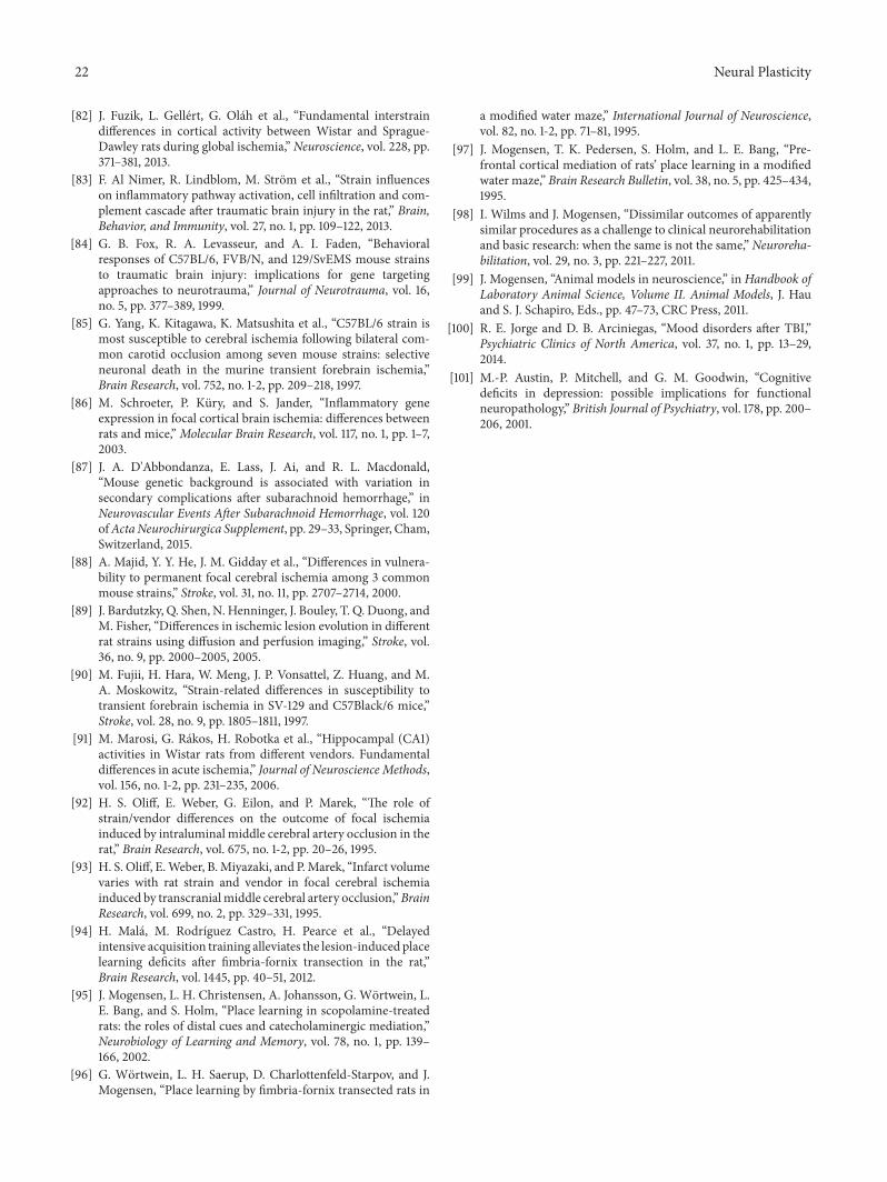
22 Neural Plasticity
[82] J. Fuzik, L. Gellert, G. Olah et al., “Fundamental interstraindifferences in cortical activity between Wistar and Sprague-Dawley rats during global ischemia,” Neuroscience, vol. 228, pp.371–381, 2013.
[83] F. Al Nimer, R. Lindblom, M. Strom et al., “Strain influenceson inflammatory pathway activation, cell infiltration and com-plement cascade after traumatic brain injury in the rat,” Brain,Behavior, and Immunity, vol. 27, no. 1, pp. 109–122, 2013.
[84] G. B. Fox, R. A. Levasseur, and A. I. Faden, “Behavioralresponses of C57BL/6, FVB/N, and 129/SvEMS mouse strainsto traumatic brain injury: implications for gene targetingapproaches to neurotrauma,” Journal of Neurotrauma, vol. 16,no. 5, pp. 377–389, 1999.
[85] G. Yang, K. Kitagawa, K. Matsushita et al., “C57BL/6 strain ismost susceptible to cerebral ischemia following bilateral com-mon carotid occlusion among seven mouse strains: selectiveneuronal death in the murine transient forebrain ischemia,”Brain Research, vol. 752, no. 1-2, pp. 209–218, 1997.
[86] M. Schroeter, P. Kury, and S. Jander, “Inflammatory geneexpression in focal cortical brain ischemia: differences betweenrats and mice,”Molecular Brain Research, vol. 117, no. 1, pp. 1–7,2003.
[87] J. A. D'Abbondanza, E. Lass, J. Ai, and R. L. Macdonald,“Mouse genetic background is associated with variation insecondary complications after subarachnoid hemorrhage,” inNeurovascular Events After Subarachnoid Hemorrhage, vol. 120ofActaNeurochirurgica Supplement, pp. 29–33, Springer, Cham,Switzerland, 2015.
[88] A. Majid, Y. Y. He, J. M. Gidday et al., “Differences in vulnera-bility to permanent focal cerebral ischemia among 3 commonmouse strains,” Stroke, vol. 31, no. 11, pp. 2707–2714, 2000.
[89] J. Bardutzky, Q. Shen, N. Henninger, J. Bouley, T. Q. Duong, andM. Fisher, “Differences in ischemic lesion evolution in differentrat strains using diffusion and perfusion imaging,” Stroke, vol.36, no. 9, pp. 2000–2005, 2005.
[90] M. Fujii, H. Hara, W. Meng, J. P. Vonsattel, Z. Huang, and M.A. Moskowitz, “Strain-related differences in susceptibility totransient forebrain ischemia in SV-129 and C57Black/6 mice,”Stroke, vol. 28, no. 9, pp. 1805–1811, 1997.
[91] M. Marosi, G. Rakos, H. Robotka et al., “Hippocampal (CA1)activities in Wistar rats from different vendors. Fundamentaldifferences in acute ischemia,” Journal of Neuroscience Methods,vol. 156, no. 1-2, pp. 231–235, 2006.
[92] H. S. Oliff, E. Weber, G. Eilon, and P. Marek, “The role ofstrain/vendor differences on the outcome of focal ischemiainduced by intraluminal middle cerebral artery occlusion in therat,” Brain Research, vol. 675, no. 1-2, pp. 20–26, 1995.
[93] H. S. Oliff, E.Weber, B.Miyazaki, and P.Marek, “Infarct volumevaries with rat strain and vendor in focal cerebral ischemiainduced by transcranialmiddle cerebral artery occlusion,”BrainResearch, vol. 699, no. 2, pp. 329–331, 1995.
[94] H. Mala, M. Rodrıguez Castro, H. Pearce et al., “Delayedintensive acquisition training alleviates the lesion-inducedplacelearning deficits after fimbria-fornix transection in the rat,”Brain Research, vol. 1445, pp. 40–51, 2012.
[95] J. Mogensen, L. H. Christensen, A. Johansson, G. Wortwein, L.E. Bang, and S. Holm, “Place learning in scopolamine-treatedrats: the roles of distal cues and catecholaminergic mediation,”Neurobiology of Learning and Memory, vol. 78, no. 1, pp. 139–166, 2002.
[96] G. Wortwein, L. H. Saerup, D. Charlottenfeld-Starpov, and J.Mogensen, “Place learning by fimbria-fornix transected rats in
a modified water maze,” International Journal of Neuroscience,vol. 82, no. 1-2, pp. 71–81, 1995.
[97] J. Mogensen, T. K. Pedersen, S. Holm, and L. E. Bang, “Pre-frontal cortical mediation of rats’ place learning in a modifiedwater maze,” Brain Research Bulletin, vol. 38, no. 5, pp. 425–434,1995.
[98] I. Wilms and J. Mogensen, “Dissimilar outcomes of apparentlysimilar procedures as a challenge to clinical neurorehabilitationand basic research: when the same is not the same,” Neuroreha-bilitation, vol. 29, no. 3, pp. 221–227, 2011.
[99] J. Mogensen, “Animal models in neuroscience,” inHandbook ofLaboratory Animal Science, Volume II. Animal Models, J. Hauand S. J. Schapiro, Eds., pp. 47–73, CRC Press, 2011.
[100] R. E. Jorge and D. B. Arciniegas, “Mood disorders after TBI,”Psychiatric Clinics of North America, vol. 37, no. 1, pp. 13–29,2014.
[101] M.-P. Austin, P. Mitchell, and G. M. Goodwin, “Cognitivedeficits in depression: possible implications for functionalneuropathology,” British Journal of Psychiatry, vol. 178, pp. 200–206, 2001.

Submit your manuscripts athttp://www.hindawi.com
Neurology Research International
Hindawi Publishing Corporationhttp://www.hindawi.com Volume 2014
Alzheimer’s DiseaseHindawi Publishing Corporationhttp://www.hindawi.com Volume 2014
International Journal of
ScientificaHindawi Publishing Corporationhttp://www.hindawi.com Volume 2014
Hindawi Publishing Corporationhttp://www.hindawi.com Volume 2014
BioMed Research International
Hindawi Publishing Corporationhttp://www.hindawi.com Volume 2014
Research and TreatmentSchizophrenia
The Scientific World JournalHindawi Publishing Corporation http://www.hindawi.com Volume 2014
Hindawi Publishing Corporationhttp://www.hindawi.com Volume 2014
Neural Plasticity
Hindawi Publishing Corporationhttp://www.hindawi.com Volume 2014
Parkinson’s Disease
Hindawi Publishing Corporationhttp://www.hindawi.com Volume 2014
Research and TreatmentAutism
Sleep DisordersHindawi Publishing Corporationhttp://www.hindawi.com Volume 2014
Hindawi Publishing Corporationhttp://www.hindawi.com Volume 2014
Neuroscience Journal
Epilepsy Research and TreatmentHindawi Publishing Corporationhttp://www.hindawi.com Volume 2014
Hindawi Publishing Corporationhttp://www.hindawi.com Volume 2014
Psychiatry Journal
Hindawi Publishing Corporationhttp://www.hindawi.com Volume 2014
Computational and Mathematical Methods in Medicine
Depression Research and TreatmentHindawi Publishing Corporationhttp://www.hindawi.com Volume 2014
Hindawi Publishing Corporationhttp://www.hindawi.com Volume 2014
Brain ScienceInternational Journal of
StrokeResearch and TreatmentHindawi Publishing Corporationhttp://www.hindawi.com Volume 2014
Neurodegenerative Diseases
Hindawi Publishing Corporationhttp://www.hindawi.com Volume 2014
Journal of
Cardiovascular Psychiatry and NeurologyHindawi Publishing Corporationhttp://www.hindawi.com Volume 2014