review article phyllodes tumor of breast: a review...
TRANSCRIPT

Hindawi Publishing CorporationISRN SurgeryVolume 2013, Article ID 361469, 10 pageshttp://dx.doi.org/10.1155/2013/361469
Review ArticlePhyllodes Tumor of Breast: A Review Article
Shashi Prakash Mishra, Satyendra Kumar Tiwary,Manjaree Mishra, and Ajay Kumar Khanna
Department of Surgery, Institute of Medical Sciences, Banaras Hindu University, Varanasi, Ultar Pradesh 221005, India
Correspondence should be addressed to Ajay Kumar Khanna; [email protected]
Received 19 January 2013; Accepted 11 February 2013
Academic Editors: M. G. Chiofalo, M. Frascio, H. Hirose, K.-E. Kahnberg, and M. Wronski
Copyright © 2013 Shashi Prakash Mishra et al. This is an open access article distributed under the Creative Commons AttributionLicense, which permits unrestricted use, distribution, and reproduction in any medium, provided the original work is properlycited.
Introduction. Phyllodes tumours are rare fibroepithelial lesions. Accurate preoperative pathological diagnosis allows correct surgicalplanning and avoidance of reoperation. Treatment can be either wide local excision or mastectomy to achieve histologically clearmargins. Discussion. The exact aetiology of phyllodes tumour and its relationship with fibroadenoma are unclear. Women agedbetween 35 and 55 years are commonly involved. The median tumour size is 4 cm but can grow even larger having dilated veinsand a blue discoloration over skin. Palpable axillary lymphadenopathy can be identified in up to 10–15% of patients but <1% hadpathological positive nodes. Mammography and ultrasonography are main imaging modalities. Cytologically the presence of bothepithelial and stromal elements supports the diagnosis.The value of FNAC in diagnosis of phyllodes tumour remains controversial,but core needle biopsy has high sensitivity and negative predictive value. Surgical management is the mainstay and local recurrencein phyllodes tumours has been associated with inadequate local excision. The role of adjuvant radiotherapy and chemotherapyremains uncertain and use of hormonal therapy has not been fully investigated. Conclusion. The preoperative diagnosis and propermanagement are crucial in phyllodes tumours because of their tendency to recur andmalignant potential in some of these tumours.
1. IntroductionPhyllodes tumors are rare fibroepithelial lesions. They makeup 0.3 to 0.5% of female breast tumors [1] and have anincidence of about 2.1 permillion, the peak of which occurs inwomen aged 45 to 49 years [2, 3]. The tumor is rarely foundin adolescents and the elderly. They have been described asearly as 1774, as a giant type of fibroadenoma [4]. Chelius[5] in 1827 first described this tumor. Johannes Muller (1838)was the first person to use the term cystosarcoma phyllodes.It was believed to be benign until 1943, when Cooper andAckerman reported on the malignant biological potential ofthis tumor. In 1981 [6] theWorld Health Organization adoptedthe term phyllodes tumor and as described by Rosen [7]subclassified them histologically as benign, borderline, ormalignant according to the features such as tumor margins,stromal overgrowth, tumor necrosis, cellular atypia, andnumber of mitosis per high power field. The majority ofphyllodes tumors have been described as benign (35% to64%), with the remainder divided between the borderline andmalignant subtypes. The term phyllodes tumor represents abroad range of fibroepithelial diseases and presence of an
epithelial component with stromal components differentiatesthe phyllodes tumor from other stromal sarcomas.
Accurate preoperative pathological diagnosis allows cor-rect surgical planning and avoidance of reoperation, either toachieve wider excision or for subsequent tumor recurrence[8–10]. At one extreme, malignant phyllodes tumors, ifinadequately treated, have a propensity for rapid growthand metastatic spread. In contrast, benign phyllodes tumorson clinical, radiological, and cytological examination areoften indistinguishable fromfibroadenomas and can be curedby local surgery. With the nonoperative management offibroadenomas widely adopted, the importance of phyllodestumors today lies in the need to differentiate them fromother benign breast lesions. Treatment can be either widelocal excision or mastectomy provided histologically clearspecimen margins are ensured [2, 11, 12].
2. Etiology
At present time, the exact etiology of phyllodes tumor andits relationship with fibroadenoma are unclear. Noguchi

2 ISRN Surgery
Table 1
Criteria Benign Borderline MalignantStromalcellularityand atypia
Minimal Moderate Marked
Stromalovergrowth Minimal Moderate Marked
Mitoses/10high powerfields
0–4 5–9 ≥10
Tumormargins
Wellcircumscribedwith pushingtumor margins
Zone ofmicroscopicinvasion aroundtumor margins
Infiltrativetumor margins
et al. [13] showed that most fibroadenomas have polyclonalelements and should be regarded as hyperplasic rather thanneoplastic lesions. It has been suggested that, in a proportionof fibroadenomas, a somatic mutation can result in a mono-clonal proliferation, histologically indistinguishable from thepolyclonal element, but with a propensity to local recurrenceand progression to a phyllodes tumor which has also beensupported by clonal analysis. It has also been postulatedthat stromal induction of phyllodes tumors can occur as aresult of growth factors produced by the breast epithelium.Trauma, lactation, pregnancy, and increased estrogen activityoccasionally have been implicated as factors stimulatingtumor growth. The nature of these factors is unclear butendothelin-1, a stimulator of breast fibroblast growth, may beimportant.
3. Pathogenesis
Unlike carcinoma breast, phyllodes tumors start outside ofthe ducts and lobules, in the breast’s connective tissue, calledthe stroma which includes the fatty tissue and ligaments thatsurround the ducts, lobules, and blood and lymph vessels inthe breast. In addition to stromal cells, phyllodes tumors canalso contain cells from the ducts and lobules.
4. Classification
4.1. WHO Criteria. World Health Organization dividedphyllodes tumor into benign, borderline, and malignantcategories based on the degree of stromal cellular atypia,mitotic activity per 10 high power fields, degree of stromalovergrowth (these three are main), tumor necrosis, andmargin appearance (see Table 1).
4.2. Criteria Proposed by Azzopardi et al. [14] and Salvadori etal. [2]. See Table 2.
5. Diagnosis
5.1. Clinical Presentation. Most of the tumor arises in womenaged between 35 and 55 years (approximately 20 years
Table 2
Criteria Histological typeBenign Borderline Malignant
Tumor margins Pushing ↔ InfiltrativeStroma cellularity Low Moderate HighMitotic rate (per 10 hpf) <5 5–9 ≥10Pleomorphism Mild Moderate Severe
Figure 1: Giant phyllodes tumor.
later than fibroadenoma), 3, 15, 16 more prevalent in theLatin American white and Asian populations [3]. Few caseshave been reported in men and these have invariably beenassociated with the presence of gynaecomastia. It usuallypresents as a rapidly growing but clinically benign breastlump. In some patients a lesion may have been apparentfor several years, with clinical presentation precipitated by asudden increase in size [15, 16].
(i) The skin over large tumorsmay have dilated veins anda blue discoloration but nipple retraction is rare.
(ii) Fixation to skin and pectoralis muscles has beenreported, but ulceration is uncommon.
(iii) More commonly found in upper outer quadrant withan equal propensity to occur in either breast.
(iv) Rarely presentation may be bilateral.(v) The median size of phyllodes tumors is around 4 cm.
20% of tumors grow larger than 10 cm (giant phyl-lodes tumor). These tumors can reach sizes up to40 cm in diameter (see Figure 1).
(vi) A significant proportion of patients have history offibroadenoma and in a minority these have beenmultiple.
(vii) Palpable axillary lymphadenopathy can be identifiedin up to 10–15% of patients but <1% had pathologicalpositive nodes.
5.2. Radiological Investigations. Mammography and ultra-sonography are mainstay of routine imaging of breast lumps.Wurdinger et al. [17] show that round or lobulated shape,well-defined margins, heterogeneous internal structure, andnonenhancing internal septations aremore commonfindingsin phyllodes tumors than in fibroadenomas.

ISRN Surgery 3
Figure 2: Phyllodes tumor on mammography.
5.2.1. Ultrasonography [18, 19]. Lobulated shape (in somecases round or oval) well circumscribed with smooth mar-gins, echogenic rim, and low level homogenous internalechoes. Fluid-filled clefts in a predominantly solid mass(highly suggestive of phyllodes tumor) with good thoroughtransmission and lack of microcalcification are seen.
5.2.2. Color Doppler Ultrasonography. Features suggestingmalignant behavior are
(i) marked hypoechogenicity,(ii) posterior acoustic shadowing,(iii) ill-defined tumor margins.(iv) higher values of RI (resistance index),(v) increased PI (pulsatility index),(vi) increased Vmax (systolic peak flow velocity).
5.2.3. Mammography [18, 20, 21] (see Figure 2)
(i) It shows well circumscribed oval or lobulated masswith rounded borders.
(ii) A radiolucent halo may be seen around the lesion dueto compression of the surroundings.
(iii) Coarse calcification (but malignant microcalcifica-tion is rare) may be present.
5.2.4. Magnetic Resonance Imaging (MRI) [21–28]. The fol-lowing features aremainly found in phyllodes tumor onMRI:
(i) round or lobulated shape and well-defined margins,(ii) heterogeneous internal structure/nonenhancing sep-
tations,(iii) exhibits hypointense signals on T1-weighted images,(iv) exhibits hyper/isointense signals on T2-weighted
images,(v) contrast enhancement pattern:
(a) benign lesion:(1) slow initial enhancement with persistent
delayed phase;(b) malignant lesion:
(1) fast initial enhancement with plateauphage,
(2) fast initial enhancement with wash-outphenomenon.
5.3. Pathological/Histological Assessment. As both phyllodestumors and fibroadenomas belong to a spectrum of fibroep-ithelial lesions, accurate cytological diagnosis of phyllodestumors by fine needle aspiration can be difficult.
Cytologically, it is often easier to differentiate benignfrom malignant phyllodes tumors than to separate benignphyllodes tumors from fibroadenomas. In the correct clinicalsetting, the presence of both epithelial and stromal elementswithin the cytological smear supports the diagnosis. Epithe-lial cells may, however, be absent from specimens taken frommalignant lesions. The presence of cohesive stromal cells(phyllodes fragments), isolatedmesenchymal cells, clusters ofhyperplastic duct cells, foreign body giant cells, blood vesselscrossing the stromal fragments, and bipolar naked nucleiand the absence of apocrine metaplasia are highly suggestiveof a phyllodes tumor. However, the value of FNAC in thediagnosis of phyllodes tumor remains controversial, with anoverall accuracy of about 63% [29, 30]. Core tissue biopsyis an attractive alternative to FNAC because of the extraarchitectural information provided by histology comparedwith cytology. Komenaka et al. [31] found the sensitivity ofcore needle biopsy to be 99% and negative predictive valueand positive predictive value 93% and 83%, respectively, forthe diagnosis.
5.3.1. Macroscopic Appearance. Macroscopically most smalltumors have a uniform white consistency with a lobulatedsurface, similar to that of a fibroadenoma. Large tumors oncut section often have a red or grey “meaty” consistency withfibrogelatinous, hemorrhagic, and necrotic areaswith leaf likeprotrusions into the cystic spaces.
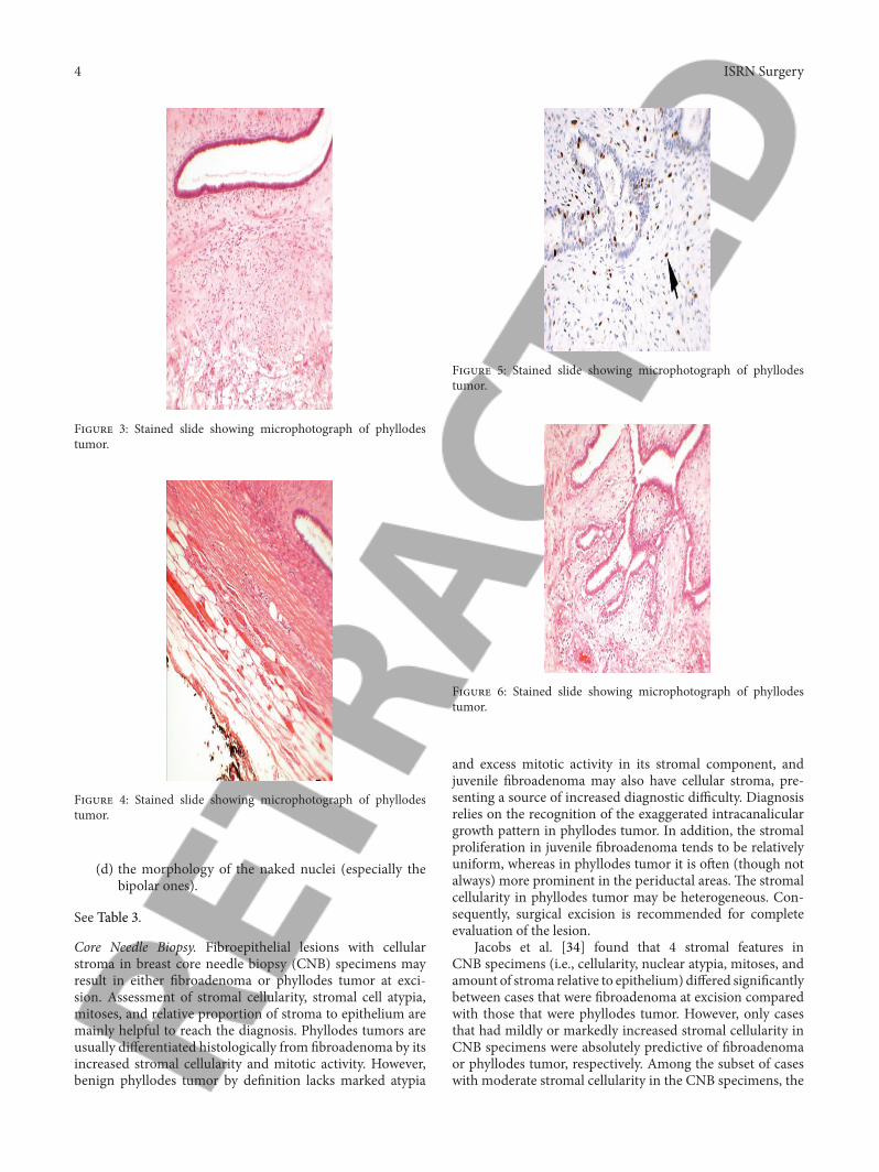
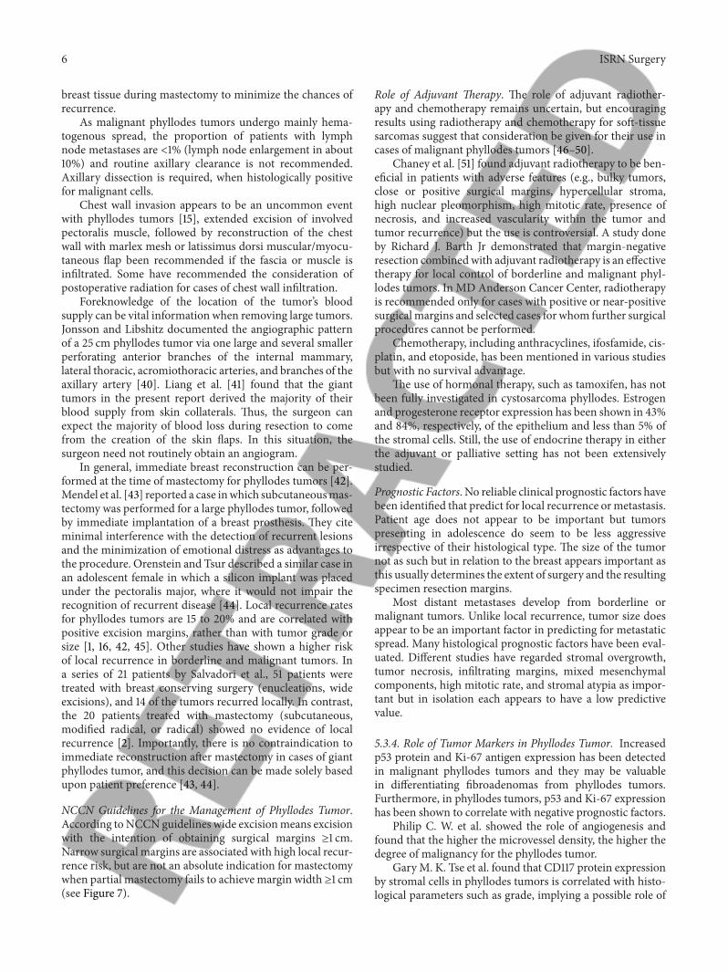
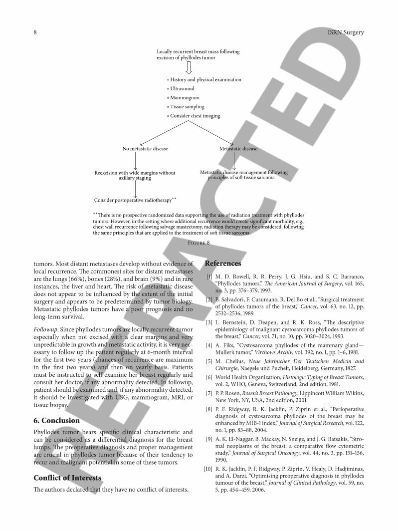
5.3.2. Microscopic Appearance (see Figures 3–6). Fine NeedleAspiration Cytology. The cytological diagnosis of phyllodestumors is mainly suggested by the presence of hypercellularstroma and the stromal elements on the smears being morenumerous than the epithelial ones. The cells on the smearswere classified by Deen et al. [32] in 1999, and Jayaramand Sthaneshwar in 2002 [33], by comparison with smalllymphocytes, in
(1) short, round/oval cells, two-size smaller than the sizeof a lymphocyte: considered to be epithelial cells;
(2) long, spindle cells, three-size larger than the size of alymphocyte: considered to be stromal cells.
Many authors considered that the following aspectsshould also be taken into consideration in the case ofcytological diagnosis of phyllodes tumors:
(a) the presence of hypercellular stromal fragments;(b) the cellular composition of the stromal fragments;(c) the amount of naked nuclei on the background of the
smears;
4 ISRN Surgery
Figure 3: Stained slide showing microphotograph of phyllodestumor.
Figure 4: Stained slide showing microphotograph of phyllodestumor.
(d) the morphology of the naked nuclei (especially thebipolar ones).
See Table 3.
Core Needle Biopsy. Fibroepithelial lesions with cellularstroma in breast core needle biopsy (CNB) specimens mayresult in either fibroadenoma or phyllodes tumor at exci-sion. Assessment of stromal cellularity, stromal cell atypia,mitoses, and relative proportion of stroma to epithelium aremainly helpful to reach the diagnosis. Phyllodes tumors areusually differentiated histologically from fibroadenoma by itsincreased stromal cellularity and mitotic activity. However,benign phyllodes tumor by definition lacks marked atypia
Figure 5: Stained slide showing microphotograph of phyllodestumor.
Figure 6: Stained slide showing microphotograph of phyllodestumor.
and excess mitotic activity in its stromal component, andjuvenile fibroadenoma may also have cellular stroma, pre-senting a source of increased diagnostic difficulty. Diagnosisrelies on the recognition of the exaggerated intracanaliculargrowth pattern in phyllodes tumor. In addition, the stromalproliferation in juvenile fibroadenoma tends to be relativelyuniform, whereas in phyllodes tumor it is often (though notalways) more prominent in the periductal areas. The stromalcellularity in phyllodes tumor may be heterogeneous. Con-sequently, surgical excision is recommended for completeevaluation of the lesion.
Jacobs et al. [34] found that 4 stromal features inCNB specimens (i.e., cellularity, nuclear atypia, mitoses, andamount of stroma relative to epithelium) differed significantlybetween cases that were fibroadenoma at excision comparedwith those that were phyllodes tumor. However, only casesthat had mildly or markedly increased stromal cellularity inCNB specimens were absolutely predictive of fibroadenomaor phyllodes tumor, respectively. Among the subset of caseswith moderate stromal cellularity in the CNB specimens, the

ISRN Surgery 5
Table 3
Benign phyllodes tumors Borderline phyllodes tumors Malignant phyllodes tumors
The stromal compound, represented bystromal fragments, isolated stromal cells,and naked stromal nuclei are found to bemore numerous than the epithelial one inmost of the cases.
(i) There is predominance of the stromalcomponent as compared to the epithelialone.(ii) Frequent hypercellular stromalfragments, an average of 2 in eachmicroscopical field.(iii) Frequent large spindle cells andmonomorphic naked stromal nuclei.
(i) Stromal fragments of variabledimensions, with moderate cellularity,made of discohesive spindle cells, withatypical nuclei.(ii) Minimal/no epithelial elements foundon the smears.(iii) Presence of atypical multinucleatedgiant cells.
Clinical findings(i) Sudden increase in size in a longstanding breast lesion(ii) Apparent fibroadenoma> 3 cm diameter or in patient >35 years
Imaging findings(i) Rounded borders/lobulated appearance at mammography(ii) Attenuation or cystic areas within a solid mass on Ultrasonography
FNAC findings(i) Presence of hypercellular stromal fragments(ii) Indeterminate features
ANY 2 features mandate core biopsy
Box 1
presence of stromal mitoses remained the single histologicalfeature significantly different between the phyllodes tumorand fibroadenoma groups.
Sarcomatous stromal elements, including angiosarcoma,chondrosarcoma, leiomyosarcoma, osteosarcoma, liposar-coma, and rhabdomyosarcoma, are rarely encountered inmalignant phyllodes tumors.
Paddington Clinicopathological Suspicion Score. This outlinescriteria to assist in the selection of patients for core biopsy,for use in conjunction with existing local protocols. The aimof developing the score is to improve the rates of preoperativediagnosis (see Box 1).
5.3.3. Differential Diagnosis. It includes the following:
(i) fibroadenoma,(ii) adenoma,(iii) hamartoma,(iv) lipoma,(v) juvenile papillomatosis,(vi) carcinoma,(vii) sarcomas,(viii) metastatic tumor.
Management. Surgical management is the mainstay but thetype of surgery has been a source of debate over the years.Studies have shown no differences between breast conservingsurgery versus mastectomy in terms of metastasis-free sur-vival or overall survival, despite the higher incidence of localrecurrence that comes with breast conserving surgery [35].
If diagnosed preoperatively, tumor should be resectedwith at least 1 cm margins particularly in the borderline andmalignant phyllodes tumors. This can be accomplished byeither lumpectomy or mastectomy, depending upon the sizeof the tumor relative to the breast. For benign phyllodestumors diagnosed after local excision of what appeared tobe a fibroadenoma, a “watch and wait” policy does appearto be safe. With such an approach, local recurrence and fiveyear survival rates of 4% and 96% respectively have beenreported for benign phyllodes tumors.Whether patients withbenign phyllodes tumors who have undergone local excisionand have histologically positive specimen margins shouldundergo further surgery or be entered in a surveillance pro-gram is controversial. Reexcision of borderline andmalignantphyllodes tumors identified after local excision should beconsidered.
Twenty percent of tumors grow larger than 10 cm, thearbitrary cutoff point for the designation as giant phyllodestumor, an entity that presents the surgeon with severalunique management problems. These tumors can reach sizesup to 40 cm in diameter [15]. Since an excision with therequired margins is often impossible in giant phyllodestumors, mastectomy should be reserved for larger tumorsand should be considered in recurrent tumors, especiallyof the malignant histotype [2, 36–38]. Local recurrence inphyllodes tumors has been associated with inadequate localexcision and various histological characteristics, includingmitotic activity, tumor margin, and stromal cellular atypia.Depending on the size of the breast and the location ofthe phyllodes tumor, mastectomy may also be requiredfor tumors that are between 5 and 10 cm in diameter[39]. While managing a giant phyllodes tumor, emphasisshould be on complete extirpation of all visible tumors and
6 ISRN Surgery
breast tissue during mastectomy to minimize the chances ofrecurrence.
As malignant phyllodes tumors undergo mainly hema-togenous spread, the proportion of patients with lymphnode metastases are <1% (lymph node enlargement in about10%) and routine axillary clearance is not recommended.Axillary dissection is required, when histologically positivefor malignant cells.
Chest wall invasion appears to be an uncommon eventwith phyllodes tumors [15], extended excision of involvedpectoralis muscle, followed by reconstruction of the chestwall with marlex mesh or latissimus dorsi muscular/myocu-taneous flap been recommended if the fascia or muscle isinfiltrated. Some have recommended the consideration ofpostoperative radiation for cases of chest wall infiltration.
Foreknowledge of the location of the tumor’s bloodsupply can be vital information when removing large tumors.Jonsson and Libshitz documented the angiographic patternof a 25 cm phyllodes tumor via one large and several smallerperforating anterior branches of the internal mammary,lateral thoracic, acromiothoracic arteries, and branches of theaxillary artery [40]. Liang et al. [41] found that the gianttumors in the present report derived the majority of theirblood supply from skin collaterals. Thus, the surgeon canexpect the majority of blood loss during resection to comefrom the creation of the skin flaps. In this situation, thesurgeon need not routinely obtain an angiogram.
In general, immediate breast reconstruction can be per-formed at the time of mastectomy for phyllodes tumors [42].Mendel et al. [43] reported a case inwhich subcutaneousmas-tectomy was performed for a large phyllodes tumor, followedby immediate implantation of a breast prosthesis. They citeminimal interference with the detection of recurrent lesionsand the minimization of emotional distress as advantages tothe procedure. Orenstein and Tsur described a similar case inan adolescent female in which a silicon implant was placedunder the pectoralis major, where it would not impair therecognition of recurrent disease [44]. Local recurrence ratesfor phyllodes tumors are 15 to 20% and are correlated withpositive excision margins, rather than with tumor grade orsize [1, 16, 42, 45]. Other studies have shown a higher riskof local recurrence in borderline and malignant tumors. Ina series of 21 patients by Salvadori et al., 51 patients weretreated with breast conserving surgery (enucleations, wideexcisions), and 14 of the tumors recurred locally. In contrast,the 20 patients treated with mastectomy (subcutaneous,modified radical, or radical) showed no evidence of localrecurrence [2]. Importantly, there is no contraindication toimmediate reconstruction after mastectomy in cases of giantphyllodes tumor, and this decision can be made solely basedupon patient preference [43, 44].
NCCN Guidelines for the Management of Phyllodes Tumor.According toNCCN guidelines wide excisionmeans excisionwith the intention of obtaining surgical margins ≥1 cm.Narrow surgical margins are associated with high local recur-rence risk, but are not an absolute indication for mastectomywhen partial mastectomy fails to achieve margin width ≥1 cm(see Figure 7).
Role of Adjuvant Therapy. The role of adjuvant radiother-apy and chemotherapy remains uncertain, but encouragingresults using radiotherapy and chemotherapy for soft-tissuesarcomas suggest that consideration be given for their use incases of malignant phyllodes tumors [46–50].
Chaney et al. [51] found adjuvant radiotherapy to be ben-eficial in patients with adverse features (e.g., bulky tumors,close or positive surgical margins, hypercellular stroma,high nuclear pleomorphism, high mitotic rate, presence ofnecrosis, and increased vascularity within the tumor andtumor recurrence) but the use is controversial. A study doneby Richard J. Barth Jr demonstrated that margin-negativeresection combinedwith adjuvant radiotherapy is an effectivetherapy for local control of borderline and malignant phyl-lodes tumors. In MD Anderson Cancer Center, radiotherapyis recommended only for cases with positive or near-positivesurgicalmargins and selected cases for whom further surgicalprocedures cannot be performed.
Chemotherapy, including anthracyclines, ifosfamide, cis-platin, and etoposide, has been mentioned in various studiesbut with no survival advantage.
The use of hormonal therapy, such as tamoxifen, has notbeen fully investigated in cystosarcoma phyllodes. Estrogenand progesterone receptor expression has been shown in 43%and 84%, respectively, of the epithelium and less than 5% ofthe stromal cells. Still, the use of endocrine therapy in eitherthe adjuvant or palliative setting has not been extensivelystudied.
Prognostic Factors. No reliable clinical prognostic factors havebeen identified that predict for local recurrence ormetastasis.Patient age does not appear to be important but tumorspresenting in adolescence do seem to be less aggressiveirrespective of their histological type. The size of the tumornot as such but in relation to the breast appears important asthis usually determines the extent of surgery and the resultingspecimen resection margins.
Most distant metastases develop from borderline ormalignant tumors. Unlike local recurrence, tumor size doesappear to be an important factor in predicting for metastaticspread. Many histological prognostic factors have been eval-uated. Different studies have regarded stromal overgrowth,tumor necrosis, infiltrating margins, mixed mesenchymalcomponents, high mitotic rate, and stromal atypia as impor-tant but in isolation each appears to have a low predictivevalue.
5.3.4. Role of Tumor Markers in Phyllodes Tumor. Increasedp53 protein and Ki-67 antigen expression has been detectedin malignant phyllodes tumors and they may be valuablein differentiating fibroadenomas from phyllodes tumors.Furthermore, in phyllodes tumors, p53 and Ki-67 expressionhas been shown to correlate with negative prognostic factors.
Philip C. W. et al. showed the role of angiogenesis andfound that the higher the microvessel density, the higher thedegree of malignancy for the phyllodes tumor.
GaryM. K. Tse et al. found that CD117 protein expressionby stromal cells in phyllodes tumors is correlated with histo-logical parameters such as grade, implying a possible role of

ISRN Surgery 7
Clinical presentation Clinical suspicion of phyllodes tumorPalpable massRapid growth
Imaging with ultrasound suggestive offibroadenoma except for size and/orhistory of growth
Workup History and physical examinationUltrasound
Findings Treatment
ObserveFibroadenomaExcisional biopsy
Phyllodes tumorsincluding benign, borderline,and malignant axillary staging
Core needle biopsy Fibroadenoma or indeterminate Excisional biopsy
Phyllodes tumoraxillary staging
∙
∙
∙
∙
∙
∙
H
Mammogram for women ≥30 yrs
Wide excision (≥1 cm) without
Wide excision (≥1 cm) without
Large size (>2 cm)
Figure 7
their being used as adjunctivemarkers ofmalignancy in thesetumors. Inmalignant phyllodes tumors, the rate of expressionis up to 46%. This provides additional strong evidencethat c-kit receptor-mediated tyrosine kinase activity may beinvolved early on in the pathogenesis of phyllodes tumors,and the new therapeutic agent, STI571, Glivec, may be auseful drug therapy for this disease, particularly in the tumorrecurrences and advanced-stage disease. Marick Lae et al.showed that chromosomal changes detected by comparativegenomic hybridization (CGH) could be helpful in gradingphyllodes tumors. In flow cytometric studies, correlationsbetween DNA content, cell proliferation, and histologicalgrade have been demonstrated. Some studies have identifieda correlation between these markers of cellular proliferationand clinical outcome, however, most have not. Recent smallstudies have suggested that telomerase, a ribonucleoproteinenzyme that generates telomeres (DNA sequences importantin determining cell immortality), may be a useful prognosticfactor in phyllodes tumors.
Recurrence. To date, local recurrence rates ranging from 10%to 40% have been reported with most series averaging about15%. Local recurrence appears to be related to the extentof the initial surgery and should be regarded as a failure ofprimary surgical treatment. Whether malignant tumors have
an increased risk of recurrence is unclear but when it doesoccur it is invariably seen earlier than with benign tumors.In multivariate analysis, the surgical margin is found to bethe only independent predictive factor for local recurrence.In most patients, local recurrence is isolated and is notassociated with the development of distant metastases.
In aminority of patients repeated local recurrence occurs.This is often seen irrespective of either the histologicaltype of the tumor or the extent of the specimen margins.Local recurrence can usually be controlled by further wideexcision (with 1 cm margins) and mastectomy is not invari-ably required. Mastectomy should, however, be consideredfor local recurrence after local surgery for borderline ormalignant tumors. Occasionally aggressive local recurrencecan result in widespread chest wall disease with directinvasion of the underlying lung parenchyma. Isolated reportsof good palliation in this situation with radiotherapy havebeen published.
NCCN Guidelines for the Management of Recurrence. SeeFigure 8.
Metastasis. Overall, 10% of patients with phyllodes tumorsdevelop distant metastases and these eventually occur inapproximately 25% of patients with histologically malignant
8 ISRN Surgery
Locally recurrent breast mass followingexcision of phyllodes tumor
History and physical examination
Ultrasound
Mammogram
Tissue sampling
Consider chest imaging
No metastatic disease Metastatic disease
Reexcision with wide margins without Metastatic disease management followingaxillary staging principles of soft tissue sarcoma
∘
∘
∘
∘
∘
Consider postoperative radiotherapy∗∗
∗∗There is no prospective randomized data supporting the use of radiation treatment with phyllodestumors. However, in the setting where additional recurrence would create significant morbidity, e.g.,chest wall recurrence following salvage mastectomy, radiation therapy may be considered, followingthe same principles that are applied to the treatment of soft tissue sarcoma.
Figure 8
tumors. Most distant metastases develop without evidence oflocal recurrence. The commonest sites for distant metastasesare the lungs (66%), bones (28%), and brain (9%) and in rareinstances, the liver and heart. The risk of metastatic diseasedoes not appear to be influenced by the extent of the initialsurgery and appears to be predetermined by tumor biology.Metastatic phyllodes tumors have a poor prognosis and nolong-term survival.
Followup. Since phyllodes tumors are locally recurrent tumorespecially when not excised with a clear margins and veryunpredictable in growth andmetastatic activity, it is very nec-essary to follow up the patient regularly at 6-month intervalfor the first two years (chances of recurrence are maximumin the first two years) and then on yearly basis. Patientsmust be instructed to self examine her breast regularly andconsult her doctor, if any abnormality detected. In followup,patient should be examined and, if any abnormality detected,it should be investigated with USG, mammogram, MRI, ortissue biopsy.
6. Conclusion
Phyllodes tumor bears specific clinical characteristic andcan be considered as a differential diagnosis for the breastlumps. The preoperative diagnosis and proper managementare crucial in phyllodes tumor because of their tendency torecur and malignant potential in some of these tumors.
Conflict of Interests
The authors declared that they have no conflict of interests.
References
[1] M. D. Rowell, R. R. Perry, J. G. Hsiu, and S. C. Barranco,“Phyllodes tumors,” The American Journal of Surgery, vol. 165,no. 3, pp. 376–379, 1993.
[2] B. Salvadori, F. Cusumano, R. Del Bo et al., “Surgical treatmentof phyllodes tumors of the breast,” Cancer, vol. 63, no. 12, pp.2532–2536, 1989.
[3] L. Bernstein, D. Deapen, and R. K. Ross, “The descriptiveepidemiology of malignant cystosarcoma phyllodes tumors ofthe breast,” Cancer, vol. 71, no. 10, pp. 3020–3024, 1993.
[4] A. Fiks, “Cystosarcoma phyllodes of the mammary gland—Muller’s tumor,” Virchows Archiv, vol. 392, no. 1, pp. 1–6, 1981.
[5] M. Chelius, Neue Jahrbucher Der Teutschen Medicin andChirurgie, Naegele und Puchelt, Heidelberg, Germany, 1827.
[6] World Health Organization,Histologic Typing of Breast Tumors,vol. 2, WHO, Geneva, Switzerland, 2nd edition, 1981.
[7] P. P. Rosen,Rosen’s Breast Pathology, LippincottWilliamWikins,New York, NY, USA, 2nd edition, 2001.
[8] P. F. Ridgway, R. K. Jacklin, P. Ziprin et al., “Perioperativediagnosis of cystosarcoma phyllodes of the breast may beenhanced byMIB-1 index,” Journal of Surgical Research, vol. 122,no. 1, pp. 83–88, 2004.
[9] A. K. El-Naggar, B. Mackay, N. Sneige, and J. G. Batsakis, “Stro-mal neoplasms of the breast: a comparative flow cytometricstudy,” Journal of Surgical Oncology, vol. 44, no. 3, pp. 151–156,1990.
[10] R. K. Jacklin, P. F. Ridgway, P. Ziprin, V. Healy, D. Hadjiminas,and A. Darzi, “Optimising preoperative diagnosis in phyllodestumour of the breast,” Journal of Clinical Pathology, vol. 59, no.5, pp. 454–459, 2006.

ISRN Surgery 9
[11] A. W. Chaney, A. Pollack, D. Marsha et al., “Primary treatmentof cystosarcoma phyllodes of the breast,” Cancer, vol. 89, pp.1502–1510, 2000.
[12] I. Kapiris, N. Nasiri, R. A’Hern, V. Healy, and G. P. H. Gui,“Outcome and predictive factors of local recurrence and distantmetastases following primary surgical treatment of high-grademalignant phyllodes tumours of the breast,” European Journalof Surgical Oncology, vol. 27, no. 8, pp. 723–730, 2001.
[13] S. Noguchi, K. Motomura, H. Inaji, S. Imaoka, and H. Koyama,“Clonal analysis of fibroadenoma and phyllodes tumor of thebreast,” Cancer Research, vol. 53, no. 17, pp. 4071–4074, 1993.
[14] J. G. Azzopardi, A. Ahmed, and R. R.Millis, “Problems in breastpathology,” Major Problems in Pathology, vol. 11, pp. 346–364,1979.
[15] M. Reinfuss, J. Mitus, K. Duda, A. Stelmach, J. Rys, and K. Smo-lak, “The treatment and prognosis of patients with phyllodestumor of the breast: an analysis of 170 cases,” Cancer, vol. 77,pp. 910–916, 1996.
[16] C. L. Chua, A.Thomas, and B. K. Ng, “Cystosarcoma phyllodes:a review of surgical options,” Surgery, vol. 105, no. 2 I, pp. 141–147, 1989.
[17] S. Wurdinger, A. B. Herzog, D. R. Fischer et al., “Differentiationof phyllodes breast tumors from fibroadenomas on MRI,”American Journal of Roentgenology, vol. 185, no. 5, pp. 1317–1321,2005.
[18] J. M. Feder, E. S. de Paredes, J. P. Hogge, and J. J. Wilken,“Unusual breast lesions: radiologic-pathologic correlation,”Radiographics, vol. 19, pp. S11–S26, 1999.
[19] C. Cole Beuglet, R. Soriano, and A. B. Kurtz, “Ultrasound,X-ray mammography, and histopathology of cystosarcomaphylloides,” Radiology, vol. 146, no. 2, pp. 481–486, 1983.
[20] A. Jorge Blanco, B. Vargas Serrano, R. Rodriguez Romero et al.,“Phyllodes tumors of the breast,” European Radiology, vol. 9, pp.356–360, 1999.
[21] P. Cosmacini, P. Veronesi, S. Zurrida, C. Bartoli, C. Ferranti,andG. CoopmansDeYoldi, “Mammography in the diagnosis ofphyllodes tumors of the breast. Analysis of 99 cases,” RadiologiaMedica, vol. 82, no. 1-2, pp. 52–55, 1991.
[22] T. Kinoshita, T. Fukutomi, and K. Kubochi, “Magnetic reso-nance imaging of benign phyllodes tumors of the breast,” BreastJournal, vol. 10, no. 3, pp. 232–236, 2004.
[23] G. M. K. Tse, H. S. Cheung, L. M. Pang et al., “Characterizationof lesions of the breast with proton MR spectroscopy: com-parison of carcinomas, benign lesions, and phyllodes tumors,”American Journal of Roentgenology, vol. 181, no. 5, pp. 1267–1272,2003.
[24] N. Katayama, Y. Inoue, T. Ichikawa et al., “Increased activityin benign phyllodes tumor on Tc-99mMDP scintimammogra-phy,” Clinical Nuclear Medicine, vol. 25, no. 7, pp. 551–552, 2000.
[25] H. Ohta, T. Komibuchi, T. Nishio et al., “Technetium-99m-ses-tamibi scintimammography of benign andmalignant phyllodestumors,” Annals of Nuclear Medicine, vol. 11, no. 1, pp. 37–39,1997.
[26] J. E. Page and J. E. Williams, “The radiological features ofphylloides tumour of the breast with clinico-pathological cor-relation,” Clinical Radiology, vol. 44, no. 1, pp. 8–12, 1991.
[27] W. Buchberger, K. Strasser, K. Heim, E.Muller, andH. Schrock-snadel, “Phylloides tumor: findings on mammography, sonog-raphy, and aspiration cytology in 10 cases,” American Journal ofRoentgenology, vol. 157, no. 4, pp. 715–719, 1991.
[28] L. Liberman, E. Bonaccio, D. Hamele-Bena, A. F. Abramson,M.A. Cohen, andD.D.Dershaw, “Benign andmalignant phyllodestumors: mammographic and sonographic findings,” Radiology,vol. 198, no. 1, pp. 121–124, 1996.
[29] D. C. Chhieng, J. F. Cangiarella, J. Waisman et al., “Fine-needleaspiration cytology of spindle cell lesions of the breast,” Cancer,vol. 87, pp. 359–371, 1999.
[30] U. Simi, D. Moretti, P. Iacconi et al., “Fine needle aspirationcytopathology of phyllodes tumor. Differential diagnosis withfibroadenoma,” Acta Cytologica, vol. 32, no. 1, pp. 63–66, 1988.
[31] I. K. Komenaka, M. El-Tamer, E. Pile-Spellman, and H. Hib-shoosh, “Core needle biopsy as a diagnostic tool to differentiatephyllodes tumor from fibroadenoma,” Archives of Surgery, vol.138, no. 9, pp. 987–990, 2003.
[32] S. A. Deen, G. T. McKee, and M. W. Kissin, “Differential cyto-logic features of fibroepithelial lesions of the breast,” DiagnosticCytopathology, vol. 20, pp. 53–56, 1999.
[33] G. Jayaram and P. Sthaneshwar, “Fine-needle aspiration cytol-ogy of phyllodes tumors,”Diagnostic Cytopathology, vol. 26, no.4, pp. 222–227, 2002.
[34] T. W. Jacobs, Y. Y. Chen, D. G. Guinee et al., “Fibroepitheliallesions with cellular stroma on breast core needle biopsy: arethere predictors of outcome on surgical excision?” AmericanJournal of Clinical Pathology, vol. 124, no. 3, pp. 342–354, 2005.
[35] G. Cohn-Cedermark, L. E. Rutqvist, I. Rosendahl, and C.Silfversward, “Prognostic factors in cystosarcoma phyllodes: aclinicopathologic study of 77 patients,” Cancer, vol. 68, no. 9,pp. 2017–2022, 1991.
[36] H. J. Norris andH. B. Taylor, “Relationship of histologic featuresto behavior of cystosarcoma phyllodes. Analysis of ninety-fourcases,” Cancer, vol. 20, no. 12, pp. 2090–2099, 1967.
[37] O. Contarini, L. F. Urdaneta, W. Hagan, and S. E. Stephenson,“Cystosarcoma phylloides of the breast: a new therapeuticproposal,” American Surgeon, vol. 48, no. 4, pp. 157–166, 1982.
[38] R. R. Baker, “Unusual lesions and their management,” SurgicalClinics of North America, vol. 70, no. 4, pp. 963–975, 1990.
[39] G. Singh and R. K. Sharma, “Immediate breast reconstructionfor phyllodes tumors,” Breast, vol. 17, no. 3, pp. 296–301, 2008.
[40] K. Jonsson and H. I. Libshitz, “Arteriographic pattern in cys-tosarcoma phylloides,” British Journal of Radiology, vol. 50, no.598, pp. 751–753, 1977.
[41] M. I. Liang, B. Ramaswamy, C. C. Patterson et al., “Giant breasttumors: surgicalmanagement of phyllodes tumors, potential forreconstructive surgery and a review of literature,”World Journalof Surgical Oncology, vol. 6, article 117, 2008.
[42] A. A. Mangi, B. L. Smith, M. A. Gadd, K. K. Tanabe, M. J. Ott,and W. W. Souba, “Surgical management of phyllodes tumors,”Archives of Surgery, vol. 134, no. 5, pp. 487–493, 1999.
[43] M. A. Mendel, R. G. DePalma, C. Vogt, and J. W. Reagan, “Cys-tosarcoma phyllodes: treatment by subcutaneous mastectomywith immediate prosthetic implantation,”TheAmerican Journalof Surgery, vol. 23, pp. 718–721, 1972.
[44] A. Orenstein and H. Tsur, “Cystosarcoma phylloides treated byexcision and immediate reconstruction with silicon implant,”Annals of Plastic Surgery, vol. 18, no. 6, pp. 520–523, 1987.
[45] S. Khanna, S. Gupta, andN.N. Khanna, “Sarcomas of the breast:homogenous or heterogenous?” Journal of Surgical Oncology,vol. 18, no. 2, pp. 119–128, 1981.
[46] S. C. Carabell and R. L. Goodman, “Radiation therapy for softtissue sarcoma,” Seminars in Oncology, vol. 8, no. 2, pp. 201–206,1981.

10 ISRN Surgery
[47] R. E. Hawkins, J. B. Schofield, E. Wiltshaw, C. Fisher, and J. A.McKinna, “Ifosfamide is an active drug for chemotherapy ofmetastatic cystosarcoma phyllodes,” Cancer, vol. 69, no. 9, pp.2271–2275, 1992.
[48] G. V. Burton, L. L. Hart, G. S. Leight, J. D. Iglehart, K. S.McCarty, and E. B. Cox, “Cystosarcoma phyllodes. Effectivetherapy with cisplatin and etoposide chemotherapy,” Cancer,vol. 63, no. 11, pp. 2088–2092, 1989.
[49] S. C. Joshi, D. N. Sharma, A. K. Bahadur, R. Maurya, S. Kumar,and N. Khurana, “Cystosarcoma phyllodes: our institutionalexperience,” Australasian Radiology, vol. 47, no. 4, pp. 434–437,2003.
[50] T. Palshof,M. Blickert-Taft, and L.Daehnfelt, “Estradiol bindingprotein in cystosarcoma phylloides of the breast,” EuropeanJournal of Cancer, vol. 16, pp. 591–593, 1980.
[51] A. W. Chaney, A. Pollack, M. D. McNeese, and G. K. Zagars,“Adjuvant radiotherapy for phyllodes tumor of breast,” Radia-tion Oncology Investigations, vol. 6, no. 6, pp. 264–267, 1998.

Submit your manuscripts athttp://www.hindawi.com
Stem CellsInternational
Hindawi Publishing Corporationhttp://www.hindawi.com Volume 2014
Hindawi Publishing Corporationhttp://www.hindawi.com Volume 2014
MEDIATORSINFLAMMATION
of
Hindawi Publishing Corporationhttp://www.hindawi.com Volume 2014
Behavioural Neurology
EndocrinologyInternational Journal of
Hindawi Publishing Corporationhttp://www.hindawi.com Volume 2014
Hindawi Publishing Corporationhttp://www.hindawi.com Volume 2014
Disease Markers
Hindawi Publishing Corporationhttp://www.hindawi.com Volume 2014
BioMed Research International
OncologyJournal of
Hindawi Publishing Corporationhttp://www.hindawi.com Volume 2014
Hindawi Publishing Corporationhttp://www.hindawi.com Volume 2014
Oxidative Medicine and Cellular Longevity
Hindawi Publishing Corporationhttp://www.hindawi.com Volume 2014
PPAR Research
The Scientific World JournalHindawi Publishing Corporation http://www.hindawi.com Volume 2014
Immunology ResearchHindawi Publishing Corporationhttp://www.hindawi.com Volume 2014
Journal of
ObesityJournal of
Hindawi Publishing Corporationhttp://www.hindawi.com Volume 2014
Hindawi Publishing Corporationhttp://www.hindawi.com Volume 2014
Computational and Mathematical Methods in Medicine
OphthalmologyJournal of
Hindawi Publishing Corporationhttp://www.hindawi.com Volume 2014
Diabetes ResearchJournal of
Hindawi Publishing Corporationhttp://www.hindawi.com Volume 2014
Hindawi Publishing Corporationhttp://www.hindawi.com Volume 2014
Research and TreatmentAIDS
Hindawi Publishing Corporationhttp://www.hindawi.com Volume 2014
Gastroenterology Research and Practice
Hindawi Publishing Corporationhttp://www.hindawi.com Volume 2014
Parkinson’s Disease
Evidence-Based Complementary and Alternative Medicine
Volume 2014Hindawi Publishing Corporationhttp://www.hindawi.com