review article hyaluronan in medical practice current medicinal... · hyaluronan in medical...
TRANSCRIPT

Send Orders for Print-Reprints and e-prints to [email protected] Current Medicinal Chemistry, 2016, 23, 1-11 1
REVIEW ARTICLE
0929-8673/16 $58.00+.00 © 2016 Bentham Science Publishers
Hyaluronan in Medical Practice
Katarína Valachová1, Nicola Volpi2,*, Robert Stern3 and Ladislav Šoltés1
1Institute of Experimental Pharmacology and Toxicology, Slovak Academy of Sciences, Bratislava, Slovakia; 2Department of Life Sciences, University of Modena and Reggio Emilia, Modena, Italy; 3Touro College of Osteopathic Medicine, New York, USA
A R T I C L E H I S T O R Y
Received: February 16, 2016 Revised: June 15, 2016 Accepted: June 29, 2016 DOI: 10.2174/092986732366616082 4162133
Abstract: Hyaluronan is the major extracellular matrix glycosaminoglycan polymer present in vertebrate tissues, with a molar mass that can reach sev-eral megaDaltons. It is particularly prominent in the matrix of tissues under-going rapid turnover, in fetal tissues, and wherever regeneration and repair are occurring. Hyaluronan has highly varied biological functions often de-pendent on molar mass, however they are highly dependent on source of hyaluronan, its purity and nature of contaminants. Hyaluronan of high-molar-mass is known for its anti-angiogenic, anti-inflammatory and immu-nosuppressive properties, unlike hyaluronan of low-molar-mass that has the opposite effects. Hyaluronan also has a broad range of clinical applications, such as intra-articular injection, in ophthalmology, otolaryngology, wound healing, and commercially in the cosmetic industry, as well as in drug delivery systems. Currently, polymers of hyaluronan are modified in order to improve their properties, including bioavailability and resistance to degradation. Because of greatly increased interest currently in hyaluronan, the multiple functions of the polymer are presented here, including medicine and industry, as well as recent progress in the formulation of hyaluronan-based materials.
Keywords: Hyaluronic acid, hyaluronan production, tissue engineering, viscoelasticity, viscosupplemenation, vis-coprotection.
INTRODUCTION
Hyaluronan (hyaluronic acid, HA, Fig. 1a, 1c), a linear polymer composed of two disaccharides-D-glucuronic acid and N-acetyl-D-glucosamine, is linked via β-1,4 and β-1,3 glycosidic bonds. Unlike other GAGs (glycosaminoglycans), HA lacks a sulfate group. The number of disaccharide units in HA can vary from 25 to 25,000, with a linear uncoiled polymer length from 10 nm to 25 µm [1]. Molar mass of HA reaches from 100 kDa in serum to 8 MDa in the vitreous of the eye [2] (Table 1).
In addition, various aspects of the metabolism of HA are indicated in Table 1 for purposes of orientation. The biological significance of the tetrasaccharide and *Address correspondence to this author at the Department of Life Sciences, University of Modena & Reggio Emilia, Via Campi 213/D, 41100 Modena, Italy; Tel: 0039 (0)59 2055543; Fax: 0039 (0)59 2055548; E-mail: [email protected]
Fig. (1). A highly viscous solution of HA; an adult naked mole rat [36]; structure of HA demonstrating a tetrasac- charide or two repeating units of N-acetyl-D-glucosamine and D-glucuronic acid. the oligomeric HA with ~ 25 disaccharide units within the metabolic cycle are pointed out. A remarkable property of HA is its capacity to surround itself with a large volume of water that can reach up to several thousand-fold its original weight [3-6]. The very large
Please provide corresponding author(s)
photographsize should be 4" x 4" inches
Nicola Volpi

2 Current Medicinal Chemistry, 2016, Vol. 23, No. 30 Valachová et al.
negative charge associated with HA at neutral pH ac-counts for this extraordinary water domain. In Table 1 the hydrodynamic radius (Rh) and the gyration radius (Rg), related to HA molecules in the dissolved state, are also provided [7-10].
In 1934, Meyer and Palmer became the first to pu-rify a substance that was later identified as HA, isolat-ing it from the bovine vitreous body [11]. It took an-other 20 years to solve its chemical structure. HA to-gether with cellulose and chitin constitute the three ma-jor β-chain sugar polymers on Earth [12]. However, cellulose and chitin, homopolymers of glucose and N-acetyl-D-glucosamine respectively, contain exclusively linkage by β-1,4 bonds. It is the interspersed β-1,3 bonds that provide the HA polymer with its enormous flexibility and solubility. HAs can associate physio-logically in various manners, either electrostatically or covalently, to form a remarkable variety of forms and structures: single self-associating molecules, as a moi-ety that binds to proteins (referred to as hyaladherins), or other GAGs, ability to form fibers, cables, fibrous networks, sheets, stacks, as space-filling volume ex-panders that functions as shock absorbers, or as resis-tance to compression. An example of the latter is the
Wharton’s jelly of the umbilical cord. It is 99% HA, and prevents vessel compression during neonatal deliv-ery. The smaller HAs are able to aggregate or self-associate as readily as the higher-molar-mass polymers, following complex size-dependent formulas that are not well understood.
HA occurs as the predominant component in the ECM (extracellular matrix) of almost every tissue of vertebrates, particularly in the umbilical cord, skin, vit-reous of the eye, and in heart valves. Additionally, high concentrations of HA are present in lymphatic and synovial fluid (SF) and in brain [13-16]. HA also oc-curs intracellularly but its function therein remains un-clear [17]. The concentrations of HA in several human organs as well as in that of other mammals are pro-vided in Table 2 [18].
HA is involved in cell migration, adhesion, prolif-eration and differentiation, in other physiological proc-esses including embryological development, wound healing, regeneration and repair, and whenever rapid tissue growth occurs. It functions through a broad range of signal transduction pathways [19, 20]. At a body-wide scale, HA modulates water homeostasis, maintains osmotic pressure, buffers physiological solu-
Table 1. The size, uncoiled length, radius of gyration, hydrodynamic radius, and molar mass of HA molecules [7-10].
Physiological moiety Number of di-saccharide units
Uncoiled length Radius of gyration, Rg
a(nm) Hydrodynamic radius, Rhb(nm)
Molar mass (Da)
1 10 Å - - 400
2 20 Å - - 800
HYAL 1 cleavage productc
10 10 nm ~ 3.8 ~ 2.4 4.0 × 10³
HYAL 2 cleavage productd 25 25 nm ~ 6.7 ~ 4.2 1 × 10⁴
Serum HA 75 75 nm ~ 12.9 ~ 8.1 3 × 10⁴
250 250 nm ~ 26.5 ~ 16.6 1 × 10⁵
2,500 2.5 µm ~ 106 ~ 66.3 1 × 10⁶
High-molar-mass HAs
10,000 10e µm ~ 242 ~ 151 4 × 10⁶
HA in the vitreous of the eye 25,000 25.0 µm ~ 420 ~ 263f 1 × 10⁷
aRg in 0.2M aqueous NaCl. bSince Rg/Rh = 1.5-1.8 for linear flexible chain of hyaluronan, an approximate estimation of the hydrodynamic radius Rh = Rg/1.6 (R. Mendichi, personal communication). cThe tetrasaccharide is the predominant cleavage product of hyaluronidase-1 (HYAL 1), the acid-active lysosomal enzyme. The tetrasaccharide corresponds to the size that fits precisely into the active site of the enzyme [7]. dHyaluronidase-2 (HYAL 2) is the first enzyme encountered by the large HA of the ECM. The enzyme, attached to the cell surface, cuts the polymer into intermediate sized fragments of approximately 25 disaccharide units. These fragments are then internalized and passed to early lysosomes for further degrada-tion by HYAL 1 [1, 8, 9] followed by further cleavage by the lysosomal acid exoglycosidases, β-glucuronidase and N-acetyl glucosaminidase. The single sug-ars are then able to pass out of the lysosomes into the cell cytoplasm, where they participate in other carbohydrate pathways. eHA molecule (10,000 repeats) could be spread to 10 µm if stretched from end to end, a length similar to the diameter of a human erythrocyte [10]. fThe hydrodynamic diameter, equal to approximately 0.5 µm (Rh ≈ 263 nm), of even a 10 MDa HA molecule is able to flow readily through blood capillaries.

Hyaluronan in Medical Practice Current Medicinal Chemistry, 2016, Vol. 23, No. 30 3
tions, lubricates joints, is involved in chondroprotection and functions as a space filling molecule and volume expander [1, 21-24]. HA is also associated with a vari-ety of other processes including inflammation, immune regulation and malignant transformation [25-28]. In fact, the aggressiveness of human cancers often corre-lates with levels of HA [29].
The sizes of HA macromolecules often determine their biological functions. Under normal conditions, HA is of high molar mass (mean molar mass greater than 5×10⁵ Da) and possesses space filling, anti-angiogenic, anti-inflammatory and immuno-suppre-ssive properties. The high-molar-mass HAs usually occur in normal health tissues. Medium-sized-HA-chains (mean molar mass ranging from 2×10⁴ to 1×10⁵ Da) participate in ovulation, embryogenesis, and wound repair. However, under pathological conditions, HA can be degraded to oligosaccharides composed of 15-50 repeating disaccharide units. Such oligosaccha-rides are highly inflammatory, immuno-stimulatory and angiogenic, a reflection of tissues under stress. On the contrary, very small HA oligomers (400-4,000 Da) ap-pear to mollify the severity of such reactions and are anti-apoptotic and able to induce the production of heat shock proteins [30, 31].
HA as a large polymer is also present at elevated levels in many malignant tumors and, in some cases, is an accurate predictor of patient morbidity. For exam-ple, experimental overexpression of HA synthases in human tumor cells results in elevated HA production, causes increased tumor growth in vivo [32, 33]. On the other hand, oligosaccharides of HA may inhibit growth of tumors via competing for endogenous polymeric HA, replacing high-affinity, multivalent receptor inter-actions with low affinity, low-valency interactions. HA oligosaccharides significantly inhibit cell proliferation, motility, invasiveness in LM-8 murine osteosarcoma cells and MG-63 human osteoblastic osteosarcoma cells by suppressing cell-associated ECM formation. In the studies, these effects were due to the presence of oligosaccharides longer than octasaccharides [34].
Taken together, it appears that HA length, despite the fact that it is a simple unadorned polymer, is an in-formation-rich system [35].
RECENT INCREASED INTEREST IN HA
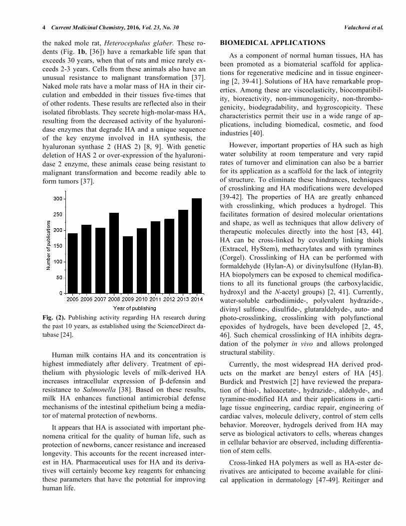
Interest in HA has been increased steadily. There has been an increase in the number of HA-related pub-lications (Fig. 2). Further increases are certain to occur following the dramatic observations made recently with
Table 2. Concentrations of HA (µg/g) in various organs in man and other animal species [18].
Organ or fluid Human Sheep Rabbit Rat
Umbilical cord 4100
Synovial fluid 1400-3600 540 3890
Dermis 200
Vitreous body 140-338 260 29
Lung 98-243 34
Kidney 93-113 30
Renal papillae 250
Renal cortex 4
Brain 35-115 54-76 74
Muscle 27
Intestine 44
Thoracic lymph 8.5-18 1-34 5.4
Liver 1.5 4
Aqueous humor 0.3-2.2 1.6-5.4 0.6-2.5 0.2
Urine 0.1-0.3
Lumbar celebrospinal fluid 0.02-0.32
Blood plasma/serum 0.01-0.1 0.12-0.31 0.019-0.086 0.048-0.26
4 Current Medicinal Chemistry, 2016, Vol. 23, No. 30 Valachová et al.
the naked mole rat, Heterocephalus glaber. These ro-dents (Fig. 1b, [36]) have a remarkable life span that exceeds 30 years, when that of rats and mice rarely ex-ceeds 2-3 years. Cells from these animals also have an unusual resistance to malignant transformation [37]. Naked mole rats have a molar mass of HA in their cir-culation and embedded in their tissues five-times that of other rodents. These results are reflected also in their isolated fibroblasts. They secrete high-molar-mass HA, resulting from the decreased activity of the hyaluroni-dase enzymes that degrade HA and a unique sequence of the key enzyme involved in HA synthesis, the hyaluronan synthase 2 (HAS 2) [8, 9]. With genetic deletion of HAS 2 or over-expression of the hyaluroni-dase 2 enzyme, these animals cease being resistant to malignant transformation and become readily able to form tumors [37].
Fig. (2). Publishing activity regarding HA research during the past 10 years, as established using the ScienceDirect da-tabase [24].
Human milk contains HA and its concentration is highest immediately after delivery. Treatment of epi-thelium with physiologic levels of milk-derived HA increases intracellular expression of β-defensin and resistance to Salmonella [38]. Based on these results, milk HA enhances functional antimicrobial defense mechanisms of the intestinal epithelium being a media-tor of maternal protection of newborns.
It appears that HA is associated with important phe-nomena critical for the quality of human life, such as protection of newborns, cancer resistance and increased longevity. This accounts for the recent increased inter-est in HA. Pharmaceutical uses for HA and its deriva-tives will certainly become key reagents for enhancing these parameters that have the potential for improving human life.
BIOMEDICAL APPLICATIONS
As a component of normal human tissues, HA has been promoted as a biomaterial scaffold for applica-tions for regenerative medicine and in tissue engineer-ing [2, 39-41]. Solutions of HA have remarkable prop-erties. Among these are viscoelasticity, biocompatibil-ity, bioreactivity, non-immunogenicity, non-thrombo-genicity, biodegradability, and hygroscopicity. These characteristics permit their use in a wide range of ap-plications, including biomedical, cosmetic, and food industries [40].
However, important properties of HA such as high water solubility at room temperature and very rapid rates of turnover and elimination can also be a barrier for its application as a scaffold for the lack of integrity of structure. To eliminate these hindrances, techniques of crosslinking and HA modifications were developed [39-42]. The properties of HA are greatly enhanced with crosslinking, which produces a hydrogel. This facilitates formation of desired molecular orientations and shape, as well as techniques that allow delivery of therapeutic molecules directly into the host [43, 44].
HA can be cross-linked by covalently linking thiols (Extracel, HyStem), methacrylates and with tyramines (Corgel). Crosslinking of HA can be performed with formaldehyde (Hylan-A) or divinylsulfone (Hylan-B). HA biopolymers can be exposed to chemical modifica-tions to all its functional groups (the carboxylacidic, hydroxyl and the N-acetyl groups) [2, 41]. Currently, water-soluble carbodiimide-, polyvalent hydrazide-, divinyl sulfone-, disulfide-, glutaraldehyde-, auto- and photo-crosslinking, crosslinking with polyfunctional epoxides of hydrogels, have been developed [2, 45, 46]. Such chemical crosslinking of HA inhibits degra-dation of the polymer in vivo and allows prolonged structural stability.
Currently, the most widespread HA derived prod-ucts on the market are benzyl esters of HA [45]. Burdick and Prestwich [2] have reviewed the prepara-tion of thiol-, haloacetate-, hydrazide-, aldehyde-, and tyramine-modified HA and their applications in carti-lage tissue engineering, cardiac repair, engineering of cardiac valves, molecule delivery, control of stem cells behavior. Moreover, hydrogels derived from HA may serve as biological activators to cells, whereas changes in cellular behavior are observed, including differentia-tion of stem cells.
Cross-linked HA polymers as well as HA-ester de-rivatives are anticipated to become available for clini-cal application in dermatology [47-49]. Reitinger and

Hyaluronan in Medical Practice Current Medicinal Chemistry, 2016, Vol. 23, No. 30 5
Lepperdinger [50] report the development of a new composite biomaterial based on HA-hydrogel contain-ing cross-linked fibronectin. A well-established system is thiolated HA (e.g. ExtracelTM, Glycosan BioSystems, Inc.). Köwitsch et al. [51] have examined the bioactiv-ity of HA derivatives such as aldehyde-HA and thiol-HA immobilized on model substrata such as amino-terminated surfaces or on gold. The major drug deliv-ery applications of HA, both biomedical applications and HA-based treatments are summarized in Table 3 [18].
VISCOSEPARATION
Cross-linked HA at various concentrations has ap-plications in a number of fields of medicine [21, 39, 41]. Modified HA-based solutions, e.g. Seprafilm Ad-hesion Barrier (Genzyme Corp.), are routinely applied to minimize postsurgical adhesion formation as an anti-adhesion material. Concerning to the design of cell non-adhesive surfaces, the mechanism of cell and sur-face interaction should be considered. In case of adher-ing cells to the surface of a material, several physico-chemical reactions occur between the cells and the ma-terial interface. Immediately after implanting a bioma-terial into an organism or into contact with cell culture environments, protein adsorption to its surface occurs, which mediates the cell adhesion and also provides signals to the cell through the cell adhesion receptors, mainly integrins [52]. Modifications of HA structure lead to lowered water solubility. Such materials thus become more stable in a physiological environment [53].
VISCOSUPPLEMENTATION
Osteoarthritis is a chronic, degenerative disease that commonly afflicts tissues such as joints, cartilages,
bones, synovium, ligaments and muscle [54, 55]. The concentration of HA in the SF of the normal human articular joint is 2.5-4.0 mg/mL. Under pathological conditions, the concentration of HA can fall precipi-tously to values below 1.2 mg/mL. The molar mass of HA is also reduced resulting in solutions that are mark-edly decreased in elasticity and viscosity.
Intra-articular applications of sterile HA solutions, termed viscosupplementation, provide analgesic, ana-bolic, chondro-protective and anti-inflammatory effects that diminish pain and disability and thus enhance function of joints, decrease cartilage degradation, and promote cartilage matrix biosynthesis. The use of vis-cosupplementation for other joints, such as shoulder, hip, and ankle that are currently under investigation, has introduced the term “visco-induction”, indicating that clinical efficacy can be long-term observed (sev-eral months), even though the mean-residence time for such intra-articularly introduced HA being only a few days [54-63].
Japan and Italy were the first countries, where vis-cosupplementation was applied in clinical practice (1980s), followed by Canada, Europe and the USA (1990s) [24]. Commercially available products for vis-cosupplementation contain sodium HA of different mo-lar masses. Synvisc®, also known as Hylan G-F 20 (Genzyme Corp, Cambridge, MA, USA), is a prepara-tion containing cross-linked HA (Hylan-B), while Hyalgan® (Fidia, Abano Terme, Italy) and Artz Dispo (Seikagaku, Tokyo, Japan) are the products containing HA of molar mass of 1 MDa. Other products are Monovisc (biscarbodiimide XL, Anika) and Gel-One (cinnamate XL, Seikagaku).
The proposed mechanism underlying the therapeutic effects of intra-articularly introduced HA derivatives on symptomatic osteoarthritic joints are as follows: 1)
Table 3. Summary of the drug delivery applications of HA [18].
Route Efficacy Therapeutic agents
Ophthalmic Elevated ocular persistence of drug, which can lead to in-creased bioavailability
Pilocarpine, tropicamide, timolol, gentimicin, tobramicin, arecaidine polyester, (S)-aceclidine
Nasal Bioadhesion resulting in increased bioavailability Xylometazoline, vasopressin, gentamicin
Pulmonary Absorption enhancer and dissolution rate modification Insulin
Parenteral Drug carrier and facilitator of liposomal entrapment Taxol, superoxide dismutase, human recombinant insu-lin-like growth factor, doxorubicin
Implant Dissolution rate modification Insulin
Gene Dissolution rate modification and protection Plasmid DNA/monoclonal antibodies

6 Current Medicinal Chemistry, 2016, Vol. 23, No. 30 Valachová et al.
maintenance of elasticity and viscous properties of the SF, 2) anti-inflammatory and anti-nociceptive effects and 3) normalization of HA synthesis by synoviocytes. The material acceptable for visco-supplementation should fulfill the following criteria: 1) tissue and blood compatibility, 2) permeability to low- and high-molar-mass substances, 3) rheological properties similar to those of normal SF and 4) slowing elimination rates in order to maintain extended protection [54, 60].
VISCOSURGERY
Ophthalmology. In the eye, HA is present in lacri-mal glands, the vitreous body, human tear fluid, cor-neal epithelium and also in conjunctiva [24]. HA is used as a viscoelastic gel to support healing and regen-eration of surgical wounds, to prevent damage of the endothelial layer of the cornea, to function as a lubricant, to eliminate physical damage of other tis-sues, to maintain operative space and depth of the ante-rior chamber. This is particularly important in anterior segment surgery, glaucoma surgery, and during corneal transplantation [42]. During implant surgery, when the intraocular pressure is high, HA reduces this pressure. Furthermore, HA increases corneal humidity due to elevated water retention on the corneal surface. Also, HA is used to treat syndrome of dry eye [24, 63].
Ocular tissue deterioration can occur on a heredity basis or as a result of other pathophysiological proc-esses. Currently increased attention is being paid to diabetic cataracts because of the rapid worldwide in-crease of diabetes mellitus. Healon® (sodium hyaluro-nate, a former product of the Swedish company Phar-macia) was for a long time the only available material for visco-surgical procedures. Today, similar products are used world-wide in surgery as a soft instrument to remove cataract, glaucoma surgery, for posterior seg-ment surgery, intraocular lens implantation and kerato-plasty. Furthermore, it can be used for repositioning and unrolling of the retina following detachment, lysis of anterior synechiae and for separation of tissues and adhesions mechanically. The use of HA with contact lenses in several types of applications have been re-ported in several studies [24]. Viscoat® by Cilco (USA) is a product applied for extraction of cataract and intra-ocular lens implantation and it is produced as a combi-nation of sodium HA and sodium chondroitin sulphate. Hyalistil®, a 0.2% HA solution produced by Sifi (Italy), is used to stabilize the tear film, to hydrate and lubri-cate the cornea and is desirable in increasing the com-fort when applying contact lenses. Blink Contacts, eye
drops produced by AMO (USA), are for users of con-tact lenses containing 0.15% HA [46, 64].
VISCO-AUGMENTATION
Otolaryngology. Properties of HA such as osmosis, viscoelasticity and space-filling ability are critical for voice production. HA directly affects the thickness and viscosity of vocal folds. Moreover, HA derivatives can be applied in visco-augmentation of vocal cords, for the treatment of glottal insufficiency and the repair of injured or scarred vocal cords. A short residence time of HA is however a main disadvantage of applying HA as a lamina propria bioimplant for the treatment of vo-cal fold disorders. The HA mean-residence-time in the rabbit vocal fold is only 3-5 days. The solution is to modify the molecular structure of HA to prolong the material residence time [40]. One of the simplest hy-drogels used for vocal fold augmentation is the divinyl-sulfone cross-linked HA derivative Hylan-B which, in animal models, is found to be anti-inflammatory, non-antigenic and non-toxic.
To date, Hylan-B and its derivatives serve as a space filling hydrogel, whereas their only possible ap-plication is by injection. Several clinical studies involv-ing rheological studies and animal models support the mechanism of HA derivatives to be beneficial to re-place a lamina propria for vocal fold scars and sulci [65, 66].
COSMETIC APPLICATIONS
About 50% of the total HA in humans occurs in skin [67]. HA therefore is a reasonable matrix component for enhancing dermal regeneration and augmentation [68]. The excellent moisturizing properties of HA are the basis of its inclusion in skin-care products [24, 40, 47].
Materials for deep augmentation injection into skin are temporary and permanent/long-term. Long-term or permanent injectable materials include calcium hy-droxyapatite, autologous fat, (Radiesse™, Merz Phar-ma GmbH, Germany), polydimethylsiloxane (or par-ticulate silicone), and polytef paste (Teflon™). Among temporary injection materials rank collagen-based products (Cymetra™, Zyplast™, Cosmoplast/Cosmo-derm™) bovine gelatin (Gelfoam™, Surgifoam™), and 1-carboxymethylcellulose (Radiesse Voice Gel™, Merz Aestetics, Germany). These materials differ in the endurance of integration, in their biocompatibility and specific viscoelastic properties. The injectable ma-terials based on HA include Hylaform® (Genzyme

Hyaluronan in Medical Practice Current Medicinal Chemistry, 2016, Vol. 23, No. 30 7
Corp., MA, USA) and Restylane™ (Q-med, Sweden) [45, 65].
VISCOPROTECTION
Wound healing. HA stimulates wound healing as can be demonstrated for tendon, bone, corneal, diabetic foot, nasal mucosal and venous leg ulcers [21]. HA participates in several stages of wound healing [69]. In an early stage, high-molar-mass HA is bound to fi-brinogen during clot formation. In the adult, wound healing frequently results in scar formation. However, in fetal wounds, scar formation is inhibited by the pres-ence of high-molar-mass HA [70]. This was demon-strated in experimental fetal rabbit and fetal sheep models as well as in term infants following mid-gestation in utero surgery [71].
In 2011, Cutting [72] reported that in spite of posi-tive effects of several HA-containing products on wound healing, some adverse effects were also ex-pressed. This may be explained by possible wound in-fections (empirical observations) associated with appli-cation of some HA-based dressings. For this reason, the current author [72] reports application of a newly de-veloped wound preparation that includes HA associated with an antimicrobial iodine-containing compound. This is a novel and innovative solution that has the po-tential to provide improved clinical outcomes for com-plex wound-healing situations. Fragmentation of high-molar-mass HA occurs naturally within the wound. This leads to altered properties of the healing material that affects the potential benefits of topical applica-tions. One option, in order to overcome this problem is esterification of the HA moieties. The product thus be-comes more resistant to its degradation by hyaluroni-dases. However, such esterification increases the hy-drophobicity of the HA-based material.
Another approach also uses the aforementioned HA-iodine complex. Contipro Pharma Inc. (Czech Repub-lic) produces Hyiodine®, a wound-healing product for a wide range of chronic and acute wounds. Its efficacy is based on the synergistic effect of HA and iodine [73].
Combinations of HA with other biomaterials for particular wound healing situations have been studied in the past few years. HA conjugated with gelatin or collagen in the presence or absence of growth factors such as EGF (epidermal growth factor) or bFGF (basic fibroblast growth factor) showed promising results in experimental wound-healing models [74-81]. The an-timicrobial HA-based wound dressings based on HA are formulated combining HA with antibiotics, nano-
silver or mixed with antimicrobial polymers such as chitosan [82-85].
Hunt and Grover [86] report on the application of biopolymer gel encapsulation in regenerative medicine. Properties and applications of biopolymer gels based on alginate, fibrin, collagen, agarose, gelatin and HA are described. Table 4 summarizes some of the func-tions of HA in the several steps of wound healing [69].
HYALURONAN LABELING FOR DIAGNOSTIC PURPOSES
Lindqvist et al. [87] developed a HA-loading test for assessment of HA kinetics to use it in patients with liver and joint diseases. The test describes the metabo-lism of HA but cannot define the specific contribution of different organs. A method for labelling of HA with the short-lived positron-emitting radionuclide 11C was applied in healthy individuals and in patients suffering from liver diseases. The finding is that positron elec-tron topography with [11C] HA may be an accurate method to assess liver dysfunction, when endothelial cell function is impaired.
Near-infrared (NIR) fluorescence was used for func-tional lymphatic imaging in the abdomen and anterior hindlimb of anesthetized, intact Yorkshire swine [88]. The results show the capability to image the immediate trafficking of indocyanine green from the plexus, through the vessels and lymphangions, and to the su-perficial mammary, sub iliac, and middle iliac lymph nodes, which were located as deep as 3 cm beneath the tissue surface. The results suggest that microgram quantities of NIR optical imaging agents and their con-jugates have a potential to image lymph function in patients suffering from lymph-related disorders.
Jadhav et al. [89] synthesized a macrocyclic 68Ga-NOTA-chelated oligonucleotide-HA conjugates using a solid supported technique to introduce NOTA-chelator (NOTA - 1,4,7-triazacyclononane-N,N',N''-triacetic acid) into the 3′-terminus and a copper-free strain promoted azide alkyne cycloaddition to HA/olig-onucleotide conjugation. As a method positron emis-sion tomography was used in healthy rats to monitor distribution kinetic studies and a potential of HA-induced targeting of oligonucleotides into rats afflicted with myocardial infarction was determined.
DRUG DELIVERY A number of studies report that HA facilitates the
prolonged residence and the increased bioavailability of certain drug molecules. Typical examples are pilo-carpine and vasopressin. HA of higher size rather than

8 Current Medicinal Chemistry, 2016, Vol. 23, No. 30 Valachová et al.
Table 4. Functions of HA during wound healing [69].
Phases Effects
Binding to fibrinogen to initiate clotting pathway
Ensuring inflammatory cell migration
Creating edema to allow cell infiltration
Inflammatory phase
Inhibiting neutrophil migration to dampen inflammatory response
Accumulating of fibroblasts to wound site
Filling in gaps of newly formed extracellular matrix, creating cushioning and structural organization
Stimulating matrix metaloproteinases for angiogenesis
Proliferative phase
Promoting keratinocyte migration and proliferation
Remodeling phase Contribution to normal and pathological scarring
Table 5. Practical applications of visco-elastic solutions of HA or its gels [18, 40].
Application Treatment
Viscosupplementation To replace or supplement tissue fluids, such as replacement of SF in painful arthritis, and to relieve pain
Viscosurgery To protect delicate tissues and provide space during surgical manipulations, as in ophthalmological sur-geries
Viscoaugmentation To fill and augment tissue spaces, as in skin, sphincter muscles, vocal and pharyngeal tissues
Viscoseparation To separate connective tissue surfaces traumatized by surgical procedures or injury, in order to prevent adhesions and excessive scar formation
Viscoprotection To protect healthy, wounded, or injured tissue surfaces from dryness or noxious environmental agents, and to promote the healing of such surfaces
Drug delivery Matrix and tissue engineering
the low-molar-mass HA species supports increased drug bioavailability. HA as a non-immunogenic sub-stance is used in parental/pulmonary drug delivery sys-tems to allow constant release and longer retention of the therapeutic agents. For example, the anti-cancer drug, taxol, when linked to HA was observed to target breast cancer, human colon and ovarian cell lines in vitro selectively. Furthermore, HA was used to dis-cover an implantable delivery system for long-term delivery of anti-inflammatory and antibiotic drugs in vivo [21].
HA can be also included in nasal, ophthalmic and parenteral drug delivery. Novel applications, including pulmonary implantation and gene delivery, are summa-rized in Table 5 [18, 40]. Kogan et al. [40] reported that HA can be either directly conjugated to drugs or used for preparation of microcapsules to enhance drug de-livery. HA is also involved in improving biocompati-bility of chitosan microspheres, which function as cap-sules for drug delivery. It is apparent that HA has mul-
tiple uses, both direct and indirect, in the pharmaceuti-cal industry.
CONCLUSION New insights into mammalian longevity and malig-
nancy were discovered recently in research on the na-ked mole rat. These observations can play key roles in future treatment of human cancer. They will also facili-tate approaches to improve quality of life while extend-ing human longevity. The new treatment modalities and therapeutic applications will involve the production of high-molar-mass HAs. These considerations promp-ted the current overview, anticipating opportunities for new techniques for preparation and isolation of HA, for modifications of structure that improve availability and resistance to degradation. Such materials will stimulate many new investigations.
In the future, more attention will be devoted to en-zymatic production of HA. A huge number of modifi-cations of HA can be performed with the aim of en-

Hyaluronan in Medical Practice Current Medicinal Chemistry, 2016, Vol. 23, No. 30 9
hancing the quality of the material for medicine, phar-maceuticals, cosmetics as well as for industrial produc-tion. We are only now beginning to appreciate the large numbers of applications for this versatile molecule as well as the innumerable modifications possible with this multifaceted, highly ionic polymer.
CONFLICT OF INTEREST
The authors confirm that this article content has no conflict of interest.
ACKNOWLEDGEMENTS
This work was supported by grant VEGA 2/0065/ 15. The authors report no conflict of interest.
REFERENCES [1] Menaa, T.; Menaa, A.; Menaa, B. Hyaluronic acid and de-
rivatives for tissue engineering. J. Biotechnol. Biomater., 2011, 51, 17.
[2] Burdick, J.A.; Prestwich, G.D. Hyaluronic acid hydrogels for biomedical applications. Adv. Mater., 2011, 23(12), H41-H56.
[3] Lee, J.Y.; Spicer, A.P. Hyaluronan: a multifunctional, me-gaDalton, stealth molecule. Curr. Opin. Cell Biol., 2000, 12(5), 581-586.
[4] Davies, A.; Gormally, J.; Wyn-Jones, E.; Wedlock, D.J.; Phillips, G.O. A study of factors influencing hydration of sodium hyaluronate from compressibility and high-preci-sion densimetric measurements. Biochem. J., 1983, 213(2), 363-369.
[5] Almond, A.; Sheehan, J.K. Predicting the molecular shape of polysaccharides from dynamic interactions with water. Glycobiology, 2003, 13(4), 255-264.
[6] Granger, H.J.; Laine, G.A.; Barnes, G.E.; Lewis, R.E. Car-diovascular responses to elevation of intra-abdominal hy-drostatic pressure. Am. J. Physiol., 1985, 248, R208-13.
[7] Stern, R.; Jedrzejas, M.J. Hyaluronidases: their genomics, structures, and mechanisms of action. Chem. Rev., 2006, 106(3), 818-839.
[8] Stern, R. Devising a pathway for hyaluronan catabolism: are we there yet? Glycobiology, 2003, 13(12), 105R-115R.
[9] Stern, R. Hyaluronan catabolism: a new metabolic pathway. Eur. J. Cell Biol., 2004, 83(7), 317-325.
[10] Cowman, M.K.; Matsuoka, S. Experimental approaches to hyaluronan structure. Carbohydr. Res., 2005, 340(5), 791-809.
[11] Meyer, K.; Palmer, J. The polysaccharide of the vitreous humor. J. Biol. Chem., 1935, 107, 629-634.
[12] Csoka, A.B.; Stern, R. Hypotheses on the evolution of hyaluronan: a highly ironic acid. Glycobiology, 2013, 23(4), 398-411.
[13] Margolis, R.U.; Margolis, R.K.; Chang, L.B.; Preti, C. Gly-cosaminoglycans of brain during development. Biochemis-try, 1975, 14(1), 85-88.
[14] Jenkins, H.G.; Bachelard, H.S. Developmental and age-related changes in rat brain glycosaminoglycans. J. Neuro-chem., 1988, 51(5), 1634-1640.
[15] Fraser, J.R.; Laurent, T.C.; Laurent, U.B. Hyaluronan: its nature, distribution, functions and turnover. J. Intern. Med., 1997, 242(1), 27-33.
[16] Price, R.D.; Berry, M.G.; Navsaria, H.A. Hyaluronic acid: the scientific and clinical evidence. J. Plast. Reconstr. Aes-thet. Surg., 2007, 60(10), 1110-1119.
[17] Hascall, V.C.; Majors, A.K.; De La Motte, C.A. Intracellu-lar hyaluronan: A new frontier for inflammation? Biochim. Biophys. Acta., 2004, 1673, 3-12.
[18] Tamer, T.M. Engineering of Polymer and Chemical Com-plexity; Frocke, W.W., Ed.; Apple Academic Press: Water-town, 2014, pp. 109-143.
[19] Turley, E.A. Hyaluronan and cell locomotion. Cancer Me-tastasis Rev., 1992, 11(1), 21-30.
[20] Turley, E.A.; Noble, P.W.; Bourguignon, L.Y. Signaling properties of hyaluronan receptors. J. Biol. Chem., 2002, 277(7), 4589-4592.
[21] Girish, K.S.; Kemparaju, K. The magic glue hyaluronan and its eraser hyaluronidase: a biological overview. Life Sci., 2007, 80(21), 1921-1943.
[22] Papakonstantinou, E.; Roth, M.; Karakiulakis, G. Hyaluronic acid: A key molecule in skin aging. Derma-toendocrinol, 2012, 4(3), 253-258.
[23] Erickson, M.; Stern, R. Chain gangs: New aspects of hyaluronan metabolism. Int. Biochem. Res., 2012, 2012, 19.
[24] Necas, J.; Bartosikova, L.; Brauner, P. Hyaluronic acid (hyaluronan): A review. Vet. Med., 2008, 53, 397-411.
[25] Jiang, D.; Liang, J.; Noble, P.W. Hyaluronan in tissue in-jury and repair. Annu. Rev. Cell Dev. Biol., 2007, 23, 435-461.
[26] Jiang, D.; Liang, J.; Noble, P.W. Hyaluronan as an immune regulator in human diseases. Physiol. Rev., 2011, 91(1), 221-264.
[27] Itano, N.; Kimata, K. Altered hyaluronan biosynthesis in cancer progression. Semin. Cancer Biol., 2008, 18(4), 268-274.
[28] Itano, N.; Zhuo, L.; Kimata, K. Impact of the hyaluronan-rich tumor microenvironment on cancer initiation and pro-gression. Cancer Sci., 2008, 99(9), 1720-1725.
[29] Zhang, L.; Underhill, C.B.; Chen, L. Hyaluronan on the surface of tumor cells is correlated with metastatic behav-ior. Cancer Res., 1995, 55(2), 428-433.
[30] Soltés, L.; Kogan, G.; Stankovská, M.; Mendichi, R.; Rychlý, J.; Schiller, J.; Gemeiner, P. Degradation of high-molar-mass hyaluronan and characterization of fragments. Biomacromolecules, 2007, 8(9), 2697-2705.
[31] Xu, H.; Ito, T.; Tawada, A.; Maeda, H.; Yamanokuchi, H.; Isahara, K.; Yoshida, K.; Uchiyama, Y.; Asari, A. Effect of hyaluronan oligosaccharides on the expression of heat shock protein 72. J. Biol. Chem., 2002, 277(19), 17308-17314.
[32] Schwertfeger, K.L.; Cowman, M.K.; Telmer, P.G.; Turley, E.A.; McCarthy, J.B. Hyaluronan, inflammation, and breast cancer progression. Front. Immunol., 2015, 6, 236.
[33] Ghatak, S.; Misra, S.; Toole, B.P. Hyaluronan oligosaccha-rides inhibit anchorage-independent growth of tumor cells by suppressing the phosphoinositide 3-kinase/Akt cell sur-vival pathway. J. Biol. Chem., 2002, 277(41), 38013-38020.
[34] Urakawa, H.; Nishida, Y.; Knudson, W.; Knudson, C.B.; Arai, E.; Kozawa, E.; Futamura, N.; Wasa, J.; Ishiguro, N. Therapeutic potential of hyaluronan oligosaccharides for bone metastasis of breast cancer. J. Orthop. Res., 2012, 30(4), 662-672.
[35] Stern, R.; Asari, A.R.; Sugahara, K.N. Size- specific frag-ments of hyaluronan: An information-rich system. Eur. J. Cell Biol., 2006, 85, 699-715.
[36] http://quotesgram.com/mole-rat-quotes-quotes/, [37] Tian, X.; Azpurua, J.; Hine, C.; Vaidya, A.; Myakishev-
Rempel, M.; Ablaeva, J.; Mao, Z.; Nevo, E.; Gorbunova, V.; Seluanov, A. High-molecular-mass hyaluronan mediates

10 Current Medicinal Chemistry, 2016, Vol. 23, No. 30 Valachová et al.
the cancer resistance of the naked mole rat. Nature, 2013, 499(7458), 346-349.
[38] Hill, D.R.; Rho, H.K.; Kessler, S.P.; Amin, R.; Homer, C.R.; McDonald, C.; Cowman, M.K.; de la Motte, C.A. Human milk hyaluronan enhances innate defense of the in-testinal epithelium. J. Biol. Chem., 2013, 288(40), 29090-29104.
[39] Collins, M.N.; Birkinshaw, C. Hyaluronic acid based scaf-folds for tissue engineering--a review. Carbohydr. Polym., 2013, 92(2), 1262-1279.
[40] Kogan, G.; Soltés, L.; Stern, R.; Gemeiner, P. Hyaluronic acid: a natural biopolymer with a broad range of biomedical and industrial applications. Biotechnol. Lett., 2007, 29(1), 17-25.
[41] Fakhari, A.; Berkland, C. Applications and emerging trends of hyaluronic acid in tissue engineering, as a dermal filler and in osteoarthritis treatment. Acta Biomater., 2013, 9(7), 7081-7092.
[42] Volpi, N.; Schiller, J.; Stern, R.; Soltés, L. Role, metabo-lism, chemical modifications and applications of hyaluro-nan. Curr. Med. Chem., 2009, 16(14), 1718-1745.
[43] Krüger, J.P.; Ketzmar, A.K.; Endres, M.; Pruss, A.; Siclari, A.; Kaps, C. Human platelet-rich plasma induces chondro-genic differentiation of subchondral progenitor cells in po-lyglycolic acid-hyaluronan scaffolds. J. Biomed. Mater. Res. B Appl. Biomater., 2014, 102(4), 681-692.
[44] Mathews, S.; Bhonde, R.; Gupta, P.K.; Totey, S. Novel biomimetic tripolymer scaffolds consisting of chitosan, col-lagen type 1, and hyaluronic acid for bone marrow-derived human mesenchymal stem cells-based bone tissue engineer-ing. J. Biomed. Mater. Res. B Appl. Biomater., 2014, 102(8), 1825-1834.
[45] Allison, D.D.; Grande-Allen, K.J. Review. Hyaluronan: a powerful tissue engineering tool. Tissue Eng., 2006, 12(8), 2131-2140.
[46] Prestwich, G.D.; Kuo, J.W. Chemically-modified HA for therapy and regenerative medicine. Curr. Pharm. Biotech-nol., 2008, 9(4), 242-245.
[47] Stern, R.; Maibach, H.I. Hyaluronan in skin: aspects of aging and its pharmacologic modulation. Clin. Dermatol., 2008, 26(2), 106-122.
[48] Mineo, A.; Suzuki, R.; Kuroyanagi, Y. Development of an artificial dermis composed of hyaluronic acid and collagen. J. Biomater. Sci. Polym. Ed., 2013, 24(6), 726-740.
[49] Sawa, M.; Kuroyanagi, Y. Potential of a cryopreserved cultured dermal substitute composed of hyaluronic acid and collagen to release angiogenic cytokine. J. Biomater. Sci. Polym. Ed., 2013, 24(2), 224-238.
[50] Reitinger, S.; Lepperdinger, G. Hyaluronan, a ready choice to fuel regeneration: a mini-review. Gerontology, 2013, 59(1), 71-76.
[51] Köwitsch, A.; Yang, Y.; Ma, N.; Kuntsche, J.; Mäder, K.; Groth, T. Bioactivity of immobilized hyaluronic acid de-rivatives regarding protein adsorption and cell adhesion. Biotechnol. Appl. Biochem., 2011, 58(5), 376-389.
[52] Liha, E.; Ohb, S.H.; Jounga, Y.K.; Lee, J.H.; Han, D.K. Polymers for cell/tissue anti-adhesion. Prog. Polym. Sci., 2015, 44, 28-61.
[53] Diamond, M.P.; Burns, E.L.; Accomando, B.; Mian, S.; Holmdahl, L. Seprafilm(®) adhesion barrier: (2) a review of the clinical literature on intraabdominal use. Gynecol. Surg., 2012, 9(3), 247-257.
[54] McArthur, B.A.; Dy, C.J.; Fabricant, P.D.; Valle, A.G. Long term safety, efficacy, and patient acceptability of hyaluronic acid injection in patients with painful os-teoarthritis of the knee. Patient Prefer. Adherence, 2012, 6, 905-910.
[55] Gigante, A.; Callegari, L. The role of intra-articular hyaluronan (Sinovial) in the treatment of osteoarthritis. Rheumatol. Int., 2011, 31(4), 427-444.
[56] Soltes, L.; Mendichi, R.; Kogan, G.; Mach, M. Associating hyaluronan derivatives: a novel horizon in viscosupplemen-tation of osteoarthritic joints. Chem. Biodivers., 2004, 1(3), 468-472.
[57] Soltes, L.; Steiner, B.; Machova, E.; Kogan, G.; Bystricky, S.; Mendichi, R.; Bauer, V.; Mach, M.; Alfoldi, J.; Strati-lova, E. Clathrate complexes formed by hyaluronic acid de-rivatives and use tehreof as pharmaceuticals. Australian Patent, 2001252180, 2005. B1 European Patent, EP 1272530, 2006.7563824 B2 US Patent, 2009. Canadian Pat-ent, CA 2402421 2009.
[58] Rodell, C.B.; Kaminski, A.L.; Burdick, J.A. Rational design of network properties in guest-host assembled and shear-thinning hyaluronic acid hydrogels. Biomacromolecules, 2013, 14(11), 4125-4134.
[59] Strauss, E.J.; Hart, J.A.; Miller, M.D.; Altman, R.D.; Rosen, J.E. Hyaluronic acid viscosupplementation and osteoarthri-tis: current uses and future directions. Am. J. Sports Med., 2009, 37(8), 1636-1644.
[60] Van Den Bekerom, M.P.; Mylle, G.; Rys, B.; Mulier, M. Viscosupplementation in symptomatic severe hip os-teoarthritis: a review of the literature and report on 60 pa-tients. Acta Orthop. Belg., 2006, 72(5), 560-568.
[61] Volpi, N.; Schiller, J.; Stern, R.; Soltés, L. Role, metabo-lism, chemical modifications and applications of hyaluronan. Curr. Med. Chem., 2009, 16(14), 1718-1745.
[62] Migliore, A.; Giovannangeli, F.; Bizzi, E.; Massafra, U.; Alimonti, A.; Laganà, B.; Diamanti Picchianti, A.; Ger-mano, V.; Granata, M.; Piscitelli, P. Viscosupplementation in the management of ankle osteoarthritis: a review. Arch. Orthop. Trauma Surg., 2011, 131(1), 139-147.
[63] Schiller, J.; Volpi, N.; Hrabárová, E. Hyaluronic acid: a natural biopolymer. In: Biopolymers: Biomedical and Envi-ronmental Applications Scrivener, S.; Eds.; Publishing, 2008, LLC, 334.
[64] Williams, D.L.; Mann, B.K. Efficacy of a crosslinked hyaluronic acid-based hydrogel as a tear film supplement: a masked controlled study. PLoS One, 2014, 9(6), e99766.
[65] Gaston, J.; Thibeault, S.L. Hyaluronic acid hydrogels for vocal fold wound healing. Biomatter, 2013, 3(1), 17.
[66] Mallur, P.S.; Rosen, C.A. Vocal fold injection: review of indications, techniques, and materials for augmentation. Clin. Exp. Otorhinolaryngol., 2010, 3(4), 177-182.
[67] Reed, R.K.; Lilja, K.; Laurent, T.C. Hyaluronan in the rat with special reference to the skin. Acta Physiol. Scand., 1988, 134(3), 405-411.
[68] Meyer, L.J.; Stern, R. Age-dependent changes of hyaluronan in human skin. J. Invest. Dermatol., 1994, 102(3), 385-389.
[69] Frenkel, J.S. The role of hyaluronan in wound healing. Int. Wound. J., 2013, 2012, 17.
[70] Longaker, M.T.; Chiu, E.S.; Adzick, N.S.; Stern, M.; Harrison, M.R.; Stern, R. Studies in fetal wound healing. V. A prolonged presence of hyaluronic acid characterizes fetal wound fluid. Ann. Surg., 1991, 213(4), 292-296.
[71] Longaker, M.T.; Whitby, D.J.; Adzick, N.S.; Cromble-holme, T.M.; Langer, J.C.; Duncan, B.W.; Bradley, S.M.; Stern, R.; Ferguson, M.W.; Harrison, M.R. Studies in fetal wound healing, VI. Second and early third trimester fetal wounds demonstrate rapid collagen deposition without scar formation. J. Pediatr. Surg., 1990, 25(1), 63-68.
[72] Cutting, K.F. Wound healing through synergy of hyaluronan and an iodine complex. J. Wound Care, 2011, 20(9), 424-430, 426, 428-430.

Hyaluronan in Medical Practice Current Medicinal Chemistry, 2016, Vol. 23, No. 30 11
[73] Hyiodine®. http://www.contipro.com/woundhealing/ heal-ing-products, 2013.
[74] Cai, S.; Liu, Y.; Zheng Shu, X.; Prestwich, G.D. Injectable glycosaminoglycan hydrogels for controlled release of hu-man basic fibroblast growth factor. Biomaterials, 2005, 26(30), 6054-6067.
[75] Liu, Y.; Cai, S.; Shu, X.Z.; Shelby, J.; Prestwich, G.D. Re-lease of basic fibroblast growth factor from a crosslinked glycosaminoglycan hydrogel promotes wound healing. Wound Repair Regen., 2007, 15(2), 245-251.
[76] Matsumoto, Y.; Arai, K.; Momose, H.; Kuroyanagi, Y. Development of a wound dressing composed of a hyaluronic acid sponge containing arginine. J. Biomater. Sci. Polym. Ed., 2009, 20(7-8), 993-1004.
[77] Matsumoto, Y.; Kuroyanagi, Y. Development of a wound dressing composed of hyaluronic acid sponge containing arginine and epidermal growth factor. J. Biomater. Sci. Po-lym. Ed., 2010, 21(6-7), 715-726.
[78] Kondo, S.; Niiyama, H.; Yu, A.; Kuroyanagi, Y. Evaluation of a wound dressing composed of hyaluronic acid and col-lagen sponge containing epidermal growth factor in diabetic mice. J. Biomater. Sci. Polym. Ed., 2011, 3.
[79] Kondo, S.; Kuroyanagi, Y. Development of a wound dress-ing composed of hyaluronic acid and collagen sponge with epidermal growth factor. J. Biomater. Sci. Polym. Ed., 2012, 23(5), 629-643.
[80] Yamamoto, A.; Shimizu, N.; Kuroyanagi, Y. Potential of wound dressing composed of hyaluronic acid containing epidermal growth factor to enhance cytokine production by fibroblasts. J. Artif. Organs, 2013, 16(4), 489-494.
[81] Yu, A.; Niiyama, H.; Kondo, S.; Yamamoto, A.; Suzuki, R.; Kuroyanagi, Y. Wound dressing composed of hyaluronic acid and collagen containing EGF or bFGF: comparative culture study. J. Biomater. Sci. Polym. Ed., 2013, 24(8), 1015-1026.
[82] Correia, C.R.; Moreira-Teixeira, L.S.; Moroni, L.; Reis, R.L.; van Blitterswijk, C.A.; Karperien, M.; Mano, J.F. Chi-
tosan scaffolds containing hyaluronic acid for cartilage tis-sue engineering. Tissue Eng. Part C Methods, 2011, 17(7), 717-730.
[83] Lu, H.D.; Zhao, H.Q.; Wang, K.; Lv, L.L. Novel hyaluronic acid-chitosan nanoparticles as non-viral gene delivery vec-tors targeting osteoarthritis. Int. J. Pharm., 2011, 420(2), 358-365.
[84] Mathews, S.; Bhonde, R.; Gupta, P.K.; Totey, S. A novel tripolymer coating demonstrating the synergistic effect of chitosan, collagen type 1 and hyaluronic acid on osteogenic differentiation of human bone marrow derived mesenchy-mal stem cells. Biochem. Biophys. Res. Commun., 2011, 414(1), 270-276.
[85] Park, H.; Choi, B.; Hu, J.; Lee, M. Injectable chitosan hyaluronic acid hydrogels for cartilage tissue engineering. Acta Biomater., 2013, 9(1), 4779-4786.
[86] Hunt, N.C.; Grover, L.M. Cell encapsulation using bio-polymer gels for regenerative medicine. Biotechnol. Lett., 2010, 32(6), 733-742.
[87] Lindqvist, U.; Westerberg, G.; Bergström, M.; Torsteindot-tir, I.; Gustafson, S.; Sundin, A.; Lööf, L.; Långström, B. [11C]Hyaluronan uptake with positron emission tomogra-phy in liver disease. Eur. J. Clin. Invest., 2000, 30(7), 600-607.
[88] Sharma, R.; Wang, W.; Rasmussen, J.C.; Joshi, A.; Hous-ton, J.P.; Adams, K.E.; Cameron, A.; Ke, S.; Kwon, S.; Mawad, M.E.; Sevick-Muraca, E.M. Quantitative imaging of lymph function. Am. J. Physiol. Heart Circ. Physiol., 2007, 292(6), H3109-H3118.
[89] Jadhav, S.; Käkelä, M.; Mäkilä, J.; Kiugel, M.; Liljenbäck, H.; Virta, J.; Poijärvi-Virta, P.; Laitala-Leinonen, T.; Kytö, V.; Jalkanen, S.; Saraste, A.; Roivainen, A.; Lönnberg, H.; Virta, P. Synthesis and in vivo PET imaging of hyaluronan conjugates of oligonucleotides. Bioconjug. Chem., 2016, 27(2), 391-403.