review article - hindawi publishing...
TRANSCRIPT

Hindawi Publishing CorporationInternational Journal of Antennas and PropagationVolume 2012, Article ID 807165, 14 pagesdoi:10.1155/2012/807165
Review Article
Ingestible Wireless Capsule Technology: A Review ofDevelopment and Future Indication
M. R. Basar,1 F. Malek,2 Khairudi M. Juni,3 M. Shaharom Idris,3 and M. Iskandar M. Saleh3
1 School of Computer and Communication Engineering, Universiti Malaysia Perlis (UniMAP), 02000 Kuala Perlis, Perlis, Malaysia2 School of Electrical Systems Engineering, Universiti Malaysia Perlis (UniMAP), 02600 Arau, Perlis, Malaysia3 Electrical Engineering Department, Politeknik Tuanku Syed Sirajuddin (PTSS), 02600 Arau, Perlis, Malaysia
Correspondence should be addressed to M. R. Basar, [email protected]
Received 27 July 2012; Accepted 3 December 2012
Academic Editor: Ahmed A. Kishk
Copyright © 2012 M. R. Basar et al. This is an open access article distributed under the Creative Commons Attribution License,which permits unrestricted use, distribution, and reproduction in any medium, provided the original work is properly cited.
Ingestible wireless capsule endoscopy (WCE) is the one and only painless, effective, novel, diagnostic technology for inspecting theentire gastrointestinal (GI) tract for various diseases, such as obscure gastrointestinal bleeding (OGIB), tumors, cancer, Crohn’sdisease, and celiac disease. Since the development of this technology, several companies have made remarkable improvements intheir clinical products, but there are still some limitations that relate to the use of conventional wired endoscopy. Some of themajor limitations that currently impede its wider application include its inability to repeat the view of critical areas, working timeconstraints, and poor image resolution. Many research groups currently are working on ways to solve these limitations. Presently,developing the ability to control the movement of the capsule, increasing its image transmission speed, and obtaining high-qualityimages are the main issues in the research area. A complex capsule with some therapeutic tools for the treatment of diseases of theGI tract also is at the beginning of development for the next generation of an active medical robot. In this paper, we report thestatus of several activities related to WCE, including improvement of capsule technology, research progress, technical challenges,and key indicators concerning the next-generation, active, medical robot.
1. Introduction
Angiography, ultrasonography, X-radiography, computertomography (CT), and magnetic resonance imaging (MRI)are conventional, indirect technologies for examining GItract diseases, such as obscure gastrointestinal bleeding(OGIB), tumors, cancer, Crohn’s disease, and celiac disease.Regrettably, these technologies have low diagnostic yieldsbecause of their inability to show the wall of the GI tract.Another typical technology, probe endoscopy, is used fordiagnosing diseases of the GI tract, but, in addition to beingpainful and creating discomfort, it is incapable of reachingsome critical locations in the GI tract.
In 2000, Iddan et al. first developed a wireless videocapsule endoscopy system [1, 2] that patients could swallow,but its development has been started since long ago. The firstattempt at endoscopic examination was conducted in 1806by Philipp Bozzini. The effective visualization of the GI tractbegan with the development of typical wire endoscopy, such
as rigid-wire endoscopy, flexible endoscopy, and fiberopticpush endoscopy. William Beaumont, an army surgeon, firstintroduced an endoscope into a human in 1822 and, 30years later, the effective, rigid-wire endoscope was developed[3–5]. The use of the rigid-wire endoscope was limitedto the curly, upper anatomical area of the digestive path.The flexible endoscope provided a unique solution for thatproblem, and it led to the development of the fiberopticendoscope in 1957 [6–8]. However, in addition to the painand discomfort they caused, the main problem associatedwith these enteroscopes was their limited insertion into theGI tract, only reaching as far as the upper portion of the smallbowel [9].
In the 1950s, the first wireless capsule was developed,but it had very limited capabilities in that it could onlymeasure a few physiological parameters of the GI tract,such as temperature, pH, and pressure. This device wasknown as an endoradiosonder or “radio pill” (sometimesreferred to as “gutnick”). The wireless capsule of this

2 International Journal of Antennas and Propagation
period was a tiny, swallowable transmitter either with pH,pressure, or temperature sensors that created variations inthe L-C oscillator’s output frequency of a tiny transmitteraccording to the sensor’s output [10–13]. Heidelberg MedicalInc. in Germany first developed the pH parameter capsulesystem. This capsule was 15.4 mm long and 7.1 mm indiameter with a battery that had a six-hour lifetime, aradio frequency transmitter, and an electrode, and it wasused for diagnosing hypochlorhydria and achlorhydria [14].Later, the American SmartPill Company developed a medicalproduct, known as SmartPill, which combined those threesensors for measuring pressure, temperature, and pH. TheSmartPill is approved by the Food and Drug Administration(FDA). The SmartPill system consisted of four subsystems,that is, (1) the ingestible capsule itself, containing pressure,temperature, and pH sensors, along with a battery and atiny radio transmitter (with a frequency of 434 MHz); (2)a portable receiver inside a jacket for receiving sensor data;(3) MotiliGI monitoring software; and (4) a receiver dockingstation [15].
In 1981, Iddan first developed the concept of wirelessvideo capsule endoscopy to see the wall of the GI tractpainlessly, but the state of the development of the requiredtechnology deterred the realization of this concept. Fourteenyears later, Swain et al. (1997) developed several prototypesof a capsule endoscopy system and successfully conducted avast number of experiments on postmortem and live pigs[16]. In those prototypes, they used a miniature charge-coupled device (CCD) camera, a video processor, a 10 mWmicrowave transmitter with a 1.5 cm dipole antenna, a lightsource, and a battery. In 2000, the introduction of the low-power, complementary, metal oxide semiconductor-based(CMOS-based) image sensor and ASIC made the videocapsule possible [1]. Given Imaging, Ltd. (Yoqneam, Israel)announced the first clinical product, M2A (mouth to anus),for examination of the small bowel, and the product wasapproved by the US FDA in the same year [17]. Due tothe dissimilarities of different areas of the GI tract, GivenImaging, Ltd. launched its esophagus-specific and colon-specific capsules, that is, PillCam ESo and PillCam Colon,respectively. In addition to Given Imaging Ltd., Jinshan(China), Olympus (Japan), and IntroMedic (Korea) madesignificant improvements in the performances of their ownendoscopic capsules. Even so, all of the present capsulesthat are used move through the GI tract by the naturalmotility of the tract itself. There is no control of the capsules’movements or camera orientation, which limits the accuracyof medical diagnoses. Along with this, the poor imageresolution and low frame rate confine the wider applicationof the capsules for sensitive detection. Overcoming theselimitations and developing the next generation of suchcapsules with enhanced functionality and control is theobjective of present research in this area. The aim of this
paper mainly was to indicate promising ways for developingactive medical robots along with an overview of presentcapsule endoscopy systems, including their limitations andthe progress of related research.
Esophagus
Stomach
Large intestine
Anus
Liver
Pancreas
Small intestine
Rectum
Gall-bladder
Figure 1: Architecture of the human GI tract [18].
2. Human GI Tract and Capsule System
Knowledge of the architecture of the human GI tractor digestive tract is very important for the appropriatedevelopment of wireless capsule endoscopy systems. Figure 1[18] shows that the human gastrointestinal tract consistsof four main parts, that is, the esophagus, stomach, smallintestine/bowel, and large intestine or colon. The total lengthof an adult human’s digestive tract is approximately eightmeters, with the small intestine, which is approximately3-4 cm in diameter, comprising 5-6 m of the total length[19, 20]. Because of its complex, curvy structure, the smallintestine is the most convoluted part of the digestive path,and conventional endoscopes are unable to pass through it[8, 21]. Conversely, the esophagus is a straight tube that is25–30 cm in length, and a typical, rigid, wire endoscope canpass through it easily. The third part of the GI tract is thecolon, which is about 1.5 m long and has a diameter of about6.5 cm. Its larger diameter increases the likelihood that thesmall, bowel-specific capsule could fail to detect potentialproblems. Due to the anatomical disparities between thedifferent sections of GI tract, developing the capsule arisesthe diverse complexity, especially for the active capsule’scontrol mechanisms.
The motion of endoscopic capsule systems that arecurrently being used cannot be actively controlled as theypass through the GI tract. Rather, it passively moves basedon the peristaltic motion of the digestive path. This passivecapsule system contains three key parts (Figure 2), that is,the ingestible capsule, a portable image-recording belt orjacket, and workstation computer with image processingsoftware [2, 22]. Some recent capsule systems, such asPillCam Colon 2, use an additional real-time image displayto perceive the progress of examination. The endoscopiccapsule is an electronic pill of swallowable size that capturesimages during its transit through the GI tract, processesthe images, and transmits the data to the data-receivingunit through the digital, radiofrequency channel [23, 24].Figure 2(a) depicts the inner architecture of the capsule,showing the CMOS image sensor with a light-emitting diodeand lens for capturing images while passing through the GItract, the ASIC transceiver with an antenna for transmitting

International Journal of Antennas and Propagation 3
4
1 2 3 6 6 8
1 2 3 4 5 6 7 8
4
(a) (b)
(c)
Figure 2: (a) Endoscopic capsule—1, Optical dome; 2, lens holder; 3, lens; 4, lEDs; 5, COMS imager; 6, battery; 7, ASIC transceiver; 8,antenna [25]. (b) Image recording unit [22]. (c) Workstation [26].
captured image data to the receiving unit, and two batteriesthat provide power [25].
In the image-recording subsystem, almost every sys-tem uses eight body leads/antennas (except three leads inoesophageal operation and 14 leads in the OMOM capsulesystem). The body leads are attached to the abdomen andthe chest according to the guidelines. These body leads allowthe detection of the position of the capsule and continuouslyreceive the signals that indicate the different positions of thecapsule [28]. Figure 3 shows the setup of the body leads onthe patient’s body [27]. In the workstation computer, power-ful software is used to process the recorded image. In the soft-ware, an advanced algorithm is used to determine the loca-tion of the capsule based on the location of the lead, receivingthe strongest signal. The algorithm also can detect differentcolor pixels that distinguish the disorders of GI tract [29].
3. Present Endoscopic Capsule
Since the development of the endoscopic capsule, somecompanies have introduced the capsule using differentnames for use in medical diagnoses for three different areasof the GI tract, that is, the esophagus, the small intestine, and
the colon. The first clinical capsule developed by GivenImaging, Ltd. for small bowel diagnoses was called the M2A(mouth to anus) capsule, which later became known asPillCam SB [30, 31] (Figure 4(a)). The inadequate view angleof PillCam SB precludes a highly sensitive disease detec-tion. The second generation, small-bowel capsule, PillCamSB 2, was developed to overcome the limitations of thefirst generation, small-bowel capsule. The main differencebetween PillCam SB and PillCam SB 2 is a three-lens systemthat is used in PillCam SB 2, powerfully increasing theview angle from 140◦ to 156◦, which enables the systemto view nearly twice as much area on the surface of theintestine [33]. In addition, it uses four LEDs instead ofsix, and it has an automatic light exposure sensor thatincreases the effective life of the battery. Also, in 2010, GivenImaging, Ltd. marketed another capsule system, PillCamSB 2 Ex with manually guided GI scope [34]. PillCam SB2 Ex was specially designed for patients who have slowanatomical motion, because its power supply can last for12 h during the procedure. The Given Imaging, Ltd. alsointroduced oesophageal and colon-specific capsules PillCamEso (Figure 4(b)) [35, 36] and PillCam Colon (Figure 4(c))[38, 39] which can capture images from both ends at higherframe rate. Likewise PillCam SB 2 the second generation

4 International Journal of Antennas and Propagation
A
B
C
D
E
F
G
H
Intersection of right 7th intercostal space and right midclavicular line
Xiphoid process
Intersection of left 7th intercostal space and left midclavicular line
Right lumbar region at umbilical level
Above umbilicus (navel)
Left lumbar region at umbilical level
Figure 3: Common setup of antenna leads on the patient’s body [27].
(a) (b) (c)
(d) (e) (f)
Figure 4: Present clinical capsules: (a) PillCam SB; (b) PillCam Eso;(c) PillCam Colon; [31] (d) OMOM [26]; (e) Olympus [44]; (f)MiroCam [47].
oesophageal and colon-specific capsule PillCam ESO 2 [37]and PillCam Colon 2 [40–42] also have been developed.
Some other companies like Jinshan Science and Technol-ogy, Olympus Medical System Corp., and Intro-Medic Co.,
Ltd. have contributed to improve the performance of small-bowel-specific capsule by developing their own productOMOM (Figure 4(d)) [26], Endo Capsule (Figure 4(e)) [44],and MiroCam (Figure 4(f)) [47], respectively. The OMOMcapsule, which is also approved by the State FDA of thePeople’s Republic of China, minimized the cost up to $235,which is half of the cost of PillCam SB. On the otherhand, the image resolution has been boosted up to 1920× 1080 pixels by using charge-coupled device (CCD) inEndo Capsule system. Rather than using a conventionalradiofrequency channel a novel communication technologyis used in MiroCam capsule. In this novel communicationsystem (first introduced by Kim et al.), the human body isused as a communication channel for transmitting the datafrom inside to outside of the body [50]. MiroCam used Kim’ssystem with the human body as the transmission medium.In this system, the baseband electrical signal was transmitteddirectly through the body (in a frequency range from 1 to3 MHz) without any modulation that reduces the power and

International Journal of Antennas and Propagation 5
LED
Valve
Tank B forspray medication
Electric powergenerating magnetic coil
Microwave videosignal transmitter
Direction control rotor coil(A, B, C)
CapacitorTank A fortissue sampling
Focusingmagnetic coil
Near-infraredLED
White LED
Optical dome
Tube
Valve
(a)
CoilXYZ
CoreA
Rotation axis
Transmitter
CCD
CPU processing part
Capsule
Inner capsule
White LED
Lens
Fluorescent LED
CoilXYZ
CoreB (45◦)
Coil
Electromagnet
Rotation axis
Permanent magnet
(b)
Figure 5: Architecture of the (a) Norika and (b) Sayaka capsule [49].
area consumption by the chip by elimination of the RF parts,such as the oscillator, modulator, and antenna.
Unlike using battery, the wireless power transmitter(WPT) technique is used by RF system Lab, Japan anddeveloped Norika (Figure 5(a)) and Sayaka (Figure 5(b))capsule system. Because of the intelligent camera placing,the camera view angle of these capsule boomed excellently[24, 49]. Table 1 shows the specification of existing capsulesystems.
4. Limitation of Present Capsules
Compared to push endoscopy, capsule endoscopy is superiorin identifying abnormalities in the GI tract and in visualizingthe entire length of the small intestine [51, 52]. Even so,present capsule endoscopes do not provide high imageresolution and frame rates. Although the limited power isan issue, a highly efficient transceiver chip can boost thedata rate with the optimized power. The most concerninglimitation is the limited control of the movement of thecapsules. The movement of present capsules depend onthe natural transit of the bowel, which makes the systemincapable of repeated examination of the same area, andthe asymmetrical bowel transit (slow or fast) increases the
likelihood important information chance not to be detected[53]. In addition, all of the capsules are powered by twocoin-cell batteries, and, if transit is too slow for any reason,the batteries may be depleted before the examination iscompleted [54].
5. Research Progress forNext-Generation Capsules
The current research on WCE is focused on the developmentof miniaturized, multifunctional, medical robots that canovercome the limitations of the current capsule endoscopes.In this regard, research to develop low power, high data rate,mixedmode ASIC, microactuation system, and miniaturizedproficient power source is considered to be vitally importantfor the design and implementation of next-generation endo-scopes. In the next-generation capsules, the incorporation ofmini-surgical tools and artificial intelligence of the capsulesis anticipated, but, due to the technical challenges, extensiveresearch in these areas has not been initiated. However, manyresearch groups throughout the world are working in privateresearch institutes and university laboratories on the differ-ent sections of such capsules. In this section, some potentialresearch outcomes are discussed in three subsections that
6 International Journal of Antennas and Propagation
Table 1: Specification of current wireless endoscopic capsules [26, 28, 30–49].
By Model Size (L×D)in mm
Imageresolution
Famerate
Viewangle
Lifetime
Imagedisplay
Weight Approved by
Given imaging
PillCam SB 26 × 11 256 × 256 2 140◦ 6–8 h Offline 3.7 g FDA, 2001
PillCam SB 2 26 × 11 256 × 256 2 156◦ 8 h Offline 3.7 g FDA, 2007
PillCam ESO 26 × 11 256 × 256 2 × 7 140◦ 20 min Offline 3.7 g FDA, 2004
PillCam ESO 2 26 × 11 256 × 256 2 × 9 169◦ 20 min Offline 3.7 g FDA, 2007
PillCam Colon 31 × 11 256 × 256 2 × 2 156◦ 10 h Offline 2.9 g CE, 2006
PillCam Colon 2 31.5 × 11.6 256 × 256 4–35 172◦ 10 h Real time N/A N/A
Jinshan OMOM 27.9 × 13 640 × 480 2 140◦ 7–9 h Real time 6 g CE, 2007
Intromedic MiroCam 24 × 10.8 320 × 320 3 150◦ 10–12 h Offline 3.3 g CE, 2007
Olympus EndoCapsule 26 × 11 1920 × 1080 2 145◦ 8–10 h Real time 3.8 g FDA, 2007
RF system Lab.Norika 23 × 9 410000 30 N/A WPT Offline N/A N/A
Sayaka 23 × 9 410000 30 360◦ WPT Offline N/A N/A
address ASIC chip designing, capsule actuation system, andminiaturized power sources.
5.1. ASIC Chip Design. In wireless endoscopic capsules, theASIC is responsible for processing and transmitting theimage data through the RF channel as well as receivingand acting on the command signals. Superior optimizationin ASIC design can reduce the power consumption ofthe capsules and increase the number of image framesacquired per second. The higher level of integration in ASICcompresses the chip area, facilitating the installation of theactuation system and additional functions in the capsule. Thefirst clinical capsule was launched with a 2.7 Mbps, 434 MHzradio transmitter chip with power consumption 5.2 mW.This chip was designed specially by Zarlink Semiconductor,Inc. (Ottawa, Canada). From its inception to the presenttime, remarkable progress has been reported at the researchlevel for the ASIC chip.
A low-power, near-field transmitter for capsuleendoscopy was developed by Thone et al. They used144 MHz carrier frequency to minimize the attenuationloss, because the attenuation loss increases with the carrierfrequency. This scheme reduces the power consume toonly 2 mW to transmit the image data at 2 Mbps rate[55]. In order to achieve greater miniaturization of theantenna system, a high-frequency carrier is more suitable.This technique was pursued by Goa et al. to develop anultrawideband (3–5 GHz), low-power telemetry transceiversystem that was designed with 0.18 µm CMOS process withinthe outer dimension of 4 ∗ 3 mm2. This system is able totransmit image data at the rate of 10 Mbps with a 1.8 V powersupply [56, 57]. The same design was optimized by Diao etal., who increased the data rate to 15 Mbps at 900 MHz ISMband [58]. Kim et al. fabricated a high-speed, high-efficiencytransceiver system using the 0.13 µm CMOS process [59].This chip used a simple on-off keying transmitter that couldtransmit the image data at a rate of 20 Mbps using a 500 MhzRF channel. The entire chip occupied an area of only 1 mm2.
The compression of image data is an additional functionof the chip that can increase the frame rate through the
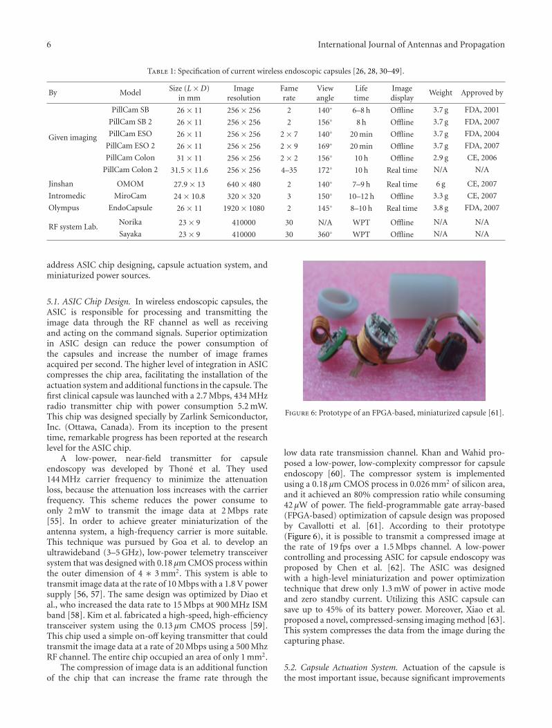
Figure 6: Prototype of an FPGA-based, miniaturized capsule [61].
low data rate transmission channel. Khan and Wahid pro-posed a low-power, low-complexity compressor for capsuleendoscopy [60]. The compressor system is implementedusing a 0.18 µm CMOS process in 0.026 mm2 of silicon area,and it achieved an 80% compression ratio while consuming42 µW of power. The field-programmable gate array-based(FPGA-based) optimization of capsule design was proposedby Cavallotti et al. [61]. According to their prototype(Figure 6), it is possible to transmit a compressed image atthe rate of 19 fps over a 1.5 Mbps channel. A low-powercontrolling and processing ASIC for capsule endoscopy wasproposed by Chen et al. [62]. The ASIC was designedwith a high-level miniaturization and power optimizationtechnique that drew only 1.3 mW of power in active modeand zero standby current. Utilizing this ASIC capsule cansave up to 45% of its battery power. Moreover, Xiao et al.proposed a novel, compressed-sensing imaging method [63].This system compresses the data from the image during thecapturing phase.
5.2. Capsule Actuation System. Actuation of the capsule isthe most important issue, because significant improvements

International Journal of Antennas and Propagation 7
and advancements of capsule endoscopy technology arepredicated on the development of a highly effective andhighly efficient actuation system. Present capsules travelpassively through the GI tract by natural peristaltic motion,which does not allow the doctor to control the motion ofcapsule or the orientation of the camera. The major problemis that doctors are unable to view according to their interest,which most likely is beyond the autonomous view of thecapsule. In the open literature, vast categories of actuationmechanisms have been published concerning the controlof the motion of the capsule. Some of more significantmechanisms are discussed below.
Shape memory alloys (SMAs) are kinds of alloys thatcan return to their previous shapes before their shapeswere altered by temperature variations. Kim et al. usedthis principle and developed a pair of SMA springs. Thesesprings, along with microhooks, were applied in endoscopiccapsule locomotion and successfully tested in a pig’s colonat the speed of 14.7 mm/min for three surface contacts[64]. The same principle was used by Karagozler et al. todevelop an SMA spring-based robot with adhesive type legsfor crawling motion [65] (Figure 7(a)), but the bottleneckproblem of the SMA-based system requires high power aswell as very slow movement.
The four leg-based and eight leg-based locomotionmechanism was tested by Quirini et al. [66] (Figure 7(b)).One brushless DC motor was used to supply sufficient torqueto the four leg-based system, while two motors were usedin the eight leg-based prototype. During in vitro testingthe eight leg-based prototype achieved a speed of 6 cm/minwith a two-degree of freedom (DOF) controlling option. Amotor-based 12-leg capsule was proposed by Quaglia et al.[67] (Figure 7(c)) to increase the effectiveness of locomotion.A paddling-based, autonomous locomotion approach wasintroduced by Yang et al. [68] (Figure 7(d)), which attainedan average speed of 28.4 cm/min in tests on a live pig underanesthesia. One brushless DC motor with a diameter of6 mm was used in this system to push the paddle forward.
A different concept using an anchoring mechanism forthe capsule’s propulsive force was proposed by Glass et al.[69] (Figure 7(e)). They simulated a mathematical model ofa three-legged capsule and found that the system was able togenerate a propulsive force greater than 0.27 N.
In order to optimize the size and power consumptionof the robotic capsule, some research groups have usedan external mechanism combined with an internal mech-anism based on magnetic theory. A capsule navigationsystem (Figure 7(g)) that used a three-dimensional, externalmagnetic field was developed by Olympus. The permanentmagnet inside the capsule interacts with the external mag-netic field and creates a rotating force due to the spiralbody structure, allowing the capsule to be pushed forwardor backward [70]. Likewise, Gao et al. proposed a self-propelled capsule system with a magnetic sheet (ø11 mm× ø6 mm × 10 mm) attached to the capsule’s outer bodyand external rectangular and cylindrical permanent magnets(80 mm and 90 mm, resp.) [71] (Figure 7(f)). A hybridlocomotion system was introduced by Simi et al. that used anexternal permanent magnet to drag the capsule containing
an internal, small, permanent magnet [72] (Figure 7(h)).Recently, a magnetically actuated, rolling, locomotion-based,soft capsule platform was proposed by Yim and Sitti [73](Figure 7(i)) for diagnostic and therapeutic applications. Theplatform used two internal permanent magnets and a large,external magnet to deform the shape of the capsule and helpto release a liquid drug from a 174 mm3 drug chamber.
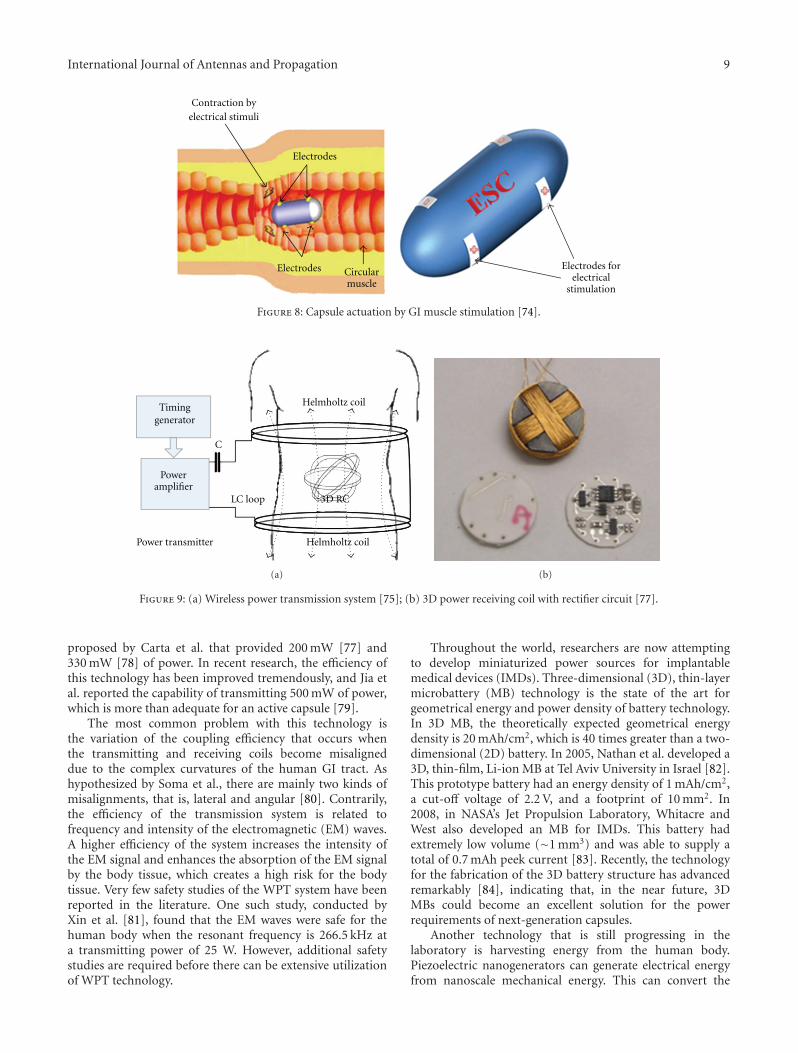
Conversely, the concept of electrical stimulation of GImuscle excludes all of the bulky internal and externalmechanical components for capsule actuation. Woo et al.[74] (Figure 8) proposed a mathematical model of an elec-trically propelled endoscopic capsule. The proposed modelused double pairs of electrodes that had dimensions of 5 ×6 mm2 for a 5 ms pulse (up to 9 V, 10–40 Hz), which was ableto move the capsule at a speed of 2.91 mm/s in the forwarddirection and 2.23 mm/s in the backward direction.
However, to date, several changed actuation mechanismshave been proposed for propelling the capsule and usingsome sophisticated option to control the capsule. So far,however, all of these mechanisms have been tested outsidethe human body due to concerns about the safety of thesmall bowel and smooth movement. Likewise, due to high-power consumption, these systems were tested using externalpower supplies. The major technical goal is the successfuldevelopment and implementation of MEMS. Because all thepresent capsules are highly integrated and some of them arealready hardly swallowable, making space for further toolswithin the ingestible dimensions is quite challenging. Thenagain, power is another issue of concern if the features areexpanded and the performance is enhanced.
5.3. Miniaturized Power Source. Power is the most criti-cal commodity for the successful development of capsuleendoscopy technology, since most of the key characteristicsthat are required of the technology consume power. Thus,the development of a successful, fully functional capsuledepends on the development of stronger, more reliable powersources. Today’s capsules generally use two coin-shaped,silver-oxide batteries that can generate 20 mW of power.Other batteries, such as lithium ion polymer (LiPo) batteries,can generate more power, but only silver-oxide batteries havebeen approved for clinical use.
The alternative to the on-board battery system is WPTtechnology. RF System Lab was the first to utilize thistechnology successfully in their capsule systems namedNorika and Sayaka, which do not have batteries [49].The wireless powering system consists of two main partsto transmit power by the induction process, that is, aHelmholtz transmitting coil that must be worn by the patientand a three-dimensional, ferrite-core receiving coil with arectification circuit [75] (Figure 9). The three-dimensionalcoil is put inside the capsule to ensure power is receivedefficiently irrespective of the orientation of the capsule.In recent years, there have been many publications in theliterature on WPT technology for capsule endoscopy. Aninductive power link for powering endoscopic capsules wasintroduced by Lenaerts and Puers transmitted 150 mW ofpower [76]. Similarly, power transmission systems were

8 International Journal of Antennas and Propagation
(a) (b)
Gears Leg-holder
ScrewNut
MotorLegCap
(c) (d)
(e)
MagnetRF transmitter
LEDs
Image sensor
Batteries
Antenna
(f)
Propelling direction
Rotationalmagnetic
fieldSpring
Capsule
Permanentmagnet
S
S
N
N
(g)
EPM
S N S N S N
Abdominal wall
Colon
(h)
(i)
Figure 7: Capsule actuation mechanisms proposed by (a) Karagozler et al. [65], (b) Quirini et al. [66], (c) Quaglia et al. [67], (d) Yang et al.[68] (e) Glass et al. [69], (f) Gao et al. [71], (g) Olympus, Inc. [70], (h) Simi et al. [72], and (i) Yim and Sitti [73].
International Journal of Antennas and Propagation 9
Circularmuscle
Electrodes
Electrodes
Contraction byelectrical stimuli
Electrodes forelectrical
stimulation
Figure 8: Capsule actuation by GI muscle stimulation [74].
Timinggenerator
Helmholtz coil
C
Poweramplifier
LC loop
Power transmitter Helmholtz coil
3D RC
(a) (b)
Figure 9: (a) Wireless power transmission system [75]; (b) 3D power receiving coil with rectifier circuit [77].
proposed by Carta et al. that provided 200 mW [77] and330 mW [78] of power. In recent research, the efficiency ofthis technology has been improved tremendously, and Jia etal. reported the capability of transmitting 500 mW of power,which is more than adequate for an active capsule [79].
The most common problem with this technology isthe variation of the coupling efficiency that occurs whenthe transmitting and receiving coils become misaligneddue to the complex curvatures of the human GI tract. Ashypothesized by Soma et al., there are mainly two kinds ofmisalignments, that is, lateral and angular [80]. Contrarily,the efficiency of the transmission system is related tofrequency and intensity of the electromagnetic (EM) waves.A higher efficiency of the system increases the intensity ofthe EM signal and enhances the absorption of the EM signalby the body tissue, which creates a high risk for the bodytissue. Very few safety studies of the WPT system have beenreported in the literature. One such study, conducted byXin et al. [81], found that the EM waves were safe for thehuman body when the resonant frequency is 266.5 kHz ata transmitting power of 25 W. However, additional safetystudies are required before there can be extensive utilizationof WPT technology.
Throughout the world, researchers are now attemptingto develop miniaturized power sources for implantablemedical devices (IMDs). Three-dimensional (3D), thin-layermicrobattery (MB) technology is the state of the art forgeometrical energy and power density of battery technology.In 3D MB, the theoretically expected geometrical energydensity is 20 mAh/cm2, which is 40 times greater than a two-dimensional (2D) battery. In 2005, Nathan et al. developed a3D, thin-film, Li-ion MB at Tel Aviv University in Israel [82].This prototype battery had an energy density of 1 mAh/cm2,a cut-off voltage of 2.2 V, and a footprint of 10 mm2. In2008, in NASA’s Jet Propulsion Laboratory, Whitacre andWest also developed an MB for IMDs. This battery hadextremely low volume (∼1 mm3) and was able to supply atotal of 0.7 mAh peek current [83]. Recently, the technologyfor the fabrication of the 3D battery structure has advancedremarkably [84], indicating that, in the near future, 3DMBs could become an excellent solution for the powerrequirements of next-generation capsules.
Another technology that is still progressing in thelaboratory is harvesting energy from the human body.Piezoelectric nanogenerators can generate electrical energyfrom nanoscale mechanical energy. This can convert the

10 International Journal of Antennas and Propagation
body’s mechanical energy, such as body motion, blood flow,muscle vibration, and breathing, to electrical energy for bodycentric devices.
6. Discussion
The endoscopic capsule has achieved little development inseveral areas, such as the capsule’s lifetime, image resolution,and viewing angle, in individual medical products. But inorder to efficiently identify diseases, high-quality imagesand longer capsule lifetimes are essential. In addition tocapsule localization, the development of diagnosis and tissuemanipulation tools associated with the capsule is still beingpursued. In the open literature, many research groups haveproposed diverse techniques for capsule localization. So far,none of them satisfies the two mandatory conditions, thatis, the safety of GI muscle and an adequate, low-powersupply that can maintain the capsule’s activation until theexamination of the entire GI tract has been completed. Dueto the knotty structure of the human digestive tract, safetyis a very important issue in the capsule’s localization system.On the other hand, the present capsules are already highlyintegrated, and there is inadequate space available to installadditional actuation mechanisms.
Thus, this paper is focused on two different steps thatcould lead to the successful development of next-generationcapsules that can possess active motion-controlling systemsand that can accommodate some additional, advancedfeatures, that is, (1) the development of mixed-mode,multifunctional ASIC with the latest CMOS technology and(2) the design of an MEMS/NEMS-based microactuationsystem.
Recently, the CMOS technology has experienced exten-sive, exponential miniaturization of the size of its transistors.Table 2 shows the progress in the development of minia-turization technology. Recently, the Taiwan SemiconductorManufacturing Company (TSMC) introducing its 28 nmprocess in 2011 [85]. The 28 nm technology uses high-Kmetal that increases the gate density by a factor of twoand decreases the area of the chip by 50% [69]. In thistechnology, the chip can operate at very low power (0.6 V)in high-power efficiency mode [86]. The OCA and antenna-in-package (AiP) are smart technologies that provide thesingle-chip radio solution. These technologies allow thecodesign of the antenna and the radio chip (in micro/nanoCMOS technology) into the same high-sensitivity siliconsubstrate package, which compresses the die area of the radiosystem [87–92]. There is a tradeoff between antenna sizeand performance in terms of operating frequency, that is,the higher (lower) the frequency, the smaller (larger) thesize can be. Because of this complexity, the OCA is easierto incorporate in radio systems that have frequencies of60 GHz or greater. Recently, ISM band OCAs have beendesigned and tested by several research groups. Okabe etal. proposed a 2.3 × 2 mm, silicon-based OCA in [92],and Mohammadpour-Aghdam et al. proposed 3 × 1.5 mm,miniaturized, integrated antennas in three different ISMbands (900 MHz, 2.4 GHz, and 5.8 GHz) in [88].
Table 2: Development of miniaturization technology.
CMOS process Year introduced
180 nm process 2001
130 nm process 2003
90 nm process 2004
65 nm process 2006
40 nm process 2008
28 nm process 2011
20 nm process Under processing
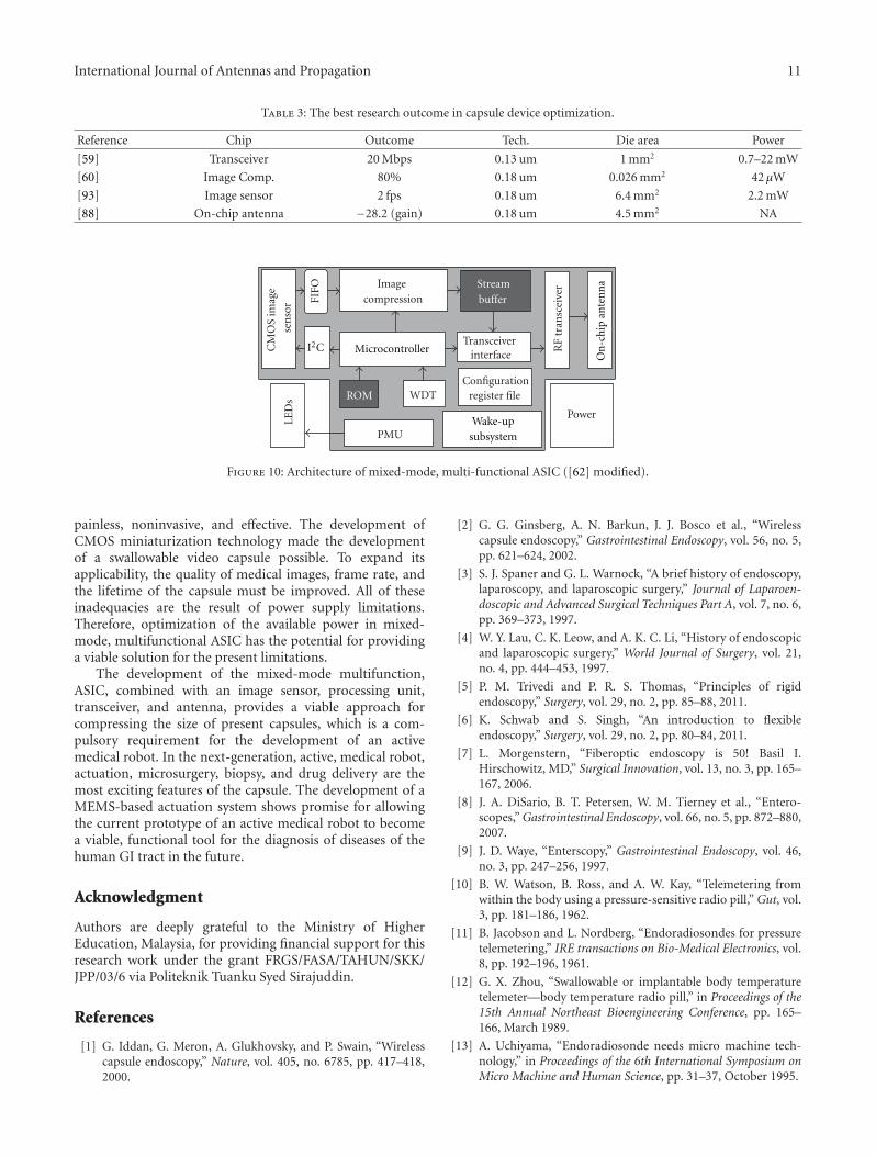
Many research groups are working on optimization ofchips for the capsule system. The best outcomes for thedifferent functional chips are listed in Table 3. From thetable, all the research carried out for the individual functionof capsule and with earlier developed CMOS technology.Clearly, there is a big opportunity to further optimizethe whole capsule designing with latest CMOS technology.Therefore, developing the mixed-mode, multifunctional chip(image sensor, image compressor, transceiver, and antennain a single chip) with the latest CMOS process can bethe greatest development. Figure 10 shows the modifiedarchitecture of the mixed-mode chip (modified from Chenet al.’s architecture [62]). The realization of such a design cancondense the size of present capsules and create free spacefor an enhanced actuation system. In addition, the capsule’spower consumption can be reduced.
Capsule actuation is still the first and most significanttechnical challenge standing on the way of the successfuldevelopment and deployment of medical robots or roboticendoscopic capsules. Among all of the capsule actuationsystems proposed by different research groups, the electricalstimulation of GI muscles is one that excludes all of theon-board mechanical components. But in terms of powerconsumption, the external magnetic actuation system hasless power demand. A hybrid capsule actuation systemwith the combination of electric stimulation and externalmagnetic actuation could result in a viable solution tothe capsule actuation issue. Conversely, the concept of anactive medical robot is not only limited by the locomotionconcerns associated with endoscopic capsules; it also is amajor concern in other applications, including microsurgery,drug delivery, microgrippers, and microsyringes. So thedevelopment of MEMS/NEM-based active medical robotsmust consider these areas of concern in addition to endo-scopic capsule localization. This perception is already beingaddressed by some research projects. One such project isthe Vector project funded by the European Commission,which aims to develop an intelligent, endoscopic capsulewith micronanotechnology. The Vector project already hasdeveloped a capsule that is able to detect blood insidethe bowel, and the researchers are still considering anddeveloping the suitable design.
7. Conclusions
Over the past decade, WCE has become the sole technol-ogy for the direct diagnosis of GI diseases because it is
International Journal of Antennas and Propagation 11
Table 3: The best research outcome in capsule device optimization.
Reference Chip Outcome Tech. Die area Power
[59] Transceiver 20 Mbps 0.13 um 1 mm2 0.7–22 mW
[60] Image Comp. 80% 0.18 um 0.026 mm2 42 µW
[93] Image sensor 2 fps 0.18 um 6.4 mm2 2.2 mW
[88] On-chip antenna −28.2 (gain) 0.18 um 4.5 mm2 NA
CM
OS
imag
e
sen
sor FI
FO Image compression
LED
s
Power
PMU
RF
tran
scei
verStream
buffer
Transceiver interface
Configuration register fileROM WDT
Wake-upsubsystem
On
-ch
ip a
nte
nn
a
I2C Microcontroller
Figure 10: Architecture of mixed-mode, multi-functional ASIC ([62] modified).
painless, noninvasive, and effective. The development ofCMOS miniaturization technology made the developmentof a swallowable video capsule possible. To expand itsapplicability, the quality of medical images, frame rate, andthe lifetime of the capsule must be improved. All of theseinadequacies are the result of power supply limitations.Therefore, optimization of the available power in mixed-mode, multifunctional ASIC has the potential for providinga viable solution for the present limitations.
The development of the mixed-mode multifunction,ASIC, combined with an image sensor, processing unit,transceiver, and antenna, provides a viable approach forcompressing the size of present capsules, which is a com-pulsory requirement for the development of an activemedical robot. In the next-generation, active, medical robot,actuation, microsurgery, biopsy, and drug delivery are themost exciting features of the capsule. The development of aMEMS-based actuation system shows promise for allowingthe current prototype of an active medical robot to becomea viable, functional tool for the diagnosis of diseases of thehuman GI tract in the future.
Acknowledgment
Authors are deeply grateful to the Ministry of HigherEducation, Malaysia, for providing financial support for thisresearch work under the grant FRGS/FASA/TAHUN/SKK/JPP/03/6 via Politeknik Tuanku Syed Sirajuddin.
References
[1] G. Iddan, G. Meron, A. Glukhovsky, and P. Swain, “Wirelesscapsule endoscopy,” Nature, vol. 405, no. 6785, pp. 417–418,2000.
[2] G. G. Ginsberg, A. N. Barkun, J. J. Bosco et al., “Wirelesscapsule endoscopy,” Gastrointestinal Endoscopy, vol. 56, no. 5,pp. 621–624, 2002.
[3] S. J. Spaner and G. L. Warnock, “A brief history of endoscopy,laparoscopy, and laparoscopic surgery,” Journal of Laparoen-doscopic and Advanced Surgical Techniques Part A, vol. 7, no. 6,pp. 369–373, 1997.
[4] W. Y. Lau, C. K. Leow, and A. K. C. Li, “History of endoscopicand laparoscopic surgery,” World Journal of Surgery, vol. 21,no. 4, pp. 444–453, 1997.
[5] P. M. Trivedi and P. R. S. Thomas, “Principles of rigidendoscopy,” Surgery, vol. 29, no. 2, pp. 85–88, 2011.
[6] K. Schwab and S. Singh, “An introduction to flexibleendoscopy,” Surgery, vol. 29, no. 2, pp. 80–84, 2011.
[7] L. Morgenstern, “Fiberoptic endoscopy is 50! Basil I.Hirschowitz, MD,” Surgical Innovation, vol. 13, no. 3, pp. 165–167, 2006.
[8] J. A. DiSario, B. T. Petersen, W. M. Tierney et al., “Entero-scopes,” Gastrointestinal Endoscopy, vol. 66, no. 5, pp. 872–880,2007.
[9] J. D. Waye, “Enterscopy,” Gastrointestinal Endoscopy, vol. 46,no. 3, pp. 247–256, 1997.
[10] B. W. Watson, B. Ross, and A. W. Kay, “Telemetering fromwithin the body using a pressure-sensitive radio pill,” Gut, vol.3, pp. 181–186, 1962.
[11] B. Jacobson and L. Nordberg, “Endoradiosondes for pressuretelemetering,” IRE transactions on Bio-Medical Electronics, vol.8, pp. 192–196, 1961.
[12] G. X. Zhou, “Swallowable or implantable body temperaturetelemeter—body temperature radio pill,” in Proceedings of the15th Annual Northeast Bioengineering Conference, pp. 165–166, March 1989.
[13] A. Uchiyama, “Endoradiosonde needs micro machine tech-nology,” in Proceedings of the 6th International Symposium onMicro Machine and Human Science, pp. 31–37, October 1995.
12 International Journal of Antennas and Propagation
[14] “The Heidelberg pH diagnostic system,” http://www.phcap-sule.com/prodinfo.htm.
[15] S. Maqbool, H. P. Parkman, and F. K. Friedenberg, “Wirelesscapsule motility: comparison of the smartPill GI monitoringsystem with scintigraphy for measuring whole gut transit,”Digestive Diseases and Sciences, vol. 54, no. 10, pp. 2167–2174,2009.
[16] C. P. Swain, F. Gong, and T. N. Mills, “Wireless transmissionof a colour television moving image from the stomachusing a miniature ccd camera, light source and microwavetransmitter,” Gastrointestinal Endoscopy, vol. 45, no. 4, p.AB40, 1997.
[17] G. J. Iddan and C. P. Swain, “History and developmentof capsule endoscopy,” Gastrointestinal Endoscopy Clinics ofNorth America, vol. 14, no. 1, pp. 1–9, 2004.
[18] “Human digestive,” http://www.medical-reference.net/2012/07/digestive-system-digestive-system-is.html.
[19] L. T. Weaver, S. Austin, and T. J. Cole, “Small intestinal length:a factor essential for gut adaptation,” Gut, vol. 32, no. 11, pp.1321–1323, 1991.
[20] B. M. Underhill, “Intestinal length in man,” British MedicalJournal, vol. 2, no. 4950, pp. 1243–1246, 1955.
[21] B. S. Lewis, “The history of enteroscopy,” GastrointestinalEndoscopy Clinics of North America, vol. 9, no. 1, pp. 1–11,1999.
[22] D. G. Adler and C. J. Gostout, “Wireless capsule endoscopy,”Hospital Physician, pp. 14–22, 2003.
[23] G. D. Meron, “The development of the swallowable videocapsule (M2A),” Gastrointestinal Endoscopy, vol. 52, no. 6, pp.817–819, 2000.
[24] C. Mc Caffrey, O. Chevalerias, C. O’Mathuna, and K. Twomey,“Swallowable-capsule technology,” IEEE Pervasive Computing,vol. 7, no. 1, pp. 23–29, 2008.
[25] M. Yu, “M2A capsule endoscopy,” Gastroenterology Nursing,vol. 25, pp. 24–27, 2001.
[26] “OMOM capsule,” http://english..jinshangroup.com/con-tents/1091/1348.html.
[27] R. Eliakim, K. Yassin, I. Shlomi, A. Suissa, and G. M. Eisen, “Anovel diagnostic tool for detecting oesophageal pathology: thePillCam oesophageal video capsule,” Alimentary Pharmacologyand Therapeutics, vol. 20, no. 10, pp. 1083–1089, 2004.
[28] C. Y. Li, B. L. Zhang, C. X. Chen, and Y. M. Li, “OMOMcapsule endoscopy in diagnosis of small bowel disease,”Journal of Zhejiang University: Science B, vol. 9, no. 11, pp. 857–862, 2008.
[29] B. S. Lewis, “The utility of capsule endoscopy in obscuregastrointestinal bleeding,” Techniques in GastrointestinalEndoscopy, vol. 5, no. 3, pp. 115–120, 2003.
[30] “Given imaging,” http://www.givenimaging.com/en-us/Pages/Legal-Notice.aspx.
[31] Given Imaging company, http://www.givenimaging.com/.
[32] D. S. Mishkin, R. Chuttani, J. Croffie et al., “ASGE technologystatus evaluation report: wireless capsule endoscopy,” Gas-trointestinal Endoscopy, vol. 63, no. 4, pp. 539–545, 2006.
[33] Y. C. Metzger, S. N. Adler, A. B. G. Shitrit, B. Koslowsky, and I.Bjarnason, “Comparison of a new PillCam SB2 video capsuleversus the standard PillCam SB for detection of small boweldisease,” Reports in Medical Imaging, vol. 2, pp. 7–11, 2009.
[34] “PillCam SB 2-Ex,” http://medgadget.com/2010/10/givenimaging announces new pillcam offerings.html.
[35] G. M. Eisen, “Pillcam ESO: a primer,” Techniques in Gastroin-testinal Endoscopy, vol. 8, no. 4, pp. 154–159, 2006.
[36] B. Koslowsky, H. Jacob, R. Eliakim, and S. N. Adler, “PillCamESO in esophageal studies: improved diagnostic yield of 14frames per second (fps) compared with 4 fps,” Endoscopy, vol.38, no. 1, pp. 27–30, 2006.
[37] I. M. Gralnek, S. N. Adler, K. Yassin, B. Koslowsky, Y.Metzger, and R. Eliakim, “Detecting esophageal disease withsecond-generation capsule endoscopy: initial evaluation of thePillCam ESO 2,” Endoscopy, vol. 40, no. 4, pp. 275–279, 2008.
[38] D. G. Adler, B. Chand, J. D. Conway et al., “Capsule endoscopyof the colon,” Gastrointestinal Endoscopy, vol. 68, no. 4, pp.621–623, 2008.
[39] Z. Fireman and Y. Kopelman, “The colon-the latest terrain forcapsule endoscopy,” Digestive and Liver Disease, vol. 39, no. 10,pp. 895–899, 2007.
[40] C. Spada, C. Hassan, M. Munoz-Navas et al., “Second-generation colon capsule endoscopy compared withcolonoscopy,” Gastrointestinal Endoscopy, vol. 74, no. 3,pp. 581–591, 2011.
[41] P. N. Figueiredo, I. N. Figueiredo, S. Prasath, and R. Tsai,“Automatic polyp detectionin Pillcam Colon 2 capsule imagesand videos: preliminary feasibility report,” Diagnostic andTherapeutic Endoscopy, pp. 1–7, 2011.
[42] S. N. Adler and Y. C. Metzger, “PillCam COLON capsuleendoscopy: recent advances and new insights,” TherapeuticAdvances in Gastroenterology, vol. 4, no. 4, pp. 265–268, 2011.
[43] Z. Liao, R. Gao, F. Li et al., “Fields of applications, diagnosticyields and findings of OMOM capsule endoscopy in 2400Chinese patients,” World Journal of Gastroenterology, vol. 16,no. 21, pp. 2669–2676, 2010.
[44] “Olympus capsule,” http://www.european-hospital.com/en/article/7244-Video Capsule Endoscopy.html.
[45] C. Gheorghe, R. Iacob, and I. Bancila, “Olympus capsuleendoscopy for small bowel examination,” Journal of Gastroin-testinal and Liver Diseases, vol. 16, no. 3, pp. 309–313, 2007.
[46] D. R. Cave, D. E. Fleischer, J. A. Leighton et al., “A multicenterrandomized comparison of the Endocapsule and the PillcamSB,” Gastrointestinal Endoscopy, vol. 68, no. 3, pp. 487–494,2008.
[47] “Brochure of MiroCam capsule,” http://www.synmed.co.uk/news mirocam.htm.
[48] H. M. Kim, Y. J. Kim, H. J. Kim et al., “A pilot study ofsequential capsule endoscopy using MiroCam and PillCamSB devices with different transmission technologies,” Gut andLiver, vol. 4, no. 2, pp. 192–200, 2010.
[49] “Sayaka capsule system,” http://www.rfsystemlab.com/en/sayaka/index.html.
[50] T. S. Kim, J. O. Park, S. W. Moon, B. K. Kim, J. Y. Kang,and J. Han, “Method and apparatus for communicationbetween inside and outside of transmission medium usingtransmission medium as communication line,” US PatentDocument, 2007.
[51] K. R. Canlas, B. M. Dobozi, S. Lin et al., “Using capsuleendoscopy to identify GI tract lesions in cirrhotic patients withportal hypertension and chronic anemia,” Journal of ClinicalGastroenterology, vol. 42, no. 7, pp. 844–848, 2008.
[52] G. C. Sturniolo, V. Di Leo, M. G. Vettorato et al., “Small bowelexploration by wireless capsule endoscopy: results from 314procedures,” American Journal of Medicine, vol. 119, no. 4, pp.341–347, 2006.
[53] A. K. Hara, J. A. Leighton, V. K. Sharma, R. I. Heigh, and D.E. Fleischer, “Imaging of small bowel disease: comparison of

International Journal of Antennas and Propagation 13
capsule endoscopy, standard endoscopy, barium examination,and CT,” Radiographics, vol. 25, no. 3, pp. 697–711, 2005.
[54] E. Rondonotti, J. M. Herrerias, M. Pennazio, A. Caunedo,M. Mascarenhas-Saraiva, and R. De Franchis, “Complications,limitations, and failures of capsule endoscopy: a review of 733cases,” Gastrointestinal Endoscopy, vol. 62, no. 5, pp. 712–716,2005.
[55] J. Thone, S. Radiom, D. Turgis, R. Carta, G. Gielen, and R.Puers, “Design of a 2 Mbps FSK near-field transmitter forwireless capsule endoscopy,” Sensors and Actuators A, vol. 156,no. 1, pp. 43–48, 2009.
[56] Y. Gao, Y. Zheng, S. Diao et al., “Low-power ultrawidebandwireless telemetry transceiver for medical sensor applications,”IEEE Transactions on Biomedical Engineering, vol. 58, no. 3, pp.768–772, 2011.
[57] Y. Gao, S. Diao, C. W. Ang, Y. Zheng, and X. Yuan, “Lowpower ultra-wideband wireless telemetry system for capsuleendoscopy application,” in Proceedings of IEEE InternationalConference on Robotics, Automation and Mechatronics (RAM’10), pp. 96–99, sgp, June 2010.
[58] S. Diao, Y. Gao, W. Toh et al., “A low-power, high data-rateCMOS ASK transmitter for wireless capsule endoscopy,” inDefense Science Research Conference and Expo, pp. 1–4.
[59] K. Kim, S. Yun, S. Lee, S. Nam, Y. Yoon, and C. Cheon, “Adesign of a high-speed and high-efficiency capsule endoscopysystem,” IEEE Transactions on Biomedical Engineering, vol. 59,pp. 1005–1011, 2012.
[60] T. H. Khan and K. A. Wahid, “Low power and low complexitycompressor for video capsule endoscopy,” IEEE Transactionson Circuits and Systems For Video Technology, vol. 21, pp.1534–1546, 2011.
[61] C. Cavallotti, P. Merlino, M. Vatteroni et al., “An FPGA-basedversatile development system for endoscopic capsule designoptimization,” Sensors and Actuators A, vol. 172, no. 1, pp.301–307, 2011.
[62] X. Chen, X. Zhang, L. Zhang et al., “A wireless capsuleendoscope system with low-power controlling and processingASIC,” IEEE Transactions on Biomedical Circuits and Systems,vol. 3, no. 1, pp. 11–22, 2009.
[63] L. Xiao, K. Liu, and D. P. Han, “CMOS low data rateimaging method based on compressed sensing,” Optics & LaserTechnology, vol. 44, pp. 1338–1345, 2012.
[64] B. Kim, S. Lee, J. H. Park, and J. O. Park, “Design andfabrication of a locomotive mechanism for capsule-typeendoscopes using shape memory alloys (SMAs),” IEEE/ASMETransactions on Mechatronics, vol. 10, no. 1, pp. 77–86, 2005.
[65] M. E. Karagozler, E. Cheung, J. Kwon, and M. Sitti, “Miniatureendoscopie capsule robot using biomimetic micro-patternedadhesives,” in Proceedings of the 1st IEEE/RAS-EMBS Interna-tional Conference on Biomedical Robotics and Biomechatronics(BioRob ’06), pp. 105–111, February 2006.
[66] M. Quirini, S. Scapellato, P. Valdastri, A. Menciassi, andP. Dario, “An approach to capsular endoscopy with activemotion,” in Proceedings of the 29th Annual InternationalConference of IEEE-EMBS, Engineering in Medicine and BiologySociety (EMBC ’07), pp. 2827–2830, August 2007.
[67] C. Quaglia, E. Buselli, R. J. Webster III, P. Valdastri, A.Menciassi, and P. Dario, “An endoscopic capsule robot: ameso-scale engineering case study,” Journal of Micromechanicsand Microengineering, vol. 19, no. 10, Article ID 105007, 2009.
[68] S. Yang, K. Park, J. Kim, T. S. Kim, I. J. Cho, and E. S.Yoon, “Autonomous locomotion of capsule endoscope in gas-trointestinal,” in Proceedings of the 33rd Annual InternationalConference of the IEEE EMBS, pp. 6659–6663, 2011.
[69] P. Glass, E. Cheung, and M. Sitti, “A legged anchoring mecha-nism for capsule endoscopes using micropatterned adhesives,”IEEE Transactions on Biomedical Engineering, vol. 55, pp.2759–2767, 2008.
[70] Y. Kusuda, “A further step beyond wireless capsule endoscopy,”Sensor Review, vol. 25, no. 4, pp. 259–260, 2005.
[71] M. Gao, C. Hu, Z. Chen, H. Zhang, and S. Liu, “Design andfabrication of a magnetic propulsion system for self-propelledcapsule endoscope,” IEEE Transactions on Biomedical Engi-neering, vol. 57, no. 12, pp. 2891–2902, 2010.
[72] M. Simi, P. Valdastri, C. Quaglia, A. Menciassi, and P.Dario, “Design, fabrication, and testing of a capsule withhybrid locomotion for gastrointestinal tract exploration,”IEEE/ASME Transactions on Mechatronics, vol. 15, no. 2, pp.170–180, 2010.
[73] S. Yim and M. Sitti, “Design and rolling locomotion of a mag-netically actuated soft capsule endoscope,” IEEE Transactionson Robotics, vol. 28, pp. 183–194, 2012.
[74] S. H. Woo, T. W. Kim, Z. Mohy-Ud-Din, I. Y. Park, and J. H.Cho, “Small intestinal model for electrically propelled capsuleendoscopy,” BioMedical Engineering OnLine, vol. 10, 2011.
[75] G. Pan, W. Xin, G. Yan, and J. Chen, “A video wireless capsuleendoscopy system powered wirelessly: design, analysis andexperiment,” Measurement Science and Technology, vol. 22, no.6, Article ID 065802, pp. 1–9, 2011.
[76] B. Lenaerts and R. Puers, “An inductive power link for awireless endoscope,” Biosensors and Bioelectronics, vol. 22, no.7, pp. 1390–1395, 2007.
[77] R. Carta, G. Tortora, J. Thone et al., “Wireless powering for aself-propelled and steerable endoscopic capsule for stomachinspection,” Biosensors and Bioelectronics, vol. 25, no. 4, pp.845–851, 2009.
[78] R. Carta, J. Thone, and R. Puers, “A wireless power supplysystem for robotic capsular endoscopes,” Sensors and ActuatorsA, vol. 162, no. 2, pp. 177–183, 2010.
[79] Z. Jia, G. Yan, H. Liu, Z. Wang, P. Jiang, and Y. Shi, “Theoptimization of wireless power transmission: design andrealization,” The International Journal of Medical Robotics andComputer Assisted Surgery, vol. 8, 2012, (abstract).
[80] M. Soma, D. C. Galbraith, and R. L. White, “Radio frequencycoils in implantable devices: misalignment analysis and designprocedure,” IEEE Transactions on Biomedical Engineering, vol.34, no. 4, pp. 276–282, 1987.
[81] W. H. Xin, G. Z. Yan, and W. X. Wang, “Study on human safetyin wireless power transmission system for capsule endoscopy,”Chinese Journal of Biomedical Engineering, vol. 28, no. 5, pp.719–724, 2009.
[82] M. Nathan, D. Golodnitsky, V. Yufit et al., “Three-dimensionalthin-film Li-ion microbatteries for autonomous MEMS,”Journal of Microelectromechanical Systems, vol. 14, no. 5, pp.879–885, 2005.
[83] J. Whitacre and W. West, “Integrated microbatteries forimplantable medical devices,” NASA Tech Briefs, 2008.
[84] M. Roberts, P. Johns, J. Owen et al., “3D lithium ionbatteries—from fundamentals to fabrication,” Journal ofMaterials Chemistry, vol. 21, no. 27, pp. 9876–9890, 2011.
[85] “CMOS process,” http://www.st.com/internet/com/pressrelease/t3173.jsp.
[86] N. Ickes, G. Gammie, M. E. Sinangil, R. Rithe, J. Gu, A. Wanget al., “A 28 nm 0.6 V low power DSP for mobile applications,”IEEE Journal of Solid-State Circuits, vol. 47, pp. 35–46, 2012.
[87] Y. P. Zhang, “Antenna-in-package technology for modernradio systems,” in Proceedings of IEEE International Workshopon Antenna Technology (IWAT ’06), pp. 37–40, March 2006.

14 International Journal of Antennas and Propagation
[88] K. Mohammadpour-Aghdam, S. Radiom, Z. Faraji-Dana, G.A. E. Vandenbosch, and G. G. E. Gielen, “Miniaturized inte-grated antennas for far-field wireless powering,” InternationalJournal of Electronics and Communications, vol. 66, no. 10, pp.789—7796, 2012.
[89] S. Pan, L. Gilreath, P. Heydari, and F. Capolino, “Designs offully on-chip antennas in (Bi)CMOS technology,” in Proceed-ings of IEEE International Workshop on Antenna Technology,pp. 343–346, 2012.
[90] Y. P. Zhang and D. Liu, “Antenna-on-chip and antenna-in-package solutions to highly integrated millimeter-wave devicesfor wireless communications,” IEEE Transactions on Antennasand Propagation, vol. 57, no. 10, pp. 2830–2841, 2009.
[91] S. Radiom, M. Baghaei-Nejad, G. Vandenbosch, L. R. Zheng,and G. Gielen, “Far-field RF powering system for RFID andimplantable devices with monolithically integrated on-chipantenna,” in Proceedings of IEEE Radio Frequency IntegratedCircuits Symposium (RFIC ’10), pp. 113–116, May 2010.
[92] K. Okabe, W. Lee, Y. Harada, and M. Ishida, “Silicon based on-chip antenna using an LC resonator for near-field RF systems,”Solid-State Electronics, vol. 67, pp. 100–104, 2011.
[93] M. Zhang, A. Bermak, X. Li, and Z. Wang, “A low powerCMOS image sensor design for wireless endoscopy capsule,” inProceedings of IEEE-BIOCAS Biomedical Circuits and SystemsConference (BIOCAS ’08), pp. 397–400, November 2008.

International Journal of
AerospaceEngineeringHindawi Publishing Corporationhttp://www.hindawi.com Volume 2010
RoboticsJournal of
Hindawi Publishing Corporationhttp://www.hindawi.com Volume 2014
Hindawi Publishing Corporationhttp://www.hindawi.com Volume 2014
Active and Passive Electronic Components
Control Scienceand Engineering
Journal of
Hindawi Publishing Corporationhttp://www.hindawi.com Volume 2014
International Journal of
RotatingMachinery
Hindawi Publishing Corporationhttp://www.hindawi.com Volume 2014
Hindawi Publishing Corporation http://www.hindawi.com
Journal ofEngineeringVolume 2014
Submit your manuscripts athttp://www.hindawi.com
VLSI Design
Hindawi Publishing Corporationhttp://www.hindawi.com Volume 2014
Hindawi Publishing Corporationhttp://www.hindawi.com Volume 2014
Shock and Vibration
Hindawi Publishing Corporationhttp://www.hindawi.com Volume 2014
Civil EngineeringAdvances in
Acoustics and VibrationAdvances in
Hindawi Publishing Corporationhttp://www.hindawi.com Volume 2014
Hindawi Publishing Corporationhttp://www.hindawi.com Volume 2014
Electrical and Computer Engineering
Journal of
Advances inOptoElectronics
Hindawi Publishing Corporation http://www.hindawi.com
Volume 2014
The Scientific World JournalHindawi Publishing Corporation http://www.hindawi.com Volume 2014
SensorsJournal of
Hindawi Publishing Corporationhttp://www.hindawi.com Volume 2014
Modelling & Simulation in EngineeringHindawi Publishing Corporation http://www.hindawi.com Volume 2014
Hindawi Publishing Corporationhttp://www.hindawi.com Volume 2014
Chemical EngineeringInternational Journal of Antennas and
Propagation
International Journal of
Hindawi Publishing Corporationhttp://www.hindawi.com Volume 2014
Hindawi Publishing Corporationhttp://www.hindawi.com Volume 2014
Navigation and Observation
International Journal of
Hindawi Publishing Corporationhttp://www.hindawi.com Volume 2014
DistributedSensor Networks
International Journal of