review article hemoglobinopathies in …khuri/avl/articles/northafrica_review.pdfhemoglobinopathies...
TRANSCRIPT
Hemoglobin, 34(1):1–23, (2010)Copyright © Informa UK Ltd.ISSN: 0363-0269 print/1532-432X onlineDOI: 10.3109/03630260903571286
1
LHEM0363-02691532-432XHemoglobin, Vol. 34, No. 1, Dec 2009: pp. 0–0Hemoglobin
REVIEW ARTICLE
HEMOGLOBINOPATHIES IN NORTH AFRICA: A REVIEW
Hemoglobinopathies in North AfricaA. Haj Khelil et al.
Amel Haj Khelil,1 Sabri Denden,1 Nadia Leban,1 Houria Daimi,1
Ramzi Lakhdhar,1 Gérard Lefranc,2 Jemni Ben Chibani,1 and Pascale Perrin3
1Laboratory of Biochemistry and Molecular Biology, Faculty of Pharmacy, Monastir, Tunisia2Laboratory of Molecular Immuno-Genetics, University of Montpellier 2, Montpellier, France3Institute of Evolution Sciences, CNRS, UMR 5554, University of Montpellier 2, Montpellier, France
� Hemolytic anemias are very common diseases. Among these diseases, hemoglobinopathies arewidely spread throughout the Mediterranean Basin, including North Africa (Tunisia, Algeria andMorocco). Their severity and disabling nature make them a major public health problem. This studyincludes our data on the Tunisian hemoglobinopathies together with all the reports concerning epide-miological, clinical and molecular aspects in Algerian and Moroccan populations. Investigationmethods begin with the application of several techniques for hemoglobin (Hb) analyses [electrophoresisand isoelectric focusing (IEF), micro-chromatography assay] of anemic patients in various hospitaldepartments. Molecular investigation by DNA analyses completes the hematological and biochemicalstudies using polymerase chain reaction (PCR) followed by enzymatic digestion and/or denaturinggradient gel electrophoresis (DGGE), single strand conformation polymorphism (SSCP) andsequencing. These methods offer screening for a large number of families affected by sickle cell dis-ease and thalassemia. In Tunisia, Algeria, and Morocco, more than 45 mutations have been iden-tified on the b-globin gene. The most common in Tunisia and in Algeria are codon 39 (C>T) andIVS-I-110 (G>A), which together account for more than 50% of all mutations. In Morocco, the pre-dominant mutations are codon 39 and frameshift codon (FSC) 8 (–AA). The identification of molec-ular defects in the bgene contributes to the development of diagnostic tests (prenatal diagnosis), andgives us the opportunity to help many couples. Our studies of the haplotypes of the bS, codon 39 andIVS-I-110 origins allowed the hypothesis of a Benin origin for bS, a local North African origin forcodon 39 and an Eastern Mediterranean origin for IVS-I-110. The analysis of polymorphisms asso-ciated with a moderate phenotype of b-thalassemia (b-thal) and sickle cell disease in North Africa hasshown, in several cases, a strong association with some mutations and restriction fragment lengthpolymorphisms (RFLP) haplotype IX on the b-globin locus and the -158 (C>T) polymorphism in 5¢
Received 15 May 2009; Accepted 11 October 2009.Address correspondence to Dr. Amel Haj Khelil, Laboratory of Biochemistry and Molecular
Biology, Faculty of Pharmacy, Street Avicenne 5019, Monastir, Tunisia; Tel: +216-73461000; Fax: +216-73461830; E-mail: [email protected]
Hem
oglo
bin
Dow
nloa
ded
from
info
rmah
ealth
care
.com
by
Uni
vers
ity o
f C
alif
orni
a Sa
n Fr
anci
sco
on 0
6/08
/11
For
pers
onal
use
onl
y.
2 A. Haj Khelil et al.
on the Gg-globin gene. Finally, more knowledge on the regulation of the b-globin locus may contributeto the improvement of investigation, monitoring and treatment of hemoglobinopathies.
Keywords Hemoglobinopathies, Thalassemia, Sickle cell disease, Mutation, Haplotype
INTRODUCTION
Hemoglobinopathies are defined by the presence of qualitative and/orquantitative abnormalities affecting the globin chains (1). To date, morethan 700 abnormal b-globin gene hemoglobin (Hb) variants have beendescribed (2,3). Qualitative abnormalities lead to the production of anabnormal structure of Hb. Among these variants, the best known are Hb S[b6(A3)Glu→Val] causing sickle cell disease, Hb C [b6(A3)Glu→Lys] andHb E [b26(B8)Glu→Lys] (2,3), mostly because of the biological conse-quences they generate. However, the majority of variants are asymptomaticand therefore remain unknown. Hemoglobin quantitative abnormalitiesresult from either reduced or absent synthesis of a- or b-globin chains. Theydefine the thalassemias [a- and b-thalassemia (a- and b-thal), respectively)]For a-thalassemias, the common molecular defects are deletions involvingeither one, two, or three or the four alleles at the a locus (4). In contrast,b-thalassemias are predominantly the consequence of punctual mutationsaffecting a single b gene or both, resulting in a decrease (b+-thal) or a totallack of synthesis of b-globin (b0-thal) (1). Molecular abnormalities of regu-latory regions on b or g genes affecting gene expression result in silentthalassemias (b++). They are the source of hereditary persistence of fetal Hb(HPFH) (5). Many forms are associated with qualitative and quantitativeabnormalities of globin chains such as bS/bthal hemoglobinopathies or Hb Eassociated with a- or b-thalassemias (compound hemoglobinopathies).
Hemoglobinopathies are common all around the Mediterranean Basin.The Maghreb, with its geographical location (migration crossroad betweensub-Saharan Africa, the Arab countries and Europe), its history and its socio-economic and cultural system, is one of the interesting geographic hot-spots.Moreover, the selective pressure of Plasmodium falciparum in regions endemicfor malaria could have increased the frequencies of the b and a mutated genes(balanced selection) (6). Finally, the common endogamous system and con-sanguinity favor the increasing number of individuals affected. These patholo-gies are incapacitating by their severity and the lack of any cure so far. Theyrepresent a major public health problem. The fruitful collaboration of obstetri-cians and hematologists allowed the performance, in a few years, of a signifi-cant number of tests, with better reliability and increased security. Molecularanalyses of the b-globin gene allowed the identification of numerous b-thalmutations. Many polymorphisms were found to be associated with some ofthese mutations. They helped us to understand the origins of these mutations.
Hem
oglo
bin
Dow
nloa
ded
from
info
rmah
ealth
care
.com
by
Uni
vers
ity o
f C
alif
orni
a Sa
n Fr
anci
sco
on 0
6/08
/11
For
pers
onal
use
onl
y.
Hemoglobinopathies in North Africa 3
This study provides, for the first time, a thorough overview of a-, b-thaland Hb variants in the Maghreb countries. Knowledge of the moleculardefects and haplotypes associated with these defects allows the developmentand improvement of diagnostic tests and the management of these diseases.In addition, it helps us to understand the origins and the migrationschemes of these mutations in the Mediterranean area.
EPIDEMIOLOGICAL STUDY
Among the abnormal Hbs, sickle cell disease is by far the most frequentone. It affects mostly people from Africa, Madagascar, Reunion and Caribbeanislands, Central America, the Mediterranean Basin and the Middle East (7).In Tunisia, the average frequency of the disease is 1.89% (8). It is between0.8 to 3.5% in Algeria (9) and 1.2% in Morocco (10).b-Thalassemias are frequently identified in subjects from the Mediterra-
nean area (Italy, Sardinia, Sicily, Greece, North Africa), but they are also foundin patients coming from Africa and Asia (Iran, India, Vietnam, Thailand) (11).In the Maghreb, b-thal reaches 2.21% in Tunisia (10) and almost thesame frequency (1.5 to 3.0%) in Algeria (12,13) and Morocco (10). Thea-thalassemias, however, are found mainly in populations of Southeast Asia(Cambodia, Laos, Myanmar, Thailand) (14) but also in the MediterraneanBasin and Central African countries (11). a-Thalassemias show an inci-dence of 4.8 to 5.48% in Tunisia (8) (7.38% for Hb Bart’s (15), 9.0% inAlgeria (12) and 2.2% in Morocco (10). However, the frequency of a-thal isprobably underestimated: indeed, it remains difficult to identify it in adult-hood by simple methods, especially when only one or two of the four agenes are affected. Today, population migrations and mixing favor a spreadof these abnormalities which are found almost everywhere in the world.Some abnormal Hbs, once specific to ethnic groups, are now also found inother groups.
PHENOTYPICAL ANALYSIS
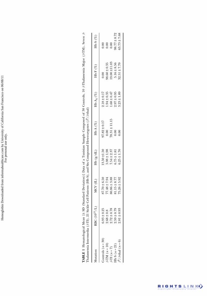
The identification of abnormal Hbs by phenotype analyses requiresaccurate information: ethnic origin of patients and their relatives, familyhistory concerning hemoglobinopathies, clinical data and recent transfu-sion data. The diagnosis requires a recent complete blood count (CBC).Hemoglobin, mean corpuscular volume (MCV) and the number of redblood cells (RBC) must be interpreted according to the age of the patient(16). The examination of blood smears allows the recognition of any abnor-malities of RBC: anisocytosis, microcytosis, polychromatosis, presence oftarget or sickled cells, basophilic stippling. Chromatographic methodsallow the quantification of different fractions of Hb (Table 1). Anionic
Hem
oglo
bin
Dow
nloa
ded
from
info
rmah
ealth
care
.com
by
Uni
vers
ity o
f C
alif
orni
a Sa
n Fr
anci
sco
on 0
6/08
/11
For
pers
onal
use
onl
y.
TA
BL
E 1
Hem
atol
ogic
al M
ean
[±
SD (
Stan
dard
Dev
iati
on)]
Dat
a of
a T
unis
ian
Sam
ple
Com
pose
d of
30
Con
trol
s, 1
0 b-
Th
alas
sem
ia M
ajor
(b-
TM
), S
even
b-
Th
alas
sem
ia I
nte
rmed
ia (b-
TI)
, 21
Sick
le C
ell P
atie
nts
(H
b S)
, an
d Si
x C
ompo
und
Het
eroz
ygot
es (bS
/b-th
al)
Mut
atio
nR
BC
(10
12/L
)M
CV
(fL
)H
b (g
/dL
)H
b A
(%
)H
b A
2 (%
)H
b F
(%)
Hb
S (%
)
Con
trol
s (n
= 3
0)6.
93 ±
0.2
587
.70
± 6.
5013
.50
±1.5
097
.82
± 0.
172.
18 ±
0.1
70.
000.
00b-
TM
(n
= 10
)2.
68 ±
0.8
77.4
8 ±
7.94
5.98
± 1
,98
0.00
1.94
± 0
.35
98.6
0 ±
0.35
0.00
b-T
I (n
= 7
)2.
72 ±
0.7
880
.86
± 3.
887.
54 ±
2.3
778
.31
± 11
.15
1.68
± 0
.47
19.0
0 ±1
1.03
0.00
Hb
S (n
= 2
1)2.
59 ±
0.7
981
.15
± 8.
776.
75 ±
1.8
10.
002.
97 ±
0.9
55.
16 ±
8.5
698
.77
± 8.
72bS
/b-th
al (
n =
6)2.
81 ±
0.9
375
.26
± 5.
926.
25 ±
1.7
00.
003.
23 ±
1.8
932
.51
± 7.
7963
.73
± 7.
68
Hem
oglo
bin
Dow
nloa
ded
from
info
rmah
ealth
care
.com
by
Uni
vers
ity o
f C
alif
orni
a Sa
n Fr
anci
sco
on 0
6/08
/11
For
pers
onal
use
onl
y.
Hemoglobinopathies in North Africa 5
exchanger micro-chromatography gives a rapid measurement of the Hb A2level (17): a rate exceeding 3.5% suggests a heterozygous b-thal. Highperformance liquid chromatography (HPLC) makes possible the identi-fication and quantification of most Hbs (18). Other complementary testsare sometimes necessary such as the determination of Hb F by measuringthe resistance to alkaline denaturation and a set of other tests specific toeach case. These include the sickling test, Itano test, isopropanol instabilitytest, cresyl blue instability test and Kleihauer-Betke test (19).
Among the electrophoretic methods, electrophoresis at alkaline pH oncellulose acetate or agar is one of the basic tests for the detection of hemo-globinopathies (20). Isoelectric focusing allows the detection of all normaland abnormal Hbs (19).
SCREENING AND GENETIC COUNSELING
Programs of systematic screening of hemoglobinopathies by phenotypeanalysis were established for subjects at risk. The detection of heterozygotes,asymptomatic in most cases, allowed us to identify couples at risk and eventuallyto propose genetic counseling. In Tunisia, the first step of genetic counselingfor hemoglobinopathies was started in 1986 (21). In November 2006, a worldcongress was held in Marrakech, Morocco, on the theme “Strengthening ofneonatal screening in North Africa and the Middle East.” This congress wasdesigned to establish a national screening program in North African countries,and the Middle East in general, and in Morocco in particular, for genetic dis-eases often detected in newborns including hemoglobinopathies.
The aim of genetic counseling is to help families carrying mutant alleles.It helps the medical staff for the potential screening of individuals at riskand to propose the antenatal diagnosis. The study requires informed andwritten consent from the patient(s) and family members. First, the investi-gation of the abnormalities responsible for the pathology is performed, fol-lowed by confirmation of the diagnosis and then an appropriate managementstrategy is proposed.
MOLECULAR ANALYSIS
The phenotype, even with a complete family investigation, sometimesneeds a genotype study to detect the mutation allowing unambiguous diagnosisconfirmation. Such an analysis requires a maximum of clinical and biologicalinformation and, if possible, samples of parents and relatives of the index cases.
Venous blood was collected in EDTA for DNA extraction by the phenol/chloroform and the salting extraction methods. Molecular hybridization,polymerase chain reaction (PCR) and sequencing of the b-globin gene areused in Tunisia, some in Algeria, and to a lesser extent, in Morocco. Early
Hem
oglo
bin
Dow
nloa
ded
from
info
rmah
ealth
care
.com
by
Uni
vers
ity o
f C
alif
orni
a Sa
n Fr
anci
sco
on 0
6/08
/11
For
pers
onal
use
onl
y.

6 A. Haj Khelil et al.
investigations used the Southern blot method for the restriction fragmentlength polymorphism (RFLP) studies in Tunisia (22,23), in Algeria (24–26)and in Morocco (13). The dot-blot method was used in Tunisia (27–29).The PCR-based methods for the identification of mutations have beenwidely used in Tunisia [PCR, allele-specific PCR, gap-PCR, RFLP-PCR,amplification refractory mutant system (ARMS)-PCR] (15,28–37), in Algeria(PCR, AS PCR, RFLP-PCR) (24,25,38–41) and in Morocco (PCR, AS PCR,RFLP-PCR) (13,42). Analysis of known and unknown mutations can bedone by electrophoresis of PCR products on polyacrylamide gels in nondenaturing [single-strand conformational polymorphisms (SSCP)] anddenaturing conditions [denaturing gradient gel electrophoresis (DGGE)].This method is employed in Tunisia (29,32,34) together with sequencingfor the identification of the globin gene mutations (15,34,37,43–45).
IDENTIFICATION OF MUTATIONS
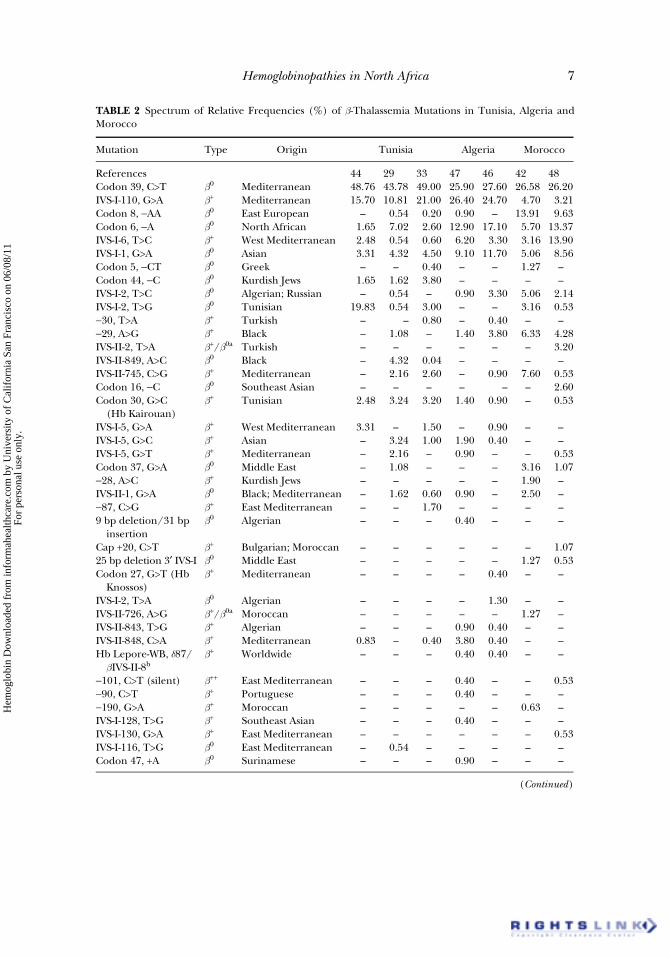
The investigations on thalassemias in Tunisia, Algeria and Moroccocontributed to establishing the spectrum of mutations in the three coun-tries. The total number of b-thal mutations identified in Tunisia was 24(Table 2) (13,29,33,42,44,46–49). The codon 39 (C>T) and IVS-I-110(G>A) mutations are largely predominant (from 54 to 70% of b-thalassemicmutations). Twenty-six mutations are described in Algeria (Table 2). Themost frequent mutations are codon 39, IVS-I-110 (equal frequency), codon6 (–A) and IVS-I-1 (G>A) which account for about 80% of the independentchromosomes (Table 2). In Morocco, recent studies showed a spectrum of26 different b-thalassemic mutations. The first six [codon 39, codon 8 (–AA),−29 (A>G), codon 6, IVS-I-6 (T>C) and IVS-I-1] represent 60 to 75% of theb-thalassemic Moroccan chromosomes (Table 2).
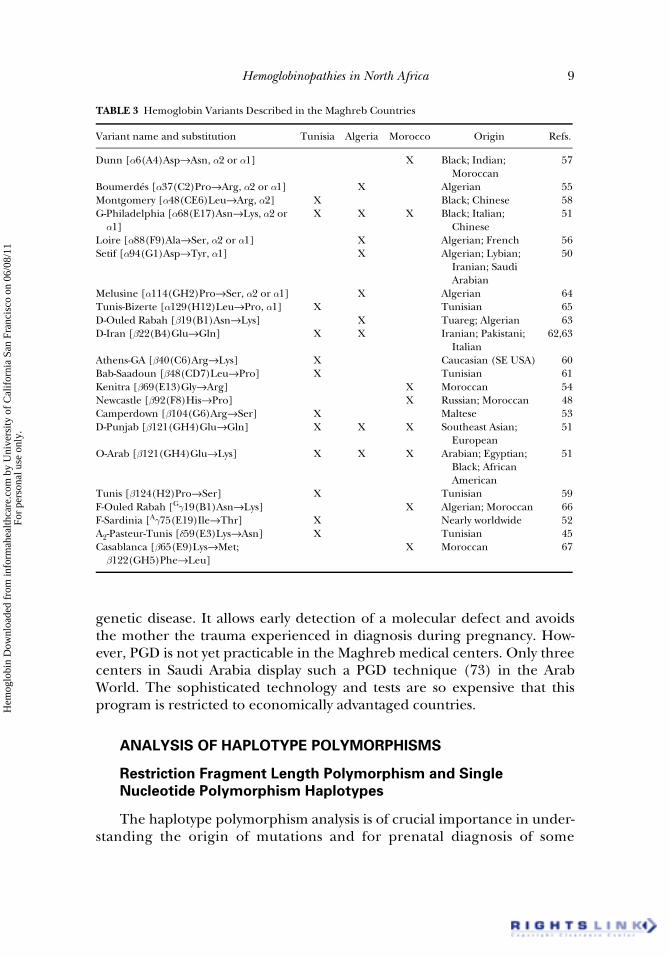
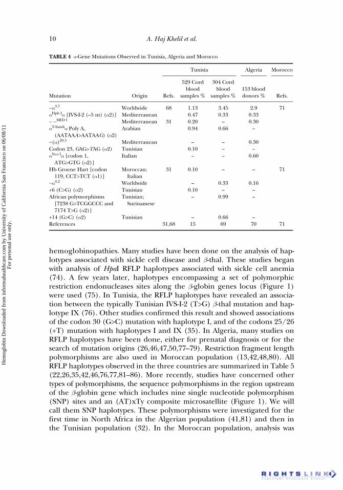
In addition to all these mutations observed in cases of b0- or b+-thal, 22 Hbvariants were described in the three countries (Table 3) (45,48,50–67). Threeof them are found in all the Maghreb countries: Hb G-Philadelphia[a68(E17)Asn→Lys], Hb D-Punjab or Hb D-Los Angeles [b121(GH4)Glu→Gln]and Hb O-Arab [b121(GH4)Glu→Lys] (51,57). Concerning a-thal, 12 mutationshave been detected (Table 4) (15,31,68–71). Not surprisingly, two of them arewidespread, i.e., −a3.7 and − −MED-I. This reduced heterogeneity could beexplained by the underestimation of these defects which cannot be observed inadult life. Together, thalassemia disorders and Hb variants account for 81mutations or variants in the three Maghreb countries.
All these genetic studies show that Maghreb populations display someheterogeneity, especially between Algeria, Tunisia and Morocco. This het-erogeneity is due to new mutational events or gene flow due to humanmigrations, and probably reflects some differences in the historical patternof migration and colonization. The three populations show a large spectrum of
Hem
oglo
bin
Dow
nloa
ded
from
info
rmah
ealth
care
.com
by
Uni
vers
ity o
f C
alif
orni
a Sa
n Fr
anci
sco
on 0
6/08
/11
For
pers
onal
use
onl
y.
Hemoglobinopathies in North Africa 7
TABLE 2 Spectrum of Relative Frequencies (%) of b-Thalassemia Mutations in Tunisia, Algeria andMorocco
Mutation Type Origin Tunisia Algeria Morocco
References 44 29 33 47 46 42 48Codon 39, C>T b0 Mediterranean 48.76 43.78 49.00 25.90 27.60 26.58 26.20IVS-I-110, G>A b+ Mediterranean 15.70 10.81 21.00 26.40 24.70 4.70 3.21Codon 8, −AA b0 East European – 0.54 0.20 0.90 – 13.91 9.63Codon 6, −A b0 North African 1.65 7.02 2.60 12.90 17.10 5.70 13.37IVS-I-6, T>C b+ West Mediterranean 2.48 0.54 0.60 6.20 3.30 3.16 13.90IVS-I-1, G>A b0 Asian 3.31 4.32 4.50 9.10 11.70 5.06 8.56Codon 5, −CT b0 Greek – – 0.40 – – 1.27 –Codon 44, −C b0 Kurdish Jews 1.65 1.62 3.80 – – – –IVS-I-2, T>C b0 Algerian; Russian – 0.54 – 0.90 3.30 5.06 2.14IVS-I-2, T>G b0 Tunisian 19.83 0.54 3.00 – – 3.16 0.53−30, T>A b+ Turkish – – 0.80 – 0.40 – –−29, A>G b+ Black – 1.08 – 1.40 3.80 6.33 4.28IVS-II-2, T>A b+/b0a Turkish – – – – – – 3.20IVS-II-849, A>C b0 Black – 4.32 0.04 – – – –IVS-II-745, C>G b+ Mediterranean – 2.16 2.60 – 0.90 7.60 0.53Codon 16, −C b0 Southeast Asian – – – – – – 2.60Codon 30, G>C
(Hb Kairouan)b+ Tunisian 2.48 3.24 3.20 1.40 0.90 – 0.53
IVS-I-5, G>A b+ West Mediterranean 3.31 – 1.50 – 0.90 – –IVS-I-5, G>C b+ Asian – 3.24 1.00 1.90 0.40 – –IVS-I-5, G>T b+ Mediterranean – 2.16 – 0.90 – – 0.53Codon 37, G>A b0 Middle East – 1.08 – – – 3.16 1.07−28, A>C b+ Kurdish Jews – – – – – 1.90 –IVS-II-1, G>A b0 Black; Mediterranean – 1.62 0.60 0.90 – 2.50 –−87, C>G b+ East Mediterranean – – 1.70 – – – –9 bp deletion/31 bp
insertionb0 Algerian – – – 0.40 – – –
Cap +20, C>T b+ Bulgarian; Moroccan – – – – – – 1.0725 bp deletion 3′ IVS-I b0 Middle East – – – – – 1.27 0.53Codon 27, G>T (Hb
Knossos)b+ Mediterranean – – – – 0.40 – –
IVS-I-2, T>A b0 Algerian – – – – 1.30 – –IVS-II-726, A>G b+/b0a Moroccan – – – – – 1.27 –IVS-II-843, T>G b+ Algerian – – – 0.90 0.40 – –IVS-II-848, C>A b+ Mediterranean 0.83 – 0.40 3.80 0.40 – –Hb Lepore-WB, d87/bIVS-II-8b
b+ Worldwide – – – 0.40 0.40 – –
−101, C>T (silent) b++ East Mediterranean – – – 0.40 – – 0.53−90, C>T b+ Portuguese – – – 0.40 – – –−190, G>A b+ Moroccan – – – – – 0.63 –IVS-I-128, T>G b+ Southeast Asian – – – 0.40 – – –IVS-I-130, G>A b+ East Mediterranean – – – – – – 0.53IVS-I-116, T>G b0 East Mediterranean – 0.54 – – – – –Codon 47, +A b0 Surinamese – – – 0.90 – – –
(Continued)
Hem
oglo
bin
Dow
nloa
ded
from
info
rmah
ealth
care
.com
by
Uni
vers
ity o
f C
alif
orni
a Sa
n Fr
anci
sco
on 0
6/08
/11
For
pers
onal
use
onl
y.
8 A. Haj Khelil et al.
mutations. Indeed, the Maghreb countries, by their privileged geographicalposition and diverse ethnic origins of their populations, represent an inter-esting area for the study of Hb disorders. Moreover, the high level of con-sanguinity, more particularly in rural areas where the frequency ofmarriages between first cousins is as high as 25–30% due to cultural tradi-tions, increases considerably the probability of genetic disorders.
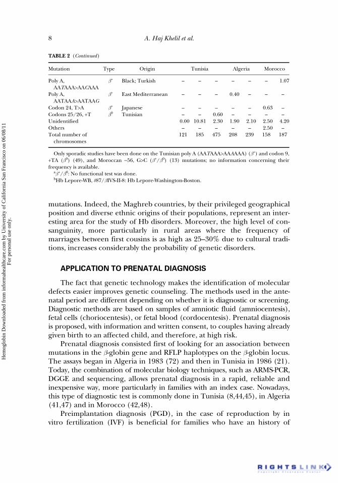
APPLICATION TO PRENATAL DIAGNOSIS
The fact that genetic technology makes the identification of moleculardefects easier improves genetic counseling. The methods used in the ante-natal period are different depending on whether it is diagnostic or screening.Diagnostic methods are based on samples of amniotic fluid (amniocentesis),fetal cells (choriocentesis), or fetal blood (cordocentesis). Prenatal diagnosisis proposed, with information and written consent, to couples having alreadygiven birth to an affected child, and therefore, at high risk.
Prenatal diagnosis consisted first of looking for an association betweenmutations in the b-globin gene and RFLP haplotypes on the b-globin locus.The assays began in Algeria in 1983 (72) and then in Tunisia in 1986 (21).Today, the combination of molecular biology techniques, such as ARMS-PCR,DGGE and sequencing, allows prenatal diagnosis in a rapid, reliable andinexpensive way, more particularly in families with an index case. Nowadays,this type of diagnostic test is commonly done in Tunisia (8,44,45), in Algeria(41,47) and in Morocco (42,48).
Preimplantation diagnosis (PGD), in the case of reproduction by invitro fertilization (IVF) is beneficial for families who have an history of
TABLE 2 (Continued)
Mutation Type Origin Tunisia Algeria Morocco
Poly A,AATAAA>AACAAA
b+ Black; Turkish – – – – – – 1.07
Poly A, AATAAA>AATAAG
b+ East Mediterranean – – – 0.40 – – –
Codon 24, T>A b+ Japanese – – – – – 0.63 –Codons 25/26, +T b0 Tunisian – – 0.60 – – – –Unidentified 0.00 10.81 2.30 1.90 2.10 2.50 4.20Others – – – – – 2.50 –Total number of
chromosomes121 185 475 208 239 158 187
Only sporadic studies have been done on the Tunisian poly A (AATAAA>AAAAAA) (b+) and codon 9,+TA (b0) (49), and Moroccan −56, G>C (b+/b0) (13) mutations; no information concerning theirfrequency is available.
ab+/b0: No functional test was done.bHb Lepore-WB, d87/bIVS-II-8: Hb Lepore-Washington-Boston.
Hem
oglo
bin
Dow
nloa
ded
from
info
rmah
ealth
care
.com
by
Uni
vers
ity o
f C
alif
orni
a Sa
n Fr
anci
sco
on 0
6/08
/11
For
pers
onal
use
onl
y.
Hemoglobinopathies in North Africa 9
genetic disease. It allows early detection of a molecular defect and avoidsthe mother the trauma experienced in diagnosis during pregnancy. How-ever, PGD is not yet practicable in the Maghreb medical centers. Only threecenters in Saudi Arabia display such a PGD technique (73) in the ArabWorld. The sophisticated technology and tests are so expensive that thisprogram is restricted to economically advantaged countries.
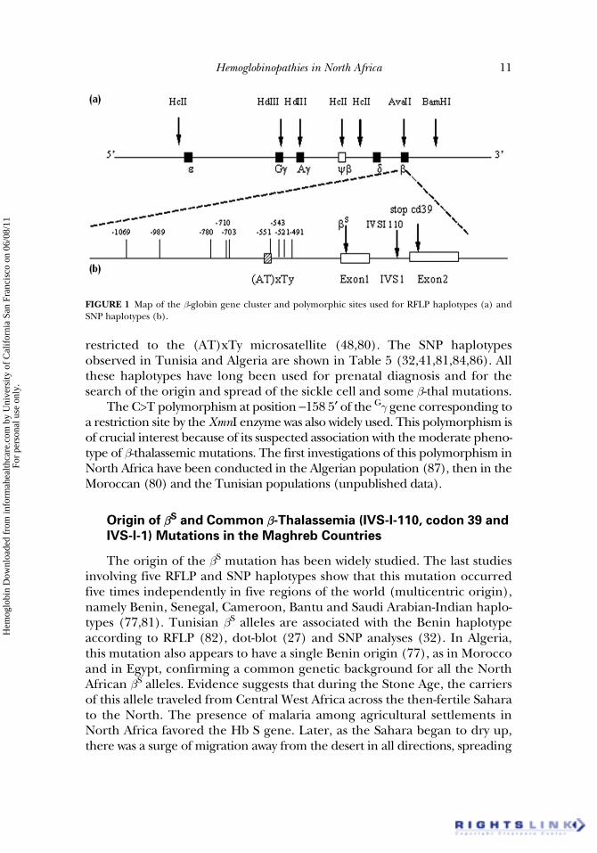
ANALYSIS OF HAPLOTYPE POLYMORPHISMS
Restriction Fragment Length Polymorphism and Single
Nucleotide Polymorphism Haplotypes
The haplotype polymorphism analysis is of crucial importance in under-standing the origin of mutations and for prenatal diagnosis of some
TABLE 3 Hemoglobin Variants Described in the Maghreb Countries
Variant name and substitution Tunisia Algeria Morocco Origin Refs.
Dunn [a6(A4)Asp→Asn, a2 or a1] X Black; Indian; Moroccan
57
Boumerdés [a37(C2)Pro→Arg, a2 or a1] X Algerian 55Montgomery [a48(CE6)Leu→Arg, a2] X Black; Chinese 58G-Philadelphia [a68(E17)Asn→Lys, a2 or a1]
X X X Black; Italian; Chinese
51
Loire [a88(F9)Ala→Ser, a2 or a1] X Algerian; French 56Setif [a94(G1)Asp→Tyr, a1] X Algerian; Lybian;
Iranian; Saudi Arabian
50
Melusine [a114(GH2)Pro→Ser, a2 or a1] X Algerian 64Tunis-Bizerte [a129(H12)Leu→Pro, a1] X Tunisian 65D-Ouled Rabah [b19(B1)Asn→Lys] X Tuareg; Algerian 63D-Iran [b22(B4)Glu→Gln] X X Iranian; Pakistani;
Italian62,63
Athens-GA [b40(C6)Arg→Lys] X Caucasian (SE USA) 60Bab-Saadoun [b48(CD7)Leu→Pro] X Tunisian 61Kenitra [b69(E13)Gly→Arg] X Moroccan 54Newcastle [b92(F8)His→Pro] X Russian; Moroccan 48Camperdown [b104(G6)Arg→Ser] X Maltese 53D-Punjab [b121(GH4)Glu→Gln] X X X Southeast Asian;
European51
O-Arab [b121(GH4)Glu→Lys] X X X Arabian; Egyptian; Black; African American
51
Tunis [b124(H2)Pro→Ser] X Tunisian 59F-Ouled Rabah [Gg19(B1)Asn→Lys] X Algerian; Moroccan 66F-Sardinia [Ag75(E19)Ile→Thr] X Nearly worldwide 52A2-Pasteur-Tunis [d59(E3)Lys→Asn] X Tunisian 45Casablanca [b65(E9)Lys→Met;b122(GH5)Phe→Leu]
X Moroccan 67
Hem
oglo
bin
Dow
nloa
ded
from
info
rmah
ealth
care
.com
by
Uni
vers
ity o
f C
alif
orni
a Sa
n Fr
anci
sco
on 0
6/08
/11
For
pers
onal
use
onl
y.
10 A. Haj Khelil et al.
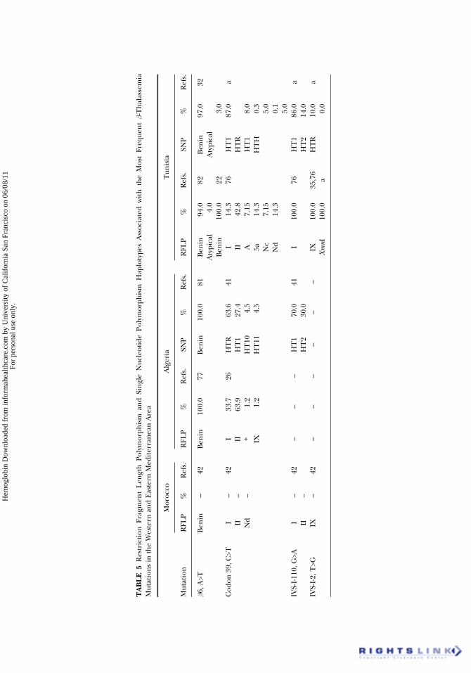
hemoglobinopathies. Many studies have been done on the analysis of hap-lotypes associated with sickle cell disease and b-thal. These studies beganwith analysis of HpaI RFLP haplotypes associated with sickle cell anemia(74). A few years later, haplotypes encompassing a set of polymorphicrestriction endonucleases sites along the b-globin genes locus (Figure 1)were used (75). In Tunisia, the RFLP haplotypes have revealed an associa-tion between the typically Tunisian IVS-I-2 (T>G) b-thal mutation and hap-lotype IX (76). Other studies confirmed this result and showed associationsof the codon 30 (G>C) mutation with haplotype I, and of the codons 25/26(+T) mutation with haplotypes I and IX (35). In Algeria, many studies onRFLP haplotypes have been done, either for prenatal diagnosis or for thesearch of mutation origins (26,46,47,50,77–79). Restriction fragment lengthpolymorphisms are also used in Moroccan population (13,42,48,80). AllRFLP haplotypes observed in the three countries are summarized in Table 5(22,26,35,42,46,76,77,81–86). More recently, studies have concerned othertypes of polymorphisms, the sequence polymorphisms in the region upstreamof the b-globin gene which includes nine single nucleotide polymorphism(SNP) sites and an (AT)xTy composite microsatellite (Figure 1). We willcall them SNP haplotypes. These polymorphisms were investigated for thefirst time in North Africa in the Algerian population (41,81) and then inthe Tunisian population (32). In the Moroccan population, analysis was
TABLE 4 a-Gene Mutations Observed in Tunisia, Algeria and Morocco
Mutation Origin
Tunisia Algeria Morocco
Refs.
529 Cord blood
samples %
304 Cord blood
samples %153 blood donors % Refs.
−a3.7 Worldwide 68 1.13 3.45 2.9 71aHph Ia [IVS-I-2 (–5 nt) (a2)] Mediterranean 0.47 0.33 0.33− −MED I Mediterranean 31 0.20 – 0.30aT-Saudia Poly A,
(AATAAA>AATAAG) (a2)Arabian 0.94 0.66 –
−(a)20.5 Mediterranean – – 0.30Codon 23, GAG>TAG (a2) Tunisian 0.10 – –aNco Ia [codon 1,
ATG>GTG (a2)]Italian – – 0.60
Hb Groene Hart [codon 119, CCT>TCT (a1)]
Moroccan; Italian
31 0.10 – – 71
−a4.2 Worldwide – 0.33 0.16+6 (C>G) (a2) Tunisian 0.10 – –African polymorphisms
[7238 G>TCGGCCC and 7174 T>G (a2)]
Tunisian; Surinamese
– 0.99 –
+14 (G>C) (a2) Tunisian – 0.66 –References 31,68 15 69 70 71
Hem
oglo
bin
Dow
nloa
ded
from
info
rmah
ealth
care
.com
by
Uni
vers
ity o
f C
alif
orni
a Sa
n Fr
anci
sco
on 0
6/08
/11
For
pers
onal
use
onl
y.
Hemoglobinopathies in North Africa 11
restricted to the (AT)xTy microsatellite (48,80). The SNP haplotypesobserved in Tunisia and Algeria are shown in Table 5 (32,41,81,84,86). Allthese haplotypes have long been used for prenatal diagnosis and for thesearch of the origin and spread of the sickle cell and some b-thal mutations.
The C>T polymorphism at position −158 5′ of the Gg gene corresponding toa restriction site by the XmnI enzyme was also widely used. This polymorphism isof crucial interest because of its suspected association with the moderate pheno-type of b-thalassemic mutations. The first investigations of this polymorphism inNorth Africa have been conducted in the Algerian population (87), then in theMoroccan (80) and the Tunisian populations (unpublished data).
Origin of bS and Common b-Thalassemia (IVS-I-110, codon 39 and
IVS-I-1) Mutations in the Maghreb Countries
The origin of the bS mutation has been widely studied. The last studiesinvolving five RFLP and SNP haplotypes show that this mutation occurredfive times independently in five regions of the world (multicentric origin),namely Benin, Senegal, Cameroon, Bantu and Saudi Arabian-Indian haplo-types (77,81). Tunisian bS alleles are associated with the Benin haplotypeaccording to RFLP (82), dot-blot (27) and SNP analyses (32). In Algeria,this mutation also appears to have a single Benin origin (77), as in Moroccoand in Egypt, confirming a common genetic background for all the NorthAfrican bS alleles. Evidence suggests that during the Stone Age, the carriersof this allele traveled from Central West Africa across the then-fertile Saharato the North. The presence of malaria among agricultural settlements inNorth Africa favored the Hb S gene. Later, as the Sahara began to dry up,there was a surge of migration away from the desert in all directions, spreading
FIGURE 1 Map of the b-globin gene cluster and polymorphic sites used for RFLP haplotypes (a) andSNP haplotypes (b).
Hem
oglo
bin
Dow
nloa
ded
from
info
rmah
ealth
care
.com
by
Uni
vers
ity o
f C
alif
orni
a Sa
n Fr
anci
sco
on 0
6/08
/11
For
pers
onal
use
onl
y.
TA
BL
E 5
Res
tric
tion
Fra
gmen
t L
engt
h P
olym
orph
ism
an
d Si
ngl
e N
ucle
otid
e Po
lym
orph
ism
Hap
loty
pes
Ass
ocia
ted
wit
h t
he
Mos
t Fr
eque
nt b-
Th
alas
sem
iaM
utat
ion
s in
the
Wes
tern
an
d E
aste
rn M
edit
erra
nea
n A
rea
Mor
occo
Alg
eria
Tun
isia
Mut
atio
nR
FLP
%R
efs.
RFL
P%
Ref
s.SN
P%
Ref
s.R
FLP
%R
efs.
SNP
%R
efs.
b6, A
>TB
enin
–42
Ben
in10
0.0
77B
enin
100.
081
Ben
in94
.082
Ben
in97
.032
Aty
pica
l4.
0A
typi
cal
Ben
in10
0.0
223.
0C
odon
39,
C>T
I–
42I
33.7
26H
TR
63.6
41I
14.3
76H
T1
87.0
aII
–II
63.9
HT
127
.4II
42.8
HT
RN
d–
*1.
2H
T10
4.5
A7.
15H
T1
8.0
IX1.
2H
T11
4.5
5a14
.3H
TH
0.3
Nc
7.15
5.0
Nd
14.3
0.1
5.0
IVS-
I-11
0, G
>AI
–42
––
–H
T1
70.0
41I
100.
076
HT
186
.0a
II–
HT
230
.0H
T2
14.0
IVS-
I-2,
T>G
IX–
42–
––
––
–IX
100.
035
,76
HT
R10
.0a
Xm
nI10
0.0
a0.
0
Hem
oglo
bin
Dow
nloa
ded
from
info
rmah
ealth
care
.com
by
Uni
vers
ity o
f C
alif
orni
a Sa
n Fr
anci
sco
on 0
6/08
/11
For
pers
onal
use
onl
y.
Tur
key
Leb
anon
Mut
atio
nR
FLP
%R
efs.
SNP
%R
efs.
RFL
P%
Ref
s.SN
P%
Ref
s.
b6, A
>T–
––
––
–B
enin
73.0
85–
––
Cam
eroo
n15
.0Sa
udi A
rabi
an-I
ndi
an10
.0Se
neg
al2.
0C
odon
39,
C>T
II50
.083
HT
155
.084
II66
.786
HT
110
.086
IV50
.0H
TR
45.0
I33
.3–
0.0
IVS-
I-11
0, G
>AI
93.1
83H
T1
88.0
84I
93.2
86H
T1
76.0
86II
4.1
HT
R5.
05′
-12
5.4
HT
R24
.0IX
1.4
HT
33.
5II
1.0
IV1.
4H
T4
2.0
HT
50.
5H
T7
1.0
IVS-
I-2,
T>G
––
––
––
––
––
––
Th
e R
FLP
hap
loty
pes:
Ben
in [
− −
− −
+ +
+]; I
[+
− −
− −
+ +]
; II
[− +
+ −
+ +
+];
IV
[−
+ −
+ +
− +]
; IX
[−
+ −
+ +
+ +]
; * [
− +
+ −
+ +
−]; A
[−
− −
− −
+ +]
; 5a
[− +
+ −
− +
+]; N
c [+
+ +
− −
+ +
]; N
d [−
− −
− +
+ +
]; 5
′−12
[−
− −
+ +
+ +]
. SN
P h
aplo
type
s: −
1069
, −98
9, −
780,
−71
0, −
703,
−55
1, −
543,
−52
1, −
491,
(A
T)x
-Ty.
Ben
in:
AG
AT
TT
TC
C 8
-4; H
T1:
AC
AT
TT
CC
A 7
-7; H
T2:
AC
AT
CC
CC
A 9
-5; H
T3:
GC
AT
CC
CC
A 1
1-3;
HT
4: G
CA
TC
CC
CA
7-7
; HT
5: G
CA
TC
TC
CA
7-7
; HT
Y: A
CA
TT
C-
CC
A 7
-7; H
T10
: GC
A T
TT
CC
A 1
1-3;
HT
11: G
GA
TT
CC
CA
11-
3; H
TR
: GC
AT
TT
CC
A 7
-7; H
TH
: AC
AT
TC
CC
A 9
-5; H
TI:
GG
AT
TT
CC
A 8
-5.
a Haj
Kh
elil
et a
l., u
npu
blis
hed
dat
a.
Hem
oglo
bin
Dow
nloa
ded
from
info
rmah
ealth
care
.com
by
Uni
vers
ity o
f C
alif
orni
a Sa
n Fr
anci
sco
on 0
6/08
/11
For
pers
onal
use
onl
y.
14 A. Haj Khelil et al.
the gene further (78). However, all approaches used to estimate the age ofthe bS mutation suggest that it arose about 3,000 years ago (88). Until now,there has been no evidence of a more ancient presence of this mutation. Itsintroduction in North Africa is probably more recent and due to the forcedmigrations from black Africa through the slavery roads and/or to the con-tinuous influx of sub-Saharan Africans through the caravan routes (46).
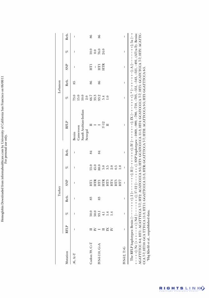
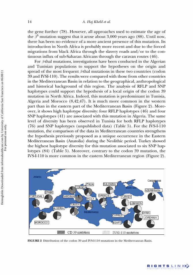
For b-thal mutations, investigations have been conducted in the Algerianand Tunisian populations to support the hypotheses on the origin andspread of the most frequent b-thal mutations in these two countries (codon39 and IVSI-110). The results were compared with those from other countriesin the Mediterranean Basin in relation to the geographical, anthropologicaland historical background of this region. The analysis of RFLP and SNPhaplotypes could support the hypothesis of a local origin of the codon 39mutation in North Africa. Indeed, this mutation is predominant in Tunisia,Algeria and Morocco (8,42,47). It is much more common in the westernpart than in the eastern part of the Mediterranean Basin (Figure 2). More-over, it shows high haplotype diversity: four RFLP haplotypes (46) and fourSNP haplotypes (41) are associated with this mutation in Algeria. The samelevel of diversity has been observed in Tunisia for both RFLP haplotypes(76) and SNP haplotypes (unpublished data) (Table 5). For the IVS-I-110mutation, the comparison of the data in Mediterranean countries strengthensthe hypothesis previously proposed as a unique occurrence in the EasternMediterranean Basin (Anatolia) during the Neolithic period. Turkey showedthe highest haplotype diversity for this mutation associated to six SNP hap-lotypes (84) (Table 5). Moreover, contrary to the codon 39 mutation, theIVS-I-110 is more common in the eastern Mediterranean region (Figure 2).
FIGURE 2 Distribution of the codon 39 and IVS-I-110 mutations in the Mediterranean Basin.
Hem
oglo
bin
Dow
nloa
ded
from
info
rmah
ealth
care
.com
by
Uni
vers
ity o
f C
alif
orni
a Sa
n Fr
anci
sco
on 0
6/08
/11
For
pers
onal
use
onl
y.
Hemoglobinopathies in North Africa 15
This mutation could have been introduced into North Africa (Tunisia andAlgeria) during the Ottoman rule in the 17th century. The fact that this muta-tion is the most frequent in Algeria, the second in Tunisia and the seventh inMorocco (infrequent), confirms this hypothesis. Indeed, the Ottoman occupa-tion did not reach Morocco. Its limited introduction in this country would bethe result of sporadic Algerian migrations. The results of haplotype analysesargue in favor of this hypothesis. Indeed, a single RFLP haplotype (I) was foundin Tunisia (76). Two haplotypes (I and II) were found in Algeria (26,46). Thepredominant haplotype (I) is also described in Tunisia (92%). These twohaplotypes are the most frequent of the four haplotypes found in Turkey (I, II,IV and IX) (83) (Table 5). These findings were confirmed by SNP haplotypeanalysis in Algeria (41) and in Tunisia (unpublished data) showing the associa-tion of the IVS-I-110 mutation with the same haplotypes: HT1 and HT2. HT1 isby far the most frequent haplotype as found previously in Turkey. The stronglinkage disequilibrium between this haplotype and the IVS-I-110 mutationobserved in Turkey, Algeria, and Tunisia strengthens the hypothesis of an EastMediterranean origin of this mutation in North Africa.
If we compare all the data, we notice important differences in the propor-tion of frequent mutations such as codon 39, IVS-I-110 and IVS-I-1. This proba-bly reflects an unequal distribution of common Mediterranean mutationsthroughout the Maghreb. The IVS-I-110 mutation is predominant in the west-ern part of Tunisia and in northern Algeria. This fits well with an Ottomanimportation. This mutation is rare in Morocco which was never occupied by theOttomans. Codon 39 is largely predominant in Tunisia, in Morocco and in thenorth-western part of Algeria. Codon 39 is likely to have an occidental andancient origin (chromosomes are recombined in one-third of the cases)(Table 5). It could have been introduced into the Maghreb during the Romanperiod through Italy (41%) and Spain (35%) (89). The Roman Empire cov-ered a large part of the Mediterranean area until the 5th century BC when theByzantine reconquest occurred. The IVS-I-1 mutation is found mostly in thecentral region of Algeria and in Morocco. But it reaches its highest frequencyin the Czech Republic and in Hungary (45 and 31%, respectively) (89). Thelack of information concerning RFLP and SNP haplotypes in central Europeanpopulations did not give us the opportunity to discuss a unique or recurrentorigin for the mutation even when Bennani et al. (46) described an associationwith five RFLP haplotypes (i.e., I, III, V, IX and A) in Algeria, and Lemsaddeket al. (48), three RFLP haplotypes (IV, V and IX) in Morocco and Portugal.
Origin of Other b-Thalassemia Mutations in the Maghreb
Countries
The codon 8 (−AA) is the second most common mutation encounteredin Morocco. It appears rarely in Algeria and Tunisia. If this mutation was
Hem
oglo
bin
Dow
nloa
ded
from
info
rmah
ealth
care
.com
by
Uni
vers
ity o
f C
alif
orni
a Sa
n Fr
anci
sco
on 0
6/08
/11
For
pers
onal
use
onl
y.
16 A. Haj Khelil et al.
originally observed in a Turkish patient (90) and later in 5.5% of b-thalassemicTurkish alleles (91), nowhere is the frequency higher than in Russia (39%)and Azerbaijan (30%) (89). Some interesting features also appear concerninggeographic distribution of the IVS-I-5 (G>T) and IVS-II-848 (C>A) muta-tions. They are restricted to central Algeria and northern and central Tunisia(33,46). It could reflect a common historical genetic event. The IVS-I-2mutation, first detected in the region of Essouassi-El Djem in central Tunisia(76), accounts for 17% of b-thal mutations in central Tunisia, suggesting itis of Tunisian origin. Amazingly, some cases have recently been describedin Morocco (48). Some sporadic genetic flow could explain this result. HbKnossos [b27(B9)Ala→Ser, codon 27 (G>T)] has been observed both inAlgeria (Algiers region) and Tunisia (92). A founder effect is probablethrough Palestine or Jordan where this mutation reaches a highest fre-quency (2.1 and 3.3%, respectively) (89). One case of the IVS-I-130(G>A) b-thalassemic mutation is described in Morocco (Table 2). Such amutant allele has also been described in a Turkish patient (93) and inan Egyptian patient (94). The −101 (C>T), b-thal allele is observed spe-cifically in Morocco and eastern Algeria. It is sporadically found allaround the Mediterranean Basin but with very low frequencies (around1%). In this context, it seems very difficult to have some speculationsconcerning its origin. Finally, the sub-Saharan −29 mutation observed inMorocco (5.4%), in Algeria (3%) and sporadically in the northern partof Tunisia (1.08%), could be explained by caravan routes or could bethe result of the Almoravid Dynasty that spread over a wide area ofnorthwestern Africa (present-day Morocco, west part of Algeria, Mauritania,Senegal, Mali) and the Iberian peninsula (Southern Portugal and Spain) assuggested by Lemsaddek et al. (48).
Maghreb Autochthonous b-Thalassemia Mutations
A set of more than 10 mutations are clearly the result of autochtho-nous genetic occurrence. There are seven mutations prevalent inMorocco: −190 (G>A), −56 (G>C), Cap +20 (C>T) promoter mutation,codon 24 (T>A), IVS-II-726 (A>G), 25 bp deletion (+252 through +276)and the polyadenylation (poly A) site (AATAAA>AACAAA). Codon 30(G>C) [Hb Kairouan, b30(B12)Arg→Thr], −87 (C>G), codons 25/26(+T), and IVS-II-849 (A>C); these b-thal anomalies clearly seem to havea Tunisian origin. The codon 44 (−C), exclusively observed in Tunisia(2.4%), was first described in a Kurdish patient, but the highest fre-quency has been found in the Israeli population with a frequency of9.4% (89). Four b-thal mutations could be considered autochthonous inAlgeria: IVS-I-2 (T>A), IVS-II-843 (T>G), the −9 bp deletion/+31 bpinsertion and poly A (AATAAA>AATAAG).
Hem
oglo
bin
Dow
nloa
ded
from
info
rmah
ealth
care
.com
by
Uni
vers
ity o
f C
alif
orni
a Sa
n Fr
anci
sco
on 0
6/08
/11
For
pers
onal
use
onl
y.
Hemoglobinopathies in North Africa 17
Maghreb Autochthonous a-Thalassemia Mutations
Nine molecular defects responsible for a-thal and one a gene polymor-phism have been reported in Tunisia (15,69) (Table 4). Six mutations weredescribed in Algeria (70). The −a3.7 mutation is the most frequent allele inNorth Africa (15,69–71). It is specific to the Mediterranean regions but itreaches its highest frequencies in Iran (79.1%) (95) and Saudi Arabia(64%) (96) indicating that it could have been introduced in North Africaby Arab conquests. Previous studies have demonstrated that the majority ofpatients with the another two Mediterranean alleles [− −MED I and aHph Ia(IVS-I-2 (−5 nucleotide) (−5 nt) (a2) deletions], observed in both Tunisiaand Algeria probably have a Sicilian (97) and a Western Mediterranean ori-gin (98), respectively. On the other hand, the aT−Saudi allele observed inTunisia is more prevalent in the eastern part of the Mediterranean and inthe Arabian Peninsula where it reaches 41% in Saudi Arabia (96). The −a4.2
mutation observed in Tunisia and Algeria seems to have a Southeast Asianorigin. The Hb Groene Hart [a119(H2)Pro→Ser (a1)] mutation has beensporadically reported in association with the common −a3.7 deletion in het-erozygotes of Moroccan (71) and Tunisian (68) origin. A Tunisian andthen an autochthonous origin is plausible for some alleles [codon 23(GAG>TAG) (a2), +6 (C>G) (a2) and the +14 (G>C) (a2) polymorphism inthe 5′UTR (5′ untranslated region)] because until now they have only beenobserved in Tunisia (15,69). The aNco Ia (a2, codon 1, ATG>GTG) firstdescribed in an Italian patient and the −(a)20.5 of Mediterranean origin,were detected only in Algeria (70).
Maghreb Autochthonous Hemoglobin Variants
More than 20 Hb variants have been observed in North Africa (Table 3).The high variability is comparable with the range of variants described inthe Mediterranean region. Moreover, many of these variants are commonin different countries, denoting the ongoing genetic exchange occurring inthis region over time. However, each country seems to have its own muta-tions that could have appeared de novo in its population. It is the case of HbTunis [b124(H2)Pro→Ser] (59), Hb Bab-Saadoun [b48(CD7)Leu→Pro](61), Hb Tunis-Bizerte [a129(H12)Leu→Pro] (65), and Hb A2-Pasteur-Tunis[d59(E3)Lys→Asn] (45) for Tunisia, and Hb Setif [a94(G1)Asp→Tyr] (50),Hb Boumerdés [a37(C2)Pro→Arg] (55), Hb Loire [a88(F9)Ala→Ser](56), Hb Melusine [a114(GH2)Pro→Ser] (64), Hb D-Ouled Rabah[b19(B1)Asn→Lys] (63) for Algeria, and Hb Kenitra [b69(E13)Gly→Arg](54), Hb Dunn [a6(A4)Asp→Asn] (57), Hb Casablanca [b65(E9)Lys→Met;b122(GH5)Phe→Leu] (67) and Hb Newcastle [b92(F8)His→Pro] (48) forMorocco.
Hem
oglo
bin
Dow
nloa
ded
from
info
rmah
ealth
care
.com
by
Uni
vers
ity o
f C
alif
orni
a Sa
n Fr
anci
sco
on 0
6/08
/11
For
pers
onal
use
onl
y.
18 A. Haj Khelil et al.
Association Between Haplotype Polymorphism and the Moderate
Phenotype of Sickle Cell Disease and b-Thalassemia
Some patients with sickle cell disease or b-thal show moderate phenotypesof their pathologies. Genetic polymorphisms have been described in associ-ation with these moderate phenotypes. Analysis using the RFLP haplotypesshowed an association between haplotype IX and the IVS-I-2 (T>G) muta-tion for which homozygous patients show a moderate phenotype (35,76).The XmnI polymorphism corresponding to the C>T variation at position−158 upstream to the Cap site of the Gg gene was found in strong associa-tion with the persistence of Gg/Ag fetal rate in adulthood and most often ahigher rate of Hb F in patients with sickle cell disease, b-thal and Hb E/b-thal(99). Some studies have shown that homozygosity for this polymorphismwas significantly associated with an intermediate phenotype of b-thal insoutheast Iran (100) and Turkey (101). In Tunisia, a recent study showed astrong association between the T allele of this polymorphism and the IVS-I-2(T>G) mutation (unpublished data). In Algeria, this polymorphism wasdescribed long ago (87), and more recently in an Algerian patient showinga phenotype of b0-thal intermedia (102). In Morocco, the XmnI polymor-phism has been shown in linkage with RFLP haplotypes III, IV and IX(48,80). Other studies have shown a positive correlation between this poly-morphism and the mutation codon 8 (−AA) in linkage with haplotype IV(13). The determination of the molecular genetic marker for early child-hood could help to provide better care for patients such as the identifica-tion of candidates for the pharmacological Hb F switch by hydroxyurea(13). However, other studies in Morocco showed an absence of correlationbetween the XmnI polymorphism and Hb F levels (48,80). The (AT)xTymicrosatellite polymorphism has also been described as influencing thephenotype of b-thal by increasing the rate of Hb F synthesis (103). On theother hand, some studies in Algeria have demonstrated the absence of thisassociation (48,80) showing that correlation between the genotype and thephenotype can change from one population to another.
Finally, the genotype-phenotype relationship has recently been studiedin a Tunisian family carrying the IVS-I-2 (T>G) mutation by studying themRNA metabolism in the context of this molecular defect (104). The vari-ous splicing forms identified for these mRNA could provide new elementsfor understanding the moderate phenotype observed in patients carryingthis mutation.
CONCLUSIONS
Hemoglobinopathies, particularly in the homozygous state, are severediseases of the Mediterranean area. In North Africa, the prevalence of these
Hem
oglo
bin
Dow
nloa
ded
from
info
rmah
ealth
care
.com
by
Uni
vers
ity o
f C
alif
orni
a Sa
n Fr
anci
sco
on 0
6/08
/11
For
pers
onal
use
onl
y.
Hemoglobinopathies in North Africa 19
diseases is more or less known. It is estimated at about 1.5 to 4.8% in thegeneral population of the Maghreb countries. However, given the high rateof endogamy still observed in these countries, it is likely that genetic diseasesin general and hemoglobinopathies in particular are especially prevalent.They now take an increasingly important place both in everyday medicalpractice and research. The development of genetics has led to a betterunderstanding of the molecular basis of diseases studied, including theidentification of mutations and polymorphisms associated with these muta-tions. It also makes possible an accurate diagnosis, including in the prenatalperiod, and the development of appropriate protocols for these diseases.Preimplantation diagnosis followed by IVF, represents “state of the art” pro-cedures which allow the transfer of only unaffected embryos. This expensivetechnique is still not offered in clinical centers of the Maghreb countries.Finally, some hope could come from gene therapy. In the area of geneticdisorders, ideas are changing the concept of preventive or curative medi-cine to that of predictive medicine.
ACKNOWLEDGMENTS
The authors express their thanks to CNRS, the TEMPRA (RégionRhône-Alpes and Gouvernorat de Monastir), MIRA, CMCU, and OHLL(Origine de l’Homme, des Langages et des Langues) programs for support-ing the training of Amel Haj Khelil in France. The authors wish to thankDr. Faouzi Baklouti, (Research Director) INSERM, CNRS, UMR 5534 at theCenter of Molecular and Cellular Genetics in Lyon, France; Amel Haj Khelilstudied “Cell differentiation and alternative splicing” in his laboratory. Wethank the referee for his very helpful comments.
Declaration of Interest: The authors report no conflicts of interest. Theauthors alone are responsible for the content and writing of this article.
REFERENCES
1. Weatherall DJ, Clegg JB. The Thalassaemia Syndromes, 3rd ed. Oxford: Blackwell Scientific; 1981.2. Hardison RC, Chui DHK, Giardine B, et al. HbVar: a relational database of human hemoglobin
variants and thalassemia mutations at the globin gene server. Hum Mutat. 2002;19(3):225–223(http://globin.cse.psu.edu).
3. Patrinos GP, Giardine B, Riemer C, et al. Improvements in the HbVar database of human hemoglobinvariants and thalassemia mutations for population and sequence variation studies. Nucleic AcidsRes. 2004;32(Database issue):D537-D541 (http://globin.cse.psu.edu).
4. Kaplan JC, Delpech M. Génétique moléculaire de quelques maladies constitutionnelles. BiologieMoléculaire et Médecine. Paris: Flammarion, Médecine-Sciences. 1989:273–338.
5. Pissard S, M’Rad A, Beuzard Y, Roméo PH. A new type of hereditary persistence of fetal haemoglobin(HPFH): HPFH Tunisia b+ (+C−200) Gg. Br J Haematol. 1996;95(1): 67–72.
6. Flint J, Hill AVS, Bowden DK, Oppenheimer SJ. High frequencies of thalassemia are the result ofnatural selection by malaria. Nature. 1986;321(6072):744–750.
Hem
oglo
bin
Dow
nloa
ded
from
info
rmah
ealth
care
.com
by
Uni
vers
ity o
f C
alif
orni
a Sa
n Fr
anci
sco
on 0
6/08
/11
For
pers
onal
use
onl
y.
20 A. Haj Khelil et al.
7. Galactéros F. Drépanocytose. Physiopathologie et diagnostic. Rev Prat. 1995;45(3):351–361.8. Fattoum S. Hemoglobinopathies in Tunisia. An updated review of the epidemiologic and molecular
data. Tunis Med. 2006;84(11):687–696.9. Fawzi ZO, Al Hilali A, Fakhroo N, Al Bin Ali A, Al Mansour S. Distribution of Hemoglobinopathies
and Thalassemias in Qatari Nationals Seen at Hamad Hospital in Qatar. Qatar Med J. 2003;12(1):20–24.
10. Benkirane Agoumi N, Sebar A. Les hémoglobinopathies au Maroc. Arch Pédiatr. 2003;10(7):648–657.11. Rosa J, Wajcman H, Blouquit Y. Hémoglobine. Encycl Méd Chir, Hématologie. France: Editions
Techniques. 1993;14 (13000-S-10).12. Babiker MM, Bachir N, Sarsour N. Prevalence of thalassaemia in schoolchildren in north-eastern
Badia, Jordan. East Mediterr Health J. 1999;5(6):1165–1170.13. Agouti I, Badens C, Abouyoub A, et al. Genotypic correlation between six common b-thalassemia
mutations and the XmnI polymorphism in the Moroccan population. Hemoglobin. 2007;31(2):141–149.
14. Fuchaoren S, Winichagoon P. Hemoglobinopathies in Southeast Asia. Hemoglobin.1987;11(1): 65–88.
15. Siala H, Ouali F, Messaoud T, Bibi A, Fattoum S. a-Thalassaemia in Tunisia: some epidemiologicaland molecular data. J Genet. 2008;87(3):229–234.
16. Williams TN, Mailand K, Ganczakowski M, et al. Red blood cell phenotypes in a+ thalassaemiasfrom early childhood to maturity. Br J Haematol. 1996;95(2):266–272.
17. Favier R, Ozsahin H, Laire V, Douay L. Apport du laboratoire dans le dépistage et le diagnostic deshémoglobinopathies en milieu pédiatrique. Rev Fr Lab. 1993;248:53–62.
18. Wilson JB, Headlee ME, Huisman THJ. A new high-performance liquid chromatography proce-dure for the separation and quantitation of various hemoglobin variants in adults and new bornbabies. J Lab Clin Med. 1983;102(2):174–186.
19. Siguret V, Andreux J-P. Diagnostic biologique des hémoglobinopathies par analyse du phénotype.Ann Biol Clin. 1997;55(2):103–112.
20. Maier-Redelsperger M, Girot R. Diagnostic des maladies de l’hémoglobine. Les Feuilles de Biolo-gie. 1989;30:29–38.
21. Chibani J, Gritli E, Khelif A, Ben Ahmed S. Hémoglobinopathies en Tunisie centarle: les premierspas du conseil génétique. Nouv Rev Fr Hématol. 1986;28(4):231–233.
22. Frikha M, Fakhfakh F, Mseddi S, et al. Hemoglobin bS haplotype in the Kebili region (southernTunisia). Transfus Clin Biol. 1998;5(2):166–172.
23. Sassi R, Hmida S, Kaabi H, et al. Prevalence of C282Y and H63D mutations in the haemochromatosis(HFE) gene in Tunisian population. Ann Genet. 2004;47(4):325–330.
24. Leclerc T, Guetarni D, Bernet A, Colonna P, Godet J, Morlé F. Identification of three differenta-thalassemic haplotypes: −a3.7, − −MED and aHpha in the same Algerian family. Hum Mutat.1995;5(2):182–183.
25. Bennani C, Tamouza R, Rouabhi F, et al. The spectrum of b-thalassaemia in Algeria: possible ori-gins of the molecular heterogeneity and a tentative diagnostic strategy. Br J Haematol. 1993;84(2):335–337.
26. Rouabhi F, Lapoumeroulie C, Amselem S, et al. DNA haplotype distribution in Algerian b thalas-semia patients: an extended evaluation by family studies and representative molecular characterization.Hum Genet. 1988;79(4):373–376.
27. Fattoum S, Guemira F, Oner C, Li HW, Kutlar F, Huisman THJ. b-Thalassemia, Hb S-b-thalassemiaand sickle cell anemia among Tunisians. Hemoglobin. 1991;15(1):11–21.
28. Chebil-Laradi S, Pousse H, Khelif A, et al. Screening of hemoglobinopathies and molecular analysisof b-thalassemia in Central Tunisia. Arch Pediatr. 1994;1(12):1100–1105.
29. Chouk I, Daoud BB, Mellouli F, et al. Contribution to the description of the b-thalassemia spec-trum in Tunisia and the origin of mutation diversity. Hemoglobin. 2004;28(3):189–195.
30. Abdennebi M, Messaoud T, Zouari F, Fattoum S. Prenatal diagnosis of sickle cell anemia. TunisMed. 1994;72(11):601–606.
31. Zorai A, Abbes S, Préhu C, et al. Hb H disease among Tunisians: molecular characterization ofa-thalassemia determinants and hematological findings. Hemoglobin. 2003;27(1):57–61.
32. Haj Khelil A, Laradi S, Miled A, Omar Tadmouri G, Ben Chibani J, Perrin P. Clinical and molecularaspects of haemoglobinopathies in Tunisia. Clin Chim Acta. 2004;340(1–2):127–137.
Hem
oglo
bin
Dow
nloa
ded
from
info
rmah
ealth
care
.com
by
Uni
vers
ity o
f C
alif
orni
a Sa
n Fr
anci
sco
on 0
6/08
/11
For
pers
onal
use
onl
y.
Hemoglobinopathies in North Africa 21
33. Fattoum S, Messaoud T, Bibi A. Molecular basis of b-thalassemia in the population of Tunisia.Hemoglobin. 2004;28(3):177–187.
34. Siala H, Fattoum S, Messaoud T, Ouali F, Gerard N, Krishnamoorthy R. A novel a-thalassemia non-sense mutation in codon 23 of the a2-globin gene (GAG→TAG) in a Tunisian family. Hemoglobin.2004;28(3):249–254.
35. Bibi A, Messaoud T, Fattoum S. Haplotypes linked to three rare b-thalassemia mutations, originallyreported in Tunisia. Hemoglobin. 2006;30(2):175–181.
36. Mellouli F, El Borgi W, Kaabi H, et al. HFE gene mutations in Tunisian major b-thalassemia andiron overload. Transfus Clin Biol. 2006;13(6):353–357.
37. Regaya F, Oussaief L, Bejaoui M, Karoui M, Zili M, Khelifa R. Parvovirus B19 infection in Tunisianpatients with sickle-cell anemia and acute erythroblastopenia. BMC Infect Dis. 2007;25(7):123–129.
38. Bouhass R, Aguercif M, Trabuchet G, Godet J. A new mutation at IVS1 nt 2(T→A), in b-thalassemiafrom Algeria. Blood. 1990;76(5):1054–1055.
39. Bienvenu T, Sebillon P, Labie D, Kaplan JC, Beldjord C. Rapid and direct detection of the most fre-quent Mediterranean b-thalassemic mutations by multiplex allele-specific enzymatic amplification.Hum Biol. 1992;64(1):107–113.
40. Périchon B, Ragusa A, Lapouméroulie C, et al. Inter-ethnic polymorphism of the b-globin genelocus control region (LCR) in sickle-cell anemia patients. Hum Genet. 1993;91(5):464–468.
41. Perrin P, Bouhassa R, Mselli L, et al. Diversity of SNP haplotypes associated with b-thalassemiamutations in Algeria: implications for their origin. Gene. 1998;213(1–2):169–177.
42. Agouti I, Badens C, Abouyoub A, Levy N, Bennani M. Molecular basis of b-thalassemia in Morocco:possible origins of the molecular heterogeneity. Genet Test. 2008;12(4):563–568.
43. Guemira F, Abbes S, Ducrocq R, Elion J, Fattoum S. Hb D Iran-b-thalassemia association in a Tunisianfamily. Ann Pediatr (Paris). 1992;39(6):369–374.
44. Laradi S, Haj Khelil A, Omri H, et al. Analyse moléculaire et diagnostic prénatal de la b-thalassemie: àpropos de notre expérience en Tunisie centrale. Ann Biol Clin. 2000;58(4):459–460.
45. Moumni I, Zorai A, Daoued BB, et al. Hb A2-Pasteur-Tunis [d59(E3)Lys→Asn, AAG→AAC]: a new dchain variant detected by DNA sequencing in a Tunisian carrier of the codon 39 (C→T) b0-thalassemiamutation. Hemoglobin. 2007;31(1):23–29.
46. Bennani C, Bouhass R, Perrin-Pécontal P, et al. Anthropological approach to the heterogeneity ofb-thalassemia mutations in northern Africa. Hum Biol. 1994;66(3):369–382.
47. Boudrahem-Addour N, Zidani N, Carion N, Labie D, Belhani M, Beldjord C. Molecular heteroge-neity of b-thalassemia in Algeria: how to face up to a major health problem. Hemoglobin.2009;33(1): 24–36.
48. Lemsaddek W, Picanço I, Seuanes F, et al. The b-thalassemia mutation/haplotype distribution inthe Moroccan population. Hemoglobin. 2004;28(1):25–37.
49. Jacquette A, Le Roux G, Lacombe C, Goossens M, Pissard S. Compound heterozygosity for two newmutations in the b-globin gene [codon 9 (+TA) and polyadenylation site (AATAAA→AAAAAA)]leads to thalassemia intermedia in a Tunisian patient. Hemoglobin. 2004;28(3):243–248.
50. Wajcman H, Belkhodja O, Labie D. Hb Setif: G1 (94) Asp→Tyr. A new chain hemoglobin variantwith substitution of the residue involved in hydrogen bond between unlike subunits. FEBS Lett.1972;27(2):298–300.
51. Chami B, Blouquit Y, Bardakdjian-Michau J, et al. Hemoglobin variants in North Africa. Hemoglobin.1994;18(1):39–51.
52. Lefranc MP, Lefranc G, Farhat M, et al. Frequency of human Ag75 Thr globin chain in a popula-tion from Tunisia. Hum Genet. 1981;59(1):89–91.
53. Blouquit Y, Lacombe C, Arous N, et al. Seven new cases of Hemoglobin Camperdowna2b2104(G6)Arg→Ser found in Malta, Sicily and Tunisia. Hemoglobin. 1984;8(6):613–619.
54. Delanoe-Garin J, Arous N, Blouquit Y, et al. Hemoglobin Kenitra a2b269(E13)Gly→Arg. A new b vari-ant of elevated expression associated with a-thalassemia, found in a Moroccan woman. Hemoglobin.1985;9(1):1–9.
55. Dahmane-Arbane M, Blouquit Y, Arous N, et al. Hemoglobin Boumerdès a237(C2) Pro→Argb2: anew variant of the a chain associated with Hemoglobin S in an Algerian family. Nouv Rev FrHematol. 1987;29(5):317–320.
56. Baklouti F, Baudin-Chich V, Kister J, et al. Increased oxygen affinity with normal heterotropiceffects in Hemoglobin Loire [a88(F9)Ala→Ser]. Eur J Biochem. 1988;177(2):307–312.
Hem
oglo
bin
Dow
nloa
ded
from
info
rmah
ealth
care
.com
by
Uni
vers
ity o
f C
alif
orni
a Sa
n Fr
anci
sco
on 0
6/08
/11
For
pers
onal
use
onl
y.
22 A. Haj Khelil et al.
57. Baklouti F, Francina A, Dorléac E, et al. Asymptomatic association of Hemoglobin Dunn(a6[A4]Asp→Asn) and Hemoglobin O-Arab (b121[GH4]Glu→Lys) in a Moroccan man. AmJ Hematol. 1988;27(4):253–256.
58. Mrad A, Arous N, Kastally R, Blibech R, Rosa J, Galacteros F. First observation of Hb Montgomery[a48(CD6)Leu→Arg] in Tunisia. Hemoglobin. 1988;12(1):67–70.
59. Mrad A, Blouquit Y, Lacombe C, et al. Hb Tunis [a2b2124(H2)Pro→Ser], a new b chain variantidentified by HPLC. Hemoglobin. 1988;12(1):23–30.
60. Mrad A, Kister J, Feo C, et al. Hemoglobin Athens-Georgia [a2b240(C6)Arg→Lys] in associationwith b0-thalassemia in Tunisia. Am J Hematol. 1989;2(2):117–122.
61. Molchanova TP, Wilson JB, Gu LH, Guemira F, Fattoum S, Huisman THJ. Hb Bab-Saadoun ora2b248(CD7)Leu→Pro, a mildly unstable variant found in an Arabian boy from Tunisia. Hemoglobin.1992;16(4):267–273.
62. Guemira F, Abbes S, Ducrocq R, Elion J, Fattoum S. Hb D Iran-b-thalassemia association in a Tunisianfamily. Ann Pediatr (Paris). 1992;39(6):369–374.
63. Elion J, Belkhodja O, Wajcman H, Labie D. Two variants of Hemoglobin D in the Algerian population:Hemoglobin D Ouled Rabah 19(BI)Asn→Lys and Hemoglobin D Iran 22(Br)Glu→Gln. BiochimBiophys Acta. 1973;310(2):360–364.
64. Wajcman H, Kalmès G, Groff P, Promé D, Riou J, Galacteros F. Hb Melusine [a114(GH2)Pro→Ser]: anew neutral hemoglobin variant. Hemoglobin. 1993,17(5):397–405.
65. Darbellay R, Mach-Pascual S, Rose K, Graf J, Beris Ph. Haemoglobin Tunis-Bizerte: a new a1globin 129 Leu→Pro unstable variant with thalassaemic phenotype. Br J Haematol. 1995;90(1):71–76.
66. Wajcman H, Borensztajn K, Riou J, et al. Two new Gg chain variants: Hb F-Clamart [g17(A14)Lys→Asn]and Hb F-Ouled Rabah [g19(B1)Asn→Lys]. Hemoglobin. 2000;24(1):45–52.
67. Wajcman H, Drupt F, Henthorn JS, et al. Two new variants with the same substitution at position b122:Hb Bushey [b122(GH5)Phe→Leu] and Hb Casablanca [b65(E9)lys→Met;b122(GH5)Phe→Leu].Hemoglobin. 2000;24(2):125–132.
68. Siala H, Ouali F, Messaoud T, Sfar R, Fattoum S. First description in Tunisia of a point mutation atcodon 119 (CCT→TCT) in the a1-globin gene: Hb Groene Hart associated with −a3.7. Hemoglobin.2005;29(4):263–268.
69. Zorai A, Harteveld CL, Bakir A, van Delft P, Falfoul A, Dellagi K. Molecular spectrum of a-thalassemia inTunisia: epidemiology and detection at birth. Hemoglobin. 2002;26(4):353–362.
70. Mesbah-Amroun H, Rouabhi F, Ducrocq R, Elion J. Molecular basis of a-thalassemia in Algeria.Hemoglobin. 2008;32(3):273–278.
71. Giordano PC, Zweegman S, Akkermans N, et al. The first case of Hb Groene Hart [a119(H2)Pro→Ser, CCT→TCT (a1)] homozygosity confirms that a thalassemia phenotype is associated withthis abnormal hemoglobin variant. Hemoglobin. 2007;31(2):179–182.
72. Beldjord C, Lapouméroulie C, Baird ML, et al. D. Four new haplotypes observed in Algerianb-thalassemia patients. Hum Genet. 1983;65(2):204–206.
73. Eskandarani HA. Ethical concerns to the use of pre-implantation genetic diagnosis in the GulfCooperative Council States. Second Pan Arab Human Genetics Conference, Dubai, United ArabEmirates, 20–22 November 2007 (http//cags.org.ae/2ndpahgcabstracts.html).
74. Kan YW, Dozy AM. Polymorphism of DNA sequence adjacent to human b-globin structural gene:relationship to sickle mutation. Proc Natl Acad Sci USA. 1978;5(11):5631–5635.
75. Orkin SH, Little PF, Kazazian HH Jr, Boehm CD. Improved detection of the sickle mutation byDNA analysis: application to prenatal diagnosis. N Engl J Med. 1982;307(1):32–36.
76. Chibani J, Vidaud M, Duquesnoy P, et al. The peculiar spectrum of b-thalassemia genes in Tunisia.Hum Genet. 1988;78(2):190–192.
77. Pagnier J, Mears JG, Dunda-Belkhodja O, et al. Evidence for the multicentric origin of the sicklecell hemoglobin gene in Africa. Proc Natl Acad Sci USA. 1984;81(6):1771–1773.
78. Bloom M, Ed. Understanding Sickle Cell Disease. Understanding Health and Sickness Series.Jackon: University Press of Mississippi. 1995:33–34.
79. Henni T, Morlé F, Lopez B, Colonna P, Godet J. a-Thalassemia haplotypes in the Algerian population.Hum Genet. 1987;75(3):272–276.
80. Lemsaddek W, Picanço I, Seuanes F, et al. Spectrum of b thalassemia mutations and Hb F levels inthe heterozygous Moroccan population. Am J Hematol. 2003;73(3):161–168.
Hem
oglo
bin
Dow
nloa
ded
from
info
rmah
ealth
care
.com
by
Uni
vers
ity o
f C
alif
orni
a Sa
n Fr
anci
sco
on 0
6/08
/11
For
pers
onal
use
onl
y.
Hemoglobinopathies in North Africa 23
81. Trabuchet G, Elion J, Baudot G, et al. Origin and spread of b-globin gene mutations in India,Africa and Mediterranea: analysis of the 5′-flanking and intragenic sequences of bS and bC genes.Hum Biol. 1991;63(3):241–252.
82. Abbes S, Fattoum S, Vidaud M, Goossens M, Rosa J. Sickle cell anemia in the Tunisian population:haplotyping and Hb F expression. Hemoglobin. 1991;15(1–2):1–9.
83. Flint J, Harding RM, Clegg JB, Boyce AJ. Why are some genetic diseases common? Distinguishingselection from other processes by molecular analysis of globin gene variants. Hum Genet. 1993;91(2):91–117.
84. Tadmouri GO, Garguier N, Demont J, Perrin P, Basak AN. History and origin of b-thalassemia inTurkey: SNP haplotype diversity of the b globin genes. Hum Biol. 2001;73(5):661–674.
85. Inati A, Taher A, Bou Alawi W, et al. b-Globin gene cluster haplotypes and Hb F levels are not theonly modulators of sickle cell disease in Lebanon. Eur J Haematol. 2003;70(2):79–83.
86. Zahed L, Demont J, Bouhass R, et al. Origin and history of the IVS-I-110 and codon 39 b-thalassemiamutations in the Lebanese population. Hum Biol. 2002;74(6):837–847.
87. Labie D, Dunda-Belkhodja O, Rouabhi F, Pagnier J, Ragusa A, Nagel RL. The −158 site 5′ to theGg gene and Gg expression. Blood. 1985;66(6):1463–1465.
88. Currat M, Trabuchet G, Rees D, et al. Molecular analysis of the b-globin gene cluster in theNiokholo Mandenka population reveals a recent orgin of the bS Senegal mutation. Am J HumGenet. 2002;70(1):207–223.
89. Henderson S, Timbs A, McCarthy J, et al. Incidence of hemoglobinopathies in various populations −the impact of immigration. Clin Biochem. 2009;42(18):1745–1756.
90. Orkin SH, Goff SC, Nathan DG. Heterogeneity of DNA deletion in adb-thalassemia. J Clin Invest.1981;67(3):878–884.
91. Tadmouri GO, Tüzman S, Ozçelik H, et al. Molecular and population genetic analyses of b-thalassemiain Turkey. Am J Hematol. 1998;57(3):215–220.
92. Baklouti F, Dorléac E, Morlé L, et al. Homozygous Hemoglobin Knossos (a2b227(B9)Ala→Ser): anew variety of b+-thalassemia intermedia associated with d0-thalassemia. Blood. 1986;67(4):957–961.
93. Tadmouri GO, Bilenoglu O, Kantarci S, Kayserili H, Perrin P, Basak AN. A rare mutation [IVS-I-130 (G→A)] in a Turkish b-thalassemia major patient. Am J Hematol. 2000;63(4): 223–225.
94. Deidda G, Novelletto A, Hafez M, et al. A new b-thalassemia mutation produced by a single nucle-otide substitution in the conserved dinucleotide sequence of the IVS-I consensus acceptor site(AG→AA). Hemoglobin. 1990;14(4):431–440.
95. Harteveld CL, Yavarian M, Zorai A, Quakkelaar ED, van Delft P, Giordano PC. Molecular spec-trum of a-thalassemia in the Iranian population of Hormozgan: three novel point mutation defects.Am J Hematol. 2003;74(2):99–103.
96. Hellani A, Fadel E, El-Sadadi S, El-Sweilam H, El-Dawood A, Abu-Amero KK. Molecular spectrumof a-thalassemia mutations in microcytic hypochromic anemia patients from Saudi Arabia. GenetTest Mol Biomarkers. 2009;13(2):219–221.
97. Fichera M, Spalletta A, Fiorenza F, et al. Molecular basis of a-thalassemia in Sicily. Hum Genet.1997;99(3):381–386.
98. Bernini LF. Geographic distribution of a-thalassemia. In: Steinberg MH, Forget BG, Higgs DR,Nagel RL, Eds. Disorders of Hemoglobin: Genetics, Pathophysiology, and Clinical Management.Cambridge: Cambridge University Press. 2001;878–894.
99. Gilman JG, Huisman THJ. DNA sequence variation associated with elevated fetal Gg globin pro-duction. Blood. 1985;66(4):783–787.
100. Karimi M, Yarmohammadi H, Farjadian S, et al. b-Thalassemia intermedia from southern Iran:IVS-II-1 (G→A) is the prevalent thalassemia intermedia allele. Hemoglobin. 2002;26(2):147–154.
101. Birben E, Oner C, Oner R, et al. Severe b-thalassemia in frameshift codon 6 (−A) homozygotes:effects of haplotype on phenotype. Hemoglobin. 2001;25(4):441–445.
102. Hemat C, Nibourel O, Maboudou P, et al. Thalassémie b0/b0 avec phénotype “intermédiaire”: undiagnostic rare auquel il faut penser. Ann Biol Clin. 2006;64(4):341–345.
103. Fullerton SM, Bond J, Schneider JA, et al. Polymorphism and divergence in the b-globin replica-tion origin initiation region. Mol Biol Evol. 2000;17(1):179–188.
104. Haj Khelil A, Deguillien M, Morinière M, Ben Chibani J, Baklouti F. Cryptic splicing sites are dif-ferentially utilized in vivo. FEBS J. 2008;275(6):1150–1162.
Hem
oglo
bin
Dow
nloa
ded
from
info
rmah
ealth
care
.com
by
Uni
vers
ity o
f C
alif
orni
a Sa
n Fr
anci
sco
on 0
6/08
/11
For
pers
onal
use
onl
y.