review article functional popliteal artery entrapment...
TRANSCRIPT

Review ArticleFunctional Popliteal Artery Entrapment Syndrome:Poorly Understood and Frequently Missed? A Review of ClinicalFeatures, Appropriate Investigations, and Treatment Options
Matthew Hislop,1 Dominic Kennedy,2 Brendan Cramp,2 and Sanjay Dhupelia2
1 Brisbane Sports and Exercise Medicine Specialists Clinic, 87 Riding Road, Brisbane, QLD 4171, Australia2 Queensland X-Ray, Greenslopes Private Hospital, Brisbane, QLD 4120, Australia
Correspondence should be addressed to Matthew Hislop; [email protected]
Received 15 June 2014; Revised 12 August 2014; Accepted 18 August 2014; Published 7 September 2014
Academic Editor: Adrian W. Midgley
Copyright © 2014 Matthew Hislop et al.This is an open access article distributed under theCreative CommonsAttribution License,which permits unrestricted use, distribution, and reproduction in any medium, provided the original work is properly cited.
Functional popliteal artery entrapment syndrome (PAES) is an important and possibly underrecognized cause of exertional leg pain(ELP). As it is poorly understood, it is at risk of misdiagnosis and mismanagement. The features indicative of PAES are outlined,as it can share features with other causes of ELP. Investigating functional PAES is also fraught with potential problems and if it isperformed incorrectly, it can result in false negative and false positive findings. A review of the current vascular investigations isprovided, highlighting some of the limitations standard tests have in determining functional PAES. Once a clinical suspicion forPAES is satisfied, it is necessary to further distinguish the subcategories of anatomical and functional entrapment and the groupof asymptomatic occluders. When definitive entrapment is confirmed, it is important to identify the level of entrapment so thatprecise intervention can be performed. Treatment strategies for functional PAES are discussed, including the possibility of a new,less invasive intervention of guided Botulinum toxin injection at the level of entrapment as an alternative to vascular surgery.
1. Introduction
Functional popliteal artery entrapment syndrome (PAES)is an important and possibly underrecognized cause ofexertional leg pain. It shares many features with other causesof exertional leg pain, and more than one condition can bepresent at once, confusing the clinical picture. An under-standing of the typical presenting features of the commoncauses of exertional leg pain is essential, allowing the clinicianto determine those suggestive of PAES and requiring furtherinvestigation.
Investigating functional PAES is fraught with potentialproblems and, if performed incorrectly, can result in falsenegative and false positive findings. The authors believe thatcurrently accepted vascular investigations such as ankle-brachial indices and Doppler ultrasound performed at restare not accurate in investigating functional PAES. Rather, areview of the literature would suggest that investigations such
as provocative Doppler ultrasound andMRI angiography areperformed as soon as possible after reproducing symptoms to“capture” the occlusion while it is occurring.
Once the diagnosis of functional PAES is confirmed,there are a number of treatment strategies available. Untilrecently, definitive interventionwas only available in the formof vascular surgery with variable myotomies and releases.We provide information on a Pilot study suggesting a newless invasive intervention of guided botulinum toxin injectionto the level of entrapment, as an alternative to surgicalintervention.
2. Clinical Features and Differential Diagnosisof Exertional Leg Pain
PAES shares many clinical features with other causes ofexertional leg pain, most of which are thought to be more
Hindawi Publishing CorporationJournal of Sports MedicineVolume 2014, Article ID 105953, 8 pageshttp://dx.doi.org/10.1155/2014/105953

2 Journal of Sports Medicine
Table 1: Differential diagnosis and clinical features of exertional leg pain.
Condition Incidence Male/femalepreponderance
Unilateral/bilateraltendency Site of pain Pain present
at rest Pattern of pain
MTSS 13–42% Possibly female Bilateral Posteromedialtibial border
Yes (onpalpation)
Pain with activity canwarm up and returns
on cessation
Stress fracture
Unknown(0.7–20%exercisingpopulation)
Possibly female UnilateralVariable dependingon site of stress
fracture
Yes (onpalpation)
Pain with impactactivity
CECS 27–33% Nil Bilateral
Typically anteriorand/or deepposterior
compartments
No
Crescendo-decrescendo pattern:pain can last for
minutes to hours oncessation
PAES(anatomical)
0.6–3.5%(rare) Possibly male Possibly unilateral
Typicallysuperficialposterior
compartment
Can be at rest(positional)
Crescendo-decrescendo pattern:pain can last for
seconds to minutes oncessation
PAES(functional)
Unknown(possibly common
andunderrecognized)
Possibly female Likely bilateral
Typicallysuperficialposterior
compartment
Can be at rest(positional)
Crescendo-decrescendo pattern:pain can last for
seconds to minutes oncessation
common [1]. Adding to the complexity, more than onecause of exertional leg pain may be present in an individualpatient at any one time. Chronic exertional compartmentsyndrome (CECS) in particular hasmany of the same featuresof PAES and the two conditions can be confused [2, 3].Also, it is not possible to distinguish between anatomicaland functional PAES on clinical symptoms alone. A detailedhistory and examination provide very useful information andhelp determine whether further investigation is warranted.An outline of the more common differential diagnoses ofexertional leg pain and their features follows and a summaryis provided in Table 1.
2.1. Medial Tibial Stress Syndrome (MTSS). This condition isthought to represent the most common cause of exertionalleg pain [4] with an incidence between 13 and 42% [5].Patients describe typically bilateral leg pain along a strip ofthe distal third of the posteromedial tibial border that comeson early with exercise and may warm up but usually willache for some time after stopping [6].The pain is also impactrelated [7]. Examination at rest will usually reveal palpabletenderness along the distal third of the posteromedial tibialborder and pain with hopping [6]. Some studies suggest apossible predisposition of MTSS in females [8].
2.2. Stress Fractures. While stress fractures are not commonin the nonexercising population, they are thought to rep-resent 0.7–20% of sports medicine clinic presentations [6].Patients typically complain of pain that is focal and palpable(unless the site is covered by a significant muscle layer wheresymptoms can be vague and nondescript). With lower limb
stress fractures, pain often presents early with impact relatedexercise and is worse during the landing phase of running[7]. Stress fractures are typically unilateral and can affect anybone in the lower limbs, although the tibia is most common[1, 9]. Female athletes are thought to be at greater risk ofdeveloping stress fractures than males [8] and Bennell et al.suggest thatmenstrual disturbances, caloric restriction, lowerbone density, muscle weakness, and leg length differences areimportant risk factors for stress fracture [10].
2.3. CECS. CECS is thought to have an incidence between27 and 33% [5]. Patients by definition are pain-free at restand develop a steadily worsening pain with exertion [11].The pain will gradually build up over 10–20 minutes and isnot rapid in onset (unlike some cases of PAES). The painis typically isolated to one or more compartments, mostcommonly anterior and deep posterior, with the least mostcommon being the superficial posterior compartment (wherethe majority of PAES pain is described) [5, 9]. Patientsdescribe feelings of “hardness” in their affected compartment.The pain will typically dissipate slowly over minutes to hoursbut will return quickly if the patient attempts to return toexercise again. Fascial defects are more common but notdiagnostic [1, 5]. The leg pain is overwhelmingly bilateral(although unilateral cases can present) and there is no differ-ence in predisposition according to the sex of the individual[7, 11].
2.4. PAES. PAES can be further divided into two groups,anatomical and functional. In the case of anatomical PAES,there is a clearly defined anatomical lesion that directly leads
Journal of Sports Medicine 3
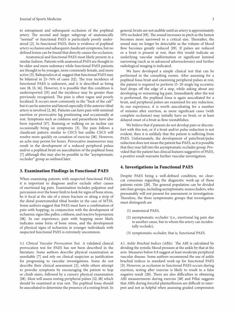
to entrapment and subsequent occlusion of the poplitealartery. The second and larger subgroup of anatomically“normal” or functional PAES is particularly poorly under-stood [2]. In functional PAES, there is evidence of poplitealartery occlusion and subsequent claudicant symptoms, but nodefined lesion can be found that directly causes the occlusion.
Anatomical and functional PAES most likely present in asimilar fashion. Patients with anatomical PAES are thought tobe older and more sedentary while functional PAES patientsare thought to be younger, more commonly female, andmoreactive [3]. Baltopoulos et al. suggest that functional PAESmaybe bilateral in 25–76% of cases [12]. The true incidence offunctional PAES is unknown, and it is described as beingrare [8, 13, 14]. However, it is possible that this condition isunderreported [15] and the incidence may be greater thanpreviously recognized. The pain is often vague and poorlylocalized. It occurs most commonly in the “back of the calf ”but it can be anterior and lateral especially if the anterior tibialartery is involved [2, 16]. Patients can have pain withminimalexertion or provocative leg positioning and occasionally atrest. Symptoms such as coldness and paraesthesia have alsobeen reported [17]. Running or walking on an incline canoccasionally bring on symptoms [3]. The pain follows aclaudicant pattern similar to CECS but unlike CECS willresolve more quickly on cessation of exercise [18]. However,an ache may persist for hours. Provocative manoeuvres mayresult in the development of a reduced peripheral pulsesand/or a popliteal bruit on auscultation of the popliteal fossa[7] although this may also be possible in the “asymptomaticoccluder” group as outlined later.
3. Examination Findings in Functional PAES
When examining patients with suspected functional PAES,it is important to diagnose and/or exclude other causesof exertional leg pain. Examination includes palpation andpercussion over the lower limb to look for signs of bone stress,be it focal at the site of a stress fracture or along a strip ofthe distal posteromedial tibial border in the case of MTSS.Some authors suggest that PAES must have a combination ofpain with hopping, in conjunction with the development ofischaemic signs like pallor, coldness, and reactive hyperaemia[18]. In our experience, pain with hopping more likelyindicates some form of bone stress, and the developmentof physical signs of ischaemia in younger individuals withsuspected functional PAES is extremely uncommon.
3.1. Clinical Vascular Provocation Test. A validated clinicalprovocation test for PAES has not been described in theliterature. Some authors describe physical examination asunreliable [7] and rely on clinical suspicion as justificationfor progressing to vascular investigations. Some do notdescribe their clinical assessment [2], while others attemptto provoke symptoms by encouraging the patient to hopor climb stairs, followed by a cursory physical examination[18]. Most will assess resting peripheral pulses [12, 18] whichshould be examined at true rest. The popliteal fossa shouldbe auscultated to determine the presence of a resting bruit. In
general, bruits are not audible until an artery is approximately50% occluded [19].The sound increases in pitch as the lumenbecomes more narrowed to a critical size. Thereafter, thesound may no longer be detectable as the volume of bloodflow becomes greatly reduced [19]. If pulses are reducedor a bruit is present at rest, than this would indicate anunderlying vascular malformation or significant luminalnarrowing (such as in advanced atherosclerosis) and furtherradiological imaging is indicated.
We have developed a simple clinical test that can beperformed in the consulting rooms. After assessing for apopliteal fossa bruit and examining peripheral pulses at rest,the patient is required to perform 15–20 single leg eccentricheel drops off the edge of a step, while asking about anydeveloping or worsening leg pain. Immediately after the testis performed, the popliteal fossa is again auscultated for abruit, and peripheral pulses are examined for any reduction.In our experience, it is worth auscultating for a numberof minutes after exertion, as more significant cases (withcomplete occlusion) may initially have no bruit, or at leastdelayed onset of a bruit as flow reestablishes.
We believe that if patients do not develop pain or discom-fort with this test, or if a bruit and/or pulse reduction is notevident, then it is unlikely that the patient is suffering fromPAES. Unfortunately, the development of pain/bruit/pulsereduction does notmean the patient has PAES, as it is possiblethat theymay fall into the asymptomatic occluder group. Pro-vided that the patient has clinical features suggestive of PAES,a positive result warrants further vascular investigation.
4. Investigations in Functional PAES
Despite PAES being a well-defined condition, no clear-cut consensus regarding the diagnostic work-up of thesepatients exists [20]. The general population can be dividedinto four groups, including asymptomatic nonoccluders, whopresumably will not present for assessment or investigation.Therefore, the three symptomatic groups that investigationmust distinguish are
(1) anatomical PAES,
(2) asymptomatic occluder (i.e., exertional leg pain dueto another cause, but in whom the artery can inciden-tally occlude),
(3) symptomatic occluder, that is, functional PAES.
4.1. Ankle Brachial Indices (ABIs). The ABI is calculated bydividing the systolic blood pressure at the ankle by that at thearm.Measures below 0.8 suggest at least moderate peripheralvascular disease. Some authors recommend the use of anklebrachial indices in standard work-up for functional PAES[3]. However, as occlusion in functional PAES occurs duringexertion, testing after exercise is likely to result in a falsenegative result [20]. There are also difficulties in obtainingABI measurements during exercise [18] and Pillai suggeststhat ABIs during forceful plantarflexion are difficult to inter-pret and not as helpful when assessing graded compression

4 Journal of Sports Medicine
of a patent artery [17]. We agree that ABIs are unreliable forinvestigating functional PAES.
4.2. Compartment Pressure Testing. Some authors will rou-tinely perform compartment pressure studies in additionto vascular studies in the work-up of suspected PAES [3].This seems to be excessive and our review of the literaturecould find no compelling reason as to why this should bemandatory. For this reason we do not recommend routinecompartment pressure studies, unless symptoms and historyare strongly suggestive of possible concomitant CECS.
4.3. Doppler Ultrasound. Doppler ultrasound provides arelatively cheap, noninvasive, and accessible procedure toassess flow through the popliteal artery, and it is generallyrecommended that this be the first line investigation for PAES[20, 21]. Despite this, a review of the literature shows nodefinite consensus on how to perform Doppler ultrasoundin investigating PAES [20]. Some authors suggest hand heldDoppler units to complement clinical assessment in therooms. This assesses loss of the posterior tibial artery distalpulse during sustained passive dorsiflexion and plantarflex-ion of the foot [12]. Doppler ultrasound is user dependent,which can affect reliability. Also, there is a risk of overcallingentrapment with movement of the artery, muscles, or probeduring exercise giving the illusion of occlusion [21], buttechnique variations can usually overcome this.
4.3.1. Provocative Doppler Protocols. Many authors research-ing PAES suggest that the most important feature in itsdiagnosis is the reproduction of symptoms with the helpof provocative manoeuvres and verification by duplex ultra-sonography [17, 18, 21]. Typically, investigations at rest willshow a patent popliteal artery with normal distal pulses.In functional PAES, occlusion will often only occur duringexertion and can resolve almost immediately. This can givethe false impression of no pathology if the assessment isdelayed. Even the 30–60 seconds taken from getting off atreadmill to an examination bed and applying the ultrasoundprobe can be enough for arterial occlusion to cease. Becauseocclusion can resolve very quickly, immediate assessmentafter reproduction of symptoms is essential [18].The positionof most occlusions occurs when the patient has their kneeextended, and they hold active plantarflexion. It is notpossible to hold this position for sustained periods duringa MRI due to discomfort and fatigue [21]. Ultrasound is auseful modality, as it allows real time assessment whilst someform of provocation to reproduce symptoms is performed.Despite the agreement that provocation is necessary, a clearanddetailed description of the protocols to do this is generallynot provided [20]. Our protocols are included below forcompleteness.
We perform a resting anatomical study first, with thepatient in the supine position. We record waveforms andvelocities of the peripheral arteries as well as assess forevidence of intimal thickening or fixed arterial disease. Theanatomy of the popliteal fossa is assessed, looking particularly
for any popliteal artery or soft tissue structure variations orwhether the anterior tibial artery has a high bifurcation.
4.4. Provocation. Hoffman et al. [22] found that the force ofplantarflexion required to occlude the popliteal artery duringprovocative positioning is important, with the majority ofasymptomatic occluders occluding the popliteal artery withsustained holding of ≥70% of maximal plantarflexion force[22]. For this reason, we use a graded ultrasound protocolprogressing through
(1) prone patient holding a position of provocation (kneeextension and plantarflexion) against no load,
(2) patient is prone and pushing against approximately25% of maximal plantarflexion force (Figure 1(a)),
(3) dynamic loading by pushing against 50–100% ofmaximal plantarflexion force (Figure 1(b)).
Symptomatic patients who occlude in the first 2 categoriesare presumed to represent more severe cases of functionalPAES. If the initial 2 assessments are negative, the patientis assessed in an erect weight-bearing position while cyclingthrough range of motion. This is thought to representsomewhere between 50 and 100% maximal plantarflexionforce and may more reliably represent what is occurringfunctionally during activity. If the erect assessment is nega-tive, the patient may attempt similar exertion that elucidatessymptoms in a normal training experience, until they aresymptomatic.
At this point, we again assess the patient in an erectposition as previously described. We will use cine loopsto demonstrate the popliteal artery from resting patencythrough compression to occlusion and mark the site of com-pression on skin for MRI correlation. We feel that Dopplerultrasound does not adequately demonstrate the anatomy torule out anatomical PAES and confirmation of occlusion in asymptomatic patient means the patient may be in either theanatomical or functional PAES groups.
4.5. MRI and MRI Angiography. MRI is a valuable noninva-sivemodality that allows optimal visualization of the poplitealartery as well as the surrounding structures [23, 24]. MRIcan also distinguish intrinsic vascular disease from extrinsiccompression. MRI can demonstrate a variety of findingsincluding abnormal lateralized insertion of the medial headof gastrocnemius, medial displacement, and occlusion of thepopliteal artery in the popliteal fossa and fat tissue filling thenormal location of themedial head of gastrocnemius [23, 25].In this way, anatomical PAES can be distinguished fromfunctional PAES and the muscles and anatomical boundariescontributing can be accurately delineated [3].This in turn candirect injection therapy (as discussed in Section 5) or the siteof surgical intervention.
MRI angiography can demonstrate the level of occlusion,but limitations include the development ofmovement artifactwith forceful plantarflexion positions and inability to holdsuch a position for prolonged periods [17].This is a significantlimitation as for reasons outlined earlier; investigating for

Journal of Sports Medicine 5
(a) (b)
Figure 1: (a) Patient prone and pushing against a wall (in the direction of the arrow) at 25% maximum plantarflexion force. (b) Patient erectand plantarflexing against full body weight.
occlusion in functional PAES should occur during provo-cation. Most papers reviewed do not describe provocativemanoeuvres with MRI angiography [23–25] while one sug-gests holding sustained forceful plantarflexion continuouslyfor 20–29 seconds whilst the angiogram is performed [26].Again for completeness we have provided our protocolsbelow.
After positive ultrasound studies, a fiducial marker isplaced on the patient’s skin where the popliteal artery is beingoccluded to help correlate between the ultrasound and MRIsite of occlusion. T1 weighted axial and coronal images areacquired to demonstrate the medial head and lateral headsof gastrocnemius, popliteus, and plantaris muscles and theiralignment and associations with the femoral condyles andpopliteal arteries and nerves. These images are acquired withthe patient at rest.
Following these static images, the patient is instructedto dorsiflex and plantarflex their feet whilst acquiring T2weighted 2D steady state images axially across the poplitealregion. Before the final contrast MRI angiogram is per-formed, the patient is instructed to alternate a neutralankle position with maximal plantarflexion force until theystimulate the pain that they usually experience (rather than asingle sustained forceful contraction). Once they experiencethis pain, they keep their ankles in plantarflexion whilst weinject the contrast and perform the angiogram (Figure 2).One of the disadvantages of this technique is that there is anapproximate 30-seconddelay before the contrast arrives at thepopliteal artery and the angiogram can be performed and theartery may re-establish flow during this time. Patients quiteoften are in pain and or exhausted during this last series andmay shake because of this.
5. Treatment
Based on our review of the literature, Figure 3 represents adecision-making flowchart in the assessment, work-up, and
Figure 2: MRI angiogram of the popliteal fossa showing completeocclusion of the popliteal artery in the left leg.
treatment of suspected PAES. Once confirmed, treatmentdepends on the type of PAES identified.
5.1. Anatomical PAES. Given the described rapid progressionof arterial injury, it is recommended that anatomical PAESpatients undergo surgery to remove the site of entrapment[17]. This typically involves exploration, limited fasciotomy,myotomy to varying degrees, and possible excision of occlu-sive fibrous bands [2, 17]. Results of popliteal fossa explo-ration, bypass, or muscle detachment, or a combination ofthese, and fossa decompression are generally good. Mostseries report a small number of patients, but >90% appear toreturn to activities in sports ≤ 3 months with resolution of allprevious symptoms [17].
5.2. Functional PAES. The pathogenesis and progression offunctionalPAES are uncertain, but it may be that these

6 Journal of Sports Medicine
Suspected PAES assessment and treatment protocols
Clinical features suggesting PAES
Yes
Negative
Consider other causes of ELP
Negative
Positive
Clinical provocative test performed in rooms: development of pain and
bruit and reduction of peripheral pulses
Doppler ultrasound study as per protocols
Occlusion
MRI angiography with stress provocation
Normal anatomy
Functional PAES
Anatomical occlusion
Anatomical PAES
Surgical decompression
Symptoms do not resolve or returnafter successful Botox injection
Consider trial ultrasound guided Botox injection at site of occlusion; regular
follow-up with option of top-up injections as indicated
Figure 3: Assessment and treatment protocols for suspected PAES.
patients develop arterial injury more gradually with onsetof more significant symptoms later in life [12]. Surgery hasbeen recommended in functional occluders with significantrepetitive and typical symptoms [17]. Turnipseed recom-mends resection of plantaris muscle and the crural band ofsoleus fascia that forms the outlet of the popliteal fossa as hefeels that this fascial band is the fulcrum against which theneurovascular bundle is compressed in functional PAES [3].
However the site and amount of muscle necessary to beremoved to prevent further occlusion is not always obvious. Itis possible that a large segment ofmusclewill require excision.The popliteal artery will need exploration and there areincreased risks of postoperative complications such as seroma(4.6%) and infection (2%) [2]. Also surgery for functionalPAES does not seem to be as successful as that for anatomicalPAES, with reports suggesting only 77% of patients aftersurgery reporting complete resolution of symptoms [14]. Forthis reason, a less invasive treatment option is desirable.
5.3. Guided Botulinum Toxin (Botox BTX-A) Injection Ther-apy. The use of botulinum injections for paralysis of musclesto manage medical conditions is well established. There areseveral descriptions of the use of botulinum in the treatmentof muscle spasticity, particularly in cerebral palsy patients[27–29], and piriformis injection of BTX-A has been usedsuccessfully to treat sacral plexus and proximal sciatic nervecompression [30]. Bilici et al. described the use of BTX-Ainjection into a crus of the hemidiaphragm to treat renalartery stenosis [31].
The proposed mechanisms of action for intramuscularperiarterial botulinum therapy for PAES are
(1) paralysis of the muscular slip of muscle responsiblefor the dynamic arterial occlusion,
(2) localised muscle atrophy caused by the botulinumwhich may increase space for the vessel and wouldexplain the prolonged effect of botulinum on this
Journal of Sports Medicine 7
condition beyond the expected therapeutic effect ofthe medication,
(3) possible arterial smooth muscle relaxation of thepopliteal artery resulting in vasodilatation.
Unfortunately, to date, there is no published data onthe efficacy of botulinum injection in the management offunctional PAES. We have commenced a pilot study usingintramuscular periarterial injection of BTX-A to treat func-tional PAESwith promising initial results.We hope to publishthe outcomes as our cohort size increases, but at present thisremains an unproven intervention.
6. Summary
Functional PAES is a condition that is possibly underrecog-nized and, if left untreated, can result in progressive arterialdamage and the risk of developing lower limb ischaemia. Itshares many features with other causes of exertional leg pain(especially chronic exertional compartment syndrome) andmay coexist with one or more of these. A suggestive clinicalhistory includes features of pain aggravated by exercise, butalso possibly at rest with positions of knee extension andplantarflexion. The pain will typically resolve quickly onceprovocative manoeuvres are ceased, although an ache maypersist for hours. Anatomical and functional PAES cannot bedistinguished on clinical features alone, and possibly over halfof the “normal” population can demonstrate some arterialocclusion with provocative manoeuvres. For this reason,specialized vascular investigations are indicated, particularlya Doppler ultrasound protocol performed at rest and duringprovocation and immediately after, which can demonstratereal time arterial occlusion and the level it is occurring at.Once occlusion is demonstrated, MRI can demonstrate thedefinitive anatomy of the popliteal fossa, whether anatomicalPAES exists, and the site and extent of functional entrap-ment. From here, the best treatment can be provided, withconsideration of guided Botox injection for functional PAESas a potential new intervention, or progression to surgicalintervention.
Conflict of Interests
The authors declare that there is no conflict of interestsregarding the publication of this paper.
Acknowledgments
The authors acknowledge Queensland X-ray for their assis-tance in providing high quality affordable investigations totheir subjects.
References
[1] P. G. Blackman, “A review of chronic exertional compartmentsyndrome in the lower leg,” Medicine & Science in Sports &Exercise, vol. 32, supplement, pp. S4–S10, 2000.
[2] W. D. Turnipseed, “Functional popliteal artery entrapmentsyndrome: a poorly understood and oftenmissed diagnosis that
is frequentlymistreated,” Journal of Vascular Surgery, vol. 49, no.5, pp. 1189–1195, 2009.
[3] W. D. Turnipseed, “Popliteal entrapment in runners,” Clinics inSports Medicine, vol. 31, no. 2, pp. 321–328, 2012.
[4] S. B. Thacker, J. Gilchrist, D. F. Stroup, and C. D. Kimsey, “Theprevention of shin splints in sports: a systematic review ofliterature,”Medicine and Science in Sports and Exercise, vol. 34,no. 1, pp. 32–40, 2002.
[5] C. A. George and M. R. Hutchinson, “Chronic exertionalcompartment syndrome,” Clinics in Sports Medicine, vol. 31, no.2, pp. 307–319, 2012.
[6] N. Reshef and D. R. Guelich, “Medial tibial stress syndrome,”Clinics in Sports Medicine, vol. 31, no. 2, pp. 273–290, 2012.
[7] T. T. Pham, R. Kapur, and M. I. Harwood, “Exertional legpain: teasing out arterial entrapments,” Current Sports MedicineReports, vol. 6, no. 6, pp. 371–375, 2007.
[8] M. Reinking, “Exercise Related Leg Pain (ERLP): a review of theliterature,” North American Journal of Sports Physical Therapy,vol. 2, no. 3, pp. 170–180, 2007.
[9] M. Hislop, P. Tierney, P. Murray, M. O’Brien, and N. Mahony,“Chronic exertional compartment syndrome: the controversial“fifth” compartment of the leg,”The American Journal of SportsMedicine, vol. 31, no. 5, pp. 770–776, 2003.
[10] K. Bennell, G. Matheson, W. Meeuwisse, and P. Brukner, “Riskfactors for stress fractures,” Sports Medicine, vol. 28, no. 2, pp.91–122, 1999.
[11] M. Hislop and M. E. Batt, “Chronic exertional compartmentsyndrome testing: a minimalist approach,” British Journal ofSports Medicine, vol. 45, no. 12, pp. 954–955, 2011.
[12] P. Baltopoulos, D. K. Filippou, and F. Sigala, “Popliteal arteryentrapment syndrome: anatomic or functional syndrome?”Clinical Journal of Sport Medicine, vol. 14, no. 1, pp. 8–12, 2004.
[13] J. W. Love and T. J. Whelan, “Popliteal artery entrapmentsyndrome,”The American Journal of Surgery, vol. 109, no. 5, pp.620–624, 1965.
[14] S. Sinha, J. Houghton, P. J. Holt, M. M.Thompson, I. M. Loftus,and R. J. Hinchliffe, “Popliteal entrapment syndrome,” Journalof Vascular Surgery, vol. 55, no. 1, pp. 252.e30–262.e30, 2012.
[15] L. J. Levien and M. G. Veller, “Popliteal artery entrapmentsyndrome: more common than previously recognized,” Journalof Vascular Surgery, vol. 30, no. 4, pp. 587–598, 1999.
[16] E. Mavili, H. Donmez, G. Kahriman, A. Ozaslamaci, N. Ozcan,and K. Tasdemir, “Popliteal artery branching patterns detectedby digital subtraction angiography,” Diagnostic and Interven-tional Radiology, vol. 17, no. 1, pp. 80–83, 2011.
[17] J. Pillai, “A current interpretation of popliteal vascular entrap-ment,” Journal of Vascular Surgery, vol. 48, pp. 61S–65S, 2008.
[18] R. Lane, T. Nguyen, M. Cuzzilla, D. Oomens,W.Mohabbat, andS. Hazelton, “Functional popliteal entrapment syndrome in thesportsperson,” European Journal of Vascular and EndovascularSurgery, vol. 43, no. 1, pp. 81–87, 2012.
[19] R. D. Hill and R. Smith, “Examination of the extremities: pulses,bruits, and phlebitis,” in Clinical Methods: The History, Physical,and Laboratory Examinations, H. K. Walker, W.D. Hall, and J.W. Hurst, Eds., pp. 148–152, Butterworths, Boston, Mass, USA,1990.
[20] U. Altinatis, U. Helgstrand, and M. Hansen, “Popliteal arteryentrapment syndrome: ultrasound imaging, intra-operativefindings and clinical outcome,” Vascular and EndovascularSurgery, vol. 47, no. 7, pp. 513–518, 2013.

8 Journal of Sports Medicine
[21] L. S. Erdoes, J. J. Devine, V. M. Bernhard, M. R. Baker, S. S.Berman, and G. C. Hunter, “Popliteal vascular compression in anormal population,” Journal of Vascular Surgery, vol. 20, no. 6,pp. 978–986, 1994.
[22] U. Hoffmann, J. Vetter, L. Rainoni, A. J. Leu, and A. Bollinger,“Popliteal artery compression and force of active plantar flexionin young healthy volunteers,” Journal of Vascular Surgery, vol.26, no. 2, pp. 281–287, 1997.
[23] S. Atilla, E. T. Ilgit, S. Akpek, C. Yucel, E. Turgut Tali, andS. Iik, “MR imaging and MR angiography in popliteal arteryentrapment syndrome,” European Radiology, vol. 8, no. 6, pp.1025–1029, 1998.
[24] A. W. Lambert and D. C. Wilkins, “Popliteal artery entrapmentsyndrome,” The British Journal of Surgery, vol. 86, no. 11, pp.1365–1370, 1999.
[25] U. Ozkan, L. Oguzkurt, F. Tercan et al., “MRI and DSAfindings in popliteal artery entrapment syndrome,” Diagnosticand Interventional Radiology, vol. 14, pp. 106–110, 2008.
[26] E. Di Cesare, L.Marsili, G.Marino et al., “StressMR imaging forevaluation of popliteal artery entrapment,” Journal of MagneticResonance Imaging, vol. 4, no. 4, pp. 617–622, 1994.
[27] I. S. Corry, A. P. Cosgrove, E. G. Walsh, D. McClean, andH. K. Graham, “Botulinum toxin A in the hemiplegic upperlimb: a double blind trial,” Developmental Medicine and ChildNeurology, vol. 39, no. 3, pp. 185–193, 1997.
[28] D. Fehlings, “The use of botulinum toxin in paediatric hyper-tonia,” Paediatrics and Child Health, vol. 10, no. 7, pp. 379–381,2005.
[29] L. A. Koman, J. F. Mooney III, B. P. Smith, A. Goodman,and T. Mulvaney, “Management of spasticity cerebral palsywith botulinum—a toxin: report of a preliminary, randomized,double-blind trial,” Journal of Pediatric Orthopaedics, vol. 14, no.3, pp. 299–303, 1994.
[30] S. J. Yoon, J. Ho, H. Y. Kang et al., “Low-dose botulinum toxintype A for the treatment of refractory piriformis syndrome,”Pharmacotherapy, vol. 27, no. 5, pp. 657–665, 2007.
[31] A. Bilici, M. Karcaaltincaba, A. T. Ilica, Y. Bukte, and A. Senol,“Treatment of hypertension from renal artery entrapmentby percutaneous CT-guided botulinum toxin injection intodiaphragmatic crus as alternative to surgery and stenting,” TheAmerican Journal of Roentgenology, vol. 189, no. 3, pp. W143–W145, 2007.

Submit your manuscripts athttp://www.hindawi.com
Stem CellsInternational
Hindawi Publishing Corporationhttp://www.hindawi.com Volume 2014
Hindawi Publishing Corporationhttp://www.hindawi.com Volume 2014
MEDIATORSINFLAMMATION
of
Hindawi Publishing Corporationhttp://www.hindawi.com Volume 2014
Behavioural Neurology
EndocrinologyInternational Journal of
Hindawi Publishing Corporationhttp://www.hindawi.com Volume 2014
Hindawi Publishing Corporationhttp://www.hindawi.com Volume 2014
Disease Markers
Hindawi Publishing Corporationhttp://www.hindawi.com Volume 2014
BioMed Research International
OncologyJournal of
Hindawi Publishing Corporationhttp://www.hindawi.com Volume 2014
Hindawi Publishing Corporationhttp://www.hindawi.com Volume 2014
Oxidative Medicine and Cellular Longevity
Hindawi Publishing Corporationhttp://www.hindawi.com Volume 2014
PPAR Research
The Scientific World JournalHindawi Publishing Corporation http://www.hindawi.com Volume 2014
Immunology ResearchHindawi Publishing Corporationhttp://www.hindawi.com Volume 2014
Journal of
ObesityJournal of
Hindawi Publishing Corporationhttp://www.hindawi.com Volume 2014
Hindawi Publishing Corporationhttp://www.hindawi.com Volume 2014
Computational and Mathematical Methods in Medicine
OphthalmologyJournal of
Hindawi Publishing Corporationhttp://www.hindawi.com Volume 2014
Diabetes ResearchJournal of
Hindawi Publishing Corporationhttp://www.hindawi.com Volume 2014
Hindawi Publishing Corporationhttp://www.hindawi.com Volume 2014
Research and TreatmentAIDS
Hindawi Publishing Corporationhttp://www.hindawi.com Volume 2014
Gastroenterology Research and Practice
Hindawi Publishing Corporationhttp://www.hindawi.com Volume 2014
Parkinson’s Disease
Evidence-Based Complementary and Alternative Medicine
Volume 2014Hindawi Publishing Corporationhttp://www.hindawi.com