review article efficacy and safety of topical timolol eye...
TRANSCRIPT

Review ArticleEfficacy and Safety of Topical Timolol Eye Drops inthe Treatment of Myopic Regression after Laser In SituKeratomileusis: A Systematic Review and Meta-Analysis
Xiaochen Wang, Guiqiu Zhao, Jing Lin, Nan Jiang, Qian Wang, and Qiang Xu
Department of Ophthalmology, The Affiliated Hospital of Qingdao University, Qingdao 266000, China
Correspondence should be addressed to Guiqiu Zhao; zhaoguiqiu [email protected]
Received 23 September 2015; Accepted 2 December 2015
Academic Editor: Edward Manche
Copyright © 2015 Xiaochen Wang et al.This is an open access article distributed under the Creative CommonsAttribution License,which permits unrestricted use, distribution, and reproduction in any medium, provided the original work is properly cited.
Aims. The aim of this study was to assess the efficacy and safety of timolol in the treatment of myopic regression after laser insitu keratomileusis (LASIK). Methods. We searched MEDLINE, CENTRAL, EMBASE, China National Knowledge Infrastructure(CNKI), and Chinese Biological Medicine (CBM) from the inception to July 2015 for relevant randomized controlled trials thatexamined timolol therapy formyopic regression.Themethodological quality of the studies includedwas assessed using the Revman5.3 software. Results. We included six clinical trials involving 483 eyes in this review, including 246 eyes in treated group and 237eyes in controlled group.We observed statistically significant improvements on the postoperative SE in the 3 months. However, thechange of CCT was not statistically different between the control group and the experimental group.There were fewer cases of IOP,UDVA, and CDVA in treated group having significant difference from the controlled group. Conclusions. Topical timolol could bean effective treatment for reduction of myopic regression especially the spherical errors after myopic LASIK. Further RCTs withlarger sample sizes for these trials are warranted to determine the efficacy and limitation for myopic regression after LASIK.
1. Introduction
Laser in situ keratomileusis (LASIK) is thought to be aneffective and safe refractive surgical procedure for the highmyopia [1]. Along with the continuous renewal of equipmentinstrument and the continuous improvement of surgicaltechnique, most postoperative patients obtained satisfactoryresults. However, at least 28% of refractive surgery patientsstill experience myopic regression [2–5].
In previous studies, “regression” was defined as a 0.25-diopter (D) or greatermyopic shift occurring between follow-up visits [4–7]. Nevertheless, the mechanism for refractiveregression is very complicated and is not fully understood.The main possible explanations for regression are focused onthe forward shift of the cornea [8–11]. It has been suggestedthat intraocular pressure- (IOP-) lowering agents or thecorneal biomechanical change can decrease and alleviatemyopic nonselective B-blocker with carbomer and polyvinylalcohol [12]. Timolol provides ocular comfort and lubrication
and also increases retinal and optic nerve perfusion. Itcan reduce IOP by decreasing aqueous humor productionand has no obvious side effects. Because of the propertiesnoted above, topic timolol eye drops are indicated for thetreatment of myopic regression. A number of clinical trialshad been conducted to evaluate timolol’s effectiveness andsafety. However, the results were inconsistent; therefore, weset out to conduct a systematic review and meta-analysis toassess the evidence for treating regression.
2. Materials and Methods
2.1. Search Strategy and Selection Criteria. We performed ourresearch with MEDLINE, CENTRAL, EMBASE, CBM, andCNKI for randomized controlled trials (RCTs). The searchterms used were “Timolol AND (myopic OR regressionOR regressive)”. Furthermore, we reviewed citations in theretrieved articles to search for additional relevant studies.
Hindawi Publishing CorporationJournal of OphthalmologyVolume 2015, Article ID 985071, 8 pageshttp://dx.doi.org/10.1155/2015/985071
2 Journal of Ophthalmology
Inclusion and Exclusion Criteria. RCTs were eligible forinclusion if the following criteria were satisfied:
(1) There are controlled clinical trials, including ret-rospective studies and prospective studies such asrandomized controlled trials (RCTs).
(2) There is confirmed diagnosis of high myopic, spher-ical equivalent (SE) ≥ −6.00D; age of patients is 19years or more.
(3) Studies that reported the follow-up results beyond 2weeks concerning LASIK treatment for myopia areincluded.
(4) Patients were subjected to topical timolol eye dropsdaily for more than two weeks.
(5) Treatment with topical timolol eye drops was com-pared with artificial tears, placebo (vehicle), with notopical treatment.
(6) We included any RCTs that examine at least one of thefollowing outcomes: IOP, spherical equivalent, CCT,UDVA, and CDVA.
Studies were excluded based on the following criteria:
(1) Patients had a history of other ocular diseases, espe-cially the glaucoma, active inflammation.
(2) Outcomes or data are presented in a format thatcannot be extracted for analysis.
(3) Patients had the refractive surgery but not the LASIK.
2.2. Data Extraction and Assessment of Bias Risks. All articleswere read by two independent reviewers (Xiaochen Wangand Qian Wang) independently who implemented the dataextraction according to the inclusion criteria. We use astandardized form to record data on the authors of the study,year of publication, country of origin, sample size, gender,mean age, duration of follow-up, and outcomemeasures.Therisks of bias in the included studieswere assessed according tothe recommended methods of the Cochrane handbook. Weevaluated random sequence generation and allocation con-cealment (selection), masking of participants and personnel(performance bias), masking of outcome assessment (detec-tion bias), and incomplete outcome data (attrition bias). Twoauthors (Xiaochen Wang and Qian Wang) independentlyassessed the risks of bias and any disagreements were resolvedby discussion to reach a consensus among the investigators.
2.3. Statistical Analysis. We used the Review Manager 5.3to perform our meta-analysis. We calculated the weightedmean difference for continuous data. We used the SMD toanalyze the results on a uniform scale. The absolute value isinterpreted togetherwith the𝑃 value and confidence intervals(CI). We evaluated the statistical heterogeneity by Cochrane𝜒2 tests and qualified it by calculating the 𝐼2 statistic. If
there was any significant heterogeneity between studies (𝐼2 >50%), a random effects model was used to pool the data;otherwise a fixed effect model was used. We consideredconducting a sensitivity analysis by excluding studies which
Potentially relevant articlesidentified: 787MEDLINE: 68EMBASE: 294CNKI: 409CBM: 16
Titles and abstractsscreened: 88
Excluded as irrelevant orrepetitive to our subject: 699
Not eligible: 75
Articles obtained for full textreview: 13
Studies excluded: 7Data not in usable format: 2Not RCTs: 3
Eligible RCTs: 6
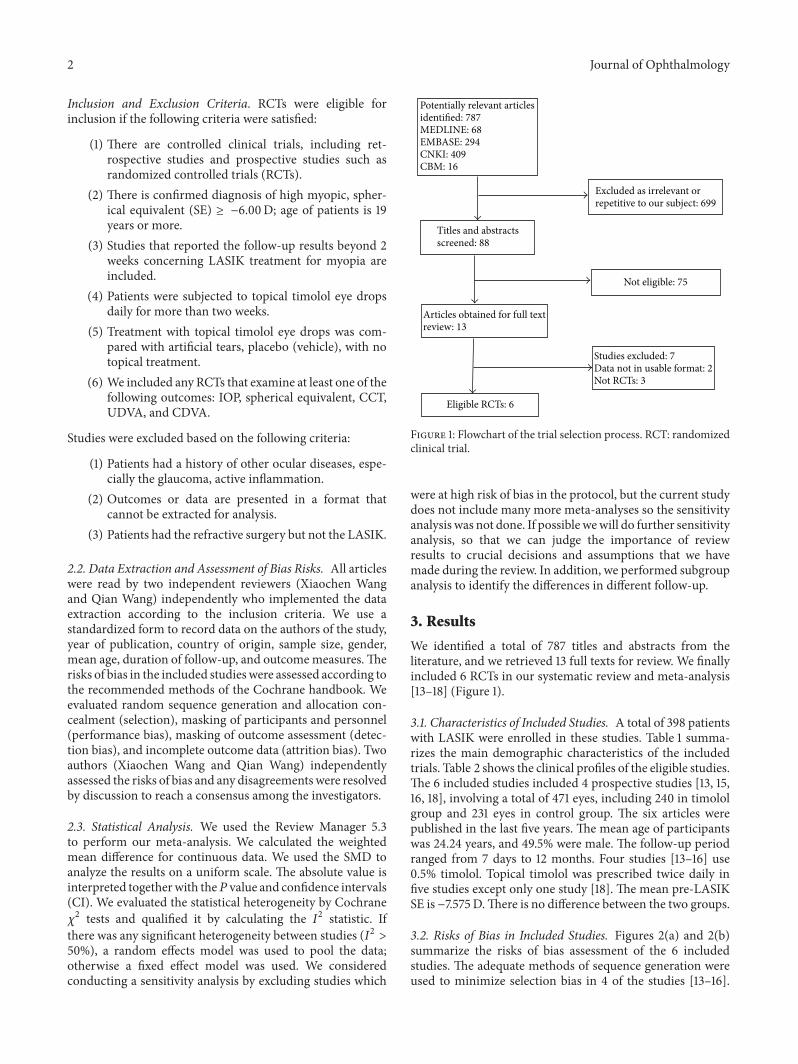
Figure 1: Flowchart of the trial selection process. RCT: randomizedclinical trial.
were at high risk of bias in the protocol, but the current studydoes not include many more meta-analyses so the sensitivityanalysis was not done. If possible wewill do further sensitivityanalysis, so that we can judge the importance of reviewresults to crucial decisions and assumptions that we havemade during the review. In addition, we performed subgroupanalysis to identify the differences in different follow-up.
3. Results
We identified a total of 787 titles and abstracts from theliterature, and we retrieved 13 full texts for review. We finallyincluded 6 RCTs in our systematic review and meta-analysis[13–18] (Figure 1).
3.1. Characteristics of Included Studies. A total of 398 patientswith LASIK were enrolled in these studies. Table 1 summa-rizes the main demographic characteristics of the includedtrials. Table 2 shows the clinical profiles of the eligible studies.The 6 included studies included 4 prospective studies [13, 15,16, 18], involving a total of 471 eyes, including 240 in timololgroup and 231 eyes in control group. The six articles werepublished in the last five years. The mean age of participantswas 24.24 years, and 49.5% were male. The follow-up periodranged from 7 days to 12 months. Four studies [13–16] use0.5% timolol. Topical timolol was prescribed twice daily infive studies except only one study [18]. The mean pre-LASIKSE is −7.575D.There is no difference between the two groups.
3.2. Risks of Bias in Included Studies. Figures 2(a) and 2(b)summarize the risks of bias assessment of the 6 includedstudies. The adequate methods of sequence generation wereused to minimize selection bias in 4 of the studies [13–16].

Journal of Ophthalmology 3
Table 1: Demographic characteristics of eligible studies.
Study(year) Country Population Gender (male : female) Mean age (Yr) ± SD
Timolol Control Timolol ControlZhongwen 2014 [13] China 60 NS NS 24.47 ± 5.45 25.07 ± 6.23Guan 2013 [14] China 60 18 : 12 16 : 14 20.0 ± 7.50 22.0 ± 5.50Shojaei et al. 2012 [15] Iran 90 9 : 36 15 : 30 33.31 ± 10.90 34.42 ± 8.57Zhang et al. 2011 [16] China 60 NS NS 25.37 ± 6.13 24.53 ± 2.31Yang et al. 2010 [17] China 53 NS NS NS NSEI-Awady et al. 2010 [18] Egypt 75 NS NS NS NSSD: standard deviation; Yr: years; NS: data not available.
Table 2: Clinical characteristics of eligible studies.
Study(year) Study design Conc. of
timolol (%)Timolol regimenand duration Follow-up
Mean pre-LASIK SE ± SD,diopters
Mean pre-LASIK IOP,mmHg
Timolol Control Timolol ControlZhongwen2014 [13] Prospective 0.5 Twice a day for
1mo1wk/1mo/3mo/6mo −7.00 ± 0.77 −7.32 ± 1.10 16.33 ± 2.69 16.90 ± 3.00
Guan 2013[14] NS 0.5 Twice a day for
3mo 3mo −5.85 ± 2.52 −5.64 ± 2.31 14.65 ± 2.35 15.45±2.13
Shojaei et al.2012 [15] Prospective 0.5 Twice a day for
6mo3mo/6mo/12mo −8.10 ± 3.41 −4.87 ± 1.88 12.73 ± 1.43 12.38 ± 1.65
Zhang et al.2011 [16] Prospective 0.5 Twice a day for
1mo1wk/1mo/3mo −4.94 ± 1.09 24.53 ± 2.31 15.22 ± 1.78 15.11 ± 2.53
Yang et al.2010 [17] NS 0.025 Twice a day for
2wk 2wk −7.01 ± 3.04 −6.53 ± 2.40 NS NS
EI-Awady etal. 2010 [18] Prospective 0.1 Once a day for
12mo 12mo NS NS NS NS
SD: standard deviation; Yr: years; mo: months; wk: weeks; Conc.: concentration; NS: data not available.
For performance and detection biases, 4 studies [13–15, 18]reported using blindingmethod to performance andoutcomeassessment. For attrition bias, only 1 trial [15] had high lossto follow-up and was judged from high risk of bias. In theother studies, attrition bias was considered to be possible.In the included trials, reporting bias was not considered tobe a major problem but it was always difficult to evaluate itsufficiently.
3.3. Outcome Measures
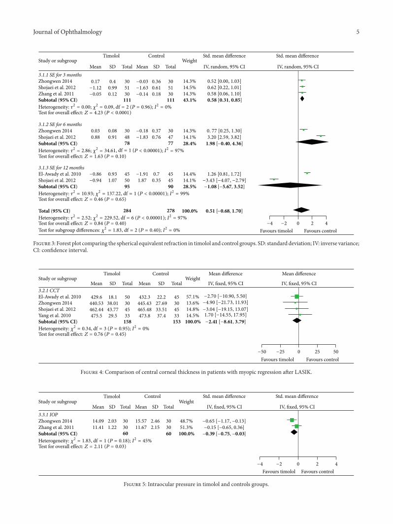
3.3.1. Spherical Equivalent. Four studies reported the finalrefractive spherical equivalent after being treated for 3months, 6 months, and 12 months, respectively, and used therandomeffectsmodel to analyze the data for heterogeneity (𝐼2= 0%, 97%, 99%).There was statistically significant differencebetween the two groups in the follow-up for 3 months (SMD= 0.58, 95% CI = 0.31 to 0.85; 𝑃 < 0.0001). However, in 6months (SMD = 1.98, 95% CI = −0.40 to 4.36; 𝑃 = 0.1) and12 months (SMD = −1.08, 95% CI = −5.67 to 3.52; 𝑃 = 0.65),there were no differences between the two groups (Figure 3).
3.3.2. Central Corneal Thickness. The data of the centralcorneal thickness were used the fixed effects model to analyzethe heterogeneity (𝐼2 = 0%). The change of CCT was not
statistically different between the two groups (MD = −2.41,95% CI = −8.61 to 3.79; 𝑃 = 0.45) (Figure 4).
3.3.3. Intraocular Pressure. There were 2 studies [13, 16]that reported the intraocular pressure, showing significantdifference between the two groups (SMD = −0.39, 95% CI= −0.75 to −0.03; 𝐼2 = 45%; 𝑃 = 0.03) (Figure 5).
3.3.4. UDVA. Each of the 2 studies reported the logMARUDVA that used the fixed effects model to analyze the datafor heterogeneity (𝐼2 = 96%, 25%) in different time points.There were significant differences between the two groups inthe follow-up for 6 months (MD = −0.02; 95% CI = −0.04 to0.00; 𝑃 = 0.05) and 12 months (MD = 0.15; 95% CI = 0.07 to0.23; 𝑃 = 0.0002) (Figure 6).
3.3.5. CDVA. There were 2 studies [15, 18] that use thelogMAR CDVA to measure the outcome and then we usedthe fixed effects model to analyze the data for heterogeneity(𝐼2 = 39%); the results show that it is significantly differentbetween the two groups in the follow-up for 12 months (MD= 0.03; 95% CI = 0 to 0.05; 𝑃 = 0.20) (Figure 7).
3.4. Heterogeneity and Publication Bias. Some outcomesdisplayed great heterogeneity. The heterogeneities of SE and

4 Journal of Ophthalmology
Random sequence generation (selection bias)Allocation concealment (selection bias)Blinding of participants and personnel (performance bias)Blinding of outcome assessment (detection bias)Incomplete outcome data (attrition bias)Selective reporting (reporting bias)Other bias+++
++++
+ + + + +
+
++++
+ +
++
+ + + +
???
?? ? ? ?
?
El-A
wad
y et
al. 2
010
Gua
n 20
13Zh
ongw
en 2
014
Shoj
aei e
t al.
2012
Yang
et al
. 201
0Zh
ang
et al
. 201
1
−
−
− − −
−
−
−
(a)
Random sequence generation (selection bias)Allocation concealment (selection bias)
Blinding of participants and personnel (performance bias)Blinding of outcome assessment (detection bias)
Incomplete outcome data (attrition bias)Selective reporting (reporting bias)
Other bias
Low risk of biasUnclear risk of bias
High risk of bias
25 50 75 1000(%)
(b)
Figure 2: (a) Risk of bias summary: authors’ judgments about each risk of bias item for each included risk. (b) Risk of bias graph: authors’judgments about each risk of bias item presented as percentages across all included studies.
IOP were significant, and dropping eligible studies by handand metaregression have not provided good results. Maybeit is because of the different measure tools. No significantpublication bias was demonstrated in the funnel plot.
4. Discussion
Meta-analysis attempts to analyze and combine the resultsof previous reports [19]. This systematic review provided acritical overview of previous clinical reports and combinedeffect measures of timolol in multiple small clinical trials toincrease statistical power. It included six trials using timololto prevent and treat the myopic regression after LASIK. Alltrials were implemented in developing countries because ofthe higher incidence than developed countries.There are stillno large multicenter randomized trials to assess the efficacyand safety of timolol on the treatment of myopic regression.
As a common clinical phenomenon, refractive regressioncan affect the predictability, efficiency, and long-term stabilityof refractive surgery and lead to deterioration in visual per-formance and even seriously affect the surgical curative effectand patients’ satisfaction. So the prevention and treatmentof refractive back after the surgery are very important to thequality of patient’s life in the future. Nevertheless, there areno unified and effective methods in the treatment of myopicregression. Secondary surgery is an inacceptable method for
patients and doctors; both of them have very big challenge. Incontrast, effective drug treatment is a lower risk more easilyaccepted by patients.
There have been many factors which associated withmyopic regression after LASIK, including preoperativerefraction [4, 5, 19–23], preoperative keratometry [20, 21, 24],corneal thickness [11, 23], flap thickness [24, 25], ablationdepth [21], optical zone size [21, 26], chronic dry eye [27], age[21], surgeon, IOP [20, 22], postoperative undercorrection,and humidity. The occurrence of refractive regression hasthe relation with the corneal wound healing response, thedestruction of the corneal biomechanics structural integrity,and relatively high intraocular pressure and closely relatedto the occurrence of postoperative dry eye. There is adebate according to the role of CCT in myopic regression.Kamiya and associates [28] present a theory to assess theeffects of nipradilol, an IOP-lowering agent; Pan et al. [11]compared regressive eyes with nonregression after LASIKand indicated that refractive regression after LASIK mightbe mainly induced by corneal protrusion, rather than centralcorneal thickening. That is what happens with any refractiveprocedure or flap; the corneal biomechanics changing maybe a factor of the myopic regression. From these stud-ies, we conclude that LASIK can lead to the destructionof the corneal biomechanics structural integrity, cornealinjury repair reshaping in the early postoperative stage,
Journal of Ophthalmology 5
−1.08 [−5.67, 3.52]
0.51 [−0.68, 1.70]
0.58 [0.31, 0.85]111 111 43.1%
78 77 28.4%
95 90
284 278 100.0%
Timolol
Mean SD Total Mean SD TotalWeight
Control Std. mean difference
IV, random, 95% CI
Std. mean difference
IV, random, 95% CI
0.17 0.4 30 −0.03 0.36 30 14.3% 0.52 [0.00, 1.03]−1.12 0.99 51 −1.63 0.61 51 14.5% 0.62 [0.22, 1.01]
0.03 0.08 30 −0.18 0.37 30 14.3% 0. 77 [0.25, 1.30]0.88 0.91 48 −1.83 0.76 47 14.1% 3.20 [2.59, 3.82]
−0.86 0.93 45 −1.91 0.7 45 14.4% 1.26 [0.81, 1.72]−0.94 1.07 50 1.87 0.35 45 14.1% −3.43 [−4.07, −2.79]
Favours control0 2 4−2−4
Favours timolol
1.98 [−0.40, 4.36]
28.5%
−0.05 0.12 30 −0.14 0.18 30 14.3% 0.58 [0.06, 1.10]
3.1.1 SE for 3 months
Subtotal (95% CI)
3.1.2 SE for 6 months
Subtotal (95% CI)
3.1.3 SE for 12 months
Test for overall effect: Z = 4.23 (P < 0.0001)
Test for overall effect: Z = 1.63 (P = 0.10)
Study or subgroup
Subtotal (95% CI)
Test for overall effect: Z = 0.46 (P = 0.65)
Heterogeneity: 𝜏2 = 0.00; 𝜒2 = 0.09, = 2 (P = 0.96); I2 = 0%df
Total (95% CI)Heterogeneity: 𝜏2 = 2.52; 𝜒2 = 229.52, = 6 (P < 0.00001); I2 = 97%df
= 1 (P < 0.00001); I2 = 99%dfHeterogeneity: 𝜏2 = 10.93; 𝜒2 = 137.22,
Test for overall effect: Z = 0.84 (P = 0.40)
I2 = 97%Heterogeneity: 𝜏2 = 2.86; 𝜒2 = 34.61, (P < 0.00001);df = 1
= 2 (P = 0.40); I2 = 0%𝜒2 = 1.83, dfTest for subgroup differences:
Zhongwen 2014
Zhang et al. 2011Shojaei et al. 2012
El-Awady et al. 2010
Zhongwen 2014Shojaei et al. 2012
Shojaei et al. 2012
Figure 3: Forest plot comparing the spherical equivalent refraction in timolol and control groups. SD: standard deviation; IV: inverse variance;CI: confidence interval.
Study or subgroupTimolol Control Mean difference Mean difference
Mean SD Total Mean SD TotalWeight
IV, fixed, 95% CI IV, fixed, 95% CI
429.6 18.1 50 432.3 22.2 45 57.1%440.53 38.01 30 445.43 27.69 30 13.6%462.44 43.77 45 465.48 33.51 45 14.8%475.5 29.5 33 473.8 37.4 33 14.5%
Test for overall effect: Z = 0.76 (P = 0.45)
−50 −25 0 25 50
Favours timolol Favours control
−3.04 [−19.15, 13.07]
= 3 (P = 0.95); I2 = 0%Heterogeneity: 𝜒2 = 0.34, df
3.2.1 CCT
Subtotal (95% CI) −2.41 [−8.61, 3.79]158 153 100.0%
El-Awady et al. 2010Zhongwen 2014Shojaei et al. 2012Yang et al. 2010
−2.70 [−10.90, 5.50]−4.90 [−21.73, 11.93]
1.70 [−14.55, 17.95]
Figure 4: Comparison of central corneal thickness in patients with myopic regression after LASIK.
Study or subgroupTimolol Control
Mean SD Total Mean SD TotalWeight
IV, fixed, 95% CI IV, fixed, 95% CI
Test for overall effect: Z = 2.11 (P = 0.03)
Std. mean difference
14.09 2.03 30 15.57 2.46 30 48.7% −0.65 [−1.17, −0.13]11.41 1.22 30 11.67 2.15 30 51.3% −0.15 [−0.65, 0.36]
−0.39 [−0.75, −0.03]
Std. mean difference
−2 0 2 4−4
Favours timolol Favours control
Heterogeneity: 𝜒2 = 1.83, df = 1 (P = 0.18); I2 = 45%Subtotal (95% CI)
3.3.1 IOP
60 60 100.0%Zhang et al. 2011Zhongwen 2014
Figure 5: Intraocular pressure in timolol and controls groups.

6 Journal of Ophthalmology
Timolol Control
Test for overall effect: Z = 1.98 (P = 0.05)
Test for overall effect: Z = 3.75 (P = 0.0002)
0 0.04 30 0.04 0.05 30 84.7% −0.04 [−0.06, −0.02]−0.2 0.16 48 −0.36 0.2 47 8.4% 0.16 [0.09, 0.23]
77 93.1% −0.02 [−0.04,−0.00]
0.44 0.2 50 0.42 0.8 45 0.8% 0.02 [−0.22, 0.26]−0.21 0.19 45 −0.38 0.22 45 6.2% 0.17 [0.09, 0.25]
95 90 6.9% 0.15 [0.07, 0.23]
Total (95% CI) 173 167 100.0%
Test for overall effect: Z = 0.92 (P = 0.36) −0.1 0 0.1 0.2−0.2
Favours timolol Favours control
3.4.1 UDVA for 6 months
Subtotal (95% CI)
3.4.2 UDVA for 12 months
Subtotal (95% CI)
78
−0.01 [−0.03, 0.01]
= 1 (P = 0.25); I2 = 25%= 1.33, dfHeterogeneity: 𝜒2
= 1 (P < 0.00001); I2 = 96%= 26.30, dfHeterogeneity: 𝜒2
= 44.78, = 3 (P < 0.00001); I2 = 93%dfHeterogeneity: 𝜒2
= 17.15,𝜒2 = 1 (P < 0.0001); I2 = 94.2%dfTest for subgroup differences:
Zhongwen 2014Shojaei et al. 2012
El-Awady et al. 2010Shojaei et al. 2012
Study or subgroupMean SD Total Mean SD Total
WeightIV, fixed, 95% CI IV, fixed, 95% CI
Mean difference Mean difference
Figure 6: Comparison of logMAR UDVA between the two groups in different time.
Test for overall effect: Z = 2.39 (P = 0.02)
0.66 0.08 50 0.65 0.07 40 44.1% 0.01 [−0.02, 0.04]−0.043 0.05 45 −0.08 0.08 45 55.9% 0.04 [0.01, 0.06]
−0.2 −0.1 0 0.1 0.2
Favours timolol Favours control
Study or subgroupTimolol Control
Mean SD Total Mean SD TotalWeight
IV, fixed, 95% CI IV, fixed, 95% CI
Mean difference Mean difference
Subtotal (95% CI)
3.5.1 CDVA
95 85 100.0% 0.03 [0.00, 0.05]Heterogeneity: 𝜒2 = 1.63, = 1 (P = 0.20); I2 = 39%df
El-Awady et al. 2010Shojaei et al. 2012
Figure 7: Comparison of logMAR CDVA between the two groups in two studies.
the strength of the corneal resistance reduced, intraocularpressure remaining unchanged, and intraocular pressuregreater than the corneal resistance. Therefore, the bulgingforward of the cornea that caused corneal diopter increasingis the primary cause of myopia refractive regression afterLASIK [29].
Timolol as a kind of commonly used ocular hypotensiveagent has a good clinical effect. So far, however, because ofLASIK postoperative corneal shape to the process and thefact that its mechanism is not clear, when we use timololpostoperatively, the use of the drug dose and time have notyet been determined. So this meta-analysis for the effects oftimolol for prevention and treatment of refractive regressionmade a systematic review.
The results of this meta-analysis show that we can usethe timolol eye drops to prevent and treat myopic patientsundergoing LASIK and occurring refractive regression. TheSE in 5 trials mentioned have statistical differences betweenthe timolol groups and the controlled groups (𝑃 < 0.05);it declared the fact that the IOP after LASIK is one ofthe reasons for the SE decline. These results indicate that
IOP reduction may have induced a backward shift of thecornea and reduction of corneal refractive power, resultingin refractive improvement in post-LASIK eyes. It may bethat the morphologic properties of the cornea are affectedeasily by subtle changes in IOP and atmospheric pressurewhen corneal rigidity is impaired by flap manipulation andlaser ablation such as LASIK. But the CCT in four trialshave no significant differences between the timolol groupsand the controlled groups (𝑃 > 0.05). The result indicatedthat the corneal hydration may not play an important rolein the refractive changes in these studies. The IOP, UDVA,and CDVA in treated groups are significantly different fromthose in the controlled groups (𝑃 < 0.05). Shojaei et al.[15] concluded that the SE, UDVA, and CDVA improvedin patients with myopic regression after timolol applicationcompared with the control group and improvement lasted forat last 6months after timololwas stopped. Zhongwen [13] alsochooses the follow-up for 6 months after LASIK to comparebecause myopic regression can be stable in 6 months. Thetimolol dose is 0.5% gel that can be better for patients.

Journal of Ophthalmology 7
This meta-analysis still has some limitations. First, thestudies only have six trials; it is not enough to analyse theoutcome and it is easy to produce bias. In addition, someparameters had relatively large heterogeneity. The hetero-geneities of SE and IOP were not explained due to differentsurgical techniques, different methods of measurement, ordifferent follow-up periods in different trials. However, westill believe that the results of this meta-analysis are useful,because the meta-analysis includes a relative large numberof studies and cases which provide a strong power and theconsonance of previous results and sensitivity analysis.
In conclusion, timolol was effective for reduction andimprovement of myopic regression especially the sphericalerrors after myopic LASIK. Importantly, further RCTs withlarge sample size are needed and the search for more effectiveand cheaper interventions for this trial would be necessary.
Conflict of Interests
The authors have no financial relationship with any organiza-tion.
Acknowledgments
This study was supported by the Key Project of NaturalScience Foundation of Shandong Province (ZR2012HZ001),the Specialized Research Fund for the Doctoral Programof Higher Education (20123706110003), the Youth Projectof Natural Science Foundation of Shandong Province(ZR2013HQ007), and National Natural Science Foundationof China (81470609 and 81170825).
References
[1] J.-R. Zhao, J.-Y. Zhang, X.-F. Li, and J. Yu, “Laser in situkeratomileusis for residual myopia after photorefractive kerate-ctomy,”Chinese Ophthalmic Research, vol. 28, no. 9, pp. 897–899,2010.
[2] C. P. Lohmann and J. L. Guell, “Regression after LASIK forthe treatment of myopia: the role of the corneal epithelium,”Seminars in Ophthalmology, vol. 13, no. 2, pp. 79–82, 1998.
[3] R. Magallanes, S. Shah, D. Zadok et al., “Stability after laser insitu keratomileusis in moderately and extremely myopic eyes,”Journal of Cataract & Refractive Surgery, vol. 27, no. 7, pp. 1007–1012, 2001.
[4] Y. I. Chen, K. L. Chien, I. J. Wang et al., “An interval-censoredmodel for predicting myopic regression after laserin situkeratomileusis,” Investigative Ophthalmology & VisualScience, vol. 48, no. 8, pp. 3516–3523, 2007.
[5] A. S. Chayet, K. K. Assil, M. Montes, M. Espinosa-Lagana, A.Castellanos, and G. Tsioulias, “Regression and its mechanismsafter laser in situ keratomileusis in moderate and high myopia,”Ophthalmology, vol. 105, no. 7, pp. 1194–1199, 1998.
[6] S. A. Melki and D. T. Azar, “LASIK complications: etiology,management, and prevention,” Survey of Ophthalmology, vol.46, no. 2, pp. 95–116, 2001.
[7] M. S. Sridhar, S. K. Rao, R. B. Vajpayee, M. K. Aasuri,S. Hannush, and R. Sinha, “Complications of laser-in-situ-keratomileusis,” Indian Journal of Ophthalmology, vol. 50, no.4, pp. 265–282, 2002.
[8] K. Kamiya, K. Miyata, T. Tokunaga et al., “Structural analysisof the cornea using scanningslitcorneal topography in eyesundergoing excimer laserrefractive surgery,”Cornea, vol. 23, no.8, pp. S59–S64, 2004.
[9] H. Qi, Y. Hao, Y. Xia, and Y. Chen, “Regression-related factorsbefore and after laser in situ keratomileusis,” Ophthalmologica,vol. 220, no. 4, pp. 272–276, 2006.
[10] T. M. Baek, K. H. Lee, F. Kagaya, A. Tomidokoro, S. Amano,and T. Oshika, “Factors affecting the forward shift of posteriorcorneal surface after laser in situ keratomileusis,” Ophthalmol-ogy, vol. 108, no. 2, pp. 317–320, 2001.
[11] Q. Pan, Y.-S. Gu, J. Wang et al., “Differences between regressiveeyes and non-regressive eyes after LASIK for myopia in thetime course of corneal changes assessed with the Orbscan,”Ophthalmologica, vol. 218, no. 2, pp. 96–101, 2004.
[12] J.-F. Rouland, P. Morel-Mandrino, P.-P. Elena, H. Polzer, and P.Sunder Raj, “Timolol 0.1% gel (nyogel 0.1%) once daily versusconventional timolol 0.5% solution twice daily: a comparisonof efficacy and safety,”Ophthalmologica, vol. 216, no. 6, pp. 449–454, 2002.
[13] L. Zhongwen, “The randomized controlled study on timololpreventing refractive regression after LASIK in high myopiceyes,” Chinese Journal of Experimental Ophthalmology, vol. 32,no. 3, pp. 257–261, 2014.
[14] F. Guan, “Clinical study of Maleate Timolol on preventingand treating refractive regression after LASIK surgery,” ChinaModern Medicine, vol. 20, no. 6, pp. 9–13, 2013.
[15] A. Shojaei, M. Eslani, Y. Vali, M. Mansouri, N. Dadman,and M. Yaseri, “Effect of timolol on refractive outcomes ineyes with myopic regression after laser in situ keratomileusis:a prospective randomized clinical trial,” American Journal ofOphthalmology, vol. 154, no. 5, pp. 790–e1, 2012.
[16] X.-X. Zhang, Z. Wang, B. Yang, and B. Zhang, “Preliminarystudy on preventive effects of timolol onmyopic regression afterlaser in situ keratomileusis,” Chinese Journal of Ophthalmology,vol. 47, no. 7, pp. 596–600, 2011.
[17] L.-J. Yang, S.-H. Yu, and Q.-S. Zhang, “Clinical analysis oftimolol eye drops treatment on 54 cases of refractive regressionafter LASIK,” International Journal of Ophthalmology, vol. 10,no. 3, pp. 584–585, 2010.
[18] H. E. El-Awady, A. A. Ghanem, and M. A. Gad, “Evaluation oftherole of timolol 0.1% gel inmyopic regression after laser insitukeratomileusis,” Saudi Journal of Ophthalmology, vol. 24, no. 3,pp. 81–86, 2010.
[19] H. S. Sacks, J. Berrier, D. Reitman, V. A. Ancona-Berk, and T. C.Chalmers, “Meta-analyses of randomized controlled trials,”TheNew England Journal of Medicine, vol. 316, no. 8, pp. 450–455,1987.
[20] W. A. Lyle and G. J. C. Jin, “Retreatment after initial laser in situkeratomileusis,” Journal of Cataract and Refractive Surgery, vol.26, no. 5, pp. 650–659, 2000.
[21] D. J. Hu, R. S. Feder, S. Basti et al., “Predictive formula forcalculating the probability of LASIK enhancement,” Journal ofCataract and Refractive Surgery, vol. 30, no. 2, pp. 363–368,2004.
[22] J. J. Perez-Santonja, J. Bellot, P. Claramonte, M. M. Ismail, andJ. L. Alio, “Laser in situ keratomileusis to correct high myopia,”Journal of Cataract andRefractive Surgery, vol. 23, no. 3, pp. 372–385, 1997.
[23] I. G. Pallikaris and D. S. Siganos, “Laser in situ keratomileusisto treat myopia: early experience,” Journal of Cataract andRefractive Surgery, vol. 23, no. 1, pp. 39–49, 1997.
8 Journal of Ophthalmology
[24] R. Magallanes, S. Shah, D. Zadok et al., “Stability after laser insitu keratomileusis in moderately and extremely myopic eyes,”Journal of Cataract and Refractive Surgery, vol. 27, no. 7, pp.1007–1012, 2001.
[25] H. Eleftheriadis, B. Prandi, A. Diaz-Rato, M. Morcillo, and J. B.Sabater, “The effect of flap thickness on the visual and refractiveoutcome of myopic laser in situ keratomileusis,” Eye, vol. 19, no.12, pp. 1290–1296, 2005.
[26] S. E. Wilson, R. R. Mohan, J.-W. Hong, J.-S. Lee, R. Choi,and R. R. Mohan, “The wound healing response after laserin situ keratomileusis and photorefractive keratectomy: elusivecontrol of biological variability and effect on custom laser visioncorrection,” Archives of Ophthalmology, vol. 119, no. 6, pp. 889–896, 2001.
[27] J. M. Albietz, L. M. Lenton, and S. G. McLennan, “Chronic dryeye and regression after laser in situ keratomileusis for myopia,”Journal of Cataract and Refractive Surgery, vol. 30, no. 3, pp.675–684, 2004.
[28] K. Kamiya, D. Aizawa, and A. Igarashi, “Effects of antiglaucomadrugs on refractive outcomes in eyes with myopic regressionafter laser in situ keratomileusis,” American Journal of Ophthal-mology, vol. 145, no. 2, pp. 233–238, 2008.
[29] K. Miyata, K. Kamiya, T. Takahashi et al., “Time course ofchanges in corneal forward shift after excimer laser photore-fractive keratectomy,” Archives of Ophthalmology, vol. 120, no.7, pp. 896–900, 2002.

Submit your manuscripts athttp://www.hindawi.com
Stem CellsInternational
Hindawi Publishing Corporationhttp://www.hindawi.com Volume 2014
Hindawi Publishing Corporationhttp://www.hindawi.com Volume 2014
MEDIATORSINFLAMMATION
of
Hindawi Publishing Corporationhttp://www.hindawi.com Volume 2014
Behavioural Neurology
EndocrinologyInternational Journal of
Hindawi Publishing Corporationhttp://www.hindawi.com Volume 2014
Hindawi Publishing Corporationhttp://www.hindawi.com Volume 2014
Disease Markers
Hindawi Publishing Corporationhttp://www.hindawi.com Volume 2014
BioMed Research International
OncologyJournal of
Hindawi Publishing Corporationhttp://www.hindawi.com Volume 2014
Hindawi Publishing Corporationhttp://www.hindawi.com Volume 2014
Oxidative Medicine and Cellular Longevity
Hindawi Publishing Corporationhttp://www.hindawi.com Volume 2014
PPAR Research
The Scientific World JournalHindawi Publishing Corporation http://www.hindawi.com Volume 2014
Immunology ResearchHindawi Publishing Corporationhttp://www.hindawi.com Volume 2014
Journal of
ObesityJournal of
Hindawi Publishing Corporationhttp://www.hindawi.com Volume 2014
Hindawi Publishing Corporationhttp://www.hindawi.com Volume 2014
Computational and Mathematical Methods in Medicine
OphthalmologyJournal of
Hindawi Publishing Corporationhttp://www.hindawi.com Volume 2014
Diabetes ResearchJournal of
Hindawi Publishing Corporationhttp://www.hindawi.com Volume 2014
Hindawi Publishing Corporationhttp://www.hindawi.com Volume 2014
Research and TreatmentAIDS
Hindawi Publishing Corporationhttp://www.hindawi.com Volume 2014
Gastroenterology Research and Practice
Hindawi Publishing Corporationhttp://www.hindawi.com Volume 2014
Parkinson’s Disease
Evidence-Based Complementary and Alternative Medicine
Volume 2014Hindawi Publishing Corporationhttp://www.hindawi.com