review article current imaging strategies in rheumatoid ... · pdf fileintroduction rheumatoid...
TRANSCRIPT
Introduction Rheumatoid arthritis (RA) is a chronic, systemic inflammatory disease of unknown etiology that affects 0.5-1.0% of the general population [1]. Although heterogeneous, RA is primarily charac-terized by symmetric, erosive synovitis, which, if uncontrolled, can lead to joint and cartilage damage, multiple co-morbidities, significant disability, and reduction in quality of life, Figure 1 [2, 3]. That being said, recent advances in therapeutic interventions have greatly improved the outlook of this disease. In particular, the introduction and widespread adoption of bio-logic agents, which target specific molecules critical for the sustenance of RA, has revolution-ized the clinical management of patients [4-6]. Several studies have demonstrated that biologic agents in combination with conventional dis-ease-modifying antirheumatic drugs (DMARDs), such as methotrexate (MTX), significantly re-duce clinical symptoms, slow or arrest erosive changes, and allow for disease remission [7-9]. Optimal patient outcomes greatly depend on aggressive and efficacious treatment that is
initiated early in the disease course [10]. This not only requires timely diagnoses, but also ob-jective measures for monitoring disease activity and therapeutic response. Currently, rheuma-tologists primarily rely on clinical examination, laboratory parameters, and conventional radiog-raphy (CR) for patient evaluation. While this ap-proach is a mainstay of rheumatology, the utility of such testing is limited in many respects. Clini-cal measures of pain and swelling are subjec-tive and have been shown to have moderate sensitivity and specificity. Similarly, laboratory parameters, such as erythrocyte sedimentation rate (ESR) and C-reactive protein (CRP) serum concentrations, are unreliable and highly unspe-cific. While CR clearly delineates bone erosions and joint space narrowing, it does not provide any information about disease activity or non-osseous components of RA, and it has low sen-sitivity in early disease. In light of these shortcomings, advanced imag-ing strategies are assuming an increasingly prominent role in the investigation and routine assessment of RA patients. Central to this para-digm shift in disease management has been the
Am J Nucl Med Mol Imaging 2012;2(2):174-220 www.ajnmmi.us /ISSN:2160-8407/ajnmmi1202002
Review Article Current imaging strategies in rheumatoid arthritis Merissa N Zeman, Peter JH Scott Department of Radiology, University of Michigan Medical School, Ann Arbor, MI, USA Received February 5, 2012; accepted February 28, 2012; Epub March 28, 2012; Published April 15, 2012 Abstract: As remission has now become a realistic therapeutic goal in the clinical management of RA due to the intro-duction and widespread adoption of biologic agents, there is a greater need for earlier diagnoses and objective meth-ods for evaluating disease activity and response to treatment. In this capacity, advanced imaging strategies are as-suming an expansive clinical role, particularly as they take advantage of newer imaging technologies and the shift toward imaging at the molecular level. Molecular imaging utilizes target-specific probes to non-invasively visualize molecular, cellular, and physiological perturbations in response to the underlying pathology. Probes for nuclear and MR imaging have been and are being developed that react with discrete aspects of inflammatory and destructive pathways specific to RA. These probes in addition to new MR sequences and contrast agents have the potential to provide an earlier and more reliable assessment of clinical outcome, disease activity, severity, and location, and therapeutic response. Furthermore, these imaging strategies may enable a more fundamental understanding of criti-cal pathophysiological processes and the advent of new molecular therapies. This review will discuss these advances in both nuclear medicine and MRI strategies for imaging RA with a particular emphasis on molecular imaging. Keywords: Molecular imaging, magnetic resonance imaging, nuclear imaging, rheumatoid arthritis
Imaging of rheumatoid arthritis
175 Am J Nucl Med Mol Imaging 2012;2(2):174-220
rapid emergence of new imaging technologies (for a review see [11]) and the field of molecular imaging, which utilizes target-specific probes to non-invasively visualize molecular, cellular, and physiological perturbations in response to the underlying pathology [12]. As more is learned about the pathophysiological changes indicative of RA, imaging agents that specifically react with discrete aspects of inflammatory and destruc-tive pathways are being constructed primarily for nuclear medicine imaging modalities, such as positron emission tomography (PET), single photon emission computed tomography (SPECT), and scintigraphy. These targeted probes relay information about molecules driv-ing RA and thus enable a more fundamental understanding of critical pathophysiological processes, the development of new molecular therapies, an earlier and more reliable progno-sis, assessment of disease activity and severity, and treatment response [12]. Additionally, as evidence suggests there is a subclinical phase of RA, in which cellular and molecular changes precede any anatomic, physiological, or meta-bolic alterations, molecular imaging may allow for diagnoses early in the disease course prior to the appearance of irreversible erosions [12-14]. While mostly experimental, molecular imag-ing has recently been applied to magnetic reso-
nance imaging (MRI) as well. Through the ad-vent of new sequences and targeted contrast agents, MRI not only provides high-resolution images with good soft tissue contrast at the anatomical level, but is now also capable of offering more ‘physiological’ data about disease pathways. This paper will explore the advances in both nuclear medicine and MRI strategies for imaging RA with a particular emphasis on mo-lecular imaging. RA pathophysiology: targets for treatment and molecular imaging While the etiology of this disease is currently unknown, a number of pathophysiological proc-esses and molecules specific to RA have been identified and well characterized. Genetically pre-disposed individuals are believed to develop RA through a number of inflammatory pathways triggered in response to endogenous and/or exogenous antigens that resemble self-determinants. Critical to the initiation of the disease, HLA-DR-positive antigen-presenting cells (APCs) are thought to present these anti-gens to auto-reactive CD4+ T Helper (TH) cells in secondary lymphoid organs. A linkage of RA to “the shared epitope” of the HLA-DRB1*04 clus-ter has been determined and is in support of
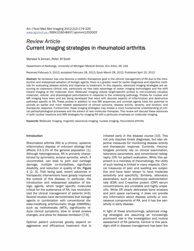
Figure 1. A joint (the place where two bones meet) is surrounded by a capsule that protects and supports it. The joint capsule is lined with a type of tissue called synovium, which produces synovial fluid that lubricates and nourishes joint tissues. In rheumatoid arthritis, the synovium becomes inflamed, causing warmth, redness, swelling, and pain. As the disease progresses, the inflamed synovium invades and damages the cartilage and bone of the joint. Sur-rounding muscles, ligaments, and tendons become weakened. Rheumatoid arthritis also can cause more generalized bone loss that may lead to osteoporosis (fragile bones that are prone to fracture) (Image courtesy of the National Institute of Arthritis and Musculoskeletal and Skin Diseases (NIAMS) (http://www.niams.nih.gov/Health_Info/Rheumatic_Disease/default.asp - last accessed February 2012)).

Imaging of rheumatoid arthritis
176 Am J Nucl Med Mol Imaging 2012;2(2):174-220
this hypothesis [15]. CD4+ TH cells are subse-quently stimulated through interactions be-tween their T-cell receptor (TCR)-CD3 complex and CD4 molecule and the type II major histo-compatibility complex (MHC-II) with the anti-genic determinant on the surface of the APCs. T cell activation additionally needs a co-stimulatory signal from the recognition of CD80 or CD86 by its CD28 cell-surface molecule [16, 17]. Activated TH cells proliferate and infiltrate into the synovial tissue, where they release inter-feron-γ (IFN-γ), interleukin-2 (IL-2), and inter-leukin-4 (IL-4) [18]. These pro-inflammatory cy-tokines not only activate other TH cells, but also macrophages, fibroblasts, osteoclasts, and chondrocytes [19, 20]. Similar to TH cells, macrophages, when activated, increasingly mi-grate into synovial tissue. This is consistent with the finding that macrophages and TH cells make up the majority of inflammatory cell infiltrates in early and late RA [13, 14]. Stimulated macro-phages and fibroblasts, in turn, produce other pro-inflammatory cytokines, including tumor necrosis factor-α (TNF-α), interleukin-1 (IL-1), and interleukin-6 (IL-6), as well as chemokines, prostaglandins, proteases, and growth factors [21-23]. From the multitude of released im-mune mediators, B lymphocytes are stimulated to produce autoantibodies such as rheumatoid factor (RF), which is present in >80% of RA pa-tients. Neutrophils are recruited as well to the synovial joints, but they are more prevalent in the synovial fluid than lining. Activated endothelial vasculature helps coordi-nate and perpetuate this mass infiltration of mononuclear cells into inflamed synovium. In the presence of IL-1β and TNF-α, endothelial cells in postcapillary venules upregulate cell adhesion molecules (CAMs), which assist in the rolling, binding, and transendothelial migration of leukocytes [24]. Increased microvascular permeability and hyperemia, non-specific mechanisms of the acute inflammatory re-sponse, may add to the accumulation of leuko-cytes in inflamed synovium; moreover, these processes, in addition to locally expanded diffu-sion space, account for the enhanced extrava-sation of macromolecules and small proteins into the interstitial space during the acute phase of the disease [25]. As RA becomes chronic, synovial proliferation, supported by neovascularization, leads to pannus formation.
This hypertrophic and hyperplastic synovial tis-sue is highly invasive, particularly at the inter-face between the synovium and juxta-articular bone and cartilage, and is responsible for mar-ginal erosions and joint space narrowing. The destructive nature of this tissue is likely the re-sult of fibroblast-like synoviocyte and chondro-cyte production and release of metallopro-teinases, which degrade proteoglycans and col-lagen [26-28]. Bone resorption due to activation of osteoclasts, however, appears to be the main mechanism through which bone erosions occur [27]. Over time, these destructive forces cou-pled with mechanical stress cause variable changes in peri-articular bone and soft tissue structures. These changes manifest clinically as chronic joint swelling, tenderness, pain, and eventual destruction. Overall, RA can be viewed as a series of coordinated events in the syno-vial, vascular, and bone compartments. The mechanisms that both initiate and perpetuate this disease within and between these compart-ments all represent potential targets for molecu-lar imaging and therapeutic intervention and will be discussed in detail below. Nuclear medicine imaging strategies Role of nuclear medicine in the management of RA While more established imaging techniques focus on morphological changes, nuclear medi-cine provides functional data about disease activity, which is critical for therapy decision-making and patient follow-up. In particular, by measuring long-term alterations in imaging pa-rameters that serve as surrogates for synovitis, nuclear imaging allows for the objective moni-toring of treatment response in RA patients. As current drug regimens are relatively expensive, determining patient response early in the treat-ment course is a cost-effective solution. Addi-tionally, it is important to determine which pa-tients are likely to develop high-risk lesions or a more severe disease course, as it may call for more aggressive treatment or more frequent monitoring. As nuclear probes visualize disease processes that are active even prior to irreversi-ble anatomic changes, imaging with these probes may allow for the early prediction of dis-ease outcomes. Furthermore, nuclear imaging has the potential to accurately select patients that are likely to

Imaging of rheumatoid arthritis
177 Am J Nucl Med Mol Imaging 2012;2(2):174-220
respond to a particular treatment based on the articular presence of the drug target. Due to the high intra- and inter-individual variation in these target molecules in the joints of RA patients, a pre-treatment scan with a radiotracer that local-izes the therapeutic target in inflamed tissues may be useful in predicting treatment efficacy and planning appropriate therapies. This ap-proach could also provide an explanation for the failure of any targeted therapy or a justification for the use of a specific treatment. As a logical extension, nuclear imaging can allow for a more personalized therapeutic program tailored to the specific perturbations in inflammatory and destructive pathways that are active in the joints of each patient. Individualized patient management is particularly important, consider-ing RA has heterogeneous clinical manifesta-tions and is often thought of as a collection of disorders. While still mostly experimental, the clinical role of nuclear medicine in imaging RA is greatly expanding, particularly as this field takes advantage of new imaging technologies for PET and SPECT and targeted molecular probes. We are gradually seeing this shift to molecular im-aging in nuclear medicine, but note that non-specific agents are still widely available for im-aging RA as well. Positron emission tomography (PET) imaging [18F]FDG 2-[18F]Fluoro-2-deoxy-D-glucose ([18F]FDG) is a well-characterized radiolabeled glucose analog that when used in conjunction with PET reflects metabolic changes in tissues. Increased uptake of [18F]FDG is mediated through glucose trans-porter type 1 (GLUT1) and glucose transporter type 3 (GLUT3) cell-surface proteins, which are overexpressed in hypermetabolic cells [29]. Upon entering the cell, [18F]FDG is rapidly phos-phorylated to [18F]FDG-6-phosphate by the pri-mary glycolytic enzyme, hexokinase, whose upregulation additionally accounts for the en-hanced [18F]FDG uptake observed in rapidly proliferating cells [29]. Unlike phosphorylated glucose, [18F]FDG-6-phosphate cannot undergo further metabolism, effectively trapping this molecule intracellularly. Due to the inclination toward anaerobic glycolysis and consequently the elevated metabolic demand for glucose in cancerous cells, [18F]FDG PET has been widely employed in the field of oncology for tumor stag-ing, diagnosis, and therapeutic evaluation. [18F]
FDG accumulation, however, is not specific for neoplastic tissue. For example, macrophages, neutrophils, and young granulation tissue in-creasingly take up glucose, and thus, [18F]FDG, as a consequence of activation and respiratory (oxidative) burst [30-32]. Due to the involve-ment of these cells in the maintenance of in-flammatory processes, it was hypothesized that [18F]FDG PET may serve as a useful tool for the study of RA. [18F]FDG PET has since been em-ployed in a number of pre-clinical and clinical RA studies (reviewed in [33]). Recently, Matsui and co-workers provided the proof-of-mechanism for this imaging approach in a murine collagen-induced arthritis (CIA) model and in vitro [3H]FDG uptake study [34]. [18F]FDG PET was shown to accurately delineate swollen joints in vivo. As confirmed histologi-cally, moderate [18F]FDG uptake was noted in regions of interstitial inflammatory cell recruit-ment, synovial cell hyperplasia, and edema early in the disease course. In comparison, later-developing sites of pannus formation and bone destruction demonstrated high levels of [18F]FDG accumulation, highlighting the capacity of this imaging strategy to reflect the progression of arthritis. Within these inflammatory regions, proliferating fibroblasts were determined to ex-hibit the highest levels of [3H]FDG uptake, fol-lowed by neutrophils. Furthermore, while resting macrophages were not shown to significantly contribute to [3H]FDG accumulation, hypoxic conditions and the presence of pro-inflammatory cytokines such as TNF-α (a micro-environment common in rheumatoid joints) greatly enhanced [3H]FDG uptake in these cells as well as fibroblasts, but not neutrophils. Con-trastingly, [3H]FDG uptake was considerably lower for T lymphocytes, indicating that these inflammatory cells only play a negligible role in [18F]FDG accumulation in vivo. Altogether, these findings suggest that proliferating fibroblasts and macrophages, particularly when in a hy-poxic, pro-inflammatory cytokine-rich microenvi-ronment, are the primary contributors to re-gional [18F]FDG uptake in vivo in pannus and interstitial inflammatory cell infiltrates. This study supports the notion set forth by a number of clinical studies (as discussed below) that [18F]FDG PET accurately reflects the disease activity of RA. Palmer and colleagues were the first to evaluate the validity of quantifying joint inflammation and
Imaging of rheumatoid arthritis
178 Am J Nucl Med Mol Imaging 2012;2(2):174-220
changes in metabolic activity in response to treatment in RA patients using [18F]FDG PET [35]. In this pivotal work, Gadolinium-enhanced MRI and [18F]FDG PET images of wrist lesions were acquired for 12 patients with inflammatory arthritis (6 RA patients and 3 patients with pso-riatic arthritis) undergoing anti-inflammatory therapy. Clinical examination and imaging stud-ies were conducted at 3 intervals: baseline, af-ter 2 weeks of treatment with prednisone or NSAIDs, and after 12-14 weeks of low-dose methotrexate (MTX) treatment. For each ses-sion, volume of enhancing pannus (VEP) was calculated from axial, fat-suppressed MR im-ages for correlation with [18F]FDG PET parame-ters (total uptake value (TUV) and regional up-take value (RUV)) and clinical findings. Visual comparison of images revealed that regions of greatest PET signal corresponded to areas of enhancing pannus on MRI [35]. While de-creases in pannus volume and [18F]FDG uptake in response to treatment paralleled clinical im-provement (in terms of pain, tenderness, and swelling) of the imaged wrist, none of the [18F]FDG PET or MRI parameters was associated with overall treatment outcome. The authors suggested that this lack of correlation with treat-ment outcome could be the result of a small patient population or the strict cut-offs imposed by the Paulus index as to what qualifies as a treatment response (need 20% improvement in each of 4 of 6 possible measures). Palmer and co-workers concluded that Gadolinium-enhanced MRI and [18F]FDG PET allow for the quantification of volumetric and metabolic changes in synovitis and the comparison of effi-cacies of anti-inflammatory treatments [36]. Expanding on this previous study, Beckers and co-workers investigated the ability of [18F]FDG PET to detect synovitis and quantify its meta-bolic activity in 21 RA patients, as compared to standard measures of disease activity [37]. In a joint-by-joint analysis, PET findings were found to significantly correlate with those of regional clinical (swelling and tenderness) and sono-graphic assessments. Furthermore, both the degree of PET positivity (visual analysis) and mean standardized uptake values (SUVs) were found to increase with synovial thickness in all joints (except metatarsophalangeal-1 joints), as measured by ultrasound (US), and the number of clinical or US parameters present simultane-ously. On an individual patient level, strong cor-relations were additionally cited for PET-derived
parameters (number of PET-positive joints and cumulative SUV) and disease duration as well as global measures of disease activity, including clinical joint counts for swelling and tenderness, erythrocyte sedimentation rate (ESR) and C-reactive protein (CRP) serum levels, the patient and physician global assessments, the disease activity score and the simplified disease activity index, and US-derived. Based upon these find-ings, the authors suggest that [18F]FDG PET of-fers unique information concerning the meta-bolic activity of synovitis specific to each pa-tient. According to Brenner et al., these findings, while promising, do not necessarily procure a role for [18F]FDG PET in the routine clinical assessment of RA patients [38]. As a relatively costly tech-nique, [18F]FDG PET must provide clinically rele-vant data that cannot be obtained from stan-dard clinical and laboratory measures of dis-ease activity to have a broader application in the study of RA. Consequently, evidencing the capacity of [18F]FDG PET to monitor disease activity and response to treatment is of particu-lar importance. In another study by Beckers et al., 16 RA patients underwent clinical and bio-logical evaluation, dynamic Gadolinium-enhanced MRI, US, and [18F]FDG PET imaging of knee joints at baseline and after 4 weeks of anti-TNF-α treatment [39]. Consistent with previous studies, SUVs were significantly correlated with all MRI-derived parameters, synovial thickness, and serum levels of matrix metalloproteinase (MMP) 3 and CRP [35, 37]. PET-positive knee joints were determined to have higher SUVs, MRI parameters, and greater synovial thickness as measured by US than PET-negative knee joints. While a number of studies have shown that [18F]FDG PET is capable of detecting treatment-related changes in disease activity, very few have investigated whether PET findings can pre-dict clinical outcomes early in the course of treatment [36, 39, 40]. In a small explorative study with 16 RA patients, Elzinga and col-leagues demonstrated that early changes in regional [18F]FDG uptake in the joints of RA pa-tients undergoing anti-TNF-α (infliximab) treat-ment were representative of later changes in global disease activity, as assessed clinically [41]. While these findings support the notion that [18F]FDG PET allows for the sensitive detec-tion of early changes in disease activity that are

Imaging of rheumatoid arthritis
179 Am J Nucl Med Mol Imaging 2012;2(2):174-220
highly predictive of subsequent responses to treatment, larger studies are needed to confirm. Although these results are already promising, new advances in PET technology, including mul-timodal image co-registration, may serve to en-hance the utility of this imaging technique in the study of RA. In particular, PET/CT hybrid acquisi-tion offers higher spatial resolution and simulta-neous integration of morphologic and functional data and thus has rapidly become standard clinic protocol. Despite these advantages, only a limited number of studies have evaluated this technology in RA patients. For example, in a case study by Vogel et al., [18F]FDG PET/CT was reported to not only have the capability of calcu-lating the degree of inflammation in the tarsus of an RA patient, but also precisely localizing the disease activity to the particular joints causing the complaints [42]. Contrastingly, neither physical examination nor conventional radiogra-phy could offer this information. Due to the in-volvement of multiple joints, as visualized by [18F]FDG PET/CT, a triple arthrodesis of the tar-sus was performed, with successful pain reduc-tion, demonstrating that this imaging technique provides clinically relevant information that can be utilized in patient management. Additional case studies have demonstrated that [18F]FDG PET/CT accurately detects extra-articular inflam-matory sites such as subcutaneous nodules and hypermetabolic lymph nodes and synovitis of the atlanto-axial and knee joints [43-45]. Fur-thermore, although not substantiated, it is plau-sible that PET/CT allows for improved discrimi-nation between juxta-articular disease and ar-ticular processes. Goerres et al. did note that PET imaging alone was able to delineate inflam-mation of the tendon sheaths and bursae [40], but mild cases may not be apparent due to the low spatial resolution of dedicated PET scan-ners. Having an anatomic framework, as is pro-vided with PET/CT, may help in the evaluation of these cases. Of particular interest, Kubota and colleagues reported that whole-body [18F]FDG PET/CT imag-ing accurately and sensitively reflected the metabolic disease activity and joint anatomy in 14 patients with active RA and 4 patients in remission [46, 47]. More specifically, [18F]FDG joint uptake, total joint score, global SUVmax, and the mean number of joints with at least a mod-erate uptake of [18F]FDG were significantly higher for patients with active disease as com-pared to those in remission. Additionally, pain-
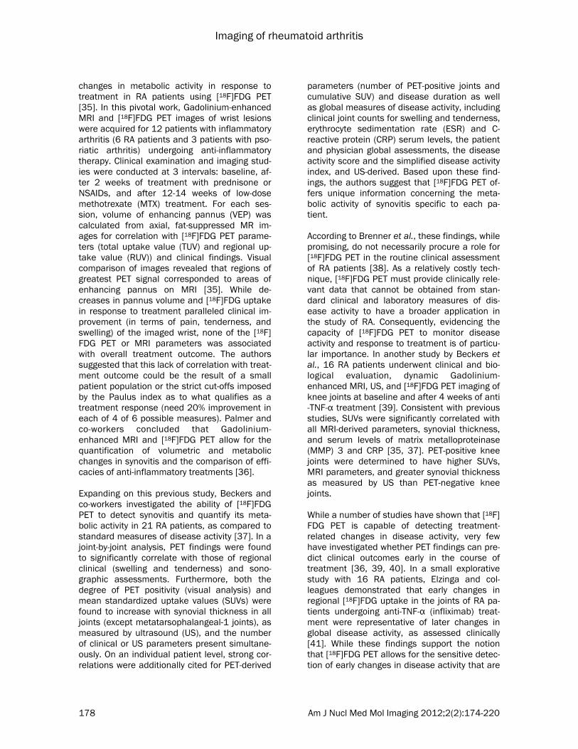
ful/swollen joints had a higher [18F]FDG uptake score and SUVmax than did clinically uninvolved joints. A representative [18F]FDG PET/CT image of a patient with recurrent RA can be seen in Figure 2, showing increased radiotracer uptake in multiple large joints and clearly delineating inflammatory foci (Figure 2A). While the wrist, elbow, and knee joints could be easily inter-preted as PET positive, more complicated large joints such as the hip and shoulder require PET images with anatomical correlation to CT find-ings, as increased FDG uptake by enthe-sopathies must be differentiated from synovitis arising from RA (Figures 2B and 2C). To com-pare, a conventional bone scan only showed mild arthritic changes in the large joints of the same patient, suggesting that this modality is not as sensitive as [18F]FDG PET/CT (Figure 2D). CT correlation was also necessary for interpreta-tion of findings in the atlanto-axial joint. As com-pression of the spinal cord and brainstem are potentially serious complications of the involve-ment of the atlanto-axial joint in RA patients, early detection of this high-risk lesion is clini-cally important. In this study, 28% (5/18) of the RA patients exhibited increased [18F]FDG up-take in the atlanto-axial joint, but most were asymptomatic. The authors speculated that these hypermetabolic lesions are most likely indicative of active subclinical synovitis. This finding is consistent with that of other studies, which have found that there is a high preva-lence of asymptomatic cervical spine subluxa-tion in this patient population [44, 48]. [18F]FDG PET/CT may thus allow for the early identifica-tion of patients at risk for developing subluxa-tion of the atlanto-axial joint. In addition, this imaging technique may have prognostic value for a more severe disease course in RA pa-tients, as it has been reported that the presence of arthritis in large joints, particularly arthritis in the knee joint, is predictive of a destructive dis-ease course [49]. Overall, whole-body [18F]FDG PET/CT has the advantage of allowing for the accurate assessment of the extent and severity of the disease even at subclinical levels. In addition to PET/CT, PET/MRI technology has also been studied in RA patients. As this tech-nology has only recently been developed, the distribution of dedicated PET/MRI scanners is fairly limited. Co-registered PET/MRI hybrid ac-quisition, however, is rapidly becoming an im-portant nuclear medicine strategy. Chaudhari and colleagues showed that this technology
Imaging of rheumatoid arthritis
180 Am J Nucl Med Mol Imaging 2012;2(2):174-220
could be employed to monitor the early re-sponse to anti-TNF-α therapy in the wrist joint of an RA patient [50]. This study, however, did not use a fully integrated PET/MRI system. Instead, the patient underwent extremity [18F]FDG PET/CT imaging separately following MR image ac-quisition; the CT image was used for post-acquisition PET/MRI image co-registration. While this study demonstrates the capability of this technology, it does not showcase its com-plete range of advantages. Since using separate PET/CT and MRI scanners does not allow for simultaneous acquisition of image data, there is room for spatial and temporal misalignment between PET and MRI images. In comparison, dedicated PET/MRI scanners greatly reduce these artifacts through simultaneous acquisi-tion of PET and MRI data without sacrificing sensitivity or spatial resolution. While they did not specifically evaluate an RA patient, El-Haddad and co-workers used a fully integrated PET/MRI scanner in a case study to accurately delineate a meniscal tear associated with syno-vitis [51]. To date, there has only been one study that has successfully performed a true hybrid PET/MRI
examination of an RA patient. In a recent study by Miese et al., a patient with early RA under-went simultaneous PET/MRI scanning of the hand using a prototype of an APD-based mag-neto-insensitive Brain PET detector (Siemens Healthcare, Erlangen, Germany) operated within a standard 3T MR scanner (MAGNETOM Trio, Siemens) [52]. Increased [18F]FDG uptake was noted surrounding the metacarpophalangeal (MCP) II and III joints, which corresponded to sites of synovitis and tenovaginitis as identified on contrast-enhanced MRI (Figure 3). A maxi-mum SUV of 3.1 was measured for the palmar portion of MCP II, which was shown to correlate with marked synovial thickening and contrast enhancement on MRI. No significant [18]FDG uptake was seen within the joint spaces or bony structures. Contrastingly, conventional radio-graphic evaluation was determined to be nega-tive. These results suggest that [18F]FDG PET/MRI is a potentially useful tool in the early diag-nosis of RA. [11C]Choline Choline is a water-soluble essential nutrient that functions as a neurotransmitter (upon acetyla-
Figure 2. A 74-year-old woman with 3.5-year history of RA who experienced a recurrence and was being considered for infliximab therapy. (A) Anterior and RAO MIP image obtained using FDG-PET/CT shows typical RA lesions in the large joints. (B and C) Axial PET/CT fusion image of the hip joint in the same patient. The large arrows indicate synovi-tis in the acetabulum and femoral head. The small arrows indicate enthesopathies at the ischium and greater tro-chanter (D) Bone scan of the same patient shows mild changes in the joints. (Reprinted from Kubota K, Ito K, Morooka M, Minamimoto R, Miyata Y, Yamashita H, Takahashi Y and Mimori A. FDG PET for rheumatoid arthritis: basic considerations and whole-body PET/CT. Ann NY Acad Sci 2011; 1228: 29-38; by permission of John Wiley and Sons).

Imaging of rheumatoid arthritis
181 Am J Nucl Med Mol Imaging 2012;2(2):174-220
tion to acetylcholine in cholinergic nerve end-ings), a methyl group donor (through degrada-tion to its primary metabolite, betaine), and a precursor to phospholipids (the main constitu-ent of all eukaryotic cell membranes) [53]. Free choline is taken up by dividing cells and pre-dominantly phosphorylated by choline kinase to phosphorylcholine, in the first step of the cytidine diphosphocholine (CDP) pathway [53]. This commits choline to the biosynthesis of phospholipids, particularly phosphatidylcholine (lecithin), and integration into eukaryotic cell membranes. Previous studies have determined that rapidly dividing cells, and their greater need for cell membrane components as compared to normal tissues, result in increased choline up-take through energy-dependent choline specific transport mechanisms and simple diffusion sec-ondary to hyperemia and hyperperfusion [54-56]. Consequently, choline, when radiolabeled with carbon-11, can serve as an in vivo bio-marker of cellular proliferation. [11C]Choline PET imaging has been shown to clearly delineate various brain tumors, lymph node metastases of esophageal cancer, and lung carcinoma [57-62]. Due to the minimal renal excretion of [11C]choline, the primary indi-
cation for this imaging procedure is the detec-tion and staging of prostate cancer and other cancers of the urogenital tract (bladder and uterine cancer) [63, 64]. Non-neoplastic appli-cations are currently under investigation, as the rate of [11C]choline uptake in tissues solely cor-relates with the level of cellular growth, irrespec-tive of histologic grade [59]. Since arthritic pan-nus and nearby vessels undergo similar prolif-erative changes to those exhibited during malig-nant transformation, it was hypothesized that [11C]choline PET could be utilized in the assess-ment of RA and other arthritic diseases. Roivainen and co-workers evaluated the capac-ity of [11C]choline PET to detect and quantify arthritic synovial proliferation by imaging the joints of 10 patients with synovitis and compar-ing the results with [18F]FDG PET and Gadolin-ium-DTPA (Gd-DTPA)-enhanced MRI findings [65]. In particular, maximum standardized up-take values (SUVmax) and kinetic influx constants (Ki), as obtained from the graphic analysis de-scribed by Patlak et al., were calculated for both PET radiotracers and compared to MRI parame-ters (synovial volume and rate of enhancement) [66]. In all patients, the PET signal intensities for [11C]choline and[18F]FDG were significantly
Figure 3. Hybrid 18F-FDG PET–MRI of the hand in early RA. a axial and coronal display of PET co-registered with b axial and coronal T1-weighted MRI. c True hybrid 18F-FDG PET–MRI of the hand. (Reprinted with kind permission from Springer Science+Business Media: Miese F, Scherer A, Osten-dorf B, Heinzel A, Lanzman RS, Kropil P, Blondin D, Hautzel H, Wittsack HJ, Schneider M, Antoch G, Herzog H and Shah NJ. Hybrid 18F-FDG PET-MRI of the hand in rheumatoid arthritis: initial re-sults. Clin Rheumatol 2011; 30: 1247-1250, Figure 1).

Imaging of rheumatoid arthritis
182 Am J Nucl Med Mol Imaging 2012;2(2):174-220
increased in diseased synovia, in contrast to clinically unaffected joints. Similarly, when visu-ally compared to coronal Gd-DTPA-enhanced T1-weighted MR images, regions of highest [18F]FDG and [11C]choline signal on PET coincided well with contrast-enhanced hypertrophic syno-vial tissue (pannus). From these findings, the authors suggested a possible role for [11C]choline PET in the functional imaging of RA. A clear advantage of [11C]choline PET is the abil-ity to quantitatively measure reproducible dis-ease activity parameters through SUVmax and/or graphic analysis. Similar standardized uptake values of [11C]choline to those measured in Roivainen et al. [65] were obtained in a later study by the same group [67]. Moreover, due to the short half-life of carbon-11, [11C]choline de-livers a relatively low radiation burden to the patient [68]. Finally, as [11C]choline can only be synthesized at facilities equipped with on-site cyclotrons, effectively limiting its broader distri-bution and application, [18F]choline is currently under development. To date, however, there have not been any pre-clinical or clinical studies that have evaluated its efficacy in imaging RA or other arthritic diseases [69]. (R)-[11C]PK11195 While both [18F]FDG and [11C]choline are sensi-tive to changes in cellular proliferation, their uptake is not specific to inflammation; normal physiologic variants, malignancy, infection, and other benign pathological processes can result in hypermetabolic PET lesions. For this reason, there is an increasing need for PET radiotracers that allow for more specific visualization of rheumatoid synovial tissue through direct tar-geting of underlying inflammatory pathways. Due to the cardinal role of macrophage infiltra-tion in the propagation and extension of RA, radiotracers that target this process are of spe-cial interest. (R)-[11C]PK11195 is a recently de-veloped isoquinolone carboxamide PET radio-tracer that targets cells of the monocyte-macrophage lineage by selectively binding as an antagonist to the peripheral benzodiazepine receptor (PBR), or newly renamed translocator protein (TSPO) [70-73]. TSPO is an 18 kDa pro-tein that is expressed on the outer mitochon-drial membrane- and to a lesser extent nucleus and plasma membrane- of mononuclear phago-cytes, other leukocyte subsets, peripheral or-gans, and neuronal, hematopoietic, and lym-
phatic tissues [71, 73-76]. While TSPO was originally discovered as a second high-affinity binding site for benzodiazepines, particularly diazepam, it has since been determined that this transmembrane protein serves many func-tions; more specifically, TSPO is thought to be a component of the trimeric mitochondrial perme-ability transition pore (MPTP) and play a role in the regulation of steroidogenesis and apoptosis, heme biosynthesis, cell proliferation, and im-mune regulation [73, 75-79]. Since activated macrophages and polymononu-clear cells upregulate TSPO during inflamma-tion, TSPO-targeting radiotracers can serve as surrogate markers for disease activity and macrophage recruitment to inflammatory foci. This imaging technique has been validated for the assessment of various inflammatory neuro-logical disorders; in particular, (R)-[11C]PK11195 PET imaging allows for the differentia-tion of neuroinflammatory lesions from normal tissues by mapping out glial cell activation and recruitment [80-82]. van der Laken and co-workers were the first to extend the application of (R)-[11C]PK11195 PET to the study of RA by comparing the imaging findings for 11 RA pa-tients and 8 healthy controls to clinical data and immunohistochemical analysis of excised syno-vial tissue samples [83]. (R)-[11C]PK11195 up-take correlated well with clinical severity of synovitis. Severely inflamed joints exhibited the highest accumulation of (R)-[11C] followed by mild to moderately inflamed joints. Uptake val-ues for these joints were, on average, signifi-cantly higher than those for clinically unin-flamed and control joints, which intimates that (R)-[11C]PK11195 imaging is sensitive in its de-tection of both severe and moderate inflamma-tion. The increased PET signal in inflamed joints was determined to be a consequence of specific PBR-mediated uptake of (R)-[11C]PK11195 by activated macrophages, as confirmed by immu-nohistochemical staining of synovial tissues. Accordingly, severely inflamed joints demon-strated the highest degree of macrophage re-cruitment and PBR expression. In comparison, control knee joints of healthy volunteers dis-played minimal PBR expression and macro-phage infiltration, and, concomitantly, no signifi-cant accumulation of radioactivity on PET. Of interest, though not confirmed in this study, van der Laken and colleagues conjectured that (R)-[11C]PK11195 PET imaging may detect sub-

Imaging of rheumatoid arthritis
183 Am J Nucl Med Mol Imaging 2012;2(2):174-220
clinical synovitis [83]. In support of this hypothe-sis, mean SUV ratios for clinically uninflamed joints in RA patients were noted to be approxi-mately 50% higher (P<0.05) than those of con-trol joints in healthy volunteers. These findings are consistent with those of previous studies that have established that macrophage infiltra-tion into synovial joints is a common feature of asymptomatic synovitis in early RA [13, 14]. Furthermore, as the presence and number of macrophages in rheumatoid synovia correlate with the progression of radiographic joint ero-sions, the application of (R)-[11C]PK11195 im-aging to RA may prove to be relevant to patient management. Gent and co-workers recently confirmed that (R)-[11C]PK11195 PET can observe subclinical synovitis in arthralgia patients [84]. In this pro-spective pilot study, high resolution (R)-[11C]PK11195 images of the metacarpophalangeal (MCP), proximal interphalangeal (PIP), and wrist joints of 29 seropositive arthralgia patients, 6 healthy volunteers (negative controls), and 3 patients with established RA (positive controls) were acquired and subsequently scored semi-quantitatively for joint uptake minus back-ground activity by 2 independent readers. Pa-tients were followed prospectively for 24 months to determine progression to RA. Four of 29 arthralgia patients (i.e. 4 of the 9 arthralgia patients that progressed to RA) were deter-mined to have PET positive scans with moder-ate to high radiotracer uptake in the joints of 3 of these patients. To compare, healthy volun-teers did not exhibit any PET positive joints while significant (R)-[11C]PK11195 accumula-tion was noted in all clinically involved joints of RA patients. During the follow-up period, all four subjects with PET positive scans showed pro-gression and developed clinical arthritis in at least 1 MCP, PIP, and/or wrist joint(s). Unex-pectedly, five patients with negative scans de-veloped clinical synovitis within 24 months. In-congruent PET and clinical findings in 3 of these patients were easily explained by the fact that the newly diagnosed arthritic lesions were not present in the hands and wrists. Since the re-searchers used a small animal and human brain 3D PET scanner for image acquisition, the field of view (FOV) was not large enough to allow visualization of joints beyond the hands and wrists. The remaining 2 arthralgia patients that developed arthritis, despite having negative PET scans, only showed subtle signs of disease ac-
tivity throughout the follow-up period, and 1 of these patients even entered remission sponta-neously at 10 months. There are also drawbacks to using (R)-[11C]PK11195 however. Although the absolute stan-dardized uptake values of (R)-[11C]PK11195 in arthritic joints are comparable to those reported for [18F]FDG, high non-specific binding of this radiotracer in hand muscles, soft tissues sur-rounding the nails, and bone marrow greatly reduces signal-to-noise ratios [83, 84]. High physiologic uptake has also been noted in the kidneys, lungs, liver, and heart [85-87]. This, in turn, could limit the detection of mild arthritic lesions. Although not evaluated in the trial of Gent et al. [84], it is important to note that co-registration of PET and CT scan images, which is the current standard clinical protocol, may allow for the accurate differentiation between articu-lar and peri-articular uptake of (R)-[11C]PK11195 by providing accurate anatomic local-ization. In addition, this radiopharmaceutical is not ideal for imaging, as it has low bioavailabil-ity, high plasma protein binding, high lipophilic-ity, and a relatively low binding affinity (Ki=1-4 nM) [85, 88, 89]. Consequently, numerous TPSO-targeting radiotracers with improved imag-ing properties are currently under development [90]. Such probes that have entered pre-clinical and clinical trials include [11C]DPA-713, [11C]DAA1106, [11C]PBR28, [11C]AC-5216, [18F]PBR06, [18F]DPA-714, and [18F]FEDAA1106 [89, 91-100]. As ligands labeled with fluorine-18 have a longer half-life than their carbon-11 la-beled counterparts, they are favored moving forward with this imaging strategy. Scintigraphy and SPECT imaging Non-specific Imaging Agents [67Ga]Citrate [67Ga]citrate (Neoscan®) is a well-characterized non-conjugated γ-emitting radiotracer that local-izes in acute and chronic inflammatory, infec-tious, and neoplastic lesions. Upon intravenous administration, cationic 67Ga3+, similar in behav-ior to the ferric ion, binds to circulating blood plasma proteins, such as transferrin and fer-ritin, and to a minority of leukocytes [101-103]. The resultant 67Ga-bound protein complex, and to a minor extent, free and neutrophil-associated [67Ga]citrate, extravascates increas-

Imaging of rheumatoid arthritis
184 Am J Nucl Med Mol Imaging 2012;2(2):174-220
ingly at sites of inflammation due to locally aug-mented microvascular permeability and ex-panded extracellular diffusion space [102, 104-107]. As there is limited reabsorption of macro-molecules at these sites, [67Ga]citrate progres-sively accumulates in inflammatory foci, includ-ing rheumatoid synovia. Following the trapping of 67Ga-bound macromolecules in the inflamma-tory interstitial space, transchelation of 67Ga to lactoferrin, ferritin, and/or bacterial sideropho-res may occur [101, 102, 108, 109]. The en-hanced neutrophil secretion of lactoferrin in synovial fluid, and expression of ferritin in in-flamed synovia are additional mechanisms by which [67Ga]citrate concentrates in these tis-sues [102]. Although not as well-supported in the literature, transferrin receptor-mediated endocytosis of the [67Ga]citrate-transferrin com-plex by synovial macrophages may play a partial role in the localization of this radiotracer in af-fected joints of RA patients [103, 110]. This mechanism is currently under review, serving as the rationale for the development of new 99mTc-labeled conjugated transferrin probes [111]. While it is well-documented that [67Ga]citrate scintigraphy is a sensitive imaging technique for inflammation, there are several disadvantages that preclude its widespread use in the evalua-tion of RA patients. In particular, due to its re-lease of high-energy γ radiation (91-393 keV) and long physical half-life (t1/2= 78.3 hours), 67Ga imposes a relatively high radiation burden on patients [112]. Moreover, similar to other radiolabeled macromolecules, [67Ga]citrate has a slow plasma clearance when bound to serum proteins. Slow blood clearance is unfavorable because it results in high background activity and longer acquisition times so as to attain opti-mal target-to-background ratios [103, 113]. Further limiting the clinical application of [67Ga]citrate is its lack of specificity. Although [67Ga]citrate imaging is able to sensitively detect RA disease activity and extent, it cannot accurately distinguish active inflammation from infection or even neoplasm [103, 114-116]. Therefore, [67Ga]citrate scintigraphy has been relegated to a secondary role in imaging patients with RA as well as other inflammatory and infectious dis-eases. [99mTc]- and [111In]HIG Polyclonal human immunoglobulin G (HIG) is a non-antigen IgG antibody that when labeled with
either 99mTc or 111In behaves as a biomarker for infection and inflammation. HIG scintigraphy is an inexpensive, accessible tool that allows visu-alization of inflammatory foci, and thus, its use has been widely explored in RA. A number of studies have indicated that this imaging tech-nique detects local joint inflammation in RA pa-tients with a higher sensitivity than that of clini-cal examination, conventional bone scanning, and leukocyte scintigraphy [117-119]. Pons and co-workers found a correlation between articu-lar HIG uptake, as assessed visually and quanti-tatively, and clinical scores for swelling, suggest-ing that HIG scintigraphic findings accurately reflect disease severity [120]. Furthermore, highlighting its prognostic value, a study by de Bois et al. demonstrated that this imaging strat-egy is able to predict progression to RA in ar-thralgia patients [121]. Similar to [67Ga]citrate, however, HIG is a non-specific marker for in-flammation. It accumulates at sites of inflam-mation due to a local increase in vascular per-meability and diffusion space, and hyperemia [122]. Consequently, HIG scintigraphy is only of limited clinical relevance. For example, it is inca-pable of distinguishing between joints with ac-tive disease and those with inflammation from secondary joint destruction. It also has no utility in evidence-based biologic therapy. HIG imaging is sensitive, but its non-specificity has greatly diminished its clinical role, as newer radiophar-maceuticals with higher specificity have been developed. [99mTc]Diphosphonates Unlike previous radiotracers, 99mTc-labeled di-phosphonate analogs, such as methylene di-phosphonate (MDP), hydroxy methylene diphos-phonate (HDP), and dicarboxy propane diphos-phonate (DPD), are not primarily inflammation-seeking agents. Instead, their uptake reflects alterations in bone metabolism, especially in-creased obsteoblastic activity occurring in re-sponse to underlying pathology. Conventional bone scintigraphy has shown some utility in the evaluation of RA, as it allows localization of ar-thritic joints and provides functional and quanti-tative information about disease activity [123-125]. Three-phase bone scanning (blood flow phase, immediate blood pool phase, and de-layed imaging phase) may allow detection of acute RA. Blood flow and blood pooling phases typically exhibit increased uptake secondary to hyperemia and augmented microvascular per-

Imaging of rheumatoid arthritis
185 Am J Nucl Med Mol Imaging 2012;2(2):174-220
meability (same vascular changes as those in surrounding inflamed synovium) during the acute phase of RA [126]. In addition, delayed images demonstrate higher radiotracer deposi-tion in diseased juxta-articular bone, reflecting increased bone turnover and remodeling in re-sponse to joint inflammation and cartilage de-struction [126]. This technique, while a sensi-tive tool in identifying osseous changes, is not specific for RA and lacks the spatial resolution of radiography or MRI [127]. Moreover, as 99mTc-labeled diphosphonates accumulate to a vari-able degree in all joints, differentiation between normal bone and juxta-articular physiologic up-take and mild arthritic changes is difficult [128]. As a further disadvantage, bone scintigraphy cannot reliably differentiate between active dis-ease and inflammation in chronically damaged joints [128, 129]. To improve the sensitivity, spatial resolution, and overall image quality of this approach, Os-tendorf and colleagues evaluated and com-pared [99mTc]DPD multi-pinhole SPECT (MPH-SPECT) to conventional bone scintigraphy and MRI for the detection of bone changes in 13 patients with early RA and 9 patients with early osteoarthritis (OA) [130]. Recently developed MPH-SPECT systems have been previously shown to have a 50-fold increased sensitivity and spatial resolution of less than 1 mm as a consequence of their inclusion of collimators that have up to 20 pinholes [131, 132]. In the Ostendorf et al. study, MPH-SPECT, in compari-son to conventional bone scanning, demon-strated better radiotracer localization and spa-tial resolution and was able to detect a greater number of diseased joints [130]. As 10 of 13 RA patients had a central tracer distribution and 7 of 9 OA patients had an eccentric pattern, the authors suggested that this distinction may have relevance to our understanding of RA pathogenesis. Furthermore, the sensitivity of this technique was found to be comparable to that of MRI. Hybrid SPECT/CT also allows for fusion of functional and anatomic information, but despite these advancements in tomo-graphic technology, bone scanning has mainly been superseded by other imaging techniques for the assessment of RA patients. [99mTc]- and [111In]-labeled Leukocytes Directly labeling autologous leukocytes with [99mTc]hexamethylpropylene amine oxime
([99mTc]HMPAO) or [111In]oxine, radiolabeled complexes that easily penetrate cell mem-branes and become trapped intracellularly as a result of their high lipophilicity and charge neu-trality, is in wide clinical use for the evaluation of a number of infectious and inflammatory dis-eases [133, 134]. As mononuclear cells and neutrophils are highly recruited to inflamed synovial tissue and fluid, respectively, leukocyte imaging has been described for use in RA. In a preliminary study, Gaál and co-workers found a significant correlation (P<0.01) between the global scores for [99mTc]HMPAO-labeled neutro-phil accumulation in the hands and feet of 21 RA patients and the number of clinically swollen joints [135]. The authors concluded that leuko-cyte scintigraphy or SPECT is an inexpensive and widely available tool that can be utilized in the localization and estimation of synovitis in RA. Al-Janabi and co-workers additionally dem-onstrated that this imaging technique is sensi-tive to alterations in disease activity following treatment with intra-articular steroid injections [136]. Autologous monocytes have also been successfully labeled with [99mTc]HMPAO and assessed in RA [134, 137]. Thurlings and col-leagues reported that [99mTc]HMPAO-labeled monocyte scintigrahpic findings positively corre-late with the swollen joint count and number of macrophages, as confirmed by immunohisto-chemical staining, in biopsied synovial tissue from 8 RA patients [138]. While it is clear that radiolabeled leukocyte joint scintigraphy allows delineation of inflammation and distribution of disease with high sensitivity, it lacks specificity for RA, ultimately limiting its clinical utility. Specific imaging agents [99mTc]J001X To improve specificity, receptor-specific radio-labeled probes that indirectly track leukocyte migration and recirculation in chronic inflamma-tory diseases have been developed. As dis-cussed earlier, radiotracers that target macro-phages are of interest because this cell sub-population is highly recruited to rheumatoid synovia and plays a critical role in the inflamma-tory process. In vitro studies have shown that macrophages specifically bind to bacterial pro-teoglycans, providing a rationale for the use of radiolabeled proteoglycan derivatives in macro-phage scintigraphy [139-141]. J001X, a 34 kDa acylated poly-(1,3)-D-galactoside isolated from

Imaging of rheumatoid arthritis
186 Am J Nucl Med Mol Imaging 2012;2(2):174-220
membrane proteoglycans of a non-pathogenic strain of Klebsiella pneumoniae, is a newly de-signed probe that when coupled with 99mTc al-lows the visualization of mononuclear phago-cyte trafficking [142, 143]. Although similar in structure to the immunogenic bacterial lipopoly-saccharide (LPS), J001X has been greatly modi-fied to not trigger phagocytic activation, while still retaining its specificity for cells of the mono-cyte-macrophage lineage [139, 140, 144]. CD14 (a glycosylphosphoinositol-anchored LPS receptor found on macrophages and neutro-phils) and CD11b (the α-chain of the comple-ment receptor-3 (CD11b/CD18) β2 leukocyte integrin expressed on mononuclear phagocytes, granulocytes, and NK cells) mediate this spe-cific receptor-ligand interaction [143, 145]. The imaging potential of [99mTc]J001X scintigra-phy has been widely explored for numerous tu-moral, inflammatory, and infectious processes in both human patients and experimental ani-mal models. [99mTc]J001X scintigraphy has suc-cessfully delineated alveolitis and mediastinal berylliotic lymph nodes in baboons; acute local-ized radiation changes in pigs; pyrogranulomas in sheep; and osteoarthritic lesions induced by severance of cruciate ligaments in rabbits [146-150]. Similarly in humans, macrophage imaging with [99mTc]J001X, administered as an aerosol, was able to localize inflammatory lesions in sar-coidosis and scleroderma [151]. The assess-ment of pulmonary involvement in RA patients was explored as a possible application of [99mTc]J001X scintigraphy, but with mixed results [152]; scintigraphic findings were incompatible with those from high-resolution CT, pulmonary function tests, and bronchoalveolar lavage. Of interest for purposes of studying RA, this nu-clear medicine technique has been investigated in an antigen-induced arthritis model in rabbits [153]. Scintigraphic images demonstrated [99mTc]J001X focal uptake in active arthritic le-sions with high contrast to normal tissues. In comparison, these same inflammatory lesions could not be clearly discerned with [99mTc]O4- and [99mTc]albumin nanocolloids due to a lower scintigraphic contrast, despite the increased uptake of these non-specific agents in the acute phase of the disease. Furthermore, at the ad-vanced disease stage when non-specific inflam-matory processes were normalized, uptake of radiolabeled nanocolloids was minimal while [99mTc]J001X scans remained positive for
macrophage infiltration. These results support previous conclusions that [99mTc]J001X scinti-graphy can serve as a functional imaging strat-egy that directly reflects the extent of macro-phage recruitment and thus evolves with dis-ease activity. To justify the use of this imaging technique in future RA clinical trials, the authors optimized the labeling procedure for the intrave-nously injectable formulation of [99mTc]J001X [153, 154]. While this study offers promising results, no clinical trials have been performed to determine the efficacy of [99mTc]J001X scintigra-phy in diagnosing early RA and monitoring dis-ease activity and treatment response to date. [99mTc]RP128 Like [99mTc]J001X, [99mTc]RP128 scintigraphy visualizes leukocyte recruitment, a process criti-cal for sustaining RA and other inflammatory diseases. RP128, a bifunctional peptide che-late, specifically targets neutrophils and mono-nuclear phagocytes by binding to receptors ex-pressed on the surface of these cell subpopula-tions [155]. As a general mechanism, the tar-geting domain of [99mTc]RP128, an antagonistic pentapeptide tuftsin analogue (TKPPR), medi-ates the receptor-specific interaction and binds to tuftsin receptors with a fourfold greater affin-ity than does their endogenous ligand, tuftsin [156]. Tuftsin is a tetrapeptide (TKPR) derived from proteolytic cleavage of the Fc domain of the heavy chain of IgG that promotes chemo-taxis and phagocytosis of its target cells [157]. Tuftsin receptors, as mediators of these key immune functions, represent important molecu-lar targets, and their upregulation in activated macrophages serves as the basis for [99mTc]RP128 imaging. Despite promising pre-clinical studies that have cited a positive correlation between [99mTc]RP128 uptake and quantitative measures of inflammation, there has only been one study that investigated the utility of [99mTc]RP128 scintigraphy in imaging RA patients. In a Phase I study, Caveliers and co-workers simultaneously evaluated the safety, normal biodistribution, and dosimetry in 8 healthy controls, and the validity of employing [99mTc]RP128 as a probe to delineate inflamed synovia in 10 RA patients [155]. The biodistribution study favorably re-vealed low radiotracer uptake in all major or-gans, except in the kidneys and bladder and, to a lesser extent, the synovia of several joints.

Imaging of rheumatoid arthritis
187 Am J Nucl Med Mol Imaging 2012;2(2):174-220
Due to the slower washout of articular activity and low background noise, synovial joints in all subjects could be clearly discerned. The accu-mulation of [99mTc]RP128 in normal joints, how-ever, was moderate in comparison to the mark-edly increased uptake observed in a large num-ber of clinically affected joints in RA patients. Consequently, [99mTc]RP128 scintigraphy was able to detect inflammatory lesions in RA pa-tients with a sensitivity of 69% for swollen joints, 76% for painful joints, and 73% for joints with bone erosions. Unfortunately, as synovial biopsies were not performed in this study, accu-racy of the results could not be confirmed and further evaluation of this radiotracer is needed. [99mTc]- and [111In]anti-E-selectin Another strategy to visualize the continuous leukocyte recruitment and infiltration into in-flamed synovium involves targeting cell adhe-sion molecules (CAMs). CAMs are responsible for the binding of leukocytes to activated endo-thelial vasculature as well as their subsequent transendothelial migration [158, 159]. While various adhesion molecules have served as targets for therapeutic intervention and imaging in other inflammatory diseases, E-selectin is the only one to be successfully described in the mo-lecular imaging of RA. E-selectin (CD62E, ELAM-1) is a transmembrane glycoprotein that is tran-siently expressed on the luminal surface of acti-vated vascular endothelium during a normal inflammatory response [24]. Following induction by interleukin-1 (IL-1), tumor necrosis factor-α (TNF-α), and bacterial LPS, E-selectin mediates the initial tethering and rolling of granulocytes, monocytes, and some lymphocytes, via specific interactions with its carbohydrate-based ligands [24, 158-161]. E-selectin is a potentially useful target for the detection of synovitis, because while this molecule is not expressed in resting endothelium, there is increasing evidence that its upregulation in postcapillary venules helps promote the sustained influx of leukocytes into inflamed tissues in RA [162-167]. As an added advantage, E-selectin is directly accessible to intravenously administered agents due to its location on the luminal surface of blood vessels. [111In]1.2B6 monoclonal antibody (mAb), an indium-labeled murine IgG1 antibody that recog-nizes human E-selectin, was first validated for in vivo imaging of synovitis in porcine models of arthritis in 1994 [168, 169]. [111In]1.2B6 mAb
was shown to immunolocalize to activated en-dothelial venules in inflamed synovia and re-gional draining lymph nodes with better sensitiv-ity and specificity than radiolabeled control IgG1 antibody. Although these early, preclinical stud-ies offered promising results, concerns were raised over the immunogenicity of this radio-pharmaceutical. Due to its murine origin, 1.2B6 mAb has the potential to elicit a human anti-mouse antibody (HAMA) response, limiting long-term repeat follow-up imaging with this radio-tracer. Furthermore, 1.2B6 mAb has intact Fc regions, which are thought to generate host immunity through non-specific activation of Fc-γ receptor-bearing effector cells. To reduce the likelihood of these clinical complications, re-searchers elected to study F(ab’)2 fragments of 1.2B6 mAb, devoid of its Fc portions. A number of animal studies have validated this substitu-tion [170, 171]. Preliminary clinical studies cor-roborated these earlier works, demonstrating that [111In]1.2B6 F(ab’)2 scintigraphy allows for clear visualization of inflamed joints in RA pa-tients through radioimmunodetection of acti-vated vascular endothelium, as early as 4 hours and optimally at 24 hours post-injection [172, 173]. In addition, this imaging technique was shown to be superior to both 111In- and 99mTc-labeled HIG scintigraphy in terms of sensitivity, specificity, lower background activity, higher radiotracer uptake, and better image contrast. However, due to the higher radiation burden imparted to the patient and lower spatial resolu-tion of 111In-labeled radiopharmaceuticals, a 99mTc-labeled anti-E-selectin radiotracer was subsequently developed and tested in RA pa-tients. In a two part study, Jamar and colleagues evalu-ated the validity of using [99mTc]1.2B6 Fab frag-ments in RA patients as compared to [111In]1.2B6 F(ab’)2 and [99mTc]HDP [128]. For the double-isotope comparative study, planar im-ages were acquired 4 and 20-24 hours follow-ing administration of either [111In]1.2B6 F(ab’)2 or [99mTc]1.2B6 Fab in 10 RA patients and 2 healthy volunteers. Additionally, 16 RA patients underwent scintigraphic evaluation for compari-son between [99mTc]1.2B6 Fab and [99mTc]HDP (740 MBq (20 mCi)) at 4 hours post-injection of either radiotracer. [99mTc]1.2B6 Fab scinti-graphic findings were found to be congruent with those of [111In]1.2B6 F(ab’)2. Deviations in radiotracer distribution were noted, but could mostly be attributed to the differences in normal
Imaging of rheumatoid arthritis
188 Am J Nucl Med Mol Imaging 2012;2(2):174-220
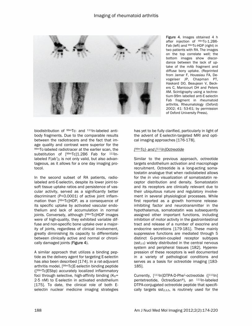
biodistribution of 99mTc- and 111In-labeled anti-body fragments. Due to the comparable results between the radiotracers and the fact that im-age quality and contrast were superior for the 99mTc-labeled radiotracer at the earlier scan, the substitution of [99mTc]1.2B6 Fab for 111In-labeled F(ab’)2 is not only valid, but also advan-tageous, as it allows for a one day imaging pro-tocol. In the second subset of RA patients, radio-labeled anti-E-selectin, despite its lower joint-to-soft tissue uptake ratios and persistence of vas-cular activity, served as a significantly better discriminant (P<0.0001) of active joint inflam-mation than [99mTc]HDP, as a consequence of its specific uptake by activated vascular endo-thelium and lack of accumulation in normal joints. Conversely, although [99mTc]HDP images were of high-quality, they exhibited variable dif-fuse and non-specific bone uptake over a major-ity of joints, regardless of clinical involvement, greatly diminishing its capacity to differentiate between clinically active and normal or chroni-cally damaged joints (Figure 4). A similar approach that utilizes a binding pep-tide as the delivery agent for targeting E-selectin has also been described [174]. In a rat-adjuvant arthritis model, [99mTc]E-selectin binding peptide ([99mTc]ESbp) accurately localized inflammatory foci through selective, high-affinity binding (KD= 2-5 nM) to E-selectin in activated endothelium [175]. To date, the clinical role of both E-selectin nuclear medicine imaging strategies
has yet to be fully clarified, particularly in light of the advent of E-selectin-targeted MRI and opti-cal imaging approaches [176-178]. [99mTc]- and [111In]Octreotide Similar to the previous approach, octreotide targets endothelium activation and macrophage recruitment. Octreotide is a long-acting soma-tostatin analogue that when radiolabeled allows for the in vivo visualization of somatostatin re-ceptor distribution and density. Somatostatin and its receptors are clinically relevant due to their ubiquitous nature and regulatory involve-ment in several physiological processes. While first reported as a growth hormone release-inhibiting factor and neurotransmitter in the hypothalamus, somatostatin was subsequently assigned other important functions, including inhibition of motor activity in the gastrointestinal tract and release of a number of exocrine and endocrine secretions [179-181]. These mainly suppressive functions are mediated through 5 distinct G-protein-coupled receptor subtypes (sst1-5) widely distributed in the central nervous system and peripheral tissues [182]. Hyperex-pression of these receptors is well documented in a variety of pathological conditions and serves as a basis for octreotide imaging [183-185]. Currently, [111In]DTPA-D-Phe1-octreotide ([111In]pentetreotide, OctreoScan®), an 111In-labeled DTPA-conjugated octreotide peptide that specifi-cally targets sst2,3,5, is routinely used for the
Figure 4. Images obtained 4 h after injection of 99mTc-1.2B6-Fab (left) and 99mTc-HDP (right) in two patients with RA. The images on the top correlate well; the bottom images show discor-dance between the lack of up-take of the mAb fragment and diffuse bony uptake. (Reprinted from Jamar F, Houssiau FA, De-vogelaer JP, Chapman PT, Haskard DO, Beaujean V, Beck-ers C, Manicourt DH and Peters AM. Scintigraphy using a techne-tium-99m labelled anti-E-selectin Fab fragment in rheumatoid arthritis. Rheumatology (Oxford) 2002; 41: 53-61; by permission of Oxford University Press).

Imaging of rheumatoid arthritis
189 Am J Nucl Med Mol Imaging 2012;2(2):174-220
scintigraphic and/or SPECT imaging of primary neuroendocrine tumors and their metastases as well as other somatostatin receptor-bearing malignancies [186-191]. Non-neoplastic appli-cations of this imaging technique have only re-cently been explored [185]. The demonstration of somatostatin’s role in the modulation of the immune response prompted investigations into the value of somatostatin receptor scintigraphy in chronic inflammatory diseases and other im-mune-mediated disorders [192]. The possibility of utilizing this imaging technique for the in vivo study of RA was raised only after researchers coincidentally observed uptake of radiolabled octreotide in the arthritic joints of a sarcoidosis patient [193]. As a logical progression, it was supposed and subsequently confirmed that the synovia of affected joints in RA patients overex-press somatostatin receptors, the target of oc-treotide. In particular, immunohistochemical staining of diseased synovial tissue samples from RA patients revealed expression of sst2 on activated venule endothelial cells and infiltrat-ing mononuclear phagocytes [193-195]. Consti-tutive expression of sst1 and sst2 on fibroblast-like synovial cells, as the result of TNF-α induc-tion, has additionally been noted [196]. In com-parison, the synovial tissue from a patient with clinically and biochemically confirmed RA who successfully underwent treatment did not stain for these receptors to any significant degree [196]. This suggests a role for octreotide imag-ing not only in disease localization, but also monitoring therapeutic response. Van Hagen and co-workers conducted a pilot study to evaluate octreotide imaging in a cohort of 14 RA patients [193]. Somatostatin receptor scintigraphic findings were found to correlate well with clinical parameters. Increased [111In]pentetreotide uptake allowed for the visualiza-tion of inflamed synovia with a lesion-related sensitivity of 76%. The specific uptake of octreo-tide by somatostatin receptors expressed in diseased joints was confirmed with in vitro autoradiographic studies. In comparison, no radiotracer accumulation was observed in the joints of control patients. Somatostatin receptor imaging continues to be an area of interest for the assessment of rheu-matoid arthritis, particularly as this approach has implications for therapy. A number of stud-ies have indicated that therapy with soma-tostatin analogues improves symptoms in RA
patients and attenuates inflammatory proc-esses such as synovial proliferation and IL-6 and IL-8 production [196-199]. Positive scinti-graphic findings may therefore serve as a ration-ale for treatment with unlabeled somatostatin analogues. However, to the best of our knowl-edge, no clinical studies have evaluated this novel role for octreotide imaging. [99mTc]Anti-CD3 mAb As mature T lymphocytes play a large role in the pathogenesis and extension of RA, radiophar-maceuticals that target this cell population can serve as a useful tool in evaluating disease course and localization. Radiolabeling mono-clonal antibodies (mAbs) directed against CD3 is a recently developed method that allows for the selective imaging of T lymphocyte migration into rheumatoid synovium. The CD3 antigen consists of 2 heterodimeric glycoproteins (CD3-δ/ε and CD3-γ/ε), embedded almost exclusively in the cell membranes of CD4+ and CD8+ T lym-phocytes [200]. This protein complex noncova-lently associates with the T-cell receptor (TCR) and 2 TCR-ζ accessory chains, and collectively, they are responsible for T-cell activation [201, 202]. The CD3 antigen more specifically partici-pates in signal transduction, following the bind-ing and recognition of the major histocompati-bility complex (MHC) proteins by the TCR [201, 202]. Muromonab (Orthoclone OKT3) was the first specifically engineered anti-CD3 mAb. This mur-ine IgG2a antibody binds to epitopes of human CD3-ε, resulting in the early, temporal activation of peripheral T cells followed by a sharp inhibi-tion and modulation of T cell functions [203]. As a potent immunosuppressant, OKT3 is indi-cated for the treatment of acute allograft rejec-tion [204]. Recently, however, interest has turned toward radiolabeling OKT3 for use in immunoscintigraphic imaging of rheumatic dis-eases. In a study of 7 RA patients and 2 pa-tients with psoriatic arthritis, Marcus and co-workers demonstrated that the [99mTc]OKT3 scintigraphic findings correlated well with pa-tient history and physical examination [205]. Increased focal radiotracer uptake was present in a minority of asymptomatic joints, but this is likely indicative of subclinical synovitis. Conse-quently, the authors suggested that [99mTc]OKT3 scintigraphy may allow for earlier diagno-sis of RA and psoriatic arthritis. Unfortunately, 2

Imaging of rheumatoid arthritis
190 Am J Nucl Med Mol Imaging 2012;2(2):174-220
patients in this study experienced shaking chills and neck pain approximately 1 hour post-injection of the anti-CD3 mAb. These adverse events appear consistent with cytokine release syndrome and will likely limit the use of OKT3 in RA patients [203, 206]. Despite these possible complications, Martins and colleagues further investigated the applica-tion of [99mTc]OKT3 imaging to the detection of synovitis in 38 RA patients [207]. Anterior pla-nar scans showed increased focal [99mTc]OKT3 accumulation in 68.8% of tender joints, 71.8% of swollen joints, and 88.1% tender and swollen joints. Correspondingly, [99mTc]OKT3 scinti-graphic findings significantly correlated with swollen joints, tender joints, and the visual ana-logue scale (VAS) (p<0.05). Moreover, [99mTc]OKT3 scintigraphy allowed the differentiation of patients in remission from those with active synovitis, according to their disease activity score. Unlike the previous study, there were no reported adverse events. [99mTc]OKT3 scintigraphy not only has the ca-pacity to assess disease activity, but also the ability to distinguish juvenile idiopathic arthritis (JIA) and RA patients from those with other rheumatic diseases, such as gouty arthritis (GA) and osteoarthritis (OA). As these patients can present clinically with overlapping signs and symptoms, this distinction is critical to optimal patient management and therapeutic interven-tion. In a study by Lopes and colleagues, the joints of 77 patients with rheumatic diseases
(44 RA, 5 JIA, 15 OA, and 13 GA patients) were evaluated by [99mTc]OKT3 scintigraphy [208]. Since activated T lymphocytes play a large role in the pathophysiological processes of RA and JIA, but not in OA or GA, there were observable differences in the [99mTc]OKT3 uptake patterns obtained for the patients with each respective disease. As expected, there was high initial radiotracer uptake in the inflamed joints of RA and JIA patients, and a subsequent increase in accumulation visualized on the delayed scans (Figure 5). Contrastingly, the initial radiotracer uptake was absent or minimal in cases of OA followed by a decrease in uptake observed at the delayed scan. This is consistent with the fact that the inflammatory process in OA is independent of T cell activation by the TCR/CD3 complex. Accord-ingly, no uptake was noted in any of the painful joints of OA patients. For joints (n=4) where the presence of edema was the main complaint, however, there was 1 joint for which the scan showed mild radiotracer accumulation. More-over, another scan exhibited slightly increased [99mTc]OKT3 uptake when OA joints (n=6) with pain and edema were considered. Interestingly, a patient with previously diagnosed OA had an elevated ESR and early and delayed scinti-graphic images showing increased uptake in the knees and hands (which on physical exam were shown to be painful and have edema), indica-tive of RA. Upon further clinical examination, this patient was later re-diagnosed with RA. This case serves to demonstrate the clinical rele-
Figure 5. Scintigraphy with 99mTc-anti-CD3 of the knees shows an increase in the uptake of these areas in late im-ages. Images taken (A) 1 h and (B) 3 h after endovenous injection of the radiopharmaceutical. (Reprinted from Lopes FP, de Azevedo MN, Marchiori E, da Fonseca LM, de Souza SA and Gutfilen B. Use of 99mTc-anti-CD3 scintigraphy in the differential diagnosis of rheumatic diseases. Rheumatology (Oxford) 2010; 49: 933-939; by permission of Oxford University Press).

Imaging of rheumatoid arthritis
191 Am J Nucl Med Mol Imaging 2012;2(2):174-220
vance of this imaging technique. GA patients, in comparison, were noted to have an initial in-crease in articular radioactivity, but this greatly declined as observed on the delayed scan. The authors suggested that the early accumulation of [99mTc]OKT3 in the joints of GA patients is likely the result of increased vascularity and cell infiltration in patients whose main complaint was edema or edema and pain. And thus, the eventual decrease in articular radioactivity lev-els reflects the absence of TCR/CD3-mediated T lymphocyte activation in GA pathophysiology. While promising results, a major drawback to this imaging technique is the safety profile of OKT3 mAbs. Although no adverse events were noted in the Martins et al. and Lopes et al. stud-ies, a whole host of side effects, particularly cytokine release syndrome (CRS), are associ-ated with OKT3 use, even at microgram doses [203, 205]. CRS appears to be the result of the binding of antibody Fc regions to Fc-γ-receptors on immune effector cells and subsequent acti-vation of these cell populations [209]. In this process, T lymphocytes are activated as well, which ultimately leads to the release of cyto-kines, mitogenicity, and antibody- and comple-ment-dependent cytotoxicity [209]. Further-more, as a murine mAb, OKT3 has the potential to elicit a human anti-mouse antibody (HAMA) response. This not only limits follow-up imaging, but also long-term therapeutic use. In an effort to limit these clinical complications, a number of humanized and chimeric OKT3 Fc variants have been developed [210-214]. Malviya et al. recently radiolabeled visilizumab (Nuvion®), a non-Fc-γ-receptor binding human-ized IgG2 mAb that binds with the CD3-ε chain with high specificity and high avidity (Ka= 0.5x10⁹) [215, 216]. Favorably, this mAb does not activate T lymphocytes and thus has limited potential to induce cytokine release or acute cytotoxicity [216]. This imaging strategy was evaluated in Balb/c and SCID irradiated mice reconstituted with human lymphocytes and shown to be able to accurately map CD3+ cell distribution in vivo [215-217]. From these find-ings, the authors suggested that in addition to offering information about disease localization, [99mTc]visilizumab could provide a rationale for therapy with unlabeled visilizumab in select can-didates. Unfortunately, visilizumab has been withdrawn from production as a therapeutic agent after phase III trials [217]. Insufficient
efficacy and, surprisingly, inferior safety profile were cited as the reasons for the withdrawal. While visilizumab could be manufactured solely for imaging purposes, it may be clinically useful to instead radiolabel anti-CD3 mAbs that offer therapeutic benefits and thus allow for the cou-pling of imaging and treatment. [99mTc]anti-CD4 mAb Anti-CD4 imaging is yet another strategy to visu-alize the highly relevant molecular process of T lymphocyte infiltration. In contrast to the previ-ous technique, however, CD4 radioimmu-nodetection does not target a pan-T cell anti-gen, but instead, a molecule only present in a subset of T lymphocytes. CD4 is a 55 kDa monomeric glycoprotein expressed on the cell surface of all mature T helper cells (TH, CD4+ T lymphocytes), a majority of thymocytes, and to a lesser extent, monocytes-macrophages and dendritic cells [218]. Its extracellular domains bind to non-polymorphic regions of major histo-compatibility complex class II (MHC-II) mole-cules, stabilizing the interaction between TH cells and MHC-II-positive antigen-presenting cells (APCs) during T cell activation [219, 220]. In addition to functioning as a co-receptor for the TCR on TH cells, CD4 plays a role in signal transduction due to the physical association of its short cytoplasmic tail with tyrosine kinase p56Ick, an important protein integral for the in-tracellular signaling cascade following T cell activation [221]. CD4 antigen represents a potentially important target in the study of RA, as CD4+ TH cell clones with abnormal phenotypes are abundant among cell infiltrates in arthritic synovial tissue and fluid [222, 223]. Synovial tissue and fluid also contain monocytes-macrophages, which ex-press, in lower density, the CD4 molecule. In-creased numbers of CD4+ TH cells in the periph-eral blood have been noted as well during active disease [224]. Furthermore, a higher frequency of certain MHC-II alleles has been described in RA, suggesting that antigen-specific responses of autoreactive CD4+ TH cells are critical to the development and possible maintenance of this disease [15, 225, 226]. Based on these considerations, Becker and colleagues evaluated anti-CD4 radioimmunos-cintigraphy in 6 patients with severe, clinically active RA [227]. Whole body and joint-specific

Imaging of rheumatoid arthritis
192 Am J Nucl Med Mol Imaging 2012;2(2):174-220
planar images were obtained for all patients at 1.5, 4, and 24 hours following intravenous in-jection of [99mTc]MAX.16H5, a 99mTc-labeled murine IgG1 mAb that binds with high affinity to the human CD4 molecule in 5 patients, and in vitro labeled [99mTc]MAX.16H5 lymphocytes (≥10 MBq) in 1 patient. Scintigraphic findings were compared to those of clinical examination (Ritchie index, joint swelling) of 44 joints and early (5 minutes) and late (2 hours) [99mTc]HDP bone scans that each patient received within 7 days prior to immunoscintigraphy. [99mTc]MAX.16H5 imaging allowed clear visualization of diseased joints at 4-6 hours after radiotracer administration in all patients. These results cor-related well with those of the early bone scan (P<0.01) and clinical scoring of joint swelling (P<0.01), but not with the late bone scan (P>0.05) or Ritchie index scoring of painful joints. A number of mismatches between joint uptake patterns in late bone scans and [99mTc]MAX.16H5 scintigrams were noted, suggesting that the latter imaging technique has higher sensitivity in detecting early, active disease, comparable to that of clinical diagnosis. Al-though not substantiated in this study, CD4 ra-diolocalization may be able to differentiate be-tween RA and other forms of arthritis, such as septic arthritis, as it binds to infiltrating mono-nuclear cells, not granulocytes characteristic of acute inflammation. This property would be valuable and an advantage over both bone scanning and radiolabeled leukocyte scintigra-phy. While the Becker et al. study demonstrated in-creased uptake of [99mTc]MAX.16H5 in arthritic joints, it is unclear from their work as to whether accumulation of radiolabeled anti-CD4 mAbs in inflamed synovium is a consequence of specific target recognition or non-specific mechanisms [227]. To validate the specificity of this tech-nique, Kinne and co-workers compared [99mTc]MAX.16H5 uptake with that of non-specific poly-clonal [99mTc]HIG in 8 patients with severe, clini-cally active RA and 2 healthy volunteers [228]. In all examined arthritic joints, accumulation of [99mTc]MAX.16H5, expressed as percentage of total body radioactivity, did not significantly dif-fer from that of non-specific [99mTc]HIG. Despite similar total uptake values, [99mTc]MAX.16H5 had lower background activity and thus signifi-cantly higher target-to-background (T/B) ratios as compared to [99mTc]HIG as early as 4 hours following radiotracer administration. Semi-
quantitative visual scoring of scans confirmed higher average counts in the synovial mem-brane over those in either blood vessels or mus-cle for radiolabeled anti-CD4 mAbs in arthritic knee and elbow joints as compared to control Ig. ROI analysis of knee joint scans allowed quantification of T/B ratios, corroborating the results from the visual assessment. Kinne et al. argued that the improved T/B ratios were solely the result of [99mTc]MAX.16H5 specificity for CD4+ inflammatory cell infiltrate, and, conse-quently, radiolabeled anti-CD4 mAbs allow for superior detection of inflammatory foci in RA than [99mTc]HIG scintigraphy. In support of this claim, another study by the same authors re-ported that [99mTc]MAX.16H5 exhibited higher total radioactivity levels and T/B ratios in ar-thritic joints of a single patient with longstand-ing, severe RA in comparison to [99mTc]anti-carcinoembryonic antigen mAb, an isotype-matched non-specific control mAb [229]. Conflicting reports exist, however, as a study utilizing a rat-adjuvant arthritis model has re-vealed similar levels of uptake and T/B ratios in inflamed synovium for both radiolabeled anti-CD4 mAbs and isotype-matched Ig controls [230]. The authors posited that the low T/B ra-tios may be the result of non-specific trapping of the anti-CD4 mAb due to its large size and/or binding to Fc receptors in arthritic joints rather than true non-specificity of the molecule [230, 231]. To test this hypothesis, Kinne and co-workers compared the joint uptake and body distribution of [99mTc]anti-CD4 (W3/25; IgG1) Fab’ fragments, which are smaller in size and devoid of Fc receptor-binding regions, to those of non-specific Fab’ fragments ([99mTc]NCA-90) in a rat-adjuvant arthritis model [231]. Although both Fab’ fragments showed increased uptake in arthritic compared to non-arthritic joints, ac-cumulation of anti-CD4 Fab’ fragments was sig-nificantly higher (approximately 1.6-fold) than that of control Fab’ at all time points. Quantifica-tion of T/B ratios by ROI analysis confirmed su-periority of anti-CD4 Fab’ fragments over control Fab’ fragments as well as whole anti-CD4 mAbs in delineating inflammatory foci. In addition to having higher specific accumulation in inflamed synovium compared to entire anti-CD4 mAbs, anti-CD4 Fab’ fragments favorably had faster blood clearance and renal excretion and lower levels of background plasma radioactivity [232]. It was noted, however, that the Fab’ fragments exhibited greater in vivo instability than the

Imaging of rheumatoid arthritis
193 Am J Nucl Med Mol Imaging 2012;2(2):174-220
whole anti-CD4 molecule [232]. From these studies, it is clear that [99mTc]anti-CD4 imaging can detect inflammatory foci with high sensitivity; as to whether this imaging tech-nique will play a large role in the routine clinical evaluation of RA patients in the future is un-known. As has been previously discussed, a critical advantage for utilizing radioimmunoscin-tigraphy over other conventional methods is the ability to localize molecules that can serve as potential targets of therapeutic intervention. Currently, a controversy in the literature exists concerning the therapeutic benefit of anti-CD4 mAbs for RA patients [233-239]. Further evalua-tion of the efficacy of scintigraphic and thera-peutic use of anti-CD4 mAbs and Fab’ frag-ments in RA is needed. [99mTc]Anti-CD20 mAb While T lymphocytes have long been favored as the principal agents in RA pathogenesis, there is increasing evidence that B cell-mediated proc-esses are critical for the development of auto-immunity, warranting the targeting of this cell population for radioimmunoscintigraphy and therapeutic interventions. B cell functions that may contribute to the propagation of chronic inflammation include T cell activation through expression of co-stimulatory molecules (CD80/86 and CD40), antigen presentation to T lymphocytes, secretion of pro-inflammatory cyto-kines, production of autoantibodies such as Rheumatoid Factor (RF), and their subsequent formation of immune complexes that activate the complement system and bind to Fcγ-receptor-bearing effector cells [240-244]. Addi-tionally, B cell infiltrates have been noted in inflamed synovial tissues, providing the ration-ale for the use of B cell-targeted imaging in the in vivo study of RA patients. Instead of directly tagging B lymphocytes, researchers have devel-oped radiolabeled mAbs that target CD20, a pan-B cell surface antigen that is important for calcium homeostasis and the development and activation of this cell population [245]. CD20 represents a potentially useful target because 95% of circulating normal and malignant B lym-phocytes expresses this transmembrane phos-phoprotein [246]. Furthermore, it is exclusive to B lymphocytes, specifically in their late pro-B phase to maturity [241, 246, 247]; CD20, how-ever, is not expressed in hematopoietic stem cells and is shed upon differentiation into
plasma cells [241, 247, 248]. In a pilot study, Malviya and co-workers radio-labeled Rituximab (Rituxan, MabThera®), a chi-meric mouse/human IgG1κ mAb that recognizes epitopes on human CD20 antigen, with 99mTc to image B cell infiltration in the affected tissues of 10 patients with chronic inflammatory diseases, including RA [249]. Planar images obtained at 6 hours post-injection displayed increased [99mTc]Rituximab uptake in a majority, but not all, clini-cally inflamed joints. In one RA patient, [99mTc]Rituximab scintigraphic results were compared to those of [99mTc]anti-TNF-α imaging. Interest-ingly, the authors found a mismatch between the degree of uptake of [99mTc]Rituximab and radiolabeled anti-TNF-α in various synovial joints. While low radiotracer sensitivity is a pos-sible explanation for this finding, it is more likely an indication that selective inflammatory path-ways are in effect in different synovial joints, causing divergent articular phenotypes. Conse-quently, [99mTc]Rituximab scintigraphy may al-low for the visualization of the degree of local and systemic involvement of B lymphocytes in disease propagation. Moving forward, this method of mapping B cell infiltration has the potential to establish person-alized therapy, select candidates for treatment with unlabeled anti-CD20 mAbs, and monitor the efficacy of these therapies [206, 217]. A later study by the same group of authors sup-ported this approach by citing a high intra-articular and inter-individual variability of [99mTc]Rituximab uptake and thus B lymphocyte infil-tration in RA patients [250]. More studies, how-ever, are needed to test the clinical efficacy and relevance of this imaging technique. As RA pa-tients have been shown to benefit from unla-beled Rituximab and other B cell-depleting therapies, the role of [99mTc]Rituximab scintigra-phy may expand in the near future [251-255]. [99mTc]Anti-TNF-α Numerous pro-inflammatory cytokines play a role in the development and maintenance of chronic inflammation. Interleukin-1 (IL-1), inter-leukin-6 (IL-6), interleukin-2 (IL-2) and tumor necrosis factor-α (TNF-α), in particular, have been identified as important inflammatory me-diators in RA [256-258]. Directly and indirectly targeting cytokines and their receptors for imag-ing purposes is a novel strategy that has only

Imaging of rheumatoid arthritis
194 Am J Nucl Med Mol Imaging 2012;2(2):174-220
recently been explored in the context of RA. One study investigated the utility of [123I]IL-1 recep-tor antagonist ([123I]anakinra), which has a simi-lar binding affinity to IL-1, but no biological activ-ity, in imaging RA patients [259]. Nevertheless, the authors concluded that this probe was not useful for the scintigraphic assessment of RA patients because it did not show higher uptake levels than non-specific radiolabeled albumin, and thus, no follow-up studies were performed. Accordingly, attention has turned toward target-ing TNF-α for radioimmunoscintigraphic evalua-tion of RA patients and unlike [123I]anakinra, this imaging technique has shown promising results. TNF-α is a pleiotropic pro-inflammatory cytokine that has been implicated as a mediator of key pathophysiological processes in a number of neoplastic and chronic inflammatory autoim-mune diseases. It is synthesized, primarily by activated monocytes-macrophages and lympho-cytes, as a 27 kDa prohormone that, upon ag-gregation into a homotrimer, serves as a trans-membrane protein [260]. Through proteolytic cleavage by metalloproteinase TNF-α converting enzyme (TACE), soluble trimeric TNF-α (sTNF-α) is produced and secreted extracellularly [261, 262]. In either form, TNF-α carries out its nu-merous immunoregulatory and inflammatory functions by binding to its cognate cell-surface receptors, type 1 TNF-α receptor (TNFRI, p55) and type 2 TNF-α receptor (TNFRII, p75) [263-265]. These are found on neutrophils, endothe-lial cells, fibroblasts, and other cell types. The pro-inflammatory functions of TNF-α include the ability to upregulate adhesion molecules on the surface of vascular endothelium, and thus, pro-mote recruitment of granulocytes and mononu-clear cells, stimulate fibroblast growth, mediate bone resorption, and induce synthesis of other pro-inflammatory mediators such as IL-1, IL-6, prostaglandin E2, and IFNγ [266-274]. Based upon these considerations and the fact that elevated levels of membrane-bound and soluble TNF-α and its receptors are found in the sera and inflamed synovial tissue and fluid of patients with active RA, this potent inflamma-tory cytokine represents a critical target for im-munotherapy in these patients [256, 275, 276]. Numerous clinical studies have supported the sustained efficacy of TNF-α antagonists by dem-onstrating significant reduction of symptoms and disease severity and slowing or arresting of
joint erosive progression in RA patients when used in combination with conventional DMARDs [277-280]. Consequently, TNF-α blockade has become an established therapeutic approach, particularly in cases of refractory RA. Current FDA-approved TNF-α inhibitors for the treatment of RA include infliximab (Remicade®), adalimu-mab (Humira®), golimumab (Simponi®), certoli-zumab pegol (Cimzia®), and etanercept (Enbrel®). While TNF-α antagonists have revolu-tionized rheumatologic intervention, it is impor-tant to note that some RA patients do not re-spond to these agents [281]. This is likely the result of high inter-individual and intra-articular variation in the concentrations of TNF-α and other inflammatory cytokines [258]. Determin-ing the quantity and distribution of TNF-α in vivo in all inflamed joints prior to initiating therapy may therefore prove to be useful in selecting candidates likely to respond to TNF-α inhibition. This technique moreover has the potential to provide an objective measure of disease activity and treatment efficacy. To date, 2 TNF-α an-tagonists have been radiolabeled and utilized in the study of RA- infliximab and adalimumab. Infliximab is a bivalent, chimeric (murine-human) IgG1 monoclonal antibody that binds to both membrane-bound and soluble TNF-α with high specificity and affinity (KD= 1x10⁻¹⁰ M) [282]. It has a biological half-life of 9.5 days and functions as a TNF-α inhibitor by blocking the interaction between TNF-α and its receptors (p55 and p75) [283]. In addition, infliximab can induce antibody-dependent cellular cytotoxicity (ADCC) or complement-mediated cytotoxicity (CDC) of membrane-bound TNF-α expressing cells [283-286]. Recently, infliximab was radio-labeled with 99mTc and shown to have the same affinity for TNF-α as its unradiolabeled counter-part [287]. It has since been tested in patients with various infectious and chronic immune-mediated inflammatory diseases. In a prelimi-nary study, Conti and co-workers scintigraphi-cally assessed the in vivo levels of TNF-α in the inflamed knee of a patient with undifferentiated spondylarthropathy [288]. Following administra-tion of [99mTc]infliximab (15 mCi), planar images of the knee joints were acquired at 6 and 24 hours post-injection. Significant uptake of [99mTc]infliximab was observed for the inflamed knee as compared to the clinically uninvolved knee, indicating high intra-lesional levels of TNF-α. These scintigraphic findings served as the rationale for the patient receiving an intra-

Imaging of rheumatoid arthritis
195 Am J Nucl Med Mol Imaging 2012;2(2):174-220
articular injection of unradiolabeled infliximab. Clinical follow-up for a period of 4 months showed no signs of active disease (VAS score for pain= 0; no swelling or tenderness) in the injected knee, which was confirmed by negative scintigraphic findings, suggesting a role for [99mTc]infliximab imaging in localizing disease activity and monitoring therapeutic response in RA patients. The utility of [99mTc]infliximab scintigraphy for the detection of TNF-α-mediated inflammation in the joints of RA patients was assessed in an-other pilot study [289], evaluating patients pre- and post-therapy. Planar images of the inflamed joints were obtained for all patients at 3, 6, and 24 hours following intravenous administration of [99mTc]infliximab. Pre-treatment scans dem-onstrated increased regional radiotracer accu-mulation in inflamed joints with high target-to-background (T/B) ratios; no uptake was ob-served for clinically uninvolved joints. [99mTc]infliximab uptake in joints, however, showed more variability in scans performed 3 months after therapy. In comparison to the previous scan, 4 joints exhibited significantly reduced or no radiotracer uptake while 6 joints showed little to no change in [99mTc]infliximab accumula-tion. Patients with higher pre-therapy levels of TNF-α, as determined by scintigraphy, and sig-nificantly decreased [99mTc]infliximab uptake post-treatment were shown to have a better clinical response (improved swelling and pain) to infliximab treatment. These findings, in addi-tion to those from the Conti et al. study, support the use of [99mTc]infliximab scintigraphy in the objective selection of potential candidates that are likely to benefit from local or systemic inflixi-mab treatment [288]. Although promising, a proof-of-mechanism study has yet to be per-formed with a larger patient cohort. Further-more, as a chimeric mAb, infliximab use can trigger a human anti-mouse antibody (HAMA) response, which not only can hamper follow-up imaging studies, but also markedly decrease the efficacy of repeated therapeutic doses of infliximab. In an effort to reduce immunogenicity, adalimu-mab, a recombinant humanized anti-TNF-α IgG1 mAb, was engineered and has been approved for use in RA patients [290]. Adalimumab binds to membrane-bound and soluble TNF-α with high specificity and affinity (KD= 6x10⁻¹⁰ M) [291]. Similar to infliximab, adalimumab neu-
tralizes TNF-α by blocking its interaction with p55 and p75 and inducing ADCC and CDC of TNF-α-expressing cells [292]. Adalimumab has been radiolabeled with 99mTc as well and em-ployed in clinical trials. In a two-part study, Barrera and co-workers investigated the feasi-bility and validity of using [99mTc]adalimumab scintigraphy in the study of 10 RA patients [293]. In the first scintigraphic session, whole-body and joint-specific planar images were ac-quired for each patient 5 minutes, 4 and 24 hours following intravenous injection of [99mTc]adalimumab to assess tissue biodistribution and imaging properties. The same imaging pro-cedure was utilized in the second scintigraphic assessment that took place 2 weeks later, but an excess of unlabeled adalimumab (10 mg/kg) was co-administered in a subset of 5 patients to evaluate the degree of specific binding. The remaining 5 patients received a single intramus-cular dose of methylprednisolone (120 mg) 2 days before scintigraphic evaluation to study the ability of [99mTc]adalimumab imaging to de-tect clinically significant changes in intra-articular disease activity in response to systemic corticosteroid therapy. In all patients studied by Barrera et al., signifi-cant physiologic uptake of [99mTc]adalimumab was seen in the liver and spleen [293]. Joints were clearly delineated at 4 and 24 hours fol-lowing radiotracer injection, with inflamed joints demonstrating a 2- to 4-fold increase in radio-tracer uptake over normal tissues. Not all clini-cally active joints, however, showed accumula-tion of radiolabeled anti-TNF-α mAbs; this was particularly true for the small joints of the hands and feet. Although no explanation was offered, this finding likely reflects the high intra- and inter-individual variability of TNF-α expression in inflammatory foci. Conversely, no uptake was observed in clinically uninvolved joints. Scinti-graphic results from the second imaging ses-sion showed that the uptake of radiolabeled adalimumab is partly the result of specific bind-ing to TNF-α; co-administered excess unlabeled adalimumab abrogated the signal of radio-labeled anti-TNF-α mAbs by a median of 16% and 25% at 4 and 24 hours, respectively. These results were reproduced in later studies [294, 295]. Anti-TNF-α radioimmunoscintigraphy was also able to detect clinically relevant changes in synovitis following systemic corticosteroid treat-ment. The 5 patients who received treatment exhibited a decrease in clinical disease activity,

Imaging of rheumatoid arthritis
196 Am J Nucl Med Mol Imaging 2012;2(2):174-220
as expected, and a concomitant decline in [99mTc]adalimumab uptake in inflamed joints. No adverse events were reported. Further validating this imaging technique, Ro-imicher and co-workers compared the results of [99mTc]adalimumab scintigraphy to those of MRI and clinical evaluation in 8 patients with active RA and 1 healthy volunteer [295]. Whole-body and anterior planar images of hands and wrists were acquired for each patient at 30 minutes and 3 hours following injection of a subthera-peutic dose (15% of treatment dose) of [99mTc]adalimumab. Two nuclear medicine specialists visually evaluated the scintigraphic scans for intensity and pattern of [99mTc]adalimumab up-take. All patients underwent clinical examina-tion and MR imaging of hands and wrists. Axial and coronal pre- and post-Gadolinium contrast T1-weighted spin echo MR images were ob-tained for each patient, as were axial and cor-onal short tau inversion recovery (STIR) images. Two radiologists evaluated the scans for synovi-tis, according to the definition put forth by the OMERACT MRI collaborative subgroup (see be-low). Representative MR and scintigraphic im-ages can be seen in Figure 6.
Of 198 examined joints in the study by Ro-imicher et al., 49 joints were determined to be positive for synovitis on MRI and 48 joints on scintigraphy [295]; agreement between the methods was found for 44 joints (sensitivity of 89.8%). One hundred and forty-five joints were found to be negative on both MRI and scintigra-phy, resulting in a specificity of 97.3%. Five false-negatives and 4 false-positives were re-ported. The authors suggested that these false-negative scintigraphs were likely the result of the absence of detectable TNF-α levels in those inflamed joints. It has previously been shown that other cytokines besides TNF-α can act as the primary mediator of inflammation in some clinically active joints [258]. The ability to indi-rectly measure intra-lesional levels of TNF-α expression is a major advantage of this imaging technique, as it allows for evidence-based bio-logic therapy and patient selection. A number of studies have indicated that patients with higher pre-therapy uptake of [99mTc]adalimumab were more likely to respond to treatment with unla-beled anti-TNF-α mAbs than patients with lower or no radiotracer accumulation [294, 296]. Con-sequently, both positive and negative scinti-graphic findings appear to offer clinically rele-
Figure 6. (A) Right hand with fourth PIP joint swelling and capsular bulging. (B) Hand scintigraphy scan, showing increase in uptake of 99mTc-anti-TNF-a in the fourth right (PIP) (arrow), third and fourth left PIP and wrists. (C and D) Right-hand MRI coronal (C) and axial (D) slices showing fourth PIP synovitis. (Reprinted from Roimicher L, Lopes FP, de Souza SA, Mendes LF, Domingues RC, da Fonseca LM and Gutfilen B. 99mTc-anti-TNF-α scintigraphy in RA: a comparison pilot study with MRI and clinical examination. Rheumatology (Oxford) 2011; 50: 2044-2050; by permission of Oxford University Press).

Imaging of rheumatoid arthritis
197 Am J Nucl Med Mol Imaging 2012;2(2):174-220
vant data. In addition to finding comparable results between MRI and [99mTc]adalimumab scintigraphy, the authors found that the latter technique was more sensitive and specific than clinical examination in detecting synovitis, sug-gesting a possible diagnostic role for [99mTc]adalimumab scintigraphy. Overall, anti-TNF-α scintigraphy represents a promising tool in the study of RA. High quality whole-body scans with relatively low doses can be rapidly acquired for RA patients. Scinti-graphic findings give clinically relevant data that may assist in accurate diagnosis, evidence-based biologic therapy, and localization and measuring of disease activity. Moving forward, it may be of interest to radiolabel anti-TNF-α Fab’ or F(ab’)2 fragments for scintigraphic use to re-duce non-specific uptake and immunogenicity associated with Fcγ-receptor binding as well as to improve imaging properties such as plasma clearance and excretion time. Larger studies that determine the prognostic value of anti-TNF-α scintigraphy are needed as well. [99mTc]Annexin V Radioimmunodetection of apoptosis is another novel imaging strategy being employed in the study of RA. As apoptosis is a highly regulated, complex cascade of enzymatic activity, there are numerous and diverse potential targets for mo-lecular imaging. Among these targets, phos-phatidylserine (PS), an anionic aminophosphol-ipid that constitutes 10-15% of plasma mem-brane content, has garnered the most interest, as its location within the lipid bilayer not only reflects but also has a direct bearing on the apoptotic status of a cell [297, 298]. In normal, healthy tissue, PS expression is actively re-stricted to the inner (cytoplasmic) leaflet of the plasma membrane through the enzymatic activ-ity of an ATP-dependent aminophospholipid translocase (APLT) [297, 299]. In comparison, other phospholipids are largely constrained to the outer (exoplasmic) leaflet, resulting in the deliberate establishment of an asymmetrical phospholipid distribution [297, 299]. With the onset of apoptosis, however, this asym-metry collapses as PS residues are rapidly exter-nalized through the simultaneous inhibition of APLT and activation of scramblase, a non-selective enzyme that bi-directionally transports phospholipids between leaflets [300, 301]. Cell-
surface exposure of PS occurs early in the apop-totic cascade and only later serves as a signal for macrophages to phagocytize the PS-expressing cell [302-305]. Under normal cir-cumstances, macrophages and neighboring cells can efficiently remove and thus minimize the presence of apoptotic cells and cell rem-nants. On the other hand, in diseased states, there is a sustained presence of PS-expressing cells due to the increased activation of apop-totic cellular pathways and the inability of phagocytes to efficiently clear dying cells in pathological tissue [306-308]. Radiotracers that specifically target and bind to cell-surface ex-posed PS, therefore, have the potential to non-invasively assess the presence, extent, and, with long-term follow-up, rate of apoptosis in its early to intermediate phase. Although various PS-targeting compounds have been reported in the literature, radiolabeled annexin V is the only radioligand that has been validated as a reliable in vivo cell marker of apoptosis and has been utilized in both pre-clinical and clinical imaging studies. Annexin V is an endogenous human protein (molecular weight, 35.8 kDa) that is widely expressed in-tracellularly and has been supposed to play a role in anti-coagulation and inhibition of protein kinase C and phospolipase A2 [309-311]. While the exact physiologic function of annexin V is unknown, its biological activity is thought to stem from its ability to bind to anionic phosphol-ipids in the presence of calcium [312]. In par-ticular, annexin V selectively binds to the phos-phoserine head of cell-surface exposed PS resi-dues in all cell lines with high affinity (kD= 0.1-2 nM) [313]. Consequently, upon initiation of apoptosis and subsequent PS externalization, there is a concurrent increase in the concentra-tion of annexin V and the number of annexin V binding sites per cell [314-316]. Binding to PS, annexin V is internalized by the PS-expressing cell through a modified form of pinocytosis [317]. To date, several radiolabeled recombi-nant human annexin V derivatives with various chelators and co-ligands have been developed for use in apoptotic imaging. Apoptosis imaging as it relates to the study of RA has only recently been explored. To investi-gate this potential application, Post and co-workers performed autoradiographic imaging studies with [99mTc]annexin V in a murine colla-gen-induced arthritis model [318]. The front and

Imaging of rheumatoid arthritis
198 Am J Nucl Med Mol Imaging 2012;2(2):174-220
hind paws of collagen-inoculated DBA/1 mice showed increased multifocal uptake of [99mTc]annexin V in peri-articular and ligamentous structures. Localization of [99mTc]annexin V to these sites of scattered mononuclear infiltra-tion, confirmed histologically, was evidenced to be specific when compared to that of [125I]bovine serum albumin, a non-specific radio-tracer. At maximum effect size, [99mTc]annexin V uptake values in the digits and pads of front and rear paws and Achilles tendon sheaths of collagen-inoculated mice were 2- to 6 times higher than those for age-matched controls. Advantageously, this difference was discernible early in the disease course when there was no evidence of bone or joint erosions. Minimal fo-cal radiotracer accumulation, however, was noted for control mice in the physeal growth plates of the digits and the Achilles tendon sheaths; uptake in these regions is most likely physiologic in nature, as previous studies have reported that normal apoptosis occurs at these sites [319]. Post and co-workers did not specifi-cally identify the apoptotic cells that served as the radiotracer’s target(s). Clarifying the exact mechanism of [99mTc]annexin V localization in arthritic joints will prove to be crucial moving forward. Additionally, this study opened the door to the possibility of using [99mTc]annexin V imag-ing to monitor treatment response in RA pa-tients by showing that collagen-inoculated mice exhibited reduced radiotracer uptake in their extremities following 5 days of treatment with methylprednisolone. While only the Post et al. [318] study specifically explored apoptosis imaging in RA, there are sev-eral pre-clinical and clinical studies that have validated the use of this technique in the study of cardiac ischemic-reperfusion injury, heart failure, acute allograft rejection, active myocar-ditis, malignant intracardiac tumors, Alzheimer’s disease, neonatal and adult cerebral hypoxic-ischemic injury, and atherosclerotic plaques vulnerable to thrombosis [320-327]. This imag-ing strategy also has been shown to accurately monitor and predict response to chemotherapy in oncology patients [328]. These studies lend themselves to the validation of apoptosis imag-ing in RA since they demonstrate that [99mTc]annexin V has a high sensitivity and specificity for detecting apoptosis, rapid blood clearance (t1/2 =24 minutes) and renal excretion devoid of bowel activity (23% at 24 hours), favorable hu-man biodistribution (although high radioactivity
concentration in renal cortex), and a high signal-to-background ratio that is achieved relatively quickly, allowing for shorter imaging times [329, 330]. Moreover, this radiotracer has an accept-able effective dose comparable to that of other 99mTc-labeled radiopharmaceuticals and a low immunogenicity due to its human origin [331, 332]. Possible disadvantages are that it cannot accurately differentiate between apoptosis and necrosis, as the latter process results in in-creased PS availability as well, and the biologic half-life of the radiopharmaceutical (t1/2= 70 hours) is long in comparison to its physical half-life [333, 334]. Overall, apoptosis imaging is in its infancy. Al-though [99mTc]annexin V is the most prevalent probe for apoptosis scintigraphy and SPECT, various other agents have been or are being developed. Moving forward, it is likely that these other apoptosis-seeking radiotracers will be tested so as to maximize the sensitivity and specificity of this technique. Future options in-clude [99mTc]annexin V derivatives with different chelators and co-ligands, radiolabeled anti-annexin V mAbs, and non-annexin V PS-binding radiotracers [325, 336, 335]. As PET has better spatial resolution than SPECT and allows for quantitative analysis, efforts to develop and test annexin V derivatives labeled with positron-emitting radionuclides are underway [337, 338]. Annexin V derivates conjugated to super-paramagnetic iron oxide nanoparticles for MRI have been introduced, as this imaging modality offers high tissue contrast without patient radia-tion [339]. It is unclear at this time as to the direction apoptosis imaging will take. Other molecular imaging strategies for RA Magnetic resonance imaging (MRI) The goals for MRI imaging of RA are similar to those for nuclear and molecular imaging: namely, to detect the manifestations of RA at an earlier stage, identify patients at risk for pro-gressive disease, and monitor progression that may prompt the need for altering therapy. Early and aggressive treatment may prevent subse-quent development of irreversible joint damage, which underscores the need for accurate early diagnoses. While MRI is fundamentally an anat-omic technique, it bridges the gap between anatomy and physiology. T1-weighted images, for example, clearly show peri-articular erosions

Imaging of rheumatoid arthritis
199 Am J Nucl Med Mol Imaging 2012;2(2):174-220
and synovial thickening, but it is the contrast enhancement pattern following Gadolinium ad-ministration that identifies the hyperemia sug-gestive of active inflammation from synovitis. There continues to be progressive advancement and better understanding of the significance of MRI findings in RA [340-342]. MRI versus radiography Radiography has long been the standard for detection of joint damage in established RA. It is readily available, low-cost, and reliably demon-strates many of the more advanced changes, such as erosions, joint space narrowing, and juxta-articular bone loss. Radiographically-based scoring systems such as the Larsen and Sharp scoring methods are extremely reproducible [343, 344]. It is over 20 years, however, since the initial report of the utility of MRI over radio-graphs in early RA [345]. Since then, numerous authors have concluded that MRI is superior to plain radiographs in detecting erosions in early active disease. In one study of RA patients evaluated in their first year after presentation by McQueen et al., MRI revealed erosions in 45% of patients versus only 15% of patients on radi-ography at 4 months. This increased to 75% on MRI and 29% on radiographs at 1-year follow-up [346, 347]. This not only shows the superior sensitivity of MRI, but also that the development of erosions is a very dynamic process. Less than 20% of patients with RA of less than 6 months duration will have erosions on radiographs [347]. There are 3 reasons why it is logical that MRI is more sensitive than radiography in early RA. MRI is a 2D/3D technique and does not attempt to project the findings onto a single planar im-age (i.e., a film). The superimposition that oc-curs on a "flat", single dimensional image can hide significant pathology. Second, since plain radiographs lack the contrast resolution of MRI, they do not discretely visualize and discriminate the various soft tissue structures from one an-other; in particular, they do not differentiate the synovium from other overlying soft-tissues. Third, for erosions to be visible on radiographs, significant loss of bony mineral must have oc-curred. This is most pronounced in established RA. MRI technical issues While there has been some variability in the MRI
imaging techniques used in the literature, most exams are conducted consistent with the OM-ERACT working group recommendations [348]. These include imaging in at least 2 planes, with T1-weighted images before and after Gadolin-ium-based contrast administration, plus a T2-weighted fat-saturated image (or STIR sequence if fat-saturation is not available). Most contem-porary systems do allow fat-saturation, and this also is commonly used on post-contrast T1-weighted images. If available, many sites per-form gradient echo 3D acquisitions pre- and post-contrast, and reconstruct the images in 2 dimensions to reduce exam time and improve spatial resolution; a minority prefer or are lim-ited to direct axial and coronal acquisition of 2D images using spin-echo sequences for pre- and post-contrast imaging. Most clinical practices focus on imaging the dominant symptomatic wrist and metacarpal-phalangeal (MCP) joints [349]. This has proven equivalent for follow-up and detection of pro-gression as compared to serial radiographs of the hands and feet [242, 350]. Recently, it has been shown that creative combinations of sur-face coils can produce whole body assessment of the major joints (13 per patient including the hands and wrists) in examination times compa-rable to single joint assessments [351]. This technique, while close to true MR screening, suffers from reduced spatial resolution com-pared to the traditional approach. Furthermore, the overall synovitis burden correlated quite closely with semi-quantitative hand/MCP synovi-tis scores and therefore yielded little additional information. There is extensive experience now with both low-field (.2-.6 Tesla (T)) and high-field (1.0-1.5 Tesla) MRI. The low-field machines have lower spatial resolution and somewhat longer exam times, and earlier studies suggested that this adversely affected the detection of bone mar-row edema (osteitis) [352]. More recent experi-ence [353] has shown no such difference. In addition, erosions and synovitis appear equally well seen with both field strengths [354] with good intra- and inter-reader reproducibility [355]. MRI findings and their significance in RA MRI is capable of demonstrating the broad spectrum of findings seen in rheumatoid arthri-tis [349].

Imaging of rheumatoid arthritis
200 Am J Nucl Med Mol Imaging 2012;2(2):174-220
Synovitis: Synovitis is one of the earliest abnor-malities detected in RA. The synovial lining should be barely perceptible in normal patients with minimal or no contrast enhancement with Gadolinium administration. Thickening, in-creased water content producing high signal on T2-weighted images, or more than minimal con-trast enhancement suggests synovitis (Figures 7 and 8). MRI appears to be more sensitive than clinical exam in detecting synovitis [356], and synovitis correlates well with development of subsequent erosive disease [357, 358]. The degree of enhancement can be used as a surro-gate marker for hyperemia, and can help distin-guish active from inactive disease [359]. Bone Marrow Edema: Bone marrow edema or
osteitis, albeit non-specific, also occurs in early RA. On fat-suppressed T2-weighted images, it appears as high signal in the subchondral bone (Figure 9). At times osteitis is seen surrounding an erosion or clusters of erosions. Since marrow edema is a marker for generalized early inflam-mation, it is not surprising that it often corre-lates with an elevation of C-reactive protein and erythrocyte sedimentation rate serum levels; all 3 (along with synovitis) are early predictors of subsequent erosive change [353, 358]. Erosions: Erosions, when they develop in early RA, strongly imply that irreversible joint damage has developed (Figure 10). It is controversial as to whether erosions may "heal" but this appears to be a rare event. In a detailed analysis of spe-cific erosions, McQueen and colleagues found that virtually all erosions seen on baseline MRI studies were also visualized one year later by 2 radiologists [360]. In that same study, erosion scores on baseline MRI were predictive of the radiographic erosion score at 2 years. Bony ero-sions in early RA predict a more aggressive dis-
Figure 7. Tenosynovitis. (A) T2-weighted fat-suppressed axial image shows high signal surround-ing the flexor tendons (arrows), which represents fluid or edema. (B) T1-weighted axial image shows inter-mediate signal surrounding the flexor tendons (arrows). (C) T1-weighted fat-suppressed axial image after Gadolinium contrast administration shows high signal enhancement (arrows), representing tenosyno-vitis surrounding the flexor tendons (Image courtesy of Dr. Kathleen Brindle, the George Washington Uni-versity, Washington D.C.).
Figure 8. Synovitis/synovial hypertrophy. (A) T1-weighted fat-suppressed axial image shows interme-diate signal of the synovium surrounding the meta-carpal bases. (B) T1-weighted fat-suppressed axial image after Gadolinium contrast administration shows high signal enhancing synovium (arrows) around the base of the fourth metacarpal. The find-ings suggest synovitis or synovial hypertrophy (Image courtesy of Dr. Kathleen Brindle, the George Wash-ington University, Washington D.C.).

Imaging of rheumatoid arthritis
201 Am J Nucl Med Mol Imaging 2012;2(2):174-220
ease with radiographic progression and reduced functional outcomes. In one such study, hand and wrist tendon function were strongly pre-
dicted by the presence of erosions (and even more strongly predicted by bone edema) [361]. Other Findings: Tendinopathy is a common find-ing in RA. Most commonly this takes the form of tenosynovitis, which exhibits MR evidence of fluid in the tendon sheath or increased thick-ness or enhancement of the tendon sheath synovium. Tendonitis may also occur. Tenosyno-vitis is not included in the RAMRIS scoring sys-tem (see below), but is important since early evidence of tendinopathy has been reported to predict tendon rupture at 6 years [362]. Joint effusions occur in RA often in association with synovitis. Fibrotic pannus may also be present in RA, but is more common in long-standing dis-ease. Loss of cartilage and joint space narrow-ing occur late in long-standing disease. While not an imaging finding, it is worth men-tioning anticyclic citrullinated peptide (anti-CCP) antibodies. This autoantibody has similar sensi-tivity to rheumatoid factor, but with greater specificity and greater likelihood of being posi-tive in early disease [363]. If positive it repre-sents a risk factor for development of erosive disease.
Figure 9. (A) Bone edema. T2-weighted fat-suppressed coronal image shows intermediate signal in the bone of the second metacarpal (arrow) and in the carpal bones (arrowheads). In the setting of RA this can represent edema. (B) T1-weighted fat-suppressed axial image shows uniform intermediate signal through the metacarpal bases. T1-weighted images without contrast are not sensitive for detec-tion of bone edema. (C) T1-weighted fat-suppressed axial image after Gadolinium contrast enhancement shows diffusely increased bone signal at the base of the second metacarpal (arrow), consistent with en-hancing edema (Image courtesy of Dr. Kathleen Brin-dle, the George Washington University, Washington D.C.).
Figure 10. Erosions. T1-weighted coronal image shows erosive changes of the navicular, multangular, and first metacarpal (arrows) (Image courtesy of Dr. Kathleen Brindle, the George Washington University, Washington D.C.).
Imaging of rheumatoid arthritis
202 Am J Nucl Med Mol Imaging 2012;2(2):174-220
Three of the above major imaging findings have been the focus of the OMERACT Rheumatoid Arthritis Magnetic Resonance Imaging Scoring (RAMRIS) system [348]. These are synovitis, bone erosions, and bone edema. Synovitis is scored in 3 regions of the wrist from 0 to 3 with 3 representing severe synovitis (thickening and enhancement). Bone erosions are scored from 0 to 10 based on 10% volume intervals of bone eroded within any given area of assessed bone volume. Bone edema or osteitis is scored on a scale of 0 to 3, each representing intervals of 33% volume involvement. Edema may be the most difficult to assess of the RAMRIS catego-ries, as it is often feathery and indistinct in na-ture. While it requires training and experience to rec-ognize the varied manifestations of RA, there is generally good agreement among readers. Prior to the development of the RAMRIS scoring sys-tem, Østergaard and colleagues, using a similar scoring system, showed good agreement be-tween readers for synovitis (86%) and erosions/bone lesions (97%) [364]. Using the RAMRIS system, similar results were obtained with strong agreement for MCP erosions, wrist ero-sions, and wrist synovitis [365]. Slightly less agreement was seen for MCP synovitis, MCP bone edema, and wrist bone edema. Progression of disease The advent of biologic therapies such as the anti-TNF-α class of drugs and continued use of immunosuppressive agents such as meth-otrexate have created a growing emphasis on treating early disease before the development of structural joint damage. As mentioned above, MR excels at identifying erosions, and does so far earlier than radiographs. If erosions are pre-sent, there is ample evidence supporting early treatment [366, 367]. If no erosions are present on initial MRI, however, McQueen et al. found that 82% of his RA cohort at 2 years did not de-velop erosions on radiograph [347]. Conserva-tive management to avoid the toxicity, immuno-suppressive effects, and cost of the biologic agents may better serve patients without early erosions. This issue, however, remains contro-versial, as the presence of bone marrow edema, positive anti-CCP antibodies, and synovitis may pose sufficient risk of developing erosions, that treatment is warranted even in the absence of early erosions.
For patients undergoing treatment, the promise of MRI is its great potential for monitoring re-sponse to therapy and disease arrest or pro-gression. There have been numerous reports using parameters such as the RAMRIS score, the number of erosions, the volume of erosions, the volume or thickness of synovitis, and the peak synovial enhancement to track response to treatment. An earlier version of the synovial component of the RAMRIS score showed good correlation to the volume of enhancing syno-vium [368]. Similarly, Dohn et al. showed good correlation between RAMRIS score, MR erosion volume, and CT erosion volume [369]. Until more automated techniques are developed to monitor disease activity and progression, one can expect to see variation in the parameters followed by individual investigators. Examples of the successful use of these MRI parameters to track response to treatment in-cluded reduction in synovial and effusion vol-umes in response to intra-articular steroid injec-tion [370], reduction in thickness of synovial enhancement in response to intra-articular ster-oids and methotrexate [371], reduced degree of enhancement of the synovium in response to infliximab [372], reduction in tenosynovitis in response to etanercept [373], reduced synovitis and arrested erosion development in response to adalimumab [374], and reduction in MRI findings of synovitis and bone edema in re-sponse to golimumab and methotrexate [375]. While all of these studies exhibited success in using MR to monitor response, low-grade synovi-tis may persist without erosion progression [376] or clinical progression [374]. Bone mar-row edema may persist in patients in clinical remission as well [377]. The latter may explain why patients continue to develop erosions, de-spite clinically doing well and appearing to be in remission. New MRI techniques MRI scanning techniques have continued to be refined and are being actively investigated. There has been a gradual migration to using 3T systems for imaging because of their enhanced signal-to-noise ratios, and better spatial resolu-tion as compared to 1.5T. Some of the resultant gain in signal may also be used to shorten exam times. 3T magnets are not as universally avail-able as 1.5T, and equipment costs are consid-erably greater. Nevertheless, it can be expected
Imaging of rheumatoid arthritis
203 Am J Nucl Med Mol Imaging 2012;2(2):174-220
to see more studies done at 3T. Future studies of cartilage and cartilage loss in small joints may really benefit from use of the higher field 3T systems [378]. Diffusion-weighted imaging is a relatively new technique that is currently being used on 1.5T scanners for a wide variety of neuroradiologic and body imaging applications. By applying ad-ditional RF pulses and assessing the relative dephasing of spins of mobile versus less mobile protons, MRI can assess the relative diffusion protons in structures and abnormalities. In the context of cerebral infarcts, edema appears high signal on diffusion-weighted images but low signal on apparent diffusion coefficient (ADC) maps due to restricted diffusion of edema fluid. There are many MRI-related physical fac-tors, which affect diffusion, not just the biologic properties of the tissue or fluid being imaged, but it may be possible to apply some of the quantitative measures such as ADC maps to bone marrow edema assessment. Clinical trials studying this are underway. Synovitis is an important predictor of future ero-sion development. The degree of contrast en-hancement has been used as a marker for the degree of synovitis [359]. Increasingly sophisti-cated pharmacokinetic modeling is also being applied [379]. The relative early enhancement and "shape" of the enhancement curve may have prognostic implications that can be quanti-tated in a more precise way than visual analysis of the enhancement of the synovium on the images. Accurate placement of representative region-of-interest curves to generate this data is a potential pitfall, but more rapid automated methods are now available that allow multiple enhancement data sets to be quickly reviewed and optimized. Non-contrast techniques for assessing perfu-sion, such as arterial spin labeling, are becom-ing established techniques as well, and show good correlation with disease activity and tradi-tional Gadolinium-based enhancement [380]. In patients who may need multiple MRI exams throughout their clinical care, non-contrast tech-niques may be beneficial in reducing cost and risk, especially if the patient has significant re-nal dysfunction that would preclude the use of Gadolinium. Progress continues to be made on MRI quantifi-
cation of rheumatoid arthritis in the hopes that less subjective and less time consuming auto-mated methods may replace manual scoring. Scoring, however, still remains the standard against which other techniques are compared [381]. While subjective, manual scoring does, in general, result in good inter-reader reliability. There are 2 excellent reviews of quantification techniques, that the reader is referred to, which describe scoring methods, manual, and auto-mated segmentation methods that are being developed [341, 382]. Conclusion The clinical role of imaging in RA is greatly ex-panding. As this field rapidly progresses, imag-ing strategies are becoming increasingly inte-grated into the multi-disciplinary approach to RA patient management. Spearheading this change are the advances in imaging technologies and a further understanding of the molecular basis of RA. In particular, the current trend we are see-ing is a shift toward more functional imaging. This not only means a more inclusive role for functional imaging modalities, such as SPECT and PET, but also a push for newer imaging se-quences and strategies that allow MRI to move beyond anatomic imaging. Gadolinium-enhanced MRI and diffusion-weighted MRI are just two examples of how this imaging modality can provide more ‘physiological’ data. Further-more, it is becoming more commonplace to use multi-modal imaging. PET/CT, SPECT/CT, and PET/MRI allow the fusion of complementary anatomic and functional data and thus offer high sensitivity, excellent tissue contrast, and improved spatial resolution. These hybrid imag-ing modalities are likely to be a future mainstay of the rapidly developing field of molecular im-aging. As we learn more about the active inflammatory processes specific to RA, we are gradually mov-ing toward a molecular basis for imaging this disease. Imaging reporter agents have been and are being constructed that relay informa-tion about leukocyte trafficking, endothelial acti-vation, cytokine-mediated inflammatory path-ways, and apoptosis. When used in conjunction with the appropriate imaging modality, these probes have the potential to provide an earlier and more reliable assessment of clinical out-come, disease activity, severity, and location, and treatment response. Molecular imaging

Imaging of rheumatoid arthritis
204 Am J Nucl Med Mol Imaging 2012;2(2):174-220
may also allow for diagnoses early in the dis-ease course prior to irreversible changes in joint anatomy, as evidence suggests there is a sub-clinical phase of RA, in which cellular and mo-lecular changes precede any anatomic, physio-logical, or metabolic alterations. The future holds great promise for this field. Acknowledgements A special thanks is given to Dr. Kathleen Brin-dle, MD for her assistance in gathering the in-cluded imaging studies and Dr. Esma Akin, MD for her guidance on the covered content. We also thank the University of Michigan Under-graduate Research Opportunity Program (UROP) for financial support of MNZ. Address correspondence to: Dr. Peter J. H. Scott, Department of Radiology, University of Michigan Medical School, 2276 Medical Science Building I / SPC 5610, Ann Arbor, MI, USA E-mail: [email protected] References [1] Gabriel SE. The epidemiology of rheumatoid
arthritis. Rheum Dis Clin North Am 2001; 27: 269-281.
[2] Pincus T, Callahan LF, Sale WG, Brooks AL, Payne LE and Vaughn WK. Severe functional declines, work disability, and increased mortal-ity in seventy-five rheumatoid arthritis patients studied over nine years. Arthritis Rheum 1984; 27: 864-872.
[3] Wolfe F, Mitchell DM, Sibley JT, Fries JF, Bloch DA, Williams CA, Spitz PW, Haga M, Kleinheksel SM and Cathey MA. The mortality of rheuma-toid arthritis. Arthritis Rheum 1994; 37: 481-494.
[4] Choy EH, Kingsley GH, Panayi GS. Monoclonal antibody therapy in rheumatoid arthritis. Br J Rheumatol 1998; 37: 484-490.
[5] Fan PT and Leong KH. The use of biological agents in the treatment of rheumatoid arthritis. Ann Acad Med Singapore 2007; 36: 128-134.
[6] Smolen JS, Aletaha D, Koeller M, Weisman MH and Emery P. New therapies for treatment of rheumatoid arthritis. Lancet 2007; 370: 1861-1874.
[7] O'Dell JR, Haire CE, Erikson N, Drymalski W, Palmer W, Eckhoff PJ, Garwood V, Maloley P, Klassen LW, Wees S, Klein H and Moore GF. Treatment of rheumatoid arthritis with meth-otrexate alone, sulfasalazine and hydroxy-chloroquine, or a combination of all three medications. N Engl J Med 1996; 334: 1287-1291.
[8] Landewè RB, Boers M, Verhoeven AC, West-
hovens R, van de Laar MA, Markusse HM, van Denderen JC, Westedt ML, Peeters AJ, Dijkmans BA, Jacobs P, Boonen A, van der Hei-jde DM and van der Linden S. COBRA combina-tion therapy in patients with early rheumatoid arthritis: long-term structural benefits of a brief intervention. Arthritis Rheum 2002; 46: 347-356.
[9] Scott DL, Kingsley GH. Tumor necrosis factor inhibitors for rheumatoid arthritis. N Engl J Med 2006; 355: 704-712.
[10] Emery P. The Roche Rheumatology Prize Lec-ture. The optimal management of early rheu-matoid disease: the key to preventing disability. Br J Rheumatol 1994; 33: 765-768.
[11] Pichler BJ, Wehrl HF and Judenhofer MS. Latest advances in molecular imaging instrumenta-tion. J Nucl Med 2008; 49 (Suppl 2): 5S-23S.
[12] Wunder A, Straub RH, Gay S, Funk J and Müller-Ladner U. Molecular imaging: novel tools in visualizing rheumatoid arthritis. Rheumatology (Oxford) 2005; 44: 1341-1349.
[13] Tak PP, Smeets TJ, Daha MR, Kluin PM, Meijers KA, Brand R, Meinders AE and Breedveld FC. Analysis of the synovial cell infiltrate in early rheumatoid synovial tissue in relation to local disease activity. Arthritis Rheum 1997; 40: 217-225.
[14] Kraan MC, Versendaal H, Jonker M, Bresnihan B, Post WJ, t Hart BA, Breedveld FC and Tak PP. Asymptomatic synovitis precedes clinically manifest arthritis. Arthritis Rheum 1998; 41: 1481-1488.
[15] Gregersen PK, Silver J and Winchester RJ. The shared epitope hypothesis. An approach to understanding the molecular genetics of sus-ceptibility to rheumatoid arthritis. Arthritis Rheum 1987; 30: 1205-1213.
[16] Mueller DL, Jenkins MK and Schwartz RH. Clonal expansion versus functional clonal inac-tivation: a costimulatory signalling pathway determines the outcome of T cell antigen recep-tor occupancy. Annu Rev Immunol 1989; 7: 445-480.
[17] Frauwirth KA, Thompson CB. Activation and inhibition of lymphocytes by costimulation. J Clin Invest 2002; 109: 295-299.
[18] Steiner G, Tohidast-Akrad M, Witzmann G, Ve-sely M, Studnicka-Benke A, Gal A, Kunaver M, Zenz P and Smolen JS. Cytokine production by synovial T cells in rheumatoid arthritis. Rheu-matology (Oxford) 1999; 38: 202-213.
[19] Bombara MP, Webb DL, Conrad P, Marlor CW, Sarr T, Ranges GE, Aune TM, Greve JM, and Blue ML. Cell contact between T cells and syno-vial fibroblasts causes induction of adhesion molecules and cytokines. J Leukoc Biol 1993; 54: 399-406.
[20] Kong YY, Feige U, Sarosi I, Bolon B, Tafuri A, Morony S, Capparelli C, Li J, Elliott R, McCabe S, Wong T, Campagnuolo G, Moran E, Bogoch ER, Van G, Nguyen LT, Ohashi PS, Lacey DL, Fish E,

Imaging of rheumatoid arthritis
205 Am J Nucl Med Mol Imaging 2012;2(2):174-220
Boyle WJ and Penninger JM. Activated T cells regulate bone loss and joint destruction in adju-vant arthritis through osteoprotegerin ligand. Nature 1999; 402: 304-309.
[21] Firestein GS, Alvaro-Gracia JM and Maki R. Quantitative analysis of cytokine gene expres-sion in rheumatoid arthritis. J Immunol 1990; 144: 3347-3353.
[22] Feldmann M, Brennan FM, Foxwell BM and Maini RN. The role of TNF α and IL-1 in rheuma-toid arthritis. Curr Dir Autoimmun 2001; 3: 188-199.
[23] Zwerina J, Redlich K, Schett G and Smolen JS. Pathogenesis of rheumatoid arthritis: targeting cytokines. Ann N Y Acad Sci 2005; 1051: 716-729.
[24] Springer TA. Adhesion receptors of the immune system. Nature 1990; 346: 425-434.
[25] Levick JR. Permeability of rheumatoid and nor-mal human synovium to specific plasma pro-teins. Arthritis Rheum 1981; 24: 1550-1560.
[26] Firestein GS. Invasive fibroblast-like synovio-cytes in rheumatoid arthritis. Passive respond-ers or transformed aggressors? Arthritis Rheum 1996; 39: 1781-1790.
[27] Gravallese EM, Harada Y, Wang JT, Gorn AH, Thornhill TS and Goldring SR. Identification of cell types responsible for bone resorption in rheumatoid arthritis and juvenile rheumatoid arthritis. Am J Pathol 1998; 152: 943-951.
[28] Pap T, Claus A, Ohtsu S, Hummel KM, Schwartz P, Drynda S, Pap G, Machner A, Stein B, George M, Gay RE, Neumann W, Gay S and Aicher WK. Osteoclast-independent bone resorption by fibroblast-like cells. Arthritis Res Ther 2003; 5: R163-173.
[29] Mettler FA Jr, Guiberteau MJ. Positron Emission Tomography (PET) Imaging. Essentials of Nu-clear Medicine Imaging. 5th edition. Philadel-phia: Saunders; 2006; p. 359-423.
[30] Kubota R, Yamada S, Kubota K, Ishiwata K, Tamahashi N and Ido T. Intratumoral distribu-tion of fluorine-18-fluorodeoxyglucose in vivo: high accumulation in macrophages and granu-lation tissues studied by microautoradiography. J Nucl Med 1992; 33: 1972-1980.
[31] Kubota K, Kubota R and Yamada S. FDG accu-mulation in tumor tissue. J Nucl Med 1993; 34: 419-421.
[32] Yamada S, Kubota K, Kubota R, Ido T and Tamahashi N. High accumulation of fluorine-18-fluorodeoxyglucose in turpentine-induced in-flammatory tissue. J Nucl Med 1995; 36: 1301-1306.
[33] Carey K, Saboury B, Basu S, Brothers A, Ogdie A, Werner T, Torigian DA and Alavi A. Evolving role of FDG PET imaging in assessing joint dis-orders: a systematic review. Eur J Nucl Med Mol Imaging 2011; 38: 1939-1955.
[34] Matsui T, Nakata N, Nagai S, Nakatani A, Taka-hashi M, Momose T, Ohtomo K and Koyasu S. Inflammatory cytokines and hypoxia contribute
to 18F-FDG uptake by cells involved in pannus formation in rheumatoid arthritis. J Nucl Med 2009; 50: 920-926.
[35] Palmer WE, Rosenthal DI, Schoenberg OI, Fischman AJ, Simon LS, Rubin R and Polisson RP. Quantification of inflammation in the wrist with gadolinium-enhanced MR imaging and PET with 2-[F-18]-fluoro-2-deoxy-D-glucose. Radiol-ogy 1995; 196: 647-655.
[36] Polisson RP, Schoenberg OI, Fischman A, Rubin R, Simon LS, Rosenthal D and Palmer WE. Use of magnetic resonance imaging and positron emission tomography in the assessment of synovial volume and glucose metabolism in patients with rheumatoid arthritis. Arthritis Rheum 1995; 38: 819-825.
[37] Beckers C, Ribbens C, Andre B, Marcelis S, Kaye O, Mathy L, Kaiser MJ, Hustinx R, Foidart J and Malaise MG. Assessment of disease activ-ity in rheumatoid arthritis with 18F-FDG PET. J Nucl Med 2004; 45: 956-964.
[38] Brenner W. 18F-FDG PET in rheumatoid arthritis: there still is a long way to go. J Nucl Med 2004; 45: 927-929.
[39] Beckers C, Jeukens X, Ribbens C, Andre B, Marcelis S, Leclercq P, Kaiser MJ, Foidart J, Hustinx R and Malaise MG. 18F-FDG PET imag-ing of rheumatoid knee synovitis correlates with dynamic magnetic resonance and sonographic assessments as well as with the serum level of metalloproteinase-3. Eur J Nucl Med Mol Imag-ing 2006; 33: 275-280.
[40] Goerres GW, Forster A, Uebelhart D, Seifert B, Treyer V, Michel B, von Schulthess GK and Kaim AH. F-18 FDG whole-body PET for the assessment of disease activity in patients with rheumatoid arthritis. Clin Nucl Med 2006; 31: 386-390.
[41] Elzinga EH, van der Laken CJ, Comans EF, Boel-laard R, Hoekstra OS, Dijkmans BA, Lam-mertsma AA and Voskuyl AE. 18F-FDG PET as a tool to predict the clinical outcome of infliximab treatment of rheumatoid arthritis: an explor-ative study. J Nucl Med 2011; 52: 77-80.
[42] Vogel WV, van Riel PL and Oyen WJ. FDG-PET/CT can visualise the extent of inflammation in rheumatoid arthritis of the tarsus. Eur J Nucl Med Mol Imaging 2007; 34: 439.
[43] dos Anjos DA, do Vale GF, Campos Cde M, do Prado LF, Sobrinho AB, da Cunha AL and San-tos AC. Extra-articular inflammatory sites de-tected by F-18 FDG PET/CT in a patient with rheumatoid arthritis. Clin Nucl Med 2010; 35: 540-541.
[44] Kaneta T, Hakamatsuka T, Yamada T, Takase K, Sato A, Higano S, Ito H, Fukuda H, Takahashi S and Yamada S. Atlantoaxial osteoarthritis in rheumatoid arthritis: FDG PET/CT findings. Clin Nucl Med 2006; 31: 209.
[45] Ju JH, Kang KY, Kim IJ, Yoon JU, Kim HS, Park SH and Kim HY. Visualization and localization of rheumatoid knee synovitis with FDG-PET/CT

Imaging of rheumatoid arthritis
206 Am J Nucl Med Mol Imaging 2012;2(2):174-220
images. Clin Rheumatol 2008; 27(Suppl 2): S39-41.
[46] Kubota K, Ito K, Morooka M, Mitsumoto T, Kuri-hara K, Yamashita H, Takahashi Y and Mimori A. Whole-body FDG-PET/CT on rheumatoid ar-thritis of large joints. Ann Nucl Med 2009; 23: 783-791.
[47] Kubota K, Ito K, Morooka M, Minamimoto R, Miyata Y, Yamashita H, Takahashi Y and Mimori A. FDG PET for rheumatoid arthritis: basic con-siderations and whole-body PET/CT. Ann N Y Acad Sci 2011; 1228: 29-38.
[48] Neva MH, Hakkinen A, Makinen H, Hannonen P, Kauppi M and Sokka T. High prevalence of asymptomatic cervical spine subluxation in patients with rheumatoid arthritis waiting for orthopaedic surgery. Ann Rheum Dis 2006; 65: 884-888.
[49] Linn-Rasker SP, van der Helm-van Mil AH, Breedveld FC and Huizinga TW. Arthritis of the large joints - in particular, the knee - at first presentation is predictive for a high level of radiological destruction of the small joints in rheumatoid arthritis. Ann Rheum Dis 2007; 66: 646-650.
[50] Chaudhari AJ, Bowen SL, Burkett GW, Packard NJ, Godinez F, Joshi AA, Naguwa SM, Shelton DK, Hunter JC, Boone JM, Buonocore MH and Badawi RD. High-resolution 18F-FDG PET with MRI for monitoring response to treatment in rheumatoid arthritis. Eur J Nucl Med Mol Imag-ing 2010; 37: 1047.
[51] El-Haddad G, Kumar R, Pamplona R and Alavi A. PET/MRI depicts the exact location of menis-cal tear associated with synovitis. Eur J Nucl Med Mol Imaging 2006; 33: 507-508.
[52] Miese F, Scherer A, Ostendorf B, Heinzel A, Lanzman RS, Kropil P, Blondin D, Hautzel H, Wittsack HJ, Schneider M, Antoch G, Herzog H and Shah NJ. Hybrid 18F-FDG PET-MRI of the hand in rheumatoid arthritis: initial results. Clin Rheumatol 2011; 30: 1247-1250.
[53] Zeisel SH. Dietary choline: biochemistry, physi-ology, and pharmacology. Annu Rev Nutr 1981; 1: 95-121.
[54] Ishidate K. Choline transport and choline kinase. Phoshatidylcholine metabolism. Edited by Vance DE. Boca Raton: CRC Press; 1989; p. 9-32.
[55] Zeisel SH. Choline phospholipids: signal trans-duction and carcinogenesis. FASEB J 1993; 7: 551-557.
[56] Katz-Brull R, Degani H. Kinetics of choline transport and phosphorylation in human breast cancer cells; NMR application of the zero trans method. Anticancer Res 1996; 16: 1375-1380.
[57] Shinoura N, Nishijima M, Hara T, Haisa T, Ya-mamoto H, Fujii K, Mitsui I, Kosaka N and Kondo T. Brain tumors: detection with C-11 choline PET. Radiology 1997; 202: 497-503.
[58] Ohtani T, Kurihara H, Ishiuchi S, Saito N, Oriu-chi N, Inoue T and Sasaki T. Brain tumour imag-
ing with carbon-11 choline: comparison with FDG PET and gadolinium enhanced MR imag-ing. Eur J Nucl Med 2001; 28: 1664-1670.
[59] Utriainen M, Komu M, Vuorinen V, Lehikoinen P, Sonninen P, Kurki T, Utriainen T, Roivainen A, Kalimo H and Minn H. Evaluation of brain tu-mor metabolism with [11C]choline PET and 1H-MRS. J Neurooncol 2003; 62: 329-338.
[60] Kobori O, Kirihara Y, Kosaka N and Hara T. Positron emission tomography of esophageal carcinoma using 11C-choline and 18F-fluorodeoxyglucose: a novel method of preop-erative lymph node staging. Cancer 1999; 86: 1638-1648.
[61] Jager PL, Que TH, Vaalburg W, Pruim J, Elsinga P and Plukker JT. Carbon-11 choline or FDG-PET for staging of oesophageal cancer? Eur J Nucl Med 2001; 28: 1845-1849.
[62] Hara T, Inagaki K, Kosaka N and Morita T. Sen-sitive detection of mediastinal lymph node me-tastasis of lung cancer with 11C-choline PET. J Nucl Med 2000; 41: 1507-1513.
[63] Kotzerke J, Prang J, Neumaier B, Volkmer B, Guhlmann A, Kleinschmidt K, Hautmann R and Reske SN. Experience with carbon-11 choline positron emission tomography in p r o s -tate carcinoma. Eur J Nucl Med 2000; 27: 1415-1419.
[64] de Jong IJ, Pruim J, Elsinga PH, Jongen MM, Mensink HJ and Vaalburg W. Visualisation of bladder cancer using 11C-choline PET: first clini-cal experience. Eur J Nucl Med Mol Imaging 2002; 29: 1283-1288.
[65] Roivainen A, Parkkola R, Yli-Kerttula T, Lehi-koinen P, Viljanen T, Möttönen T, Nuutila P and Minn H. Use of positron emission tomography with methyl-11C-choline and 2-18F-fluoro-2-deoxy-D-glucose in comparison with magnetic reso-nance imaging for the assessment of inflamma-tory proliferation of synovium. Arthritis Rheum 2003; 48: 3077-3084.
[66] Patlak CS, Blasberg RG and Fenstermacher JD. Graphical evaluation of blood-to-brain transfer constants from multiple-time uptake data. J Cereb Blood Flow Metab 1983; 3: 1-7.
[67] Roivainen A and Yli-Kerttula T. Whole-body dis-tribution of 11C-choline and uptake in knee synovitis. Eur J Nucl Med Mol Imaging 2006; 33: 1372-1373.
[68] Tolvanen T, Yli-Kerttula T, Ujula T, Autio A, Lehi-koinen P, Minn H and Roivainen A. Biodistribu-tion and radiation dosimetry of [11C]choline: a comparison between rat and human data. Eur J Nucl Med Mol Imaging 2010; 37: 874-883.
[69] DeGrado TR, Reiman RE, Price DT, Wang S and Coleman RE. Pharmacokinetics and radiation dosimetry of 18F-fluorocholine. J Nucl Med 2002; 43: 92-96.
[70] Zavala F, Lenfant M. Benzodiazepines and PK 11195 exert immunomodulating activities by binding on a specific receptor on macrophages. Ann N Y Acad Sci 1987; 496: 240-249.

Imaging of rheumatoid arthritis
207 Am J Nucl Med Mol Imaging 2012;2(2):174-220
[71] Canat X, Carayon P, Bouaboula M, Cahard D, Shire D, Roque C, Le Fur G and Casellas P. Distribution profile and properties of peripheral-type benzodiazepine receptors on human he-mopoietic cells. Life Sci 1993; 52: 107-118.
[72] Bazzichi L, Betti L, Giannaccini G, Rossi A and Lucacchini A. Peripheral-type benzodiazepine receptors in human mononuclear cells of pa-tients affected by osteoarthritis, rheumatoid arthritis or psoriasic arthritis. Clin Biochem 2003; 36: 57-60.
[73] Papadopoulos V, Baraldi M, Guilarte TR, Knud-sen TB, Lacapere JJ, Lindemann P, Norenberg MD, Nutt D, Weizman A, Zhang MR and Gavish M. Translocator protein (18kDa): new nomen-clature for the peripheral-type benzodiazepine receptor based on its structure and molecular function. Trends Pharmacol Sci 2006; 27: 402-409.
[74] Giusti L, Betti L, Giannaccini G, Mascia G, Bazzichi L and Lucacchini A. [3H]PK11195 binding sites in human neutrophils: effect of fMLP stimulation and modulation in rheumatic diseases. Clin Biochem 2004; 37: 61-66.
[75] Veenman L, Papadopoulos V and Gavish M. Channel-like functions of the 18-kDa transloca-tor protein (TSPO): regulation of apoptosis and steroidogenesis as part of the host-defense response. Curr Pharm Des 2007; 13: 2385-2405.
[76] Scarf AM, Kassiou M. The translocator protein. J Nucl Med 2011; 52: 677-680.
[77] Krueger KE. Molecular and functional proper-ties of mitochondrial benzodiazepine receptors. Biochim Biophys Acta 1995; 1241: 453-470.
[78] Casellas P, Galiegue S and Basile AS. Periph-eral benzodiazepine receptors and mitochon-drial function. Neurochem Int 2002; 40: 475-486.
[79] Rosenberg N, Rosenberg O, Weizman A, Leschiner S, Sakoury Y, Fares F, Soudry M, Weisinger G, Veenman L and Gavish M. In vitro mitochondrial effects of PK 11195, a synthetic translocator protein 18 kDa (TSPO) ligand, in human osteoblast-like cells. J Bioenerg Biomembr 2011; 43: 739-746.
[80] Banati RB, Newcombe J, Gunn RN, Cagnin A, Turkheimer F, Heppner F, Price G, Wegner F, Giovannoni G, Miller DH, Perkin GD, Smith T, Hewson AK, Bydder G, Kreutzberg GW, Jones T, Cuzner ML and Myers R. The peripheral benzo-diazepine binding site in the brain in multiple sclerosis: quantitative in vivo imaging of micro-glia as a measure of disease activity. Brain 2000; 123: 2321-2337.
[81] Hammoud DA, Endres CJ, Chander AR, Guilarte TR, Wong DF, Sacktor NC, McArthur JC and Pomper MG. Imaging glial cell activation with [11C]-R-PK11195 in patients with AIDS. J Neuro-virol 2005; 11: 346-355.
[82] Rojas S, Martín A, Arranz MJ, Pareto D, Purroy J, Verdaguer E, Llop J, Gómez V, Gispert JD, Millán
O, Chamorro A and Planas AM. Imaging brain inflammation with [11C]PK11195 by PET and induction of the peripheral-type benzodiazepine receptor after transient focal ischemia in rats. J Cereb Blood Flow Metab 2007; 27: 1975-1986.
[83] van der Laken CJ, Elzinga EH, Kropholler MA, Molthoff CF, van der Heijden JW, Maruyama K, Boellaard R, Dijkmans BA, Lammertsma AA and Voskuyl AE. Noninvasive imaging of macro-phages in rheumatoid synovitis using 11C-(R)-PK11195 and positron emission tomography. Arthritis Rheum 2008; 58: 3350-3355.
[84] Gent YY, Voskuyl AE, Kloet RW, van Schaarden-burg D, Hoekstra OS, Dijkmans BA, Lam-mertsma AA and van der Laken CJ. Macro-phage positron emission tomography imaging as a biomarker for preclinical rheumatoid ar-thritis: Findings of a prospective pilot study. Arthritis Rheum 2012; 64: 62-66.
[85] Luoto P, Laitinen I, Suilamo S, Nagren K and Roivainen A. Human dosimetry of carbon-11 labeled N-butan-2-yl-1-(2-chlorophenyl)-N-methylisoquinoline-3 carboxamide extrapolated from whole-body distribution kinetics and radi-ometabolism in rats. Mol Imaging Biol 2010; 12: 435-442.
[86] Roivainen A, Någren K, Hirvonen J, Oikonen V, Virsu P, Tolvanen T and Rinne JO. Whole body distribution and metabolism of [N-methyl-11C](R)-1-(2-chlorophenyl)-N-(1 methylpropyl)-3-isoquinolinecarb oxamide in humans; an imag-ing agent for in vivo assessment of peripheral benzodiazepine receptor activity with positron emission tomography. Eur J Nucl Med Mol Im-aging 2009; 36: 671-682.
[87] Hirvonen J, Roivainen A, Virta J, Helin S, Någren K and Rinne JO. Human biodistribution and radiation dosimetry of 11C-(R)-PK11195, the prototypic PET ligand to image inflammation. Eur J Nucl Med Mol Imaging 2010; 37: 606-612.
[88] Venneti S, Lopresti BJ and Wiley CA. The periph-eral benzodiazepine receptor (Translocator protein 18kDa) in microglia: from pathology to imaging. Prog Neurobiol 2006; 80: 308-322.
[89] Arlicot N, Vercouillie J, Ribeiro MJ, Tauber C, Venel Y, Baulieu JL, Maia S, Corcia P, Stabin MG, Reynolds A, Kassiou M, Guilloteau D. Initial evaluation in healthy humans of [18F]DPA-714, a potential PET biomarker for neuroinflamma-tion. Nucl Med Biol 2011.
[90] Chauveau F, Boutin H, Van Camp N, Dollé F and Tavitian B. Nuclear imaging of neuroinflamma-tion: a comprehensive review of [11C]PK11195 challengers. Eur J Nucl Med Mol Imaging 2008; 35: 2304-2319.
[91] Boutin H, Chauveau F, Thominiaux C, Grégoire MC, James ML, Trebossen R, Hantraye P, Dollé F, Tavitian B and Kassiou M. 11C-DPA-713: a novel peripheral benzodiazepine receptor PET ligand for in vivo imaging of neuroinflammation.

Imaging of rheumatoid arthritis
208 Am J Nucl Med Mol Imaging 2012;2(2):174-220
J Nucl Med 2007; 48: 573-581. [92] Endres CJ, Pomper MG, James M, Uzuner O,
Hammoud DA, Watkins CC, Reynolds A, Hilton J, Dannals RF and Kassiou M. Initial evaluation of 11C-DPA-713, a novel TSPO PET ligand, in hu-mans. J Nucl Med 2009; 50: 1276-1282.
[93] Chauveau F, Van Camp N, Dollé F, Kuhnast B, Hinnen F, Damont A, Boutin H, James M, Kas-siou M and Tavitian B. Comparative evaluation of the translocator protein radioligands 11C-DPA-713, 18F-DPA-714, and 11C-PK11195 in a rat model of acute neuroinflammation. J Nucl Med 2009; 50: 468-476.
[94] Doorduin J, Klein HC, Dierckx RA, James M, Kassiou M and de Vries EF. [11C]-DPA-713 and [18F]-DPA-714 as new PET tracers for TSPO: a comparison with [11C]-(R)-PK11195 in a rat model of herpes encephalitis. Mol Imaging Biol 2009; 11: 386-398.
[95] Ikoma Y, Yasuno F, Ito H, Suhara T, Ota M, To-yama H, Fujimura Y, Takano A, Maeda J, Zhang MR, Nakao R and Suzuki K. Quantitative analy-sis for estimating binding potential of the pe-ripheral benzodiazepine receptor with [11C]DAA1106. J Cereb Blood Flow Metab 2007; 27: 173-184.
[96] Venneti S, Wang G and Wiley CA. The high affin-ity peripheral benzodiazepine receptor ligand DAA1106 binds to activated and infected brain macrophages in areas of synaptic degenera-tion: implications for PET imaging of neuroin-flammation in lentiviral encephalitis. Neurobiol Dis 2008; 29: 232-241.
[97] Miyoshi M, Ito H, Arakawa R, Takahashi H, Ta-kano H, Higuchi M, Okumura M, Otsuka T, Ko-daka F, Sekine M, Sasaki T, Fujie S, Seki C, Maeda J, Nakao R, Zhang MR, Fukumura T, Matsumoto M and Suhara T. Quantitative analy-sis of peripheral benzodiazepine receptor in the human brain using PET with 11C-AC-5216. J Nucl Med 2009; 50: 1095-1101.
[98] Dickstein LP, Zoghbi SS, Fujimura Y, Imaizumi M, Zhang Y, Pike VW, Innis RB and Fujita M. Comparison of 18F- and 11C-labeled aryloxyani-lide analogs to measure translocator protein in human brain using positron emission tomogra-phy. Eur J Nucl Med Mol Imaging 2011; 38: 352-357.
[99] Fujimura Y, Ikoma Y, Yasuno F, Suhara T, Ota M, Matsumoto R, Nozaki S, Takano A, Kosaka J, Zhang MR, Nakao R, Suzuki K, Kato N and Ito H. Quantitative analyses of 18F-FEDAA1106 binding to peripheral benzodiazepine receptors in living human brain. J Nucl Med 2006; 47: 43-50.
[100] Takano A, Gulyas B, Varrone A, Karlsson P, Sjoholm N, Larsson S, Jonsson C, Odh R, Sparks R, Al Tawil N, Hoffmann A, Zimmermann T, Thiele A and Halldin C. Biodistribution and radiation dosimetry of the 18 kDa translocator protein (TSPO) radioligand [18F]FEDAA1106: a human whole-body PET study. Eur J Nucl Med
Mol Imaging 2011; 38: 2058-2065. [101] Hoffer P. Gallium: mechanisms. J Nucl Med
1980; 21: 282-285. [102] Tzen KY, Oster ZH, Wagner HN Jr and Tsan MF.
Role of iron-binding proteins and enhanced capillary permeability on the accumulation of gallium-67. J Nucl Med 1980; 21: 31-35.
[103] Weiner R. The role of transferrin and other re-ceptors in the mechanism of 67Ga localization. Int J Rad Appl Instrum B 1990; 17: 141-149.
[104] Gelrud LG, Arseneau JC, Milder MS, Kramer RJ, Swann SJ, Canellos GP and Johnston GS. The kinetics of 67gallium incorporation into inflam-matory lesions: experimental and clinical stud-ies. J Lab Clin Med 1974; 83: 489-495.
[105] Handmaker H and O'Mara RE. Gallium imaging in pediatrics. J Nucl Med 1977; 18: 1057-1063.
[106] Tsan MF, Chen WY, Scheffel U and Wagner HN Jr. Studies on gallium accumulation in inflam-matory lesions: I. Gallium uptake by human polymorphonuclear leukocytes. J Nucl Med 1978; 19: 36-43.
[107] Otsuki H, Brunetti A, Owens ES, Finn RD and Blasberg RG. Comparison of iron-59, indium-111, and gallium-69 transferrin as a macromo-lecular tracer of vascular permeability and the transferrin receptor. J Nucl Med 1989; 30: 1676-1685.
[108] Menon S, Wagner HN Jr and Tsan MF. Studies on gallium accumulation in inflammatory le-sions: II. Uptake by Staphylococcus aureus: concise communication. J Nucl Med 1978; 19: 44-47.
[109] Brunetti A, Blasberg RG, Finn RD and Larson SM. Gallium-transferrin as a macromolecular tracer of vascular permeability. Int J Rad Appl Instrum B 1988; 15: 665-672.
[110] Wilkins M, Williams P and Cavill I. Transferrin iron uptake by human synovium. Ann Rheum Dis 1977; 36: 474-475.
[111] Lee SI, Kim EM, Kim SL, Lee CM, Jang KY, Yun HJ, Yoo WH, Sohn MH and Jeong HJ. Molecular scintigraphic imaging using 99mTc-transferrin is useful for early detection of synovial inflamma-tion of collagen-induced arthritis mouse. Rheu-matol Int 2008; 29: 153-157.
[112] Seabold JE, Palestro CJ, Brown ML, Datz FL, Forstrom LA, Greenspan BS, McAfee JG, Schau-wecker DS and Royal HD. Procedure guideline for gallium scintigraphy in inflammation. Soci-ety of Nuclear Medicine. J Nucl Med 1997; 38: 994-997.
[113] Ercan MT, Aras T, Ünlenen E, Ünlü M, Ünsal IS and Hasçelik Z. 99mTc-citrate versus 67Ga-citrate for the scintigraphic visualization of inflamma-tory lesions. Nucl Med Biol 1993; 20: 881-887.
[114] Coleman RE, Samuelson CO Jr, Baim S, Chris-tian PE and Ward JR. Imaging with Tc-99m MDP and Ga-67 citrate in patients with rheumatoid arthritis and suspected septic arthritis: concise communication. J Nucl Med 1982; 23: 479-

Imaging of rheumatoid arthritis
209 Am J Nucl Med Mol Imaging 2012;2(2):174-220
482. [115] McCall IW, Sheppard H, Haddaway M, Park WM
and Ward DJ. Gallium 67 scanning in rheuma-toid arthritis. Br J Radiol 1983; 56: 241-243.
[116] Palestro CJ. The current role of gallium imaging in infection. Semin Nucl Med 1994; 24: 128-141.
[117] Liberatore M, Clemente M, Iurilli AP, Zorzin L, Marini M, Di Rocco E, Colella AC. Scintigraphic evaluation of disease activity in rheumatoid arthritis: a comparison of technetium-99m hu-man non-specific immunoglobulins, leucocytes and albumin nanocolloids. Eur J Nucl Med 1992; 19: 853-857.
[118] Vesterskiöld L, Schnell PO and Jacobsson H. Comparison between scintigraphy with poly-clonal immunoglobulin (99Tcm-HIG) and physical examination in polyarthritic disease. Scand J Rheumatol 1996; 25: 159-163.
[119] Sahin M, Bernay I, Basoglu T and Canturk F. Comparison of Tc-99m MDP, Tc-99m HAS and Tc-99m HIG uptake in rheumatoid arthritis and its variants. Ann Nucl Med 1999; 13: 389-395.
[120] Pons F, Sanmarti R, Herranz R, Collado A, Piera C, Vidal-Sicart S, Muñoz-Gomez J and Setoain J. Scintigraphic evaluation of the severity of in-flammation of the joints with 99Tcm-HIG in rheu-matoid arthritis. Nucl Med Commun 1996; 17: 523-528.
[121] de Bois MH, Arndt JW, Speyer I, Pauwels EK and Breedveld FC. Technetium-99m labelled human immunoglobulin scintigraphy predicts rheumatoid arthritis in patients with arthralgia. Scand J Rheumatol 1996; 25: 155-158.
[122] de Bois MH, Tak PP, Arndt JW, Kluin PM, Pau-wels EK and Breedveld FC. Joint scintigraphy for quantification of synovitis with 99mTc-labelled human immunoglobulin G compared to histological examination. Clin Exp Rheumatol 1995; 13: 155-159.
[123] Desaulniers M, Fuks A, Hawkins D, Lacourciere Y and Rosenthall L. Radiotechnetium polyphos-phate joint imaging. J Nucl Med 1974; 15: 417-423.
[124] Möttönen TT, Hannonen P, Toivanen J, Re-konen A and Oka M. Value of joint scintigraphy in the prediction of erosiveness in early rheu-matoid arthritis. Ann Rheum Dis 1988; 47: 183-189.
[125] Peepre K, Shah D, Solanki S, Tiwari AN and Pisal R. Clinical utility of nuclear medicine 99mTc-MDP bone scan in the early diagnosis of rheumatoid arthritis. Turk J Pediatr 2008; 50: 194-195.
[126] Rosenthall L. Nuclear medicine techniques in arthritis. Rheum Dis Clin North Am 1991; 17: 585-597.
[127] Sandrock D, Backhaus M, Burmester G and Munz DL. Imaging techniques in rheumatology: scintigraphy in rheumatoid arthritis. Z Rheuma-tol 2003; 62: 476-480.
[128] Jamar F, Houssiau FA, Devogelaer JP, Chapman
PT, Haskard DO, Beaujean V, Beckers C, Mani-court DH and Peters AM. Scintigraphy using a technetium 99m labelled anti-E-selectin Fab fragment in rheumatoid arthritis. Rheumatology (Oxford) 2002; 41: 53-61.
[129] Helfgott S, Rosenthall L, Esdaile J and Tan-nenbaum H. Generalized skeletal response to 99mtechnetium methylene diphosphonate in rheumatoid arthritis. J Rheumatol 1982; 9: 939-941.
[130] Ostendorf B, Mattes-Gyorgy K, Reichelt DC, Blondin D, Wirrwar A, Lanzman R, Muller HW, Schneider M, Modder U and Scherer A. Early detection of bony alterations in rheumatoid and erosive arthritis of finger joints with high-resolution single photon emission computed tomography, and differentiation between them. Skeletal Radiol 2010; 39: 55-61.
[131] Schramm NU, Ebel G, Engeland U, Schurrat T, Behe M and Behr TM. High-resolution SPECT using multipinhole collimation. IEEE Trans Nucl Sci 2003; 50: 315-320.
[132] Ostendorf B, Scherer A, Wirrwar A, Hoppin JW, Lackas C, Schramm NU, Cohnen M, Modder U, van den Berg WB, Muller HW, Schneider M and Joosten LA. High-resolution multipinhole single-photon-emission computed tomography in ex-perimental and human arthritis. Arthritis Rheum 2006; 54: 1096-1104.
[133] Ercan MT, Kostakoğlu L. Radiopharmaceuticals for the visualization of infectious and inflamma-tory lesions. Curr Pharm Des 2000; 6: 1159-1177.
[134] Van Hemert FJ, Voermans C, Van Eck-Smit BL and Bennink RJ. Labeling monocytes for imag-ing chronic inflammation. Q J Nucl Med Mol Imaging 2009; 53: 78-88.
[135] Gaál J, Mezes A, Siro B, Varga J, Galuska L, Janoky G, Garai I, Bajnok L and Suranyi P. 99mTc-HMPAO labelled leukocyte scintigraphy in pa-tients with rheumatoid arthritis: a comparison with disease activity. Nucl Med Commun 2002; 23: 39-46.
[136] Al-Janabi MA, Jones AK, Solanki K, Sobnack R, Bomanji J, Al-Nahhas AA, Doyle DV, Britton KE and Huskisson EC. 99Tcm-labelled leucocyte imaging in active rheumatoid arthritis. Nucl Med Commun 1988; 9: 987-991.
[137] Bennink RJ, Thurlings RM, van Hemert FJ, Voer-mans C, Dohmen SE, van Eck-Smit BL, Tak PP and Busemann-Sokole E. Biodistribution and radiation dosimetry of 99mTc HMPAO-labeled monocytes in patients with rheumatoid arthri-tis. J Nucl Med 2008; 49: 1380-1385.
[138] Thurlings RM, Wijbrandts CA, Bennink RJ, Doh-men SE, Voermans C, Wouters D, Izmailova ES, Gerlag DM, van Eck-Smit BL and Tak PP. Mono-cyte scintigraphy in rheumatoid arthritis: the dynamics of monocyte migration in immune-mediated inflammatory disease. PLoS One 2009; 4: e7865.
[139] Hmama Z, Kouassi E, Panaye G, Delassan S,

Imaging of rheumatoid arthritis
210 Am J Nucl Med Mol Imaging 2012;2(2):174-220
Normier G, Binz H and Revillard JP. Binding of a bacterial acylpoly(1,3)galactoside to human blood leucocytes. Scand J Immunol 1992; 36: 11-20.
[140] Hmama Z, Normier G, Kouassi E, Flacher M, Binz H and Revillard JP. Binding of a membrane proteoglycan from Klebsiella pneumoniae and its derivatives to human leukocytes. Immunobi-ology 1992; 186: 183-198.
[141] Kouassi E, Hmama Z, Lina G, Vial J, Faure-Barba F, Normier G, Binz H and Revillard JP. Activation of human monocyte chemilumines-cence response by acylpoly(1,3)galactosides derived from Klebsiella pneumoniae. J Leukoc Biol 1992; 52: 529-536.
[142] Le Pape A. The targeting of macrophages with bacterial proteoglycans: a new strategy for the scintigraphic imaging of tumors and inflamma-tory lesions. Eur J Nucl Med 1989; 15: 424.
[143] Hmama Z, Mey A, Normier G, Binz H and Revil-lard JP. CD14 and CD11b mediate serum-independent binding to human monocytes of an acylpolygalactoside isolated from Klebsiella pneumoniae. Infect Immun 1994; 62: 1520-1527.
[144] Hmama Z, Lina G, Normier G, Binz H and Revil-lard JP. Role of acyl residues in polyclonal mur-ine B cell activation by acylpoly(1,3)galactosides from Klebsiella pneumoniae. J Immunol 1993; 151: 5440-5449.
[145] Fenton MJ, Golenbock DT. LPS-binding proteins and receptors. J Leukoc Biol 1998; 64: 25-32.
[146] Diot P, Le Pape A, Nolibe D, Normier G, Binz H, Revillard JP, Lasfargues G, Lavandier M and Lemarié E. Scintigraphy with J001X, a Kleb-siella membrane glycolipid, for the early diagno-sis of chronic berylliosis: results from an experi-mental model. Br J Ind Med 1992; 49: 359-364.
[147] Perin F, Pittet JC, Hoffschir D, Normier G, Binz H and Le Pape A. Scintigraphic potentials of J001X acylated poly-galactoside for imaging inflammatory lesions in pigs. Nucl Med Biol 1993; 20: 963-971.
[148] Perin F, Pittet JC, Hoffschir D, Daburon F, Norm-ier G and Le Pape A. 201T1 and 99Tcm J001X macrophage scintigraphy: two radionuclide imaging techniques for the surveillance of acute localized radiation over-exposures. Nucl Med Commun 1995; 16: 608-614.
[149] Pittet JC, Pepin M, Normier G, Binz H and Le Pape A. Lymphoscintigraphy via the targeting of macrophages with 99mTc-J001X poly-galactoside in a model of pyogranulomas devel-oped in sheep lymph nodes. Nucl Med Biol 1995; 22: 355-365.
[150] Goupille P, Chevalier X, Valat JP, Garaud P, Perin F and Le Pape A. Macrophage targeting with 99mTc-labelled J001 for scintigraphic as-sessment of experimental osteoarthritis in the rabbit. Br J Rheumatol 1997; 36: 758-762.
[151] Diot P, Diot E, Lemarie E, Guilmot JL, Baulieu
JL, Asquier E, Valat C, Delarue A and Le Pape A. Imaging of pulmonary disease in scleroderma with J001X scintigraphy. Thorax 1994; 49: 504-508.
[152] Goupille P, Diot P, Valat JP, Lemarie E, Valat C, Asquier E, Delarue A and Le Pape A. Imaging of pulmonary disease in rheumatoid arthritis us-ing J001X scintigraphy: preliminary results. Eur J Nucl Med 1995; 22: 1411-1415.
[153] Miot-Noirault E, Perin F, Routledge L, Normier G and Le Pape A. Macrophage targeting with technetium-99m labelled J001 acylated poly-galactoside for scintigraphy of inflammation: optimization and assessment of imaging speci-ficity in experimental arthritis. Eur J Nucl Med 1996; 23: 61-68.
[154] Perin F, Routledge L, N'Doye O, M'Bodj M, Normier G and Le Pape A. Influence of techne-tium-99m-labeling conditions on physico-chemical and related biological properties of an acylated poly-galactosidic macrophage target-ing agent for inflammation imaging. Nucl Med Biol 1996; 23: 947-955.
[155] Caveliers V, Goodbody AE, Tran LL, Peers SH, Thornback JR and Bossuyt A. Evaluation of 99mTc-RP128 as a potential inflammation imag-ing agent: human dosimetry and first clinical results. J Nucl Med 2001; 42: 154-161.
[156] Chaudhuri MK, Konopinska D, Bump NJ and Najjar VA. The similarity between tuftsin (Thr-Lys-Pro-Arg) receptors and tuftsin antibody: a case of induced molecular mimicry. Ann N Y Acad Sci 1983; 419: 135-142.
[157] Fridkin M, Najjar VA. Tuftsin: its chemistry, biol-ogy, and clinical potential. Crit Rev Biochem Mol Biol 1989; 24: 1-40.
[158] Albelda SM, Smith CW and Ward PA. Adhesion molecules and inflammatory injury. FASEB J 1994; 8: 504-512.
[159] Tarrant TK, Patel DD. Chemokines and leuko-cyte trafficking in rheumatoid arthritis. Patho-physiology 2006; 13: 1-14.
[160] Austrup F, Vestweber D, Borges E, Löhning M, Bräuer R, Herz U, Renz H, Hallmann R, Schef-fold A, Radbruch A and Hamann A. P- and E-selectin mediate recruitment of T helper-1 but not T-helper-2 cells into inflamed tissues. Na-ture 1997; 385: 81-83.
[161] Garrood T, Lee L and Pitzalis C. Molecular mechanisms of cell recruitment to inflamma-tory sites: general and tissue-specific pathways. Rheumatology (Oxford) 2006; 45: 250-260.
[162] Koch AE, Burrows JC, Haines GK, Carlos TM, Harlan JM and Leibovich SJ. Immunolocaliza-tion of endothelial and leukocyte adhesion molecules in human rheumatoid and os-teoarthritic synovial tissues. Lab Invest 1991; 64: 313-320.
[163] Johnson BA, Haines GK, Harlow LA and Koch AE. Adhesion molecule expression in human synovial tissue. Arthritis Rheum 1993; 36: 137-146.

Imaging of rheumatoid arthritis
211 Am J Nucl Med Mol Imaging 2012;2(2):174-220
[164] Kriegsmann J, Keyszer GM, Geiler T, Lagoo AS, Lagoo-Deenadayalan S, Gay RE and Gay S. Expression of E-selectin messenger RNA and protein in rheumatoid arthritis. Arthritis Rheum 1995; 38: 750-754.
[165] Tak PP, Thurkow EW, Daha MR, Kluin PM, Smeets TJ, Meinders AE and Breedveld FC. Expression of adhesion molecules in early rheu-matoid synovial tissue. Clin Immunol Im-munopathol 1995; 77: 236-242.
[166] Elewaut D, De Keyser F, De Wever N, Baeten D, Van Damme N, Verbruggen G, Cuvelier C and Veys EM. A comparative phenotypical analysis of rheumatoid nodules and rheumatoid syno-vium with special reference to adhesion mole-cules and activation markers. Ann Rheum Dis 1998; 57: 480-486.
[167] Kuuliala A, Eberhardt K, Takala A, Kautiainen H, Repo H and Leirisalo-Repo M. Circulating soluble E-selectin in early rheumatoid arthritis: a prospective five year study. Ann Rheum Dis 2002; 61: 242-246.
[168] Keelan ET, Harrison AA, Chapman PT, Binns RM, Peters AM and Haskard DO. Imaging v a s -cular endothelial activation: an approach using radiolabeled monoclonal a n t i b o d i e s against the endothelial cell adhesion molecule E-selectin. J Nucl Med 1994; 35: 276-281.
[169] Keelan ET, Licence ST, Peters AM, Binns RM and Haskard DO. Characterization of E selectin expression in vivo with use of a radiolabeled monoclonal antibody. Am J Physiol 1994; 266: H278-290.
[170] Chapman PT, Jamar F, Harrison AA, Binns RM, Peters AM and Haskard DO. Noninvasive imag-ing of E-selectin expression by activated endo-thelium in urate crystal-induced arthritis. Arthri-tis Rheum 1994; 37: 1752-1756.
[171] Jamar F, Chapman PT, Harrison AA, Binns RM, Haskard DO and Peters AM. Inflammatory ar-thritis: imaging of endothelial cell activation with an indium-111 labeled F(ab')2 fragment of anti-E-selectin monoclonal antibody. Radiology 1995; 194: 843-850.
[172] Chapman PT, Jamar F, Keelan ET, Peters AM and Haskard DO. Use of a radiolabeled mono-clonal antibody against E-selectin for imaging of endothelial activation in rheumatoid arthritis. Arthritis Rheum 1996; 39: 1371-1375.
[173] Jamar F, Chapman PT, Manicourt DH, Glass DM, Haskard DO and Peters AM. A comparison between 111In-anti-E-selectin mAb and 99Tcm-labelled human non-specific immunoglobulin in radionuclide imaging of rheumatoid arthritis. Br J Radiol 1997; 70: 473-481.
[174] Martens CL, Cwirla SE, Lee RY, Whitehorn E, Chen EY, Bakker A, Martin EL, Wagstrom C, Gopalan P, Smith CW, Tate E, Koller KJ, Schatz PJ, Dower WJ and Barrett RW. Peptides which bind to E-selectin and block neutrophil adhe-sion. J Biol Chem 1995; 270: 21129-21136.
[175] Zinn KR, Chaudhuri TR, Smyth CA, Wu Q, Liu
HG, Fleck M, Mountz JD and Mountz JM. Spe-cific targeting of activated endothelium in rat adjuvant arthritis with a 99mTc radiolabeled E-selectin-binding peptide. Arthritis Rheum 1999; 42: 641-649.
[176] Funovics M, Montet X, Reynolds F, Weissleder R and Josephson L. Nanoparticles for the opti-cal imaging of tumor E-selectin. Neoplasia 2005; 7: 904-911.
[177] Reynolds PR, Larkman DJ, Haskard DO, Hajnal JV, Kennea NL, George AJ and Edwards AD. Detection of vascular expression of E-selectin in vivo with MR imaging. Radiology 2006; 241: 469-476.
[178] Gompels LL, Madden L, Lim NH, Inglis JJ, McConnell E, Vincent TL, Haskard DO and Pa-leolog EM. In vivo fluorescence imaging of E-selectin: quantitative detection of endothelial activation in a mouse model of arthritis. Arthri-tis Rheum 2010; 63: 107-117.
[179] Brazeau P. Somatostatin: a peptide with unex-pected physiologic activities. Am J Med 1986; 81: 8-13.
[180] Pearse AG, Polak JM and Bloom SR. The newer gut hormones. Cellular sources, physiology, pathology, and clinical aspects. Gastroenterol-ogy 1977; 72: 746-761.
[181] Reichlin S. Somatostatin. N Engl J Med 1983; 309: 1495-1501.
[182] Patel YC. Somatostatin and its receptor family. Front Neuroendocrinol 1999; 20: 157-198.
[183] Dalm VA, van Hagen PM and Krenning EP. The role of octreotide scintigraphy in rheumatoid arthritis and sarcoidosis. Q J Nucl Med 2003; 47: 270-278.
[184] Duet M and Lioté F. Somatostatin and soma-tostatin analog scintigraphy: any benefits for rheumatology patients? Joint Bone Spine 2004; 71: 530-535.
[185] Cascini GL, Cuccurullo V and Mansi L. The non tumour uptake of 111In-octreotide creates new clinical indications in benign diseases, but also in oncology. Q J Nucl Med Mol Imaging 2010; 54: 24-36.
[186] Bakker WH, Krenning EP, Reubi JC, Breeman WA, Setyono-Han B, de Jong M, Kooij PP, Bruns C, van Hagen PM, Marbach P, Visser TJ, Pless J and Lamberts SWJ. In vivo application of [111In-DTPA-D-Phe1]-octreotide for detection of soma-tostatin receptor positive tumors in rats. Life Sci 1991; 49: 1593-1601.
[187] Kwekkeboom DJ, Krenning EP, Bakker WH, Oei HY, Kooij PP and Lamberts SW. Somatostatin analogue scintigraphy in carcinoid tumours. Eur J Nucl Med 1993; 20: 283-292.
[188] van Eijck CH, Krenning EP, Bootsma A, Oei HY, van Pel R, Lindemans J, Jeekel J, Reubi JC and Lamberts SW. Somatostatin-receptor scintigra-phy in primary breast cancer. Lancet 1994; 343: 640-643.
[189] Postema PT, De Herder WW, Reubi JC, Oei HY, Kwekkeboom DJ, Bruining HJ, Bonjer J, van
Imaging of rheumatoid arthritis
212 Am J Nucl Med Mol Imaging 2012;2(2):174-220
Toor H, Hennemann G and Krenning EP. Soma-tostatin receptor scintigraphy in non-medullary thyroid cancer. Digestion 1996; 57 (Suppl 1): 36-37.
[190] Lebtahi R, Cadiot G, Mignon M and Le Guludec D. Somatostatin receptor scintigraphy: a first-line imaging modality for gastroenteropan-creatic neuroendocrine tumors. Gastroenterol-ogy 1998; 115: 1025-1027.
[191] Reisinger I, Bohuslavitzki KH, Brenner W, Braune S, Dittrich I, Geide A, Kettner B, Otto HJ, Schmidt S and Munz DL. Somatostatin receptor scintigraphy in small-cell lung cancer: results of a multicenter study. J Nucl Med 1998; 39: 224-227.
[192] van Hagen PM, Krenning EP, Kwekkeboom DJ, Reubi JC, Anker-Lugtenburg PJ, Lowenberg B and Lamberts SW. Somatostatin and the im-mune and haematopoetic system; a review. Eur J Clin Invest 1994; 24: 91-99.
[193] Vanhagen PM, Markusse HM, Lamberts SW, Kwekkeboom DJ, Reubi JC and Krenning EP. Somatostatin receptor imaging. The presence of somatostatin receptors in rheumatoid arthri-tis. Arthritis Rheum 1994; 37: 1521-1527.
[194] Reubi JC, Waser B, Markusse HM, Krenning EP, VanHagen M and Laissue JA. Vascular soma-tostatin receptors in synovium from patients with rheumatoid arthritis. Eur J Pharmacol 1994; 271: 371-378.
[195] ten Bokum AM, Melief MJ, Schonbrunn A, van der Ham F, Lindeman J, Hofland LJ, Lamberts SW and van Hagen PM. Immunohistochemical localization of somatostatin receptor sst2A in human rheumatoid synovium. J Rheumatol 1999; 26: 532-535.
[196] Takeba Y, Suzuki N, Takeno M, Asai T, Tsuboi S, Hoshino T and Sakane T. Modulation of syno-vial cell function by somatostatin in patients with rheumatoid arthritis. Arthritis Rheum 1997; 40: 2128-2138.
[197] Paran D, Elkayam O, Mayo A, Paran H, Amit M, Yaron M and Caspi D. A pilot study of a long acting somatostatin analogue for the treatment of refractory rheumatoid arthritis. Ann Rheum Dis 2001; 60: 888-891.
[198] Paran D, Kidron D, Mayo A, Ziv O, Chowers Y, Caspi D, Yaron M and Paran H. Somatostatin analogue treatment attenuates histological findings of inflammation and increases mRNA expression of interleukin-1 beta in the articular tissues of rats with ongoing adjuvant-induced arthritis. Rheumatol Int 2005; 25: 350-356.
[199] Imhof AK, Gluck L, Gajda M, Lupp A, Brauer R, Schaible HG and Schulz S. Differential antiin-flammatory and antinociceptive effects of the somatostatin analogs octreotide and pasireo-tide in a mouse model of immune-mediated arthritis. Arthritis Rheum 2011; 63: 2352-2362.
[200] Geisler C. The T cell receptor: structural and functional studies. Dan Med Bull 1995; 42:
342-357. [201] Cantrell DA. T cell antigen receptor signal trans-
duction pathways. Cancer Surv 1996; 27: 165-175.
[202] Kane LP, Lin J and Weiss A. Signal transduction by the TCR for antigen. Curr Opin Immunol 2000; 12: 242-249.
[203] Ferran C, Sheehan K, Dy M, Schreiber R, Merite S, Landais P, Noel LH, Grau G, Bluestone J, Bach JF and Chatenoud L. Cytokine-related syndrome following injection of anti-CD3 mono-clonal antibody: further evidence for transient in vivo T cell activation. Eur J Immunol 1990; 20: 509-515.
[204] Hooks MA, Wade CS and Millikan WJ. Muro-monab CD-3: a review of its pharmacology, pharmacokinetics, and clinical use in transplan-tation. Pharmacotherapy 1991; 11: 26-37.
[205] Marcus C, Thakur ML, Huynh TV, Louie JS, Leib-ling M, Minami C and Diggles L. Imaging rheu-matic joint diseases with anti-T lymphocyte antibody OKT-3. Nucl Med Commun 1994; 15: 824-830.
[206] Malviya G, Conti F, Chianelli M, Scopinaro F, Dierckx RA and Signore A. Molecular imaging of rheumatoid arthritis by radiolabelled mono-clonal antibodies: new imaging strategies to guide molecular therapies. Eur J Nucl Med Mol Imaging 2010; 37: 386-398.
[207] Martins FP, Gutfilen B, de Souza SA, de Azevedo MN, Cardoso LR, Fraga R, and da Fonseca LM. Monitoring rheumatoid arthritis synovitis with 99mTc-anti-CD3. Br J Radiol 2008; 81: 25-29.
[208] Lopes FP, de Azevedo MN, Marchiori E, da Fonseca LM, de Souza SA and Gutfilen B. Use of 99mTc-anti-CD3 scintigraphy in the differential diagnosis of rheumatic diseases. Rheumatology (Oxford) 2010; 49: 933-939.
[209] Bugelski PJ, Achuthanandam R, Capocasale RJ, Treacy G and Bouman-Thio E. Monoclonal anti-body-induced cytokine-release syndrome. Ex-pert Rev Clin Immunol 2009; 5: 499-521.
[210] Alegre ML, Peterson LJ, Xu D, Sattar HA, Jeyara-jah DR, Kowalkowski K, Thistlethwaite JR, Zivin RA, Jolliffe L and Bluestone JA. A non-activating "humanized" anti-CD3 monoclonal antibody retains immunosuppressive properties in vivo. Transplantation 1994; 57: 1537-1543.
[211] Routledge EG, Falconer ME, Pope H, Lloyd IS and Waldmann H. The effect of aglycosylation on the immunogenicity of a humanized thera-peutic CD3 monoclonal antibody. Transplanta-tion 1995; 60: 847-853.
[212] Cole MS, Anasetti C and Tso JY. Human IgG2 variants of chimeric anti-CD3 are nonmitogenic to T cells. J Immunol 1997; 159: 3613-3621.
[213] Cole MS, Stellrecht KE, Shi JD, Homola M, Hsu DH, Anasetti C, Vasquez M and Tso JY. HuM291, a humanized anti-CD3 antibody, is immunosuppressive to T cells while exhibiting reduced mitogenicity in vitro. Transplantation
Imaging of rheumatoid arthritis
213 Am J Nucl Med Mol Imaging 2012;2(2):174-220
1999; 68: 563-571. [214] Woodle ES, Xu D, Zivin RA, Auger J, Charette J,
O'Laughlin R, Peace D, Jollife LK, Haverty T, Bluestone JA and Thistlethwaite JR Jr. Phase I trial of a humanized, Fc receptor nonbinding OKT3 antibody, huOKT3γ1(Ala-Ala) in the treat-ment of acute renal allograft rejection. Trans-plantation 1999; 68: 608-616.
[215] Malviya G, de Vries EFJ, Dierckx RA and Signore A. Radiopharmaceuticals for imaging chronic lymphocytic inflammation. Brazilian Archives of Biology and Technology 2007; 50: 1-13.
[216] Malviya G, D'Alessandria C, Bonanno E, Vexler V, Massari R, Trotta C, Scopinaro F, Dierckx R and Signore A. Radiolabeled humanized anti-CD3 monoclonal antibody visilizumab for imag-ing human T-lymphocytes. J Nucl Med 2009; 50: 1683-1691.
[217] Malviya G, Signore A, Lagana B and Dierckx RA. Radiolabelled peptides and monoclonal anti-bodies for therapy decision making in inflam-matory diseases. Curr Pharm Des 2008; 14: 2401-2414.
[218] Scoazec JY, Feldmann G. Both macrophages and endothelial cells of the human hepatic sinusoid express the CD4 molecule, a receptor for the human immunodeficiency virus. Hepa-tology 1990; 12: 505-510.
[219] Doyle C, Strominger JL. Interaction between CD4 and class II MHC molecules mediates cell adhesion. Nature 1987; 330: 256-259.
[220] Janeway CA Jr. The role of CD4 in T-cell activa-tion: accessory molecule or co receptor? Immu-nol Today 1989; 10: 234-238.
[221] Barber EK, Dasgupta JD, Schlossman SF, Tre-villyan JM and Rudd CE. The CD4 and CD8 anti-gens are coupled to a protein-tyrosine kinase (p56lck) that phosphorylates the CD3 complex. Proc Natl Acad Sci USA 1989; 86: 3277-3281.
[222] Namekawa T, Wagner UG, Goronzy JJ and Wey-and CM. Functional subsets of CD4 T cells in rheumatoid synovitis. Arthritis Rheum 1998; 41: 2108-2116.
[223] Dejaco C, Duftner C, Klauser A and Schirmer M. Altered T-cell subtypes in spondyloarthritis, rheumatoid arthritis and polymyalgia rheu-matica. Rheumatol Int 2010; 30: 297-303.
[224] Veys EM, Hermanns P, Verbruggen G, Schindler G and Goldstein G. T lymphocytes in blood and synovial fluid in rheumatoid arthritis. Lancet 1982; 1: 225-226.
[225] Schiff B, Mizrachi Y, Orgad S, Yaron M and Ga-zit E. Association of HLA-Aw31 and HLA DR1 with adult rheumatoid arthritis. Ann Rheum Dis 1982; 41: 403-404.
[226] Fox DA. The role of T cells in the immunopatho-genesis of rheumatoid arthritis: new perspec-tives. Arthritis Rheum 1997; 40: 598-609.
[227] Becker W, Emmrich F, Horneff G, Burmester G, Seiler F, Schwarz A, Kalden J and Wolf F. Imag-ing rheumatoid arthritis specifically with tech-netium 99m CD4-specific (T helper lympho-
cytes) antibodies. Eur J Nucl Med 1990; 17: 156-159.
[228] Kinne RW, Becker W, Schwab J, Horneff G, Schwarz A, Kalden JR, Emmrich F, Burmester GR and Wolf F. Comparison of 99Tcm-labelled specific murine anti-CD4 monoclonal antibod-ies and nonspecific human immunoglobulin for imaging inflamed joints in rheumatoid arthritis. Nucl Med Commun 1993; 14: 667-675.
[229] Kinne RW, Becker W, Schwab J, Schwarz A, Kalden JR, Emmrich F, Burmester GR and Wolf F. Imaging rheumatoid arthritis joints with tech-netium-99m labelled specific anti-CD4- and non-specific monoclonal antibodies. Eur J Nucl Med 1994; 21: 176-180.
[230] Kinne RW, Becker W, Simon G, Paganelli G, Palombo-Kinne E, Wolski A, Bloch S, Schwarz A, Wolf F and Emmrich F. Joint uptake and body distribution of a technetium 99m-labeled anti-rat-CD4 monoclonal antibody in rat adjuvant arthritis. J Nucl Med 1993; 34: 92-98.
[231] Kinne RW, Becker W, Koscheck T, Kuhlmann J, Sharkey RM, Behr T, Palombo-Kinne E, Golden-berg DM, Wolf F and Emmrich F. Rat adjuvant arthritis: imaging with technetium-99m-anti-CD4 Fab' fragments. J Nucl Med 1995; 36: 2268-2275.
[232] Kinne RW, Schemer K, Behr T, Sharkey RM, Palombokinne E, Emmrich F, Goldenberg D M , Wolf F and Becker W. In vivo stability and me-tabolism of 99Tcm-labelled anti-CD4 monoclonal antibodies and Fab' fragments. Nucl Med Com-mun 1999; 20: 67-75.
[233] Walker C, Herzog C, Rieber P, Riethmuller G, Muller W and Pichler WJ. Anti-CD4 antibody treatment of patients with rheumatoid arthritis: II. Effect of in vivo treatment on in vitro prolif-erative response of CD4 cells. J Autoimmun 1989; 2: 643-649.
[234] Fox DA. Biological therapies: a novel approach to the treatment of autoimmune disease. AM J Med 1995; 99: 82-88.
[235] Moreland LW, Pratt PW, Mayes MD, Pos-tlethwaite A, Weisman MH, Schnitzer T, Lightfoot R, Calabrese L, Zelinger DJ, Woody JN and Koopman WJ. Double-blind, placebo con-trolled multicenter trial using chimeric mono-clonal anti-CD4 antibody, cM-T412, in rheuma-toid arthritis patients receiving concomitant methotrexate. Arthritis Rheum 1995; 38: 1581-1588.
[236] Tak PP, van der Lubbe PA, Cauli A, Daha MR, Smeets TJ, Kluin PM, Meinders AE, Yanni G, Panayi GS and Breedveld FC. Reduction of synovial inflammation after anti CD4 mono-clonal antibody treatment in early rheumatoid arthritis. Arthritis Rheum 1995; 38: 1457-1465.
[237] van der Lubbe PA, Dijkmans BA, Markusse HM, Nassander U and Breedveld FC. A randomized, double-blind, placebo-controlled study of CD4 monoclonal antibody therapy in early rheuma-

Imaging of rheumatoid arthritis
214 Am J Nucl Med Mol Imaging 2012;2(2):174-220
toid arthritis. Arthritis Rheum 1995; 38: 1097-1106.
[238] Choy EH, Pitzalis C, Cauli A, Bijl JA, Schantz A, Woody J, Kingsley GH and Panayi GS. Percent-age of anti-CD4 monoclonal antibody-coated lymphocytes in the rheumatoid joint is associ-ated with clinical improvement. Implications for the development of immunotherapeutic dosing regimens. Arthritis Rheum 1996; 39: 52-56.
[239] Choy EH, Kingsley GH and Panayi GS. Anti-CD4 monoclonal antibodies in rheumatoid arthritis. Springer Semin Immunopathol 1998; 20: 261-273.
[240] Kim HJ, Berek C. B cells in rheumatoid arthritis. Arthritis Res 2000; 2: 126-131.
[241] Shaw T, Quan J and Totoritis MC. B cell therapy for rheumatoid arthritis: the rituximab (anti-CD20) experience. Ann Rheum Dis 2003; 62 (Suppl 2): ii55-59.
[242] Kotzin BL. The role of B cells in the pathogene-sis of rheumatoid arthritis. J Rheumatol Suppl 2005; 73: 14-18; discussion 29-30.
[243] Waubant E. Spotlight on anti-CD20. Int MS J 2008; 15: 19-25.
[244] Taylor PC. Rituximab in the treatment of rheu-matoid arthritis. Expert Rev Clin Immunol 2007; 3: 17-26.
[245] Riley JK, Sliwkowski MX. CD20: a gene in search of a function. Semin Oncol 2000; 27: 17-24.
[246] Stashenko P, Nadler LM, Hardy R and Schlossman SF. Characterization of a human B lymphocyte-specific antigen. J Immunol 1980; 125: 1678-1685.
[247] Stopar TG, Mlinaric-Rascan I, Fettich J, Hojker S and Mather SJ. 99mTc-rituximab radiolabelled by photo-activation: a new non-Hodgkin's lym-phoma imaging agent. Eur J Nucl Med Mol Im-aging 2006; 33: 53-59.
[248] Tedder TF, Boyd AW, Freedman AS, Nadler LM and Schlossman SF. The B cell surface mole-cule B1 is functionally linked with B cell activa-tion and differentiation. J Immunol 1985; 135: 973-979.
[249] Malviya G, Laganà B, Milanetti F, Del Mastro C, Familiari D, Dierckx RA, Scopinaro F, D'Amelio R and Signore A. Use of 99m-technetium labelled Rituximab for imaging of patients with chronic inflammatory diseases. Eur J Nucl Med Mol Imaging 2008; 35 (Suppl 2): S142-S143.
[250] Malviya G, Anzola KL, Podesta E, Lagana B, Del Mastro C, Dierckx RA, Scopinaro F and Signore A. 99mTc-labeled Rituximab for Imaging B Lym-phocyte Infiltration in Inflammatory Autoim-mune Disease Patients. Mol Imaging Biol 2011.
[251] Kneitz C, Wilhelm M and Tony HP. Improvement of refractory rheumatoid arthritis after deple-tion of B cells. Scand J Rheumatol 2004; 33: 82-86.
[252] Roll P, Palanichamy A, Kneitz C, Dorner T and Tony HP. Regeneration of B cell subsets after transient B cell depletion using anti-CD20 anti-
bodies in rheumatoid arthritis. Arthritis Rheum 2006; 54: 2377-2386.
[253] Smolen JS, Aletaha D, Koeller M, Weisman MH and Emery P. New therapies for treatment of rheumatoid arthritis. Lancet 2007; 370: 1861-1874.
[254] Kormelink TG, Tekstra J, Thurlings RM, Bou-mans MH, Vos K, Tak PP, Bijlsma JW, Lafeber FP, Redegeld FA and van Roon JA. Decrease in immunoglobulin free light chains in patients with rheumatoid arthritis upon rituximab (anti-CD20) treatment correlates with decrease in disease activity. Ann Rheum Dis 2010; 69: 2137-2144.
[255] Taylor PC, Quattrocchi E, Mallett S, Kurrasch R, Petersen J and Chang DJ. Ofatumumab, a fully human anti-CD20 monoclonal antibody, in bio-logical-naive, rheumatoid arthritis patients with an inadequate response to methotrexate: a randomised, double-blind, placebo-controlled clinical trial. Ann Rheum Dis 2011; 70: 2119-2125.
[256] Tetta C, Camussi G, Modena V, Di Vittorio C and Baglioni C. Tumour necrosis factor in serum and synovial fluid of patients with active and severe rheumatoid arthritis. Ann Rheum Dis 1990; 49: 665-667.
[257] McNiff PA, Stewart C, Sullivan J, Showell HJ and Gabel CA. Synovial fluid from rheumatoid arthri-tis patients contains sufficient levels of IL-1 beta and IL-6 to promote production of serum amyloid A by Hep3B cells. Cytokine 1995; 7: 209-219.
[258] Ulfgren AK, Grondal L, Lindblad S, Khademi M, Johnell O, Klareskog L and Andersson U. Inter-individual and intra-articular variation of proin-flammatory cytokines in patients with rheuma-toid arthritis: potential implications for treat-ment. Ann Rheum Dis 2000; 59: 439-447.
[259] Barrera P, van der Laken CJ, Boerman OC, Oyen WJ, van de Ven MT, van Lent PL, van de Putte LB and Corstens FH. Radiolabelled interleukin-1 receptor antagonist for detection of synovitis in patients with rheumatoid arthritis. Rheuma-tology (Oxford) 2000; 39: 870-874.
[260] Kriegler M, Perez C, DeFay K, Albert I and Lu SD. A novel form of TNF/cachectin is a cell surface cytotoxic transmembrane protein: rami-fications for the complex physiology of TNF. Cell 1988; 53: 45-53.
[261] Black RA, Rauch CT, Kozlosky CJ, Peschon JJ, Slack JL, Wolfson MF, Castner BJ, Stocking KL, Reddy P, Srinivasan S, Nelson N, Boiani N, Schooley KA, Gerhart M, Davis R, Fitzner JN, Johnson RS, Paxton RJ, March CJ and Cerretti DP. A metalloproteinase disintegrin that re-leases tumour-necrosis factor-α from cells. Nature 1997; 385: 729-733.
[262] Moss ML, Jin SL, Becherer JD, Bickett DM, Burkhart W, Chen WJ, Hassler D, Leesnitzer MT, McGeehan G, Milla M, Moyer M, Rocque W, Seaton T, Schoenen F, Warner J and Willard D.

Imaging of rheumatoid arthritis
215 Am J Nucl Med Mol Imaging 2012;2(2):174-220
Structural features and biochemical properties of TNF-α converting enzyme (TACE). J Neuroim-munol 1997; 72: 127-129.
[263] Brockhaus M, Schoefeld H-J. Schlaeger EJ, Hunziker W, Lesslauer W and Loetscher H. Identification of two types of tumor necrosis factor receptors on human cell lines by mono-clonal antibodies. Proc Natl Acad Sci USA 1990; 87: 3127-3131.
[264] Erikstein BK, Smeland EB, Blomhoff HK, Fun-derud S, Prydz K, Lesslauer W and Espevik T. Independent regulation of 55-kDa and 75-kDa tumor necrosis factor receptors during activa-tion of human peripheral blood B lymphocytes. Eur J lmmunol 1991; 21: 1033-1037.
[265] Naume B, Shalaby R, Leslauer W and Espevik T. Involvement of the 55- and 75-kDa tumor necrosis factor receptors in the generation of lymphokine-activated killer cell activity and proliferation of natural killer cells. J Immunol 1991; 146: 3045-3048.
[266] Dayer JM, Beutler B and Cerami A. Cachectin/tumor necrosis factor stimulates collagenase and prostaglandin E2 production by human synovial cells and dermal fibroblasts. J Exp Med 1985; 162: 2163-2168.
[267] Bertolini DR, Nedwin GE, Bringman TS, Smith DD and Mundy GR. Stimulation of bone resorp-tion and inhibition of bone formation in vitro by human tumour necrosis factors. Nature 1986; 319: 516-518.
[268] Bevilacqua MP, Pober JS, Majeau GR, Fiers W, Cotran RS and Gimbrone MA Jr. Recombinant tumor necrosis factor induces procoagulant activity in cultured human vascular endothe-lium: characterization and comparison with the actions of interleukin 1. Proc Natl Acad Sci USA 1986; 83: 4533-4537.
[269] Nawroth PP, Bank I, Handley D, Cassimeris J, Chess L and Stern D. Tumor necrosis factor/cachectin interacts with endothelial cell recep-tors to induce release of interleukin 1. J Exp Med 1986; 163: 1363-1375.
[270] Saklatvala J. Tumour necrosis factor alpha stimulates resorption and inhibits synthesis of proteoglycan in cartilage. Nature 1986; 322: 547-549.
[271] Vilçek J, Palombella VJ, Henriksen-DeStefano D, Swenson C, Feinman R, Hirai M and Tsujimoto M. Fibroblast growth enhancing activity of tu-mor necrosis factor and its relationship to other polypeptide growth factors. J Exp Med 1986; 163: 632-643.
[272] Cavender D, Saegusa Y and Ziff M. Stimulation of endothelial cell binding of lymphocytes by tumor necrosis factor. J Immunol 1987; 139: 1855-1860.
[273] Le J and Vilçek J. Tumor necrosis factor and interleukin 1: cytokines with multiple overlap-ping biological activities. Lab Invest 1987; 56: 234-248.
[274] Brennan FM, Chantry D, Jackson A, Maini R and
Feldmann M. Inhibitory effect of TNF-α antibod-ies on synovial cell interleukin-1 production in rheumatoid arthritis. Lancet 1989; 2: 244-247.
[275] Chu CQ, Field M, Feldmann M and Maini RN. Localization of tumor necrosis factor α in syno-vial tissues and at the cartilage-pannus junc-tion in patients with rheumatoid arthritis. Arthri-tis Rheum 1991; 34: 1125-1132.
[276] Deleuran BW, Chu CQ, Field M, Brennan FM, Mitchell T, Feldmann M and Maini RN. Localiza-tion of tumor necrosis factor receptors in the synovial tissue and cartilage pannus junction in patients with rheumatoid arthritis. Implications for local actions of tumor necrosis factor α. Arthritis Rheum 1992; 35: 1170-1178.
[277] den Broeder AA, Joosten LA, Saxne T, Heine-gard D, Fenner H, Miltenburg AM, Frasa WL, van Tits LJ, Buurman WA, van Riel PL, van de Putte LB and Barrera P. Long term anti tumour necrosis factor α monotherapy in rheumatoid arthritis: effect on radiological course and prog-nostic value of markers of cartilage turnover and endothelial activation. Ann Rheum Dis 2002; 61: 311-318.
[278] Keystone EC, Kavanaugh AF, Sharp JT, Tan-nenbaum H, Hua Y, Teoh LS, Fischkoff SA and Chartash EK. Radiographic, clinical, and func-tional outcomes of treatment with adalimumab (a human anti-tumor necrosis factor mono-clonal antibody) in patients with active rheuma-toid arthritis receiving concomitant meth-otrexate therapy: a randomized, placebo-controlled, 52-week trial. Arthritis Rheum 2004; 50: 1400-1411.
[279] Breedveld FC, Weisman MH, Kavanaugh AF, Cohen SB, Pavelka K, van Vollenhoven R, Sharp J, Perez JL and Spencer-Green GT. The PRE-MIER study: A multicenter, randomized, double-blind clinical trial of combination therapy with adalimumab plus methotrexate versus meth-otrexate alone or adalimumab alone in patients with early, aggressive rheumatoid arthritis who had not had previous methotrexate treatment. Arthritis Rheum 2006; 54: 26-37.
[280] Smolen JS, Van Der Heijde DM, St Clair EW, Emery P, Bathon JM, Keystone E, Maini RN, Kalden JR, Schiff M, Baker D, Han C, Han J and Bala M. Predictors of joint damage in patients with early rheumatoid arthritis treated with high-dose methotrexate with or without concomitant infliximab: results from the ASPIRE trial. Arthri-tis Rheum 2006; 54: 702-710.
[281] Hochberg MC, Lebwohl MG, Plevy SE, Hobbs KF and Yocum DE. The benefit/risk profile of TNF-blocking agents: findings of a consensus panel. Semin Arthritis Rheum 2005; 34: 819-836.
[282] Knight DM, Trinh H, Le J, Siegel S, Shealy D, McDonough M, Scallon B, Moore MA, Vilcek J, Daddona P and Ghrayeb J. Construction and initial characterization of a mouse-human chi-meric anti-TNF antibody. Mol Immunol 1993; 30: 1443-1453.

Imaging of rheumatoid arthritis
216 Am J Nucl Med Mol Imaging 2012;2(2):174-220
[283] Scallon BJ, Moore MA, Trinh H, Knight DM and Ghrayeb J. Chimeric anti-TNF-alpha monoclonal antibody cA2 binds recombinant transmem-brane TNF-α and activates immune effector functions. Cytokine 1995; 7: 251-259.
[284] Siegel SA, Shealy DJ, Nakada MT, Le J, Woulfe DS, Probert L, Kollias G, Ghrayeb J, Vilcek J and Daddona PE. The mouse/human chimeric monoclonal antibody cA2 neutralizes TNF in vitro and protects transgenic mice from cachexia and TNF lethality in vivo. Cytokine 1995; 7: 15-25.
[285] Barone D, Krantz C, Lambert D, Maggiora K and Mohler K. Comparative analysis of the ability of Etanercept and infliximab to lyse TNF-expressing cells in a complement dependent fashion. Arthritis Rheum 1999; 42 (Suppl): S90.
[286] ten Hove T, van Montfrans C, Peppelenbosch MP and van Deventer SJ. Infliximab treatment induces apoptosis of lamina propria T lympho-cytes in Crohn's disease. Gut 2002; 50: 206-211.
[287] Annovazzi A, D’Alessandria C, Caprilli R, Viscido A, Corsetti F and Parisella MG. Radiolabelling of a monoclonal anti-TNF-α antibody with 99mTc: in vitro studies. Q J Nucl Med 2002; 46 (Suppl 1): 27.
[288] Conti F, Priori R, Chimenti MS, Coari G, Anno-vazzi A, Valesini G and Signore A. Successful treatment with intraarticular infliximab for resis-tant knee monarthritis in a patient with spondy-larthropathy: a role for scintigraphy with 99mTc-infliximab. Arthritis Rheum 2005; 52: 1224-1226.
[289] Chianelli M, D'Alessandria C, Conti F, Priori R, Valesini G, Annovazzi A and Signore A. New radiopharmaceuticals for imaging rheumatoid arthritis. Q J Nucl Med Mol Imaging 2006; 50: 217-225.
[290] Anderson PJ. Tumor necrosis factor inhibitors: clinical implications of their different immuno-genicity profiles. Semin Arthritis Rheum 2005; 34: 19-22.
[291] Salfeld J, Kaymakçalan Z, Tracey D, Roberts A and Kamen R. Generation of fully human anti-TNF antibody D2E7. Ann Rheum Dis 1999; 58 (Suppl): 170-172.
[292] Rau R. Adalimumab (a fully human anti-tumour necrosis factor alpha monoclonal a n t i -body) in the treatment of active rheumatoid arthritis: the initial results of five trials. Ann Rheum Dis 2002; 61 (Suppl 2): ii70-73.
[293] Barrera P, Oyen WJ, Boerman OC and van Riel PL. Scintigraphic detection of tumour necrosis factor in patients with rheumatoid arthritis. Ann Rheum Dis 2003; 62: 825-828.
[294] Malviya G, D’Alessandria C, Lanzolla T, Lenza A, Conti F and Valesini G. 99mTechnetium labelled anti-TNF-α antibodies for the therapy-decision making and follow-up of patients with rheuma-toid arthritis. Q J Nucl Med 2008; 52: 13-14.
[295] Roimicher L, Lopes FP, de Souza SA, Mendes LF, Domingues RC, da Fonseca LM and Gutfilen B. 99mTc-anti-TNF-α scintigraphy in RA: a com-parison pilot study with MRI and clinical exami-nation. Rheumatology (Oxford) 2011; 50: 2044-2050.
[296] Annovazzi A, D’Alessandria C, Lenza A, Lanzolla T, Conti F, Priori R, Valesini G, Scopinaro F and Signore A. Radiolabelled anti-TNF-α antibodies for therapy decision making and follow-up in rheumatoid arthritis. Eur J Nucl Med 2006; 33 (Suppl 2): S146.
[297] Williamson P, Schlegel RA. Back and forth: the regulation and function of transbilayer phos-pholipid movement in eukaryotic cells. Mol Membr Biol 1994; 11: 199-216.
[298] Zwaal RF, Schroit AJ. Pathophysiologic implica-tions of membrane phospholipid asymmetry in blood cells. Blood 1997; 89: 1121-1132.
[299] Reutelingsperger CP, van Heerde WL. Annexin V, the regulator of phosphatidylserine catalyzed inflammation and coagulation during apoptosis. Cell Mol Life Sci 1997; 53: 527-532.
[300] Schroit AJ, Zwaal RF. Transbilayer movement of phospholipids in red cell and platelet mem-branes. Biochim Biophys Acta 1991; 1071: 313-329.
[301] Bevers EM, Comfurius P, Dekkers DW and Zwaal RF. Lipid translocation across the plasma membrane of mammalian cells. Biochim Bio-phys Acta 1999; 1439: 317-330.
[302] Martin SJ, Reutelingsperger CP, McGahon AJ, Rader JA, van Schie RC, LaFace DM and Green DR. Early redistribution of plasma membrane phosphatidylserine is a general feature of apop-tosis regardless of the initiating stimulus: inhibi-tion by overexpression of Bcl-2 and Abl. J Exp Med 1995; 182: 1545-1556.
[303] Vermes I, Haanen C, Steffens-Nakken H and Reutelingsperger C. A novel assay for apop-tosis. Flow cytometric detection of phosphati-dylserine expression on early apoptotic cells using fluorescein labelled Annexin V. J Immunol Methods 1995; 184: 39-51.
[304] Denecker G, Dooms H, Van Loo G, Vercammen D, Grooten J, Fiers W, Declercq W and Vande-nabeele P. Phosphatidyl serine exposure during apoptosis precedes release of cytochrome c and decrease in mitochondrial transmembrane potential. FEBS Lett 2000; 465: 47-52.
[305] Li MO, Sarkisian MR, Mehal WZ, Rakic P and Flavell RA. Phosphatidylserine receptor is re-quired for clearance of apoptotic cells. Science 2003; 302: 1560-1563.
[306] Tait JF, Gibson D. Measurement of membrane phospholipid asymmetry in normal and sickle-cell erythrocytes by means of annexin V bind-ing. J Lab Clin Med 1994 123: 741-748.
[307] Wood BL, Gibson DF and Tait JF. Increased erythrocyte phosphatidylserine exposure in sickle cell disease: flow-cytometric measure-ment and clinical associations. Blood 1996;

Imaging of rheumatoid arthritis
217 Am J Nucl Med Mol Imaging 2012;2(2):174-220
88: 1873-1880. [308] Schutters K, Reutelingsperger C. Phosphatidyl-
serine targeting for diagnosis and treatment of human diseases. Apoptosis 2010; 15: 1072-1082.
[309] Reutelingsperger CP, Hornstra G and Hemker HC. Isolation and partial purification of a novel anticoagulant from arteries of human umbilical cord. Eur J Biochem 1985; 151: 625-629.
[310] Huber R, Romisch J and Paques EP. The crystal and molecular structure of human annexin V, an anticoagulant protein that binds to calcium and membranes. EMBO J 1990; 9: 3867-3874.
[311] Blankenberg FG, Strauss HW. Nuclear medicine applications in molecular imaging: 2007 up-date. Q J Nucl Med Mol Imaging 2007; 51: 99-110.
[312] Walker JH, Boustead CM, Koster JJ, Bewley M and Waller DA. Annexin V, a calcium dependent phospholipid-binding protein. Biochem Soc Trans 1992; 20: 828-833.
[313] van Heerde WL, de Groot PG and Reuteling-sperger CP. The complexity of the phospholipid binding protein Annexin V. Thromb Haemost 1995; 73: 172-179.
[314] Sugimura M, Donato R, Kakkar VV and Scully MF. Annexin V as a probe of the contribution of anionic phospholipids to the procoagulant ac-tivity of tumour cell surfaces. Blood Coagul Fibrinolysis 1994; 5: 365-373.
[315] Meers P, Mealy T. Calcium-dependent annexin V binding to phospholipids: stoichiometry, specificity, and the role of negative charge. Biochemistry 1993; 32: 11711-11721.
[316] Bennett MR, Gibson DF, Schwartz SM and Tait JF. Binding and phagocytosis of apoptotic vas-cular smooth muscle cells is mediated in part by exposure of phosphatidylserine. Circ Res 1995; 77: 1136-1142.
[317] Kenis H, van Genderen H, Bennaghmouch A, Rinia HA, Frederik P, Narula J, Hofstra L and Reutelingsperger CP. Cell surface-expressed phosphatidylserine and annexin A5 open a novel portal of cell entry. J Biol Chem 2004; 279: 52623-52629.
[318] Post AM, Katsikis PD, Tait JF, Geaghan SM, Strauss HW and Blankenberg FG. Imaging cell death with radiolabeled annexin V in an experi-mental model of rheumatoid arthritis. J Nucl Med 2002; 43: 1359-1365.
[319] Bronckers AL, Goei W, van Heerde WL, Dumont EA, Reutelingsperger CP and van den Eijnde SM. Phagocytosis of dying chondrocytes by osteoclasts in the mouse growth plate as dem-onstrated by annexin-V labelling. Cell Tissue Res 2000; 301: 267-272.
[320] Blankenberg FG, Katsikis PD, Tait JF, Davis RE, Naumovski L, Ohtsuki K, Kopiwoda S, Abrams MJ, Darkes M, Robbins RC, Maecker HT and Strauss HW. In vivo detection and imaging of phosphatidylserine expression during pro-grammed cell death. Proc Natl Acad Sci USA
1998; 95: 6349-6354. [321] Hofstra L, Liem IH, Dumont EA, Boersma HH,
van Heerde WL, Doevendans PA, De Muinck E, Wellens HJ, Kemerink GJ, Reutelingsperger CP and Heidendal GA. Visualisation of cell death in vivo in patients with acute myocardial infarc-tion. Lancet 2000; 356: 209-212.
[322] Narula J, Acio ER, Narula N, Samuels LE, Fyfe B, Wood D, Fitzpatrick JM, Raghunath PN, Tomaszewski JE, Kelly C, Steinmetz N, Green A, Tait JF, Leppo J, Blankenberg FG, Jain D and Strauss HW. Annexin-V imaging for noninvasive detection of cardiac allograft rejection. Nat Med 2001; 7: 1347-1352.
[323] Thimister PW, Hofstra L, Liem IH, Boersma HH, Kemerink G, Reutelingsperger CP and Heiden-dal GA. In vivo detection of cell death in the area at risk in acute myocardial infarction. J Nucl Med 2003; 44: 391-396.
[324] Tokita N, Hasegawa S, Maruyama K, Izumi T, Blankenberg FG, Tait JF, Strauss HW and Nishi-mura T. 99mTc-Hynic-annexin V imaging to evalu-ate inflammation and apoptosis in rats with autoimmune myocarditis. Eur J Nucl Med Mol Imaging 2003; 30: 232-238.
[325] Blankenberg FG. Recent advances in the imag-ing of programmed cell death. Curr Pharm Des 2004; 10: 1457-1467.
[326] Kietselaer BL, Reutelingsperger CP, Heidendal GA, Daemen MJ, Mess WH, Hofstra L and Na-rula J. Noninvasive detection of plaque instabil-ity with use of radiolabeled annexin A5 in pa-tients with carotid-artery atherosclerosis. N Engl J Med 2004; 350: 1472-1473.
[327] Lampl Y, Lorberboym M, Blankenberg FG, Sadeh M and Gilad R. Annexin V SPECT imaging of phosphatidylserine expression in patients with dementia. Neurology 2006; 66: 1253-1254.
[328] Belhocine T, Steinmetz N, Hustinx R, Bartsch P, Jerusalem G, Seidel L, Rigo P and Green A. Increased uptake of the apoptosis-imaging agent 99mTc recombinant human Annexin V in human tumors after one course of chemother-apy as a predictor of tumor response and pa-tient prognosis. Clin Cancer Res 2002; 8: 2766-2774.
[329] Boersma HH, Liem IH, Kemerink GJ, Thimister PW, Hofstra L, Stolk LM, van Heerde W L , Pakbiers MT, Janssen D, Beysens AJ, Reuteling-sperger CP and Heidendal GA. Comparison between human pharmacokinetics and imaging properties of two conjugation methods for 99mTc-annexin A5. Br J Radiol 2003; 76: 553-560.
[330] Kemerink GJ, Liu X, Kieffer D, Ceyssens S, Mor-telmans L, Verbruggen AM, Steinmetz ND, Van-derheyden JL, Green AM and Verbeke K. Safety, biodistribution, and dosimetry of 99mTc-HYNIC-annexin V, a novel human recombinant annexin V for human application. J Nucl Med 2003; 44: 947-952.
[331] Kemerink GJ, Boersma HH, Thimister PW, Hof-
Imaging of rheumatoid arthritis
218 Am J Nucl Med Mol Imaging 2012;2(2):174-220
stra L, Liem IH, Pakbiers MT, Janssen D, Reu-telingsperger CP and Heidendal GA. Biodistribu-tion and dosimetry of 99mTc BTAP annexin-V in humans. Eur J Nucl Med 2001; 28: 1373-1378.
[332] Kemerink GJ, Liem IH, Hofstra L, Boersma HH, Buijs WC, Reutelingsperger CP and Heidendal GA. Patient dosimetry of intravenously adminis-tered 99mTc-annexin V. J Nucl Med 2001; 42: 382-387.
[333] Ohtsuki K, Akashi K, Aoka Y, Blankenberg FG, Kopiwoda S, Tait JF and Strauss HW. Techne-tium-99m HYNIC-annexin V: a potential radio-pharmaceutical for the in-vivo detection of apoptosis. Eur J Nucl Med 1999; 26: 1251-1258.
[334] Vannier MW. Imaging apoptosis in rheumatoid arthritis. J Nucl Med 2002; 43: 1366-1367.
[335] Kietselaer BL, Hofstra L, Dumont EA, Reuteling-sperger CP and Heidendal GA. The role of la-beled Annexin A5 in imaging of programmed cell death. From animal to clinical imaging. Q J Nucl Med 2003; 47: 349-361.
[336] Lahorte CM, Vanderheyden JL, Steinmetz N, Van de Wiele C, Dierckx RA and Slegers G. Apoptosis-detecting radioligands: current state of the art and future perspectives. Eur J Nucl Med Mol Imaging 2004; 31: 887-919.
[337] Grierson JR, Yagle KJ, Eary JF, Tait JF, Gibson DF, Lewellen B, Link JM and Krohn KA. Produc-tion of [F-18]fluoroannexin for imaging apop-tosis with PET. Bioconjug Chem 2004; 15: 373-379.
[338] Murakami Y, Takamatsu H, Taki J, Tatsumi M, Noda A, Ichise R, Tait JF and Nishimura S. 18F-labelled annexin V: a PET tracer for apoptosis imaging. Eur J Nucl Med Mol Imaging 2004; 31: 469-474.
[339] Zhao M, Beauregard DA, Loizou L, Davletov B and Brindle KM. Non-invasive detection of apoptosis using magnetic resonance imaging and a targeted contrast agent. Nat Med 2001; 7: 1241-1244.
[340] McQueen FM, Østergaard M. Established rheu-matoid arthritis-new imaging modalities. Best Practices and Research Clinical Rheumatology 2007; 21: 841-856.
[341] Boesen M, Østergaard M, Cimmino MA, Kubassova O, Jensen KE and Bliddal H. MRI quantification of rheumatoid arthritis: Current knowledge and future perspectives. Eur J Ra-diol 2009; 71: 189-196.
[342] Bird P, Joshua F. New applications of imaging techniques for monitoring progression of rheu-matoid arthritis and predicting outcome. Imag-ing Med 2011; 3: 107-122.
[343] Larsen A, Dale K and Eck M. Radiographic evaluation of rheumatoid arthritis and related conditions by standard reference films. Acta Radiolo Diag 1977; 18: 481-491.
[344] Sharp JT, Young DY, Bluhm GB, Brook A, Brower AC, Corbett M, Decker JL, Genant HK, Gofton
JP, Goodman N, Larsen A, Lidsky MD, Pussila P, Weinstein AS and Weissman BN. How many joints in the hands and wrists should be in-cluded in a score of radiologic abnormalities used to assess rheumatoid arthritis? Arthritis Rheum 1985; 28: 1326-1335.
[345] Gilkeson G, Polisson R, Sinclair H, Vogler J, Rice J, Caldwell D, Spritzer C and Martinez S. Early detection of carpal erosions in patients with rheumatoid arthritis; a pilot study of magnetic resonance imaging. J Rheumatol 1988; 15: 1361-1366.
[346] McQueen FM, Stewart N, Crabbe J, Robinson E, Yeoman S, Tan PL and McLean L. Magnetic resonance imaging of the wrist in early rheuma-toid arthritis reveals a high prevalence of ero-sions at four months after symptom onset. Ann Rheum Dis 1998; 57: 350-356.
[347] McQueen FM, Stewart N, Crabbe J, Robinson E, Yeoman S, Tan PL and McLean L. Magnetic resonance imaging of the wrist in early rheuma-toid arthritis reveals progression of erosions despite clinical improvements. Ann Rheum Dis 1999; 58: 156-163.
[348] Østergaard M, Peterfy C, Conaghan P, McQueen F, Bird P, Ejbjerg B, Shnier R, O'Connor P, Klar-lund M, Emery P, Genant H, Lassere M and Edmonds J. OMERACT rheumatoid arthritis magnetic resonance imaging studies. Core set of MRI acquisitions, joint pathology definitions, and the OMERACT RA-MRI scoring system. J Rheumatol 2003; 30: 1385-1386.
[349] Narvaez JA, Narvaez J, De Lama E and De Al-bert M. MR imaging of early rheumatoid arthri-tis. Radiographics 2010; 30: 143-165.
[350] Ejbjerg BJ, Vestergaard A, Jacobsen S, Thomsen HS and Østergaard M. The smallest detectable difference and sensitivity to change of magnetic resonance imaging and radio-graphic scoring of structural joint damage in rheumatoid arthritis finger, wrist, and toe joints: a comparison of the OMERACT rheumatoid arthritis magnetic esonance imaging score applied to different joint combinations and the Sharp/van der Heijde radiographic score. Ar-thritis Rheum 2005; 52: 2300-2306.
[351] Kamishima T, Fujieda Y, Atsumi T, Mimura R, Koike T, Terae S and Shirato H. Contrast-enhanced whole-body joint MRI in patients with unclassified arthritis who develop early rheu-matoid arthritis within 2 years: feasibility study and correlation with MRI findings of the hands. AJR Am J Roentgenol 2010; 195: W287-W292.
[352] Ejbjerg B, Narvestad E, Jacobsen S, Thomsen HS and Østergaard M. Optimised low cost, low field dedicated extremity MRI is highly specific and sensitive for synovitis and bone erosions in rheumatoid arthritis wrist and finger joints: a comparison with conventional high-field MRI and radiography. Ann Rheum Dis 2004; 50: 1097-1106.
[353] Hetland ML, Ejbjerg BJ Horslev-Petersen K,
Imaging of rheumatoid arthritis
219 Am J Nucl Med Mol Imaging 2012;2(2):174-220
Jacobsen S, Vestergaard A, Jurik AG, Stengaard-Pedersen K, Junker P, Lottenburger T, Hansen I, Andersen LS, Tarp U, Skjodt H, Pedersen JK, Majgaard O, Svendsen AJ, Ellingsen T, Linde-gaard H, Christensen AF, Vallo J, Torfing T, Narvestad E, Thomsen HS and Østergaard M. MRI bone oedema is the strongest predictor of subsequent radiographic progression in early rheumatoid arthritis. Results from a 2 year randomized controlled trial (CIMESTRA). Ann Rheum Dis 2009; 68: 384-390.
[354] Taouli B, Zaim S, Peterfy CG, Lynch JA, Stork A, Guermazi A, Fan B, Fye KH and Genant HK. Rheumatoid arthritis of the hand and wrist: comparison of three imaging techniques. AJR Am J Roentgenol 2004; 182: 937-943.
[355] Haavardsholm EA, Østergaard M, Ejbjerg BJ, Kvan NP, Uhlig TA, Lilleas FG and Kvien TK. Reliability and sensitivity to change of the OM-ERACT rheumatoid arthritis magnetic reso-nance imaging score in a multi-reader, longitu-dinal setting. Arthritis Rheum 2005; 52: 3860-3867.
[356] Goupille P, Roulot B, Akoka S, Avimadje AM, Garaud P, Naccache L, Le Pape A and Valat JP. Magnetic resonance imaging: a valuable method for the detection of synovial inflamma-tion in rheumatoid arthritis. J Rheumatol 2001; 28: 35-40.
[357] Conaghan P, O'Connor P, McGonacle D, Astin P, Wakefield RJ, Gibbon WW, Quinn M, Karim Z, Green MJ, Proudman S, Isaacs J and Emery P. Elucidation of the relationship between syno-vitis and bone damage: a randomized magnetic resonance imaging study of individual joints in patients with early rheumatoid arthritis. Arthritis Rheum 2003; 48: 64-71.
[358] Boyesen P, Haavardsholm EA, Østergaard M, van der Heijde D, Sesseng S and Kvien TK. MRI in early rheumatoid arthritis: synovitis and bone marrow oedema are independent predictors of subsequent radiographic progression. Ann Rheum 2011; 70: 428-433.
[359] Cimmino MA, Innocenti S, Livrone F, Magna-guagno F, Silvestri E and Garlaschi G. Dynamic gadolinium-enhanced magnetic resonance imaging of the wrist in patients with rheumatoid arthritis can discriminate active from inactive disease. Arthritis Rheum 2003; 48: 120-1213.
[360] McQueen FM, Benton N, Crabbe J, Robinson E, Yeoman S, McLean L and Stewart N. What is the fate of erosions in early rheumatoid arthri-tis? Tracking individual lesions using x-rays and magnetic resonance imaging over the first two years of disease. Ann Rheum Dis 2001; 60: 859-868.
[361] Zheng S, Robinson E, Yeoman S, Stewart N, Crabbe J, Rouse J and McQueen FM. MRI bone oedema predicts eight year tendon function of the wrist but not requirements for orthopedic surgery in rheumatoid arthritis. Ann Rheum Dis 2006; 65: 607-611.
[362] McQueen FM, Beckley V, Crabbe J, Robinson E, Yeoman S and Stewart N. Magnetic resonance imaging of tendinopathy in early RA predicts tendon rupture at six years. Arthritis Rheum 2005; 52: 744-751.
[363] Bell DA. Can we rely on anti-citrulline antibody determination for the diagnosis of e a r l y rheumatoid arthritis? J Rheumatol 2006; 33: 2369-2371.
[364] Østergaard M, Klarlund M, Lassere M, Cona-ghan P, Peterfy C, McQueen F, O'Connor P, Shnier R, Stewart N, McGonagle D, Emery P, Genant H and Edmonds J. Interreader agree-ment in the assessment of magnetic resonance images of rheumatoid arthritis wrist and finger joints-an international multicenter study. J Rheumatol 2001; 28: 1143-1150.
[365] Lassere M, McQueen F, Østergaard M, Cona-ghan P, Shnier R, Peterfy C, Klarlund M, Bird P, O'Connor P, Stewart N, Emery P, Genant H and Edmonds J. OMERACT rheumatoid arthritis magnetic resonance imaging studies. exercise 3: an international multicenter reliability study using the RA-MRI score. J Rheumatol 2003; 30: 1366-1375.
[366] Irvine S, Munro R and Porter D. Early referral, diagnosis, and treatment of rheumatoid arthri-tis: evidence for changing medical practice. Ann Rheum Dis 1999; 58: 510-513.
[367] Emery P. Evidence supporting the benefit of early intervention in rheumatoid arthritis. J Rheumatol 2002; 29 (suppl 66): 3-6.
[368] Østergaard M, Hansen M, Stoltenberg M and Lorenzen I. Quantitative assessment of the synovial membrane in the rheumatoid wrist: an early obtained MRI score reflects the synovial volume. Br J Rheumatol 1996; 35: 965-971.
[369] Dohn UM, Ejbjerg BJ, Hasselquist M, Narvestad E, Court-Payen M, Szkudlarek M, Moller J, Thomsen HS and Østergaard M. Rheumatoid arthritis bone erosion volumes on CT and MRI: reliability and correlation with erosion scores on CT, MRI, and radiography. Ann Rheum Dis 2007; 66: 1388-1392.
[370] Østergaard M, Stoltenberg M, Gideon P, Soren-sen K, Henriksen O and Lorenzen I. Changes in synovial membrane and joint effusion volumes after intraarticular methylprednisolone. Quanti-tative assessment of inflammatory and destruc-tive changes in arthritis by MRI. J Rheumatol 1996; 23: 1151-1161.
[371] Conaghan PG, O' Connor P, McGonacle D, Astin P, Wakefield RJ, Gibbon WW, Quinn M, Karim Z, Green MJ, Proudman S, Isaacs J and Emery P. Elucidation of the relationship between synovi-tis and bone damage: a randomized magnetic resonance imaging study of individual joints in patients with early rheumatoid arthrits. Arthritis Rheum 2003; 48: 64-71.
[372] Kalden-Nemeth D, Grebmeier J, Antoni C, Man-ger B, Wolf F and Kalden JR. NMR monitoring of rheumatoid arthritis patients receiving anti-TNF-

Imaging of rheumatoid arthritis
220 Am J Nucl Med Mol Imaging 2012;2(2):174-220
α monoclonal antibody therapy. Rheumatol Int 1997; 16: 249-255.
[373] Lisbona MP, Maymo J, Perich J, Almirall M and Carbonell J. Rapid reduction in tenosynovitis of the wrist and fingers evaluated by MRI in pa-tients with rheumatoid arthritis after treatment with etanercept. Ann Rheum Dis 2010; 69: 1117-1122.
[374] Dohn UM, Ejbjerg B, Boonen A, Hetland ML, Hansen MS, Knudsen LS, Hansen A, Madsen OR, Hasselquist M, Moller JM and Østergaard M. No overall progression and occassional re-pair of erosions despite persistent inflamma-tion in adalimumab treated rheumatoid arthritis patients: results from a longitudinal compara-tive MRI, ultrasonography, CT, and radiography study. Ann Rheum Dis 2011; 70: 252-258.
[375] Conaghan PG, Emery P, Østergaard M, Key-stone EC, Genovese MC, Hsia EC, Xu W and Rahman MU. Assessment by MRI of inflamma-tion and damage in rheumatoid arthritis pa-tients with methotrexate inadequate response receiving golimumab: results of the GO-FORWARD trial. Ann Rheum Dis 2011; 70: 1968-1974.
[376] Fonseca JE, Canhao H, Tavares NJ, Cruz M, Branco J and Queiroz MV. Persistent low grade synovitis without erosive progression in mag-netic resonance imaging of rheumatoid arthritis patients treated with infliximab over 1 year. Clin Rheumatol 2009; 28: 1213-1216.
[377] Gandjbakhch F, Conaghan PG, Ejbjerg B, Haavardsholm EA, Foltz V, Brown AK, Moller Dohn U, Lassere M, Freeston J, Boyesen P, Bird P, Fautrel B, Hetland ML, Emery P, Bourgeois P, Horslev-Petersen K, Kvien TK, McQueen F and Østergaard M. Synovitis and osteitis are very frequent in rheumatoid arthritis clinical remis-sion: results from an MRI study of 294 patients in clinical remission or low disease activity state. J Rheumatol 2011; 38: 2039-2044.
[378] McQueen F, Clarke A, McHaffie A, Reeves Q, Williams M, Robinson E, Dong J, Chand A, Mulders D and Dalbeth N. Assessment of carti-lage loss at the wrist in rheumatoid arthritis using a new MRI scoring system. Ann Rheum Dis 2010; 11: 1971-1975.
[379] Hodgson RJ, Connolly S, Barnes T, Eyes B, Campbell RS and Moots R. Pharmacokinetic modeling of dynamic contrast enhanced MRI of the hand and wrist in rheumatoid arthritis and the response to anti-tumor necrosis factor-α therapy. Magn Reson Med 2007; 58: 482-489.
[380] Boss A, Martirosian P, Fritz J, Kotter I, Henes JC, Claussen CD, Schick F and Horger M. Mag-netic resonance spin-labeling perfusion imag-ing of synovitis in inflammatory arthritis at 3.0T. MAGMA 2009; 22: 175-180.
[381] Crowley AR, Dong J, McHaffie A, Clarke AW, Reeves Q, Williams M, Robinson E, Dalbeth N and McQueen FM. Measuring bone erosion and edema in rheumatoid arthritis: a comparison of manual segmentation and RAMRIS methods. J Magn Reson Imaging 2011; 33: 364-71.
[382] Hodgson RJ, O'Connor P and Moots R. MRI of rheumatoid arthritis-image application for the assessment of disease activity, progression and response to therapy. Rheumatology (Oxford) 2008; 47: 13-21.