review article clinical diagnostics of hepatopathies in ... · review article clinical diagnostics...
TRANSCRIPT
Review ArticleCLINICAL DIAGNOSTICS OF HEPATOPATHIES IN SMALL MAMMALS:
EVALUATION OF IMPORTANCE OF INDIVIDUAL METHODS
K. HAUPTMAN1, F. TICH¯2, Z. KNOTEK1
1Small Animal Clinic, 2Department of Anatomy, Histology and Embryology, Faculty of Veterinary Medicine,University of Veterinary and Pharmaceutical Sciences, Brno, Czech Republic
Received December 14, 2000Accepted May 28, 2001
Abstract
Hauptman, K. , F. Tich˘, Z. Knotek: Clinical Diagnostics of Hepatopathies in SmallMammals: Evaluation of Importance of Individual Methods. Acta Vet. Brno 2001, 70: 297-311.
Until recently the possibilities of intravital diagnosis of liver function affection in smallmammals have only been utilised to a minimum extent. The necessity of early hepatic diseasediagnostics in these patients is highly topical. The presented paper summarises diagnosticprocedures and will show the evaluation of their practical use in small mammals. Anamnestic dataevaluation, palpation of the liver and collection of fluid from the abdominal cavity for the purposeof laboratory examination play an irreplaceable role in the diagnostics of liver diseases in smallmammals. Biochemical blood indices analysis includes enzymes indicating hepatocellular damage(ALT, AST, LDH), cholestasis or enzymatic activity alteration (ALP, GGT), values monitoringthe liver synthesis (albumin, glucose, urea, coagulation factors, bilirubin, bile acids, ammonium).Cytology and histology of biopsy specimens make it possible to evaluate the liver state precisely.In small mammals the size of a rabbit, ferret, adult guinea pig or rat utilisable methods for the liverbiopsy include: fine needle aspiration biopsy, percutaneous biopsy, biopsy under the guidance ofultrasonography, biopsy under endoscopic or otoscopic guidance and biopsy during exploratorylaparotomy. In patients the size of a hamster, Djungarian hamster or mouse the indication of thesemethods are questionable they can casuse serious hazards to the animals life. In spite of this,modifications of liver biopsy sampling are of conclusive importance in the diagnostics ofhepathopathies in small mammals. In minute small mammals we can use a modification of thepercutaneous biopsy using a key-hole technique and biopsy during laparotomy.
Rabbit, ferret, rodents, liver biopsy, ascites, endoscopy
Veterinary medicine deals with questions of the diagnostics of hepatopathies traditionallyin large farm animals as well as small domestic ones (House 1992; Carl ton andMcGavin 1995). It is also the group of small mammals which are relatively frequentlyaffected by various forms of liver disease (Meredi th and Rayment 2000). There areprimary liver parenchyma affections as well as secondary hepatopathies caused bymetabolic disorders, enteropathies, intoxication, nephropathies or some other systemicchanges in these animals. Differentiation of primary and secondary liver parenchymaaffections is very difficult and in many cases practically impossible (Dial 1995). Thediagnostics of liver damage in small mammals is much more complicated than in dogs andcats. Difficulty in handling the patients is one of the reasons. They are often semi-wild andthe examining surgeon is in danger of being hurt. That is why thorough examination mostof small mammal patients require sedation which, however, may be an exacerbating factor.The size of the patient presents another limitation due to complications of both the basicclinical examination and the use of some diagnostic imaging methods (radiographic andultrasound). The body weight and size also influence the volume of blood samples (it ispossible to collect blood volume representing 0.5% of the body weight without majorproblems). It is particularly necessary to consider the financial aspect, because the cost of
ACTA VET. BRNO 2001, 70: 297–311
Address for correspondence:MVDr. K. Hauptmann1Small Animal Clinic,Faculty of Veterinary Medicine University of Veterinary and Pharmaceutical Sciences Brno Palackého 1-3, 612 42 Brno, Czech Republic
Phone: +420 5 4156 2382Fax: +420 5 4156 2382 E-mail: [email protected]://www.vfu.cz/acta-vet/actavet.htm

298
the already mentioned methods often exceeds many times the price of the patient.Commercial large-scale fur-animal and professional laboratory animal farms base theirdiagnostics of hepatopathies mainly on post-mortem pathoanatomical examination ofanimals that died or were killed (Jel ínek et al. 1993; Fox 1998). The microscopicstructure and functions of liver in small mammals are to a greater extent similar to those inother vertebrates. High rates of metabolism and limited energy reserves (unlike in dogs andcats) call for active participation of the liver in the redistribution of energetic sources,especially during reproduction. Alterations of liver functions are reflected in the productionof antibodies and influence the results of the electrophoretic examination of blood sera ofpatients (Sevel ius and Andersson 1995; Knotek 1996). A rise in the antibodyproduction in these cases results in higher proportion of γ-globulins. Higher levels of γ-globulins in the blood serum are detected in animals suffering from necrotic processes,while lower concentrations of this fraction accompany states of malabsorption, malnutrition,and advanced liver disease (Center 1993).
Possibilities of intravital diagnostics of liver function alterations in small mammals haveuntil recently been used to a minimum extent in comparation to dogs or cats. The necessity ofearly disease diagnostics, including liver function alterations, in small mammals is,nevertheless, more than topical. The reason are in the endeavour to make an exact diagnosis,objective prognosis and, when possible, start a regimen of specific and non-specific therapy(Egen and Ernst 1995; Meredith and Rayment 2000). The possibility to monitor liverstate using cytology and histology of liver biopsy specimens seems very practical (Day 2000).
The aim of this paper is to briefly bring together current possibilities of intravitaldiagnostics of hepathopathies of small mammals and evaluate their practical use in thesepatients.
History
Anamnestic data evaluation plays an important role in the diagnostics of liver diseases insmall mammals. It frequently warns the veterinary surgeon of a possibility of liver diseasebeing in progress in the patient (Table 1). Owners notice inappetence lasting several days,lethargy and wasting (Giebler 1995; Rothuizen and Meyer 2000). In other casesdiscovered errors in nutrition leading to obesity in the patient. In mammals suffering fromchronic liver function damage it is to be expected that considerably higher levels of productsof protein metabolism exceeding the filtration capacity of kidneys. In mammals sufferingfrom chronic alteration of liver functions it is supposed, due to the overall catabolism,a marked rise in products of protein metabolism that exceeds the filtration capacity ofkidneys. Hepathopathies in such patients are therefore often accompanied by nephropathiesleading to chronic renal failure (Rothuizen and Meyer 2000). Disruption of liver andkidney functions may be manifested in these patients with signs of diarrhoea, polyuria andpolydipsia (Giebler 1995; Rothuizen and Meyer 2000).
Table 1 Selected anamnestic data and clinical signs associated with the liver damage in small mammals
Anamnestic data mentioned by the owner Clinical signs
Anorexia Icterus
Lethargy, depression, apathy Polyuria/Polydipsia
Progressive wasting Ascites, abdominal distension
Vomiting, diarrhoea Coagulopathies, tendency to bleeding
Changes to the abdominal configuration Neurological signs, seizures, collapse
299
Clinical examination
Insufficient liver functions accompanied by clinical signs are evident in animals onlywhen more than 70-80% of liver tissue gets damaged (Klime‰ 1997). Clinical signs ofhepathopathies are frequently blurred by symptoms of damage to other organ systems. Butdamage to other organ systems also may lead to liver tissue affection secondarily (Dial1995). Long-lasting anorexia, for example, caused by maldigestion (due to chronicdiarrhoea, foreign body presence, intestinal invagination, pancreatic insufficiency) leads tochanges in the metabolism of fats (Clarenburg 1992a; Center 1993; Egen and Ernst1995; Gabrisch 1995). These processes then often result in liver steatosis (Yeager 1992).Serious enteritis cases enable bacteria to penetrate through the altered intestinal mucosa andas a sequel to this damage the liver tissue (Greene 1998). Systemic diseases such as theTyzzer’s disease, salmonellosis, listeriosis and toxoplasmosis damage the liver tissue as well(Jel ínek 1992). It is therefore necessary, when examining patients with clinical signs ofaffection of other organ systems, to find out whether it is not a complicating factor to liverdamage.
Thorough clinical examination in some patients suffering from considerable liverdamage may reveal marked changes of mucous membranes, i.e., petechiae due tocoagulopathies or icterus (Kerwin 1995; Rothu izen and Meyer 2000). But inanimals such as ferrets and rabbits these signs are seen only rarely (Hoefe r 1992;Mered i th and Rayment 2000). As far as other symptoms associated with liverdamage are concerned, there may be found neurological deficits (stupor, circling, headpressing) and later seizures and coma (Bunch 1998a). The patient’s respiration isimpaired by the increased size of the liver (hepatomegaly) as well as the effusion in thethoracic cavity. In these cases a patient may suffering from a progressive dyspnea. Therealso may be abdominal distension caused by ascites, liver enlargement or liver masses.It is of practical diagnostic importance in such a patient to perform abdominal palpationand collection of the fluid from the abdominal cavity for the purpose of further laboratoryanalysis.
In the dog and cat it is possible to use a combination of diagnostic imaging methods forthe purpose of intravital liver size and shape evaluation. These methods are used only toa limited extent in small mammals.
Radiography
A combination of laterolateral and ventrodorsal projections is suitable for liver imaging.The size, the position and the density characteristics of the liver are evaluated. As a generalrule, the liver image considerably beyond the rib arch may be considered to be liverenlargement (Popesko et al. 1990a, 1990b). It is, however, necessary to know that in allsmall mammals the liver exceeds the lower margin of the rib arch physiologically (Popeskoet al. 1990a, Popesko et al. 1990b; Brown 1992).
Evaluating some hepatomegaly in a more exact way, it is necessary to examine theaxis of the stomach in relation to the axis of the body, which changes in such cases.Radiographs reveal also masses that are associated or only adjacent to the liver(tumours, abscesses), as well as position changes due to a hernia and a torsion of liverlobes (Mi le s 1997). Radiography in small mammals may confirm the presence ofascitic fluid in the abdominal cavity – manifested as a loss of clarity and detail of theabdominal cavity (Mered i th and Raymen t 2000). In our opinion and practice thisexamination requires sedation of the patient for a proper positioning and a qualityradiograph taking.
Contrast angiography may be used to visualise radiologically the vascular system and

300
demonstrate vascular shunts (Center 1993; Levei l le-Webster 2000). Because of itsexacting character it is not commonly used in small mammals.
Ultrasonography
It is a non-invasive technique that makes it possible to characterise the liver parenchymastructure, liver size, and also masses or focal changes such as abscesses, tumours and cysts(Miles 1997).
Using ultrasonography it is possible to localise lesions larger than 0.5 cm in size (Center1998). This technique may be used to obtain biopsy specimens from the liver tissue andmasses adjacent to the liver. Its practical use, however, is considerably limited by the sizeof the patient (the probe is quite often of the same size as the patient).
Doppler ultrasonography
Doppler ultrasonography is a non-invasive technique for the evaluation of tissueperfusion. In comparison with other organs there are two kinds of blood circulation inthe liver. The portal venous system has low blood pressure in vessels without strongpulsation. In the arterial system on the other hand, there is a marked stronger pulsatileblood flow because of higher blood pressure in vessels. In patients suffering from livercirrhosis the intrahepatic resistance of vessels increases up to five times and,proportionately to it, the portal system blood pressure rise leads to portocaval shuntformation. It is possible to examine the hepatic arterial blood flow using eithertranscutaneous or intravascular Doppler ultrasonography techniques (Hubner et al.2000). These methods, however, are not commonly employed. The minute size ofpatients poses also some technical limitation.
Computer tomography, magnetic resonance, scintigraphy
Computer tomography makes it possible to generate images of structures and organs onlyslightly differing from their surroundings. The degree of resolution of structures andimaging of pathologic changes further enhances with additional use of contrast mediathrough i.v. injection. Computer tomography is reliable in demonstrating focal hepaticchanges (cysts, tumours, abscesses). In diffuse changes, its reliability is lower (Kelner1999a). The method, however, is beyond the possibilities of a common veterinary practice.Considering the need for the i.v. administration of contrast media, it is difficult to use insmall mammals.
The method of magnetic resonance is valuable for the examination of soft tissues andvascular changes (Kelner 1999b). When examining the liver, this method can besupplemented by the administration of contrast media resulting in a complete biliary tractimaging (Angulo et al. 2000).
Scintigraphy is a method to examine the hepatobiliary system. It uses a 99mTc markedderivative of iminodiacetate (Rothuizen et al. 1990) and identifies intra- or extra-hepaticobstructions, biliary atresia and other cholestatic diseases (Lofsted et al. 1988;Rothuizen and Meyer 1990; Boothe et al. 1992; Bunch 1998b).
Abdominocentesis
Characterising the ascitic fluid is an important step in the investigation to search for causesof liver function affection. Fluid aspiration through a puncture of the abdominal cavity(abdominocentesis) is an uncomplicated procedure without serious hazards for the patient.It is performed using a 20-22 G needle inserted paramedially in the right cranial quadrant

301
(Anderson 1992). This procedure is employed in small mammals, too. Abdominocentesisis then followed by the collected fluid analysis – cell numbers and their kinds (inflammatoryor neoplastic ones, etc.), protein concentration, specific gravity measurement, biochemicalvalues (Perman 1989). The diagnostic efficacy is rather high. When the effusion in theabdominal cavity was formed by transudation (Table 2), it may supposed low albuminproduction by the liver parenchyma leading to low protein concentration in the blood serumand subsequent fluid passage from blood to body cavities (Rebar 1989a). Contrary to thisbacterial peritonitis results in exudation (Table 3).
Examination of faeces
Coprology of the patient’s faeces is aimed at the parasitological and bacteriologicalexamination.
As far as parasites in small mammals are concerned, protozoans of the genus Eimeriawhich damage primarily the liver tissue in rabbits, e.g., Eimeria stiedae (Napier 1969;
Table 2 Differential diagnosis of abdominal effusions (Anderson 1992; Lorenz and Cornelius 1992)
Exudate Modified transudate Transudate
Colour, clarity Various colours, Very often colourless Mostly colourless and clear
turbid or opaque
Proteins (g/l) >35 0 - 50 < 25
Cell numbers > 20 000 1000 – 20 000 < 1000
per 1 mm3
Specific gravity > 1.025 1.010 – 1.025 < 1.016
Kinds of cells Neutrophilic granulocytes, Mononuclear leukocytes, Mononuclears,
macrophages, there may be low percentage of mesothelial cells
degenerated cells neutrophilic granulocytes
Presence ofVariable No No
Bacteria
Associated Sepsis Cardiac failure Hypoalbuminaemia
processes Ruptured abscesses Cardiomyopathy Hepatic insufficiency
Pancreatitis Abdominal neoplasia Diarrhoea
Intraperitoneal drug Obstruction of hepatic Fasting
administration vessels or vena cava caudalis
Bile, urine in theLymphadenitis
Persistent portal
peritoneal cavity hypertension
Thrombosis Liver cirrhosis
Torsion Chronic active hepatitis
Intussusception Chronic cholangiohepatitis
Abdominal or hepatic
Trauma neoplasia with persistent
portal hypertension
302
Schal l 1995), are the most well-known group. Bacterial culture can prove helpful indiagnosing such diseases as salmonellosis, Tyzzer’s disease, infections by Pseudomonasaeruginosa (Jel ínek 1992; Donel ly 1997; Greene 1998).
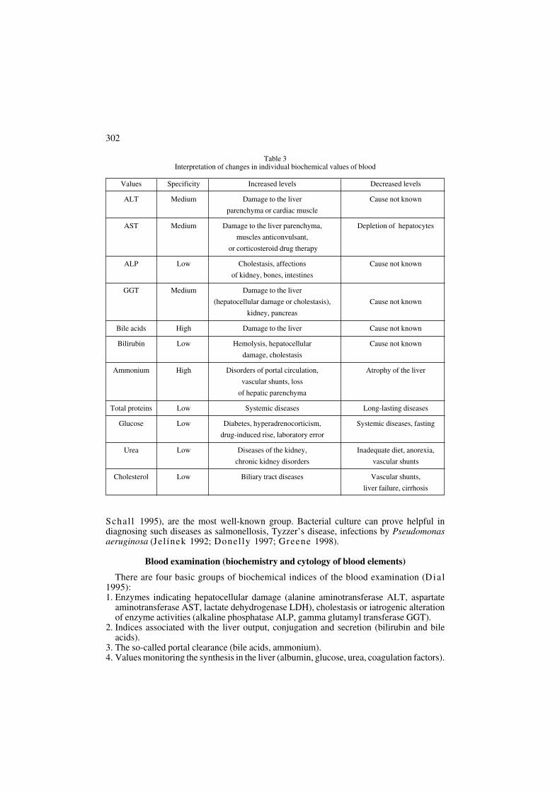
Blood examination (biochemistry and cytology of blood elements)
There are four basic groups of biochemical indices of the blood examination (Dia l1995):1. Enzymes indicating hepatocellular damage (alanine aminotransferase ALT, aspartate
aminotransferase AST, lactate dehydrogenase LDH), cholestasis or iatrogenic alterationof enzyme activities (alkaline phosphatase ALP, gamma glutamyl transferase GGT).
2. Indices associated with the liver output, conjugation and secretion (bilirubin and bileacids).
3. The so-called portal clearance (bile acids, ammonium).4. Values monitoring the synthesis in the liver (albumin, glucose, urea, coagulation factors).
Table 3Interpretation of changes in individual biochemical values of blood
Values Specificity Increased levels Decreased levels
ALT Medium Damage to the liver Cause not known
parenchyma or cardiac muscle
AST Medium Damage to the liver parenchyma, Depletion of hepatocytes
muscles anticonvulsant,
or corticosteroid drug therapy
ALP Low Cholestasis, affections Cause not known
of kidney, bones, intestines
GGT Medium Damage to the liver
(hepatocellular damage or cholestasis), Cause not known
kidney, pancreas
Bile acids High Damage to the liver Cause not known
Bilirubin Low Hemolysis, hepatocellular Cause not known
damage, cholestasis
Ammonium High Disorders of portal circulation, Atrophy of the liver
vascular shunts, loss
of hepatic parenchyma
Total proteins Low Systemic diseases Long-lasting diseases
Glucose Low Diabetes, hyperadrenocorticism, Systemic diseases, fasting
drug-induced rise, laboratory error
Urea Low Diseases of the kidney, Inadequate diet, anorexia,
chronic kidney disorders vascular shunts
Cholesterol Low Biliary tract diseases Vascular shunts,
liver failure, cirrhosis

ALT and AST
These enzymes are localised mainly in the cytoplasm of hepatocytes (Sparkes andGruffydd-Jones 1993). There are also isoenzymes of alanine aminotransferase (ALT) ofmitochondrial origin and aspartate aminotransferase (AST) is present also in the heartmuscle. The half-time of ALT in the rabbit is very short, i.e. only 5 hours, as compared tothe dog in which it varies from 45 to 60 hours (Meredi th and Rayment 2000). The levelof AST gets higher in cases of muscle tissue damage (Divers and Cooper 2000). Diffusereversible processes, such as for example hypoxia, cause the rise in the enzyme level that ishigher than in solitary lesions (hepatic abscesses). Final stages of liver diseases in mammalsare due to the depletion of hepatocytes accompanied by levels of enzymes that are withinlimits or only slightly elevated (Center 1993; Sparkes and Gruffydd-Jones 1993). Itis also necessary to keep in mind that long-lasting administration of anticonvulsant agentsand corticosteroids results in the liver enzyme level rise (Dial 1995).
Cholestatic enzymes
Alkaline phosphatase (ALP) and γ-glutamyltransferase (GGT) are the two mostcommonly used enzymatic markers of cholestasis in the clinical practice of small animals(Sparkes and Gruffydd-Jones 1993). ALP, however, is also localised in bones,kidneys, intestines, placenta and leukocytes. The main part of the serum ALP activity isinfluenced by the damage to the liver and bones or a drug-induced rise. The half-time of ALPproduced in the liver and bones in the cat amounts to 3 days. ALP produced in other organsystems is reduced to half within 6 minutes in dogs and 6 hours in cats (Center et al. 1992).The highest ALP activity is found on border membranes of bile ducts. High level of thisenzyme indicates stasis in the bile ducts. The level rises in cases of bone and, in someauthors´opinion, other organ damage as well (Lorenz and Cornel ius 1992).
GGT is produced in the liver, pancreas and kidney; the renal insoenzyme, however, is notcommonly detectable in the blood. Serum GGT is mainly of hepatic origin and increasedlevel indicates hepatocellular damage and stasis in bile ducts (Center 1993; Sparkes andGruffydd-Jones 1993; Meredi th and Rayment 2000). Acute liver damage in theblood serum of patients is manifested by the alteration of enzymes like AST, GGT, ALP,ALT and LDH. It is necessary to determine the level of creatine kinase (CK) and theconcentration of bile acid to differentiate from myopathies.
Indices associated with the liver output, conjugation, secretion and portal clearance
There are no simple and reliable tests demonstrating the functionality of the liverparenchyma. Serum bilirubin and bile acids are not dependent only on the functionality ofthe liver parenchyma; their concentrations are related also to the alteration of the biliary tract(Center 1993; Sparkes and Gruffydd-Jones 1993). The determination of serum bileacid concentration is used as one of the liver function tests. The result depends on threecomponents of the enterohepatic transport: passage of bile acids from the serum tohepatocytes, secretion of bile acids into the biliary system and re-circulation into the liverthrough the portal vascular system. Bile acids are produced in the liver, conjugated withtaurine or glycine and secreted into the bile (Clarenburg 1992b). Bile acids play a role inthe digestion and absorption of fat. The main bolus of bile acids is released in the duodenumand re-absorbed in the ileum. Only a small percentage of bile acids is excreted in faeces.Normal levels of bile acids are low and rise only after feeding. This effect is used in thediagnostics of hepatopathies when we measure their levels before and two hours afterfeeding (Center et al. 1985; Center and Baldwin et al. 1986). The determination of bileacids can be useful in animals having normal bilirubin levels in the serum (Dial 1995).
303

Increased bile acid levels together with other characteristics of hepatic functions indicateliver parenchyma damage. The measurement of bile acid levels, however, is not useful inthe differentiation of hepatocellular, vascular or cholestatic diseases.
Hyperbilirubinemia is caused by increased destruction of erythrocytes (pre-hepaticorigin), hepatocellular dysfunction (damage to the liver) or cholestasis (post-hepatic origin).The aetiology of hyperbilirubinemia can be further differentiated by the determination ofconjugated and unconjugated bilirubin levels, in small mammals, however, even these testsdo not result in absolute diagnosis. In the ferret, for example, increased bilirubin level is notreliably for signified liver damage and jaundice is found only occasionally (Hoefer 1992).
The concentration of serum ammonia in the blood is a very reliable marker of hepaticfunctions (Sparkes and Gruffydd-Jones 1993). The level of ammonium in the serumis determined by the absorption from the small intestine where it is formed by proteinbreakdown. It is mainly absorbed and through the portal system transported to thehepatocytes, metabolised to urea and then excreted by the kidney (Levei l le-Webster2000). Disorders in the portal circulation, intra- or extra-hepatic shunts as well as functionalchanges of liver parenchyma result in the rise of the ammonium in the blood serum and, inits final effect, neurological signs (Center 1993; Bunch 1998a). As far as the methodsare concerned, it is crucial for the ammonium measurement that the concentration bedetermined within 15 to 30 minutes after sampling. Heparin is used for the blood collection,the sample is cooled immediately and centrifuged for the purpose of separation oferythrocytes (Dial 1995).
Indices monitoring the synthesis in the liver
The liver is the primary source of many serum components: albumin, glucose, urea,cholesterol and the majority of coagulation factors. Their concentrations in the blood serumdecrease only when the liver gets seriously damaged (Dial 1995). The glucose level isusually influenced by serious liver damage and other systemic disorders. It is only oforientation importance as an indicator of liver affection. Hypoproteinemia in the liverdisease is associated with decreased production of albumin. Even this value is of orientationimportance because it indicate only acute and chronic diseases. Chronic diseases result inhypoalbuminemia. Concurrent blood loss with hemostasis can, nevertheless, result inhypoalbuminemia even in cases of acute liver failure. In chronic inflammation cases,however, there are levelled concentrations of proteins due to hypergammaglobulinemia(Dial 1995). Levels of urea in the blood serum can be influenced by the functionality ofliver parenchyma as well as the intake of proteins in the food. Anorectic animals and animalsfed low-protein diet have a low urea blood level in comparation to reference values(Jergens 1997). Anorectic dogs with normal hepatic functions can have their urea serumlevel low because of low intake of proteins; nevertheless, a rise in the plasmatic urea is notexceptional. Dogs suffering from some affection to the functionality of liver parenchymahave normal serum urea levels if they are at the same time dehydrated or their kidneyfunctions are impaired. Decreased production of urea is one of the causes of polyuria owingto the drop in the osmotic gradient in the kidney cortex (Dial 1995).
Serum cholesterol levels are variable in diseases of the liver. Cholesterol is excreted fromthe organism primarily through the biliary system and its rise is usually associated withdiseases of this system. Hypocholesterolemia is associated with a long-lasting liver disease.The reason for this is the drop in the production or absorption from the intestines or higherconversion to bile acids. The most frequent liver disorder associated withhypocholesterolemia is the portosystemic shunt, in which increased conversion to bile acidsis the primary mechanism (Levei l le-Webster 2000).
The liver is the source of most proteins taking part in the blood coagulation (fibrinogen,
304

prothrombin, factors V, VII, IX, X, XI, and XII together with factors II, VII, IX and X,which are K-vitamin dependent) and blood coagulation inhibitors (antithrombin III,plasminogen, α2-macroglobulin, α2-antiplasmin) (Feldman 1980). Contrary to humanmedicine blood coagulation tests are only used to a limited extent for the monitoring ofhepatic functions.
Cytology of the blood
There are changes in the blood differential count in diseases of the liver. Morphologicalchanges of erythrocytes associated with the liver diseases include microcytosis,acanthocytosis, schistocytes and Heinz bodies (Levei l le-Webster 2000). Mostinflammatory diseases of the organism are associated with leukocytosis; septic cases, on theother hand, are accompanied by leukopenia. Bacterial infections cause neutrophilia witha left-shift and a higher proportion of toxic neutrophils as well as monocytes (Center 1998).For the examination of blood smears it is necessary to notice the prospective presence ofdevelopmental stages of parasites. In the mouse and rat we can find the protozoanHepatozoon muris, which infects cells of the liver, spleen, bone marrow and leukocytes(Jel ínek 1992).
Serology of the blood collected is used in a routine way in experimental laboratories forthe purpose of health state screening. It is very precise and the centre of its use lies in thediagnostics of viral infections (Jel ínek 1992; Jel ínek et al. 1993; Gabrisch 1995;Pearson and Gorham 1998). These examinations are not routinely used in a commonclinical practice dealing with small pet mammals.
Invasive diagnostic procedures Biopsy and endoscopic examination
Biopsy is a method aiding in the determination of a precise diagnosis and diseaseprognosis (Konrád 1989). Liver biopsy is indicated in cases of abnormal enzymaticactivities associated with liver functions and their persistence for as long as 30 days andmore, hepatomegaly of undetermined origin, liver complications of systemic diseases,suspected neoplasia, therapy response determination, disease progression (Hoefer 1992;Kerwin 1995). A common finding mentioned in association with histology of liverbiopsies from small mammals is the liver steatosis. It is a result of general illness (obesity,fasting, infection, etc.) influencing the morphology of the liver and subsequently itsphysiological functions. Carl ton and McGavin (1995) defined the liver steatosis asexcessive accumulation of lipids in hepatocytes. The presence of fat in the liver does nothave to be necessarily a pathologic finding. Intrahepatic lipid deposition is to some extentquite physiological. It is clear that the diagnostics of liver steatosis is not only based on thedetermination of lipids in the liver tissue, but also on their precise localisation andquantification. In small mammals we can utilise most of the methods used in the cat and dog.Following methods can be considered in small mammals: fine-needle aspiration biopsy,percutaneous biopsy, biopsy under the guidance of ultrasonography, biopsy underendoscopic / otoscopic guidance, biopsy at the time of exploratory laparotomy.
Fine-needle aspiration biopsy
The method is suitable for the cytology of the liver tissue. If there are diffuse processesaffecting the liver tissue (steatosis, steroid-induced hepatopathy, diffuse neoplasia), thismethod has a high diagnostic value (Rebar 1989b). The animal is positioned in the rightlateral recumbency for the sampling. The needle is inserted through a spot caudal tocartilago xiphoidea, it goes along the rib arch at a 45-degree angle to the diaphragm. The
305

306
probability of liver tissue penetration rises in animals suffering from hepatomegaly. Thereare minimum equipment requirements (22 – 25 G needles, 5 – 10 ml syringes and slides forthe smear) (Rebar 1989c). Complications in patients the size of a dog are relativelyinfrequent (Teske 1998). There can be bleeding in serious cases of coagulopathies (anti-coagulant-agent intoxications, disseminated intravascular coagulation) or injury to largevessels. We have encountered this problem in our practice. It is therefore necessary to checkthe patient 12 and 24 hours after the procedure. Sampling other tissues is yet anothercomplication. Small mammals have always to be sedated.
Percutaneous biopsy
This technique is common in veterinary medicine. Indications for the use of thistechnique are the same as in the fine-needle aspiration biopsy (diffuse processes). Specialbiopsy needles are used (Tru-Cut system, gun system, Kerwin 1995). Contrary to the fine-needle aspiration biopsy, using this method we obtain larger samples providing enoughmaterial both for the cytology and histology which enhances the precision of the diagnosisup to 80 to 90% (Hardy 1990). The patient requires general anaesthesia for the preventionof whatever movements. The animal is restrained in dorsal recumbency with 30 to 45%rotation to the right. This position minimises the risk of injury to the gall bladder andsubsequent peritonitis. There is no such danger in the rat having no gall bladder (Popeskoet al. 1990b). The area outlined by cartilago xiphoidea, the left rib arch and umbilicus isclipped and surgically prepared. Strict aseptic measures are essential (Hi t t et al. 1992). Theneedle is inserted so as to pass through the abdominal wall and then the biopsy cannula goes
Table 4Cytology and biochemical indices of blood in small mammals (Knotková and Knotek 2000)
Values Units Hamster Mouse Rat Chinchilla Rabbit Guinea pig
Erythrocytes T/l 5 - 9.2 7.9 - 10.1 5.4 - 8.5 6.6 - 10.7 5.1 - 7.9 3.2 - 8.0
Haemoglobin g/l 146 – 200 110 - 145 115 - 160 117 - 135 100 - 174 100 - 172
Haematocrit l/l 0.46 - 0.52 0.37 - 0.46 0.37 - 0.49 0.38 0.33 - 0.50 0.32 - 0.50
Leukocytes G/l 5 – 10 5 - 13.7 4 - 10.2 7.6 - 11.5 5.2 - 12.5 5.5 - 17.5
Neutrophils G/l 1.5 - 3.5 0.4 - 2.7 1.3 - 3.6 0.7 – 6.0 0.6 – 9.9 1.1 –4.0
Lymphocytes G/l 6.1 – 7 7.1 - 9.5 5.6 - 8.3 1.6 – 6.8 2.6 – 11.2 2.1 - 7
Monocytes G/l 0 – 1.0 0 – 0.9 0 – 0.6 0 – 0.4 0 – 0.9 0 – 0.5
Eosinophils G/l 0 – 0.4 0 – 0.5 0 – 0.7 0 – 0.7 0 – 0.2 0 – 0.4
Basophils G/l 0 – 0.1 0 – 0.1 0 – 0.1 0 – 0.1 0 – 0.7 0 – 0.3
TP g/l 64 – 73 42 - 60 63 - 86 50 - 60 54 - 83 42 - 68
Albumin g/l 32 – 37 21 - 34 33 - 49 25 - 42 24 - 46 21 - 39
Globulin g/l 27 – 42 18 - 82 24 - 39 18 - 25 15 - 28 17 - 26
Glucose mmol/l 3.60 - 4.05 9.66 - 18.59 4.72 - 7.33 3.33 - 6.66 4.16 - 8.60 3.33 - 6.94
Urea mmol/l 6.99 - 9.99 5.66 - 9.66 5.33 - 8.99 1.67 - 4.16 2.16 - 4.83 1.50 - 5.25
Creatinine mmol/l 53 – 89 26.5 – 61.8 17.5 – 70.8 None 44.2 - 221 53.0 - 194.5
Cholesterol µmol/l 4.71 - 6.14 1.27 - 2.49 1.19 - 2.38 1.04 - 2.59 0.26 - 2.07 0.41 - 1.11
AST µkat/l 0.88 - 2.07 0.92 - 4.18 0.65 - 1.53 0.25 - 0.75 0.23 - 1.88 0.43 - 1.13
ALT µkat/l 0.35 - 0.83 0.47 - 3.07 0.28 - 0.83 0.17 - 0.58 0.80 - 1.33 0.42 - 0.98
ALP µkat/l 0.13 - 0.30 0.47 - 1.57 0.65 - 3.60 0.05 - 0.20 0.07 - 0.27 0.92 - 1.80

307
1 to 2 cm deep (depending on the size of the animal). There was described a variation tothis method called “key-hole” in the dog and cat. The first thing to do using this method isto make a small incision through the abdominal wall. It is advantageous that the cannula isthen inserted directly into the liver tissue (Day 2000). This modification can be quitesuccessfully utilised in small mammals. The surgeon is able to stabilise the liver lobe wheninserting the needle. Complications of these techniques include: inadequate sedation of thepatient and movement of the patient during sampling, inadequate reaction of the patient onsedation or anaesthesia, bleeding, presence of greater quantity of gall in the sample,contamination by puss following abscess puncture, sampling other tissue, shock aftersampling (Kerwin 1995).
There is a danger of injury to the diaphragm and thoracic cavity organs or large vesselsand bleeding in patients the size of a mouse. We, therefore, recommend checking thepatient during 12 to 24 hours after the procedure. Regarding the above-mentioned hazards,in our experience it is better to use other safer methods for the sampling (biopsy underendoscopic guidance, biopsy using an otoscope for the guidance, biopsy duringlaparotomy). These methods considerably reduce the risk of injuring the patient duringsampling.
Biopsy under ultrasonographic guidance
Examination using ultrasonography before the biopsy provides information on the hepatictissue state, identification of focal lesions (neoplasia, cysts, abscesses) or even generalchanges of the liver (fibrosis, steatosis). The method allows to control the movement of thebiopsy needle and thus gives precision to the sampling (Lévei l lé et al. 1993). The patienthas to be deep sedated or is given general anaesthesia. The area of insertion of the biopsyneedle is prepared in a similar way as it is explained for the percutaneous biopsy.Complications are less prevalent owing to the visualisation of sampling. We employ thismethod practically in small mammals the size of a rabbit or a ferret.
Biopsy under endoscopic guidance
Endoscopy is the procedure suitable for the exact evaluation of liver surface and guidedsampling for the cytology and histology. It requires instrumentation and experience of thepractitioner with surgical procedures in small mammals. As diagnostic biopsies are takenmainly from patients suffering from suspected hepatopathies, it is necessary to select a safeand reliable system of general anaesthesia which influences the liver and kidney functionsto a minimum extent. Inhalation anaesthesia using isoflurane is the most suitable system inthis respect. Endoscopy is the least invasive method because there is no need for a largeincision to visualise the liver tissue in situ. It provides the possibility to take a biopsyspecimen directly from the affected liver part, focal lesions or diffuse changes (Jones1989). It is a good alternative to laparotomy. The veterinary surgeon is able to evaluate thestate of other organs such as the gall bladder, small and large intestines, pancreas at the sametime (Twedt 1999). Linea alba (Meredi th and Rayment 2000) is the most commonlyselected laparotomy access line in small mammals. The patient is restrained in dorsalrecumbency and the area of entry is prepared for a surgical incision. Abdominal cavityinsufflation is recommended for the procedure. The examination is contra-indicated inpatients in which the disease can be surgically solved through laparotomy or in animals indanger of life. Expensive instrumentation, longer duration of the procedure and thus higherload to the patient are the disadvantages of this diagnostic approach (Day 2000). Thepressure of the insufflated gas can decrease the minute respiratory volume; assistedventilation using the breathing bag is therefore essential (Kerwin 1995).

308
Bunch et al. (1985) described a procedure using a sterile otoscope instead of anendoscope. We are employing this modification successfully in small mammals.
Biopsy during exploratory laparotomy
Exploratory laparotomy is indicated in cases of diseases which have to be surgicallytreated, in extra-hepatic biliary tract obstructions, and neoplasia affecting only one liverlobe. The advantages of this method without any doubt include direct visualisation of allorgans in the abdomen and the possibility to obtain samples from a number of organs (lymphnodes can be sampled in suspected cases of neoplasia) and immediate surgical treatment(Day 2000). The method is associated with a considerable load of the patient (it is essentialthat the patient be clinically evaluated and the hazards of the procedure considered). Thebiopsy specimen can be sampled using a punch biopsy technique, partial lobectomy anda ligature (Kerwin 1995).
Utilisation of the above-mentioned methods in minute mammals
Without any problems, we used the above-mentioned methods in the diagnostics ofhepatopathies in rabbits, ferrets, adult guinea pigs and rats (Table 5). On the other hand,utilisation of these methods in hamsters, Djungarian hamsters or mice is rather questionable,but possible. History and clinical examination often lead the veterinarian to consider a liverdamage. Radiography reveals liver enlargement and prospective masses. It is, however,considerably limited by the size of the patient. This limitation concerns alsoultrasonography. It must be admitted that computer tomography, magnetic resonance andscintigraphy belong to progressive methods which find their way to utilisation in theveterinary medicine, their use in small mammals is, however, still limited. Examinations of
Table 5Methods of liver parenchyma examination and their utilisation in the clinical practice engaged with small mammals
MethodsUtilisation in small mammals
Ferret Hamster Mouse Rat Chinchilla Rabbit Guinea pig
Liver palpation Yes Limited No Limited Yes Yes Limited
Blood sampling Yes Limited Limited Limited Yes Yes Limited
Abdominocentesis Yes Yes Yes Yes Yes Yes Yes
Coprology Yes Yes Yes Yes Yes Yes Yes
RadiographicYes Yes Limited Yes Yes Yes Yes
examination
Ultrasonography Yes No No Limited Limited Yes Limited
ComputerNon-utilisable
tomography
Scintigraphy Non-utilisable
Magnetic resonance Inaccessible but utilisable
Fine-needleYes Limited Limited Limited Limited Yes Limited
aspiration biopsy
Percutaneous biopsy Yes Limited No Limited Limited Yes Limited
Endoscopic biopsy Yes Yes Limited Yes Yes Yes Yes
Laparotomy Yes Yes Yes Yes Yes Yes Yes

309
abdominal effusions and coprology are much more efficient. There are problems with bloodcollection and subsequent haematology and biochemistry owing to insufficient bloodvolume. Liver biopsy sampling can be a life-threatening procedure requiring experience andcare. This procedure, nevertheless, provides enough information on the current state of theliver parenchyma. In our experience and practice, the described modifications of liverbiopsy are essential in the diagnostics of hepatopathies in small mammals.
Posouzení v˘znamu jednotliv˘ch metod klinické diagnostiky hepatopatiíu drobn˘ch savcÛ
MoÏnosti intravitálního prÛkazu naru‰ené funkce jater u drobn˘ch savcÛ jsou doposudvyuÏívány minimálnû. Pfiitom je nutnost vãasné diagnostiky hepatopatií u tûchto pacientÛaktuální. Pfiedkládaná práce shrnuje diagnostické postupy a je zamûfiena na posouzení jejichpraktického vyuÏití u drobn˘ch savcÛ. Nezastupitelnou roli hraje posouzeníanamnestick˘ch dat a dÛkladné palpaãní vy‰etfiení jater s odbûrem tekutiny z dutiny bfii‰níza úãelem laboratorní anal˘zy. Anal˘za biochemick˘ch parametrÛ krve zahrnuje sledováníenzymÛ indikujících hepatocelulární po‰kození (ALT, AST, LDH), cholestazu nebo alteraciaktivit enzymÛ (ALP, GMT), parametrÛ monitorujících jaterní syntézu (albumin, glukóza,urea, koagulaãní faktory), úroveÀ konjugace a sekrece (bilirubin a Ïluãové kyseliny) a tzv.portal clearens (Ïluãové kyseliny, amoniak). Pfii potvrzení ascitu v dutinû bfii‰ní je dÛleÏitébliωí urãení jeho charakteru. Pfiesné posouzení stavu jater umoÏÀují cytologickáa histologická vy‰etfiení bioptátÛ. Pro drobné savce velikosti králíka, fretky, dospûléhomorãete a potkana jsou vhodn˘mi metodami jaterních biopsií: aspiraãní tenkojehelná,perkutánní, pod kontrolou ultrasonografu, pod kontrolou endoskopu nebo otoskopua biopsie pfii probatorní laparotomii. U pacientÛ velikosti kfieãka, kfieãíka dÏungarskéhonebo my‰i je indikace tûchto metod diskutabilní, neboÈ pfiedstavují váÏné riziko ohroÏeníÏivota. Pfiesto mají uvedené modifikace biopsií jater pfii diagnostice hepatopatií drobn˘chsavcÛ rozhodující v˘znam.
Acknowledgements
This paper originated as a part of the research project of the Ministry of Education, Youth and PhysicalEducation of the Czech Republic (No. 161/700002). Authors wish to thank Dagmar Kare‰ová, Vladimír Jekl andRoman ·ebesta for their technical assistance in the anaesthesia of patients, biological material sampling andsmall animal breeding.
References
ANDERSON, N.V. 1992: Excessive fluid in abdomen: effusions. In: ANDERSON, N.V.: Veterinarygastroenterology. Lea and Febiger, London, pp. 382-388
ANGULO, P., PEARCE, D.H., JOHNSON, C.D., HENRY, J.J., LARUSSO, N.F., PETERSEN, B.T., LINDOR,K.D. 2000: Magnetic resonance cholangiography in patients with biliary disease: its role in primary sclerosingcholangitis. J. Hepatol. 33: 520-527
BOOTHE, H.W., BOOTHE, D.M., KOMKOV, A., HIGHTOWER, D. 1992: Use of hepatobiliary scintigraphy inthe diagnosis of extrahepatic biliary obstruction in dog and cats: 25 cases (1982 - 1989). J. Am. Vet. Med. Assoc.201: 134-141
BROWN, S.A. 1992: Basic anatomy, physiology, and husbandry. In: HILLYER, E.V. QUESENBERRY, K.E.:Ferrets, rabbits, and rodents clinical medicine and surgery. W.B. Saunders, Philadelphia, pp. 3-13
BUNCH, S.E. 1998a: Hepatobiliary and exocrine pancreatic disorders. In: NELSON, R.W., COUTO, C.G.: Smallanimal internal medicine. 2th ed. Mosby, St. Louis, pp. 475-570
BUNCH, S.E. 1998b: Scintigraphy. In: NELSON, R.W., COUTO, C.G.: Small Animal Internal Medicine. Mosby,Inc., Missouri, pp. 501-502
BUNCH, S.E., POLAK D.M., HORBUCKLE W.E. 1985: A modified laparoscopic approach for liver biopsyin dogs. J. Am. Vet. Med. Assoc. 187: 1032-1035
CARLTON, W.W., MCGAVIN, M.D. 1995: Thompson’s special veterinary pathology. Mosby, St Louis, pp. 88 – 89CENTER, S.A. 1993: Disorders of the hepatobiliary system. In: WILLS, J., WOLF, A.: Handbook of feline
medicine. Pergamon Press, Oxford, pp. 175-192
CENTER, S.A. 1998: Hepatobiliary infections. In: GREENE, C.E.: Infectious diseases of the dog and cat. 2th ed.W.B. Saunders, Philadelphia, pp. 615-625
CENTER, S.A., BALDWIN, B.H., HOLLIS, N.E., TENNANT, B.C. 1985: Bile acid concentration in the diagnosisof hepatobiliary disease in the dog. J. Am. Vet. Med. Assoc. 187: 935-940
CENTER, S.A., BALDWIN, B.H., HOLLIS, N.E., TENNANT, B.C. 1986: Bile acid concentration in the diagnosisof hepatobiliary disease in the cat. J. Am. Vet. Med. Assoc. 189: 891-896
CENTER, S.A., SLATER, M.R., MANWARREN, T., PRYMAK, K. 1992: Diagnostic efficacy of serum alcalinephosphatase and -glutamyltransferase in dogs with histologically confirmed hepatobiliary disease: 270 cases(1980 - 1990). J. Am. Vet. Med. Assoc. 201: 1258-1264
CLARENBURG, R. 1992a: Lipid metabolism. In: CLARENBURG, R.: Physiological chemistry of domesticanimals. Mosby-Year Book, Inc., St. Louis, pp. 292–333
CLARENBURG, R. 1992b: Gastrointestinal functions. In: CLARENBURG, R.: Physiological chemistry ofdomestic animals. Mosby-Year Book, Inc., St. Louis, pp. 203-217
DAY D.G. 2000: Indications and techniques for liver biopsy. In: ETTINGER, S.J., FELDMAN, E.C.: Textbookof veterinary internal medicine. 5th ed. Vol 2, W.B. Saunders, Philadelphia, pp. 1294-1298
DIAL, S.M. 1995: Clinicopathologic evaluation of the liver. Vet. Clin. North Am. Small Anim. Pract. 25: 257-273DIVERS, S.J., COOPER, J.E. 2000: Hepatic lipidosis. Seminars in Avian and Exotic Pet Medicine. 9: 153-164DONELLY, T.M. 1997: Disease problems of small rodents. In: HILLYER, E.V., QUESENBERRY, K.E.: Ferrets,
rabbits, and rodents - clinical medicine and surgery. W.B. Saunders, Philadelphia, pp. 307-327EGEN, H., ERNST, H. 1995: Chinchilla. In: GABRISCH, K., ZWART, P.: Krankheiten der Heimtiere. 3th ed.
Schlütersche, Hannover, pp. 173-196FELDMAN, B.F. 1980: Clinical Pathology of the Liver. In: KIRK, R.W. et al.: Current veterinary therapy VII.
W.B. Saunders, Philadelphia, pp. 875-885GABRISCH, K. 1995: Frettchen und Marder. In: GABRISCH, K., ZWART, P.: Krankheiten der Heimtiere. 3th ed.
Schlütersche, Hannover, pp. 235-274GIEBLER, D. 1995: Hörnchen. In: GABRISCH, K., ZWART, P.: Krankheiten der Heimtiere. 3th ed. Schlütersche.
Hannover, pp. 217–233GREENE, C.E. 1998: Salmonellosis. In: GREENE, C.E.: Infectious diseases of the dog and cat. 2th ed. W.B.
Saunders, Philadelphia, pp. 235 – 240HARDY, H. 1990: The percutaneous approach. Vet. Med. Rep. 2: 192-194HITT, M.E., HANNA, P., SINGH, A. 1992: Percutaneous transabdominal hepatic needle biopsies in dogs. Am. J.
Vet. Res. 53: 785-787HOEFER H.L. 1992: Liver disease. In: HILLYER, E.V. QUESENBERRY, K.E.: Ferrets, rabbits, and rodents
clinical medicine and surgery. W.B. Saunders, Philadelphia, pp. 32-33HOUSE, J.K., SMITH, B.P., VANMETRE, D.C., FECTEAU, G.,CRAYCHEE, T., NEVES, J. 1992: Ancillary
tests for assessment of the ruminant digestive system. Vet. Clin. North Am. Food Anim. Pract. 8: 203–232HUBNER, G.H., STEUDEL, N., KLEBER, G., BEHRMANN, C., LOTTERER, E., FLEIG, W.E. 2000: Hepatic
arterial blood flow velocities: assessment by transcutaneous and intravascular Doppler sonography. J. Hepatol.32: 893-899
JELÍNEK, F. 1992: Játra a v˘vodní cesty Ïluãové. In: JELÍNEK, F.: Úvod do patologie laboratorních zvífiat.Stanislav Hojek, Praha, pp. 77-95
JELÍNEK, F., ALDOVÁ, E., LÁVIâKOVÁ, M., ·KARDOVÁ, O., ZAJÍâEK, D. 1993: Nemoci laboratorníchzvífiat. V·VF, Brno, 120 p.
JERGENS A.E. 1997: Gastrointestinal disease and its management. Vet. Clin. North Am. Small Anim. Pract. 27:1373-1402
JONES, B.D. 1989: The use of laparoscopy to obtain liver biopsies. In: AAHA´s 56th Annual meeting proceedings,7. 14. 5. 1989, St. Louis, pp. 149-150
KAYA, M., ANGULO, P., LINDOR, K.D. 2000: Overlap of autoimmune hepatitis and primary sclerosingcholangitis: an evaluation of a modified scoring system. J. Hepatol. 33: 537-542
KELNER, P. 1999a: Poãítaãová tomografie. In: KELNER, P. a kol.: Vnitfiní lékafiství: Zobrazovací metody.Karolinum, Praha, p. 526
KELNER, P. 1999b: Magnetická rezonance. In: KELNER, P. a kol.: Vnitfiní lékafiství: Zobrazovací metody.Karolinum, Praha, p. 527
KERWIN, S.C. 1995: Hepatic aspiration and biopsy techniques. Vet. Clin. North Am. Small Anim. Pract. 25: 275-291
KLIME·, J. 1997: Laboratorní diagnostika onemocnûní jater. Sborník referátÛ Klinická interpretace laboratorníchvy‰etfiení u psa a koãky, âAVLMZ, Hradec Králové, pp. 16-20
KNOTEK, Z. 1999: Infekãní peritonitida koãek. In: SVOBODA, M., POSPÍ·IL, Z. et al.: Infekãní nemoci psaa koãky, âAVLMZ,Brno, pp. 196-210
KNOTKOVÁ, Z., KNOTEK, Z., 2000: Fyziologické hodnoty u drobn˘ch savcÛ. Noviko, Brno, 69 p.KONRÁD, J. 1989: Intravitální punkce (biopsie) jater. In: KONRÁD, J.: Nemoci koÏe‰inov˘ch zvífiat. SZN, Praha,
pp. 45-46
310

LÉVEILLÉ, R., PARTINGTON, B.P., BILLER, D.S., MIYABAYSHI, T. 1993: Complication after ultrasound -guided biopsy of abdominal structures in dogs and cats: 246 cases (1984 - 1991). J. Am. Vet. Med. Assoc. 203:413-415
LEVEILLE-WEBSTER, C.R. 2000: Laboratory diagnosis of hepatobiliary diseases. In: ETTINGER, S.J.,FELDMAN, E.C.: Textbook of veterinary internal medicine. 5th ed. Vol 2. W.B. Saunders, Philadelphia, pp.1277-293
LOFSTED, J., KOBLIK, D., JAKOWSKI, R.M., McMILLAN, M.C., ENGELKING, L.R. 1988: Use ofhepatobiliary scintigraphy to diagnose bile duct atresia a lamb. J. Am. Vet. Med. Assoc. 193: 95-98
LORENZ, M.D., CORNELIUS, L.M. 1992: Abdominal distention. In: LORENZ, M.D. CORNELIUS, L.M.:Small animal medical diagnosis. J.B. Lippincott, Philadelphia, pp. 73-77
MEREDITH, A., RAYMENT, L. 2000: Liver disease in rabbits. Seminars Avian and Exotic Pet Medicine. 9: 146-152
MILES, K. 1997: Imaging abdominal masess. Vet. Clin. North Am. Small Anim. Pract. 27: 1403-1431NAPIER, R.A.N. 1969: Králíci. In: LANE-PETTER W.: Laboratorní zvífiata. ACADEMIA, Praha, pp. 425- 471PEARSON, R.C., GORHAM, J.R. 1998: Viral disease models. In: FOX, J.G.: Biology and diseases of the ferret.
2th ed. Williams a Wilkins, Baltimore, pp. 487-497PERMAN, V. 1989: Examination of the inflammatory exudate. In: AAHA’s 56th Annual meeting proceedings, 7.
14. 5. 1989, St. Louis, pp. 154-157POPESKO, P., RAITOVÁ, V., HORÁK, J. 1990a: Atlas anatómie mal˘ch laboratornych zvierat 1. Príroda,
Bratislava, 256 p.POPESKO, P., RAITOVÁ, V., HORÁK, J. 1990b: Atlas anatómie mal˘ch laboratornych zvierat 2. Príroda,
Bratislava, 253 p.REBAR, A.H. 1989a: Evaluation of pleural, peritoneal, and pericardial effusions. In: AAHA’s 56th Annual meeting
proceedings, 7. 14. 5. 1989, St. Louis, pp. 162-168REBAR, A.H. 1989b: Diagnostic cytology as an aid to the evaluation of hepatic disease. In: AAHA’s 56th Annual
meeting proceedings, 7. 14. 5. 1989, St. Louis, pp. 169-171REBAR, A.H. 1989c: Diagnostic cytology I: Collection techniques. In: AAHA’s 56th Annual meeting proceedings,
7. 14. 5. 1989, St. Louis, pp. 151-153ROTHUIZEN, J., MEYER, H.P. 2000: History, physical examination, and signs of liver disease. In: ETTINGER,
S.J., FELDMAN, E.C.: Textbook of veterinary internal medicine. 5th ed. Vol. 2. W.B. Saunders, Philadelphia,pp. 1272-1277
ROTHUIZEN, J., Van den BROM, W.E. 1990: Quantitative scintigraphy as a measure of bile flow in dogs withcholestatic disease. Am. J. Vet. Res. 51: 253-256
SCHALL, H. 1995: Kaninchen. In: GABRISCH, K., ZWART, P.: Krankheiten der Heimtiere. 3th ed. Schlütersche,Hannover, 3.ed., pp. 1-45
SEVELIUS, E. ANDERSSON, M. 1995: Serum protein electrophoresis as a prognostic marker of chronic liverdisease in dogs. Vet. Rec. 137: 663–667
SPARKES, A., GRUFFYDD-JONES, T.J. 1993: Laboratory diagnostic aids. In: WILLS, J., WOLF, A.: Handbookof feline medicine. Pergamon Press, Oxford, pp. 91-112
TESKE, E. 1998: Klinická cytologie v medicínû zvífiat chovan˘ch ze záliby. In: Sborník 6. konference âAVLMZ,Onkologie mal˘ch zvífiat, 17 - 18.10.1998, pp. 31-35
TWEDT, D.C. 1999: Laparoscopy of the liver and pancreas. In: TAMS, T.R.: Small animal endoscopy. Mosby, St.Luis, pp. 409-418
YEAGER, A.E. 1992: Accuracy of ultrasonography in the detection of severe hepatic lipidosis in cats. Am. J. Vet.Res. 53: 597-599
311
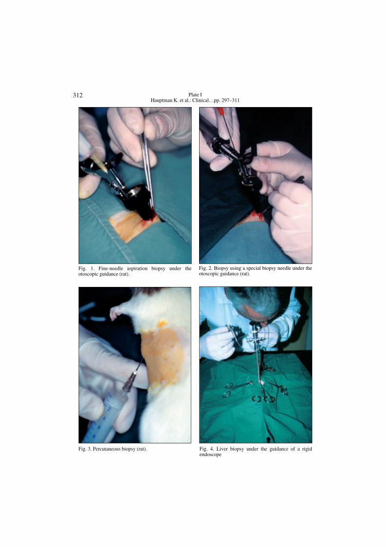
312 Plate IHauptman K. et al.: Clinical…pp. 297–311
Fig. 1. Fine-needle aspiration biopsy under theotoscopic guidance (rat).
Fig. 2. Biopsy using a special biopsy needle under theotoscopic guidance (rat).
Fig. 3. Percutaneous biopsy (rat). Fig. 4. Liver biopsy under the guidance of a rigidendoscope
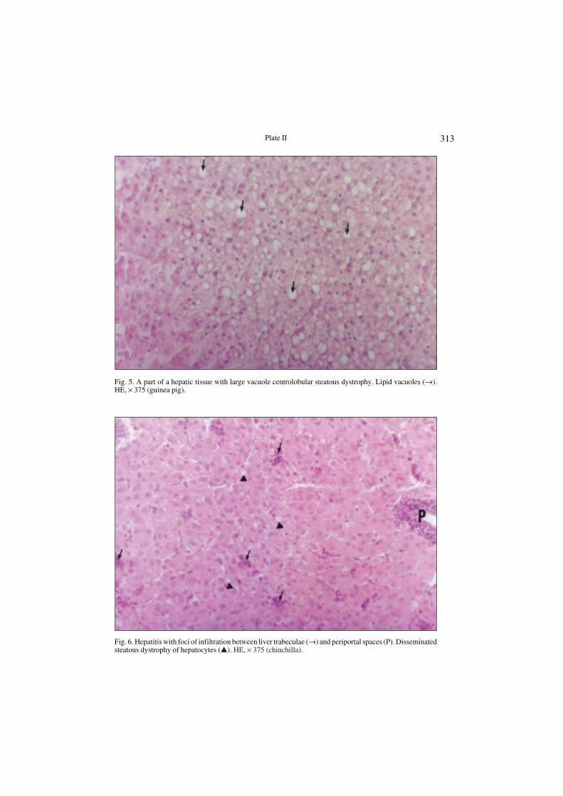
313Plate II
Fig. 5. A part of a hepatic tissue with large vacuole centrolobular steatous dystrophy. Lipid vacuoles (→).HE, × 375 (guinea pig).
Fig. 6. Hepatitis with foci of infiltration between liver trabeculae (→) and periportal spaces (P). Disseminatedsteatous dystrophy of hepatocytes (�). HE, × 375 (chinchilla).